Guidelines for the use and interpretation of assays for ...
215
EDITORIAL Guidelines for the use and interpretation of assays for monitoring autophagy (2 nd edition) Daniel J Klionsky 1766,1771 *, Kotb Abdelmohsen 851 , Akihisa Abe 1253 , Md Joynal Abedin 1784 , Hagai Abeliovich 428 , 5 Abraham Acevedo Arozena 800 , Hiroaki Adachi 1822 , Christopher M Adams 1690 , Peter D Adams 58 , Khosrow Adeli 2010 , Peter J Adhihetty 1645 , Sharon G Adler 710 , Galila Agam 68 , Rajesh Agarwal 1607 , Manish K Aghi 1556 , Maria Agnello 1848 , Patrizia Agostinis 673 , Patricia V Aguilar 1987 , Julio Aguirre-Ghiso 795,797 , Edoardo M Airoldi 90,425 , Slimane Ait-Si-Ali 1394 , Takahiko Akematsu 2039 , Emmanuel T Akporiaye 1112 , Mohamed Al-Rubeai 1412 , Guillermo M Albaiceta 1311 , Chris Albanese 366 , Diego Albani 569 , Matthew L Albert 524 , Jesus Aldudo 129 , Hana Alg€ ul 1179 , Mehrdad Alirezaei 1213 , 10 Iraide Alloza 650,899 , Alexandru Almasan 207 , Maylin Almonte-Becerril 531 , Emad S Alnemri 1228 , Covadonga Alonso 552 , Nihal Altan-Bonnet 859 , Dario C Altieri 1220 , Silvia Alvarez 1516 , Lydia Alvarez-Erviti 1413 , Sandro Alves 108 , Giuseppina Amadoro 871 , Atsuo Amano 942 , Consuelo Amantini 1573 , Santiago Ambrosio 1477 , Ivano Amelio 767 , Amal O Amer 929 , Mohamed Amessou 2122 , Angelika Amon 737 , Zhenyi An 1557 , Frank A Anania 294 , Stig U Andersen 6 , Usha P Andley 2111 , Catherine K Andreadi 1711 , Nathalie Andrieu-Abadie 509 , Alberto Anel 2058 , David K Ann 59 , 15 Shailendra Anoopkumar-Dukie 391 , Manuela Antonioli 843,1911 , Hiroshi Aoki 1813 , Nadezda Apostolova 1432 , Saveria Aquila 1519 , Katia Aquilano 1900 , Koichi Araki 295 , Eli Arama 2131 , Agustin Aranda 460 , Jun Araya 599 , Alexandre Arcaro 1491 , Esperanza Arias 27 , Hirokazu Arimoto 1241 , Aileen R Ariosa 1770 , Jane L Armstrong 1956 , Thierry Arnould 1795 , Ivica Arsov 2153 , Katsuhiko Asanuma 684 , Valerie Askanas 1949 , Eric Asselin 1891 , Ryuichiro Atarashi 805 , Sally S Atherton 372 , Julie D Atkin 724 , Laura D Attardi 1146 , Patrick Auberger 1809 , Georg Auburger 382 , Laure Aurelian 1748 , 20 Riccardo Autelli 2021 , Laura Avagliano 1043,1777 , Maria Laura Avantaggiati 367 , Limor Avrahami 1181 , Suresh Awale 2015 , Tiziana Bachetti, Jonathan M Backer 29 , Dong-Hun Bae 1959 , Jae-sung Bae 686 , Ok-Nam Bae 412 , Soo Han Bae 2150 , Eric H Baehrecke 1750 , Seung-Hoon Baek 18 , Stephen Baghdiguian 1386 , Agnieszka Bagniewska-Zadworna 2 , Hua Bai 91 , Jie Bai 676 , Xue-Yuan Bai 1148 , Yannick Bailly 895 , Kithiganahalli Narayanaswamy Balaji 478 , Walter Balduini 2031 , Andrea Ballabio 319 , Rena Balzan 1732 , Rajkumar Banerjee 241 ,G abor B anhegyi 1067 , Haijun Bao 2142 , Benoit Barbeau 1381 , 25 Maria D Barrachina 2036 , Esther Barreiro 472 , Bonnie Bartel 1011 , Alberto Bartolom e 223 , Diane C Bassham 558 , Maria Teresa Bassi 1061 , Robert C Bast Jr 1290 , Alakananda Basu 1820 , Maria Teresa Batista 1597 , Henri Batoko 1353 , Maurizio Battino 984 , Kyle Bauckman 2117 , Bradley L Baumgarner 1933 , K Ulrich Bayer 1614 , Rupert Beale 1572 , Jean-Fran¸ cois Beaulieu 1378 , George R. Beck Jr 49,297 , Christoph Becker 339 , J David Beckham 1615 , Pierre-Andr eB edard 760 , Patrick J Bednarski 304 , Thomas J Begley 1150 , Christian Behl 1437 , Christian Behrends 768 , Georg MN Behrens 409 , 30 Kevin E Behrns 1647 , Eloy Bejarano 8 , Amine Belaid 496 , Francesca Belleudi 1056 , Giovanni B enard 503 , Guy Berchem 716 , Daniele Bergamaschi 997 , Matteo Bergami 1419 , Ben Berkhout 1459 , Laura Berliocchi 725 , Am elie Bernard 1770 , Monique Bernard 1372 , Francesca Bernassola 1904 , Anne Bertolotti 802 , Amanda S Bess 275 ,S ebastien Besteiro 1368 , Saverio Bettuzzi 1851 , Savita Bhalla 924 , Shalmoli Bhattacharyya 987 , Sujit K Bhutia 849 , Caroline Biagosch 1174 , Michele Wolfe Bianchi 527,1396,1399 , Martine Biard-Piechaczyk 211 , Viktor Billes 301 , Claudia Bincoletto 1331 , Baris Bingol 353 , 35 Sara W Bird 1143 , Marc Bitoun 1127 , Ivana Bjedov 1274 , Craig Blackstone 854 , Lionel Blanc 1198 , Guillermo A Blanco 1515 , Heidi Kiil Blomhoff 1834 , Emilio Boada-Romero 1314 , Stefan B€ ockler 1483 , Marianne Boes 1441 , Kathleen Boesze-Battaglia 1858 , Lawrence H Boise 289,290 , Alessandra Bolino 2094 , Andrea Boman 703 , Paolo Bonaldo 1845 , Luis M Botana 1325 , Matteo Bordi 908 , J€ urgen Bosch 616 , Joelle Botti 1393 , German Bou 1423 , Marina Bouch e 1053 , Marion Bouchecareilh 1348 , Marie-Jos ee Boucher 1925 , Michael E Boulton 486 , Sebastien G Bouret 1951 , Patricia Boya 134 , Micha€ el Boyer-Guittaut 1362 , 40 Peter Bozhkov 1167 , Nathan Brady 377 , Vania MM Braga 474 , Claudio Brancolini 2026 , Gerhard H Braus 356 , Jos e M Bravo-San Pedro 1308 , Lisa A Brennan 325 , Emery H Bresnick 2051 , Patrick Brest 496 , Dave Bridges 1966 , Marie-Agn es Bringer 125,491,1473 , Marisa Brini 1844 , Glauber C Brito 1328 , Bertha Brodin 639 , Paul S Brookes 1898 , Eric J Brown 355 , Karen Brown 1711 , Hal E Broxmeyer 485 , Alain Bruhat 492,1281,1356 , Patricia Chakur Brum 1917 , John H Brumell 450 , Nicola Brunetti-Pierri 318,1186 , Robert J Bryson-Richardson 792 , Shilpa Buch 1799 , Alastair M Buchan 1841 , Hikmet Budak 1036 , 45 Dmitry V Bulavin 119,512,1811 , Scott J Bultman 1814 , Geert Bultynck 674 , Vladimir Bumbasirevic 1489 , Yan Burelle 1374 , Robert E Burke 217,218 , Margit Burmeister 1772 , Peter B€ utikofer 1492 , Laura Caberlotto 2016 , Ken Cadwell 907 , Monika Cahova 113 , Dongsheng Cai 25 , Jingjing Cai 2132 , Qian Cai 1032 , Sara Calatayud 2036 , Nadine Camougrand 1360 , Michelangelo Campanella 1721 , Grant R Campbell 1544 , Matthew Campbell 1265 , Silvia Campello 564 , Robin Candau 1791 , Isabella Caniggia 2012 , Lavinia Cantoni 568 , Lizhi Cao 117 , Allan B Caplan 1677 , Michele Caraglia 1066 , Claudio Cardinali 1058 , 50 Sandra Morais Cardoso 1598 , Jennifer S Carew 209 , Laura A Carleton 885 , Cathleen R Carlin 102 , Silvia Carloni 2031 , CONTACT Daniel J Klionsky [email protected] Life Sciences Institute, University of Michigan, Ann Arbor, MI 48109-2216. © 2016 Taylor & Francis Group, LLC AUTOPHAGY 2016, VOL. 0, NO. 0, 1–215 http://dx.doi.org/10.1080/15548627.2015.1100356
-
Upload
khangminh22 -
Category
Documents
-
view
0 -
download
0
Transcript of Guidelines for the use and interpretation of assays for ...

EDITORIAL
Guidelines for the use and interpretation of assays for monitoring autophagy(2nd edition)
Daniel J Klionsky1766,1771*, Kotb Abdelmohsen851, Akihisa Abe1253, Md Joynal Abedin1784, Hagai Abeliovich428,5 Abraham Acevedo Arozena800, Hiroaki Adachi1822, Christopher M Adams1690, Peter D Adams58, Khosrow Adeli2010,
Peter J Adhihetty1645, Sharon G Adler710, Galila Agam68, Rajesh Agarwal1607, Manish K Aghi1556, Maria Agnello1848,Patrizia Agostinis673, Patricia V Aguilar1987, Julio Aguirre-Ghiso795,797, Edoardo M Airoldi90,425, Slimane Ait-Si-Ali1394,Takahiko Akematsu2039, Emmanuel T Akporiaye1112, Mohamed Al-Rubeai1412, Guillermo M Albaiceta1311,Chris Albanese366, Diego Albani569, Matthew L Albert524, Jesus Aldudo129, Hana Alg€ul1179, Mehrdad Alirezaei1213,
10 Iraide Alloza650,899, Alexandru Almasan207, Maylin Almonte-Becerril531, Emad S Alnemri1228, Covadonga Alonso552,Nihal Altan-Bonnet859, Dario C Altieri1220, Silvia Alvarez1516, Lydia Alvarez-Erviti1413, Sandro Alves108,Giuseppina Amadoro871, Atsuo Amano942, Consuelo Amantini1573, Santiago Ambrosio1477, Ivano Amelio767,Amal O Amer929, Mohamed Amessou2122, Angelika Amon737, Zhenyi An1557, Frank A Anania294, Stig U Andersen6,Usha P Andley2111, Catherine K Andreadi1711, Nathalie Andrieu-Abadie509, Alberto Anel2058, David K Ann59,
15 Shailendra Anoopkumar-Dukie391, Manuela Antonioli843,1911, Hiroshi Aoki1813, Nadezda Apostolova1432,Saveria Aquila1519, Katia Aquilano1900, Koichi Araki295, Eli Arama2131, Agustin Aranda460, Jun Araya599,Alexandre Arcaro1491, Esperanza Arias27, Hirokazu Arimoto1241, Aileen R Ariosa1770, Jane L Armstrong1956,Thierry Arnould1795, Ivica Arsov2153, Katsuhiko Asanuma684, Valerie Askanas1949, Eric Asselin1891, Ryuichiro Atarashi805,Sally S Atherton372, Julie D Atkin724, Laura D Attardi1146, Patrick Auberger1809, Georg Auburger382, Laure Aurelian1748,
20 Riccardo Autelli2021, Laura Avagliano1043,1777, Maria Laura Avantaggiati367, Limor Avrahami1181, Suresh Awale2015,Tiziana Bachetti, Jonathan M Backer29, Dong-Hun Bae1959, Jae-sung Bae686, Ok-Nam Bae412, Soo Han Bae2150,Eric H Baehrecke1750, Seung-Hoon Baek18, Stephen Baghdiguian1386, Agnieszka Bagniewska-Zadworna2, Hua Bai91,Jie Bai676, Xue-Yuan Bai1148, Yannick Bailly895, Kithiganahalli Narayanaswamy Balaji478, Walter Balduini2031,Andrea Ballabio319, Rena Balzan1732, Rajkumar Banerjee241, G"abor B"anhegyi1067, Haijun Bao2142, Benoit Barbeau1381,
25 Maria D Barrachina2036, Esther Barreiro472, Bonnie Bartel1011, Alberto Bartolom"e223, Diane C Bassham558,Maria Teresa Bassi1061, Robert C Bast Jr1290, Alakananda Basu1820, Maria Teresa Batista1597, Henri Batoko1353,Maurizio Battino984, Kyle Bauckman2117, Bradley L Baumgarner1933, K Ulrich Bayer1614, Rupert Beale1572,Jean-Francois Beaulieu1378, George R. Beck Jr49,297, Christoph Becker339, J David Beckham1615, Pierre-Andr"e B"edard760,Patrick J Bednarski304, Thomas J Begley1150, Christian Behl1437, Christian Behrends768, Georg MN Behrens409,
30 Kevin E Behrns1647, Eloy Bejarano8, Amine Belaid496, Francesca Belleudi1056, Giovanni B"enard503, Guy Berchem716,Daniele Bergamaschi997, Matteo Bergami1419, Ben Berkhout1459, Laura Berliocchi725, Am"elie Bernard1770,Monique Bernard1372, Francesca Bernassola1904, Anne Bertolotti802, Amanda S Bess275, S"ebastien Besteiro1368,Saverio Bettuzzi1851, Savita Bhalla924, Shalmoli Bhattacharyya987, Sujit K Bhutia849, Caroline Biagosch1174,Michele Wolfe Bianchi527,1396,1399, Martine Biard-Piechaczyk211, Viktor Billes301, Claudia Bincoletto1331, Baris Bingol353,
35 Sara W Bird1143, Marc Bitoun1127, Ivana Bjedov1274, Craig Blackstone854, Lionel Blanc1198, Guillermo A Blanco1515,Heidi Kiil Blomhoff1834, Emilio Boada-Romero1314, Stefan B€ockler1483, Marianne Boes1441, Kathleen Boesze-Battaglia1858,Lawrence H Boise289,290, Alessandra Bolino2094, Andrea Boman703, Paolo Bonaldo1845, Luis M Botana1325, Matteo Bordi908,J€urgen Bosch616, Joelle Botti1393, German Bou1423, Marina Bouch"e1053, Marion Bouchecareilh1348,Marie-Jos"ee Boucher1925, Michael E Boulton486, Sebastien G Bouret1951, Patricia Boya134, Micha€el Boyer-Guittaut1362,
40 Peter Bozhkov1167, Nathan Brady377, Vania MM Braga474, Claudio Brancolini2026, Gerhard H Braus356,Jos"e M Bravo-San Pedro1308, Lisa A Brennan325, Emery H Bresnick2051, Patrick Brest496, Dave Bridges1966,Marie-Agn#es Bringer125,491,1473, Marisa Brini1844, Glauber C Brito1328, Bertha Brodin639, Paul S Brookes1898, Eric J Brown355,Karen Brown1711, Hal E Broxmeyer485, Alain Bruhat492,1281,1356, Patricia Chakur Brum1917, John H Brumell450,Nicola Brunetti-Pierri318,1186, Robert J Bryson-Richardson792, Shilpa Buch1799, Alastair M Buchan1841, Hikmet Budak1036,
45 Dmitry V Bulavin119,512,1811, Scott J Bultman1814, Geert Bultynck674, Vladimir Bumbasirevic1489, Yan Burelle1374,Robert E Burke217,218, Margit Burmeister1772, Peter B€utikofer1492, Laura Caberlotto2016, Ken Cadwell907, Monika Cahova113,Dongsheng Cai25, Jingjing Cai2132, Qian Cai1032, Sara Calatayud2036, Nadine Camougrand1360,Michelangelo Campanella1721, Grant R Campbell1544, Matthew Campbell1265, Silvia Campello564, Robin Candau1791,Isabella Caniggia2012, Lavinia Cantoni568, Lizhi Cao117, Allan B Caplan1677, Michele Caraglia1066, Claudio Cardinali1058,
50 Sandra Morais Cardoso1598, Jennifer S Carew209, Laura A Carleton885, Cathleen R Carlin102, Silvia Carloni2031,
CONTACT Daniel J Klionsky [email protected] Life Sciences Institute, University of Michigan, Ann Arbor, MI 48109-2216.© 2016 Taylor & Francis Group, LLC
AUTOPHAGY2016, VOL. 0, NO. 0, 1–215http://dx.doi.org/10.1080/15548627.2015.1100356

Sven R Carlsson1284, Didac Carmona-Gutierrez1664, Leticia AM Carneiro315, Oliana Carnevali985, Serena Carra1335,Alice Carrier237, Bernadette Carroll911, Caty Casas1341, Josefina Casas1131, Giuliana Cassinelli327, Perrine Castets1481,Susana Castro-Obregon215, Gabriella Cavallini1864, Isabella Ceccherini576, Francesco Cecconi256,563,1908,Arthur I Cederbaum463, Valent"ın Ce~na200,1298, Simone Cenci2095,1340, Claudia Cerella448, Davide Cervia2025,
55 Silvia Cetrullo1497, Hassan Chaachouay2059, Han-Jung Chae188, Andrei S Chagin642, Chee-Yin Chai634,636,Gopal Chakrabarti1521, Georgios Chamilos1621, Edmond YW Chan1157, Matthew TV Chan182, Dhyan Chandra1017,Pallavi Chandra556, Chih-Peng Chang829, Raymond Chuen-Chung Chang1674, Ta Yuan Chang348, John C Chatham1452,Saurabh Chatterjee1934, Santosh Chauhan535, Yongsheng Che63, Michael E Cheetham1279, Rajkumar Cheluvappa1805,Chun-Jung Chen1168, Gang Chen606,1697, Guang-Chao Chen10, Guoqiang Chen1093, Hongzhuan Chen1092,
60 Jeff W Chen1533, Jian-Kang Chen373, Min Chen251, Mingzhou Chen2137, Peiwen Chen1845, Qi Chen1695, Quan Chen173,Shang-Der Chen139, Si Chen328, Steve S-L Chen11, Wei Chen2158, Wei-Jung Chen840, Wen Qiang Chen993, Wenli Chen1128,Xiangmei Chen1148, Yau-Hung Chen1172, Ye-Guang Chen1266, Yin Chen1465, Yingyu Chen967,969, Yongshun Chen2168,Yu-Jen Chen723, Yue-Qin Chen1160, Yujie Chen1224, Zhen Chen342, Zhong Chen2156, Alan Cheng1723,Christopher HK Cheng185, Hua Cheng1749, Heesun Cheong825, Sara Cherry1859, Jason Chesney1724,
65 Chun Hei Antonio Cheung828, Eric Chevet1377, Hsiang Cheng Chi141, Sung-Gil Chi665, Fulvio Chiacchiera311,Hui-Ling Chiang972, Roberto Chiarelli1848, Mario Chiariello236,575,585, Marcello Chieppa846, Lih-Shen Chin722,Mario Chiong1302, Gigi NC Chiu889, Dong-Hyung Cho685, Ssang-Goo Cho659, William C Cho996, Yong-Yeon Cho106,Young-Seok Cho1079, Augustine MK Choi2128, Eui-Ju Choi665, Eun-Kyoung Choi390,403,694, Jayoung Choi1582,Mary E Choi2126, Seung-Il Choi2149, Tsui-Fen Chou415, Salem Chouaib398, Divaker Choubey1593, Vinay Choubey1963,
70 Kuan-Chih Chow833, Kamal Chowdhury741, Charleen T Chu1879, Tsung-Hsien Chuang838, Taehoon Chun666,Hyewon Chung661, Taijoon Chung992, Yuen-Li Chung1209, Yong-Joon Chwae19, Valentina Cianfanelli257,Roberto Ciarcia1797, Iwona A Ciechomska897, Maria Rosa Ciriolo1900, Mara Cirone1057, Sofie Claerhout1715,Michael J Clague1719, Joan Cl#aria1476, Peter GH Clarke1708, Robert Clarke364, Emilio Clementi1060,1416, C"edric Cleyrat1803,Miriam Cnop1384, Eliana M Coccia582, Tiziana Cocco1478, Patrice Codogno1393, J€orn Coers274, Ezra EW Cohen1552,
75 David Colecchia236,575,585, Luisa Coletto8, N"uria S Coll124, Emma Colucci-Guyon523, Sergio Comincini1852,Maria Condello586, Katherine L Cook2105, Graham H Coombs1955, Cynthia D Cooper2108, J Mark Cooper1413,Isabelle Coppens609, Maria Tiziana Corasaniti1405, Marco Corazzari490,1908, Ramon Corbalan1585,Elisabeth Corcelle-Termeau253, Mario D Cordero1923, Cristina Corral-Ramos1306, Olga Corti514,1124, Andrea Cossarizza1789,Paola Costelli2022, Safia Costes1537, Susan L Cotman732, Ana Coto-Montes960, Sandra Cottet574,1709, Eduardo Couve1318,
80 Lori R Covey1029, L Ashley Cowart773, Jeffery S Cox1555, Fraser P Coxon1445, Carolyn B Coyne1869, Mark S Cragg1943,Rolf J Craven1700, Tiziana Crepaldi2024, Jose L Crespo1317, Alfredo Criollo1302, Valeria Crippa566, Maria Teresa Cruz1595,Ana Maria Cuervo27, Jose M Cuezva1294, Taixing Cui1931, Pedro R Cutillas1001, Mark J Czaja28, Maria F Czyzyk-Krzeska1591,Ruben K Dagda, Uta Dahmen1422, Chunsun Dai811, Wenjie Dai1202, Yun Dai2090, Kevin N Dalby1967, Luisa Dalla Valle1844,Guillaume Dalmasso1357, Marcello D’Amelio565, Markus Damme189, Arlette Darfeuille-Michaud1357,
85 Catherine Dargemont964, Victor M Darley-Usmar1451, Srinivasan Dasarathy206, Biplab Dasgupta203, Srikanta Dash1270,Crispin R Dass244, Hazel Marie Davey9, Lester M Davids1579, David D"avila228, Roger J Davis1752, Ted M Dawson612,Valina L Dawson614, Paula Daza1922, Jackie de Belleroche475, Paul de Figueiredo1195,1197,Regina Celia Bressan Queiroz de Figueiredo136, Jos"e de la Fuente1037, Luisa De Martino1797,Maria Antonietta De Matteis1186, Guido RY De Meyer1461, Angelo De Milito639, Mauro De Santi2031,
90 Wanderley de Souza1014, Vincenzo De Tata1863, Daniela De Zio255, Jayanta Debnath1558, Reinhard Dechant306,Jean-Paul Decuypere670,1430, Shane Deegan885, Benjamin Dehay1359, Barbara Del Bello1926, Dominic P Del Re1031,R"egis Delage-Mourroux1361, Lea MD Delbridge1756, Louise Deldicque1354, Elizabeth Delorme-Axford1770,Yizhen Deng1187, Joern Dengjel1650, Melanie Denizot1705, Paul Dent2082, Channing J Der1816, Vojo Deretic1804,Benoıt Derrien309, Eric Deutsch1400, Timothy P Devarenne1194, Rodney J Devenish789, Sabrina Di Bartolomeo1900,
95 Nicola Di Daniele1906, Fabio Di Domenico1055, Alessia Di Nardo154, Simone Di Paola1186, Antonio Di Pietro1306,Livia Di Renzo1057, Aaron DiAntonio2114, Guillermo D"ıaz-Araya1303, Ines D"ıaz-Laviada1455, Maria T Diaz-Meco1048,Javier Diaz-Nido1292, Chad A Dickey1938, Robert C Dickson1699, Marc Diederich1076, Paul Digard1632, Ivan Dikic384,Savithramma P Dinesh-Kumar1530, Chan Ding1103, Wen-Xing Ding1695, Zufeng Ding1467, Luciana Dini1910,J€org H W Distler1635, Abhinav Diwan2119, Mojgan Djavaheri-Mergny1358, Kostyantyn Dmytruk819, Renwick CJ Dobson1577,
100 Volker Doetsch383, Karol Dokladny1802, Svetlana Dokudovskaya1401, Massimo Donadelli2038, X Charlie Dong483,Xiaonan Dong1969,1992, Zheng Dong373, Terrence M Donohue Jr1798,2069, Kelly S Doran1041, Gabriella D’Orazi1404,Gerald W Dorn II2110, Victor Dosenko78, Sami Dridi1470, Liat Drucker1183, Jie Du62, Li-Lin Du845, Lihuan Du346,Andr"e du Toit1151, Priyamvada Dua999, Lei Duan1026, Pu Duann934, Vikash Kumar Dubey480, Michael R Duchen1415,Michel A Duchosal1426, Helene Duez1433, Isabelle Dugail510, Ver"onica I Dumit336, Mara C Duncan1763, Elaine A Dunlop99,
105 William A Dunn Jr1641, Nicolas Dupont1393, Luc Dupuis499,1379, Ra"ul V Dur"an505, Thomas M Durcan759,St"ephane Duvezin-Caubet1350, Umamaheswar Duvvuri2070, Vinay Eapen83, Darius Ebrahimi-Fakhari1025,Arnaud Echard521, Leopold Eckhart779, Charles L Edelstein1605, Aimee L Edinger1532, Ludwig Eichinger1603,Tobias Eisenberg1664, Avital Eisenberg-Lerner2130, N Tony Eissa55, Wafik S El-Deiry976, Victoria El-Khoury718,Zvulun Elazar2129, Hagit Eldar-Finkelman1181, Chris JH Elliott2057, Enzo Emanuele1853, Urban Emmenegger2011,
2 D. J. KLIONSKY ET. AL.

110 Nikolai Engedal1835, Anna-Mart Engelbrecht1151, Simone Engelender1175, Jorrit M Enserink954, Ralf Erdmann1023,Jekaterina Erenpreisa696, Rajaraman Eri1965, Jason L Eriksen1675, Andreja Erman1720, Ricardo Escalante94,Eeva-Liisa Eskelinen1669, Lucile Espert1371, Lorena Esteban-Mart"ınez134, Thomas J Evans1659, Mario Fabri1600,Gemma Fabrias1131, Cinzia Fabrizi1054, Antonio Facchiano567, Nils J Færgeman1953, Alberto Faggioni1057,W Douglas Fairlie688,690,691, Chunhai Fan168, Daping Fan1931, Jie Fan1872, Shengyun Fang1742, Manolis Fanto653,
115 Alessandro Fanzani1501, Thomas Farkas253, Mathias Faure1366, Francois B Favier493,1370, Howard Fearnhead886,Massimo Federici1907, Erkang Fei810, Tania C Felizardo855, Hua Feng1223, Yibin Feng1673, Yuchen Feng1766,1771,Thomas A Ferguson2111, "Alvaro F Fern"andez1312, Maite G Fernandez-Barrena1310, Jose C Fernandez-Checa546,1950,Arsenio Fern"andez-L"opez1309, Martin E Fernandez-Zapico747, Olivier Feron1352, Elisabetta Ferraro562,Carmen Ver"ıssima Ferreira-Halder1576, Laszlo Fesus1623, Ralph Feuer1042, Fabienne C Fiesel744, Giuseppe Filomeni255,1900,
120 Eduardo C Filippi-Chiela1320, Gian Maria Fimia843,1911, John H Fingert1152,1692, Steven Finkbeiner1561, Toren Finkel859,Filomena Fiorito1797, Paul B Fisher2087, Marc Flajolet1016, Flavio Flamigni1497, Oliver Florey53, Salvatore Florio1797,R Andres Floto1567, Marco Folini327, Carlo Follo1336, Edward A Fon758, Francesco Fornai561,1863, Franco Fortunato1408,Alessandro Fraldi1186, Rodrigo Franco1800, Arnaud Francois120,1383, Aur"elie Francois518, Lisa B Frankel1616,Iain DC Fraser863, Norbert Frey1702, Damien G Freyssenet1365, Christian Frezza799, Scott L Friedman798,
125 Daniel E Frigo452,1676, Dongxu Fu1003, Jos"e M Fuentes1308, Juan Fueyo1981, Yoshio Fujitani620, Yuuki Fujiwara848,Mikihiro Fujiya46, Mitsunori Fukuda1237, Simone Fulda386, Carmela Fusco560, Bozena Gabryel772, Matthias Gaestel410,Philippe Gailly1355, Malgorzata Gajewska2107, Sehamuddin Galadari22,1273, Gad Galili1219, Inmaculada Galindo552,Maria F Galindo227, Giovanna Galliciotti1436, Lorenzo Galluzzi302,396,515,1392, Luca Galluzzi2031, Vincent Galy1123,Noor Gammoh1633, Sam Gandy464,590, Anand K Ganesan1535, Swamynathan Ganesan1446, Ian G Ganley1628,
130 Monique Gannag"e1654, Fen-Biao Gao1751, Feng Gao1204, Jian-Xin Gao1098, Lorena Garc"ıa Nannig1585,Eleonora Garc"ıa V"escovi542, Marina Garcia-Mac"ıa32, Carmen Garcia-Ruiz550, Abhishek D Garg1713, Pramod Kumar Garg38,Ricardo Gargini1293, Nils Christian Gassen742, Dami"an Gatica1766,1771, Evelina Gatti17,213,513, Julie Gavard122,Evripidis Gavathiotis31, Liang Ge1525, Pengfei Ge321, Shengfang Ge1091, Po-Wu Gean830, Vania Gelmetti1913,Armando A Genazzani1337, Jiefei Geng417, Pascal Genschik126, Lisa Gerner1835, Jason E Gestwicki1559, David A Gewirtz2091,
135 Saeid Ghavami1737, Eric Ghigo16, Debabrata Ghosh39, Anna Maria Giammarioli584,1044, Francesca Giampieri984,Claudia Giampietri1052, Alexandra Giatromanolaki259, Derrick J Gibbings1839, Lara Gibellini1788, Spencer B Gibson1736,Vanessa Ginet1710, Antonio Giordano1188, Flaviano Giorgini1712, Elisa Giovannetti578,2096, Stephen E Girardin2006,Suzana Gispert382, Sandy Giuliano497, Candece L Gladson207, Alvaro Glavic1304, Martin Gleave1511, Nelly Godefroy1792,Robert M Gogal Jr1655, Kuppan Gokulan2067, Gustavo H Goldman1326, Delia Goletti844, Michael S Goligorsky904,
140 Aldrin V Gomes1529, Ligia C Gomes257, Hernando Gomez1868, Candelaria Gomez-Manzano1981,Rub"en G"omez-S"anchez1308, Dawit AP Goncalves1915, Ebru Goncu287, Qingqiu Gong816, C"eline Gongora572,Carlos B Gonzalez1291, Pedro Gonzalez-Alegre1856, Pilar Gonzalez-Cabo198,989, Rosa Ana Gonz"alez-Polo1308,Ing Swie Goping1454, Carlos Gorbea2032, Nikolai V Gorbunov1288, Daphne R Goring2005, Adrienne M Gorman885,Sharon M Gorski88,1113, Sandro Goruppi733, Shino Goto-Yamada682, Cecilia Gotor233, Roberta A Gottlieb109,
145 Illana Gozes1185, Devrim Gozuacik1036, Yacine Graba15, Martin Graef738, Giovanna E Granato1188, Gary Dean Grant391,Steven Grant2085, Giovanni Luca Gravina1706, Douglas R Green1138, Alexander Greenhough1503, Michel T Greenwood1019,Benedetto Grimaldi577, Fr"ed"eric Gros1380, Charles Grose1688, Jean-Francois Groulx1047, Florian Gruber778,Paolo Grumati1845, Tilman Grune379, Jun-Lin Guan1591, Kun-Liang Guan1545, Barbara Guerra1952, Carlos Guillen1296,Kailash Gulshan208, Jan Gunst669, Chuanyong Guo1257, Lei Guo328, Ming Guo1280, Wenjie Guo598, Xu-Guang Guo1217,
150 Andrea A Gust2019, A!sa B Gustafsson1554, Elaine Gutierrez1960, Maximiliano G Gutierrez335, Ho-Shin Gwak389,
Albert Haas1499, James E Haber83, Shinji Hadano1244, Monica Hagedorn69, David R Hahn201, Andrew J Halayko1738,Anne Hamacher-Brady376, Kozo Hamada1012, Ahmed Hamai1393, Andrea Hamann387, Maho Hamasaki946,Isabelle Hamer1796, Qutayba Hamid756, Ester M Hammond1842, Feng Han2163, Weidong Han2166, James T Handa617,John A Hanover866, Malene Hansen1048, Masaru Harada1823, Ljubica Harhaji-Trajkovic1485, J Wade Harper421,
155 Abdel Halim Harrath651, James Harris788, Udo Hasler1653, Peter Hasselblatt1421, Kazuhisa Hasui626, Robert G Hawley361,Teresa S Hawley362, Congcong He922, Cynthia Y He888, Fengtian He1222, Gu He1111, Rong-Rong He602, Xian-Hui He603,You-Wen He272, Yu-Ying He1581, Joan K Heath1218, Marie-Jos"ee H"ebert1372, Robertb A Heinzen867,Gudmundur Vignir Helgason1660, Michael Hensel1836, Elizabeth P Henske86, Chengtao Her2109, Paul K Herman931,Agust"ın Hern"andez1327, Carlos Hernandez322, Sonia Hern"andez-Tiedra228, Claudio Hetz1589, P Robin Hiesinger1993,
160 Katsumi Higaki1260, Sabine Hilfiker232, Bradford G Hill1725, Joseph A Hill1997, William D Hill369,370,371,373, Keisuke Hino645,Daniel Hofius1167, Paul Hofman1810, G€unter U H€oglinger378,1178, J€org H€ohfeld1499, Marina K Holz24,2146,Yonggeun Hong489, David A Hood2154, Jeroen JM Hoozemans2099, Thorsten Hoppe1601, Chin Hsu1696, Chin-Yuan Hsu142,Li-Chung Hsu880, Dong Hu42, Guochang Hu1680, Hong-Ming Hu990, Hongbo Hu158, Ming Chang Hu1994, Yu-Chen Hu882,Zhuo-Wei Hu164, Fang Hua164, Ya Hua1773, Canhua Huang1110, Huey-Lan Huang144, Kuo-How Huang878,
165 Kuo-Yang Huang143, Shile Huang711, Shiqian Huang1096, Wei-Pang Huang876, Yi-Ran Huang1094, Yong Huang462,Yunfei Huang23, Tobias B Huber36,77,1434, Patricia Huebbe1704, Won-Ki Huh1075, Juha J Hulmi1670,1693, Gang Min Hur194,James H Hurley1524, Zvenyslava Husak1133, Sabah NA Hussain750,755, Salik Hussain861, Jung Jin Hwang47,Seungmin Hwang1582, Thomas IS Hwang1106, Atsuhiro Ichihara1256, Yuzuru Imai618, Carol Imbriano1787,
AUTOPHAGY 3
Megumi Inomata45, Takeshi Into45, Valentina Iovane1912, Juan L Iovanna121,520, Renato V Iozzo1229, Nancy Y Ip445,170 Javier E Irazoqui422, Pablo Iribarren135, Yoshitaka Isaka943, Aleksandra J Isakovic1406, Harry Ischiropoulos153,980,
Jeffrey S Isenberg1883, Mohammad Ishaq536, Hiroyuki Ishida1240, Isao Ishii648, Jane E Ishmael940, Ciro Isidoro1336,Ken-ichi Isobe808, Erika Isono1180, Shohreh Issazadeh-Navikas1616, Koji Itahana276, Eisuke Itakura151, Andrei I Ivanov2083,Anand Krishnan V Iyer407, Jos"e M Izquierdo130, Yotaro Izumi1039, Valentina Izzo302,396,515,1392, Marja J€a€attel€a254,Nadia Jaber1156, Daniel John Jackson1661, William T Jackson1746, Tony George Jacob37, Thomas S Jacques1276,
175 Chinnaswamy Jagannath1971, Ashish Jain953,1827, Nihar Ranjan Jana823, Byoung Kuk Jang647, Alkesh Jani1608,Bassam Janji717, Paulo Roberto Jannig1917, Patric J Jansson1960, Steve Jean1378, Marina Jendrach147, Ju-Hong Jeon1071,Niels Jessen5, Eui-Bae Jeung192, Kailiang Jia324, Lijun Jia343, Jiang Hong1981, Hongchi Jiang1201, Liwen Jiang187,Teng Jiang812, Xiaoyan Jiang1512, Xuejun Jiang178,178, Ying Jiang112,909, Yongjun Jiang338,456, Alberto Jim"enez1313,Cheng Jin178, Hongchuan Jin2159, Lei Jin1808, Meiyan Jin1766,1771, Shengkan Jin1034, Umesh Kumar Jinwal1942,
180 Eun-Kyeong Jo195, Terje Johansen2018, Daniel E Johnson1866, Gail VW Johnson1896, James D Johnson1506, Eric Jonasch1983,Chris Jones532, Leo AB Joosten1005, Joaquin Jordan1299, Anna-Maria Joseph1643, Bertrand Joseph639, Annie M Joubert1890,Dianwen Ju344, Jingfang Ju1155, Hsueh-Fen Juan875, Katrin Juenemann1457, G"abor Juh"asz299, Hye Seung Jung1073,Jae U Jung1948, Yong-Keun Jung1075, Heinz Jungbluth313,654,655, Matthew J Justice482,868, Barry Jutten719,Nadeem O Kaakoush1806, Kai Kaarniranta1631, Allen Kaasik1964, Tomohiro Kabuta848, Bertrand Kaeffer1285,
185 Katarina Ka!gedal701, Alon Kahana1774, Shingo Kajimura1563, Or Kakhlon401, Manjula Kalia1261, Dhan V Kalvakolanu1746,
Yoshiaki Kamada842, Konstantinos Kambas260, Vitaliy O Kaminskyy643, Mustapha Kandouz2122, Chanhee Kang418,453,Rui Kang1871, Tae-Cheon Kang402, Tomotake Kanki914, Thirumala-Devi Kanneganti1135, Haruo Kanno1243,Anumantha G Kanthasamy557, Marc Kantorow325, Maria Kaparakis-Liaskos458, Orsolya Kapuy265, Vassiliki Karantza785,Md Razaul Karim1785, Parimal Karmakar589, Arthur Kaser1565, Susmita Kaushik26, Thomas Kawula1818,
190 A Murat Kaynar1877,1876, Po-Yuan Ke140, Zun-Ji Ke1102, John H Kehrl857, Kate E Keller938, Jongsook Kim Kemper1684,Anne K Kenworthy2078, Oliver Kepp500, Andreas Kern764, Santosh Kesari608, David Kessel2124, Robin Ketteler1414,Isis C Kettelhut1915, Bilon Khambu487, Muzamil Majid Khan638, Vinoth KM Khandelwal2081, Sangeeta Khare2067,Juliann G Kiang1289, Amy A Kiger1548, Akio Kihara440, Arianna L Kim219, Cheol Hyeon Kim663, Deok Ryong Kim399,Do-Hyung Kim1782, Eung Kweon Kim2149, Hye Young Kim266, Hyung-Ryong Kim2136, Jae-Sung Kim1648,
195 Jeong Hun Kim1069,1069, Jin Cheon Kim2029, Jin Hyoung Kim1069,1070, Kwang woon Kim2076, Michael D Kim1758,Moon-Moo Kim267, Peter K Kim2009, Seong Who Kim2028, Soo-Youl Kim824, Yong-Sun Kim404, Yonghyun Kim1453,Adi Kimchi2131, Alec C Kimmelman419, Tomonori Kimura1804, Jason S King1924, Karla Kirkegaard1143, Vladimir Kirkin784,Lorrie A Kirshenbaum1739, Shuji Kishi1214, Yasuo Kitajima1239, Katsuhiko Kitamoto2002, Yasushi Kitaoka1140,Kaio Kitazato806, Rudolf A Kley1024, Walter T Klimecki1466, Michael Klinkenberg382, Jochen Klucken1420,
200 Helene Knævelsrud1375, Erwin Knecht132, Laura Knuppertz387, Jiunn-Liang Ko196, Satoru Kobayashi903, Jan C Koch1442,Christelle Koechlin-Ramonatxo1790, Ulrich Koenig2134, Young Ho Koh405, Katja K€ohler308, Sepp D Kohlwein1663,Masato Koike619, Masaaki Komatsu915, Eiki Kominami625, Dexin Kong1235, Hee Jeong Kong836,Eumorphia G Konstantakou1471, Benjamin T Kopp894, Tamas Korcsmaros1206, Laura Korhonen435, Viktor I Korolchuk911,Nadezhda V Koshkina1983, Yanjun Kou1187, Michael I Koukourakis262, Constantinos Koumenis1854, Attila L Kov"acs299,
205 Tibor Kov"acs301, Werner J Kovacs305, Daisuke Koya630, Claudine Kraft2040, Dimitri Krainc925, Helmut Kramer1996,Tamara Kravic Stevovic1486, Wilhelm Krek307, Carole Kretz-Remy214,1364, Roswitha Krick359, Malathi Krishnamurthy2004,Janos Kriston-Vizi1414, Guido Kroemer397,447,511,1388, Michael C Kruer1463, Rejko Kruger1730, Nicholas T Ktistakis52,Kazuyuki Kuchitsu1255, Christian Kuhn1702, A Pratap Kumar1976, Anuj Kumar1766, Ashok Kumar1728, Deepak Kumar1999,Dhiraj Kumar556, Rakesh Kumar, Sharad Kumar1929, Mondira Kundu1136, Hsing-Jien Kung837,1526, Atsushi Kuno1059,
210 Sheng-Han Kuo217, Jeff Kuret930, Tino Kurz702, Terry Kwok-Schuelein790,791, Taeg Kyu Kwon646, Yong Tae Kwon1078,Irene Kyrmizi1621, Albert R La Spada1046,1547, Frank Lafont522, Tim Lahm488, Aparna Lakkaraju2050, Truong Lam1989,Trond Lamark2017, Steve Lancel508, Terry H Landowski1462, Jon D Lane1502, Cinzia Lanzi327, Pierre Lapaquette1351,Louis R Lapierre92, Jocelyn Laporte507, Johanna Laukkarinen1173, Gordon W Laurie2042, Sergio Lavandero1302,Lena Lavie1176, Matthew J LaVoie85, Betty Yuen Kwan Law722, Helen Ka-wai Law444, Kelsey B Law2009, Rob Layfield1821,
215 Pedro A Lazo235,543, Laurent Le Cam467,504,519, Karine G Le Roch1538, Herv"e Le Stunff1395, Vijittra Leardkamolkarn726,Marc Lecuit525, Byung-Hoon Lee1074, Che-Hsin Lee159, Erinna F Lee688,690,691, Gyun Min Lee628, He-Jin Lee662,Hsinyu Lee874, Jae Keun Lee665, Jongdae Lee1540, Ju-hyun Lee112, Jun Hee Lee1765, Michael Lee477, Myung-Shik Lee1165,Patty J Lee2144, Sam W Lee733, Seung-Jae Lee1072, Shiow-Ju Lee839, Stella Y Lee633, Sug Hyung Lee107, Sung Sik Lee306,310,Sung-Joon Lee664, Sunhee Lee269, Ying-Ray Lee150, Yong J Lee1872, Young H Lee858, Christiaan Leeuwenburgh1649,
220 Sylvain Lefort89, Renaud Legouis1398, Jinzhi Lei1268, Qun-Ying Lei340, David A Leib349, Gil Leibowitz400, Istvan Lekli1624,St"ephane D Lemaire127, John J Lemasters777, Marius K Lemberg1666, Antoinette Lemoine449, Shuilong Leng392,Guido Lenz1320, Paola Lenzi1863, Lilach O Lerman745, Daniele Lettieri Barbato1900, Julia I-Ju Leu979, Hing Y Leung533,1657,Beth Levine454,1993, Patrick A Lewis1277,1895, Frank Lezoualc’h2063, Chi Li1727, Faqiang Li2048, Feng-Jun Li888, Jun Li2077,Ke Li162, Lian Li293, Min Li442,1159, Qiang Li167, Rui Li751, Sheng Li174, Wei Li96,179, Xiaotao Li281, Yumin Li1062, Jiqin Lian1222,
225 Chengyu Liang1946, Qiangrong Liang903, Yulin Liao1129, Joana Liberal1595, Pawel P Liberski771, Pearl Lie112,Andrew P Lieberman1761, Hyunjung Jade Lim660, Kah-Leong Lim870,891, Kyu Lim193, Raquel T Lima1885,1887,1888,Chang-Shen Lin635,873, Chiou-Feng Lin1170, Fang Lin1121, Fangming Lin220, Fu-Cheng Lin2160, Kui Lin354,
4 D. J. KLIONSKY ET. AL.

Kwang-Huei Lin141, Pei-Hui Lin936, Tianwei Lin2138, Wan-Wan Lin877, Yee-Shin Lin829, Yong Lin713, Rafael Linden1282,Dan Lindholm1668, Lisa M Lindqvist1754, Paul Lingor1662, Andreas Linkermann190, Lance A Liotta360, Marta M Lipinski1744,
230 Vitor A Lira1689, Michael P Lisanti1733, Paloma B Liton271, Bo Liu1109, Chong Liu1064, Chun-Feng Liu1118, Fei Liu1775,Hung-Jen Liu834, Jianxun Liu155, Jing-Jing Liu1550, Jing-Lan Liu137, Ke Liu1108, Leyuan Liu1191, Liang Liu721, Quentin Liu252,Rong-Yu Liu1200, Shiming Liu1216, Shuwen Liu1130, Wei Liu2165, Xian-De Liu1979, Xiangguo Liu1083, Xiao-Hong Liu2160,Xinfeng Liu813, Xu Liu1766,1771, Xueqin Liu338,456,1771, Yang Liu1969,1992, Yule Liu1266, Zexian Liu457, Zhe Liu1234,Juan P Liuzzi326, G"erard Lizard1407, Irfan J Lodhi2118, Susan E Logue885, Bal L Lokeshwar368, Yun Chau Long892,
235 Sagar Lonial298, Ben Loos1151, Carlos L"opez-Ot"ın1312, Cristina L"opez-Vicario1476, Mar Lorente228, Philip L Lorenzi1978,1984,P"eter L"orincz299, Marek Los700, Michael T Lotze1873, Penny E Lovat912, Binfeng Lu1878, Bo Lu1227, Jiahong Lu1731,Qing Lu40, Shemin Lu2140, Shuyan Lu981, Yingying Lu204, Fr"ed"eric Luciano495, Shirley Luckhart1527,John Milton Lucocq1954, Paula Ludovico1779,1781, Aurelia Lugea111, Mila Ljujic885, Nicholas W Lukacs1761, Julian J Lum1262,Anders H Lund1616, Honglin Luo1508, Jia Luo1697, Shouqing Luo982, Claudio Luparello1849, Timothy Lyons1003,
240 Jianjie Ma932, Yi Ma1090, Yong Ma1201, Zhenyi Ma1233, Juliano Machado1915, Glaucia M Machado-Santelli1914,Fernando Macian30, Gustavo C MacIntosh559, Jeffrey P MacKeigan2074, Kay F Macleod1584, John D MacMicking2143,Lee Ann MacMillan-Crow1469, Frank Madeo1664, Muniswamy Madesh1189, Julio Madrigal-Matute26, Akiko Maeda101,Tatsuya Maeda2003, Gustavo Maegawa1646, Emilia Maellaro1926, Hannelore Maes673, Marta Magari~nos1295,Kenneth Maiese1753, Tapas K Maiti481, Luigi Maiuri2093, Maria Chiara Maiuri1287, Carl G Maki1026, Roland Malli770,
245 Walter Malorni584,1044, Alina Maloyan939, Fathia Mami-Chouaib398, Na Man1759,1918, Joseph D Mancias420,Eva-Maria Mandelkow279, Michael A Mandell1804, Angelo A Manfredi2095, Serge N Mani"e1286, Claudia Manzoni1278,1894,Kai Mao734, Zixu Mao292, Zong-Wan Mao1161, Philippe Marambaud1199, Anna Maria Marconi1777, Zvonimir Marelja963,Gabriella Marfe1065, Marta Margeta1562, Eva Margittai1068, Muriel Mari1438, Francesca V Mariani1947, Concepcio Marin571,Sara Marinelli210, Guillermo Mari~no1840, Ivanka Markovic1487, Rebecca Marquez1694, Alberto M Martelli1496,
250 Sascha Martens2040, Katie R Martin2074, Seamus J Martin1263, Shaun Martin671, Miguel A Martin-Acebes131,Paloma Mart"ın-Sanz544, Camille Martinand-Mari1792, Wim Martinet1461, Jennifer Martinez1138, Nuria Martinez-Lopez33,Ubaldo Martinez-Outschoorn1231, Mois"es Mart"ınez-Vel"azquez133, Marta Martinez-Vicente2072,Waleska Kerllen Martins1051, Hirosato Mashima21, James A Mastrianni1583, Giuseppe Matarese570,1338, Paola Matarrese583,Roberto Mateo1143, Satoaki Matoba678, Naomichi Matsumoto2147, Takehiko Matsushita657, Akira Matsuura151,
255 Takeshi Matsuzawa941, Mark P Mattson850, Soledad Matus901,1587, Norma Maugeri2092, Caroline Mauvezin1783,Andreas Mayer1707, Dusica Maysinger752, Guillermo D Mazzolini50, Mary Kate McBrayer112, Kimberly McCall81,Craig McCormick246, Gerald M McInerney641, Skye C McIver2051, Sharon McKenna1410, John J McMahon270,Iain A McNeish1658, Fatima Mechta-Grigoriou502, Jan Paul Medema1456, Diego L Medina1186, Klara Megyeri1961,Maryam Mehrpour1393, Jawahar L Mehta1467, Yide Mei1919, Ute-Christiane Meier999, Alfred J Meijer1458,
260 Alicia Mel"endez205, Gerry Melino804,1905, Sonia Melino1902, Edesio Jose Tenorio de Melo1329, Maria eA Mena451,Marc D Meneghini2007, Javier A Menendez381, Regina Menezes461,548, Liesu Meng2140, Ling-hua Meng1085,Songshu Meng249, Rossella Menghini1907, A Sue Menko1230, Rubem FS Menna-Barreto555, Manoj B Menon410,Marco A Meraz-R"ıos114, Giuseppe Merla560, Luciano Merlini579, Angelica M Merlot1959, Andreas Meryk1686,Stefania Meschini586, Joel N Meyer275, Mantian Mi1226, Chao-Yu Miao1064, Lucia Micale560, Simon Michaeli73,
265 Carine Michiels1795, Anna Rita Migliaccio794, Anastasia Susie Mihailidou1020,1957, Dalibor Mijaljica789,Katsuhiko Mikoshiba1012, Enrico Milan2095,1340, Leonor Miller-Fleming1569, Gordon B Mills1982, Ian G Mills955,1830,1833,Georgia Minakaki1476, Berge A Minassian1208, Xiu-Fen Ming1652, Farida Minibayeva1027, Elena A Minina1167,Justine D Mintern72, Saverio Minucci1776, Antonio Miranda-Vizuete1315, Claire H Mitchell1857, Shigeki Miyamoto1546,Keisuke Miyazawa1253, Noboru Mizushima2001, Katarzyna Mnich885, Baharia Mograbi496, Simin Mohseni701,
270 Luis Ferreira Moita549, Marco Molinari565, Maurizio Molinari283, Andreas Buch Møller7, Bertrand Mollereau1367,Faustino Mollinedo234, Marco Mongillo1847, Martha M Monick1691, Serena Montagnaro1797, Craig Montell900,1564,Darren J Moore2073, Michael N Moore1636, Rodrigo Mora-Rodriguez1307, Paula I Moreira1594, Etienne Morel1393,Maria Beatrice Morelli1058, Sandra Moreno2065, Michael J Morgan1614, Arnaud Moris1126, Yuji Moriyasu1040,Janna L Morrison1930, Lynda A Morrison1038, Eugenia Morselli986, Jorge Moscat1045, Pope L Moseley1802,
275 Serge Mostowy476, Elisa Motori738, Denis Mottet1716, Jeremy C Mottram2056, Charbel E-H Moussa363,Vassiliki E Mpakou1472, Hasan Mukhtar2047, Jean M Mulcahy Levy1609, Sylviane Muller212, Raquel Mu~noz-Moreno552,Cristina Mu~noz-Pinedo65, Christian M€unz2061, Maureen E Murphy1221, James T Murray1264, Aditya Murthy351,Indira U Mysorekar2117, Ivan R Nabi1506, Massimo Nabissi1574, Gustavo A Nader642, Yukitoshi Nagahara1247,Yoshitaka Nagai826, Kazuhiro Nagata680, Anika Nagelkerke1007, P"eter Nagy299, Samisubbu R Naidu484, Sreejayan Nair2055,
280 Hiroyasu Nakano1236, Hitoshi Nakatogawa1249, Meera Nanjundan1939, Gennaro Napolitano1186, Naweed I Naqvi1187,Roberta Nardacci843, Derek P Narendra423, Masashi Narita1568, Anna Chiara Nascimbeni1393, Ramesh Natarajan2084,Luiz C Navegantes1916, Steffan T Nawrocki1973, Taras Y Nazarko1549, Volodymyr Y Nazarko1682, Thomas Neill1229,Luca M Neri1639, Mihai G Netea1005, Romana T Netea-Maier1004, Bruno M Neves1475, Paul A Ney902, Ioannis P Nezis2043,Hang TT Nguyen1357, Huu Phuc Nguyen2020, Anne-Sophie Nicot507, Hilde Nilsen20,1831, Per Nilsson640,693,
285 Mikio Nishimura841, Ichizo Nishino827, Mireia Niso-Santano1308, Hua Niu1119, Ralph A Nixon910, Vincent CO Njar1745,Takeshi Noda947, Angelika A Noegel1602, Elsie Magdalena Nolte1889, Erik Norberg642, Koenraad K Norga1460,
AUTOPHAGY 5

Sakineh Kazemi Noureini, Shoji Notomi424, Lucia Notterpek1642, Karin Nowikovsky780, Nobuyuki Nukina621,Thorsten N€urnberger2019, Valerie B O’Donnell100, Tracey O’Donovan1410, Peter J O’Dwyer1855, Ina Oehme375,Clara L Oeste231, Michinaga Ogawa847, Besim Ogretmen774, Yuji Ogura1141, Young J Oh2148, Masaki Ohmuraya675,
290 Takayuki Ohshima1245, Rani Ojha988, Koji Okamoto948, Toshiro Okazaki629, F Javier Oliver547, Karin Ollinger701,Stefan Olsson1618, Daniel P Orban1766,1771, Paulina Ordonez1544, Idil Orhon1393, Laszlo Orosz1961, Eyleen J O’Rourke2042,Helena Orozco2034,2035, Angel L Ortega2037, Elena Ortona580, Laura D Osellame789, Junko Oshima2044, Shigeru Oshima1251
, Heinz D Osiewacz387, Takanobu Otomo944, Kinya Otsu652, Jing-hsiung James Ou1946, Tiago F Outeiro1440,Dong-yun Ouyang603, Hongjiao Ouyang1875, Michael Overholtzer783, Michelle A Ozbun1801, P Hande Ozdinler923,
295 Bulent Ozpolat1998, Consiglia Pacelli1373, Paolo Paganetti692, Guyl#ene Page1884, Gilles Pages498, Ugo Pagnini1797,Beata Pajak793,2106, Stephen C Pak1880, Karolina Pakos-Zebrucka885, Nazzy Pakpour1527, Zdena Palkov"a149,Francesca Palladino1385, Kathrin Pallauf1704, Nicolas Pallet501, Marta Palmieri2038, Søren R Paludan4, Camilla Palumbo1903,Silvia Palumbo1852, Olatz Pampliega8, Hongming Pan2167, Wei Pan1216, Theocharis Panaretakis639, Aseem Pandey1195,1197,Areti Pantazopoulou134, Zuzana Papackova526, Daniela L Papademetrio1297, Issidora Papassideri822, Alessio Papini1640,
300 Nirmala Parajuli1469, Julian Pardo1319, Vrajesh V Parekh2080, Giancarlo Parenti319, Jong-In Park765, Junsoo Park2152,Ohkmae K Park667, Roy Parker1610, Rosanna Parlato1667,2027, Jan B Parys674, Katherine R Parzych1766,1771,Jean-max Pasquet1349, Benoit Pasquier1050, Kishore BS Pasumarthi248, Daniel Patschan1425, Cam Patterson913,Sophie Pattingre573,1369, James Scott Pattison1935, Arnim Pause753, Hermann Pavenst€adt1424, Flaminia Pavone210,Zully Pedrozo1586, Fernando J Pe~na1638, Miguel A Pe~nalva134, Mario Pende1390, Jianxin Peng115, Fabio Penna2022,
305 Josef M Penninger538, Anna Pensalfini112, Salvatore Pepe1757, Paulo C Pereira1599, Ver"onica P"erez-de la Cruz553,Mar"ıa Esther P"erez-P"erez1317, Diego P"erez-Rodr"ıguez1309, Dolores P"erez-Sala231, Celine Perier2071, Andras Perl1149,David H Perlmutter1874, Ida Perrotta1518, Shazib Pervaiz243,884,891, Maija Pesonen1630, Jeffrey E Pessin33,Godefridus J Peters2097, Morten Petersen1617, Irina Petrache869, Basil J Petrof754, Goran Petrovski949,1832,1962,James M Phang896, Mauro Piacentini1900, Marina Pierdominici580, Philippe Pierre17,213,513,1474, Val"erie Pierrefite-Carle1387,
310 Federico Pietrocola302,396,515,1392, Felipe X Pimentel-Mui~nos1314, Mario Pinar134, Benjamin Pineda554,Ronit Pinkas-Kramarski1182, Marcello Pinti1787, Paolo Pinton1639, Bilal Piperdi35, James M Piret1513,Leonidas C Platanias592,926, Harald W Platta1021, Edward D Plowey1145, Stefanie P€oggeler357, Marc Poirot2013,Peter Pol$cic226, Angelo Poletti1334, Audrey H Poon756, Hana Popelka699, Blagovesta Popova356, Izabela Poprawa1927,Shibu M Poulose2068, Joanna Poulton1843, Scott K Powers1645, Ted Powers1528, Mercedes Pozuelo-Rubio128,
315 Krisna Prak1414, Reinhild Prange607, Mark Prescott789, Muriel Priault1347, Sharon Prince1578, Richard L Proia865,Tassula Proikas-Cezanne282, Holger Prokisch1174, Vasilis J Promponas1622, Karin Przyklenk2121, Rosa Puertollano853,Subbiah Pugazhenthi1611, Luigi Puglielli2049, Aurora Pujol66,199,468, Julien Puyal1710, Dohun Pyeon1613, Xin Qi103,Wenbin Qian2161, Zheng-Hong Qin1122, Yu Qiu1137, Ziwei Qu1187, Joe Quadrilatero2046, Frederick Quinn1656,Nina Raben864, Hannah Rabinowich1870, Flavia Radogna448, Michael J Ragusa258, Mohamed Rahmani2089,
320 Komal Raina1604, Sasanka Ramanadham1448, Rajagopal Ramesh1825, Abdelhaq Rami1409, Sarron Randall-Demllo1965,Felix Randow802,1571, Hai Rao1974, V Ashutosh Rao1272, Blake B Rasmussen1986, Tobias M Rasse429, Edward A Ratovitski610,Pierre-Emmanuel Rautou446,516,962,1391, Swapan K Ray1932, Babak Razani2113,2116, Bruce H Reed2045, Fulvio Reggiori1438,Markus Rehm1018, Andreas S Reichert1346, Theo Rein742, David J Reiner1196, Eric Reits14, Jun Ren2054, Xingcong Ren974,Maurizio Renna1570, Jane EB Reusch263,1612, Jose L Revuelta1333, Leticia Reyes2052, Alireza R Rezaie1139,
325 Robert I Richards1447, Des R Richardson1959, Cl"emence Richetta1126, Michael A Riehle1464, Bertrand H Rihn709,Yasuko Rikihisa933, Brigit E Riley1049, Gerald Rimbach1704, Maria Rita Rippo1339, Konstantinos Ritis260, Federica Rizzi1850,Elizete Rizzo1330, Peter J Roach483, Jeffrey Robbins1592, Michel Roberge1504, Gabriela Roca1177,Maria Carmela Roccheri1848, Sonia Rocha1627, Cecilia MP Rodrigues1324, Clara I Rodr"ıguez238,Santiago Rodriguez de Cordoba264, Natalia Rodriguez-Muela134, Jeroen Roelofs633, Vladimir V Rogov383, Troy T Rohn79,
330 B€arbel Rohrer776, Davide Romanelli1687, Luigina Romani1862, Patricia Silvia Romano1321, M Isabel G Roncero1306,Jose Luis Rosa1344, Alicia Rosello991, Kirill V Rosen245,247, Philip Rosenstiel1703, Magdalena Rost-Roszkowska1927,Kevin A Roth1450, Gael Rou"e517, Mustapha Rouis2064, Kasper M Rouschop719, Daniel T Ruan70, Diego Ruano1316,David C Rubinsztein1566, Edmund B Rucker III1698, Assaf Rudich67, Emil Rudolf148, Ruediger Rudolf730,Markus A Ruegg1481, Carmen Ruiz-Roldan1306, Avnika Ashok Ruparelia792, Paola Rusmini1334, David W Russ937,
335 Gian Luigi Russo872, Giuseppe Russo1188, Rossella Russo1520, Tor Erik Rusten953,1827, Victoria Ryabovol530,Kevin M Ryan1657, Stefan W Ryter2127, David M Sabatini2135, Michael Sacher230,748, Carsten Sachse312, Michael N Sack852,Junichi Sadoshima1028, Paul Saftig189, Ronit Sagi-Eisenberg1184, Sumit Sahni1959, Pothana Saikumar1975,Tsunenori Saito917, Tatsuya Saitoh1246, Koichi Sakakura393, Machiko Sakoh-Nakatogawa1248, Yasuhito Sakuraba1077,Mar"ıa Salazar-Roa1132, Paolo Salomoni1275, Ashok K Saluja1786, Paul M Salvaterra60, Rosa Salvioli581, Afshin Samali885,
340 Anthony MJ Sanchez1861, Jos"e A S"anchez-Alc"azar1322, Ricardo Sanchez-Prieto1300, Marco Sandri1847, Miguel A Sanjuan781,Stefano Santaguida737, Laura Santambrogio34, Giorgio Santoni1575, Claudia Nunes dos Santos461,548, Shweta Saran591,Marco Sardiello56, Graeme Sargent2009, Pallabi Sarkar112, Sovan Sarkar1494, Maria-Rosa Sarrias427, Minnie M Sarwal1560,Chihiro Sasakawa152, Motoko Sasaki631, Miklos Sass299, Ken Sato395, Miyuki Sato394, Joseph Satriano1541,Niramol Savaraj786, Svetlana Saveljeva95, Liliana Schaefer385, Ulrich E Schaible1009, Michael Scharl1429,
345 Hermann M Schatzl1523, Randy Schekman1525, Wiep Scheper2100,2101,2102,2103, Alfonso Schiavi587,1901,
6 D. J. KLIONSKY ET. AL.

Hyman M Schipper594,757, Hana Schmeisser860, Jens Schmidt1439, Ingo Schmitz434,957,958,959, Bianca E Schneider1009,E Marion Schneider1428, Jaime L Schneider8, Eric A Schon219, Miriam J Sch€onenberger2062, Axel H Sch€onthal1945,Daniel F Schorderet574,1709, Bernd Schr€oder189, Sebastian Schuck430, Ryan J Schulze743, Melanie Schwarten329,Thomas L Schwarz80, Sebastiano Sciarretta561,1028,1899, Kathleen Scotto1035, A Ivana Scovassi539, Robert A Screaton1166,
350 Mark Screen436, Hugo Seca1885,1886,1888, Simon Sedej769, Laura Segatori1010, Nava Segev1682, Per O Seglen1828,Jose M Segu"ı-Simarro1345, Juan Segura-Aguilar1588, Iban Seiliez494, Ekihiro Seki110, Christian Sell268, Iban Selliez494,Clay F Semenkovich2112, Gregg L Semenza613, Utpal Sen1726, Andreas L Serra2060, Ana Serrano-Puebla134,Hiromi Sesaki610, Takao Setoguchi627, Carmine Settembre277, John J Shacka1450, Ayesha N Shajahan-Haq705,Irving M Shapiro1232, Shweta Sharma1740, Hua She293, C-K James Shen12, Chiung-Chyi Shen459, Han-Ming Shen891,
355 Sanbing Shen887, Weili Shen1095, Rui Sheng1120, Xianyong Sheng97, Zu-Hang Sheng916, Trevor G Shepherd2133,Junyan Shi1142,1509, Qiang Shi2066, Qinghua Shi1920, Yuguang Shi972, Shusaku Shibutani2145, Kenichi Shibuya818,Yoshihiro Shidoji1794, Jeng-Jer Shieh835, Chwen-Ming Shih1169, Yohta Shimada600, Shigeomi Shimizu1252,Dong Wook Shin41, Mari L Shinohara272, Michiko Shintani656, Takahiro Shintani1242, Tetsuo Shioi683, Ken Shirabe687,Ronit Shiri-Sverdlov720, Orian Shirihai82, Gordon C Shore749, Chih-Wen Shu637, Deepak Shukla1681,
360 Andriy A Sibirny821,1909, Valentina Sica302,396,515,1392, Christina J Sigurdson1542, Einar M Sigurdsson906,Puran Singh Sijwali240, Beata Sikorska771, Wilian A Silveira1916, Sandrine Silvente-Poirot2013, Gary A Silverman1880,Jan Simak1271, Thomas Simmet1283, Anna Katharina Simon801, Hans-Uwe Simon1493, Cristiano Simone1480,Matias Simons963, Anne Simonsen1834, Rajat Singh25, Shivendra V Singh1865, Shrawan K Singh988, Debasish Sinha615,Sangita Sinha918, Frank A Sinicrope746, Agnieszka Sirko983, Kapil Sirohi123, Balindiwe JN Sishi1151, Annie Sittler1125,
365 Parco M Siu443, Efthimios Sivridis261, Ana Skwarska347, Ruth Slack1838, Iva Slaninov"a731, Nikolai Slavov920,Soraya S Smaili317, Keiran SM Smalley787, Duncan R Smith728, Stefaan J Soenen672, Scott A Soleimanpour1760,Anita Solhaug927, Kumaravel Somasundaram479, Jin H Son314, Avinash Sonawane471, Chunjuan Song43, Fuyong Song1081,Hyun Kyu Song667, Ju-Xian Song442, Wei Song593, Kai Y Soo689, Anil K Sood761,763, Tuck Wah Soong890,Virawudh Soontornniyomkij1553, Maurizio Sorice1057, Federica Sotgia1735, David R Soto-Pantoja2104,
370 Areechun Sotthibundhu727, Maria Jo~ao Sousa1780, Herman P Spaink697, Paul N Span1006, Anne Spang1482,Janet D Sparks1897, Peter G Speck323, Stephen A Spector1543, Claudia D Spies146, Wolfdieter Springer744, Daret St Clair1701,Alessandra Stacchiotti84, Bart Staels1717, Michael T Stang1882, Daniel T Starczynowski202, Petro Starokadomskyy1995,Clemens Steegborn1484, John W Steele1539, Leonidas Stefanis75, Joan Steffan1534, Christine M Stellrecht1983,Harald Stenmark952, Tomasz M Stepkowski541, Stephan T Stern698, Craig Stevens285, Brent R Stockwell221,222,
375 Veronika Stoka588, Zuzana Storchova739, Bj€orn Stork433, Vassilis Stratoulias1669, Dimitrios J Stravopodis822,Pavel Strnad1417, Anne Marie Strohecker935, Anna-Lena Str€om1153, Per Stromhaug71, Jiri Stulik540, Yu-xiong Su1672,Zhaoliang Su597, Carlos S Subauste104, Srinivasa Subramaniam1215, Carolyn M Sue1958, Sang Won Suh406, Xinbing Sui2167,Supawadee Sukseree779, David Sulzer217, Fang-Lin Sun1258, Jiaren Sun1985, Jun Sun1683, Shi-Yong Sun296, Yang Sun815,Yi Sun1769, Yingjie Sun1103, Vinod Sundaramoorthy724, Joseph Sung184, Hidekazu Suzuki649, Kuninori Suzuki2000,
380 Naoki Suzuki1238, Tadashi Suzuki1013, Yuichiro J Suzuki365, Michele S Swanson1764, Charles Swanton708, Karl Sw€ard715,Ghanshyam Swarup123, Sean T Sweeney2057, Paul W Sylvester1722, Zsuzsanna Szatmari299, Eva Szegezdi885,Peter W Szlosarek998, Heinrich Taegtmeyer1989, Marco Tafani1057, Emmanuel Taillebourg1382, Stephen WG Tait1657,Krisztina Takacs-Vellai300, Yoshinori Takahashi977, Szabolcs Tak"ats299, Genzou Takemura44, Nagio Takigawa644,Nicholas J Talbot1637, Elena Tamagno2023, Jerome Tamburini1389, Cai-Ping Tan1161, Lan Tan995, Mei Lan Tan729,1403,
385 Ming Tan1928, Yee-Joo Tan1,893, Keiji Tanaka1254, Masaki Tanaka677, Daolin Tang1871, Dingzhong Tang180, Guomei Tang217,Isei Tanida623, Kunikazu Tanji438, Bakhos A Tannous735, Jose A Tapia1638, Inmaculada Tasset-Cuevas8, Marc Tatar91,Iman Tavassoly796, Nektarios Tavernarakis331,1619,1620, Allen Taylor1269, Graham S Taylor1495, Gregory A Taylor272,273,274,278,J Paul Taylor1134, Mark J Taylor704, Elena V Tchetina820, Andrew R Tee98, Steven Teitelbaum2120,Fatima Teixeira-Clerc506,1397, Sucheta Telang1724, Tewin Tencomnao191, Ba-Bie Teng1970, Ru-Jeng Teng766, Faraj Terro1718,
390 Gianluca Tettamanti1687, Arianne L Theiss57, Anne E Theron1890, Kelly Jean Thomas216, Marcos P Thom"e1320,Paul G Thomes1798, Andrew Thorburn1614, Jeremy Thorner1524, Thomas Thum411, Michael Thumm359,Teresa LM Thurston473, Ling Tian174, Andreas Till1500,1551, Jenny Pan-yun Ting1817,1815, Vladimir I Titorenko229,Lilach Toker1510, Stefano Toldo2088, Sharon A Tooze707, Ivan Topisirovic595,757, Maria Lyngaas Torgersen950,1210,1829,Liliana Torosantucci242, Alicia Torriglia500, Maria Rosaria Torrisi1056, Cathy Tournier1734, Roberto Towns1762,
395 Vladimir Trajkovic1488, Leonardo H Travassos316, Gemma Triola529, Durga Nand Tripathi1192, Daniela Trisciuoglio1008,Rodrigo Troncoso1301,1305, Ioannis P Trougakos1471, Anita C Truttmann1418, Kuen-Jer Tsai831, Mario P Tschan1490,Yi-Hsin Tseng141, Takayuki Tsukuba807, Allan Tsung1867, Andrey S Tsvetkov1988, Shuiping Tu1100, Hsing-Yu Tuan881,Marco Tucci1479, David A Tumbarello1944, Boris Turk588, Vito Turk588, Robin FB Turner1514, Anders A Tveita951,Suresh C Tyagi1729, Makoto Ubukata441, Yasuo Uchiyama623, Andrej Udelnow956, Takashi Ueno622, Midori Umekawa1015,
400 Rika Umemiya-Shirafuji928, Benjamin R Underwood61, Christian Ungermann1837, Ryo Ushioda681, Vladimir N Uversky1940,N"estor L Uzc"ategui118, Thomas Vaccari470, Maria I Vaccaro1517, Libu$se V"achov"a537, Helin Vakifahmetoglu-Norberg642,Rut Valdor1793, Enza Maria Valente1913, Francois Vallette1376, Angela M Valverde545, Greet Van den Berghe669,Ludo Van Den Bosch668, Gijs R van den Brink13, F Gisou van der Goot284, Ida J van der Klei1665, Luc JW van der Laan303,Wouter G van Doorn1531, Marjolein van Egmond2098, Kenneth L van Golen1207,1625,1626, Luc Van Kaer2079,
AUTOPHAGY 7

405 Menno van Lookeren Campagne352, Peter Vandenabeele380, Wim Vandenberghe1431,1714, Ilse Vanhorebeek669,Isabel Varela-Nieto239, M Helena Vasconcelos1885,1886,1888, Radovan Vasko358, Demetrios G Vavvas424,Ignacio Vega-Naredo1596, Guillermo Velasco228, Athanassios D Velentzas1471, Panagiotis D Velentzas1750, Tibor Vellai301,Edo Vellenga1435, Mikkel Holm Vendelbo3, Kartik Venkatachalam1977, Natascia Ventura431,587, Salvador Ventura1342,Patr"ıcia ST Veras388, Mireille Verdier1363, Beata G Vertessy93, Andrea Viale762, Michel Vidal1444, Helena LA Vieira1332,
410 Richard D Vierstra2048, Nadarajah Vigneswaran1972, Neeraj Vij116, Miquel Vila51,105,898, Margarita Villar1037,Victor H Villar505, Joan Villarroya8, C"ecile Vindis2014, Giampietro Viola1846, Maria Teresa Viscomi565, Giovanni Vitale1778,Dan T Vogl1855, Olga V Voitsekhovskaja658, Clarissa von Haefen146, Karin von Schwarzenberg714, Daniel E Voth1468,Val"erie Vouret-Craviari1812, Kristiina Vuori1048, Jatin M Vyas736, Christian Waeber1411, Cheryl Lyn Walker1191,Mark J Walker1892, Jochen Walter1498, Lei Wan48,160, Xiangbo Wan1116, Bo Wang994, Caihong Wang2117,
415 Chao-Yung Wang138, Chengshu Wang172, Chenran Wang1591, Chuangui Wang280, Dong Wang413, Fen Wang1190,Fuxin Wang171, Guanghui Wang1117, Hai-jie Wang1101, Haichao Wang919, Hong-Gang Wang973, Hongmin Wang1936,Horng-Dar Wang883, Jing Wang1443, Junjun Wang156, Mei Wang276, Mei-Qing Wang333, Pei-Yu Wang879, Peng Wang342,Richard C Wang1991, Shuo Wang166, Ting-Fang Wang12, Xian Wang1115, Xiao-jia Wang2155, Xiao-Wei Wang2162,Xin Wang87, Xuejun Wang1937, Yan Wang1266, Yanming Wang978, Ying Wang64, Ying-Jan Wang832, Yipeng Wang279,
420 Yu Wang1671, Yu Tian Wang1507, Yuqing Wang2009, Zhinong Wang145, Pablo Wappner551, Carl Ward1494,Diane McVey Ward2033, Gary Warnes1000, Hirotaka Watada620, Yoshihisa Watanabe679, Kei Watase1250,Timothy E Weaver1590, Colin D Weekes1606, Jiwu Wei814, Thomas Weide1427, Conrad C Weihl2115, G€unther Weindl337,Simone Nardin Weis1323, Longping Wen1918, Xin Wen1766,1771, Yunfei Wen761,763, Benedikt Westermann1483,Cornelia M Weyand1147, Anthony R White1755, Eileen White1033, J Lindsay Whitton1213, Alexander J Whitworth803,
425 Jo€elle Wiels1402, Franziska Wild730, Manon E Wildenberg13, Tom Wileman1629, Deepti Srinivas Wilkinson1048,Simon Wilkinson1634, Dieter Willbold330,432,469, Chris Williams76,1665, Katherine Williams2067, Peter R Williamson856,Konstanze F Winklhofer1022, Steven S Witkin2125, Stephanie E Wohlgemuth1644, Thomas Wollert740,Ernst J Wolvetang1893, Esther Wong817, G William Wong611, Richard W Wong632, Vincent Kam Wai Wong722,Elizabeth A Woodcock54, Karen L Wright695, Chunlai Wu712, Defeng Wu466, Gen Sheng Wu2123, Jian Wu341,
430 Junfang Wu1743, Mian Wu1921, Min Wu1819, Shengzhou Wu2132, William KK Wu181, Yaohua Wu1202, Zhenlong Wu157,Cristina PR Xavier1885,1888, Ramnik J Xavier416, Gui-Xian Xia171, Tian Xia1536, Weiliang Xia1088,1099, Yong Xia1217,Hengyi Xiao1107, Jian Xiao1063, Shi Xiao1163, Wuhan Xiao170, Chuan-Ming Xie1768, Zhiping Xie1089, Zhonglin Xie1826,Maria Xilouri74, Yuyan Xiong1652, Chuanshan Xu186, Congfeng Xu1096, Feng Xu345, Haoxing Xu1766, Hongwei Xu414,Jian Xu1824, Jianzhen Xu1105, Jinxian Xu373,374, Liang Xu1694, Xiaolei Xu746, Yangqing Xu421, Ye Xu601, Zhi-Xiang Xu1449,
435 Ziheng Xu1766,1771, Yu Xue457, Takahiro Yamada439, Ai Yamamoto224, Koji Yamanaka809, Shunhei Yamashina624,Shigeko Yamashiro1030, Bing Yan1082, Bo Yan605, Xianghua Yan455, Zhen Yan2041, Yasuo Yanagi1114, Dun-Sheng Yang905,Jin-Ming Yang975, Liu Yang1203, Minghua Yang117, Pei-Ming Yang1171, Peixin Yang1747, Qian Yang1205, Wannian Yang350,Wei Yuan Yang10, Xuesong Yang604, Yi Yang408, Ying Yang1087, Zhifen Yang1144, Zhihong Yang1651, Meng-Chao Yao12,Pamela J Yao862, Xiaofeng Yao250, Zhenyu Yao175, Zhiyuan Yao1762, Linda S Yasui921, Mingxiang Ye332,
440 Barry Yedvobnick288, Behzad Yeganeh2008, Elizabeth S Yeh775, Patricia L Yeyati286, Fan Yi1084, Long Yi1226,Xiao-Ming Yin487, Calvin K Yip1505, Yeong-Min Yoo2151, Young Hyun Yoo266, Seung-Yong Yoon2030, Kenichi Yoshida782,Tamotsu Yoshimori945, Ken H Young1980, Huixin Yu596, Jane J Yu86, Jin-Tai Yu995, Jun Yu183, Li Yu1267, W Haung Yu225,Xiao-Fang Yu320, Zhengping Yu1225, Junying Yuan421, Zhi-Min Yuan426, Beatrice YJT Yue1678, Jianbo Yue204,Zhenyu Yue465, David N Zacks1767, Eldad Zacksenhaus1259, Nadia Zaffaroni327, Tania Zaglia1847, Zahra Zakeri1002,
445 Vincent Zecchini799, Jinsheng Zeng1158, Min Zeng1212, Qi Zeng1, Antonis S Zervos1580, Donna D Zhang1465,Fan Zhang2075, Guo Zhang1211, Guo-Chang Zhang1685, Hao Zhang1104, Hong Zhang169, Hongbing Zhang161,965,Jian Zhang332,334, Jiangwei Zhang1193, Jianhua Zhang1450, Jing-pu Zhang162, Li Zhang596, Lin Zhang1860,1881,Long Zhang2164, Ming-Yong Zhang176, Xiangnan Zhang342, Xu Dong Zhang1807, Yan Zhang1080, Yang Zhang1675,Yanjin Zhang1741, Yingmei Zhang2053,2141, Yunjiao Zhang1918, Mei Zhao2139, Wei-Li Zhao1097, Xiaonan Zhao1096,
450 Yan G Zhao177, Ying Zhao970, Yongchao Zhao1769, Yu-xia Zhao994, Zhendong Zhao163, Zhizhuang J Zhao1825,Dexian Zheng165, Xi-Long Zheng1522, Xiaoxiang Zheng2157, Boris Zhivotovsky534,706, Qing Zhong1968,1969,1990,Guang-Zhou Zhou437, Guofei Zhou1679, Huiping Zhou2086,968,971, Shu-Feng Zhou1941, Xu-jie Zhou966, Hongxin Zhu1086,Hua Zhu936, Wei-Guo Zhu970, Wenhua Zhu2140, Xiao-Feng Zhu1164, Yuhua Zhu156, Shi-Mei Zhuang1162,Xiaohong Zhuang187, Elio Ziparo1052, Christos E Zois961, Teresa Zoladek983, Wei-Xing Zong1154,
455 Antonio Zorzano197,528,1343, and Susu M Zughaier291
1A"STAR (Agency for Science, Technology and Research), Department of Microbiology, Institute of Molecular and Cell Biology, Singapore; 2A.Mickiewicz University, Department of General Botany, Institute of Experimental Biology, Faculty of Biology, Pozna"n, Poland; 3Aarhus UniversityHospital, Department of Nuclear Medicine and PET Center, Aarhus, Denmark; 4Aarhus University, Department of Biomedicine, Aarhus, Denmark;5Aarhus University, Department of Clinical Medicine, Aarhus, Denmark; 6Aarhus University, Department of Molecular Biology and Genetics, Aarhus,
460 Denmark; 7Aarhus University, Medical Research Laboratory, Institute for Clinical Medicine, Aarhus, Denmark; 8Abert Einstein College of Medicine,Department of Developmental and Molecular Biology, Institute for Aging Studies, Bronx, NY, USA; 9Aberystwyth University, Institute of Biological,Environmental and Rural Sciences, Penglais, Aberystwyth, Wales, UK; 10Academia Sinica, Institute of Biological Chemistry, Taipei, Taiwan; 11AcademiaSinica, Institute of Biomedical Sciences, Taipei, Taiwan; 12Academia Sinica, Institute of Molecular Biology, Taipei, Taiwan; 13Academic Medical Center,
8 D. J. KLIONSKY ET. AL.

Department of Gastroenterology and Hepatology, Amsterdam, The Netherlands; 14Academic Medical Center, University of Amsterdam, Department of465 Cell Biology and Histology, Amsterdam, The Netherlands; 15Aix Marseille Universit"e, CNRS, IBDM, UMR 7288, Campus de Luminy, Marseille, France;
16Aix-Marseille Universit"e, CNRS UMR 7278, IRD198, INSERM U1095, Medicine Faculty, Marseille, France; 17Aix-Marseille Universit"e, U2M, Centred’Immunologie de Marseille-Luminy, Marseille, France; 18Ajou University, College of Pharmacy, Gyeonggido, Korea; 19Ajou University, School ofMedicine, Department of Microbiology, Gyeonggi-do, Korea; 20Akershus University Hospital, Oslo, Norway; 21Akita University, Graduate School ofMedicine, Akita, Japan; 22Al Jalila Foundation Research Centre, Dubai, UAE; 23Albany Medical College, Center for Neuropharmacology and Neurosci-
470 ence, Albany, NY, USA; 24Albert Einstein Cancer Center, New York, NY, USA; 25Albert Einstein College of Medicine, Bronx, NY, USA; 26Albert Einstein Col-lege of Medicine, Department of Developmental and Molecular Biology, Bronx, NY, USA; 27Albert Einstein College of Medicine, Department ofDevelopmental and Molecular Biology, Institute for Aging Studies, Bronx, NY, USA; 28Albert Einstein College of Medicine, Department of Medicine,Bronx, NY, USA; 29Albert Einstein College of Medicine, Department of Molecular Pharmacology, Bronx, NY, USA; 30Albert Einstein College of Medicine,Department of Pathology, Bronx, NY, USA; 31Albert Einstein College of Medicine, Departments of Biochemistry and of Medicine, Bronx, NY, USA;
475 32Albert Einstein College of Medicine, Departments of Medicine (Endocrinology) and Molecular Pharmacology, Bronx, NY, USA; 33Albert EinsteinCollege of Medicine, Departments of Medicine and Molecular Pharmacology, Bronx, NY, USA; 34Albert Einstein College of Medicine, Departments ofPathology, Microbiology and Immunology, New York, NY, USA; 35Albert Einstein College of Medicine, Montefiore Medical Center, Bronx, NY, USA;36Albert Ludwigs University, Renal Division, Freiburg, Germany; 37All India Institute of Medical Sciences, Department of Anatomy, New Delhi, India; 38AllIndia Institute of Medical Sciences, Department of Gastroenterology, New Delhi, India; 39All India Institute of Medical Sciences, Department of Physiol-
480 ogy, New Delhi, India; 40Alpert Medical School of Brown University, Vascular Research Laboratory, Providence Veterans Affairs Medical Center, Depart-ment of Medicine, Providence, RI, USA; 41Amorepacific Corporation RandD Center, Bioscience Research Institute, Gyeonggi, Korea; 42Anhui University ofScience and Technology, Department of Immunology and Medical Inspection, Huainan, Anhui, China; 43Applied Genetic Technologies Corporation, Ala-chua, FL, USA; 44Asahi University, Department of Internal Medicine, Gifu, Japan; 45Asahi University, School of Dentistry, Department of Oral Microbiol-ogy, Division of Oral Infections and Health Sciences, Mizuho, Gifu, Japan; 46Asahikawa Medical University, Division of Gastroenterology and
485 Hematology/Oncology, Department of Medicine, Hokkaido, Japan; 47Asan Medical Center, Asan Institute for life Sciences, Seoul, Korea; 48Asia Univer-sity, Department of Biotechnology, Taichung, Taiwan; 49Atlanta Department of Veterans Affairs Medical Center, Decatur, GA; 50Austral University-CONI-CET, Gene and Cell Therapy Laboratory, Pilar, Buenos Aires, Argentin; 51Autonomous University of Barcelona (UAB), Department of Biochemistry andMolecular Biology, Barcelona, Spain; 52Babraham Institute, Cambridge, UK; 53Babraham Institute, Signalling Program, Cambridge, UK; 54Baker IDI Heartand Diabetes Institute, Molecular Cardiology Laboratory, Melbourne, Australia; 55Baylor College of Medicine, Department of Medicine, Houston, TX,
490 USA; 56Baylor College of Medicine, Department of Molecular and Human Genetics, Houston, TX, USA; 57Baylor University Medical Center, Departmentof Internal Medicine, Division of Gastroenterology, Baylor Research Institute, Dallas, TX; 58Beatson Institute for Cancer Research, University of Glasgow,Glasgow, UK; 59Beckman Research Institute, City of Hope, Department of Molecular Pharmacology, Duarte, CA, USA; 60Beckman Research Institute, Cityof Hope, Department of Neuroscience, Irell and Manella Graduate School of Biological Science, Duarte, CA, USA; 61Beechcroft, Fulbourn Hospital, Cam-bridge, UK; 62Beijing Anzhen Hospital, Capital Medical University, Beijing Institute of Heart, Lung, and Blood Vessel Diseases, Beijing, China; 63Beijing
495 Institute of Pharmacology and Toxicology, State Key Laboratory and Medical Countermeasures, Beijing, China; 64Beijing Jishuitan Hospital, Departmentof Molecular Orthopedics, Beijing Institute of Traumatology and Orthopedics, Beijing, China; 65Bellvitge Biomedical Research Institute (IDIBELL), L’Ho-spitalet, Cell Death Regulation Group, Barcelona, Spain; 66Bellvitge Biomedical Research Institute (IDIBELL), Neurometabolic Diseases Laboratory, Barce-lona, Spain; 67Ben-Gurion University, Department of Clinical Biochemistry and the National Institute of Biotechnology in the Negev, Beer-Sheva, Israel;68Ben-Gurion University, Negev and Mental Health Center, Department of Clinical Biochemistry and Pharmacology and Psychiatry Research Unit, Beer-
500 Sheva, Israel; 69Bernhard Nocht Institute for Tropical Medicine, Hamburg, Germany; 70Beth Israel Deaconess Medical Center, Medical Genetics, Boston,MA, USA; 71Binghamton University, State University of New York, Binghamton, NY, USA; 72Bio21 Molecular Science and Biotechnology Institute,Department of Biochemistry and Molecular Biology, Parkville, Victoria, Australia; 73Biochimie et Physiologie Mol"eculaire des Plantes, UMR5004 CNRS/INRA/UM2/SupAgro, Institut de Biologie Int"egrative des Plantes, Montpellier, France; 74Biomedical Research Foundation of the Academy of Athens,Center of Clinical, Experimental Surgery and Translational Research, Athens, Greece; 75Biomedical Research Foundation of the Academy of Athens, Lab-
505 oratory of Neurodegenerative Diseases, Athens, Attiki, Greece; 76Biomolecular Sciences and Biotechnology Institute (GBB), Groningen, The Netherlands;77BIOSS Centre for Biological Signalling Studies, Freiburg, Germany; 78Bogomoletz Institute of Physiology, National Academy of Sciences Ukraine, Gen-eral and Molecular Pathophysiology Department, Kiev, Ukraine; 79Boise State University, Boise, ID, USA; 80Boston Children’s Hospital, F.M. Kirby Neuro-science Center, Boston, MA, USA; 81Boston University, Department of Biology, Boston, MA, USA; 82Boston University, Department of Medicine, Boston,MA, USA; 83Brandeis University, Department of Biology, Waltham, MA, USA; 84Brescia University, Department of Clinical and Experimental Sciences,
510 Brescia, Italy; 85Brigham and Women’s Hospital, Ann Romney Center for Neurologic Diseases, Department of Neurology, Harvard Medical School, Bos-ton, MA, USA; 86Brigham and Women’s Hospital, Harvard Medical School, Boston, MA, USA; 87Brigham and Women’s Hospital, Harvard Medical School,Department of Neurosurgery, Boston MA; 88British Columbia Cancer Agency, Department of Molecular Biology and Biochemistry, Vancouver, BC, Can-ada; 89British Columbia Cancer Agency, Terry Fox Laboratory, Vancouver, BC, Canada; 90Broad Institute of MIT and Harvard, Cambridge, MA, USA;91Brown University, Department of Ecology and Evolutionary Biology, Providence, RI, USA; 92Brown University, Department of Molecular Biology, Cell
515 Biology and Biochemistry, Providence, RI, USA; 93Budapest University of Technology and Economics, Institute of Enzymology, RCNC, HAS and Depart-ment of Applied Biotechnology, Budapest, Hungary; 94C.S.I.C./U.A.M., Instituto de Investigaciones Biom"edicas Alberto Sols, Madrid, Spain; 95CambridgeUniversity, Department of Medicine, Cambridge, UK; 96Capital Medical University, Center for Medical Genetics, Beijing Children’s Hospital, Beijing,China; 97Capital Normal University, Beijing, China; 98Cardiff University, Heath Park, Institute of Cancer and Genetics, Cardiff, Wales, UK; 99Cardiff Univer-sity, Institute of Cancer and Genetics, Cardiff, UK; 100Cardiff University, Systems Immunity Research Institute, Cardiff, UK; 101Case Western Reserve Uni-
520 versity, Department of Ophthalmology and Visual Sciences, Cleveland, OH, USA; 102Case Western Reserve University, Molecular Biology andMicrobiology, Cleveland, OH, USA; 103Case Western Reserve University, School of Medicine, Department of Physiology and Biophysics, Cleveland, OH,USA; 104Case Western Reserve University, School of Medicine, Division of Infectious Diseases and HIV Medicine, Department of Medicine, Cleveland,OH, USA; 105Catalan Institution for Research and Advanced Studies (ICREA), Barcelona, Spain; 106Catholic University of Korea, College of Pharmacy,Bucheon, Korea; 107Catholic University of Korea, Seoul, Korea; 108CEA/DSV/I2;BM, INSERM U1169, Gene therapy for neurodegenerative diseases, Fonte-
525 nay-aux-Roses Cedex, France; 109Cedars-Sinai Heart Institute, Barbra Streisand Women’s Heart Center, Los Angeles, CA, USA; 110Cedars-Sinai MedicalCenter, Department of Medicine, Los Angeles, CA, USA; 111Cedars-Sinai Medical Center, VAGLAHS-UCLA, Pancreatic Research Group, Los Angeles, CA,USA; 112Center for Dementia Research, Nathan S. Kline Institute, Orangeburg, NY, USA; 113Center of Experimental Medicine, Institute for Clinical andExperimental Medicine, Prague, Czech Republic; 114Center of Investigation and Advanced Studies, Cinvestav-IPN, Mexico City, Mexico; 115Central ChinaNormal University, College of Science, Wuhan, China; 116Central Michigan University, College of Medicine, Mt. Pleasant, MI, USA; 117Central South Uni-
530 versity, Department of Pediatrics, Xiangya Hospital, Changsha, Hunan, China; 118Central University of Venezuela, Institute for Anatomy, Caracas, Vene-zuela; 119Centre Antoine Lacassagne, Nice, France; 120Centre de recherche du CHU de Qu"ebec, Faculty of Pharmacy, Qu"ebec, Canada; 121Centre deRecherche en Canc"erologie de Marseille (CRCM), INSERM U1068, CNRS UMR 7258, Aix-Marseille Universit"e, Marseille, France; 122Centre de Recherche enCanc"erologie de Nantes-Angers, CNRS UMR6299, INSERM U892, Nantes, France; 123Centre for Cellular and Molecular Biology, Council of Scientific and
AUTOPHAGY 9

Industrial Research, Hyderabad, India; 124Centre for Research in Agricultural Genomics (CSIC-IRTA-UAB-UB), Bellaterra, Catalonia, Spain; 125Centre Hos-535 pitalier Universitaire, M2iSH, UMR 1071INSERM, Clermont-Ferrand, France; 126Centre National de la Recherche Scientifique, Institut de Biologie Mol"ecu-
laire des Plantes, Unit"e Propre de Recherche, Strasbourg, France; 127Centre National de la Recherche Scientifique, Sorbonne Universit"es UPMC UnivParis 06, UMR 8226, Laboratoire de Biologie Mol"eculaire et Cellulaire des Eucaryotes, Institut de Biologie Physico-Chimique, Paris, France; 128CentroAndaluz de Biolog"ıa Molecular y Medicina Regenerativa, Consejo Superior de Investigaciones Cient"ıficas, Sevilla, Spain; 129Centro de Biologia Molecular“Severo Ochoa” (UAM/CSIC), Centro de Investigacion Biomedica en Red sobre Enfermedades Neurodegenerativas (CIBERNED), Madrid, Spain; 130Centro
540 de Biologia Molecular “Severo Ochoa” (UAM/CSIC), Consejo Superior de Investigaciones Cient"ıficas, Universidad Aut"onoma de Madrid, Department ofCell Biology and Immunology, Madrid, Spain; 131Centro de Biologia Molecular “Severo Ochoa” (UAM/CSIC), Department of Virology and Microbiology,Madrid, Spain; 132Centro de Investigaci"on Pr"ıncipe Felipe, Valencia, Spain; 133Centro de Investigaci"on y Asistencia en Tecnolog"ıa y Dise~no del Estado deJalisco, AC, Unidad de Biotecnolog"ıa M"edica y Farmac"eutica, Guadalajara, Jalisco, M"exico; 134Centro de Investigaciones Biol"ogicas (CSIC), Department ofCellular and Molecular Biology, Madrid, Spain; 135Centro de Investigaciones en Bioqu"ımica Cl"ınica e Inmunolog"ıa (CIBICI-CONICET), Universidad Nacio-
545 nal de C"ordoba, Departamento de Bioqu"ımica Cl"ınica, Facultad de Ciencias Qu"ımicas, C"ordoba, Argentina; 136Centro de Pesquisas Aggeu Magalh~aes/FIOCRUZ-PE, Departamento de Microbiologia, Recife, PE, Brazil; 137Chang Gung Memorial Hospital, Department of Pathology, Chiayi, Taiwan; 138ChangGung University, Chang Gung Memorial Hospital, Department of Cardiology, Internal Medicine, Taoyuan, Taiwan; 139Chang Gung University, College ofMedicine, Department of Neurology, Kaohsiung Chang Gung Memorial Hospital, Kaohsiung, Taiwan; 140Chang Gung University, Department of Bio-chemistry and Molecular Biology and Graduate Institute of Biomedical Sciences, College of Medicine, Taoyuan County, Taiwan; 141Chang Gung Univer-
550 sity, Department of Biochemistry, College of Medicine, Taoyuan, Taiwan; 142Chang Gung University, Department of Biomedical Sciences, College ofMedicine, Taoyuan, Taiwan; 143Chang Gung University, Molecular Regulation and Bioinformatics Laboratory, Department of Parasitology, Taoyuan,Taiwan; 144Chang Jung Christian University, Department of Bioscience Technology, Tainan, Taiwan; 145Changzheng Hospital, The Second Military Medi-cal University, Department of Cardiothoracic Surgery, Shanghai, China; 146Charit"e - Universit€atsmedizin Berlin, Department of Anesthesiology andIntensive Care Medicine, Campus Charit"e Mitte and Campus Virchow-Klinikum, Berlin, Germany; 147Charit"e - Universit€atsmedizin Berlin, Department of
555 Neuropathology, Campus Charit"e Mitte, Berlin, Germany; 148Charles University in Prague, Faculty of Medicine in Hradec Kralove, Department of MedicalBiology and Genetics, Hradec Kralove, Czech Republic; 149Charles University in Prague, Faculty of Science, Department of Genetics and Microbiology,Prague, Czech Republic; 150Chia-Yi Christian Hospital, Center for Translational Medicine, Ditmanson Medical Foundation, Chiayi City, Taiwan; 151ChibaUniversity, Department of Nanobiology, Chiba, Japan; 152Chiba University, Medical Mycology Research Center, Chiba, Japan; 153Children’s Hospital ofPhiladelphia, Research Institute, Philadelphia, PA, USA; 154Children’s Hospital, Department of Neurology, Boston, MA, USA; 155China Academy of Chi-
560 nese Medical Sciences, Institute of Basic Medical Sciences of Xiyuan Hospital, Beijing, China; 156China Agricultural University, College of Animal Scienceand Technology, State Key Laboratory of Animal Nutrition, Beijing, China; 157China Agricultural University, Department of Animal Nutrition and FeedScience, Beijing, China; 158China Agricultural University, Department of Nutrition and Food Safety, Beijing, China; 159China Medical University, Depart-ment of Microbiology, Taichung, Taiwan; 160China Medical University, School of Chinese Medicine, Taichung, Taiwan; 161Chinese Academy of MedicalSciences and Peking Union Medical College, Department of Physiology, Institute of Basic Medical Sciences, Beijing, China; 162Chinese Academy of Med-
565 ical Sciences and Peking Union Medical College, Institute of Medicinal Biotechnology, Beijing, China; 163Chinese Academy of Medical Sciences andPeking Union Medical College, MOH Key Laboratory of Systems Biology of Pathogens, Institute of Pathogen Biology, Beijing, China; 164Chinese Acad-emy of Medical Sciences and Peking Union Medical College, Molecular Immunology and Cancer Pharmacology Group, State Key Laboratory of Bioac-tive Substance and Function of Natural Medicines, Institute of Materia Medica, Beijing, China; 165Chinese Academy of Medical Sciences and PekingUnion Medical College, National Laboratory of Medical Molecular Biology, Institute of Basic Medical Sciences, Beijing, China; 166Chinese Academy of
570 Sciences, CAS Key Laboratory of Infection and Immunity, Institute of Biophysics, Beijing, China; 167Chinese Academy of Sciences, Division of MedicalPhysics, Institute of Modern Physics, Lanzhou, Gansu province, China; 168Chinese Academy of Sciences, Division of Physical Biology and BioimagingCenter, Shanghai Synchrotron Radiation Facility, Shanghai Institute of Applied Physics, Shanghai, China; 169Chinese Academy of Sciences, Institute ofBiophysics, State Key Laboratory of Biomacromolecules, Beijing, China; 170Chinese Academy of Sciences, Institute of Hydrobiology, Wuhan, Hubei,China; 171Chinese Academy of Sciences, Institute of Microbiology, Beijing, China; 172Chinese Academy of Sciences, Institute of Plant Physiology and
575 Ecology, Shanghai Institutes for Biological Sciences, Shanghai, China; 173Chinese Academy of Sciences, Institute of Zoology, Beijing, China; 174ChineseAcademy of Sciences, Key Laboratory of Developmental and Evolutionary Biology, Institute of Plant Physiology and Ecology, Shanghai Institutes forBiological Sciences, Shanghai, China; 175Chinese Academy of Sciences, Shenzhen Institutes of Advanced Technology, Guangdong, China; 176ChineseAcademy of Sciences, South China Botanical Garden, Guangzhou, China; 177Chinese Academy of Sciences, State Key Laboratory of Biomacromolecules,Institute of Biophysics, Beijing, China; 178Chinese Academy of Sciences, State Key Laboratory of Mycology, Institute of Microbiology, Beijing, China;
580 179Chinese Academy of Sciences, State Key Laboratory of Stem Cell and Reproductive Biology, Institute of Zoology, Beijing, China; 180Chinese Academyof Sciences, The State Key Laboratory of Plant Cell and Chromosome Engineering, Institute of Genetics and Developmental Biology, Beijing, China;181Chinese University of Hong Kong, Department of Anaesthesia and Intensive Care, Hong Kong; 182Chinese University of Hong Kong, Department ofAnaesthesia and Intensive Care, Shatin, N.T., Hong Kong; 183Chinese University of Hong Kong, Institute of Digestive Diseases, Department of Medicineand Therapeutics, State Key Laboratory of Digestive Disease, Hong Kong; 184Chinese University of Hong Kong, Institute of Digestive Diseases, Shatin,
585 Hong Kong; 185Chinese University of Hong Kong, School of Biomedical Sciences, Faculty of Medicine, Shatin, NT, Hong Kong; 186Chinese University ofHong Kong, School of Chinese Medicine, Faculty of Medicine, Shatin, N.T., Hong Kong; 187Chinese University of Hong Kong, School of Life Science, Cen-tre for Cell and Developmental Biology and State Key Laboratory of Agrobiotechnology, Sha Tin, Hong Kong; 188Chonbuk National University, Depart-ment of Pharmacology, Medical School, Chonbuk, Korea; 189Christian Albrechts University, Institut f€ur Biochemie, Kiel, Germany; 190Christian-Albrechts-University of Kiel, Department of Nephrology and Hypertension, Kiel, Germany; 191Chulalongkorn University, Department of Clinical Chemistry, Faculty
590 of Allied Health Sciences, Bangkok, Thailand; 192Chungbuk National University, College of Veterinary Medicine, Cheongju, Chungbuk, Korea; 193Chung-nam National University, School of Medicine, Department of Biochemistry, Infection Signaling Network Research Center, Cancer Research Institute,Daejeon, Korea; 194Chungnam National University, School of Medicine, Department of Pharmacology, Daejeon, Korea; 195Chungnam National Univer-sity, School of Medicine, Infection Signaling Network Research Center, Daejeon, Korea; 196Chung-Shan Medical University, Institute of Medicine, Tai-chung, Taiwan; 197CIBER de Diabetes y Enfermedades Metab"olicas Asociadas (CIBERDEM), Instituto de Salud Carlos III, Barcelona, Spain; 198CIBER de
595 Enfermedades Raras (CIBERER), Valencia, Spain; 199CIBERER Spanish Network for Rare Diseases, Madrid, Spain; 200CIBERNED, ISCIII, Unidad AsociadaNeurodeath, Madrid, Spain; 201Cincinnati Children’s Hospital Medical Center, Division of Clinical Pharmacology, Cincinnati, OH, USA; 202Cincinnati Child-ren’s Hospital Medical Center, Division of Experimental Hematology and Cancer Biology, Cincinnati, OH, USA; 203Cincinnati Children’s Hospital MedicalCenter, Division of Oncology, Cincinnati, OH, USA; 204City University of Hong Kong, Department of Biomedical Sciences, Kowloon Tong, Hong Kong,China; 205City University of New York, Department of Biology, Queens College and The Graduate Center, Flushing, NY, USA; 206Cleveland Clinic, Cleve-
600 land, OH, USA; 207Cleveland Clinic, Department of Cancer Biology, Cleveland, OH, USA; 208Cleveland Clinic, Department of Cellular and Molecular Medi-cine, Cleveland, OH, USA; 209Cleveland Clinic, Taussig Cancer Institute, Cleveland, OH, USA; 210CNR and IRCCS Santa Lucia Foundation, Cell Biology andNeurobiology Institute, Rome, Italy; 211CNRS UM 1, UM 2, Centre d’"etudes d’agents Pathog#enes et Biotechnologies pour la Sant"e, Montpellier, France;212CNRS, Immunopathology and therapeutic chemistry, Institut de Biologie Mol"eculaire et Cellulaire, Strasbourg, France; 213CNRS, UMR 7280, Marseille,
10 D. J. KLIONSKY ET. AL.

France; 214CNRS, UMR 5534, Villeurbanne, France; 215Colonia Ciudad Universitaria, Neurodevelopment and Physiology Department, Neuroscience Divi-605 sion, Instituto de Fisiologia Celular, UNAM, Mexico, DF, Mexico; 216Colorado Mesa University, Department of Biological Sciences, Grand Junction, CO,
USA; 217Columbia University Medical Center, Department of Neurology, New York, NY, USA; 218Columbia University Medical Center, Department ofPathology and Cell Biology, New York, NY, USA; 219Columbia University Medical Center, New York, NY, USA; 220Columbia University, College of Physi-cians and Surgeons, Department of Pediatrics, New York, NY, USA; 221Columbia University, Department of Biological Sciences, New York, NY, USA;222Columbia University, Department of Chemistry, New York, NY, USA; 223Columbia University, Department of Medicine, New York, NY, USA; 224Colum-
610 bia University, Department of Neurology, New York, NY, USA; 225Columbia University, Taub Institute for Alzheimer’s Disease Research, Department ofPathology and Cell Biology, New York, NY, USA; 226Comenius University, Department of Biochemistry, Faculty of Natural Sciences, Bratislava, SlovakRepublic; 227Complejo Hospitalario Universitario de Albacete, Unidad de Neuropsicofarmacolog"ıa, Albacete, Spain; 228Complutense University, Institutode Investigaciones Sanitarias San Carlos (IdISSC), Department of Biochemistry and Molecular Biology I, School of Biology, Madrid, Spain; 229ConcordiaUniversity, Biology Department, Montreal, Quebec, Canada; 230Concordia University, Department of Biology, Montreal, Canada; 231Consejo Superior de
615 Investigaciones Cient"ıficas (CSIC), Centro de Investigaciones Biol"ogicas, Madrid, Spain; 232Consejo Superior de Investigaciones Cient"ıficas (CSIC), Insti-tute of Parasitology and Biomedicine L"opez-Neyra, Granada, Spain; 233Consejo Superior de Investigaciones Cient"ıficas (CSIC), Instituto de Bioqu"ımicaVegetal y Fotos"ıntesis, Sevilla, Spain; 234Consejo Superior de Investigaciones Cient"ıficas (CSIC), Universidad de Salamanca, Campus Miguel deUnamuno, Instituto de Biolog"ıa Molecular y Celular del C"ancer, Centro de Investigaci"on del C"ancer, Salamanca, Spain; 235Consejo Superior de Investiga-ciones Cient"ıficas (CSIC), Universidad de Salamanca, Hospital Universitario de Salamanca, Experimental Therapeutics and Translational Oncology Pro-
620 gram, Instituto de Biolog"ıa Molecular y Celular del C"ancer, Salamanca, Spain; 236Consiglio Nazionale delle Ricerche, Core Research Laboratory, Siena,Italy; 237CRCM, INSERM U1068, Institut Paoli-Calmettes, Aix-Marseille Universit"e, UM 105, CNRS, UMR7258, Marseille, France; 238Cruces University Hospi-tal, Stem Cells and Cell Therapy Laboratory, BioCruces Health Research Institute, Barakaldo, Spain; 239CSIC-UAM and CIBERER, Institute for BiomedicalResearch “Alberto Sols”, Madrid, Spain; 240CSIR, Centre for Cellular and Molecular Biology, Hyderabad, India; 241CSIR, Indian Institute of Chemical Tech-nology, Biomaterials Group, Hyderabad, India; 242CSS-Mendel Institute, Neurogenetics Unit, Rome, Italy; 243Curtin University, School of Biomedical Sci-
625 ences, Perth, Australia; 244Curtin University, School of Pharmacy, Bentley, Australia; 245Dalhousie University, Biochemistry and Molecular Biology,Halifax, NS, Canada; 246Dalhousie University, Department of Microbiology and Immunology, Halifax, Nova Scotia, Canada; 247Dalhousie University,Department of Pediatrics, Halifax, Nova Scotia, Canada; 248Dalhousie University, Department of Pharmacology, Halifax, Nova Scotia, Canada; 249DalianMedical University, Cancer Center, Institute of Cancer Stem Cell, Dalian, Liaoning Province, China; 250Dalian Medical University, Department of Environ-mental and Occupational Hygiene, Dalian, China; 251Dalian Medical University, Department of Food Nutrition and Safety, Dalian, China; 252Dalian Medi-
630 cal University, Institute of Cancer Stem Cell, Dalian, China; 253Danish Cancer Society Research Center, Cell Death and Metabolism Unit, Center forAutophagy, Recycling and Disease, Copenhagen, Denmark; 254Danish Cancer Society Research Center, Cell Death and Metabolism, Copenhagen, Den-mark; 255Danish Cancer Society Research Center, Cell Stress and Survival Unit, Copenhagen, Denmark; 256Danish Cancer Society Research Center,Department of Biology, Copenhagen, Denmark; 257Danish Cancer Society Research Center, Unit of Cell Stress and Survival, Copenhagen, Denmark;258Dartmouth College, Department of Chemistry, Hanover, NH, USA; 259Democritus University of Thrace, Department of Pathology, Alexandroupolis,
635 Greece; 260Democritus University of Thrace, Laboratory of Molecular Hematology, Alexandroupolis, Greece; 261Democritus University of Thrace, MedicalSchool, Department of Pathology, Alexandroupolis, Greece; 262Democritus University of Thrace, School of Medicine, Alexandroupolis, Greece; 263DenverVAMC, Denver, CO, USA; 264Department of Cellular and Molecular Medicine, Center for Biological Research and Center for Biomedical NetworkResearch on Rare Diseases, Madrid, Spain; 265Department of Medical Chemistry, Molecular Biology and Pathobiochemistry, Budapest, Hungary;266Dong-A University, College of Medicine and Mitochondria Hub Regulation Center, Department of Anatomy and Cell Biology,Busan, Korea; 267Dong-
640 Eui University, Department of Chemistry, Busan, Korea; 268Drexel University, College of Medicine, Department of Pathology, Philadelphia, PA, USA;269Duke University, Department of Medicine, Human Vaccine Institute, Durham, NC, USA; 270Duke University, Department of Molecular Genetics andMicrobiology, Durham, NC, USA; 271Duke University, Department of Ophthalmology, Durham, NC, USA; 272Duke University, Medical Center, Departmentof Immunology, Durham, NC, USA; 273Duke University, Medical Center, Department of Medicine, Durham, NC, USA; 274Duke University, Medical Center,Department of Molecular Genetics and Microbiology, Durham, NC, USA; 275Duke University, Nicholas School of the Environment, Durham, NC, USA;
645 276Duke-NUS Graduate Medical School, Cancer and Stem Cell Biology Program, Singapore; 277Dulbecco Telethon Institute and Telethon Institute ofGenetics and Medicine (TIGEM), Naples, Italy; 278Durham VA Medical Center, GRECC, Durham, NC, USA; 279DZNE, German Center for NeurodegenerativeDiseases, Bonn, Germany; 280East China Normal University, School of Life Science, Shanghai, China; 281East China Normal University, Shanghai, China;282Eberhard Karls University T€ubingen, Interfaculty Institute of Cell Biology, T€ubingen, Germany; 283Ecole Polytechnique F"ed"erale de Lausanne (EPFL),Institute for Research in Biomedicine, School of Life Sciences, Bellinzona, Switzerland; 284Ecole Polytechnique Federale de Lausanne, Global Health
650 Institute, School of Life Sciences, Lausanne, Switzerland; 285Edinburgh Napier University, School of Life, Sport and Social Sciences, Edinburgh, UK;286Edinburgh University, MRC Human Genetics Unit, Edinburgh, UK; 287Ege University, Faculty of Science, Department of Biology, Bornova, Izmir, Tur-key; 288Emory University, Department of Biology, Atlanta, GA, USA; 289Emory University, Department of Cell Biology, Atlanta, GA, USA; 290Emory Univer-sity, Department of Hematology and Medical Oncology, Atlanta, GA, USA; 291Emory University, School of Medicine, Department of Microbiology andImmunology, Atlanta, GA, USA; 292Emory University, School of Medicine, Department of Pharmacology and Neurology, Atlanta, GA, USA; 293Emory Uni-
655 versity, School of Medicine, Department of Pharmacology, Atlanta, GA, USA; 294Emory University, School of Medicine, Division of Digestive Diseases,Atlanta, GA, USA; 295Emory University, School of Medicine, Emory Vaccine Center and Department of Microbiology and Immunology, Atlanta, GA, USA;296Emory University, School of Medicine, Winship Cancer Institute, Atlanta, GA, USA; 297Emory University, The Division of Endocrinology, Metabolism,and Lipids, Department of Medicine, Atlanta, GA, USA; 298Emory University, Winship Cancer Institute, Department of Hematology and Medical Oncol-ogy, Atlanta, GA, USA; 299E€otv€os Lor"and University, Department of Anatomy, Cell and Developmental Biology, Budapest, Hungary; 300E€otv€os Lor"and
660 University, Department of Biological Anthropology, Budapest, Hungary; 301E€otv€os Lor"and University, Department of Genetics, Budapest, Hungary;302Equipe 11labellis"ee par la Ligue Nationale contre le Cancer, Paris, France; 303Erasmus MC-University Medical Center Rotterdam, Department of Sur-gery, Rotterdam, The Netherlands; 304Ernst-Moritz-Arndt University, Institute of Pharmacy, Greifswald, Germany; 305ETH Zurich, Department of Biology,Institute of Molecular Health Sciences, Zurich, Switzerland; 306ETH Zurich, Institute of Biochemistry, Zurich, Switzerland; 307ETH Zurich, Institute ofMolecular Health Sciences, Zurich, Switzerland; 308ETH Zurich, Institute of Molecular Systems Biology, Zurich, Switzerland; 309ETH Z€urich, LFW D 18.1,
665 Z€urich, Switzerland; 310ETH Zurich, ScopeM (Scientific Center for Optical and Electron Microscopy), Zurich, Switzerland; 311European Institute of Oncol-ogy (IEO), Department of Experimental Oncology, Milan, Italy; 312European Molecular Biology Laboratory (EMBL), Structural and Computation BiologyUnit, Heidelberg, Germany; 313Evelina’s Children Hospital, Guy’s and St. Thomas’ Hospital NHS Foundation Trust, Department of Paediatric Neurology,Neuromuscular Service, London, UK; 314Ewha W. University, Brain and Cognitive Sciences/Pharmacy, Seoul, Korea; 315Federal University of Rio deJaneiro, Insititute of Microbiology, Department of Immunology, Rio de Janeiro/RJ, Brazil; 316Federal University of Rio de Janeiro, Institute of Biophysics
670 Carlos Chagas Fiho, Laboratory of Immunoreceptors and Signaling, Rio de Janeiro, Brasil; 317Federal University of S~ao Paulo, Department of Pharmacol-ogy, Paulista School of Medicine, S~ao Paulo, Brazil; 318Federico II University, Department of Translational Medicine, Naples, Italy; 319Federico II Univer-sity, Telethon Institute of Genetics and Medicine (TIGEM), Department of Medical and Translational Sciences, Naples, Italy; 320First Hospital of JilinUniversity, Changchun, Jilin, China; 321First hospital of Jilin University, Department of Neurosurgery, Changchun, China; 322FISABIO, Hospital Dr. Peset,
AUTOPHAGY 11
Valencia, Spain; 323Flinders University, School of Biological Sciences, Bedford Park, South Australia, Australia; 324Florida Atlantic University, Department675 of Biological Sciences, Jupiter, FL, USA; 325Florida Atlantic University, Schmidt College of Medicine, Department of Biomedical Sciences, Boca Raton, FL,
USA; 326Florida International University, Department of Nutrition and Dietetics, Miami, FL, USA; 327Fondazione IRCCS Istituto Nazionale dei Tumori,Department of Experimental Oncology and Molecular Medicine, Milan, Italy; 328Food and Drug Administration (FDA), Division of Biochemical Toxicol-ogy, National Center for Toxicological Research (NCTR), Jefferson, AR, USA; 329Forschungszentrum Juelich, ICS-6/Structural Biochemistry, Juelich, Ger-many; 330Forschungszentrum Juelich, Institut fuer Physikalische Biologie, Juelich, Germany; 331Foundation for Research and Technology - Hellas,
680 Heraklion, Crete, Greece; 332Fourth Military Medical University, Department of Biochemistry and Molecular Biology, Xi’an, China; 333Fourth MilitaryMedical University, Department of Oral Anatomy and Physiology and TMD, College of Stomatology, Xi’an, China; 334Fourth Military Medical University,Department of Pulmonary Medicine, Xijing Hospital, Xi’an, Shaanxi Province, China; 335Francis Crick Institute, Mill Hill Laboratory, London, UK; 336Frei-burg University, Center for Biological Systems Analysis (ZBSA), Core Facility Proteomics, Freiburg, Germany; 337Freie Universit€at Berlin, Institute of Phar-macy (Pharmacology and Toxicology), Berlin, Germany; 338Freshwater Aquaculture Collaborative Innovation Center of Hubei Province, Wuhan, China;
685 339Friedrich-Alexander-University Erlangen-N€urnberg, Department of Medicine 1, Erlangen, Germany; 340Fudan University Shanghai Medical College,Department of Biochemistry and Molecular Biology, School of Basic Medical Sciences, Institute of Biomedical Sciences, Shanghai, China; 341Fudan Uni-versity Shanghai Medical College, Key Laboratory of Molecular Virology, Shanghai, China; 342Fudan University, Cancer Center, Department of Integra-tive Oncology, Shanghai, China; 343Fudan University, Cancer Institute, Fudan University Shanghai Cancer Center, Department of Oncology, ShanghaiMedical College, Shanghai, China; 344Fudan University, Department of Biosynthesis, The Key Laboratory of Smart Drug Delivery, Ministry of Education,
690 School of Pharmacy, Shanghai, China; 345Fudan University, Department of Neurosugery, Shanghai, China; 346Fujian Provincial Hospital, Department ofUrology, Fuzhou, China; 347Gdansk University of Technology, Department of Pharmaceutical Technology and Biochemistry, Gdansk, Poland; 348GeiselSchool of Medicine at Dartmouth, Department of Biochemistry, Hanover, NH, USA; 349Geisel School of Medicine at Dartmouth, Department of Microbi-ology and Immunology, Lebanon, NH, USA; 350Geisinger Clinic, Weis Center for Research, Danville, PA, USA; 351Genentech Inc., Department of CancerImmunology, South San Francisco, CA, USA; 352Genentech Inc., Department of Immunology, South San Francisco, CA, USA; 353Genentech Inc., Depart-
695 ment of Neuroscience, South San Francisco, CA, USA; 354Genentech Inc., Department of Translational Oncology, South San Francisco, CA, USA; 355Gen-entech Inc., Immunology and Infectious Diseases, South San Francisco, CA, USA; 356Georg-August-Universit€at G€ottingen, Department of MolecularMicrobiology and Genetics, Institute of Microbiology and Genetics, G€ottingen, Germany; 357Georg-August-Universit€at G€ottingen, Institute of Microbiol-ogy and Genetics, Department of Genetics of Eukaryotic Microorganisms, G€ottingen, Germany; 358Georg-August-University G€ottingen, Department ofNephrology and Rheumatology, G€ottingen, Germany; 359Georg-August-University G€ottingen, Institute of Cellular Biochemistry, G€ottingen, Germany;
700 360George Mason University, Manassas, VA, USA; 361George Washington University, Department of Anatomy and Regenerative Biology, Washington,DC, USA; 362George Washington University, Flow Cytometry Core Facility, Washington, DC, USA; 363Georgetown University Medical Center, Departmentof Neuroscience, Washington, DC, USA; 364Georgetown University Medical Center, Department of Oncology, Washington, DC, USA; 365Georgetown Uni-versity, Department of Pharmacology and Physiology, Washington, DC, USA; 366Georgetown University, Lombardi Comprehensive Cancer Center,Departments of Oncology and Pathology, Washington, DC, USA; 367Georgetown University, Lombardi Comprehensive Cancer Center, Washington, DC,
705 USA; 368Georgia Regents University, Cancer Center, Department of Medicine, Augusta, GA, USA; 369Georgia Regents University, Department of Neurol-ogy, Augusta, GA, USA; 370Georgia Regents University, Department of Orthopaedic Surgery, Augusta, GA, USA; 371Georgia Regents University, Institutefor Regenerative and Reparative Medicine, Augusta, GA, USA; 372Georgia Regents University, Medical College of Georgia, Augusta, GA, USA; 373GeorgiaRegents University, Medical College of Georgia, Department of Cellular Biology and Anatomy, Augusta, GA, USA; 374Georgia Regents University, Medi-cal College of Georgia, Department of Medicine, Augusta, GA, USA; 375German Cancer Research Center (DKFZ), Clinical Cooperation Unit (CCU) Pediatric
710 Oncology, Heidelberg, Germany; 376German Cancer Research Center (DKFZ), Lysosomal Systems Biology, Heidelberg, Germany; 377German CancerResearch Center (DKFZ), Systems Biology of Cell Death Mechanisms, Heidelberg, Germany; 378German Center for Neurodegenerative Diseases (DZNE),Munich, Germany; 379German Institute of Human Nutrition, Department of Molecular Toxicology, Nuthetal, Germany; 380Ghent University, Departmentof Biomedical Molecular Biology, Inflammation Research Center, VIB, Methusalem Program, Gent, Belgium; 381Girona Biomedical Research Institute(IDIBGI), Catalan Institute of Oncology (ICO), Catalonia, Spain; 382Goethe University Medical School, Experimental Neurology, Frankfurt am Main, Ger-
715 many; 383Goethe University of Frankfurt, Institute of Biophysical Chemistry, Frankfurt am Main, Germany; 384Goethe University School of Medicine,Institute of Biochemistry II and Buchmann Institute for Molecular Life Sciences, Frankfurt am Main, Germany; 385Goethe University, Institue of Pharma-cology and Toxicology, Frankfurt am Main, Germany; 386Goethe University, Institute for Experimental Cancer Research in Pediatrics, Frankfurt, Germany;387Goethe University, Institute for Molecular Biology, Molecular Developmental Biology, Frankfurt, Hesse, Germany; 388Goncalo Moniz Research Center,Oswaldo Cruz Foundation, Laboratory of Pathology and Biointervention, Salvador, BA, Brazil; 389Graduate School of Cancer Science and Policy, Depart-
720 ment of System Cancer Science, Goyang, Korea; 390Graduate School of Hallym University, Chuncheon, Kangwon-do, Korea; 391Griffith University, GriffithHealth Institute, Gueensland, Australia; 392Guangzhou Medical University, Department of Human Anatomy, School of Basic Science, Guangzhou,Guangdong, China; 393Gunma University Graduate School of Medicine, Department of Otolaryngology-Head and Neck Surgery, Gunma, Japan;394Gunma University, Laboratory of Molecular Membrane Biology, Institute for Moleclualr and Cellular Regulation, Gunma, Japan; 395Gunma University,Laboratory of Molecular Traffic, Institute for Moleclualr and Cellular Regulation, Gunma, Japan; 396Gustave Roussy Cancer Campus, Villejuif, France;
725 397Gustave Roussy Comprehensive Cancer Center, Villejuif, France; 398Gustave Roussy Institute, Villejuif, France; 399Gyeongsang National UniversitySchool of Medicine, Department of Biochemistry and Convergence Medical Science and Institute of Health Sciences, JinJu, Korea; 400Hadassah HebrewUniversity Medical Center, Endocrinology and Metabolism Service, Department of Medicine, Jerusalem, Israel; 401Hadassah Hebrew-University MedicalCenter, Department of Neurology, Jerusalem, Israel; 402Hallym University, Department of Anatomy and Neurobiology, College of Medicine, Kangwon-Do, Korea; 403Hallym University, Department of Biomedical Gerontology, Chuncheon, Kangwon-do, Korea; and Anyang, Gyeonggi-do, Korea; 404Hallym
730 University, Department of Microbiology, College of Medicine, Chuncheon, Gangwon, Korea; 405Hallym University, Ilsong Institute of Life Science, Chun-cheon, Korea; 406Hallym University, School of Medicine, Department of Physiology, Chuncheon, Korea; 407Hampton University, Department of Pharma-ceutical Sciences, School of Pharmacy, Hampton, VA, USA; 408Hangzhou Normal University, Department of Pharmacology, School of Medicine,Hangzhou, China; 409Hannover Medical School, Department for Clinical Immunology and Rheumotology, Hannover, Germany; 410Hannover MedicalSchool, Department of Biochemistry, Hannover, Germany; 411Hannover Medical School, Institute of Molecular and Translational Therapeutic Strategies
735 (IMTTS), Hannover, Germany; 412Hanyang University, College of Pharmacy, Ansan, Korea; 413Harbin Medical University, College of Bioinformatics Sci-ence and Technology, Harbin, Heilongjiang, China; 414Harbin Medical University, Department of Immunology, Heilongjiang Provincial Key Laboratoryfor Infection and Immunity, Harbin, China; 415Harbor-UCLA Medical Center and Los Angeles Biomedical Research Institute, Division of Medical Genetics,Department of Pediatrics, Torrance, CA, USA; 416Harvard Medical School and Broad Institute, Boston, MA, USA; 417Harvard Medical School, Boston, MA,USA; 418Harvard Medical School, Brigham and Women’s Hospital, Department of Genetics, Division of Genetics, Boston, MA, USA; 419Harvard Medical
740 School, Dana Farber Cancer Institute, Boston, MA, USA; 420Harvard Medical School, Dana-Farber Cancer Institute and Beth Israel Deaconess MedicalCenter, Department of Radiation Oncology, Boston, MA, USA; 421Harvard Medical School, Department of Cell Biology, Boston, MA, USA; 422HarvardMedical School, Laboratory of Comparative Immunology, Center for the Study of Inflammatory Bowel Disease, Massachusetts General HospitalResearch Institute, Boston, MA, USA; 423Harvard Medical School, Neurology Residency Program, Brigham and Women’s Hospital and Massachusetts
12 D. J. KLIONSKY ET. AL.

General Hospital, Boston, MA, USA; 424Harvard Medical School, Ophthalmology, Boston, MA, USA; 425Harvard University, Department of Statistics, Cam-745 bridge, MA, USA; 426Harvard University, School of Public Health, Department of Genetics and Complex Diseases, Boston, MA, USA; 427Health Research
Institute Germans Trias i Pujol, Badalona, Spain; 428Hebrew University of Jerusalem, Faculty of Agriculture, Food, and Environment, Biochemistry andFood Science, Rehovot, Israel; 429Heidelberg University, Deutsches Krebsforschungszentrum, Proteostasis in Neurodegenerative Disease (B180), CHSResearch Group at CellNetworks, Heidelberg, Germany; 430Heidelberg University, Zentrum f€ur Molekulare Biologie der Universit€at Heidelberg (ZMBH),Heidelberg, Germany; 431Heinrich Heine University, Institute of Clinical Chemistry and Laboratory Diagnostic, Medical Faculty, Duesseldorf, Germany;
750 432Heinrich-Heine-University, Duesseldorf, Germany; 433Heinrich-Heine-University, Institute of Molecular Medicine, D€usseldorf, Germany; 434HelmholtzCentre for Infection Research, Braunschweig, Germany; 435Helsinki University, Central Hospital, Medical Faculty, Division of Child Psychiatry, Helsinki,Finland; 436Helsinki University, Department of Medical Genetics, Helsinki, Finland; 437Henan University of Technology, College of Bioengineering,Zhengzhou, Henan Province, China; 438Hirosaki University Graduate School of Medicine, Hirosaki, Japan; 439Hokkaido University Graduate School ofMedicine, Department of Obstetrics and Gynecology, Sapporo, Hokkaido, Japan; 440Hokkaido University, Faculty of Pharmaceutical Sciences, Sapporo,
755 Japan; 441Hokkaido University, Research Faculty of Agriculture, Sapporo, Japan; 442Hong Kong Baptist University, School of Chinese Medicine, KowloonTong, Hong Kong; 443Hong Kong Polytechnic University, Department of Health Technology and Informatics, Faculty of Health and Social Sciences, Kow-loon, Hong Kong; 444Hong Kong Polytechnic University, Department of Health Technology and Informatics, Hunghom, Hong Kong; 445Hong Kong Uni-versity of Science and Technology, Clear Water Bay, Kowloon, Hong Kong; 446Hopital Beaujon, Paris, France; 447Hopital Europ"een Georges Pompidou,AP-HP, Paris, France; 448Hopital Kirchberg, Laboratoire de Biologie Mol"eculaire et Cellulaire du Cancer, Luxembourg; 449Hopital Paul Brousse - Hopitaux
760 Universitaires Paris-Sud, Biochimie et Oncog"en"etique, Villejuif, France; 450Hospital for Sick Children, Toronto, ON, Canada; 451Hospital UniversitarioRam"on y Cajal, CIBERNED, Neurobiology Department, Madrid, Spain; 452Houston Methodist Research Institute, Genomic Medicine Program, Houston,TX, USA; 453Howard Hughes Medical Institute, Boston, MA, USA; 454Howard Hughes Medical Institute, Dallas, TX; 455Huazhong Agricultural University,College of Animal Sciences and Technology, Wuhan, Hubei, China; 456Huazhong Agricultural University, Department of Aquatic Animal Medicine, Col-lege of Fisheries, Wuhan, China; 457Huazhong University of Science and Technology, Department of Biomedical Engineering, College of Life Science
765 and Technology, Wuhan, Hubei, China; 458Hudson Institute of Medical Research, Centre for Innate Immunity and Infectious Diseases, Clayton, Mel-bourne, Victoria, Australia; 459Hungkuang University, Department of Physical Therapy, Taichung, Taiwan; 460IATA-CSIC, Institute of Agrochemistry andFood Technology, Paterna (Valencia), Spain; 461iBET, Instituto de Biologia Experimental e Tecnol"ogic, Oeiras, Portugal; 462Icahn School of Medicine atMount Sinai, Department of Neuroscience, New York, NY, USA; 463Icahn School of Medicine at Mount Sinai, Department of Pharmacology and SystemsTherapeutics, New York, NY, USA; 464Icahn School of Medicine at Mount Sinai, Departments of Neurology and Psychiatry, Center for Cognitive Health,
770 Mount Sinai Alzheimer’s Disease Research Center, New York, NY, USA; 465Icahn School of Medicine at Mount Sinai, Friedman Brain Institute, New York,NY, USA; 466Icahn School of Medicine at Mount Sinai, New York, NY, USA; 467ICM, Institut de Recherche en Canc"erologie de Montpellier, Montpellier,France; 468ICREA Catalan Institution for Research and Advanced Studies, Catalonia, Spain; 469ICS-6/Structural Biochemistry, Duesseldorf, Germany;470IFOM - The FIRC Institute of Molecular Oncology, Milan, Italy; 471IIT University, School of Biotechnology, Orissa, India; 472IMIM-Hospital del Mar, Pom-peu Fabra University, Barcelona Biomedical Research Park, Respiratory Medicine Department, Lung Cancer and Muscle Research Group, Barcelona,
775 Spain; 473Imperial College London, MRC Centre for Molecular Bacteriology and Infection, London, UK; 474Imperial College London, National Heart andLung Institute, London, UK; 475Imperial College London, Neurogenetics Group, Division of Brain Sciences, London, UK; 476Imperial College London, Sec-tion of Microbiology, MRC Centre for Molecular Bacteriology and Infection, London, UK; 477Incheon National University, Division of Life Siences,Incheon, Korea; 478Indian Institute of Science, Department of Microbiology and Cell Biology, Bangalore, India; 479Indian Institute of Science, Microbiol-ogy and Cell Biology, Bangalore, India; 480Indian Institute of Technology Guwahati, Department of Biosciences and Bioengineering, Guwahati, Assam,
780 India; 481Indian Institute of Technology Kharagpur, Department of Biotechnology, Kharagpur, India; 482Indiana University School of Medicine, Biochem-istry and Molecular Biology, Denver, CO, USA; 483Indiana University School of Medicine, Department of Biochemistry and Molecular Biology, Indianapo-lis, IN, USA; 484Indiana University School of Medicine, Department of Dermatology, Indianapolis, IN, USA; 485Indiana University School of Medicine,Department of Microbiology and Immunology, Indianapolis, IN, USA; 486Indiana University School of Medicine, Department of Ophthalmology, Indian-apolis, IN, USA; 487Indiana University School of Medicine, Department of Pathology and Laboratory Medicine, Indianapolis, IN, USA; 488Indiana Univer-
785 sity School of Medicine, Richard L. Roudebush VA Medical Center, Division of Pulmonary, Critical Care, Sleep and Occupational Medicine, Indianapolis,IN, USA; 489Inje University, Gimhae, Korea; 490INMI-IRCCS “L. Spallanzani”, Rome, Italy; 491INRA USC 2018, Clermont-Ferrand, France; 492INRA, UMR 1019Nutrition Humaine, Saint Gen#es Champanelle, France; 493INRA, UMR866 Dynamique Musculaire et M"etabolisme, Montpellier, France; 494INRA, UR1067,Nutrion M"etabolisme Aquaculture, St-P"ee-sur-Nivelle, France; 495INSERM U1065, C3M, Team 2, Nice, France; 496INSERM U1081, CNRS UMR7284, Insti-tute of Research on Cancer and Ageing of Nice (IRCAN), Nice, France; 497INSERM U1081, CNRS UMR7285, Institute of Research on Cancer and Ageing of
790 Nice (IRCAN), Nice, France; 498INSERM U1081, CNRS UMR7286, Institute of Research on Cancer and Ageing of Nice (IRCAN), Nice, France; 499INSERMU1118, M"ecanismes centraux et p"eriph"etiques de la neurod"eg"en"erescence, Strasbourg, France; 500INSERM U1138, Paris, France; 501INSERM U1147,Paris, France; 502INSERM U830, Stress and Cancer laboratory, Institut Curie, Paris, France; 503INSERM U862, Neurocentre Magendie, Bordeaux, France;504INSERM U896, Montpellier, France; 505INSERM U916, Universit"e de Bordeaux, Institut Europ"een de Chimie et Biologie, Pessac, France; 506INSERMU955, Facult"e de M"edecine de Cr"eteil, UMR-S955, Cr"eteil, France; 507INSERM U964, CNRS UMR7104, Universit"e de Strasbourg, Department of Transla-
795 tional Medecine, Institut de G"en"etique et de Biologie Mol"eculaire et Cellulaire (IGBMC), Illkirch, France; 508INSERM UMR1011, Institut Pasteur de Lille,Universit"e de Lille, European genomic Institute for Diabetes (EGID), Lille, France; 509INSERM UMR1037, Centre de Recherches en Canc"erologie de Tou-louse, Toulouse, France; 510INSERM UMRS 1166, Unit"e de recherche sur les maladies cardiovasculaires, du m"etabolisme et de la nutrition, Paris, France;511INSERM, Cordeliers Research Cancer, Paris, France; 512INSERM, U1081-UMR CNRS 7284, Nice, France; 513INSERM, U1104, Marseille, France; 514INSERM,U1127, CNRS, UMR 7225, Paris, France; 515INSERM, U1138, Paris, France; 516INSERM, U970, Paris, France; 517Institut d"Investigacions Biom#ediques August
800 Pi I Sunyer (IDIBAPS), Hemato-oncology Department, Barcelona, Spain; 518Institut de Canc"erologie de Lorraine, Vandoeuvre-L#es-Nancy Cedex, France;519Institut du Cancer de Montpellier, Montpellier, France; 520Institut Paoli-Calmettes, Parc Scientifique et Technologique de Luminy, Marseille, France;521Institut Pasteur, CNRS URA2582, Cell Biology and Infection Department, Membrane Traffic and Cell Division Lab, Paris, France; 522Institut Pasteur,CNRS, INSERM, Lille University, Lille, France; 523Institut Pasteur, CNRS, URA2578, Unit"e Macrophages et D"eveloppement de l’Immunit"e, D"epartement deBiologie du D"eveloppement et des Cellules Souches, Paris, France; 524Institut Pasteur, Department of Immunology, Paris, France; 525Institut Pasteur,
805 INSERM, Biology of Infection Unit, Paris, France; 526Institute for Clinical and Experimental Medicine, Centre for Experimental Medicine, Department ofMetabolism and Diabetes, Prague, Czech Republic; 527Institute for Integrative Biology of the Cell, Universit"e Paris-Saclay, Gif-sur-Yvette, France; 528Insti-tute for Research in Biomedicine (IRB Barcelona), Barcelona, Spain; 529Institute of Advanced Chemistry of Catalonia, Spanish Research Council (IQAC-CSIC), Department of Biomedicinal Chemistry, Barcelona, Spain; 530Institute of Biochemistry and Biophysics, Kazan, Russia; 531Institute of BiomedicalInvestigation (INIBIC), Aging, Inflamation and Regenerative Medicine, Coru~na, Spain; 532Institute of Cancer Research, Divisions of Molecular Pathology
810 and Cancer Therapeutics, London, UK; 533Institute of Cancer Sciences, Glasgow, UK; 534Institute of Environmental Medicine, Division of Toxicology, Kar-olinska Institute, Stockholm, Sweden; 535Institute of Life Sciences, Bhubaneshwar, Odisa, India; 536Institute of Microbial Technology (IMTECH), Cell Biol-ogy and Immunology Division, Chandigarh, India; 537Institute of Microbiology ASCR, v.v.i., Prague, Czech Republic; 538Institute of MolecularBiotechnology of the Austrian Academy of Sciences (IMBA), Vienna, Austria; 539Institute of Molecular Genetics, National Research Council, Pavia, Italy;
AUTOPHAGY 13

540Institute of Molecular Pathology and Biology, FMHS UO, Hradec Kralove, Czech Republic; 541Institute of Nuclear Chemistry and Technology, Centre815 for Radiobiology and Biological Dosimetry, Dorodna, Poland; 542Instituto de Biolog"ıa Molecular y Celular de Rosario (IBR-CONICET), Rosario, Argentina;
543Instituto de Investigaci"on Biom"edica de Salamanca (IBSAL), Salamanca, Spain; 544Instituto de Investigaciones Biom"edicas Alberto Sols, CSIC/UAM,Centro de Investigaci"on Biom"edica en Red de Enfermedades Hep"aticas y Digestivas (CIBERehd), Madrid, Spain; 545Instituto de Investigaciones Biom"edi-cas Alberto Sols, CSIC/UAM, Madrid, Spain; 546Instituto de Investigaciones Biomedicas de Barcelona, CSIC-IDIBAPS, Barcelona, Spain; 547Instituto de Par-asitolog"ıa y Biomedicina L"opez Neyra (IPBLN), CSIC, Granada, Spain; 548Instituto de Tecnologia Qu"ımica e Biol"ogica Ant"onio Xavier, Universidade Nova
820 de Lisboa, Oeiras, Portugal; 549Instituto Gulbenkian de Ciencia, Oeiras, Portugal; 550Instituto Investigaciones Biom"edicas de Barcelona IIBB-CSIC, Barce-lona, Spain; 551Instituto Leloir, Buenos Aires, Argentina; 552Instituto Nacional de Investigaci"on y Tecnolog"ıa Agraria y Alimentaria (INIA), Departamentoof Biotechnology, Madrid, Spain; 553Instituto Nacional de Neurolog"ıa y Neurocirug"ıa, Neurochemistry Unit, Mexico City, Mexico; 554Instituto Nacional deNeurolog"ıa y Neurocirug"ıa, Neuroimmunology and Neuro-Oncology Unit, Mexico City, Mexico; 555Instituto Oswaldo Cruz, FIOCRUZ, Laborat"orio de Biol-ogia Celular, Rio de Janeiro, Brazil; 556International Center for Genetic Engineering and Biotechnology, Immunology Group, New Delhi, India; 557Iowa
825 State University, Department of Biomedical Science, Iowa Center for Advanced Neurotoxiclogy, Ames, IA, USA; 558Iowa State University, Department ofGenetics, Development and Cell Biology, Ames, IA, USA; 559Iowa State University, Roy J. Carver Department of Biochemistry, Biophysics, and MolecularBiology, Ames, IA, USA; 560IRCCS Casa Sollievo della Sofferenza, Medical Genetics Unit, San Giovanni Rotondo (FG), Italy; 561IRCCS Neuromed, Pozzilli, IS,Italy; 562IRCCS San Raffaele Pisana, Laboratory of Skeletal Muscle Development and Metabolism, Rome, Italy; 563IRCCS Santa Lucia Foundation, CellStress and Survival Unit, Roma, Italy; 564IRCCS Santa Lucia Foundation, Department of experimental neurosciences, Roma, Italy; 565IRCCS Santa Lucia
830 Foundation, Rome, Italy; 566IRCCS, “C. Mondino” National Neurological Institute, Experimental Neurobiology Lab, Pavia, Italy; 567IRCCS, Istituto Dermo-patico dell’Immacolata, Rome, Italy; 568IRCCS-Istituto di Ricerche Farmacologiche Mario Negri, Department of Molecular Biochemistry and Pharmacol-ogy, Milan, Italy; 569IRCCS-Istituto di Ricerche Farmacologiche Mario Negri, Department of Neuroscience, Milan, Italy; 570IRCCS-MultiMedica, Milano,Italy; 571IRCE, Institut d’Investigacions Biom#ediques August Pi i Sunyer (IDIBAPS), Barcelona, Spain; 572IRCM, INSERM, U896, Institut de Recherche enCanc"erologie de Montpellier, Montpellier, France; 573IRCM, Institut de Recherche en Canc"erologie de Montpellier, Montpellier, France; 574IRO, Institute
835 for Research in Ophthalmology, Sion, Switzerland; 575Istituto di Fisiologia Clinica, Siena, Italy; 576Istituto Giannina Gaslini, UOC Medical Genetics, Gen-ova, Italy; 577Istituto Italiano di Tecnologia, Department of Drug Discovery and Development, Laboratory of Molecular Medicine, Genoa, Italy; 578IstitutoNanoscienze - CNR, Pisa, Italy; 579Istituto Ortopedico Rizzoli IOR-IRCCS, Laboratory of Musculoskeletal Cell Biology, Bologna, Italy; 580Istituto Superiore diSanit#a, Department of Cell Biology and Neurosciences, Rome, Italy; 581Istituto Superiore di Sanit#a, Department of Haematology, Oncology and Molecu-lar Medicine, Rome, Italy; 582Istituto Superiore di Sanit#a, Department of Infectious, Parasitic and Immunomediated Diseases, Rome, Italy; 583Istituto
840 Superiore di Sanit#a, Department of Therapeutic Research and Medicine, Evaluation Section of Cell Aging, Degeneration and Gender Medicine, Rome,Italy; 584Istituto Superiore di Sanit#a, Rome, Italy; 585Istituto Toscano Tumori, Siena, Italy; 586Italian National Institute of Health, Rome, Italy; 587IUF-LeibnizResearch Institute for Environmental Medicine, Duesseldorf, Germany; 588J. Stefan Institute, Department of Biochemistry and Molecular and StructuralBiology, Ljubljana, Slovenia; 589Jadavpur University, Life Science and Biotechnology, Kolkata, West Bengal, India; 590James J. Peters VA Medical Center,Bronx, NY, USA; 591Jawaharlal Nehru University, School of Life Sciences, New Delhi, India; 592Jesse Brown VA Medical Center, Department of Medicine,
845 Chicago, IL, USA; 593Jewish General Hospital, Bloomfield Centre for Research in Aging, Lady Davis Institute for Medical Research, Montreal, Quebec,Canada; 594Jewish General Hospital, Department of Neurology and Neurosurgery, Department of Medicine, Montreal, Quebec, Canada; 595Jewish Gen-eral Hospital, Department of Oncology, Montreal, Quebec, Canada; 596Jiangsu Institute of Nuclear Medicine, Wuxi, Jiangsu, China; 597Jiangsu University,Department of Immunology, Zhenjiang, Jiangsu, China; 598Jiangsu University, School of Pharmacy, Zhenjiang, Jiangsu, China; 599Jikei University Schoolof Medicine, Divison of Respiratory Disease, Department of Internal Medicine, Tokyo, Japan; 600Jikei University School of Medicine, Research Center for
850 Medical Sciences, Division of Gene Therapy, Tokyo, Japan; 601Jilin Medical University, Medical Research Laboratory, Jilin City, Jilin Province, China;602Jinan University, Anti-stress and Health Center, College of Pharmacy, Guangzhou, China; 603Jinan University, Department of Immunobiology, Collegeof Life Science and Technology, Guangzhou, China; 604Jinan University, Medical College, Division of Histology and Embryology, Guangzhou, Guang-dong, China; 605Jining Medical University, Shandong Provincial Sino-US Cooperation Research Center for Translational Medicine, Shandong, China;606Jinshan Hospital of Fudan University, Department of Urology, Shanghai, China; 607Johannes Gutenberg University Mainz, University Medical Center,
855 Department of Medical Microbiology and Hygiene, Mainz, Germany; 608John Wayne Cancer Institute, Department of Neurosciences, Santa Monica, CA,USA; 609Johns Hopkins University, Bloomberg School of Public Health, Malaria Research Institute, Department of Molecular Microbiology and Immunol-ogy, Baltimore, MD, USA; 610Johns Hopkins University, School of Medicine, Baltimore, MD, USA; 611Johns Hopkins University, School of Medicine,Department of Physiology and Center for Metabolism and Obesity Research, Baltimore, MD, USA; 612Johns Hopkins University, School of Medicine,Departments of Neurology, Neuroscience and Pharmacology and Molecular Sciences; Neuroregeneration Program, Institute for Cell Engineering, Balti-
860 more, MD, USA; 613Johns Hopkins University, School of Medicine, Institute for Cell Engineering and McKusick-Nathans Institute of Genetic Medicine,Baltimore, MD, USA; 614Johns Hopkins University, School of Medicine, Neuroregeneration and Stem Cell Programs, Institute for Cell Engineering,Department of Neurology, Department of Physiology, Baltimore, MD, USA; 615Johns Hopkins University, School of Medicine, Wilmer Eye Institute, Balti-more, MD, USA; 616Johns Hopkins, Bloomberg School of Public Health, Department of Biochemistry and Molecular Biology and Johns Hopkins MalariaResearch Institute, Baltimore, MD, USA; 617Johns Hopkins, School of Medicine, Wilmer Eye Institute, Baltimore, MD, USA; 618Juntendo University,
865 Department of Research for Parkinson’s Disease, Tokyo, Japan; 619Juntendo University, Graduate School of Medicine, Department of Cell Biology andNeuroscience, Tokyo, Japan; 620Juntendo University, Graduate School of Medicine, Department of Metabolism and Endocrinology, Tokyo, Japan;621Juntendo University, Graduate School of Medicine, Department of Neuroscience for Neurodegenerative Disorders, Tokyo 113-8421, Japan; 622Jun-tendo University, Graduate School of Medicine, Laboratory of Proteomics and Biomolecular Science, Tolyo 113-8421 Japan; 623Juntendo University,School of Medicine, Department of Cell Biology and Neuroscience, Tokyo, Japan; 624Juntendo University, School of Medicine, Department of Gastroen-
870 terology, Tokyo, Japan; 625Juntendo University, Tokyo, Japan; 626Kagoshima University, Graduate School of Medical and Dental Sciences, Division ofHuman Pathology, Department of Oncology, Course of Advanced Therapeutics, Kagoshima, Japan; 627Kagoshima University, The Near-Future Loco-moter Organ Medicine Creation Course, Graduate School of Medical and Dental Sciences, Kagoshima, Japan; 628KAIST, Department of Biological Scien-ces, Daejon, Korea; 629Kanazawa Medical University, Department of Medicine, Ishikawa, Japan; 630Kanazawa Medical University, Diabetology andEndocrinology, Ishikawa, Japan; 631Kanazawa University Graduate School of Medical Sciences, Department of Human Pathology, Kanazawa, Japan;
875 632Kanazawa University, Cell-bionomics Unit and Laboratory of Molecular and Cellular Biology, Department of Biology, Faculty of Natural Systems, Insti-tute of Science and Engineering, Ishikawa, Japan; 633Kansas State University, Division of Biology, Manhattan, KS, USA; 634Kaohsiung Medical UniversityHospital, Department of Pathology, Kaohsiung City, Taiwan; 635Kaohsiung Medical University, Department of Biological Sciences, Kaohsiung, Taiwan;636Kaohsiung Medical University, Faculty of Medicine, Department of Pathology, Kaohsiung City, Taiwan; 637Kaohsiung Veterans General Hospital,Department of Medical Education and Research, Kaohsiung, Taiwan; 638Karlsruhe Institute of Technology, Institute of Toxicology andGenetics, Karls-
880 ruhe, Germany; 639Karolinska Institute, Cancer Center Karolinska, Department of Oncology-Pathology, Stockholm, Sweden; 640Karolinska Institute, Cen-ter for Alzheimer Research, Department of Neurobiology, Care Sciences and Society, Division for Neurogeriatrics, Huddinge, Sweden; 641KarolinskaInstitute, Department of Microbiology, Tumor and Cell Biology, Stockholm, Sweden; 642Karolinska Institute, Department of Physiology and Pharmacol-ogy, Stockholm, Sweden; 643Karolinska Institute, Institute of Environmental Medicine, Stockholm, Sweden; 644Kawasaki Medical School, Department of
14 D. J. KLIONSKY ET. AL.

General Internal Medicine 4, Okayama, Japan; 645Kawasaki Medical School, Department of Hepatology and Pancreatology, Kurashiki, Okayama, Japan;885 646Keimyung University, Daegu, Korea; 647Keimyung University, School of Medicine, Division of Gastroenterology and Hepatology, Department of
Internal Medicine, Daegu, Korea; 648Keio University, Graduate School of Pharmaceutical Sciences, Department of Biochemistry, Tokyo, Japan; 649KeioUniversity, School of Medicine, Department of Internal Medicine, Tokyo, Japan; 650KERBASQUE, Basque Foundation for Sciences, Bilbao, Spain; 651KingSaud University, College of Science, Department of Zoology, Riyadh, Saudi Arabia; 652King’s College London, Cardiovascular Division, London, UK;653King’s College London, MRC Centre for Developmental Neurobiology, London, UK; 654King’s College, Department of Clinical and Basic Neuroscience,
890 IoPPN, London, UK; 655King’s College, Randall Division of Cell and Molecular Biophysics, Muscle Signalling Section, London, UK; 656Kobe University,Graduate School of Health Sciences, Laboratory of Pathology, Division of Medical Biophysics, Hyogo, Japan; 657Kobe University, Graduate School ofMedicine, Department of Orthopaedic Surgery, Hyogo, Japan; 658Komarov Botanical Institute RAS, Plant Ecological Physiology Laboratory, Saint Peters-burg, Russian Federation; 659Konkuk University, Department of Animal Biotechnology, Seoul, Korea; 660Konkuk University, Department of VeterinaryMedicine, Seoul, Korea; 661Konkuk University, Medical Center, Department of Ophthalmology, Konkuk University School of Medicine, Seoul, Korea;
895 662Konkuk University, School of Medicine, Department of Anatomy, Seoul, Korea; 663Korea Cancer Center Hospital, Department of Internal Medicine,Seoul, Korea; 664Korea University, Department of Biotechnology, BK21-PLUS Graduate School of Life Sciences and Biotechnology, Seoul, Korea; 665KoreaUniversity, Department of Life Science and Biotechnology, Seoul, Korea; 666Korea University, Division of Biotechnology, College of Life Sciences andBiotechnology, Seoul, Korea; 667Korea University, Division of Life Sciences, Seoul, Korea; 668KU Leuven and VIB, Vesalius Research Center, Laboratory ofNeurobiology, Leuven, Belgium; 669KU Leuven, Department and Laboratory of Intensive Care Medicine, Division Cellular and Molecular Medicine,
900 Leuven, Belgium; 670KU Leuven, Department of Abdominal Transplant Surgery, Leuven, Belgium; 671KU Leuven, Department of Cellular and MolecularMedicine, Leuven, Belgium; 672KU Leuven, Department of Imaging and Pathology, Leuven, Belgium; 673KU Leuven, Laboratory for Cell Death Researchand Therapy, Department of Cellular and Molecular Medicine, Campus Gasthuisberg, Leuven, Belgium; 674KU Leuven, Laboratory of Molecular and Cel-lular Signaling, Department of Cellular and Molecular Medicine, Leuven, Belgium; 675Kumamoto University, Institute of Resource Development andAnalysis, Kumamoto, Japan; 676Kunming University of Science and Technology, Medical School, Kunmimg, Yunnan, China; 677Kyoto Prefectural Univer-
905 sity of Medicine, Department of Basic Geriatrics, Kyoto, Japan; 678Kyoto Prefectural University of Medicine, Department of Cardiovascular Medicine,Graduate School of Medical Science, Kyoto, Japan; 679Kyoto Prefectural University of Medicine, Kyoto, Japan; 680Kyoto Sangyo University, Departmentof Life Sciences, Kyoto, Japan; 681Kyoto Sangyo University, Department of Molecular Biosciences, Faculty of Life Sciences, Kyoto, Japan; 682Kyoto Uni-versity, Department of Botany, Kyoto, Japan; 683Kyoto University, Department of Cardiovascular Medicine, Kyoto, Japan; 684Kyoto University, GraduateSchool of Medicine, Medical Innocation Center (TMK project), Kyoto, Japan; 685Kyung Hee University, Graduate School of East-West Medical Science,
910 Seoul, Korea; 686Kyungpook National University, Department of Physiology, School of Medicine, Jung-gu, Daegu, Korea; 687Kyushu University, Depart-ment of Surgery and Science, Fukuoka, Japan; 688La Trobe University, Cell Death and Survival Laboratory, Olivia Newton-John Cancer Research Institute,Melbourne, Victoria, Australia; 689La Trobe University, Department of Biochemistry and Genetics, La Trobe Institute for Molecular Science, Melbourne,Victoria, Australia; 690La Trobe University, Department of Chemistry and Physics, Melbourne, Victoria, Australia; 691La Trobe University, School of CancerMedicine, Melbourne, Victoria, Australia; 692Laboratory for Biomedical Neurosciences NSI/EOC, Neurodegeneration Group, Torricella-Taverne, Switzer-
915 land; 693Laboratory for Proteolytic Neuroscience, RIKEN Brain Science Institute, Wako, Saitama, Japan; 694Laboratory of Cellular Aging and Neurodegen-eration, Ilsong Institute of Life Science, Anyang, Gyeonggi-do, Korea; 695Lancaster University, Faculty of Health and Medicine, Division of Biomedicaland Life Sciences, Lancaster, UK; 696Latvian Biomedical Research and Study Centre, Riga, Latvia; 697Leiden University, Institute of Biology, Leiden, TheNetherlands; 698Leidos Biomedical Research, Inc., Frederick National Laboratory for Cancer Research, Nanotechnology Characterization Lab, CancerResearch Technology Program, Frederick, MD, USA; 699Life Sciences Institute, University of Michigan, Ann Arbor, MI, USA; 700Link€oping University,
920 Department of Clinical and Experimental Medicine (IKE), Linkoping, Sweden; 701Link€oping University, Department of Clinical and Experimental Medi-cine, Link€oping, Sweden; 702Link€oping University, Department of Medical and Health Sciences, Link€oping, Sweden; 703Link€oping University, Experimen-tal Pathology, Department of Clinical and Experimental Medicine, Faculty of Health Sciences, Link€oping, Sweden; 704Liverpool School of TropicalMedicine, Department of Parasitology, Liverpool, Merseyside, UK; 705Lombardi Comprehensive Cancer Center, Georgetown University Medical Center,Department of Oncology, Washington, DC, USA; 706Lomonosov Moscow State University, Faculty of Basic Medicine, Moscow, Russia; 707London
925 Research Institute, Cancer Research UK, London, UK; 708London University College London Cancer Institute, Cancer Research UK London Research Insti-tute, London, UK; 709Lorraine University, CITH"eFOR EA3452, Facult"e de Pharmacie, Nancy, France; 710Los Angeles Biomedical Research Institute at Har-bor-UCLA Medical Center, Torrance, CA; 711Louisiana State University Health Sciences Center, Department of Biochemistry and Molecular Biology,Shreveport, LA, USA; 712Louisiana State University Health Sciences Center, Neuroscience Center of Excellence, New Orleans, LA, USA; 713Lovelace Respi-ratory Research Institute, Molecular Biology and Lung Cancer Program, Albuquerque, NM, USA; 714Ludwig-Maximilians-University Munich, Department
930 of Pharmacy, Munich, Germany; 715Lund University, Biomedical Centre, Department of Experimental Medical Science, Lund, Sweden; 716LuxembourgInstitute of Health and Centre Hospitalier de Luxembourg, Luxembourg; 717Luxembourg Institute of Health, Laboratory of Experimental Hemato-Oncol-ogy, Department of Oncology, Luxembourg City, Luxembourg; 718Luxembourg Institute of Health, Laboratory of Experimental Hemato-Oncology, Lux-embourg; 719Maastricht University, Maastricht Radiation Oncology (MaastRO) lab, GROW – School for Oncology and Developmental Biology,Maastricht, The Netherlands; 720Maastricht University, Medical Centre, NUTRIM, Department of Molecular Genetics, Maastricht, The Netherlands;
935 721Macau University of Science and Technology, State Key Laboratory and Information, Hunghom, Hong Kong; 722Macau University of Science andTechnology, State Key Laboratory of Quality Research in Chinese Medicine, Macau, China; 723Mackay Memorial Hospital, Department of RadiationOncology, Taipei, Taiwan; 724Macquarie University, Department of Biomedical Sciences, Faculty of Medicine and Health Sciences, Sydney, NSW, Aus-tralia; 725Magna Graecia University, Department of Health Sciences, Catanzaro, Italy; 726Mahidol University, Department of Anatomy, Faculty of Science,Bangkok, Thailand; 727Mahidol University, Institute of Molecular Biosciences, Nakhon Pathom, Thailand; 728Mahidol University, Salaya Campus, Institute
940 of Molecular Biosciences, Nakorn Pathom, Thailand; 729Malaysian Institute of Pharmaceuticals and Nutraceuticals, Pulau Pinang, Malaysia; 730MannheimUniversity of Applied Sciences, Institute of Molecular and Cell Biology, Mannheim, Germany; 731Masaryk University, Department of Biology, Faculty ofMedicine, Brno, Czech Republic; 732Massachusetts General Hospital and Harvard Medical School, Center for Human Genetic Research and Departmentof Neurology, Boston, MA, USA; 733Massachusetts General Hospital and Harvard Medical School, Cutaneous Biology Research Center, Charlestown, MA;734Massachusetts General Hospital and Harvard Medical School, Department of Molecular Biology; Department of Genetics, Boston, MA, USA; 735Massa-
945 chusetts General Hospital and Harvard Medical School, Experimental Therapeutics and Molecular Imaging Laboratory, Neuroscience Center, Charles-town, MA, USA; 736Massachusetts General Hospital, Division of Infectious Disease, Boston, MA, USA; 737Massachusetts Institute of Technology, KochInstitute for Integrative Cancer Research, Cambridge, MA, USA; 738Max Planck Institute for Biology of Ageing, Cologne, Germany; 739Max Planck Insti-tute of Biochemistry, Group Maintenance of Genome Stability, Martinsried, Germany; 740Max Planck Institute of Biochemistry, Molecular Membrane andOrganelle Biology, Martinsried, Germany; 741Max Planck Institute of Biophysical Chemistry, Department of Molecular Cell Biology, G€ottingen, Germany;
950 742Max Planck Institute of Psychiatry, Translational Research in Psychiatry, Munich, Germany; 743Mayo Clinic, Department of Biochemistry, Rochester,MN, USA; 744Mayo Clinic, Department of Neuroscience, Jacksonville, FL, USA; 745Mayo Clinic, Division of Nephrology and Hypertension, Rochester, MN,USA; 746Mayo Clinic, Rochester, MN, USA; 747Mayo Clinic, Schulze Center for Novel Therapeutics, Division of Oncology Research, Department of Oncol-ogy, Rochester, MN, USA; 748McGill University, Department of Anatomy and Cell Biology, Montreal, Canada; 749McGill University, Department of
AUTOPHAGY 15

Bochemistry, Montreal, Quebec, Canada; 750McGill University, Department of Critical Care, Montreal, Quebec, Canada; 751McGill University, Department955 of Neuroscience, Montreal Neurological Institute, Montreal, QC, Canada; 752McGill University, Department of Pharmacology and Therapeutics, Montreal,
Quebec, Canada; 753McGill University, Goodman Cancer Research Centre and Department of Biochemistry, Montreal, Quebec, Canada; 754McGill Univer-sity, Health Centre Research Institute, Meakins Christie Laboratories, Montreal, Quebec, Canada; 755McGill University, Health Centre, Department ofMedicine, Montreal, Quebec, Canada; 756McGill University, Health Centre, Meakins-Christie Laboratories, Montreal, Quebec; 757McGill University, LadyDavis Institute for Medical Research, Montreal, Quebec, Canada; 758McGill University, McGill Parkinson Program, Department of Neurology and Neuro-
960 surgery, Montreal, QC CANADA; 759McGill University, Montreal Neurological Institute, Montreal, QC H3A 2B4, Canada; 760McMaster University, Depart-ment of Biology, Hamilton, Ontario, Canada; 761MD Anderson Cancer Center, Department of Cancer Biology, Houston, TX, USA; 762MD Anderson CancerCenter, Department of Genomic Medicine, Houston, TX, USA; 763MD Anderson Cancer Center, Department of Gynecologic Oncology and ReproductiveMedicine, Houston, TX, USA; 764Medical Center of the Johannes Gutenberg University, Mainz, Germany; 765Medical College of Wisconsin, Department ofBiochemistry, Milwaukee, WI, USA; 766Medical College of Wisconsin, Department of Pediatrics, Milwaukee, WI, USA; 767Medical Research Council (MRC),
965 Toxicology Unit, Leicester, UK; 768Medical School Goethe University, Institute of Biochemistry II, Frankfurt, Germany; 769Medical University of Graz, Divi-sion of Cardiology, Graz, Austria; 770Medical University of Graz, Institute of Molecular Biology and Biochemistry, Centre of Molecular Medicine, Graz,Austria; 771Medical University of Lodz, Department of Molecular Patholology and Neuropathololgy, Lodz, Poland; 772Medical University of Silesia,Department of Pharmacology, Katowice, Poland; 773Medical University of South Carolina, Biochemistry and Molecular Biology, Charleston, SC, USA;774Medical University of South Carolina, Department of Biochemistry and Molecular Biology, Hollings Cancer Center, Charleston, SC, USA; 775Medical
970 University of South Carolina, Department of Cell and Molecular Pharmacology and Experimental Therapeutics, Charleston, SC, USA; 776Medical Univer-sity of South Carolina, Department of Ophthalmology, Charleston, SC, USA; 777Medical University of South Carolina, Departments of Drug Discovery andBiomedical Sciences, and Biochemistry and Molecular Biology, Charleston, SC, USA; 778Medical University of Vienna, Department of Dermatology, CDLab - Skin Aging, Vienna, Austria; 779Medical University of Vienna, Department of Dermatology, Vienna, Austria; 780Medical University of Vienna, Inter-nal Medicine I, Vienna, Austria; 781MedImmune, Respiratory, Inflammation and Autoimmunity Research Department, Gaithersburg, MD, USA; 782Meiji
975 University, Department of Life Sciences, Kanagawa, Japan; 783Memorial Sloan Kettering Cancer Center, New York, NY, USA; 784Merck KGaA, RandDMerck Serono, Darmstadt, Germany; 785Merck Research Laboratories, Rahway, NJ, USA; 786Miami VA Healthcare system and University of Miami MillerSchool of Medicine, Oncology/Hematology, Miami, FL, USA; 787Moffitt Cancer Center, Department of Tumor Biology, Tampa, FL, USA; 788Monash Uni-versity, Centre for Inflammatory Diseases, Lupus Research Laboratory, Clayton, Victoria, Australia; 789Monash University, Clayton Campus, Departmentof Biochemistry and Molecular Biology, Melbourne, Victoria Australia; 790Monash University, Department of Biochemistry and Molecular Biology, Victo-
980 ria, Australia; 791Monash University, Department of Microbiology, Victoria, Australia; 792Monash University, School of Biological Sciences, Melbourne,Victoria, Australia; 793Mossakowski Medical Research Centre, Polish Academy of Sciences, Electron Microscopy Platform, Warsaw, Poland; 794MountSinai School of Medicine, Department of Medicine, New York, NY, USA; 795Mount Sinai School of Medicine, Department of Otolaryngology, Tisch CancerInstitute at Mount Sinai, New York, NY, USA; 796Mount Sinai School of Medicine, Department of Pharmacology and Systems Therapeutics, New York,NY, USA; 797Mount Sinai School of Medicine, Division of Hematology and Oncology, Department of Medicine, New York, NY, USA; 798Mount Sinai
985 School of Medicine, Division of Liver Diseases, New York, NY, USA; 799MRC Cancer Unit, University of Cambridge, Hutchison/MRC Research Centre, Cam-bridge, UK; 800MRC Harwell, Mammalian Genetics Unit, Oxfordshire, UK; 801MRC Human Immunology Unit, Weatherall Institute of Molecular Medicineand BRC Translational Immunology Lab, NDM, Oxford, UK; 802MRC Laboratory of Molecular Biology, Cambridge, UK; 803MRC Mitochondrial Biology Unit,Cambridge, UK; 804MRC Toxicology Unit, Leicester, UK; 805Nagasaki University Graduate School of Biomedical Sciences, Department of Molecular Micro-biology and Immunology, Nagasaki, Japan; 806Nagasaki University, Department of Molecular Microbiology and Immunology, Graduate School of Bio-
990 medical Sciences, Nagasaki, Japan; 807Nagasaki University, Division of Dental Pharmacology, Graduate School of Biomedical Sciences, Nagasaki, Japan;808Nagoya University School of Medicine, Nagoya, Japan; 809Nagoya University, Environmental Medicine, Nagoya, Aichi, Japan; 810Nanchang University,Institute of Life Science, Nanchang, China; 811Nanjing Medical University, Center for Kidney Disease, 2nd Affiliated Hospital, Jiangsu, China; 812NanjingMedical University, Department of Neurology, Nanjing First Hospital, Nanjing, China; 813Nanjing University School of Medicine, Jinling Hospital, Depart-ment of Neurology, Nanjing, China; 814Nanjing University, Jiangsu Key Laboratory of Molecular Medicine, Medical School and the State Key Laboratory
995 of Pharmaceutical Biotechnology, Nanjing, Jiangsu Province, China; 815Nanjing University, School of Life Sciences, State Key Laboratory of Pharmaceuti-cal Biotechnology, Nanjing, Jiangsu, China; 816Nankai University, College of Life Sciences, Tianjin, China; 817Nanyang Technological University, School ofBiological Sciences, Singapore; 818NARO Institute of Floricultural Science, Tsukuba, Japan; 819NAS of Ukraine, Department of Molecular Genetics andBiotechnology, Institute of Cell Biology, Lviv, Ukraine; 820Nasonova Research Institute of Rheumatology, Immunology and Molecular Biology Labora-tory, Moscow, Russia; 821National Academy of Sciences of Ukraine, Department of Biotechnology and Microbiology, Lviv, Ukraine; 822National and
1000 Kapodistrian University of Athens, Department of Cell Biology and Biophysics, Faculty of Biology, Athens, Greece; 823National Brain Research Centre,Manesar, Gurgaon, India; 824National Cancer Center, Cancer Cell and Molecular Biology Branch, Division of Cancer Biology, Research Institute, Goyang,Korea; 825National Cancer Center, Division of Cancer Biology, Research Institute, Gyeonggi, Korea; 826National Center of Neurology and Psychiatry,Department of Degenerative Neurological Diseases, Kodaira, Tokyo, Japan; 827National Center of Neurology and Psychiatry, Department of Neuromus-cular Research, National Institute of Neuroscience, Tokyo, Japan; 828National Cheng Kung University, College of Medicine, Department of Pharmacology
1005 and Institute of Basic Medical Sciences, Tainan, Taiwan; 829National Cheng Kung University, Department of Microbiology and Immunology, College ofMedicine, Tainan, Taiwan; 830National Cheng Kung University, Department of Pharmacology, Tainan, Taiwan; 831National Cheng Kung University, Insti-tute of Clinical Medicine, Tainan, Taiwan; 832National Cheng Kung University, Medical College, Department of Environmental and Occupational Health,Tainan, Taiwan; 833National Chung Hsing University, Graduate Institute of Biomedical Sciences, Taichung, Taiwan; 834National Chung Hsing University,Institute of Molecular Biology, Taichung, Taiwan; 835National Chung-Hsing University, Institute of Biomedical Sciences, College of Life Sciences, Tai-
1010 chung, Taiwan; 836National Fisheries Research and Development Institute (NFRDI), Busan, Korea; 837National Health Research Institutes, Department ofBiochemistry and Molecular Medicine, Taipei, Taiwan; 838National Health Research Institutes, Immunology Research Center, Miaoli, Taiwan; 839NationalHealth Research Institutes, Institute of Biotechnology and Pharmaceutical Research, Miaoli County, Taiwan; 840National Ilan University, Department ofBiotechnology and Animal Science, Yilan City, Taiwan; 841National Institute for Basic Biology, Department of Cell Biology, Okazaki, Japan; 842NationalInstitute for Basic Biology, SOKENDAI, Okazaki, Japan; 843National Institute for Infectious Diseases “L. Spallanzani” IRCCS, Rome, Italy; 844National Insti-
1015 tute for Infectious Diseases, Department of Epidemiology and Preclinical Research, Translational Research Unit, Rome, Italy; 845National Institute of Bio-logical Sciences, Beijing, China; 846National Institute of Gastoenterology, Laboratory of Experimental Immunopathology, Castellana Grotte (BA), Italy;847National Institute of Infectious Diseases, Department of Bacteriology I, Tokyo, Japan; 848National Institute of Neuroscience, National Center of Neu-rology and Psychiatry, Department of Degenerative Neurological Diseases, Tokyo, Japan; 849National Institute of Technology Rourkela, Department ofLife Science, Rourkela, Odisha, India; 850National Institute on Aging, Intramural Research Program, Laboratory of Neurosciences, Baltimore, MD, USA;
1020 851National Institute on Aging, National Institutes of Health, Biomedical Research Center, RNA Regulation Section, Laboratory of Genetics, Baltimore,MD, USA; 852National Institutes of Health, Cardiovascular Branch, NHLB, Bethesda, MD, USA; 853National Institutes of Health, Cell Biology and PhysiologyCenter, National Heart, Lung, and Blood Institute, Bethesda, MD, USA; 854National Institutes of Health, Cell Biology Section, Neurogenetics Branch,National Institute of Neurological Disorders and Stroke, Bethesda, MD, USA; 855National Institutes of Health, Experimental Transplantation and
16 D. J. KLIONSKY ET. AL.

Immunology Branch, National Cancer Institute, Bethesda, MD, USA; 856National Institutes of Health, Laboratory of Clinical Infectious Diseases, National1025 Institute of Allergy and Infectious Diseases, Bethesda, MD, USA; 857National Institutes of Health, Laboratory of Immunoregulation, National Institute of
Allergy and Infectious Diseases, Bethesda, MD, USA; 858National Institutes of Health, National Cancer Institute, Urologic Oncology Branch, Bethesda,MD, USA; 859National Institutes of Health, National Heart, Lung, and Blood Institute, Bethesda, MD, USA; 860National Institutes of Health, National Insti-tute of Allergy and Infectious Disease, Cytokine Biology Section, Bethesda, MD, USA; 861National Institutes of Health, National Institute of Environmen-tal Health Sciences, Clinical Research Program, Research Triangle Park, NC, USA; 862National Institutes of Health, National Institute on Aging, Biomedical
1030 Research Center, Laboratory of Neurosciences, Baltimore, MD, USA; 863National Institutes of Health, NIAID, Laboratory of Systems Biology, Bethesda,MD, USA; 864National Institutes of Health, NIAMS, Laboratory of Muscle Stem Cells and Gene Regulation, Bethesda, MD, USA; 865National Institutes ofHealth, NIDDK, Genetics of Development and Disease Branch, Bethesda, MD, USA; 866National Institutes of Health, NIDDK, LCMB, Bethesda, MD, USA;867National Institutes of Health, Rocky Mountain Laboratories, NIAID, Coxiella Pathogenesis Section, Hamilton, MT, USA; 868National Jewish Health, Den-ver, CO, USA; 869National Jewish Health, Medicine, Denver, CO, USA; 870National Neuroscience Institute, Singapore; 871National Research Council (CNR),
1035 Institute of Translational Pharmacology (IFT), Rome, Italy; 872National Research Council, Institute of Food Sciences, Avellino, Italy; 873National Sun Yat-Sen University, Graduate Institute of Medicine, Kaohsiung, Taiwan; 874National Taiwan University, Department of Life Science and Center for Biotech-nology, Taipei, Taiwan; 875National Taiwan University, Department of Life Science, Institute of Molecular and Cellular Biology, Taipei, Taiwan;876National Taiwan University, Department of Life Science, Taipei, Taiwan; 877National Taiwan University, Department of Pharmacology, College ofMedicine, Taipei, Taiwan; 878National Taiwan University, Department of Urology, College of Medicine, Taipei, Taiwan; 879National Taiwan University,
1040 Graduate Institute of Brain and Mind Sciences, College of Medicine, Taipei, Taiwan; 880National Taiwan University, Institute of Molecular Medicine, Col-lege of Medicine, Taipei, Taiwan; 881National Tsing Hua University, Chemical Engineering, Hsinchu, Taiwan; 882National Tsing Hua University, Depart-ment of Chemical Engineering, Hsinchu, Taiwan; 883National Tsing Hua University, Institute of Biotechnology, Institute of Systems Neuroscience, andDepartment of Life Science, HsinChu City, Taiwan; 884National University Cancer Institute, National University Health System, Singapore; 885NationalUniversity of Ireland, Apoptosis Research Centre, Galway, Ireland; 886National University of Ireland, Pharmacology and Therapeutics, Galway, Ireland;
1045 887National University of Ireland, Regenerative Medicine Institute, Galway, Ireland; 888National University of Singapore, Department of BiologicalSciences, Singapore; 889National University of Singapore, Department of Pharmacy, Singapore; 890National University of Singapore, Department ofPhysiology, Singapore; 891National University of Singapore, Department of Physiology, Yong Loo Lin School of Medicine, Singapore; 892National Univer-sity of Singapore, Yong Loo Lin School of Medicine, Department of Biochemistry, Singapore; 893National University of Singapore, Yong Loo Lin Schoolof Medicine, National University Health System (NUHS), Singapore; 894Nationwide Children’s Hospital, Center for Microbial Pathogenesis, Columbus,
1050 OH, USA; 895NCI, CNRS UPR3212, Institut des Neurosciences Cellulaires and Int"egratives, Strasbourg, France; 896NCI/CCR, Basic Research Laboratory,Frederick, MD, USA; 897Nencki Institute of Experimental Biology, Neurobiology Center, Laboratory of Molecular Neurobiology, Warsaw, Poland; 898Neu-rodegenerative Diseases Research Group, Vall d’Hebron Research Institute-CIBERNED, Barcelona, Spain; 899Neurogenomiks, Neurosciences Department,Faculty of Medicine and Odontology, University of Basque, Leioa, Spain; 900Neuroscience Research Institute, Santa Barbara, CA, USA; 901NeurounionBiomedical Foundation, Santiago, Chile; 902New York Blood Center, Lindsley F. Kimball Research Institute, New York, NY, USA; 903New York Institute of
1055 Technology, Department of Biomedical Sciences, College of Osteopathic Medicine, Old Westbury, NY, USA; 904New York Medical College, Departmentof Medicine, Pharmacology, and Physiology, Valhalla, NY, USA; 905New York University Langone Medical Center, Nathan Kline Institute for PsychiatricResearch, Orangeburg, NY, USA; 906New York University School of Medicine, Departments of Neuroscience and Physiology, and Psychiatry, New York,NY, USA; 907New York University School of Medicine, Skirball Institute, Department of Microbiology, New York, NY, USA; 908New York University,Department of Psychiatry, New York NY; and Center for Dementia Research, Nathan S. Kline Institute, Orangeburg, NY, USA; 909New York University,
1060 Department of Psychiatry, New York, NY, USA; 910New York University, Nathan Kline Institute, Orangeburg, NY, USA; 911Newcastle University, Campusfor Ageing and Vitality, Institute for Cell and Molecular Biosciences and Institute for Ageing, Newcastle upon Tyne, UK; 912Newcastle University, TheMedical School, Institute of Cellular Medicine, Newcastle upon Tyne, UK; 913NewYork-Presbyterian Hospital/Weill-Cornell Medical Center, New York, NY,USA; 914Niigata University Graduate School of Medical and Dental Sciences, Laboratory of Biosignaling, Niigata, Japan; 915Niigata University, School ofMedicine, Department of Biochemistry, Niigata, Japan; 916NINDS, National Institutes of Health, Synaptic Function Section, Bethesda, MD, USA; 917Nip-
1065 pon Medical School, Department of Cardiovascular Medicine, Tokyo, Japan; 918North Dakota State University, Department of Chemistry and Biochemis-try, Fargo, ND, USA; 919North Shore University Hospital, Department of Emergency Medicine, Manhasset, NY, USA; 920Northeastern University,Department of Bioengineering, Boston, MA, USA; 921Northern Illinois University, Department of Biological Sciences, DeKalb, IL, USA; 922NorthwesternUniversity, Department of Cell and Molecular Biology, Feinberg School of Medicine, Chicago, IL, USA; 923Northwestern University, Department of Neu-rology, Feinberg School of Medicine, Chicago, IL, USA; 924Northwestern University, Division of Hematology/Oncology, Chicago, IL, USA; 925Northwestern
1070 University, Feinberg School of Medicine, Department of Neurology, Chicago, IL, USA; 926Northwestern University, Robert H. Lurie Comprehensive Can-cer Center, Chicago, IL, USA; 927Norwegian Veterinary Institute, Oslo, Norway; 928Obihiro University of Agriculture and Veterinary Medicine, NationalResearch Center for Protozoan Diseases, Obihiro, Hokkaido, Japan; 929Ohio State University, Department of Microbial Infection and Immunity, Colum-bus, OH, USA; 930Ohio State University, Department of Molecular and Cellular Biochemistry, Columbus, OH, USA; 931Ohio State University, Departmentof Molecular Genetics, Columbus, OH, USA; 932Ohio State University, Department of Surgery, Davis Heart and Lung Research Institute, Columbus, OH,
1075 USA; 933Ohio State University, Department of Veterinary Biosciences, College of Veterinary Medicine, Columbus, OH, USA; 934Ohio State University,DHLRI, Department of Medicine, Columbus, OH, USA; 935Ohio State University, The James Comprehensive Cancer Center. Department of MolecularVirology, Immunology and Medical Genetics and Department of Surgery, Division of Surgical Oncology, Columbus, OH, USA; 936Ohio State University,Wexner Medical Center, Department of Surgery, Davis Heart and Lung Research Institute, Columbus, OH, USA; 937Ohio University, Division of PhysicalTherapy, Athens, OH, USA; 938Oregon Health and Science University, Casey Eye Institute, Portland, OR, USA; 939Oregon Health and Science University,
1080 Knight Cardiovascular Institute, Portland, OR, USA; 940Oregon State University, Department of Pharmaceutical Sciences, College of Pharmacy, Corvallis,OR, USA; 941Osaka Prefecture University, Graduate School of Life and Environmental Science, Osaka, Japan; 942Osaka University Graduate School ofDentistry, Department of Preventive Dentistry, Osaka, Japan; 943Osaka University Graduate School of Medicine, Department of Nephrology, Osaka,Japan; 944Osaka University Graduate School of Medicine, Department of Pediatrics, Osaka, Japan; 945Osaka University, Department of Genetics, Gradu-ate School of Medicine, Laboratory of Intracellular Membrane Dynamics, Graduate School of Frontier Biosciences, Osaka, Japan; 946Osaka University,
1085 Department of Genetics, Graduate School of Medicine, Osaka, Japan; 947Osaka University, Graduate School of Dentistry, Osaka, Japan; 948Osaka Univer-sity, Graduate School of Frontier Biosciences, Osaka, Japan; 949Oslo University Hospital, Center for Eye Research, Oslo, Norway; 950Oslo University Hospi-tal, Centre for Cancer Biomedicine, Oslo, Norway; 951Oslo University Hospital, Centre for Immune Regulation, Oslo, Norway; 952Oslo University Hospital,Department of Biochemistry, Institute for Cancer Research, Oslo, Norway; 953Oslo University Hospital, Department of Molecular Cell Biology, Institutefor Cancer Research, Oslo, Norway; 954Oslo University Hospital, Institute for Microbiology, Oslo, Norway; 955Oslo University Hospitals, Prostate Cancer
1090 Research Group, Centre for Molecular Medicine (Norway), Oslo, Norway; 956Otto-von-Guericke-University Magdeburg, Department of General, Visceraland Vascular Surgery, Magdeburg, Germany; 957Otto-von-Guericke-University Magdeburg, Department of Immune Control, Magdeburg, Germany;958Otto-von-Guericke-University Magdeburg, Institute of Molecular and Clinical Immunology, Magdeburg, Germany; 959Otto-von-Guericke-UniversityMagdeburg, Research Group of Systems-oriented Immunology and Inflammation Research, Magdeburg, Germany; 960Oviedo University, Morphology
AUTOPHAGY 17

and Cellular Biology Department, Oviedo, Spain; 961Oxford University Department of Oncology, Weatherall Institute of Molecular Medicine, John Rad-1095 cliffe Hospital, Molecular Oncology Laboratories, Oxford, UK; 962Paris Cardiovascular Research Center - PARCC, Clichy, France; 963Paris Descartes Univer-
sity–Sorbonne Paris Cit"e, Imagine Institute, Paris, France; 964Paris Diderot University, Sorbonne Paris Cit"e, INSERM, CNRS, Paris, France; 965PekingUniversity First Hospital, Department of Internal Medicine, Beijing, China; 966Peking University First Hospital, Renal Division, Beijing, China; 967PekingUniversity, Department of Immunology, Beijing, China; 968Peking University, Department of Medicine, Beijing, China; 969Peking University, Health Sci-ence Center, Center for Human Disease Genomics, Beijing, China; 970Peking University, Health Science Center, Department of Biochemistry and Molec-
1100 ular Biology, Beijing, China; 971Peking University, Institute of Nephrology, Key Laboratory of Renal Disease, Ministry of Health of China, Key Laboratoryof Chronic Kidney Disease Prevention and Treatment, Ministry of Education, Beijing, China; 972Pennsylvania State University, College of Medicine,Department of Cellular and Molecular Physiology, Hershey, PA, USA; 973Pennsylvania State University, College of Medicine, Department of Pediatrics,Hershey, PA, USA; 974Pennsylvania State University, College of Medicine, Department of Pharmacology, Hershey, PA, USA; 975Pennsylvania State Univer-sity, College of Medicine, Department of Pharmacology, Pennsylvania State University Hershey Cancer Institute, Hershey, PA, USA; 976Pennsylvania
1105 State University, College of Medicine, Hematology/Oncology Division, Pennsylvania State University Hershey Cancer Institute, Hershey, PA, USA;977Pennsylvania State University, College of Medicine, Hershey Cancer Institute and Department of Pediatrics, Hershey, PA, USA; 978Pennsylvania StateUniversity, Department of Biochemistry and Molecular Biology, Center for Eukaryotic Gene Regulation, University Park, PA, USA; 979Perelman School ofMedicine at the University of Pennsylvania, Department of Genetics, Philadelphia, PA, USA; 980Perelman School of Medicine at the University of Penn-sylvania, Departments of Pediatrics and Systems Pharmacology and Translational Therapeutics, Philadelphia, PA, USA; 981Pfizer Inc., Drug Safety
1110 Research and Development, San Diego, CA, USA; 982Plymouth University, Peninsula School of Medicine and Dentistry, Plymouth, UK; 983Polish Academyof Sciences, Institute of Biochemistry and Biophysics, Warsaw, Poland; 984Polytechnic University of Marche, Department of Clinical Science, Faculty ofMedicine, Ancona, Italy; 985Polytechnic University of Marche, Department of Life and Environmental Sciences, Ancona, Italy; 986Pontificia UniversidadCat"olica de Chile, Physiology Department, Santiago, Chile; 987Post Graduate Institute of Medical Education and Research (PGIMER), Department of Bio-physics, Chandigarh, India; 988Post Graduate Institute of Medical Education and Research (PGIMER), Department of Urology, Chandigarh, India; 989Pro-
1115 gram in Rare and Genetic Diseases, Centro de Investigaci"on Pr"ıncipe Felipe (CIPF), IBV/CSIC Associated Unit at CIPF, Valencia, Spain; 990ProvidencePortland Medical Center, Earle A. Chiles Research Institute, Portland, OR, USA; 991Public Health England, Health Protection Services, Modelling and Eco-nomics Unit, Colindale, London, UK; 992Pusan National University, Department of Biological Sciences, Busan, Korea; 993Qilu hospital of Shandong Uni-versity, Cardiology, Jinan, Shandong, China; 994Qilu Hospital of Shandong University, Department of Traditional Chinese Medicine, Jinan, China;995Qingdao University, Department of Neurology, Qingdao Municipal Hospital, School of Medicine, Qingdao, Shandong province, China; 996Queen Eliz-
1120 abeth Hospital, Department of Clinical Oncology, Kowloon, Hong Kong; 997Queen Mary University of London, Barts and The London School of Medicineand Dentistry, Blizard Institute, Centre for Cutaneous Research, London, UK; 998Queen Mary University of London, Barts Cancer Institute, Center forMolecular Oncology, London, UK; 999Queen Mary University of London, Blizard Institute, Department of Neuroscience and Trauma, London, UK;1000Queen Mary University of London, Blizard Institute, Flow Cytometry Core Facility, London, UK; 1001Queen Mary University of London, Centre forHaemato-Oncology, Barts Cancer Institute, London, UK; 1002Queens College of the City University of New York, Department of Biology, Flushing, NY,
1125 USA; 1003Queen’s University of Belfast, Centre for Experimental Medicine, Belfast, UK; 1004Radboud University Nijmegen Medical Center, Department ofInternal Medicine, Division of Endocrinology, Nijmegen, The Netherlands; 1005Radboud University Nijmegen Medical Center, Department of InternalMedicine, Nijmegen, The Netherlands; 1006Radboud University Nijmegen Medical Center, Department of Radiation Oncology, Nijmegen, The Nether-lands; 1007Radboud University, Institute for Molecules and Materials, Department of Molecular Materials, Nijmegen, The Netherlands; 1008Regina ElenaNational Cancer Institute, Experimental Chemotherapy Laboratory, Rome, Italy; 1009Research Center Borstel, Borstel, Germany; 1010Rice University,
1130 Chemical and Biomolecular Engineering, Houston, TX, USA; 1011Rice University, Department of BioSciences, Houston, TX, USA; 1012RIKEN Brain ScienceInstitute, Laboratory for Developmental Neurobiology, Saitama, Japan; 1013RIKEN Global Research Cluster, Glycometabolome Team, Systems Glycobiol-ogy Research Group, Saitama, Japan; 1014Rio de Janeiro Federal University, Instituto de Biof"ısica Carlos Chagas Filho, Rio de Janeiro, Brazil; 1015Ritsumei-kan University, Department of Biotechnology, Shiga, Japan; 1016Rockefeller University, New York, NY, USA; 1017Roswell Park Cancer Institute,Department of Pharmacology and Therapeutics, Buffalo, NY, USA; 1018Royal College of Surgeons in Ireland, Department of Physiology and Medical
1135 Physics, Dublin, Ireland; 1019Royal Military College, Chemistry and Chemical Engineering, Kingston, ON, Canada; 1020Royal North Shore Hospital, Cardio-vascular and Hormonal Research Laboratory, Royal North Shore Hospital and Kolling Institute, Sydney, NSW, Australia; 1021Ruhr University Bochum, Bio-chemie Intrazellul€arer Transportprozesse, Bochum, Germany; 1022Ruhr University Bochum, Department of Molecular Cell Biology, Institute ofBiochemistry and Pathobiochemistry, Bochum, Germany; 1023Ruhr University Bochum, Medical Faculty, System Biochemistry, Bochum, Germany;1024Ruhr University Bochum, University Hospital Bergmannsheil, Department of Neurology, Heimer Institute for Muscle Research, Bochum, Germany;
1140 1025Ruprecht-Karls-University Heidelberg, Division of Pediatric Neurology, Department of Pediatrics, Heidelberg University Hospital, Heidelberg, Ger-many; 1026Rush University Medical Center, Department of Anatomy and Cell Biology, Chicago, IL, USA; 1027Russian Academy of Sciences, Kazan Instituteof Biochemistry and Biophysics, Kazan, Tatarstan, Russia; 1028Rutgers New Jersey Medical School, Department of Cell Biology and Molecular Medicine,Newark, NJ, USA; 1029Rutgers University, Department of Cell Biology and Neuroscience, Piscataway, NJ, USA; 1030Rutgers University, Molecular biologyand Biochemistry, Piscataway, NJ, USA; 1031Rutgers University, New Jersey Medical School, Department of Cell Biology and Molecular Medicine, New-
1145 ark, NJ, USA; 1032Rutgers University, The State University of New Jersey, Department of Cell Biology and Neuroscience, Piscataway, NJ, USA; 1033RutgersUniversity, The State University of New Jersey, Rutgers Cancer Institute of New Jersey, New Brunswick, NJ, USA; 1034Rutgers University-Robert WoodJohnson Medical School, Pharmacology Department, Piscataway, NJ, USA; 1035Rutgers University-Robert Wood Johnson Medical School, Rutgers CancerInstitute of New Jersey, Piscataway, NJ, USA; 1036Sabanci University, Molecular Biology, Genetics and Bioengineering Program, Istanbul, Turkey;1037SaBio, Instituto de Investigaci"on en Recursos Cineg"eticos IREC-CSIC-UCLM-JCCM, Ciudad Real, Spain; 1038Saint Louis University School of Medicine,
1150 Department of Molecular Microbiology, St. Louis, MO, USA; 1039Saitama Medical University, Saitama Medical Center, Department of General ThoracicSurgery, Saitama, Japan; 1040Saitama University, Graduate School of Science and Engineering, Saitama, Japan; 1041San Diego State University, Depart-ment of Biology and Center for Microbial Sciences, San Diego, CA, USA; 1042San Diego State University, Department of Biology, San Diego, CA, USA;1043San Paolo Hospital Medical School, Unit of Obstetrics and Gynecology, Milano, Italy; 1044San Raffaele Institute, Dept. of Therapeutic Research andMedicine Evaluation, Sulmona, L’Aquila, Italy; 1045Sanford Burnham Prebys NCI-Cancer Center, Cell Death and Survival Networks Program, La Jolla, CA,
1155 USA; 1046Sanford Consortium for Regenerative Medicine, La Jolla, CA, USA; 1047Sanford-Burham Medical Research Institute, Cell Death and Survival Net-works Program, La Jolla, CA, USA; 1048Sanford-Burnham Medical Research Institute, La Jolla, CA, USA; 1049Sangamo Biosciences, Richmond, CA, USA;1050Sanofi, Vitry Sur Seine, France; 1051S~ao Paulo University, Biochemistry Department; and Santo Amaro University, Life Sciences, S~ao Paulo, Brazil;1052Sapienza University of Rome, DAHFMO-Section of Histology, Rome, Italy; 1053Sapienza University of Rome, Department AHFMO, Histology Unit,Rome, Italy; 1054Sapienza University of Rome, Department of Anatomy, Histology, Forensic Medicine and Orthopedics, Rome, Italy; 1055Sapienza Univer-
1160 sity of Rome, Department of Biochemical Sciences “A. Rossi Fanelli”, Rome, Italy; 1056Sapienza University of Rome, Department of Clinical and MolecularMedicine, Rome, Italy; 1057Sapienza University of Rome, Department of Experimental Medicine, Rome, Italy; 1058Sapienza University of Rome, Depart-ment of Molecular Medicine, Rome, Italy; 1059Sapporo Medical University School of Medicine, Department of Pharmacology, Sapporo, Japan; 1060Scien-tific Institute IRCCS Eugenio Medea, Bosisio Parini, Italy; 1061Scientific Institute IRCCS Eugenio Medea, Laboratory of Molecular Biology, Bosisio Parini,
18 D. J. KLIONSKY ET. AL.

Lecco, Italy; 1062Second Hospital of Lanzhou University, Key Laboratory of Digestive System Tumors, Gansu, China; 1063Second Military Medical Univer-1165 sity, Department of Cardiothoracic Surgery, Changzheng Hospital, Shanghai, China; 1064Second Military Medical University, Department of Pharmacol-
ogy, Shanghai, China; 1065Second University of Naples, Department of Biochemistry and Biophysics, Naples, Italy; 1066Second University of Naples,Department of Biochemistry, Biophysics and General Pathology, Naples, Italy; 1067Semmelweis University, Department of Medical Chemistry, MolecularBiology and Pathobiochemistry, Budapest, Hungary; 1068Semmelweis University, Institute of Human Physiology and Clinical Experimental Research,Budapest, Hungary; 1069Seoul National University College of Medicine, Department of Advanced Education for Clinician-Scientists (AECS), Seoul, Korea;
1170 1070Seoul National University College of Medicine, Department of Ophthalmology, Seoul, Korea; 1071Seoul National University College of Medicine,Department of Physiology and Biomedical Sciences, Seoul, Korea; 1072Seoul National University College of Medicine, Neuroscience Research Institute,Department of Medicine, Seoul, Korea; 1073Seoul National University Hospital, Department of Internal Medicine, Seoul, Korea; 1074Seoul National Uni-versity, College of Pharmacy and Research Institute of Pharmaceutical Science, Seoul, Korea; 1075Seoul National University, Department of BiologicalSciences, Seoul, Korea; 1076Seoul National University, Department of Pharmacy, Seoul, Korea; 1077Seoul National University, Department of Plant Sci-
1175 ence, Seoul, Korea; 1078Seoul National University, Protein Metabolism Medical Research Center and Department of Biomedical Sciences, College ofMedicine, Seoul, Korea; 1079Seoul St. Mary’s Hospital, Department of Internal Medicine, Seoul; 1080Shandong Agricultural University, State Key Labora-tory of Crop Science, Tai’an, China; 1081Shandong University, Department of Toxicology, Jinan, Shandong, China; 1082Shandong University, School ofChemistry and Chemical Engineering, Jinan, Shandong, China; 1083Shandong University, School of Life Sciences, Jinan, China; 1084Shandong University,School of Medicine, Department of Pharmacology, Jinan, Shandong Province, China; 1085Shanghai Institute of Materia Medica, Division of Antitumor
1180 Pharmacology, Shanghai, China; 1086Shanghai Jiao Tong University, Bio-X Institutes, Shanghai, China; 1087Shanghai Jiao Tong University, Department ofEndocrinology and Metabolism, Affiliated Sixth People’s Hospital, Shanghai Diabetes Institute, Shanghai Key Laboratory of Diabetes Mellitus, ShanghaiClinical Center for Diabetes, Shanghai, China; 1088Shanghai Jiao Tong University, School of Biomedical Engineering and Med-X Research Institute,Shanghai, China; 1089Shanghai Jiao Tong University, School of Life Sciences and Biotechnology, Shanghai, China; 1090Shanghai Jiao Tong University,School of Medicine, Center for Reproductive Medicine, Renji Hospital, Shanghai, China; 1091Shanghai Jiao Tong University, School of Medicine, Depart-
1185 ment of Biochemistry and Molecular Biology, Shanghai, China; 1092Shanghai Jiao Tong University, School of Medicine, Department of Pharmacologyand Chemical Biology, Shanghai, China; 1093Shanghai Jiao Tong University, School of Medicine, Key Laboratory of Cell Differentiation and Apoptosis ofChinese Ministry of Education, Shanghai, China; 1094Shanghai Jiao Tong University, School of Medicine, Renji Hospital, Shanghai, China; 1095ShanghaiJiao Tong University, School of Medicine, Shanghai Institute of Hypertension, Shanghai, China; 1096Shanghai Jiao Tong University, School of Medicine,Shanghai Institute of Immunology, Shanghai, China; 1097Shanghai Jiao Tong University, School of Medicine, State Key Laboratory of Medical Genomics;
1190 Shanghai Institute of Hematology; Shanghai Rui Jin Hospital, Shanghai, China; 1098Shanghai Jiao Tong University, State Key Laboratory of Oncogenesand Related Genes, Renji-Med X Clinical Stem Cell Research Center, Ren Ji Hospital, School of Medicine, Shanghai, China; 1099Shanghai Jiao Tong Uni-versity, State Key Laboratory of Oncogenes and Related Genes, Stem Cell Research Center, Ren Ji Hospital, School of Medicine, Shanghai, China;1100Shanghai Jiao University, School of Medicine, Renji Hospital, Shanghai, China; 1101Shanghai Medical School of Fudan University, Department ofAnatomy, Histology and Embryology, Shanghai, China; 1102Shanghai University of Traditional Chinese Medicine, Department of Biochemistry, Shanghai,
1195 China; 1103Shanghai Veterinary Research Institute, Shanghai, China; 1104Shantou University Medical College, Cancer Research Center, Shantou, Guang-dong, China; 1105Shantou University Medical College, Department of Biochemistry and Molecular Biology, Shantou, China; 1106Shin Kong Wu Ho-SuMemorial Hospital, Department of Urology, Taipei, Taiwan; 1107Sichuan University, Aging Research Group, State Key Lab for Biotherapy, West ChinaHospital, Chengdu, China; 1108Sichuan University, Key Laboratory of Bio-Resources and Eco-Environment of Ministry of Education, College of Life Sci-ence, Chengdu, Sichuan, China; 1109Sichuan University, State Key Laboratory of Biotherapy/Collaborative Innovation Center of Biotherapy, West China
1200 Hospital, Chengdu, China; 1110Sichuan University, The State Key Laboratory of Biotherapy/Collaborative Innovation Center of Biotherapy; West ChinaHospital, Chengdu, China; 1111Sichuan University, West China Hospital, State Key Labortary of Biotherapy, Sichuan, China; 1112Sidra Medical andResearch Centre, Doha, Qatar; 1113Simon Fraser University, Genome Sciences Centre, Burnaby, BC, Canada; 1114Singapore Eye Research Institute, Singa-pore National Eye Center, Singapore; 1115Sir Runrun Shaw Hospital, Medical School of Zhejiang University, Department of Medical Oncology, Hang-zhou, China; 1116Sixth Affiliated Hospital of Sun Yat-Sen University, Gastrointestinal Institute, Department of Radiation Oncology, Guangzhou,
1205 Guangdong, China; 1117Soochow University, College of Pharmaceutical Sciences, Jiangsu, China; 1118Soochow University, Department of Neurology, theSecond Affiliated Hospital of Soochow University and Institute of Neuroscience, Suzhou, China; 1119Soochow University, Department of PathogenicBiology, Suzhou, Jiangsu, China; 1120Soochow University, School of Pharmaceutical Science, Department of Pharmacology and Laboratory of Aging andNervous Diseases, Suzhou, China; 1121Soochow University, School of Pharmaceutical Science, Department of Pharmacology, Laboratory of Aging andNervous Diseases, Su Zhou, Jiangsu Province, China; 1122Soochow University, School of Pharmaceutical Science, Department of Pharmacology, Suzhou,
1210 China; 1123Sorbonne Universit"es, CNRS, UPMC, Univ Paris 06, UMR 7622, IBPS, Paris, France; 1124Sorbonne Universit"es, UMR S1127, Paris, France; 1125Sor-bonne Universit"es, University Pierre and Marie Curie, Paris 6, Brain and Spine Institute, INSERM U1127, CNRS UMR722, Paris, France; 1126Sorbonne Uni-versit"es, UPMC Univ Paris 06, INSERM U1135, CNRS ERL 8255, Center for Immunology and Microbial Infections – CIMI-Paris, Paris, France; 1127SorbonneUniversit"es, UPMC Univ Paris 06, INSERM UMRS974, CNRS FRE 3617, Center for Research in Myology, Paris, France; 1128South China Normal University,College of Biophotonics, Guangdong, China; 1129Southern Medical University, Department of Cardiology, Nanfang Hospital, Guangzhou, China;
1215 1130Southern Medical University, School of Pharmaceutical Sciences, Guangzhou, Guangdong, China; 1131Spanish Council for Scientific Research,Institute for Advanced Chemistry of Catalonia, Department of Biomedicinal Chemistry, Barcelona, Spain; 1132Spanish National Cancer Research Centre(CNIO), Cell Division and Cancer Group, Madrid, Spain; 1133St. Anna Kinderkrebsforschung, Children’s Cancer Research Institute, Immunological Diag-nostics, Austria, Vienna; 1134St. Jude Children’s Research Hospital, Cell and Molecular Biology, Memphis, TN, USA; 1135St. Jude Children’s Research Hos-pital, Department of Immunology, Memphis, TN, USA; 1136St. Jude Children’s Research Hospital, Department of Pathology, Memphis, TN, USA; 1137St.
1220 Jude Children’s Research Hospital, Department of Structural Biology, Memphis, TN, USA; 1138St. Jude Children’s Research Hospital, Memphis, TN, USA;1139St. Louis University School of Medicine, Department of Biochemistry and Molecular Biology, St. Louis, MO, USA; 1140St. Marianna University Schoolof Medicine, Department of Ophthalmology, Kawasaki, Kanagawa, Japan; 1141St. Marianna University School of Medicine, Department of Physiology,Kanagawa, Japan; 1142St. Paul’s Hospital, Centre for Heart Lung Innovation, Vancouver, BC, Canada; 1143Stanford University, Department of Microbiol-ogy and Immunology, Stanford, CA, USA; 1144Stanford University, Department of Radiation Oncology, Stanford, CA, USA; 1145Stanford University, School
1225 of Medicine, Department of Pathology, Stanford, CA, USA; 1146Stanford University, School of Medicine, Departments of Radiation Oncology and Genet-ics, Stanford, CA, USA; 1147Stanford University, School of Medicine, Stanford, CA, USA; 1148State Key Laboratory of Kidney Diseases, National ClinicalResearch Center of Kidney Diseases, Department of Nephrology, Chinese PLA General Hospital, Chinese PLA Institute of Nephrology, Beijing, China;1149State University of New York, College of Medicine, Departments of Medicine, Microbiology and Immunology, Biochemistry and Molecular Biology,Syracuse, NY, USA; 1150State University of New York, College of Nanoscale Science and Engineering, Albany, NY, USA; 1151Stellenbosch University,
1230 Department of Physiological Sciences, Stellenbosch, South Africa; 1152Stephen A. Wynn Institute for Vision Research, Iowa City, IA, USA; 1153StockholmUniversity, Department of Neurochemistry, Stockholm, Sweden; 1154Stony Brook University, Department of Molecular Genetics and Microbiology, StonyBrook, NY, USA; 1155Stony Brook University, Department of Pathology, Stony Brook, NY, USA; 1156Stony Brook University, Microbiology Department,Stony Brook, NY, USA; 1157Strathclyde Institute of Pharmacy and Biomedical Sciences, Glasgow, UK; 1158Sun Yat-Sen University, Department of
AUTOPHAGY 19
Neurology and Stroke Center, The First Affiliated Hospital, Guangzhou, China; 1159Sun Yat-Sen University, Department of Pharmacology and toxicology,1235 School of Pharmaceutical Sciences, Guangzhou, China; 1160Sun Yat-Sen University, Key Laboratory of Gene Engineering of the Ministry of Education,
School of Life Science, Guangzhou, China; 1161Sun Yat-Sen University, School of Chemistry and Chemical Engineering, Guangzhou, China; 1162Sun Yat-Sen University, School of Life Sciences, Guangzhou, China; 1163Sun Yat-Sen University, State Key Laboratory of Biocontrol, School of Life Sciences,Guangzhou, China; 1164Sun Yat-Sen University, State Key Laboratory of Oncology in South China, Cancer Center, Guangzhou, China; 1165SungkyunkwanUniversity, Samsung Medical Center, Seoul, Korea; 1166Sunnybrook Research Institute; and University of Toronto, Department of Biochemistry, Toronto,
1240 Ontario, Canada; 1167Swedish University of Agricultural Sciences and Linnean Center for Plant Biology, Department of Plant Biology, Uppsala BioCenter,Uppsala, Sweden; 1168Taichung Veterans General Hospital, Department of Medical Research, Taichung City, Taiwan; 1169Taipei Medical University,Department of Biochemistry, College of Medicine, Taipei City, Taiwan; 1170Taipei Medical University, Department of Microbiology and Immunology,Institute of Medical Sciences, Taipei, Taiwan; 1171Taipei Medical University, Graduate Institute of Cancer Biology and Drug Discovery, College of MedicalScience and Technology, Taipei, Taiwan; 1172Tamkang University, Department of Chemistry, Tamsui, New Taipei City, Taiwan; 1173Tampere University
1245 Hospital, Department of Gastroenterology and Alimentary Tract Surgery, Tampere, Finland; 1174Technical University Munich, Institute of Human Genet-ics, Munich, Bavaria, Germany; 1175Technion-Israel Institute of Technology, The Rappaport Faculty of Medicine and Research Institute, Department ofBiochemistry, Haifa, Israel; 1176Technion-Israel Institute of Technology, Unit of Anatomy and Cell Biology, The Ruth and Bruce Rappaport Faculty ofMedicine, Haifa, Israel; 1177Technische Universit€at Braunschweig, Biozentrum, Braunschweig, Germany; 1178Technische Universit€at M€unchen, Depart-ment of Neurology, Munich, Germany; 1179Technische Universit€at M€unchen, II. Medizinische Klinik, Klinikum rechts der Isar, Munich, Germany; 1180Tech-
1250 nische Universit€at M€unchen, Plant Systems Biology, Freising, Germany; 1181Tel Aviv University, Department of Human Molecular Genetics andBiochemistry, Sackler School of Medicine, Tel Aviv, Israel; 1182Tel Aviv University, Department of Neurobiology, Tel-Aviv, Israel; 1183Tel Aviv University,Oncogenetic laboratory, Meir Medical Center, Kfar Saba and Sackler Faculty of Medicine, Tel Aviv, Israel; 1184Tel Aviv University, Sackler Faculty of Med-icine, Department of Cell and Developmental Biology, Tel Aviv, Israel; 1185Tel Aviv University, Sackler Faculty of Medicine, Tel Aviv, Israel; 1186TelethonInstitute of Genetics and Medicine (TIGEM), Pozzuoli, Naples, Italy; 1187Temasek Life Sciences Laboratory, Singapore; 1188Temple University, Sbarro Insti-
1255 tute for Cancer Research and Molecular Medicine, Center for Biotechnology, College of Science and Technology, Philadelphia, PA, USA; 1189Temple Uni-versity, School of Medicine, Department of Biochemistry; and Center for Translational Medicine, Philadelphia, PA, USA; 1190Texas A&M Health ScienceCenter, Center for Cancer and Stem Cell Biology, Institute of Biosciences and Technology, Houston, TX, USA; 1191Texas A&M Health Science Center,Institute of Biosciences and Technology, Houston, TX, USA; 1192Texas A&M University Health Science Center, Center for Translational Cancer Research,Institute of Bioscience and Technology, Houston, TX, USA; 1193Texas A&M University Health Science Center, Center for Translational Cancer Research,
1260 Institute of Biosciences and Technology, Houston, TX, USA; 1194Texas A&M University, Department of Biochemistry and Biophysics, College Station, TX,USA; 1195Texas A&M University, Department of Microbial Pathogenesis and Immunology, Texas A&M Health Science Center, Bryan, TX, USA; 1196TexasA&M University, Texas A&M Health Science Center, College of Medicine, Institute of Biosciences and Technology, Houston, TX, USA; 1197Texas A&M Uni-versity, The Norman Borlaug Center, College Station, TX, USA; 1198The Feinstein Institute for Medical Research, Laboratory of Developmental Erythro-poiesis, Manhasset, NY; 1199The Feinstein Institute for Medical Research, North Shore LIJ Health System, Litwin-Zucker Research Center for the Study of
1265 Alzheimer’s Disease, New York, NY, USA; 1200The First Affiliated Hospital of Anhui Medical University, Department of Pulmonary, Anhui Geriatric Insti-tute, Anhui, China; 1201The First Affiliated Hospital of Harbin Medical University, Department of General Surgery, Harbin, Heilongjiang Province, China;1202The First Affiliated Hospital of Harbin Medical University, Key Laboratory of Hepatosplenic Surgery, Department of General Surgery, Harbin, China;1203The Fourth Military Medical University, Institute of Orthopaedics, Xijing Hospital, Xi’an, Shanxi, China; 1204The Fourth Military Medical University,School of Basic Medical Sciences, Department of Physiology, Xi’an, China; 1205The Fourth Military Medical University, Xi’an, China; 1206The Genome
1270 Analysis Centre (TGAC), Institute of Food Research, Gut Health and Food Safety Programme, Norwich, UK; 1207The Helen F. Graham Cancer Center,Newark, DE, USA; 1208The Hospital for Sick Children, Department of Paediatrics, Toronto, Ontario, Canada; 1209The Institute of Cancer Research, CancerResearch UK Cancer Imaging Centre, Division of Radiotherapy and Imaging, Sutton, Surrey, UK; 1210The Norwegian Radium Hospital, Faculty of Medi-cine, Oslo, Norway; 1211The People’s Hospital of Guangxi Zhuang Autonomous Region, Department of Gastroenterology, Nanning, Guangxi, China;1212The People’s Hospital of Hainan Province, Medical Care Center, Haikou, Hainan, China; 1213The Scripps Research Institute, Department of Immunol-
1275 ogy and Microbial Science, La Jolla, CA, USA; 1214The Scripps Research Institute, Department of Metabolism and Aging, Jupiter, FL, USA; 1215The ScrippsResearch Institute, Department of Neuroscience, Jupiter, FL, USA; 1216The Second Hospital Affiliated to Guangzhou Medical University, GuangzhouInstitute of Cardiovascular Disease, Guangzhou, Guangdong Province, China; 1217The Third Affiliated Hospital of Guangzhou Medical University, Depart-ment of Clinical Laboratory Medicine, Guangzhou, Guangdong, China; 1218The Walter and Eliza Hall Institute of Medical Research, Development andCancer Division, Parkville, VIC, Australia; 1219The Weizmann Institute of Science, Department of Plant Sciences, Rehovot, Israel; 1220The Wistar Institute,
1280 Philadelphia, PA, USA; 1221The Wistar Institute, Program in Molecular and Cellular Oncogenesis, Philadelphia, PA, USA; 1222Third Military Medical Uni-versity, Department of Biochemistry and Molecular Biology, Chongqing, China; 1223Third Military Medical University, Department of Neurosurgery,Southwest Hospital, Chongqing, China; 1224Third Military Medical University, Department of Neurosurgery, Southwest Hospital, Shapingba District,Chongqing, China; 1225Third Military Medical University, Department of Occupational Health, Chongqing, China; 1226Third Military Medical University,Research Center for Nutrition and Food Safety, Institute of Military Preventive Medicine, Chongqing, China; 1227Thomas Jefferson University Hospitals,
1285 Department of Radiation Oncology, Philadelphia, PA, USA; 1228Thomas Jefferson University, Department of Biochemistry and Molecular Biology, Phila-delphia, PA, USA; 1229Thomas Jefferson University, Department of Pathology, Anatomy and Cell Biology, Philadelphia, PA, USA; 1230Thomas JeffersonUniversity, Department of Pathology, Anatomy, and Cell Biology, Sydney Kimmel Medical College, Philadelphia, PA, USA; 1231Thomas Jefferson Univer-sity, Philadelphia, PA, USA; 1232Thomas Jefferson University, Sidney Kimmel Medical College, Philadelphia, PA, USA; 1233Tianjin Medical University,Department of Biochemistry and Molecular Biology, School of Basic Medical Sciences, Tianjin Key Laboratory of Medical Epigenetics, Tianjin, China;
1290 1234Tianjin Medical University, Department of Immunology, Tianjin Key Laboratory of Medical Epigenetics, Tianjin, China; 1235Tianjin Medical University,School of Pharmaceutical Sciences, Tianjin, China; 1236Toho University, School of Medicine, Department of Biochemistry, Tokyo, Japan; 1237Tohoku Uni-versity, Department of Developmental Biology and Neurosciences, Graduate School of Life Sciences, Sendai, Miyagi, Japan; 1238Tohoku University,Department of Neurology, Sendai, Japan; 1239Tohoku University, Division of Biomedical Engineering for Health and Welfare, Sendai, Japan; 1240TohokuUniversity, Graduate School of Agricultural Sciences, Sendai, Japan; 1241Tohoku University, Graduate School of Life Sciences, Sendai, Miyagi, Japan;
1295 1242Tohoku University, Laboratory of Bioindustrial Genomics, Graduate School of Agricultural Science, Miyagi, Japan; 1243Tohoku University, School ofMedicine, Department of Orthopaedic Surgery, Miyagi, Japan; 1244Tokai University School of Medicine, Department of Molecular Life Sciences, Kana-gawa, Japan; 1245Tokushima Bunri University, Faculty of Pharmaceutical Sciences at Kagawa Campus, Sanuki City, Kagawa, Japan; 1246Tokushima Uni-versity, Division of Molecular Genetics, Institute for Enzyme Research, Tokushima, Japan; 1247Tokyo Denki University, Division of Life Science andEngineering, Hatoyama, Hiki-gun, Saitama, Japan; 1248Tokyo Institute of Technology, Frontier Research Center, Yokohama, Japan; 1249Tokyo Institute of
1300 Technology, Graduate School of Bioscience and Biotechnology, Tokyo, Japan; 1250Tokyo Medical and Dental University, Center for Brain IntegrationResearch, Bunkyo, Tokyo, Japan; 1251Tokyo Medical and Dental University, Department of Gastroenterology and Hepatology, Tokyo, Japan; 1252TokyoMedical and Dental University, Medical Research Institute, Pathological Cell Biology, Tokyo, Japan; 1253Tokyo Medical University, Department of Bio-chemistry, Tokyo, Japan; 1254Tokyo Metropolitan Institute of Medical Science, Laboratory of Protein Metabolism, Tokyo, Japan; 1255Tokyo University of
20 D. J. KLIONSKY ET. AL.

Science, Department of Applied Biological Science and Imaging Frontier Center, Noda, Chiba, Japan; 1256Tokyo Women’s Medical University, Depart-1305 ment of Endocrinology and Hypertension, Tokyo, Japan; 1257Tongji University School of Medicine, Department of Gastroenterology, Shanghai Tenth
People’s Hospital, Shanghai, China; 1258Tongji University, School of Life Science and Technology, Shanghai, China; 1259Toronto General Research Insti-tute - University Health Network, Division of Advanced Diagnostics, Toronto, Ontario, Canada; 1260Tottori University, Research Center for Bioscience andTechnology, Yonago, Japan; 1261Translational Health Science and Technology Institute, Vaccine and Infectious Disease Research Centre, Faridabad,India; 1262Trev and Joyce Deeley Research Centre; and University of Victoria, BC Cancer Agency; and Department of Biochemistry and Microbiology, Vic-
1310 toria, BC, Canada; 1263Trinity College Dublin, Department of Genetics, The Smurfit Institute, Dublin, Ireland; 1264Trinity College Dublin, School of Bio-chemistry and Immunology, Trinity Biomedical Sciences Institute, Dublin, Ireland; 1265Trinity College Dublin, Smurfit Institute of Genetics, Dublin,Ireland; 1266Tsinghua University, School of Life Sciences, Beijing, China; 1267Tsinghua University, State Key Laboratory of Biomembrane and MembraneBiotechnology, Tsinghua University-Peking University Joint Center for Life Sciences, School of Life Science, Beijing, China; 1268Tsinghua University, ZhouPei-Yuan Center for Applied Mathematics, Beijing, China; 1269Tufts University, USDA Human Nutrition Research Center on Aging, Boston, MA, USA;
1315 1270Tulane University Health Sciences Center, Department of Pathology and Laboratory Medicine, New Orleans, LA, USA; 1271U.S. Food and DrugAdministration, Center for Biologics Evaluation and Research, Silver Spring, MD, USA; 1272U.S. Food and Drug Administration, Center for Drug Evalua-tion and Research, Silver Spring, MD, USA; 1273UAE University, Cell Signaling Laboratory, Department of Biochemistry, College of Medicine and HealthSciences, Al Ain, Abu Dhabi, UAE; 1274UCL Cancer Institute, London, UK; 1275UCL Cancer Institute, Samantha Dickson Brain Cancer Unit, London, UK;1276UCL Institute of Child, Great Ormond Street Hospital for Children NHS Foundation Trust, London, UK; 1277UCL Institute of Neurology, Department of
1320 Molecular Neuroscience, London, UK; 1278UCL Institute of Neurology, London, UK; 1279UCL Institute of Ophthalmology, London, UK; 1280UCLA DavidGeffen School of Medicine, Brain Research Institute, Los Angeles, CA, USA; 1281UFR M"edecine, Clermont-Ferrand, France; 1282UFRJ, Instituto de BiofisicaCarlos Chagas Filho, Rio de Janeiro, Brazil; 1283Ulm University, Institute of Pharmacology of Natural Compounds and Clinical Pharmacology, Ulm, Ger-many; 1284Umea
!University, Department of Medical Biochemistry and Biophysics, Umea
!, Sweden; 1285UMR 1280, Nantes, France; 1286UMR CNRS 5286,
INSERM 1052, Cancer Research Center of Lyon, Lyon, France; 1287UMRS 1138, Centre de Recherche des Cordeliers, Paris, France; 1288Uniformed Services1325 University of the Health Sciences, Department of Anesthesiology, Bethesda, MD, USA; 1289Uniformed Services University of The Health Sciences, Radia-
tion Combined Injury Program, Armed Forces Radiobiology Research Institute, Bethesda, MD, USA; 1290Univeristy of Texas, MD Anderson Cancer Center,Department of Experimental Therapeutics, Houston, TX, USA; 1291Universidad Austral de Chile, Department of Physiology, Valdivia, Chile; 1292Universi-dad Aut"onoma de Madrid, Centro de Biolog"ıa Molecular Severo Ochoa, CIBERER, Madrid, Spain; 1293Universidad Aut"onoma de Madrid, Centro Nacionalde Biotecnolog"ıa (CNB-CSIC), Centro de Biolog"ıa Molecular Severo Ochoa, Departamento de Biolog"ıa Molecular, Madrid, Spain; 1294Universidad Auton-
1330 oma de Madrid, Departamento de Biologia Molecular, Madrid, Spain; 1295Universidad Aut"onoma de Madrid, Departamento de Biolog"ıa, Madrid, Spain;1296Universidad Complutense, School of Pharmacy, Madrid, Spain; 1297Universidad de Buenos Aires, Inmunolog"ıa, Facultad de Farmacia y Bioqu"ımica,Buenos Aires, Argentina; 1298Universidad de Castilla-La Mancha, Albacete, Spain; 1299Universidad de Castilla-La Mancha, Facultad de Medicina, Departa-mento Ciencias Medicas, Albacete, Spain; 1300Universidad de Castilla-La Mancha, Laboratorio de Oncolog"ıa Molecular, Centro Regional de Investiga-ciones Biom"edicas, Albacete, Spain; 1301Universidad de Chile, Advanced Center for Chronic Diseases (ACCDiS), Facultad de Ciencias Qu"ımicas y
1335 Farmac"euticas, Santiago, Chile; 1302Universidad de Chile, Advanced Center for Chronic Diseases (ACCDiS), Santiago, Chile; 1303Universidad de Chile, Fac-ultad de Ciencias Qu"ımicas y Farmac"euticas, Santos Dumont, Santiago de Chile; 1304Universidad de Chile, Facultad de Ciencias, Departamento de Biol-og"ıa, Centro de Regulaci"on del Genoma, Santiago, Chile; 1305Universidad de Chile, Instituto de Nutrici"on y Tecnolog"ıa de los Alimentos (INTA), Santiago,Chile; 1306Universidad de C"ordoba, Campus de Excelencia Agroalimentario (ceiA3), Departamento de Gen"etica, C"ordoba, Spain; 1307Universidad deCosta Rica, CIET, San Jos"e, Costa Rica; 1308Universidad de Extremadura, Centro de Investigaci"on Biom"edica en Red sobre Enfermedades Neurodeneger-
1340 ativas (CIBERNED), Departamento de Bioquimica y Biologia Molecular y Gen"etica, Facultad de Enfermer"ıa y Terapia Ocupacional, C"aceres, Spain;1309Universidad de Le"on, "Area de Biolog"ıa Celular, Instituto de Biomedicina, Le"on, Spain; 1310Universidad de Navarra, Centro de Investigacion MedicaAplicada, Pamplona, Spain; 1311Universidad de Oviedo, Departamento de Biolog"ıa Funcional, Oviedo, Spain; 1312Universidad de Oviedo, Instituto Uni-versitario de Oncolog"ıa, Departamento de Bioqu"ımica y Biolog"ıa Molecular, Oviedo, Spain; 1313Universidad de Salamanca, Campus Miguel deUnamuno, Departamento de Microbiolog"ıa y Gen"etica, Salamanca, Spain; 1314Universidad de Salamanca, Campus Unamuno, Instituto de Biologia
1345 Molecular y Celular del Cancer (IBMCC), Centro de Investigacion del Cancer, Salamanca, Spain; 1315Universidad de Sevilla, Instituto de Biomedicina deSevilla, Hospital Universitario Virgen del Roc"ıo, CSIC, Sevilla, Spain; 1316Universidad de Sevilla, Instituto de Biomedicina de Sevilla, Sevilla, Spain;1317Universidad de Sevilla, Instituto de Bioqu"ımica Vegetal y Fotos"ıntesis, CSIC, Sevilla, Spain; 1318Universidad de Valpara"ıso, Instituto de Biolog"ıa; Facul-tad de Ciencias, Valpara"ıso, Chile; 1319Universidad de Zaragoza/Araid, Centro de Investigaci"on Biom"edica de Arag"on, Zaragoza, Spain; 1320UniversidadFederal do Rio Grande do Sul (UFRGS), Department of Biophysics and Center of Biotechnology, Porto Alegre, Brazil; 1321Universidad Nacional de Cuyo
1350 (FCM-UNCUYO), Instituto de Histologia y Embriologia (IHEM-CONICET), Facultad de Ciencias Medicas, Mendoza, Argentina; 1322Universidad Pablo deOlavide, Centro Andaluz de Biolog"ıa del Desarrollo (CABD), Consejo Superior de Investigaciones Cient"ıficas-Junta de Andaluc"ıa, Sevilla, Spain; 1323Uni-versidade de Bras"ılia, Departamento de Biologia Celular, Bras"ılia, DF, Brasil; 1324Universidade de Lisboa, Research Institute for Medicines (iMed.ULisboa),Faculty of Pharmacy, Lisboa, Portugal; 1325Universidade de Santiago de Compostela, Departamento Farmacolox"ıa, Facultade de Veterinaria, Lugo,Spain; 1326Universidade de S~ao Paulo (FCFRP, USP), Laborat"orio Nacional de Ciencia e Tecnologia do Bioetanol (CTBE, CNPEM), Faculdade de Ciencias
1355 Farmaceuticas de Ribeirao Preto, S~ao Paulo, Brazil; 1327Universidade de S~ao Paulo, Departamento de Parasitolog"ıa, Instituto de Ciencias Biom"edicas,S~ao Paulo, Brazil; 1328Universidade de S~ao Paulo, Instituto do Cancer do Estado de S~ao Paulo, Faculdade de Medicina, S~ao Paulo, SP, Brazil; 1329Universi-dade Estadual do Norte Fluminense Darcy Ribeiro, Centro de Biociencias e Biotecnologia, Lab Biologia celular e tecidual, setor de toxicologia Celular,Campos dos Goytacazes, Rio de Janeiro, Brazil; 1330Universidade Federal de Minas Gerais, UFMG, Departamento de Morfologia, Instituto de CienciasBiol"ogicas, Belo Horizonte, Minas Gerais, Brasil; 1331Universidade Federal de S~ao Paulo (UNIFESP), Departamento de Farmacologia, Escola Paulista de
1360 Medicina, S~ao Paulo, SP, Brasil; 1332Universidade Nova de Lisboa, CEDOC, NOVA Medical School, Lisboa, Portugal; 1333Universidal de Salamanca, CampusMiguel de Unamuno, Departamento de Microbiologia y Genetica, Salamanca, Spain; 1334Universita’ degli Studi di Milano, Dipartimento di ScienzeFarmacologiche e Biomolecolari, Milano, Italy; 1335Universita’ degli Studi di Modena e Reggio Emilia, Dipartimento di Scienze Biomediche, Metabolichee Neuroscienze, Modena, Italy; 1336Universit#a del Piemonte Orientale “A. Avogadro”, Dipartimento di Scienze della Salute, Novara, Italy; 1337Universit#adel Piemonte Orientale, Novara, Italy; 1338Universit#a di Salerno, Dipartimento di Medicina e Chirurgia, Baronissi, Salerno, Italy; 1339Universit#a Politecnica
1365 delle Marche, Department of Clinical and Molecular Sciences, Ancona, Italy; 1340Universit#a Vita-Salute San Raffaele, Milano, Italy; 1341UniversitatAut
Rnoma de Barcelona, Department of Cell Biology, Physiology and Immunology, Institut de Neuroci#encies, Barcelona, Spain; 1342Universitat
AutRnoma de Barcelona, Institut de Biotecnologia i Biomedicina and Departament de Bioqu"ımica i Biologia Molecular, Bellaterra (Barcelona), Spain;
1343Universitat de Barcelona, Departament de Bioqu"ımica i Biologia Molecular, Facultat de Biologia, Barcelona, Spain; 1344Universitat de Barcelona,L’Hospitalet de Llobregat, Departament de Ci#encies Fisiol
Rgiques II, Campus de Bellvitge, Institut d’Investigaci"o Biom#edica de Bellvitge (IDIBELL), Bar-
1370 celona, Spain; 1345Universitat Polit#ecnica de Val#encia, COMAV Institute, Valencia, Spain; 1346Universit€atsklinikum D€usseldorf, Institute for Biochemistryand Molecular Biology I, D€usseldorf, Germany; 1347Universit"e Bordeaux S"egalen, Institut de Biochimie et de G"en"etique Cellulaires, CNRS, UMR 5095,Bordeaux, France; 1348Universit"e Bordeaux Segalen, Institut de Biochimie et G"en"etique Cellulaires, CNRS UMR 5095, Bordeaux, France; 1349Universit"eBordeaux Segalen, U1035 INSERM, H"ematopo€ı#ese Leuc"emique et Cibles Th"erapeutiques, Bordeaux, France; 1350Universit"e Bordeaux, CNRS, Institut de
AUTOPHAGY 21

Biochimie et G"en"etique Cellulaires, UMR 5095, Bordeaux, France; 1351Universit"e Bourgogne Franche-Comt"e, Agrosup Dijon, UMR PAM, "Equipe Vin, Ali-1375 ment, Microbiologie, Stress, Dijon, France; 1352Universit"e Catholique de Louvain (UCL), Institut de Recherche Exp"erimentale et Clinique (IREC), Brussels,
Belgium; 1353Universit"e Catholique de Louvain (UCL), Institut des Sciences de la Vie, Louvain-la-Neuve, Belgium; 1354Universit"e Catholique de Louvain(UCL), Institute of Neuroscience, Louvain-la-Neuve, Belgium; 1355Universit"e Catholique de Louvain (UCL), Laboratory of Cell Physiology, Brussels, Bel-gium; 1356Universit"e Clermont 1, Centre de Clermont-Ferrand-Theix, Clermont-Ferrand, France; 1357Universit"e d’Auvergne, M2iSH “Microbes, Intestin,Inflammation, susceptibility of the Host”, UMR 1071 INSERM, Centre Biom"edical de Recherche et Valorisation, Facult"e de M"edecine, Clermont-Ferrand,
1380 France; 1358Universit"e de Bordeaux, INSERM U916, Institut Bergoni"e, Bordeaux cedex, France; 1359Universit"e de Bordeaux, Institut des Maladies Neuro-d"eg"en"eratives, CNRS UMR 5293, Bordeaux, France; 1360Universit"e de Bordeaux, UMR 5095, CNRS, Institut de Biochimie et g"en"etique Cellulaires, Bor-deaux, France; 1361Universit"e de Franche-Comt"e, UFR Sciences et Techniques EA3922/SFR IBCT FED 4234, Estrog#enes, Expression G"enique etPathologies du Syst#eme Nerveux Central, Besancon, France; 1362Universit"e de Franche-Comt"e, UFR Sciences et Techniques, Laboratoire de Biochimie,Besancon, France; 1363Universit"e de Limoges, EA 3842, LHCP, Facult"e de M"edecine, Limoges, France; 1364Universit"e de Lyon, Centre de G"en"etique et de
1385 Physiologie Mol"eculaire et Cellulaire, Lyon, France; 1365Universit"e de Lyon, Faculty of Medicine, Saint Etienne, France; 1366Universit"e de Lyon, INSERM, U1111, Centre International de Recherche en Infectiologie (CIRI), Ecole Normale Sup"erieure de Lyon, CNRS, UMR 5308, Lyon, France; 1367Universit"e deLyon, UMR 5239 CNRS, Laboratory of Molecular Biology of the Cell, Ecole Normale Sup"erieure de Lyon, Lyon, France; 1368Universit"e de Montpellier,DIMNP, UMR 5235, CNRS, Montpellier, France; 1369Universit"e de Montpellier, Institut r"egional du Cancer de Montpellier, INSERM, U 1194, Montpellier,France; 1370Universit"e de Montpellier, Montpellier, France; 1371Universit"e de Montpellier, UMR 5236 CNRS, CPBS, Montpellier, France; 1372Universit"e de
1390 Montr"eal, Department of Medicine, Montr"eal, Quebec, Canada; 1373Universit"e de Montr"eal, Department of Pharmacology, Faculty of Medicine, Mon-treal, QC, Canada; 1374Universit"e de Montr"eal, Faculty of Pharmacy, Montr"eal, Qu"ebec, Canada; 1375Universit"e de Montr"eal, Institute for Research inImmunology and Cancer, Montr"eal, Qu"ebec, Canada; 1376Universit"e de Nantes, CRCNA, UMRINSERM 892/CNRS 6299, Nantes, France; 1377“Universit"e deRennes-1, Oncogenesis, stress, Signaling” (OSS), ERL 440 INSERM, Centre de Lutte Contre le Cancer Eugene Marquis, Rennes, France; 1378Universit"e deSherbrooke, Department of Anatomy and Cell Biology, Faculty of Medicine and Health Sciences, Sherbrooke, QC, Canada; 1379Universit"e de Strasbourg,
1395 Facult"e de M"edecine, UMRS 1118, Strasbourg, France; 1380Universit"e de Strasbourg/CNRS UPR3572, Immunopathologie et Chimie Th"erapeutique, IBMC,Strasbourg, France; 1381Universit"e du Qu"ebec #a Montr"eal, D"epartement des sciences biologiques and Centre de recherche BioMed, Montr"eal, Qu"ebec,Canada; 1382Universit"e Grenoble-Alpes, CEA-DSV-iRTSV-BGE-GenandChem, INSERM, U1038, Grenoble, France; 1383Universit"e Laval, Neurosciences Axis,Qu"ebec, Canada; 1384Universite Libre de Bruxelles, ULB Center for Diabetes Research, Brussels, Belgium; 1385Universit"e Lyon, Ecole Normale Sup"erieurede Lyon, Lyon, France; 1386Universit"e Montpellier 2, Institut des Sciences de l’Evolution - UMR CNRS 5554, Montpellier, Languedoc-Roussillon, France;
1400 1387Universit"e Nice Sophia Antipolis, UMR E-4320TIRO-MATOs CEA/iBEB, Facult"e de M"edecine, Nice, France; 1388Universit"e Paris Descartes, Apoptosis,Cancer and Immunity Laboratory, Team 11, Equipe labellis"ee Ligue contre le Cancer and Cell Biology and Metabolomics Platforms, Paris, France;1389Universit"e Paris Descartes, Institut Cochin, Facult"e de M"edecine Sorbonne Paris Cit"e, Paris, France; 1390Universit"e Paris Descartes, Institut Necker-Enfants Malades, INSERM, U1151, Paris, France; 1391Universit"e Paris Descartes, Service d’H"epatologie, Paris, France; 1392Universit"e Paris Descartes/ParisV, Centre de Recherche des Cordeliers, Paris, France; 1393Universit"e Paris Descartes-Sorbonne Paris Cit"e, Institut Necker Enfants-Malades (INEM), INSERM
1405 U1151-CNRS UMR 8253, Paris, France; 1394Universit"e Paris Diderot, Sorbonne Paris Cit"e, Centre Epig"en"etique et Destin Cellulaire, UMR 7216, CentreNational de la Recherche Scientifique CNRS, Paris, France; 1395Universit"e Paris Diderot, Unit"e Biologie Fonctionnelle et Adaptative - CNRS UMR 8251,Paris, France; 1396Universit"e Paris-Est Cr"eteil, Cr"eteil, France; 1397Universit"e Paris-Est, Institut Mondor de Recherche Biom"edicale, Paris, France; 1398Uni-versit"e Paris-Sud, CEA, CNRS, Institute for Integrative Biology of the Cell, Gif-sur-Yvette Cedex, France; 1399Universit"e Paris-Sud, CEA, CNRS, Paris, France;1400Universit"e Paris-Sud, INSERM 1030, Gustave Roussy Cancer Campus, Paris, France; 1401Universit"e Paris-Sud, Institut Gustave Roussy, CNRS UMR 8126,
1410 Villejuif, France; 1402Universit"e Paris-Sud, Universit"e Paris-Saclay, UMR 8126CNRS, Institut Gustave Roussy, Villejuif, France; 1403Universiti Sains Malaysia,Advanced Medical and Dental Institute, Ministry of Science, Technology and Innovation, Pulau Pinang, Malaysia; 1404University “G. dAnnunzio”, Depart-ment of Medical, Oral and Biotechnological Sciences, Chieti, Italy; 1405University “Magna Graecia” of Catanzaro, Department of Health Sciences, Cata-nzaro, Italy; 1406University Belgrade, School of Medicine, Belgrade, Serbia; 1407University Bourgogne Franche Comt"e, EA 7270/INSERM, Dijon, France;1408University Clinic Heidelberg, Department of Experimental Surgery, Heidelberg, Germany; 1409University Clinics, Institute of Cellular and Molecular
1415 Anatomy (Anatomie 3), Frankfurt, Germany; 1410University College Cork, Cork Cancer Research Centre, BioSciences Institute, Co. Cork, Ireland; 1411Uni-versity College Cork, School of Pharmacy, Department of Pharmacology and Therapeutics, Cork, Ireland; 1412University College Dublin, School of Chem-ical and Bioprocess Engineering, Dublin, Ireland; 1413University College London, Department of Clinical Neurosciences, London, UK; 1414UniversityCollege London, MRC Laboratory for Molecular Cell Biology, London, UK; 1415University College London, UCL Consortium for Mitochondrial Researchand Department of Cell and Developmental Biology, London, UK; 1416University Hospital “Luigi Sacco”, Universit#a di Milano, Unit of Clinical Pharmacol-
1420 ogy, National Research Council-Institute of Neuroscience, Department of Biomedical and Clinical Sciences “Luigi Sacco”, Milano, Italy; 1417UniversityHospital Aachen, IZKF and Department of Internal Medicine III, Aachen, Germany; 1418University Hospital Center, University of Lausanne, Clinic of Neo-natology, Department of Pediatrics and Pediatric Surgery, Lausanne, Switzerland; 1419University Hospital Cologne, CECAD Research Center, Cologne,Germany; 1420University Hospital Erlangen, Friedrich-Alexander University of Erlangen-N€urnberg, Erlangen, Germany; 1421University Hospital Freiburg,Department of Medicine II, Freiburg, Germany; 1422University Hospital Jena, Department of General, Visceral and Vascular Surgery, Experimental Trans-
1425 plantation Surgery, Jena, Germany; 1423University Hospital La Coru~na, Microbiology Department, La Coru~na, Spain; 1424University Hospital MuensterAlbert-Schweitzer-Campus, Internal Medicine D, Department of Nephrology, Hypertension and Rheumatology, M€unster, Germany; 1425University Hos-pital of G€ottingen, Department of Nephrology and Rheumatology, G€ottingen, Germany; 1426University Hospital of Lausanne, Service and Central Labo-ratory of Hematology, Lausanne, Switzerland; 1427University Hospital of Muenster, Department of Internal Medicine D, Molecular Nephrology,Muenster, Germany; 1428University Hospital Ulm, Sektion Experimentelle Anaestesiologie, Ulm, Germany; 1429University Hospital Z€urich, Division of Gas-
1430 troenterology and Hepatolog, Z€urich, Switzerland; 1430University Hospitals Leuven, Department of Microbiology and Immunology, Laboratory ofAbdominal Transplantation, Leuven, Belgium; 1431University Hospitals Leuven, Department of Neurosciences, Leuven, Belgium; 1432University Jaume I,Faculty of Health Sciences, Castellon, Spain; 1433University Lille, INSERM, CHU Lille, Institut Pasteur de Lille, U1011, EGID, Lille, France; 1434UniversityMedical Center Freiburg, Freiburg, Germany; 1435University Medical Center Groningen, University of Groningen, Department of Hematology, Gronin-gen, The Netherlands; 1436University Medical Center Hamburg-Eppendorf, Institute of Neuropathology, Hamburg, Germany; 1437University Medical
1435 Center of the Johannes Gutenberg-University, Institute for Pathobiochemistry, Mainz, Germany; 1438University Medical Center Utrecht, Department ofCell Biology, Groningen, The Netherlands; 1439University Medical Centre G€ottingen, Clinic for Neurology and Department of Neuroimmunology,G€ottingen, Germany; 1440University Medical Centre G€ottingen, Department of Neurodegeneration and Restorative Research, G€ottingen, Germany;1441University Medical Centre Utrecht, Laboratory of Translational Immunology and Department of Pediatric Immunology, Utrecht, The Netherlands;1442University Medicine G€ottingen, Department of Neurology, G€ottingen, Germany; 1443University Montpellier 1, INSERM U1051, Montpellier, France;
1440 1444University Montpellier, UMR5235, Montpellier, France; 1445University of Aberdeen, Division of Applied Medicine, Aberdeen, UK; 1446University ofAdelaide, Alzheimer’s Disease Genetics Laboratory, Adelaide, Australia; 1447University of Adelaide, Department of Genetics and Evolution, School ofBiological Sciences, Adelaide, SA, Australia; 1448University of Alabama at Birmingham, Department of Cell, Developmental, and Integrative Biology(CDIB), Comprehensive Diabetes Center (UCDC), Birmingham, AL, USA; 1449University of Alabama at Birmingham, Department of Medicine, Division of
22 D. J. KLIONSKY ET. AL.

Hematology and Oncology, Comprehensive Cancer Center, Birmingham, AL, USA; 1450University of Alabama at Birmingham, Department of Pathology,1445 Birmingham, AL, USA; 1451University of Alabama at Birmingham, Department of Pathology, Center for Free Radical Biology, Birmingham, AL, USA;
1452University of Alabama at Birmingham, Division of Molecular and Cellular Pathology, Department of Pathology, Birmingham, AL, USA; 1453Universityof Alabama, Department of Chemical and Biological Engineering, Tuscaloosa, AL, USA; 1454University of Alberta, Department of Biochemistry, Edmon-ton, Alberta, Canada; 1455University of Alcala, Department of System Biology, Biochemistry and Molecular Biology Unit, School of Medicine, Madrid,Spain; 1456University of Amsterdam, Academic Medical Center, Laboratory of Experimental Oncology and Radiobiology, Amsterdam, North Holland, The
1450 Netherlands; 1457University of Amsterdam, Department of Cellbiology and Histology, Academic Medical Center, Amsterdam, The Netherlands; 1458Uni-versity of Amsterdam, Department of Medical Biochemistry, Academic Medical Center, Amsterdam, The Netherlands; 1459University of Amsterdam,Laboratory of Experimental Virology, Center for Infection and Immunity Amsterdam (CINIMA), Academic Medical Center (AMC), Amsterdam, The Neth-erlands; 1460University of Antwerp, Department of Paediatric Oncology, Antwerp, Belgium; 1461University of Antwerp, Laboratory of Physiopharmacol-ogy, Wilrijk, Antwerp, Belgium; 1462University of Arizona Cancer Center, Department of Medicine, Tucson, AZ, USA; 1463University of Arizona College of
1455 Medicine, Barrow Neurological Institute, Phoenix Children’s Hospital, Department of Child Health, Phoenix, AZ, USA; 1464University of Arizona, Depart-ment of Entomology, Tucson, AZ, USA; 1465University of Arizona, Department of Pharmacology and Toxicology, College of Pharmacy, Tucson, AZ, USA;1466University of Arizona, Department of Pharmacology and Toxicology, Tucson, AZ, USA; 1467University of Arkansas for Medical Sciences, Departmentof Cardiology, Little Rock, AR, USA; 1468University of Arkansas for Medical Sciences, Department of Microbiology and Immunology, Little Rock, AR, USA;1469University of Arkansas for Medical Sciences, Department of Pharmacology/Toxicology, Little Rock, AR, USA; 1470University of Arkansas, Center of
1460 Excellence for Poultry Science, Fayetteville, AR, USA; 1471University of Athens, Department of Cell Biology and Biophysics, Faculty of Biology, Athens,Greece; 1472University of Athens, Medical School, Second Department of Internal Medicine and Research Institute, Attikon University General Hospital,Athens, Greece; 1473University of Auvergne, Clermont-Ferrand, France; 1474University of Aveiro, Institute for Research in Biomedicine - iBiMED, AveiroHealth Sciences Program, Aveiro, Portugal; 1475University of Aveiro/QOPNA, Department of Chemistry, Aveiro, Portugal; 1476University of Barcelona,Department of Biochemistry and Molecular Genetics, Hospital Cl"ınic, IDIBAPS-CIBERehd, Barcelona, Spain; 1477University of Barcelona, School of Medi-
1465 cine, Campus Bellvitge, Hospitalet del Llobregat, Spain; 1478University of Bari ‘Aldo Moro’, Department of Basic Medical Sciences, Neurosciences andOrgans of Senses, Bari, Italy; 1479University of Bari ‘Aldo Moro’, Department of Biomedical Sciences and Clinical Oncology, Bari, Italy; 1480University ofBari ‘Aldo Moro’, Division of Medical Genetics, DIMO, School of Medicine, Bari, Italy; 1481University of Basel, Biozentrum, Basel, BS, Switzerland; 1482Uni-versity of Basel, Biozentrum, Basel, Switzerland; 1483University of Bayreuth, Cell Biology, Bayreuth, Germany; 1484University of Bayreuth, Department ofBiochemistry, Bayreuth, Germany; 1485University of Belgrade, Institute for Biological Research “Sinisa Stankovic”, Belgrade, Serbia; 1486University of Bel-
1470 grade, Institute of Histology and Embryology, School of Medicine, Belgrade, Serbia; 1487University of Belgrade, Institute of Medical and Clinical Bio-chemistry, Faculty of Medicine, Belgrade, Serbia; 1488University of Belgrade, School of Medicine, Belgrade, Serbia; 1489University of Belgrade, School ofMedicine, Institute of Histology and Embryology, Belgrade, Serbia; 1490University of Bern, Division of Experimental Pathology, Institute of Pathology,Bern, Switzerland; 1491University of Bern, Division of Pediatric Hematology/Oncology, Department of Clinical Research, Bern, Switzerland; 1492Universityof Bern, Institute of Biochemistry and Molecular Medicine, Bern, Switzerland; 1493University of Bern, Institute of Pharmacology, Bern, Switzerland;
1475 1494University of Birmingham, Institute of Biomedical Research, College of Medical and Dental Sciences, Edgbaston, Birmingham, UK; 1495University ofBirmingham, Institute of Immunology and Immunotherapy, Birmingham, West Midlands, UK; 1496University of Bologna, Department of Biomedical andNeuromotor Sciences, Bologna, Italy; 1497University of Bologna, Dipartimento di Scienze Biomediche e Neuromotorie, Bologna, Italy; 1498University ofBonn, Department of Neurology, Bonn, Germany; 1499University of Bonn, Institute for Cell Biology, Bonn, Germany; 1500University of Bonn, Section ofMolecular Biology, Bonn, Germany; 1501University of Brescia, Department of Molecular and Translational Medicine, Brescia, Italy; 1502University of Bris-
1480 tol, School of Biochemistry, Bristol, UK; 1503University of Bristol, School of Cellular and Molecular Medicine, Bristol, UK; 1504University of British Colum-bia, Department of Biochemistry and Molecular Biology, Vancouver, BC Canada; 1505University of British Columbia, Department of Biochemistry andMolecular Biology, Vancouver, British Columbia, Canada; 1506University of British Columbia, Department of Cellular and Physiological Sciences, Vancou-ver, BC, Canada; 1507University of British Columbia, Department of Medicine and Brain Research Center,Vancouver, BC, Canada; 1508University of BritishColumbia, Department of Pathology and Laboratory Medicine, James Hogg Research Centre,Vancouver, BC, Canada; 1509University of British Columbia,
1485 Department of Pathology and Laboratory Medicine,Vancouver, BC, Canada; 1510University of British Columbia, Department of Psychiatry,Vancouver, BC,Canada; 1511University of British Columbia, Department of Urological Sciences,Vancouver, BC, Canada; 1512University of British Columbia, MedicalGenetics, and BC Cancer Agency, Terry Fox Laboratory,Vancouver, BC, Canada; 1513University of British Columbia, Michael Smith Laboratories, Depart-ment of Chemical and Biological Engineering,Vancouver, BC, Canada; 1514University of British Columbia, Michael Smith Laboratories, Vancouver, BritishColumbia, Canada; 1515University of Buenos Aires, IDEHU-CONICET, Faculty of Pharmacy and Biochemistry, Buenos Aires, Argentina; 1516University of
1490 Buenos Aires, Institute of Biochemistry and Biophysics, School of Pharmacy and Biochemistry, Buenos Aires, Argentina; 1517University of Buenos Aires,National Council for Scientific and Technical Research (CONICET), Institute for Biochemistry and Molecular Medicine, Department of Pathophysiology,School of Pharmacy and Biochemistry, Buenos Aires, Argentina; 1518University of Calabria, Department of Biology, Ecology and Earth Science, Labora-tory of Electron Microscopy, Cosenza, Italy; 1519University of Calabria, Department of Pharmacy, Health and Nutritional Sciences, Arcavacata di Rende(Cosenza), Italy; 1520University of Calabria, Department of Pharmacy, Health and Nutritional Sciences, Section of Preclinical and Translational Pharma-
1495 cology, Rende (Cosenza), Italy; 1521University of Calcutta, Department of Biotechnology, Dr.B.C. Guha Centre for Genetic Engineering and Biotechnol-ogy, Kolkata, WB, India; 1522University of Calgary, Department of Biochemistry and Molecular Biology, Libin Cardiovascular Institute of Alberta, Calgary,AB, Canada; 1523University of Calgary, Faculty of Veterinary Medicine, Calgary, AB, Canada; 1524University of California Berkeley, Department of Molecu-lar and Cell Biology, Berkeley, CA, USA; 1525University of California Berkeley, Howard Hughes Medical Institute, Department of Molecular and Cell Biol-ogy, Berkeley, CA, USA; 1526University of California Davis, Cancer Center, Davis, CA, USA; 1527University of California Davis, Department of Medical
1500 Microbiology and Immunology, School of Medicine, Davis, CA, USA; 1528University of California Davis, Department of Molecular and Cellular Biology,Davis, CA, USA; 1529University of California Davis, Department of Neurobiology, Physiology, and Behavior, Davis, CA, USA; 1530University of CaliforniaDavis, Department of Plant Biology and the Genome Center, College of Biological Sciences, Davis, CA, USA; 1531University of California Davis, Mann Lab-oratory, Department of Plant Sciences, Davis, CA, USA; 1532University of California Irvine, Department of Developmental and Cell Biology, Irvine, CA,USA; 1533University of California Irvine, Department of Neurosurgery, Irvine, CA, USA; 1534University of California Irvine, Department of Psychiatry and
1505 Human Behavior, Irvine, CA, USA; 1535University of California Irvine, Irvine, CA, USA; 1536University of California Los Angeles, Department of Medicine,Los Angeles, CA, USA; 1537University of California Los Angeles, Larry Hillblom Islet Research Center, David Geffen School of Medicine, Los Angeles, CA,USA; 1538University of California Riverside, Department of Cell Biology and Neuroscience, Riverside, CA, USA; 1539University of California San Diego,Department of Cellular and Molecular Medicine, La Jolla, CA, USA; 1540University of California San Diego, Department of Medicine, La Jolla, CA, USA;1541University of California San Diego, Department of Medicine, San Diego, CA, USA; 1542University of California San Diego, Department of Pathology, La
1510 Jolla, CA, USA; 1543University of California San Diego, Department of Pediatrics, Division of Infectious Diseases, La Jolla, CA, USA; 1544University of Cali-fornia San Diego, Department of Pediatrics, La Jolla, CA, USA; 1545University of California San Diego, Department of Pharmacology and Moores CancerCenter, La Jolla, CA, USA; 1546University of California San Diego, Department of Pharmacology, La Jolla, CA, USA; 1547University of California San Diego,Departments of Cellular and Molecular Medicine, Neurosciences, and Pediatrics, Division of Biological Sciences Institute for Genomic Medicine, La Jolla,
AUTOPHAGY 23

CA, USA; 1548University of California San Diego, Division of Biological Sciences, La Jolla, CA, USA; 1549University of California San Diego, Division of Bio-1515 logical Sciences, Section of Molecular Biology, Houston, TX, USA; 1550University of California San Diego, Division of Biological Sciences, Section of
Molecular Biology, La Jolla, CA, USA; 1551University of California San Diego, Institute of Reconstructive Neurobiology, La Jolla, CA, USA; 1552University ofCalifornia San Diego, Moores Cancer Center, La Jolla, CA, USA; 1553University of California San Diego, School of Medicine, Department of Psychiatry, LaJolla, CA, USA; 1554University of California San Diego, Skaggs School of Pharmacy and Pharmaceutical Sciences, La Jolla, CA, USA; 1555University of Cali-fornia San Francisco, Department of Microbiology and Immunology, San Francisco, CA, USA; 1556University of California San Francisco, Department of
1520 Neurological Surgery, San Francisco, CA, USA; 1557University of California San Francisco, Department of Neurology, San Francisco, CA, USA; 1558Univer-sity of California San Francisco, Department of Pathology, San Francisco, CA, USA; 1559University of California San Francisco, Department of Pharmaceu-tical Chemistry, San Francisco, CA, USA; 1560University of California San Francisco, Department of Surgery, San Francisco, CA, USA; 1561University ofCalifornia San Francisco, Departments of Neurology and Physiology; Gladstone Institute of Neurological Disease, San Francisco, CA, USA; 1562Universityof California San Francisco, School of Medicine, Department of Pathology, San Francisco, CA, USA; 1563University of California San Francisco, UCSF Dia-
1525 betes Center, Department of Cell and Tissue Biology, San Francisco, CA, USA; 1564University of California Santa Barbara, Department of Molecular, Cellu-lar, and Developmental Biology, Santa Barbara, CA, USA; 1565University of Cambridge, Addenbrooke’s Hospital, Department of Medicine, Cambridge,UK; 1566University of Cambridge, Cambridge Institute for Medical Research, Addenbrooke’s Hospital, Department of Medical Genetics, Cambridge, UK;1567University of Cambridge, Cambridge Institute for Medical Research, Cambridge, UK; 1568University of Cambridge, Cancer Research UK CambridgeInstitute, Li Ka Shing Centre, Cambridge, UK; 1569University of Cambridge, Department of BIochemistry, Cambridge, UK; 1570University of Cambridge,
1530 Department of Medical Genetics, Cambridge Institute for Medical Research, Cambridge, UK; 1571University of Cambridge, Department of Medicine,Addenbrooke’s Hospital, Cambridge, UK; 1572University of Cambridge, Division of Virology, Department of Pathology, Cambridge, UK; 1573University ofCamerino, School of Biosciences and Veterinary Medicine, Camerino, Italy; 1574University of Camerino, School of Pharmacy, Camerino, Italy; 1575Univer-sity of Camerino, School of Pharmacy, Section of Experimental Medicine, Camerino, MC, Italy; 1576University of Campinas, Department of Biochemistryand Tissue Biology, Campinas, S~ao Paulo, Brazil; 1577University of Canterbury, Biomolecular Interaction Centre, School of Biological Sciences, Christ-
1535 church, New Zealand; 1578University of Cape Town, Department of Human Biology, Cape Town, Western Province, South Africa; 1579University of CapeTown, Redox Laboratory, Department of Human Biology, Cape Town, South Africa; 1580University of Central Florida College of Medicine, Burnett Schoolof Biomedical Sciences, Orlando, FL, USA; 1581University of Chicago, Department of Medicine, Section of Dermatology, Chicago, IL, USA; 1582Universityof Chicago, Department of Pathology, Chicago, IL, USA; 1583University of Chicago, Pritzker School of Medicine, Department of Neurology, Chicago, IL,USA; 1584University of Chicago, The Ben May Department for Cancer Research, Chicago, IL, USA; 1585University of Chile, Advanced Center for Chronic
1540 Diseases (ACCDiS), Division of Cardiovascular Diseases, Faculty of Medicine, Santiago, Chile; 1586University of Chile, Advanced Center for Chronic Dis-eases (ACCDiS), Faculty of Medicine, Santiago, Chile; 1587University of Chile, Biomedical Neuroscience Institute, Santiago, Chile; 1588University of Chile,Faculty of Medicine, ICBM, Molecular and Clinical Pharmacology, Santiago, Chile; 1589University of Chile, Institute of Biomedical Sciences, Center forMolecular Studies of the Cell, Program of Cellular, Molecular Biology and Biomedical Neuroscience Institute, Faculty of Medicine, Santiago, Chile;1590University of Cincinnati College of Medicine, Cincinnati Children’s Research Foundation and Department of Pediatrics, Cincinnati, OH, USA; 1591Uni-
1545 versity of Cincinnati College of Medicine, Department of Cancer Biology, Cincinnati, OH, USA; 1592University of Cincinnati, Cincinnati Children’s Hospital,Cincinnati, OH, USA; 1593University of Cincinnati, Cincinnati, OH, USA; 1594University of Coimbra, Center for Neuroscience and Cell Biology and Faculty ofMedicine, Coimbra, Portugal; 1595University of Coimbra, Center for Neuroscience and Cell Biology and Faculty of Pharmacy, Coimbra, Portugal; 1596Uni-versity of Coimbra, CNC-Center for Neuroscience and Cell Biology, Cantanhede, Portugal; 1597University of Coimbra, Coimbra, Portugal; 1598University ofCoimbra, Faculty of Medicine, Center for Neuroscience and Cell Biology, Coimbra, Portugal; 1599University of Coimbra, IBILI, Faculty of Medicine, Coim-
1550 bra, Portugal; 1600University of Cologne, Department of Dermatology, Cologne, Germany; 1601University of Cologne, Institute for Genetics, CECADResearch Center, Cologne, Germany; 1602University of Cologne, Institute of Biochemistry I, Medical Faculty, Koeln, Germany; 1603University of Cologne,Medical Faculty, Center for Biochemistry, Cologne, Germany; 1604University of Colorado Denver, Anschutz Medical Campus, Skaggs School of Pharmacy,Department of Pharmaceutical Sciences, Boulder, CO, USA; 1605University of Colorado Denver, Boulder, CO, USA; 1606University of Colorado Denver,Division of Medical Oncology, Department of Medicine, Boulder, CO, USA; 1607University of Colorado Denver, Skaggs School of Pharmacy and Pharma-
1555 ceutical Sciences, Department of Pharmaceutical Sciences, Boulder, CO, USA; 1608University of Colorado, Denver; and Denver VAMC, Denver, CO, USA;1609University of Colorado, Department of Pediatrics, Center for Cancer and Blood Disorders, Boulder, CO, USA; 1610University of Colorado, HHMI,Department of Chemistry and Biochemistry, Boulder, CO, USA; 1611University of Colorado, School of Medicine, Anschutz Medical Campus, Boulder, CO,USA; 1612University of Colorado, School of Medicine, Boulder, CO, USA; 1613University of Colorado, School of Medicine, Department of Immunology andMicrobiology, Boulder, CO, USA; 1614University of Colorado, School of Medicine, Department of Pharmacology, Boulder, CO, USA; 1615University of Colo-
1560 rado, School of Medicine, Division of Infectious Diseases, Denver, CO, USA; 1616University of Copenhagen, Biotech Research and Innovative Center(BRIC), Copenhagen, Denmark; 1617University of Copenhagen, Department of Biology, Copenhagen, Denmark; 1618University of Copenhagen, Depart-ment of Plant and Environmental Sciences, Section for Genetics and Microbiology, Copenhagen, Denmark; 1619University of Crete, Department of BasicSciences, Faculty of Medicine, Heraklion, Crete, Greece; 1620University of Crete, Institute of Molecular Biology and Biotechnology, Heraklion, Crete,Greece; 1621University of Crete, School of Medicine, Department of Infectious Diseases, Heraklion, Crete, Greece; 1622University of Cyprus, Department
1565 of Biological Sciences, Bioinformatics Research Laboratory, Nicosia, Cyprus; 1623University of Debrecen, Debrecen, Hungary; 1624University of Debrecen,Faculty of Pharmacy, Department of Pharmacology, Debrecen, Hungary; 1625University of Delaware, Department of Biological Sciences, Newark, DE,USA; 1626University of Delaware, The Center for Translational Cancer Research, Newark, DE, USA; 1627University of Dundee, Centre for Gene Regulationand Expression, College of Life Sciences, UK; 1628University of Dundee, MRC Protein Phosphorylation and Ubiquitylation Unit, School of Life Sciences,Dundee, UK; 1629University of East Anglia, Norwich Medical School, Norfolk, UK; 1630University of Eastern Finland, Faculty of Health Science, School of
1570 Pharmacy/Toxicology, Kuopio, Finland; 1631University of Eastern Finland, Kuopio University Hospital, Department of Ophthalmology, Kuopio, Finland;1632University of Edinburgh, Easter Bush, The Roslin Insitute, Midlothian, UK; 1633University of Edinburgh, Edinburgh Cancer Research Centre, Edin-burgh, UK; 1634University of Edinburgh, Edinburgh Cancer Research UK Centre, MRC Institute of Genetics and Molecular Medicine, Edinburgh, UK;1635University of Erlangen-Nuremberg, Department of Internal Medicine 3, Erlangen, Germany; 1636University of Exeter Medical School, European Cen-tre for Environment and Human Health (ECEHH), Truro, Cornwall, UK; 1637University of Exeter, School of Biosciences, Exeter, UK; 1638University of
1575 Extremadura, Department of Medicine, Faculty of Veterinary Medicine, C"aceres, Spain; 1639University of Ferrara, Department of Morphology, Surgeryand Experimental Medicine, Ferrara, Italy; 1640University of Florence, Department of Biology, Florence, Italy; 1641University of Florida, College of Medi-cine, Department of Anatomy and Cell Biology, Gainesville, FL, USA; 1642University of Florida, College of Medicine, Department of Neuroscience, Gain-esville, FL, USA; 1643University of Florida, Department of Aging and Geriatric Research, Gainesville, FL, USA; 1644University of Florida, Department ofAnimal Sciences, IFAS/College of Agriculture and Life Science, Gainesville, FL, USA; 1645University of Florida, Department of Applied Physiology and
1580 Kinesiology, Gainesville, FL, USA; 1646University of Florida, Department of Pediatrics/Genetics and Metabolism, Gainesville, FL, USA; 1647University ofFlorida, Department of Surgery, Gainesville, FL, USA; 1648University of Florida, Gainesville, FL, USA; 1649University of Florida, Institute on Aging, Gaines-ville, FL, USA; 1650University of Freiburg, Department of Dermatology, Medical Center, Center for Biological Systems Analysis (ZBSA), Freiburg, Germany;1651University of Fribourg, Department of Medicine, Division of Physiology, Faculty of Science, Fribourg, Switzerland; 1652University of Fribourg,
24 D. J. KLIONSKY ET. AL.
Department of Medicine, Division of Physiology, Fribourg, Switzerland; 1653University of Geneva, Department of Cellular Physiology and Metabolism,1585 Geneva, Switzerland; 1654University of Geneva, School of Medicine, Department of Pathology and Immunology, Geneva, Switzerland; 1655University of
Georgia, College of Veterinary Medicine, Department of Biosciences and Diagnostic Imaging, Athens, GA, USA; 1656University of Georgia, Department ofInfectious Diseases, Athens, GA, USA; 1657University of Glasgow, Cancer Research UK Beatson Institute, Glasgow, UK; 1658University of Glasgow, Instituteof Cancer Sciences, Glasgow, UK; 1659University of Glasgow, Institute of Infection, Immunity and Inflammation, Glasgow, UK; 1660University of Glasgow,Wolfson Wohl Cancer Research Centre, MVLS, Institute of Cancer Sciences, Glasgow, UK; 1661University of G€ottingen, Courant Research Centre Geobiol-
1590 ogy, G€ottingen, Germany; 1662University of G€ottingen, Department of Neurology, G€ottingen, Germany; 1663University of Graz, Institute of Molecular Bio-sciences, BioTechMed Graz, Graz, Austria; 1664University of Graz, Institute of Molecular Biosciences, Graz, Austria; 1665University of Groningen, MolecularCell Biology, Groningen, The Netherlands; 1666University of Heidelberg, Center for Molecular Biology, Heidelberg, Germany; 1667University of Heidel-berg, Institute of Anatomy and Cell Biology, Heidelberg, Germany; 1668University of Helsinki, Biomedicum, Helsinki, Finland; 1669University of Helsinki,Department of Biosciences, Helsinki, Finland; 1670University of Helsinki, Department of Physiology, Faculty of Medicine, Helsinki, Finland; 1671University
1595 of Hong Kong, Department of Pharmacology and Pharmacy, Hong Kong, China; 1672University of Hong Kong, Division of Oral and Maxillofacial Surgery,Faculty of Dentistry, Hong Kong; 1673University of Hong Kong, Hong Kong, China; 1674University of Hong Kong, Laboratory of Neurodegenerative Dis-eases, Department of Anatomy, LKS Faculty of Medicine, Hong Kong, China; 1675University of Houston, College of Pharmacy, Pharmacological and Phar-maceutical Sciences, Houston, TX, USA; 1676University of Houston, Department of Biology and Biochemistry, Center for Nuclear Receptors and CellSignaling, Houston, TX, USA; 1677University of Idaho, Plant, Soil, and Entomological Sciences, Moscow, ID, USA; 1678University of Illinois at Chicago, Col-
1600 lege of Medicine, Department of Ophthalmology and Visual Sciences, Chicago, IL, USA; 1679University of Illinois at Chicago, College of Medicine,Department of Pediatrics, Chicago, IL, USA; 1680University of Illinois at Chicago, Departments of Anesthesiology and Pharmacology, Chicago, IL, USA;1681University of Illinois at Chicago, Departments of Ophthalmology and Microbiology and Immunology, Chicago, IL, USA; 1682University of Illinois atChicago, Deprtment of Biochemistry and Molecular Genetics, Chicago, IL, USA; 1683University of Illinois at Chicago, Division of Gastroenterology andHepatology, Department of Medicine, Chicago, IL, USA; 1684University of Illinois at Urbana-Champaign, Department of Molecular and Integrative Physi-
1605 ology, Urbana, IL, USA; 1685University of Illinois at Urbana-Champaign, Institute for Genomic Biology, Urbana, IL, USA; 1686University of Innsbruck, Insti-tute for Biomedical Aging Research, Innsbruck, Austria; 1687University of Insubria, Department of Biotechnology and Life Sciences, Varese, Italy;1688University of Iowa, Children’s Hospital, Iowa City, IA, USA; 1689University of Iowa, Department of Health and Human Physiology, Iowa City, IA, USA;1690University of Iowa, Department of Internal Medicine, Iowa City, IA, USA; 1691University of Iowa, Department of Medicine, Iowa City, IA, USA; 1692Uni-versity of Iowa, Department of Ophthalmology and Visual Sciences, Iowa City, IA, USA; 1693University of Jyv€askyl€a, Department of Biology of Physical
1610 Activity, Jyv€askyl€a, Finland; 1694University of Kansas and University of Kansas Cancer Center, Departments of Molecular Biosciences and RadiationOncology, Lawrence, KS, USA; 1695University of Kansas Medical Center, Department of Pharmacology, Toxicology and Therapeutics, Kansas City, KS,USA; 1696University of Kaohsiung Medical University, Department of Physiology, Faculty of Medicine, College of Medicine, Kaohsiung, Taiwan; 1697Uni-versity of Kentucky, College of Medicine, Department of Pharmacology and Nutritional Sciences, Lexington, KY, USA; 1698University of Kentucky,Department of Biology, Lexington, KY, USA; 1699University of Kentucky, Department of Molecular and Cellular Biochemistry, Lexington, KY, USA;
1615 1700University of Kentucky, Department of Pharmacology and Nutritional Sciences, Lexington, KY, USA; 1701University of Kentucky, Lexington, KY, USA;1702University of Kiel, Department of Cardiology, Kiel, Germany; 1703University of Kiel, Institute of Clinical Molecular Biology, Kiel, Germany; 1704Univer-sity of Kiel, Institute of Human Nutrition and Food Science, Kiel, Germany; 1705University of La R"eunion, CYROI, IRG Immunopathology and InfectionResearch Grouping, Reunion, France; 1706University of L’Aquila, Department of Biotechnological and Applied Clinical Sciences, Division of Radiotherapyand Radiobiology, L’Aquila, Italy; 1707University of Lausanne, Department of Biochemistry, Epalinges, Switzerland; 1708University of Lausanne, Depart-
1620 ment of Fundamental Neurosciences, Faculty of Biology and Medicine, Lausanne, Switzerland; 1709University of Lausanne, Department of Ophthalmol-ogy, Lausanne, Switzerland; 1710University of Lausanne, Lausanne University Hospital, Department of Fundamental Neurosciences, Faculty of Biologyand Medicine, Clinic of Neonatology, Department of Pediatrics and Pediatric Surgery, Lausanne, Switzerland; 1711University of Leicester, Department ofCancer Studies and Molecular Medicine, Leicester, UK; 1712University of Leicester, Department of Genetics, Leicester, UK; 1713University of Leuven, Cam-pus Gasthuisberg, Department of Cellular and Molecular Medicine, Laboratory for Cell Death Research and Therapy, Leuven, Belgium; 1714University of
1625 Leuven, Department of Neurology, Leuven, Belgium; 1715University of Leuven, VIB Center for the Biology of Disease, and KU Leuven Center for HumanGenetics, Leuven, Belgium; 1716University of Liege, GIGA-Signal Transduction Department, Protein Signalisation and Interaction Laboratory, Li#ege, Bel-gium; 1717University of Lille, INSERM UMR1011, Institut Pasteur de Lille, EGID, Lille, France; 1718University of Limoges, Department of Histology and CellBiology, Limoges, France; 1719University of Liverpool, Cellular and Molecular Physiology, Institute of Translational Medicine, Liverpool, UK; 1720Universityof Ljubljana, Institute of Cell Biology, Faculty of Medicine, Ljubljana, Slovenia; 1721University of London, RVC Department of Comparative Biomedical
1630 Sciences, UCL Consortium for Mitochondrial Research, London, UK; 1722University of Louisiana at Monroe, School of Pharmacy, Monroe, LA, USA;1723University of Louisville, Department of Biochemistry and Molecular Genetics, Louisville, KY, USA; 1724University of Louisville, Department of Medi-cine (Hem-Onc), Louisville, KY, USA; 1725University of Louisville, Department of Medicine, Institute of Molecular Cardiology, Diabetes and Obesity Cen-ter, Louisville, KY, USA; 1726University of Louisville, Department of Physiology, Louisville, KY, USA; 1727University of Louisville, James Graham BrownCancer Center, Department of Medicine, Department of Pharmacology and Toxicology, Louisville, KY, USA; 1728University of Louisville, School of Medi-
1635 cine, Department of Anatomical Sciences and Neurobiology, Louisville, KY, USA; 1729University of Louisville, School of Medicine, Department of Physiol-ogy and Biophysics, Louisville, KY, USA; 1730University of Luxembourg, Luxembourg Center for Systems Biomedicine, Luxembourg; 1731University ofMacau, State Key Lab of Quality Research in Chinese Medicine, Institute of Chinese Medical Sciences, Macao, China; 1732University of Malta, Departmentof Physiology and Biochemistry, Faculty of Medicine and Surgery, Msida, Malta; 1733University of Manchester, Breakthrough Breast Cancer ResearchUnit, Manchester Centre for Cellular Metabolism, UK; 1734University of Manchester, Faculty of Life Sciences, Manchester, UK; 1735University of Manches-
1640 ter, Institute of Cancer Sciences, Faculty of Medical and Human Sciences, Manchester, UK; 1736University of Manitoba, CancerCare Manitoba, ManitobaInstitute of Cell Biology, Departments of Biochemistry and Medical Genetics and Immunology, Winnipeg, Manitoba, Canada; 1737University of Mani-toba, Department of Human Anatomy and Cell Science, Winnipeg, Manitoba, Canada; 1738University of Manitoba, Department of Physiology and Path-ophysiology, Winnipeg, Manitoba, Canada; 1739University of Manitoba, Institute of Cardiovascular Sciences, College of Medicine, Faculty of HealthSciences, Winnipeg, Manitoba, Canada; 1740University of Maryland, Department of Nutrition and Food Science, College Park, MD, USA; 1741University of
1645 Maryland, Department of Veterinary Medicine, College Park, MD, USA; 1742University of Maryland, School of Medicine, Center for Biomedical Engineer-ing and Technology, Department of Physiology, Baltimore, MD, USA; 1743University of Maryland, School of Medicine, Department of Anesthesiologyand Center for Shock, Trauma and Anesthesiology Research (STAR), National Study Center for Trauma and EMS, Baltimore, MD, USA; 1744University ofMaryland, School of Medicine, Department of Anesthesiology, Baltimore, MD, USA; 1745University of Maryland, School of Medicine, Department ofChemistry, Baltimore, MD, USA; 1746University of Maryland, School of Medicine, Department of Microbiology and Immunology, Baltimore, MD, USA;
1650 1747University of Maryland, School of Medicine, Department of Obstetrics, Gynecology and Reproductive Sciences, Baltimore, MD, USA; 1748University ofMaryland, School of Medicine, Department of Pharmacology, Baltimore, MD, USA; 1749University of Maryland, School of Medicine, Institute of HumanVirology, Baltimore, MD, USA; 1750University of Massachusetts, Medical School, Department of Molecular, Cell and Cancer Biology, Worcester, MA, USA;1751University of Massachusetts, Medical School, Department of Neurology, Worcester, MA, USA; 1752University of Massachusetts, Medical School,
AUTOPHAGY 25

Howard Hughes Medical Institute, Worcester, MA, USA; 1753University of Medicine and Dentistry of New Jersey, Cellular and Molecular Signaling, New-1655 ark, NJ, USA; 1754University of Melbourne, Cell Signalling and Cell Death Division, Walter and Eliza Hall Institute of Medical Research, Department of
Medical Biology, Melbourne, Victoria, Australia; 1755University of Melbourne, Department of Pathology, Parkville, Victoria, Australia; 1756University ofMelbourne, Department of Physiology, Parkville, Australia; 1757University of Melbourne, Murdoch Childrens Research Institute, Department of Paediat-rics, Royal Children’s Hospital, Melbourne, Victoria, Australia; 1758University of Miami, Miller School of Medicine, Department of Molecular and CellularPharmacology, Miami, FL, USA; 1759University of Miami, Miller School of Medicine, Sylvester Comprehensive Cancer Center, Miami, FL, USA; 1760Univer-
1660 sity of Michigan Medical School, Department of Internal Medicine, Ann Arbor, MI, USA; 1761University of Michigan Medical School, Department ofPathology, Ann Arbor, MI, USA; 1762University of Michigan, Ann Arbor, MI, USA; 1763University of Michigan, Department of Cell and Developmental Biol-ogy, Ann Arbor, MI, USA; 1764University of Michigan, Department of Microbiology and Immunology, Ann Arbor, MI, USA; 1765University of Michigan,Department of Molecular and Integrative Physiology, Ann Arbor, MI, USA; 1766University of Michigan, Department of Molecular, Cellular, and Develop-mental Biology, Ann Arbor, MI, USA; 1767University of Michigan, Department of Ophthalmology and Visual Sciences, Ann Arbor, MI, USA; 1768University
1665 of Michigan, Department of Radiation Oncology, Ann Arbor, MI, USA; 1769University of Michigan, Department of Radiation Oncology, Division of Radia-tion and Cancer Biology, Ann Arbor, MI, USA; 1770University of Michigan, Life Sciences Institute, Ann Arbor, MI, USA; 1771University of Michigan, Life Sci-ences Institute, Department of Molecular, Cellular and Developmental Biology, Ann Arbor, MI, USA; 1772University of Michigan, Molecular andBehavioral Neuroscience Institute, Departments of Computational Medicine and Bioinformatics, Psychiatry, and Human Genetics, Ann Arbor, MI, USA;1773University of Michigan, Neurosurgery, Ann Arbor, MI, USA; 1774University of Michigan, Ophthalmology and Visual Sciences, Kellogg Eye Center, Ann
1670 Arbor, MI, USA; 1775University of Michigan, School of Dentistry, Department of Biologic and Materials Sciences, Ann Arbor, MI, USA; 1776University ofMilan, Department of Experimental Oncology, European Institute of Oncology and Department of Biosciences, Milan, Italy; 1777University of Milan,Department of Health Sciences, Milan, Italy; 1778University of Milan, Istituto Auxologico Italiano, Department of Clinical Sciences and CommunityHealth, Milan, Italy; 1779University of Minho, ICVS/3B’s, Life and Health Sciences Research Institute (ICVS), PT Government Associate Laboratory,Guimar~aes, Portugal; 1780University of Minho, Molecular and Environmental Biology Centre (CBMA)/Department of Biology, Braga, Portugal; 1781Univer-
1675 sity of Minho, School of Health Sciences, Braga, Portugal; 1782University of Minnesota, Department of Biochemistry, Molecular Biology and Biophysics,Minneapolis, MN, USA; 1783University of Minnesota, Department of Genetics, Cell Biology and Development, Minneapolis, MN, USA; 1784University ofMinnesota, Department of Lab Medicine and Pathology, Minneapolis, MN, USA; 1785University of Minnesota, Department of Neuroscience, Minneapolis,MN, USA; 1786University of Minnesota, Department of Surgery, Minneapolis, MN, USA; 1787University of Modena and Reggio Emilia, Department of LifeSciences, Modena, Italy; 1788University of Modena and Reggio Emilia, Department of Surgery, Medicine, Dentistry and Morphological Sciences, Modena,
1680 Italy; 1789University of Modena and Reggio Emilia, School of Medicine, Department of Surgery, Medicine, Dentistry and Morphological Sciences, Mod-ena, Italy; 1790University of Montpellier, INRA, UMR 866, Dynamique Musculaire et M"etabolisme, Montpellier, France; 1791University of Montpellier, UMR866, Dynamique Musculaire et M"etabolisme, Montpellier, France; 1792University of Montpellier, UMR 5554, Montpellier, France; 1793University of Murcia-IMIB Virgen de la Arrixaca Hospital, Human Anatomy and Psycobiology Department, Cell Therapy and Hematopoietic Transplantation Unit, Murcia,Spain; 1794University of Nagasaki, Molecular and Cellular Biology, Graduate School of Human Health Science, Nagasaki, Japan; 1795University of Namur,
1685 Laboratory of Biochemistry and Cell Biology (URBC), Namur Research Institute for Life Sciences (NARILIS), Namur, Belgium; 1796University of Namur,Research Unit in Molecular Physiology (URPhyM), Namur, Belgium; 1797University of Naples Federico II, Department of Veterinary Medicine and AnimalProduction, Naples, Italy; 1798University of Nebraska Medical Center, Department of Internal Medicine, Omaha, NE, USA; 1799University of NebraskaMedical Center, Omaha, NE, USA; 1800University of Nebraska-Lincoln, Redox Biology Center and School of Veterinary Medicine and Biomedical Sciences,Lincoln, NE, USA; 1801University of New Mexico, Comprehensive Cancer Center, Department of Molecular Genetics and Microbiology, Albuquerque, NM,
1690 USA; 1802University of New Mexico, Department of Internal Medicine, Albuquerque, NM, USA; 1803University of New Mexico, Department of Pathologyand Cancer Research and Treatment Center, Albuquerque, NM, USA; 1804University of New Mexico, Health Sciences Center, Department of MolecularGenetics and Microbiology, Albuquerque, NM, USA; 1805University of New South Wales, Inflammation and Infection Research Centre, School of MedicalSciences, Sydney, NSW, Australia; 1806University of New South Wales, School of Biotechnology and Biomolecular Sciences, Sydney, NSW, Australia;1807University of Newcastle, School of Biomedical Sciences and Pharmacy, Newcastle, NSW, Australia; 1808University of Newcastle, School of Medicine
1695 and Public Health, Callaghan, NSW, Australia; 1809University of Nice, INSERM U1065, C3M, Nice, France; 1810University of Nice-Sophia Antipolis, INSERMU1081, CNRS 7284, Faculty of Medicine, Nice, France; 1811University of Nice-Sophia Antipolis, Institute for Research on Cancer and Aging of Nice(IRCAN), Nice, France; 1812University of Nice-Sophia Antipolis, IRCAN, Nice, France; 1813University of Niigata, Department of Neurosurgery, BrainResearch Institute, Niigata, Japan; 1814University of North Carolina, Department of Genetics, Chapel Hill, NC, USA; 1815University of North Carolina,Department of Microbiology-Immunology, Chapel Hill, NC, USA; 1816University of North Carolina, Lineberger Comprehensive Cancer Center, Chapel Hill,
1700 NC, USA; 1817University of North Carolina, Lineberger Comprehensive Cancer Center, Institute of Inflammatory Diseases, Center for Translational Immu-nology, Chapel Hill, NC, USA; 1818University of North Carolina, Microbiology and Immunology, Chapel Hill, NC, USA; 1819University of North Dakota,Department of Biomedical Sciences, School of Medicine and Health Sciences, Grand Forks, ND, USA; 1820University of North Texas Health Science Cen-ter, Department of Molecular and Medical Genetics, Fort Worth, TX, USA; 1821University of Nottingham, School of Life Sciences, Nottingham, UK;1822University of Occupational and Environmental Health School of Medicine, Department of Neurology, Fukuoka, Japan; 1823University of Occupational
1705 and Environmental Health, Third Department of Internal Medicine, Kitakyushu, Japan; 1824University of Oklahoma Health Sciences Center, Departmentof Medicine, Oklahoma City, OK, USA; 1825University of Oklahoma Health Sciences Center, Department of Pathology, Oklahoma City, OK, USA; 1826Uni-versity of Oklahoma, Health Sciences Center, Section of Molecular Medicine, Department of Medicine, Oklahoma City, OK, USA; 1827University of Oslo,Centre for Cancer Biomedicine, Oslo, Norway; 1828University of Oslo, Centre for Molecular Medicine Norway (NCMM), Oslo, Norway; 1829University ofOslo, Department of Biochemistry, Institute for Cancer Research, Oslo, Norway; 1830University of Oslo, Department of Cancer Prevention, Department of
1710 Urology, Oslo, Norway; 1831University of Oslo, Department of Clinical Molecular Biology, Oslo, Norway; 1832University of Oslo, Department of Ophthal-mology, Oslo, Norway; 1833University of Oslo, Department of Urology, Oslo, Norway; 1834University of Oslo, Institute of Basic Medical Sciences, Oslo,Norway; 1835University of Oslo, Oslo University Hospital, Centre for Molecular Medicine Norway, Nordic EMBL Partnership, Oslo, Norway; 1836Universityof Osnabrueck, Division of Microbiology, Osnabrueck, Germany; 1837University of Osnabrueck, Fachbereich Biologie/Chemie, Osnabrueck, Germany;1838University of Ottawa, Department of Cellular and Molecular Medicine, Faculty of Medicine, Ottawa, Ontario, Canada; 1839University of Ottawa,
1715 Department of Cellular and Molecular Medicine, Ottawa, Ontario, Canada; 1840University of Oviedo, Department of Animal Phisiology, Faculty of Medi-cine, Campus del Cristo, Oviedo, Spain; 1841University of Oxford, Acute Stroke Programme, Radcliffe Department of Medicine, Oxford, UK; 1842Universityof Oxford, CRUK/MRC Oxford Institute for Radiation Oncology, Oxford, UK; 1843University of Oxford, Nuffield Department of Obstetrics and Gynaecol-ogy, Oxford, UK; 1844University of Padova, Department of Biology, Padova, Italy; 1845University of Padova, Department of Molecular Medicine, Padova,Italy; 1846University of Padova, Department of Woman’s and Child’s Health, laboratory of Oncohematology, Padova, Italy; 1847University of Padova,
1720 Venetian Institute of Molecular Medicine, Department of Biomedical Science, Padova, Italy; 1848University of Palermo, Department STEBICEF (Cell Biol-ogy), Palermo, Italy; 1849University of Palermo, Dipartimento di Scienze e Tecnologie Biologiche, Chimiche e Farmaceutiche (STEBICEF), Palermo, Italy;1850University of Parma, Department of Biomedical, Biotechnological and Translational Sciences, Parma, Italy; 1851University of Parma, Department ofBiomedicine, Biotechnology and Translational Research, Parma, Italy; 1852University of Pavia, Department of Biology and Biotechnology, Pavia, Italy;
26 D. J. KLIONSKY ET. AL.

1853University of Pavia, Department of Health Sciences, Pavia, Italy; 1854University of Pennsylvania Perelman School of Medicine, Department of Radia-1725 tion Oncology, Philadelphia, PA, USA; 1855University of Pennsylvania, Abramson Cancer Center, Philadelphia, PA, USA; 1856University of Pennsylvania,
Center for Cell and Molecular Therapy, The Children Hospital of Philadelphia, Department of Neurology, Perelman School of Medicine, Philadelphia, PA,USA; 1857University of Pennsylvania, Department Of Anatomy and Cell Biology, Philadelphia, PA, USA; 1858University of Pennsylvania, Department ofBiochemistry, SDM, Philadelphia, PA, USA; 1859University of Pennsylvania, Department of Microbiology, Philadelphia, PA, USA; 1860University of Pennsyl-vania, Department of Obstetrics and Gynecology; Perelman School of Medicine, Philadelphia, PA, USA; 1861University of Perpignan Via Domitia, Labora-
1730 toire Performance Sant"e Altitude, Font-Romeu, France; 1862University of Perugia, Department of Experimental Medicine, Perugia, Italy; 1863University ofPisa, Department of Translational Research and New Technologies in Medicine and Surgery, Pisa, Italy; 1864University of Pisa, InterdepartmentalResearch Centre on Biology and Pathology of Aging, Pisa, Italy; 1865University of Pittsburgh Cancer Institute, Hillman Cancer Center Research Pavilion,Pittsburgh, PA, USA; 1866University of Pittsburgh Cancer Institute, Pittsburgh, PA, USA; 1867University of Pittsburgh Medical Center, Department of Sur-gery, Pittsburgh, PA, USA; 1868University of Pittsburgh, Department of Critical Care Medicine, Center for Critical Care Nephrology, Clinical Research
1735 Investigation and Systems Modeling of Acute Illness (CRISMA) Center, Pittsburgh, PA, USA; 1869University of Pittsburgh, Department of Microbiologyand Molecular Genetics, Pittsburgh, PA, USA; 1870University of Pittsburgh, Department of Pathology, Pittsburgh, PA, USA; 1871University of Pittsburgh,Department of Surgery, Hillman Cancer Center, Pittsburgh, PA, USA; 1872University of Pittsburgh, Department of Surgery, Pittsburgh, PA, USA; 1873Uni-versity of Pittsburgh, Department of Surgery, University of Pittsburgh Cancer Institute, Pittsburgh, PA, USA; 1874University of Pittsburgh, Pittsburgh, PA,USA; 1875University of Pittsburgh, School of Dental Medicine, Department of Comprehensive Care and Restorative Dentistry, Pittsburgh, PA, USA;
1740 1876University of Pittsburgh, School of Medicine, Department of Anesthesiology, Pittsburgh, PA, USA; 1877University of Pittsburgh, School of Medicine,Department of Critical Care Medicine, Pittsburgh, PA, USA; 1878University of Pittsburgh, School of Medicine, Department of Immunology, Pittsburgh,PA, USA; 1879University of Pittsburgh, School of Medicine, Department of Pathology and Center for Neuroscience, Pittsburgh, PA, USA; 1880University ofPittsburgh, School of Medicine, Department of Pediatrics, Pittsburgh, PA, USA; 1881University of Pittsburgh, School of Medicine, Department of Pharma-cology and Chemical Biology, Pittsburgh, PA, USA; 1882University of Pittsburgh, School of Medicine, Department of Surgery, Division of Endocrine Sur-
1745 gery, Pittsburgh, PA, USA; 1883University of Pittsburgh, Vascular Medicine Institute, Pittsburgh, PA, USA; 1884University of Poitiers, EA3808 moleculartargets and Therapeutics in Alzheimer’s disease, Poitiers, France; 1885University of Porto, Cancer Drug Resistance Group, IPATIMUP - Institute of Molecu-lar Pathology and Immunology, Porto, Portugal; 1886University of Porto, Department of Biological Sciences, Faculty of Pharmacy, Porto, Portugal;1887University of Porto, Department of Pathology and Oncology, Faculty of Medicine, Porto, Portugal; 1888University of Porto, i3S-Instituto de Inves-tigac~ao e Inovac~ao em Sa"ude, Porto, Portugal; 1889University of Pretoria, Department of Phsiology, Pretoria, South Africa; 1890University of Pretoria,
1750 Department of Physiology, Pretoria, Gauteng, South Africa; 1891University of Quebec at Trois-Rivieres, Department of Biology and Medicine, Trois-Riv-ieres, Quebec, Canada; 1892University of Queensland, Australian Infectious Diseases Research Centre and School of Chemistry and Molecular Bioscien-ces, Brisbane, Queensland, Australia; 1893University of Queensland, Australian Institute for Bioengineering and Nanotechnology (AIBN), Brisbane,Australia; 1894University of Reading, School of Pharmacy, Reading, UK; 1895University of Reading, School of Pharmacy, Whiteknights, Reading, UK;1896University of Rochester Medical Center, Department of Anesthesiology, Rochesterm, NY, USA; 1897University of Rochester Medical Center, Depart-
1755 ment of Pathology and Laboratory Medicine, Rochester, NY, USA; 1898University of Rochester Medical Center, Rochester, NY, USA; 1899University ofRome “Sapienza”, Department of Medical-Surgical Sciences and Biotechnologies, Latina, Italy; 1900University of Rome “Tor Vergata”, Department ofBiology, Rome, Italy; 1901University of Rome “Tor Vergata”, Department of Biomedicine and Prevention, Rome, Italy; 1902University of Rome “Tor Ver-gata”, Department of Chemistry, Rome, Italy; 1903University of Rome “Tor Vergata”, Department of Clinical Sciences and Translational Medicine, Rome,Italy; 1904University of Rome “Tor Vergata”, Department of Experimental Medicine and Surgery, Rome, Italy; 1905University of Rome “Tor Vergata”,
1760 Department of Surgery and Experimental Medicine, Rome, Italy; 1906University of Rome “Tor Vergata”, Department of System Medicine, Rome, Italy;1907University of Rome “Tor Vergata”, Department of Systems Medicine, Rome, Italy; 1908University of Rome “Tor Vergata”, Rome, Italy; 1909University ofRzeszow, Institute of Cell Biology, Rzeszow, Poland; 1910University of Salento, Department of Biological and Environmental Science and Technology,Lecce, Italy; 1911University of Salento, Department of Biological and Environmental Sciences and Technologies (DiSTeBA), Lecce, Italy; 1912University ofSalerno, Department of Pharmacy, Fisciano, Salerno, Italy; 1913University of Salerno, Section of Neurosciences, Department of Medicine and Surgery,
1765 Salerno, Italy; 1914University of S~ao Paulo, Institute of Biomedical Science, Department of Cell and Developmental Biology, S~ao Paulo, SP, Brazil; 1915Uni-versity of S~ao Paulo, Ribeir~ao Preto Medical School, Department of Biochemistry and Immunology, Ribeir~ao Preto, S~ao Paulo, Brazil; 1916University ofS~ao Paulo, Ribeir~ao Preto Medical School, Department of Physiology, Ribeir~ao Preto, S~ao Paulo, Brazil; 1917University of S~ao Paulo, School of PhysicalEducation and Sport, Cellular and Molecular Exercise Physiology Laboratory, S~ao Paulo, Brazil; 1918University of Science and Technology of China,Anhui, China; 1919University of Science and Technology of China, CAS key Laboratory of Innate Immunity and Chronic Disease, School of Lifesciences,
1770 Hefei, Anhui, China; 1920University of Science and Technology of China, School of Life Sciences, and Hefei National Laboratory for Physical Sciences atMicroscale, Hefei, Anhui, China; 1921University of Science and Technology of China, School of Life Sciences, Hefei, Anhui, China; 1922University of Sevilla,Department of Cell Biology, Sevilla, Spain; 1923University of Sevilla, Instituto de Biomedicina de Sevilla (IBIS), Oral Medicine Department, Sevilla, Spain;1924University of Sheffield, Department of Biomedical Sciences, Sheffield, UK; 1925University of Sherbrooke, Facult"e de M"edecine et des Sciences de laSant"e, Department of Medicine/Gastroenterology Division, Sherbrooke, Qu"ebec, Canada; 1926University of Siena, Department of Molecular and Devel-
1775 opmental Medicine, Siena, Italy; 1927University of Silesia, Department of Animal Histology and Embryology, Katowice, Poland; 1928University of SouthAlabama, Mitchell Cancer Institute, Mobile, AL, USA; 1929University of South Australia and SA Pathology, Centre for Cancer Biology, Adelaide, SA, Aus-tralia; 1930University of South Australia, Early Origins of Adult Health Research Group, School of Pharmacy and Medical Sciences, Sansom Institute forHealth Research, Adelaide, SA, Australia; 1931University of South Carolina School of Medicine, Department of Cell Biology and Anatomy, Columbia, SC,USA; 1932University of South Carolina School of Medicine, Department of Pathology, Microbiology, and Immunology, Columbia, SC, USA; 1933University
1780 of South Carolina Upstate, Department of Biology, Division of Natural Sciences and Engineering, Spartanburg, SC; 1934University of South Carolina,Environmental Health and Disease Laboratory, Department of Environmental Health Sciences, Columbia, SC, USA; 1935University of South Dakota, Divi-sion of Basic Biomedical Sciences, Vermillion, SD, USA; 1936University of South Dakota, Sanford School of Medicine, Division of Basic Biomedical Scien-ces, Vermillion, SD, USA; 1937University of South Dakota, Vermillion, SD, USA; 1938University of South Florida, Byrd Alzheimer’s Institute, Tampa, FL, USA;1939University of South Florida, Department of Cell Biology, Microbiology, and Molecular Biology, Tampa, FL, USA; 1940University of South Florida,
1785 Department of Molecular Medicine, Tampa, FL, USA; 1941University of South Florida, Department of Pharmaceutical Science, Tampa, FL, USA; 1942Uni-versity of South Florida, Department of Pharmaceutical Sciences, College of Pharmacy, Byrd Alzheimer’s Institute, Tampa, FL, USA; 1943University ofSouthampton, Cancer Sciences, Southampton, UK; 1944University of Southampton, Centre for Biological Sciences, Highfield Campus, Southampton, UK;1945University of Southern California, Department of Molecular Microbiology and Immunology, Keck School of Medicine, Los Angeles, CA, USA; 1946Uni-versity of Southern California, Keck School of Medicine, Department of Molecular Microbiology and Immunology, Los Angeles, CA, USA; 1947University
1790 of Southern California, Keck School of Medicine, Eli and Edythe Broad CIRM Center for Regenerative Medicine and Stem Cell Research, Department ofCell and Neurobiology, Los Angeles, CA, USA; 1948University of Southern California, Keck School of Medicine, Microbiology and Immunology Depart-ment, Los Angeles, CA, USA; 1949University of Southern California, Keck School of Medicine, Neurology and Pathology, Los Angeles, CA, USA; 1950Uni-versity of Southern California, Research ALPD and Cirrhosis center, Keck School of Medicine, Los Angeles, CA, USA; 1951University of Southern California,
AUTOPHAGY 27

The Saban Research Institute, Developmental Neuroscience Program, Children’s Hospital Los Angeles, Los Angeles, CA, USA; 1952University of Southern1795 Denmark, Department of Biochemistry and Molecular Biology, Odense, Denmark; 1953University of Southern Denmark, Villum Center for Bioanalytical
Sciences, Department of Biochemistry and Molecular Biology, Odense, Denmark; 1954University of St Andrews, School of Medicine, St Andrews, Fife, UK;1955University of Strathclyde, Strathclyde Institute of Pharmacy and Biomedical Sciences, Glasgow, UK; 1956University of Sunderland, Department ofPharmacy, Health and Wellbeing, Faculty of Applied Sciences, Sunderland, UK; 1957University of Sydney, Department of Cardiology, Sydney, NSW, Aus-tralia; 1958University of Sydney, Department of Neurogenetics, Kolling Institute, St Leonards, NSW, Australia; 1959University of Sydney, Department of
1800 Pathology and Bosch Institute, Sydney, New South Wales, Australia; 1960University of Sydney, Department of Pathology, Sydney, New South Wales,Australia; 1961University of Szeged, Department of Medical Microbiology and Immunobiology, Szeged, Csongr"ad, Hungary; 1962University of Szeged,Department of Ophthalmology, Faculty of Medicine, Szeged, Hungary; 1963University of Tartu, Department of Pharmacology, Tartu, Estonia; 1964Univer-sity of Tartu, Institute of Biomedicine and Translational Medicine, Tartu, Estonia; 1965University of Tasmania, School of Health Sciences, Launceston, Tas-mania; 1966University of Tennessee Health Science Center, Department of Physiology, Memphis, TN, USA; 1967University of Texas at Austin, College of
1805 Pharmacy, Division of Medicinal Chemistry, Austin, TX, USA; 1968University of Texas, Department of Biochemistry, Dallas, TX, USA; 1969University ofTexas, Department of Internal Medicine, Dallas, TX, USA; 1970University of Texas, Health Science Center at Houston, Center for Human Genetics, Instituteof Molecular Medicine, Houston, TX, USA; 1971University of Texas, Health Science Center at Houston, Department of Pathology and Laboratory Medi-cine, Houston, TX, USA; 1972University of Texas, Health Science Center at Houston, School of Dentistry, Houston, TX, USA; 1973University of Texas, HealthScience Center at San Antonio, CTRC Institute for Drug Development, San Antonio, TX, USA; 1974University of Texas, Health Science Center at San Anto-
1810 nio, Department of Molecular Medicine, San Antonio, TX, USA; 1975University of Texas, Health Science Center at San Antonio, Department of Pathology,San Antonio, TX, USA; 1976University of Texas, Health Science Center at San Antonio, Department of Urology, San Antonio, TX, USA; 1977University ofTexas, Health Sciences Center-Houston (UTHSC), Department of Integrative Biology and Pharmacology, Houston, TX, USA; 1978University of Texas, MDAnderson Cancer Center, Department of Bioinformatics and Computational Biology, Houston, TX, USA; 1979University of Texas, MD Anderson CancerCenter, Department of Genitourinary Medical Oncology, Houston, TX, USA; 1980University of Texas, MD Anderson Cancer Center, Department of Hema-
1815 topathology, Houston, TX, USA; 1981University of Texas, MD Anderson Cancer Center, Department of Neuro-Oncology, Houston, TX, USA; 1982Universityof Texas, MD Anderson Cancer Center, Department of Systems Biology, Houston, TX, USA; 1983University of Texas, MD Anderson Cancer Center, Hous-ton, TX, USA; 1984University of Texas, MD Anderson Cancer Center, The Proteomics and Metabolomics Core Facility, Houston, TX, USA; 1985University ofTexas, Medical Branch, Department of Microbiology and Immunology, Galveston, TX, USA; 1986University of Texas, Medical Branch, Department ofNutrition and Metabolism, Galveston, TX, USA; 1987University of Texas, Medical Branch, Department of Pathology, Galveston, TX, USA; 1988University of
1820 Texas, Medical School at Houston, Department of Neurobiology and Anatomy, Houston, TX, USA; 1989University of Texas, Medical School at Houston,Division of Cardiovascular Medicine, Department of Medicine, Houston, TX, USA; 1990University of Texas, Southwestern Medical Center at Dallas, Centerfor Autophagy Research, Dallas, TX, USA; 1991University of Texas, Southwestern Medical Center at Dallas, Department of Dermatology, Dallas, TX;1992University of Texas, Southwestern Medical Center, Center for Autophagy Research, Dallas, TX, USA; 1993University of Texas, Southwestern MedicalCenter, Dallas, TX, USA; 1994University of Texas, Southwestern Medical Center, Department of Internal Medicine, Center for Mineral Metabolism and
1825 Clinical Research, Dallas, TX, USA; 1995University of Texas, Southwestern Medical Center, Department of Internal Medicine, Dallas, TX; 1996University ofTexas, Southwestern Medical Center, Department of Neuroscience, Dallas, TX; 1997University of Texas, Southwestern Medical Center, Medicine andMolecular Biology, Dallas, TX; 1998University of Texas-Houston, MD Anderson Cancer Center, Department of Experimental Therapeutics, Houston, TX,USA; 1999University of the District of Columbia, Cancer Research Laboratory, Washington, DC, USA; 2000University of Tokyo, Bioimaging Center, GraduateSchool of Frontier Sciences, Chiba, Japan; 2001University of Tokyo, Department of Biochemistry and Molecular Biology, Graduate School and Faculty of
1830 Medicine, Tokyo, Japan; 2002University of Tokyo, Department of Biotechnology, Tokyo, Japan; 2003University of Tokyo, Institute of Molecular and CellularBiosciences, Tokyo, Japan; 2004University of Toledo, Department of Biological Sciences, Toledo, OH, USA; 2005University of Toronto, Department of Celland Systems Biology, Toronto, Ontario, Canada; 2006University of Toronto, Department of Laboratory Medicine and Pathobiology, Toronto, Ontario,Canada; 2007University of Toronto, Department of Molecular Genetics, Toronto, Ontario, Canada; 2008University of Toronto, Hospital for Sick ChildrenResearch Institute, Department of Physiology and Experimental Medicine, Toronto, Canada; 2009University of Toronto, Hospital for Sick Children, Tor-
1835 onto, Ontario, Canada; 2010University of Toronto, Molecular Structure and Function, Research Institute, The Hospital for Sick Children, Toronto, ON, Can-ada; 2011University of Toronto, Sunnybrook Research Institute, Sunnybrook Health Sciences Centre, Toronto, Ontario, Canada; 2012University of Toronto/Lunenfeld-Tanenbaum Research Institute, Mount Sinai Hospital, Department of Obstetrics and Gynecology, Toronto, Ontario, Canada; 2013University ofToulouse, INSERM UMR 1037, Cancer Research Center of Toulouse, Toulouse, France; 2014University of Toulouse, INSERM UMR 1048, Toulouse, France;2015University of Toyama, Division of Natural Drug Discovery, Institute of Natural Medicine, Toyama, Japan; 2016University of Trento, The Microsoft
1840 Research, Centre for Computational and Systems Biology (COSBI), Rovereto, TN, Italy; 2017University of Tromsø - The Arctic University of Norway,Department of Medical Biology, Tromsø, Norway; 2018University of Tromsø - The Arctic University of Norway, Molecular Cancer Research Group, Insti-tute of Medical Biology, Tromsø, Norway; 2019University of T€ubingen, Center for Plant Molecular Biology (ZMBP), Department of Plant Biochemistry,T€ubingen, Germany; 2020University of T€ubingen, Institute of Medical Genetics and Applied Genomics, T€ubingen, Germany; 2021University of Turin,Department of Clinical and Biological Sciences, Turin, TO, Italy; 2022University of Turin, Department of Clinical and Biological Sciences, Unit of Experi-
1845 mental Medicine and Clinical Pathology, Turin, Italy; 2023University of Turin, Neuroscience Institute Cavalieri Ottolenghi, Turin, Italy; 2024University ofTurin, Turin, Italy; 2025University of Tuscia, Department for Innovation in Biological, Agro-food and Forest systems (DIBAF), Viterbo, Italy; 2026Universityof Udine, Dipartimento di Scienze Mediche e Biologiche, Udine, Italy; 2027University of Ulm, Institute of Applied Physiolog, Ulm, Germany; 2028Universityof Ulsan College of Medicine, Asan Medical Center, Department of Biochemistry and Molecular Biology, Seoul, Korea; 2029University of Ulsan College ofMedicine, Asan Medical Center, Department of Surgery, Seoul, Korea; 2030University of Ulsan College of Medicine, Department of Brain Science, Seoul,
1850 Korea; 2031University of Urbino “Carlo Bo”, Department of Biomolecular Sciences, Urbino, Italy; 2032University of Utah School of Medicine, Department ofBiochemistry, Salt Lake City, UT, USA; 2033University of Utah, School of Medicine, Department of Pathology, Salt Lake City, UT, USA; 2034University ofValencia, Departamento de Bioquimica y Biologia Molecular, IATA-CSIC, Valencia, Spain; 2035University of Valencia, Departamento de Biotecnolog"ıa,IATA-CSIC, Valencia, Spain; 2036University of Valencia, Department of Pharmacology, Valencia, Spain; 2037University of Valencia, Department of Physiol-ogy, Burjassot, Valencia, Spain; 2038University of Verona, Department of Life and Reproduction Sciences, Verona, Italy; 2039University of Vienna, Depart-
1855 ment of Chromosome Biology; Max F. Perutz Laboratories, Vienna, Austria; 2040University of Vienna, Max F. Perutz Laboratories, Vienna, Austria;2041University of Virginia, Charlottesville, VA, USA; 2042University of Virginia, Department of Cell Biology, Charlottesville, VA, USA; 2043University of War-wick, Life Sciences, Coventry, UK; 2044University of Washington, Department of Pathology, Seattle, WA; 2045University of Waterloo, Department of Biol-ogy, Waterloo, Ontario, Canada; 2046University of Waterloo, Department of Kinesiology, Waterloo, Ontario, Canada; 2047University of Wisconsin,Department of Dermatology, Madison, WI, USA; 2048University of Wisconsin, Department of Genetics, Madison, WI, USA; 2049University of Wisconsin,
1860 Department of Medicine, Madison, WI, USA; 2050University of Wisconsin, Department of Ophthalmology and Visual Sciences, McPherson Eye ResearchInstitute, Madison, WI, USA; 2051University of Wisconsin, School of Medicine and Public Health, Department of Cell and Regenerative Biology, CarboneCancer Center, Madison, WI, USA; 2052University of Wisconsin, School of Veterinary Medicine, Department of Pathobiological Sciences, Madison, WI,USA; 2053University of Wyoming, Department of Cardiology and Division of Pharmaceutical Science, Laramie, WY, USA; 2054University of Wyoming,
28 D. J. KLIONSKY ET. AL.

Laramie, WY, USA; 2055University of Wyoming, School of Pharmacy, College of Health Sciences, Laramie, WY, USA; 2056University of York, Centre for1865 Immunology and Infection, Department of Biology, Hull York Medical School, York, UK; 2057University of York, Department of Biology, Heslington, York,
UK; 2058University of Zaragoza, Department of Biochemistry and Molecular and Cell Biology, Faculty of Sciences, Zaragoza, Spain; 2059University ofZ€urich, Department of Radiation Oncology, Zurich, Switzerland; 2060University of Zurich, Epidemiology, Biostatistics and Prevention Institute, Zurich,Switzerland; 2061University of Zurich, Institute of Experimental Immunology, Zurich, Switzerland; 2062University of Z€urich, Institute of Physiology, Z€urich,Switzerland; 2063University Paul Sabatier, INSERM U1048, Toulouse, France; 2064University Pierre et Marie Curie, UMR8256/INSERM U-1164, Biological
1870 Adaptation and Ageing (B2A), Paris, France; 2065University Roma Tre, Department of Science, Rome, Italy; 2066US Food and Drug Administration,National Center for Toxicological Research, Division of Systems Biology, Jefferson, AR, USA; 2067US Food and Drug Administration, National Center forToxicology Research, Division of Microbiology, Jefferson, AR, USA; 2068USDA-Human Nutrition Research Center on Aging at Tufts University, Depart-ment of Neuroscience and Aging, Boston, MA, USA; 2069VA Nebraska-Western Iowa Health Care System, Omaha, NE, USA; 2070VA Pittsburgh Health Sys-tem, University of Pittsburgh Medical Center, Pittsburgh, PA, USA; 2071Vall d’Hebron Research Institute, Neurodegenerative Diseases Lab, Barcelona,
1875 Spain; 2072Vall d’Hebron Research Institute-CIBERNED, Neurodegenerative Diseases Research Group, Barcelona, Spain; 2073Van Andel Institute, Centerfor Neurodegenerative Science, Grand Rapids, MI, USA; 2074Van Andel Research Institute, Laboratory of Systems Biology, Grand Rapids, MI, USA;2075Vancouver Prostate Centre,Vancouver, BC, Canada; 2076Vanderbilt University Medical Center, Department of Pediatric Surgery, Nashville, TN, USA;2077Vanderbilt University, Department of Neurology, Nashville, TN, USA; 2078Vanderbilt University, School of Medicine, Department of Molecular Physi-ology and Biophysics, Nashville, TN, USA; 2079Vanderbilt University, School of Medicine, Department of Pathology, Microbiology and Immunology,
1880 Nashville, TN, USA; 2080Vanderbilt University, School of Medicine, Pathology Microbiology and Immunology, Nashville, TN, USA; 2081Venus MedicineResearch Center (VMRC), Baddi, Himachal Pradesh, India; 2082Virginia Commonwealth University, Department of Biochemistry and Molecular Biology,Richmond, VA, USA; 2083Virginia Commonwealth University, Department of Human and Molecular Genetics, Richmond, VA, USA; 2084Virginia Common-wealth University, Department of Internal Medicine, Division of Pulmonary Disease and Critical Care Medicine, Richmond, VA, USA; 2085Virginia Com-monwealth University, Department of Internal Medicine, Richmond, VA, USA; 2086Virginia Commonwealth University, Department of Microbiology and
1885 Immunology, Richmond, VA, USA; 2087Virginia Commonwealth University, Institute of Molecular Medicine, Massey Cancer Center, Virginia Common-wealth University, School of Medicine, Department of Human and Molecular Genetics, Richmond, VA, USA; 2088Virginia Commonwealth University,Internal Medicine, VCU Pauley Heart Center, Richmond, VA, USA; 2089Virginia Commonwealth University, Massey Cancer Center, Department of InternalMedicine, Richmond, VA, USA; 2090Virginia Commonwealth University, Massey Cancer Center, Department of Medicine, Richmond, VA, USA; 2091VirginiaCommonwealth University, Massey Cancer Center, Richmond, VA, USA; 2092Vita-Salute San Raffaele University, San Raffaele Scientific Institute, Autoim-
1890 munity and Vascular Inflammation Unit, Milan, Italy; 2093Vita-Salute San Raffaele University, San Raffaele Scientific Institute, European Institute forResearch in Cystic Fibrosis, Milan, Italy; 2094Vita-Salute San Raffaele University, San Raffaele Scientific Institute, INSPE-Institute of Experimental Neurol-ogy, Division of Neuroscience, Milan, Italy; 2095Vita-Salute San Raffaele University, San Raffaele Scientific Institute, Milan, Italy; 2096VU University MedicalCenter, AIRC Start-Up Unit, Department Medical Oncology, Amsterdam, The Netherlands; 2097VU University Medical Center, Department of MedicalOncology, Amsterdam, The Netherlands; 2098VU University Medical Center, Department of Molecular Cell Biology and Immunology, Amsterdam, The
1895 Netherlands; 2099VU University Medical Center, Department of Pathology, Amsterdam, The Netherlands; 2100VU University Medical Center, Neurosci-ence Campus Amsterdam, Amsterdam, The Netherlands; 2101VU University, Academic Medical Center, Department of Clinical Genetics and AlzheimerCenter and Department of Neurology, Amsterdam, Netherlands; 2102VU University, Department of Genome Analysis, Amsterdam, Netherlands; 2103VUUniversity, Departments of Functional Genomics and Molecular and Cellular Neuroscience, Center for Neurogenomics and Cognitive Research, Amster-dam, Netherlands; 2104Wake Forest University, Department of Surgery and Cancer Biology, Winston-Salem, NC, USA; 2105Wake Forest University,
1900 Department of Surgery, Hypertension and Vascular Research Center, Wake Forest Comprehensive Cancer Center, Winston-Salem, NC, USA; 2106WarsawUniversity of Life Sciences - SGGW, Faculty of Veterinary Medicine, Department of Physiological Sciences, Warsaw, Poland; 2107Warsaw University of LifeSciences (SGGW), Department of Physiological Sciences, Faculty of Veterinary Medicine, Warsaw, Poland; 2108Washington State University Vancouver,School of Molecular Biosciences, Vancouver, WA, USA; 2109Washington State University, School of Molecular Biosciences, Pullman, WA, USA; 2110Wash-ington University in St. Louis, School of Medicine, Department of Internal Medicine, St. Louis, MO, USA; 2111Washington University in St. Louis, School of
1905 Medicine, Department of Ophthalmology and Visual Sciences, St. Louis, MO, USA; 2112Washington University, Department of Medicine, St. Louis, MO,USA; 2113Washington University, School of Medicine, Cardiovascular Division, Department of Medicine, St. Louis, MO, USA; 2114Washington University,School of Medicine, Department of Developmental Biology, St. Louis, MO, USA; 2115Washington University, School of Medicine, Department of Neurol-ogy, St. Louis, MO, USA; 2116Washington University, School of Medicine, Department of Pathology and Immunology, St. Louis, MO, USA; 2117WashingtonUniversity, School of Medicine, Departments of Obstetrics and Gynecology, and Pathology and Immunology, St. Louis, MO, USA; 2118Washington Uni-
1910 versity, School of Medicine, Division of Endocrinology, Metabolism and Lipid Research, Department of Medicine, St. Louis, MO, USA; 2119WashingtonUniversity, School of Medicine, John Cochran VA Medical Center, Center for Cardiovascular Research, St. Louis, MO, USA; 2120Washington University,School of Medicine, St. Louis, MO, USA; 2121Wayne State University, School of Medicine, Cardiovascular Research Institute, Detroit, MI, USA; 2122WayneState University, School of Medicine, Department of Pathology, Karmanos Cancer Institute, Detroit, MI, USA; 2123Wayne State University, School of Med-icine, Departments of Oncology and Pathology, Detroit, MI, USA; 2124Wayne State University, School of Medicine, Detroit, MI, USA; 2125Weill Cornell
1915 Medical College, Department of Obstetrics and Gynecology, New York, NY, USA; 2126Weill Cornell Medical College, Division of Nephrology and Hyper-tension, Joan and Sanford I. Weill Department of Medicine, New York, NY, USA; 2127Weill Cornell Medical College, Joan and Sanford I. Weill Departmentof Medicine, New York, NY, USA; 2128Weill Cornell Medical College, New York, NY, USA; 2129Weizmann Institute of Science, Department of BiologicalChemistry, Rehovot, Israel; 2130Weizmann Institute of Science, Department of Chemical Biology, Rehovot, Israel; 2131Weizmann Institute of Science,Department of Molecular Genetics, Rehovot, Israel; 2132Wenzhou Medical University, School of Optometry and Ophthalmology and Eye Hospital, Wenz-
1920 hou, Zhejiang, China; 2133Western University, Department of Obstetrics and Gynaecology, London, ON, Canada; 2134Westf€alische Wilhelms-Universit€atM€unster, Albert-Schweitzer-Campus 1, Institute of Experimental Musculoskeletal Medicine, M€unster, Germany; 2135Whitehead Institute, HHMI and Mas-sachusetts Institute of Technology, Cambridge, MA, USA; 2136Wonkwang University, Department of Dental Pharmacology, School of Dentistry, Chon-buk, Korea; 2137Wuhan University, College of Life Science, State Key Laboratory of Virology, Wuhan, Hubei, China; 2138Xiamen University, School of LifeSciences, Fujian, China; 2139Xi’an Jiaotong University Health Center, Department of Pharmacology, Xi’an, Shaanxi, China; 2140Xi’an Jiaotong University
1925 Health Science Center, Department of Biochemistry and Molecular Biology, School of Basic Medical Sciences, Shaanxi, China; 2141Xijing Hospital, TheFourth Military Medical University, Xi’an, China; 2142Xuzhou Medical College, Department of Pathology, Xuzhou, Jiangsu, China; 2143Yale UniversitySchool of Medicine, Department of Microbial Pathogenesis, New Haven, CT, USA; 2144Yale University School of Medicine, Section of Pulmonary, CriticalCare and Sleep Medicine, New Haven, CT, USA; 2145Yamaguchi University, Joint Faculty of Veterinary Medicine, Laboratory of Veterinary Hygiene,Yamaguchi, Japan; 2146Yeshiva University, New York, NY, USA; 2147Yokohama City University Graduate School of Medicine, Department of Human
1930 Genetics, Yokohama, Japan; 2148Yonsei University, College of Life Science and Biotechnology, Department of Systems Biology, Seoul, Korea; 2149YonseiUniversity, College of Medicine, Corneal Dystrophy Research Institute; and Department of Ophthalmology, Seoul, Korea; 2150Yonsei University, Collegeof Medicine, Severance Biomedical Science Institute, Seoul, Korea; 2151Yonsei University, Department of Biomedical Engineering, College of Health Sci-ence, Seoul, Korea; 2152Yonsei University, Division of Biological Science and Technology, Wonju, Korea; 2153York College/The City University of New
AUTOPHAGY 29

York, Department of Biology, Jamaica, NY, USA; 2154York University, School of Kinesiology and Health Science, Toronto, Ontario, Canada; 2155Zhejiang1935 Cancer Hospital, Department of Medical Oncology, Hangzhou, China; 2156Zhejiang University, Deparment of Pharmacology, College of Pharmaceutical
Sciences, Hangzhou, Zhejiang, China; 2157Zhejiang University, Department of Biomedical Engineering, Qiushi Academy for Advanced Studies, Hang-zhou, China; 2158Zhejiang University, Department of Food Science and Nutrition, Hangzhou, China; 2159Zhejiang University, Hangzhou, China; 2160Zhe-jiang University, Institute of Agriculture and Biotechnology, Hangzhou, China; 2161Zhejiang University, Institute of Hematology, the First AffiliatedHospital, College of Medicine, Hangzhou, China; 2162Zhejiang University, Institute of Insect Science, Hangzhou, China; 2163Zhejiang University, Institute
1940 of Pharmacology, Toxicology and Biochemical Pharmaceutics, Hangzhou, China; 2164Zhejiang University, Life Sciences Institute, Zhejiang, China;2165Zhejiang University, School of Medicine, Department of Biochemistry, Hangzhou, Zhejiang, China; 2166Zhejiang University, Sir Run Run Shaw Hospi-tal, College of Medicine, Hangzhou, Zhejiang, China; 2167Zhejiang University, Sir Run Run Shaw Hospital, Department of Medical Oncology, Hangzhou,Zhejiang, China; 2168Zhengzhou University Affiliated Cancer Hospital, Zhengzhou, China; 2169Life Sciences Institute; and2170Department of Molecular,Cellular and Developmental Biology University of Michigan, Ann Arbor, MI USA
1945 ARTICLE HISTORYReceived 22 September 2015Accepted 22 September 2015
Abbreviations: 3-MA, 3-methyladenine; ABC, avidin-biotin peroxidase complex; AIM, Atg8-family interacting motif;ALIS, aggresome-like induced structures; Ape1, aminopeptidase I; ARN, Autophagy Regulatory Network; ASFV, Afri-can swine fever virus; Atg, autophagy-related; AV, autophagic vacuole; BDI, bright detail intensity; CASA, chaper-one-assisted selective autophagy; CLEAR, coordinated lysosomal enhancement and regulation; CLEM, correlativelight and electron microscopy; CMA, chaperone-mediated autophagy; cryo-SXT, cryo-soft X-ray tomography; Cvt,cytoplasm-to-vacuole targeting; DAMP, danger/damage-associated molecular pattern; DQ-BSA, dequenched bovineserum albumin; e-MI, endosomal microautophagy; EBSS, Earle’s balanced salt solution; EM, electron microscopy; ER,endoplasmic reticulum; FACS, fluorescence-activated cell sorter; FRET, fluorescence resonance energy transfer; GAP,GTPase activating protein; GBP, guanylate binding protein; GFP, green fluorescent protein; HIV-1, human immuno-deficiency virus type 1; HKP, housekeeping protein; HSV-1, herpes simplex virus type 1; Hyp-PDT, hypericin-basedphotodynamic therapy; ICD, immunogenic cell death; IHC, immunohistochemistry; IMP, intramembrane particle;LAMP2, lysosome-associated membrane protein 2; LAP, LC3-associated phagocytosis; LC3, microtubule-associatedprotein 1 light chain 3 (MAP1LC3); LIR, LC3-interacting region; LN, late nucleophagy; MDC, monodansylcadaverine;MEC, mammary epithelial cells; mRFP, monomeric red fluorescent protein; mtDNA, mitochondrial DNA; MTOR,mechanistic target of rapamycin (serine/threonine kinase); MVB, multivesicular body; NASH, nonalcoholic steatohe-patitis; ncRNA, noncoding RNA; NETs, neutrophil extracellular traps; NVJ, nucleus-vacuole junction; PAMP, patho-gen-associated molecular pattern; PAS, phagophore assembly site; PDT, photodynamic therapy; PE,phosphatidylethanolamine; PI3K, phosphoinositide 3-kinase; PMN, piecemeal microautophagy of the nucleus; PMSF,phenylmethylsulphonylfluoride; POFs, postovulatory follicles; PSSM, position-specific scoring matrix; PtdIns3K, phos-phatidylinositol 3-kinase; PtdIns3P, phosphatidylinositol 3-phosphate; PTM, posttranslational modification; PVM,parasitophorus vacuole membrane; qRT-PCR, quantitative real-time reverse transcription polymerase chain reaction;RBC, red blood cell; RCBs, Rubisco-containing bodies; Rluc, Renilla reniformis luciferase; ROS, reactive oxygen spe-cies; SD, standard deviation; SKL, serine-lysine-leucine (a peroxisome targeting signal); SOD, superoxide dismutase;TEM, transmission electron microscopy; tfLC3, tandem fluorescent LC3; TORC1, TOR complex I; TR-FRET, time-resolved fluorescence resonance energy transfer; TVA, tubulovesicular autophagosome; UPR, unfolded proteinresponse; UPS, ubiquitin-proteasome system; V-ATPase, vacuolar-type HC-ATPase; xLIR, extended LIR-motif
KEYWORDSautolysosome; autopha-
1950 gosome; chaperone-mediated autophagy;flux; LC3; lysosome; mac-roautophagy; phago-phore; stress; vacuole
1955 Table of Contents
Introduction .................................................................................................................................................................. 33A. Methods for monitoring autophagy .............................................................................................................................. 35
1. Transmission electron microscopy .................................................................................................................... 352. Atg8/LC3 detection and quantification .............................................................................................................. 42
1960 a. Western blotting and ubiquitin-like protein conjugation systems ........................................................ 42b. Turnover of LC3-II/Atg8–PE ............................................................................................................. 46c. GFP-Atg8/LC3 lysosomal delivery and partial proteolysis ................................................................... 48d. GFP-Atg8/LC3 fluorescence microscopy ............................................................................................ 51e. Tandem mRFP/mCherry-GFP fluorescence microscopy ..................................................................... 54
1965 f. Autophagic flux determination using flow and multispectral imaging cytometry ................................. 59g. Immunohistochemistry ...................................................................................................................... 60
3. SQSTM1 and related LC3 binding protein turnover assays ................................................................................ 614. MTOR, AMPK and Atg1/ULK1 ........................................................................................................................ 635. Additional autophagy-related protein markers .................................................................................................. 67
1970 a. Atg9 .................................................................................................................................................. 67b. Atg12–Atg5 ....................................................................................................................................... 67c. ATG14 .............................................................................................................................................. 68d. ATG16L1 ........................................................................................................................................... 68e. Atg18/WIPI family ............................................................................................................................ 68
1975 f. BECN1/Vps30/Atg6 ........................................................................................................................... 69
30 D. J. KLIONSKY ET. AL.

g. DRAM1 ............................................................................................................................................. 69h. ZFYVE1/DFCP1 ................................................................................................................................ 69i. STX17 ................................................................................................................................................ 70j. TECPR1 ............................................................................................................................................ 70
1980 6. Sphingolipids .................................................................................................................................................... 707. Transcriptional, translational and posttranslational regulation .......................................................................... 708. Posttranslational modification of ATG proteins ................................................................................................ 739. Autophagic protein degradation ........................................................................................................................ 7310. Selective types of autophagy .............................................................................................................................. 74
1985 a. The Cvt pathway, mitophagy, pexophagy, piecemeal microautophagy of the nucleus and latenucleophagy in yeast and filamentous fungi ....................................................................................... 74
b. Aggrephagy ....................................................................................................................................... 60c. Allophagy .......................................................................................................................................... 77d. Animal mitophagy and pexophagy ..................................................................................................... 77
1990 e. Chlorophagy ...................................................................................................................................... 81f. Chromatophagy ................................................................................................................................. 82g. Ferritinophagy ................................................................................................................................... 82h. Intraplastidial autophagy ................................................................................................................... 82i. Lipophagy .......................................................................................................................................... 83
1995 j. Lysophagy ......................................................................................................................................... 83k. Oxiapoptophagy ................................................................................................................................ 83l. Reticulophagy .................................................................................................................................... 83m. Ribophagy ......................................................................................................................................... 83n. RNA-silencing components ............................................................................................................... 84
2000 o. Vacuole import and degradation pathway .......................................................................................... 84p. Xenophagy ......................................................................................................................................... 84q. Zymophagy ........................................................................................................................................ 86
11. Autophagic sequestration assays ....................................................................................................................... 8612. Turnover of autophagic compartments ............................................................................................................. 86
2005 13. Autophagosome-lysosome colocalization and dequenching assay ...................................................................... 8714. Tissue fractionation .......................................................................................................................................... 8815. Analyses in vivo ................................................................................................................................................ 8916. Clinical setting .................................................................................................................................................. 9217. Cell death ......................................................................................................................................................... 93
2010 18. Chaperone-mediated autophagy ....................................................................................................................... 9419. Chaperone-assisted selective autophagy ............................................................................................................ 97
B. Comments on Additional Methods ............................................................................................................................... 971. Acidotropic dyes ............................................................................................................................................... 972. Autophagy inhibitors and inducers ................................................................................................................... 98
2015 3. Basal autophagy .............................................................................................................................................. 1034. Experimental systems ..................................................................................................................................... 1035. Nomenclature ................................................................................................................................................. 103
C. Methods and challenges of specialized topics/model systems ..................................................................................... 1031. C. elegans ........................................................................................................................................................ 104
2020 2. Chicken B-lymphoid DT40 cells, retina and inner ear ..................................................................................... 1053. Chlamydomonas ............................................................................................................................................. 1064. Drosophila ...................................................................................................................................................... 1065. Erythroid cells ................................................................................................................................................ 1066. Filamentous fungi ........................................................................................................................................... 107
2025 7. Food biotechnology ........................................................................................................................................ 1088. Honeybee ....................................................................................................................................................... 1089. Human ........................................................................................................................................................... 10910. Hydra ............................................................................................................................................................. 11011. Large animals ................................................................................................................................................. 111
2030 12. Lepidoptera .................................................................................................................................................... 11213. Marine invertebrates ....................................................................................................................................... 11214. Neotropical teleosts ........................................................................................................................................ 11215. Odontoblasts .................................................................................................................................................. 11316. Planarians ....................................................................................................................................................... 113
AUTOPHAGY 31

2035 17. Plants ............................................................................................................................................................. 11318. Protists ........................................................................................................................................................... 11419. Rainbow trout ................................................................................................................................................. 11520. Sea urchin ....................................................................................................................................................... 11621. Ticks .............................................................................................................................................................. 116
2040 22. Zebrafish ........................................................................................................................................................ 117D. Noncanonical use of autophagy-related proteins ........................................................................................................ 117
1. LC3-associated phagocytosis ........................................................................................................................... 1172. LC3-associated apicoplast ............................................................................................................................... 1173. LC3 conjugation system for IFNG-mediated pathogen control ........................................................................ 118
2045 4. Intracellular trafficking of bacterial pathogens ................................................................................................. 1185. Other processes .............................................................................................................................................. 118
E. Interpretation of in silico assays for monitoring autophagy ....................................................................................... 1181. Sequence comparison and comparative genomics approaches ......................................................................... 1182. Web-based resources related to autophagy ........................................................................................................ 119
2050 a. The THANATOS database ................................................................................................................ 119b. The human autophagy database (HADb) ........................................................................................... 119c. The Autophagy Database ................................................................................................................... 119d. The Autophagy Regulatory Network (ARN) ....................................................................................... 119e. Prediction of Atg8-family interacting proteins .................................................................................... 119
2055 f. The iLIR server .................................................................................................................................. 119g. The Eukaryotic Linear Motif resource (ELM) ..................................................................................... 120h. The ncRNA-associated cell death database (ncRDeathDB) ................................................................. 120
3. Dynamic and mathematical models of autophagy ............................................................................................. 120Conclusions and future perspectives ............................................................................................................................. 121
2060 Acknowledgments ......................................................................................................................................................... 122Disclaimer ..................................................................................................................................................................... 122References ...................................................................................................................................................................... 122Glossary ......................................................................................................................................................................... 180Quick guide ................................................................................................................................................................... 214
2065 Index ............................................................................................................................................................................. 214
In 2008 we published the first set of guidelines for standardiz-ing research in autophagy. Since then, research on this topichas continued to accelerate, and many new scientists haveentered the field. Our knowledge base and relevant new tech-
2070 nologies have also been expanding. Accordingly, it is importantto update these guidelines for monitoring autophagy in differ-ent organisms. Various reviews have described the range ofassays that have been used for this purpose. Nevertheless, therecontinues to be confusion regarding acceptable methods to
2075 measure autophagy, especially in multicellular eukaryotes.For example, a key point that needs to be emphasized is that
there is a difference between measurements that monitor the num-bers or volume of autophagic elements (e.g., autophagosomes orautolysosomes) at any stage of the autophagic process versus those
2080 that measure flux through the autophagy pathway (i.e., the com-plete process including the amount and rate of cargo sequesteredand degraded). In particular, a block in macroautophagy thatresults in autophagosome accumulation must be differentiatedfrom stimuli that increase autophagic activity, defined as increased
2085 autophagy induction coupledwith increased delivery to, and degra-dationwithin, lysosomes (inmost higher eukaryotes and some pro-tists such as Dictyostelium) or the vacuole (in plants and fungi). Inother words, it is especially important that investigators new to thefield understand that the appearance of more autophagosomes
2090 does not necessarily equate with more autophagy. In fact, in manycases, autophagosomes accumulate because of a block in trafficking
to lysosomes without a concomitant change in autophagosomebiogenesis, whereas an increase in autolysosomes may reflect areduction in degradative activity. It is worth emphasizing here that
2095lysosomal digestion is a stage of autophagy and evaluating its com-petence is a crucial part of the evaluation of autophagic flux, orcomplete autophagy.
Here, we present a set of guidelines for the selection andinterpretation of methods for use by investigators who aim to
2100examine macroautophagy and related processes, as well as forreviewers who need to provide realistic and reasonable critiquesof papers that are focused on these processes. These guidelinesare not meant to be a formulaic set of rules, because the appro-priate assays depend in part on the question being asked and
2105the system being used. In addition, we emphasize that no indi-vidual assay is guaranteed to be the most appropriate one inevery situation, and we strongly recommend the use of multipleassays to monitor autophagy. Along these lines, because of thepotential for pleiotropic effects due to blocking autophagy
2110through genetic manipulation it is imperative to delete orknock down more than one autophagy-related gene. In addi-tion, some individual Atg proteins, or groups of proteins, areinvolved in other cellular pathways so not all Atg proteins canbe used as a specific marker for an autophagic process. In these
2115guidelines, we consider these various methods of assessingautophagy and what information can, or cannot, be obtainedfrom them. Finally, by discussing the merits and limits of
32 D. J. KLIONSKY ET. AL.

particular autophagy assays, we hope to encourage technicalinnovation in the field.
2120 Introduction
Many researchers, especially those new to the field, need todetermine which criteria are essential for demonstratingautophagy, either for the purposes of their own research, or inthe capacity of a manuscript or grant review.1 Acceptable
2125 standards are an important issue, particularly considering thateach of us may have his/her own opinion regarding the answer.Unfortunately, the answer is in part a “moving target” as thefield evolves.2 This can be extremely frustrating for researchers
who may think they have met those criteria, only to find out2130that the reviewers of their paper have different ideas. Con-
versely, as a reviewer, it is tiresome to raise the same objectionsrepeatedly, wondering why researchers have not fulfilled someof the basic requirements for establishing the occurrence of anautophagic process. In addition, drugs that potentially modu-
2135late autophagy are increasingly being used in clinical trials, andscreens are being carried out for new drugs that can modulateautophagy for therapeutic purposes. Clearly it is important todetermine whether these drugs are truly affecting autophagy,and which step(s) of the process is affected, based on a set of
2140accepted criteria. Accordingly, we describe here a basic set ofcontemporary guidelines that can be used by researchers toplan and interpret their experiments, by clinicians to evaluatethe literature with regard to autophagy-modulating therapies,and by both authors and reviewers to justify or criticize an
2145experimental approach.Several fundamental points must be kept in mind as we
establish guidelines for the selection of appropriate methods tomonitor autophagy.2 Importantly, there are no absolute criteriafor determining autophagic status that are applicable in every
2150biological or experimental context. This is because some assaysare inappropriate, problematic or may not work at all in partic-ular cells, tissues or organisms.3-6 For example, autophagicrespones to drugs may be different in transformed versus non-transformed cells, and in confluent versus nonconfluent cells,
2155or in cells grown with or without glucose.4 In addition, theseguidelines are likely to evolve as new methodologies are devel-oped and current assays are superseded. Nonetheless, it is use-ful to establish guidelines for acceptable assays that can reliablymonitor autophagy in many experimental systems. It is impor-
2160tant to note that in this set of guidelines the term “autophagy”generally refers to macroautophagy; other autophagy-relatedprocesses are specifically designated when appropriate.
For the purposes of this review, the autophagic compart-ments (Fig. 1) are referred to as the sequestering (pre-autopha-
2165gosomal) phagophore (PG; previously called the isolation orsequestration membrane5,6),7 the autophagosome (AP),8 theamphisome (AM; generated by the fusion of autophagosomeswith endosomes),9 the lysosome, the autolysosome (AL; gener-ated by fusion of autophagosomes or amphisomes with a lyso-
2170some), and the autophagic body (AB; generated by fusion and
Figure 1. Schematic model demonstrating the induction of autophagosome for-mation when turnover is blocked versus normal autophagic flux, and illustratingthe morphological intermediates of macroautophagy. (A) The initiation of autoph-agy includes the formation of the phagophore, the initial sequestering compart-ment, which expands into an autophagosome. Completion of the autophagosomeis followed by fusion with lysosomes and degradation of the contents, allowingcomplete flux, or flow, through the entire pathway. This is a different outcomethan the situation shown in (B) where induction results in the initiation of autoph-agy, but a defect in autophagosome turnover due, for example, to a block in fusionwith lysosomes or disruption of lysosomal functions will result in an increasednumber of autophagosomes. In this scenario, autophagy has been induced, butthere is no or limited autophagic flux. (C) An autophagosome can fuse with anendosome to generate an amphisome, prior to fusion with the lysosome. (D) Sche-matic drawing showing the formation of an autophagic body in fungi. The largesize of the fungal vacuole relative to autophagosomes allows the release of the sin-gle-membrane autophagic body within the vacuole lumen. In cells that lack vacuo-lar hydrolase activity, or in the presence of inhibitors that block hydrolase activity,intact autophagic bodies accumulate within the vacuole lumen and can bedetected by light microscopy. The lysosome of most higher eukaryotes is too smallto allow the release of an autophagic body.
AUTOPHAGY 33
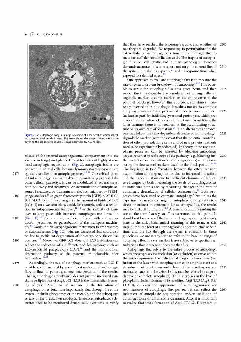
release of the internal autophagosomal compartment into thevacuole in fungi) and plants. Except for cases of highly stimu-lated autophagic sequestration (Fig. 2), autophagic bodies arenot seen in animal cells, because lysosomes/autolysosomes are
2175 typically smaller than autophagosomes.6,8,10 One critical pointis that autophagy is a highly dynamic, multi-step process. Likeother cellular pathways, it can be modulated at several steps,both positively and negatively. An accumulation of autophago-somes (measured by transmission electron microscopy [TEM]
2180 image analysis,11 as green fluorescent protein [GFP]-MAP1LC3[GFP-LC3] dots, or as changes in the amount of lipidated LC3[LC3-II] on a western blot), could, for example, reflect a reduc-tion in autophagosome turnover,12-14 or the inability of turn-over to keep pace with increased autophagosome formation
2185 (Fig. 1B).15 For example, inefficient fusion with endosomesand/or lysosomes, or perturbation of the transport machin-ery,16 would inhibit autophagosome maturation to amphisomesor autolysosomes (Fig. 1C), whereas decreased flux could alsobe due to inefficient degradation of the cargo once fusion has
2190 occurred.17 Moreover, GFP-LC3 dots and LC3 lipidation canreflect the induction of a different/modified pathway such asLC3-associated phagocytosis (LAP),18 and the noncanonicaldestruction pathway of the paternal mitochondria afterfertilization.19,20
2195 Accordingly, the use of autophagy markers such as LC3-IImust be complemented by assays to estimate overall autophagicflux, or flow, to permit a correct interpretation of the results.That is, autophagic activity includes not just the increased syn-thesis or lipidation of Atg8/LC3 (LC3 is the mammalian homo-
2200 log of yeast Atg8), or an increase in the formation ofautophagosomes, but, most importantly, flux through the entiresystem, including lysosomes or the vacuole, and the subsequentrelease of the breakdown products. Therefore, autophagic sub-strates need to be monitored dynamically over time to verify
2205that they have reached the lysosome/vacuole, and whether ornot they are degraded. By responding to perturbations in theextracellular environment, cells tune the autophagic flux tomeet intracellular metabolic demands. The impact of autopha-gic flux on cell death and human pathologies therefore
2210demands accurate tools to measure not only the current flux ofthe system, but also its capacity,21 and its response time, whenexposed to a defined stress.22
One approach to evaluate autophagic flux is to measure therate of general protein breakdown by autophagy.6,23 It is possi-
2215ble to arrest the autophagic flux at a given point, and thenrecord the time-dependent accumulation of an organelle, anorganelle marker, a cargo marker, or the entire cargo at thepoint of blockage; however, this approach, sometimes incor-rectly referred to as autophagic flux, does not assess complete
2220autophagy because the experimental block is usually induced(at least in part) by inhibiting lysosomal proteolysis, which pre-cludes the evaluation of lysosomal functions. In addition, thelatter assumes there is no feedback of the accumulating struc-ture on its own rate of formation.24 In an alternative approach,
2225one can follow the time-dependent decrease of an autophagy-degradable marker (with the caveat that the potential contribu-tion of other proteolytic systems and of new protein synthesisneed to be experimentally addressed). In theory, these nonauto-phagic processes can be assessed by blocking autophagic
2230sequestration at specific steps of the pathway (e.g., blocking fur-ther induction or nucleation of new phagophores) and by mea-suring the decrease of markers distal to the block point.12,14,25
The key issue is to differentiate between the often transientaccumulation of autophagosomes due to increased induction,
2235and their accumulation due to inefficient clearance of seques-tered cargos by both measuring the levels of autophagosomesat static time points and by measuring changes in the rates ofautophagic degradation of cellular components.17 Both pro-cesses have been used to estimate “autophagy,” but unless the
2240experiments can relate changes in autophagosome quantity to adirect or indirect measurement for autophagic flux, the resultsmay be difficult to interpret.26 A general caution regarding theuse of the term “steady state” is warranted at this point. Itshould not be assumed that an autophagic system is at steady
2245state in the strict biochemical meaning of this term, as thisimplies that the level of autophagosomes does not change withtime, and the flux through the system is constant. In theseguidelines, we use steady state to refer to the baseline range ofautophagic flux in a system that is not subjected to specific per-
2250turbations that increase or decrease that flux.Autophagic flux refers to the entire process of autophagy,
which encompasses the inclusion (or exclusion) of cargo withinthe autophagosome, the delivery of cargo to lysosomes (viafusion of the latter with autophagosomes or amphisomes) and
2255its subsequent breakdown and release of the resulting macro-molecules back into the cytosol (this may be referred to as pro-ductive or complete autophagy). Thus, increases in the level ofphosphatidylethanolamine (PE)-modified Atg8/LC3 (Atg8–PE/LC3-II), or even the appearance of autophagosomes, are
2260not measures of autophagic flux per se, but can reflect theinduction of autophagic sequestration and/or inhibition ofautophagosome or amphisome clearance. Also, it is importantto realize that while formation of Atg8–PE/LC3-II appears to
Figure 2. An autophagic body in a large lysosome of a mammalian epithelial cellin mouse seminal vesicle in vitro. The arrow shows the single limiting membranecovering the sequestered rough ER. Image provided by A.L. Kov"acs.
34 D. J. KLIONSKY ET. AL.

correlate with the induction of autophagy, we do not know, at2265 present, the actual mechanistic relationship between Atg8–PE/
LC3-II formation and the rest of the autophagic process;indeed, it may be possible to execute “self-eating” in theabsence of LC3-II.27
As a final note, we also recommend that researchers refrain2270 from the use of the expression “percent autophagy” when
describing experimental results, as in “The cells displayed a25% increase in autophagy.” Instead, it is appropriate to indi-cate that the average number of GFP-Atg8/LC3 puncta per cellis increased or a certain percentage of cells displayed punctate
2275 GFP-Atg8/LC3 that exceeds a particular threshold (and thisthreshold should be clearly defined in the Methods section), orthat there is a particular increase or decrease in the rate of cargosequestration or the degradation of long-lived proteins, whenthese are the actual measurements being quantified.
2280 In a previous version of these guidelines,2 the methods wereseparated into 2 main sections—steady state and flux. In someinstances, a lack of clear distinction between the actual method-ologies and their potential uses made such a separation some-what artificial. For example, fluorescence microscopy was
2285 initially listed as a steady-state method, although this approachcan clearly be used to monitor flux as described in this article,especially when considering the increasing availability of newtechnologies such as microfluidic chambers. Furthermore, theuse of multiple time points and/or lysosomal fusion/degrada-
2290 tion inhibitors can turn even a typically static method such asTEM into one that monitors flux. Therefore, although wemaintain the importance of monitoring autophagic flux andnot just induction, this revised set of guidelines does not sepa-rate the methods based on this criterion. Readers should be
2295 aware that this article is not meant to present protocols, butrather guidelines, including information that is typically notpresented in protocol papers. For detailed information onexperimental procedures we refer readers to various protocolsthat have been published elsewhere.28-43,44 Finally, throughout
2300 the guidelines we provide specific cautionary notes, and theseare important to consider when planning experiments andinterpreting data; however, these cautions are not meant to be adeterrent to undertaking any of these experiments or a hin-drance to data interpretation.
2305 Collectively, we propose the following guidelines for mea-suring various aspects of selective and nonselective autophagyin eukaryotes.
A. Methods for monitoring autophagy
1. Transmission electron microscopy
2310 Autophagy was first detected by TEM in the 1950s (reviewed inref. 6). It was originally observed as focal degradation of cyto-plasmic areas performed by lysosomes, which remains the hall-mark of this process. Later analysis revealed that it starts withthe sequestration of portions of the cytoplasm by a special dou-
2315 ble membrane structure (now termed the phagophore), whichmatures into the autophagosome, still bordered by a doublemembrane. Subsequent fusion events expose the cargo to thelysosome (or the vacuole in fungi or plants) for enzymaticbreakdown.
2320The importance of TEM in autophagy research lies in sev-eral qualities. It is the only tool that reveals the morphology ofautophagic structures at a resolution in the nm range; showsthese structures in their natural environment and positionamong all other cellular components; allows their exact identifi-
2325cation; and, in addition, it can support quantitative studies ifthe rules of proper sampling are followed.11
Autophagy can be both selective and nonselective, and TEMcan be used to monitor both. In the case of selective autophagy,the cargo is the specific substrate being targeted for sequestra-
2330tion—bulk cytoplasm is essentially excluded. In contrast, dur-ing nonselective autophagy, the various cytoplasmicconstituents are sequestered randomly, resulting in autophago-somes in the size range of normal mitochondria. Sequestrationof larger structures (such as big lipid droplets, extremely elon-
2335gated or branching mitochondria or the entire Golgi complex)is rare, indicating an apparent upper size limit for individualautophagosomes. However, it has been observed that underspecial circumstances the potential exists for the formation ofhuge autophagosomes, which can even engulf a complete
2340nucleus.25 Cellular components that form large confluent areasexcluding bulk cytoplasm, such as organized, functional myofi-brillar structures, do not seem to be sequestered by macroau-tophagy. The situation is less clear with regard to glycogen.45-47
After sequestration, the content of the autophagosome and2345its bordering double membrane remain morphologically
unchanged, and clearly recognizable for a considerable time,which can be measured for at least many minutes. During thisperiod, the membranes of the sequestered organelles (for exam-ple the ER or mitochondria) remain intact, and the density of
2350ribosomes is conserved at normal levels. Degradation of thesequestered material and the corresponding deterioration ofultrastructure commences and runs to completion within theamphisome and the autolysosome after fusion with a late endo-some and lysosome (the vacuole in fungi and plants), respec-
2355tively (Fig. 1).48 The sequential morphological changes duringthe autophagic process can be followed by TEM. The matura-tion from the phagophore through the autolysosome is adynamic and continuous process,49 and, thus, the classificationof compartments into discrete morphological subsets can be
2360problematic; therefore, some basic guidelines are offered below.In the preceeding sections the “autophagosome”, the
“amphisome” and the “autolysosome” were terms used todescribe or indicate 3 basic stages and compartments ofautophagy. It is important to make it clear that for instances
2365(which may be many) when we cannot or do not want to differ-entiate among the autophagosomal, amphisomal and autolyso-somal stage we use the general term “autophagic vacuole”. Inthe yeast autophagy field the term “autophagic vesicle” is usedto avoid confusion with the primary vacuole, and by now the 2
2370terms are used in parallel and can be considered synonyms. Itis strongly recommended, however, to use only the term “auto-phagic vacuole” when referring to macroautophagy in highereukaryotic cells. Autophagosomes, also referred to as initialautophagic vacuoles (AVi), typically have a double membrane.
2375This structure is usually distinctly visible by EM as 2 parallelmembrane layers (bilayers) separated by a relatively narroweror wider electron-translucent cleft, even when applying thesimplest routine EM fixation procedure (Fig. 3A).50,51 This
AUTOPHAGY 35

electron-translucent cleft, however, is less visible in freeze-fixed2380 samples, suggesting it may be an artifact of sample preparation
(see Fig. S3 in ref. 52). In the case of nonselective autophagy,autophagosomes contain cytosol and/or organelles appearingmorphologically intact as also described above.48,53
Amphisomes54 can sometimes be identified by the presence of2385small intralumenal vesicles.55 These intralumenal vesicles are
delivered into the lumen by fusion of the autophagosome/auto-phagic vacuole (AV) limiting membrane with multivesicularendosomes, and care should therefore be taken in the identifi-cation of the organelles, especially in cells that produce large
2390numbers of multivesicular body (MVB)-derived exosomes(such as tumor or stem cells).56 Late/degradative autophagicvacuoles/autolysosomes (AVd or AVl) typically have only onelimiting membrane; frequently they contain electron densecytoplasmic material and/or organelles at various stages of deg-
2395radation (Fig. 3A and B);48,53 although late in the digestion pro-cess, they may contain only a few membrane fragments and bedifficult to distinguish from lysosomes, endosomes, or tubularsmooth ER cut in cross-section. Unequivocal identification ofthese structures and of lysosomes devoid of visible content
2400requires immuno-EM detection of a cathepsin or other lyso-somal hydrolase (e.g., ACP2 [acid phosphatase 2, lyso-somal]57,58) that is detected on the limiting membrane of thelysosome.59 Smaller, often electron dense, lysosomes may pre-dominate in some cells and exhibit hydrolase immunoreactivity
2405within the lumen and on the limiting membrane.60
In addition, structural proteins of the lysosome/late endo-some, such as LAMP1 and LAMP2 or SCARB2/LIMP-2, can beused for confirmation. No single protein marker, however, hasbeen effective in discriminating autolysosomes from the com-
2410partments mentioned above, in part due to the dynamic fusionand “kiss-and-run” events that promote interchange of compo-nents that can occur between these organelle subtypes. Rigor-ous further discrimination of these compartments from eachother and other vesicles ultimately requires demonstrating the
2415colocalization of a second marker indicating the presence of anautophagic substrate (e.g., LC3-CTSD colocalization) or theacidification of the compartment (e.g., mRFP/mCherry-GFP-LC3 probes (see Tandem mRFP/mCherry-GFP fluorescencemicroscopy), or Bodipy-pepstatin A detection of CTSD in an
2420activated form within an acidic compartment), and, whenappropriate, by excluding markers of other vesicularcomponents.57,61,62
The sequential deterioration of cytoplasmic structures beingdigested can be used for identifying autolysosomes by TEM.
2425Even when the partially digested and destroyed structure can-not be recognized in itself, it can be traced back to earlier formsby identifying preceeding stages of sequential morphologicaldeterioration. Degradation usually leads first to increased den-sity of still recognizable organelles, then to vacuoles with heter-
2430ogenous density, which become more homogenous andamorphous, mostly electron dense, but sometimes light (i.e.,electron translucent). It should be noted that, in pathologicalstates, it is not uncommon that active autophagy of autolyso-somes and damaged lysosomes (“lysosophagy”) may yield pop-
2435ulations of double-membrane limited autophagosomescontaining partially digested amorphous substrate in thelumen. These structures, which are enriched in hydrolases, areseen in swollen dystrophic neurites in some neurodegenerativediseases,60 and in cerebellar slices cultured in vitro and infected
2440with prions.63
It must be emphasized that in addition to the autophagicinput, other processes (e.g., endosomal, phagosomal,
Figure 3. TEM images of autophagic vacuoles in isolated mouse hepatocytes. (A)One autophagosome or early autophagic vacuole (AVi) and one degradative auto-phagic vacuole (AVd) are shown. The AVi can be identified by its contents (mor-phologically intact cytoplasm, including ribosomes, and rough ER), and thelimiting membrane that is partially visible as 2 bilayers separated by a narrow elec-tron-lucent cleft, i. e., as a double membrane (arrow). The AVd can be identified byits contents, partially degraded, electron-dense rough ER. The vesicle next to theAVd is an endosomal/lysosomal structure containing 5-nm gold particles that wereadded to the culture medium to trace the endocytic pathway. (B) One AVi, contain-ing rough ER and a mitochondrion, and one AVd, containing partially degradedrough ER, are shown. Note that the limiting membrane of the AVi is not clearly vis-ible, possibly because it is tangentially sectioned. However, the electron-lucentcleft between the 2 limiting membranes is visible and helps in the identification ofthe AVi. The AVd contains a region filled by small internal vesicles (asterisk), indi-cating that the AVd has fused with a multivesicular endosome. mi, mitochondrion.Image provided by E.-L. Eskelinen.
36 D. J. KLIONSKY ET. AL.

chaperone-mediated) also carry cargo to the lysosomes,64,65 insome cases through the intermediate step of direct endosome
2445 fusion with an autophagosome to form an amphisome. Thisprocess is exceptionally common in the axons of neurons.66
Therefore, strictly speaking, we can only have a lytic compart-ment containing cargos arriving from several possible sources;however, we still may use the term “autolysosome” if the con-
2450 tent appears to be overwhelmingly autophagic. Note that theengulfment of apoptotic cells via phagocytosis also produceslysosomes that contain cytoplasmic structures, but in this caseit originates from the dying cell; hence the possibility of anextracellular origin for such content must be considered when
2455 monitoring autophagy in settings where apoptotic cell deathmay be reasonably expected or anticipated.
For many physiological and pathological situations, exami-nation of both early and late autophagic vacuoles yields valu-able data regarding the overall autophagy status in the cells.15
2460 Along these lines, it is possible to use immunocytochemistry tofollow particular cytosolic proteins such as SOD1/CuZn super-oxide dismutase and CA/carbonic anhydrase to determine thestage of autophagy; the former is much more resistant to lyso-somal degradation.67
2465 In some autophagy-inducing conditions it is possible toobserve multi-lamellar membrane structures in addition to theconventional double-membrane autophagosomes, although thenature of these structures is not fully understood. These multi-lamellar structures may indeed be multiple double layers of
2470 phagophores68 and positive for LC3,69 they could be autolyso-somes,70 or they may form artifactually during fixation.
Special features of the autophagic process may be clarified byimmuno-TEM with gold-labeling,71,72 using antibodies, forexample, to cargo proteins of cytoplasmic origin and to LC3 to
2475 verify the autophagic nature of the compartment. LC3 immu-nogold labeling also makes it possible to detect novel degrada-tive organelles within autophagy compartments. This is thecase with the autophagoproteasome73 where costaining for LC3and ubiquitin-proteasome system (UPS) antigens occurs. The
2480 autophagoproteasome consists of single-, double-, or multiple-membrane LC3-positive autophagosomes costaining for spe-cific components of the UPS. It may be that a rich multi-enzy-matic (both autophagic and UPS) activity takes place withinthese organelles instead of being segregated within different cell
2485 domains.Although labeling of LC3 can be difficult, an increasing
number of commercial antibodies are becoming available,among them good ones to visualize the GFP moiety of GFP-LC3 reporter constructs.74 It is important to keep in mind that
2490 LC3 can be associated with nonautophagic structures (see Xen-ophagy, and Noncanonical use of autophagy-related proteins).LC3 is involved in specialized forms of endocytosis like LC3-associated phagocytosis. In addition, LC3 can decorate vesiclesdedicated to exocytosis in nonconventional secretion systems
2495 (reviewed in ref. 75,76). Antibodies against an abundant cyto-solic protein will result in high labeling all over the cytoplasm;however, organelle markers work well. Because there are veryfew characterized proteins that remain associated with thecompleted autophagosome, the choices for confirmation of its
2500 autophagic nature are limited. Furthermore, autophagosome-associated proteins may be cell type-specific. At any rate, the
success of this methodology depends on the quality of the anti-bodies and also on the TEM preparation and fixation proce-dures utilized. With immuno-TEM, authors should provide
2505controls showing that labeling is specific. This may require aquantitative comparisons of labeling over different cellularcompartments not expected to contain antigen and those con-taining the antigen of interest.
In clinical situations it is difficult to demonstrate autophagy2510clearly in tissues of formalin-fixed and paraffin-embedded
biopsy samples retrospectively, because (1) tissues fixed in for-malin have low or no LC3 detectable by routine immunostain-ing, because phospholipids melt together with paraffin duringthe sample preparation, and (2) immunogold electron micros-
2515copy of many tissues not optimally fixed for this purpose (e.g.,using rapid fixation) produces low-quality images. Combiningantigen retrieval with the avidin-biotin peroxidase complex(ABC) method may be quite useful for these situations. Forexample, immunohistochemistry can be performed using an
2520antigen retrieval method, then tissues are stained by the ABCtechnique using a labeled anti-human LC3 antibody. Afterimaging by light microscopy, the same prepared slides can beremade into sections for TEM examination, which can revealperoxidase reaction deposits in vacuoles within the region that
2525is LC3-immunopositive by light microscopy.77
In addition, statistical information should be provided dueto the necessity of showing only a selective number of sectionsin publications. Again, we note that for quantitative data it isnecessary to use proper volumetric analysis rather than just
2530counting numbers of sectioned objects. On the one hand, itmust be kept in mind that even volumetric morphometry/stere-ology only shows either steady state levels, or a snapshot in achanging dynamic process. Such data by themselves are notinformative regarding autophagic flux, unless carried out over
2535multiple time points. Alternatively, investigation in the pres-ence and absence of flux inhibitors can reveal the dynamicchanges in various stages of the autophagic process.12,21,78,79,42
On the one hand, if the turnover of autolysosomes is very rapid,a low number/volume will not necessarily be an accurate reflec-
2540tion of low autophagic activity. However, quantitative analysesindicate that autophagosome volume in many cases does corre-late with the rates of protein degradation.80-82 One potentialcompromise is to perform whole cell quantification of autopha-gosomes using fluorescence methods, with qualitative verifica-
2545tion by TEM,83 to show that the changes in fluorescent punctareflect corresponding changes in autophagic structures.
One additional caveat with TEM, and to some extent withconfocal fluorescence microscopy, is that the analysis of a singleplane within a cell can be misleading and may make the identi-
2550fication of autophagic structures difficult. Confocal microscopyand fluorescence microscopy with deconvolution software (orwith much more work, 3-dimensional TEM) can be used togenerate multiple/serial sections of the same cell to reduce thisconcern; however, in many cases where there is sufficient struc-
2555tural resolution, analysis of a single plane in a relatively largecell population can suffice given practical limitations. NewerEM technologies, including focused ion beam dual-beam EM,should make it much easier to apply three-dimensional analy-ses. An additional methodology to assess autophagosome accu-
2560mulation is correlative light and electron microscopy (CLEM),
AUTOPHAGY 37

which is helpful in confirming that fluorescent structures areautophagosomes.84-86 Along these lines, it is important to notethat even though GFP fluorescence will be quenched in theacidic environment of the autolysosome, some of the GFP
2565 puncta detected by light microscopy may correspond to earlyautolysosomes prior to GFP quenching. The mini Singlet Oxy-gen Generator (miniSOG) fluorescent flavoprotein, which isless than half the size of GFP, provides an additional means togenetically tag proteins for CLEM analysis under conditions
2570 that are particularly suited to subsequent TEM analysis.87 Com-binatorial assays using tandem monomeric red fluorescent pro-tein (mRFP)-GFP-LC3 (see Tandem mRFP/mCherry-GFPfluorescence microscopy) along with static TEM images shouldhelp in the analysis of flux and the visualization of cargo
2575 structures.88
Another technique that has proven quite useful for analyz-ing the complex membrane structures that participate inautophagy is three-dimensional electron tomography,89,90 andcryoelectron microscopy (Fig. 4). More sophisticated, cryo-soft
2580 X-ray tomography (cryo-SXT) is an emerging imaging tech-nique used to visualize autophagosomes.91 Cryo-SXT extractsultrastructural information from whole, unstained mammaliancells as close to the “near- native” fully-hydrated (living) stateas possible. Correlative studies combining cryo-fluorescence
2585 and cryo-SXT workflow (cryo-CLXM) have been applied tocapture early autophagosomes.
Finally, although only as an indirect measurement, the com-parison of the ratio of autophagosomes to autolysosomes byTEM can support alterations in autophagy identified by other
2590 procedures.92 In this case it is important to always comparesamples to the control of the same cell type and in the samegrowth phase, and to acquire data at different time points, asthe autophagosome/autolysosome ratio varies in time in a cellcontext-dependent fashion, depending on their clearance activ-
2595 ity. It may also be necessary to distinguish autolysosomes fromtelolysosomes/late secondary lysosomes (the former are actively
engaged in degradation, whereas the latter have reached an endpoint in the breakdown of lumenal contents) because lysosomenumbers generally increase when autophagy is induced. An
2600additional category of lysosomal compartments, especiallycommon in disesase states and aged postmitotic cells such asneurons is the residual body. This category includes ceroid andlipofuscin, lobulated vesicular compartments of varying sizecomposed of highly indigestible complexes of protein and lipid
2605and abundant, mostly inactive, acid hydrolases. Reflectingend-stage unsuccessful incomplete autolysosomal digestion,lipofuscin is fairly easily distinguished from AVs and lysosomesby TEM but can be easily confused with autolysosomes inimmunocytochemistry studies at the light microscopy level.57
2610TEM observations of platinum-carbon replicas obtained bythe freeze fracture technique can also supply useful ultrastructuralinformation on the autophagic process. In quickly frozen andfractured cells the fracture runs preferentially along the hydro-phobic plane of the membranes, allowing characterization of the
2615limiting membranes of the different types of autophagic vacuolesand visualization of their limited protein intramembrane particles(IMPs, or integral membrane proteins). Several studies have beencarried out using this technique on yeast,93 as well as onmamma-lian cells or tissue; first on mouse exocrine pancreas,94 then on
2620mouse and rat liver,95,96 mouse seminal vesicle epithelium,25,68 rattumor and heart,97 or cancer cell lines (e.g., breast cancer MDA-MB-231)98 to investigate the various phases of autophagosomematuration, and to reveal useful details about the origin and evo-lution of their limiting membranes.6,99-102
2625The phagophore and the limiting membranes of autophago-somes contain few, or no detectable, IMPs (Fig. 5A, B), whencompared to other cellular membranes and to the membranes oflysosomes. In subsequent stages of the autophagic process thefusion of the autophagosome with an endosome and a lysosome
2630results in increased density of IMPs in the membrane of theformed autophagic compartments (amphisomes, autolysosomes;Fig. 5C).6,25,93-96,103,104 Autolysosomes are delimited by a single
Figure 4. Cryoelectron microscopy can be used as a three-dimensional approach to monitor the autophagic process. Four computed sections of an electron tomogram ofthe autophagic vacuole-rich cytoplasm in a hemophagocyte of a semi-thin section after high-pressure freezing preparation. The dashed area is membrane-free (A) buttomography reveals newly formed or degrading membranes with a parallel stretch (B). Image published previously2186 and provided by M. Schneider and P. Walter.
38 D. J. KLIONSKY ET. AL.

membrane because, in addition to the engulfed material, theinner membrane is also degraded by the lytic enzymes. Similarly,
2635 the limiting membrane of autophagic bodies in yeast (and pre-sumably plants) is also quickly broken down under normal con-ditions. Autophagic bodies can be stabilized, however, by theaddition of phenylmethylsulphonylfluoride (PMSF) or geneticallyby the deletion of the yeast PEP4 gene (see The Cvt pathway,
2640 mitophagy, pexophagy, piecemeal microautophagy of the nucleusand late nucleophagy in yeast and filamentous fungi.). Thus,another method to consider for monitoring autophagy in yeast(and potentially in plants) is to count autophagic bodies by TEMusing at least 2 time points.105 The advantage of this approach is
2645 that it can provide accurate information on flux even when theautophagosomes are abnormally small.106,107 Thus, although ahigh frequency of “abnormal” structures presents a challenge,TEM is still very helpful in analyzing autophagy.
Cautionary notes: Despite the introduction of many new2650 methods TEMmaintains its special role in autophagy research.
There are, however, difficulties in utilizing TEM. It is relativelytime consuming, and needs technical expertise to ensureproper handling of samples in all stages of preparation fromfixation to sectioning and staining (contrasting). After all these
2655 criteria are met, we face the most important problem of properidentification of autophagic structures. This is crucial for bothqualitative and quantitative characterization, and needs con-siderable experience, even in the case of one cell type. The diffi-culty lies in the fact that many subcellular components may be
2660 mistaken for autophagic structures. For example, some authors(or reviewers of manuscripts) assume that almost all cyto-plasmic structures that, in the section plane, are surrounded by2 (more or less) parallel membranes are autophagosomes.Structures appearing to be limited by a double membrane,
2665 however, may include swollen mitochondria, plastids in plantcells, cellular interdigitations, endocytosed apoptotic bodies,circular structures of lamellar smooth endoplasmic reticulum(ER), and even areas surrounded by rough ER. Endosomes,phagosomes and secretory vacuoles may have heterogenous
2670 content that makes it possible to confuse them with autolyso-somes. Additional identification problems may arise fromdamage caused by improper sample taking or fixationartifacts.50,51,108,109
Whereas fixation of in vitro samples is relatively straight-2675forward, fixation of excised tissues requires care to avoid sam-
pling a nonrepresentative, uninformative, or damaged part ofthe tissue. For instance, if 95% of a tumor is necrotic, TEManalysis of the necrotic core may not be informative, and ifthe sampling is from the viable rim, this needs to be specified
2680when reported. Clearly this introduces the potential for sub-jectivity because reviewers of a paper cannot request multipleimages with a careful statistical analysis with these types ofsamples. In addition, ex vivo samples are not typically ran-domized during processing, further complicating the possibil-
2685ity of valid statistical analyses. Ex vivo tissue should be fixedimmediately and systematically across samples to avoidchanges in autophagy that may occur simply due to theelapsed time ex vivo. It is recommended that for tissue sam-ples, perfusion fixation should be used when possible. For
2690yeast, rapid freezing techniques such as high pressure freezingfollowed by freeze substitution (i.e., dehydration at low tem-perature) may be particularly useful.
Quantification of autophagy by TEM morphometry hasbeen rather controversial, and unreliable procedures still
2695continue to be used. For the principles of reliable quantifi-cation and to avoid misleading results, excellent reviews areavailable.11,110-112 In line with the basic principles of mor-phometry we find it necessary to emphasize here somecommon problems with regard to quantification. Counting
2700autophagic vacuole profiles in sections of cells (i.e., numberof autophagic profiles per cell profile) may give unreliableresults, partly because both cell areas and profile areas arevariable and also because the frequency of section profilesdepends on the size of the vacuoles. However, estimation of
2705the number of autophagic profiles per cell area is more reli-able and correlates well with the volume fraction mentionedbelow.53 There are morphometric procedures to measure orestimate the size range and the number of spherical objectsby profiles in sections;111 however, such methods have been
2710used in autophagy research only a few times.32,107,113,114
Proper morphometry described in the cited reviews willgive us data expressed in mm3 autophagic vacuole/mm3
cytoplasm for relative volume (also called volume fractionor volume density), or mm2 autophagic vacuole surface/mm3
Figure 5. Different autophagic vacuoles observed after freeze fracturing in cultured osteosarcoma cells after treatment with the autophagy inducer voacamine.101 (A)Early autophagosome delimited by a double membrane. (B) Inner monolayer of an autophagosome membrane deprived of protein particles. (C) Autolysosome delimitedby a single membrane rich in protein particles. In the cross-fractured portion (on the right) the profile of the single membrane and the inner digested material are easilyvisible. Images provided by S. Meschini, M. Condello and A. Giuseppe.
AUTOPHAGY 39

2715 cytoplasm for relative surface (surface density). Examples ofactual morphometric measurements for the characterizationof autophagic processes can be found in severalarticles.21,108,111,115,116 It is appropriate to note here that achange in the volume fraction of the autophagic compart-
2720 ment may come from 2 sources; from the real growth of itssize in a given cytoplasmic volume, or from the decrease ofthe cytoplasmic volume itself. To avoid this so-called “refer-ence trap,” the reference space volume can be determinedby different methods.112,117 If different magnifications are
2725 used for measuring the autophagic vacuoles and the cyto-plasm (which may be practical when autophagy is lessintense) correction factors should always be used.
In some cases, it may be prudent to employ tomo-graphic reconstructions of the TEM images to confirm that
2730 the autophagic compartments are spherical and are notbeing confused with interdigitations observed betweenneighboring cells, endomembrane cisternae or damagedmitochondria with similar appearance in thin-sections (e.g.,see ref. 118), but this is obviously a time-consuming
2735 approach requiring sophisticated equipment. In addition,interpretation of tomographic images can be problematic.For example, starvation-induced autophagosomes shouldcontain cytoplasm (i.e., cytosol and possibly organelles),but autophagosome-related structures involved in specific
2740 types of autophagy should show the selective cytoplasmictarget, but may be relatively devoid of bulk cytoplasm.Such processes include selective peroxisome or mitochon-dria degradation (pexophagy or mitophagy, respec-tively),119,120 targeted degradation of pathogenic microbes
2745 (xenophagy),121-126 a combination of xenophagy and stress-induced mitophagy,127 as well as the yeast biosyntheticcytoplasm-to-vacuole targeting (Cvt) pathway.128 Further-more, some pathogenic microbes express membrane-dis-rupting factors during infection (e.g., phospholipases) that
2750 disrupt the normal double-membrane architecture of auto-phagosomes.129 It is not even clear if the sequestering com-partments used for specific organelle degradation orxenophagy should be termed autophagosomes or if alter-nate terms such as pexophagosome,130 mitophagosome and
2755 xenophagosome should be used, even though the mem-brane and mechanisms involved in their formation may beidentical to those for starvation-induced autophagosomes;for example, the double-membrane vesicle of the Cvt path-way is referred to as a Cvt vesicle.131
2760 The confusion of heterophagic structures with autophagicones is a major source of misinterpretation. A prominentexample of this is related to apoptosis. Apoptotic bodiesfrom neighboring cells are readily phagocytosed by surviv-ing cells of the same tissue.132,133 Immediately after phago-
2765 cytic uptake of apoptotic bodies, phagosomes may havedouble limiting membranes. The inner one is the plasmamembrane of the apoptotic body and the outer one is thatof the phagocytizing cell. The early heterophagic vacuoleformed in this way may appear similar to an autophago-
2770 some or, in a later stage, an early autolysosome in that itcontains recognizable or identifiable cytoplasmic material. Amajor difference, however, is that the surrounding mem-branes are the thicker plasma membrane type, rather than
the thinner sequestration membrane type (9–10 nm, versus27757–8 nm, respectively).109 A good feature to distinguish
between autophagosomes and double plasma membrane-bound structures is the lack of the distended empty space(characteristic for the sequestration membranes of autopha-gosomes) between the 2 membranes of the phagocytic
2780vacuoles. In addition, engulfed apoptotic bodies usuallyhave a larger average size than autophagosomes.134,135 Theproblem of heterophagic elements interfering with the iden-tification of autophagic ones is most prominent in cell typeswith particularly intense heterophagic activity (such as mac-
2785rophages, and amoeboid or ciliate protists). Special atten-tion has to be paid to this problem in cell cultures or invivo treatments (e.g., with toxic or chemotherapeuticagents) causing extensive apoptosis.
The most common organelles confused with autophagic2790vacuoles are mitochondria, ER, endosomes, and also
(depending on their structure) plastids in plants. Due tothe cisternal structure of the ER, double membrane-likestructures surrounding mitochondria or other organellesare often observed after sectioning,136 but these can also
2795correspond to cisternae of the ER coming into and out ofthe section plane.50 If there are ribosomes associated withthese membranes they can help in distinguishing themfrom the ribosome-free double-membrane of the phago-phore and autophagosome. Observation of a mixture of
2800early and late autophagic vacuoles that is modulated by thetime point of collection and/or brief pulses of bafilomycinA1 (a vacuolar-type HC-ATPase [V-ATPase] inhibitor) totrap the cargo in a recognizable early state42 increases theconfidence that an autophagic process is being observed. In
2805these cases, however, the possibility that feedback activa-tion of sequestration gets involved in the autophagic pro-cess has to be carefully considered. To minimize theimpact of errors, exact categorization of autophagic ele-ments should be applied. Efforts should be made to clarify
2810the nature of questionable structures by extensive prelimi-nary comparison in many test areas. Elements that stillremain questionable should be categorized into specialgroups and measured separately. Should their later identifi-cation become possible, they can be added to the proper
2815category or, if not, kept separate.For nonspecialists it can be particularly difficult to distin-
guish among amphisomes, autolysosomes and lysosomes,which are all single-membrane compartments containing moreor less degraded material. Therefore, we suggest in general to
2820measure autophagosomes as a separate category for a start, andto compile another category of degradative compartments(including amphisomes, autolysosomes and lysosomes). All ofthese compartments increase in quantity upon true autophagyinduction; however, in pathological states, it may be informa-
2825tive to discriminate among these different forms of degradativecompartments, which may be differentially affected by diseasefactors.
In yeast, it is convenient to identify autophagic bodiesthat reside within the vacuole lumen, and to quantify
2830them as an alternative to the direct examination of autopha-gosomes. However, it is important to keep in mind that itmay not be possible to distinguish between autophagic
40 D. J. KLIONSKY ET. AL.

bodies that are derived from the fusion of autophagosomeswith the vacuole, and the single-membrane vesicles that are
2835 generated during microautophagy-like processes such asmicropexophagy and micromitophagy.
Conclusion: EM is an extremely informative and powerfulmethod for monitoring autophagy and remains the only
technique that shows autophagy in its complex cellular environ-2840ment with subcellular resolution. The cornerstone of successfully
using TEM is the proper identification of autophagic structures,which is also the prerequisite to get reliable quantitative resultsby EMmorphometry. EM is best used in combination with othermethods to ensure the complex and holistic approach that is
Figure 6. (For figure caption See page no. 42)
AUTOPHAGY 41
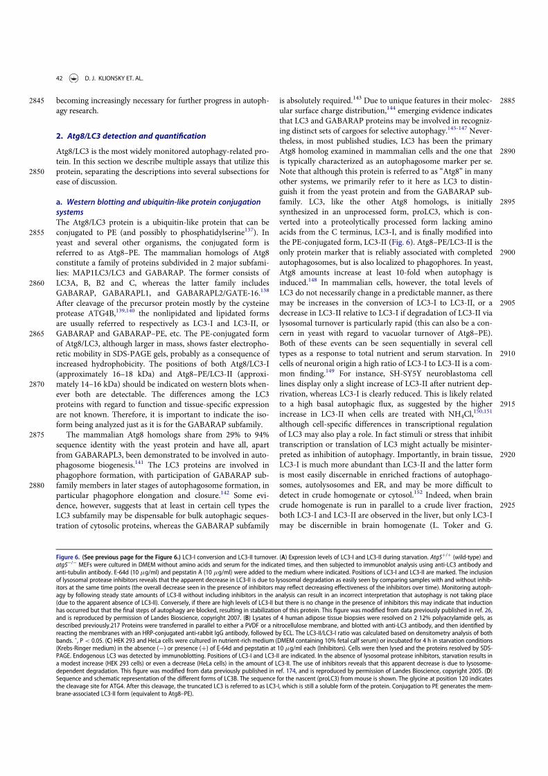
2845 becoming increasingly necessary for further progress in autoph-agy research.
2. Atg8/LC3 detection and quantification
Atg8/LC3 is the most widely monitored autophagy-related pro-tein. In this section we describe multiple assays that utilize this
2850 protein, separating the descriptions into several subsections forease of discussion.
a. Western blotting and ubiquitin-like protein conjugationsystemsThe Atg8/LC3 protein is a ubiquitin-like protein that can be
2855 conjugated to PE (and possibly to phosphatidylserine137). Inyeast and several other organisms, the conjugated form isreferred to as Atg8–PE. The mammalian homologs of Atg8constitute a family of proteins subdivided in 2 major subfami-lies: MAP1LC3/LC3 and GABARAP. The former consists of
2860 LC3A, B, B2 and C, whereas the latter family includesGABARAP, GABARAPL1, and GABARAPL2/GATE-16.138
After cleavage of the precursor protein mostly by the cysteineprotease ATG4B,139,140 the nonlipidated and lipidated formsare usually referred to respectively as LC3-I and LC3-II, or
2865 GABARAP and GABARAP–PE, etc. The PE-conjugated formof Atg8/LC3, although larger in mass, shows faster electropho-retic mobility in SDS-PAGE gels, probably as a consequence ofincreased hydrophobicity. The positions of both Atg8/LC3-I(approximately 16–18 kDa) and Atg8–PE/LC3-II (approxi-
2870 mately 14–16 kDa) should be indicated on western blots when-ever both are detectable. The differences among the LC3proteins with regard to function and tissue-specific expressionare not known. Therefore, it is important to indicate the iso-form being analyzed just as it is for the GABARAP subfamily.
2875 The mammalian Atg8 homologs share from 29% to 94%sequence identity with the yeast protein and have all, apartfrom GABARAPL3, been demonstrated to be involved in auto-phagosome biogenesis.141 The LC3 proteins are involved inphagophore formation, with participation of GABARAP sub-
2880 family members in later stages of autophagosome formation, inparticular phagophore elongation and closure.142 Some evi-dence, however, suggests that at least in certain cell types theLC3 subfamily may be dispensable for bulk autophagic seques-tration of cytosolic proteins, whereas the GABARAP subfamily
2885is absolutely required.143 Due to unique features in their molec-ular surface charge distribution,144 emerging evidence indicatesthat LC3 and GABARAP proteins may be involved in recogniz-ing distinct sets of cargoes for selective autophagy.145-147 Never-theless, in most published studies, LC3 has been the primary
2890Atg8 homolog examined in mammalian cells and the one thatis typically characterized as an autophagosome marker per se.Note that although this protein is referred to as “Atg8” in manyother systems, we primarily refer to it here as LC3 to distin-guish it from the yeast protein and from the GABARAP sub-
2895family. LC3, like the other Atg8 homologs, is initiallysynthesized in an unprocessed form, proLC3, which is con-verted into a proteolytically processed form lacking aminoacids from the C terminus, LC3-I, and is finally modified intothe PE-conjugated form, LC3-II (Fig. 6). Atg8–PE/LC3-II is the
2900only protein marker that is reliably associated with completedautophagosomes, but is also localized to phagophores. In yeast,Atg8 amounts increase at least 10-fold when autophagy isinduced.148 In mammalian cells, however, the total levels ofLC3 do not necessarily change in a predictable manner, as there
2905may be increases in the conversion of LC3-I to LC3-II, or adecrease in LC3-II relative to LC3-I if degradation of LC3-II vialysosomal turnover is particularly rapid (this can also be a con-cern in yeast with regard to vacuolar turnover of Atg8–PE).Both of these events can be seen sequentially in several cell
2910types as a response to total nutrient and serum starvation. Incells of neuronal origin a high ratio of LC3-I to LC3-II is a com-mon finding.149 For instance, SH-SY5Y neuroblastoma celllines display only a slight increase of LC3-II after nutrient dep-rivation, whereas LC3-I is clearly reduced. This is likely related
2915to a high basal autophagic flux, as suggested by the higherincrease in LC3-II when cells are treated with NH4Cl,
150,151
although cell-specific differences in transcriptional regulationof LC3 may also play a role. In fact stimuli or stress that inhibittranscription or translation of LC3 might actually be misinter-
2920preted as inhibition of autophagy. Importantly, in brain tissue,LC3-I is much more abundant than LC3-II and the latter formis most easily discernable in enriched fractions of autophago-somes, autolysosomes and ER, and may be more difficult todetect in crude homogenate or cytosol.152 Indeed, when brain
2925crude homogenate is run in parallel to a crude liver fraction,both LC3-I and LC3-II are observed in the liver, but only LC3-Imay be discernible in brain homogenate (L. Toker and G.
Figure 6. (See previous page for the Figure 6.) LC3-I conversion and LC3-II turnover. (A) Expression levels of LC3-I and LC3-II during starvation. Atg5C/C (wild-type) andatg5¡/¡ MEFs were cultured in DMEM without amino acids and serum for the indicated times, and then subjected to immunoblot analysis using anti-LC3 antibody andanti-tubulin antibody. E-64d (10 mg/ml) and pepstatin A (10 mg/ml) were added to the medium where indicated. Positions of LC3-I and LC3-II are marked. The inclusionof lysosomal protease inhibitors reveals that the apparent decrease in LC3-II is due to lysosomal degradation as easily seen by comparing samples with and without inhib-itors at the same time points (the overall decrease seen in the presence of inhibitors may reflect decreasing effectiveness of the inhibitors over time). Monitoring autoph-agy by following steady state amounts of LC3-II without including inhibitors in the analysis can result in an incorrect interpretation that autophagy is not taking place(due to the apparent absence of LC3-II). Conversely, if there are high levels of LC3-II but there is no change in the presence of inhibitors this may indicate that inductionhas occurred but that the final steps of autophagy are blocked, resulting in stabilization of this protein. This figure was modified from data previously published in ref. 26,and is reproduced by permission of Landes Bioscience, copyright 2007. (B) Lysates of 4 human adipose tissue biopsies were resolved on 2 12% polyacrylamide gels, asdescribed previously.217 Proteins were transferred in parallel to either a PVDF or a nitrocellulose membrane, and blotted with anti-LC3 antibody, and then identified byreacting the membranes with an HRP-conjugated anti-rabbit IgG antibody, followed by ECL. The LC3-II/LC3-I ratio was calculated based on densitometry analysis of bothbands. ", P< 0.05. (C) HEK 293 and HeLa cells were cultured in nutrient-rich medium (DMEM containing 10% fetal calf serum) or incubated for 4 h in starvation conditions(Krebs-Ringer medium) in the absence (¡) or presence (C) of E-64d and pepstatin at 10 mg/ml each (Inhibitors). Cells were then lysed and the proteins resolved by SDS-PAGE. Endogenous LC3 was detected by immunoblotting. Positions of LC3-I and LC3-II are indicated. In the absence of lysosomal protease inhibitors, starvation results ina modest increase (HEK 293 cells) or even a decrease (HeLa cells) in the amount of LC3-II. The use of inhibitors reveals that this apparent decrease is due to lysosome-dependent degradation. This figure was modified from data previously published in ref. 174, and is reproduced by permission of Landes Bioscience, copyright 2005. (D)Sequence and schematic representation of the different forms of LC3B. The sequence for the nascent (proLC3) from mouse is shown. The glycine at position 120 indicatesthe cleavage site for ATG4. After this cleavage, the truncated LC3 is referred to as LC3-I, which is still a soluble form of the protein. Conjugation to PE generates the mem-brane-associated LC3-II form (equivalent to Atg8–PE).
42 D. J. KLIONSKY ET. AL.

Agam, personal communication), depending on the LC3 anti-body used.153 In studies of the brain, immunoblot analysis of
2930 the membrane and cytosol fraction from a cell lysate, uponappropriate loading of samples to achieve quantifiable andcomparative signals, can be useful to measure LC3 isoforms.
The pattern of LC3-I to LC3-II conversion seems not onlyto be cell specific, but also related to the kind of stress to
2935 which cells are subjected. For example, SH-SY5Y cells displaya strong increase of LC3-II when treated with the mitochon-drial uncoupler CCCP, a well-known inducer of mitophagy(although it has also been reported that CCCP may actuallyinhibit mitophagy154). Thus, neither assessment of LC3-I con-
2940 sumption nor the evaluation of LC3-II levels would necessarilyreveal a slight induction of autophagy (e.g., by rapamycin).Also, there is not always a clear precursor/product relationshipbetween LC3-I and LC3-II, because the conversion of the for-mer to the latter is cell type-specific and dependent on the
2945 treatment used to induce autophagy. Accumulation of LC3-IIcan be obtained by interrupting the autophagosome-lysosomefusion step (e.g., by depolymerizing acetylated microtubuleswith vinblastine), by inhibiting the ATP2A/SERCA Ca2C
pump, by specifically inhibiting the V-ATPase with with bafi-2950 lomycin A1
155-157 or by raising the lysosomal pH by the addi-tion of chloroquine,158,159 although some of these treatmentsmay increase autophagosome numbers by disrupting the lyso-some-dependent activation of MTOR (mechanistic target ofrapamycin [serine/threonine kinase]) complex 1 (MTORC1;
2955 note that the original term “mTOR” was named to distinguishthe “mammalian” target of rapamycin from the yeast pro-teins160), a major suppressor of autophagy induction),161,162 orby inhibiting lysosome-mediated proteolysis (e.g., with a cyste-ine protease inhibitor such as E-64d, the aspartic protease
2960 inhibitor pepstatin A, the cysteine, serine and threonine prote-ase inhibitor leupeptin or treatment with bafilomycin A1,NH4Cl or chloroquine
158,163,164). Western blotting can be usedto monitor changes in LC3 amounts (Fig. 6);26,165 however,even if the total amount of LC3 does increase, the magnitude
2965 of the response is generally less than that documented inyeast. It is worth noting that since the conjugated forms of theGABARAP subfamily members are usually undetectable with-out induction of autophagy in mammalian and other verte-brate cells,166,167 these proteins might be more suitable than
2970 LC3 to study and quantify subtle changes in autophagyinduction.
In most organisms, Atg8/LC3 is initially synthesized with aC-terminal extension that is removed by the Atg4 protease.Accordingly, it is possible to use this processing event to moni-
2975 tor Atg4 activity. For example, when GFP is fused at the C ter-minus of Atg8 (Atg8-GFP), the GFP moiety is removed in thecytosol to generate free Atg8 and GFP. This processing can beeasily monitored by western blot.168 It is also possible to useassays with an artificial fluorogenic substrate, or a fusion of
2980 LC3B to phospholipase A2 that allows the release of the activephospholipase for a subsequent fluorogenic assay,169 and thereis a fluorescence resonance energy transfer (FRET)-based assayutilizing CFP and YFP tagged versions of LC3B and GABAR-APL2/GATE-16 that can be used for high-throughput screen-
2985 ing.170 Another method to monitor ATG4 activity in vivo usesthe release of Gaussia luciferase from the C terminus of LC3
that is tethered to actin.171 Note that there are 4 Atg4 homologsin mammals, and they have different activities with regard tothe Atg8 subfamilies of proteins.172 ATG4A is able to cleave the
2990GABARAP subfamily, but has very limited activity toward theLC3 subfamily, whereas ATG4B is apparently active againstmost or all of these proteins.139,140 The ATG4C and ATG4Disoforms have minimal activity for any of the Atg8 homologs.In particular because a C-terminal fusion will be cleaved imme-
2995diately by Atg4, researchers should be careful to specifywhether they are using GFP-Atg8/LC3 (an N-terminal fusion,which can be used to monitor various steps of autophagy) orAtg8/LC3-GFP (a C-terminal fusion, which can only be used tomonitor Atg4 activity).173
3000Cautionary notes: There are several important caveats tousing Atg8/LC3-II or GABARAP-II to visualize fluctuations inautophagy. First, changes in LC3-II amounts are tissue- andcell context-dependent.153,174 Indeed, in some cases, autopha-gosome accumulation detected by TEM does not correlate well
3005with the amount of LC3-II (Tall"oczy Z, de Vries RLA, andSulzer D, unpublished results; Eskelinen E-L, unpublishedresults). This is particularly evident in those cells that show lowlevels of LC3-II (based on western blotting) because of anintense autophagic flux that consumes this protein,175 or in cell
3010lines having high levels of LC3-II that are tumor-derived, suchas MDA-MB-231.174 Conversely, without careful quantificationthe detectable formation of LC3-II is not sufficient evidence forautophagy. For example, homozygous deletion of Becn1 doesnot prevent the formation of LC3-II in embryonic stem cells
3015even though autophagy is substantially reduced, whereas dele-tion of Atg5 results in the complete absence of LC3-II (seeFig. 5A and supplemental data in ref. 176). The same is true forthe generation of Atg8–PE in yeast in the absence of VPS30/ATG6 (see Fig. 7 in ref. 177). Thus, it is important to remember
3020that not all of the autophagy-related proteins are required forAtg8/LC3 processing, including lipidation.177 Vagaries in thedetection and amounts of LC3-I versus LC3-II present techni-cal problems. For example, LC3-I is very abundant in brain tis-sue, and the intensity of the LC3-I band may obscure detection
3025of LC3-II, unless the polyacrylamide crosslinking density isoptimized, or the membrane fraction of LC3 is first separatedfrom the cytosolic fraction.44 Conversely, certain cell lines havemuch less visible LC3-I compared to LC3-II. In addition, tis-sues may have asynchronous and heterogeneous cell popula-
3030tions, and this variability may present challenges whenanalyzing LC3 by western blotting.
Second, LC3-II also associates with the membranes of non-autophagic structures. For example, some members of theg-protocadherin family undergo clustering to form intracellu-
3035lar tubules that emanate from lysosomes.178 LC3-II is recruitedto these tubules, where it appears to promote or stabilizemembrane expansion. Furthermore, LC3 can be recruiteddirectly to apoptotic cell-containing phagosome mem-branes,179,180 macropinosomes,179 the parasitophorous vacuole
3040of Toxoplasma gondii,181 and single-membrane entoticvacuoles,179 as well as to bacteria-containing phagosome mem-branes under certain immune activating conditions, for exam-ple, toll-like receptor (TLR)-mediated stimulation inLC3-associated phagocytosis.182,183 Importantly, LC3 is
3045involved in secretory trafficking as it has been associated with
AUTOPHAGY 43

secretory granules in mast cells184 and PC12 hormone-secret-ing cells.185 LC3 is also detected on secretory lysosomes inosteoblasts186 and in amphisome-like structures involved inmucin secretion by goblet cells.187 Therefore, in studies of
3050infection of mammalian cells by bacterial pathogens, the iden-tity of the LC3-II labelled compartment as an autophagosomeshould be confirmed by a second method, such as TEM. It isalso worth noting that autophagy induced in response to bac-terial infection is not directed solely against the bacteria but
3055can also be a response to remnants of the phagocytic mem-brane.188 Similar cautions apply with regard to viral infection.For example, coronaviruses induce autophagosomes duringinfection through the expression of nsp6; however, coronavi-ruses also induce the formation of double-membrane vesicles
3060that are coated with LC3-I, a nonlipidated form of LC3 thatplays an autophagy-independent role in viral replication.189,190
Similarly, nonlipidated LC3 marks replication complexes inflavivirus (Japanese encephalitis virus)-infected cells and isessential for viral replication.191 Along these lines, during her-
3065pes simplex virus type 1 (HSV-1) infection, an LC3C autopha-gosome-like organelle that is derived from nuclear membranesand that contains viral proteins is observed,192 whereas influ-enza A virus directs LC3 to the plasma membrane via a LC3-interacting region (LIR) motif in its M2 protein.193 Moreover,
3070in vivo studies have shown that coxsackievirus (an enterovi-rus) induces formation of autophagy-like vesicles in pancreaticacinar cells, together with extremely large autophagy-relatedcompartments that have been termed megaphagosomes;194 theabsence of ATG5 disrupts viral replication and prevents the
3075formation of these structures.195
Third, caution must be exercised in general when evaluatingLC3 by western blotting, and appropriate standardization con-trols are necessary. For example, LC3-I may be less sensitive todetection by certain anti-LC3 antibodies. Moreover, LC3-I is
3080more labile than LC3-II, being more sensitive to freezing-thaw-ing and to degradation in SDS sample buffer. Therefore, freshsamples should be boiled and assessed as soon as possible andshould not be subjected to repeated freeze-thaw cycles. Alterna-tively, trichloracteic acid precipitation of protein from fresh cell
3085homogenates can be used to protect against degradation of LC3by proteases that may be present in the sample.A general pointto consider when examining transfected cells concerns the effi-ciency of transfection. A western blot will detect LC3 in theentire cell population, including those that are not transfected.
3090Thus, if transfection efficiency is too low, it may be necessaryto use methods, such as fluorescence microscopy, that allowautophagy to be monitored in single cells. The critical point isthat the analysis of the gel shift of transfected LC3 or GFP-LC3can be employed to follow LC3 lipidation only in highly trans-
3095fectable cells.196
When dealing with animal tissues, western blotting of LC3should be performed on frozen biopsy samples homogenized inthe presence of general protease inhibitors (C. Isidoro, personalcommunication; see also Human).197 Caveats regarding detec-
3100tion of LC3 by western blotting have been covered in a review.26
For example, PVDF membranes may result in a stronger LC3-II retention than nitrocellulose membranes, possibly due to ahigher affinity for hydrophobic proteins (Fig. 6B; J. Kovsan andA. Rudich, personal communication), and Triton X-100 may
3105not efficiently solubilize LC3-II in some systems.198 Heating inthe presence of 1% SDS, or analysis of membrane fractions,44
may assist in the detection of the lipidated form of this protein.This observation is particularly relevant for cells with a high
Figure 7. Effect of different inhibitors on LC3-II accumulation. SH-SY5Y humanneuroblastoma cells were plated and allowed to adhere for a minimum of 24 h,then treated in fresh medium. Treatments were as follows: rapamycin (Rap), (A)1 mM, 4 h or (B) 10 mM, 4 h; E-64d, final concentration 10 mg/ml from a 1 mg/mlstock in ethanol (ETOH); NH4Cl (NH4
C), final concentration 10 mM from a 1 M stockin water; pepstatin A (Pst), final concentration 10 mg/ml from a 1 mg/ml stock inethanol, or 68.6 mg/ml from a 6.86 mg/ml stock in DMSO; ethanol or DMSO, finalconcentration 1%. Pre-incubations in (B) were for 1 or 4 h as indicated. 10 mMNH4Cl (or 30 mM chloroquine, not shown) were the most effective compounds fordemonstrating the accumulation of LC3-II. E-64d was also effective in preventingthe degradation of LC3-II, with or without a preincubation, but ammomium chlo-ride (or chloroquine) may be more effective. Pepstatin A at 10 mg/ml with a 1 hpre-incubation was not effective at blocking degradation, whereas a 100 mM con-centration with 4 h pre-incubation had a partial effect. Thus, alkalinizing com-pounds are more effective in blocking LC3-II degradation, and pepstatin A must beused at saturating conditions to have any noticeable effect. Images provided by C.Isidoro. Note that the band running just below LC3-I at approximately 17.5 kDamay be a processing intermediate of LC3-I; it is detectable in freshly preparedhomogenates, but is less visible after the sample is subjected to a freeze-thawcycle.
44 D. J. KLIONSKY ET. AL.

nucleocytoplasmic ratio, such as lymphocytes. Under these3110 constraints, direct lysis in Laemmli loading buffer, containing
SDS, just before heating, greatly improves LC3 detection onPVDF membranes, especially when working with a small num-ber of cells (F. Gros, unpublished observations).199 Analysis ofa membrane fraction is particularly useful for brain where lev-
3115 els of soluble LC3-I greatly exceed the level of LC3-II.One of the most important issues is the quantification of
changes in LC3-II, because this assay is one of the most widelyused in the field and is often prone to misinterpretation. Levelsof LC3-II should be compared to actin (e.g., ACTB), but not to
3120 LC3-I (see the caveat in the next paragraph), and, ideally, tomore than one “housekeeping” protein (HKP). Actin and otherHKPs are usually abundant and can easily be overloaded on thegel200 such that they are not detected within a linear range.Moreover, actin levels may decrease when autophagy is induced
3125 in many organisms from yeast to mammals. For any proteinsused as “loading controls” (including actin, tubulin andGAPDH) multiple exposures of the western blot are generallynecessary to ensure that the signals are detected in the linearrange. An alternative approach is to stain for total cellular pro-
3130 teins with Coomassie Brilliant Blue and Ponceau Red.201 butthese methods are generally less sensitive and may not revealsmall differences in protein loading. Stain-Free gels, which alsostain for total cellular proteins, have been shown to be an excel-lent alternative to HKPs.202
3135 It is important to realize that ignoring the level of LC3-I infavor of LC3-II normalized to HKPs may not provide the fullpicture of the cellular autophagic response153,203 For example,in aging skeletal muscle the increase in LC3-I is at least asimportant as that for LC3-II.204,205 Quantification of both iso-
3140 forms is therefore informative, but requires adequate condi-tions of electrophoretic separation. This is particularlyimportant for samples where the amount of LC3-I is high rela-tive to LC3-II (as in brain tissues, where the LC3-I signal canbe overwhelming). Under such a scenario, it may be helpful to
3145 use gradient gels to increase the separation of LC3-I from LC3-II and/or cut away the part of the blot with LC3-I prior to thedetection of LC3-II. Furthermore, since the dynamic range ofLC3 immunoblots are generally quite limited, it is imperativethat other assays be used in parallel in order to draw valid con-
3150 clusions about changes in autophagy activity.Fourth, in mammalian cells LC3 is expressed as multiple
isoforms (LC3A, LC3B, LC3B2 and LC3C206,207), whichexhibit different tissue distributions and whose functionsare still poorly understood. A point of caution along these
3155 lines is that the increase in LC3A-II versus LC3B-II levelsmay not display equivalent changes in all organisms underautophagy-inducing conditions, and it should not beassumed that LC3B is the optimal protein to monitor.208 Akey technical consideration is that the isoforms may exhibit
3160 different specificities for antisera or antibodies. Thus, it ishighly recommended that investigators report exactly thesource and catalog number of the antibodies used to detectLC3 as this might help avoid discrepancies between studies.The commercialized anti-LC3B antibodies also recognize
3165 LC3A, but do not recognize LC3C, which shares lesssequence homology. It is important to note that LC3C pos-sesses in its primary amino acid sequence the DYKD motif
that is recognized with a high affinity by anti-FLAG anti-bodies. Thus, the standard anti-FLAG M2 antibody can
3170detect and immunoprecipitate overexpressed LC3C, andcaution has to be taken in experiments using FLAG-taggedproteins (M. Biard-Piechaczyk and L. Espert, personal com-munication). Note that according to Ensembl there is noLC3C in mouse or rat.
3175In addition, it is important to keep in mind the other sub-family of Atg8 proteins, the GABARAP subfamily (seeabove).141,209 Certain types of mitophagy induced by BNIP3L/NIX are highly dependent on GABARAP and less dependenton LC3 proteins.210,211 Furthermore, commercial antibodies for
3180GABARAPL1 also recognize GABARAP,138,143 which mightlead to misinterpretation of experiments, in particular thoseusing immunohistochemical techniques. Sometimes the prob-lem with cross-reactivity of the anti-GABARAPL1 antibodycan be overcome when analyzing these proteins by western blot
3185because the isoforms can be resolved during SDS-PAGE usinghigh concentration (15%) gels, as GABARAP migrates fasterthan GABARAPL1 (M. Boyer-Guittaut, personal communica-tion; also see Fig. S4 in ref. 143). Because GABARAP andGABARAPL1 can both be proteolytically processed and lipi-
3190dated, generating GABARAP-I or GABARAPL1-I andGABARAP-II or GABARAPL1-II, respectively, this may leadto a misassignment of the different bands. As soon as highlyspecific antibodies that are able to discriminate betweenGABARAP and GABARAPL1 become available, we strongly
3195advise their use; until then, we advise caution in interpretingresults based on the detection of these proteins by western blot.Antibody specificity can be assessed after complete inhibitionof GABARAP (or any other Atg8 family protein) expression byRNA interference.143,167 In general, we advise caution in choos-
3200ing antibodies for western blotting and immunofluorescenceexperiments and in interpreting results based on stated affini-ties of antibodies unless these have been clearly determined. Aswith any western blot, proper methods of quantification mustbe used, which are, unfortunately, often not well disseminated;
3205readers are referred to an excellent paper on this subject (seeref. 212). Unlike the other members of the GABARAP family,almost no information is available on GABARAPL3, perhapsbecause it is not yet possible to differentiate between GABAR-APL1 and GABARAPL3 proteins, which have 94% identity. As
3210stated by the laboratory that described the cloning of thehuman GABARAPL1 and GABARAPL3 genes,209 their expres-sion patterns are apparently identical. It is worth noting thatGABARAPL3 is the only gene of the GABARAP subfamily thatseems to lack an ortholog in mice.209 GABARAPL3 might
3215therefore be considered as a pseudogene without an intron thatis derived from GABARAPL1. Hence, until new data are pub-lished, GABARAPL3 should not be considered as the fourthmember of the GABARAP family.
Fifth, in non-mammalian species, the discrimination of3220Atg8–PE from the nonlipidated form can be complicated by
their nearly identical SDS-PAGE mobilities and the presence ofmultiple isoforms (e.g., there are 9 in Arabidopsis). In yeast, itis possible to resolve Atg8 (the nonlipidated form) from Atg8–PE by including 6 M urea in the SDS-PAGE separating gel,213
3225or by using a 15% resolving gel without urea (F. Reggiori, per-sonal communication). Similarly, urea combined with prior
AUTOPHAGY 45

treatment of the samples with (or without) phospholipase D(that will remove the PE moiety) can often resolve the ATG8species in plants.214,215 It is also possible to label cells with
3230 radioactive ethanolamine, followed by autoradiography to iden-tify Atg8–PE, and a C-terminal peptide can be analyzed bymass spectrometry to identify the lipid modification at the ter-minal glycine residue. Special treatments are not needed for theseparation of mammalian LC3-I from LC3-II.
3235 Sixth, it is important to keep in mind that ATG8, and to alesser extent LC3, undergoes substantial transcriptional andposttranscriptional regulation. Accordingly, to obtain an accu-rate interpretation of Atg8/LC3 protein levels it is also neces-sary to monitor the mRNA levels. Without analyzing the
3240 corresponding mRNA it is not possible to discriminate betweenchanges that are strictly reflected in altered amounts of proteinversus those that are due to changes in transcription (e.g., therate of transcription, or the stability of the message). For exam-ple, in cells treated with the calcium ionophore A23187 or the
3245 ER calcium pump blocker thapsigargin, an obvious correlationis found between the time-dependent increases in LC3B-I andLC3B-II protein levels, as well as with the observed increase inLC3B mRNA levels.216 Clinically, in human adipose tissue, pro-tein and mRNA levels of LC3 in omental fat are similarly ele-
3250 vated in obese compared to lean individuals.217
Seventh, LC3-I can be fully degraded by the 20S proteasomeor, more problematically, processed to a form appearing equalin size to LC3-II on a western blot (LC3-T); LC3-T was identi-fied in HeLa cells and is devoid of the ubiquitin conjugation
3255 domain, thus lacking its adaptor function for autophagy.218
Eighth, a general issue when working with cell lines is thatwe recommend that validation be performed to verify the cellline(s) being used, and to verify the presence of genetic altera-tions as appropriate. Depending on the goal (e.g., to indicate
3260 general applicability of a particular treatment) it may be impor-tant to use more than one cell line to confirm the results. It isalso critical to test for mycoplasma because the presence of thiscontaminant can significantly alter the results with regard toany autophagic response. For these reasons, we also recom-
3265 mend the use of low passage numbers for nonprimary cells orcell lines (no more than 40 passages or 6 months after thawing).
Finally, we would like to point out that one general issuewith regard to any assay is that experimental manipulationcould introduce some type of stress—for example, mechanical
3270 stress due to lysis, temperature stress due to heating or coolinga sample, or oxidative stress on a microscope slide, which couldlead to potential artifacts including the induction of autoph-agy—even maintaining cells in higher than physiologically nor-mal oxygen levels can be a stress condition.219 Special care
3275 should be taken with cells in suspension, as the stress resultingfrom centrifugation can induce autophagy. This point is notintended to limit the use of any specific methodology, butrather to note that there are no perfect assays. Therefore, it isimportant to verify that the positive (e.g., treatment with rapa-
3280 mycin, torin1 or other inducers) and negative (e.g., inhibitortreatment) controls behave as expected in any assays being uti-lized. Similarly, plasmid transfection or nucleofection can resultin the potent induction of autophagy (based on increases inLC3-II or SQSTM1/p62 degradation). In some cell types, the
3285 amount of autophagy induced by transfection of a control
empty vector may be so high that it is virtually impossible toexamine the effect of enforced gene expression on autophagy(B. Levine, personal communication). It is thus advisable toperform time course experiments to determine when the trans-
3290fection effect returns to acceptably low levels and to use appro-priate time-matched transfection controls (see also thediscussion in GFP-Atg8/LC3 fluorescence microscopy). Thiseffect is generally not observed with siRNA transfection; how-ever, it is an issue for plasmid expression constructs including
3295those for shRNA and for viral delivery systems. The use ofendotoxin-free DNA reduces, but does not eliminate, this prob-lem. In many cells the cationic polymers used for DNA trans-fection, such as liposomes and polyplex, induce largetubulovesicular autophagosomes (TVAs) in the absence of
3300DNA.220 These structures accumulate SQSTM1 and fuse slowlywith lysosomes. Interestingly, these TVAs appear to reducegene delivery, which increases 8–10 fold in cells that are unableto make TVAs due to the absence of ATG5. Finally, the precisecomposition of media components and the density of cells in
3305culture can have profound effects on basal autophagy levelsand may need to be modified empirically depending on the celllines being used. Along these lines various types of media, inparticular those with different serum levels (ranging from0–15%), may have profound effects with regard to how cells
3310(or organs) perceive a fed versus starved state. For example,normal serum contains significant levels of cytokines and hor-mones that likely regulate the basal levels of autophagy and orits modulation by additional stress or stimuli; thus, the use ofdialyzed serum might be an alternative for these studies. In
3315addition, the amino acid composition of the medium/assaybuffer may have profound effects on initiation or progressionof autophagy. For example, in the protozoan parasite Trypano-soma brucei starvation-induced autophagy can be prevented byaddition of histidine to the incubation buffer.221 For these rea-
3320sons, the cell culture conditions should be fully described. It isalso important to specify duration of autophagy stimulation, aslong-term autophagy can modify signal transduction pathwaysof importance in cell survival.222
Conclusion: Atg8/LC3 is often an excellent marker for auto-3325phagic structures; however, it must be kept in mind that there
are multiple LC3 isoforms, there is a second family of mamma-lian Atg8-like proteins (GABARAPs), and antibody affinity(for LC3-I versus LC3-II) and specificity (for example, forLC3A versus LC3B) must be considered and/or determined.
3330Moreover, LC3 levels on their own do not address issues ofautophagic flux. Finally, even when flux assays are carried out,there is a problem with the limited dynamic range of LC3immunoblots; accordingly, this method should not be used byitself to analyze changes in autophagy.
3335b. Turnover of LC3-II/Atg8–PEAutophagic flux is often inferred on the basis of LC3-II turn-over, measured by western blot (Fig. 6C)174 in the presence andabsence of lysosomal, or vacuolar degradation. However, itshould be cautioned that such LC3 assays are merely indicative
3340of autophagic “carrier flux”, not of actual autophagic cargo/sub-strate flux. It has, in fact, been observed that in rat hepatocytes,an autophagic-lysosomal flux of LC3-II can take place in theabsence of an accompanying flux of cytosolic bulk cargo.223
46 D. J. KLIONSKY ET. AL.

The relevant parameter in LC3 assays is the difference in the3345 amount of LC3-II in the presence and absence of saturating lev-
els of inhibitors, which can be used to examine the transit ofLC3-II through the autophagic pathway; if flux is occurring,the amount of LC3-II will be higher in the presence of theinhibitor.174 Lysosomal degradation can be prevented through
3350 the use of protease inhibitors (e.g., pepstatin A, leupeptin andE-64d), compounds that neutralize the lysosomal pH such asbafilomycin A1, chloroquine or NH4Cl,
16,149,158,164,224,225 or bytreatment with agents that block the fusion of autophagosomeswith lysosomes (note that bafilomycin A1 will ultimately cause
3355 a fusion block as well as neutralize the pH,156 but the inhibitionof fusion may be due to a block in ATP2A/SERCA activ-ity226).155-157,227 Alternatively, knocking down or knocking outLAMP2 (lysosomal-associated membrane protein 2) representsa genetic approach to block the fusion of autophagosomes and
3360 lysosomes (for example, inhibiting LAMP2 in myeloid leuke-mic cells results in a marked increase of GFP-LC3 dots andendogenous LC3-II protein compared to control cells uponautophagy induction during myeloid differentiation [M.P.Tschan, unpublished data]).228 This approach, however, is only
3365 valid when the knockdown of LAMP2 is directed against themRNA region specific for the LAMP2B spliced variant, as tar-geting the region common to the 3 variants would also inhibitchaperone-mediated autophagy, which may result in the com-pensatory upregulation of macroautophagy.92,229,230
3370 Increased levels of LC3-II in the presence of lysosomal inhi-bition or interfering with autophagosome-lysosome fusionalone (e.g., with bafilomycin A1), may be indicative of autopha-gic carrier flux (to the stage of cargo reaching the lysosome),but to assess whether a particular treatment alters complete
3375 autophagic flux through substrate digestion, the treatment plusbafilomycin A1 must be compared with results obtained withtreatment alone as well as with bafilomycin A1 alone. An addi-tive or supra-additive effect in LC3-II levels may indicate thatthe treatment enhances autophagic flux (Fig. 6C). Moreover,
3380 higher LC3-II levels with treatment plus bafilomycin A1 com-pared to bafilomycin A1 alone may indicate that the treatmentincreases the synthesis of autophagy-related membranes. If thetreatment by itself increases LC3-II levels, but the treatmentplus bafilomycin A1 does not increase LC3-II levels compared
3385 to bafilomycin A1 alone, this may indicate that the treatmentinduced a partial block in autophagic flux. Thus, a treatmentcondition increasing LC3-II on its own that has no differencein LC3-II in the presence of bafilomycin A1 compared to treat-ment alone may suggest a complete block in autophagy at the
3390 terminal stages.231 This procedure has been validated with sev-eral autophagy modulators.232 With each of these techniques, itis essential to avoid assay saturation. The duration of the bafilo-mycin A1 treatment (or any other inhibitor of autophagic fluxsuch as chloroquine) needs to be relatively short (1–4 h)233 to
3395 allow comparisons of the amount of LC3 that is lysosomallydegraded over a given time frame under one treatment condi-tion to another treatment condition. A dose-curve and time-course standardization for the use of autophagic flux inhibitorsis required for the initial optimization of the conditions to
3400 detect LC3-II accumulation and avoid nonspecific or secondaryeffects. By using a rapid screening approach, such as a colori-metric based-platform method,234 it is possible to monitor a
long time frame for autolysosome accumulation, which closelyassociates with autophagy efficiency.235 Positive control experi-
3405ments using treatment with known autophagy inducers, alongwith bafilomycin A1 versus vehicle, are important to demon-strate the utility of this approach in each experimental context.The same type of assay monitoring the turnover of Atg8–PEcan be used to monitor flux in yeast, by comparing the amount
3410of Atg8 present in a wild-type versus a pep4D strain followingautophagy induction;236 however, it is important to be awarethat the PEP4 knockout can influence yeast cell physiology.PMSF, which inhibits the activity of Prb1, can also be used toblock Atg8–PE turnover.
3415An additional methodology for monitoring autophagy relieson the observation that in some cell types a subpopulation ofLC3-II exists in a cytosolic form (LC3-IIs).237-239 The amountof cytosolic LC3-IIs and the ratio between LC3-I and LC3-IIsappears to correlate with changes in autophagy and may pro-
3420vide a more accurate measure of autophagic flux than ratiosbased on the total level of LC3-II.239 The validity of this methodhas been demonstrated by comparing autophagic proteolyticflux in rat hepatocytes, hepatoma cells and myoblasts. Oneadvantage of this approach is that it does not require the pres-
3425ence of autophagic or lysosomal inhibitors to block the degra-dation of LC3-II.
Due to the advances in time-lapse fluorescence microscopyand the development of photoswitchable fluorescent proteins,autophagic flux can also be monitored by assessing the half-life
3430of the LC3 protein240 post-photoactivation or by quantitativelymeasuring the autophagosomal pool size and its transitiontime.241 These approaches deliver invaluable information onthe kinetics of the system and the time required to clear a com-plete autophagosomal pool. Nonetheless, care must be taken
3435for this type of analysis as changes in translational/transcrip-tional regulation of LC3 might also affect the readout.
Finally, autophagic flux can be monitored based on the turn-over of LC3-II, by utilizing a luminescence-based assay. Forexample, a reporter assay based on the degradation of Renilla
3440reniformis luciferase (Rluc)-LC3 fusion proteins is well suitedfor screening compounds affecting autophagic flux.242 In thisassay, Rluc is fused N-terminally to either wild-type LC3(LC3WT) or a lipidation-deficient mutant of LC3 (G120A).Since Rluc-LC3WT, in contrast to Rluc-LC3G120A, specifically
3445associates with the autophagosomal membranes, Rluc-LC3WT
is more sensitive to autophagic degradation. A change inautophagy-dependent LC3 turnover can thus be estimated bymonitoring the change in the ratio of luciferase activitiesbetween the 2 cell populations expressing either Rluc-LC3WT
3450or Rluc-LC3G120A. In its simplest form, the Rluc-LC3-assay canbe used to estimate autophagic flux at a single time point bydefining the luciferase activities in cell extracts. Moreover, theuse of a live cell luciferase substrate makes it possible to moni-tor changes in autophagic activity in live cells in real time. This
3455method has been successfully used to identify positive and neg-ative regulators of autophagy from cells treated with micro-RNA, siRNA and small molecule libraries.242-245,246-248
Cautionary notes: The main caveat regarding the measure-ment of LC3-IIs/LC3-I is that this method has only been tested
3460in isolated rat hepatocytes and H4-II-E cells. Thus, it is not yetknown whether it is generally applicable to other cell types.
AUTOPHAGY 47

Indeed, a soluble form of LC3-II (i.e., LC3-IIs) is not observedin many standard cell types including HeLa, HEK 293 andPC12. In addition, the same concerns apply regarding detection
3465 of LC3-I by western blotting. It should be noted that the LC3-IIs/LC3-I ratio must be analyzed using the cytosolic fractionsrather than the total homogenates. Furthermore, the same cav-eats mentioned above regarding the use of LC3 for qualitativelymonitoring autophagy also apply to the use of this marker for
3470 evaluating flux.The use of a radioactive pulse-chase analysis, which
assesses complete autophagic flux, provides an alternative tolysosomal protease inhibitors,148 although such inhibitorsshould still be used to verify that degradation is lysosome-
3475 dependent. In addition, drugs must be used at concentrationsand for time spans that are effective in inhibiting fusion ordegradation, but that do not provoke cell death. Thus, thesetechniques may not be practical in all cell types or in tissuesfrom whole organisms where the use of protease inhibitors is
3480 problematic, and where pulse labeling requires artificial short-term culture conditions that may induce autophagy. Anotherconcern when monitoring flux via LC3-II turnover may beseen in the case of a partial autophagy block; in this situation,agents that disrupt autophagy (e.g., bafilomycin A1) will still
3485 result in an increase in LC3-II. Thus, care is needed in inter-pretation. For characterizing new autophagy modulators, it isideal to test autophagic flux at early (e.g., 4 h) and late (e.g.,24 h) time-points, since in certain instances, such as with cal-cium phosphate precipitates, a compound may increase or
3490 decrease flux at these 2 time-points, respectively.233 Moreover,it is important to consider assaying autophagy modulators ina long-term response in order to further understand theireffects. Finally, many of the chemicals used to inhibit autoph-agy, such as bafilomycin A1, NH4Cl (see Autophagy inhibitors
3495 and inducers) or chloroquine, also directly inhibit the endocy-tosis/uncoating of viruses (D.R. Smith, personal communica-tion), and other endocytic events requiring low pH, as well asexit from the Golgi (S. Tooze, personal communication). Assuch, agents that neutralize endosomal compartments should
3500 be used only with extreme caution in studies investigatingautophagy-virus interactions.
One additional consideration is that it may not be absolutelynecessary to follow LC3-II turnover if other substrates are beingmonitored simultaneously. For example, an increase in LC3-II
3505 levels in combination with the lysosomal (or ideally autophagy-specific) removal of an autophagic substrate (such as an organ-elle249,250) that is not a good proteasomal substrate provides anindependent assessment of autophagic flux. However, it isprobably prudent to monitor both turnover of LC3-II and an
3510 autophagosome substrate in parallel, due to the fact that LC3might be coupled to endosomal membranes and not just auto-phagosomes, and the levels of well-characterized autophago-some substrates such as SQSTM1 can also be affected byproteasome inhibitors.251
3515 Another issue relates to the use of protease inhibitors (seeAutophagy inhibitors and inducers). When using lysosomalprotease inhibitors, it is of fundamental importance to assessproper conditions of inhibitor concentration and time ofpre-incubation to ensure full inhibition of lysosomal cathe-
3520 psins. In this respect, 1 h of pre-incubation with 10 mg/ml
E-64d is sufficient in most cases, since this inhibitor is mem-brane permeable and rapidly accumulates within lysosomes,but another frequently used inhibitor, leupeptin, requires atleast 6 h pre-incubation.59 Moreover, pepstatin A is mem-
3525brane impermeable (ethanol or preferably DMSO must beemployed as a vehicle) and requires a prolonged incubation(>8 h) and a relatively high concentration (>50 mg/ml) tofully inhibit lysosomal CTSD/cathepsin D (Fig. 7). An incu-bation of this duration, however, can be problematic due to
3530indirect effects (see GFP-Atg8/LC3 lysosomal delivery andproteolysis). At least in neurons, pepstatin alone is a lesseffective lysosomal proteolytic block, and combining a cyste-ine protease inhibitor with it is most effective.59 Also, notethat the relative amount of lysosomal CTSB and CTSD is
3535cell-specific and changes with culture conditions. A possiblealternative to pepstatin A is the pepstatin A, BODIPY! FLconjugate,252,253 which is transported to lysosomes via endo-cytosis. In contrast to the protease inhibitors, chloroquine(10–40 mM) or bafilomycin A1 (1–100 nM) can be added to
3540cells immediately prior to autophagy induction. Because cys-teine protease inhibitors upregulate CTSD and have potentialinhibitory activity toward calpains and other cysteine pro-teases, whereas bafilomycin A1 can have potential significantcytotoxicity, especially in cultured neurons and pathological
3545states, the use of both methods may be important in someexperiments to exclude off-target effects of a single method.
Conclusion: It is important to be aware of the differencebetween monitoring the steady-state level of Atg8/LC3 andautophagic flux. The latter may be assessed by following Atg8/
3550LC3 in the absence and presence of autophagy inhibitors, andby examining the autophagy-dependent degradation of appro-priate substrates. In particular, if there is any evidence of anincrease in LC3-II (or autophagosomes), it is essential to deter-mine whether this represents increased flux, or a block in
3555fusion or degradation through the use of inhibitors such aschloroquine or bafilomycin A1. In the case of a suspectedimpaired degradation, assessment of lysosomal function is thenrequired to validate the conclusion and to establish the basis.
c. GFP-Atg8/LC3 lysosomal delivery and partial proteolysis3560GFP-LC3B (hereafter referred to as GFP-LC3) has also been
used to follow flux. It should be cautioned that, as with endoge-nous LC3, an assessment of autophagic GFP-LC3 flux is a car-rier flux that cannot be equated with, and is not necessarilyrepresentative of, an autophagic cargo flux. When GFP-Atg8 or
3565GFP-LC3 is delivered to a lysosome/vacuole, the Atg8/LC3 partof the chimera is sensitive to degradation, whereas the GFPprotein is relatively resistant to hydrolysis (note, however, thatGFP fluorescence is quenched by low pH; see GFP-Atg8/LC3fluorescence microscopy and Tandem mRFP/mCherry-GFP fluo-
3570rescence microscopy). Therefore, the appearance of free GFP onwestern blots can be used to monitor lysis of the inner autopha-gosome membrane and breakdown of the cargo in metazoans(Fig. 8A),236,254,255 or the delivery of autophagosomes to, andthe breakdown of autophagic bodies within, the fungal and
3575plant vacuole.214,215,236,256 Reports on Dictyostelium and mam-malian cells highlight the importance of lysosomal pH as a crit-ical factor in the detection of free GFP that results from thedegradation of fused proteins. In these cell types, free GFP
48 D. J. KLIONSKY ET. AL.

fragments are only detectable in the presence of nonsaturating3580 levels of lysosomotropic compounds (NH4Cl or choroquine) or
under conditions that attenuate lysosomal acidity; otherwise,the autophagic/degradative machinery appears to be too effi-cient to allow the accumulation of the proteolytic fragment(Fig. 8B,C).37,257 Hence, a reduction in the intensity of the free
3585 GFP band may indicate reduced flux, but it may also be due toefficient turnover. Using a range of concentrations and treat-ment times of compounds that inhibit autophagy can be usefulin distinguishing between these possibilities.258 Since the pH inthe yeast vacuole is higher than that in mammalian or Dictyos-
3590 telium lysosomes, the levels of free GFP fragments are detect-able in yeast even in the absence of lysosomotropiccompounds.30 Additionally, in yeast the diffuse fluorescenthaze from the released GFP moiety within the vacuole lumencan be observed by fluorescence microscopy.
3595The dynamic movement to lysosomes of GFP-LC3, or of itsassociated cargo, also can be monitored by time-lapse fluores-cence microscopy, although, as mentioned above, the GFP fluo-rescent signal is more sensitive to acidic pH than otherfluorophores (see GFP-Atg8/LC3 fluorescence microscopy). A
3600time-course evaluation of the cell population showing GFP-LC3puncta can serve to monitor the autophagic flux, since a constantincrease in the number of cells accumulating GFP-LC3 puncta issuggestive of defective fusion of autophagosomes with lysosomes.Conversely, a decline implies that GFP-LC3 is delivered to prop-
3605erly acidified lysosomes and may, in addition, reflect proteolyticelimination within them, although the latter needs to be indepen-dently established. In either case, it can be problematic to useGFP fluorescence to follow flux, as new GFP-LC3 is continuouslybeing synthesized. A potential solution to this problem is to fol-
3610low the fluorescence of a photoactivatable version of the
Figure 8. GFP-LC3 processing can be used to monitor delivery of autophagosomal membranes. (A) atg5¡/¡ MEFs engineered to express Atg5 under the control of theTet-off promoter were grown in the presence of doxycyline (Dox; 10 ng/ml) for one week to suppress autophagy. Cells were then cultured in the absence of drug for theindicated times, with or without a final 2 h starvation. Protein lysates were analyzed by western blot using anti-LC3 and anti-GFP antibodies. The positions of untaggedand GFP-tagged LC3-I and LC3-II, and free GFP are indicated. This figure was modified from data previously published in ref. 255, FEBS Letters, 580, Hosokawa N, Hara Y,Mizushima N, Generation of cell lines with tetracycline-regulated autophagy and a role for autophagy in controlling cell size, pp. 2623–2629, copyright 2006, with permis-sion from Elsevier. (B) Differential role of unsaturating and saturating concentrations of lysosomal inhibitors on GFP-LC3 cleavage. HeLa cells stably transfected with GFP-LC3 were treated with various concentrations of chloroquine (CQ) for 6 h. Total lysates were prepared and subjected to immunoblot analysis. (C) CQ-induced free GFPfragments require classical autophagy machinery. Wild-type and atg5¡/¡ MEFs were first infected with adenovirus GFP-LC3 (100 viral particles per cell) for 24 h. The cellswere then either cultured in regular culture medium with or without CQ (10 mM), or subjected to starvation in EBSS buffer in the absence or presence of CQ for 6 h. Totallysates were prepared and subjected to immunoblot analysis. Panel B and C are modified from the data previously published in ref. 257.
AUTOPHAGY 49

fluorescent protein,259 which allows this assay to be performedessentially as a pulse/chase analysis. Another alternative to followflux is to monitor GFP-LC3 fluorescence by adding lysosomalprotease or fusion inhibitors to cells expressing GFP-LC3 and
3615 monitoring changes in the number of puncta. In this case, thepresence of lysosomal inhibitors should increase the number ofGFP-LC3-positive structures, and the absence of an effect on thetotal number of GFP-LC3 puncta or on the percentage of cellsdisplaying numerous puncta is indicative of a defect(s) in auto-
3620 phagic flux.260 The combination of protease inhibitors (to preventthe degradation of GFP) or compounds that modify lysosomalpH such as NH4Cl or chloroquine, or compounds that blockfusion of autophagosomes with lysosomes such as bafilomycinA1 or others (e.g., vinblastine) may be most effective in prevent-
3625 ing lysosome-dependent decreases in GFP-LC3 puncta. How-ever, because the stability of GFP is affected by lysosomal pH,researchers may also consider the use of protease inhibitorswhether or not lysosomotropic compounds or fusion inhibitorsare included.
3630 Cautionary notes: The GFP-Atg8 processing assay is used rou-tinely to monitor autophagy in yeast. One caveat, however, is thatthis assay is not always carried out in a quantitative manner. Forexample, western blot exposures need to be in the linear range.Accordingly, an enzymatic assay such as the Pho8D60 assay may
3635 be preferred (see Autophagic protein degradation),261,262 espe-cially when the differences in autophagic activity need to bedetermined precisely (note that an equivalent assay has not beendeveloped for higher eukaryotic cells); however, as with anyenzyme assay, appropriate caution must be used regarding, for
3640 example, substrate concentrations and linearity. The Pho8D60also requires a control to verify equal Pho8D60 expression in thedifferent genetic backgrounds or conditions to be tested;261 differ-ences in Pho8D60 expression potentially affect its activity andmay thus cause misinterpretation of results. Another issue to
3645 keep inmind is that GFP-Atg8 processing correlates with the sur-face area of the inner sphere of the autophagosome, and thus pro-vides a smaller signal than assays that measure the volume of theautophagosome. Therefore, Pgk1-GFP processing,30 or thePho8D60 assay are generally more sensitive assays.
3650 The main limitation of the GFP-LC3 processing assay inmammalian cells is that it seems to depend on cell type andculture conditions (N. Hosokawa and N. Mizushima,unpublished data). Apparently, GFP is more sensitive tomammalian lysosomal hydrolases than to the degradative
3655 milieu of the yeast vacuole or the lysosomes in Drosophila.Alternatively, the lower pH of mammalian lysosomes rela-tive to that of the yeast vacuole may contribute to differen-ces in detecting free GFP. Under certain conditions (suchas Earle’s balanced salt solution [EBSS]-induced starvation)
3660 in some cell lines, when the lysosomal pH becomes particu-larly low, free GFP is undetectable because both the LC3-IIand free GFP fragments are quickly degraded.257 Therefore,if this method is used it should be accompanied by immu-noblotting and include controls to address the stability of
3665 nonlysosomal GFP such as GFP-LC3-I. It should also benoted that free GFP can be detected when cells are treatedwith nonsaturating doses of inhibitors such as chloroquine,E-64d and bafilomycin A1. The saturating concentrations ofthese lysosomal inhibitors vary in different cell lines, and it
3670would be better to use a saturating concentration of lyso-somal inhibitors when performing an autophagic fluxassay.257 Therefore, caution must be exercised in interpret-ing the data using this assay; it would be helpful to combinean analysis of GFP-LC3 processing with other assays, such
3675as the monitoring of endogenous LC3-II by western blot.Along these lines, a caution concerning the use of the EGFP
fluorescent protein for microscopy is that this fluorophore hasa relatively neutral pH optimum for fluorescence,263 and its sig-nal diminishes quickly during live cell imaging due to the acidic
3680environment of the lysosome. It is possible to circumvent thislatter problem by imaging paraformaldehyde-fixed culturesthat are maintained in a neutral pH buffer, which retains EGFPfluorescence (M. Kleinman and J.J. Reiners, personal communi-cation). Alternatively, it may be preferable to use a different flu-
3685orophore such as mRFP or mCherry, which retain fluorescenceeven at acidic pH.264 On the one hand, a putative advantage ofmCherry over mRFP is its enhanced photostability and inten-sity, which are an order of magnitude higher (and comparableto GFP), enabling acquisition of images at similar exposure set-
3690tings as are used for GFP, thus minimizing potential bias ininterpretation.265 On the other hand, caution is required whenevaluating the localization of mCherry fusion proteins duringautophagy due to the persistence of the mCherry signal inacidic environments; all tagged proteins are prone to show
3695enrichment in lysosomes during nonselective autophagy of thecytoplasm, especially at higher expression levels. In addition,red fluorescent proteins (even the monomeric forms) can betoxic due to oligomer formation.266 Dendra2 is an improvedversion of the green-to-red photoswitchable fluorescent protein
3700Dendra, which is derived from the octocoral Dendronephthyasp.267 Dendra2 is capable of irreversible photoconversion froma green to a red fluorescent form, but can be used also as nor-mal GFP or RFP vector. This modified version of the fluoro-phore has certain properties including a monomeric state, low
3705phototoxic activation and efficient chromophore maturation,which make it suitable for real-time tracking of LC3 andSQSTM1 (Fig. 9; K. Kaarniranta, personal communication).Another alternative to mRFP or mCherry is to use the Venus
Figure 9. Movement of activated pDendra2-hp62 (SQSTM1; orange) from thenucleus (middle) to an aggregate in ARPE-19 cells, revealed by confocal micros-copy. Cells were exposed to 5 mM MG132 for 24 h to induce the formation of peri-nuclear aggregates.2187 The cells were then exposed to a UV pulse (the UV-induced area is shown by red lines that are inside of the nucleus) that convertsDendra2 from green to red, and the time shown after the pulse is indicated.SQSTM1 is present in a small nuclear aggregrate, and is shuttled from the nucleusto a perinuclear large protein aggregate (detected as red). Scale bar: 5 mm. Imageprovided by K. Kaarniranta.
50 D. J. KLIONSKY ET. AL.

variant of YFP, which is brighter than mRFP and less sensitive3710 to pH than GFP.268
The pH optimum of EGFP is important to consider whenusing GFP-LC3 constructs, as the original GFP-LC3 marker269
uses the EGFP variant, which may result in a reduced signalupon the formation of amphisomes or autolysosomes. An addi-
3715 tional caveat when using the photoactivatable construct PA-GFP263 is that the process of activation by photons may induceDNA damage, which could, in turn, induce autophagy. Also,GFP is relatively resistant to denaturation, and boiling for5 min may be needed to prevent the folded protein from being
3720 trapped in the stacking gel during SDS-PAGE.As noted above (see Western blotting and ubiquitin-like pro-
tein conjugation systems), Atg4/ATG4 cleaves the residue(s)that follow the C-terminal glycine of Atg8/LC3 that will be con-jugated to PE. Accordingly, it is critical that any chimeras be
3725 constructed with the fluorescent tag at the amino terminus ofAtg8/LC3 (unless the goal is to monitor Atg4/ATG4 activity).
Finally, lysosomal inhibition needs to be carefully con-trolled. Prolonged inhibition of lysosomal hydrolases (>6 h) islikely to induce a secondary autophagic response triggered by
3730 the accumulated undigested autophagy cargo. This secondaryautophagic response can complicate the analysis of the auto-phagic flux, making it appear more vigorous than it would inthe absence of the lysosomal inhibitors.
Conclusion: The GFP-Atg8/LC3 processing assay, which3735 monitors free GFP generated within the vacuole/lysosome, is a
convenient way to follow autophagy, but it does not work in allcell types, and is not as easy to quantify as enzyme-based assays.Furthermore, the assay measures the flux of an autophagic car-rier, which may not necessarily be equivalent to autophagic
3740 cargo flux.
d. GFP-Atg8/LC3 fluorescence microscopyLC3B, or the protein tagged at its N terminus with a fluorescentprotein such as GFP (GFP-LC3), has been used to monitorautophagy through indirect immunofluorescence or direct fluo-
3745 rescence microscopy (Fig. 10), measured as an increase inpunctate LC3 or GFP-LC3.269,270 The detection of GFP-LC3/Atg8 is also useful for in vivo studies using transgenic organ-isms such as Caenorhabditis elegans,271 Dictyostelium discoi-deum,272 filamentous ascomycetes,273-277 Ciona intestinalis,278
3750 Drosophila melanogaster,279-281 Arabidopsis thaliana,282 Zea
mays,283 Trypanosoma brucei,221,284,285 Leishmania major286-288
and mice.153 It is also possible to use anti-LC3/Atg8 antibodiesfor immunocytochemistry or immunohistochemistry(IHC),197,289-294 procedures that have the advantages of detect-
3755ing the endogenous protein, obviating the need for transfectionand/or the generation of a transgenic organism, as well asavoiding potential artifacts resulting from overexpression. Forexample, high levels of overexpressed GFP-LC3 can result in itsnuclear localization, although the protein can still relocate to
3760the cytosol upon starvation. The use of imaging cytometryallows rapid and quantitative measures of the number of LC3puncta and their relative number in individual or mixed celltypes, using computerized assessment, enumeration, and datadisplay (e.g., see refs. 44, 295). In this respect, the alternative
3765use of an automated counting system may be helpful forobtaining an objective number of puncta per cell. For this pur-pose, the WatershedCounting3D plug-in for ImageJ may beuseful.296,297 Changes in the number of GFP-Atg8 puncta canalso be monitored using flow cytometry (see Autophagic
3770flux determination using flow and multispectral imagingcytometry).221
Monitoring the endogenous Atg8/LC3 protein obviouslydepends on the ability to detect it in the system of interest,which is not always possible. If the endogenous amount is
3775below the level of detection, the use of an exogenous constructis warranted. In this case, it is important to consider the use ofstable transformants versus transient transfections. On the onehand, stable transformants may have reduced backgroundresulting from the lower gene expression, and artifacts resulting
3780from recent exposure to transfection reagents (see below) areeliminated. Furthermore, with stable transformants more cellscan be easily analyzed because nearly 100% of the populationwill express tagged LC3. On the other hand, a disadvantage ofstable transfectants is that the integration sites cannot always
3785be predicted, and expression levels may not be optimal. There-fore, it is worth considering the use of stable episomal plasmidsthat avoid the problem of unsuitable integration.264 An impor-tant advantage of transient transfection is that this approach isbetter for examining the immediate effects of the transfected
3790protein on autophagy; however, the transient transfectionapproach restricts the length of time that the analysis can beperformed, and consideration must be given to the inductionof autophagy resulting from exposure to the transfection
Figure 10. Changes in the detection and localization of GFP-LC3 upon the induction of autophagy. U87 cells stably expressing GFP-LC3 were treated with PBS, rapamycin(200 nM), or rapamycin in combination with 3-MA (2 mM) for 24 h. Representative fluorescence images of cells counterstained with DAPI (nuclei) are shown. Scale bar:10 mm. This figure was modified from Figure 6 published in Badr et al. Lanatoside C sensitizes glioblastoma cells to tumor necrosis factor–related apoptosis-inducingligand and induces an alternative cell death pathway. Neuro-Oncology, 13(11):1213–24, 2011, by permission of Oxford University Press.
AUTOPHAGY 51
reagents (see below). One word of caution is that optimizing3795 the time of transient expression of GFP-LC3 is necessary, as
some cell types (e.g., HeLa cells) may require 1 day for achiev-ing optimal expression to visualize GFP-LC3 puncta, whereasneuronal cell lines such as SH-SY5Y cells typically need at least48 h of expression prior to performing GFP-LC3 puncta analy-
3800 ses. In addition, a double transfection can be used (e.g., withGFP-LC3 and the protein of interest) to visually tag the cellsthat express the protein being examined.
A disadvantage of transfecting GFP-LC3 with liposomes isthat frequently it leads to an unstable efficiency of transfection,
3805 causing a reduction in the number of cells effectively expressingGFP-LC3, and degradation of the plasmid, thus decreasing thenumbers of GFP-LC3 puncta. Stable cells lines expressing GFP-LC3 can be generated using lentiviral systems and efficientlyselected through antibiotic resistance leading to uniform and
3810 prolonged expression levels. These stable cell lines are sensitiveto autophagy inducers as measured by the LC3-II/LC3-I ratioby western blot, and also show increased numbers of cyto-plasmic GFP-LC3 puncta upon autophagic stimuli (unpub-lished results R. Mu~noz-Moreno, R. I. Galindo, L. Barrado-Gil
3815 and C. Alonso).In conclusion, there is no simple rule for the use of stable
versus transient transfections. When stable transfections areutilized through a nonlentiviral system it is worthwhile screen-ing for stable clones that give the best signal to noise ratio;
3820 when transient transfections are used, it is worthwhile optimiz-ing the GFP-LC3 DNA concentration to give the best signal tonoise ratio. In clones, the uniformity of expression of GFP-LC3facilitates “thresholding” when scoring puncta-positive cells(see below). However, there is also a need to be aware that a
3825 single cell clone may not be representative of the overall pool.Using a pool of multiple selected clones may reduce artifactsthat can arise from the selection and propagation of individualclones from a single transfected cell (although the use of a poolis also problematic as its composition will change over time).
3830 Another possibility is using fluorescence-activated cell sorter(FACS) sorting to select a mixed stable population with uni-form GFP-LC3 expression levels.298 Optimization, togetherwith including the appropriate controls (e.g., transfecting GFP-LC3G120A as a negative control), will help overcome the effects
3835 of the inherent variability in these analyses. For accurate inter-pretations, it is also important to assess the level of overexpres-sion of the GFP-LC3 constructs relative to endogenous LC3 bywestern blot.
An additional use of GFP-LC3 is to monitor colocalization3840 with a target during autophagy-related processes such as organ-
elle degradation or the sequestration of pathogenicmicrobes.299-302 Preincubation of cells stably expressing GFP-LC3 with leupeptin can help stabilize the GFP-LC3 signal dur-ing fluorescence microscopy, especially under conditions of
3845 induced autophagic flux. Leupeptin is an inhibitor of lysosomalcysteine and serine proteases and will therefore inhibit degrada-tion of membrane-conjugated GFP-LC3 that is present withinautolysosomes.
Cautionary notes: Quantification of autophagy by measuring3850 GFP-LC3 puncta (or LC3 by immunofluorescence) can,
depending on the method used, be more tedious than monitor-ing LC3-II by western blot; however, the former may be more
sensitive and quantitative. Ideally, it is preferable to includeboth assays and to compare the 2 sets of results. In addition, if
3855GFP-LC3 is being quantified, it is better to determine the num-ber of puncta corresponding to GFP-LC3 on a per cell basis (orper cell area basis) rather than simply the total number (or per-centage) of cells displaying puncta. This latter point is criticalbecause, even in nutrient-rich conditions, cells display some
3860basal level of GFP-LC3 puncta. There are, however, practicalissues with counting puncta manually and reliably, especially ifthere are large numbers per cell. Nevertheless, manual scoringmay be more accurate than relying on a software program, inwhich case it is important to ensure that only appropriate dots
3865are being counted (applicable programs include ImageJ, Imaris,and the open-source software CellProfiler303). Moreover, whenautophagosome-lysosome fusion is blocked, larger autophago-somes are detected, possibly due to autophagosome-autophago-some fusion, or to an inability to resolve individual
3870autophagosomes when they are present in large numbers.Although it is possible to detect changes in the size of GFP-Atg8/LC3 puncta by fluorescence microscopy, it is not possibleto correlate size with autophagy activity without additionalassay methods. Size determinations can be problematic by fluo-
3875rescence microscopy unless careful standardization is carriedout,304 and size estimation on its own without consideringpuncta number per cell is not recommended as a method formonitoring autophagy; however, it is possible to quantify thefluorescence intensity of GFP-Atg8/LC3 at specific puncta,
3880which does provide a valid measure of protein recruitment.305
In addition to autophagosome size, the number of punctavisible to the eye will also be influenced by both the level ofexpression of GFP-LC3 in a given cell (an issue that can beavoided by analyzing endogenous LC3 by immunofluores-
3885cence) and by the exposure time of the microscope, if usingwidefield microscopy. Another way to account for differentialGFP-LC3 expression levels and/or exposure is to normalize theintensity of GFP-LC3 present in the puncta to the total GFP-LC3 intensity in the cell. This can be done either on the popula-
3890tion level306 or individual cell level.298 In many cell types it maybe possible to establish a threshold value for the number ofpuncta per cell in conditions of “low” and “high” autophagy.307
This can be tested empirically by exposing cells to autophagy-inducing and -blocking agents. Thus, cell populations showing
3895significantly greater proportions of cells with autophagosomenumbers higher than the threshold in perturbation conditionscompared to the control cells could provide quantitative evi-dence of altered autophagy. It is then possible to score the pop-ulation as the percentage of cells displaying numerous
3900autophagosomes. This approach will only be feasible if thebackground number of puncta is relatively low. For thismethod, it is particularly important to count a large number ofcells and multiple representative sections of the sample. Typi-cally, it is appropriate to score on the order of 50 or more cells,
3905preferably in at least 3 different trials, depending on the partic-ular system and experiment, but the critical point is that thisdetermination should be based on statistical power analysis.Accordingly, high-content imaging analysis methods enablequantification of GFP-LC3 puncta (or overall fluorescence
3910intensity) in thousands of cells per sample (e.g. see refs. 243,258, 308). When using automated analysis methods, care must
52 D. J. KLIONSKY ET. AL.

be taken to manually evaluate parameters used to establishbackground threshold values for different treatment conditionsand cell types, particularly as many systems image at lower
3915 magnifications that may be insufficient to resolve individualpuncta. Another note of caution is that treatments affecting cellmorphology, leading to the “rounding-up” of cells for example,can result in apparent changes in the number of GFP-LC3puncta per cell. To avoid misinterpretation of results due to
3920 such potential artifacts, manual review of cell images is highlyrecommended. If cells are rounding up due to apoptosis ormitosis, it is easy to automatically remove them from analysisbased on nuclear morphology (using DAPI or Hoechst stain-ing) or cell roundness. If levels of autophagy in the rounded up
3925 cells are of particular interest, images can be acquired as z-stacks and either analyzed as a z-series or processed to generatemaximum projection or extended depth-of-field images andthan analyzed.309
To allow comparisons by other researchers attempting to3930 repeat these experiments, it is critical that the authors also spec-
ify the baseline number of puncta that are used to define “nor-mal” or “low” autophagy. Furthermore, the cells should becounted using unbiased procedures (e.g., using a random startpoint followed by inclusion of all cells at regular intervals), and
3935 statistical information should be provided for both baselineand altered conditions, as these assays can be highly variable.One possible method to obtain unbiased counting of GFP-LC3puncta in a large number of cells is to perform multispectralimaging flow cytometry (see Autophagic flux determination
3940 using flow and multispectral imaging cytometry).310 Multispec-tral imaging flow cytometry allows characterization of singlecells within a population by assessing a combination of mor-phology and immunofluorescence patterns, thereby providingstatistically meaningful data.311 This method can also be used
3945 for endogenous LC3, and, therefore, is useful for nontrans-fected primary cells.312 For adherent cell cultures, one cautionfor flow cytometry is that the techniques necessary to producesingle cell suspensions can cause significant injury to the cells,leading to secondary changes in autophagy. Therefore, staining
3950 for plasma membrane permeabilization (e.g., cell death) beforeversus after isolation is an important control, and allowing aperiod of recovery between harvesting the culture and stainingis also advisable.313
An important caveat in the use of GFP-LC3 is that this chi-3955 mera can associate with aggregates, especially when expressed
at high levels in the presence of aggregate-prone proteins,which can lead to a misinterpretation of the results.314 Of note,GFP-LC3 can associate with ubiquitinated protein aggre-gates;315 however, this does not occur if the GFP-LC3 is
3960 expressed at low levels (D.C. Rubinsztein, unpublished observa-tions). These aggregates have been described in many systemsand are also referred to as aggresome-like induced structures(ALIS),315-317 dendritic cell ALIS,318 SQSTM1/p62 bodies/sequestosomes319 and inclusions. Indeed, many pathogen-asso-
3965 ciated molecular patterns (PAMPs) described to induce the for-mation of autophagosomes in fact trigger massive formation ofSQSTM1 bodies (LH Travassos, unpublished observations).Inhibition of autophagy in vitro and in vivo leads to the accu-mulation of these aggregates, suggesting a role for autophagy in
3970 mediating their clearance.315,316,320-322 One way to control for
background levels of puncta is to determine fluorescence fromuntagged GFP.
The receptor protein SQSTM1 is required for the formationof ubiquitinated protein aggregates in vitro (see SQSTM1 and
3975related LC3 binding protein turnover assays).319 In this case, theinteraction of SQSTM1with both ubiquitinated proteins andLC3 is thought to mediate delivery of these aggregates to theautophagy system.323,324 Many cellular stresses can induce theformation of aggregates, including transfection reagents,315 or
3980foreign DNA (especially if the DNA is not extracted endotoxinfree). SQSTM1-positive aggregates are also formed by protea-some inhibition or puromycin treatment and can be found incells exposed to rapamycin for extended periods where the ratesof autophagy are elevated.325 Calcium phosphate transfection of
3985COS7 cells or lipofectamine transfection of MEFs (R. Pinkas-Kramarski, personal communication), primary neurons (A.R.La Spada, personal communication) or neuronal cells (C.T.Chu, personal communication) transiently increases basal lev-els of GFP-LC3 puncta and/or the amount of LC3-II. One solu-
3990tion to this artifact is to examine GFP-LC3 puncta in cellsstably expressing GFP-LC3; however, as transfection-inducedincreases in GFP-LC3 puncta and LC3-II are often transient,another approach is to use cells transfected with GFP, with cellssubjected to a mock time-matched transfection as the back-
3995ground (negative) control. A lipidation-defective LC3 mutantwhere glycine 120 is mutated to alanine is targeted to theseaggregates independently of autophagy (likely via its interactionwith SQSTM1, see above); as a result, this mutant can serve asanother specificity control.315 When carrying out transfections
4000it may be necessary to alter the protocol depending on the levelof background fluorescence. For example, changing themedium and waiting 24 to 48 h after the transfection can helpto reduce the background level of GFP-LC3 puncta that is dueto the transfection reagent (M. I. Colombo, personal communi-
4005cation). Similarly, when using an mCherry-GFP-SQSTM1 dou-ble tag (see Tandem mRFP/mCherry-GFP fluorescencemicroscopy) in transient transfections it is best to wait 48 h aftertransfection to reduce the level of aggregate formation andpotential inhibition of autophagy (T. Johansen, personal com-
4010munication). An additional consideration is that, in addition totransfection, viral infection can activate stress pathways insome cells and possibly induce autophagy, again emphasizingthe importance of appropriate controls, such as control virusesexpressing GFP.326
4015Ubiquitinated protein aggregate formation and clearanceappear to represent a cellular recycling process. Aggregate for-mation can occur when autophagy is either inhibited or whenits capacity for degradation is exceeded by the formation ofproteins delivered to the aggregates. In principle, formation of
4020GFP-LC3-positive aggregates represents a component of theautophagy process. However, the formation of GFP-LC3-posi-tive ubiquitinated protein aggregates does not directly reflecteither the induction of autophagy (or autophagosome forma-tion) or flux through the system. Indeed, formation of ubiquiti-
4025nated protein aggregates that are GFP-LC3 positive can occurin autophagy-deficient cells.315 Therefore, it should be remem-bered that GFP-LC3 puncta likely represent a mix of ubiquiti-nated protein aggregates in the cytosol, ubiquitinated proteinaggregates within autophagosomes and/or more “conventional”
AUTOPHAGY 53

4030 phagophores and autophagosomes bearing other cytoplasmiccargo (this is one example where CLEM could help in resolvingthis question84). In Dictyostelium, inhibition of autophagy leadsto huge ubiquitinated protein aggregates containing SQSTM1and GFP-Atg8, when the latter is co-expressed; the large size of
4035 the aggregates makes them easily distinguishable from auto-phagosomes. Saponin treatment has been used to reduce back-ground fluorescence under conditions where no aggregation ofGFP-LC3 is detected in hepatocytes, GFP-LC3 stably-trans-fected HEK 293326 and human osteosarcoma cells, and in non-
4040 transfected cells;327 however, because treatment with saponinand other detergents can provoke artifactual GFP-LC3 punctaformation,328 specificity controls need to be included in suchexperiments. In general, it is preferable to include additionalassays that measure autophagy rather than relying solely on
4045 monitoring GFP-LC3. In addition, we recommend thatresearchers validate their assays by demonstrating the absenceor reversal of GFP-LC3 puncta formation in cells treated withpharmacological or RNA interference-based autophagy inhibi-tors (Table 1). For example, 3-MA is commonly used to inhibit
4050 starvation- or rapamycin-induced autophagy,329 but it has noeffect on BECN1-independent forms of autophagy,83,151 andsome data indicate that this compound can also have stimula-tory effects on autophagy (see Autophagy inhibitors andinducers).330
4055 Another general limitation of the GFP-LC3 assay is thatit requires a system amenable to the introduction of anexogenous gene. Accordingly, the use of GFP-LC3 in pri-mary non-transgenic cells is more challenging. Here again,controls need to be included to verify that the transfection
4060 protocol itself does not artifactually induce GFP-LC3 punctaor cause LC3 aggregation. Furthermore, transfection shouldbe performed with low levels of constructs, and the trans-fected cells should be followed to determine 1) when suffi-cient expression for detection is achieved, and 2) that,
4065 during the time frame of the assay, basal GFP-LC3 punctaremain appropriately low. In addition, the demonstration ofa reduction in the number of induced GFP-LC3 punctaunder conditions of autophagy inhibition is helpful. Forsome primary cells, delivering GFP-LC3 to precursor cells
4070 by infection with recombinant lentivirus, retrovirus or ade-novirus,331 and subsequent differentiation into the cell typeof interest, is a powerful alternative to transfection of thealready differentiated cell type.74
To implement the scoring of autophagy via fluorescence4075 microscopy, one option is to measure pixel intensity. Since the
expression of GFP-LC3 may not be the same in all cells—as dis-cussed above—it is possible to use specific imaging software tocalculate the standard deviation (SD) of pixel intensity withinthe fluorescence image and divide this by the mean intensity of
4080 the pixels within the area of analysis. This will provide a ratiouseful for establishing differences in the degree of autophagybetween cells. Cells with increased levels of autophagic activity,and hence a greater number of autophagosomes in their cyto-sol, are associated with a greater variability in pixel intensity
4085 (i.e., a high SD). Conversely, in cells where autophagy is notoccurring, GFP-LC3 is uniformly distributed throughout thecytosol and a variation in pixel intensity is not observed (i.e., alow SD; M. Campanella, personal communication).
Although LC3-II is primarily membrane-associated, it is not4090necessarily associated with autophagosomes as is often
assumed; the protein is also found on phagophores, the precur-sors to autophagosomes, as well as on amphisomes and phago-somes (see Western blotting and ubiquitin-like proteinconjugation systems).183,332,333 Along these lines, yeast Atg8 can
4095associate with the vacuole membrane independent of lipidation,so that a punctate pattern does not necessarily correspond toautophagic compartments.334 Thus, the use of additionalmarkers is necessary to specify the identity of an LC3-positivestructure; for example, ATG12–ATG5-ATG16L1 would be
4100present on a phagophore, but not an autophagosome, and thuscolocalization of LC3 with any of these proteins would indicatethe former structure. In addition, the site(s) of LC3 conjugationto PE is not definitively known, and levels of Atg8–PE/LC3-IIcan increase even in autophagy mutants that cannot form auto-
4105phagosomes.335 One method that can be used to examine LC3-II membrane association is differential extraction in Triton X-114, which can be used with mammalian cells,331 or westernblot analysis of total membrane fractions following solubiliza-tion with Triton X-100, which is helpful in plants.214,215 Impor-
4110tantly, we stress again that numbers of GFP-LC3 puncta,similar to steady state LC3-II levels, reflect only a snapshot ofthe numbers of autophagy-related structures (e.g., autophago-somes) in a cell at one time, not autophagic flux.
Finally, we offer a general note of caution with regard to4115using GFP. First, the GFP tag is large, in particular relative to
the size of LC3; therefore, it is possible that a chimera maybehave differently from the native protein in some respects.Second, GFP is not native to most systems, and as such it maybe recognized as an aberrant protein and targeted for degrada-
4120tion, which has obvious implications when studying autophagy.Third, some forms of GFP tend to oligomerize, which mayinterfere with protein function and/or localization. Fourth,EGFP inhibits polyubiquitination336 and may cause defects inother cellular processes. Fifth, not all LC3 puncta represent
4125LC3-II and correspond to autophagosomes.190,191,337,338
Accordingly it would be prudent to complement any assaysthat rely on GFP fusions (to Atg8/LC3 or any protein) withadditional methods that avoid the use of this fluorophore. Simi-larly, with the emergence of “super-resolution” microscopy
4130methods such as photoactivated localization microscopy(PALM), new tags are being used (e.g., the EosFP green to redphotoconvertible fluorescent protein, or the Dronpa GFP-likeprotein) that will need to be tested and validated.339
Conclusion: GFP-LC3 provides a marker that is relatively4135easy to use for monitoring autophagy induction (based on the
appearance of puncta), or colocalization with cargo; however,monitoring this chimera does not determine flux unless utilizedin conjunction with inhibitors of lysosomal fusion and/or deg-radation. In addition, it is recommended that results obtained
4140by GFP-LC3 fluorescence microscopy be verified by additionalassays.
e. Tandem mRFP/mCherry-GFP fluorescence microscopyA fluorescence assay that is designed to monitor flux relieson the use of a tandem monomeric RFP-GFP-tagged LC3
4145(tfLC3; Fig. 11).264 The GFP signal is sensitive to the acidicand/or proteolytic conditions of the lysosome lumen,
54 D. J. KLIONSKY ET. AL.

Table 1. Genetic and pharmacological regulation of autophagy.1
Method Comments
1. 3-methyladenine A PtdIns3K inhibitor that effectively blocks an early stage of autophagy by inhibiting the class III PtdIns3K, but not a specific autophagyinhibitor. 3-MA also inhibits the class I PI3K and can thus, at suboptimal concentrations in long-term experiments, promoteautophagy in some systems, as well as affect cell survival through AKT and other kinases. 3-MA does not inhibit BECN1-independentautophagy.
2. 10-NCP 10-(40-N-diethylamino)butyl)-2-chlorophenoxazine; an AKT inhibitor that induces autophagy in neurons.1201
3. 17-AAG An inhibitor of the HSP90-CDC37 chaperone complex; induces autophagy in certain systems (e.g., neurons), but impairs starvation-induced autophagy and mitophagy in others by promoting the turnover of ULK1.458
4. Akti-1/2 An allosteric inhibitor of AKT1 and AKT2 that promotes autophagy in B-cell lymphoma.1496
5. AR7 AR7 was developed as a highly potent and selective enhancer of CMA through antagonizing RARA/RARa; AR7 is the first small moleculedeveloped to selectively stimulate CMA without affecting macroautophagy.1497
6. ARN5187 Lysosomotropic compound with a dual inhibitory activity against the circadian regulator NR1D2/REV-ERBb and autophagy.1498
7. ATG4C74A An active site mutant of ATG4 that is defective for autophagy.1499
8. Bafilomycin A1 A V-ATPase inhibitor that causes an increase in lysosomal/vacuolar pH, and, ultimately, blocks fusion of autophagosomes with thevacuole; the latter may result from inhibition of ATP2A/SERCA.226
9. Betulinic acid A pentacyclic triterpenoid that promotes paralell damage in mitochondrial and lysosomal compartments, and, ultimately, jeopardizeslysosomal degradative capacity.235
10. Calcium An autophagy activator that can be released from ER or lysosomal stores under stress conditions; however, calcium can also inhibitautophagy.216,1246
11. Chloroquine, NH4Cl Lysosomotropic compounds that elevate/neutralize the lysosomal/vacuolar pH.163
12. DFMO a-difluoromethylornithine, an irreversible inhibitor of ODC1 (ornithine decarboxylase 1) that blocks spermidine synthesis and ATG geneexpression.1500
13. E-64d A membrane-permeable cysteine protease inhibitor that can block the activity of a subset of lysosomal hydrolases; should be used incombination with pepstatin A to inhibit lysosomal protein degradation.
14. ESC8 A cationic estradiol derivative that induces autophagy and apoptosis simultaneously by downregulating the MTOR kinase pathway inbreast cancer cells.
15. Everolimus An inhibitor of MTORC1 that induces both autophagy and apoptosis in B-cell lymphoma primary cultures.1496
16. Fumonisin B1 An inhibitor of ceramide synthesis that interferes with macroautophagy.17. Gene deletion This method provides the most direct evidence for the role of an autophagic component; however, more than one gene involved in
autophagy should be targeted to avoid indirect effects.18. HMOX1 induction Mitophagy and the formation of iron-containing cytoplasmic inclusions and corpora amylacea are accelerated in HMOX1-transfected rat
astroglia and astrocytes of GFAP HMOX1 transgenic mice. Heme derived ferrous iron and carbon monoxide, products of the hemeoxygenase 1 reaction, promote macroautophagy in these cells.1501-1503
19. Knockdown This method (including miRNA, RNAi, shRNA and siRNA) can be used to inhibit gene expression and provides relatively direct evidencefor the role of an autophagic component. However, the efficiency of knockdown varies, as does the stability of the targeted protein.In addition, more than one gene involved in autophagy should be targeted to avoid misinterpreting indirect effects.
20. KU-0063794 An MTOR inhibitor that binds the catalytic site and activates autophagy.341,1504
21. Leupeptin An inhibitor of cysteine, serine and threonine proteases that can be used in combination with pepstatin A and/or E-64d to blocklysosomal protein degradation. Leupeptin is not membrane permeable, so its effect on cathepsins may depend on endocytic activity.
22. microRNA Can be used to reduce the levels of target mRNA(s) or block translation.23. MLN4924 A small molecule inhibitor of NAE (NEDD8 activating enzyme);1505 induces autophagy by blockage of MTOR signals via DEPTOR and the
HIF1A-DDIT4/REDD1-TSC1/2 axis as a result of inactivation of cullin-RING ligases.1506-1508
24. NAADP-AM Activates the lysosomal TPCN/two-pore channel and induces autophagy.1226
25. NED-19 Inhibits the lysosomal TPCN and NAADP-induced autophagy.1226
26. NVP-BEZ235 A dual inhibitor of PIK3CA/p110 and the MTOR catalytic site that activates autophagy.1509,1510
27. Pathogen-derived Virally-encoded autophagy inhibitors including HSV-1 ICP34.5, Kaposi sarcoma-associated herpesvirus vBCL2, g- herpesvirus 68 M11,ASFV vBCL2, HIV-1 Nef and influenza A virus M2.566,893,897,898,903
28. Pepstatin A An aspartyl protease inhibitor that can be used to partially block lysosomal degradation; should be used in combination with otherinhibitors such as E-64d. Pepstatin A is not membrane permeable.
29. Protease inhibitors These chemicals inhibit the degradation of autophagic substrates within the lysosome/vacuole lumen. A combination of inhibitors (e.g.,leupeptin, pepstatin A and E-64d) is needed for complete blockage of degradation.
30. PMI p62 (SQSTM1)-mediated mitophagy inducer is a pharmacological activator of autophagic selection of mitochondria that operateswithout collapsing the mitochondrial membrane potential (DCm) and hence by exploiting the autophagic component of theprocess.714
31. Rapamycin Binds to FKBP1A/FKBP12 and inhibits MTORC1; the complex binds to the FRB domain of MTOR and limits its interaction with RPTOR,thus inducing autophagy, but only providing partial MTORC1 inhibition. Rapamycin also inhibits yeast TOR.
32. Resveratrol A natural polyphenol that affects many proteins1511 and induces autophagy via activation of AMPK.1512,1513
33. RNAi Can be used to inhibit gene expression.34. RSVAs Synthetic small-molecule analogs of resveratrol that potently activate AMPK and induce autophagy.1514
35. Saikosaponin-d A natural small-molecule inhibitor of ATP2A/SERCA that induces autophagy and autophagy-dependent cell death in apoptosis-resistantcells.1515
36. Tat-Beclin 1 A cell penetrating peptide that potently induces macroautophagy.1081,1227
37. Thapsigargin An inhibitor of ATP2A/SERCA that inhibits autophagic sequestration through the depletion of intracellular Ca2C stores;216,1516 however,thapsigargin may also block fusion of autophagosomes with endosomes by interfering with recruitment of RAB7, resulting inautophagosome accumulation.1517
38 TMS Trans-3,5,4-trimethoxystilbene upregulates the expression of TRPC4, resulting in MTOR inhibition.1518
39. Torin1 A catalytic MTOR inhibitor that induces autophagy and provides more complete inhibition than rapamycin (it inhibits all forms ofMTOR).1194
40. Trehalose An inducer of autophagy that may be relevant for the treatment of different neurodegenerative diseases.1242,1519,1520
41. Tunicamycin A glycosylation inhibitor that induces autophagy due to ER stress.1521
42. Vacuolin-1 A RAB5A activator that reversibly blocks autophagosome-lysosome fusion.1522
43. Vinblastine A depolymerizer of both normal and acetylated microtubules that interferes with autophagosome-lysosome fusion.227
44. Wortmannin An inhibitor of PI3K and PtdIns3K that blocks autophagy, but not a specific inhibitor (see 3-MA above).
1This table is not meant to be complete, as there are many compounds and genetic methods that regulate autophagy, and new ones are being discovered routinely.
AUTOPHAGY 55
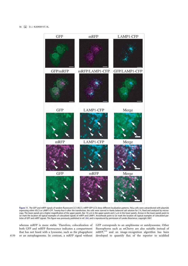
whereas mRFP is more stable. Therefore, colocalization ofboth GFP and mRFP fluorescence indicates a compartmentthat has not fused with a lysosome, such as the phagophore
4150 or an autophagosome. In contrast, a mRFP signal without
GFP corresponds to an amphisome or autolysosome. Otherfluorophores such as mCherry are also suitable instead ofmRFP,319 and an image-recognition algorithm has beendeveloped to quantify flux of the reporter to acidified
Figure 11. The GFP and mRFP signals of tandem fluorescent LC3 (tfLC3, mRFP-GFP-LC3) show different localization patterns. HeLa cells were cotransfected with plasmidsexpressing either tfLC3 or LAMP1-CFP. Twenty-four h after the transfection, the cells were starved in Hanks balanced salt solution for 2 h, fixed and analyzed by micros-copy. The lower panels are a higher magnification of the upper panels. Bar: 10 mm in the upper panels and 2 mm in the lower panels. Arrows in the lower panels point to(or mark the location of) typical examples of colocalized signals of mRFP and LAMP1. Arrowheads point to (or mark the location of) typical examples of colocalized par-ticles of GFP and mRFP signals. This figure was previously published in ref. 264, and is reproduced by permission of Landes Bioscience, copyright 2007.
56 D. J. KLIONSKY ET. AL.

4155 compartments.340-342 One of the major advantages of thetandem mRFP/mCherry-GFP reporter method is that itenables simultaneous estimation of both the induction ofautophagy and flux through autophagic compartments with-out requiring the use of any lysosomal inhibitors. The com-
4160 petence of lysosomal digestion of the substrate requiresadditional analysis using methods described above. The useof more than one time point allows visualization ofincreased early autophagosomes followed by increases inlate autophagosomes as an additional assurance that flux
4165 has been maintained.343 In addition, this method can beused to monitor autophagy in high-throughput drug screen-ing studies.341 The quantification of “yellow only” (wherethe yellow signal results from merging the red and greenchannels) and “red only” dots in a stable tandem-fluores-
4170 cent LC3-reporter cell line can be automated by a Cellomicsmicroscope that can be used to assess a huge population ofcells (1,000 or more) over a large number of random fieldsof view.233,344 Notably, organelle-specific variations of thetandem mRFP/mCherry-GFP reporter system have success-
4175 fully been used to analyze selective types of autophagy, suchas pexophagy345 and mitophagy346,347 in mammalian cells.
An alternative dual fluorescence assay involves the RosellapH biosensor. This assay monitors the uptake of material to thelysosome/vacuole and complements the use of the tandem
4180 mRFP/mCherry-GFP reporter. The assay is based upon thegenetically encoded dual color-emission biosensor Rosella, afusion between a relatively pH-stable fast-maturing RFP vari-ant, and a pH-sensitive GFP variant. When targeted to specificcellular compartments or fused to an individual protein, the
4185 Rosella biosensor provides information about the identity ofthe cellular component being delivered to the lysosome/vacuolefor degradation. Importantly, the pH-sensitive dual color fluo-rescence emission provides information about the environmentof the biosensor during autophagy of various cellular compo-
4190 nents. In yeast, Rosella has been successfully used to monitorautophagy of cytosol, mitochondria (mitophagy) and thenucleus (nucleophagy).348-350 Furthermore, the Rosella biosen-sor can be used as a reporter under various conditionsincluding nitrogen depletion-dependent induction of autoph-
4195 agy.348,349 The Rosella biosensor can also be expressed in mam-malian cells to follow either nonselective autophagy(cytoplasmic turnover), or mitophagy.349
Cautionary notes: The use of tandem mRFP/mCherry-GFP-LC3/Atg8 reporters in live imaging experiments can be compli-
4200 cated by the motion of LC3/Atg8 puncta. As a consequence,conventional confocal microscopy may not allow visualizationof colocalized mRFP/mCherry-GFP puncta. In this case, GFPcolocalized puncta represent newly formed autophagic struc-tures whereas mRFP/mCherry-only puncta are ambiguous.
4205 Spinning disk confocal microscopy or rapid acquisition timesmay be required for imaging tandem mRFP/mCherry-GFPproteins, although these techniques require a brighter fluores-cent signal associated with what may be undesirably higher lev-els of transgene expression. One solution is to use the
4210 mTagRFP-mWasabi-LC3 chimera,351 as mTagRFP is brighterthan mRFP1 and mCherry, and mWasabi is brighter thanEGFP.352 Another possibility is to use fixed cells; however, thispresents an additional concern: The use of tandem mRFP/
mCherry-GFP relies on the quenching of the GFP signal in the4215acidic autolysosome; however, fixation solutions are often neu-
tral or weak bases, which will increase the pH of the entire cell.Accordingly, the GFP signal may be restored after fixation(Fig. 12), which would cause an underestimation of the amountof signal that corresponds only to RFP (i.e., in the autolyso-
4220some). Thus, the tissue or cell samples must be properly proc-essed to avoid losing the acidic environment of theautolysosomes. In addition, there may be weak fluorescence ofEGFP even in an acidic environment (pH between 4 and5).263,331 Therefore, it may be desirable to choose a monomeric
4225green fluorescent protein that is more acid sensitive than EGFPfor assaying autophagic flux.
Another caution in the interpretation of the tandem fluores-cent marker is that colocalization of GFP and mRFP/mCherrymight also be seen in the case of impaired proteolytic degrada-
4230tion within autolysosomes or altered lysosomal pH. Finally,expression of tandem mRFP-GFP-LC3 is toxic to some cancercell lines relative to GFP-LC3 or RFP-LC3 (K.S. Choi, personalcommunication). The cytotoxicity of DsRed and its variantssuch as mRFP1 is associated with downregulation of BCL2L1/
4235Bcl-xL.353 In contrast to mRFP-GFP-LC3, overexpression of
mTagRFP-mWasabi-LC3 does not appear to be toxic to HeLacells (J. Lin, personal communication).
The Rosella assay has not been tested in a wide range ofmammalian cell types. Accordingly, the sensitivity and the spec-
4240ificity of the assay must be verified independently until thismethod has been tested more extensively and used more widely.
Finally, it may be desirable to capture the dynamic behaviorof autophagy in real time, to generate data revealing the rate offormation and clearance of autophagosomes over time, rather
4245than single data points. For example, by acquiring signals from2 fluorescent constructs in real time, the rate of change incolocalization signal as a measure of the fusion rate and recy-cling rate between autophagosomes and lysosomes can beassessed.354 Importantly, due to the integral dynamic relation-
4250ship of autophagic flux with the onset of apoptosis and necro-sis, it is advantageous to monitor cell death and autophagic fluxparameters concomitantly over time, which FRET-basedreporter constructs make possible.355
In addition, as the metabolic control of autophagy is becom-4255ing increasingly clear, highlighting a tight network between the
autophagy machinery, energy sensing pathways and the cell’smetabolic circuits,356,357 mitochondrial parameters such as fis-sion and fusion rate as well as the cell’s ATP demand should bemonitored and correlated with autophagic flux data. This will
4260provide a better understanding on the variability of autophagyand cell death susceptibility.
Tandem fluorescent markers show real-time changes inautophagosome fusion with lysosomes, due to entry into anacidic environment; however, fusion is not definitive evidence
4265of substrate or carrier degradation. Lysosomes may be able tofuse, but be unable to degrade newly delivered cargo, as occursin some lysosomal storage diseases. Best practice would be toperform an autophagic flux assay in parallel with quantificationof tandem fluorescent markers to confirm completion of carrier
4270flux.Conclusion: The use of tandem fluorescent constructs,
which display different emission signals depending on the
AUTOPHAGY 57

Figure 12. GFP fluorescence in the autolysosome can be recovered upon neutralization of the pH. (A) GFP-LC3 emits green fluorescence in the autolysosomes of post-mortem processed heart sections. Cryosections of 3.8% paraformaldehyde fixed ventricular myocardium from 3-week-old GFP-LC3 transgenic mice at the baseline (con-trol) or starved for 24 h (starved) were processed for immunostaining using a standard protocol (buffered at pH 7.4). Most of the GFP-LC3 puncta are positive for LAMP1,suggesting that the autolysosomes had recovered GFP fluorescence. (B) Colocalization between GFP-LC3 direct fluorescence (green) and indirect immunostaining for GFP(red). Sections processed as in (A) were immunostained for GFP using a red fluorescence-tagged secondary antibody, and the colocalization with GFP fluorescence wasexamined by confocal microscopy. Almost all of the red puncta emit green fluorescence. Image provided by X. Wang.
58 D. J. KLIONSKY ET. AL.

environment (in particular, GFP fluorescence is sensitive to anacidic pH), provides a convenient way to monitor autophagic
4275 flux in many cell types.
f. Autophagic flux determination using flow andmultispectral imaging cytometryWhereas fluorescence microscopy, in combination with novelautophagy probes, has permitted single-cell analysis of autopha-
4280 gic flux, automation for allowing medium- to high-throughputanalysis has been challenging. A number of methods have beendeveloped that allow the determination of autophagic flux usingflow cytometry,225,311,327,358-361 and commercial kits are nowavailable for monitoring autophagy by flow cytometry. These
4285 approaches make it possible to capture data or, in specializedinstruments, high-content, multiparametric images of cells inflow (at rates of up to 1,000 cells/sec for imaging, and higher innonimaging flow cytometers), and are particularly useful forcells that grow in suspension. Optimization of image analysis
4290 permits the study of cells with heterogeneous LC3 puncta, thusmaking it possible to quantify autophagic flux accurately in sit-uations that might perturb normal processes (e.g., microbialinfection).360,362 Since EGFP-LC3 is a substrate for autophagicdegradation, total fluorescence intensity of EGFP-LC3 can be
4295 used to indicate levels of autophagy in living mammaliancells.358 When autophagy is induced, the decrease in total cellu-lar fluorescence can be precisely quantified in large numbers ofcells to obtain robust data. In another approach, soluble EGFP-LC3-I can be depleted from the cell by a brief saponin extraction
4300 so that the total fluorescence of EGFP-LC3 then represents thatof EGFP-LC3-II alone (Fig. 13A).326,327 Since EGFP-LC3 trans-fection typically results in high relative levels of EGFP-LC3-I,this treatment significantly reduces the background fluorescencedue to nonautophagosome-associated reporter protein. By com-
4305 paring treatments in the presence or absence of lysosomal deg-radation inhibitors, subtle changes in the flux rate of the GFP-LC3 reporter construct can be detected. If it is not desirable totreat cells with lysosomal inhibitors to determine rates of
autophagic flux, a tandem mRFP/mCherry-EGFP-LC3 (or simi-4310lar) construct can also be used for autophagic flux measure-
ments in flow cytometry experiments (see Tandem mRFP/mCherry-GFP fluorescence microscopy).359
These methods, however, require the cells of interest to betransfected with reporter constructs. Since the saponin extrac-
4315tion method can also be combined with intracellular stainingfor endogenous LC3 protein, subtle changes in autophagic fluxcan be measured without the need for reporter transfections(Fig. 13B).
Cautionary notes: Care must be taken when applying flow4320cytometry measurements to adherent cells, particularly neurons
and other cells with interdigitated processes, as the preparationof single cell suspensions entails significant levels of plasmamembrane disruption and injury that can secondarily induceautophagy.
4325Users of the saponin extraction method should carefully titratesaponin concentrations and times of treatment to ensure specificextraction of LC3-I in their systems. Also, it has been observed insome cell types that saponin treatment can lead to nonautophagicaggregation of LC3,328 which should be controlled for in these
4330assays (see GFP-Atg8/LC3 fluorescence microscopy).Cell membrane permeabilization with digitonin and extrac-
tion of the nonmembrane-bound form of LC3 allows combinedstaining of membrane-associated LC3-II protein and anymarkers for detection of autophagy in relation to other cellular
4335events/processes. Based on this approach, a method for moni-toring autophagy in different stages of the cell cycle was devel-oped.363 Thus, the presence of basal or starvation-inducedautophagy is detected in G1, S, and G2/M phases of the cellcycle in MEFs with doxycycline-regulated ATG5 expression. In
4340these experiments cells were gated based on their DNA contentand the relative intensity of GFP-LC3-II and LC3-II expression.This approach might also be used for the detection of autopha-gic flux in different stages of the cell cycle or subG1 apoptoticcell population by measuring accumulation of LC3-II in the
4345presence or absence of lysosomal inhibitors.
Figure 13. Saponin extraction allows quantification of LC3-II fluorescence by FACS. (A) Schematic diagram of the effects of the saponin wash. Due to the reorganization ofthe EGFP-LC3 reporter protein, induction of autophagosome formation does not change the total levels of fluorescence in EGFP-LC3-transfected cells. However, extractionof EGFP-LC3-I with saponin results in a higher level of fluorescence in cells with proportionally higher levels of EGFP-LC3-II-containing autophagosomes. This figure waspreviously published in ref. 327. (B) Saponin extraction can also be used to measure flux of endogenous LC3 protein. Human osteosarcoma cells were starved of aminoacids and serum by incubation in EBSS, for the indicated times in the presence or absence of a 1 h chloroquine (50 mM) treatment. Cells were then washed with PBS con-taining 0.05% saponin and processed for FACS analysis for endogenous LC3. Image provided by K.E. Eng and G.M. McInerney.
AUTOPHAGY 59

Although GFP-LC3 can be used as a reporter for flowcytometry, it is more stable (which is not necessarily idealfor flux measurements) than GFP-SQSTM1 or GFP-NBR1(NBR1 is a selective autophagic substrate with structural
4350 similarity to SQSTM1364). GFP-SQSTM1 displays the largestmagnitude change following the induction of autophagy byamino acid deprivation or rapamycin treatment, and maythus be a better marker for following autophagic flux bythis method (confirmed in SH-SY5Y neuronal cell lines sta-
4355 bly expressing GFP-SQSTM1; E.M. Valente, personalcommunication).365
Conclusion: Medium- to high-throughput analysis ofautophagy is possible using flow and multispectral imagingcytometry (Fig. 14). The advantage of this approach is that
4360 larger numbers of cells can be analyzed with regard to GFP-LC3puncta, cell morphology and/or autophagic flux, and concomi-tant detection of surface markers can be included, potentiallyproviding more robust data than is achieved with other meth-ods. A major disadvantage, however, is that flow cytometry only
4365 measures changes in total GFP-LC3 levels, which can be subjectto modification by changes in transcription or translation, or bypH, and this approach cannot accurately evaluate localization(e.g., to autophagosomes) or lipidation (generation of LC3-II)without further permeabilization of the cell.
4370g. ImmunohistochemistryImmunodetection of ATG proteins (particularly LC3 andBECN1) has been reported as a prognostic factor in varioushuman carcinomas, including lymphoma,197,366 breast carci-noma,367 endometrial adenocarcinoma,368,369 head and neck
4375squamous cell carcinoma,370-372 hepatocellular carcinoma,373,374
gliomas,375 non-small cell lung carcinomas,376 pancreatic377 andcolon adenocarcinomas,378-380 as well as in cutaneous and uvealmelanomas.381,382 Unfortunately, the reported changes oftenreflect overall diffuse staining intensity rather than appropriately
4380compartmentalized puncta. Therefore, the observation ofincreased levels of diffuse LC3 staining (which may reflect adecrease in autophagy) should not be used to draw conclusionsthat autophagy is increased in cancer or other tissue samples.Importantly, this kind of assay should be performed as recom-
4385mended by the Reporting Recommendations for Tumor MarkerPrognostic Studies (REMARK).383 As we identify new drugs formodulating autophagy in clinical applications, this type of infor-mation may prove useful in the identification of subgroups ofpatients for targeted therapy.384-386
4390In mouse and rat tissues, endogenous LC3, ATG4B, andATG9A have been detected by immnohistochemical analysesusing both paraffin sections and cryosections.293,387-389 Whenautophagosomes are absent, the localization pattern of LC3 in
Figure 14. Assessing autophagy with multispectral imaging cytometry. (A) Bright Detail Intensity (BDI) measures the foreground intensity of bright puncta (that are 3 pix-els or less) within the cell image. For each cell, the local background around the spots is removed before intensity calculation. Thus, autophagic cells with puncta havehigher BDI values. (B) Media control (untreated wild type), rapamycin-treated wild-type and atg5¡/¡ MEFs were gated based on BDI. Representative images of cells withhigh or low BDI values. Scale bar: 10 mm. Images provided by M.L. Albert.
60 D. J. KLIONSKY ET. AL.

the cells of various tissues is diffuse and cytosolic. Moreover,4395 intense fibrillary staining of LC3 is detectable along dendrites
of intact neurons, whereas granular staining for LC3 appearsmainly in the perikarya of neurons in CTSD- or CTSB- andCTSL-deficient mouse brains.293 LC3 puncta are also observedin mice in the peripheral nerves, specifically in Schwann cells
4400 after neurodegeneration,390 and Paneth cells of the small intes-tine from human Crohn disease patients and mouse models ofintestinal inflammation driven by ER-stress exhibit strong LC3puncta staining.391,392 In various neurodegenerative states, LC3puncta may be numerous in neurites, especially within dystro-
4405 phic swellings and, in many cases, these vacuoles are amphi-somes or autolysosomes, reflecting the delayed or inhibiteddegradation of LC3 despite the presence of abundant hydrolaseactivity.57,66 In developing inner ear and retinal tissue inchicken, BECN1 is detected by immunofluorescence; in chick
4410 retina AMBRA1 is also detected.393-395 Finally, in non-mamma-lian vertebrates, BECN1 was detected during follicular atresia inthe ovary of 3 fish species using paraffin sections; a punctateimmunostaining for BECN1 is scattered throughout the cyto-plasm of the follicular cells when they are in intense phagocytic
4415 activity for yolk removal.396
Cautionary notes: One problem with LC3 IHC is that insome tissues this protein can be localized in structures otherthan autophagosomes. For example, in murine hepatocytesand cardiomyocytes under starved conditions, endogenous
4420 LC3 is detected not only in autophagosomes but also onlipid droplets.397 In neurons in ATG7-deficient mice, LC3accumulates in ubiquitin- and SQSTM1-positive aggre-gates.398 In neurons in aging or neurodegenerative diseasestates, LC3 is commonly present in autolysosomes and may
4425 be abundant in lipofuscin and other lysosomal residual bod-ies.57 Thus, immunodetection of LC3 in cytoplasmic gran-ules is not sufficient to monitor autophagy in vivo. Toevaluate autophagy by the methods of immunohistochemis-try, it is necessary to identify the autophagosomes directly
4430 using the ABC technique for TEM observation (see Trans-mission electron microscopy).77
Conclusion: It has not been clearly demonstrated that IHCof ATG proteins in tissues corresponds to autophagy activity,and this area of research needs to be further explored before we
4435 can make specific recommendations.
3. SQSTM1 and related LC3 binding protein turnoverassays
In addition to LC3, SQSTM1/p62 or other receptors such asNBR1, can also be used as protein markers, at least in certain
4440 settings.26,399 For example, SQSTM1 can be detected as punctaby IHC in cancer cells, similar to LC3.372 The SQSTM1 proteinserves as a link between LC3 and ubiquitinated substrates.84
SQSTM1 and SQSTM1-bound polyubiquitinated proteinsbecome incorporated into the completed autophagosome and
4445 are degraded in autolysosomes, thus serving as an index ofautophagic degradation (Fig. 15). Inhibition of autophagy cor-relates with increased levels of SQSTM1 in mammals andDrosophila, suggesting that steady state levels of this proteinreflect the autophagic status.61,389,400-404 Similarly, decreased
4450 SQSTM1 levels are associated with autophagy activation. The
phosphorylation of SQSTM1 at Ser403 appears to regulate itsrole in the autophagic clearance of ubiquitinated proteins, andanti-phospho-SQSTM1 antibodies can be used to detect themodified form of the protein.324
4455Cautionary notes: SQSTM1 changes can be cell type andcontext specific. In some cell types, there is no change in theoverall amount of SQSTM1 despite strong levels of autophagyinduction, verified by the tandem mRFP/mCherry-GFP-LC3reporter as well as ATG7- and lysosome-dependent turnover of
4460cargo proteins (C.T. Chu, personal observation). In other con-texts, a robust loss of SQSTM1 does not correlate withincreased autophagic flux as assessed by a luciferase-based mea-sure of flux;245 a decrease of SQSTM1 can even relate to ablockage of autophagy due to cleavage of the protein, together
4465with other autophagy proteins, by caspases or calpains.405
SQSTM1 may be transcriptionally upregulated under certainconditions,317,406-409 further complicating the interpretation ofresults. For example, SQSTM1 upregulation, and at least tran-sient increases in the amount of SQSTM1, is seen in some sit-
4470uations where there is an increase in autophagic flux.410-412
One such case is seen during retinoic acid-induced differentia-tion of AML cells where SQSTM1 is upregulated407 with con-comitant increased autophagic flux.413 Activation of a signalingpathway, e.g. RAF1/Raf-MAP2K/MEK-MAPK/ERK, can also
4475upregulate SQSTM1 transcription.414 SQSTM1 mRNA is also
Figure 15. Regulation of the SQSTM1 protein during autophagy. (A) The level ofSQSTM1 during starvation. Atg5C/C and atg5¡/¡ MEFs were cultured in DMEMwithout amino acids and serum for the indicated times, and then subjected toimmunoblot analysis using anti-SQSTM1antibody (Progen Biotechnik, GP62). Thisfigure was previously published in ref. 26, and is reproduced by permission ofLandes Bioscience, copyright 2007. (B) The level of SQSTM1 in the brain of neural-cell specific Atg5 knockout mice. Image provided by T. Hara.
AUTOPHAGY 61

upregulated following prolonged starvation, which can restorethe SQSTM1 protein level to that before starvation.415,416 In thesame way, physical exercise, especially when performed duringstarvation, increases the SQSTM1 mRNA level in skeletal mus-
4480 cle, and can lead to an incorrect interpretation of autophagicflux if only the protein level is measured.417,418 Anotherinstance when both mRNA and protein levels of SQSTM1 areelevated even though autophagic flux is not impaired isobserved in aneuploid human and murine cells that are gener-
4485 ated by introduction of 1 or 2 extra chromosomes.419,420 Thus,appropriate positive and negative controls are needed prior tothe use of SQSTM1 as a flux indicator in a particular cellularcontext, and we recommend monitoring the SQSTM1 mRNAlevel as part of a complete analysis, or determining the
4490 SQSTM1 protein level in the presence of actinomycin D.Of interest, SQSTM1 hyperexpression at both gene and pro-
tein levels can be observed in muscle atrophy induced by can-cer, though not by glucocorticoids, suggesting that the stimulusinducing autophagy may also be relevant to the differential reg-
4495 ulation of autophagy-related proteins.421 One solution to prob-lems relating to variations in SQSTM1 expression levels is touse a HaloTag!-p62 (SQSTM1) chimera.422 The chimeric pro-tein can be covalently labeled with HaloTag! ligands, and theloss of signal can then be monitored without interference by
4500 subsequent changes in protein synthesis. Similarly, a stable cellline expressing EGFP-tagged SQSTM1 under the control of aninducible promoter can be used to assess the rates of SQSTM1degradation, taking into account the limitations outlined above(see Autophagic flux determination using flow and multispectral
4505 imaging cytometry).365 A similar system exists in Drosophila inwhich a GFP-tagged SQSTM1 can be expressed using the UAS-GAL4 system.423 It is worth noting that tetracycline can reduceautophagy levels; therefore, the appropriate control of only tet-racycline addition has to be included if using an inducible pro-
4510 moter that responds to this drug.424 Yet another solution is toemploy a radioactive pulse-chase assay to measure the rates ofSQSTM1 degradation.425
SQSTM1 contains a LIR as well as a ubiquitin bindingdomain, and appears to act by linking ubiquitinated substrates
4515 with the autophagic machinery. Nonetheless, it would be pru-dent to keep in mind that SQSTM1 contains domains thatinteract with several signaling molecules,426 and SQSTM1 maybe part of MTORC1.427 Thus, it may have additional functionsthat need to be considered with regard to its role in autophagy.
4520 In the context of autophagy as a stress response, the complexityof using SQSTM1 as an autophagy marker protein is under-scored by its capacity to modulate the NFE2L2/NRF2 anti-oxi-dant response pathway through a KEAP1 bindingdomain.428,429 In fact, SQSTM1 may, itself, be transcriptionally
4525 induced by NFE2L2.430 Furthermore, it is preferable to examineendogenous SQSTM1 because overexpression of this proteinleads to the formation of protein inclusions. In fact, evenendogenous SQSTM1 becomes Triton X-100-insoluble in thepresence of protein aggregates and when autophagic degrada-
4530 tion is inhibited; thus, results with this protein are often con-text-dependent. Indeed, there is a reciprocal crosstalk betweenthe UPS and autophagy, with SQSTM1 being a key linkbetween them.431 First, SQSTM1 participates in proteasomaldegradation, and its level may also increase when the
4535proteasome is inhibited.432 Accordingly, the SQSTM1 degrada-tion rate should be analyzed in the presence of an inhibitorsuch as epoxomicin or lactacystin to determine the contribu-tion from the proteasome (see Autophagy inhibitors andinducers for potential problems with MG132).433 Second, the
4540accumulation of SQSTM1 due to autophagy inhibition canimpair UPS function by competitively binding ubiquitinatedproteins, preventing their delivery to, and degradation by, theproteasome.434 Accordingly, it may be advisable to measure theUPS flux by using UbG76V-GFP, a ubiquitin-proteasome activ-
4545ity reporter, when SQSTM1 accumulation is observed. Thus, itis very important to determine whether autophagy alone or inconjunction with the UPS accounts for substrate degradationinduced by a particular biological change. A number of stres-sors that impair the UPS induce the aggregation/dimerization
4550of SQSTM1, and this can be seen by the detection of a highmolecular mass (~150 kDa) protein complex by western blot,which is recognized by SQSTM1 antibodies (R. Franco, per-sonal communication).435,436 Although the accumulation ofthis protein complex can be related to the accumulation of
4555ubiquitinated SQSTM1-bound proteins, or the dimerization/inactivation of SQSTM1 (R. Franco, personal communica-tion),437 evaluation of the ratio between SQSTM1 (aggregates/dimers) and SQSTM1 monomers is likely a better measurementof changes in SQSTM1 dynamics linked to autophagy or the
4560UPS.SQSTM1 is also a substrate for CASP6/caspase 6 and
CASP8 (as well as CAPN1/calpain 1), which may confoundits use in examining cell death and autophagy.438 This isone reason why SQSTM1 degradation should also be ana-
4565lyzed in the presence of a pan-caspase inhibitor such as Q-VD-OPh before concluding that autophagy is activatedbased on a decrease of this protein.405 Another issue is thatsome phosphatidylinositol 3-kinase (PtdIns3K) inhibitorssuch as LY294002, and to a lesser extent wortmannin (but
4570apparently not 3-MA),329 can inhibit protein synthesis;439
this might in turn affect the turnover of SQSTM1 and LC3,which could influence conclusions that are drawn from thestatus of these proteins regarding autophagic flux or ALISformation. Accordingly, it may be advisable to measure pro-
4575tein synthesis and proteasome activity along with autophagyunder inhibitory or activating conditions. With regard toprotein synthesis, it is worth noting that this can be moni-tored through a nonradioactive method.440
Western blot analysis of cell lysates prepared using NP40- or4580Triton X-100-containing lysis buffers in autophagic conditions
typically shows a reduction in SQSTM1 levels. However, thisdoes not necessarily indicate that SQSTM1 is degraded, becauseSQSTM1 aggregates are insoluble in these detergent lysis condi-tions.317,441 Moreover, in some instances SQSTM1 levels do not
4585change in the soluble fractions despite autophagic degradation,a finding that might be explained by simultaneous transcrip-tional induction of the gene encoding SQSTM1, since the solu-ble fraction accounts only for the diffuse or free form ofSQSTM1. Accumulation of SQSTM1 in the Triton X-100-insol-
4590uble fraction can be observed when autophagy-mediated degra-dation is inhibited. Under conditions of higher autophagic flux,accumulation of SQSTM1 in Triton X-100-insoluble fractionsmay not be observed and SQSTM1 levels may be reduced or
62 D. J. KLIONSKY ET. AL.
maintained. The simplest approach to circumvent many of4595 these problems is using lysis buffer that allows identification of
the entire cellular pool of SQSTM1 (e.g., containing 1% SDS);however, additional assessment of both Triton X-100-solubleand -insoluble fractions will provide further informationregarding the extent of SQSTM1 oligomerization.398 Note,
4600 when performing a western blot using an SQSTM1 antibody, itis always a good idea to include a positive control in whichSQSTM1 accumulates, such as an atg8a mutant (e.g., seeFig. S3 in ref. 442).
To conclusively establish SQSTM1 degradation by4605 autophagy, SQSTM1 levels in both Triton X-100-soluble and
-insoluble fractions need to be determined upon treatmentwith autophagy inducers in combination with autophagyinhibitors, such as those that inhibit the autolysosomal deg-radation steps (e.g., protease inhibitors, chloroquine or bafi-
4610 lomycin A1). Additionally, an alteration in the level ofSQSTM1 may not be immediately evident with changesobserved in autophagic flux upon certain chemical perturba-tions (S. Sarkar, personal communication). Whereas LC3changes may be rapid, clearance of autophagy substrates
4615 may require a longer time. Therefore, if LC3 changes areassessed at 6 h or 24 h after a drug treatment, SQSTM1 lev-els can be tested not only at the same time points, but alsoat later time points (24 h or 48 h) to determine the maximalimpact on substrate clearance. An alternative method is
4620 immunostaining, with and without autophagy inhibitors, forSQSTM1, which will appear as either a diffuse or punctatepattern. Experiments with autophagy inducers and inhibi-tors, in combination with western blot and immunostaininganalyses, best establish autophagic degradation based on
4625 SQSTM1 turnover. A final point, however, is that empiricalevidence suggests that the species-specificity of antibodiesfor detecting SQSTM1 must be taken into account. Forexample, some commercial antibodies recognize both humanand mouse SQSTM1, whereas others detect the human, but
4630 not the mouse protein.443 Another issue with detectingSQSTM1 in the context of human diseases is that it can bemutated (e.g., in Paget disease of bone).444 Thus, care shouldbe taken to ensure that potential mutations are not affectingthe epitopes that are recognized by anti-SQSTM1 antibodies
4635 when using western blotting to detect this protein.As an alternative, the SQSTM1:BECN1 protein level ratio
can be used as a readout of autophagy.445 Since both decreasedSQSTM1 levels and increased BECN1 levels correlate withenhanced autophagy (as noted in the present review), a
4640 decreased SQSTM1:BECN1 protein level ratio (when derivedfrom the same protein extract) may, cautiously, be interpretedas augmented autophagy, keeping in mind that SQSTM1 geneexpression varies significantly under different conditions andmay obscure the meaning of a change in the amount of
4645 SQSTM1 protein. As a general note, using ratios of the levels ofproteins changing in opposite directions, rather than the pro-tein levels themselves, could be beneficial since it overcomesthe loading normalization issue. The often-used alternativeapproach of housekeeping proteins to normalize for loading
4650 biases among samples is sometimes problematic as levels of theHKPs change under various physiological, pathological andpharmacological conditions.446-450
Finally, a novel protein family of autophagy receptors,named CUET (from Cue5/Tollip), was identified, which in con-
4655trast to SQSTM1 and NBR1 has members that are present in alleukaryotes.451 The CUET proteins also possess a ubiquitin-binding CUE-domain and an Atg8-family interacting motif(AIM)/LIR sequence that interacts with Atg8/LC3. In theirabsence, cells are more vulnerable to the toxicity resulting from
4660aggregation-prone proteins showing that CUET proteins, andmore generally autophagy, play a critical evolutionarily con-served role in the clearance of cytotoxic protein aggregates.451
Experiments in yeast have shown that Cue5 and the cyto-plasmic proteins that require this autophagy receptor for rapid
4665degradation under starvation conditions could be potentiallygood marker proteins for measuring autophagic flux.
Special caution must be taken when evaluating SQSTM1 lev-els in models of protein aggregation. Small protoaggregatesoften stain positively for SQSTM1 and may be similar in size to
4670autophagic puncta. Similarly, GFP-u/GFP-degron reporters(designed as an unstable variant that undergoes proteasome-dependent degradation) will mark SQSTM1-positive proteininclusions. Last, some types of aggregates and inclusions willrelease soluble SQSTM1 or GFP-u/GFP-degron under cell lysis
4675or denaturing conditions, which can skew the interpretation ofsoluble SQSTM1 and/or proteasomal function, accordingly.
Conclusion: There is not always a clear correlation betweenincreases in LC3-II and decreases in SQSTM1. Thus, althoughanalysis of SQSTM1 can assist in assessing the impairment of
4680autophagy or autophagic flux, we recommend using SQSTM1only in combination with other methods detailed in theseguidelines to monitor flux. See also the discussion in Autopha-gic flux determination using flow and multispectral imagingcytometry.
46854. TOR/MTOR, AMPK and Atg1/ULK1
Atg1/ULK1 are central components in autophagy that likely actat more than one stage of the process. There are multiple ULKisoforms in mammalian cells including ULK1, ULK2, ULK3,ULK4 and STK36.452 ULK3 is a positive regulator of the
4690Hedgehog signaling pathway,453 and its overexpression inducesboth autophagy and senescence.454 Along these lines, ectopicULK3 displays a punctate pattern upon starvation-inducedautophagy induction.454 ULK3, ULK4 and STK36, however,lack the domains present on ULK1 and ULK2 that bind
4695ATG13 and RB1CC1/FIP200.455 Thus, ULK3 may play a rolethat is restricted to senescence and that is independent of thecore autophagy machinery. ULK2 has a higher degree of iden-tity with ULK1 than any of the other homologs, and they mayhave similar functions that are tissue specific. However, ULK1
4700may be the predominant isoform involved in autophagy, asknockdown of ULK2 does not affect movement of ATG9.456
Similarly, pharmacological inhibition of ULK1 and ULK2, withthe compound MRT68921, blocks macroautophagy andexpression of a drug-resistant ULK1 mutant is sufficient to res-
4705cue this block.457 The stability and activation of ULK1, but notULK2, is dependent on its interaction with the HSP90-CDC37chaperone complex. Pharmacological or genetic inhibition ofthe chaperone complex increases proteasome-mediated
AUTOPHAGY 63

turnover of ULK1, impairing its kinase activity and ability to4710 promote both starvation-induced autophagy and mitophagy.458
AMPK (AMP-activated protein kinase) is a multimeric ser-ine/threonine protein kinase comprised of PRKAA1/AMPKa1or PRKAA2/AMPKa2 (a, catalytic), the PRKAB1/AMPKb1 orPRKAB2/AMPKb2 (b, scaffold), and the PRKAG1/AMPKg1,
4715 PRKAG2/AMPKg2 or PRKAG3/AMPKg3 (g, regulatory) sub-units. The enzyme activity of AMPK is dependent on phosphor-ylation of the a-subunit on Thr172,459,460 and, therefore, can beconveniently monitored by western blotting with a phosphospe-cific antibody against this site. In some cells, Thr172 is phos-
4720 phorylated by CAMKK2/CaMKKb, whereas in others it is asubstrate of the STK11/LKB1 kinase. Regulation of AMPKactivity is mediated primarily by Thr172-dephosphorylatingprotein phosphatases such as PPP1/PP1 (protein phosphatase1) and PPP2/PP2A (protein phosphatase 2).461 Thr172 dephos-
4725 phorylation is modulated by adenine nucleotides that bind com-petitively to regulatory sites in the PRKAG/g-subunit. AMP andADP inhibit dephosphorylation and promote AMPK activity,whereas Mg2,C-ATP has the opposite effect.460 Thus, AMPKacts as a fine-tuned sensor of the overall cellular energy charge
4730 that regulates cellular metabolism to maintain energy homeosta-sis. Overexpression of a dominant negative mutant (R531G) ofPRKAG2, the g¡subunit isoform 2 of AMPK that is unable tobind AMP, makes it possible to analyze the relationship betweenAMP modulation (or alteration of energetic metabolism) and
4735 AMPK activity.462,463 Activation of AMPK is also associatedwith the phosphorylation of downstream enzymes involved inATP-consuming processes, such as fatty acid (ACAC [acetyl-CoA carboxylase]) and cholesterol (HMGCR [3-hydroxy-3-methylglutaryl-CoA reductase]) biosynthesis.
4740 The role of AMPK in autophagy is complex and highlydependent on both cell type and metabolic conditions. Further-more, as noted above, there are 2 isoforms of the catalytic sub-unit, PRKAA1/AMPKa1 and PRKAA2/AMPKa2, and thesemay have distinct effects with regard to autophagy (C. Koume-
4745 nis, personal communication). In liver cells, AMPK suppressesautophagy at the level of cargo sequestration, as indicated bythe rapid sequestration-inhibitory effects of a variety of AMPKactivators, whereas it appears to stimulate autophagy in manyother cell types, including fibroblasts, colon carcinoma cells
4750 and skeletal muscle.464-473 Autophagy-promoting effects ofAMPK are most evident in cells cultured in a complete mediumwith serum and amino acids, where cargo sequestration is oth-erwise largely suppressed.470 Presumably, AMPK antagonizesthe autophagy-inhibitory effect of amino acids (at the level of
4755 phagophore assembly) by phosphorylating proteins involved inMTORC1 signaling, such as TSC2474 and RPTOR475 as well theMTORC1 target ULK1 (see below).476-478
Compound C is an effective and widely used inhibitor ofactivated (phosphorylated) AMPK.479,480 However, being a
4760 nonspecific inhibitor of oxidative phosphorylation,481,482
this drug has been observed to inhibit autophagy underconditions where AMPK is already inactive or knockedout,483 and it has even been shown to stimulate autophagyby an AMP-independent mechanism.482,484 Compound C
4765 thus cannot be used as a stand-alone indicator of AMPKinvolvement, but can be used along with shRNA-mediatedinhibition of AMPK.
TORC1 is an autophagy-suppressive regulator that integra-tes growth factor, nutrient and energy signals. In most systems,
4770inhibition of MTOR leads to induction of autophagy, andAMPK activity is generally antagonistic toward MTOR func-tion. MTORC1 mediates the autophagy-inhibitory effect ofamino acids, which stimulate the MTOR protein kinasethrough a RRAG GTPase dimer. INS/insulin and growth fac-
4775tors activate MTORC1 through upstream kinases includingAKT/protein kinase B and MAPK1/ERK2-MAPK3/ERK1when the energy supply is sufficient, whereas energy depletionmay induce AMPK-mediated MTORC1 inhibition and autoph-agy stimulation, for example, during glucose starvation. In con-
4780trast, amino acid starvation can strongly induce autophagyeven in cells completely lacking AMPK catalytic activity.485
AMPK and MTORC1 regulate autophagy through coordi-nated phosphorylation of ULK1. Under glucose starvation,AMPK promotes autophagy by directly activating ULK1 through
4785phosphorylation, although the exact AMPK-mediated ULK1phosphorylation site(s) remains unclear (Table 2).473,476-478
Under conditions of nutrient sufficiency, high MTORC1 activ-ity prevents ULK1 activation by phosphorylating alternateULK1 residues and disrupting the interaction between ULK1
4790and AMPK. There are commercially available phospho-specificantibodies that recognize different forms of ULK1. For exam-ple, phosphorylation at Ser555, an AMPK site, is indicative ofincreased autophagy in response to nutrient stress, whereasSer757 is targeted by MTOR to inhibit autophagy. Even the
4795autophagy-suppressive effects of AMPK could, conceivably, bemediated through ULK1 phosphorylation, for example, at theinhibitory site Ser638.486 AMPK inhibits MTOR by phosphory-lating and activating TSC2.487 Therefore, AMPK is involved inprocesses that synergize to activate autophagy, by directly acti-
4800vating ULK1, and indirectly impairing MTOR-dependent inhi-bition of ULK1. The identification of ULK1 as a direct target ofMTORC1 and AMPK represents a significant step toward thedefinition of new tools to monitor the induction of autophagy.However, further studies directed at identifying physiological
4805substrates of ULK1 will be essential to understand how ULK1activation results in initiation of the autophagy program. Alongthese lines, ULK1 phosphorylates AMBRA1,488 and theMLCK-like protein Sqa,489 as well as ATG13, ATG9 andRB1CC1/FIP200.423,490-493 Furthermore, following amino acid
4810starvation or MTOR inhibition, the activated ULK1 phosphor-ylates BECN1 on Ser14, enhancing the activity of the complexescontaining ATG14 and PIK3C3/VPS34. This BECN1 phos-phorylation by ULK1 is required for full autophagic induc-tion.494 In addition, ULK1 binds to, and phosphorylates,
4815RPTOR, leading to inhibition of MTORC1.495 Furthermore,ULK1 itself appears to be able to mediate inhibitory AMPKphosphorylation to generate a negative feedback loop.496 Notethat caution should be taken to use appropriate inhibitors ofphosphatases (e.g, sodium fluoride, and beta-glycerophosphate)
4820in cell lysis buffer before analyzing the phosphorylation ofAMPK and ULK1 at serine and threonine sites.
TORC1 activity can be monitored by following the phos-phorylation of its substrates, such as EIF4EBP1/4E-BP1/PHAS-I and RPS6KB/p70S6 kinase or the latter’s down-
4825stream target, RPS6/S6, for which good commercial antibod-ies are available.497-499 In mammalian cells, the analysis
64 D. J. KLIONSKY ET. AL.

should focus on the phosphorylation of S6K1 at Thr389, andEIF4EBP1 at Thr37 and Thr46, which are directly phosphor-ylated by MTORC1.500 The MTORC1-dependent phosphory-
4830 lation of EIF4EBP1 can be detected as a molecular mass shiftby western blot.499 Examining the phosphorylation status ofRPS6KB and EIF4EBP1 may be a better method for monitor-ing MTORC1 activity than following the phosphorylation ofproteins such as RPS6, because the latter is not a direct sub-
4835 strate of MTORC1 (although RPS6 phosphorylation is agood readout for RPS6KB1/2 activities, which are directlydependent on MTOR), and it can also be phosphorylated byother kinases such as RPS6KA/RSK. Furthermore, the mech-anisms that determine the selectivity as well as the sensitivity
4840 of MTORC1 for its substrates seem to be dependent on theintegrity and configuration of MTORC1. For example, rapa-mycin strongly reduces RPS6KB1 phosphorylation, whereasits effect on EIF4EBP1 is more variable. In the case of rapa-mycin treatment, EIF4EBP1 can be phosphorylated by
4845 MTORC1 until rapamycin disrupts MTORC1 dimerizationand its integrity, whereas RPS6KB1 phosphorylation isquickly reduced when rapamycin simply interacts withMTOR in MTORC1 (see Autophagy inhibitors and inducersfor information on catalytic MTOR inhibitors such as
4850 torin1).500 Since it is likely that other inhibitors, stress, andstimuli may also affect the integrity of MTORC1, a decreaseor increase in the phosphorylation status of one MTORC1substrate does not necessarily correlate with changes inothers, including ULK1. Therefore, reliable anti-phospho-
4855 ULK1 antibodies should be used to directly examine thephosphorylation state of ULK1, along with additional
experimental approaches to analyze the role of the MTORcomplex in regulating autophagy. The MTORC1-mediatedphosphorylation of AMBRA1 on Ser52 has also been
4860described as relevant to ULK1 regulation and autophagyinduction.488,501 In line with what is described for ULK1, theanti-phospho-AMBRA1 antibody, which is commerciallyavailable, could be used to indirectly measure MTORC1activity.501
4865Activation/assembly of the Atg1 complex in yeast (com-posed of at least Atg1-Atg13-Atg17-Atg31-Atg29) or the ULK1complex in mammals (ULK1-RB1CC1/FIP200-ATG13-ATG101) is one of the first steps of autophagy induction.Therefore, activation of this complex can be assessed to moni-
4870tor autophagy induction. In yeast, dephosphorylation of Atg13is associated with activation/assembly of the core complex thatreflects the reduction of TORC1 and PKA activities. Therefore,assessing the phosphorylation levels of this protein by immu-noprecipitation or western blotting502-505 can be used not only
4875to follow the early steps of autophagy but also to monitor theactivity of some of the upstream nutrient-sensing kinases.Because this protein is not easily detected when cells are lysedusing conventional procedures, a detailed protocol has beendescribed.506 In addition, the autophosphorylation of Atg1 at
4880Thr226 is required for its kinase activity and for autophagyinduction; this can be detected using phospho-specific antibod-ies, by immunoprecipitation or western blotting (Fig. 16).507,508
In Drosophila, TORC1-dependent phosphorylation of Atg1and Atg1-dependent phosphorylation of Atg13 can be indi-
4885rectly determined by monitoring phosphorylation-inducedelectromobility retardation (gel shift) of protein bands in
Table 2. Phosphorylation targets of AKT, AMPK, GSK3B, MTORC1, PKA and Atg1/ULK1.
Protein and phosphorylation site Main kinase Function Ref
AMBRA1 S52 TORC1 Inhibits AMBRA1-dependent activation of ULK1 501
Atg1 TORC1 Inhibits Atg1 kinase activity 504
Atg1 PKA Regulation of kinase activity 1523
Atg9 Atg1 Recruitment of Atg protein to the PAS 493
Atg13 TORC1 Interaction with Atg1, assembly of Atg1 kinase complex 504,1524
Atg13 PKA Regulates localization to the PAS 1525
BECN1 S14 ULK1 Increases the activity of the PtdIns3K 494
BECN1 S90 MAPKAPK2-MAPKAPK3
Stimulates macroautophagy 1526
BECN1 S91, S94 (S93, S96 in human) AMPK Required for glucose starvation-induced macroautophagy 1527
BECN1 Y229, Y233 EGFR Inhibits macroautophagy 523
BECN1 S234, S295 AKT Suppresses macroautophagy 522
LC3 S12 PKA Inhibits macroautophagy by reducing recruitment to phagophores 343
MTOR S2448 AKT Correlates with the activity of MTORC1 1528
MTOR S2481 Autophosphorylation Necessary for MTORC1 formation and kinase activity 1529
NBR1 T586 GSK3A/B Modulates protein aggregation 1530
RPS6KB T389 MTORC1 (apparentlyindirect, throughreduction ofdephosphorylation)
Necessary for protein activity 1531
RPS6KB S371 GSK3B Necessary for T389 phosphorylation and the activity of RPS6KB 1532
RPTOR S792 AMPK Suppresses MTORC1 475
SQSTM1 S403 ULK1 (also TBK1,CSNK, CDK1)
Promotes autophagic degradation of SQSTM1 and its substrates 1533
ULK1 S555 AMPK (direct) Necessary for ATG13-ULK1 interaction and for autophagy mediated by ULK complex 477
ULK1 S317, S467, S555, S574, S777 AMPK (direct) Necessary for the kinase activity of ULK1 477,478
ULK1 S757 MTORC1 Prevents ULK1 interaction with AMPK 478
ULK1 S758 MTORC1 Facilitates ULK1 interaction with AMPK 478,512
ULK1 S637 MTORC1, AMPK Facilitates ULK1 interaction with AMPK 477,512
ULK1 (uncertain site between 278 and 351) Autophosphorylation Modulates the conformation of the C-terminal tail and prevents its interaction with ATG13 492,1534
AUTOPHAGY 65

immunoblot images.423,509,510 Nutritional starvation suppressesTORC1-mediated Atg1 phosphorylation,423,509 while stimulat-ing Atg1-mediated Atg13 phosphorylation.423,509,510 In mam-
4890 malian cells, the phosphorylation status of ULK1 at theactivating sites (Ser317, 777, 467, 555, 637, or Thr574) ordephosphorylation at inactivating sites (Ser637, 757) can bedetermined by western blot using phospho-specific antibod-ies.477,478,480,486,511,512 In general, the core complex is stable in
4895 mammalian cells, although, as noted above, upstream inhibi-tors (MTOR) or activators (AMPK) may interact dynamicallywith it, thereby determining the status of autophagy.
One additional topic that bears on ULK1 concerns the pro-cess of LC3-associated phagocytosis (see Noncanonical use of
4900 autophagy-related proteins). LAP is a type of phagocytosis inmacrophages that involves the conjugation of LC3 to single-membrane pathogen-containing phagosomes, a process thatpromotes phagosome acidification and fusion with lyso-somes.182 Although ULK1 is not required for LAP, in this con-
4905 text it is important to note that UNC-51 (the Atg1 homolog inC. elegans) is required for apoptotic cell corpse clearance (aprocess corresponding to LAP) during embryonic developmentin worms,513 whereas this process is mediated by LAP in mam-mals,180 and does not require UNC-51 in C. elegans Q cell neu-
4910 roblasts.514 In human macrophages infected withMycobacterium tuberculosis, it has been shown that MORN2 isrecruited at the phagosome membrane containing M. tubercu-losis to induce the recruitment of LC3, and subsequent matura-tion into phagolysosomes. In addition, MORN2 drives
4915 trafficking of M. tuberculosis to a single-membrane compart-ment. Thus, in certain conditions MORN2 can be used to helpto make the distinction between autophagy and LAP.515
Cautionary notes: A decrease in TORC1 activity is a goodmeasure for autophagy induction; however, TORC1 activity
4920 does not necessarily preclude autophagy induction because thereare TOR-independent mechanisms that induce autophagy bothin mammals and yeast.516-520 Along these lines, whereas in mostsystems inhibition of MTOR leads to the induction of autoph-agy, there are instances in commonly used cancer cell lines in
4925 which MTOR appears to be a positive effector.521 Also, MTORsuppression does not always induce autophagy, such as whenBECN1 undergoes inhibitory phosphorylation by the growthfactor signaling molecules EGFR and AKT.522,523 Note that theeffect of everolimus in EGFR-transgenic mice is not mainly
4930 attributable to autophagy although it suppresses MTOR andinduces autophagy in EGFR-driven lung cancer cell lines.524 Inadult skeletal muscle, active MTORC1 phosphorylates ULK1 atSer757 to inhibit the induction of autophagosome formation.Thus, induction of autophagy requires inhibition of MTORC1
4935 and not of MTORC2.525,526 There is also evidence that inhibition
of MTORC1 is not sufficient to maintain autophagic flux, butrequires additional activation of FOXO transcription factors forthe upregulation of autophagy gene expression.468 In addition,MTORC1 is downstream of AKT; however, oxidative stress
4940inhibits MTOR, thus allowing autophagy induction, despite theconcomitant activation of AKT.150 Also, persistent MTORC1inhibition can cause downregulation of negative feedback loopson IRS-MTORC2-AKT that results in the reactivation ofMTORC2 under conditions of ongoing starvation.222,415,527
4945Along these lines, both TORC1 and autophagy can be active inspecific cell subpopulations of yeast colonies.520 Thus, it is neces-sary to be cautious in deciding how to monitor the TOR/MTORpathway, and to verify that the pathway being analyzed displaysTOR/MTOR-dependent inhibition.
4950In addition, the regulation of autophagy by MTOR can beULK1-independent. During mycobacterial infection of macro-phages, MTOR induces the expression of MIR155 and MIR31to sustain the activation of the WNT5A and SHH/sonic hedge-hog pathways. Together, these pathways contribute to the
4955expression of lipoxygenases and downregulation of IFNG-induced autophagy.528 Signaling pathways can be monitored bywestern blotting, and TaqMan miRNA assays are available todetect these miRNAs.
One problem in monitoring assembly of the ULK1 complex4960is the low abundance of endogenous ULK1 in many systems,
which makes it difficult to detect phospho-ULK1 by westernblot analysis. In addition, Atg1/ULK1 is phosphorylated bymultiple kinases, and the amount of phosphorylation at differ-ent sites can increase or decrease during autophagy induction.
4965Thus, although there is an increase in phosphorylation at theactivating sites upon induction, the overall phosphorylationstates of ULK1 and ATG13 are decreased under conditions thatlead to induction of autophagy; therefore, monitoring changesin phosphorylation by following molecular mass shifts upon
4970SDS-PAGE may not be informative. In addition, such phos-phorylation/dephosphorylation events are expected to occurrelatively early (1–2 h) in the signaling cascade of autophagy.Therefore, it is necessary to optimize treatment time condi-tions. Finally, in Arabidopsis and possibly other eukaryotes, the
4975ATG1 and ATG13 proteins are targets of autophagy, whichmeans that their levels may drop substantially under conditionsthat induce autophagic turnover.256
At present, the use of Atg1/ULK1 kinase activity as a tool tomonitor autophagy is limited because only a few physiological
4980substrates have been identified, and the importance of theAtg1/ULK1-dependent phosphorylation has not always beendetermined. Nonetheless, Atg1/ULK1 kinase activity appears toincrease when autophagy is induced, irrespective of the path-way leading to induction. As additional physiological substrates
4985of Atg1/ULK1 are identified, it will be possible to follow theirphosphorylation in vivo as is done with analyses for MTOR.Nonetheless, it must be kept in mind that monitoring changesin the activity of Atg1/ULK1 is not a direct assay for autophagy,although such changes may correlate with autophagy activity.
4990Furthermore, in some cells ULK1 has functions in addition toautophagy, such as in axonal transport and outgrowth, and itsactivity state may thus reflect its role in these processes.529-534
Accordingly, other methods as described throughout theseguidelines should also be used to follow autophagy directly.
Figure 16. S. cerevisae cells transformed with a plasmid encoding HA-Atg1 werecultured to mid-log phase and shifted to SD-N (minimal medium lacking nitrogenthat induces a starvation response). Immunoblotting was done with anti-HA anti-body. The upper band corresponds to autophosphorylation of Atg1. This figurewas modified from data previously published in ref. 508, and is reproduced by per-mission of the American Society for Cell Biology, copyright 2011.
66 D. J. KLIONSKY ET. AL.

4995 Finally, there is not a complete consensus on the specific res-idues of ULK1 that are targeted by AMPK or MTOR. Similarly,apparently contradictory data have been published regardingthe association of AMPK and MTOR with the ULK1 kinasecomplex under different conditions. Therefore, caution should
5000 be used in monitoring ULK1 phosphorylation or the status ofULK1 association with AMPK until these issues are resolved.
Conclusion: Assays for Atg1/ULK1 can provide detailedinsight into the induction of autophagy, but they are not adirect measurement of the process. Similarly, since MTOR sub-
5005 strates such as RPS6KB1 and EIF4EBP1 are not recommendedreadouts for autophagy, their analysis needs to be combinedwith other assays that directly monitor autophagy activity.
5. Additional autophagy-related protein markers
Although Atg8/LC3 has been the most extensively used protein5010 for monitoring autophagy, other proteins can also be used for
this purpose. Here, we discuss some of the more commonlyused or better-characterized possibilities.
a. Atg9Atg9 is the only integral membrane Atg protein that is essential
5015 for autophagosome formation in all eukaryotes. MammalianATG9 displays partial colocalization with GFP-LC3.535 Perhapsthe most unique feature of Atg9, however, is that it localizes tomultiple discrete puncta, whereas most Atg proteins are detectedprimarily in a single punctum or diffusely within the cytosol.
5020 Yeast Atg9 may cycle between the phagophore assembly site(PAS) and peripheral reservoirs;536 the latter correspond to tubu-lovesicular clusters that are precursors to the phagophore.537
Anterograde movement to the PAS is dependent on Atg11,Atg23, Atg27 and actin. Retrograde movement requires Atg1-
5025 Atg13, Atg2-Atg18 and the PtdIns3K complex I.538 Mutantssuch as atg1D accumulate Atg9 primarily at the PAS, and thisphenotype forms the basis of the “transport of Atg9 after knock-ing out ATG1” (TAKA) assay.106 In brief, this is an epistasisanalysis in which a double-mutant strain is constructed (one of
5030 the mutations being atg1D) that expresses Atg9-GFP. If the sec-ond mutated gene encodes a protein that is needed for Atg9anterograde transport, the double mutant will display multiple
Atg9-GFP puncta. In contrast, if the protein acts along with orafter Atg1, all of the Atg9-GFP will be confined to the PAS.
5035Monitoring the localization of ATG9 has not been used exten-sively in higher eukaryotes, but this protein displays the sametype of dependence on Atg1/ULK1 and PtdIns3P for cycling asseen in yeast,535,538 suggesting that it is possible to follow thisATG9 as an indication of ULK1 and ATG13 function.492
5040b. Atg12–Atg5ATG5, ATG12 and ATG16L1 associate with the phagophoreand have been detected by fluorescence or immunofluorescence(Fig. 17).539,540 The endogenous proteins form puncta that canbe followed to monitor autophagy upregulation. Under physio-
5045logical conditions, these proteins are predominantly diffuselydistributed throughout the cytoplasm. Upon induction ofautophagy, for example during starvation, there is a markedincrease in the proportion of cells with punctate ATG5, ATG12and ATG16L1. Furthermore, upstream inhibitors of autopha-
5050gosome formation result in a block in this starvation-inducedpuncta formation, and this assay is very robust in some mam-malian cells. Conversely, downstream inhibition of autophagyat the level of autophagosome elongation, such as with inhibi-tion of LC3/GABARAP expression, results in an accumulation
5055of the phagophore-associated ATG5, ATG12 and ATG16L1immunofluorescent puncta.541
ATG12–ATG5 conjugation has been used in some studies tomeasure autophagy. In Arabidopsis and some mammalian cellsit appears that essentially all of the ATG5 and ATG12 proteins
5060exist in the conjugated form and the expression levels do notchange, at least during short-term starvation.214,539,540,542
Therefore, monitoring ATG12–ATG5 conjugation per se maynot be a useful method for following the induction of autoph-agy. It is worth noting, however, that in some cell lines free
5065ATG5 can be detected,543 suggesting that the amount of freeATG5 may be cell line-dependent; free ATG5 levels also varyin response to stress such as DNA damage.544 One final param-eter that may be considered is that the total amount of theATG12–ATG5 conjugate may increase following prolonged
5070starvation as has been observed in hepatocytes and both mouseand human fibroblasts (A.M. Cuervo, personal communication;S. Sarkar, personal communication).
Figure 17. Confocal microscopy image of HCT116 cells immunostained with antibody specific to human ATG12. Cells were starved for 8 h or treated with chloroquine(50 mM) for 3 h. Scale bar: 10 mm. Image provided by M. Llanos Valero, M.A de la Cruz and R. Sanchez-Prieto.
AUTOPHAGY 67

c. ATG14Yeast Atg14 is the autophagy-specific subunit of the Vps34 com-
5075 plex I,545 and a human homolog, named ATG14/ATG14L/BAR-KOR, has been identified.546-549 ATG14 localizes primarily tophagophores. The C-terminal fragment of the protein, namedthe BATS domain, is able to direct GFP and BECN1 to autopha-gosomes in the context of a chimeric protein.550 ATG14-GFP or
5080 BATS-GFP detected by fluorescence microscopy or TEM can beused as a phagophore marker protein; however, ATG14 is notlocalized exclusively to phagophores, as it can also be detectedon mature autophagosomes as well as the ER.550,551 Accordingly,detection of ATG14 should be carried out in combination with
5085 other phagophore and autophagosome markers. A good anti-body that can be used to detect endogenous ATG14 is now avail-able commercially (D.-H. Kim, personal communication).
d. ATG16L1ATG16L1 has been used to monitor the movement of plasma
5090 membrane as a donor for autophagy, and thus an early step in theprocess. Indeed, ATG16L1 is located on phagophores, but not oncompleted autophagosomes.344,552 ATG16L1 can be detected byimmuno-TEM, by immunostaining of Flag epitope-taggedATG16L1, and/or by the use of GFP-tagged ATG16L1.
5095 e. Atg18/WIPI familyYeast Atg18553,554 and Atg21335 (or the mammalian WIPI homo-logs555) are required for both macroautophagy (i.e., nonselectivesequestration of cytoplasm) and autophagy-related processes (e.g.,the Cvt pathway,556,557 specific organelle degradation,119 and
5100autophagic elimination of invasive microbes122,123,125,126,558).553
These proteins bind phosphatidylinositol 3-phosphate (PtdIns3P)that is present at the phagophore and autophagosome559,560 andalso PtdIns(3,5)P2. Human WIPI1 and WIPI2 function down-stream of the class III phosphatidylinositol 3-kinase complex I
5105(PIK3C3/VPS34, BECN1, PIK3R4/VPS15, ATG14) and upstreamof both the ATG12 and LC3 ubiquitin-like conjugation sys-tems.559,561,562 Upon the initiation of the autophagic pathway,WIPI1 and WIPI2 bind PtdIns3P and accumulate at limitingmembranes, such as those of the ER, where they participate in the
5110formation of omegasomes and/or autophagosomes. On the basisof quantitative fluorescence microscopy, this specific WIPI proteinlocalization has been used as an assay to monitor autophagy inhuman cells.560 Using either endogenous WIPI1 or WIPI2,detected by indirect fluorescence microscopy or EM, or transiently
5115or stably expressed tagged fusions of GFP to WIPI1 or WIPI2,basal autophagy can be detected in cells that display WIPI punctaat autophagosomal membranes. In circumstances of increasedautophagic activity, such as nutrient starvation or rapamycinadministration, the induction of autophagy is reflected by the ele-
5120vated number of cells that display WIPI puncta when comparedto the control setting. Also, in circumstances of reduced autopha-gic activity such as wortmannin treatment, the reduced number ofWIPI puncta-positive cells reflects the inhibition of autophagy.Basal, induced and inhibited formation of WIPI puncta closely
5125correlates with both the protein level of LC3-II and the formationof GFP-LC3 puncta.560,562 Accordingly, WIPI puncta can beassessed as an alternative to LC3. Automated imaging and analysisof fluorescent WIPI1 (Fig. 18) or WIPI2 puncta represent an
Figure 18. Automated WIPI1 puncta image acquisition and analysis monitors the induction and inhibition of autophagy. Stable U2OS clones expressing GFP-WIPI1 wereselected using 0.6 mg/ml G418 and then cultured in 96-well plates. Cells were treated for 3 h with nutrient-rich medium (control), nutrient-free medium (EBSS), or with233 nM wortmannin. Cells were fixed in 3.7% paraformaldehyde and stained with DAPI (5 mg/ml in PBS). An automated imaging and analysis platform was used to deter-mine the number of both GFP-WIPI1 puncta-positive cells and the number of GFP-WIPI1 puncta per individual cell.470 Cells without GFP-WIPI1 puncta are highlighted inred (cell detection) and purple (nuclei detection), whereas GFP-WIPI1 puncta-positive cells are highlighted in yellow (GFP-WIPI1 puncta detection), green (cell detection)and blue (nuclei detection). Bars: 20 mm. Images provided by S. Pfisterer and T. Proikas-Cezanne.
68 D. J. KLIONSKY ET. AL.
efficient and reliable opportunity to combine the detection of5130 WIPI proteins with other parameters. It should be noted that there
are 2 isoforms of WIPI2 (2B and 2D),562 and in C. elegans WIPI4(EPG-6) has been identified as the WIPI homolog required forautophagy.563 Thus, these proteins, along with the currentlyuncharacterized WDR45B/WIPI3, provide additional possibilities
5135 for monitoring phagophore and autophagosome formation.Cautionary notes: With regard to detection of the WIPI pro-
teins, endogenous WIPI1 puncta cannot be detected in many celltypes,559 and the level of transiently expressed GFP-WIPI1 punctais cell context-dependent559,560 However, this approach has been
5140 used in human and mouse cell systems470,560 and mCherry-Atg18also works well for monitoring autophagy in transgenic Drosoph-ila,135 although one caution with regard to the latter is that GFP-Atg18 expression enhances Atg8 lipidation in the fat body of fedlarvae. GFP-WIPI1 and GFP-WIPI2 have been detected on the
5145 completed (mature) autophagosome by freeze-fracture analysis,102
but endogenous WIPI2 has not been detected on mRFP-LC3- orLAMP2-positive autophagosomes or autolysosomes using immu-nolabeling.559 Accordingly, it may be possible to follow the forma-tion and subsequent disappearance of WIPI puncta to monitor
5150 autophagy induction and flux using specific techniques. As withGFP-LC3, overexpression of WIPI1 or WIPI2 can lead to the for-mation of aggregates, which are stable in the presence of PtdIns3Kinhibitors.
f. BECN1/Vps30/Atg65155 BECN1 (yeast Vps30/Atg6) and PIK3C3/VPS34 are essential
partners in the autophagy interactome that signals the onset ofautophagy,545,564,565 and many researchers use this protein as away to monitor autophagy. BECN1 is inhibited by its bindingto the anti-apoptotic protein BCL2.566 Autophagy is induced by
5160 the release of BECN1 from BCL2 by pro-apoptotic BH3 pro-teins, phosphorylation of BECN1 by DAPK1 (at Thr119,located in the BH3 domain),567 or phosphorylation of BCL2 byMAPK8/JNK1 (at Thr69, Ser70 and Ser87).568,569 The relation-ship between BECN1 and BCL2 is more complex in developing
5165 cerebellar neurons, as it appears that the cellular levels of BCL2are, in turn, post-translationally regulated by an autophagicmechanism linked to a switch from immaturity to matu-rity.570,571 It is important to be aware, however, that certainforms of macroautophagy are induced in a BECN1-indepen-
5170 dent manner and are not blocked by PtdIns3K inhibitors.83,572
Interestingly, caspase-mediated cleavage of BECN1 inactivatesBECN1-induced autophagy and enhances apoptosis in severalcell types,573 emphasizing that the crosstalk between apoptosisand autophagy is complex.
5175 Although a population of BECN1 may localize in proximityto the trans-Golgi network,574 it is also present at the ER andmitochondria.566 In keeping with these observations, in cerebel-lar organotypic cultures BECN1 co-immunoprecipitates withBCL2 that is primarily localized at the mitochondria and ER;
5180 and in a mouse model of neurodegeneration, autophagicvacuoles in Purkinje neurons contain partially digested organ-elles that are immunoreactive for BCL2.571,575 In addition,BECN1 and PIK3C3/VPS34 can be present in multiplecomplexes, so caution must be exercised when monitoring
5185 localization. On induction of autophagy by various stimuli thepresence of BECN1- and PIK3C3/VPS34-positive
macroaggregates can be detected in the region of the Golgicomplex by immunofluorescence.150,576 Thus, BECN1-GFPpuncta detected by fluorescence microscopy or TEM may serve
5190as an additional marker for autophagy induction;577 however, itshould be noted that caspase cleavage of BECN1 can bedetected in normal culture conditions (S Luo, personal commu-nication), and cleaved BECN1 is translocated into thenucleus,578 thus care needs to be taken with these assays under
5195stress conditions in which more pronounced BECN1 cleavageoccurs. In addition, as with any GFP chimeras there is a con-cern that the GFP moiety interferes with correct localization ofBECN1. To demonstrate that BECN1 or PtdIns3K macroaggre-gates are an indirect indication of ongoing autophagy, it is
5200mandatory to show their specific association with the processby including appropriate controls with inhibitors (e.g., 3-MA)or autophagy gene silencing. When a BECN1-independentautophagy pathway is induced, such aggregates are not formedregardless of the fact that the cell expresses BECN1 (e.g., as
5205assessed by western blotting; C. Isidoro, personal communica-tion). As BECN1-associated PtdIns3K activity is crucial inautophagosome formation in BECN1-dependent autophagy,the measurement of PtdInsk3K in vitro lipid kinase activity inBECN1 immunoprecipitates can be a useful technique to moni-
5210tor the functional activity of this complex during autophagymodulation.522,523,579
g. DRAM1DRAM1 is a gene induced by activated TP53 in response to dif-ferent types of cellular stress, including DNA damage.580,581
5215DRAM1 is a small hydrophobic protein with 6 transmembranedomains. It is detected as a subpopulation in the Golgi and cis-Golgi, colocalizing with GOLGB1/giantin and GOLGA2/GM130, and also in early and late endosomes and lysosomes,colocalizing with EEA1 and LAMP2.581 The elimination of
5220DRAM1 by siRNA blocks autophagy,581,582 as effectively aselimination of BECN1, indicating it is an essential componentfor this process, although its mechanism of action is notknown. The time course of autophagy as a consequence ofDRAM1 activation can be monitored by immunoblot by fol-
5225lowing the disappearance of the VRK1 protein, a direct targetof this process.581 Detection of DRAM1 RNA is very easy byquantitative real-time reverse transcription polymerase chainreaction (qRT-PCR) during autophagy; 580,581 however, detec-tion of the DRAM1 protein is very difficult because of its small
5230size and hydrophobicity, features that complicate the genera-tion of specific antibodies, which in general have very low sen-sitivity. A commercial DRAM1 antibody may allow thedetection of this protein in rat skeletal muscle (D.W. Russ, per-sonal communication).
5235h. ZFYVE1/DFCP1ZFYVE1 binds PtdIns3P that localizes to the ER and Golgi.Starvation induces the translocation of ZFYVE1 to punctatestructures on the ER; the ER population of ZFYVE1 marks thesite of omegasome formation.583 ZFYVE1 partially colocalizes
5240with WIPI1 upon nutrient starvation562 and also withWIPI2.559
AUTOPHAGY 69

i. STX17STX17 is a SNARE protein that is recruited to completelysealed autophagosomes, but not to phagophores.584,585 As little
5245 STX17 is present on autolysosomes, STX17 is enriched on com-pleted autophagosomes among autophagy-related structures.However, STX17 as a competence factor may be recruited justprior to fusion of autophagosomes with lysosomes, and not allautophagosomes are positive for this protein. Moreover, it is
5250 also present in the ER and mitochondria.
j. TECPR1TECPR1 binds ATG5 through an AFIM (ATG5 [five] interact-ing motif). TECPR1 competes with ATG16L1 for binding toATG5, suggesting that there is a transition from the ATG5-
5255 ATG16L1 complex that is involved in phagophore expansionto an ATG5-TECPR1 complex that plays a role in autophago-some-lysosome fusion. TECPR1 thus marks lysosomes andautolysosomes.586
Conclusion: Proteins other than Atg8/LC3 can be moni-5260 tored to follow autophagy, and these can be important tools to
define specific steps of the process. For example, WIPI punctaformation can be used to monitor autophagy, but, similar toAtg8/LC3, should be examined in the presence and absence oflysosomal inhibitors. Analysis of WIPI puncta should be com-
5265 bined with other assays because individual members of theWIPI family might also participate in additional, uncharacter-ized functions apart from their role in autophagy. At present,we caution against the use of changes in BECN1 localization asa marker of autophagy induction. It is also worth considering
5270 the use of different markers depending on the specific autopha-gic stimuli.
6. Sphingolipids
Sphingolipids are ubiquitous membrane lipids that can be pro-duced in a de novo manner from the ER or by cleavage of
5275 sphingomyelin by phosphodiesterases (sphingomyelinases).The multiple different metabolites of the sphingolipid pathway,which are distinct by even a single double bond, carbon chainlength of the fatty acid, or presence of a phosphate group, canhave quite varied cellular functions. Sphingolipids were first
5280 recognized for their role in the architecture of membranebilayers affecting parameters such as bilayer stiffness, neighbor-ing lipid order parameter and microdomain/raft formation.They also act as second messengers in vital cellular signalingpathways and as key determinants of cellular homostasis in
5285 what is called a sphingolipid rheostat.587 Sphingolipids partici-pate in the formation of different membrane structures andsubcellular organelles, such as mitochondria and ER, and arealso involved in the fusion and biophysical properties of cellmembranes.588
5290 Ceramides, positioned at the core of sphingolipid metabo-lism, play several roles that affect multiple steps of macroau-tophagy, by inhibition of nutrient transporters,589 bymodulation of BCL2-BECN1 association at the level of AKTsignaling,590 and by regulation of mitophagy.591 The latter func-
5295 tion is regulated by a particular ceramide species, steroyl(C18:0)-ceramide, a sphingolipid generated by CERS1 (cer-amide synthase 1). C18-ceramide, in associaction with LC3-II,
targets damaged mitochondria for autophagosomal sequestra-tion in response to ceramide stress, leading to tumor suppres-
5300sion.591-593 The binding of ceramide to LC3-II can be detectedusing anti-ceramide and anti-LC3 antibodies by immunofluo-rescence and confocal microscopy, co-immunoprecipitationusing anti-LC3 antibody followed by liquid chromatography-tandem mass spectrometry, using appropriate standards (tar-
5305geted lipidomics), or labeling cells with biotin-sphingosine togenerate biotin-ceramide, and immunoprecipitation using avi-din-columns followed by western blotting to detect LC3-II. Itshould be noted that inhibitors of ceramide generation,mutants of LC3 with altered ceramide binding (F52A or I35A),
5310and/or that are conjugation defective (e.g., G120A), should beused as negative controls.
Other sphingolipids are also involved in autophagy. Forexample, accumulation of endogenous sphingosine-1-phos-phate, a pro-survival downstream metabolite from ceramide
5315triggers ER-stress associated macroautophagy, by activation ofAKT.594 In addition, gangliosides, have been implicated inautolysosome morphogenesis.595 To analyze the role of ganglio-sides in autophagy, 2 main technical approaches can be used:co-immunoprecipitation and fluorescence resonance energy
5320transfer. For the first method, lysates from untreated or autoph-agy-induced cells have to be immunoprecipitated with an anti-LC3 polyclonal antibody (a rabbit IgG isotypic control shouldbe used as a negative control). The obtained immunoprecipi-tates are subjected to ganglioside extraction, and the extracts
5325run on an HPTLC aluminum-backed silica gel and analyzedfor the presence of specific gangliosides by using monoclonalantibodies. Alternatively, the use of FRET by flow cytometryappears to be highly sensitive to small changes in distancebetween 2 molecules and is thus suitable to study molecular
5330interactions, for example, between ganglioside and LC3. Fur-thermore, FRET requires »10 times less biological materialthan immunoprecipitation.
Conclusion: Sphingolipids are bioactive molecules that playkey roles in the regulation of autophagy at various stages,
5335including upstream signal transduction pathways to regulateautophagy via transcriptional and/or translational mechanisms,autolysosome morphogenesis, and/or targeting phagophoresto mitochondria for degradation via sphingolipid-LC3association.204,593,596
53407. Transcriptional, translational and posttranslationalregulation
The induction of autophagy in certain scenarios is accompanied byan increase in the mRNA levels of certain autophagy genes, such asATG7,597,598 ATG8/Lc3,599,600 ATG9,601 Atg12,602 and Atg14,603
5345and an autophagy-dedicated microarray was developed as a high-throughput tool to simultaneously monitor the transcriptional reg-ulation of all genes involved in, and related to, autophagy.604 Themammalian gene that shows the greatest transcriptional regulationin the liver (in response to starvation and circadian signals) isUlk1,
5350but others also showmore limited changes in mRNA levels includ-ing Gabarapl1, Bnip3 and, to a minor extent, Lc3b (JD Lin, per-sonal communication). In several mouse and human cancer celllines, ER stress and hypoxia increase the transcription of Lc3/LC3,Atg5/ATG5 and Atg12/ATG12 by a mechanism involving the
70 D. J. KLIONSKY ET. AL.
5355 unfolded protein response (UPR). Similarly, a stimulus-dependentincrease in LC3B expression is detected in neural stem cells under-going autophagy induction.605 Increased expression ofAtg5 in vivoafter optic nerve axotomy in mice606 and increased expression ofAtg7, Becn1 and Lc3a during neurogenesis at different embryonic
5360 stages in the mouse olfactory bulb are also seen.607 LC3 and ATG5are not required for the initiation of autophagy, but mediate phag-ophore expansion and autophagosome formation. In this regard,the transcriptional induction of LC3may be necessary to replenishthe LC3 protein that is turned over during extensive ER stress- and
5365 hypoxia-induced autophagy.602,608 In the clinical setting, tissueexpression of ATG5, LC3A and LC3B and their respective proteinsaccompanies elevated autophagy flux in human adipose tissue inobesity.217,609 Thus, assessing the mRNA levels of LC3 and otherautophagy-related genes by northern blot or qRT-PCR may pro-
5370 vide correlative data relating to the induction of autophagy. Down-regulation of autophagy-related mRNAs has been observed inhuman islets under conditions of lipotoxicity409 that impair auto-phagic flux.610 It is not clear if these changes are sufficient to regu-late autophagy, however, and therefore these are not direct
5375 measurements.Several transcription factors of the nuclear receptor super-
family modulate gene expression of autophagy genes. Forinstance, NR1D1/Rev-erba represses Ulk1, Bnip3, Atg5, Park2/parkin and Becn1 gene expression in mouse skeletal muscle by
5380 directly binding to regulatory regions in their DNA sequences.Consistently, nr1d1¡/- mice display an increased LC3-II/LC3-Iratio, as well as PARK2 and BNIP3 protein levels, elevatedautophagic flux as measured upon different inhibitor (3-MA,NH4Cl, bafilomycin A1 and chloroquine) treatment and auto-
5385 phagosomes detected by EM of skeletal muscle sections.611 Thenuclear receptors PPARA (peroxisome proliferator-activatedreceptor alpha) and NR1H4/FXR (nuclear receptor subfamily1, group H, member 4) also regulate hepatic autophagy inmice. Indeed, PPARA and NR1H4 compete for the control of
5390 lipophagy in response to fasting and feeding nutritional cues,respectively.612 NR1H4 may also inhibit autophagy via inhibi-tion of CREB-CRTC2 complex assembly.613 Consistent with invitro studies utilizing human cancer cell lines,614,615), in humanadipose tissue explants, E2F1 binds the LC3B promoter, in
5395 association with increased expression of several autophagygenes and elevated adipose tissue autophagic flux.217,609 In thisinstance, classical promoter analysis studies, including chroma-tin immunoprecipitation and ATG promoter-luciferase con-structs provide insights on the putative transcriptional
5400 regulation of autophagy genes by demonstrating promoterbinding in situ, and promoter activity in vitro.609
Of note, large changes in Atg gene transcription just prior toDrosophila salivary gland cell death (that is accompanied by anincrease in autophagy) are detected for Atg2, Atg4, Atg5 and
5405 Atg7, whereas there is no significant change in Atg8a or Atg8bmRNA.616,617 Autophagy is critical for Drosophila midgut celldeath, which is accompanied by transcriptional upregulation ofall of the Atg genes tested, including Atg8a (Fig. 19).281,618 Simi-larly, in the silkworm (Bombyx mori) larval midgut619 and fat
5410 body,620 the occurrence of autophagy is accompanied by anupregulation of the mRNA levels of several Atg genes. Tran-scriptional upregulation of Drosophila Atg8a and Atg8b is alsoobserved in the fat body following induction of autophagy at
the end of larval development,621 and these genes as well as5415Atg2, Atg9 and Atg18 show a more than 10-fold induction dur-
ing starvation.622 Atg5, Atg6, Atg8a and Atg18 are upregulatedin the ovary of starved flies,623 and an increase in DrosophilaAtg8b is observed in cultured Drosophila l(2)mbn cells follow-ing starvation (S. Gorski, personal communication). An upre-
5420gulation of plant ATG8 may be needed during the adaptationto reproductive growth; a T-DNA inserted mutation of riceATG8b blocked the change from vegetative growth to repro-ductive growth in both homozygous and heterozygous plantlines (M.-Y. Zhang, unpublished results).
5425Similarly, the upregulation of autophagy-related genes (Lc3,Gabarapl1, Bnip3, Atg4b, Atg12l) has been documented at thetranscriptional and translational level in several other species(e.g., C. elegans,624 mouse, rat, human,625 trout, Arabidopsisand maize) under conditions of ER stress,602 and diverse types
5430of prolonged (several days) catabolic situations including can-cer cachexia, diabetes mellitus, uremia and fasting.215,468,626-628
Along these lines, ATG9 and ATG16L1 are transcriptionallyupregulated upon influenza virus infection (H. Khalil, personalcommunication), and in C. elegans, the FOXA transcription
5435factor PHA-4 and the TFEB ortholog HLH-30 regulate theexpression of several autophagy-related genes (see Methods
Figure 19. pGFP-Atg8a can be used to monitor autophagy in Drosophila mela-nogaster. The autophagosome marker pGFP-Atg8a, results in expression of Atg8afused to GFP from the endogenous Atg8a promoter.281 Live imaging of gastriccaeca from Drosophila melanogaster midgut pGFP-Atg8a puncta (green) andHoechst 33342 (blue). Midgut from early third instar larvae prior to the onset ofcell death (top) and from dying midgut at 2 h after puparium formation (bottom).Bar: 25 mm. Image provided by D. Denton and S. Kumar.
AUTOPHAGY 71
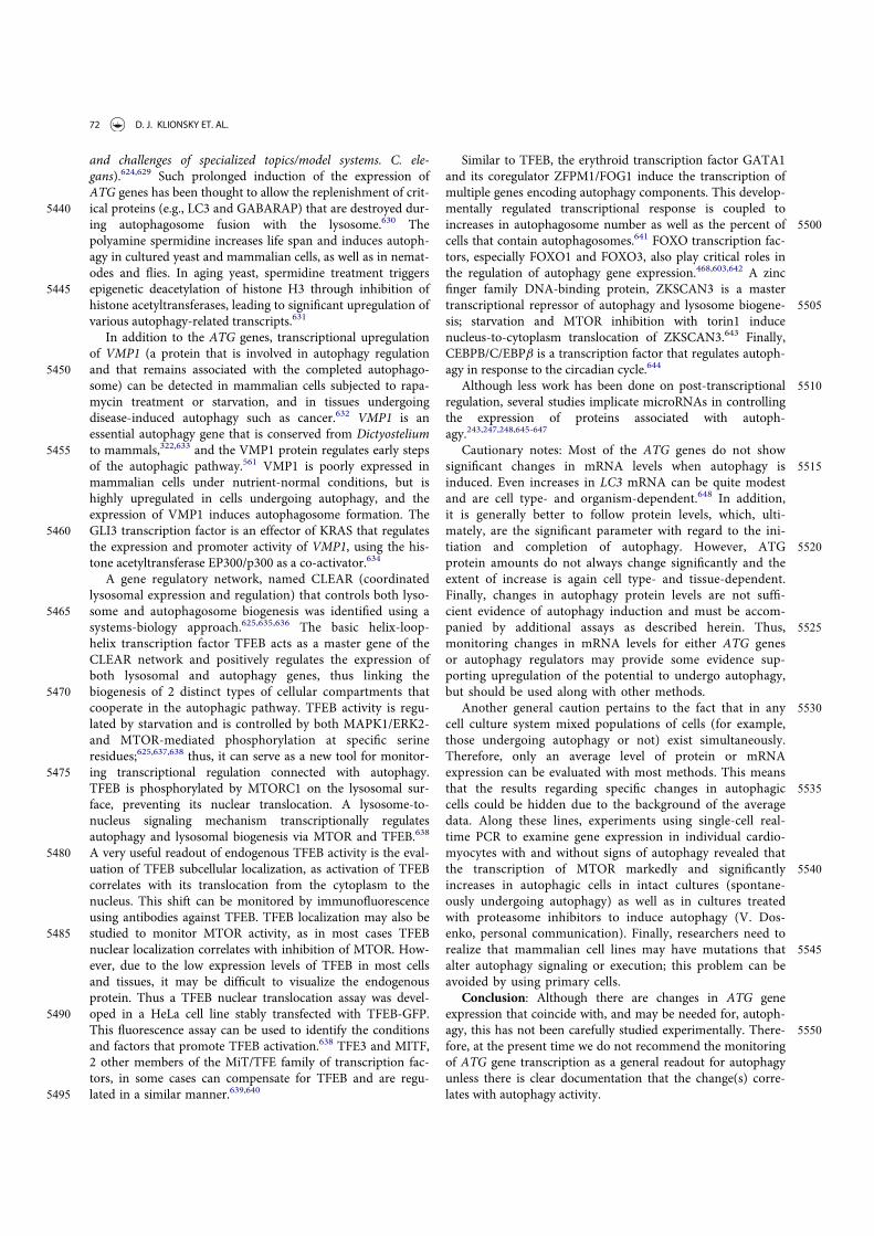
and challenges of specialized topics/model systems. C. ele-gans).624,629 Such prolonged induction of the expression ofATG genes has been thought to allow the replenishment of crit-
5440 ical proteins (e.g., LC3 and GABARAP) that are destroyed dur-ing autophagosome fusion with the lysosome.630 Thepolyamine spermidine increases life span and induces autoph-agy in cultured yeast and mammalian cells, as well as in nemat-odes and flies. In aging yeast, spermidine treatment triggers
5445 epigenetic deacetylation of histone H3 through inhibition ofhistone acetyltransferases, leading to significant upregulation ofvarious autophagy-related transcripts.631
In addition to the ATG genes, transcriptional upregulationof VMP1 (a protein that is involved in autophagy regulation
5450 and that remains associated with the completed autophago-some) can be detected in mammalian cells subjected to rapa-mycin treatment or starvation, and in tissues undergoingdisease-induced autophagy such as cancer.632 VMP1 is anessential autophagy gene that is conserved from Dictyostelium
5455 to mammals,322,633 and the VMP1 protein regulates early stepsof the autophagic pathway.561 VMP1 is poorly expressed inmammalian cells under nutrient-normal conditions, but ishighly upregulated in cells undergoing autophagy, and theexpression of VMP1 induces autophagosome formation. The
5460 GLI3 transcription factor is an effector of KRAS that regulatesthe expression and promoter activity of VMP1, using the his-tone acetyltransferase EP300/p300 as a co-activator.634
A gene regulatory network, named CLEAR (coordinatedlysosomal expression and regulation) that controls both lyso-
5465 some and autophagosome biogenesis was identified using asystems-biology approach.625,635,636 The basic helix-loop-helix transcription factor TFEB acts as a master gene of theCLEAR network and positively regulates the expression ofboth lysosomal and autophagy genes, thus linking the
5470 biogenesis of 2 distinct types of cellular compartments thatcooperate in the autophagic pathway. TFEB activity is regu-lated by starvation and is controlled by both MAPK1/ERK2-and MTOR-mediated phosphorylation at specific serineresidues;625,637,638 thus, it can serve as a new tool for monitor-
5475 ing transcriptional regulation connected with autophagy.TFEB is phosphorylated by MTORC1 on the lysosomal sur-face, preventing its nuclear translocation. A lysosome-to-nucleus signaling mechanism transcriptionally regulatesautophagy and lysosomal biogenesis via MTOR and TFEB.638
5480 A very useful readout of endogenous TFEB activity is the eval-uation of TFEB subcellular localization, as activation of TFEBcorrelates with its translocation from the cytoplasm to thenucleus. This shift can be monitored by immunofluorescenceusing antibodies against TFEB. TFEB localization may also be
5485 studied to monitor MTOR activity, as in most cases TFEBnuclear localization correlates with inhibition of MTOR. How-ever, due to the low expression levels of TFEB in most cellsand tissues, it may be difficult to visualize the endogenousprotein. Thus a TFEB nuclear translocation assay was devel-
5490 oped in a HeLa cell line stably transfected with TFEB-GFP.This fluorescence assay can be used to identify the conditionsand factors that promote TFEB activation.638 TFE3 and MITF,2 other members of the MiT/TFE family of transcription fac-tors, in some cases can compensate for TFEB and are regu-
5495 lated in a similar manner.639,640
Similar to TFEB, the erythroid transcription factor GATA1and its coregulator ZFPM1/FOG1 induce the transcription ofmultiple genes encoding autophagy components. This develop-mentally regulated transcriptional response is coupled to
5500increases in autophagosome number as well as the percent ofcells that contain autophagosomes.641 FOXO transcription fac-tors, especially FOXO1 and FOXO3, also play critical roles inthe regulation of autophagy gene expression.468,603,642 A zincfinger family DNA-binding protein, ZKSCAN3 is a master
5505transcriptional repressor of autophagy and lysosome biogene-sis; starvation and MTOR inhibition with torin1 inducenucleus-to-cytoplasm translocation of ZKSCAN3.643 Finally,CEBPB/C/EBPb is a transcription factor that regulates autoph-agy in response to the circadian cycle.644
5510Although less work has been done on post-transcriptionalregulation, several studies implicate microRNAs in controllingthe expression of proteins associated with autoph-agy.243,247,248,645-647
Cautionary notes: Most of the ATG genes do not show5515significant changes in mRNA levels when autophagy is
induced. Even increases in LC3 mRNA can be quite modestand are cell type- and organism-dependent.648 In addition,it is generally better to follow protein levels, which, ulti-mately, are the significant parameter with regard to the ini-
5520tiation and completion of autophagy. However, ATGprotein amounts do not always change significantly and theextent of increase is again cell type- and tissue-dependent.Finally, changes in autophagy protein levels are not suffi-cient evidence of autophagy induction and must be accom-
5525panied by additional assays as described herein. Thus,monitoring changes in mRNA levels for either ATG genesor autophagy regulators may provide some evidence sup-porting upregulation of the potential to undergo autophagy,but should be used along with other methods.
5530Another general caution pertains to the fact that in anycell culture system mixed populations of cells (for example,those undergoing autophagy or not) exist simultaneously.Therefore, only an average level of protein or mRNAexpression can be evaluated with most methods. This means
5535that the results regarding specific changes in autophagiccells could be hidden due to the background of the averagedata. Along these lines, experiments using single-cell real-time PCR to examine gene expression in individual cardio-myocytes with and without signs of autophagy revealed that
5540the transcription of MTOR markedly and significantlyincreases in autophagic cells in intact cultures (spontane-ously undergoing autophagy) as well as in cultures treatedwith proteasome inhibitors to induce autophagy (V. Dos-enko, personal communication). Finally, researchers need to
5545realize that mammalian cell lines may have mutations thatalter autophagy signaling or execution; this problem can beavoided by using primary cells.
Conclusion: Although there are changes in ATG geneexpression that coincide with, and may be needed for, autoph-
5550agy, this has not been carefully studied experimentally. There-fore, at the present time we do not recommend the monitoringof ATG gene transcription as a general readout for autophagyunless there is clear documentation that the change(s) corre-lates with autophagy activity.
72 D. J. KLIONSKY ET. AL.
5555 8. Posttranslational modification of ATG proteins
Autophagy is controlled by posttranslational modification(PTM) of ATG proteins such as phosphorylation, ubiquitina-tion, acetylation, oxidation and cleavage, which can be moni-tored to analyze the status of the process.343,438,519,523,649-652
5560 The global deacetylation of proteins, which often accompaniesautophagy, can be conveniently measured by quantitativeimmunofluorescence with antibodies specifically recognizingacetylated lysine residues.653 Indeed, depletion of the nutrientsupply causes autophagy in yeast or mammalian cells by reduc-
5565 ing the nucleo-cytosolic pool of acetyl-coenzyme A, which pro-vides acetyl groups to acetyltransferases, thus reducing theacetylation level of hundreds of cytoplasmic and nuclear pro-teins.654 A global deacetylation of cellular proteins is alsoobserved in response to so-called “caloric restriction mimetics”,
5570 that is, a class of pharmacological agents that deplete thenucleo-cytosolic pool of acetyl-coenzyme A, inhibit acetyltrans-ferases (such as EP300) or activate deacetylases (such asSIRT1). All these agents reduce protein acetylation levels incells as they induce autophagy.655 One prominent ATG protein
5575 that is subjected to pro-autophagic deacetylation is LC3.656,657
9. Autophagic protein degradation
Protein degradation assays represent a well-established meth-odology for measuring autophagic flux, and they allow goodquantification. The general strategy is first to label cellular pro-
5580 teins by incorporation of a radioactive amino acid (e.g., [14C]-or [3H]-leucine, [14C]-valine or [35S]-methionine; althoughvaline may be preferred over leucine due to the strong inhibi-tory effects of the latter on autophagy), preferably for a periodsufficient to achieve labeling of the long-lived proteins that best
5585 represent autophagic substrates, and then to follow this with along cold-chase so that the assay starts well after labeled short-lived proteins are degraded (which occurs predominantly viathe proteasome). Next, the time-dependent release of acid-solu-ble radioactivity from the labeled protein in intact cells or per-
5590 fused organs is measured.3,658,659 Note that the inclusion of theappropriate unlabeled amino acid (i.e., valine, leucine or methi-onine) in the starvation medium at a concentration equivalentto that of other amino acids in the chase medium is necessary;otherwise, the released [14C]-amino acid is effectively re-incor-
5595 porated into cellular proteins, which results in a significantunderestimation of protein degradation. A newer method ofquantifying autophagic protein degradation is based on L-azi-dohomoalanine (AHA) labeling.660 When added to culturedcells, L-azidohomoalanine is incorporated into proteins during
5600 active protein synthesis. After a click reaction between an azideand an alkyne, the azide-containing proteins can be detectedwith an alkyne-tagged fluorescent dye, coupled with flowcytometry. The turnover of specific proteins can also be mea-sured in a pulse-chase regimen using the Tet-ON/OFF or Gen-
5605 eSwitch systems and subsequent western blot analysis.661-663
In this type of assay a considerable fraction of the measureddegradation will be nonautophagic, and thus it is important toalso measure, in parallel, cell samples treated with autophagy-suppressive concentrations of 3-MA or amino acids, or
5610 obtained from mutants missing central ATG components
(however, it is important to note that these controls are onlyappropriate assuming that nonautophagic proteolytic activityremains unchanged, which is unlikely); these values are thensubtracted from the total readouts. The complementary
5615approach of using compounds that block other degradativepathways, such as proteasome inhibitors, may cause unex-pected results and should be interpreted with caution due tocrosstalk among the degradative systems. For example, block-ing proteasome function may activate autophagy.664-667 Thus,
5620when using inhibitors it is critical to know whether the inhibi-tors being used alter autophagy in the particular cell type andcontext being examined. In addition, because 3-MA could havesome autophagy-independent effects in particular settings it isadvisable to verify that the 3-MA-sensitive degradation is also
5625sensitive to general lysosomal inhibitors (such as NH4Cl orleupeptin).
The use of stable isotopes, such as 13C and 15N, in quantita-tive mass spectrometry-based proteomics allows the recordingof degradation rates of thousands of proteins simultaneously.
5630These assays may be applied to autophagy-related questionsenabling researchers to investigate differential effects in globalprotein or even organelle degradation studies.668,669 Stable iso-tope labeling with amino acids in cell culture (SILAC) can alsoprovide comparative information between different treatment
5635conditions, or between a wild type and mutant.Another assay that could be considered relies on the limited
proteolysis of a BHMT (betaine–homocysteine S-methyltrans-ferase) fusion protein. The 44-kDa full-length BHMT protein iscleaved in hepatocyte amphisomes in the presence of leupeptin
5640to generate 32-kDa and 10-kDa fragments.670-673 Accumulationof these fragments is time dependent and is blocked by treat-ment with autophagy inhibitors. A modified version of thismarker, GST-BHMT, can be expressed in other cell lines whereit behaves similar to the wild-type protein.674 Additional sub-
5645strates may be considered for similar types of assays. For exam-ple, the neomycin phosphotransferase II-GFP (NeoR-GFP)fusion protein is a target of autophagy.675 Transfection of lym-phoblastoid cells with a plasmid encoding NeoR-GFP followedby incubation in the presence of 3-MA leads to an accumula-
5650tion of the NeoR-GFP protein as measured by flowcytometry.676
A similar western blot assay is based on the degradationof a cytosolic protein fused to GFP. This method has beenused in yeast and Dictyostelium cells using GFP-Pgk1 and
5655GFP-Tkt-1 (phosphoglycerate kinase and transketolase,respectively). In this case the relative amount of the freeGFP and the complete fusion protein is the relevant param-eter for quantification; although it may not be possible todetect clear changes in the amount of the full-length chi-
5660mera, especially under conditions of limited flux.30,37 Asdescribed above for the marker GFP-Atg8/LC3, nonsaturat-ing levels of lysosomal inhibitors are also needed in Dictyos-telium cells to slow down the autophagic degradation,allowing the accumulation and detection of free GFP. It
5665should be noted that this method monitors bulk autophagysince it relies on the passive transit of a cytoplasmic markerto the lysosome. Consequently, it is important to determinethat the marker is distributed homogeneously in thecytoplasm.
AUTOPHAGY 73

5670 One of the most useful methods for monitoring autoph-agy in Saccharomyces cerevisiae is the Pho8D60 assay.PHO8 encodes the vacuolar alkaline phosphatase, which issynthesized as a zymogen before finally being transportedto and activated in the vacuole.677 A molecular genetic
5675 modification that eliminates the first 60 amino acids pre-vents the mutant (Pho8D60) from entering the ER, leavingthe zymogen in the cytosol. When autophagy is induced,the mutant zymogen is delivered to the vacuole nonselec-tively inside autophagosomes along with other cytoplasmic
5680 material. The resulting activation of the zymogen can beeasily measured by enzymatic assays for alkaline phospha-tase.261 To minimize background activity, it is preferable tohave the gene encoding cytosolic alkaline phosphatase(PHO13) additionally deleted (although this is not necessary
5685 when assaying certain substrates).Cautionary notes: Measuring the degradation of long-lived
proteins requires prior radiolabeling of the cells, and subse-quent separation of acid-soluble from acid-insoluble radioactiv-ity. The labeling can be done with relative ease both in cultured
5690 cells and in live animals.3 In cells, it is also possible to measurethe release of an unlabeled amino acid by chromatographicmethods, thereby obviating the need for prelabeling;678 how-ever, it is important to keep in mind that amino acid release isalso regulated by protein synthesis, which in turn is modulated
5695 by many different factors. In either case, one potential problemis that the released amino acid may be further metabolized. Forexample, branched chain amino acids are good indicators ofproteolysis in hepatocytes, but not in muscle cells where theyare further oxidized (A.J. Meijer, personal communication). In
5700 addition, the amino acid can be reincorporated into protein;for this reason, such experiments can be carried out in the pres-ence of cycloheximide, but this raises additional concerns (seeTurnover of autophagic compartments). In the case of labeledamino acids, a nonlabeled chase is added where the tracer
5705 amino acid is present in excess (being cautious to avoid usingan amino acid that inhibits autophagy), or by use of single passperfused organs or superfused cells.679,680 The perfused organsystem also allows for testing the reversibility of effects on pro-teolysis and the use of autophagy-specific inhibitors in the
5710 same experimental preparation, which are crucial controls forproper assessment.
If the autophagic protein degradation is low (as it will be incells in replete medium), it may be difficult to measure it reli-ably above the relatively high background of nonautophagic
5715 degradation. It should also be noted that the usual practice ofincubating the cells under “degradation conditions,” that is, ina saline buffer, indicates the potential autophagic capacity(maximal attainable activity) of the cells rather than the auto-phagic activity that prevails in vivo or under rich culture condi-
5720 tions. Finally, inhibition of a particular degradative pathway istypically accompanied by an increase in a separate pathway asthe cell attempts to compensate for the loss of degradativecapacity.229,666,681 This compensation might interfere with con-trol measurements under conditions that attempt to inhibit
5725 macroautophagy; however, as the latter is the major degradativepathway, the contributions of other types of degradation overthe course of this type of experiment are most often negligible.Another issue of concern, however, is that most
pharmacological protease inhibitors have “off target” effects5730that complicate the interpretation of the data.
The Pho8D60 assay requires standard positive and negativecontrols (such as an atg1D strain), and care must be taken toensure the efficiency of cell lysis. Glass beads lysis works well ingeneral, provided that the agitation speed of the instrument is
5735adequate. Instruments designed for liquid mixing with lowerspeeds should be avoided. We also recommend against holdingindividual sample tubes on a vortex, as it is difficult to maintainreproducibility; devices or attachments are available to allowmultiple tubes to be agitated simultaneously. Finally, it is also
5740important to realize that the deletion of PHO8 can affect yeastcell physiology, especially depending on the growth conditions,and this may in turn have consequences for the cell wall; cellsunder starvation stress generate thicker cell walls that can bedifficult to degrade enzymatically.
5745Conclusion: Measuring the turnover of long-lived proteinsis a standard method for determining autophagic flux. Newerproteomic techniques that compare protein levels in autoph-agy-deficient animals relative to wild-type animals are promis-ing,682 but the current ratiometric methods are affected by both
5750protein synthesis and degradation, and thus analyze proteinturnover, rather than degradation.
10. Selective types of autophagy
Although autophagy can be nonselective, in particular duringstarvation, there are many examples of selective types of
5755autophagy.
a. The Cvt pathway, mitophagy, pexophagy, piecemealmicroautophagy of the nucleus and late nucleophagy inyeast and filamentous fungiThe precursor form of aminopeptidase I (prApe1) is the major
5760cargo of the Cvt pathway in yeast, a biosynthetic autophagy-related pathway.128 The propeptide of prApe1 is proteolyticallycleaved upon vacuolar delivery, and the resulting shift inmolecular mass can be monitored by western blot. Under star-vation conditions, prApe1 can enter the vacuole through non-
5765selective autophagy, and thus has been used as a marker forboth the Cvt pathway and autophagy. The yeast Cvt pathway isunique in that it is a biosynthetic route that utilizes the autoph-agy-related protein machinery, whereas other types of selectiveautophagy are degradative. The latter include pexophagy,
5770mitophagy, reticulophagy, ribophagy and xenophagy, and eachprocess has its own marker proteins, although these are typi-cally variations of other assays used to monitor the Cvt pathwayor autophagy. One common type of assay involves the process-ing of a GFP chimera similar to the GFP-Atg8/LC3 processing
5775assay (see GFP-Atg8/LC3 lysosomal delivery and proteolysis).For example, yeast pexophagy utilizes the processing of Pex14-GFP and Pot1/Fox3/thiolase-GFP,683,684 whereas mitophagycan be monitored by the generation of free GFP from Om45-GFP, Idh1-GFP, Idp1-GFP or mito-DHFR-GFP.685,686-689
5780Localization of these mitochondrially-targeted proteins (or spe-cific MitoTracker dyes) or similar organelle markers such asthose for the peroxisome (e.g., GFP-SKL with Ser-Lys-Leu atthe C terminus that acts as a peroxisomal targeting signal, acyl-CoA oxidase 3 [Aox3-EYFP] that allows simultaneous
74 D. J. KLIONSKY ET. AL.

5785 observation of peroxisome-vacuole dynamics with the singleFITC filter set, or GFP-catalase) can also be followed by fluores-cence microscopy.553,684,690-692 In addition, yeast mitophagyrequires both the Slt2 and Hog1 signaling pathways; the activa-tion and phosphorylation of Slt2 and Hog1 can be monitored
5790 with commercially available phospho-specific antibodies(Fig. 20).508 It is also possible to monitor pexophagy in yeastsby the disappearance of activities of specific peroxisomemarkers such as catalase, alcohol oxidase or amine oxidase incell-free extracts,693 or permeabilized cell suspensions. Catalase
5795 activity, however, is a useful marker only when peroxisomalcatalases are the only such enzymes present or when activitiesof different catalases can be distinguished. In S. cerevisiae thereare 2 genes, CTT1 and CTA1, encoding catalase activity, andonly one of these gene products, Cta1, is localized in peroxi-
5800 somes. Activities of both catalases can be distinguished usingan in-gel activity assay after PAGE under nondenaturing condi-tions by staining with diaminobenzidine.694,695 Plate assays formonitoring the activity of peroxisomal oxidases in yeast colo-nies are also available.690,696 The decrease in the level of endog-
5805 enous proteins such as alcohol oxidase, Pex14 or Pot1 can befollowed by western blotting,553,697-700 TEM,701 fluorescencemicroscopy 553,702,703 or laser confocal scanning microscopy ofGFP-labeled peroxisomes.704,705
Bimolecular fluorescence complementation (BiFC) may be5810 useful to study protein-protein interactions in the autophagic
pathway.706-708 In this assay, a protein of interest is cloned intoa vector containing one half of a fluorescent reporter (e.g.,YFP), while a second protein is cloned into a different vectorcontaining the other half of the reporter. Constructs are
5815 cotransfected into cells. If the 2 proteins of interest interact, the2 halves of the reporter are brought into close proximity and afluorescent signal is reconstituted, which can be monitored byconfocal microscopy. This assay can be used to determine pro-tein interactions without prior knowledge of the location or
5820 structural nature of the interaction interface. Moreover, it isapplicable to living cells, and relatively low concentrations ofrecombinant protein are required to generate a detectablesignal.
In yeast, nonselective autophagy can be induced by nitrogen5825starvation conditions, whereas degradative types of selective
autophagy generally require a carbon source change or ERstress for efficient induction. For example, in S. cerevisiae, toinduce a substantial level of mitophagy, cells need to be precul-tured in a nonfermentable carbon source such as lactate or
5830glycerol to stimulate the proliferation of mitochondria(although this is not the case in Pichia pastoris). After sufficientmitochondria proliferation, shifting the cells back to a ferment-able carbon source such as glucose will cause the autophagicdegradation of superfluous mitochondria.686 It should be noted
5835that in addition to carbon source change, simultaneous nitro-gen starvation is also required for efficient mitophagy induc-tion. This is possibly because excessive mitochondria can besegregated into daughter cells by cell division if growth contin-ues.686 A similar carbon source change from oleic acid or meth-
5840anol to ethanol or glucose (with or without nitrogen starvation)can be used to assay for pexophagy.709 Mitophagy can also beinduced by treatment with ROS, to induce mitochondria dam-age.710 In addition, mitophagy can be induced by culturing thecells in a nonfermentable carbon source to post-log phase. In
5845this case, mitophagy may be induced because the energydemand is lower at post-log phase and the mitochondrial massexceeds the cell’s needs.120,711,712 It has been suggested that thistype of mitophagy, also known as “stationary phase mitoph-agy,” reflects a quality-control function that culls defective
5850mitochondria that accumulate in nondividing, respiringcells.713 The recently developed tool PMI that pharmacologi-cally induces mitophagy without disrupting mitochondrial res-piration714 should provide further insight as it circumvents theacute, chemically induced, blockade of mitochondrial respira-
5855tion hitherto adopted to dissect the process. Similarly, pexoph-agy can be induced by culturing the cells in a peroxisomeproliferation medium to post-log phase (J.-C. Farr"e, unpub-lished results). Along these lines, it should also be realized thatselective types of autophagy continuously occur at a low level
5860under noninducing conditions. Thus, organelles such as peroxi-somes have a finite life span and are turned over at a slow rateby autophagy-related pathways.715
Piecemeal microautophagy of the nucleus (PMN, alsomicronucleophagy) is another selective autophagic subtype,
5865which targets portions of the nucleus for degradation.716-718 InS. cerevisiae, the nuclear outer membrane, which is continuouswith the nuclear ER, forms contact sites with the vacuolarmembrane. These nucleus-vacuole junctions (NVJs) are gener-ated by interaction of the outer nuclear membrane protein
5870Nvj1 with the vacuolar protein Vac8.719 Nvj1 further recruitsthe ER-membrane protein Tsc13, which is involved in the syn-thesis of very-long-chain fatty acids (VLCFAs) and Swh1/Osh1, a member of a family of oxysterol-binding proteins.Upon starvation the NVJs bulge into the vacuole and subse-
5875quently a PMN-vesicle pinches off into the vacuole. PMNvesicles thus contain nuclear material and are limited by 3membranes with the outermost derived from the vacuole, andthe 2 inner ones from the nuclear ER. It is not clear whichnuclear components are removed by PMN, but since PMN is
5880not a cell death mechanism per se, most likely superfluousmaterial is recycled. During PMN the NVJs are selectivelyincorporated into the PMN vesicles and degraded. Accordingly,
Figure 20. S. cerevisae cells were cultured to mid-log phase and shifted to SD-N forthe indicated times. Samples were taken before (C) and at the indicated timesafter (–) nitrogen starvation. Immunoblotting was done with anti-phospho-Slt2and anti-phospho-Hog1 antibody. This figure was modified from data previouslypublished in ref. 508, and is reproduced by permission of the American Society forCell Biology, copyright 2011.
AUTOPHAGY 75

PMN can be monitored using the proteins that are associatedwith the NVJs as markers. To quantitatively follow PMN, an
5885 assay analogous to the above-described GFP-Atg8/LC3 proc-essing assay has been established using either GFP-Swh1/Osh1or Nvj1-GFP. These GFP chimeras are, together with thePMN-vesicles, degraded in the vacuole. Thus, the formation ofthe relatively proteolysis-resistant GFP detected in western
5890 blots correlates with the PMN rate. In fluorescence microscopy,PMN can be visualized with the same constructs, and a chimeraof mCherry fused to a nuclear localization signal (NLS-mCherry) can also be used. To assure that the measured PMNrate is indeed due to selective micronucleophagy, appropriate
5895 controls such as cells lacking Nvj1 or Vac8 should be included.Detailed protocols for the described assays are provided inref. 720
Late nucleophagy (LN) is another type of selective degra-dation of the nucleus, which specifically targets bulk nucleo-
5900 plasm for degradation after prolonged periods (20–24 h) ofnitrogen starvation.721 LN induction occurs in the absenceof the essential PMN proteins Nvj1 and Vac8 and, there-fore, the formation of NVJs. Although, some components ofthe core Atg machinery are required for LN, Atg11 and the
5905 Vps34-containing PtdIns3K complex I are not needed. LNcan be monitored by employing a nuclear-targeted versionof the Rosella biosensor (n-Rosella) and following either itsaccumulation (by confocal microscopy), or degradation (byimmunoblotting), within the vacuole.721 Dual labeling of
5910 cells with Nvj1-EYFP, a nuclear membrane reporter ofPMN, and the nucleoplasm-targeted NAB35-DsRed.T3(NAB35 is a target sequence for the Nab2 RNA-bindingprotein, and DsRed.T3 is the pH-stable, red fluorescentcomponent of n-Rosella) allows detection of PMN soon
5915 after the commencement of nitrogen starvation, whereasdelivery to the vacuole of the nucleoplasm reporter, indica-tive of LN, is observed only after prolonged periods ofnitrogen starvation. Few cells show simultaneous accumula-tion of both reporters in the vacuole indicating PMN and
5920 LN are temporally and spatially separated.721
In contrast to unicellular yeasts, filamentous fungi form aninterconnected mycelium of multinucleate hyphae containingup to 100 nuclei in a single hyphal compartment. A mycelialcolony grows by tip extension with actively growing hyphae at
5925 the colony margin surrounded by an older, inner hyphal net-work that recycles nutrients to fuel the hyphal tips. By labelingorganelle markers with GFP it is possible to show in Aspergillusoryzae that macroautophagy mediates degradation of basalhyphal organelles such as peroxisomes, mitochondria and
5930 entire nuclei.722 In contrast to yeast, PMN has not beenobserved in filamentous ascomycetes.723 In Magnaporthe ory-zae germination of the condiospore and formation of theappressorium is accompanied by nuclear degeneration in thespore.275 The degradation of nuclei in spores requires the non-
5935 selective autophagy machinery, whereas conserved componentsof the PMN pathway such as Vac8 and Tsc13 are dispensablefor nuclear breakdown during plant infection.724 Nuclei areproposed to function in storage of growth-limiting nutrientssuch as phosphate and nitrogen.725,726 Similar to nuclei, mito-
5940 chondria and peroxisomes are also preferentially degraded inthe basal hyphae of filamentous ascomycetes.275,722,724-727
Cautionary notes: The Cvt pathway has been demonstratedto occur only in yeast. In addition, the sequestration of prApe1is specific, even under starvation conditions, as it involves the
5945recognition of the propeptide by a receptor, Atg19, which inturn interacts with the scaffold protein Atg11.728,729 Thus,unless the propeptide is removed, prApe1 is recognized as aselective substrate. Overexpression of prApe1 saturates importby the Cvt pathway, and the precursor form accumulates, but is
5950rapidly matured upon autophagy induction.305 In addition,mutants such as vac8D and tlg2D accumulate prApe1 underrich conditions, but not during autophagy.505,730 Accordingly,it is possible to monitor the processing of prApe1 when overex-pressed, or in certain mutant strains to follow autophagy induc-
5955tion. However, under the latter conditions it must be kept inmind that the sequestering vesicles are substantially smallerthan typical autophagosomes generated during nonselectiveautophagy; the Cvt complex (prApe1 bound to Atg19) issmaller than typical peroxisomes or mitochondrial fragments
5960that are subject to autophagic degradation. Accordingly, partic-ular mutants may display complete maturation of prApe1under autophagy-inducing conditions, but may still have adefect in other types of selective autophagy, as well as beingunable to induce a normal level of nonselective autophagy.106
5965For this reason, it is good practice to evaluate autophagosomesize and number by TEM. Actually, it is much simpler to moni-tor autophagic bodies (rather than autophagosomes) in yeast.First, the vacuole is easily identified, making the identificationof autophagic bodies much simpler. Second, autophagic bodies
5970can be accumulated within the vacuole, allowing for anincreased sample size. It is best to use a strain background thatis pep4D vps4D to prevent the breakdown of the autophagicbodies, and to eliminate confounding vesicles from the multive-sicular body pathway. One caveat to the detection of autopha-
5975gic bodies, however, is that they may coalesce in the vacuolelumen, making it difficult to obtain an accurate quantification.Finally, it is important to account for biases in sample section-ing to obtain an accurate estimate of autophagic body numberor size.105
5980In general, when working with yeast it is preferable to usestrains that have the marker proteins integrated into the chro-mosome rather than relying on plasmid-based expression,because plasmid numbers can vary from cell to cell. The GFP-Atg8, or similar, processing assay is easy to perform and is suit-
5985able for analysis by microscopy as well as western blotting;however, particular care is needed to obtain quantitative datafor GFP-Atg8, Pex14-GFP or Om45-GFP, etc. processingassays (see cautionary notes for GFP-Atg8/LC3 lysosomal deliv-ery and proteolysis). An alternative is an organelle-targeted
5990Pho8D60 assay. For example, mitoPho8D60 can be used toquantitatively measure mitophagy.687 In addition, for the GFP-Atg8 processing assay, 2 h of starvation is generally sufficient todetect a significant level of free (i.e., vacuolar) GFP by westernblotting as a measure of nonselective autophagy. For selective
5995types of autophagy, the length of induction needed for a clearlydetectable free GFP band will vary depending on the rate ofcargo delivery/degradation. Usually 6 h of mitophagy inductionis needed to be able to detect free GFP (e.g., from Om45-GFP)by western blot under starvation conditions, whereas stationary
6000phase mitophagy typically requires 3 days before a free GFP
76 D. J. KLIONSKY ET. AL.
band is observed. However, as with animal systems (see Animalmitophagy and pexophagy), it would be prudent to follow morethan one GFP-tagged protein, as the kinetics, and even theoccurrence of mitophagic trafficking, seems to be protein spe-
6005 cies-dependent, even within the mitochondrial matrix.731
Care should be taken when choosing antibodies to assess thedegree of mitochondrial protein removal by autophagy; thequality and clarity of the result may vary depending on the spe-cifics of the antibody. In testing the efficiency of mitophagy
6010 clearer results may be obtained by using antibodies againstmtDNA-encoded proteins. This experimental precaution mayprove critical to uncover subtle differences that could be missedwhen evaluating the process with antibodies against nuclearencoded, mitochondrially imported proteins (M. Campanella
6015 personal communication).
b. AggrephagyAggrephagy is the selective removal of aggregates by macroau-tophagy.732 This process can be followed in vitro (in cell cul-ture) and in vivo (in mice) by monitoring the levels of an
6020 aggregate-prone protein such as an expanded polyglutamine(polyQ)-containing protein or mutant SNCA/a-synuclein (syn-uclein, alpha [non A4 component of amyloid precursor]). Lev-els are quantified by immunofluorescence, immunogoldlabeling or traditional immunoblot. In yeast, degradation of
6025 SNCA aggregates can be followed by promoter shut-off assays.Espression of the inducible GAL1 promoter of GFP-taggedSVXA is stopped by glucose repression. The removal of aggre-gates is thus monitored with fluorescence microscopy. The con-tribution of autophagy to SNCA aggregate clearance can be
6030 studied by the use of different autophagy mutants or by phar-macological treatment with the proteinase B inhibitorPMSF.733,734 Similarly, fluorescently tagged aggregated proteinssuch as polyQ80-CFP can be monitored via immunoblot andimmunofluorescence. In addition to fluorescence methods,
6035 aggregates formed by a splice variant of CCND2 (cyclin D2)can also be monitored in electron-dense lysosomes and auto-phagosomes by immunogold labeling and TEM techniques.735
A polyQ80-luciferase reporter, which forms aggregates, canalso be used to follow aggrephagy.736 A nonaggregating
6040 polyQ19-luciferase or untagged full-length luciferase serves as acontrol. The ratio of luciferase activity from these 2 constructscan be calculated to determine autophagic flux.
Autophagic degradation of endogenous aggregates such aslipofuscin can be monitored in some cell types by fluorescence
6045 microscopy, utilizing the autofluorescence of lipofuscin par-ticles. Although under normal conditions almost 99% of thelipofuscin particles are located in the autophagosomes/lyso-somes, an impairment of macroautophagy leads to free lipofus-cin in the cytosol.737,738 The amount of lipofuscin in primary
6050 human adipocytes can be reduced by activation of macroau-tophagy, and the amount of lipofuscin is dramatically reducedin adipocytes from patients with type 2 diabetes and chronicallyenhanced macroautophagy.294
Cautionary notes: Caution must be used when performing6055 immunoblots of aggregated proteins, as many protein aggregates
fail to enter the resolving gel and are retained in the stacking gel.In addition, the polyQ80-luciferase in the aggregated state lacksluciferase activity whereas soluble polyQ80-luciferase retains
activity. Therefore, caution must be used when interpreting results6060with these vectors, as treatments that increase aggrephagy or
enhance protein aggregation can lead to a decrease in luciferaseactivity.739 Finally, soluble polyQ reporters can be degraded by theproteasome; thus, changes in the ratio of polyQ19-luciferase:polyQ80-luciferase may also reflect proteasomal effects and not
6065just changes in autophagic flux.
c. AllophagyIn C. elegans, mitochondria, and hence mitochondrial DNA,from sperm are eliminated by an autophagic process. This pro-cess of allogeneic (nonself) organelle autophagy is termed
6070“allophagy.”740,741 During allophagy in C. elegans, both paternalmitochondria and membranous organelles (a sperm-specificmembrane compartment) are eliminated by the 16-cell stage(100–120 min post-fertilization).742,743 The degradation processcan be monitored in living embryos with GFP::ubiquitin, which
6075appears in the vicinity of the sperm chromatin (labeled forexample with mCherry-histone H2B) on the membranousorganelles within 3 min after fertilization. GFP fusions andantibodies specific for LGG-1 and LGG-2 (Atg8/LC3 homo-logs), which appear next to the sperm DNA, membranous
6080organelles and mitochondria (labeled with CMXRos or mito-chondria-targeted GFP) within 15 to 30 min post-fertilization,can be used to verify the autophagic nature of the degradation.TEM can also be utilized to demonstrate the presence of mito-chondria within autophagosomes in the early embryo.
6085Conclusion: There are many assays that can be used tomonitor selective types of autophagy, but caution must be usedin choosing an appropriate marker(s). The potential role ofother degradative pathways for any individual organelle orcargo marker should be considered, and it is advisable to use
6090more than one marker or technique.
d. Animal mitophagy and pexophagyThere is no consensus at the present time with regard to thebest method for monitoring mitophagy in animals. As with anyorganelle-specific form of autophagy, it is necessary to demon-
6095strate: i) increased levels of autophagosomes containing mito-chondria, ii) maturation of these autophagosomes thatculminates with mitochondrial degradation, which can beblocked by specific inhibitors of autophagy or of lysosomal deg-radation, and iii) whether the changes are due to selective
6100mitophagy or increased mitochondrial degradation duringnonselective autophagy. Techniques to address each of thesepoints have been reviewed.42,744
Antibodies against phosphorylated ubiquitin (p-S65-Ub)have very recently been described as novel tools to detect the
6105activation of PINK1-PARK2-mediated mitophagy.745 p-S65-Ub is formed by the kinase PINK1 specifically upon mitochon-drial stress, and is amplified in the presence of the E3 Ub ligasePARK2 (reviewed in ref. 746).747 p-S65-Ub antibodies havebeen used to demonstrate stress-induced activation of PINK1
6110in various cells including primary human fibroblasts (Fig. 21).Phosphorylated poly-ubiquitin chains specifically accumulateon damaged mitochondria, and staining with p-S65-Ub anti-bodies can be used, in addition to translocation of PARK2, tomonitor the intitiation of mitophagy. Given the complete con-
6115servation of the epitopes across species, mitochondrial p-S65-
AUTOPHAGY 77

Ub could also be dectected in mouse primary neurons uponmitochondrial depolarization. Furthermore, the p-S65-Ub sig-nal partially colocalizes with mitochondrial, lysosomal, andtotal ubiquitin markers in cytoplasmic granules that appear to
6120 increase with age and disease in human postmortem brain sam-ples.745 Along with the excellent performance of p-S65-Ub anti-bodies in a range of applications, these findings highlight thepotential for future biomarker development.
Ultrastructural analysis at early time points can be used to6125 establish selective mitophagy, although a maturation inhibitor
may be needed to trap early autophagosomes with recognizablecargo (Fig. 22). Depending on the use of specific imaging tech-niques, dyes for living cells or antibodies for fixed cells have tobe chosen. In any case, transfection of the phagophore and
6130 autophagosome marker GFP-LC3 to monitor the initiation ofmitophagy, or RFP-LC3 to assess mitophagy progression, and
visualization of mitochondria (independent of their mitochon-drial membrane potential) makes it possible to determine theassociation of these 2 cellular components. Qualitatively, this
6135may appear as fluorescence colocalization or as rings of GFP-LC3 surrounding mitochondria in higher resolutionimages.748,749 For live cell imaging microscopy, mitochondriashould be labeled by a matrix-targeted fluorescent proteintransfection or by mitochondria-specific dies. When using
6140matrix-targeted fluorophores for certain cell lines (e.g., SH-SY5Y), it is important to allow at least 48 h of transient expres-sion for sufficient targeting/import of mitochondrial GFP/RFPprior to analyzing mitophagy. Among the MitoTracker probesare lipophilic cations that include a chloromethyl group and a
6145fluorescent moiety. They concentrate in mitochondria due totheir negative charge and react with the reduced thiols presentin mitochondrial matrix proteins.750-752 After this reaction the
Figure 21. PINK1-dependent phosphorylation of ubiquitin (p-S65-Ub) upon mitophagic stress. (A) Human dermal fibroblasts from healthy controls or Parkinson diseasepatients carrying a PINK1 loss-of-function mutation (Q456X) were treated with valinomycin for the indicated times and lysates were analyzed by western blot. The p-S65-Ub signal is almost undetectable under nonstress conditions in controls, but is strongly induced in a PINK1 kinase-dependent manner during its stabilization on the outermitochondrial membrane. MFN2 serves as a control substrate and VCL (vinculin) as a loading control. (B) HeLa cells stably expressing GFP-PARK2 (wild type) were treatedwith CCCP for the indicated times, fixed and stained with p-S65-Ub (red) and GFP-PARK2 (green) as well as mitochondrial (TOMM20, cyan) and nuclear (Hoechst, blue)markers. The p-S65-Ub staining is almost undetectable in nonstressed cells, but rapidly accumulates on damaged mitochondria where it functions to activate PARK2. Onmitochondria, PINK1 and PARK2 together amplify the p-S65-Ub signal. Scale bar: 10 mm. Image provided by F.C. Fiesel and W. Springer.
Figure 22. Autophagosomes with recognizable cargo are rare in cells. (A) To assess relative rates of autophagosome formation, the fusion inhibitor bafilomycin A1 (10 nM)was applied for 2 h prior to fixation with 2% glutaraldehyde in order to trap newly formed autophagosomes. Two different PINK1 shRNA lines (A14 and D14) exhibitincreased AV formation over 2 h compared to the control shRNA line. ", p > 0.05 vs. Control. (B) Autophagosomes in bafilomycin A1-treated control cells contain a varietyof cytoplasmic structures (left, arrow), while mitochondria comprise a prominent component of autophagosomes in bafilomycin A1-treated (PINK1 shRNA) cells (right,arrow). Scale bar: 500 nm. These data indicate induction of selective mitophagy in PINK1-deficient cells. This figure was modified from Figure 2 published in Chu CT. A piv-otal role for PINK1 and autophagy in mitochondrial quality control: implications for Parkinson disease. Human Molecular Genetics 2010; 19:R28-R37.
78 D. J. KLIONSKY ET. AL.
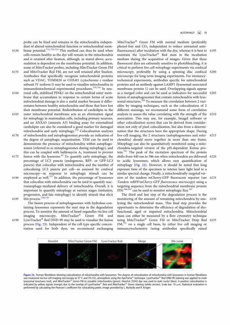
probe can be fixed and remains in the mitochondria indepen-dent of altered mitochondrial function or mitochondrial mem-
6150 brane potential.751,753,754 This method can thus be used whencells remain healthy as the dye will remain in the mitochondriaand is retained after fixation, although, as stated above, accu-mulation is dependent on the membrane potential. In addition,some of MitoTracker probes, including MitoTracker Green FM
6155 and MitoTracker Red FM, are not well retained after fixation.Antibodies that specifically recognize mitochondrial proteinssuch as VDAC, TOMM20 or COX4I1 (cytochrome c oxidasesubunit IV isoform I) may be used to visualize mitochondria inimmunohistochemical experimental procedures.755,756 In neu-
6160 ronal cells, stabilized PINK1 on the mitochondrial outer mem-brane that accumulates in response to certain forms of acutemitochondrial damage is also a useful marker because it differ-entiates between healthy mitochondria and those that have losttheir membrane potential. Redistribution of cardiolipin to the
6165 outer mitochondrial membrane acts as an elimination signalfor mitophagy in mammalian cells, including primary neurons,and an ANXA5 (annexin A5) binding assay for externalizedcardiolipin can also be considered a good marker for damagedmitochondria and early mitophagy.145 Colocalization analyses
6170 of mitochondria and autophagosomes provide an indication ofthe degree of autophagic sequestration. TEM can be used todemonstrate the presence of mitochondria within autophago-somes (referred to as mitophagosomes during mitophagy), andthis can be coupled with bafilomycin A1 treatment to prevent
6175 fusion with the lysosome.42 To quantify early mitophagy, thepercentage of LC3 puncta (endogenous, RFP- or GFP-LC3puncta) that colocalize with mitochondria and the number ofcolocalizing LC3 puncta per cell—as assessed by confocalmicroscopy—in response to mitophagic stimuli can be
6180 employed as well.757 In addition, the percentage of lysosomesthat colocalize with mitochondria can be used to quantify mac-roautophagy-mediated delivery of mitochondria. Overall, it isimportant to quantify mitophagy at various stages (initiation,progression, and late mitophagy) to identify stimuli that elicit
6185 this process.758,759
The fusion process of mitophagosomes with hydrolase-con-taining lysosomes represents the next step in the degradationprocess. To monitor the amount of fused organelles via live cellimaging microscopy, MitoTracker! Green FM and
6190 LysoTracker! Red DND-99 may be used to visualize the fusionprocess (Fig. 23). Independent of the cell-type specific concen-tration used for both dyes, we recommend exchanging
MitoTracker! Green FM with normal medium (preferablyphenol-free and CO2 independent to reduce unwanted auto-
6195fluorescence) after incubation with the dye, whereas it is best tomaintain the LysoTracker! Red stain in the incubationmedium during the acquisition of images. Given that thesefluorescent dyes are extremely sensitive to photobleaching, it iscritical to perform live cell mitophagy experiments via confocal
6200microscopy, preferably by using a spinning disc confocalmicroscope for long-term imaging experiments. For immuncy-tochemical experiments, antibodies specific for mitochondrialproteins and an antibody against LAMP1 (lysosomal-associatedmembrane protein 1) can be used. Overlapping signals appear
6205as a merged color and can be used as indicators for successfulfusion of autophagosomes that contain mitochondria with lyso-somal structures.760 To measure the correlation between 2 vari-ables by imaging techniques, such as the colocalization of 2different stainings, we recommend some form of correlation
6210analysis to assess the value correlating with the strength of theassociation. This may use, for example, ImageJ software orother colocalization scores that can be derived from consider-ation not only of pixel colocalization, but also from a determi-nation that the structures have the appropriate shape. During
6215live-cell imaging, the 2 structures (autophagosomes and mito-chondria) should move together in more than one frame.Mitophagy can also be quantitatively monitored using a mito-chondria-targeted version of the pH-dependent Keima pro-tein.761 The peak of the excitation spectrum of the protein
6220shifts from 440 nm to 586 nm when mitochondria are deliveredto acidic lysosomes, which allows easy quantification ofmitophagy (Fig. 24). However, it should be noted that longexposure time of the specimen to intense laser light lead to asimilar spectral change. Finally, a mitochondrially-targeted ver-
6225sion of the tandem mCherry-GFP fluorescent reporter (seeTandem mRFP/mCherry-GFP fluorescence microscopy) using atargeting sequence from the mitochondrial membrane proteinFIS1346,347 can be used to monitor mitophagic flux.347
The third and last step of the degradation process is the6230monitoring of the amount of remaining mitochondria by ana-
lyzing the mitochondrial mass. This final step provides theopportunity to determine the efficiency of degradation of dys-functional, aged or impaired mitochondria. Mitochondrialmass can either be measured by a flow cytometry technique
6235using MitoTracker! Green FM or MitoTracker Deep RedFM,751 on a single cell basis, by either live cell imaging orimmuncytochemistry (using antibodies specifically raised
Figure 23. Human fibroblasts showing colocalization of mitochondria with lysosomes. The degree of colocalization of mitochondria with lysosomes in human fibroblastswas measured via live cell imaging microscopy at 37!C and 5% CO2 atmosphere using the ApoTome! technique. LysoTracker! Red DND-99 staining was applied to marklysosomal structures (red), and MitoTracker! Green FM to visualize mitochondria (green). Hoechst 33342 dye was used to stain nuclei (blue). A positive colocalization isindicated by yellow signals (merge) due to the overlap of LysoTracker! Red and MitoTracker! Green staining (white arrows). Scale bar: 10 mm. Statistical evaluation isperformed by calculating the Pearson’s coefficient for colocalizing pixels. Image provided by L. Burbulla and R. Kr€uger.
AUTOPHAGY 79

against different mitochondrial proteins). Alternatively, mito-chondrial content in response to mitophagic stimuli (in the
6240 presence and absence of autophagy inhibitors to assess the con-tribution of mitophagy) in live or fixed cells can be quantifiedat the single-cell level as the percentage of cytosol occupied bymitochondrial-specific fluorescent pixels using NIH ImageJ.759
Immunoblot analysis of the levels of mitochondrial proteins6245 from different mitochondrial subcompartments is valuable for
validating the data from flow cytometry or microscopy studies,and it should be noted that outer mitochondrial membraneproteins in particular can be degraded by the proteasome, espe-cially in the context of mitochondrial depolarization.762,763 EM
6250 can also be used to verify loss of entire mitochondria, and PCR(or fluorescence microscopy) to quantify mitochondrial DNA(mtDNA). A reliable estimation of mtDNA can be performedby real-time PCR of the MT-ND2 (mitochondrially encodedNADH dehydrogenase 2) gene expressed as a ratio of mtDNA:
6255 nuclear DNA by normalizing to that of TERT (telomerasereverse transcriptase) genomic DNA.764 The spectrophotomet-ric measurement of the activity of CS (citrate synthase), a mito-chondrial matrix enzyme of the TCA cycle, which remainshighly constant in these organelles and is considered a reliable
6260 marker of their intracellular content, can also be used to esti-mate the mitochondrial mass.764
In addition to monitoring the steady state levels of differentsteps of mitophagy—whether by single-cell analyses of LC3mitochondrial colocalization or by immunoblotting for mito-
6265 chondrial markers—investigation of the mitophagic flux isneeded to determine whether mitophagy is impaired or acti-vated in response to stimuli, and at which steps. Therefore,appropriate treatment (pharmacological inhibition and/orsiRNA-mediated knockdown of ATG genes) may be applied to
6270 prevent mitochondrial degradation at distinct steps of the pro-cess. A recent method using flow cytometry in combinationwith autophagy and mitophagy inhibitors has been developedto determine mitophagic flux using MitoTracker probes.751
Certain cellular models require stress conditions to measure6275 the mitochondrial degradation capacity, as basal levels are too
low to reliably assess organelle clearance. However, one excep-tion has been identified in Drosophila where large numbers ofmitochondria are cleared by mitophagy during developmen-tally-triggered autophagy.765 Hence, in many cases, it may be
6280useful to pretreat the cells with uncoupling agents, such asCCCP, that stimulate mitochondrial degradation and allowmeasurements of mitophagic activity; however, it should bekept in mind that, although helpful to stimulate mitochondrialdegradation, this treatment is not physiological and promotes
6285the rapid degradation of outer membrane-localized mitochon-drial proteins. In part for this reason a milder mitophagy stim-ulus has been developed that relies on a combination ofantimycin A and oligomycin, inhibitors of the electron trans-port chain and ATP synthase, respectively;766 this treatment is
6290less toxic, and the resulting damage is time dependent. Anothermethod to induce mitophagy is by expressing and activating amitochondrially-localized fluorescent protein photosensitizersuch as Killer Red.767 The excitation of Killer Red results in anacute increase of superoxide, due to phototoxicity, that causes
6295mitochondrial damage resulting in mitophagy.768 The advan-tage of using a genetically encoded photosensitizer is that itallows for both spatial and temporal control in inducingmitophagy. Finally, the forced targeting of AMBRA1 to theexternal mitochondrial membrane is sufficient to induce mas-
6300sive mitophagy.769
A new classification suggests that mitophagy can be dividedinto 3 types.770 Type 1 mitophagy, involves the formation of aphagophore, and typically also requires mitochondrial fission;the PtdIns3K containing BECN1 mediates this process. In con-
6305trast, type 2 mitophagy is independent of BECN1 and takesplace when mitochondria have been damaged, resulting indepolarization; sequestration involves the coalescence of GFP-LC3 membranes around the mitochondria rather than throughfission and engulfment within a phagophore. In type 3 mitoph-
6310agy, mitochondrial fragments or vesicles from damaged organ-elles are sequestered through a microautophagy-like processthat is independent of ATG5 and LC3, but requires PINK1 andPARK2.
Although the process of pexophagy is prominent and well6315described in yeast cells,697,771 relatively little work has been
done in the area of selective mammalian peroxisome degra-dation by autophagy (for a review see ref. 772). Typically,peroxisomes are induced by treatment with hypolipidemicdrugs such as clofibrate or dioctyl phthalate, which bind to a
6320subfamily of nuclear receptors, referred to as peroxisomeproliferator-activated receptors.773 Degradation of excess
Figure 24. Detection of mitophagy in primary cortical neurons using mitochondria-targeted Keima. Neurons transfected with mito-Keima were visualized using 458-nm(green, mitochondria at neutral pH) and 561-nm (red, mitochondria in acidic pH) laser lines and 575-nm band pass filter. Compared with the control (A) wild-type PINK1overexpression (B) increases the number of the mitochondria exposed to acidic conditions. Scale bar: 2 mm. (C) Quantification of red dots suggests increased mitophagyin wild-type PINK1 but not in the kinase dead (kd) PINK1K219M-overexpressing neurons. Image provided by V. Choubey and A. Kaasik.
80 D. J. KLIONSKY ET. AL.
organelles is induced by drug withdrawal, although starvationwithout prior proliferation can also be used. EPAS1 activa-tion in liver-specific vhl¡/¡ and vhl¡/¡ hif1a¡/¡ mice reduces
6325 peroxisome abundance by pexophagy, whereas ER and mito-chondrial protein levels are not affected.774 Pexophagy canalso be induced by the expression of a nondegradable activeEPAS1 variant.774 Induction of pexophagy in response toendogenous and exogenous reactive oxygen species (ROS)
6330 and reactive nitrogen species has been observed in mamma-lian cells. In this setting, pexophagy is induced via ROS/reac-tive nitrogen species-mediated activation of ATM,775,776
repression of MTORC1 and phosphorylation of PEX5 byATM;777,778 ATM phosphorylation of PEX5 at S141 triggers
6335 PEX5 ubiquitination and binding of SQSTM1 to peroxisomestargeted for pexophagy.778 Loss of peroxisomes can be fol-lowed enzymatically or by immunoblot, monitoring enzymessuch as ACOX/fatty acyl-CoA oxidase (note that this enzymeis sometimes abbreviated “AOX,” but should not be confused
6340 with the enzyme alcohol oxidase that is frequently used inassays for yeast pexophagy) or CAT/catalase, and also byEM, cytochemistry or immunocytochemistry.779-782 Finally, aHaloTag!-PTS1 marker that is targeted to peroxisomes hasbeen used to fluorescently label the organelle.783 An alterna-
6345 tive approach uses a peroxisome-specific tandem fluoro-chrome assay (RFP-EGFP localizing to peroxisomes by theC-terminal addition of the tripeptide SKL, or a peroxisomalmembrane protein tagged with mCherry-mGFP), which hasbeen used to demonstrate the involvement of ACBD5/
6350 ATG37, NBR1 and SQSTM1 in mammalian pexophagy.345,784
Cautionary notes: There are many assays that can be used tomonitor specific types of autophagy, but caution must be usedin choosing an appropriate marker(s). To follow mitophagy it isbest to monitor more than one protein and to include an inner
6355 membrane or matrix component in the analysis. In particular,it is not sufficient to follow a single mitochondrial outer mem-brane protein because these can be degraded independently ofmitophagy. Although the localization of PARK2 to mitochon-dria as monitored by fluorescence microscopy is associated
6360 with the early stages of protonophore uncoupler (CCCP)-driven mitochondria degradation,250 this by itself cannot beused as a marker for mitophagy, as these events can be dissoci-ated.785 Moreover, mitophagy elicited in a number of diseasemodels does not involve mitochondrial PARK2 transloca-
6365 tion.145,347,786 Along these lines, recent studies implicate anessential role for TRAF2, an E3 ubiquitin ligase, as a mitophagyeffector in concert with PARK2 in cardiac myocytes; wherebymitochondrial proteins accumulate differentially with defi-ciency of either, indicating nonredundant roles for these E3
6370 ubiquitin ligases in mitophagy.787 This finding necessitates anintegrated approach to assess mitophagy based on a broad eval-uation of multiple mitochondrial effectors and proteins.
PARK2 translocates to damaged mitochondria and ubiquiti-nates a wide range of outer membrane proteins including
6375 VDAC1, MFN1/2 and TOMM20/TOM20.756,762,763,788 Thisresults in the preferential degradation of mitochondrial outermembrane proteins by the proteasome, while inner membraneproteins and mitochondrial DNA789 remain intact. Monitoringloss of a single protein such as TOMM20 by western blot or
6380 fluorescence microscopy to follow mitophagy may thus be
misleading, as noted above.788 MitoTracker dyes are widelyused to stain mitochondria and, when colocalized with GFP-LC3, they can function as a marker for mitophagy. However,staining with MitoTracker dyes depends on mitochondrial
6385membrane potential (although MitoTracker Green FM is lesssensitive to loss of membrane potential), so that damaged, orsequestered nonfunctional mitochondria may not be stained. Invitro this can be avoided by labeling the cells with MitoTrackerbefore the induction by the mitophagic stimuli.790 One addi-
6390tional point is that MitoTracker dyes might influence mito-chondrial motility in axons (D. Ebrahimi-Fakhari, personalcommunication).
Although it is widely assumed that macroautophagy is themajor mechanism for degradation of entire organelles, there
6395are multiple mechanisms that may account for the disappear-ance of mitochondrial markers. These include proteasomaldegradation of outer membrane proteins and/or proteins thatfail to correctly translocate into the mitochondria, degradationdue to proteases within the mitochondria, and reduced biosyn-
6400thesis or import of mitochondrial proteins. PINK1 and PARK2also participate in an ATG gene-independent pathway for lyso-somal degradation of small mitochondria-derived vesicles.791
Furthermore, the PINK1-PARK2 mitophagy pathway is alsotranscriptionally upregulated in response to starvation-trig-
6405gered generalized autophagy, and is intertwined with the lipo-genesis pathway.792-795 In addition to mitophagy, mitochondriacan be eliminated by extrusion from the cell (mitopto-sis).796,760,756,743 Transcellular degradation of mitochondria, ortransmitophagy, also occurs in the nervous system when astro-
6410cytes degrade axon-derived mitochondria.797 Thus, it is advis-able to use a variety of complementary methods to monitormitochondria loss including TEM, single cell analysis of LC3fluorescent puncta that colocalize with mitochondria, and west-ern blot, in conjunction with flux inhibitors and specific inhibi-
6415tors of autophagy induction compared with inhibitors of theother major degradation systems (see cautions in Autophagyinhibitors and inducers). To monitor and/or rule out changesin cellular capacity to undergo mitochondrial biogenesis, a pro-cess that is tightly coordinated with mitophagy and can dictate
6420the outcome following mitophagy-inducing insults especially inprimary neurons and other mitochondria-dependent cells,colocalization analysis after double staining for the mitochon-drial marker TOMM20 and BrdU (for visualization of newlysynthesized mtDNA) can be performed (Fig. 25).
6425Likewise, although the mechanism(s) of peroxisomal proteindegradation in mammals awaits further elucidation, it canoccur by both autophagic and proteasome-dependent mecha-nisms.798 Thus, controls are needed to determine the extent ofdegradation that is due to the proteasome. Moreover, 2 addi-
6430tional degradation mechanisms have been suggested: the actionof the peroxisome-specific LONP2/Lon (lon peptidase 2, perox-isomal) protease and the membrane disruption effect of 15-lipoxygenase.799
e. Chlorophagy6435Besides functioning as the primary energy suppliers for plants,
chloroplasts represent a major source of fixed carbon andnitrogen to be remobilized from senescing leaves to storageorgans and newly developing tissues. As such, the turnover of
AUTOPHAGY 81

these organelles has long been considered to occur via an6440 autophagy-type mechanism. However, while the detection of
chloroplasts within autophagic body-like vesicles or withinvacuole-like compartments has been observed for decades,only recently has a direct connection between chloroplastturnover and autophagy been made through the analysis of
6445 atg mutants combined with the use of fluorescent ATG8reporters.800,801 In fact, it is now clear that chlorophagy, theselective degradation of chloroplasts by macroautophagy, canoccur via several routes, including the encapsulation of wholechloroplasts, or the budding of chloroplast material into small
6450 distinct autophagic vesicles called Rubisco-containing bodies(RCBs) and ATI1 plastid-associated bodies (ATI-PS), whichthen transport chloroplast cargo to the vacuole.800,802 Chloro-plasts produce long tubes called stromules that project outfrom the organelle outer membrane. Recent studies suggest
6455 that stromules are part of the chlorophagy process, by whichthe stromule tips presumably containing unwanted or dam-aged chloroplast material are engulfed by autophagic mem-branes using ESCRTII endocytic machinery that depends onATG8.803 The appearance of RCBs is tightly linked with leaf
6460 carbon status, indicating that chlorophagy through RBCs rep-resents an important route for recycling plant nutrients pro-vided in plastid stores.
f. ChromatophagyAutophagy has been known for its pro-survival role in cells
6465 under metabolic stress and other conditions. However, exces-sively induced autophagy may be cytotoxic and may lead to celldeath. Chromatophagy (chromatin-specific autophagy) comesinto view as one of the autophagic responses that can contrib-ute to cell death.804 Chromatophagy can be seen in cells during
6470 nutrient depletion, such as arginine starvation, and its pheno-type consists of giant-autophagosome formation, nucleus mem-brane rupture and histone-associated-chromatin/DNA leakagethat is captured by autophagosomes.804 Arginine starvation canbe achieved by adding purified arginine deiminase to remove
6475arginine from the culture medium, or by using arginine-drop-out medium. The degradation of leaked nuclear DNA/chroma-tin can be observed by fluorescence microscopy; with GFP-LC3or anti-LC3 antibody, and LysoTracker Red or anti-LAMP1,multiple giant autophagosomes or autolysosomes containing
6480leaked nuclear DNA can be detected. In addition, the chroma-tophagy-related autophagosomes also contain parts of thenuclear outer-membrane, including NUP98 (nucleoporin98kDa), indicating that the process involves a fusion event.804
g. Ferritinophagy6485Ferritinophagy is a selective form of autophagy that functions
in intracellular iron processing.805 Iron is recruited to ferritinfor storage and to prevent the generation of free radicaliron.806,807 To release iron from ferritin, the iron-bound form issequestered within an autophagosome.808 Fusion with a lyso-
6490some leads to breakdown of ferritin and release of iron. Fur-thermore, iron can be acidified in the lysosome, converting itfrom an inactive state of Fe3C to Fe2C.809,810 Iron can bedetected in the autolysosome via TEM.809 Colocalization ofiron with autolysosomes may also be determined utilizing cal-
6495cein AM to tag iron.809,811 NCOA4 is a cargo receptor thatrecruits ferritin to the autophagosome.805
h. Intraplastidial autophagyIntraplastidial autophagy is a process whereby plastids of somecell types adopt autophagic functions, engulfing and digesting
6500portions of the cytoplasm. These plastids are characterized byformation of invaginations in their double-membrane envelopesthat eventually generate a cytoplasmic compartment within theplastidial stroma, isolated from the outer cytoplasm. W. Naglcoined the term plastolysome to define this special plastid
6505type.812 Initially, the engulfed cytoplasm is identical to the outercytoplasm, containing ribosomes, vesicles and even larger organ-elles. Lytic activity was demonstrated in these plastids, in boththe cytoplasmic compartment and the stroma. Therefore, it wassuggested that plastolysomes digest themselves together with
Figure 25. Confocal microscopy deconvolved (AutoQuant X3) images and colocalization image analysis (ImageJ 1.47; Imaris 7.6) through a local approach showing peri-nuclear mitochondrial biogenesis in hippocampal neuronal cultures. The upper channels show TOMM20 (green channel), BrdU (for visualization of newly synthesizedmitochondrial DNA, red channel), and merged fluorescence channels. Overlay, corresponds to the spatial pattern of software thresholded colocalized structures (whitespots) layered on the merged fluorescence channels. Surface Plot, or luminance intensity height, is proportional to the colocalization strength of the colocalized structures(white spots). Plot Profile, corresponds to the spatial intensity profiles of the fluorescence channels of the white line positioned in the Merge image. Yellow arrows indi-cate a qualitative evaluation of the spatial association trends for the fluorescence intensities. Arrows pointing up indicate an increase in the colocalization, while arrowspointing down show a decrease. Scale bar: 2 mm. This figure was was modified from previously published data2188 and provided by F. Florenzano.
82 D. J. KLIONSKY ET. AL.

6510 their cytoplasmic cargo, and transform into lytic vacuoles. Intra-plastidial autophagy has been reported in plastids of suspensorcells of Phaseolus coccineus812 and Phaseolus vulgaris,813 whereplastids transformed into autophagic vacuoles during the senes-cence of the suspensor. This process was also demonstrated in
6515 petal cells of Dendrobium814 and in Brassica napus microsporesexperimentally induced towards embryogenesis.815 All thesereports established a clear link between these plastid transforma-tions and their engagement in autophagy. At present, descrip-tions of this process are limited to a few, specialized plant cell
6520 types. However, pictures of cytoplasm-containing plastids inother plant cell types have been occasionally published, althoughthe authors did not make any mention of this special plastidtype. For example, this has been seen in pictures of fertile andOgu-INRA male sterile tetrads of Brassica napus,816 and Phaseo-
6525 lus vulgaris root cells.817 Possibly, this process is not as rare asinitially thought, but authors have only paid attention to it inthose cell types where it is particularly frequent.
i. LipophagyThe specific macroautophagic degradation of lipid droplets rep-
6530 resents another type of selective autophagy.818 Lipophagyrequires the core autophagic machinery and can be monitoredby following triglyceride content, or total lipid levels usingBODIPY 493/503 or HCS LipidTOX neutral lipid stains withfluorescence microscopy, cell staining with Oil Red O, the cho-
6535 lesterol dye filipin III,819 or ideally label-free techniques such asCARS or SRS microscopy. BODIPY 493/503 should be usedwith caution, however, when performing costains (especially inthe green and red spectra) because this commonly used fluores-cent marker of neutral lipids is highly susceptible to bleed-
6540 through into the other fluorescence channels (hence oftenyielding false positives), unlike the LipidTOX stain that has anarrow emission spectrum.820 In addition, BODIPY 493/503cannot be used to monitor lipophagy in C. elegans because itstains both lipid droplets and the lysosome.821 TEM can also be
6545 used to monitor lipid droplet size and number, as well as lipiddroplet-associated double-membrane structures, which corre-spond to autophagosomes.818,822,823 The transcription factorTFEB positively regulates lipophagy,624 and promotes fatty acidb-oxidation,824 thus providing a regulatory link between differ-
6550 ent lipid degradation pathways.825 Accordingly, TFEB overex-pression rescues fat accumulation and metabolic syndrome in adiet-induced model of obesity.824,826 The regulation of expres-sion of lipid droplet regulators (such as the PLIN/perilipin fam-ily) and of autophagy adaptors (such as the TBC1D1 family)
6555 during starvation and disease is one of several areas in this topicthat deserves further exploration.827-829
Cautionary notes: With regard to changes in the cellularneutral lipid content, the presence and potential activation ofcytoplasmic lipases that are unrelated to lysosomal degradation
6560 must be considered.
j. LysophagyLysophagy is a selective macroautophagy process that partici-pates in cellular quality control through lysosome turnover. Byeliminating ruptured lysosomes, lysophagy prevents the subse-
6565 quent activation of the inflammasome complex and innateresponse.830,831
k. OxiapoptophagyThere are now several lines of evidence indicating that autoph-agy is an essential process in vascular functions. Autophagy can
6570be considered as atheroprotective in the early stages of athero-sclerosis and detrimental in advanced atherosclerotic pla-ques.832 Currently, little is known about the molecules thatpromote autophagy on the cells of the vascular wall. Asincreased levels of cholesterol oxidation products (also named
6575oxysterols) are found in atherosclerotic lesions,833 the parttaken by these molecules has been investigated, and severalstudies support the idea that some of them could contribute tothe induction of autophagy.834,835 It is now suggested that oxy-sterols, especially 7-ketocholesterol, which can be increased
6580under various stress conditions in numerous age-related dis-eases not only including vascular diseases but also neurodegen-erative diseases,836 could trigger a particular type of autophagytermed oxiapoptophagy (OXIdation C APOPTOsis C autoPH-AGY)837 characterized by the simultaneous induction of oxida-
6585tive stress associated with apoptosis and autophagic criteria indifferent cell types from different species.838,839 As oxiapop-tophagy has also been observed with 7b-hydroxycholesteroland 24S-hydroxycholesterol, which are potent inducers of celldeath, it is suggested that oxiapoptophagy could characterize
6590the effect of cytotoxic oxysterols.838
l. ReticulophagyStarvation in yeast induces a type of selective macroautophagyof the ER, which depends on the autophagy receptors Atg39and Atg40.840 ER stress also triggers an autophagic response,841
6595which includes the formation of multi-lamellar ER whorls andtheir degradation by a microautophagic mechanism.842 ER-selective autophagy has been termed ER-phagy or reticuloph-agy.843,844 Selective autophagy of the ER has also been observedin mammalian cells,845 and FAM134B has been identified as
6600ER-specific macroautophagy receptor that appears to be func-tionally homologous to Atg40.846 Since reticulophagy is selec-tive, it should be able to act in ER quality control,847 sequesterparts of the ER that are damaged, and eliminate protein aggre-gates that cannot be removed in other ways. It may also serve
6605to limit stress-induced ER expansion,842 for example by reduc-ing the ER to a normal level after a particular stress conditionhas ended.
m. RibophagyAutophagy is also used for the selective removal of ribosomes,
6610particularly upon nitrogen starvation.848 This process can bemonitored by western blot, following the generation of freeGFP from Rpl25-GFP or Rpl5-GFP,849 or the disappearance ofribosomal subunits such as Rps3. Vacuolar localization ofRpl25-GFP or Rpl5-GFP can also be seen by fluorescence
6615microscopy. The Rkr1/Ltn1 ubiquitin ligase acts as an inhibitorof 60S ribosomal subunit ribophagy via, at least, Rpl25 as a tar-get, and is antagonized by the deubiquitinase Ubp3-Bre5 com-plex.848,849 Rkr1/Ltn1 and Ubp3-Bre5 likely contribute to adaptribophagy activity to both nutrient supply and protein
6620translation.
AUTOPHAGY 83

n. RNA-silencing componentsSeveral components of the RNA-silencing machinery are selec-tively degraded by autophagy in different organisms. This wasfirst shown for the plant AGO1/ARGONAUTE1 protein, a key
6625 component of the Arabidopsis RNA-induced silencing complex(RISC) that, after ubiquitination by a virus encoded F-box pro-tein, is targeted to the vacuole.850 AGO1 colocalizes with Arabi-dopsis ATG8a-positive bodies and its degradation is impairedby various drugs such as 3-MA and E64d, or in Arabidopsis
6630 mutants in which autophagy is compromised such as the TOR-overexpressing mutant line G548 or the atg7-2 mutant allele (P.Genschik, unpublished data). Moreover, this pathway alsodegrades AGO1 in a nonviral context, especially when the pro-duction of miRNAs is impaired. In mammalian cells, not only
6635 the main miRNA effector AGO2, but also the miRNA-process-ing enzyme DICER1, is degraded as a miRNA-free entity byselective autophagy.851 Chemical inhibitors of autophagy (bafi-lomycin A1 and chloroquine) and, in HeLa cells, depletion ofkey autophagy components ATG5, ATG6 or ATG7 using short
6640 interfering RNAs, blocks the degradation of both proteins.Electron microscopy shows that DICER1 is associated withmembrane-bound structures having the hallmarks of autopha-gosomes. Moreover, the selectivity of DICER1 and AGO2 deg-radation might depend on the autophagy receptor
6645 CALCOCO2/NDP52, at least in these cell types. Finally, in C.elegans, AIN-1, a homolog of mammalian TNRC6A/GW182that interacts with AGO and mediates silencing, is alsodegraded by autophagy.852 AIN-1 colocalizes with SQST-1 thatacts as a receptor for autophagic degradation of ubiquitinated
6650 protein aggregates and also directly interacts with Atg8/LC3contributing to cargo specificity.
o. Vacuole import and degradation pathwayIn yeast, gluconeogenic enzymes such as fructose-1,6-bisphos-phatase (Fbp1/FBPase), malate dehydrogenase (Mdh2), isoci-
6655 trate lyase (Icl1) and phosphoenolpyruvate carboxykinase(Pck1) constitute the cargo of the vacuole import and degrada-tion (Vid) pathway.853 These enzymes are induced when yeastcells are glucose starved (grown in a medium containing 0.5%glucose and potassium acetate). Upon replenishing these cells
6660 with fresh glucose (a medium containing 2% glucose), theseenzymes are degraded in either the proteasome854-856 or thevacuole853,857 depending on the duration of starvation. Follow-ing glucose replenishment after 3 days glucose starvation, thegluconeogenic enzymes are delivered to the vacuole for degra-
6665 dation.858 These enzymes are sequestered in specialized 30- to50-nm Vid vesicles.859 Vid vesicles can be purified by fraction-ation and gradient centrifugation; western blotting analysisusing antibodies against organelle markers and Fbp1, and thesubsequent verification of fractions by EM facilitate their iden-
6670 tification.859 Furthermore, the amount of marker proteins inthe cytosol compared to the Vid vesicles can be examined bydifferential centrifugation. In this case, yeast cells are lysed andsubjected to differential centrifugation. The Vid vesicle-enriched pellet fraction and the cytosolic supernatant fraction
6675 are examined with antibodies against Vid24, Vid30, Sec28 andFbp1.860-862
The distribution of Vid vesicles containing cargo destinedfor endosomes, and finally for the vacuole, can be examined
using FM 4–64, a lipophilic dye that primarily stains endocytic6680compartments and the vacuole limiting membrane.863 In these
experiments, starved yeast cells are replenished with fresh glu-cose and FM 4–64, and cells are collected at appropriate timepoints for examination by fluorescence microscopy.861 The siteof degradation of the cargo in the vacuole can be determined
6685by studying the distribution of Fbp1-GFP, or other Vid cargomarkers in wild-type and pep4D cells.864 Cells can also beexamined for the distribution of Fbp1 at the ultrastructurallevel by immuno-TEM.865
As actin patch polymerization is required for the delivery of6690cargo to the vacuole in the Vid pathway, distribution of Vid
vesicles containing cargo and actin patches can be examined byactin staining (with phalloidin conjugated to rhodamine) usingfluorescence microscopy.865 The distribution of GFP taggedprotein and actin is examined by fluorescence microscopy.
6695GFP-Vid24, Vid30-GFP and Sec28-GFP colocalize with actinduring prolonged glucose starvation and for up to 30 min fol-lowing glucose replenishment in wild-type cells; however,colocalization is less obvious by the 60-min time point.860,865
p. Xenophagy6700The macroautophagy pathway has emerged as an important
cellular factor in both innate and adaptive immunity. Many invitro and in vivo studies have demonstrated that genes encod-ing macroautophagy components are required for host defenseagainst infection by bacteria, parasites and viruses. Xenophagy
6705is often used as a term to describe autophagy of microbialpathogens, mediating their capture and delivery to lysosomesfor degradation. Since xenophagy presents an immune defense,it is not surprising that microbial pathogens have evolved strat-egies to overcome it. The interactions of such pathogens with
6710the autophagy system of host cells are complex and have beenthe subject of several excellent reviews.121-126,866-872 Here wewill make note of a few key considerations when studying inter-actions of microbial pathogens with the autophagy system.Importantly, autophagy should no longer be considered as
6715strictly antibacterial, and several studies have described the factthat autophagy may serve to either restrict or promote bacterialreplication both in vivo873 and in vitro (reviewed in refs. 874,875).
LC3 is commonly used as a marker of macroautophagy.6720However, studies have established that LC3 can promote phag-
osome maturation independently of macroautophagy throughLC3-associated phagocytosis (see cautionary notes in Atg8/LC3detection and quantification, and Noncanonical use of autoph-agy-related proteins). Other studies show that macroautophagy
6725of Salmonella enterica serovar Typhimurium (S. typhimurium)is dependent on ATG9, an essential macroautophagy protein,whereas LC3 recruitment to bacteria does not require ATG9.876
In contrast, macroautophagy of these bacteria requires eitherglycan-dependent binding of LGALS8/galectin-8 (lectin, galac-
6730toside-binding, soluble, 8) to damaged membranes and subse-quent recruitment of the cargo receptor CALCOCO2/NDP52877 or ubiquitination of target proteins (not yet identi-fied) and recruitment of 4 different ubiquitin-binding receptorproteins, SQSTM1,878 CALCOCO2/NDP52,879 TAX1BP1/
6735CALCOCO3880 and OPTN.881 Therefore, the currently avail-able criteria to differentiate LAP from macroautophagy include:
84 D. J. KLIONSKY ET. AL.

i) LAP involves LC3 recruitment to bacteria in a manner thatrequires ROS production by an NADPH oxidase. It should benoted that most cells express at least one member of the
6740 NADPH oxidase family. Targeting expression of the commonCYBA/p22phox subunit is an effective way to disrupt theNADPH oxidases. Scavenging of ROS by antioxidants such asresveratrol and alpha-tocopherol is also an effective way toinhibit LAP. In contrast, N-acetylcysteine, which raises cellular
6745 glutathione levels, does not inhibit LAP.882 ii) Macroautophagyof bacteria requires ATG9, whereas LAP apparently doesnot.876 iii) LAP involves single-membrane structures. For LAP,CLEM (with LC3 as a marker) is expected to show single-mem-brane structures that are LC3C with LAP.182 In contrast, mac-
6750 roautophagy is expected to generate double-membranestructures surrounding cargo (which may include single mem-brane phagosomes, giving rise to triple-membrane struc-tures876). It is anticipated that more specific markers of LAPwill be identified as these phagosomes are further characterized.
6755 Nonmotile Listeria monocytogenes can be targeted to dou-ble-membrane autophagosomes upon antibiotic treatment,883
which indicates that macroautophagy serves as a cellulardefense to microbes in the cytosol. However, subsequent stud-ies have revealed that macroautophagy can also target patho-
6760 gens within phagosomes, damaged phagosomes or the cytosol.Therefore, when studying microbial interactions by EM, manystructures can be visualized, with any number of membranesencompassing microbes, all of which may be LC3C.884,885 Asdiscussed above, single-membrane structures that are LC3C
6765 may arise through LAP, and we cannot rule out the possibilitythat both LAP and macroautophagy may operate at the sametime to target the same phagosome. Indeed macroautophagymay facilitate phagocytosis and subsequent bacterial clearance(X. Li and M. Wu, submitted). Macroautophagy is not only
6770 induced by intracellular bacteria, but also can be activated byextracellular bacteria such as Pseudomonas aeruginosa andKlebsiella pneumoniae, which may involve complex mecha-nisms.886-888 Furthermore, macroautophagy can be induced byall intracellular and extracellular Gram-negative bacteria via a
6775 common mechanism involving naturally-produced bacterialouter membrane vesicles;889,890 these vesicles enter human epi-thelial cells, resulting in autophagosome formation and inflam-matory responses mediated via the host pathogen recognitionreceptor NOD1.889
6780 Viruses can also be targeted by autophagy, and in turn canact to inhibit autophagy. For example, infection of a cell byinfluenza and dengue viruses891 or enforced expression of thehepatitis B virus C protein892 have profound consequences forautophagy, as viral proteins such as NS4A stimulate autophagy
6785 and protect the infected cell against apoptosis, thus extendingthe time in which the virus can replicate. Conversely, the HSV-1 ICP34.5 protein inhibits autophagy by targeting BECN1.893
While the impact of ICP34.5’s targeting of BECN1 on viral rep-lication in cultured permissive cells is minimal, it has a signifi-
6790 cant impact upon pathogenesis in vivo, most likely throughinterfering with activation of CD4C T cells,894,895 and throughcell-intrinsic antiviral effects in neurons.896 Also, viral BCL2proteins, encoded by large DNA viruses, are able to inhibitautophagy by interacting with BECN1566 through their BH3
6795 homology domain. An example of these include g-herpesvirus
68,897 Kaposi sarcoma-associated herpesvirus566 and Africanswine fever virus (ASFV) vBCL2 homologs.898 ASFV encodes aprotein homologous to HSV-1 ICP34.5, which, similar to itsherpesvirus counterpart, inhibits the ER stress response activat-
6800ing PPP1/protein phosphatase1; however, in contrast to HSV-1ICP34.5 it does not interact with BECN1. ASFV vBCL2strongly inhibits both autophagy (reviewed in ref. 899) andapoptosis.900
HIV-1 utilizes the initial, nondegradative stages of autoph-6805agy to promote its replication in macrophages. In addition, the
HIV-1 protein Nef acts as an anti-autophagic maturation factorprotecting the virus from degradation by physically blockingBECN1.901-903 Autophagy contributes to limiting viral patho-genesis in HIV-1 nonprogressor-infected patients by targeting
6810viral components for degradation.904
Care must be taken in determining the role of autophagy inviral replication, as some viruses such as vaccinia virus use dou-ble-membrane structures that form independently of theautophagy machinery.905 Similarly, dengue virus replication,
6815which appears to involve a double-membrane compartment,requires the ER rather than autophagosomes,906 whereas coro-naviruses and Japanese encephalitis virus use a nonlipidatedversion of LC3 (see Atg8/LC3 detection and quantifica-tion).190,191 Yet another type of variation is seen with hepatitis
6820C virus, which requires BECN1, ATG4B, ATG5 and ATG12for initiating replication, but does not require these proteinsonce an infection is established.907
Finally, it is important to realize that there may be othermacroautophagy-like pathways that have yet to be character-
6825ized. For example, in response to cytotoxic stress (treatmentwith etoposide), autophagosomes are formed in an ATG5- andATG7-independent manner (see Noncanonical use of autoph-agy-related proteins).27 While this does not rule out involve-ment of other macroautophagy regulators/components in the
6830formation of these autophagosomes, it does establish that thecanonical macroautophagy pathway involving LC3 conjugationis not involved. In contrast, RAB9 is required for this alterna-tive pathway, potentially providing a useful marker for analysisof these structures. Returning to xenophagy, M. tuberculosis
6835can be targeted to autophagosomes in an ATG5-independentmanner.908 Furthermore, up to 25% of intracellular S. typhimu-rium are observed in multi-lamellar membrane structuresresembling autophagosomes in atg5¡/¡ MEFs.878 These find-ings indicate that an alternate macroautophagy pathway is rele-
6840vant to host-pathogen interactions. Moreover, differences areobserved that depend on the cell type being studied. Yersiniapseudotuberculosis is targeted to autophagosomes where theycan replicate in bone marrow-derived macrophages,909 whereasin RAW264.7 and J774 cells, bacteria are targeted both to auto-
6845phagosomes, and LC3-negative, single-membrane vacuoles (F.Lafont, personal communication).
One key consideration has recently emerged in studyingxenophagy. Whereas the basal autophagic flux in most cells isessential for their survival, infecting pathogens can selectively
6850modulate antibacterial autophagy (i.e., xenophagy) withoutinfluencing basal autophagy. This may help pathogens ensureprolonged cellular (i.e., host) survival. Thus, in the case of xen-ophagy it would be prudent to monitor substrate (pathogen)-specific autophagic flux to understand the true nature of the
AUTOPHAGY 85

6855 perturbation of infecting pathogens on autophagy (D. Kumar,personal communication). Furthermore, this considerationparticularly limits the sensitivity of LC3 western blots for use inmonitoring autophagy regulation.
q. Zymophagy6860 Zymophagy was originally defined as a specific mechanism that
eliminates pancreatitis-activated zymogen granules in the pan-creatic acinar cells and, thus, prevents deleterious effects of pre-maturely activated and intracellularly released proteolyticenzymes, when impairment of secretory function occurs.910
6865 Therefore, zymophagy is primarily considered to be a protec-tive mechanism implemented to sustain secretory homeostasisand to mitigate pancreatitis. The presence of zymogen granules,however, is not only attributed to pancreatic acinar cells. Thus,zymophagy was also reported in activated secretory Paneth
6870 cells of the crypts of Lieberkuhn in the small intestine.911 Notethat one of the major functions of Paneth cells is to preventtranslocation of intestinal bacteria by secreting hydrolyticenzymes and antibacterial peptides to the crypt lumens. Thesimilarity in mechanisms of degradation of secretory granules
6875 in these 2 different types of secretory cells sustains the conceptof the protective role of autophagy when “self-inflicted” damagemay occur due to overreaction and/or secretory malfunction inspecialized cells.
Zymophagy can be monitored by TEM, identifying autopha-6880 gosomes containing secretory granules, by following SQSTM1
degradation by western blot, and by examining the subcellularlocalization of VMP1-EGFP, which relocates to granular areasof the cell upon zymophagy induction. Colocalization ofPRSS1/trypsinogen (which is packaged within zymogen gran-
6885 ules) and LC3, or of GFP-ubiquitin (which is recruited to theactivated granules) with RFP-LC3 can also be observed by indi-rect or direct immunofluorescence microscopy, respectively.Active trypsin is also detectable in zymophagosomes and par-ticipates in the early onset of acute pancreatitis (F. Fortunato
6890 et al., unpublished data).
11. Autophagic sequestration assays
Although it is useful to employ autophagic markers such asLC3 in studies of autophagy, LC3-II levels or LC3 dots cannotquantify actual autophagic activity, since LC3-II is not involved
6895 in all cargo sequestration events, and LC3-II can be found onphagophores and nonautophagosomal membranes in additionto autophagosomes. Thus, quantification of autophagicmarkers such as LC3 does not tell how much cargo materialhas actually been sequestered inside autophagosomes. More-
6900 over, LC3 and several other autophagic markers cannot be usedto monitor noncanonical autophagy. Autophagic sequestrationassays constitute marker-independent methods to measure thesequestration of autophagic cargo into autophagosomal com-partments, and are among the few functional autophagy assays
6905 described to date. Macroautophagic cargo sequestration activitycan be monitored using either an (electro)injected, inert cyto-solic marker such as [3H]-raffinose912 or an endogenous cyto-solic protein such as LDH/lactate dehydrogenase,913 in thelatter case along with treatment with a protease inhibitor (e.g.,
6910 leupeptin) or other inhibitors of lysosomal activity (e.g.,
bafilomycin A1)216 to prevent intralysosomal degradation of
the protein marker. The assay simply measures the transfer ofcargo from the soluble (cytosol) to the insoluble (sedimentable)cell fraction (which includes autophagic compartments), with
6915no need for a sophisticated subcellular fractionation. Electro-disruption of the plasma membrane followed by centrifugationthrough a density cushion was originally used to separate cyto-sol from sedimentable cell fractions in primary hepatocytes.914
This method has also been used in various human cancer cell6920lines and mouse embryonic fibroblasts, where the LDH seques-
tration assay has been validated with pharmacological agents aswell as genetic silencing or knockout of key factors of the auto-phagic machinery (N. Engedal, unpublished results).143,216,915
Moreover, a downscaling and simplification of the method that6925avoids the density cushion has been introduced.915 Homogeni-
zation and sonication techniques have also been successfullyused for the LDH sequestration assay.658,916 The endogenousLDH cargo marker can be quantified by an enzymatic assay, orby western blotting. In principle, any intracellular component
6930can be used as a cargo marker, but cytosolic enzymes havinglow sedimentable backgrounds are preferable. Membrane-asso-ciated markers are less suitable, and proteins such as LC3,which are part of the sequestering system itself, will have amuch more complex relationship to the autophagic flux than a
6935pure cargo marker such as LDH.In yeast, sequestration assays are typically done by moni-
toring protease protection of an autophagosome marker ora cargo protein. For example, prApe1, and GFP-Atg8 havebeen used to follow completion of the autophagosome.917
6940The relative resistance or sensitivity to an exogenous prote-ase in the absence of detergent is an indication of whetherthe autophagosome (or other sequestering vesicle) is com-plete or incomplete, respectively. Thus, this method alsodistinguishes between a block in autophagosome formation
6945versus fusion with the vacuole. The critical issues to keep inmind involve the use of appropriate control strains and/orproteins, and deciding on the correct reporter protein. Inaddition to protease protection assays, sequestration can bemonitored by fluorescence microscopy during pexophagy of
6950methanol-induced peroxisomes, using GFP-Atg8 as a pexo-phagosome marker and BFP-SKL to label the peroxisomes.The vacuolar sequestration process during micropexophagycan also be monitored by formation of the vacuolar seques-tering membrane stained with FM 4–64.690,698
6955Sequestration assays can be designed to measure fluxthrough individual steps of the autophagy pathway. Forexample, intralysosomally degraded sequestration probessuch as [14C]-lactate or LDH will mark prelysosomal com-partments in the absence of degradation inhibitors. Hence,
6960their accumulation in such compartments can be observedwhen fusion with lysosomes is suppressed, for example, bya microtubule inhibitor such as vinblastine.918 Furthermore,lactate hydrolysis can be used to monitor the overall auto-phagic pathway (autophagic lactolysis).919 One caveat, how-
6965ever, is that inhibitors may affect sequestration indirectly,for example, by modifying the uptake and metabolism(including protein synthesis) of autophagy-suppressiveamino acids (see Autophagy inhibitors and inducers). Undersome conditions, such as amino acid starvation, sequestered
86 D. J. KLIONSKY ET. AL.

6970 LDH en route through the autophagosome-lysosome path-way can also be detected in the absence of inhibitors.216
A variation of this approach applicable to mammaliancells includes live cell imaging. Autophagy induction ismonitored as the movement of cargo, such as mitochondria,
6975 to GFP-LC3-colocalizing compartments, and then fusion/flux is measured by delivery of cargo to lysosomal compart-ments.331,920 In addition, sequestration of fluorescentlytagged cytosolic proteins into membranous compartmentscan be measured, as fluorescent puncta become resistant to
6980 the detergent digitonin.921 Use of multiple time points andmonitoring colocalization of a particular cargo with GFP-LC3 and lysosomes can also be used to assess sequestrationof cargo with autophagosomes as well as delivery tolysosomes.759
6985 In the Drosophila fat body, the localization of free cytosolicmCherry changes from a diffuse to a punctate pattern in an Atggene-dependent manner, and these mCherry dots colocalizewith the lysosomal marker Lamp1-GFP during starvation (G.Juhasz, unpublished data). Thus, the redistribution of free cyto-
6990 solic mCherry may be used to follow bulk, nonselective autoph-agy due to its stability and accumulation in autolysosomes.
Cautionary notes: The electro-injection of radiolabeledprobes is technically demanding, but the use of an endogenouscytosolic protein probe is very simple and requires no pretreat-
6995 ment of the cells other than with a protease inhibitor. Anotherconcern with electro-injection is that it can affect cellular physi-ology, so it is necessary to verify that the cells behave properlyunder control situations such as amino acid deprivation. Analternate approach for incorporating exogenous proteins into
7000 mammalian cell cytosol is to use “scrape-loading,” a methodthat works for cells that are adherent to tissue culture plates.922
Finally, these assays work well with hepatocytes but may beproblematic with other cell types, and it can be difficult to loadthe cell while retaining the integrity of the compartments in the
7005 post-nuclear supernatant (S. Tooze, unpublished results). Gen-eral points of caution to be addressed with regard to live cellimaging relate to photobleaching of the fluorophore, cell injurydue to repetitive imaging, autofluorescence in tissues contain-ing lipofuscin, and the pH sensitivity of the fluorophore.
7010 There are several issues to keep in mind when monitoringsequestration by the protease protection assay in yeast.917 First,as discussed in Selective types of autophagy, prApe1 is not anaccurate marker for nonselective autophagy; import of prApe1utilizes a receptor (Atg19) and a scaffold (Atg11) that make the
7015 process specific. In addition, vesicles that are substantiallysmaller than autophagosomes can effectively sequester the Cvtcomplex. Another problem is that prApe1 cannot be used as anautophagy reporter for mutants that are not defective in theCvt pathway, although this can be bypassed by using a vac8D
7020 background.923 At present, the prApe1 assay cannot be used inany system other than yeast. The GFP-Atg8 protease protectionassay avoids these problems, but the signal-to-noise ratio is typ-ically substantially lower. In theory, it should be possible to usethis assay in other cell types, and protease protection of GFP-
7025 LC3 and GFP-SQSTM1 has been analyzed in HeLa cells.924
Finally, tendencies of GFP-LC3 and particularly GFP-SQSTM1to aggregate may make LC3 and SQSTM1 inaccesible toproteases.
Conclusion: Sequestration assays represent the most direct7030method for monitoring autophagy, and in particular for dis-
criminating between conditions where the autophagosome iscomplete (but not fused with the lysosome/vacuole) or open (i.e., a phagophore). These assays can also be modified to measureautophagic flux.
703512. Turnover of autophagic compartments
Inhibitors of autophagic sequestration (e.g., amino acids, 3-MAor wortmannin) can be used to monitor the disappearance ofautophagic elements (phagophores, autophagosomes, autolyso-somes) to estimate their half-life by TEM morphometry/stere-
7040ology. The turnover of the autophagosome or the autolysosomewill be differentially affected if fusion or intralysosomal degra-dation is inhibited.12,14,25,925 The duration of such experimentsis usually only a few hours; therefore, long-term side effects ordeclining effectiveness of the inhibitors can be avoided. It
7045should be noted that fluorescence microscopy has also beenused to monitor the half-life of autophagosomes, monitoringGFP-LC3 in the presence and absence of bafilomycin A1 or fol-lowing GFP-LC3 after starvation and recovery in amino acid-rich medium (see Atg8/LC3 detection and quantification).16,926
7050Cautionary notes: The inhibitory effect must be strong andthe efficiency of the inhibitor needs to be tested under theexperimental conditions to be employed. Cycloheximide issometimes used as an autophagy inhibitor, but its use in long-term experiments is problematic because of the many potential
7055indirect effects. Cycloheximide inhibits translational elonga-tion, and therefore protein synthesis. In addition, it decreasesthe efficiency of protein degradation in several cell types (A.M.Cuervo, personal communication) including hematopoieticcells (A. Edinger, personal communication). Treatment with
7060cycloheximide causes a potent increase in MTORC1 activity,which can decrease autophagy in part as a result of the increasein the amino acid pool resulting from suppressed protein syn-thesis (H.-M. Shen, personal communication; I. Topisirovic,personal communication).927,928 In addition, at high concentra-
7065tions (in the millimolar range) cycloheximide inhibits complexI of the mitochondrial respiratory chain,929,930 but this is not aproblem, at least in hepatocytes, at low concentrations(10 –20 mM) that are sufficient to prevent protein synthesis (A.J. Meijer, personal communication).
7070Conclusion: The turnover of autophagic compartments is avalid method for monitoring autophagic-lysosomal flux, butcycloheximide must be used with caution in long-termexperiments.
13. Autophagosome-lysosome colocalization and7075dequenching assay
Another method to demonstrate the convergence of the auto-phagic pathway with a functional degradative compartment isto incubate cells with the bovine serum albumin derivativedequenched (DQ)-BSA that has been labeled with the red-fluo-
7080rescent BODIPY TR-X dye; this conjugate will accumulate inlysosomes. The labeling of DQ-BSA is so extensive that the flu-orophore is self-quenched. Proteolysis of this compound resultsin dequenching and the release of brightly fluorescent
AUTOPHAGY 87

fragments. Thus, DQ-BSA is useful for detecting intracellular7085 proteolytic activity as a measure of a functional lysosome.931
Furthermore, DQ-BSA labeling can be combined withGFP-LC3 to monitor colocalization, and thus visualize the con-vergence, of amphisomes with a functional degradative com-partment (DQ-BSA is internalized by endocytosis). This
7090 method can also be used to visualize fusion events in real-timeexperiments by confocal microscopy (live cell imaging). Alongsimilar lines, other approaches for monitoring convergence areto follow the colocalization of RFP-LC3 and LysoSensor Green(M. Bains and K.A. Heidenreich, personal communication),
7095 mCherry-LC3 and LysoSensor Blue,332 or tagged versions ofLC3 and LAMP1 (K. Macleod, personal communication) orCD63331 as a measure of the fusion of autophagosomes withlysosomes. It is also possible to trace autophagic events by visu-alizing the pH-dependent excitation changes of the coral pro-
7100 tein Keima.761 This quantitative technique is capable ofmonitoring the fusion of autophagosomes with lysosomes, thatis, the formation of an autolysosome, and the assay does notdepend on the analysis of LC3.
Cautionary notes: Some experiments require the use of7105 inhibitors (e.g., 3-MA or wortmannin) or overexpression of
proteins (e.g., RAB7 dominant negative mutants) that may alsoaffect the endocytic pathway or the delivery of DQ-BSA to lyso-somes (e.g., wortmannin causes the swelling of late endo-somes932). In this case, the lysosomal compartment can be
7110 labeled with DQ-BSA overnight before treating the cells withthe drugs, or prior to the transfection.
Conclusion: DQ-BSA provides a relatively convenientmeans for monitoring lysosomal protease function and canalso be used to follow the fusion of amphisomes with the lyso-
7115 some. Colocalization of autophagosomes (fluorescently taggedLC3) with lysosomal proteins or dyes can also be monitored.
14. Tissue fractionation
The study of autophagy in the organs of larger animals, in largenumbers of organisms with very similar characteristics, or in
7120 tissue culture cells provides an opportunity to use tissue frac-tionation techniques as has been possible with autophagy in ratliver.35,54,933-938 Because of their sizes (smaller than nuclei butlarger than membrane fragments [microsomes]), differentialcentrifugation can be used to obtain a subcellular fraction
7125 enriched in mitochondria and organelles of the autophagy-lysosomal system, which can then be subjected to density gradi-ent centrifugation to enrich autophagosomes, amphisomes,autolysosomes and lysosomes.35,54,938-942 Any part of such afraction can be considered to be a representative sample of tis-
7130 sue constituents and used in quantitative biochemical, centrifu-gational and morphological studies of autophagic particlepopulations.
The simplest studies of the autophagic process take advan-tage of sequestered marker enzymes, changes in location of
7135 these enzymes, differences in particle/compartment size anddifferential sensitivity of particles of different sizes to mechani-cal and osmotic stress (e.g., acid hydrolases are found primarilyin membrane-bound compartments and their latent activitiescannot be measured unless these membranes are lysed). Such a
7140 change in enzyme accessibility can be used to follow the time
course of an exogenously induced, or naturally occurring, auto-phagic process.933,935,937
Quantitative localization of enzymatic activity (or any othermarker) to specific cytoplasmic particle populations and
7145changes in the location of such markers during autophagy canbe assessed by using rate sedimentation ultracentrifugation.939
Similar results can be obtained with isopycnic centrifugationwhere particles enter a density gradient (sometimes made withsucrose but iso-osmotic media such as iodixanol, metrizamide
7150and Nycodenz may be preferred as discussed below under Cau-tionary notes) and are centrifuged until they reach locations inthe gradient where their densities are equal to those of thegradient.939
The fractionation of organelles can also be evaluated by pro-7155tein-correlation-profiling, a quantitative mass spectrometry-
based proteomics approach. Similar to the biochemical assaysdescribed above, gradient profiles of marker proteins can berecorded and compared to proteins of interest.362 Compared toclassical biochemical approaches, protein-correlation-profiling
7160allows the proteome-wide recording of protein gradientprofiles.
Particle populations in subcellular fractions evaluated withquantitative biochemical and centrifugational approaches canalso be studied with quantitative morphological methods.
7165Detailed morphological study of the particle populationsinvolved in the autophagic process usually requires the use ofEM. The thin sections required for such studies pose majorsampling problems in both intact cells943 and subcellular frac-tions.939 With the latter, 2,000,000 sections can be obtained
7170from each 0.1 ml of pellet volume, so any practical sample sizeis an infinitesimally small subsample of the total sample.939
However, through homogenization and resuspension, complexand heterogeneous components of subcellular fractions becomerandomly distributed throughout the fraction volume. There-
7175fore, any aliquot of that volume can be considered a randomsample of the whole volume. What is necessary is to conservethis property of subcellular fractions in the generation of aspecimen that can be examined with the electron microscope.This can be done with the use of a pressure filtration proce-
7180dure.944,939 Because of the thinness of the sections, multiple sec-tions of individual particles are possible so morphometric/stereological methods943 must be used to determine the volumeoccupied by a given class of particles, as well as the size distri-bution and average size of the particle class. From this informa-
7185tion the number of particles in a specific particle class can becalculated.945 Examination of individual profiles gives informa-tion on the contents of different types of particles andtheir degree of degradation, as well as their enclosingmembranes.933,935
7190Cautionary notes: When isolating organelles from tissuesand cells in culture it is essential to use disruption methods thatdo not alter the membrane of lysosomes and autophagosomes,compartments that are particularly sensitive to some of thoseprocedures. For example teflon/glass motor homogenization is
7195suitable for tissues with abundant connective tissue, such asliver, but for circulating cells or cells in culture, disruption bynitrogen cavitation is a good method to preserve lysosomalmembrane stability;946 however, this method is not suitable forsmall samples and may not be readily available. Other methods,
88 D. J. KLIONSKY ET. AL.

7200 including “Balch” or “Dounce” homogenizers also workwell.947,948 During the isolation procedure it is essential toalways use iso-osmotic solutions to avoid hypotonic or hyper-tonic disruption of the organelles. In that respect, because lyso-somes are able to take up sucrose if it is present at high
7205 concentrations, the use of sucrose gradients for the isolation ofintact lysosome-related organelles is strongly discouraged. Itshould also be noted that several commercially available kitsfor subcellular fractionation contain reducing compounds suchas dithiothreitol, which may affect the redox status of any pre-
7210 pared fractions. Since numerous proteins involved in autoph-agy are redox sensitive (an area requiring much additionalexperimentation), there exists the potential for redox-activecompounds in kits to interfere with results. As such, it is sug-gested to make solutions for fractionation within the labora-
7215 tory, whenever possible.As with the isolation of any other intracellular organelle, it is
essential to assess the purity of each preparation, as there isoften considerable variability from experiment to experimentdue to the many steps involved in the process. Correction for
7220 purity can be done through calculation of recovery (percentageof the total activity present in the homogenate) and enrichment(dividing by the specific activity in the homogenate) of enzymesor protein markers for those compartments (e.g., HEX/b-hexosaminidase is routinely used to assess lysosomal purity,
7225 but enzymes such as CTSB may also be used and may providemore accurate readouts).946 Because of the time-consumingnature of quantitative morphological studies, such studiesshould not be carried out until simpler biochemical procedureshave established the circumstances most likely to give meaning-
7230 ful morphometric/stereological results.Finally, it is worthwhile noting that not all lysosomes are
alike. For example, there are differences among primary lyso-somes, autolysosomes and telolysosomes. Furthermore, whatwe refer to as “lysosomes” are actually a very heterogeneous
7235 pool of organelles that simply fulfill 5 classical criteria, having apH <5.6, mature cathepsins, the presence of LAMP proteins, asingle membrane, and the absence of endosomal and recyclingcompartment markers (e.g., M6PR/mannose-6-phosphatereceptor or RAB5). But even applying those criteria we can sep-
7240 arate lysosomes with clear differences in their proteome andother properties, and these distinct populations of lysosomesare likely to participate in different functions in the cell (seeChaperone-mediated autophagy).949
Conclusion: Considering the limited methods available for7245 in vivo analysis of autophagy, tissue fractionation is a valid,
although relatively laborious, method for monitoring autoph-agy. Care must be taken to ensure that sample analysis isrepresentative.
15. Analyses in vivo
7250 Monitoring autophagic flux in vivo or in organs is one of theleast developed areas at present, and ideal methods relative tothe techniques possible with cell culture may not exist. Impor-tantly, the level of basal autophagy, time course of autophagicinduction, and the bioavailability of autophagy-stimulating and
7255 -inhibiting drugs is likely tissue specific. Moreover, basalautophagy or sensitivity to autophagic induction may vary with
animal age, sex or strain background. Therefore methods mayneed to be optimized for the tissue of interest. One method forin vivo studies is the analysis of GFP-LC3/Atg8 (see GFP-Atg8/
7260LC3 fluorescence microscopy). Autophagy can be monitored intissue (e.g., skeletal muscle, liver, brain and retina) in vivo intransgenic mice systemically expressing GFP-LC3,153,606,950,951
or in other models by transfection with GFP-LC3 plasmids orin transgenic strains that possess either mCherry- or GFP-LC3/
7265Atg8 under control of either inducible or LC3/Atg8 promotersequences.281,468,765 It should be noted that tissues such as whiteadipose tissue, ovary, and testes and some brain regions such asthe hypothalamus do not appear to express the Actb promoter-driven GFP-Lc3 transgene strongly enough to allow detection
7270of the fluorescent protein.153 In addition, tissue-specific GFP-LC3 mice have been generated for monitoring cardiac myo-cytes.952,953 In these settings, GFP fluorescent puncta are indic-ative of autophagic structures; however, the use of a lysosomalfusion or protease inhibitor would be needed to assess flux.
7275Cleavage of GFP-LC3 to generate free GFP can be evaluated asone method to monitor the completion of autophagy. This hasbeen successfully performed in mouse liver,257,748 suggestingthe GFP-LC3 cleavage assay may also be applied to in vivostudies. Note that the accumulation of free GFP in the mouse
7280brain is minimal after autophagy is induced with rapamycin(autophagy induction based on GFP-LC3 imaging andSQSTM1 IHC; M. Lipinski, personal communication), but sig-nificant when autophagic flux is partially blocked after trau-matic brain injury.951 Thus, caution needs to be taken when
7285interpreting results of these assays in different tissues. We alsorecommend including a control under conditions known toinduce autophagic flux such as starvation. A simple methodol-ogy to measure autophagic flux in the brain was described.954
This strategy combines the generation of adeno-associated7290virus and the use of the dynamic fluorescent reporter mCherry-
GFP-LC3, that allows an extended transduction and stableexpression of mCherry-GFP-LC3 after intracerebroventricularinjection in newborn animals. With this approach, a wide-spread transduction level is achieved along neurons at the cen-
7295tral nervous system when newborn pups are injected, includingpyramidal cortical and hippocampal neurons, Purkinje cells,and motor neurons in the spinal cord and also, to a lesserextent, in oligodendrocytes.954 The use of different serotypes ofadeno-associated virus could be used to transduce other cell
7300types at the CNS.954,955 This methodology allows a reproducibleand sensitive mCherry-GFP-LC3 detection, and a strong LC3flux when animals are treated with autophagy inducers includ-ing rapamycin and trehalose.956 Therefore, using these com-bined strategies can be applied to follow autophagy activity in
7305mice and can be particulary useful to evaluate it in animalsmodels of diseases affecting the nervous system.954,955 Alterna-tively, confocal laser scanning microscopy, which makes it pos-sible to obtain numerous sections and substantial data aboutspatial localization features, can be a suitable system for study-
7310ing autophagic structures (especially for whole mount embryoin vivo analysis).957 In addition, this method can be used toobtain quantitative data through densitometric analysis of fluo-rescent signals.958
Another possibility is immunohistochemical staining, an7315important procedure that may be applicable to human studies
AUTOPHAGY 89

as well considering the role of autophagy in neurodegeneration,myopathies and cardiac disease where samples may be limitedto biopsy/autopsy tissue. Immunodetection of LC3 as definitepuncta is possible in paraffin-embedded tissue sections and
7320 fresh frozen tissue, by either IHC or immunofluores-cence;197,959-965 however, this methodology has not receivedextensive evaluation, and does not lend itself well to dynamicassays. Other autophagic substrates can be evaluated via IHCand include SQSTM1, NBR1, ubiquitinated inclusions and pro-
7325 tein aggregates. Similarly, autophagy can be evaluated by mea-suring levels of these autophagic substrates via traditionalimmunoblot; however, their presence or absence needs to becautiously interpreted as some of these substrates can accumu-late with either an increase or a decrease in autophagic flux (see
7330 SQSTM1 and related LC3 binding protein turnover assays).Bone marrow transfer has been used to document in vivo therole of autophagy in the reverse cholesterol transport pathwayfrom peripheral tissues or cells (e.g., macrophages) to the liverfor secretion in bile and for excretion,966 and a study shows
7335 that TGM2 (transglutaminase 2) protein levels decrease inmouse liver in vivo upon starvation in an autophagy-dependentmanner (and in human cell lines in vitro in response to variousstimuli; M. Piacentini, personal communication), presentingadditional possible methods for following autophagy activity.
7340 In that respect, it is noteworthy to mention that TGM2 cannegatively affect autophagy by modifying ITPR1 (inositol 1,4,5-trisphosphate receptor, type 1) and suppressing its Ca2C-releaseactivity.967
It is also possible to analyze tissues ex vivo, and these studies7345 can be particularly helpful in assessing autophagic flux as they
avoid the risks of toxicity and bioavailabilty of compoundssuch as bafilomycin A1 or other autophagy inhibitors. Alongthese lines, autophagic flux can be determined by western blotin retinas placed in culture for 4 h with protease inhibi-
7350 tors.968,969 This method could be used in tissues that can remain“alive” for several hours in culture such as the retina,968,969
brain slices,951,970 and spinal cord slices.971
Several studies have demonstrated the feasibility of monitor-ing autophagic flux in vivo in skeletal muscle. Starvation is one
7355 of the easiest and most rapid methods for stimulating the auto-phagic machinery in skeletal muscles. 12 h of fasting in micemay be sufficient to trigger autophagy in muscle,972,973 but theappropriate time should be determined empirically. It is alsoimportant to consider that the expression of autophagy-related
7360 factors, as well as the autophagic response to various stimuliand disease states, can differ between muscles of different fibertype, metabolic, and contractile properties.153,974-976 Thus,which muscle(s) or portion of muscle(s) used for analysisshould be carefully considered and clearly outlined. Although
7365 food deprivation does not induce detectable macroautophagyin the brain it induces macroautophagy in the retina, and bythe use of in vivo injection of leupeptin autophagic flux can beevaluated with LC3 lipidation by western blot.969 Although dif-ficult to standardize and multifactorial, exercise may be a par-
7370 ticularly appropriate stimulus to use for assessingmacroautophagy in skeletal muscle.950,977 Data about the auto-phagic flux can be obtained by treating mice with, for example,chloroquine,973 leupeptin969,978 or colchicine224 and then moni-toring the change in accumulation of LC3 (see cautionary
7375notes). This type of analysis can also be done with liver, bycomparing the LC3-II level in untreated liver (obtained by apartial hepatectomy) to that following subsequent exposure tochloroquine (V. Skop, Z. Papackova and M. Cahov"a, personalcommunication). Additional reporter assays to monitor auto-
7380phagic flux in vivo need to be developed, including tandemfluorescent-LC3 transgenic mice, or viral vectors to express thisconstruct in vivo in localized areas. One of the challenges ofstudying autophagic flux in intact animals is the demonstrationof cargo clearance, but studies of fly intestines that combine
7385sophisticated mosaic mutant cell genetics with imaging of mito-chondrial clearance reveal that such analyses are possible.765
Another organ particularly amenable to ex vivo analysis isthe heart, with rodent hearts easily subjected to perfusion bythe methods of Langendorff established in 1895 (for review see
7390ref. 979). Autophagy has been monitored in perfused hearts,980
where it is thought to be an important process in several modesof cardioprotection against ischemic injury.981 It should benoted that baseline autophagy levels (as indicated by LC3-II)appear relatively high in the perfused heart, although this may
7395be due to perceived starvation by the ex vivo organ, highlight-ing the need to ensure adequate delivery of metabolic substratesin perfusion media, which may include the addition of INS/insulin. Another concern is that the high partial pressure ofoxygen of the perfusate (e.g., buffer perfused with 95%/5% [O2/
7400CO2] used in the Langendorff method makes this preparationproblematic for the study of autophagy because of the high lev-els of oxidation (redox disturbances) resulting from the prepa-ration. Therefore, great caution should be exercised ininterpretation of these results.
7405Human placenta also represents an organ suitable for exvivo studies, such as to investigate pregnancy outcome abnor-malities. Autophagy has been evaluated in placentas from nor-mal pregnancies982-984 identifying a baseline autophagy level(as indicated by LC3-II) in uneventful gestation. In cases with
7410abnormal pregnancy outcome, LC3-II is increased in placentascomplicated by intrauterine growth restriction in cases bothfrom singleton pregnancies985 and from monochorionic twinspregnancies.986 Moreover, placentas from pregnancies compli-cated by preeclampsia show a higher level of LC3-II than nor-
7415mal pregnancies.987 Finally, placentas from acidotic newbornsdeveloping neonatal encephalopathy exhibit a higher IHC LC3expression than placentas from newborn without neonatalencephalopathy.988 For this reported association, further inves-tigations are needed to assess if autophagy protein expression
7420in placentas with severe neonatal acidosis could be a potentialmarker for poor neurological outcome.
The retina is a very suitable organ for ex vivo as well as invivo autophagy determination. The retina is a part of the cen-tral nervous system, is readily accessible and can be maintained
7425in organotypic cultures for some time allowing treatment withprotease and autophagy inhibitors. This allows determinationof autophagic flux ex vivo in adult and embryonic retinas bywestern blot394,968 as well as by flow cytometry and microscopyanalysis.969 Moreover, only 4 h of leupeptin injection in fasted
7430mice allows for autophagic flux assessment in the retina969 indi-cating 2 things: first, food deprivation induces autophagy inselected areas of the central nervous system; and second, leu-peptin can cross the blood-retinal barrier.
90 D. J. KLIONSKY ET. AL.

In vivo analysis of the autophagic flux in the brain tissue7435 of neonatal rats can also be performed. These studies use
the intraperitoneal administration of the acidotropic dyemonodansylcadaverine (MDC) to pup rats 1 h before sacri-fice, followed by the analysis of tissue labeling through fluo-rescence or confocal laser scanning microscopy (365/525-
7440 nm excitation/emission filter). This method was adapted tostudy autophagy in the central nervous system after its vali-dation in cardiac tissue.989 MDC labels acidic endosomes,lysosomes, and late-stage autophagosomes, and its labelingis upregulated under conditions that increase autophagy.990
7445 In a neonatal model of hypoxic-ischemic brain injury,where autophagy activation is a direct consequence of theinsult,991 MDC labeling is detectable only in the ischemictissue, and colocalizes with LC3-II.992 The number ofMDC- and LC3-II-positive structures changes when autoph-
7450 agy is pharmacologically up- or downregulated.992,993
Whether this method can also be used in adult animalsneeds to be determined. Furthermore, it should be kept inmind that staining with MDC is not, by itself, a sufficientmethod for monitoring autophagy (see Acidotropic dyes).
7455 Another approach that can be used in vivo in brain tissue isto stain for lysosomal enzymes. In situations where an increasein autophagosomes has been shown (e.g., by immunostainingfor LC3 and immunoblotting for LC3-II), it is important toshow whether this is due to a shutdown of the lysosomal sys-
7460 tem, causing an accumulation of autophagosomes, or whetherthis is due to a true increase in autophagic flux. The standardmethods described above for in vitro research, such as the studyof clearance of a substrate, are difficult to use in vivo, but if itcan be demonstrated that the increase in autophagosomes is
7465 accompanied by an increase in lysosomes, this makes it verylikely that there has been a true increase in autophagic flux.Lysosomal enzymes can be detected by IHC (e.g., for LAMP1or CTSD) or by classical histochemistry to reveal their activity(e.g., ACP/acid phosphatase or HEX/b-hexosaminidase).994-996
7470 Some biochemical assays may be used to at least provideindirect correlative data relating to autophagy, in particularwhen examining the role of autophagy in cell death. For exam-ple, cellular viability is related to high CTSB activity and lowCTSD activities.997 Therefore, the appearance of the opposite
7475 levels of activities may be one indication of the initiation ofautophagy (lysosome)-dependent cell death. The question of“high” versus “low” activities can be determined by comparisonto the same tissue under control conditions, or to a different tis-sue in the same organism, depending on the specific question.
7480 Cautionary notes: The major hurdle with in vivo analyses isthe identification of autophagy-specific substrates and the abil-ity to “block” autophagosome degradation with a compoundsuch as bafilomycin A1. Regardless, it is still essential to adaptthe same rigors for measuring autophagic flux in vitro to meas-
7485 urements made with in vivo systems. Moreover, as with cellculture, to substantiate a change in autophagic flux it is not ade-quate to rely solely on the analysis of static levels or changes inLC3-II protein levels on western blot using tissue samples. Totruly measure in vivo autophagic flux using LC3-II as a bio-
7490 marker, it is necessary to block lysosomal degradation of theprotein. Several studies have successfully done this in select tis-sues in vivo. Certain general principles need to be kept in
mind: (a) Any autophagic blocker, whether leupeptin, bafilo-mycin A1, chloroquine or microtubule depolarizing agents
7495such as colchicine or vinblastine, must significantly increasebasal LC3-II levels. The turnover of LC3-II or rate of basalautophagic flux is not known for tissues in vivo, and thereforeshort treatments (e.g., 4 h) may not be as effective as blockingfor longer times (e.g., 12 to 24 h). (b) The toxicity of the block-
7500ing agent needs to be considered (e.g., treating animals withbafilomycin A1 for 2 h can be quite toxic), and food intakemust be monitored. If long-term treatment is needed to see achange in LC3-II levels, then confirmation that the animalshave not lost weight may be needed. Mice may lose a substan-
7505tial portion of their body weight when deprived of food for24 h, and starvation is a potent stimulus for the activation ofautophagy. (c) The bioavailability of the agent needs to be con-sidered. For example, many inhibitors such as bafilomycin A1
or chloroquine have relatively poor bioavailability to the central7510nervous system. To overcome this problem, intracerebroven-
tricular injection can be performed.A dramatic increase of intracellular free poly-unsaturated
fatty acid levels can be observed by proton nuclear magneticresonance spectroscopy in living pancreatic cancer cells within
75154 h of autophagy inhibition by omeprazole, which interactswith the V-ATPase and probably inhibits autophagosome-lyso-some fusion. Omeprazole is one of the most frequently pre-scribed drugs worldwide and shows only minor side effectseven in higher doses. Proton nuclear magnetic resonance spec-
7520troscopy is a noninvasive method that can be also applied aslocalized spectroscopy in magnetic resonance tomography andtherefore opens the possibility of a noninvasive, clinically appli-cable autophagy monitoring method, although technical issuesstill have to be solved.998
7525When analyzing autophagic flux in vivo, one major limita-tion is the variability between animals. Different animals donot always activate autophagy at the same time. To improvethe statistical relevance and avoid unclear results, these experi-ments should be repeated more than once, with each experi-
7530ment including several animals. Induction of autophagy in atime-dependent manner by fasting mice for different timesrequires appropriate caution. Mice are nocturnal animals, sothey preferentially move and eat during the night, while theymostly rest during daylight. Therefore, in such experiments it is
7535better to start food deprivation early in the morning, to avoidthe possibility that the animals have already been fasting forseveral hours. The use of chloroquine is technically easier, sinceit only needs one intraperitoneal injection per day, but themain concern is that chloroquine has some toxicity. Chloro-
7540quine suppresses the immunological response in a manner thatis not due to its pH-dependent lysosomotropic accumulation(chloroquine interferes with lipopolysaccharide-induced Tnf/Tnf-a gene expression by a nonlysosmotropic mechanism),999
as well as through its pH-dependent inhibition of antigen pre-7545sentation.1000 Therefore, chloroquine treatment should be used
for short times and at doses that do not induce severe collateraleffects, which may invalidate the measurement of the autopha-gic flux, and care must be exercised in using chloroquine forstudies on autophagy that involve immunological aspects. It is
7550also important to have time-matched controls for in vivo analy-ses. That is, having only a zero hour time point control is not
AUTOPHAGY 91

sufficient because there may be substantial diurnal changes inbasal autophagy.644 For example, variations in basal flux in theliver associated with circadian rhythm may be several fold,1001
7555 which can equal or exceed the changes due to starvation. Alongthese lines, to allow comparisons of a single time-point it isimportant to specify what time of day the measurement is takenand the lighting conditions under which the animals arehoused. It is also important that the replicate experiments are
7560 conducted at the same time of day. Controlling for circadianeffects can greatly reduce the mouse-to-mouse variability inautophagy markers and flux (J.A. Haspel and A.M.K. Choi, per-sonal communication).
When analyzing the basal autophagic level in vivo using7565 GFP-LC3 transgenic mice,153 one pitfall is that GFP-LC3
expression is driven by the Cmv/cytomegalovirus enhancer andActb/b-actin (CAG) promoter, so that the intensity of the GFPsignal may not always represent the actual autophagic activity,but rather the CAG promoter activity in individual cells. For
7570 example, GFP-LC3 transgenic mice exhibit prominent fluores-cence in podocytes, but rarely in tubular epithelial cells in thekidney,153 but a similar GFP pattern is observed in transgenicmice carrying CAG promoter-driven non-tagged GFP.1002 Fur-thermore, proximal tubule-specific ATG5-deficient mice1003
7575 display a degeneration phenotype earlier than podocyte-specificATG5-deficient mice,1004 suggesting that autophagy, and henceLC3 levels, might actually be more prominent in the former.
One caution in using approaches that monitor ubiquitinatedaggregates is that the accumulation of ubiquitin may indicate a
7580 block in autophagy or inhibition of proteasomal degradation,or it may correspond to structural changes in the substrate pro-teins that hinder their degradation. In addition, only cytosolicand not nuclear ubiquitin is subject to autophagic degradation.It is helpful to analyze aggregate degradation in an autophagy-
7585 deficient control strain, such as an autophagy mutant mouse,whenever possible to determine whether an aggregate is beingdegraded by an autophagic mechanism. This type of controlwill be impractical for some tissues such as those of the centralnervous system because the absence of autophagy leads to rapid
7590 degeneration. Accordingly, the use of Atg16l1 hypomorphs orBecn1 heterozygotes may help circumvent this problem.
Conclusion: Although the techniques for analyzing autoph-agy in vivo are not as advanced as those for cell culture, it is stillpossible to follow this process (including flux) by monitoring,
7595 for example, GFP-LC3 by fluorescence microscopy, andSQSTM1 and NBR1 by IHC and/or western blotting.
16. Clinical setting
Altered autophagy is clearly relevant in neurodegenerative dis-ease, as demonstrated by the accumulation of protein aggre-
7600 gates, for example in Alzheimer disease,1005,1006 Parkinsondisease,1007 polyglutamine diseases,1008 muscle diseases,1009 andamyotrophic lateral sclerosis.1010 Further evidence comes fromthe observations that the crucial mitophagy regulators PINK1and PARK2 show loss-of-function mutations in autosomal
7605 recessive juvenile parkinsonism,1011 and that the putativeribophagy regulator VCP/p97 (an ortholog of yeast Cdc48) aswell as the autophagy receptor OPTN are mutated in motorneuron disease.1012,1013 In addition to neurodegenerative
diseases, alterations in autophagy have also been implicated in7610other neurological diseases including some epilepsies, neuro-
metabolic and neurodevelopmental disorders.970,1014-1016 Avery useful nonspecific indicator of deficient aggrephagy inautopsy brain or biopsy tissue is SQSTM1 IHC.1017,1018 Forclinical attempts to monitor autophagy alterations in peripheral
7615tissues such as blood, it is important to know that eating behav-ior may be altered as a consequence of the disease,1019 resultingin a need to control feeding-fasting conditions during the anal-yses. Recently, altered autophagy was also implicated in schizo-phrenia, with BECN1 transcript levels decreasing in the
7620postmortem hippocampus in comparison to appropriate con-trols.1020 In the same hippocampal postmortem samples, thecorrelation between the RNA transcript content for ADNP(activity-dependent neuroprotective homeobox) and its sisterprotein ADNP2 is deregulated,1021 and ADNP as well as
7625ADNP2 RNA levels increase in peripheral lymphocytes fromschizophrenia patients compared to matched healthy controls,suggesting a potential biomarker.1020
Similarly, autophagy inhibition plays a key role in the patho-genesis of inherited autophagic vacuolar myopathies (including
7630Danon disease, X-linked myopathy with excessive autophagy,and infantile autophagic vacuolar myopathy), all of which arecharacterized by lysosomal defects and an accumulation ofautophagic vacuoles.1022 Autophagic vacuolar myopathies andcardiomyopathies can also be secondary to treatment with
7635autophagy-inhibiting drugs (chloroquine, hydroxychloroquineand colchicine), which are used experimentally to interrogateautophagic flux and clinically to treat malaria, rheumatologicaldiseases, and gout.965 Autophagy impairment has also beenimplicated in the pathogenesis of inclusion body myositis, an
7640age-associated inflammatory myopathy that is currently refrac-tory to any form of treatment,1023-1025 along with other muscu-lar dystrophies such as tibial muscular dystrophy.1026 In allthese striated muscle disorders, definitive tissue diagnosis usedto require ultrastructural demonstration of accumulated auto-
7645phagic vacuoles; more recently, it has been shown that IHC forLC3 and/or SQSTM1 can be used instead.963-965,1027
In addition, altered basal autophagy levels are seen inrheumatoid arthritis,1028,1029 and osteoarthritis.1030 Otheraspects of the immune response associated with dysfunc-
7650tional autophagy are seen in neutrophils from patients withfamilial Mediterranean fever1031 and in monocytes frompatients with TNF receptor-associated periodic syn-drome,1032 2 autoinflammatory disorders. Moreover, auto-phagy regulates an important neutrophil function, the
7655generation of neutrophil extracellular traps (NETs).1025,1033
The important role of autophagy in the induction of NETformation has been studied in several neutrophil-associateddisorders such as gout,1034 sepsis,1035 and lung fibrosis.1036
Furthermore, there is an intersection between autophagy7660and the secretory pathway in mammalian macrophages for
the release of IL1B,1037 demonstrating a possible alternativerole of autophagy for protein trafficking. This role has alsobeen implied in neutrophils through exposure of proteinepitopes on NETs by acidified LC3-positive vacuoles in sep-
7665sis1035 and anti-neutrophil cytoplasmic antibody associatedvasculitis.1038 Patients with chronic kidney disease also haveimpaired autophagy activation in leukocytes, which is
92 D. J. KLIONSKY ET. AL.
closely related to their cardiac abnormalities. There is alsoevidence for altered autophagy in pancreatic beta
7670 cells,1039,1040 and in adipocytes 217,1041,1042 of patients withtype 2 diabetes.1043 However, autophagy was also shown toplay an important role in the development in vitro of giantphagocytes, a long-lived neutrophil subpopulation, derivedfrom neutrophils of healthy individuals.1044,1045
7675 Photodynamic therapy (PDT), an FDA-approved anticancertherapy, has high selectivity for tumor cell elimination by elicit-ing efficient apoptosis and autophagy induction and fulfills theneed to merge a direct cytotoxic action on tumor cells withpotent immunostimulatory effects (i.e., immunogenic cell
7680 death, ICD).1046 A few photosensitizers, such as Photofrin,Hypericin, Foscan, 5-ALA and Rose Bengal acetate, are associ-ated with danger/damage-associated molecular pattern(DAMP) exposure and/or release that is a requisite to elicitICD. Rose Bengal acetate PDT is the first treatment to induce
7685 autophagic HeLa cells to express and release DAMPS, thus sug-gesting a possible role of the autophagic cells in ICD induc-tion.1047 Similarly, the photosensitizer Hypocrellin B-acetate isable to induce macroautophagy at very low concentrations.1048
A crucial role for therapy-induced autophagy in cancer7690 cells has recently emerged, in modulating the interface of can-
cer cells and the immune system;1049 primarily, by affectingthe nature of danger signaling (i.e., the signaling cascade thatfacilitates the exposure and/or release of danger signals) asso-ciated with ICD.1046,1049-1052 This is an important point con-
7695 sidering the recent clinical surge in the success of cancerimmunotherapy in patients, and the emerging clinical rele-vance of ICD for positive patient prognosis. Several notoriousautophagy-inducing anticancer therapies induce ICD includ-ing mitoxantrone, doxorubicin, oxaliplatin, radiotherapy, cer-
7700 tain oncolytic viruses and hypericin-based photodynamictherapy (Hyp-PDT).1052-1055 In fact, in the setting of Hyp-PDT, ER stress-induced autophagy in human cancer cells sup-presses CALR (calreticulin) surface exposure (a danger signalcrucial for ICD) thereby leading to suppression of human
7705 dendritic cell maturation and human CD4C and CD8C T cellstimulation.1054 Conversely, chemotherapy (mitoxantrone oroxaliplatin)-induced autophagy facilitates ATP secretion(another crucial ICD-associated danger signal) thereby facili-tating ICD and anti-tumor immunity in the murine system,
7710 the first documented instance of autophagy-based ICD modu-lation.1056 The role of ATP as a DAMP becomes clear whenthe extracellular concentration of ATP becomes high and elic-its activation of the purinergic receptor P2RX7. P2RX7 isinvolved in several pathways, including the sterile immune
7715 response, and its activation induces cancer cell death throughPI3K, AKT and MTOR.1057,1058 In addition, cells lacking theessential chaperone-mediated autophagy (CMA) geneLAMP2A fail to expose surface CALR after treatment withboth Hyp-PDT and mitoxantrone.1059 These observations
7720 have highlighted the important, context-dependent role oftherapy-induced autophagy, in modulating the cancer cell-immune cell interface by regulating the emission of ICD-asso-ciated danger signals.1060 Recent studies also have implicatedinsufficient autophagy in the pathogenesis of nonresolving
7725 vital organ failure and muscle weakness during critical illness,2 leading causes of death in prolonged critically ill
patients.1061,1062 Finally, a block of autophagy with consequentaccumulation of autophagy substrates is detected in liverfibrosis,1063,1064 and lysosomal storage diseases.1065
7730Finally, it is important to note that disease-associatedautophagy defects are not restricted to macroautophagy butalso concern other forms of autophagy. CMA impairment, forinstance, is associated with several disease conditions, includingneurodegenerative disorders,229,1066 lysosomal storage dis-
7735eases,1067,1068, nephropathies1069 and diabetes.1070 In addition,it is very important to keep in mind that although human dis-ease is mostly associated with inhibited autophagy, enhancedautophagy has also been proposed to participate in, and evencontribute to, the pathogenesis of human diseases, such as
7740chronic obstructive pulmonary disease,1071 and adipocyte/adi-pose tissue dysfunction in obesity.217,1041). Along these lines,chloroquine was reported to decrease diabetes risk in patientstreated with the drug for rheumatoid arthritis.1072
A set of recommendations regarding the design of clinical7745trials modulating autophagy can be found in ref. 1073.
Cautionary notes: To establish a role for autophagy in mod-ulating the interface with the immune system, specific testsneed to be performed where genes encoding autophagy-rele-vant components (e.g., ATG5, ATG7 or BECN1) have been
7750knocked down through RNA silencing or other protein- orgene-specific targeting technologies.1054,1056,1059 Usage of chem-ical inhibitors such as bafilomycin A1, 3-MA or chloroquinecan create problems owing to their off-target effects, especiallyon immune cells, and thus their use should be subjected to due
7755caution, and relevant controls are critical to account for anyoff-target effects. In the context of ICD, consideration shouldbe given to the observations that autophagy can play a context-dependent role in modulating danger signaling;1054,1056,1059 andthus, all the relevant danger signals (e.g., surface exposed
7760CALR or secreted ATP) should be (re-)tested for new agents/therapies in the presence of targeted ablation of autophagy-rel-evant proteins/genes, accompanied by relevant immunologicalassays (e.g., in vivo rodent vaccination/anti-tumor immunitystudies or ex vivo immune cell stimulation assays), in order to
7765imply a role for autophagy in regulating ICD or generalimmune responses.
17. Cell death
In several cases, autophagy has been established as the cause ofcell death;83,281,354,765,1074-1082 although opposite results have
7770been reported using analogous experimental settings.1083 Fur-thermore, many of the papers claiming a causative role ofautophagy in cell death fail to provide adequate evidence.1084
Other papers suffer from ambiguous use of the term “autopha-gic cell death,” which was coined in the 1970s1085 in a purely
7775morphological context to refer to cell death with autophagicfeatures (especially the presence of numerous secondary lyso-somes); this was sometimes taken to suggest a role of autophagyin the cell death mechanism, but death-mediation was not partof the definition.1086 Unfortunately, the term “autophagic cell
7780death” is now used in at least 3 different ways: (a) Autophagy-associated cell death (the original meaning). (b) Autophagy-mediated cell death (which could involve a standardmechanism of cell death such as apoptosis, but triggered by
AUTOPHAGY 93

autophagy). (c) A distinct mechanism of cell death, indepen-7785 dent of apoptosis or necrosis. Clearly claim (b) is stronger than
claim (a), and needs to be justified by proof that inhibitingautophagy, through either genetic or chemical means, preventscell death.1087 Claim (c) is still stronger, because, even if the celldeath is blocked by autophagy inhibition, proof needs to be
7790 provided that the cell death mechanism is not apoptosis ornecrosis.1088 In view of the current confusion it may be prefera-ble to replace the term “autophagic cell death” by other termssuch as “autophagy-associated cell death” or “autophagy-medi-ated cell death,” unless the criteria in claim (c) above have been
7795 satisfied. Along these lines, it is preferable to use the term“autophagy-dependent cell death” instead of “autophagy-medi-ated cell death” when it is proven that autophagy is a pre-requi-site for the occurrence of cell death, but it is not proven thatautophagy mechanistically mediates the switch to cell death. It
7800 is important to note that a stress/stimulus can in many circum-stances induce different cell death pathways at the same time,which might lead to a “type” of cell death with mixed pheno-types.1089,1090 Furthermore, inhibition of one cell death pathway(e.g., apoptosis) can either induce the compensatory activation
7805 of a secondary mechanism (e.g., necrosis),1091,1092 or attenuatea primary mechanism (e.g., liponecrosis).1089
The role of autophagy in the death of plant cells is lessambiguous, because plants are devoid of the apoptotic machin-ery and use lytic vacuoles to disassemble dying cells from
7810 inside.1093 This mode of cell death governs many plant develop-mental processes and was named “vacuolar cell death”.1094
Recent studies have revealed a key role of autophagy in the exe-cution of vacuolar cell death, where autophagy sustains thegrowth of lytic vacuoles.1095,1096 Besides being an executioner
7815 of vacuolar cell death, autophagy can also play an upstream,initiator role in immunity-associated cell death related to thepathogen-triggered hypersensitive response.1093,1097
Upon induction by starvation of multicellular developmentin the protist Dictyostelium, autophagy (or at least Atg1) is
7820 required to protect against starvation-induced cell death, allow-ing vacuolar developmental cell death to take placeinstead.1098,1099 Autophagy may be involved not only in allow-ing this death to occur, but also, as during vacuolar cell deathin plants, in the vacuolization process itself.1100
7825 Recently, a novel form of autophagy-dependent cell deathhas been described, autosis, which not only meets the criteriain claim (c) (i.e., blocked by autophagy inhibition, independentof apoptosis or necrosis), but also demonstrates unique mor-phological features and a unique ability to be suppressed by
7830 pharmacological or genetic inhibition of the NaC,KC-ATPase.1081 In addition, the demonstration that autophagy isrequired for cell death during Drosophila development wherecaspases and necrosis do not appear to be involved may be thebest known physiologically relevant model of cell death that
7835 involves autophagy.281,765
Cautionary notes: In brief, rigorous criteria must be met inorder to establish a death-mediating role of autophagy, as thisprocess typically promotes cell survival. These include a cleardemonstration of autophagic flux as described in this article, as
7840 well as verification that inhibition of autophagy prevents celldeath (claim [b] above; if using a knockdown approach, at least2 ATG genes should be targeted), and that other mechanisms
of cell death are not responsible (claim [c] above). As part ofthis analysis, it is necessary to examine the effect of the specific
7845treatment, conditions or mutation on cell viability using severalmethods.1091 In the case of postmitotic cells such as neurons orretinal cells, cell death—and cell rescue by autophagy inhibi-tion—can usually be established in vivo by morphological anal-ysis,1101 and in culture by cell counts and/or measurement of
7850the release of an enzyme such as LDH into the medium at earlyand late time points; however, a substantial amount of neuronalcell death occurs during neurogenesis, making it problematic tocarry out a correct analysis in vivo or ex vivo.1102,1103 In popula-tions of rapidly dividing cells, the problems may be greater. A
7855commonly used method is the 3-(4,5-dimethylthiazol-2-yl)-2,5-diphenyltetrazolium bromide (MTT) assay or a relatedassay using a similar, or a water-soluble, tetrazolium salt. Themain concern with the MTT assay is that it measures mito-chondrial activity, but does not allow a precise determination
7860of cellular viability or cell death, whereas methods that showcell death directly (e.g., trypan blue exclusion, or LDH releaseassay) fail to establish the viability of the remaining cell popula-tion.1104 Accordingly, a preferred alternative is to accuratelyquantify cell death by appropriate cytofluorometric or micros-
7865copy assays.1091 Moreover, long-term clonogenic assays shouldbe employed when possible to measure the effective functionalsurvival of cells.
Conclusion: In most systems, ascribing death to autophagybased solely on morphological criteria is insufficient; autopha-
7870gic cell death can only be demonstrated as death that is sup-pressed by the inhibition of autophagy, through either geneticor chemical means.1087 In addition, more than one assay shouldbe used to measure cell death. In this regard, it is important tomention that neither changes in mitochondrial activity/poten-
7875tial, nor caspase activation or externalization of phosphatidyl-serine can be accurately used to determine cell death as allthese phenomena have been reported to be reversible. Only thedetermination of cellular viability (ratio between dead/live cells)can be used to accurately determine cell death progression.
788018. Chaperone-mediated autophagy
The primary characteristic that makes CMA different from theother autophagic variants described in these guidelines is that itdoes not require formation of intermediate vesicular compart-ments (autophagosomes or microvesicles) for the import of
7885cargo into lysosomes.1105,1106 Instead, the CMA substrates aretranslocated across the lysosomal membrane through theaction of HSPA8/HSC70 (heat shock 70kDa protein 8) locatedin the cytosol and lysosome lumen, and the lysosome mem-brane protein LAMP2A. To date, CMA has only been identified
7890in mammalian cells, and accordingly this section refers only tostudies in mammals.
The following section discusses methods commonly utilizedto determine if a protein is a CMA substrate (see ref. 1107 forexperimental details):
7895a. Analysis of the amino acid sequence of the protein toidentify the presence of a KFERQ-related motif that is anabsolute requirement for all CMA substrates.1108
b. Colocalization studies with lysosomal markers (typicallyLAMP2A and/or LysoTracker) to identify a fraction of
94 D. J. KLIONSKY ET. AL.

7900 the protein associated with lysosomes. The increase inassociation of the putative substrate under conditionsthat upregulate CMA (such as prolonged starvation) orupon blockage of lysosomal proteases (to prevent thedegradation of the protein) helps support the hypothesis
7905 that the protein of interest is a CMA substrate. However,association with lysosomes is necessary but not sufficientto consider a protein an authentic CMA substrate,because proteins delivered by other pathways to lyso-somes will also behave in a similar manner. A higher
7910 degree of confidence can be attained if the association ispreferentially with the subset of lysosomes active forCMA (i.e., those containing HSPA8 in their lumen),which can be separated from other lysosomes followingpublished procedures.949
7915 c. Co-immunoprecipitation of the protein of interest withcytosolic HSPA8. Due to the large number of proteinsthat interact with this chaperone, it is usually better toperform affinity isolation with the protein of interest andthen analyze the isolated proteins for the presence of
7920 HSPA8 rather than vice versa.d. Co-immunoprecipitation of the protein of interest with
LAMP2A.1109 Due to the fact that the only antibodiesspecific for the LAMP2A variant (the only 1 of the 3LAMP2 variants involved in CMA92,1110) are generated
7925 against the cytosolic tail of LAMP2A, where the substratealso binds, it is necessary to affinity isolate the protein ofinterest and then analyze for the presence of LAMP2A.Immunoblot for LAMP2A in the precipitate can only bedone with the antibodies specific for LAMP2A and not
7930 just those that recognize the lumenal portion of the pro-tein that is identical in the other LAMP2 variants. If theprotein of interest is abundant inside cells, co-immuno-precipitations with LAMP2A can be done in total cellularlysates, but for low abundance cellular proteins, prepara-
7935 tion of a membrane fraction (enriched in lysosomes) bydifferential centrifugation may facilitate the detection ofthe population of the protein bound to LAMP2A.
e. Selective upregulation and blockage of CMA to demon-strate that degradation of the protein of interest changes
7940 with these manipulations. Selective chemical inhibitorsfor CMA are not currently available. Note that generalinhibitors of lysosomal proteases (e.g., bafilomycin A1,NH4Cl, leupeptin) also block the degradation of proteinsdelivered to lysosomes by other autophagic and endoso-
7945 mal pathways. The most selective way to block CMA isby knockdown of LAMP2A, which causes this protein tobecome a limiting factor.92 The other componentsinvolved in CMA, including HSPA8, HSP90AA1, GFAP,and EEF1A/eF1a, are all multifunctional cellular pro-
7950 teins, making it difficult to interpret the effects of knock-downs. Overexpression of LAMP2A1109 is also a betterapproach to upregulate CMA than the use of chemicalmodulators. The 2 compounds demonstrated to affectdegradation of long-lived proteins in lysosomes,1111
7955 6-aminonicotinamide and geldanamycin, lack selectivity,as they affect many other cellular processes. In addition,in the case of geldanamycin, the effect on CMA can bethe opposite (inhibition rather than stimulation)
depending on the cell type (this is due to the fact that the7960observed stimulation of CMA is actually a compensatory
response to the blockage of HSP90AA1 in lysosomes,and different cells activate different compensatoryresponses).1112
f. The most conclusive way to prove that a protein is a7965CMA substrate is by reconstituting its direct transloca-
tion into lysosomes using a cell-free system.1107 Thismethod is only possible when the protein of interest canbe purified, and it requires the isolation of the populationof lysosomes active for CMA. Internalization of the pro-
7970tein of interest inside lysosomes upon incubation withthe isolated organelle can be monitored using proteaseprotection assays (in which addition of an exogenousprotease removes the protein bound to the cytosolic sideof lysosomes, whereas it is inaccessible to the protein that
7975has reached the lysosomal lumen; note that pre-incuba-tion of lysosomes with lysosomal protease inhibitorsbefore adding the substrate is required to prevent thedegradation of the translocated substrate inside lyso-somes).1113 The use of exogenous protease requires
7980numerous controls (see ref. 1107) to guarantee that theamount of protease is sufficient to remove all the sub-strate outside lysosomes, but will not penetrate inside thelysosomal lumen upon breaking the lysosomalmembrane.
7985The difficulties in the adjustment of the amount of proteasehave led to the development of a second method that is moresuitable for laboratories that have no previous experience withthese procedures. In this case, the substrate is incubated withlysosomes untreated or previously incubated with inhibitors of
7990lysosomal proteases, and then uptake is determined as the dif-ference of protein associated with lysosomes not incubatedwith inhibitors (in which the only remaining protein will be theone associated with the cytosolic side of the lysosomal mem-brane) and those incubated with the protease inhibitors (which
7995contain both the protein bound to the membrane and thattranslocated into the lumen).1114
Confidence that the lysosomal internalization is by CMAincreases if the uptake of the substrate can be competed withproteins previously identified as substrates for CMA (e.g.,
8000GAPDH/glyceraldehyde-3-phosphate dehydrogenase orRNASE1/ribonuclease A, both commercially available as purifiedproteins), but is not affected by the presence of similar amountsof nonsubstrate proteins (such as SERPINB/ovalbumin or PPIA/cyclophilin A). Blockage of uptake by pre-incubation of the lyso-
8005somes with antibodies against the cytosolic tail of LAMP2A alsoreinforces the hypothesis that the protein is a CMA substrate. Itshould be noted that several commercially available kits for lyso-some isolation separate a mixture of lysosomal populations anddo not enrich in the subgroup of lysosomes active for CMA,
8010which limits their use for CMA uptake assays.In other instances, rather than determining if a particular
protein is a CMA substrate, the interest may be to analyze pos-sible changes in CMA activity under different conditions or inresponse to different modifications. We enumerate here the
8015methods, from lower to higher complexity, that can be utilizedto measure CMA in cultured cells and in tissues (see ref. 1107for detailed experimental procedures).
AUTOPHAGY 95

a. Measurement of changes in the intracellular rates of deg-radation of long-lived proteins, when combined with
8020 inhibitors of other autophagic pathways, can provide afirst demonstration in support of changes that are due toCMA. For example, CMA is defined as lysosomal degra-dation upregulated in response to serum removal butinsensitive to PtdIns3K inhibitors.
8025 b. Measurement of levels of CMA components is insuffi-cient to conclude changes in CMA because this does notprovide functional information, and changes in CMAcomponents can also occur under other conditions.However, analysis of the levels of LAMP2A can be used
8030 to support changes in CMA detected by other proce-dures. Cytosolic levels of HSPA8 remain constant andare not limiting for CMA, thus providing no informationabout this pathway. Likewise, changes in total cellularlevels of LAMP2A do not have an impact on this path-
8035 way unless they also affect their lysosomal levels (i.e.,conditions in which LAMP2A is massively overexpressedlead to its targeting to the plasma membrane where itcannot function in CMA). It is advisable that changes inthe levels of these 2 CMA components are confirmed to
8040 occur in lysosomes, either by colocalization with lyso-somal markers when using image-based procedures orby performing immunoblot of a lysosomal enriched frac-tion (purification of this fraction does not require thelarge amounts of cells/tissue necessary for the isolation of
8045 the subset of lysosomes active for CMA).c. Tracking changes in the subset of lysosomes active for
CMA. This group of lysosomes is defined as those con-taining HSPA8 in their lumen (note that LAMP2A ispresent in both lysosomes that are active and inactive for
8050 CMA, and it is the presence of HSPA8 that confers CMAcapability). Immunogold or immunofluorescence againstthese 2 proteins (LAMP2A and HSPA8) makes it possi-ble to quantify changes in the levels of these lysosomespresent at a given time, which correlates well with CMA
8055 activity.949
d. Analysis of lysosomal association of fluorescent artificialCMA substrates. Two different fluorescent probes havebeen generated to track changes in CMA activity in cul-tured cells using immunofluorescence or flow cytometry
8060 analysis.949 These probes contain the KFERQ and contextsequences in frame with photoswitchable or photoacti-vated fluorescent proteins. Activation of CMA results inthe mobilization of a fraction of the cytosolic probe tolysosomes and the subsequent change from a diffuse to a
8065 punctate pattern. CMA activity can be quantified as thenumber of fluorescent puncta per cell or as the decay influorescence activity over time because of degradation ofthe artificial substrate. Because the assay does not allowmeasuring accumulation of the substrate (which must
8070 unfold for translocation), it is advisable to perform atime-course analysis to determine gradual changes inCMA activity. Antibodies against the fluorescent proteinin combination with inhibitors of lysosomal proteasescan be used to monitor accumulation of the probe in
8075 lysosomes over a period of time, but both the photo-switchable and the unmodified probe will be detected by
this procedure.1115 As for any other fluorescence probebased on analysis of intracellular “puncta” it is essentialto include controls to confirm that the puncta are indeed
8080lysosomes (colocalization with LysoTracker or LAMPsand lack of colocalization with markers of cytosolicaggregation such as ubiquitin) and do not reach the lyso-somes through other autophagic pathways (insensitivityto PtdIns3K inhibitors and sensitivity to LAMP2A
8085knockdown are good controls in this respect).e. Direct measurement of CMA using in vitro cell free
assays. Although the introduction of the fluorescentprobes should facilitate measurement of CMA in manyinstances, they are not applicable for tissue samples. In
8090addition, because the probes measure binding of sub-strate to lysosomal membranes it is important to confirmthat enhanced binding does not result from defectivetranslocation. Last, the in vitro uptake assays are also themost efficient way to determine primary changes in
8095CMA independently of changes in other proteolytic sys-tems in the cells. These in vitro assays are the same onesdescribed in the previous section on the identification ofproteins as substrates of CMA, but are performed in thiscase with purified proteins previously characterized to be
8100substrates for CMA. In this case the substrate protein isalways the same, and what changes is the source of lyso-somes (from the different tissues or cells that are to becompared). As described in the previous section, bindingand uptake can be analyzed separately using lysosomes
8105previously treated or not with protease inhibitors. Theanalysis of the purity of the lysosomal fractions prior toperforming functional analysis is essential to concludethat changes in the efficiency to take up the substratesresults from changes in CMA rather than from different
8110levels of lysosomes in the isolated fractions. Control ofthe integrity of the lysosomal membrane and sufficiencyof the proteases are also essential to discard the possibil-ity that degradation is occurring outside lysosomesbecause of leakage, or that accumulation of substrates
8115inside lysosomes is due to enhanced uptake rather thanto decreased degradation.
Cautionary notes: The discovery of a new selective form ofprotein degradation in mammals named endosomal microau-tophagy (e-MI)1116 has made it necessary to reconsider some
8120of the criteria that applied in the past for the definition of aprotein as a CMA substrate. The KFERQ-like motif, previ-ously considered to be exclusive for CMA, is also used tomediate selective targeting of cytosolic proteins to the surfaceof late endosomes. Once there, substrates can be internalized
8125in microvesicles that form from the surface of these organellesin an ESCRT-dependent manner. HSPA8 has been identifiedas the chaperone that binds this subset of substrates anddirectly interacts with lipids in the late endosomal membrane,acting thus as a receptor for cytosolic substrates in this com-
8130partment. At a practical level, to determine if a KFERQ-con-taining protein is being degraded by CMA or e-MI thefollowing criteria can be applied: (a) Inhibition of lysosomalproteolysis (for example with NH4Cl and leupeptin) blocksdegradation by both pathways. (b) Knockdown of LAMP2A
8135inhibits CMA but not e-MI. (c) Knockdown of components
96 D. J. KLIONSKY ET. AL.
of ESCRTI and II (e.g., VPS4 and TSG101) inhibits e-MI butnot CMA. (d) Interfering with the capability to unfold thesubstrate protein blocks its degradation by CMA, but doesnot affect e-MI of the protein. In this respect, soluble proteins,
8140 oligomers and protein aggregates can undergo e-MI, but onlysoluble proteins can be CMA substrates. (e) In vitro uptake ofe-MI substrates can be reconstituted using isolated late endo-somes whereas in vitro uptake of CMA substrates can only bereconstituted using lysosomes.
8145 Another pathway that needs to be considered relative toCMA is chaperone-assisted selective autophagy (CASA).1117
CASA is dependent on HSPA8 and LAMP2 (although it is notyet known if it is dependent solely on the LAMP2A isoform).Thus, a requirement for these 2 proteins is not sufficient to con-
8150 clude that a protein is degraded by CMA. It should also benoted that LAMP1 and LAMP2 share common function asrevealed by the embryonic lethal phenotype of lamp1¡/¡
lamp2y/¡ double-deficient mice.1118 In addition to CMA,LAMP2 is involved in the fusion of late endosomes and auto-
8155 phagosomes or phagosomes.1119,1120 LAMP2C, one of theLAMP2 isoforms, can also function as an RNA/DNA receptorin RNautophagy and DNautophagy pathways, where RNA orDNA is taken up directly by lysosomes in an ATP-dependentmanner.1121-1124 LAMP1 and LAMP2 deficiency does not nec-
8160 essarily affect protein degradation under conditions whenCMA is active,1118 and the expression levels of neuronal CMAsubstrates does not change upon loss of LAMP2.1121,1125,1126
Conclusion: One of the key issues with the analysis of CMAis verifying that the protein of interest is an authentic substrate.
8165 Methods for monitoring CMA that utilize fluorescent probesare available that eliminate the need for the isolation of CMA-competent lysosomes, one of the most difficult aspects of assay-ing this process.
19. Chaperone-assisted selective autophagy
8170 CASA is a recently identified, specialized form of autophagywhereby substrate proteins are ubiquitinated and targeted forlysosomal degradation by chaperone and co-chaperone pro-teins.1117 The substrate protein does not require a KFERQmotif, which differentiates CASA from CMA. In CASA the
8175 substrate protein is recognized by the co-chaperone BAG3,which forms a multidomain complex with HSPA8, the smallheat shock proteins HSPB6 and HSPB8, the ubiquitin ligaseSTUB1/CHIP, HSPA8 associated co-chaperone DNAJB6, andthe receptor proteins SYNPO2/myopodin (synaptopodin 2)
8180 and SQSTM1. Following ubiquitination the substrate protein isloaded onto the CASA machinery. SYNPO2 and SQSTM1 thenbind to core components of the autophagosome (VPS18 andLC3, respectively) resulting in engulfment of the substrate pro-tein and associated multidomain complex into the autophago-
8185 some, and subsequent lysosomal degradation.1117,1127
To date, CASA has only been reported in muscle with theFLN (filamin) family of proteins being the most studied target.However, CASA may also be capable of targeting nonmuscleproteins for degradation as demonstrated by an in vitro study
8190 on BAG3-mediated degradation of mutant HTT.1128,1129
Conclusion: Given that the autophagy machinery involvedin CASA is very similar to that in other forms of autophagy
there are currently no specific markers or inhibitors available tostudy this process specifically, but the involvement of BAG3
8195and ubiquitination of client proteins is highly suggestive ofCASA activity.
B. Comments on additional methods
1. Acidotropic dyes
Among the older methods for following autophagy is staining8200with acidotropic dyes such as monodansylcadaverine,1130 acri-
dine orange,1131 Neutral Red,957 LysoSensor Blue1132 and Lyso-Tracker Red.280,1133 It should be emphasized that, whereasthese dyes are useful to identify acidified vesicular compart-ments, they should not be relied upon to compare differences
8205in endosomal or lysosomal pH between cells due to variablesthat can alter the intensity of the signal. For example, excessiveincubation time and/or concentrations of LysoTracker Red canoversaturate labeling of the cell and mask differences in signalintensity that reflect different degrees of acidification within
8210populations of compartments.1134 Use of these dyes to detect,size, and quantify numbers of acidic compartments mustinvolve careful standardization of the conditions of labelingand ideally should be confirmed by ancillary TEM and/orimmunoblot analysis. Reliable measurements of vesicle pH
8215require ratiometric measurements of 2 dyes with differentpeaks of optimal fluorescence (e.g., LysoSensor Blue and Lyso-Sensor Yellow) to exclude variables related to uptake.62,1134
Cautionary notes: Although MDC was first described as aspecific marker of autophagic vacuoles1135 subsequent studies
8220have suggested that this, and other acidotropic dyes, are notspecific markers for early autophagosomes,331 but rather labellater stages in the degradation process. For example, autopha-gosomes are not acidic, and MDC staining can be seen inautophagy-defective mutants540 and in the absence of autoph-
8225agy activation.1136 MDC may also show confounding levels ofbackground labeling unless narrow bandpass filters are used.However, in the presence of vinblastine, which blocks fusionwith lysosomes, MDC labeling increases, suggesting that underthese conditions MDC can label late-stage autophagosomes.990
8230Along these lines, cells that overexpress a dominant negativeversion of RAB7 (the T22N mutant) show colocalization of thisprotein with MDC; in this case fusion with lysosomes is alsoblocked1137 indicating that MDC does not just label lysosomes.Nevertheless, MDC labeling could be considered to be an indi-
8235cator of autophagy when the increased labeling of cellular com-partments by this dye is prevented by treatment with specificautophagy inhibitors.
Overall, staining with MDC or its derivative monodansyla-mylamine (MDH)1130 is not, by itself, a sufficient method for
8240monitoring autophagy. Similarly, LysoTracker Red, NeutralRed and acridine orange are not ideal markers for autophagybecause they primarily detect lysosomes and an increase inlysosome size or number could reflect an increase in nonprofes-sional phagocytosis (often seen in embryonic tissues1138) rather
8245than autophagy. These markers are, however, useful for moni-toring selective autophagy when used in conjunction with pro-tein markers or other dyes. For example, increasedcolocalization of mitochondria with both GFP-LC3 and
AUTOPHAGY 97
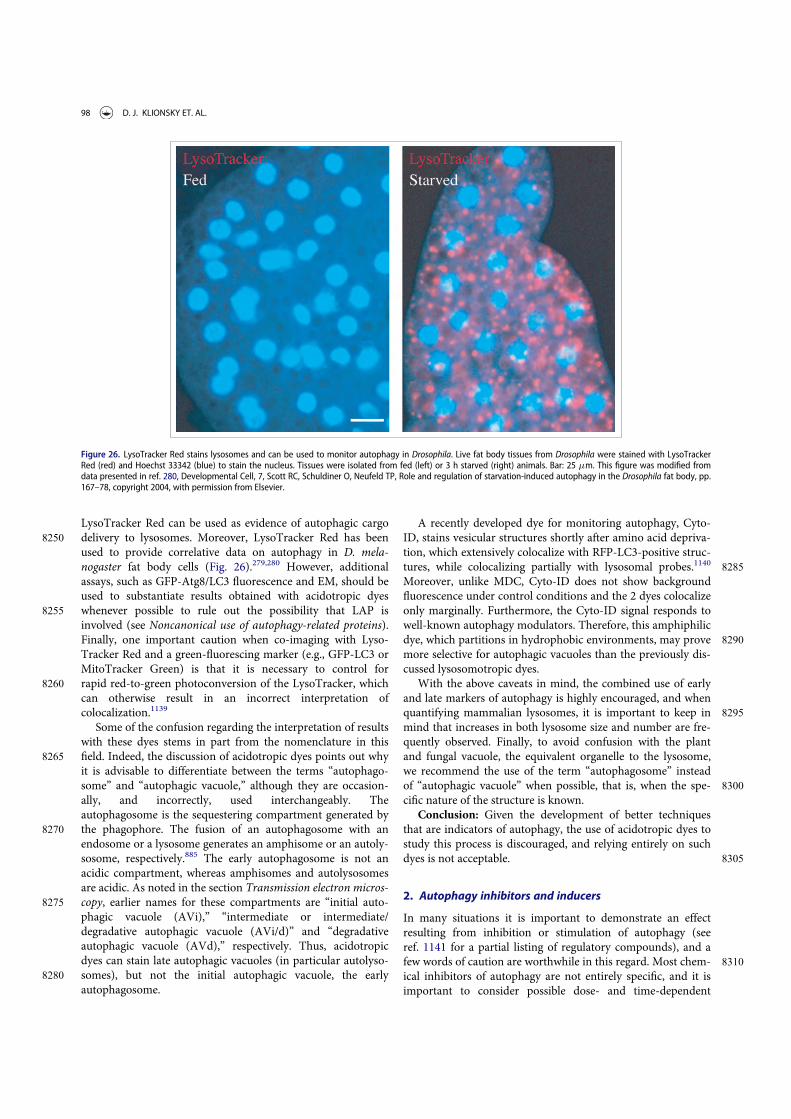
LysoTracker Red can be used as evidence of autophagic cargo8250 delivery to lysosomes. Moreover, LysoTracker Red has been
used to provide correlative data on autophagy in D. mela-nogaster fat body cells (Fig. 26).279,280 However, additionalassays, such as GFP-Atg8/LC3 fluorescence and EM, should beused to substantiate results obtained with acidotropic dyes
8255 whenever possible to rule out the possibility that LAP isinvolved (see Noncanonical use of autophagy-related proteins).Finally, one important caution when co-imaging with Lyso-Tracker Red and a green-fluorescing marker (e.g., GFP-LC3 orMitoTracker Green) is that it is necessary to control for
8260 rapid red-to-green photoconversion of the LysoTracker, whichcan otherwise result in an incorrect interpretation ofcolocalization.1139
Some of the confusion regarding the interpretation of resultswith these dyes stems in part from the nomenclature in this
8265 field. Indeed, the discussion of acidotropic dyes points out whyit is advisable to differentiate between the terms “autophago-some” and “autophagic vacuole,” although they are occasion-ally, and incorrectly, used interchangeably. Theautophagosome is the sequestering compartment generated by
8270 the phagophore. The fusion of an autophagosome with anendosome or a lysosome generates an amphisome or an autoly-sosome, respectively.885 The early autophagosome is not anacidic compartment, whereas amphisomes and autolysosomesare acidic. As noted in the section Transmission electron micros-
8275 copy, earlier names for these compartments are “initial auto-phagic vacuole (AVi),” “intermediate or intermediate/degradative autophagic vacuole (AVi/d)” and “degradativeautophagic vacuole (AVd),” respectively. Thus, acidotropicdyes can stain late autophagic vacuoles (in particular autolyso-
8280 somes), but not the initial autophagic vacuole, the earlyautophagosome.
A recently developed dye for monitoring autophagy, Cyto-ID, stains vesicular structures shortly after amino acid depriva-tion, which extensively colocalize with RFP-LC3-positive struc-
8285tures, while colocalizing partially with lysosomal probes.1140
Moreover, unlike MDC, Cyto-ID does not show backgroundfluorescence under control conditions and the 2 dyes colocalizeonly marginally. Furthermore, the Cyto-ID signal responds towell-known autophagy modulators. Therefore, this amphiphilic
8290dye, which partitions in hydrophobic environments, may provemore selective for autophagic vacuoles than the previously dis-cussed lysosomotropic dyes.
With the above caveats in mind, the combined use of earlyand late markers of autophagy is highly encouraged, and when
8295quantifying mammalian lysosomes, it is important to keep inmind that increases in both lysosome size and number are fre-quently observed. Finally, to avoid confusion with the plantand fungal vacuole, the equivalent organelle to the lysosome,we recommend the use of the term “autophagosome” instead
8300of “autophagic vacuole” when possible, that is, when the spe-cific nature of the structure is known.
Conclusion: Given the development of better techniquesthat are indicators of autophagy, the use of acidotropic dyes tostudy this process is discouraged, and relying entirely on such
8305dyes is not acceptable.
2. Autophagy inhibitors and inducers
In many situations it is important to demonstrate an effectresulting from inhibition or stimulation of autophagy (seeref. 1141 for a partial listing of regulatory compounds), and a
8310few words of caution are worthwhile in this regard. Most chem-ical inhibitors of autophagy are not entirely specific, and it isimportant to consider possible dose- and time-dependent
Figure 26. LysoTracker Red stains lysosomes and can be used to monitor autophagy in Drosophila. Live fat body tissues from Drosophila were stained with LysoTrackerRed (red) and Hoechst 33342 (blue) to stain the nucleus. Tissues were isolated from fed (left) or 3 h starved (right) animals. Bar: 25 mm. This figure was modified fromdata presented in ref. 280, Developmental Cell, 7, Scott RC, Schuldiner O, Neufeld TP, Role and regulation of starvation-induced autophagy in the Drosophila fat body, pp.167–78, copyright 2004, with permission from Elsevier.
98 D. J. KLIONSKY ET. AL.
effects. Accordingly, it is generally preferable to analyze specificloss-of-function Atg mutants. However, it must be kept in
8315 mind that some apparently specific Atg gene products mayhave autophagy-independent roles (e.g., ATG5 in cell death,and the PIK3C3/VPS34-containing complexes—includingBECN1—in apoptosis, endosomal function and protein traf-ficking), or may be dispensable for autophagy (see Noncanoni-
8320 cal use of autophagy-related proteins).27,543,573,1142-1145
Therefore, the experimental conditions of inhibitor applicationand their side effects must be carefully considered. In addition,it must be emphasized once again that autophagy, as a multi-step process, can be inhibited at different stages. Sequestration
8325 inhibitors, including 3-MA, LY294002 and wortmannin, inhibitclass I phosphoinositide 3-kinases (PI3Ks) as well as class IIIPtdIns3Ks.132,330,1146 The class I enzymes generate productssuch as PtdIns(3,4,5)P3 that inhibit autophagic sequestration,whereas the class III product (PtdIns3P) generally stimulates
8330 autophagic sequestration. The overall effect of these inhibitorsis typically to block autophagy because the class III enzymesthat are required to activate autophagy act downstream of thenegative regulatory class I enzymes, although cell death mayensue in cell types that are dependent upon high levels of AKT
8335 for survival. The effect of 3-MA (but not that of wortmannin)is further complicated by the fact that it has different temporalpatterns of inhibition, causing a long-term suppression of theclass I PI3K, but only a transient inhibition of the class IIIenzyme. In cells incubated in a complete medium for extended
8340 periods of time, 3-MA may, therefore (particularly at subopti-mal concentrations), promote autophagy by inhibition of theclass I enzyme.330 Thus, wortmannin may be considered as analternative to 3-MA for autophagy inhibition.330 However,wortmannin can induce the formation of vacuoles that may
8345 have the appearance of autophagosomes, although they areswollen late endocytic compartments.932 Furthermore, studieshave demonstrated that inhibition of autophagy with 3-MA orwortmannin can have effects on cytokine transcription, proc-essing and secretion, particularly of IL1 family members,1147-
83501149 but 3-MA and wortmannin also inhibit the secretion ofsome cytokines and chemokines (e.g., TNF, IL6, CCL2/MCP-1)in an autophagy-independent manner (J. Harris, unpublishedobservations).1147,1150 Thus, in studies where the effect ofautophagy inhibition on specific cellular processes is being
8355 investigated, it is important to confirm results using othermethods, such as RNA silencing. Due to these issues, it is ofgreat interest that inhibitors with specificity for the class IIIPtdIns3Ks, and their consequent effects on autophagy, havebeen described.244,1151,1152
8360 A mutant mouse line carrying a floxed allele of Pik3c3has been created.1153 This provides a useful genetic toolthat will help in defining the physiological role of the classIII PtdIns3K with bona fide specificity by deleting the classIII kinase in a cell type-specific manner in a whole animal
8365 using the Cre-LoxP strategy. For example, the phenotyperesulting from a knockout of Pik3c3 specifically in the kid-ney glomerular podocytes (Pik3c3pdKO) indicates that thereis no compensation by other classes of PtdIns3Ks orrelated Atg genes, thus highlighting the functional specific-
8370 ity and physiological importance of class III PtdIns3K inthese cells.
Cycloheximide, a commonly used protein synthesis inhibi-tor in mammals, is also an inhibitor of sequestration invivo,12-14,78,925,1154-1158 and in various cell types in vitro,466,1159
8375and it has been utilized to investigate the dynamic nature of theregression of various autophagic elements.12-14,25,78,1155,1156 Themechanism of action of cycloheximide in short-term experi-ments is not clear, but it has no direct relation to the inhibitionof protein synthesis.466 This latter activity, however, may com-
8380plicate certain types of analysis when using this drug.A significant challenge for a more detailed analysis of the
dynamic role of autophagy in physiological and pathophysio-logical processes, for instance with regard to cancer and cancertherapy, is to find more specific inhibitors of autophagy signal-
8385ing which do not affect other signaling cascades. For example,in the context of cellular radiation responses it is well knownthat PI3Ks, in addition to signaling through the PI3K-AKTpathway, have a major role in the regulation of DNA-damagerepair.1160 However, 3-MA, which is a nonspecific inhibitor of
8390these lipid kinases, can alter the function of other classes of thisenzyme, which are involved in the DNA-damage repairresponse. This is of particular importance for investigationsinto the role of radiation-induced autophagy in cellular radia-tion sensitivity or resistance.1161,1162
8395Most other inhibitory drugs act at post-sequestration steps.These types of agents have been used in many experiments toboth inhibit endogenous protein degradation and to increasethe number of autophagic compartments. They cause the accu-mulation of sequestered material in either autophagosomes or
8400autolysosomes, or both, because they allow autophagic seques-tration to proceed. The main categories of these types of inhibi-tors include the vinca alkaloids (e.g., vinblastine) and othermicrotubule poisons that inhibit fusion, inhibitors of lysosomalenzymes (e.g., leupeptin, pepstatin A and E-64d), and com-
8405pounds that elevate lysosomal pH (e.g., inhibitors of V-ATPases such as bafilomycin A1 and concanamycin A [anotherV-ATPase inhibitor], and weak base amines including methyl-or propylamine, chloroquine, and Neutral Red, some of whichslow down fusion). Ammonia is a very useful agent for the ele-
8410vation of lysosomal pH in short-term experiments, but it hasbeen reported to cause a stimulation of autophagy during long-term incubation of cells in a full medium,1163 under which con-ditions a good alternative might be methylamine or propyl-amine.1164 Along these lines, it should be noted that the half-
8415life of glutamine in cell culture media is approximately 2 weeksdue to chemical decomposition, which results in media withlowered glutamine and elevated ammonia concentrations thatcan affect the autophagic flux (either inhibiting or stimulatingautophagy, depending on the concentration1165). Thus, to help
8420reduce experimental variation, the use of freshly prepared cellculture media with glutamine is advised. A special note of cau-tion is also warranted in regard to chloroquine. Although thischemical is commonly used as an autophagy inhibitor, chloro-quine may initially stimulate autophagy (F.C. Dorsey, personal
8425communication; R. Franco, personal communication). In addi-tion, culture conditions requiring acidic media preclude the useof chloroquine because intracellular accumulation of the chem-ical is dramatically reduced by low pH.1166 To overcome thisissue, it is possible to use acid compounds that modulate
8430autophagy, such as betulinic acid and its derivatives.235,1167-1169
AUTOPHAGY 99

Betulinic acid damages lysosomal function differing from tradi-tional inbibitors (e.g., chloroquine, NH4Cl or bafilomycin A1)that raise the lysosomal pH; betulinic acid interacts with purephospholipid membranes,235,1170 and is capable of changing
8435 membrane permeability.235,1171,1172 The lysosomal damagemediated by betulinic acid is capable of compromising autoph-agy without any incremental damage when lysosomal functionis altered by lysosomal inhibitors (e.g., chloroquine or bafilo-mycin A1);
235 however, betulinic acid is not lysosome specific,8440 and will affect other organelles such as mitochondria.
Some data suggest that particular nanomaterials may also benovel inhibitors of autophagy, by as yet unidentifiedmechanisms.1173
It is worth noting that lysosomal proteases fall into 3 general8445 groups, cysteine, aspartic acid and serine proteases. Therefore,
the fact that leupeptin, a serine and cysteine protease inhibitor,has little or no effect does not necessarily indicate that lyso-somal degradation is not taking place; a combination of leupep-tin, pepstatin A and E-64d may be a more effective treatment.
8450 However, it should also be pointed out that these proteaseinhibitors can exert inhibitory effects not only on lysosomalproteases, but also on cytosolic proteases; that is, degradationof proteins might be blocked through inhibition of cytosolicinstead of lysosomal proteases. Conversely, it should be noted
8455 that MG132 (Z-leu-leu-leu-al) and its related peptide aldehydesare commonly used as proteasomal inhibitors, but they canalso inhibit certain lysosomal hydrolases such as cathepsinsand calpains.1174 Thus, any positive results using MG132 donot rule out the possibility of involvement of the autophagy-
8460 lysosomal system. Therefore, even if MG132 is effective ininhibiting autophagy, it is important to confirm the result usingmore specific proteasomal inhibitors such as lactacystin orepoxomicin. Finally, there are significant differences in cell per-meability among protease inhibitors. For example, E-64d is
8465 membrane permeable, whereas leupeptin and pepstatin A arenot (although there are derivatives that display greater perme-ability such as pepstatin A methyl ester).1175 Thus, when ana-lyzing whether a protein is an autophagy substrate, cautionshould be taken in utilizing these protease inhibitors to block
8470 autophagy.As with the PtdIns3K inhibitors, many autophagy-suppres-
sive compounds are not specific. For example, okadaic acid1176
is a powerful general inhibitor of both type 1 (PPP1) and type2A (PPP2) protein phosphatases.1177 Bafilomycin A1 and other
8475 compounds that raise the lysosomal pH may have indirecteffects on any acidified compartments. Moreover, treatmentwith bafilomycin A1 for extended periods (18 h) can cause sig-nificant disruption of the mitochondrial network in culturedcells (M.E. Gegg, personal communication), and either bafilo-
8480 mycin A1 or concanamycin A cause swelling of the Golgi inplants,1178 and increase cell death by apoptosis in cancer cells(V.A. Rao, personal communication). Furthermore, bafilomy-cin A1 may have off-target effects on the cell, particularly onMTORC1.487,527,1179 Bafilomycin A1 is often used at a final con-
8485 centration of 100 nM, but much lower concentrations such as1 nM may be sufficient to inhibit autophagic-lysosomal degra-dation and are less likely to cause indirect effects.157,225,1180 Forexample, in pulmonary A549 epithelial cells bafilomycin A1
exhibits concentration-dependent effects on cellular
8490morphology and on protein expression; at concentrations of 10and 100 nM the cells become more rounded accompanied byincreased expression of VIM (vimentin) and a decrease inCDH1/E-cadherin (B. Yeganeh, M. Post and S. Ghavami,unpublished observations). Thus, appropriate inhibitory con-
8495centrations should be empirically determined for each celltype.231
Although these various agents can inhibit different steps ofthe autophagic pathway, their potential side effects must beconsidered in interpretation of the secondary consequences of
8500autophagy inhibition, especially in long-term studies. Forexample, lysosomotropic compounds can increase the rate ofautophagosome formation by inhibiting MTORC1, as activa-tion of lysosomally localized MTORC1 depends on an activeV-ATPase (as well as RRAG GTPases162).487,1181 Along these
8505lines, chloroquine treatment may cause an apparent increasein the formation of autophagosomes possibly by blockingfusion with the lysosome (F.C. Dorsey and J.L. Cleveland, per-sonal communication). This conclusion is supported by thefinding that chloroquine reduces the colocalization of LC3
8510and LysoTracker despite the presence of autophagosomes andlysosomes (A.K. Simon, personal communication). This mech-anism might be cell-type specific, as other studies report thatchloroquine prevents autolysosome clearance and degradationof cargo content, but not autophagosome-lysosome
8515fusion.1182-1185 Concanamycin A blocks sorting of vacuolarproteins in plant cells in addition to inhibiting vacuolar acidi-fication.1186 Furthermore, in addition to causing the accumula-tion of autophagic compartments, many of these drugs seemto stimulate sequestration in many cell types, especially in
8520vivo.79,326,925,1155,1159,1187-1191 Although it is clear why thesedrugs cause the accumulation of autophagic compartments, itis not known why they stimulate sequestration. One possibil-ity, at least for hepatocytes, is that the inhibition of proteindegradation reduces the intracellular amino acid pool, which
8525in turn upregulates sequestration. A time-course study of thechanges in both the intra- and extracellular fractions may pro-vide accurate information regarding amino acid metabolism.For these various reasons, it is important to include appropri-ate controls; along these lines, MTOR inhibitors such as rapa-
8530mycin or amino acid deprivation can be utilized as positivecontrols for inducing autophagy. In many cell types, however,the induction of autophagy by rapamycin is relatively slow, ortransient, allowing more time for indirect effects.
Several small molecule inhibitors, including torin1, PP242,8535KU-0063794, PI-103 and NVP-BEZ235, have been developed
that target the catalytic domain of MTOR in an ATP-competi-tive manner.225,1192-1196 In comparison to rapamycin, these cat-alytic MTOR inhibitors are more potent, and hence arestronger autophagy agonists in most cell lines.341,1194,1197 The
8540use of these second-generation MTOR inhibitors may revealthat some reports of MTOR-independent autophagy may actu-ally reflect the use of the relatively weak inhibitor rapamycin.Furthermore, the use of these compounds has revealed a rolefor MTORC1 and MTORC2 as independent regulators of
8545autophagy.1198
Neurons, however, seem to be a particular case in regard totheir response to MTOR inhibitors. Rapamycin may fail to acti-vate autophagy in cultured primary neurons, despite its potent
100 D. J. KLIONSKY ET. AL.

stimulation of autophagy in some cancer cell lines,75,544,1199
8550 Interestingly, both rapamycin and catalytic MTOR inhibitorsdo not induce a robust autophagy in either cultured primarymouse neurons or human neuroblastoma SH-SY5Y cells, whichcan differentiate into neuron-like cells, whereas the drugs doelicit a potent autophagic response in cultured astrocytes (J.
8555 Diaz-Nido and R. Gargini, personal communication). This sug-gests a differential regulation of autophagy in neurons. It hasbeen suggested that control of neuronal autophagy may reflectthe particular physiological adaptations and metabolic require-ments of neurons, which are very different from most periph-
8560 eral cell types.1200 For example, acute starvation in transgenicmice expressing GFP-LC3 leads to a potent induction ofautophagy in the liver, muscle and heart but not in the brain.153
Along these lines, glucose depletion may be much more effi-cient at inducing autophagy than rapamycin or amino acid
8565 starvation in neurons in culture (M. Germain and R. Slack, per-sonal communication). Indeed treatment of cultured primarymouse neurons and human neuroblastoma SH-SY5Y cells with2-deoxy-glucose, which hampers glucose metabolism and leadsto activation of AMPK, results in robust autophagy induction
8570 (J. Diaz-Nido and R. Gargini, personal communication). Inter-estingly, a number of compounds can also be quite efficientautophagy inducers in neurons including the CAPN/calpaininhibitor calpeptin.1201-1203 Thus, it has been suggested thatautophagy induction in neurons may be achieved by molecular
8575 mechanisms relying on AMPK or increases in intracellular cal-cium concentration.1200 An example where changes in cytosoliccalcium levels, due to the incapacity of the mitochondria tobuffer Ca2C release, result in an increase in autophagy is seenin a cellular model of the neurodegenerative disease Friedreich
8580 ataxia, based on FXN/frataxin silencing in SH-SY5Y humanneuroblastoma cells.1204
Finally, a specialized class of compounds witha,b-unsaturated ketone structure tends to induce autophagiccell death, accompanied by changes in mitochondrial morphol-
8585 ogy. Since the cytotoxic action of these compounds is efficientlyblocked by N-acetyl-L-cysteine, the b-position in the structuremay interact with an SH group of the targeted molecules.1205
Due to the potential pleiotropic effects of various drug treat-ments, it is incumbent upon the researcher to demonstrate that
8590 autophagy is indeed inhibited, by using the methodologiesdescribed herein. Accordingly, it is critical to verify the effect ofa particular biochemical treatment with regard to its effects onautophagy induction or inhibition when using a cell line thatwas previously uncharacterized for the chemical being used.
8595 Similarly, cytotoxicity of the relevant chemical should beassessed.
The use of gene deletions/inactivations (e.g., in primary orimmortalized atg¡/¡ MEFs,540 plant T-DNA or transposoninsertion mutants,282,1206 or in vivo using transgenic knockout
8600 models1207,1208 including Cre-lox based “conditional” knock-outs320,321) or functional knockdowns (e.g., with RNAi againstATG genes) is the preferred approach when possible becausethese methods allow a more direct assessment of the resultingphenotype; however, different floxed genes are deleted with
8605 varying efficiency, and the proportion deleted must be carefullyquantified.1209 Studies also suggest that microRNAs may beused for blocking gene expression.243,645,646,1210,246-248 In most
contexts, it is advisable when using a knockout or knockdownapproach to examine multiple autophagy-related genes to
8610exclude the possibility that the phenotype observed is due toeffects on a nonautophagic function(s) of the correspondingprotein, especially when examining the possibility of autopha-gic cell death. This is particularly the case in evaluatingBECN1, which interacts with anti-apoptotic BCL2 family pro-
8615teins,566 or when low levels of a target protein are sufficient formaintaining autophagy as is the case with ATG5.255 Withregard to ATG5, a better approach may be to use a dominantnegative (K130R) version.1145,1199,1211 Also noteworthy is therole of ATG5 in mitotic catastrophe544 and several other non-
8620autophagic roles of ATG proteins (see Noncanonical use ofautophagy-related proteins).75 Along these lines, and as statedabove for the use of inhibitors, when employing a knockout orespecially a knockdown approach, it is again incumbent uponthe researcher to demonstrate that autophagy is actually inhib-
8625ited, by using the methodologies described herein.Finally, we note that the long-term secondary consequen-
ces of gene knockouts or knockdowns are likely much morecomplex than the immediate effects of the actual autophagyinhibition. To overcome this concern, inducible knockout
8630systems might be useful.255,404 One additional caveat toknockdown experiments is that PAMP recognition pathwayscan be triggered by double-stranded RNAs (dsRNA), likesiRNA probes, or the viral vector systems that delivershRNA.1212 Some of these, like TLR-mediated RNA recogni-
8635tion,1213 can influence autophagy by either masking anyinhibitory effect or compromising autophagy independentof the knockdown probe. Therefore, nontargeting (scram-bled) siRNA or shRNA controls should be used with therespective transfection or transduction methods in the
8640experiments that employ ATG knockdown. Another strategyto specifically interfere with autophagy is to use dominantnegative inhibitors. Delivery of these agents by transienttransfection, adenovirus, or TAT-mediated protein trans-duction offers the possibility of their use in cell culture or
8645in vivo.1211 However, since autophagy is an essential meta-bolic process for many cell types and tissues, loss of viabil-ity due to autophagy inhibition always has to be a concernwhen analyzing cell death-unrelated questions. In thisrespect it is noteworthy that some cell-types of the immune
8650system such as dendritic cells333 seem to tolerate loss ofautophagy fairly well, whereas others such as T and B cellsare compromised in their development and function afterautophagy inhibition.1214,1215
In addition to pharmacological inhibition, RNA silencing,8655gene knockout and dominant negative RAB and ATG protein
expression, pathogen-derived autophagy inhibitors can also beconsidered to manipulate autophagy. Along these linesICP34.5, viral BCL2 homologs and viral FLIP of herpesvirusesblock autophagosome formation,566,893,1216 whereas M2 of
8660influenza virus and HIV-1 Nef block autophagosome degrada-tion.362,903 However, as with other tools discussed in this sec-tion, transfection or transduction of viral autophagy inhibitorsshould be used in parallel with other means of autophagymanipulation, because these proteins are used for the regula-
8665tion of usually more than one cellular pathway by the respectivepathogens.
AUTOPHAGY 101

There are fewer compounds that act as inducers of autoph-agy, but the initial characterization of this process was due inlarge part to the inducing effects of glucagon, which appears to
8670 act through indirect inhibition of MTOR via the activation ofSTK11/LKB1- AMPK.936,937,1217 Currently, the most commonlyused inducer of autophagy is rapamycin, an allosteric inhibitorof MTORC1 (although as mentioned above, catalytic inhibitorssuch as torin1 are increasingly being used). Nevertheless, one
8675 caution is that MTOR is a major regulatory protein that is partof several signaling pathways, including for example those thatrespond to INS/insulin, EGF/epidermal growth factor andamino acids, and it thus controls processes other than autoph-agy, so rapamycin will ultimately affect many metabolic path-
8680 ways.504,1218-1220 In particular, the strong effects of MTOR onprotein synthesis may be a confounding factor when analyzingthe effects of rapamycin. MTOR-independent regulation can beachieved through lithium, sodium valproate and carbamaze-pine, compounds that lower the myo-inositol 1,4,5-triphos-
8685 phate levels,1221 as well as FDA-approved compounds such asverapamil, trifluoperazine and clonidine.1222,1223 In vivo treat-ment of embryos with cadmium results in an increase inautophagy, probably to counter the stress, allowing cell survivalthrough the elimination/recycling of damaged structures.957
8690 Autophagy may also be regulated by the release of calciumfrom the ER under stress conditions;297,1176,1224,1225 however,additional calcium signals from other stores such as the mito-chondria and lysosomes could also play an important role inautophagy induction. The activation of the lysosomal TPCN/
8695 two-pore channel (two pore segment channel), by nicotinicacid adenine dinucleotide phosphate (NAADP) inducesautophagy, which can selectively be inhibited by the TPCNblocker NED-19, or by pre-incubation with BAPTA, showingthat lysosomal calcium also modulates autophagy.1226 Cell pen-
8700 etrating autophagy-inducing peptides, such as Tat-vFLIP orTat-Beclin 1 (Tat-BECN1), are also potent inducers of autoph-agy in cultured cells as well as in mice.1216,1227 Other cell pene-trating peptides, such as Tat-wtBH3D or Tat-dsBH3D,designed to disrupt very specific regulatory interactions such as
8705 the BCL2-BECN1 interaction, are potent, yet very specific,inducers of autophagy in cultured cells.1228
In contrast to other PtdIns3K inhibitors, caffeine inducesmacroautophagy in the food spoilage yeast Zygosaccharomycesbailii,1229 mouse embryonic fibroblasts,1230 and S. cerevisiae1231
8710 at millimolar concentrations. In higher eukaroyotes this isaccompanied by inhibition of the MTOR pathway. Similarly, inbudding yeast caffeine is a potent TORC1 inhibitor suggestingthat this drug induces autophagy via inhibition of the TORC1signalling pathway; however, as with other PtdIns3K inhibitors
8715 caffeine targets other proteins, notably Mec1/ATR and Tel1/ATM, and affects the cellular response to DNA damage.
Another autophagy inducer is the histone deacetylase inhibi-tor valproic acid.1232,1233 The mechanism by which valproicacid stimulates autophagy is not entirely clear but may occur
8720 due to inhibition of the histone deacetylase Rpd3, which nega-tively regulates the transcription of ATG genes (most notablyATG81234) and, via deacetylation of Atg3, controls Atg8lipidation.1235
It is also possible, depending on the organism or cell system,8725 to modulate autophagy through transcriptional control. For
example, this can be achieved either through overexpression orpost-translational activation of the gene encoding TFEB (seeTranscriptional and translational regulation), a transcriptionalregulator of the biogenesis of both lysosomes and autophago-
8730somes.625,635 Similarly, adenoviral-mediated expression of thetranscription factor CEBPB induces autophagy in hepato-cytes.644 Recently, it has been shown that either the geneticablation or the knockdown of the nucleolar transcription factorRRN3/TIF-IA, a crucial regulator of the recruitment of
8735POLR1/RNA polymerase I to ribosomal DNA promoters, indu-ces autophagy in neurons and in MCF-7 cancer cells,respectively, linking ribosomal DNA transcription toautophagy.1236,1237
Relatively little is known about direct regulation via the ATG8740proteins, but there is some indication that tamoxifen acts to
induce autophagy by increasing the expression of BECN1 inMCF7 cells.1238 However, BECN1 does not appear to be upre-gulated in U87MG cells treated with tamoxifen, whereas thelevels of LC3-II and SQSTM1 are increased, while LAMP2B is
8745downregulated and CTSD and CTSL activities are almostcompletely blocked (K.S. Choi, personal communication).Thus, the effect of tamoxifen may differ depending on the celltype. Other data suggest that tamoxifen acts by blocking choles-terol biosynthesis, and that the sterol balance may determine
8750whether autophagy acts in a protective versus cytotoxic man-ner.1239,1240 Finally, screens have identified small molecules thatinduce autophagy independently of rapamycin and allow theremoval of misfolded or aggregate-prone proteins,1223,1241 sug-gesting that they may prove useful in therapeutic applications.
8755However, caution should be taken because of the crosstalkbetween autophagy and the proteasomal system. For example,trehalose, an MTOR-independent autophagy inducer,1242 cancompromise proteasomal activity in cultured primaryneurons.1243
8760Because gangliosides are implicated in autophagosome mor-phogenesis, pharmacological or genetic impairment of ganglio-sidic compartment integrity and function can provide usefulinformation in the analysis of autophagy. To deplete cells ofgangliosides, an inhibitor of CERS/ceramide synthase, such as a
8765fungal metabolite produced by Fusarium moniliforme (fumoni-sin B1), or, alternatively, siRNA to CERS or ST8SIA1, can beused.595
Finally, in addition to genetic and chemical compounds,it was recently reported that electromagnetic fields can
8770induce autophagy in mammalian cells. Studies of biologicaleffects of novel therapeutic approaches for cancer therapybased on the use of noninvasive radiofrequency fieldsreveals that autophagy, but not apoptosis, is induced in can-cer cells in response to this treatment, which leads to cell
8775death.1244 This effect was tumor specific and different fromtraditional ionizing radiation therapy that induces apoptosisin cells.
Conclusion: Considering that pharmacological inhibitorsor activators of autophagy have an impact on many other
8780cellular pathways, the use of more than one methodology,including molecular methods, is desirable. Rapamycin isless effective at inhibiting MTOR and inducing autophagythan catalytic inhibitors; however, it must be kept in mindthat catalytic inhibitors also affect MTORC2. The main
102 D. J. KLIONSKY ET. AL.

8785 concern with pharmacological manipulations is pleiotropiceffects of the compound being used. Accordingly, geneticconfirmation is preferred whenever possible.
3. Basal autophagy
Basal levels of LC3-II or GFP-LC3 puncta may change accord-8790 ing to the time after addition of fresh medium to cells, and this
can lead to misinterpretations of what basal autophagy means.This is particularly important when comparing the levels ofbasal autophagy between different cell populations (such asknockout versus wild-type clones). If cells are very sensitive to
8795 nutrient supply and display a high variability of basal autoph-agy, the best experimental condition is to monitor the levels ofbasal autophagy at different times after the addition of freshmedium. One example is the chicken lymphoma DT40 cells(see Chicken B-lymphoid DT40 cells) and their knockout variant
8800 for all 3 ITPR isoforms.1245-1247 In these cells, no differences inbasal levels of LC3-II can be observed up to 4 h after additionof fresh medium, but differences can be observed after longertimes (J.M. Vicencio and G. Szabadkai, personal communica-tion). This concept should also be applied to experiments in
8805 which the effect of a drug upon autophagy is the subject ofstudy. If the drugs are added after a time in which basal autoph-agy is already high, then the effects of the drug can be maskedby the cell’s basal autophagy, and wrong conclusions may bedrawn. To avoid this, fresh medium should be added first (fol-
8810 lowed by incubation for 2–4 h) in order to reduce and equili-brate basal autophagy in cells under all conditions, and thenthe drugs can be added. The basal autophagy levels of the cellunder study must be identified beforehand to know the timeneeded to reduce basal autophagy.
8815 A similar caution must be exercised with regard to cell cul-ture density and hypoxia. When cells are grown in normoxicconditions at high cell density, HIF1A/HIF-1a is stabilized atlevels similar to that obtained with low-density cultures underhypoxic conditions.1248 This results in the induction of BNIP3
8820 and BNIP3L and “hypoxia”-induced autophagy, even thoughthe conditions are theoretically normoxic.1249 Therefore,researchers need to be careful about cell density to avoid acci-dental induction of autophagy.
It should be realized that in yeast species, medium changes8825 can trigger a higher “basal” level of autophagy in the cells. In
the methylotrophic yeast species P. pastoris and Hansenula pol-ymorpha a shift of cells grown in batch from glucose to metha-nol results in stimulation of autophagy.1250,1251 A shift to a newmedium can be considered a stress situation. Thus, it appears
8830 to be essential to cultivate the yeast cells for a number of hoursto stabilize the level of basal autophagy before performingexperiments intended to study levels of (selective) autophagy(e.g., pexophagy). Finally, plant root tips cultured in nutrient-sufficient medium display constitutive autophagic flux (i.e., a
8835 basal level), which is enhanced in nutrient-deprivedmedium.1133,1252,1253
Conclusion: The levels of basal autophagy can vary substan-tially and can mask the effects of the experimental parametersbeing tested. Changes in media and growth conditions need to
8840 be examined empirically to determine affects on basal autoph-agy and the appropriate times for subsequent manipulations.
4. Experimental systems
Throughout these guidelines we have noted that it is not possi-ble to state explicit rules that can be applied to all experimental
8845systems. For example, some techniques may not work in partic-ular cell types or organisms. In each case, efficacy of autophagypromotors, inhibitors and measurement techniques must beempirically determined, which is why it is important to includeappropriate controls. Differences may also be seen between in
8850vivo or perfused organ studies and cell culture analyses. Forexample, INS/insulin has no effect on proteolysis in suspendedrat hepatocytes, in contrast to the result with perfused rat liver.The INS/insulin effect reappears, however, when isolated hepa-tocytes are incubated in stationary dishes1254,1255 or are allowed
8855to settle down on the matrix (D. H€aussinger, personal commu-nication). The reason for this might be that autophagy regula-tion by insulin and some amino acids requires volume sensingvia integrin-matrix interactions and also intact microtu-bules.1256-1258 Along these lines, the use of whole embryos
8860makes it possible to investigate autophagy in multipotent cells,which interact among themselves in their natural environment,bypassing the disadvantages of isolated cells that are deprivedof their normal network of interactions.957 In general, it isimportant to keep in mind that results from one particular sys-
8865tem may not be generally applicable to others.Conclusion: Although autophagy is conserved from yeast to
human, there may be tremendous differences in the specificdetails among systems. Thus, results based on one systemshould not be assumed to be applicable to another.
88705. Nomenclature
To minimize confusion regarding nomenclature, we makethe following notes: In general, we follow the conventionsestablished by the nomenclature committees for each modelorganism whenever appropriate guidelines are available, and
8875briefly summarize the information here using “ATG1” as anexample for yeast and mammals. The standard nomencla-ture of autophagy-related wild-type genes, mutants and pro-teins for yeast is ATG1, atg1 (or atg1D in the case ofdeletions) and Atg1, respectively, according to the guide-
8880lines adopted by the Saccharomyces Genome Database(http://www.yeastgenome.org/gene_guidelines.shtml). Formammals we follow the recommendations of the InternationalCommittee on Standardized Genetic Nomenclature for Mice(http://www.informatics.jax.org/mgihome/nomen/), which dic-
8885tates the designations Atg1, atg1 and ATG1 (for all rodents),respectively, and the guidelines for human genes established bythe HUGO Nomenclature Committee (http://www.genenames.org/guidelines.html), which states that human gene symbolsare in the form ATG1 and recommends that proteins use the
8890same designation without italics, as with ATG1; mutants arewritten for example as ATG1¡/-.1259
C. Methods and challenges of specialized topics/model systems
There are now a large number of model systems being used to8895study autophagy. These guidelines cannot cover every detail,
AUTOPHAGY 103

and as stated in the Introduction this article is not meant toprovide detailed protocols. Nonetheless, we think it is useful tobriefly discuss what techniques can be used in these systemsand to highlight some of the specific concerns and/or chal-
8900 lenges. We also refer readers to the 3 volumes of Methods inEnzymology that provide additional information for “nonstan-dard”model systems.39-41
1. C. elegans
C. elegans has a single ortholog of most yeast Atg proteins;8905 however, 2 nematode homologs exist for Atg4, Atg8 and
Atg16.1260-1262 Multiple studies have established C. elegans as auseful multicellular genetic model to delineate the autophagypathway and associated functions (see for example refs. 271,633, 742, 743, 1263). The LGG-1/Atg8/LC3 reporter is the
8910 most commonly used tool to detect autophagy in C. elegans.Similar to Atg8, which is incorporated into the double mem-brane of autophagic vacuoles during autophagy,148,269,600 the C.elegans LGG-1 localizes into cytoplasmic puncta under condi-tions known to induce autophagy. Fluorescent reporter fusions
8915 of LGG-1/Atg8 with GFP, DsRED or mCherry have been usedto monitor autophagosome formation in vivo, in the nematode.These reporters can be expressed either in specific cells and tis-sues or throughout the animal.271,743,1264,1265 Caution should betaken, however, when using protein markers fused to mCherry
8920 in worms. mCherry aggregates in autophagy-inducing condi-tions, such as fasting, even if not fused to LGG-1 or otherautophagy markers (E. O’Rourke, personal communication);therefore mCherry puncta may not be a good readout to moni-tor autophagy in C. elegans. LGG-2 is the second LC3 homolog
8925 and is also a convenient marker for autophagy either using spe-cific antibodies742 or fused to GFP,1266 especially whenexpressed from an integrated transgene to prevent its germlinesilencing.742 The exact function of LGG-1 versus LGG-2remains to be addressed.1267
8930 For observing autophagy by GFP-LC3 fluorescence in C. ele-gans, it is best to use integrated versions of the marker742,743,1268
(GFP::LGG-1 and GFP::LGG-2; Fig. 27) rather than extrachro-mosomal transgenic strains271,1266 because the latter show vari-able expression among different animals or mosaic expression
8935 (C. Kang, personal communication; V. Galy, personal commu-nication). Nevertheless, evaluation of LGG-1::GFP puncta ismostly restricted to seam cells, which is tedious because of asmall number of puncta/cell even in autophagy-inducing con-ditions (<5/cell), error prone due to high background levels in
8940 the GFP channel, and extremely difficult to visualize in theadult. To increase signal to noise, it is also possible to carry outindirect immunofluorescence microscopy using antibodiesagainst endogenous LGG-1, 633,743 or LGG-2;742 however, anti-LGG-1 and anti-LGG-2 antibodies are not commercially avail-
8945 able. In addition, with the integrated version, or with antibodiesdirected against endogenous LGG-1, it is possible to perform awestern blot analysis for lipidation, at least in embryos (LGG-1-I is the nonlipidated soluble form and LGG-1-II/LGG-1–PEis the lipidated form).1268,743,633
8950 The LGG-1 precursor accumulates in the atg-4.1 mutant, butis undetectable in wild-type embryos.1261 Moreover, the bandingpattern of LGG-1 or LGG-1 fused to fluorescent proteins in
western blots may not be easy to interpret in larvae or the adultC. elegans because enrichment for a fast running band (the lipi-
8955dated form) is not observed in some autophagy-inducing
Figure 27. GFP::LGG-1 and GFP::LGG-2 are autophagy markers in C. elegans. (A–F)Animals were generated that carry an integrated transgene expressing a GFP-tagged version of lgg-1, the C. elegans ortholog of mammalian MAP1LC3. Repre-sentative green fluorescence images in the pharyngeal muscles of (A) control RNAianimals without starvation, (B) control RNAi animals after 9 d of starvation, (C) atg-7 RNAi animals after 9 d of starvation, (D) starvation-hypersensitive gpb-2 mutantswithout leucine after 3 d of starvation, and (E) gpb-2 mutants with leucine after 3d of starvation. The arrows show representative GFP::LGG-1-positive punctate areasthat label pre-autophagosomal and autophagosomal structures. (F) The relativelevels of PE-conjugated and unconjugated GFP::LGG-1 were determined by west-ern blotting. These figures were modified from data previously published in Kang,C., Y.J. You, and L. Avery. 2007. Dual roles of autophagy in the survival of Caeno-rhabditis elegans during starvation. Genes & Development. 21:2161–2171, Copyright© 2007, Genes & Development by Cold Spring Harbor Laboratory Press and Kang,C., and L. Avery. 2009. Systemic regulation of starvation response in Caenorhabditiselegans. Genes & development. 23:12–17, Copyright © 2011, Genes & Developmentby Cold Spring Harbor Laboratory Press, www.genesdev.org. (G–H) GFP:LGG-2serves as a marker for autophagosomes in early C. elegans embryos. (G) GFP::LGG-2 expressed in the germline from an integrated transgene reveal the formation ofautophagosomes (green) around sperm inherited membranous organelles (red).DNA of the 2 pronuclei is stained (blue). (H) Later during development, GFP::LGG-2-positive structures are present in all cells of the embryo. Scale bar: 10 mm.Images provided by V. Galy.
104 D. J. KLIONSKY ET. AL.

conditions, such as fasting. In the embryos of some autophagymutants, including epg-3, epg-4, epg-5, and epg-6 mutants,levels of LGG-1-I and LGG-1-II are elevated.563,633,1269,1270 Inan immunostaining assay, endogenous LGG-1 forms distinct
8960 punctate structures, mostly at the »64- to 100-cell embryonicstage. LGG-1 puncta are absent in atg-3, atg-7, atg-5 andatg-10 mutant embryos,633,1262 but dramatically accumulate inother autophagy mutants.563,633 The widely used GFP::LGG-1reporter forms aggregates in atg-3 and atg-7 mutant embryos,
8965 in which endogenous LGG-1 puncta are absent, indicatingthat GFP::LGG-1 could be incorporated into protein aggre-gates during embryogenesis. Immunostaining for endogenousVPS-34 is also a useful marker of autophagy induction inC. elegans embryos.1271
8970 A variety of protein aggregates, including PGL granules(PGL-1-PGL-3-SEPA-1) and the C. elegans SQSTM1 homologSQST-1, are selectively degraded by autophagy during embryo-genesis; impaired autophagy activity results in their accumula-tion and the generation of numerous aggregates.633,1263 Thus,
8975 degradation of these autophagy substrates can also be used tomonitor autophagy activity, with similar cautionary notes tothose described in section A3 (see SQSTM1 and related LC3binding protein turnover assays) for the SQST-1 turnover assay.Similar to mammalian cells, the total amount of LGG-1::GFP
8980 along with SQST-1::GFP transcriptional expression coupledwith its posttranscriptional accumulation can be informativewith regard to autophagic flux in the embryo (again with thesame cautionary notes described in section A3).629,1272
As with its mammalian counterpart, loss of the C. elegans8985 TP53 ortholog, cep-1, increases autophagosome accumula-
tion1273 and extends the animal’s life span.1274 bec-1- and cep-1-regulated autophagy is also required for optimal life span exten-sion and to reduce lipid accumulation in response to silencingFRH-1/frataxin, a protein involved in mitochondrial respira-
8990 tory chain functionality.1275 FRH-1 silencing also inducesmitophagy in an evolutionarily conserved manner.1272 More-over, the products of C. elegans mitophagy regulatory genehomologs (PDR-1/PARK2, PINK-1/PINK1, DCT-1/BNIP3,and SQST-1/SQSTM1) are required for induction of mitophagy
8995 (monitored through the Rosella biosensor1276) and life spanextension following FRH-1 silencing and iron deprivation.1272
The TFEB ortholog HLH-30 transcriptionally regulates macro-autophagy and promotes lipid degradation,624,824 and worm lifespan analyses uncovered a direct role for HLH-30/TFEB in reg-
9000 ulation in C. elegans, and likely in mammals.624,629,824
For a more complete review of methods for monitoringautophagy in C. elegans see ref. 1277. Note that most theseapproaches have been optimized to monitor autophagy inembryos or early larval stages, and that autophagy markers in
9005 the adult C. elegans are poorly characterized or lacking.
2. Chicken B-lymphoid DT40 cells, retina and inner ear
The chicken B-lymphoid DT40 cell line represents a suitabletool for the analysis of autophagic processes in a nonmamma-lian vertebrate system. In DT40 cells, foreign DNA integrates
9010 with a very high frequency by homologous recombination com-pared to random integration. This makes the cell line a valuabletool for the generation of cellular gene knockouts. Generally,
the complete knockout of genes encoding autophagy-regulatoryproteins is preferable compared to RNAi-mediated knockdown,
9015because in some cases these proteins function normally whenexpressed at reduced levels.255 Different Atg-deficient DT40cell lines already exist, including atg13¡/¡, ulk1¡/¡, ulk2¡/¡,ulk1/2¡/¡,1278 becn1¡/¡, and rb1cc1/fip200¡/¡ (B. Stork, per-sonal communication). Many additional non-autophagy-
9020related gene knockout DT40 cell lines have been generated andare commercially available.1279
DT40 cells are highly proliferative (the generation time isapproximately 10 h), and knockout cells can be easily reconsti-tuted with cDNAs by retroviral gene transfer for the mutational
9025analysis of signaling pathways. DT40 cells mount an autophagicresponse upon starvation in EBSS,1278 and autophagy can beanalyzed by a variety of assays in this cell line. Steady statemethods that can be used include TEM, LC3 western blottingand fluorescence microscopy; flux measurements include mon-
9030itoring LC3-II turnover and tandem mRFP/mCherry-GFP-LC3fluorescence microscopy. Using atg13¡/- and ulk1/2¡/- DT40cells, it was shown that ATG13 and its binding capacity forRB1CC1/FIP200 are mandatory for both basal and starvation-induced autophagy, whereas ULK1/2 and in vitro-mapped
9035ULK1-dependent phosphorylation sites of ATG13 appear to bedispensable for these processes.1278
Another useful system is chick retina, which can be used formonitoring autophagy at different stages of development. Forexample, lipidation of LC3 is observed during starvation, and
9040can be blocked with a short-term incubation with 3-MA.393,394
LEP-100 antibody is commercially available for the detection ofthis lysosomal protein. In the developing chicken inner ear,LC3 flux can be detected in otic vesicles cultured in a serum-free medium exposed to either 3-MA or chloroquine.395
9045One of the salient features of chicken cells, including pri-mary cells such as chicken embryo fibroblasts, is the capacity ofobtaining rapid, efficient and sustained transcript/proteindownregulation with replication-competent retrovirus forshRNA expression.1280 In chicken embryo fibroblasts, nearly
9050complete and general (i.e., in nearly all cells) protein downregu-lation can be observed within a few days after transfection ofthe shRNA retroviral vector.167
Cautionary notes: Since the DT40 cell line derives from achicken bursal lymphoma, not all ATG proteins and autoph-
9055agy-regulatory proteins are detected by the commercially avail-able antibodies produced against their mammalian orthologs;however, commercially available antibodies for mammalianLC3 and GABARAP have been reported to detect the chickencounterparts in western blots.167 The chicken genome is almost
9060completely assembled, which facilitates the design of targetingconstructs. However, in the May 2006 chicken (Gallus gallus)v2.1 assembly, 5% of the sequence has not been anchored tospecific chromosomes, and this might also include genesencoding autophagy-regulatory proteins. It is possible that
9065there is some divergence within the signaling pathways betweenmammalian and nonmammalian model systems. One examplemight be the role of ULK1/2 in starvation-induced autophagydescribed above. Additionally, neither rapamycin nor torin1seem to be potent inducers of autophagy in DT40 cells,
9070although MTOR activity is completely repressed as detected byphosphorylated RPS6KB western blotting.1278 Finally, DT40
AUTOPHAGY 105

cells represent a transformed cell line, being derived from anavian leukosis virus-induced bursal lymphoma. Thus, DT40cells release avian leukosis virus into the medium, and the 3’-
9075 long terminal repeat has integrated upstream of the MYC gene,leading to increased MYC expression.1281 Both circumstancesmight influence basal and starvation-induced autophagy.
3. Chlamydomonas
The unicellular green alga Chlamydomonas reinhardtii is an9080 excellent model system to investigate autophagy in photosyn-
thetic eukaryotes. Most of the ATG genes that constitute theautophagy core machinery including the ATG8 and ATG12ubiquitin-like systems are conserved as single-copy genes in thenuclear genome of this model alga. Autophagy can be moni-
9085 tored in Chlamydomonas by western blotting through thedetection of Atg8 lipidation as well as an increase in the abun-dance of this protein in response to autophagy activation.292
Localization of Atg8 by immunofluorescence microscopy canalso be used to study autophagy in Chlamydomonas since the
9090 cellular distribution of this protein changes drastically uponautophagy induction. The Atg8 signal is weak and usuallydetected as a single spot in nonstressed cells, whereas autoph-agy activation results in the localization of Atg8 in multiplespots with a very intense signal.292,1282,1283 Finally, enhanced
9095 expression of ATG8 and other ATG genes has also beenreported in stressed Chlamydomonas cells.1282 These methodo-logical approaches have been used to investigate the activationof autophagy in Chlamydomonas under different stress condi-tions including nutrient (nitrogen or carbon) limitation, rapa-
9100 mycin treatment, ER stress, oxidative stress, photo-oxidativedamage or high light stress.292,1282,1283
4. Drosophila
Drosophila provides an excellent system for in vivo analysis ofautophagy, partly because the problem of animal-to-animal
9105 variability can be circumvented by the use of clonal mutant cellanalysis, a major advantage of this model system. In this sce-nario, somatic clones of cells are induced that either overex-press the gene of interest, or silence the gene throughexpression of a transgenic RNA interference construct, or
9110 homozygous mutant cells are generated. These gain- or loss-of-function clones are surrounded by wild-type cells, which serveas an internal control for autophagy induction. In such an anal-ysis, autophagy in these genetically distinct cells is always com-pared to neighboring cells of the same tissue, thus eliminating
9115 most of the variability and also ruling out potential non-cell-autonomous effects that may arise in mutant animals. Alongthese lines, clonal analysis should be an integral part of in vivoDrosophila studies when possible. Multiple steps of the auto-phagic pathway can now be monitored in Drosophila due to
9120 the recent development of useful markers, corresponding toevery step of the process. Interested readers may find furtherinformation in 2 reviews with a detailed discussion of the cur-rently available assays and reagents for the study of autophagyin Drosophila.135,1284
9125 A commercial rabbit monoclonal anti-GABARAP (anti-Atg8) antibody can be used to detect endogenous levels of
Drosophila Atg8a in both immunostaining and immunoblot-ting experiments.1285 Western blotting and fluorescencemicroscopy have been used successfully in Drosophila by moni-
9130toring flies expressing human GFP-LC3,88,279 GFP-Atg8a1286 orusing any of several antibodies directed against the endogenousAtg8 protein.510,623,1287 In addition, cultured Drosophila (S2)cells can be stably transfected with GFP fused to DrosophilaAtg8a, which generates easily resolvable GFP-Atg8a and GFP-
9135Atg8a–PE forms that respond to autophagic stimuli (S. Wilkin-son, personal communication); stable S2 cells with GFP-Atg8aunder the control of a 2-kb Atg8a 50 UTR are also available.1288
Similarly, cultured Drosophila cells (l[2]mbn or S2) stablytransfected with EGFP-HsLC3B respond to autophagy stimuli
9140(nutrient deprivation) and inhibitors (3-MA, bafilomycin A1)as expected, and can be used to quantify GFP-LC3 puncta,which works best using fixed cells with the aid of an anti-GFPantibody.1289 However, in the Drosophila eye, overexpression ofGFP-Atg8 results in a significant increase in Atg8–PE by west-
9145ern blot, and this occurs even in control flies in which punctateGFP-Atg8 is not detected by immunofluorescence (M. Fanto,unpublished results), and in transfected Drosophila Kc167 cells,uninducible but persistent GFP-Atg8 puncta are detected (A.Kiger, unpublished results). In contrast, expression of GFP-
9150LC3 under the control of the ninaE/rh1 promoter in wild-typeflies does not result in the formation of LC3-II detectable bywestern blot, nor the formation of punctate staining; however,increased GFP-LC3 puncta by immunofluorescence or LC3-IIby western blot are observed upon activation of autophagy.442
9155Autophagy can also be monitored with mCherry-Atg18, whichis displayed in punctate patterns that are very similar tomCherry-Atg8a.135 Tandem fluorescence reporters have beenestablished in Drosophila in vivo, where GFP-mCherry-Atg8acan be expressed in the nurse cells of the developing egg cham-
9160ber or in other cell types.135,1078 A Drosophila transgenic line(UAS-Ref[2]P-GFP) and different specific antibodies againstRef(2)P, the Drosophila SQSTM1 homolog, are available to fol-low Ref(2)P expression and localization.402,423,1290 The advan-tage of UAS-Ref(2)P-GFP over the antibody against
9165endogenous Ref(2)P is that its accumulation is independent ofRef(2)P promoter regulation and unambiguously reflectsautophagy impairment (M. Robin and B. Mollereau, unpub-lished results). Finally, it is worth noting that Atg5 antibodycan be used in the Drosophila eye and the staining is similar to
9170GFP-LC3.1291 In addition, Atg5-GFP and Atg6-GFP constructsare available in Drosophila.1292
5. Erythroid cells
The unique morphology of red blood cells (RBCs) is instru-mental to their function. These cells have a bi-concave shape
9175provided by a highly flexible membrane and a cytoplasm defi-cient in organelles. This architecture allows unimpeded circula-tion of the RBC even through the thinnest blood vessels,thereby delivering O2 to all the tissues of the body. Erythroidcells acquire this unique morphology upon terminal erythroid
9180maturation, which commences in the bone marrow and is com-pleted in the circulation. This process involves extrusion of thepycnotic nucleus through a specialized form of asymmetricdivision, and degradation of the ribosome and mitochondria
106 D. J. KLIONSKY ET. AL.

machinery via a specialized form of autophagy (Fig. 28). In the9185 context of RBC biogenesis, autophagy exerts a unique function
to sculpt the cytoplasm, with the mature autophagic vacuolesengulfing and degrading organelles, such as mitochondria andribosomes, whose presence would impair the flexibility of thecells.
9190 Another unique feature of erythropoiesis is that expressionof genes required for autophagosome assembly/function, suchas LC3B, does not appear to be regulated by nutrient depriva-tion, but rather is upregulated by the erythroid-specific tran-scription factor GATA1.641 FOXO3, a transcription factor that
9195 modulates RBC production based on the levels of O2 present inthe tissues,1293 amplifies GATA1-mediated activation ofautophagy genes641 and additional genes required for erythroidmaturation.1294 Furthermore, lipidation of the cytosolic form ofLC3B into the lipidated LC3-II form is controlled by EPO
9200 (erythropoietin), the erythroid-specific growth factor thatensures survival of the maturing erythroid cells. The fact thatthe genes encoding the autophagic machinery are controlled bythe same factors that regulate expression of genes encodingimportant red cell constituents (such as red blood cell antigens
9205 and cytoskeletal components, globin, and proteins mediatingheme biosynthesis),1295-1297 ensures that the process of terminalmaturation progresses in a highly ordered fashion.
The importance of autophagy for RBC production has beenestablished through the use of mutant mouse strains lacking
9210 genes encoding proteins of the autophagy machinery (BNIP3L,ULK1, ATG7).1298-1301 These mutant mice exhibit erythroidcells blocked at various stages of terminal erythroid maturationand are anemic. Abnormalities of the autophagic machineryare also linked to anemia observed in certain human diseases,
9215 especially those categorized as ribosomopathies. As in othercell types, in erythroid cells TP53 activation may influence thefunctional consequences of autophagy—to determine cell deathrather than maturation. TP53, through MDM2, is the
gatekeeper to ensure normal ribosome biosynthesis by inducing9220death of cells lacking sufficient levels of ribosomal proteins.
Diseases associated with congenic or acquired loss-of-functionmutations of genes encoding ribosomal proteins, such as Dia-mond-Blackfan anemia or myelodysplastic syndrome, are char-acterized by activated TP53 and abnormally high levels of
9225autophagic death of erythroid cells and anemia. Conversely, theanemia of at least certain Diamond-Blackfan anemia patientsmay be treated with glucocorticoids that inhibit TP53 activity.
6. Filamentous fungi
As in yeast, autophagy is involved in nutrient recycling during9230starvation.275,276,1302-1307 In addition, macroautophagy seems to
be involved in many normal developmental processes such assexual and asexual reproduction, where there is a need for reallo-cation of nutrients from one part of the mycelium to another tosupply the developing spores and spore-bearing struc-
9235tures.276,727,1302,1303,1305,1308-1310 Similarly, autophagy also affectsconidial germination under nitrogen-limiting conditions.276 InPodospora anserina, autophagy has been studied in relation toincompatibility reactions between mating strains where it seemsto play a prosurvival role.274,1308 During aging of this long-stand-
9240ing aging model, autophagy is increased (numbers of GFP-Atg8puncta and increased autophagy-dependent degradation of aGFP reporter protein) and acts as a prosurvival pathway.1311 Ofspecial interest to many researchers of autophagy in filamentousfungi has been the possible involvement of autophagy in plant
9245and insect pathogen infection and growth inside thehost.275,710,1302,1303,1312-1315 Autophagy also appears to be neces-sary for the development of aerial hyphae,276,1302,1308,1313 and forappresorium function in M. oryzae, Colletotrichum orbiculareand Metarhizium robertsii.275,1312,1313,1315 Some of these effects
9250could be caused by the absence of autophagic processing of stor-age lipids (lipophagy) to generate glycerol for increasing turgorand recycling the contents of spores into the incipient appresso-rium, as a prerequisite to infection.1302,1313,1314
Methods for functional analysis of autophagy have been cov-9255ered in a review article (see ref. 1316). Most studies on autoph-
agy in filamentous fungi have involved deleting some of the keygenes necessary for autophagy, followed by an investigation ofwhat effects this has on the biology of the fungus. Most com-monly, ATG1, ATG4 and/or ATG8 have been
9260deleted.275,1302,1303,1305,1306,1308,1313,1315,1317,1318 To confirm thatthe deletion(s) affects autophagy, the formation of autophagicbodies in the wild type and the mutant can be compared. In fil-amentous fungi the presence of autophagic bodies can bedetected using MDC staining,275,1302 TEM275,1303 or fluores-
9265cence microscopy to monitor Atg8 tagged with a fluorescentprotein.276,1305,1308 This type of analysis is most effective afterincreasing the number of autophagic bodies by starvation oralternatively by adding the autophagy-inducing drug rapamy-cin,276,1302 in combination with decreasing the degradation of
9270the autophagic bodies through the use of the protease inhibitorPMSF.275,1303,1305,1308 In filamentous fungi it might also be pos-sible to detect the accumulation of autophagic bodies in thevacuoles using differential interference contrast microscopy,especially following PMSF treatment.1305,1308 Additional infor-
9275mation regarding the timing of autophagy induction can be
Figure 28. Transmission electron micrograph of erythroblasts obtained from theblood of regular donors after 10 days of culture in the presence of KITLG/SCF, IL3,EPO and dexamethasone. Original magnification 3000X. This figure shows 2 eryth-roblasts containing autophagic vacuoles. One erythroblast (red arrow) has the mor-phology of a live cell with several autophagic vacuoles that have engulfedcytoplasmic organelles. The other erythroblast (black arrow) has the electron-dense cytoplasm characteristic of a dead cell and is in the process of shedding itsautolysosomes from the cytoplasm to the extracellular space. Image provided byA.R. Migliaccio and M. Zingariello.
AUTOPHAGY 107

gained by monitoring transcript accumulation of ATG1 and/orATG8 using qRT-PCR.1303
Autophagy has been investigated intensively in Aspergilli, andin particular in the genetically amenable species Aspergillus nidu-
9280 lans, which is well suited to investigate intracellular traffic.1319 InA. oryzae, autophagy has been monitored by the rapamycin-induced and Atg8-dependent delivery of DsRed2, which is nor-mally cytosolic, to the vacuoles.276 In A. nidulans, autophagy hasbeen monitored by the more “canonical” GFP-Atg8 proteolysis
9285 assays, by monitoring the delivery of GFP-Atg8 to the vacuole(by time-lapse microscopy), and by directly following the bio-genesis of GFP-Atg8-labeled phagophores and autophagosomes,which can be tracked in large numbers using kymographs tracedacross the hyphal axis. In these kymographs, the autophagosome
9290 cycle starting from a PAS “draws” a cone whose apex and basecorrespond to the “parental” PAS punctum and to the diameterof the “final” autophagosome, respectively.1320 Genetic analysesrevealed that autophagosomes normally fuse with the vacuole ina Rab7-dependent manner. However, should Rab7 fusogenic
9295 activity be mutationally inactivated, autophagosomes can trafficto the endosomes in a RabB/Rab5- and CORVET-dependentmanner.1320 An important finding was that RabO/Rab1 plays akey role in A. nidulans autophagy (and actually can be observedon the phagophore membranes). This finding agrees with previ-
9300 ous work in S. cerevisiae demonstrating that Ypt1 (the homologof RAB1) is activated by the Trs85-containing version of TRAPP,TRAPPIII, for autophagy.1321,1322 This crucial involvement ofRabO/Ypt1 points at the ER as one source of membrane forautophagosomes. The suitability of A. nidulans for in vivo
9305 microscopy has been exploited to demonstrate that nascentphagophores are cradled by ER-associated structures resemblingmammalian omegasomes.1320 The macroautophagic degradationof whole nuclei that has been observed in A. oryzae722 might beconsidered as a specialized version of reticulophagy. Finally,
9310 autophagosome biogenesis has also been observed using aPtdIns3P-binding GFP-tagged FYVE domain probe in mutantcells lacking RabB/Rab5. Under these genetic conditions Vps34cannot be recruited to endosomes and is entirely at the disposi-tion of autophagy,1320 such that PtdIns3P is only present in auto-
9315 phagic membranes.Mitophagy has been studied in M. oryzae, by detecting the
endogenous level of porin (a mitochondrial outer membraneprotein) by western blot, and by microscopy observation of vacu-olar accumulation of mito-GFP.710 Mitophagy is involved in reg-
9320 ulating the dynamics of mitochondrial morphology and/ormitochondrial quality control, during asexual development andinvasive growth in M. oryzae. Pexophagy has also been studiedin rice-blast fungus and it serves no obvious biological function,but is naturally induced during appressorial development, likely
9325 for clearance of excessive peroxisomes prior to cell death.1323
Methods to monitor pexophagy inM. oryzae include microscopyobservation of the vacuolar accumulation of GFP-SRL (peroxi-some-localized GFP), and detection of the endogenous thio-lase,1323 or Pex14 levels.
9330 7. Food biotechnology
Required for yeast cell survival under a variety of stress condi-tions, autophagy has the potential to contribute to the outcome
of many food fermentation processes. For example, autophagyinduction is observed during the primary fermentation of syn-
9335thetic grape must1324 and during sparkling wine production(secondary fermentation).1325 A number of genome-wide stud-ies have identified vacuolar functions and autophagy as relevantprocesses during primary wine fermentation or for ethanol tol-erance, based on gene expression data or cell viability of knock-
9340out yeast strains.1324,1326-1330 However, determining therelevance of autophagy to yeast-driven food fermentation pro-cesses requires experimentation using some of the methodsavailable for S. cerevisiae as described in these guidelines.
Autophagy is a target for some widespread food preserva-9345tives used to prevent yeast-dependent spoilage. For example,
the effect of benzoic acid is exacerbated when concurrent withnitrogen starvation.1331 This observation opened the way todevise strategies to improve the usefulness of sorbic and ben-zoic acid, taking advantage of their combination with stress
9350conditions that would require functional autophagy for yeastcell survival.1229 Practical application of these findings wouldalso require extending this research to other relevant foodspoilage yeast species, which would be of obvious practicalinterest.
9355In the food/health interface, the effect of some food bioactivecompounds on autophagy in different human cell types hasalready attracted some attention.1332,1333 Interpreting the resultsof this type of research, however, warrants 2 cautionarynotes.1334 First, the relationship between health status and auto-
9360phagic activity is obviously far from being direct. Second,experimental design in this field must take into account theactual levels of these molecules in the target organs after inges-tion, as well as exposure time and their transformations in thehuman body. In addition, attention must be paid to the fact
9365that several mechanisms might contribute to the observed bio-logical effects. Thus, relevant conclusions about the actualinvolvement of autophagy on the health-related effect of foodbioactive compounds would only be possible by assaying thecorrect molecules in the appropriate concentrations.
93708. Honeybee
The reproductive system of bees, or insects whose ovariesexhibit a meroistic polytrophic developmental cycle can be auseful tool to analyze and monitor physiological autophagy.Both queen and worker ovaries of Africanized A. mellifera dis-
9375play time-regulated features of cell death that are, however,linked to external stimuli.1335 Features of apoptosis and autoph-agy are frequently associated with the degeneration process inbee organs, but only more recently has the role of autophagybeen highlighted in degenerating bee tissues. The primary
9380method currently being used to monitor autophagy is followingthe formation of autophagosomes and autolysosomes by TEM.This technique can be combined with cytochemical and immu-nohistochemical detection of acid phosphatase as a marker forautolysosomes.1336,1337 Acidotropic dyes can also be used to fol-
9385low autophagy in bee organs, as long as the cautions noted inthis article are followed. The honeybee genome has beensequenced, and differential gene expression has been used tomonitor Atg18 in bees parasitized by Varroa destructor.1338
108 D. J. KLIONSKY ET. AL.

9. Human
9390 Considering that much of the research conducted today isdirected at understanding the functioning of the human body, inboth normal and disease states, it is pertinent to include humansand primary human tissues and cells as important models forthe investigation of autophagy. Although clinical studies are not
9395 readily amenable to these types of analyses, it should be kept inmind that the MTORC1 inhibitor rapamycin, the lysosomalinhibitors chloroquine and hydroxychloroquine, and the micro-tubule depolymerizing agent colchicine are all available as clini-cally approved drugs. However, these drugs have serious side
9400 effects, which often impede their clinical use to study autophagy(e.g., severe immunesuppressive effects of rapamycin; gastroin-testinal complaints, bone marrow depression, neuropathy andacute renal failure induced by colchicine; gastrointestinal com-plaints, neuropathy and convulsions, retinopathy induced by
9405 [hydroxyl]chloroquine). Theses side effects may in part be exac-erbated by potential inhibition of macroautophagy in itself bythese drugs.1339 In cancer treatetment, for example, autophagyinhibiting drugs are used in combination with other anticancerdrugs to increase their potency. Conversely, normal tissues such
9410 as kidney induce macroautophagy in response to anticancerdrugs to resist against their toxicity;1340 additional blockade ofautophagy could worsen normal tissue toxicity and cause seriousside effects. Therefore, the potential for serious adverse effectsand toxicity of these drugs warrants caution, especially when
9415 studying a role of autophagy in high-risk patients, such as thecritically ill. Fortunately, it is possible to obtain fresh biopsies ofsome human tissues. Blood, in particular, as well as samples ofadipose and muscle tissues, can be obtained from needle biopsiesor from elective surgery. For example, in a large study, adipo-
9420 cytes were isolated from pieces of adipose tissue (obtained duringsurgery) and examined for INS/insulin signaling and autophagy.It was demonstrated that autophagy was strongly upregulated(based on LC3 flux, EM, and lipofuscin degradation) in adipo-cytes obtained from obese patients with type 2 diabetes com-
9425 pared with nondiabetic subjects.294 In another study utilizinghuman adipose tissue biopsies and explants, elevated autophagicflux in obesity was associated with increased expression of sev-eral autophagy genes.217,609
The study of autophagy in the blood has revealed that SNCA9430 may represent a further marker to evaluate the autophagy level
in T lymphocytes isolated from peripheral blood.1341 In thesecells it has been shown that (a) knocking down the SNCA generesults in increased macroautophagy, (b) autophagy inductionby energy deprivation is associated with a significant decrease
9435 of SNCA levels, (c) macroautophagy inhibition (e.g., with3-MA or knocking down ATG5) leads to a significant increaseof SNCA levels, and d) SNCA levels negatively correlate withLC3-II levels. Thus, SNCA, and in particular the 14 kDa mono-meric form, can be detected by western blot as a useful tool for
9440 the evaluation of macroautophagy in primary T lymphocytes.In contrast, the analysis of SQSTM1 or NBR1 in freshly isolatedT lymphocytes fails to reveal any correlation with either LC3-IIor SNCA, suggesting that these markers cannot be used to eval-uate basal macroautophagy in these primary cells. Conversely,
9445 LC3-II upregulation is correlated with SQSTM1 degradation inneutrophils, as demonstrated in a human sepsis model.1035
A major caveat of the work concerning autophagy onhuman tissue is the problem of postmortem times, agonal state,premortem clinical history (medication, diet, etc.) and tissue
9450fixation. Time to fixation is typically longer in autopsy materialthan when biopsies are obtained. For tumors, careful samplingto avoid necrosis, hemorrhagic areas and non-neoplastic tissueis required. The problem of fixation is that it can diminish theantibody binding capability; in addition, especially in autopsies,
9455material is not obtained immediately after death.1342,1343 Thepossibilities of postmortem autolysis and fixation artifacts mustalways be taken into consideration when interpreting changesattributed to autophagy.1344 Analyses of these types of samplesrequire not only special antigen retrieval techniques, but also
9460histopathological experience to interpret autophagy studies byIHC, immunofluorescence or TEM. Nonetheless, at least onerecent study demonstrated that LC3 and SQSTM1 accumula-tion can be readily detected in autopsy-derived cardiac tissuefrom patients with chloroquine- and hydroxychloroquine-
9465induced autophagic vacuolar cardiomyopathy.963 Despite sig-nificant postmortem intervals, sections of a few millimetersthickness cut from fresh autopsy brain and fixed in appropriateglutaraldehyde-formalin fixative for EM, can yield TEM imagesof sufficient ultrastructural morphology to discriminate differ-
9470ent autophagic vacuole subtypes and their relative regionalabundance in some cases (R. Nixon, personal communication).
The situation is even worse with TEM, where postmortemdelays can cause vacuolization. Researchers experienced in theanalysis of TEM images corresponding to autophagy should be
9475able to identify these potential artifacts because autophagicvacuoles should contain cytoplasm. While brain biopsies maybe usable for high quality TEM (Fig. 29, 30), this depends uponproper handling at the intraoperative consultation stage, andsuch biopsies are performed infrequently except for brain
9480tumor diagnostic studies. Conversely, biopsies of organs suchas the digestive tract, the liver, muscle and the skin are routinelyperformed and thus nearly always yield high-quality TEMimages. When possible, nonsurgical biopsies are preferablesince surgery is usually performed in anesthetized and fasting
Figure 29. A large dystrophic neurite from a brain biopsy of a patient with Gerst-mann-Str€aussler-Scheinker disease not unlike those reported for Alzheimer dis-ease.60 This structure is filled with innumerable autophagic vacuoles, some ofwhich are covered by a double membrane. Electron dense lysosomal-like struc-tures are also visible. The red arrow points to a double-membrane autophagic vac-uole. Scale bar: 200 nm. Image provided by P. Liberski.
AUTOPHAGY 109

9485 patients, 2 conditions possibly affecting autophagy. Moreover,certain surgical procedures require tissue ischemia-reperfusionstrategies that can also affect autophagy level.1345 An analysisthat examined liver and skeletal muscle from critically illpatients utilized tissue biopsies that were taken within 30 §
9490 20 min after death and were flash-frozen in liquid nitrogen fol-lowed by storage at ¡80!C.1062 Samples could subsequently beused for EM and western blot analysis.
A major limitation of studying patient biopsies is that onlystatic measurements can be performed. This limitation does
9495 not apply, however, for dynamic experiments on tissue biopsiesor cells derived from biopsies, as described above.294 Multiplemeasurements over time, especially when deep (vital) organsare involved, are impossible and ethically not justifiable. Hence,quantitative flux measurements are virtually impossible in
9500 patients. To overcome these problems to the extent possibleand to gain a more robust picture of the autophagic status,observational studies need to include 2 different aspects. First, astatic marker for phagophore or autophagosome formationneeds to be measured. This can be done by assessing ultrastruc-
9505 tural changes with TEM and/or on the molecular level by mea-suring LC3-II protein levels. Second, accumulation ofautophagy substrates, such as SQSTM1 and (poly)ubiquitinatedproteins, can provide information on the overall efficacy of thepathway and can be a surrogate marker of the consequences of
9510 altered autophagic flux, especially when autophagy is insuffi-cient, although these changes can also be affected by the ubiqui-tin-proteasome system as mentioned above. In addition, andeven more so when problems with specific pathways are sus-pected (e.g., mitophagy), specific substrates of these pathways
9515 should be determined. Again, none of these measurements onits own provides enough information on (the efficacy of)autophagy, because other processes may confound every singleparameter. However, the combination of multiple analysesshould be informative. Of note, there has been recent interest
9520 in assessing markers of autophagy and autophagic flux in rightatrial biopsy samples obtained from patients undergoing car-diac surgery.1346,1347 Evidence to date suggests that cardiac sur-gery may be associated with an increase in autophagic flux, andthat this response may protect the heart from perioperative
9525cardiac ischemia-reperfusion injury.1346 Although still in itsinfancy with regard to autophagy, it is worth pointing out thatmathematical modeling has the power to bridge whole body invivo data with in vitro data from tissues and cells. The useful-ness of so-called hierarchical or multilevel modeling has thus
9530been demonstrated when examining the relevance of INS/insu-lin signaling to glucose uptake in primary human adipocytescompared with whole-body glucose homeostasis.1348
Lipophagy is an important pathway of lipid droplet clear-ance in hepatocytes, and the extent of lipophagy modulates the
9535lipid content in these cells. Hepatocytes break down lipid drop-lets through lipophagy as a pathway of endogenous lipid clear-ance in response to hormones or daily rhythms of nutrientsupply.1063 LC3-II colocalizes with lipid droplets, indicating arole for autophagy in the mobilization of free fatty acids.818 Lit-
9540tle is known regarding the changes of lipophagy under patho-logical conditions, such as drug toxicity, alcoholicsteatohepatitis or nonalcoholic steatohepatitis (NASH). Theaccumulation of lipid droplets in hepatocytes activates ATG5in the droplets, and initiates a lipophagy process; in addition,
9545increased influx of fatty acids in hepatocytes results in oxidantstress, ER stress and autophagy,1349,1350 as indicated by the factthat there is enhanced staining of LC3-II in NASH tis-sue.1349,1351 However, autophagic flux is impaired in liver speci-mens of NASH patients as indicated by increased levels of
9550SQSTM1.1352 Therefore, the value of using LC3-II staining intissue as an indication of autophagy or lipophagy is in question.
A stepwise process can be proposed for linking changes inthe autophagic pathway to changes in disease outcome. First, inan observational study, the changes in the autophagic pathway
9555(see above) should be quantified and linked to changes in dis-ease outcome. To prove causality, a subsequent autophagy-modifying intervention should be tested in a randomized study.Before an intervention study is performed in human patients,the phenotype of (in)active autophagy contributing to poor
9560outcome should be established in a validated animal model ofthe disease. For the validation of the hypothesis in an animalmodel, a similar two-step process is suggested, with the assess-ment of the phenotype in a first stage, followed by a proof-of-concept intervention study (see Large animals).
956510. Hydra
Hydra is a freshwater cnidarian animal that provides a uniquemodel system to test autophagy. The process can be analyzedeither in the context of nutrient deprivation, as these animalseasily survive several weeks of starvation,1353,1354 or in the con-
9570text of regeneration, because in the absence of protease inhibi-tors, bisection of the animals leads to an uncontrolled wave ofautophagy. In the latter case, an excess of autophagy in theregenerating tip immediately after amputation is deleteri-ous.1355-1357 Most components of the autophagy and MTOR
9575pathways are evolutionarily conserved in Hydra.1354 Forsteady-state measurements, autophagy can be monitored bywestern blot for ATG8/LC3, by immunofluorescence (usingantibodies to ATG8/LC3, lysobisphosphatidic acid or RPS6KA/RSK), or with dyes such as MitoFluor Red 589 and LysoTracker
9580Red. Flux measurements can be made by following ATG8/LC3turnover using lysosomal protease inhibitors (leupeptin and
Figure 30. A high-power electron micrograph from a brain biopsy showing auto-phagic vacuoles in a case of ganglioglioma. Scale bar: 200 nm. Image provided byP. Liberski.
110 D. J. KLIONSKY ET. AL.

pepstatin A) or in vivo labeling using LysoTracker Red. It isalso possible to monitor MTOR activity with phosphospecificantibodies to RPS6KB and EIF4EBP1 or to examine gene
9585 expression by semiquantitative RT-PCR, using primers that aredesigned for Hydra. Autophagy can be induced by RNAi-medi-ated knockdown of Kazal1,1355,1356 or with rapamycin treat-ment, and can be inhibited with wortmannin or bafilomycinA1.
1353,1354
9590 11. Large animals
This section refers in particular to mammals other thanhumans. Assessment of autophagy (and, in particular, autopha-gic flux) in clinically relevant large animal models is critical inestablishing its (patho)physiological role in multiple disease
9595 states. For example, evidence obtained in swine suggests thatupregulation of autophagy may protect the heart against dam-age caused by acute myocardial infarction/heart attack.1358
Ovine models of placental insufficiency leading to intrauterinegrowth restriction have shown that there is no change in the
9600 expression of markers of autophagy in the fetus in late gesta-tion1359 or in the lamb at 21 days after birth.1360 Furthermore,there is an increase in markers of autophagy in the placenta ofhuman intrauterine growth restriction pregnancies.1361 Studiesin rabbits suggest a protective role of upregulated autophagy
9605 against critical illness-induced multiple organ failure and mus-cle weakness,1362,1363 which is corroborated by human stud-ies.1061,1062 Conversely, autophagy may contribute to thepathogenesis of some types of tissue injury, at least in thelung.1364,1365
9610 Autophagy also plays an important role in the developmentand remodeling of the bovine mammary gland. In vitro studieswith the use of a 3-dimensional culture model of bovine mam-mary epithelial cells (MECs) have shown that this process isinvolved in the formation of fully developed alveoli-like struc-
9615 tures.1366 Earlier studies show that intensified autophagy isobserved in bovine MECs at the end of lactation and during thedry period, when there is a decrease in the levels of lactogenichormones, increased expression of auto/paracrine apoptogenicpeptides, increased influence of sex steroids and enhanced
9620 competition between the intensively developing fetus and themother organism for nutritional and bioactive com-pounds.1367,1368 These studies were based on some of the meth-ods described elsewhere in these guidelines, including GFP-Atg8/LC3 fluorescence microscopy, TEM, and western blotting
9625 of LC3 and BECN1. Creation of a specific GFP-LC3 constructby insertion of cDNA encoding bovine LC3 into the pEGFP-C1vector makes it possible to observe induction of autophagy inbovine MECs in a more specific manner than can be achievedby immunofluoresce techniques, in which the antibodies do
9630 not show specific reactivity to bovine cells and tissues.1366,1368
However, it is important to remember that definitive confirma-tion of cause-and-effect is challenging for studies on large ani-mals, given the lack or poor availability of specific antibodiesand other molecular tools, the frequent inability to utilize
9635 genetic approaches, and the often prohibitive costs of adminis-tering pharmacological inhibitors in these translationalpreparations.
In contrast with cell culture experiments, precise monitoringof autophagic flux is practically impossible in vivo in large ani-
9640mals. Theoretically, repetitive analyses of small tissue biopsiesshould be performed to study ultrastructural and molecularalterations over time in the presence or absence of an autoph-agy inhibitor (e.g., chloroquine). However, several practicalproblems impede applicability of this approach. First, repetitive
9645sampling of small needle biopsies in the same animal (a majorchallenge by itself) could be assumed to induce artifacts follow-ing repetitive tissue destruction, especially when deep (vital)organs are involved. In addition, chemical inhibitors of autoph-agy have considerable side effects and toxicity, hampering their
9650usage. Also, the general physical condition of an animal mayconfound results obtained with administration of a certaincompound, for instance altered uptake of the compound whenperfusion is worse.
Therefore, in contrast to cells, where it is more practical to9655accurately document autophagic flux, we suggest the use of a
stepwise approach in animal models to provide a proof of con-cept with an initial evaluation of sequellae of (in)active autoph-agy and the relation to the outcome of interest.
First, prior to an intervention, the static ultrastructural and9660molecular changes in the autophagic pathway should be docu-
mented and linked to the outcome of interest (organ function,muscle mass or strength, survival, etc.). These changes can beevaluated by light microscopy, EM and/or by molecularmarkers such as LC3-II. In addition, the cellular content of spe-
9665cific substrates normally cleared by autophagy should be quan-tified, as, despite its static nature, such measurement couldprovide a clue about the results of altered autophagic flux invivo. These autophagic substrates can include SQSTM1 and(poly)ubiquitinated substrates or aggregates, but also specific
9670substrates such as damaged mitochondria. As noted above,measurement of these autophagic substrates is mainly informa-tive when autophagic flux is prohibited/insufficient, and, indi-vidually, all have specific limitations for interpretation. Asmentioned several times in these guidelines, no single measure-
9675ment provides enough information on its own to reliably assessautophagy, and all measurements should be interpreted in viewof the whole picture. In every case, both static measurementsreflecting the number of autophagosomes (ultrastructural and/or molecular) and measurements of autophagic substrates as
9680surrogate markers of autophagic flux need to be combined.Depending on the study hypothesis, essential molecularmarkers can further be studied to pinpoint at which stage ofthe process autophagy may be disrupted.
Second, after having identified a potential role of autophagy9685in mediating an outcome in a clinically relevant large animal
model, an autophagy-modifying intervention should be tested.For this purpose, an adequately designed, randomized con-trolled study of sufficient size on the effect of a certain interven-tion on the phenotype and outcome can be performed in a large
9690animal model. Alternatively, the effect of a genetic interventioncan be studied in a small animal model with clinical relevanceto the studied disease.
As mentioned above, exact assessment of autophagic fluxrequires multiple time points, which cannot be done in the
9695same animal. Alternatively, different animals can be studied fordifferent periods of time. Due to the high variability between
AUTOPHAGY 111

animals, however, it is important to include an appropriatecontrol group and a sufficiently high number of animals pertime point as corroborated by statistical power analyses. This
9700 requirement limits feasibility and the number of time pointsthat can be investigated. The right approach to studyingautophagy in large animals likely differs depending on thequestion that is being addressed. Several shortcomings regard-ing the methodology, inherent to working with large animals,
9705 can be overcome by an adequate study design. As for everystudy question, the use of an appropriate control group with asufficient number of animals is crucial in this regard.
12. Lepidoptera
Some of the earliest work in the autophagy field was carried out9710 in the area of insect metamorphosis.1085 Microscopy and bio-
chemical research revealed autophagy during the metamorpho-sis of American silkmoths and the tobacco hornworm,Manduca sexta, and included studies of the intersegmentalmuscles, but they did not include molecular analysis of autoph-
9715 agy. Overall, these tissues cannot be easily maintained in cul-ture, and antibodies against mammalian proteins do not oftenwork. Accordingly, these studies were confined to biochemicalmeasurements and electron micrographs. During metamor-phosis, the bulk of the larval tissue is removed by autophagy
9720 and other forms of proteolysis.1369 Bombyx mori is now used asa representative model among Lepidoptera, for studying notonly the regulation of autophagy in a developmental setting,but also the relations between autophagy and apoptosis. Theadvantages of this model are the large amount of information
9725 gathered on its developmental biology, physiology and endocri-nology, the availability of numerous genetic and molecularbiology tools, and a completely sequenced genome.1370 Thebasic studies of B. mori autophagy have been carried out in 4main larval systems: the silk gland, the fat body, the midgut
9730 and the ovary.The techniques used for these studies are comparatively sim-
ilar, starting from EM, which is the most widely used method tofollow the changes of various autophagic structures and otherfeatures of the cytosol and organelles that are degraded during
9735 autophagy.619,1371-1374 Immuno-TEM also can be used, whenspecific antibodies for autophagic markers are available. As inother model systems the use of Atg8 antibodies has beenreported in Lepidoptera. In B. mori midgut619 and fat body,620
as well as in various larval tissues of Galleria mellonella1375 and9740 Helicoverpa armigera,1376 the use of custom antibodies makes it
possible to monitor Atg8 conversion to Atg8–PE by westernblotting. Moreover transfection of GFP-Atg8 or mCherry-GFP-Atg8 has been used to study autophagy in several lepidopterancell lines.1376 Activation of TOR can be monitored with a phos-
9745 phospecific antibody against EIF4EBP1.620 Acidotropic dyessuch as MDC and LysoTracker Red staining have been used asmarkers for autophagy in silkmoth egg chambers combinedalways with additional assays.1371,1372 Acid phosphatase alsocan be used as a marker for autolysosomal participation in
9750 these tissues.619,1373,1377 Systematic cloning and analysisrevealed that homologs of most of the Atg genes identified inother insect species such as Drosophila are present in B. mori,and 14 Atg genes have now been identified in the silkworm
genome, as well as other genes involved in the TOR signal9755transduction pathway.1378-1380 Variations in the expression of
several of these genes have been monitored not only in silk-worm larval organs, where autophagy is associated with devel-opment,619,1378,1379,1381 but also in the fat body of larvaeundergoing starvation.1378,1382
9760In the IPLB-LdFB cell line, derived from the fat body of thecaterpillar of the gypsy moth Lymantria dispar, indirect immu-nofluorescence experiments have demonstrated an increasednumber of Atg8-positive dots in cells with increased autophagicactivity; however, western blotting did not reveal the conver-
9765sion of Atg8 into Atg8–PE. Instead, a single band with anapproximate molecular mass of 42 kDa was observed that wasindependent of the percentage of cells displaying punctate Atg8(D. Malagoli, unpublished results). In contrast, with B. morimidgut, the use of an antibody specific for BmAtg8 makes it
9770possible to monitor BmAtg8 processing to BmAtg8–PE bywestern blotting.619 Thus, the utility of monitoring Atg8 ininsects may depend on the particular organism and antibody.
13. Marine invertebrates
The invaluable diversity of biological properties in marine9775invertebrates offers a unique opportunity to explore the differ-
ent facets of autophagy at various levels from cell to tissue, andthroughout development and evolution. For example, work onthe tunicate Ciona intestinalis has highlighted the key role ofautophagy during the late phases of development in lecithotro-
9780phic organisms (larvae during metamorphosis feed exclusivelyfrom the egg yolk resources).278,1383 This work has also helpedin pinpointing the coexistence of autophagy and apoptosis incells as well as the beneficial value of combining complemen-tary experimental data such as LC3 immunolabeling and
9785TUNEL detection. This type of approach could shed a new lighton the close relationship between autophagy and apoptosis andprovide valuable information about how molecular mecha-nisms control the existing continuum between these 2 forms ofprogrammed cell death. Autophagy plays a key role in the resis-
9790tance to nutritional stress as is known to be the case in manyMediterranean bivalve molluscs in the winter. For example, theEuropean clam Ruditapes decussatus is able to withstand strictfasting for 2 months, and this resistant characteristic is accom-panied by massive macroautophagy in the digestive gland
9795(Fig. 31). This phenomenon, observed by TEM, demonstratesonce again the advantage of using this classical ultrastructuralmethod to study autophagy in unconventional biological mod-els for which molecular tools may not be operational. Finally,autophagy also appears to play a role in the cell renewal process
9800observed during the regeneration of the carnivorous spongeAsbestopluma hypogea.1384 The presence of the autophagicmachinery in this sister group of Eumetazoans should inciteinterest into considering the study of the molecular networksthat regulate autophagy within an evolutionary framework.
980514. Neotropical teleosts
In tropical environments, fish have developed different repro-ductive strategies, and many species have the potential for useas a biological model in cell and molecular biology, especially
112 D. J. KLIONSKY ET. AL.

for studying the mechanisms that regulate gametogenesis and9810 embryo development. In these fish, the ovary is a suitable
experimental model system for studying autophagy and itsinterplay with cell death programs due to the presence of posto-vulatory follicles (POFs) and atretic follicles, which follow dif-ferent routes during ovarian remodeling after spawning.1385 In
9815 the fish reproductive biology, POFs are excellent morphologicalindicators of spawning, whereas atretic follicles are relevantbiomarkers of environmental stress. In addition, many freshwa-ter teleosts of commercial value do not spawn spontaneously incaptivity, providing a suitable model for studying the mecha-
9820 nisms of follicular atresia under controlled conditions.1386
When these species are subjected to induced spawning, the finaloocyte maturation (resumption of meiosis) occurs, and POFsare formed and quickly reabsorbed in ovaries after spawn-ing.1387 Assessment of autophagy in fish has been primarily
9825 made using TEM at different times of ovarian regression.1388
Due to the difficulty of obtaining antibodies specific for eachfish species, immunodetection of ATG-proteins (mainly LC3and BECN1) by IHC associated with analyses by western blot-ting can be performed using antibodies that are commercially
9830 available for other vertebrates.396 Such studies suggest dualroles for autophagy in follicular cells;1385 however, evaluationof the autophagic flux in different conditions is critical forestablishing its physiological role during follicular regressionand ovarian remodeling after spawning. Given the ease of
9835 obtaining samples and monitoring them during development,embryos of these fish are also suitable models for studyingautophagy that is activated in response to different environ-mental stressors, particularly in studies in vivo.
15. Odontoblasts
9840 Odontoblasts are long-lived dentin-forming postmitotic cells,which evolved from neural crest cells early during vertebrate
evolution. These cells are aligned at the periphery of the dentalpulp and are maintained during the entire healthy life of atooth. As opposed to other permanent postmitotic cells such as
9845cardiac myocytes or central nervous system neurons, odonto-blasts are significantly less protected from environmental insultsuch as dental caries and trauma. Mature odontoblasts developa well-characterized autophagy-lysosomal system, including aconspicuous autophagic vacuole that ensures turnover and deg-
9850radation of cell components. Immunocytochemical and TEMstudies make it possible to monitor age-related changes in auto-phagic activity in human odontoblasts.1389
16. Planarians
Because planarians are one of the favorite model systems in9855which to study regeneration and stem cell biology, these flat-
worms represent a unique model where it is possible to investi-gate autophagy in the context of regeneration, stem cells andgrowth. Currently the method used to detect autophagy isTEM. A detailed protocol adapted to planarians has been
9860described.1390,1391 However, complementary methods to detectautophagy are also needed, since TEM cannot easily distinguishbetween activation and blockage of autophagy, which wouldboth be observed as an accumulation of autophagosomes.Other methods to detect autophagy are being developed (C.
9865Gonz"alez-Est"evez, personal communication), including IHCand western blotting approaches for the planarian homolog ofLC3. Several commercial antibodies against human LC3 havebeen tried for cross-reactivity without success, and 3 planarian-specific antibodies have been generated. Some preliminary
9870results show that LysoTracker Red can be a useful reagent toanalyze whole-mount planarians. Most of the components ofthe autophagy and MTOR signaling machinery are evolution-arily conserved in planarians. Whether autophagy genes vary atthe mRNA level during starvation and after depletion of
9875MTOR signaling components is still to be determined.
17. Plants
As stated above with regard to other organisms, staining withMDC or derivatives (such as monodansylamylamine) is notsufficient for detection of autophagy, as these stains also detect
9880vacuoles. Similarly, the use of LysoTracker Red, Neutral Red oracridine orange is not proof of autophagy, because these stainsalso detect vacuoles. The fluorophore of the red fluorescentprotein shows a relatively high stability under acidic pH condi-tions. Thus, chimeric RFP fusion proteins that are sequestered
9885within autophagosomes and delivered to the plant vacuole canbe easily detected by fluorescence microscopy. Furthermore,fusion proteins with some versions of RFP tend to form intra-cellular aggregates, allowing the development of a visible auto-phagic assay for plant cells.1392 For example, fusion of
9890cytochrome b5 and the original (tetrameric) RFP generate anaggregated cargo protein that displays cytosolic puncta of redfluorescence and, following vacuolar delivery, diffuse stainingthroughout the vacuolar lumen. However, it is not certainwhether these puncta represent autophagosomes or small
9895vacuoles, and therefore these data should be combined with
Figure 31. Macroautophagy in the digestive gland of Ruditapes decussatus (Mol-lusca, Bivalvia) subjected to a strict starvation of 2 months. Image provided by S.Baghdiguian.
AUTOPHAGY 113

immuno-TEM or with conventional TEM using high-pressurefrozen and freeze-substituted samples.1393
In plant studies, GFP-Atg8 fluorescence is typically assumedto correspond to autophagosomes; however, as with other sys-
9900 tems, caution needs to be exercised because it cannot be ruledout that Atg8 is involved in processes other than autophagy.Immunolabeled GFP-Atg8 can be detected both on the innerand outer membrane of an autophagosome in an Arabidopsisroot cell, using chemical fixation (see Fig. 6b in ref. 1394), sug-
9905 gesting that it will be a useful marker to monitor autophagy.Arabidopsis cells can be stably transfected with GFP fused toplant ATG8, and the lipidated and nonlipidated forms can beseparated by SDS-PAGE.214 Furthermore, the GFP-ATG8processing assay is particularly robust in Arabidopsis and can
9910 be observed by western blotting.215,256 Two kinds of GFP-ATG8 transgenic seeds are currently available from the Arabi-dopsis Biological Resource Center, each expressing similarGFP-ATG8a transgenes but having different promoter strength.One transgene is under the control of the stronger Cauliflower
9915 mosaic virus 35S promoter,542 while the other uses a promoterof the Arabidopsis ubiquitin10 gene.1395 In the GFP-ATG8processing assay, the former has a higher ratio of GFP-ATG8aband intensity to that of free GFP than does the latter.1395 Sincefree GFP level reflects vacuolar delivery of GFP-ATG8, the
9920 ubiquitin promoter line may be useful when studying an inhibi-tory effect of a drug/mutation on autophagic delivery. Likewise,the 35S promoter line may be used for testing potential autoph-agy inducers.
Thus, as with other systems, autophagosome formation in9925 plants can be monitored through the combined use of fluores-
cent protein fusions to ATG8, immunolabeling and TEM(Fig. 32). A tandem fluorescence reporter system is also avail-able in Arabidopsis.1396 The number of fluorescent Atg8-labeledvesicles can be increased by pretreatment with concanamycin
9930 A, which inhibits vacuolar acidification;1096,1394 however, thismay interfere with the detection of MDC and LysoTracker Red.It is also possible to use plant homologs of SQSTM1 and NBR1in Arabidopsis1396 (the NBR1 homolog is called JOKA2 intobacco1397) as markers for selective autophagy when con-
9935 structed as fluorescent chimeras. In addition, detection of theNBR1 protein level by western blot, preferably accompanied byqPCR analysis of its transcript level, provides reliable semi-quantitative data about autophagic flux in plant cells.1398
It has been assumed that, just as in yeast, autophagic bodies9940are found in the vacuoles of plant cells, since both microau-
tophagy and macroautophagy are detected in plant cells.1399
The data supporting this conclusion are mainly based on EMstudies showing vesicles filled with material in the vacuole ofthe epidermis cells of Arabidopsis roots; these vesicles are
9945absent in ATG4a and ATG4b mutant plants.282 However, itcannot be excluded that these vacuolar vesicles are in fact cyto-plasmic/protoplasmic strands, or that they arrived at the vacu-ole independent of macroautophagy; although the amount ofsuch strands would not be expected to increase following treat-
9950ment with concanamycin. Immunolabeling with an antibody todetect ATG8 could clarify this issue.
Other methods described throughout these guidelines canalso be used in plants.1400 For example, in tobacco cells culturedin sucrose starvation medium, the net degradation of cellular
9955proteins can be measured by a standard protein assay; this deg-radation is inhibited by 3-MA and E-64c (an analog of E-64d),and is thus presumed to be due to autophagy.1401-1403
Cautionary notes: Although the detection of vacuolar RFPcan be applied to both plant cell lines and to intact plants, it is
9960not practical to measure RFP fluorescence in intact plant leaves,due to the very high red autofluorescence of chlorophyll in thechloroplasts. Furthermore, different autophagic induction con-ditions cause differences in protein synthesis rates; thus, specialcare should be taken to monitor the efficiency of autophagy by
9965quantifying the intact and processed cargo proteins.
18. Protists
An essential role of autophagy during the differentiation of par-asitic protists (formerly called protozoa) is clearly emerging.Only a few of the known ATG genes are present in these organ-
9970isms, which raises the question about the minimal system thatis necessary for the normal functioning of autophagy. Thereduced complexity of the autophagic machinery in many pro-tists provides a simplified model to investigate the core mecha-nisms of autophagosome formation necessary for selective
9975proteolysis; accordingly, protist models have the potential toopen a completely new area in autophagy research. Some of thestandard techniques used in other systems can be applied toprotists including indirect immunofluorescence using antibod-ies generated against ATG8 and the generation of stable lines
Figure 32. Detection of macroautophagy in tobacco BY-2 cells. (A) Induction of autophagosomes in tobacco BY-2 cells expressing YFP-NtAtg8 (shown in green for ease ofvisualization) under conditions of nitrogen limitation (Induced). Arrowheads indicate autophagosomes that can be seen as a bright green dot. No such structure wasfound in cells grown in normal culture medium (Control). Bar: 10 mm. N, nucleus; V, vacuole. (B) Ultrastructure of an autophagosome in a tobacco BY-2 cell cultured for24 h without a nitrogen source. Bar: 200 mm. AP, autophagosome; CW, cell wall; ER, endoplasmic reticulum; P, plastid. Image provided by K. Toyooka.
114 D. J. KLIONSKY ET. AL.

9980 expressing mCherry- or GFP-fused ATG8 for live microscopyand immuno-TEM analyses. Extrachromosomal constructs ofGFP-ATG8 also work well with lower eukaryotes,287,288,1404 asdo other fluorescently-tagged ATG proteins including ATG5and ATG12.
9985 The unicellular amoeba Dictyostelium discoideum providesanother useful system for monitoring autophagy.1405 The pri-mary advantage of Dictyostelium is that it has a unique life cyclethat involves a transition from a unicellular to a multicellularform. Upon starvation, up to 100,000 single cells aggregate by
9990 chemotaxis and form a multicellular structure that undergoesmorphogenesis and cell-type differentiation. Development pro-ceeds via the mound stage, the tipped aggregate and a motileslug, and culminates with the formation of a fruiting body thatis composed of a ball of spores supported by a thin, long stalk
9995 made of vacuolized dead cells. Development is dependent onautophagy and, at present, all of the generated mutants in Dic-tyostelium autophagy genes display developmental phenotypesof varying severity.1405,1406 D. discoideum is also a versatilemodel to study infection with human pathogens and the role of
10000 autophagy in the infection process. The susceptibility of D. dis-coideum to microbial infection and its strategies to counteractpathogens are similar to those in higher eukaryotes.1407 Alongthese lines, Dictyostelium utilizes some of the proteins involvedin autophagy that are not present in S. cerevisiae including
10005 ATG101 and VMP1, in addition to the core Atg proteins. Theclassical markers GFP-ATG8 and GFP-ATG18 can be used todetect autophagosomes by fluorescence microscopy. Fluxassays based on the proteolytic cleavage of cytoplasmic sub-strates are also available.37,322
10010 One cautionary note with regard to the use of GFP-ATG8 inprotists is that these organisms display some “nonclassical” var-iations in their ATG proteins (see LC3-associated apicoplast)and possibly a wide phylogenetic variation since they constitutea paraphyletic taxon.1408 For example, Leishmania contains
10015 many apparent ATG8-like proteins (the number varying perspecies; e.g., up to 25 in L. major) grouped in 4 families, butonly one labels true autophagosomes even though the othersform puncta,287 and ATG12 requires truncation to provide theC-terminal glycine before it functions in the canonical way.
10020 Unusual variants in protein structures also exist in other pro-tists, including apicomplexan parasites, for example, themalaria parasite Plasmodium spp. or Toxoplasma gondii, whichexpress ATG8 with a terminal glycine not requiring cleavage tobe membrane associated.1409 Thus, in each case care needs to
10025 be applied and the use of the protein to monitor autophagy val-idated. In addition, due to possible divergence in the upstreamsignaling kinases, classical inhibitors such as 3-MA, or inducerssuch as rapamycin, which are not as potent for trypano-somes1410 or apicomplexan parasites as in mammalian cells or
10030 yeast, must be used with caution (I. Coppens, personal commu-nication);1404 however, RNAi knockdown of TORC1 (e.g.,TOR1 or RPTOR) is effective in inducing autophagy in trypa-nosomes. In addition, small molecule inhibitors of the protein-protein interaction of ATG8 and ATG3 in Plasmodium falcipa-
10035 rum have been discovered that are potent in cell-based assaysand useable at 1–10 mM final concentration.1411,1412 Note thatalthough the lysosomal protease inhibitors E64 and pepstatinblock lysosomal degradative activity in Plasmodium, these
inhibitors do not affect ATG8 levels and associated structures,10040suggesting a need for alternate methodologies to investigate
autophagy in this model system.1413
In conventional autophagy, the final destination of autopha-gosomes is their fusion with lysosomes for intracellular degra-dation. However, T. gondii and certain stages of Plasmodium
10045(insect and hepatic) lack degradative lysosomes, which makesquestionable the presence of canonical autophagosomes and aprocess of autophagy in these parasites. Nevertheless, if proto-zoa employ their autophagic machineries in unconventionalmanners, studies of their core machinery of autophagy will pro-
10050vide information as to how autophagy has changed andadapted through evolution.
The scuticociliate Philasterides dicentrarchi has proven to bea good experimental organism for identifying autophagy-inducing drugs or for autophagy initiation by starvation-like
10055conditions, since this process can be easily induced and visual-ized in this ciliate.1414 In scuticociliates, the presence of auto-phagic vacuoles can be detected by TEM, fluorescencemicroscopy or confocal laser scanning microscopy by usingdyes such as MitoTracker Deep Red FM and MDC.
10060Finally, a novel autophagy event has been found in Tetrahy-mena thermophila, which is a free-living ciliated protist. Aremarkable, virtually unique feature of the ciliates is that theymaintain spatially differentiated germline and somatic nucleargenomes within a single cell. The germline genome is housed
10065in the micronucleus, while the somatic genome is housed in themacronucleus. These nuclei are produced during sexual repro-duction (conjugation), which involves not only meiosis andmitosis of the micronucleus and its products, but also degrada-tion of some of these nuclei as well as the parental old macro-
10070nucleus. Hence, there should be a mechanism governing thedegradation of these nuclei. The inhibition of PtdIns3Ks withwortmannin or LY294002 results in the accumulation of addi-tional nuclei during conjugation.1415 During degradation of theparental old macronucleus, the envelope of the nucleus
10075becomes MDC- and LysoTracker Red-stainable withoutsequestration of the nucleus by a double membrane and withthe exposure of certain sugars and phosphatidylserine on theenvelope.1416 Subsequently, lysosomes fuse only to the oldparental macronucleus, but other co-existing nuclei such as
10080developing new macro- and micronuclei are unaffected.1416
Using gene technology it has been shown that ATG8 andVPS34 play critical roles in nuclear degradation.1417,1418 Knock-out mutations of the corresponding genes result in a block innuclear acidification, suggesting that these proteins function in
10085lysosome-nucleus fusion. In addition, the envelope of thenucleus in the VPS34 knockout mutant does not become stain-able with MDC. This evidence suggests that selective autophagymay be involved in the degradation of the parental macronu-cleus and implies a link between VPS34 and ATG8 in control-
10090ling this event.
19. Rainbow trout
Salmonids (e.g., salmon, rainbow trout) experience long peri-ods of fasting often associated with seasonal reductions in watertemperature and prey availability or spawning migrations. As
10095such, they represent an interesting model system for studying
AUTOPHAGY 115

and monitoring the long-term induction of autophagy. More-over, the rainbow trout (Oncorhynchus mykiss) displaysunusual metabolic features that may allow us to gain a betterunderstanding of the nutritional regulation of this degradative
10100 system (i.e., a high dietary protein requirement, an importantuse of amino acids as energy sources, and an apparent inabilityto metabolize dietary carbohydrates). It is also probably one ofthe most deeply studied fish species with a long history ofresearch carried out in physiology, nutrition, ecology, genetics,
10105 pathology, carcinogenesis and toxicology.1419 Its relatively largesize compared to model fish, such as zebrafish or medaka,makes rainbow trout a particularly well-suited alternativemodel to carry out biochemical and molecular studies on spe-cific tissues or cells that are impossible to decipher in small fish
10110 models. The genomic resources in rainbow trout are now beingextensively developed; a high-throughput DNA sequencingprogram of EST has been initiated associated with numeroustranscriptomics studies,1420-1423 and the full genome sequenceis now available.
10115 Most components of the autophagy and associated sig-naling pathways (AKT, TOR, AMPK, FOXO) are evolution-arily conserved in rainbow trout;628,1424-1426 however, not allATG proteins and autophagy-regulatory proteins aredetected by the commercially available antibodies produced
10120 against their mammalian orthologs. Nonetheless, theexpressed sequence transcript databases facilitate the designof targeting constructs. For steady-state measurement,autophagy can be monitored by western blot or by immu-nofluorescence using antibodies to ATG8/LC3.1426 Flux
10125 measurements can be made in a trout cell culture model (e.g., in primary culture of trout myocytes) by followingATG8/LC3 turnover in the absence and presence of bafilo-mycin A1. It is also possible to monitor the mRNA levels ofATG genes by real-time PCR using primer sequences cho-
10130 sen from trout sequences available in the above-mentionedexpressed sequence transcript database. A major challengein the near future will be to develop for this model the useof RNAi-mediated gene silencing to analyze the role ofsome signaling proteins in the control of autophagy, and
10135 also the function of autophagy-related proteins in thisspecies.
20. Sea urchin
Sea urchin embryo is an appropriate model system forstudying and monitoring autophagy and other defense
10140 mechanisms activated during physiological developmentand in response to stress.957 This experimental model offersthe possibility of detecting LC3 through both western blotand immunofluorescence in situ analysis. Furthermore, invivo staining of autolysosomes with acidotropic dyes can
10145 also be carried out. Studies on whole embryos make it pos-sible to obtain qualitative and quantitative data for autoph-agy and also to get information about spatial localizationaspects in cells that interact among themselves in their nat-ural environment. Furthermore, since embryogenesis of this
10150 model system occurs simply in a culture of sea water, it isvery easy to study the effects of inducers or inhibitors ofautophagy by adding these substances directly into the
culture. Exploiting this potential, it has recently been possi-ble to understand the functional relationship between
10155autophagy and apoptosis induced by cadmium stress duringsea urchin development. In fact, inhibition of autophagy by3-MA results in a concurrent reduction of apoptosis; how-ever, using a substrate for ATP production, methyl pyru-vate, apoptosis (assessed by TUNEL assay and cleaved
10160CASP3 immunocytochemistry) is substantially induced incadmium-treated embryos where autophagy is inhibited.Therefore, autophagy could play a crucial role in the stressresponse of this organism since it could energetically con-tribute to apoptotic execution through its catabolic role.1427
10165Cautionary notes include the standard recommendationthat it is always preferable to combine molecular and mor-phological parameters to validate the data.
21. Ticks
In the hard tick Haemaphysalis longicornis, endogenous10170autophagy-related proteins (Atg6 and Atg12) can be detected
by western blotting and/or by immunohistochemical analysisof midgut sections.1428,1429 It is also possible to detect endoge-nous Atg3 and Atg8 by western blotting using antibodies pro-duced against the H. longicornis proteins (R. Umemiya-
10175Shirafuji, unpublished results). Commercial antibodies againstmammalian ATG orthologs (ATG3, ATG5, and BECN1) canbe also used for western blotting. However, when the ticksamples include blood of a host animal, the animal speciesimmunized with autophagy-related proteins should be
10180checked before use to avoid nonspecific background cross-reactivity. In addition to these methods, TEM is recom-mended to detect autophagosomes and autolysosomes.Although acidotropic dyes can be useful as a marker for auto-lysosomes in some animals, careful attention should be taken
10185when using the dyes in ticks. Since the midgut epithelial cellscontain acidic organelles (e.g., lysosomes) that are related toblood digestion during blood feeding, this method may causeconfusion. It is difficult to distinguish between autophagy(autolysosomes) and blood digestion (lysosomes) with acido-
10190tropic dyes. Another available monitoring method is to assessthe mRNA levels of tick ATG genes by real-time PCR.1430,1431
However, this method should be used along with otherapproaches such as western blotting, immunostaining, andTEM as described in this article. Unlike model insects, such as
10195Drosophila, powerful genetic tools to assess autophagy are stillnot established in ticks. However, RNAi-mediated gene silenc-ing is now well established in ticks,1432 and is currently beingdeveloped to analyze the function of autophagy-related genesin ticks during nonfeeding periods (R. Umemiya-Shirafuji,
10200unpublished results) and in response to pathogen infection.Recently, “omics” technologies such as transcriptomics andproteomics have been applied to the study of apoptosis path-ways in Ixodes scapularis ticks in response to infection withAnaplasma phagocytophilum.1433 I. scapularis, the vector of
10205Lyme disease and human granulocytic anaplasmosis, is theonly tick species for which genome sequence information isavailable (assembly JCVI_ISG_i3_1.0; http://www.ncbi.nlm.nih.gov/nuccore/NZ_ABJB000000000). For related tick speciessuch as I. ricinus, mapping to the I. scapularis genome sequence
116 D. J. KLIONSKY ET. AL.
10210 is possible,1434 but for other tick species more sequence infor-mation is needed for these analyses.
22. Zebrafish
Zebrafish (Danio rerio) have many characteristics that makethem a valuable vertebrate model organism for the analysis of
10215 autophagy. For example, taking advantage of the transparencyof embryos, autophagosome formation can be visualized invivo during development using transgenic GFP-Lc3 and GFP-Gabarap fish.36,1435,1436 Visualization of later-stage embryos isenhanced when medium is supplemented with 1-phenyl-2-
10220 thiourea, which inhibits melanogenesis, or through the use ofstrains with mutations affecting pigment production. Lyso-somes can also be readily detected in vivo by the addition ofLysoTracker Red to fish media prior to visualization. Addition-ally, protocols have been developed to monitor Lc3 protein lev-
10225 els and conjugation to PE by western blot analysis usingcommercially available Lc3 antibodies.36,1437
Because of their translucent character and external fertilizationand development, zebrafish have proven to be an exceptionalchoice for developmental research. In situ hydridization of whole
10230 embryos can be performed to determine expression patterns.Knockdown of gene function is performed by treatment with mor-pholinos; the core autophagy machinery protein Gabarap,1438 andregulatory proteins such as the phosphoinositide phosphataseMtmr14,1439 Raptor and Mtor,1440 have all been successfully
10235 knocked down by morpholino treatment. The CRISPR/Cas systemis now being used for efficient targeted gene deletions.
Zebrafish are ideal organisms for in vivo drug discovery and/orverification because of their relatively small size and aqueous habi-tat, and several chemicals have been identified that modulate
10240 zebrafish autophagy activity.1437 Many chemicals can be added tothe media and are absorbed directly through the skin. Because ofsimple drug delivery and rapid embryonic development, zebrafishare a promising organism for the study of autophagy’s role in dis-ease including Huntington disease,1202 Alzheimer disease,1441 and
10245 myofibrillar myopathy.1442,1443 In the case of infection, studies inzebrafish have made important contributions to understandingthe role of bacterial autophagy in vivo.1444,1445 Zebrafish studieshave also contributed to understanding the role of autophagy indifferent aspects of development, including cardiac morphogene-
10250 sis, caudal fin regeneration,1446 and muscle and braindevelopment.1435,1447,1448
D. Noncanonical use of autophagy-related proteins
1. LC3-associated phagocytosis
Although the lipidation of LC3 to form LC3-II is a commonly10255 used marker of macroautophagy, studies have established that
LC3-II can also be targeted to phagosomes to promote matura-tion independently of traditional autophagy, in a noncanonicalautophagic process termed LC3-associated phagocytosis.1,26,1449
LAP occurs upon engulfment of particles that engage a recep-10260 tor-mediated signaling pathway, resulting in the recruitment of
some but not all of the autophagic machinery to the phago-some. These autophagic components facilitate rapid phago-some maturation and degradation of engulfed cargo, and play
roles in the generation of signaling molecules and regulation of10265immune responses.179,180,1450 LAP thus represents a unique
process that marries the ancient pathways of phagocytosis andautophagy.
Despite overlap in molecular machinery, there currentlyexist several criteria by which to differentiate LAP from
10270macroautophagy: (a) Whereas LC3-decorated autophago-somes can take hours to form, LC3 can be detected onLAP-engaged phagosomes as early as 10 min after phagocy-tosis, and PtdIns3P can also be seen at LAP-engaged phago-somes minutes after phagocytosis.180,182,1450 (b) EM analysis
10275reveals that LAP involves single-membrane structures.182 Incontrast, macroautophagy is expected to generate double-membrane structures surrounding cargo. (c) Whereas mostof the core autophagy components are required for LAP,the 2 processes can be distinguished by the involvement of
10280the pre-initiation complex. RB1CC1, ATG13, and ULK1 aredispensable for LAP, which provides a convenient meansfor distinguishing between the 2 processes.180,1450 (d) LAPinvolves LC3 recruitment in a manner that requires ROSproduction by the NADPH oxidase family, notably CYBB/
10285NOX2/gp91phox. It should be noted that most cells expressat least one member of the NADPH oxidase family. Silenc-ing of the common subunits, CYBB or CYBA/p22phox, is aneffective way to disrupt NADPH oxidase activity andtherefore LAP. It is anticipated that more specific markers
10290of LAP will be identified as this process is furthercharacterized.
Finally, an ATG5- and CTSL-dependent cell death pro-cess has been reported that can be activated by the smallmolecule NID-1; this process depends on PtdIns3K signal-
10295ing, generates LC3B puncta and single-membrane vacuoles,and results in the clearance of SQSTM1. Thus, LAP and/orrelated processes can be co-opted to cause cell death insome cases.1451
2. LC3-associated apicoplast
10300In the Apicomplexa parasitic protists (e.g., T. gondii and Plas-modium spp.), the single ATG8 homolog localizes to an endo-symbiotic nonphotosynthetic plastid, called theapicoplast.1409,1452-1455 This organelle is the product of a sec-ondary endosymbiotic event, in which a red alga was endocy-
10305tosed by an auxotrophic eukaryote (ancestor of anapicomplexan parasite); the apicoplast is the main remnant ofthis red alga. This organelle is approximately 300 nm in diame-ter, and is composed of 4 membranes that trace their ancestryto 3 different organisms. The outermost membranes of the api-
10310coplast are derived from the plasma membrane of the auxotro-phic eukaryote and the plasma membrane of the internalizedalga. ATG8 is located in the outermost membranes that areenriched in PtdIns3P, which marks autophagic structures inmammalian cells; at that location it plays a role in the centro-
10315some-mediated inheritance of the organelle in daughter cellsduring parasite division (M. L"eveque and S. Besteiro, unpub-lished results). Consequently, caution must be taken whenidentifying stress-induced autophagosomes by electron micros-copy or by fluorescence microscopy with ATG8 labeling in
10320these parasites.
AUTOPHAGY 117

3. LC3 conjugation system for IFNG-mediated pathogencontrol
Similar to LAP, LC3 localizes on the parasitophorus vacuolemembrane (PVM) of T. gondii.181 The parasitophorus vacuole
10325 is a vesicle-like structure formed from host plasma membraneduring the invasion of T. gondii, and it sequesters and protectsthe invasive T. gondii from the hostile host cytoplasm. The cell-autonomous immune system uses IFNG-induced effectors,such as immunity related GTPases and guanylate binding pro-
10330 teins (GBPs), to attack and disrupt this type of membranestructure; consequently, naked T. gondii in the cytoplasm arekilled by a currently unknown mechanism. Intriguingly, propertargeting of these effectors onto the PVM of T. gondii requiresthe autophagic ubiquitin-like conjugation system, including
10335 ATG7, ATG3, and the ATG12–ATG5-ATG16L1 complex,although the necessity of LC3-conjugation itself for the target-ing is not yet clear. In contrast, up- or downregulation ofcanonical autophagy using rapamycin, wortmannin, or starva-tion do not significantly affect the IFNG-mediated control of T.
10340 gondii. Furthermore, the degradative function or other compo-nents of the autophagy pathway, such as ULK1/2 and ATG14,are dispensable. Many groups have confirmed the essentialnature of the LC3-conjugation system for the control of T. gon-dii,1456-1458 and the same or a similar mechanism also functions
10345 against other pathogens such as murine norovirus and Chla-mydia trachomatis.1209,1456 Although topologically and mecha-nistically similar to LAP, the one notable difference is that theparasitophorous vacuole of T. gondii is actively made by thepathogen itself using host membrane, and the LC3-conjugation
10350 system-dependent targeting happens even in nonphagocyticcells. GBP-mediated lysis of pathogen-containing vacuoles isimportant for the activation of noncanonical inflamma-somes,1459 but the targeting mechanism of GBPs to the vacuolesis unknown. Considering the necessity of the LC3-conjugation
10355 system to target GBPs to the PVM of T. gondii, this systemmay play crucial roles in the general guidance of various effec-tor molecules to target membranes as well as in selective auto-phagosome-dependent sequestration, phagophore membraneexpansion and autophagosome maturation.
10360 4. Intracellular trafficking of bacterial pathogens
Some ATG proteins are involved in the intracellular traffickingand cell-to-cell spread of bacterial pathogens by noncanonicalautophagic pathways. For example, ATG9 and WIPI1, but notULK1, BECN1, ATG5, ATG7 or LC3B are required for the
10365 establishment of an endoplasmic reticulum-derived replicativeniche after cell invasion with Brucella abortus.1460 In addition,the cell-to-cell transmission of B. abortus seems to be depen-dent on ULK1, ATG14 and PIK3C3/VPS34, but independentof ATG5, ATG7, ATG4B and ATG16L1.1461
10370 5. Other processes
ATG proteins are involved in various other nonautophagic pro-cesses, particularly apoptosis and noncanonical protein secre-tion, as discussed in various papers.27,76,572,1450,1462-1466,544,75,1467
E. Interpretation of in silico assays for monitoring10375autophagy
The increasing availability of complete (or near complete)genomes for key species spanning the eukaryotic domain pro-vides a unique opportunity for delineating the spread of auto-phagic machinery components in the eukaryotic world.1468,1469
10380Fast and sensitive sequence similarity search procedures arealready available; an increasing number of experimental biolo-gists are now comfortable “BLASTing” their favorite sequencesagainst the ever-increasing sequence databases for identifyingputative homologs in different species.1470 Nevertheless, several
10385limiting factors and potential pitfalls need to be taken intoaccount.
In addition to sequence comparison approaches, a numberof computational tools and resources related to autophagy havebecome available online. All the aforementioned methods and
10390approaches may be collectively considered as “in silico assays”for monitoring autophagy, in the sense that they can be used toidentify the presence of autophagy components in differentspecies and provide information on their known or predictedassociations.
10395In the following sections we briefly present relevant in silicoapproaches, highlighting their strengths while underscoringsome inherent limitations, with the hope that this informationwill provide guidelines for the most appropriate usage of theseresources.
104001. Sequence comparison and comparative genomicsapproaches
Apart from the generic shortcomings when performingsequence comparisons (discussed in ref. 1471), there are someimportant issues that need to be taken into account, especially
10405for autophagy-related proteins. Since autophagy componentsseem to be conserved throughout the eukaryotic domain of life,the deep divergent relations of key subunits may reside in theso called “midnight zone” of sequence similarity: i.e., genuineorthologs may share even less than 10% sequence identity at
10410the amino acid sequence level.1472 This is the case with autoph-agy subunits in protists1473,1474 and with other universally con-served eukaryotic systems, as for example the nuclear porecomplex.1475 This low sequence identity is especially pro-nounced in proteins that contain large intrinsically disordered
10415regions.1476 In such cases, sophisticated (manual) iterative data-base search protocols, including proper handling of composi-tionally biased subsequences and considering domainarchitecture may assist in eliminating spurious similarities or inthe identification of homologs that share low sequence identity
10420with the search molecule.1474-1476
Genome-aware comparative genomics methods1477 can alsoprovide invaluable information on yet unidentified componentsof autophagy. However, care should be taken to avoid possibleNext Generation Sequencing artifacts (usually incorrect
10425genome assemblies): these may directly (via a similarity to aprotein encoded in an incorrectly assembled genomic region)or indirectly (via propagating erroneous annotations in data-bases) give misleading homolog assignments (V.J. Promponas,I. Iliopoulos and C.A. Ouzounis, submitted). In addition, taking
118 D. J. KLIONSKY ET. AL.

10430 into account other types of high-throughput data available inpublicly accessible repositories (e.g., EST/RNAseq data, expres-sion data) can provide orthogonal evidence for validation pur-poses when sequence similarities are marginal.1475
2. Web-based resources related to autophagy
10435 A number of autophagy related resources are now availableonline, providing access to diverse data types ranging fromgene lists and sequences to comprehensive catalogs of physicaland indirect interactions. In the following we do not attempt toreview all functionalities offered by the different servers, but to
10440 highlight those that (a) offer possibilities for identifying novelautophagy-related proteins or (b) characterize features thatmay link specific proteins to autophagic processes. Two com-ments regarding biological databases in general also apply toautophagy-related resources as well: (a) the need for regular
10445 updates, and (b) data and annotation quality. Nevertheless,these issues are not discussed further herein.
a. The THANATOS databaseTHANATOS (THe Apoptosis, Necrosis, AuTophagy Orches-tratorS) is a resource being developed by the CUCKOO Work-
10450 group at the Huazhong University of Science and Technology(Wuhan, Hubei,China). THANATOS is still under develop-ment (Y. Xue, personal communication) and it is focused onthe integration of sequence data related to the main mecha-nisms leading to programmed cell death in eukaryotes. A sim-
10455 ple web interface assists in data retrieval, using keywordsearches, browsing by species and cell death type, performingBLAST searches with user-defined sequences, and by request-ing the display of orthologs among predefined species. A Javaapplication is also available to download for standalone usage
10460 of the THANATOS resource. The THANATOS database ispublicly available online at the URL http://thanatos.biocuckoo.org/.
b. The human autophagy database (HADb)The human autophagy database, developed in the Laboratory of
10465 Experimental Hemato-Oncology (Luxembourg), lists over 200human genes/proteins related to autophagy.604 These entrieshave been manually collected from the biomedical literatureand other online resources604 and there is currently no infor-mation that the initially published list has been further updated.
10470 For each gene there exists information on its sequence, tran-scripts and isoforms (including exon boundaries) as well aslinks to external resources. HADb provides basic search andbrowsing functionalities and is publicly available online at theURL http://autophagy.lu/.
10475 c. The autophagy databaseThe Autophagy Database is a multifaceted online resource pro-viding information for proteins related to autophagy and theirhomologs across several eukaryotic species, with a focus onfunctional and structural data.1478 It is developed by the
10480 National Institute of Genetics (Japan) under the Targeted Pro-teins Research Program of the Ministry of Education, Culture,Sports, Science and Technology (http://www.tanpaku.org/).This resource is regularly updated and as of August 2014
contained information regarding 312 reviewed protein entries;10485when additional data regarding orthologous/homologous pro-
teins from more than 50 eukaryotes is considered, the totalnumber of entries reaches approximately 9,000. In addition tothe browse functionalities offered under the “Protein List” andthe “Homologs” menus, an instance of the NCBI-BLAST soft-
10490ware facilitates sequence-based queries against the databaseentries. Moreover, interested users may download the gene listor the autophagy dump files licensed under a Creative Com-mons Attribution-ShareAlike 2.1 Japan License. The Autoph-agy Database is publicly available online at the URL http://
10495www.tanpaku.org/autophagy/index.html.
d. The Autophagy Regulatory Network (ARN)The most recent addition to the web-based resources relevantto autophagy research is the Autophagy Regulatory Network(ARN), developed at the E€otv€os Lor"and University and Sem-
10500melweis University (Budapest, Hungary) in collaboration withthe Institute of Food Research and The Genome Analysis Cen-tre (Norfolk, UK). Maintanence and hosting the ARN resourceis secured at The Genome Analysis Centre until at least 2019.ARN is an integrated systems-level resource aiming to collect
10505and provide an interactive user interface enabling access to vali-dated or predicted protein-protein, transcription factor-geneand miRNA-mRNA interactions related to autophagy inhuman.1479 ARN contains data from 26 resources, including anin-house extensive manual curation, the dataset of the ChIP-
10510MS study of Behrends et al.,464 ADB and ELM. As of June2015, a total of more than 14,000 proteins and 386 miRNAs areincluded in ARN, including 38 core autophagy proteins and113 predicted regulators. Importantly, all autophagy-relatedproteins are linked to major signaling pathways. A flexible—in
10515terms of both content and format—download functionalityenables users to locally use the ARN data under the CreativeCommons Attribution-NonCommercial-ShareAlike 3.0Unported License. The autophagy regulatory network resourceis publicly available online at the URL http://autophagy-regula
10520tion.org.
e. Prediction of Atg8-family interacting proteinsBeing central components of the autophagic core machinery,Atg8-family members (e.g., LC3 and GABARAP in mammals)and their interactome have attracted substantial inter-
10525est.464,1480,1481 During the last decade, a number of proteinshave been shown to interact with Atg8 homologs via a shortlinear peptide; depending on context, different research groupshave described this peptide as the LIR,319 the LC3 recognitionsequence (LRS),661 or the AIM.1482 Recently, 2 independent
10530efforts resulted in the first online available tools for identifica-tion of these motifs (LIR-motifs for brevity) in combinationwith other sequence features, which may signify interesting tar-gets for further validation (see below).
f. The iLIR server10535The iLIR server is a specialized web server that scans an input
sequence for the presence of a degenerate version of LIR, theextended LIR-motif (xLIR).1483 Currently, the server alsoreports additional matches to the “canonical” LIR motif(WxxL), described by the simple regular expression x(2)-
AUTOPHAGY 119

10540 [WFY]-x(2)-[LIV]. Kalvari and colleagues have also compiled aposition-specific scoring matrix (PSSM) based on validatedinstances of the LIR motif, demonstrating that many of the falsepositive hits (i.e., spurious matches to the xLIR motif) are elimi-nated when a PSSM score >15 is sought. In addition, iLIR also
10545 overlays the aforementioned results to segments that reside inor are adjacent to disordered regions and are likely to form sta-bilizing interactions upon binding to another globular proteinas predicted by the ANCHOR package.1484 A combination ofan xLIR match with a high PSSM score (>13) and/or an over-
10550 lap with an ANCHOR segment is shown to give reliable predic-tions.1483 It is worth mentioning that, intentionally, iLIR doesnot provide explicit predictions of functional LIR-motifs butrather displays all the above information accompanied by agraphical depiction of query matches to known protein
10555 domains and motifs; it is up to the user to interpret the iLIRoutput. As mentioned in the original iLIR publication, a limita-tion of this tool is that it does not handle any noncanonical LIRmotifs at present. The iLIR server was jointly developed by theUniversity of Warwick and University of Cyprus and is freely
10560 available online at the URL http://repeat.biol.ucy.ac.cy/iLIR.
g. The Eukaryotic Linear Motif resource (ELM)The Eukaryotic Linear Motif resource1485 is a generic resourcefor examining functional sites in proteins in the form of shortlinear motifs, which have been manually curated from the liter-
10565 ature. Sophisticated filters based on known (or predicted) queryfeatures (such as taxonomy, subcellular localization, structuralcontext) are used to narrow down the results lists, which can bevery long lists of potential matches due to the short lengths ofELMs. This resource has incorporated 4 entries related to the
10570 LIR-motif (since May 2014; http://elm.eu.org/infos/news.html),while another 3 are being evaluated as candidate ELM additions(Table 3). Again, the ELM resource displays matches to anymotifs and users are left with the decision as to which of themare worth studying further. ELM is developed/maintained by a
10575 consortium of European groups coordinated by the EuropeanMolecular Biology Laboratory and is freely available online atthe URL http://elm.eu.org.
h. The ncRNA-associated cell death database (ncRDeathDB)The noncoding RNA (ncRNA)-associated cell death database
10580 (ncRDeathDB),1486 most recently developed at the Harbin
Medical University (Harbin, China) and Shantou UniversityMedical College (Shantou, China), documents a total of morethan 4,600 ncRNA-mediated programmed cell death entries.Compared to previous versions of the miRDeathDB,1487-1489
10585the ncRDeathDB further collected a large amount of publisheddata describing the roles of diverse ncRNAs (including micro-RNA, long noncoding RNA/lncRNA and small nucleolarRNA/snoRNA) in programmed cell death for the purpose ofarchiving comprehensive ncRNA-associated cell death interac-
10590tions. The current version of ncRDeathDB provides anall-inclusive bioinformatics resource on information detailingthe ncRNA-mediated cell death system and documents 4,615ncRNA-mediated programmed cell death entries (including1,817 predicted entries) involving 12 species, as well as 2,403
10595apoptosis-associated entries, 2,205 autophagy-associatedentries and 7 necrosis-associated entries. The ncRDeathDBalso integrates a variety of useful tools for analyzing RNA-RNA and RNA-protein binding sites and for network visuali-zation. This resource will help researchers to visualize and
10600navigate current knowledge of the noncoding RNA compo-nent of cell death and autophagy, to uncover the generic orga-nizing principles of ncRNA-associated cell death systems, andto generate valuable biological hypotheses. The ncRNA-associ-ated cell death interactions resource is publicly available
10605online at the URL http://www.rna-society.org/ncrdeathdb.
3. Dynamic and mathematical models of autophagy
Mathematical modeling methods and approaches can be usedas in silico models to study autophagy. For example, systemspharmacology approach has been used to build an integrative
10610dynamic model of interaction between macroautophagy andapoptosis in mammalian cells.1490 This model is a general pre-dictive in silico model of macroautophagy, and the model hastrasnlated the signaling networks that control the cell fate con-cerning the crosstalk of macroautophagy and apoptosis to a set
10615of ordinary differential equations.1490,1491 The model can beadapted for any type of cells including cancer cell lines anddrug interventions by adjusting the numerical parametersbased on experimental data.1491 Another example is seen withan agent-based mathematical model of autophagy that focuses
10620on the dynamic process of autophagosome formation and deg-radation in cells,1492 and there is a mathematical model of
Table 3. Eukaryotic linear motif entries related to the LIR-motif (obtained from http://elm.eu.org/).
ELM identifier ELM Description Status
LIG_LIR_Gen_1 [EDST].{0,2}[WFY]..[ILV] Canonical LIR motif that binds to Atg8 protein family members to mediateprocesses involved in autophagy.
ELM
LIG_LIR_Apic_2 [EDST].{0,2}[WFY]..P Apicomplexa-specific variant of the canonical LIR motif that binds to Atg8protein family members to mediate processes involved in autophagy.
ELM
LIG_LIR_Nem_3 [EDST].{0,2}[WFY]..[ILVFY] Nematode-specific variant of the canonical LIR motif that binds to Atg8protein family members to mediate processes involved in autophagy.
ELM
LIG_LIR_LC3C_4 [EDST].{0,2}LVV Noncanonical variant of the LIR motif that binds to Atg8 protein familymembers to mediate processes involved in autophagy.
ELM
LIG_AIM [WY]..[ILV] Atg8-family interacting motif found in Atg19, SQSTM1/p62, ATG4B and CALR/calreticulin, involved in autophagy-related processes.
Candidate
LIG_LIR WxxL or [WYF]xx[LIV] The LIR might link ubiquitinated substrates that should be degraded to theautophagy-related proteins in the phagophore membrane.
Candidate
LIG_GABARAP W.FL GABAA receptor binding to clathrin and CALR; possibly linked to trafficking. Candidate
120 D. J. KLIONSKY ET. AL.

macroautophagy that can be used to interpret the formation ofautophagosomes in single cells.1493 As this aspect of the fieldprogresses we will likely start to see these models used to help
10625 predict and understand autophagic responses to new therapeu-tic treatments.
Conclusions and future perspectives
There is no question that research on the topic of autophagyhas expanded dramatically since the publication of the first set
10630 of guidelines.2 To help keep track of the field we have publisheda glossary of autophagy-related molecules and pro-cesses,1494,1495 and now include the glossary as part of theseguidelines.
With this continued influx of new researchers we think it is10635 critical to try to define standards for the field. Accordingly, we
have highlighted the uses and caveats of an expanding set ofrecommended methods for monitoring macroautophagy in awide range of systems (Table 4). Importantly, investigatorsneed to determine whether they are evaluating levels of early or
10640 late autophagic compartments, or autophagic flux. If the ques-tion being asked is whether a particular condition changesautophagic flux (i.e., the rate of delivery of autophagy substrates
to lysosomes or the vacuole, followed by degradation andefflux), then assessment of steady state levels of autophago-
10645somes (e.g., by counting GFP-LC3 puncta, monitoring theamount of LC3-II without examining turnover, or by singletime point electron micrographs) is not sufficient as an isolatedapproach. In this case it is also necessary to directly measurethe flux of autophagosomes and/or autophagy cargo (e.g., in
10650wild-type cells compared to autophagy-deficient cells, the lattergenerated by treatment with an autophagy inhibitor or result-ing from ATG gene knockdowns). Collectively, we strongly rec-ommend the use of multiple assays whenever possible, ratherthan relying on the results from a single method.
10655As a final reminder, we stated at the beginning of this articlethat this set of guidelines is not meant to be a formulaic compi-lation of rules, because the appropriate assays depend in parton the question being asked and the system being used. Rather,these guidelines are presented primarily to emphasize key
10660issues that need to be addressed such as the difference betweenmeasuring autophagy components, and flux or substrate clear-ance; they are not meant to constrain imaginative approachesto monitoring autophagy. Indeed, it is hoped that new methodsfor monitoring autophagy will continue to be developed, and
10665new findings may alter our view of the current assays. Similar
Table 4. Recommended methods for monitoring autophagy.
Method Description
1. Electron microscopy Quantitative electron microscopy,immuno-TEM; monitor autophagosomenumber, volume, and content/cargo.
2. Atg8/LC3 western blotting Western blot. The analysis is carried out in the absence and presence oflysosomal protease or fusion inhibitors to monitor flux; an increase in theLC3-II amount in the presence of the inhibitor is usually indicative of flux.
3. GFP-Atg8/LC3 lysosomal delivery and proteolysis Western blotC/¡ lysosomal fusion or degradation inhibitors; the generationof free GFP indicates lysosomal/vacuolar delivery.
4. GFP-Atg8/LC3 fluorescence microscopy Fluorescence microscopy, flow cytometry to monitor vacuolar/lysosomallocalization. Also, increase in punctate GFP-Atg8/LC3 or Atg18/WIPI, andlive time-lapse fluorescence microscopy to track the dynamics of GFP-Atg8/LC3-positive structures.
5. Tandem mRFP/mCherry-GFP fluorescence microscopy, Rosella Flux can be monitored as a decrease in green/red (yellow) fluorescence(phagophores, autophagosomes) and an increase in red fluorescence(autolysosomes).
6. Autophagosome quantification FACS/flow cytometry.7. SQSTM1 and related LC3 binding protein turnover The amount of SQSTM1increases when autophagy is inhibited and decreases
when autophagy is induced, but the potential impact of transcriptional/translational regulation or the formation of insoluble aggregates shouldbe addressed in individual experimental systems.
8. MTOR, AMPK and Atg1/ULK1 kinase activity Western blot, immunoprecipitation or kinase assays.9. WIPI fluorescence microscopy Quantitative fluorescence analysis using endogenous WIPI proteins, or GFP-
or MYC-tagged versions. Suitable for high-throughput imagingprocedures.
10. Bimolecular fluorescence complementation Can be used to monitor protein-protein interaction in vivo.11. FRET Interaction of LC3 with gangliosides to monitor autophagosome formation.12. Transcriptional and translational regulation Northern blot, or qRT-PCR, autophagy-dedicated microarray.13. Autophagic protein degradation Turnover of long-lived proteins to monitor flux.14. Pex14-GFP, GFP-Atg8, Om45-GFP, mitoPho8D60 A range of assays can be used to monitor selective types of autophagy. These
typically involve proteolytic maturation of a resident enzyme ordegradation of a chimera, which can be followed enzymatically or bywestern blot.
15. Autophagic sequestration assays Accumulation of cargo in autophagic compartments in the presence oflysosomal protease or fusion inhibitors by biochemical or multilabelfluorescence techniques.
16. Turnover of autophagic compartments Electron microscopy with morphometry/stereology at different time points.17. Autophagosome-lysosome colocalization and dequenching assay Fluorescence microscopy.18. Sequestration and processing assays in plants Chimeric RFP fluorescence and processing, and light and electron
microscopy.19. Tissue fractionation Centrifugation, western blot and electron Microscopy.20. Degradation of endogenous lipofuscin Fluorescence microscopy.
AUTOPHAGY 121

to the process of autophagy, this is a dynamic field, and weneed to remain flexible in the standards we apply.
Acknowledgments
In a rapidly expanding and highly dynamic field such as autophagy, it is10670 possible that some authors who should have been included on this manu-
script have been missed. D.J.K. extends his apologies to researchers in thefield of autophagy who, due to oversight or any other reason, could not beincluded on this manuscript. This work was supported in part by theNational Institutes of Health, including Public Health Service grant
10675 GM053396 to D.J.K. Due to space and other limitations, it is not possibleto include all other sources of financial support.
Disclaimer
The views expressed in this article are those of the authors and do not nec-essarily reflect the official policy or position of the U.S. Food and Drug
10680 Administration and the Department of Health and Human Services, nordoes mention of trade names, commercial products, or organizationsimply endorsement by the U.S. Government.
References
1. Klionsky DJ, Abdalla FC, Abeliovich H, Abraham RT, Acevedo-10685 Arozena A, Adeli K, Agholme L, Agnello M, Agostinis P, Aguirre-
Ghiso JA, et al. Guidelines for the use and interpretation of assaysfor monitoring autophagy. Autophagy 2012; 8:445-544.
2. Klionsky DJ, Abeliovich H, Agostinis P, Agrawal DK, Aliev G,Askew DS, Baba M, Baehrecke EH, Bahr BA, Ballabio A, et al.
10690 Guidelines for the use and interpretation of assays for monitoringautophagy in higher eukaryotes. Autophagy 2008; 4:151-75.
3. Klionsky DJ, Cuervo AM, Seglen PO. Methods for monitoringautophagy from yeast to human. Autophagy 2007; 3:181-206.
4. Xia HG, Najafov A, Geng J, Galan-Acosta L, Han X, Guo Y, Shan B,10695 Zhang Y, Norberg E, Zhang T, et al. Degradation of HK2 by chaper-
one-mediated autophagy promotes metabolic catastrophe and celldeath. The Journal of cell biology 2015; 210:705-16.
5. Klionsky DJ. The autophagosome is overrated! Autophagy 2011;7:353-4.
10700 6. Eskelinen E-L, Reggiori F, Baba M, Kovacs AL, Seglen PO. Seeing isbelieving: The impact of electron microscopy on autophagyresearch. Autophagy 2011; 7:935-56.
7. Seglen PO. Regulation of autophagic protein degradation in isolatedliver cells. In: Glaumann H and Ballard FJ, eds. Lysosomes: Their
10705 Role in Protein Breakdown. London: Academic Press, 1987:369-414.
8. de Duve C, Wattiaux R. Functions of lysosomes. Annu Rev Physiol1966; 28:435-92.
9. Gordon PB, Seglen PO. Prelysosomal convergence of autophagic10710 and endocytic pathways. Biochemical and biophysical research
communications 1988; 151:40-7.10. Dice JF, Klionsky DJ. Artophagy, the art of autophagy–macroau-
tophagy. Autophagy 2010; 6.11. Lucocq JM, Hacker C. Cutting a fine figure: On the use of thin sec-
10715 tions in electron microscopy to quantify autophagy. Autophagy2013; 9:1443-8.
12. Kov"acs J, Fellinger E, Karpati AP, Kov"acs AL, Laszlo L, R"ez G. Mor-phometric evaluation of the turnover of autophagic vacuoles aftertreatment with Triton X-100 and vinblastine in murine pancreatic
10720 acinar and seminal vesicle epithelial cells. Virchows Arch B CellPathol Incl Mol Pathol 1987; 53:183-90.
13. Kov"acs J, Fellinger E, Karpati PA, Kov"acs AL, Laszlo L. The turn-over of autophagic vacuoles: evaluation by quantitative electronmicroscopy. Biomed Biochim Acta 1986; 45:1543-7.
1072514. Kov"acs J, Laszlo L, Kov"acs AL. Regression of autophagic vacuoles inpancreatic acinar, seminal vesicle epithelial, and liver parenchymalcells: a comparative morphometric study of the effect of vinblastineand leupeptin followed by cycloheximide treatment. Experimentalcell research 1988; 174:244-51.
1073015. Chu CT. Autophagic stress in neuronal injury and disease. J Neuro-pathol Exp Neurol 2006; 65:423-32.
16. Fass E, Shvets E, Degani I, Hirschberg K, Elazar Z. Microtubulessupport production of starvation-induced autophagosomes but nottheir targeting and fusion with lysosomes. The Journal of biological
10735chemistry 2006; 281:36303-16.17. Kov"acs AL, Reith A, Seglen PO. Accumulation of autophagosomes
after inhibition of hepatocytic protein degradation by vinblastine,leupeptin or a lysosomotropic amine. Experimental cell research1982; 137:191-201.
1074018. Bestebroer J, V’Kovski P, Mauthe M, Reggiori F. Hidden behindautophagy: the unconventional roles of ATG proteins. Traffic 2013;14:1029-41.
19. Luo SM, Ge ZJ, Wang ZW, Jiang ZZ, Wang ZB, Ouyang YC,Hou Y, Schatten H, Sun QY. Unique insights into maternal
10745mitochondrial inheritance in mice. Proceedings of the NationalAcademy of Sciences of the United States of America 2013;110:13038-43.
20. Politi Y, Gal L, Kalifa Y, Ravid L, Elazar Z, Arama E. Paternal mito-chondrial destruction after fertilization is mediated by a common
10750endocytic and autophagic pathway in Drosophila. Developmentalcell 2014; 29:305-20.
21. Toth S, Nagy K, Palfia Z, Rez G. Cellular autophagic capacitychanges during azaserine-induced tumour progression in the ratpancreas. Up-regulation in all premalignant stages and down-regu-
10755lation with loss of cycloheximide sensitivity of segregation alongwith malignant transformation. Cell and tissue research 2002;309:409-16.
22. Loos B, Engelbrecht AM. Cell death: a dynamic response concept.Autophagy 2009; 5:590-603.
1076023. Seglen PO, Gordon PB, Grinde B, Solheim A, Kovacs AL, Poli A.Inhibitors and pathways of hepatocytic protein degradation. ActaBiol Med Ger 1981; 40:1587-98.
24. Ktistakis NT, Andrews S, Long J. What is the advantage of a tran-sient precursor in autophagosome biogenesis? Autophagy 2011;
107657:118-22.25. Kov"acs AL, R"ez G, P"alfia Z, Kov"acs J. Autophagy in the epithelial
cells of murine seminal vesicle in vitro. Formation of large sheets ofnascent isolation membranes, sequestration of the nucleus and inhi-bition by wortmannin and 3-ethyladenine. Cell and tissue research
107702000; 302:253-61.26. Mizushima N, Yoshimori T. How to interpret LC3 immunoblot-
ting. Autophagy 2007; 3:542-5.27. Nishida Y, Arakawa S, Fujitani K, Yamaguchi H, Mizuta T, Kana-
seki T, Komatsu M, Otsu K, Tsujimoto Y, Shimizu S. Discovery of10775Atg5/Atg7-independent alternative macroautophagy. Nature 2009;
461:654-8.28. Kanki T, Kang D, Klionsky DJ. Monitoring mitophagy in yeast: the
Om45-GFP processing assay. Autophagy 2009; 5:1186-9.29. Grander D, Kharaziha P, Laane E, Pokrovskaja K, Panaretakis T.
10780Autophagy as the main means of cytotoxicity by glucocorticoids inhematological malignancies. Autophagy 2009; 5:1198-200.
30. Welter E, Thumm M, Krick R. Quantification of nonselective bulkautophagy in S. cerevisiae using Pgk1-GFP. Autophagy 2010; 6:794-7.
1078531. Raju D, Jones NL. Methods to monitor autophagy in H. pylorivacuolating cytotoxin A (VacA)-treated cells. Autophagy 2010;6:138-43.
32. Geng J, Klionsky DJ. Determining Atg protein stoichiometry at thephagophore assembly site by fluorescence microscopy. Autophagy
107902010; 6:144-7.33. Swanlund JM, Kregel KC, Oberley TD. Investigating autophagy:
quantitative morphometric analysis using electron microscopy.Autophagy 2010; 6:270-7.
122 D. J. KLIONSKY ET. AL.

34. Zhang J, Ney PA. Reticulocyte mitophagy: monitoring mitochon-10795 drial clearance in a mammalian model. Autophagy 2010; 6:405-8.
35. Seglen PO, Brinchmann MF. Purification of autophagosomes fromrat hepatocytes. Autophagy 2010; 6:542-7.
36. He C, Klionsky DJ. Analyzing autophagy in zebrafish. Autophagy2010; 6.
10800 37. Calvo-Garrido J, Carilla-Latorre S, Mesquita A, Escalante R. A pro-teolytic cleavage assay to monitor autophagy in Dictyostelium dis-coideum. Autophagy 2011; 7:1063-8.
38. Xu F, Liu XH, Zhuang FL, Zhu J, Lin FC. Analyzing autophagy inMagnaporthe oryzae. Autophagy 2011; 7:525-30.
10805 39. Klionsky DJ. Autophagy: Lower Eukaryotes and Non-MammalianSystems, Part A. Amsterdam: Academic Press/Elsevier, 2008.
40. Klionsky DJ. Autophagy in Disease and Clinical Applications, PartC. Amsterdam: Academic Press/Elsevier, 2008.
41. Klionsky DJ. Autophagy in Mammalian Systems, Part B. Amster-10810 dam: Academic Press/Elsevier, 2008.
42. Zhu J, Dagda RK, Chu CT. Monitoring mitophagy in neuronal cellcultures. Methods in molecular biology 2011; 793:325-39.
43. Klionsky DJ. Protocol URL.44. Chu CT, Plowey ED, Dagda RK, Hickey RW, Cherra SJ, 3rd, Clark
10815 RS. Autophagy in neurite injury and neurodegeneration: in vitroand in vivo models. Methods Enzymol 2009; 453:217-49.
45. Singh PK, Singh S. Changing shapes of glycogen-autophagy nexusin neurons: perspective from a rare epilepsy. Front Neurol 2015;6:14.
10820 46. Kotoulas OB, Kalamidas SA, Kondomerkos DJ. Glycogen autoph-agy. Microscopy research and technique 2004; 64:10-20.
47. Kotoulas OB, Kalamidas SA, Kondomerkos DJ. Glycogen autoph-agy in glucose homeostasis. Pathol Res Pract 2006; 202:631-8.
48. Yla-Anttila P, Vihinen H, Jokitalo E, Eskelinen E-L. Monitoring10825 autophagy by electron microscopy in Mammalian cells. Methods in
enzymology 2009; 452:143-64.49. Eskelinen E-L. Maturation of autophagic vacuoles in mammalian
cells. Autophagy 2005; 1:1-10.50. Eskelinen E-L. To be or not to be? Examples of incorrect identifica-
10830 tion of autophagic compartments in conventional transmissionelectron microscopy of mammalian cells. Autophagy 2008; 4:257-60.
51. Eskelinen E-L, Kovacs AL. Double membranes vs. lipid bilayers,and their significance for correct identification of macroautophagic
10835 structures. Autophagy 2011; 7:931-2.52. Biazik J, Yla-Anttila P, Vihinen H, Jokitalo E, Eskelinen EL. Ultra-
structural relationship of the phagophore with surrounding organ-elles. Autophagy 2015; 11:439-51.
53. Eskelinen E-L. Fine structure of the autophagosome. In: Deretic V,10840 ed. Autophagosome and Phagosome. Totowa, NJ: Humana Press,
2008:11-28.54. Berg TO, Fengsrud M, Stromhaug PE, Berg T, Seglen PO. Isolation
and characterization of rat liver amphisomes. Evidence for fusionof autophagosomes with both early and late endosomes. The Jour-
10845 nal of biological chemistry 1998; 273:21883-92.55. Eskelinen E-L. Macroautophagy in mammalian cells. In: Saftig P,
ed. Lysosomes. Georgetown, TX: LandesBioscience/Eurekah.com,2005.
56. Turturici G, Tinnirello R, Sconzo G, Geraci F. Extracellular mem-10850 brane vesicles as a mechanism of cell-to-cell communication:
advantages and disadvantages. American journal of physiology Cellphysiology 2014; 306:C621-33.
57. Yang DS, Lee JH, Nixon RA. Monitoring autophagy in Alzheimer’sdisease and related neurodegenerative diseases. Methods in enzy-
10855 mology 2009; 453:111-44.58. Yokota S, Himeno M, Kato K. Immunocytochemical localization of
acid phosphatase in rat liver. Cell structure and function 1989;14:163-71.
59. Boland B, Kumar A, Lee S, Platt FM, Wegiel J, Yu WH, Nixon RA.10860 Autophagy induction and autophagosome clearance in neurons:
relationship to autophagic pathology in Alzheimer’s disease. TheJournal of neuroscience : the official journal of the Society for Neu-roscience 2008; 28:6926-37.
60. Nixon RA, Wegiel J, Kumar A, Yu WH, Peterhoff C, Cataldo A,10865Cuervo AM. Extensive involvement of autophagy in Alzheimer dis-
ease: an immuno-electron microscopy study. Journal of neuropa-thology and experimental neurology 2005; 64:113-22.
61. Lee JH, Yu WH, Kumar A, Lee S, Mohan PS, Peterhoff CM, WolfeDM, Martinez-Vicente M, Massey AC, Sovak G, et al. Lysosomal
10870proteolysis and autophagy require presenilin 1 and are disrupted byAlzheimer-related PS1 mutations. Cell 2010; 141:1146-58.
62. Lee JH, McBrayer MK, Wolfe DM, Haslett LJ, Kumar A, Sato Y, LiePP, Mohan P, Coffey EE, Kompella U, et al. Presenilin 1 MaintainsLysosomal Ca Homeostasis via TRPML1 by Regulating vATPase-
10875Mediated Lysosome Acidification. Cell reports 2015.63. Sonati T, Reimann RR, Falsig J, Baral PK, O’Connor T, Hornemann
S, Yaganoglu S, Li B, Herrmann US, Wieland B, et al. The toxicity ofantiprion antibodies is mediated by the flexible tail of the prion pro-tein. Nature 2013; 501:102-6.
1088064. Luzio JP, Pryor PR, Bright NA. Lysosomes: fusion and function.Nature reviews Molecular cell biology 2007; 8:622-32.
65. Mizushima N, Yoshimori T, Ohsumi Y. The role of Atg proteins inautophagosome formation. Annual review of cell and developmen-tal biology 2011; 27:107-32.
1088566. Lee S, Sato Y, Nixon RA. Lysosomal proteolysis inhibition selec-tively disrupts axonal transport of degradative organelles and causesan Alzheimer’s-like axonal dystrophy. The Journal of neuroscience :the official journal of the Society for Neuroscience 2011; 31:7817-30.
1089067. Rabouille C, Strous GJ, Crapo JD, Geuze HJ, Slot JW. The differen-tial degradation of two cytosolic proteins as a tool to monitorautophagy in hepatocytes by immunocytochemistry. The Journal ofcell biology 1993; 120:897-908.
68. Kov"acs AL, P"alfia Z, R"ez G, Vellai T, Kov"acs J. Sequestration revis-10895ited: integrating traditional electron microscopy, de novo assembly
and new results. Autophagy 2007; 3:655-62.69. Gao W, Kang JH, Liao Y, Ding WX, Gambotto AA, Watkins SC,
Liu YJ, Stolz DB, Yin XM. Biochemical isolation and characteriza-tion of the tubulovesicular LC3-positive autophagosomal compart-
10900ment. The Journal of biological chemistry 2010; 285:1371-83.70. Lajoie P, Guay G, Dennis JW, Nabi IR. The lipid composition of
autophagic vacuoles regulates expression of multilamellar bodies.Journal of cell science 2005; 118:1991-2003.
71. Mayhew TM. Quantitative immunoelectron microscopy: alternative10905ways of assessing subcellular patterns of gold labeling. Methods Mol
Biol 2007; 369:309-29.72. Mayhew TM, Lucocq JM, Griffiths G. Relative labelling index: a
novel stereological approach to test for non-random immunogoldlabelling of organelles and membranes on transmission electron
10910microscopy thin sections. J Microsc 2002; 205:153-64.73. Isidoro C, Biagioni F, Giorgi FS, Fulceri F, Paparelli A, Fornai F.
The role of autophagy on the survival of dopamine neurons. CurrTop Med Chem 2009; 9:869-79.
74. Schmid D, Pypaert M, M€unz C. Antigen-loading compartments for10915major histocompatibility complex class II molecules continuously
receive input from autophagosomes. Immunity 2007; 26:79-92.75. Subramani S, Malhotra V. Non-autophagic roles of autophagy-
related proteins. EMBO reports 2013; 14:143-51.76. Ponpuak M, Mandell MA, Kimura T, Chauhan S, Cleyrat C, Deretic
10920V. Secretory autophagy. Current opinion in cell biology 2015;35:106-16.
77. Saito T, Asai K, Sato S, Takano H, Adach A, Sasaki Y, Namimatsu S,Mizuno K. Proof of myocardial autophagy by combining antigenretrieval and the avidin-biotin peroxidase complex method. Inter-
10925national journal of cardiology 2013; 168:4843-4.78. Kov"acs J. Regression of autophagic vacuoles in seminal vesicle cells
following cycloheximide treatment. Experimental cell research1983; 144:231-4.
79. R"ez G, Csak J, Fellinger E, Laszlo L, Kov"acs AL, Oliva O, Kov"acs J.10930Time course of vinblastine-induced autophagocytosis and changes
in the endoplasmic reticulum in murine pancreatic acinar cells: amorphometric and biochemical study. Eur J Cell Biol 1996; 71:341-50.
AUTOPHAGY 123

80. Kov"acs AL, Grinde B, Seglen PO. Inhibition of autophagic vacuole10935 formation and protein degradation by amino acids in isolated hepa-
tocytes. Experimental cell research 1981; 133:431-6.81. Mortimore GE, Hutson NJ, Surmacz CA. Quantitative correlation
between proteolysis and macro- and microautophagy in mousehepatocytes during starvation and refeeding. Proceedings of the
10940 National Academy of Sciences of the United States of America1983; 80:2179-83.
82. Mortimore GE, Lardeux BR, Adams CE. Regulation of microau-tophagy and basal protein turnover in rat liver. Effects of short-term starvation. The Journal of biological chemistry 1988;
10945 263:2506-12.83. Zhu JH, Horbinski C, Guo F, Watkins S, Uchiyama Y, Chu CT.
Regulation of autophagy by extracellular signal-regulated proteinkinases during 1-methyl-4-phenylpyridinium-induced cell death.The American journal of pathology 2007; 170:75-86.
10950 84. Bjørkøy G, Lamark T, Brech A, Outzen H, Perander M, ØvervatnA, Stenmark H, Johansen T. p62/SQSTM1 forms protein aggregatesdegraded by autophagy and has a protective effect on huntingtin-induced cell death. The Journal of cell biology 2005; 171:603-14.
85. Orvedahl A, Sumpter R, Jr., Xiao G, Ng A, Zou Z, Tang Y, Nari-10955 matsu M, Gilpin C, Sun Q, Roth M, et al. Image-based genome-
wide siRNA screen identifies selective autophagy factors. Nature2011; 480:113-7.
86. Razi M, Tooze SA. Correlative light and electron microscopy. Meth-ods in enzymology 2009; 452:261-75.
10960 87. Shu X, Lev-Ram V, Deerinck TJ, Qi Y, Ramko EB, Davidson MW,Jin Y, Ellisman MH, Tsien RY. A genetically encoded tag for corre-lated light and electron microscopy of intact cells, tissues, andorganisms. PLoS biology 2011; 9:e1001041.
88. Castillo K, Rojas-Rivera D, Lisbona F, Caballero B, Nassif M, Court10965 F, Schuck S, Ibar C, Walter P, Sierralta J, et al. BAX inhibitor-1 reg-
ulates autophagy by controlling the IRE1/JNK branch ofthe unfolded protein response. EMBO J 2011; in press.
89. Yla-Anttila P, Vihinen H, Jokitalo E, Eskelinen E-L. 3D tomographyreveals connections between the phagophore and endoplasmic
10970 reticulum. Autophagy 2009; 5:1180-5.90. Hayashi-Nishino M, Fujita N, Noda T, Yamaguchi A, Yoshimori T,
Yamamoto A. Electron tomography reveals the endoplasmic reticu-lum as a membrane source for autophagosome formation. Autoph-agy 2010; 6:301-3.
10975 91. Duke EM, Razi M, Weston A, Guttmann P, Werner S, Henzler K,Schneider G, Tooze SA, Collinson LM. Imaging endosomes andautophagosomes in whole mammalian cells using correlative cryo-fluorescence and cryo-soft X-ray microscopy (cryo-CLXM). Ultra-microscopy 2014; 143:77-87.
10980 92. Massey AC, Kaushik S, Sovak G, Kiffin R, Cuervo AM. Consequen-ces of the selective blockage of chaperone-mediated autophagy.Proceedings of the National Academy of Sciences of the UnitedStates of America 2006; 103:5805-10.
93. Baba M, Osumi M, Ohsumi Y. Analysis of the membrane structures10985 involved in autophagy in yeast by freeze-replica method. Cell struc-
ture and function 1995; 20:465-71.94. Rez G, Meldolesi J. Freeze-fracture of drug-induced autophagocyto-
sis in the mouse exocrine pancreas. Laboratory investigation; a jour-nal of technical methods and pathology 1980; 43:269-77.
10990 95. Punnonen E-L, Pihakaski K, Mattila K, Lounatmaa K, Hirsimaki P.Intramembrane particles and filipin labelling on the membranes ofautophagic vacuoles and lysosomes in mouse liver. Cell and tissueresearch 1989; 258:269-76.
96. Fengsrud M, Erichsen ES, Berg TO, Raiborg C, Seglen PO. Ultra-10995 structural characterization of the delimiting membranes of isolated
autophagosomes and amphisomes by freeze-fracture electronmicroscopy. Eur J Cell Biol 2000; 79:871-82.
97. Dickey JS, Gonzalez Y, Aryal B, Mog S, Nakamura AJ, Redon CE,Baxa U, Rosen E, Cheng G, Zielonka J, et al. Mito-tempol and dex-
11000 razoxane exhibit cardioprotective and chemotherapeutic effectsthrough specific protein oxidation and autophagy in a syngeneicbreast tumor preclinical model. PloS one 2013; 8:e70575.
98. Rao VA, Klein SR, Bonar SJ, Zielonka J, Mizuno N, Dickey JS, Kel-ler PW, Joseph J, Kalyanaraman B, Shacter E. The antioxidant tran-
11005scription factor Nrf2 negatively regulates autophagy and growtharrest induced by the anticancer redox agent mitoquinone. TheJournal of biological chemistry 2010; 285:34447-59.
99. Klionsky DJ. Autophagy: from phenomenology to molecular under-standing in less than a decade. Nature reviews Molecular cell biol-
11010ogy 2007; 8:931-7.100. Krick R, M(u)he Y, Prick T, Bredschneider M, Bremer S, Wenzel D,
Eskelinen E-L, Thumm M. Piecemeal microautophagy of thenucleus: genetic and morphological traits. Autophagy 2009; 5:270-2.
101. Meschini S, Condello M, Calcabrini A, Marra M, Formisano G,11015Lista P, De Milito A, Federici E, Arancia G. The plant alkaloid voac-
amine induces apoptosis-independent autophagic cell death onboth sensitive and multidrug resistant human osteosarcoma cells.Autophagy 2008; 4:1020-33.
102. Proikas-Cezanne T, Robenek H. Freeze-fracture replica immuno-11020labelling reveals human WIPI-1 and WIPI-2 as membrane proteins
of autophagosomes. Journal of cellular and molecular medicine2011; 15:2007-10.
103. Kovacs J, Rez G, Kovacs AL, Csak J, Zboray G. Autophagocytosis:freeze-fracture morphology, effects of vinblastine and influence of
11025transcriptional and translational inhibitors. Acta biologica etmedica Germanica 1982; 41:131-5.
104. Hirsimaki Y, Hirsimaki P, Lounatmaa K. Vinblastine-induced auto-phagic vacuoles in mouse liver and Ehrlich ascites tumor cells asassessed by freeze-fracture electron microscopy. Eur J Cell Biol
110301982; 27:298-301.105. Backues SK, Chen D, Ruan J, Xie Z, Klionsky DJ. Estimating the
size and number of autophagic bodies by electron microscopy.Autophagy 2014; 10:155-64.
106. Cheong H, Yorimitsu T, Reggiori F, Legakis JE, Wang C-W, Klion-11035sky DJ. Atg17 regulates the magnitude of the autophagic response.
Molecular biology of the cell 2005; 16:3438-53.107. Xie Z, Nair U, Klionsky DJ. Atg8 controls phagophore expansion
during autophagosome formation. Molecular biology of the cell2008; 19:3290-8.
11040108. Sigmond T, Feher J, Baksa A, Pasti G, Palfia Z, Takacs-Vellai K,Kovacs J, Vellai T, Kovacs AL. Qualitative and quantitative charac-terization of autophagy in Caenorhabditis elegans by electronmicroscopy. Methods in enzymology 2008; 451:467-91.
109. Kov"acs AL, Vellai T, M€uller F. Autophagy in Caenorhabditis ele-11045gans. In: Klionsky DJ, ed. Autophagy. Georgetown, Texas: Landes
Bioscience, 2004:217-23.110. Weibel ER. Practical Methods for Biological Morphometry. Aca-
demic Press, New York, 1979.111. Williams MA. Quantitative methods in biology: Practical methods
11050in electron microscopy. Amsterdam, New York, Oxford: North-Holland Publishing Company, 1977.
112. Howard V, Reed MG. Unbiased stereology; three dimensional mea-surement in microscopy. U Bios Scientific Publishers, 1998.
113. Kovacs AL. A simple method to estimate the number of auto-11055phagic elements by electron microscopic morphometry in real
cellular dimensions. BioMed research international 2014;2014:578698.
114. Xie Z, Nair U, Geng J, Szefler MB, Rothman ED, Klionsky DJ. Indi-rect estimation of the area density of Atg8 on the phagophore.
11060Autophagy 2009; 5:217-20.115. Punnonen EL, Reunanen H. Effects of vinblastine, leucine, and his-
tidine, and 3-methyladenine on autophagy in Ehrlich ascites cells.Exp Mol Pathol 1990; 52:87-97.
116. Kovacs AL, Laszlo L, Fellinger E, Jakab A, Orosz A, Rez G, Kovacs J.11065Combined effects of fasting and vinblastine treatment on serum
insulin level, the size of autophagic-lysosomal compartment, pro-tein content and lysosomal enzyme activities of liver and exocrinepancreatic cells of the mouse. Comparative biochemistry and physi-ology B, Comparative biochemistry 1989; 94:505-10.
11070117. Griffiths G. Fine structure immunocytochemistry Heidelberg, Ger-many: Springer-Verlag, 1993.
124 D. J. KLIONSKY ET. AL.
118. Reyes FC, Chung T, Holding D, Jung R, Vierstra R, Otegui MS.Delivery of prolamins to the protein storage vacuole in maize aleu-rone cells. The Plant cell 2011; 23:769-84.
11075 119. Dunn WA, Jr., Cregg JM, Kiel JAKW, van der Klei IJ, Oku M, SakaiY, Sibirny AA, Stasyk OV, Veenhuis M. Pexophagy: the selectiveautophagy of peroxisomes. Autophagy 2005; 1:75-83.
120. Wang K, Klionsky DJ. Mitochondria removal by autophagy.Autophagy 2011; 7:297-300.
11080 121. Belanger M, Rodrigues PH, Dunn WA, Jr., Progulske-Fox A.Autophagy: a highway for Porphyromonas gingivalis in endothelialcells. Autophagy 2006; 2:165-70.
122. Birmingham CL, Brumell JH. Autophagy recognizes intracellularSalmonella enterica serovar Typhimurium in damaged vacuoles.
11085 Autophagy 2006; 2:156-8.123. Colombo MI, Gutierrez MG, Romano PS. The two faces of autoph-
agy: Coxiella andMycobacterium. Autophagy 2006; 2:162-4.124. Ogawa M, Sasakawa C. Shigella and autophagy. Autophagy 2006;
2:171-4.11090 125. Vergne I, Singh S, Roberts E, Kyei G, Master S, Harris J, de Haro S,
Naylor J, Davis A, Delgado M, et al. Autophagy in immune defenseagainstMycobacterium tuberculosis. Autophagy 2006; 2:175-8.
126. Yoshimori T. Autophagy vs. Group A Streptococcus. Autophagy2006; 2:154-5.
11095 127. Gorbunov NV, McDaniel DP, Zhai M, Liao PJ, Garrison BR, KiangJG. Autophagy and mitochondrial remodelling in mouse mesenchy-mal stromal cells challenged with Staphylococcus epidermidis. Jour-nal of cellular and molecular medicine 2015; 19:1133-50.
128. Lynch-Day MA, Klionsky DJ. The Cvt pathway as a model for11100 selective autophagy. FEBS letters 2010; 584:1359-66.
129. Birmingham CL, Canadien V, Gouin E, Troy EB, Yoshimori T,Cossart P, Higgins DE, Brumell JH. Listeria monocytogenes evadeskilling by autophagy during colonization of host cells. Autophagy2007; 3:442-51.
11105 130. Klionsky DJ. Protein transport from the cytoplasm into the vacuole.J Membr Biol 1997; 157:105-15.
131. Baba M, Osumi M, Scott SV, Klionsky DJ, Ohsumi Y. Two distinctpathways for targeting proteins from the cytoplasm to the vacuole/lysosome. The Journal of cell biology 1997; 139:1687-95.
11110 132. Dini L, Pagliara P, Carla EC. Phagocytosis of apoptotic cells by liver:a morphological study. Microscopy research and technique 2002;57:530-40.
133. Kroemer G, El-Deiry WS, Golstein P, Peter ME, Vaux D, Vandena-beele P, Zhivotovsky B, Blagosklonny MV, Malorni W, Knight RA,
11115 et al. Classification of cell death: recommendations of the Nomen-clature Committee on Cell Death. Cell death and differentiation2005; 12:1463-7.
134. Rez G, Palfia Z, Fellinger E. Occurrence and inhibition by cyclohex-imide of apoptosis in vinblastine-treated murine pancreas. A role
11120 for autophagy? Acta biologica Hungarica 1991; 42:133-40.135. Nagy P, Varga A, Kov"acs AL, Tak"ats S, Juh"asz G. How and why to
study autophagy in Drosophila: It’s more than just a garbage chute.Methods 2015; 75:151-61.
136. Giammarioli AM, Gambardella L, Barbati C, Pietraforte D, Tinari11125 A, Alberton M, Gnessi L, Griffin RJ, Minetti M, Malorni W. Differ-
ential effects of the glycolysis inhibitor 2-deoxy-D-glucose on theactivity of pro-apoptotic agents in metastatic melanoma cells, andinduction of a cytoprotective autophagic response. Internationaljournal of cancer Journal international du cancer 2011; in press.
11130 137. Sou YS, Tanida I, Komatsu M, Ueno T, Kominami E. Phosphatidyl-serine in addition to phosphatidylethanolamine is an in vitro targetof the mammalian Atg8 modifiers, LC3, GABARAP, and GATE-16.The Journal of biological chemistry 2006; 281:3017-24.
138. Le Grand JN, Chakrama FZ, Seguin-Py S, Fraichard A, Delage-11135 Mourroux R, Jouvenot M, Boyer-Guittaut M. GABARAPL1
(GEC1): Original or copycat? Autophagy 2011; 7:1098-107.139. Hemelaar J, Lelyveld VS, Kessler BM, Ploegh HL. A single protease,
Apg4B, is specific for the autophagy-related ubiquitin-like proteinsGATE-16, MAP1-LC3, GABARAP, and Apg8L. The Journal of bio-
11140 logical chemistry 2003; 278:51841-50.
140. Tanida I, Sou YS, Ezaki J, Minematsu-Ikeguchi N, Ueno T, Komi-nami E. HsAtg4B/HsApg4B/autophagin-1 cleaves the carboxyl ter-mini of three human Atg8 homologues and delipidatesmicrotubule-associated protein light chain 3- and GABAA recep-
11145tor-associated protein-phospholipid conjugates. The Journal of bio-logical chemistry 2004; 279:36268-76.
141. Kabeya Y, Mizushima N, Yamamoto A, Oshitani-Okamoto S,Ohsumi Y, Yoshimori T. LC3, GABARAP and GATE16 localize toautophagosomal membrane depending on form-II formation. Jour-
11150nal of cell science 2004; 117:2805-12.142. Weidberg H, Shvets E, Shpilka T, Shimron F, Shinder V, Elazar Z.
LC3 and GATE-16/GABARAP subfamilies are both essential yetact differently in autophagosome biogenesis. The EMBO journal2010; 29:1792-802.
11155143. Szalai P, Hagen LK, Saetre F, Luhr M, Sponheim M, Overbye A,Mills IG, Seglen PO, Engedal N. Autophagic bulk sequestration ofcytosolic cargo is independent of LC3, but requires GABARAPs.Experimental cell research 2015; 333:21-38.
144. Sugawara K, Suzuki NN, Fujioka Y, Mizushima N, Ohsumi Y, Ina-11160gaki F. The crystal structure of microtubule-associated protein light
chain 3, a mammalian homologue of Saccharomyces cerevisiaeAtg8. Genes to cells : devoted to molecular & cellular mechanisms2004; 9:611-8.
145. Chu CT, Ji J, Dagda RK, Jiang JF, Tyurina YY, Kapralov AA, Tyurin11165VA, Yanamala N, Shrivastava IH, Mohammadyani D, et al. Cardio-
lipin externalization to the outer mitochondrial membrane acts asan elimination signal for mitophagy in neuronal cells. Nature cellbiology 2013; 15:1197-205.
146. Lystad AH, Ichimura Y, Takagi K, Yang Y, Pankiv S, Kanegae Y,11170Kageyama S, Suzuki M, Saito I, Mizushima T, et al. Structural deter-
minants in GABARAP required for the selective binding andrecruitment of ALFY to LC3B-positive structures. EMBO reports2014; 15:557-65.
147. von Muhlinen N, Akutsu M, Ravenhill BJ, Foeglein A, Bloor S,11175Rutherford TJ, Freund SM, Komander D, Randow F. LC3C, bound
selectively by a noncanonical LIR motif in NDP52, is required forantibacterial autophagy. Molecular cell 2012; 48:329-42.
148. Huang W-P, Scott SV, Kim J, Klionsky DJ. The itinerary of a vesiclecomponent, Aut7p/Cvt5p, terminates in the yeast vacuole via the
11180autophagy/Cvt pathways. The Journal of biological chemistry 2000;275:5845-51.
149. Cai Q, Lu L, Tian J-H, Zhu Y-B, Qiao H, Sheng Z-H. Snapin-regu-lated late endosomal transport is critical for efficient autophagy-lysosomal function in neurons. Neuron 2010; 68:73-86.
11185150. Castino R, Fiorentino I, Cagnin M, Giovia A, Isidoro C. Chelationof lysosomal iron protects dopaminergic SH-SY5Y neuroblastomacells from hydrogen peroxide toxicity by precluding autophagy andAkt dephosphorylation. Toxicological sciences : an official journalof the Society of Toxicology 2011:523-41.
11190151. Michiorri S, Gelmetti V, Giarda E, Lombardi F, Romano F, Maron-giu R, Nerini-Molteni S, Sale P, Vago R, Arena G, et al. The Parkin-son-associated protein PINK1 interacts with Beclin1 and promotesautophagy. Cell death and differentiation 2010; 17:962-74.
152. Yang DS, Stavrides P, Mohan PS, Kaushik S, Kumar A, Ohno M,11195Schmidt SD, Wesson D, Bandyopadhyay U, Jiang Y, et al. Reversal
of autophagy dysfunction in the TgCRND8 mouse model of Alz-heimer’s disease ameliorates amyloid pathologies and memory defi-cits. Brain : a journal of neurology 2011; 134:258-77.
153. Mizushima N, Yamamoto A, Matsui M, Yoshimori T, Ohsumi Y. In11200vivo analysis of autophagy in response to nutrient starvation using
transgenic mice expressing a fluorescent autophagosome marker.Molecular biology of the cell 2004; 15:1101-11.
154. Padman BS, Bach M, Lucarelli G, Prescott M, Ramm G. Theprotonophore CCCP interferes with lysosomal degradation of
11205autophagic cargo in yeast and mammalian cells. Autophagy2013; 9:1862-75.
155. Jahreiss L, Menzies FM, Rubinsztein DC. The itinerary of autopha-gosomes: from peripheral formation to kiss-and-run fusion withlysosomes. Traffic 2008; 9:574-87.
AUTOPHAGY 125

11210 156. Klionsky DJ, Elazar Z, Seglen PO, Rubinsztein DC. Does bafilomy-cin A1 block the fusion of autophagosomes with lysosomes?Autophagy 2008; 4:849-950.
157. Yamamoto A, Tagawa Y, Yoshimori T, Moriyama Y, Masaki R,Tashiro Y. Bafilomycin A1 prevents maturation of autophagic
11215 vacuoles by inhibiting fusion between autophagosomes and lyso-somes in rat hepatoma cell line, H-4-II-E cells. Cell structure andfunction 1998; 23:33-42.
158. Ahlberg J, Berkenstam A, Henell F, Glaumann H. Degradation ofshort and long lived proteins in isolated rat liver lysosomes. Effects
11220 of pH, temperature, and proteolytic inhibitors. The Journal of bio-logical chemistry 1985; 260:5847-54.
159. Yoon YH, Cho KS, Hwang JJ, Lee SJ, Choi JA, Koh JY. Induction oflysosomal dilatation, arrested autophagy, and cell death by chloro-quine in cultured ARPE-19 cells. Invest Ophthalmol Vis Sci 2010;
11225 51:6030-7.160. Thomas G, Hall MN. TOR signalling and control of cell growth.
Current opinion in cell biology 1997; 9:782-7.161. Juhasz G. Interpretation of bafilomycin, pH neutralizing or protease
inhibitor treatments in autophagic flux experiments: novel consid-11230 erations. Autophagy 2012; 8:1875-6.
162. Li M, Khambu B, Zhang H, Kang JH, Chen X, Chen D, Vollmer L,Liu PQ, Vogt A, Yin XM. Suppression of lysosome function inducesautophagy via a feedback down-regulation of MTOR complex 1(MTORC1) activity. The Journal of biological chemistry 2013;
11235 288:35769-80.163. Seglen PO, Grinde B, Solheim AE. Inhibition of the lysosomal path-
way of protein degradation in isolated rat hepatocytes by ammonia,methylamine, chloroquine and leupeptin. European journal of bio-chemistry / FEBS 1979; 95:215-25.
11240 164. Yoshimori T, Yamamoto A, Moriyama Y, Futai M, Tashiro Y. Bafi-lomycin A1, a specific inhibitor of vacuolar-type H(C)-ATPase,inhibits acidification and protein degradation in lysosomes of cul-tured cells. The Journal of biological chemistry 1991; 266:17707-12.
165. McLeland CB, Rodriguez J, Stern ST. Autophagy monitoring assay:11245 qualitative analysis of MAP LC3-I to II conversion by immunoblot.
Methods in molecular biology 2011; 697:199-206.166. Chakrama FZ, Seguin-Py S, Le Grand JN, Fraichard A, Delage-
Mourroux R, Despouy G, Perez V, Jouvenot M, Boyer-Guittaut M.GABARAPL1 (GEC1) associates with autophagic vesicles. Autoph-
11250 agy 2010; 6:495-505.167. Maynard S, Ghosh R, Wu Y, Yan S, Miyake T, Gagliardi M,
Rethoret K, Bedard PA. GABARAP is a determinant of apoptosis ingrowth-arrested chicken embryo fibroblasts. J Cell Physiol 2015;230:1475-88.
11255 168. Kim J, Huang W-P, Klionsky DJ. Membrane recruitment of Aut7pin the autophagy and cytoplasm to vacuole targeting pathwaysrequires Aut1p, Aut2p, and the autophagy conjugation complex.The Journal of cell biology 2001; 152:51-64.
169. Shu CW, Drag M, Bekes M, Zhai D, Salvesen GS, Reed JC. Synthetic11260 substrates for measuring activity of autophagy proteases: autopha-
gins (Atg4). Autophagy 2010; 6:936-47.170. Li M, Chen X, Ye Q-Z, Vogt A, Yin X-M. A High-throughput
FRET-based Assay for Determination of Atg4 Activity. Autophagy2012; 8:in press.
11265 171. Ketteler R, Seed B. Quantitation of autophagy by luciferase releaseassay. Autophagy 2008; 4:801-6.
172. Li M, Hou Y, Wang J, Chen X, Shao ZM, Yin X-M. Kinetics com-parisons of mammalian Atg4 homologues indicate selective prefer-ences toward diverse Atg8 substrates. The Journal of biological
11270 chemistry 2011; 286:7327-38.173. Klionsky DJ. For the last time, it is GFP-Atg8, not Atg8-GFP (and
the same goes for LC3). Autophagy 2011; 7:1093-4.174. Tanida I, Minematsu-Ikeguchi N, Ueno T, Kominami E. Lysosomal
turnover, but not a cellular level, of endogenous LC3 is a marker for11275 autophagy. Autophagy 2005; 1:84-91.
175. Castino R, Lazzeri G, Lenzi P, Bellio N, Follo C, Ferrucci M, FornaiF, Isidoro C. Suppression of autophagy precipitates neuronal celldeath following low doses of methamphetamine. Journal of neuro-chemistry 2008; 106:1426-39.
11280176. Matsui Y, Takagi H, Qu X, Abdellatif M, Sakoda H, Asano T, LevineB, Sadoshima J. Distinct roles of autophagy in the heart duringischemia and reperfusion: roles of AMP-activated protein kinaseand Beclin 1 in mediating autophagy. Circ Res 2007; 100:914-22.
177. Suzuki K, Kirisako T, Kamada Y, Mizushima N, Noda T, Ohsumi Y.11285The pre-autophagosomal structure organized by concerted func-
tions of APG genes is essential for autophagosome formation. TheEMBO journal 2001; 20:5971-81.
178. Hanson HH, Kang S, Fernandez-Monreal M, Oung T, Yildirim M,Lee R, Suyama K, Hazan RB, Phillips GR. LC3-dependent intracel-
11290lular membrane tubules induced by gamma-protocadherins A3 andB2: a role for intraluminal interactions. The Journal of biologicalchemistry 2010; 285:20982-92.
179. Florey O, Kim SE, Sandoval CP, Haynes CM, Overholtzer M.Autophagy machinery mediates macroendocytic processing and
11295entotic cell death by targeting single membranes. Nature cell biol-ogy 2011; 13:1335-43.
180. Martinez J, Almendinger J, Oberst A, Ness R, Dillon CP, FitzgeraldP, Hengartner MO, Green DR. Microtubule-associated protein 1light chain 3 alpha (LC3)-associated phagocytosis is required for
11300the efficient clearance of dead cells. Proceedings of the NationalAcademy of Sciences of the United States of America 2011;108:17396-401.
181. Choi J, Park S, Biering SB, Selleck E, Liu CY, Zhang X, Fujita N, Sai-toh T, Akira S, Yoshimori T, et al. The parasitophorous vacuole
11305membrane of Toxoplasma gondii is targeted for disruption by ubiq-uitin-like conjugation systems of autophagy. Immunity 2014;40:924-35.
182. Sanjuan MA, Dillon CP, Tait SW, Moshiach S, Dorsey F, Connell S,Komatsu M, Tanaka K, Cleveland JL, Withoff S, et al. Toll-like
11310receptor signalling in macrophages links the autophagy pathway tophagocytosis. Nature 2007; 450:1253-7.
183. Sanjuan MA, Milasta S, Green DR. Toll-like receptor signaling inthe lysosomal pathways. Immunological reviews 2009; 227:203-20.
184. Ushio H, Ueno T, Kojima Y, Komatsu M, Tanaka S, Yamamoto A,11315Ichimura Y, Ezaki J, Nishida K, Komazawa-Sakon S, et al. Crucial
role for autophagy in degranulation of mast cells. The Journal ofallergy and clinical immunology 2011; 127:1267-76 e6.
185. Ishibashi K, Uemura T, Waguri S, Fukuda M. Atg16L1, an essentialfactor for canonical autophagy, participates in hormone secretion
11320from PC12 cells independently of autophagic activity. Molecularbiology of the cell 2012; 23:3193-202.
186. DeSelm CJ, Miller BC, Zou W, Beatty WL, van Meel E, Takahata Y,Klumperman J, Tooze SA, Teitelbaum SL, Virgin HW. Autophagyproteins regulate the secretory component of osteoclastic bone
11325resorption. Developmental cell 2011; 21:966-74.187. Patel KK, Miyoshi H, Beatty WL, Head RD, Malvin NP, Cadwell K,
Guan JL, Saitoh T, Akira S, Seglen PO, et al. Autophagy proteinscontrol goblet cell function by potentiating reactive oxygen speciesproduction. The EMBO journal 2013; 32:3130-44.
11330188. Dupont N, Lacas-Gervais S, Bertout J, Paz I, Freche B, Van NhieuGT, van der Goot FG, Sansonetti PJ, Lafont F. Shigella phagocyticvacuolar membrane remnants participate in the cellular response topathogen invasion and are regulated by autophagy. Cell host µbe 2009; 6:137-49.
11335189. Cottam EM, Maier HJ, Manifava M, Vaux LC, Chandra-Schoen-felder P, Gerner W, Britton P, Ktistakis NT, Wileman T. Coronavi-rus nsp6 proteins generate autophagosomes from the endoplasmicreticulum via an omegasome intermediate. Autophagy 2011;7:1335-47.
11340190. Reggiori F, Monastyrska I, Verheije MH, Cali T, Ulasli M, BianchiS, Bernasconi R, de Haan CA, Molinari M. Coronaviruses Hijackthe LC3-I-positive EDEMosomes, ER-derived vesicles exportingshort-lived ERAD regulators, for replication. Cell host & microbe2010; 7:500-8.
11345191. Sharma M, Bhattacharyya S, Nain M, Kaur M, Sood V, Gupta V,Khasa R, Abdin MZ, Vrati S, Kalia M. Japanese encephalitis virusreplication is negatively regulated by autophagy and occurs onLC3-I- and EDEM1-containing membranes. Autophagy 2014;10:1637-51.
126 D. J. KLIONSKY ET. AL.

11350 192. English L, Chemali M, Duron J, Rondeau C, Laplante A, Gingras D,Alexander D, Leib D, Norbury C, Lippe R, et al. Autophagy enhan-ces the presentation of endogenous viral antigens on MHC class Imolecules during HSV-1 infection. Nature immunology 2009;10:480-7.
11355 193. Beale R, Wise H, Stuart A, Ravenhill BJ, Digard P, Randow F. ALC3-interacting motif in the influenza A virus M2 protein isrequired to subvert autophagy and maintain virion stability. Cellhost & microbe 2014; 15:239-47.
194. Kemball CC, Alirezaei M, Flynn CT, Wood MR, Harkins S, Kiosses11360 WB, Whitton JL. Coxsackievirus infection induces autophagy-like
vesicles and megaphagosomes in pancreatic acinar cells in vivo.Journal of virology 2010; 84:12110-24.
195. Alirezaei M, Flynn CT, Wood MR, Whitton JL. Pancreatic acinarcell-specific autophagy disruption reduces coxsackievirus replica-
11365 tion and pathogenesis in vivo. Cell host & microbe 2012; 11:298-305.
196. Plowey ED, Cherra SJ, 3rd, Liu YJ, Chu CT. Role of autophagy inG2019S-LRRK2-associated neurite shortening in differentiated SH-SY5Y cells. Journal of neurochemistry 2008; 105:1048-56.
11370 197. Nicotra G, Mercalli F, Peracchio C, Castino R, Follo C, Valente G,Isidoro C. Autophagy-active beclin-1 correlates with favourableclinical outcome in non-Hodgkin lymphomas. Modern pathology :an official journal of the United States and Canadian Academy ofPathology, Inc 2010; 23:937-50.
11375 198. Tanida I, Ueno T, Kominami E. LC3 and autophagy. Methods inMolecular Biology 2008; 445:77-88.
199. Gros F, Arnold J, Page N, Decossas M, Korganow AS, Martin T,Muller S. Macroautophagy is deregulated in murine and humanlupus T lymphocytes. Autophagy 2012; 8:1113-23.
11380 200. Welinder C, Ekblad L. Coomassie staining as loading control inWestern blot analysis. Journal of proteome research 2011; 10:1416-9.
201. Colella AD, Chegenii N, Tea MN, Gibbins IL, Williams KA, Chat-away TK. Comparison of Stain-Free gels with traditional immuno-
11385 blot loading control methodology. Analytical biochemistry 2012;430:108-10.
202. Ghosh R, Gilda JE, Gomes AV. The necessity of and strategies forimproving confidence in the accuracy of western blots. Expert RevProteomics 2014; 11:549-60.
11390 203. Yan L, Vatner DE, Kim SJ, Ge H, Masurekar M, Massover WH,Yang G, Matsui Y, Sadoshima J, Vatner SF. Autophagy inchronically ischemic myocardium. Proceedings of the NationalAcademy of Sciences of the United States of America 2005;102:13807-12.
11395 204. Russ DW, Boyd IM, McCoy KM, McCorkle KW. Muscle-specificityof age-related changes in markers of autophagy and sphingolipidmetabolism. Biogerontology 2015.
205. Russ DW, Krause J, Wills A, Arreguin R. “SR stress” in mixedhindlimb muscles of aging male rats. Biogerontology 2012;
11400 13:547-55.206. He H, Dang Y, Dai F, Guo Z, Wu J, She X, Pei Y, Chen Y, Ling W,
Wu C, et al. Post-translational modifications of three members ofthe human MAP1LC3 family and detection of a novel type of modi-fication for MAP1LC3B. The Journal of biological chemistry 2003;
11405 278:29278-87.207. Shpilka T, Weidberg H, Pietrokovski S, Elazar Z. Atg8: an autoph-
agy-related ubiquitin-like protein family. Genome biology 2011;12:226.
208. Zois CE, Koukourakis MI. Radiation-induced autophagy in normal11410 and cancer cells: towards novel cytoprotection and radio-sensitiza-
tion policies? Autophagy 2009; 5:442-50.209. Xin Y, Yu L, Chen Z, Zheng L, Fu Q, Jiang J, Zhang P, Gong R,
Zhao S. Cloning, expression patterns, and chromosome localizationof three human and two mouse homologues of GABA(A) receptor-
11415 associated protein. Genomics 2001; 74:408-13.210. Novak I, Kirkin V, McEwan DG, Zhang J, Wild P, Rozenknop A,
Rogov V, Lohr F, Popovic D, Occhipinti A, et al. Nix is a selectiveautophagy receptor for mitochondrial clearance. EMBO reports2010; 11:45-51.
11420211. Schwarten M, Mohrluder J, Ma P, Stoldt M, Thielmann Y, StanglerT, Hersch N, Hoffmann B, Merkel R, Willbold D. Nix directly bindsto GABARAP: a possible crosstalk between apoptosis and autoph-agy. Autophagy 2009; 5:690-8.
212. Gassmann M, Grenacher B, Rohde B, Vogel J. Quantifying Western11425blots: pitfalls of densitometry. Electrophoresis 2009; 30:1845-55.
213. Kirisako T, Ichimura Y, Okada H, Kabeya Y, Mizushima N, Yoshi-mori T, Ohsumi M, Takao T, Noda T, Ohsumi Y. The reversiblemodification regulates the membrane-binding state of Apg8/Aut7essential for autophagy and the cytoplasm to vacuole targeting
11430pathway. The Journal of cell biology 2000; 151:263-76.214. Chung T, Phillips AR, Vierstra RD. ATG8 lipidation and ATG8-
mediated autophagy in Arabidopsis require ATG12 expressed fromthe differentially controlled ATG12A AND ATG12B loci. The Plantjournal : for cell and molecular biology 2010; 62:483-93.
11435215. Chung T, Suttangkakul A, Vierstra RD. The ATG autophagic con-jugation system in maize: ATG transcripts and abundance of theATG8-lipid adduct are regulated by development and nutrientavailability. Plant physiology 2009; 149:220-34.
216. Engedal N, Torgersen ML, Guldvik IJ, Barfeld SJ, Bakula D, Saetre11440F, Hagen LK, Patterson JB, Proikas-Cezanne T, Seglen PO, et al.
Modulation of intracellular calcium homeostasis blocks autophago-some formation. Autophagy 2013; 9:1475-90.
217. Kovsan J, Bluher M, Tarnovscki T, Kloting N, Kirshtein B, Madar L,Shai I, Golan R, Harman-Boehm I, Schon MR, et al. Altered
11445autophagy in human adipose tissues in obesity. The Journal of clini-cal endocrinology and metabolism 2011; 96:E268-77.
218. Gao Z, Gammoh N, Wong PM, Erdjument-Bromage H, Tempst P,Jiang X. Processing of autophagic protein LC3 by the 20S protea-some. Autophagy 2010; 6:126-37.
11450219. King JS, Veltman DM, Insall RH. The induction of autophagy bymechanical stress. Autophagy 2011; 7:1490-9.
220. Roberts R, Al-Jamal WT, Whelband M, Thomas P, Jefferson M, vanden Bossche J, Powell PP, Kostarelos K, Wileman T. Autophagy andformation of tubulovesicular autophagosomes provide a barrier
11455against nonviral gene delivery. Autophagy 2013; 9:667-82.221. Schmidt RS, Butikofer P. Autophagy in Trypanosoma brucei: amino
acid requirement and regulation during different growth phases.PloS one 2014; 9:e93875.
222. Bernard M, Dieude M, Yang B, Hamelin K, Underwood K, Hebert11460MJ. Autophagy fosters myofibroblast differentiation through
MTORC2 activation and downstream upregulation of CTGF.Autophagy 2014:0.
223. Saetre F, Hagen LK, Engedal N, Seglen PO. Novel steps in the auto-phagic-lysosomal pathway. The FEBS journal 2015; 282:2202-14.
11465224. Ju JS, Varadhachary AS, Miller SE, Weihl CC. Quantitation of“autophagic flux” in mature skeletal muscle. Autophagy 2010;6:929-35.
225. Degtyarev M, De Maziere A, Orr C, Lin J, Lee BB, Tien JY,Prior WW, van Dijk S, Wu H, Gray DC, et al. Akt inhibition
11470promotes autophagy and sensitizes PTEN-null tumors to lyso-somotropic agents. The Journal of cell biology 2008; 183:101-16.
226. Mauvezin C, Nagy P, Juhasz G, Neufeld TP. Autophagosome-lyso-some fusion is independent of V-ATPase-mediated acidification.
11475Nature communications 2015; 6:7007.227. Xie R, Nguyen S, McKeehan WL, Liu L. Acetylated microtubules
are required for fusion of autophagosomes with lysosomes. BMCcell biology 2010; 11:89.
228. Gonzalez-Polo RA, Boya P, Pauleau AL, Jalil A, Larochette N, Sou-11480quere S, Eskelinen EL, Pierron G, Saftig P, Kroemer G. The apopto-
sis/autophagy paradox: autophagic vacuolization before apoptoticdeath. Journal of cell science 2005; 118:3091-102.
229. Cuervo AM, Stefanis L, Fredenburg R, Lansbury PT, Sulzer D.Impaired degradation of mutant {alpha}-synuclein by chaperone-
11485mediated autophagy. Science 2004; 305:1292-5.230. Trincheri NF, Follo C, Nicotra G, Peracchio C, Castino R, Isidoro C.
Resveratrol-induced apoptosis depends on the lipid kinase activityof Vps34 and on the formation of autophagolysosomes. Carcino-genesis 2008; 29:381-9.
AUTOPHAGY 127

11490 231. Rubinsztein DC, Cuervo AM, Ravikumar B, Sarkar S, Korolchuk V,Kaushik S, Klionsky DJ. In search of an “autophagomometer”.Autophagy 2009; 5:585-9.
232. Sarkar S, Ravikumar B, Rubinsztein DC. Autophagic clearance ofaggregate-prone proteins associated with neurodegeneration. Meth-
11495 ods in enzymology 2009; 453:83-110.233. Sarkar S, Korolchuk V, Renna M, Winslow A, Rubinsztein DC.
Methodological considerations for assessing autophagy modulators:a study with calcium phosphate precipitates. Autophagy 2009;5:307-13.
11500 234. Martins WK, Severino D, Souza C, Stolf BS, Baptista MS. Rapidscreening of potential autophagic inductor agents using mammaliancell lines. Biotechnol J 2013; 8:730-7.
235. Martins WK, Costa ET, Cruz MC, Stolf BS, Miotto R, Cordeiro RM,Baptista MS. Parallel damage in mitochondrial and lysosomal com-
11505 partments promotes efficient cell death with autophagy: The case ofthe pentacyclic triterpenoids. Scientific reports 2015; 5:12425.
236. Shintani T, Klionsky DJ. Cargo proteins facilitate the formation oftransport vesicles in the cytoplasm to vacuole targeting pathway.The Journal of biological chemistry 2004; 279:29889-94.
11510 237. Karim MR, Kanazawa T, Daigaku Y, Fujimura S, Miotto G, Kado-waki M. Cytosolic LC3 ratio as a sensitive index of macroautophagyin isolated rat hepatocytes and H4-II-E cells. Autophagy 2007;3:553-60.
238. Kim CH, Kim KH, Yoo YM. Melatonin protects against apoptotic11515 and autophagic cell death in C2C12 murine myoblast cells. Journal
of pineal research 2011; 50:241-9.239. Karim MR, Kanazawa T, Daigaku Y, Fujimura S, Miotto G, Kado-
waki M. Cytosolic LC3 ratio as a sensitive index of macroautophagyin isolated rat hepatocytes and H4-II-E cells. Autophagy 2007; 3:in
11520 press.240. Tsvetkov AS, Arrasate M, Barmada S, Ando DM, Sharma P, Shaby
BA, Finkbeiner S. Proteostasis of polyglutamine varies among neu-rons and predicts neurodegeneration. Nature chemical biology2013; 9:586-92.
11525 241. Loos B, du Toit A, Hofmeyr JH. Defining and measuring autopha-gosome flux-concept and reality. Autophagy 2014:0.
242. Farkas T, Hoyer-Hansen M, Jaattela M. Identification of novelautophagy regulators by a luciferase-based assay for the kinetics ofautophagic flux. Autophagy 2009; 5:1018-25.
11530 243. Frankel LB, Wen J, Lees M, H(o)yer-Hansen M, Farkas T, Krogh A,Jaattela M, Lund AH. microRNA-101 is a potent inhibitor ofautophagy. The EMBO journal 2011:4628-41.
244. Farkas T, Daugaard M, Jaattela M. Identification of small moleculeinhibitors of phosphatidylinositol 3-kinase and autophagy. The
11535 Journal of biological chemistry 2011; in press:38904-12.245. Szyniarowski P, Corcelle-Termeau E, Farkas T, Hoyer-Hansen M,
Nylandsted J, Kallunki T, Jaattela M. A comprehensive siRNAscreen for kinases that suppress macroautophagy in optimal growthconditions. Autophagy 2011; 7:892-903.
11540 246. Nguyen HT, Dalmasso G, Muller S, Carriere J, Seibold F, Darfeuille-Michaud A. Crohn’s disease-associated adherent invasive Escheri-chia coli modulate levels of microRNAs in intestinal epithelial cellsto reduce autophagy. Gastroenterology 2014; 146:508-19.
247. Frankel LB, Di Malta C, Wen J, Eskelinen EL, Ballabio A, Lund AH.11545 A non-conserved miRNA regulates lysosomal function and impacts
on a human lysosomal storage disorder. Nature communications2014; 5:5840.
248. Frankel LB, Lund AH. MicroRNA regulation of autophagy. Carci-nogenesis 2012; 33:2018-25.
11550 249. Iwata J, Ezaki J, Komatsu M, Yokota S, Ueno T, Tanida I, Chiba T,Tanaka K, Kominami E. Excess peroxisomes are degraded by auto-phagic machinery in mammals. The Journal of biological chemistry2006; 281:4035-41.
250. Narendra D, Tanaka A, Suen DF, Youle RJ. Parkin is recruited11555 selectively to impaired mitochondria and promotes their autophagy.
The Journal of cell biology 2008; 183:795-803.251. Nogalska A, Terracciano C, D’Agostino C, King Engel W, Askanas
V. p62/SQSTM1 is overexpressed and prominently accumulated ininclusions of sporadic inclusion-body myositis muscle fibers, and
11560can help differentiating it from polymyositis and dermatomyositis.Acta neuropathologica 2009; 118:407-13.
252. Chahory S, Keller N, Martin E, Omri B, Crisanti P, Torriglia A.Light induced retinal degeneration activates a caspase-independentpathway involving cathepsin D. Neurochemistry international
115652010; 57:278-87.253. Padron-Barthe L, Courta J, Lepretre C, Nagbou A, Torriglia A. Leu-
kocyte Elastase Inhibitor, the precursor of L-DNase II, inhibits apo-ptosis by interfering with caspase-8 activation. Biochimica etbiophysica acta 2008; 1783:1755-66.
11570254. Gutierrez MG, Saka HA, Chinen I, Zoppino FC, Yoshimori T,Bocco JL, Colombo MI. Protective role of autophagy against Vibriocholerae cytolysin, a pore-forming toxin from V. cholerae. Proceed-ings of the National Academy of Sciences of the United States ofAmerica 2007; 104:1829-34.
11575255. Hosokawa N, Hara Y, Mizushima N. Generation of cell lines withtetracycline-regulated autophagy and a role for autophagy in con-trolling cell size. FEBS letters 2006; 580:2623-9.
256. Suttangkakul A, Li F, Chung T, Vierstra RD. The ATG1/13 proteinkinase complex is both a regulator and a substrate of autophagic
11580recycling in Arabidopsis. The Plant cell 2011; 23:3761-79.257. Ni HM, Bockus A, Wozniak AL, Jones K, Weinman S, Yin XM,
Ding WX. Dissecting the dynamic turnover of GFP-LC3 in theautolysosome. Autophagy 2011; 7:188-204.
258. Balgi AD, Fonseca BD, Donohue E, Tsang TC, Lajoie P, Proud CG,11585Nabi IR, Roberge M. Screen for chemical modulators of autophagy
reveals novel therapeutic inhibitors of mTORC1 signaling. PloS one2009; 4:e7124.
259. Patterson GH, Lippincott-Schwartz J. Selective photolabeling ofproteins using photoactivatable GFP. Methods 2004; 32:445-50.
11590260. Hamacher-Brady A, Brady NR, Gottlieb RA. Enhancing macroau-tophagy protects against ischemia/reperfusion injury in cardiacmyocytes. The Journal of biological chemistry 2006; 281:29776-87.
261. Noda T, Klionsky DJ. The quantitative Pho8Delta60 assay of non-specific autophagy. Methods in enzymology 2008; 451:33-42.
11595262. Klionsky DJ. Monitoring autophagy in yeast: the Pho8Delta60assay. Methods in molecular biology 2007; 390:363-71.
263. Patterson GH, Knobel SM, Sharif WD, Kain SR, Piston DW. Use ofthe green fluorescent protein and its mutants in quantitative fluo-rescence microscopy. Biophys J 1997; 73:2782-90.
11600264. Kimura S, Noda T, Yoshimori T. Dissection of the autophagosomematuration process by a novel reporter protein, tandem fluores-cent-tagged LC3. Autophagy 2007; 3:452-60.
265. Shaner NC, Campbell RE, Steinbach PA, Giepmans BN, Palmer AE,Tsien RY. Improved monomeric red, orange and yellow fluorescent
11605proteins derived from Discosoma sp. red fluorescent protein.Nature biotechnology 2004; 22:1567-72.
266. Strack RL, Keenan RJ, Glick BS. Noncytotoxic DsRed derivatives forwhole-cell labeling. Methods in molecular biology 2011; 699:355-70.
267. Gurskaya NG, Verkhusha VV, Shcheglov AS, Staroverov DB,11610Chepurnykh TV, Fradkov AF, Lukyanov S, Lukyanov KA. Engi-
neering of a monomeric green-to-red photoactivatable fluores-cent protein induced by blue light. Nature biotechnology 2006;24:461-5.
268. Rekas A, Alattia JR, Nagai T, Miyawaki A, Ikura M. Crystal struc-11615ture of venus, a yellow fluorescent protein with improved matura-
tion and reduced environmental sensitivity. The Journal ofbiological chemistry 2002; 277:50573-8.
269. Kabeya Y, Mizushima N, Ueno T, Yamamoto A, Kirisako T, NodaT, Kominami E, Ohsumi Y, Yoshimori T. LC3, a mammalian
11620homologue of yeast Apg8p, is localized in autophagosome mem-branes after processing. The EMBO journal 2000; 19:5720-8.
270. Badr CE, Wurdinger T, Nilsson J, Niers JM, Whalen M, Deg-terev A, Tannous BA. Lanatoside C sensitizes glioblastoma cellsto tumor necrosis factor-related apoptosis-inducing ligand and
11625induces an alternative cell death pathway. Neuro-oncology2011; 13:1213-24.
271. Mel"endez A, Tall"oczy Z, Seaman M, Eskelinen E-L, Hall DH, LevineB. Autophagy genes are essential for dauer development and life-span extension in C. elegans. Science 2003; 301:1387-91.
128 D. J. KLIONSKY ET. AL.
11630 272. Otto GP, Wu MY, Kazgan N, Anderson OR, Kessin RH. Macroau-tophagy is required for multicellular development of the socialamoeba Dictyostelium discoideum. The Journal of biological chem-istry 2003; 278:17636-45.
273. Liu XH, Liu TB, Lin FC. Monitoring autophagy in Magnaporthe11635 oryzae. Methods in enzymology 2008; 451:271-94.
274. Pinan-Lucarre B, Paoletti M, Dementhon K, Coulary-Salin B, ClaveC. Autophagy is induced during cell death by incompatibility and isessential for differentiation in the filamentous fungus Podosporaanserina. Molecular microbiology 2003; 47:321-33.
11640 275. Veneault-Fourrey C, Barooah M, Egan M, Wakley G, Talbot NJ.Autophagic fungal cell death is necessary for infection by the riceblast fungus. Science 2006; 312:580-3.
276. Kikuma T, Ohneda M, Arioka M, Kitamoto K. Functional analysisof the ATG8 homologue Aoatg8 and role of autophagy in differenti-
11645 ation and germination in Aspergillus oryzae. Eukaryotic cell 2006;5:1328-36.
277. Nolting N, Bernhards Y, Poggeler S. SmATG7 is required for viabil-ity in the homothallic ascomycete Sordaria macrospora. Fungalgenetics and biology : FG & B 2009; 46:531-42.
11650 278. Baghdiguian S, Martinand-Mari C, Mangeat P. Using Ciona tostudy developmental programmed cell death. Seminars in cancerbiology 2007; 17:147-53.
279. Rusten TE, Lindmo K, Juhasz G, Sass M, Seglen PO, Brech A, Sten-mark H. Programmed autophagy in the Drosophila fat body is
11655 induced by ecdysone through regulation of the PI3K pathway.Developmental cell 2004; 7:179-92.
280. Scott RC, Schuldiner O, Neufeld TP. Role and regulation of starva-tion-induced autophagy in the Drosophila fat body. Developmentalcell 2004; 7:167-78.
11660 281. Denton D, Shravage B, Simin R, Mills K, Berry DL, Baehrecke EH,Kumar S. Autophagy, not apoptosis, is essential for midgut celldeath in Drosophila. Current biology : CB 2009; 19:1741-6.
282. Yoshimoto K, Hanaoka H, Sato S, Kato T, Tabata S, Noda T,Ohsumi Y. Processing of ATG8s, ubiquitin-like proteins, and their
11665 deconjugation by ATG4s are essential for plant autophagy. ThePlant cell 2004; 16:2967-83.
283. Li F, Chung T, Pennington JG, Federico ML, Kaeppler HF, Kaep-pler SM, Otegui MS, Vierstra RD. Autophagic recycling plays a cen-tral role in maize nitrogen remobilization. The Plant cell 2015;
11670 27:1389-408.284. Brennand A, Rico E, Rigden DJ, Van Der Smissen P, Courtoy PJ,
Michels PA. ATG24 Represses Autophagy and Differentiation andIs Essential for Homeostasy of the Flagellar Pocket in Trypanosomabrucei. PloS one 2015; 10:e0130365.
11675 285. Li FJ, Shen Q, Wang C, Sun Y, Yuan AY, He CY. A role of autoph-agy in Trypanosoma brucei cell death. Cell Microbiol 2012;14:1242-56.
286. Besteiro S, Williams RA, Morrison LS, Coombs GH, Mottram JC.Endosome sorting and autophagy are essential for differentiation
11680 and virulence of Leishmania major. The Journal of biological chem-istry 2006; 281:11384-96.
287. Williams RA, Tetley L, Mottram JC, Coombs GH. Cysteine pepti-dases CPA and CPB are vital for autophagy and differentiation inLeishmania mexicana. Molecular microbiology 2006; 61:655-74.
11685 288. Williams RA, Woods KL, Juliano L, Mottram JC, Coombs GH.Characterization of unusual families of ATG8-like proteins andATG12 in the protozoan parasite Leishmania major. Autophagy2009; 5:159-72.
289. Elsasser A, Vogt AM, Nef H, Kostin S, Mollmann H, Skwara W,11690 Bode C, Hamm C, Schaper J. Human hibernating myocardium is
jeopardized by apoptotic and autophagic cell death. J Am Coll Car-diol 2004; 43:2191-9.
290. Knaapen MW, Davies MJ, De Bie M, Haven AJ, Martinet W, KockxMM. Apoptotic versus autophagic cell death in heart failure. Cardi-
11695 ovasc Res 2001; 51:304-12.291. Kostin S, Pool L, Elsasser A, Hein S, Drexler HC, Arnon E, Haya-
kawa Y, Zimmermann R, Bauer E, Klovekorn WP, et al. Myocytesdie by multiple mechanisms in failing human hearts. Circ Res 2003;92:715-24.
11700292. Perez-Perez ME, Florencio FJ, Crespo JL. Inhibition of target ofrapamycin signaling and stress activate autophagy in Chlamydomo-nas reinhardtii. Plant physiology 2010; 152:1874-88.
293. Koike M, Shibata M, Waguri S, Yoshimura K, Tanida I, KominamiE, Gotow T, Peters C, von Figura K, Mizushima N, et al. Participa-
11705tion of autophagy in storage of lysosomes in neurons from mousemodels of neuronal ceroid-lipofuscinoses (Batten disease). TheAmerican journal of pathology 2005; 167:1713-28.
294. O]st A, Svensson K, Ruishalme I, Brannmark C, Franck N, KrookH, Sandstrom P, Kjolhede P, Stralfors P. Attenuated mTOR signal-
11710ing and enhanced autophagy in adipocytes from obese patientswith type 2 diabetes. Molecular medicine 2010; 16:235-46.
295. Tang D, Kang R, Livesey KM, Cheh CW, Farkas A, Loughran P,Hoppe G, Bianchi ME, Tracey KJ, Zeh HJ, 3rd, et al. EndogenousHMGB1 regulates autophagy. The Journal of cell biology 2010;
11715190:881-92.296. Gniadek TJ, Warren G. WatershedCounting3D: a new method for
segmenting and counting punctate structures from confocal imagedata. Traffic 2007; 8:339-46.
297. Decuypere J-P, Welkenhuyzen K, Luyten Y, Ponsaerts R, Dewaele11720M, Molg"o J, Agostinis P, Missiaen L, De Smedt H, Parys JB, et al.
IP3 receptor-mediated Ca2C signaling and autophagy inductionare interrelated. Autophagy 2011; 7:1472-89.
298. Xu Y, Yuan J, Lipinski MM. Live imaging and single-cell analysisreveal differential dynamics of autophagy and apoptosis. Autophagy
117252013; 9:1418-30.299. Amer AO, Swanson MS. Autophagy is an immediate macrophage
response to Legionella pneumophila. Cell Microbiol 2005; 7:765-78.300. Gutierrez MG, Master SS, Singh SB, Taylor GA, Colombo MI,
Deretic V. Autophagy is a defense mechanism inhibiting BCG and11730Mycobacterium tuberculosis survival in infected macrophages. Cell
2004; 119:753-66.301. Ogawa M, Sasakawa C. Intracellular survival of Shigella. Cell Micro-
biol 2006; 8:177-84.302. Reyes L, Eiler-McManis E, Rodrigues PH, Chadda AS, Wallet SM,
11735Belanger M, Barrett AG, Alvarez S, Akin D, Dunn WA, Jr., et al.Deletion of lipoprotein PG0717 in Porphyromonas gingivalis W83reduces gingipain activity and alters trafficking in and response byhost cells. PloS one 2013; 8:e74230.
303. Kamentsky L, Jones TR, Fraser A, Bray MA, Logan DJ, Madden KL,11740Ljosa V, Rueden C, Eliceiri KW, Carpenter AE. Improved structure,
function and compatibility for CellProfiler: modular high-through-put image analysis software. Bioinformatics 2011; 27:1179-80.
304. Wu JQ, Pollard TD. Counting cytokinesis proteins globally andlocally in fission yeast. Science 2005; 310:310-4.
11745305. Geng J, Baba M, Nair U, Klionsky DJ. Quantitative analysis ofautophagy-related protein stoichiometry by fluorescence micros-copy. The Journal of cell biology 2008; 182:129-40.
306. Lipinski MM, Hoffman G, Ng A, Zhou W, Py BF, Hsu E, Liu X,Eisenberg J, Liu J, Blenis J, et al. A genome-wide siRNA screen
11750reveals multiple mTORC1 independent signaling pathways regulat-ing autophagy under normal nutritional conditions. Developmentalcell 2010; 18:1041-52.
307. Brady NR, Hamacher-Brady A, Yuan H, Gottlieb RA. The auto-phagic response to nutrient deprivation in the HL-1 cardiac myo-
11755cyte is modulated by Bcl-2 and sarco/endoplasmic reticulumcalcium stores. The FEBS journal 2007; 274:3184-97.
308. Qadir MA, Kwok B, Dragowska WH, To KH, Le D, Bally MB, Gor-ski SM. Macroautophagy inhibition sensitizes tamoxifen-resistantbreast cancer cells and enhances mitochondrial depolarization.
11760Breast cancer research and treatment 2008; 112:389-403.309. Furuya T, Kim M, Lipinski M, Li J, Kim D, Lu T, Shen Y, Rameh L,
Yankner B, Tsai LH, et al. Negative regulation of Vps34 by Cdkmediated phosphorylation. Molecular cell 2010; 38:500-11.
310. Dolloff NG, Ma X, Dicker DT, Humphreys RC, Li LZ, El-Deiry WS.11765Spectral imaging-based methods for quantifying autophagy and
apoptosis. Cancer biology & therapy 2011; 12:349-56.311. Lee HK, Lund JM, Ramanathan B, Mizushima N, Iwasaki A.
Autophagy-dependent viral recognition by plasmacytoid dendriticcells. Science 2007; 315:1398-401.
AUTOPHAGY 129

11770 312. Phadwal K, Alegre-Abarrategui J, Watson AS, Pike L, Anbalagan S,Hammond EM, Wade-Martins R, McMichael A, Klenerman P,Simon AK. A novel method for autophagy detection in primarycells: Impaired levels of macroautophagy in immunosenescent Tcells. Autophagy 2012; 8:in press.
11775 313. Davey HM, Hexley P. Red but not dead? Membranes of stressedSaccharomyces cerevisiae are permeable to propidium iodide. Envi-ronmental microbiology 2011; 13:163-71.
314. Kuma A, Matsui M, Mizushima N. LC3, an autophagosome marker,can be incorporated into protein aggregates independent of autoph-
11780 agy: caution in the interpretation of LC3 localization. Autophagy2007; 3:323-8.
315. Szeto J, Kaniuk NA, Canadien V, Nisman R, Mizushima N, Yoshi-mori T, Bazett-Jones DP, Brumell JH. ALIS are stress-induced pro-tein storage compartments for substrates of the proteasome and
11785 autophagy. Autophagy 2006; 2:189-99.316. Kaniuk NA, Kiraly M, Bates H, Vranic M, Volchuk A, Brumell JH.
Ubiquitinated-protein aggregates form in pancreatic [beta]-cellsduring diabetes-induced oxidative stress and are regulated byautophagy. Diabetes 2007; 56:930-9.
11790 317. Fujita K, Maeda D, Xiao Q, Srinivasula SM. Nrf2-mediated induc-tion of p62 controls Toll-like receptor-4-driven aggresome-likeinduced structure formation and autophagic degradation. Proceed-ings of the National Academy of Sciences of the United States ofAmerica 2011; 108:1427-32.
11795 318. Pierre P. Dendritic cells, DRiPs, and DALIS in the control of anti-gen processing. Immunological reviews 2005; 207:184-90.
319. Pankiv S, Høyvarde Clausen T, Lamark T, Brech A, Bruun JA, Out-zen H, Øvervatn A, Bjørkøy G, Johansen T. p62/SQSTM1 bindsdirectly to Atg8/LC3 to facilitate degradation of ubiquitinated pro-
11800 tein aggregates by autophagy. The Journal of biological chemistry2007; 282:24131-45.
320. Hara T, Nakamura K, Matsui M, Yamamoto A, Nakahara Y,Suzuki-Migishima R, Yokoyama M, Mishima K, Saito I, Okano H,et al. Suppression of basal autophagy in neural cells causes neurode-
11805 generative disease in mice. Nature 2006; 441:885-9.321. Komatsu M, Waguri S, Chiba T, Murata S, Iwata J, Tanida I, Ueno
T, Koike M, Uchiyama Y, Kominami E, et al. Loss of autophagy inthe central nervous system causes neurodegeneration in mice.Nature 2006; 441:880-4.
11810 322. Calvo-Garrido J, Escalante R. Autophagy dysfunction and ubiqui-tin-positive protein aggregates in Dictyostelium cells lacking Vmp1.Autophagy 2010; 6:100-9.
323. Bjorkoy G, Lamark T, Johansen T. p62/SQSTM1: a missing linkbetween protein aggregates and the autophagy machinery. Autoph-
11815 agy 2006; 2:138-9.324. Matsumoto G, Wada K, Okuno M, Kurosawa M, Nukina N. Serine
403 Phosphorylation of p62/SQSTM1 Regulates Selective Autopha-gic Clearance of Ubiquitinated Proteins. Molecular cell 2011;44:279-89.
11820 325. Lerner C, Bitto A, Pulliam D, Nacarelli T, Konigsberg M, Van Rem-men H, Torres C, Sell C. Reduced mammalian target of rapamycinactivity facilitates mitochondrial retrograde signaling and increaseslife span in normal human fibroblasts. Aging cell 2013; 12:966-77.
326. K€ochl R, Hu XW, Chan EYW, Tooze SA. Microtubules facilitate11825 autophagosome formation and fusion of autophagosomes with
endosomes. Traffic 2006; 7:129-45.327. Eng KE, Panas MD, Karlsson Hedestam GB, McInerney GM. A
novel quantitative flow cytometry-based assay for autophagy.Autophagy 2010; 6:634-41.
11830 328. Ciechomska IA, Tolkovsky AM. Non-autophagic GFP-LC3 punctainduced by saponin and other detergents. Autophagy 2007; 3:586-90.
329. Seglen PO, Gordon PB. 3-Methyladenine: specific inhibitor of auto-phagic/lysosomal protein degradation in isolated rat hepatocytes.
11835 Proceedings of the National Academy of Sciences of the UnitedStates of America 1982; 79:1889-92.
330. Wu YT, Tan HL, Shui G, Bauvy C, Huang Q, Wenk MR, Ong CN,Codogno P, Shen H-M. Dual role of 3-methyladenine in modula-tion of autophagy via different temporal patterns of inhibition on
11840class I and III phosphoinositide 3-kinase. The Journal of biologicalchemistry 2010; 285:10850-61.
331. Bampton ET, Goemans CG, Niranjan D, Mizushima N, TolkovskyAM. The dynamics of autophagy visualized in live cells: from auto-phagosome formation to fusion with endo/lysosomes. Autophagy
118452005; 1:23-36.332. Tormo D, Checinska A, Alonso-Curbelo D, Perez-Guijarro E,
Canon E, Riveiro-Falkenbach E, Calvo TG, Larribere L, Megias D,Mulero F, et al. Targeted activation of innate immunity for thera-peutic induction of autophagy and apoptosis in melanoma cells.
11850Cancer cell 2009; 16:103-14.333. Lee HK, Mattei LM, Steinberg BE, Alberts P, Lee YH, Chervonsky
A, Mizushima N, Grinstein S, Iwasaki A. In vivo requirement forAtg5 in antigen presentation by dendritic cells. Immunity 2010;32:227-39.
11855334. Tamura N, Oku M, Sakai Y. Atg8 regulates vacuolar membranedynamics in a lipidation-independent manner in Pichia pastoris.Journal of cell science 2010; 123:4107-16.
335. Stromhaug PE, Reggiori F, Guan J, Wang C-W, Klionsky DJ.Atg21 is a phosphoinositide binding protein required for effi-
11860cient lipidation and localization of Atg8 during uptake of ami-nopeptidase I by selective autophagy. Molecular biology of thecell 2004; 15:3553-66.
336. Baens M, Noels H, Broeckx V, Hagens S, Fevery S, Billiau AD, Van-kelecom H, Marynen P. The dark side of EGFP: defective polyubi-
11865quitination. PloS one 2006; 1:e54.337. Cali T, Galli C, Olivari S, Molinari M. Segregation and rapid turn-
over of EDEM1 by an autophagy-like mechanism modulates stan-dard ERAD and folding activities. Biochemical and biophysicalresearch communications 2008; 371:405-10.
11870338. Al-Younes HM, Al-Zeer MA, Khalil H, Gussmann J, Karlas A,Machuy N, Brinkmann V, Braun PR, Meyer TF. Autophagy-inde-pendent function of MAP-LC3 during intracellular propagation ofChlamydia trachomatis. Autophagy 2011; 7:814-28.
339. Shroff H, Galbraith CG, Galbraith JA, White H, Gillette J, Olenych11875S, Davidson MW, Betzig E. Dual-color superresolution imaging of
genetically expressed probes within individual adhesion complexes.Proceedings of the National Academy of Sciences of the UnitedStates of America 2007; 104:20308-13.
340. Nicklin P, Bergman P, Zhang B, Triantafellow E, Wang H, Nyfeler11880B, Yang H, Hild M, Kung C, Wilson C, et al. Bidirectional transport
of amino acids regulates mTOR and autophagy. Cell 2009; 136:521-34.
341. Nyfeler B, Bergman P, Triantafellow E, Wilson CJ, Zhu Y, RadetichB, Finan PM, Klionsky DJ, Murphy LO. Relieving autophagy and
118854EBP1 from rapamycin resistance. Molecular and cellular biology2011; 31:2867-76.
342. Singh K, Sharma A, Mir MC, Drazba JA, Heston WD, Magi-Gal-luzzi C, Hansel D, Rubin BP, Klein EA, Almasan A. Autophagicflux determines cell death and survival in response to Apo2L/
11890TRAIL (dulanermin). Molecular cancer 2014; 13:70.343. Cherra SJ, III, Kulich SM, Uechi G, Balasubramani M, Mountzouris
J, Day BW, Chu CT. Regulation of the autophagy protein LC3 byphosphorylation. The Journal of cell biology 2010; 190:533-9.
344. Sarkar S, Korolchuk VI, Renna M, Imarisio S, Fleming A, Williams11895A, Garcia-Arencibia M, Rose C, Luo S, Underwood BR, et al. Com-
plex inhibitory effects of nitric oxide on autophagy. Molecular cell2011; 43:19-32.
345. Nazarko TY, Ozeki K, Till A, Ramakrishnan G, Lotfi P, Yan M,Subramani S. Peroxisomal Atg37 binds Atg30 or palmitoyl-CoA to
11900regulate phagophore formation during pexophagy. The Journal ofcell biology 2014; 204:541-57.
346. Kim SJ, Syed GH, Khan M, Chiu WW, Sohail MA, Gish RG, Siddi-qui A. Hepatitis C virus triggers mitochondrial fission and attenu-ates apoptosis to promote viral persistence. Proceedings of the
11905National Academy of Sciences of the United States of America2014; 111:6413-8.
347. Allen GF, Toth R, James J, Ganley IG. Loss of iron triggersPINK1/Parkin-independent mitophagy. EMBO reports 2013;14:1127-35.
130 D. J. KLIONSKY ET. AL.

11910 348. Rosado CJ, Mijaljica D, Hatzinisiriou I, Prescott M, Devenish RJ.Rosella: a fluorescent pH-biosensor for reporting vacuolar turnoverof cytosol and organelles in yeast. Autophagy 2008; 4:205-13.
349. Mijaljica D, Rosado CJ, Devenish RJ, Prescott M. Biosensors formonitoring autophagy In: Serra PA, ed. Biosensors-Emerging Mate-
11915 rials and Applications Croatia: InTech, 2011:383-400.350. Nowikovsky K, Reipert S, Devenish RJ, Schweyen RJ. Mdm38 pro-
tein depletion causes loss of mitochondrial KC/HC exchange activ-ity, osmotic swelling and mitophagy. Cell death and differentiation2007; 14:1647-56.
11920 351. Chudakov DM, Matz MV, Lukyanov S, Lukyanov KA. Fluorescentproteins and their applications in imaging living cells and tissues.Physiological reviews 2010; 90:1103-63.
352. Zhou C, Zhong W, Zhou J, Sheng F, Fang Z, Wei Y, Chen Y, DengX, Xia B, Lin J. Monitoring autophagic flux by an improved tandem
11925 fluorescent-tagged LC3 (mTagRFP-mWasabi-LC3) reveals thathigh-dose rapamycin impairs autophagic flux in cancer cells.Autophagy 2012; 8:1215-26.
353. Zhou J, Lin J, Zhou C, Deng X, Xia B. Cytotoxicity of red fluores-cent protein DsRed is associated with the suppression of Bcl-xL
11930 translation. FEBS letters 2011; 585:821-7.354. Wen Y, Zand B, Ozpolat B, Szczepanski MJ, Lu C, Yuca E, Carroll
AR, Alpay N, Bartholomeusz C, Tekedereli I, et al. Antagonism oftumoral prolactin receptor promotes autophagy-related cell death.Cell reports 2014; 7:488-500.
11935 355. Loos B, Genade S, Ellis B, Lochner A, Engelbrecht AM. At the coreof survival: autophagy delays the onset of both apoptotic andnecrotic cell death in a model of ischemic cell injury. Experimentalcell research 2011; 317:1437-53.
356. Galluzzi L, Pietrocola F, Levine B, Kroemer G. Metabolic Control of11940 Autophagy. Cell 2014; 159:1263-76.
357. Loos B, Engelbrecht AM, Lockshin RA, Klionsky DJ, Zakeri Z. Thevariability of autophagy and cell death susceptibility: Unansweredquestions. Autophagy 2013; 9:1270-85.
358. Shvets E, Fass E, Elazar Z. Utilizing flow cytometry to monitor11945 autophagy in living mammalian cells. Autophagy 2008; 4:
621-8.359. Hundeshagen P, Hamacher-Brady A, Eils R, Brady NR. Concurrent
detection of autolysosome formation and lysosomal degradation byflow cytometry in a high-content screen for inducers of autophagy.
11950 BMC biology 2011; 9:38.360. de la Calle C, Joubert PE, Law HK, Hasan M, Albert ML. Simulta-
neous assessment of autophagy and apoptosis using multispectralimaging cytometry. Autophagy 2011; 7:1045-51.
361. Degtyarev M, Reichelt M, Lin K. Novel quantitative autophagy11955 analysis by organelle flow cytometry after cell sonication. PloS one
2014; 9:e87707.362. Gannage M, Dormann D, Albrecht R, Dengjel J, Torossi T, Ramer
PC, Lee M, Strowig T, Arrey F, Conenello G, et al. Matrix protein 2of influenza A virus blocks autophagosome fusion with lysosomes.
11960 Cell host & microbe 2009; 6:367-80.363. Kaminskyy V, Abdi A, Zhivotovsky B. A quantitative assay for the
monitoring of autophagosome accumulation in different phases ofthe cell cycle. Autophagy 2011; 7:83-90.
364. Kirkin V, Lamark T, Sou YS, Bjorkoy G, Nunn JL, Bruun JA, Shvets11965 E, McEwan DG, Clausen TH, Wild P, et al. A role for NBR1 in
autophagosomal degradation of ubiquitinated substrates. Molecularcell 2009; 33:505-16.
365. Larsen KB, Lamark T, Overvatn A, Harneshaug I, Johansen T, Bjor-koy G. A reporter cell system to monitor autophagy based on p62/
11970 SQSTM1. Autophagy 2010; 6:784-93.366. Huang JJ, Li HR, Huang Y, Jiang WQ, Xu RH, Huang HQ, Lv Y,
Xia ZJ, Zhu XF, Lin TY, et al. Beclin 1 expression: a predictor ofprognosis in patients with extranodal natural killer T-cell lym-phoma, nasal type. Autophagy 2010; 6:777-83.
11975 367. Sivridis E, Koukourakis MI, Zois CE, Ledaki I, Ferguson DJ,Harris AL, Gatter KC, Giatromanolaki A. LC3A-positive lightmicroscopy detected patterns of autophagy and prognosis inoperable breast carcinomas. The American journal of pathology2010; 176:2477-89.
11980368. Sivridis E, Giatromanolaki A, Liberis V, Koukourakis MI. Autoph-agy in endometrial carcinomas and prognostic relevance of ‘stone-like’ structures (SLS): what is destined for the atypical endometrialhyperplasia? Autophagy 2011; 7:74-82.
369. Giatromanolaki A, Koukourakis MI, Koutsopoulos A, Chloropou-11985lou P, Liberis V, Sivridis E. High Beclin 1 expression defines a poor
prognosis in endometrial adenocarcinomas. Gynecologic oncology2011; 123:147-51.
370. Chen Y, Lu Y, Lu C, Zhang L. Beclin-1 expression is a predictor ofclinical outcome in patients with esophageal squamous cell carci-
11990noma and correlated to hypoxia-inducible factor (HIF)-1alphaexpression. Pathology oncology research : POR 2009; 15:487-93.
371. Wan XB, Fan XJ, Chen MY, Xiang J, Huang PY, Guo L, Wu XY, XuJ, Long ZJ, Zhao Y, et al. Elevated Beclin 1 expression is correlatedwith HIF-1[a] in predicting poor prognosis of nasopharyngeal car-
11995cinoma. Autophagy 2010; 6:395-404.372. Sakakura K, Takahashi H, Kaira K, Toyoda M, Oyama T, Chika-
matsu K. Immunological significance of the accumulation ofautophagy components in oral squamous cell carcinoma. Cancerscience 2015; 106:1-8.
12000373. Shi YH, Ding ZB, Zhou J, Qiu SJ, Fan J. Prognostic significance ofBeclin 1-dependent apoptotic activity in hepatocellular carcinoma.Autophagy 2009; 5:380-2.
374. Ding ZB, Shi YH, Zhou J, Qiu SJ, Xu Y, Dai Z, Shi GM, Wang XY,Ke AW, Wu B, et al. Association of autophagy defect with a malig-
12005nant phenotype and poor prognosis of hepatocellular carcinoma.Cancer research 2008; 68:9167-75.
375. Pirtoli L, Cevenini G, Tini P, Vannini M, Oliveri G, Marsili S,Mourmouras V, Rubino G, Miracco C. The prognostic role ofBeclin 1 protein expression in high-grade gliomas. Autophagy
120102009; 5:930-6.376. Karpathiou G, Sivridis E, Koukourakis MI, Mikroulis D, Bouros D,
Froudarakis ME, Giatromanolaki A. Light-chain 3A autophagicactivity and prognostic significance in non-small cell lung carcino-mas. Chest 2011; 140:127-34.
12015377. Fujii S, Mitsunaga S, Yamazaki M, Hasebe T, Ishii G, Kojima M,Kinoshita T, Ueno T, Esumi H, Ochiai A. Autophagy is activated inpancreatic cancer cells and correlates with poor patient outcome.Cancer science 2008; 99:1813-9.
378. Li BX, Li CY, Peng RQ, Wu XJ, Wang HY, Wan DS, Zhu XF, Zhang12020XS. The expression of beclin 1 is associated with favorable prognosis
in stage IIIB colon cancers. Autophagy 2009; 5:303-6.379. Koukourakis MI, Giatromanolaki A, Sivridis E, Pitiakoudis M, Gat-
ter KC, Harris AL. Beclin 1 over- and underexpression in colorectalcancer: distinct patterns relate to prognosis and tumour hypoxia.
12025British journal of cancer 2010; 103:1209-14.380. Giatromanolaki A, Koukourakis MI, Harris AL, Polychronidis A,
Gatter KC, Sivridis E. Prognostic relevance of light chain 3 (LC3A)autophagy patterns in colorectal adenocarcinomas. Journal of clini-cal pathology 2010; 63:867-72.
12030381. Sivridis E, Koukourakis MI, Mendrinos SE, Karpouzis A, Fiska A,Kouskoukis C, Giatromanolaki A. Beclin-1 and LC3A expression incutaneous malignant melanomas: a biphasic survival pattern forbeclin-1. Melanoma research 2011; 21:188-95.
382. Giatromanolaki AN, St Charitoudis G, Bechrakis NE, Kozobolis12035VP, Koukourakis MI, Foerster MH, Sivridis EL. Autophagy patterns
and prognosis in uveal melanomas. Modern pathology : an officialjournal of the United States and Canadian Academy of Pathology,Inc 2011; 24:1036-45.
383. McShane LM, Altman DG, Sauerbrei W, Taube SE, Gion M, Clark12040GM. Reporting recommendations for tumor marker prognostic
studies (REMARK). Journal of the National Cancer Institute 2005;97:1180-4.
384. Kuwahara Y, Oikawa T, Ochiai Y, Roudkenar MH, Fukumoto M,Shimura T, Ohtake Y, Ohkubo Y, Mori S, Uchiyama Y. Enhance-
12045ment of autophagy is a potential modality for tumors refractory toradiotherapy. Cell death & disease 2011; 2:e177.
385. Hou YJ, Dong LW, Tan YX, Yang GZ, Pan YF, Li Z, Tang L, WangM, Wang Q, Wang HY. Inhibition of active autophagy induces apo-ptosis and increases chemosensitivity in cholangiocarcinoma.
AUTOPHAGY 131

12050 Laboratory investigation; a journal of technical methods andpathology 2011; 91:1146-57.
386. O’Donovan TR, O’Sullivan GC, McKenna SL. Induction of autoph-agy by drug-resistant esophageal cancer cells promotes their sur-vival and recovery following treatment with chemotherapeutics.
12055 Autophagy 2011; 7:509-24.387. Yoshimura K, Shibata M, Koike M, Gotoh K, Fukaya M, Watanabe
M, Uchiyama Y. Effects of RNA interference of Atg4B on the lim-ited proteolysis of LC3 in PC12 cells and expression of Atg4B invarious rat tissues. Autophagy 2006; 2:200-8.
12060 388. Tamura H, Shibata M, Koike M, Sasaki M, Uchiyama Y. Atg9A pro-tein, an autophagy-related membrane protein, is localized in theneurons of mouse brains. The journal of histochemistry and cyto-chemistry : official journal of the Histochemistry Society 2010;58:443-53.
12065 389. Cui J, Bai XY, Shi S, Cui S, Hong Q, Cai G, Chen X. Age-relatedchanges in the function of autophagy in rat kidneys. Age 2011;10.1007/s11357-011-9237-1.
390. Marinelli S, Nazio F, Tinari A, Ciarlo L, D’Amelio M, Pieroni L,Vacca V, Urbani A, Cecconi F, Malorni W, et al. Schwann cell
12070 autophagy counteracts the onset and chronification of neuropathicpain. Pain 2014; 155:93-107.
391. Adolph TE, Tomczak MF, Niederreiter L, Ko HJ, Bock J, Martinez-Naves E, Glickman JN, Tschurtschenthaler M, Hartwig J, HosomiS, et al. Paneth cells as a site of origin for intestinal inflammation.
12075 Nature 2013; 503:272-6.392. Thachil E, Hugot JP, Arbeille B, Paris R, Grodet A, Peuchmaur M,
Codogno P, Barreau F, Ogier-Denis E, Berrebi D, et al. Abnormalactivation of autophagy-induced crinophagy in Paneth cells frompatients with Crohn’s disease. Gastroenterology 2012; 142:1097-9
12080 e4.393. Mell[e]n MA, de la Rosa EJ, Boya P. The autophagic machinery is
necessary for removal of cell corpses from the developing retinalneuroepithelium. Cell death and differentiation 2008; 15:1279-90.
394. Mellen MA, de la Rosa EJ, Boya P. Autophagy is not universally12085 required for phosphatidyl-serine exposure and apoptotic cell
engulfment during neural development. Autophagy 2009; 5:964-72.395. Aburto MR, Sanchez-Calderon H, Hurle JM, Varela-Nieto I, Mag-
arinos M. Early otic development depends on autophagy for apo-ptotic cell clearance and neural differentiation. Cell death & disease
12090 2012; 3:e394.396. Morais RD, Thome RG, Lemos FS, Bazzoli N, Rizzo E. Autophagy
and apoptosis interplay during follicular atresia in fish ovary: amorphological and immunocytochemical study. Cell and tissueresearch 2012; 347:467-78.
12095 397. Shibata M, Yoshimura K, Furuya N, Koike M, Ueno T, Komatsu M,Arai H, Tanaka K, Kominami E, Uchiyama Y. The MAP1-LC3 con-jugation system is involved in lipid droplet formation. Biochemicaland biophysical research communications 2009; 382:419-23.
398. Komatsu M, Waguri S, Koike M, Sou Y-S, Ueno T, Hara T, Mizush-12100 ima N, Iwata J-I, Ezaki J, Murata S, et al. Homeostatic levels of p62
control cytoplasmic inclusion body formation in autophagy-defi-cient mice. Cell 2007; 131:1149-63.
399. Germain M, Nguyen AP, Le Grand JN, Arbour N, Vanderluit JL,Park DS, Opferman JT, Slack RS. MCL-1 is a stress sensor that reg-
12105 ulates autophagy in a developmentally regulated manner. TheEMBO journal 2011; 30:395-407.
400. Komatsu M, Wang QJ, Holstein GR, Friedrich VL, Jr., Iwata J,Kominami E, Chait BT, Tanaka K, Yue Z. Essential role for autoph-agy protein Atg7 in the maintenance of axonal homeostasis and the
12110 prevention of axonal degeneration. Proceedings of the NationalAcademy of Sciences of the United States of America 2007;104:14489-94.
401. Wang QJ, Ding Y, Kohtz DS, Mizushima N, Cristea IM, Rout MP,Chait BT, Zhong Y, Heintz N, Yue Z. Induction of autophagy in
12115 axonal dystrophy and degeneration. The Journal of neuroscience :the official journal of the Society for Neuroscience 2006; 26:8057-68.
402. Nezis IP, Simonsen A, Sagona AP, Finley K, Gaumer S, ContamineD, Rusten TE, Stenmark H, Brech A. Ref(2)P, the Drosophila
12120melanogaster homologue of mammalian p62, is required for theformation of protein aggregates in adult brain. The Journal of cellbiology 2008; 180:1065-71.
403. Bartlett BJ, Isakson P, Lewerenz J, Sanchez H, Kotzebue RW, Cum-ming RC, Harris GL, Nezis IP, Schubert DR, Simonsen A, et al.
12125p62, Ref(2)P and ubiquitinated proteins are conserved markers ofneuronal aging, aggregate formation and progressive autophagicdefects. Autophagy 2011; 7:572-83.
404. Masiero E, Agatea L, Mammucari C, Blaauw B, Loro E, Komatsu M,Metzger D, Reggiani C, Schiaffino S, Sandri M. Autophagy is
12130required to maintain muscle mass. Cell metabolism 2009; 10:507-15.
405. El-Khoury V, Pierson S, Szwarcbart E, Brons NH, Roland O, Cher-rier-De Wilde S, Plawny L, Van Dyck E, Berchem G. Disruption ofautophagy by the histone deacetylase inhibitor MGCD0103 and its
12135therapeutic implication in B-cell chronic lymphocytic leukemia.Leukemia 2014; 28:1636-46.
406. Nakaso K, Yoshimoto Y, Nakano T, Takeshima T, Fukuhara Y,Yasui K, Araga S, Yanagawa T, Ishii T, Nakashima K. Transcrip-tional activation of p62/A170/ZIP during the formation of the
12140aggregates: possible mechanisms and the role in Lewy body forma-tion in Parkinson’s disease. Brain Res 2004; 1012:42-51.
407. Trocoli A, Bensadoun P, Richard E, Labrunie G, Merhi F, SchlafliAM, Brigger D, Souquere S, Pierron G, Pasquet JM, et al. p62/SQSTM1 upregulation constitutes a survival mechanism that occurs
12145during granulocytic differentiation of acute myeloid leukemia cells.Cell death and differentiation 2014.
408. B’Chir W, Maurin AC, Carraro V, Averous J, Jousse C, MuranishiY, Parry L, Stepien G, Fafournoux P, Bruhat A. The eIF2alpha/ATF4 pathway is essential for stress-induced autophagy gene
12150expression. Nucleic acids research 2013; 41:7683-99.409. Cnop M, Abdulkarim B, Bottu G, Cunha DA, Igoillo-Esteve M,
Masini M, Turatsinze JV, Griebel T, Villate O, Santin I, et al. RNAsequencing identifies dysregulation of the human pancreatic islettranscriptome by the saturated fatty acid palmitate. Diabetes 2014;
1215563:1978-93.410. Colosetti P, Puissant A, Robert G, Luciano F, Jacquel A, Gounon P,
Cassuto JP, Auberger P. Autophagy is an important event for mega-karyocytic differentiation of the chronic myelogenous leukemiaK562 cell line. Autophagy 2009; 5:1092-8.
12160411. Toepfer N, Childress C, Parikh A, Rukstalis D, Yang W. Atorvasta-tin induces autophagy in prostate cancer PC3 cells through activa-tion of LC3 transcription. Cancer biology & therapy 2011; 12:691-9.
412. Zheng Q, Su H, Ranek MJ, Wang X. Autophagy and p62 in cardiacproteinopathy. Circ Res 2011; 109:296-308.
12165413. Trocoli A, Mathieu J, Priault M, Reiffers J, Souquere S, Pierron G,Besancon F, Djavaheri-Mergny M. ATRA-induced upregulation ofBeclin 1 prolongs the life span of differentiated acute promyelocyticleukemia cells. Autophagy 2011; 7:1108-14.
414. Kim JH, Hong SK, Wu PK, Richards AL, Jackson WT, Park JI. Raf/12170MEK/ERK can regulate cellular levels of LC3B and SQSTM1/p62 at
expression levels. Experimental cell research 2014; 327:340-52.415. Sahani MH, Itakura E, Mizushima N. Expression of the autophagy
substrate SQSTM1/p62 is restored during prolonged starvationdepending on transcriptional upregulation and autophagy-derived
12175amino acids. Autophagy 2014; 10:431-41.416. B’Chir W, Chaveroux C, Carraro V, Averous J, Maurin AC, Jousse
C, Muranishi Y, Parry L, Fafournoux P, Bruhat A. Dual role forCHOP in the crosstalk between autophagy and apoptosis to deter-mine cell fate in response to amino acid deprivation. Cellular signal-
12180ling 2014; 26:1385-91.417. Jamart C, Naslain D, Gilson H, Francaux M. Higher activation
of autophagy in skeletal muscle of mice during endurance exer-cise in the fasted state. Am J Physiol Endocrinol Metab 2013;305:E964-74.
12185418. Sanchez AM, Bernardi H, Py G, Candau RB. Autophagy is essentialto support skeletal muscle plasticity in response to endurance exer-cise. Am J Physiol Regul Integr Comp Physiol 2014; 307:R956-69.
419. Stingele S, Stoehr G, Peplowska K, Cox J, Mann M, Storchova Z.Global analysis of genome, transcriptome and proteome reveals the
132 D. J. KLIONSKY ET. AL.

12190 response to aneuploidy in human cells. Molecular systems biology2012; 8:608.
420. Tang YC, Williams BR, Siegel JJ, Amon A. Identification of aneu-ploidy-selective antiproliferation compounds. Cell 2011; 144:499-512.
12195 421. Penna F, Costamagna D, Pin F, Camperi A, Fanzani A, ChiarpottoEM, Cavallini G, Bonelli G, Baccino FM, Costelli P. Autophagicdegradation contributes to muscle wasting in cancer cachexia. TheAmerican journal of pathology 2013; 182:1367-78.
422. BenYounes A, Tajeddine N, Tailler M, Malik SA, Shen S,12200 Metivier D, Kepp O, Vitale I, Maiuri MC, Kroemer G. A fluo-
rescence-microscopic and cytofluorometric system for monitor-ing the turnover of the autophagic substrate p62/SQSTM1.Autophagy 2011; 7:883-91.
423. Chang Y-Y, Neufeld TP. An Atg1/Atg13 complex with multiple12205 roles in TOR-mediated autophagy regulation. Molecular biology of
the cell 2009; 20:2004-14.424. Jiang Y, Zhu J, Wu L, Xu G, Dai J, Liu X. Tetracycline inhibits local
inflammation induced by cerebral ischemia via modulating autoph-agy. PloS one 2012; 7:e48672.
12210 425. Bjorkoy G, Lamark T, Pankiv S, Overvatn A, Brech A, Johansen T.Monitoring autophagic degradation of p62/SQSTM1. Methods inenzymology 2009; 452:181-97.
426. Moscat J, Diaz-Meco MT. p62 at the crossroads of autophagy, apo-ptosis, and cancer. Cell 2009; 137:1001-4.
12215 427. Duran A, Amanchy R, Linares JF, Joshi J, Abu-Baker S, Porollo A,Hansen M, Moscat J, Diaz-Meco MT. p62 is a key regulator ofnutrient sensing in the mTORC1 pathway. Molecular cell 2011;44:134-46.
428. Komatsu M, Kurokawa H, Waguri S, Taguchi K, Kobayashi A, Ichi-12220 mura Y, Sou YS, Ueno I, Sakamoto A, Tong KI, et al. The selective
autophagy substrate p62 activates the stress responsive transcrip-tion factor Nrf2 through inactivation of Keap1. Nature cell biology2010; 12:213-23.
429. Gonzalez Y, Aryal B, Chehab L, Rao VA. Atg7- and Keap1-depen-12225 dent autophagy protects breast cancer cell lines against mitoqui-
none-induced oxidative stress. Oncotarget 2014; 5:1526-37.430. Jain A, Lamark T, Sjottem E, Larsen KB, Awuh JA, Overvatn A,
McMahon M, Hayes JD, Johansen T. p62/SQSTM1 is a target genefor transcription factor NRF2 and creates a positive feedback loop
12230 by inducing antioxidant response element-driven gene transcrip-tion. The Journal of biological chemistry 2010; 285:22576-91.
431. Korolchuk VI, Menzies FM, Rubinsztein DC. Mechanisms of cross-talk between the ubiquitin-proteasome and autophagy-lysosomesystems. FEBS letters 2010; 584:1393-8.
12235 432. Bardag-Gorce F, Francis T, Nan L, Li J, He Lue Y, French BA,French SW. Modifications in p62 occur due to proteasome inhibi-tion in alcoholic liver disease. Life Sci 2005; 77:2594-602.
433. Myeku N, Figueiredo-Pereira ME. Dynamics of the degradation ofubiquitinated proteins by proteasomes and autophagy: association
12240 with sequestosome 1/p62. The Journal of biological chemistry 2011;286:22426-40.
434. Korolchuk VI, Mansilla A, Menzies FM, Rubinsztein DC. Autoph-agy inhibition compromises degradation of ubiquitin-proteasomepathway substrates. Molecular cell 2009; 33:517-27.
12245 435. Monick MM, Powers LS, Walters K, Lovan N, Zhang M, Gerke A,Hansdottir S, Hunninghake GW. Identification of an autophagydefect in smokers’ alveolar macrophages. Journal of immunology2010; 185:5425-35.
436. Vallelian F, Deuel JW, Opitz L, Schaer CA, Puglia M, Lonn M,12250 Engelsberger W, Schauer S, Karnaukhova E, Spahn DR, et al. Pro-
teasome inhibition and oxidative reactions disrupt cellular homeo-stasis during heme stress. Cell death and differentiation 2015;22:597-611.
437. Long J, Garner TP, Pandya MJ, Craven CJ, Chen P, Shaw B, Wil-12255 liamson MP, Layfield R, Searle MS. Dimerisation of the UBA
domain of p62 inhibits ubiquitin binding and regulates NF-kappaBsignalling. Journal of molecular biology 2010; 396:178-94.
438. Norman JM, Cohen GM, Bampton ET. The in vitro cleavage of thehAtg proteins by cell death proteases. Autophagy 2010; 6:1042-56.
12260439. Lelouard H, Schmidt EK, Camosseto V, Clavarino G, Ceppi M, HsuHT, Pierre P. Regulation of translation is required for dendritic cellfunction and survival during activation. The Journal of cell biology2007; 179:1427-39.
440. Schmidt EK, Clavarino G, Ceppi M, Pierre P. SUnSET, a nonradio-12265active method to monitor protein synthesis. Nat Methods 2009;
6:275-7.441. Lim J, Kim HW, Youdim MB, Rhyu IJ, Choe KM, Oh YJ. Binding
preference of p62 towards LC3-ll during dopaminergic neurotoxin-induced impairment of autophagic flux. Autophagy 2011; 7:51-60.
12270442. Fouillet A, Levet C, Virgone A, Robin M, Dourlen P, Rieusset J,Belaidi E, Ovize M, Touret M, Nataf S, et al. ER stress inhibits neu-ronal death by promoting autophagy. Autophagy 2012; 8:915-26.
443. Waguri S, Komatsu M. Biochemical and morphological detection ofinclusion bodies in autophagy-deficient mice. Methods in enzymol-
12275ogy 2009; 453:181-96.444. Hocking LJ, Lucas GJ, Daroszewska A, Mangion J, Olavesen M,
Cundy T, Nicholson GC, Ward L, Bennett ST, Wuyts W, et al.Domain-specific mutations in sequestosome 1 (SQSTM1) causefamilial and sporadic Paget’s disease. Human molecular genetics
122802002; 11:2735-9.445. Kara NZ, Toker L, Agam G, Anderson GW, Belmaker RH, Einat H.
Trehalose induced antidepressant-like effects and autophagyenhancement in mice. Psychopharmacology 2013; 229:367-75.
446. Beasley CL, Pennington K, Behan A, Wait R, Dunn MJ, Cotter D.12285Proteomic analysis of the anterior cingulate cortex in the major psy-
chiatric disorders: Evidence for disease-associated changes. Proteo-mics 2006; 6:3414-25.
447. Behan AT, Byrne C, Dunn MJ, Cagney G, Cotter DR. Proteomicanalysis of membrane microdomain-associated proteins in the dor-
12290solateral prefrontal cortex in schizophrenia and bipolar disorderreveals alterations in LAMP, STXBP1 and BASP1 protein expres-sion. Molecular psychiatry 2009; 14:601-13.
448. Chetcuti A, Adams LJ, Mitchell PB, Schofield PR. Microarray geneexpression profiling of mouse brain mRNA in a model of lithium
12295treatment. Psychiatric genetics 2008; 18:64-72.449. Focking M, Dicker P, English JA, Schubert KO, Dunn MJ, Cotter
DR. Common proteomic changes in the hippocampus in schizo-phrenia and bipolar disorder and particular evidence for involve-ment of cornu ammonis regions 2 and 3. Archives of general
12300psychiatry 2011; 68:477-88.450. Nielsen J, Hoffert JD, Knepper MA, Agre P, Nielsen S, Fenton RA.
Proteomic analysis of lithium-induced nephrogenic diabetes insipi-dus: mechanisms for aquaporin 2 down-regulation and cellular pro-liferation. Proceedings of the National Academy of Sciences of the
12305United States of America 2008; 105:3634-9.451. Lu K, Psakhye I, Jentsch S. Autophagic Clearance of PolyQ Proteins
Mediated by Ubiquitin-Atg8 Adaptors of the Conserved CUETProtein Family. Cell 2014; 158:549-63.
452. Mizushima N, Levine B. Autophagy in mammalian development12310and differentiation. Nature cell biology 2010; 12:823-30.
453. Maloverjan A, Piirsoo M, Michelson P, Kogerman P, Osterlund T.Identification of a novel serine/threonine kinase ULK3 as a positiveregulator of Hedgehog pathway. Experimental cell research 2010;316:627-37.
12315454. Young ARJ, Narita M, Ferreira M, Kirschner K, Sadaie M, Darot JF,Tavar{e} S, Arakawa S, Shimizu S, Watt FM. Autophagy mediatesthe mitotic senescence transition. Genes & development 2009;23:798-803.
455. Chan EY, Tooze SA. Evolution of Atg1 function and regulation.12320Autophagy 2009; 5:758-65.
456. Chan EY, Kir S, Tooze SA. siRNA screening of the kinome identi-fies ULK1 as a multidomain modulator of autophagy. The Journalof biological chemistry 2007; 282:25464-74.
457. Petherick KJ, Conway OJ, Mpamhanga C, Osborne SA, Kamal A,12325Saxty B, Ganley IG. Pharmacological inhibition of ULK1 kinase
blocks mammalian target of rapamycin (mTOR)-dependentautophagy. The Journal of biological chemistry 2015; 290:11376-83.
458. Joo JH, Dorsey FC, Joshi A, Hennessy-Walters KM, Rose KL,McCastlain K, Zhang J, Iyengar R, Jung CH, Suen DF, et al. Hsp90-
AUTOPHAGY 133
12330 Cdc37 chaperone complex regulates Ulk1- and Atg13-mediatedmitophagy. Molecular cell 2011; 43:572-85.
459. Hardie DG. AMP-activated protein kinase: an energy sensor thatregulates all aspects of cell function. Genes & development 2011;25:1895-908.
12335 460. Carling D, Mayer FV, Sanders MJ, Gamblin SJ. AMP-activated pro-tein kinase: nature’s energy sensor. Nature chemical biology 2011;7:512-8.
461. Samari HR, Moller MT, Holden L, Asmyhr T, Seglen PO. Stimula-tion of hepatocytic AMP-activated protein kinase by okadaic acid
12340 and other autophagy-suppressive toxins. The Biochemical journal2005; 386:237-44.
462. Dando I, Donadelli M, Costanzo C, Dalla Pozza E, D’Alessandro A,Zolla L, Palmieri M. Cannabinoids inhibit energetic metabolismand induce AMPK-dependent autophagy in pancreatic cancer cells.
12345 Cell death & disease 2013; 4:e664.463. Hawley SA, Ross FA, Chevtzoff C, Green KA, Evans A, Fogarty
S, Towler MC, Brown LJ, Ogunbayo OA, Evans AM, et al. Useof cells expressing gamma subunit variants to identify diversemechanisms of AMPK activation. Cell metabolism 2010;
12350 11:554-65.464. Behrends C, Sowa ME, Gygi SP, Harper JW. Network organization
of the human autophagy system. Nature 2010; 466:68-76.465. Chiacchiera F, Matrone A, Ferrari E, Ingravallo G, Lo Sasso G,
Murzilli S, Petruzzelli M, Salvatore L, Moschetta A, Simone C.12355 p38alpha blockade inhibits colorectal cancer growth in vivo by
inducing a switch from HIF1alpha- to FoxO-dependent transcrip-tion. Cell death and differentiation 2009; 16:1203-14.
466. Kov"acs AL, Seglen PO. Inhibition of hepatocytic protein degrada-tion by methylaminopurines and inhibitors of protein synthesis.
12360 Biochimica et biophysica acta 1981; 676:213-20.467. Liu HY, Han J, Cao SY, Hong T, Zhuo D, Shi J, Liu Z, Cao W.
Hepatic autophagy is suppressed in the presence of insulin resis-tance and hyperinsulinemia: inhibition of FoxO1-dependentexpression of key autophagy genes by insulin. The Journal of bio-
12365 logical chemistry 2009; 284:31484-92.468. Mammucari C, Milan G, Romanello V, Masiero E, Rudolf R, Del
Piccolo P, Burden SJ, Di Lisi R, Sandri C, Zhao J, et al. FoxO3 con-trols autophagy in skeletal muscle in vivo. Cell metabolism 2007;6:458-71.
12370 469. Mihaylova MM, Vasquez DS, Ravnskjaer K, Denechaud PD, Yu RT,Alvarez JG, Downes M, Evans RM, Montminy M, Shaw RJ. ClassIIa histone deacetylases are hormone-activated regulators of FOXOand mammalian glucose homeostasis. Cell 2011; 145:607-21.
470. Pfisterer SG, Mauthe M, Codogno P, Proikas-Cezanne T. Ca2C/cal-12375 modulin-dependent kinase (CaMK) signaling via CaMKI and
AMP-activated protein kinase contributes to the regulation ofWIPI-1 at the onset of autophagy. Molecular pharmacology 2011;80:1066-75.
471. Rodgers JT, Lerin C, Gerhart-Hines Z, Puigserver P. Metabolic12380 adaptations through the PGC-1 alpha and SIRT1 pathways. FEBS
letters 2008; 582:46-53.472. Samari HR, Seglen PO. Inhibition of hepatocytic autophagy by
adenosine, aminoimidazole-4-carboxamide riboside, and N6-mer-captopurine riboside. Evidence for involvement of amp-activated
12385 protein kinase. The Journal of biological chemistry 1998;273:23758-63.
473. Sanchez AM, Csibi A, Raibon A, Cornille K, Gay S, Bernardi H,Candau R. AMPK promotes skeletal muscle autophagy throughactivation of Forkhead FoxO3a and interaction with Ulk1. Journal
12390 of cellular biochemistry 2011.474. Inoki K, Zhu T, Guan KL. TSC2 mediates cellular energy response
to control cell growth and survival. Cell 2003; 115:577-90.475. Gwinn DM, Shackelford DB, Egan DF, Mihaylova MM, Mery A,
Vasquez DS, Turk BE, Shaw RJ. AMPK phosphorylation of raptor12395 mediates a metabolic checkpoint. Molecular cell 2008; 30:214-26.
476. Egan D, Kim J, Shaw RJ, Guan K-L. The autophagy initiating kinaseULK1 is regulated via opposing phosphorylation by AMPK andmTOR. Autophagy 2011; 7:643-4.
477. Egan DF, Shackelford DB, Mihaylova MM, Gelino S, Kohnz RA,12400Mair W, Vasquez DS, Joshi A, Gwinn DM, Taylor R, et al. Phos-
phorylation of ULK1 (hATG1) by AMP-activated protein kinaseconnects energy sensing to mitophagy. Science 2011; 331:456-61.
478. Kim J, Kundu M, Viollet B, Guan K-L. AMPK and mTOR regulateautophagy through direct phosphorylation of Ulk1. Nature cell
12405biology 2011; 13:132-41.479. Zhou G, Myers R, Li Y, Chen Y, Shen X, Fenyk-Melody J, Wu M,
Ventre J, Doebber T, Fujii N, et al. Role of AMP-activated proteinkinase in mechanism of metformin action. The Journal of clinicalinvestigation 2001; 108:1167-74.
12410480. Sharma A, Singh K, Mazumder S, Hill BT, Kalaycio M, Almasan A.BECN1 and BIM interactions with MCL-1 determine fludarabineresistance in leukemic B cells. Cell death & disease 2013; 4:e628.
481. Emerling BM, Viollet B, Tormos KV, Chandel NS. Compound Cinhibits hypoxic activation of HIF-1 independent of AMPK. FEBS
12415letters 2007; 581:5727-31.482. Vucicevic L, Misirkic M, Janjetovic K, Vilimanovich U, Sudar E,
Isenovic E, Prica M, Harhaji-Trajkovic L, Kravic-Stevovic T, Bum-basirevic V, et al. Compound C induces protective autophagy incancer cells through AMPK inhibition-independent blockade of
12420Akt/mTOR pathway. Autophagy 2011; 7:40-50.483. Meley D, Bauvy C, Houben-Weerts JH, Dubbelhuis PF, Helmond
MT, Codogno P, Meijer AJ. AMP-activated protein kinase and theregulation of autophagic proteolysis. The Journal of biologicalchemistry 2006; 281:34870-9.
12425484. Grotemeier A, Alers S, Pfisterer SG, Paasch F, Daubrawa M, Diet-erle A, Viollet B, Wesselborg S, Proikas-Cezanne T, Stork B.AMPK-independent induction of autophagy by cytosolic Ca2Cincrease. Cellular signalling 2010; 22:914-25.
485. Williams T, Forsberg LJ, Viollet B, Brenman JE. Basal autophagy12430induction without AMP-activated protein kinase under low glucose
conditions. Autophagy 2009; 5:1155-65.486. Shang L, Chen S, Du F, Li S, Zhao L, Wang X. Nutrient starvation
elicits an acute autophagic response mediated by Ulk1 dephosphor-ylation and its subsequent dissociation from AMPK. Proceedings of
12435the National Academy of Sciences of the United States of America2011; 108:4788-93.
487. Zoncu R, Bar-Peled L, Efeyan A, Wang S, Sancak Y, Sabatini DM.mTORC1 senses lysosomal amino acids through an inside-outmechanism that requires the vacuolar H-ATPase. Science 2011;
12440334:678-83.488. Di Bartolomeo S, Corazzari M, Nazio F, Oliverio S, Lisi G, Anto-
nioli M, Pagliarini V, Matteoni S, Fuoco C, Giunta L, et al. Thedynamic interaction of AMBRA1 with the dynein motor complexregulates mammalian autophagy. The Journal of cell biology 2010;
12445191:155-68.489. Tang HW, Wang YB, Wang SL, Wu MH, Lin SY, Chen GC. Atg1-
mediated myosin II activation regulates autophagosome formationduring starvation-induced autophagy. The EMBO journal 2011;30:636-51.
12450490. Jung CH, Jun CB, Ro S-H, Kim Y-M, Otto NM, Cao J, Kundu M,Kim D-H. ULK-Atg13-FIP200 complexes mediate mTOR signalingto the autophagy machinery. Molecular biology of the cell 2009;20:1992-2003.
491. Hosokawa N, Hara T, Kaizuka T, Kishi C, Takamura A, Miura Y,12455Iemura S, Natsume T, Takehana K, Yamada N, et al. Nutrient-
dependent mTORC1 association with the ULK1-Atg13-FIP200complex required for autophagy. Molecular biology of the cell 2009;20:1981-91.
492. Chan EYW, Longatti A, McKnight NC, Tooze SA. Kinase-inacti-12460vated ULK proteins inhibit autophagy via their conserved C-termi-
nal domains using an Atg13-independent mechanism. Molecularand cellular biology 2009; 29:157-71.
493. Papinski D, Schuschnig M, Reiter W, Wilhelm L, Barnes CA,Maiolica A, Hansmann I, Pfaffenwimmer T, Kijanska M, Stof-
12465fel I, et al. Early steps in autophagy depend on direct phos-phorylation of Atg9 by the Atg1 kinase. Molecular cell 2014;53:471-83.
134 D. J. KLIONSKY ET. AL.

494. Russell RC, Tian Y, Yuan H, Park HW, Chang YY, Kim J, Kim H,Neufeld TP, Dillin A, Guan KL. ULK1 induces autophagy by phos-
12470 phorylating Beclin-1 and activating VPS34 lipid kinase. Nature cellbiology 2013; 15:741-50.
495. Jung CH, Seo M, Otto NM, Kim DH. ULK1 inhibits the kinaseactivity of mTORC1 and cell proliferation. Autophagy 2011;7:1212-21.
12475 496. Loffler AS, Alers S, Dieterle AM, Keppeler H, Franz-Wachtel M,Kundu M, Campbell DG, Wesselborg S, Alessi DR, Stork B. Ulk1-mediated phosphorylation of AMPK constitutes a negative regula-tory feedback loop. Autophagy 2011; 7:696-706.
497. Erlich S, Alexandrovich A, Shohami E, Pinkas-Kramarski R. Rapa-12480 mycin is a neuroprotective treatment for traumatic brain injury.
Neurobiology of disease 2007; 26:86-93.498. Lavieu G, Scarlatti F, Sala G, Carpentier S, Levade T, Ghidoni R,
Botti J, Codogno P. Regulation of autophagy by sphingosine kinase1 and its role in cell survival during nutrient starvation. The Journal
12485 of biological chemistry 2006; 281:8518-27.499. Brunn GJ, Hudson CC, Sekulic A, Williams JM, Hosoi H,
Houghton PJ, Lawrence JC, Jr., Abraham RT. Phosphorylation ofthe translational repressor PHAS-I by the mammalian target ofrapamycin. Science 1997; 277:99-101.
12490 500. Yip CK, Murata K, Walz T, Sabatini DM, Kang SA. Structure of thehuman mTOR complex I and its implications for rapamycin inhibi-tion. Molecular cell 2010; 38:768-74.
501. Nazio F, Strappazzon F, Antonioli M, Bielli P, Cianfanelli V, BordiM, Gretzmeier C, Dengjel J, Piacentini M, Fimia GM, et al. mTOR
12495 inhibits autophagy by controlling ULK1 ubiquitylation, self-associa-tion and function through AMBRA1 and TRAF6. Nature cell biol-ogy 2013; 15:406-16.
502. Cheong H, Nair U, Geng J, Klionsky DJ. The Atg1 kinase com-plex is involved in the regulation of protein recruitment to ini-
12500 tiate sequestering vesicle formation for nonspecific autophagyin Saccharomyces cerevisiae. Molecular biology of the cell 2008;19:668-81.
503. Kabeya Y, Kamada Y, Baba M, Takikawa H, Sasaki M, Ohsumi Y.Atg17 functions in cooperation with Atg1 and Atg13 in yeast
12505 autophagy. Molecular biology of the cell 2005; 16:2544-53.504. Kamada Y, Funakoshi T, Shintani T, Nagano K, Ohsumi M,
Ohsumi Y. Tor-mediated induction of autophagy via an Apg1protein kinase complex. The Journal of cell biology 2000;150:1507-13.
12510 505. Scott SV, Nice DC, III, Nau JJ, Weisman LS, Kamada Y, Keizer-Gunnink I, Funakoshi T, Veenhuis M, Ohsumi Y, Klionsky DJ.Apg13p and Vac8p are part of a complex of phosphoproteins thatare required for cytoplasm to vacuole targeting. The Journal of bio-logical chemistry 2000; 275:25840-9.
12515 506. Miller-Fleming L, Cheong H, Antas P, Klionsky DJ. Detection ofSaccharomyces cerevisiae Atg13 by western blot. Autophagy 2014;10:514-7.
507. Yeh YY, Wrasman K, Herman PK. Autophosphorylation within theAtg1 activation loop is required for both kinase activity and the
12520 induction of autophagy in Saccharomyces cerevisiae. Genetics 2010;185:871-82.
508. Mao K, Wang K, Zhao M, Xu T, Klionsky DJ. Two MAPK-signalingpathways are required for mitophagy in Saccharomyces cerevisiae.The Journal of cell biology 2011; 193:755-67.
12525 509. Kim M, Park HL, Park HW, Ro SH, Nam SG, Reed JM, Guan JL,Lee JH. Drosophila Fip200 is an essential regulator of autophagythat attenuates both growth and aging. Autophagy 2013; 9:1201-13.
510. Nagy P, Karpati M, Varga A, Pircs K, Venkei Z, Takats S, Varga K,Erdi B, Hegedus K, Juhasz G. Atg17/FIP200 localizes to perilysoso-
12530 mal Ref(2)P aggregates and promotes autophagy by activation ofAtg1 in Drosophila. Autophagy 2014; 10:453-67.
511. Singh K, Matsuyama S, Drazba JA, Almasan A. Autophagy-depen-dent senescence in response to DNA damage and chronic apoptoticstress. Autophagy 2012; 8:in press.
12535 512. Shang L, Wang X. AMPK and mTOR coordinate the regulation ofUlk1 and mammalian autophagy initiation. Autophagy 2011;7:924-6.
513. Ruck A, Attonito J, Garces KT, Nunez L, Palmisano NJ, Rubel Z,Bai Z, Nguyen KC, Sun L, Grant BD, et al. The Atg6/Vps30/Beclin
125401 ortholog BEC-1 mediates endocytic retrograde transport in addi-tion to autophagy in C. elegans. Autophagy 2011; 7:386-400.
514. Li W, Zou W, Yang Y, Chai Y, Chen B, Cheng S, Tian D, Wang X,Vale RD, Ou G. Autophagy genes function sequentially to promoteapoptotic cell corpse degradation in the engulfing cell. The Journal
12545of cell biology 2012; 197:27-35.515. Abnave P, Mottola G, Gimenez G, Boucherit N, Trouplin V, Torre
C, Conti F, Ben Amara A, Lepolard C, Djian B, et al. Screening inplanarians identifies MORN2 as a key component in LC3-associ-ated phagocytosis and resistance to bacterial infection. Cell host &
12550microbe 2014; 16:338-50.516. Djavaheri-Mergny M, Amelotti M, Mathieu J, Besancon F, Bauvy C,
Souquere S, Pierron G, Codogno P. NF-[kappa]B activationrepresses tumor necrosis factor-alpha-induced autophagy. TheJournal of biological chemistry 2006; 281:30373-82.
12555517. Liu Z, Lenardo MJ. Reactive oxygen species regulate autophagythrough redox-sensitive proteases. Developmental cell 2007;12:484-5.
518. Scarlatti F, Bauvy C, Ventruti A, Sala G, Cluzeaud F, Vandewalle A,Ghidoni R, Codogno P. Ceramide-mediated macroautophagy
12560involves inhibition of protein kinase B and up-regulation of beclin1. The Journal of biological chemistry 2004; 279:18384-91.
519. Scherz-Shouval R, Shvets E, Fass E, Shorer H, Gil L, Elazar Z. Reac-tive oxygen species are essential for autophagy and specifically regu-late the activity of Atg4. The EMBO journal 2007; 26:1749-60.
12565520. Cap M, Stepanek L, Harant K, Vachova L, Palkova Z. Cell differen-tiation within a yeast colony: metabolic and regulatory parallelswith a tumor-affected organism. Molecular cell 2012; 46:436-48.
521. Zeng X, Kinsella TJ. Mammalian target of rapamycin and S6 kinase1 positively regulate 6-thioguanine-induced autophagy. Cancer
12570research 2008; 68:2384-90.522. Wang RC, Wei Y, An Z, Zou Z, Xiao G, Bhagat G, White M, Reich-
elt J, Levine B. Akt-mediated regulation of autophagy and tumori-genesis through Beclin 1 phosphorylation. Science 2012; 338:956-9.
523. Wei Y, Zou Z, Becker N, Anderson M, Sumpter R, Xiao G, Kinch L,12575Koduru P, Christudass CS, Veltri RW, et al. EGFR-mediated Beclin
1 phosphorylation in autophagy suppression, tumor progression,and tumor chemoresistance. Cell 2013; 154:1269-84.
524. Yasugi M, Takigawa N, Ochi N, Ohashi K, Harada D, Ninomiya T,Murakami T, Honda Y, Ichihara E, Tanimoto M, et al. Everolimus
12580prolonged survival in transgenic mice with EGFR-driven lungtumors. Experimental cell research 2014; 326:201-9.
525. Castets P, Lin S, Rion N, Di Fulvio S, Romanino K, Guridi M, FrankS, Tintignac LA, Sinnreich M, Ruegg MA. Sustained activation ofmTORC1 in skeletal muscle inhibits constitutive and starvation-
12585induced autophagy and causes a severe, late-onset myopathy. Cellmetabolism 2013; 17:731-44.
526. Castets P, Ruegg MA. MTORC1 determines autophagy throughULK1 regulation in skeletal muscle. Autophagy 2013; 9:1435-7.
527. Yu L, McPhee CK, Zheng L, Mardones GA, Rong Y, Peng J, Mi N,12590Zhao Y, Liu Z, Wan F, et al. Termination of autophagy and refor-
mation of lysosomes regulated by mTOR. Nature 2010; 465:942-6.528. Holla S, Kurowska-Stolarska M, Bayry J, Balaji KN. Selective inhibi-
tion of IFNG-induced autophagy by Mir155- and Mir31-responsiveWNT5A and SHH signaling. Autophagy 2014; 10:311-30.
12595529. Mochizuki H, Toda H, Ando M, Kurusu M, Tomoda T, Furukubo-Tokunaga K. Unc-51/ATG1 controls axonal and dendritic develop-ment via kinesin-mediated vesicle transport in the Drosophilabrain. PloS one 2011; 6:e19632.
530. Wairkar YP, Toda H, Mochizuki H, Furukubo-Tokunaga K,12600Tomoda T, Diantonio A. Unc-51 controls active zone density and
protein composition by downregulating ERK signaling. The Journalof neuroscience : the official journal of the Society for Neuroscience2009; 29:517-28.
531. Loh SH, Francescut L, Lingor P, Bahr M, Nicotera P. Identification12605of new kinase clusters required for neurite outgrowth and retraction
by a loss-of-function RNA interference screen. Cell death and dif-ferentiation 2008; 15:283-98.
AUTOPHAGY 135
532. Zhou X, Babu JR, da Silva S, Shu Q, Graef IA, Oliver T, Tomoda T,Tani T, Wooten MW, Wang F. Unc-51-like kinase 1/2-mediated
12610 endocytic processes regulate filopodia extension and branching ofsensory axons. Proceedings of the National Academy of Sciences ofthe United States of America 2007; 104:5842-7.
533. Tomoda T, Kim JH, Zhan C, Hatten ME. Role of Unc51.1 and itsbinding partners in CNS axon outgrowth. Genes & development
12615 2004; 18:541-58.534. Okazaki N, Yan J, Yuasa S, Ueno T, Kominami E, Masuho Y, Koga
H, Muramatsu M. Interaction of the Unc-51-like kinase and micro-tubule-associated protein light chain 3 related proteins in the brain:possible role of vesicular transport in axonal elongation. Brain
12620 research Molecular brain research 2000; 85:1-12.535. Young ARJ, Chan EYW, Hu XW, K€ochl R, Crawshaw SG, High S,
Hailey DW, Lippincott-Schwartz J, Tooze SA. Starvation andULK1-dependent cycling of mammalian Atg9 between the TGNand endosomes. Journal of cell science 2006; 119:3888-900.
12625 536. Reggiori F, Shintani T, Nair U, Klionsky DJ. Atg9 cycles betweenmitochondria and the pre-autophagosomal structure in yeasts.Autophagy 2005; 1:101-9.
537. Mari M, Griffith J, Rieter E, Krishnappa L, Klionsky DJ, Reggiori F.An Atg9-containing compartment that functions in the early steps
12630 of autophagosome biogenesis. The Journal of cell biology 2010;190:1005-22.
538. Reggiori F, Tucker KA, Stromhaug PE, Klionsky DJ. The Atg1-Atg13complex regulates Atg9 and Atg23 retrieval transport from the pre-autophagosomal structure. Developmental cell 2004; 6:79-90.
12635 539. Mizushima N, Kuma A, Kobayashi Y, Yamamoto A, Matsubae M,Takao T, Natsume T, Ohsumi Y, Yoshimori T. Mouse Apg16L, anovel WD-repeat protein, targets to the autophagic isolation mem-brane with the Apg12-Apg5 conjugate. Journal of cell science 2003;116:1679-88.
12640 540. Mizushima N, Yamamoto A, Hatano M, Kobayashi Y, Kabeya Y,Suzuki K, Tokuhisa T, Ohsumi Y, Yoshimori T. Dissection of auto-phagosome formation using Apg5-deficient mouse embryonic stemcells. The Journal of cell biology 2001; 152:657-68.
541. Mikhaylova O, Stratton Y, Hall D, Kellner E, Ehmer B, Drew AF, Gallo12645 CA, Plas DR, Biesiada J, Meller J, et al. VHL-regulated MiR-204 sup-
presses tumor growth through inhibition of LC3B-mediated autophagyin renal clear cell carcinoma. Cancer cell 2012; 21:532-46.
542. Thompson AR, Doelling JH, Suttangkakul A, Vierstra RD. Auto-phagic nutrient recycling in Arabidopsis directed by the ATG8 and
12650 ATG12 conjugation pathways. Plant physiology 2005; 138:2097-110.
543. Yousefi S, Perozzo R, Schmid I, Ziemiecki A, Schaffner T, ScapozzaL, Brunner T, Simon HU. Calpain-mediated cleavage of Atg5switches autophagy to apoptosis. Nature cell biology 2006; 8:1124-
12655 32.544. Maskey D, Yousefi S, Schmid I, Zlobec I, Perren A, Friis R, Simon
HU. ATG5 is induced by DNA-damaging agents and promotesmitotic catastrophe independent of autophagy. Nature communica-tions 2013; 4:2130.
12660 545. Kihara A, Noda T, Ishihara N, Ohsumi Y. Two distinct Vps34phosphatidylinositol 3-kinase complexes function in autophagy andcarboxypeptidase Y sorting in Saccharomyces cerevisiae. The Jour-nal of cell biology 2001; 152:519-30.
546. Matsunaga K, Saitoh T, Tabata K, Omori H, Satoh T, Kurotori N,12665 Maejima I, Shirahama-Noda K, Ichimura T, Isobe T, et al. Two
Beclin 1-binding proteins, Atg14L and Rubicon, reciprocally regu-late autophagy at different stages. Nature cell biology 2009; 11:385-96.
547. Zhong Y, Wang QJ, Li X, Yan Y, Backer JM, Chait BT, Heintz N,12670 Yue Z. Distinct regulation of autophagic activity by Atg14L and
Rubicon associated with Beclin 1-phosphatidylinositol-3-kinasecomplex. Nature cell biology 2009; 11:468-76.
548. Sun Q, Fan W, Chen K, Ding X, Chen S, Zhong Q. Identification ofBarkor as a mammalian autophagy-specific factor for Beclin 1 and
12675 class III phosphatidylinositol 3-kinase. Proceedings of the NationalAcademy of Sciences of the United States of America 2008;105:19211-6.
549. Itakura E, Kishi C, Inoue K, Mizushima N. Beclin 1 forms two dis-tinct phosphatidylinositol 3-kinase complexes with mammalian
12680Atg14 and UVRAG. Molecular biology of the cell 2008; 19:5360-72.550. Fan W, Nassiri A, Zhong Q. Autophagosome targeting and mem-
brane curvature sensing by Barkor/Atg14(L). Proceedings of theNational Academy of Sciences of the United States of America2011; 108:7769-74.
12685551. Matsunaga K, Morita E, Saitoh T, Akira S, Ktistakis NT, Izumi T,Noda T, Yoshimori T. Autophagy requires endoplasmic reticulumtargeting of the PI3-kinase complex via Atg14L. The Journal of cellbiology 2010; 190:511-21.
552. Ravikumar B, Moreau K, Jahreiss L, Puri C, Rubinsztein12690DC. Plasma membrane contributes to the formation of
pre-autophagosomal structures. Nature cell biology 2010;12:747-57.
553. Guan J, Stromhaug PE, George MD, Habibzadegah-Tari P, BevanA, Dunn WA, Jr., Klionsky DJ. Cvt18/Gsa12 is required for cyto-
12695plasm-to-vacuole transport, pexophagy, and autophagy in Saccha-romyces cerevisiae and Pichia pastoris. Molecular biology of the cell2001; 12:3821-38.
554. Barth H, Meiling-Wesse K, Epple UD, Thumm M. Autophagy andthe cytoplasm to vacuole targeting pathway both require Aut10p.
12700FEBS letters 2001; 508:23-8.555. Proikas-Cezanne T, Waddell S, Gaugel A, Frickey T, Lupas A,
Nordheim A. WIPI-1alpha (WIPI49), a member of the novel 7-bladed WIPI protein family, is aberrantly expressed in human can-cer and is linked to starvation-induced autophagy. Oncogene 2004;
1270523:9314-25.556. Monastyrska I, Klionsky DJ. Autophagy in organelle homeostasis:
peroxisome turnover. Mol Aspects Med 2006; 27:483-94.557. Nair U, Klionsky DJ. Molecular mechanisms and regulation of spe-
cific and nonspecific autophagy pathways in yeast. The Journal of12710biological chemistry 2005; 280:41785-8.
558. Tall"oczy Z, Virgin HW, IV, Levine B. PKR-dependent autophagicdegradation of herpes simplex virus type 1. Autophagy 2006; 2:24-9.
559. Polson HE, de Lartigue J, Rigden DJ, Reedijk M, Urbe S, Clague MJ,Tooze SA. Mammalian Atg18 (WIPI2) localizes to omegasome-
12715anchored phagophores and positively regulates LC3 lipidation.Autophagy 2010; 6:506-22.
560. Proikas-Cezanne T, Ruckerbauer S, Stierhof YD, Berg C, NordheimA. Human WIPI-1 puncta-formation: A novel assay to assess mam-malian autophagy. FEBS letters 2007; 581:3396-404.
12720561. Itakura E, Mizushima N. Characterization of autophagosome for-mation site by a hierarchical analysis of mammalian Atg proteins.Autophagy 2010; 6:764-76.
562. Mauthe M, Jacob A, Freiberger S, Hentschel K, Stierhof YD,Codogno P, Proikas-Cezanne T. Resveratrol-mediated autophagy
12725requires WIPI-1 regulated LC3 lipidation in the absence of inducedphagophore formation. Autophagy 2011; 7:1448-61.
563. Lu Q, Yang P, Huang X, Hu W, Guo B, Wu F, Lin L, Kovacs AL, YuL, Zhang H. The WD40 repeat PtdIns(3)P-binding protein EPG-6regulates progression of omegasomes to autophagosomes. Develop-
12730mental cell 2011; 21:343-57.564. Yang Z, Klionsky DJ. Mammalian autophagy: core molecular
machinery and signaling regulation. Current opinion in cell biology2010; 22:124-31.
565. Cao Y, Klionsky DJ. Physiological functions of Atg6/Beclin 1: a12735unique autophagy-related protein. Cell research 2007; 17:839-49.
566. Pattingre S, Tassa A, Qu X, Garuti R, Liang XH, MizushimaN, Packer M, Schneider MD, Levine B. Bcl-2 antiapoptoticproteins inhibit Beclin 1-dependent autophagy. Cell 2005;122:927-39.
12740567. Zalckvar E, Berissi H, Mizrachy L, Idelchuk Y, Koren I, EisensteinM, Sabanay H, Pinkas-Kramarski R, Kimchi A. DAP-kinase-medi-ated phosphorylation on the BH3 domain of beclin 1 promotes dis-sociation of beclin 1 from Bcl-XL and induction of autophagy.EMBO reports 2009; 10:285-92.
12745568. Wei Y, Sinha S, Levine B. Dual role of JNK1-mediated phosphoryla-tion of Bcl-2 in autophagy and apoptosis regulation. Autophagy2008; 4:949-51.
136 D. J. KLIONSKY ET. AL.

569. Wei Y, Pattingre S, Sinha S, Bassik M, Levine B. JNK1-mediatedphosphorylation of Bcl-2 regulates starvation-induced autophagy.
12750 Molecular cell 2008; 30:678-88.570. Lossi L, Gambino G, Ferrini F, Alasia S, Merighi A. Posttransla-
tional regulation of BCL2 levels in cerebellar granule cells: A mech-anism of neuronal survival. Developmental neurobiology 2009;69:855-70.
12755 571. Lossi L, Gambino G, Salio C, Merighi A. Autophagy regulates thepost-translational cleavage of BCL-2 and promotes neuronal sur-vival. TheScientificWorldJournal 2010; 10:924-9.
572. Scarlatti F, Maffei R, Beau I, Codogno P, Ghidoni R. Role of non-canonical Beclin 1-independent autophagy in cell death induced by
12760 resveratrol in human breast cancer cells. Cell death and differentia-tion 2008; 15:1318-29.
573. Kang R, Zeh HJ, Lotze MT, Tang D. The Beclin 1 network regulatesautophagy and apoptosis. Cell death and differentiation 2011;18:571-80.
12765 574. Kihara A, Kabeya Y, Ohsumi Y, Yoshimori T. Beclin-phosphatidyli-nositol 3-kinase complex functions at the trans-Golgi network.EMBO reports 2001; 2:330-5.
575. Amritraj A, Peake K, Kodam A, Salio C, Merighi A, Vance JE, KarS. Increased activity and altered subcellular distribution of lyso-
12770 somal enzymes determine neuronal vulnerability in Niemann-Picktype C1-deficient mice. The American journal of pathology 2009;175:2540-56.
576. Castino R, Bellio N, Follo C, Murphy D, Isidoro C. Inhibition ofPI3k class III-dependent autophagy prevents apoptosis and necrosis
12775 by oxidative stress in dopaminergic neuroblastoma cells. Toxicolog-ical sciences : an official journal of the Society of Toxicology 2010;117:152-62.
577. Yue Z, Horton A, Bravin M, DeJager PL, Selimi F, Heintz N. Anovel protein complex linking the delta 2 glutamate receptor and
12780 autophagy: implications for neurodegeneration in lurcher mice.Neuron 2002; 35:921-33.
578. Luo S, Rubinsztein DC. Apoptosis blocks Beclin 1-dependent auto-phagosome synthesis: an effect rescued by Bcl-xL. Cell death anddifferentiation 2010; 17:268-77.
12785 579. Furuya N, Yu J, Byfield M, Pattingre S, Levine B. The evolutionarilyconserved domain of Beclin 1 is required for Vps34 binding,autophagy and tumor suppressor function. Autophagy 2005; 1:46-52.
580. Crighton D, Wilkinson S, O’Prey J, Syed N, Smith P, Harrison PR,12790 Gasco M, Garrone O, Crook T, Ryan KM. DRAM, a p53-induced
modulator of autophagy, is critical for apoptosis. Cell 2006;126:121-34.
581. Valbuena A, Castro-Obregon S, Lazo PA. Downregulation of VRK1by p53 in response to DNA damage is mediated by the autophagic
12795 pathway. PloS one 2011; 6:e17320.582. Lorin S, Pierron G, Ryan KM, Codogno P, Djavaheri-Mergny M.
Evidence for the interplay between JNK and p53-DRAM signallingpathways in the regulation of autophagy. Autophagy 2010; 6:153-4.
583. Axe EL, Walker SA, Manifava M, Chandra P, Roderick HL, Haber-12800 mann A, Griffiths G, Ktistakis NT. Autophagosome formation
from membrane compartments enriched in phosphatidylinositol 3-phosphate and dynamically connected to the endoplasmic reticu-lum. The Journal of cell biology 2008; 182:685-701.
584. Itakura E, Kishi-Itakura C, Mizushima N. The hairpin-type tail-12805 anchored SNARE syntaxin 17 targets to autophagosomes for fusion
with endosomes/lysosomes. Cell 2012; 151:1256-69.585. Takats S, Nagy P, Varga A, Pircs K, Karpati M, Varga K, Kovacs AL,
Hegedus K, Juhasz G. Autophagosomal Syntaxin17-dependent lyso-somal degradation maintains neuronal function in Drosophila. The
12810 Journal of cell biology 2013; 201:531-9.586. Chen D, Zhong Q. A tethering coherent protein in autophagosome
maturation. Autophagy 2012; 8:985-6.587. Taniguchi M, Kitatani K, Kondo T, Hashimoto-Nishimura M,
Asano S, Hayashi A, Mitsutake S, Igarashi Y, Umehara H, Takeya12815 H, et al. Regulation of autophagy and its associated cell death by
“sphingolipid rheostat”: reciprocal role of ceramide and sphingosine
1-phosphate in the mammalian target of rapamycin pathway. TheJournal of biological chemistry 2012; 287:39898-910.
588. Justice MJ, Petrusca DN, Rogozea AL, Williams JA, Schweitzer KS,12820Petrache I, Wassall SR, Petrache HI. Effects of lipid interactions on
model vesicle engulfment by alveolar macrophages. Biophys J 2014;106:598-609.
589. Guenther GG, Peralta ER, Rosales KR, Wong SY, Siskind LJ,Edinger AL. Ceramide starves cells to death by downregulating
12825nutrient transporter proteins. Proceedings of the National Academyof Sciences of the United States of America 2008; 105:17402-7.
590. Pattingre S, Bauvy C, Levade T, Levine B, Codogno P. Ceramide-induced autophagy: to junk or to protect cells? Autophagy 2009;5:558-60.
12830591. Sentelle RD, Senkal CE, Jiang W, Ponnusamy S, Gencer S, SelvamSP, Ramshesh VK, Peterson YK, Lemasters JJ, Szulc ZM, et al. Cer-amide targets autophagosomes to mitochondria and induces lethalmitophagy. Nature chemical biology 2012; 8:831-8.
592. Jiang W, Ogretmen B. Ceramide stress in survival versus lethal12835autophagy paradox: ceramide targets autophagosomes to mitochon-
dria and induces lethal mitophagy. Autophagy 2013; 9:258-9.593. Jiang W, Ogretmen B. Autophagy paradox and ceramide. Biochi-
mica et biophysica acta 2014; 1841:783-92.594. Lepine S, Allegood JC, Park M, Dent P, Milstien S, Spiegel S. Sphin-
12840gosine-1-phosphate phosphohydrolase-1 regulates ER stress-induced autophagy. Cell death and differentiation 2011; 18:350-61.
595. Matarrese P, Garofalo T, Manganelli V, Gambardella L, Marconi M,Grasso M, Tinari A, Misasi R, Malorni W, Sorice M. Evidence forthe involvement of GD3 ganglioside in autophagosome formation
12845and maturation. Autophagy 2014; 10:750-65.596. Russ DW, Wills AM, Boyd IM, Krause J. Weakness, SR function
and stress in gastrocnemius muscles of aged male rats. Experimen-tal gerontology 2014; 50:40-4.
597. Bernard A, Jin M, Gonzalez-Rodriguez P, Fullgrabe J, Delorme-12850Axford E, Backues SK, Joseph B, Klionsky DJ. Rph1/KDM4 medi-
ates nutrient-limitation signaling that leads to the transcriptionalinduction of autophagy. Current biology : CB 2015; 25:546-55.
598. Bernard A, Klionsky DJ. Rph1 mediates the nutrient-limitation sig-naling pathway leading to transcriptional activation of autophagy.
12855Autophagy 2015; 11:718-9.599. Nara A, Mizushima N, Yamamoto A, Kabeya Y, Ohsumi Y, Yoshi-
mori T. SKD1 AAA ATPase-dependent endosomal transport isinvolved in autolysosome formation. Cell structure and function2002; 27:29-37.
12860600. Kirisako T, Baba M, Ishihara N, Miyazawa K, Ohsumi M, Yoshi-mori T, Noda T, Ohsumi Y. Formation process of autophagosomeis traced with Apg8/Aut7p in yeast. The Journal of cell biology1999; 147:435-46.
601. Jin M, He D, Backues SK, Freeberg MA, Liu X, Kim JK, Klionsky12865DJ. Transcriptional regulation by Pho23 modulates the frequency
of autophagosome formation. Current biology : CB 2014; 24:1314-22.
602. Kouroku Y, Fujita E, Tanida I, Ueno T, Isoai A, Kumagai H, OgawaS, Kaufman RJ, Kominami E, Momoi T. ER stress (PERK/eIF2
12870[alpha] phosphorylation) mediates the polyglutamine-induced LC3conversion, an essential step for autophagy formation. Cell deathand differentiation 2007; 14:230-9.
603. Xiong X, Tao R, DePinho RA, Dong XC. The autophagy-relatedgene 14 (Atg14) is regulated by forkhead box O transcription fac-
12875tors and circadian rhythms and plays a critical role in hepaticautophagy and lipid metabolism. The Journal of biological chemis-try 2012; 287:39107-14.
604. Moussay E, Kaoma T, Baginska J, Muller A, Van Moer K, Nicot N,Nazarov PV, Vallar L, Chouaib S, Berchem G, et al. The acquisition
12880of resistance to TNFalpha in breast cancer cells is associated withconstitutive activation of autophagy as revealed by a transcriptomeanalysis using a custom microarray. Autophagy 2011; 7:760-70.
605. Mitroulis I, Kourtzelis I, Kambas K, Rafail S, Chrysanthopoulou A,Speletas M, Ritis K. Regulation of the autophagic machinery in
12885human neutrophils. Eur J Immunol 2010; 40:1461-72.
AUTOPHAGY 137

606. Rodriguez-Muela N, Germain F, Marino G, Fitze PS, Boya P. Autoph-agy promotes survival of retinal ganglion cells after optic nerve axot-omy in mice. Cell death and differentiation 2012; 19:162-9.
607. V"azquez P, Arroba AI, Cecconi F, de la Rosa EJ, Boya P, De Pablo F.12890 Atg5 and Ambra1 differentially modulate neurogenesis in neural
stem cells. 2012; 8:in press.608. Rouschop KM, van den Beucken T, Dubois L, Niessen H, Bussink J,
Savelkouls K, Keulers T, Mujcic H, Landuyt W, Voncken JW, et al.The unfolded protein response protects human tumor cells during
12895 hypoxia through regulation of the autophagy genes MAP1LC3Band ATG5. The Journal of clinical investigation 2010; 120:127-41.
609. Haim Y, Bl€uher M, Slutsky N, Goldstein N, Kl€oting N, Harman-Boehm I, Kirshtein B, Ginsberg D, Gericke M, Jurado EG, et al. Ele-vated autophagy gene expression in adipose tissue of obese humans:
12900 A potential noncellcycle-dependent function of E2F1. Autophagy2015; in press.
610. Las G, Serada SB, Wikstrom JD, Twig G, Shirihai OS. Fatty acidssuppress autophagic turnover in beta-cells. The Journal of biologicalchemistry 2011; 286:42534-44.
12905 611. Woldt E, Sebti Y, Solt LA, Duhem C, Lancel S, Eeckhoute J, Hesse-link MK, Paquet C, Delhaye S, Shin Y, et al. Rev-erb-alpha modu-lates skeletal muscle oxidative capacity by regulating mitochondrialbiogenesis and autophagy. Nature medicine 2013; 19:1039-46.
612. Lee JM, Wagner M, Xiao R, Kim KH, Feng D, Lazar MA, Moore12910 DD. Nutrient-sensing nuclear receptors coordinate autophagy.
Nature 2014; 516:112-5.613. Seok S, Fu T, Choi SE, Li Y, Zhu R, Kumar S, Sun X, Yoon G, Kang
Y, Zhong W, et al. Transcriptional regulation of autophagy by anFXR-CREB axis. Nature 2014; 516:108-11.
12915 614. Polager S, Ofir M, Ginsberg D. E2F1 regulates autophagy and thetranscription of autophagy genes. Oncogene 2008; 27:4860-4.
615. Jiang H, Martin V, Gomez-Manzano C, Johnson DG, Alonso M,White E, Xu J, McDonnell TJ, Shinojima N, Fueyo J. The RB-E2F1pathway regulates autophagy. Cancer research 2010; 70:7882-93.
12920 616. Gorski SM, Chittaranjan S, Pleasance ED, Freeman JD, AndersonCL, Varhol RJ, Coughlin SM, Zuyderduyn SD, Jones SJ, Marra MA.A SAGE approach to discovery of genes involved in autophagic celldeath. Current biology : CB 2003; 13:358-63.
617. Lee C-Y, Clough EA, Yellon P, Teslovich TM, Stephan DA, Baeh-12925 recke EH. Genome-wide analyses of steroid- and radiation-trig-
gered programmed cell death in Drosophila. Current biology : CB2003; 13:350-7.
618. Denton D, Shravage B, Simin R, Baehrecke EH, Kumar S. Larvalmidgut destruction in Drosophila: not dependent on caspases but
12930 suppressed by the loss of autophagy. Autophagy 2010; 6:163-5.619. Franzetti E, Huang ZJ, Shi YX, Xie K, Deng XJ, Li JP, Li QR, Yang
WY, Zeng WN, Casartelli M, et al. Autophagy precedes apoptosisduring the remodeling of silkworm larval midgut. Apoptosis 2012;17:305-24.
12935 620. Tian L, Ma L, Guo E, Deng X, Ma S, Xia Q, Cao Y, Li S. 20-Hydrox-yecdysone upregulates Atg genes to induce autophagy in theBombyx fat body. Autophagy 2013; 9:1172-87.
621. Juhasz G, Puskas LG, Komonyi O, Erdi B, Maroy P, Neufeld TP,Sass M. Gene expression profiling identifies FKBP39 as an inhibitor
12940 of autophagy in larval Drosophila fat body. Cell death and differen-tiation 2007; 14:1181-90.
622. Erdi B, Nagy P, Zvara A, Varga A, Pircs K, Menesi D, Puskas LG,Juhasz G. Loss of the starvation-induced gene Rack1 leads to glyco-gen deficiency and impaired autophagic responses in Drosophila.
12945 Autophagy 2012; 8:1124-35.623. Barth JM, Szabad J, Hafen E, Kohler K. Autophagy in Drosophila
ovaries is induced by starvation and is required for oogenesis. Celldeath and differentiation 2011; 18:915-24.
624. O’Rourke EJ, Ruvkun G. MXL-3 and HLH-30 transcriptionally link12950 lipolysis and autophagy to nutrient availability. Nature cell biology
2013; 15:668-76.625. Settembre C, Di Malta C, Polito VA, Garcia Arencibia M, Vetrini F,
Erdin S, Erdin SU, Huynh T, Medina D, Colella P, et al. TFEB linksautophagy to lysosomal biogenesis. Science 2011; 332:1429-33.
12955626. Lecker SH, Jagoe RT, Gilbert A, Gomes M, Baracos V, Bailey J, PriceSR, Mitch WE, Goldberg AL. Multiple types of skeletal muscle atro-phy involve a common program of changes in gene expression. TheFASEB journal : official publication of the Federation of AmericanSocieties for Experimental Biology 2004; 18:39-51.
12960627. Phillips AR, Suttangkakul A, Vierstra RD. The ATG12-conjugatingenzyme ATG10 Is essential for autophagic vesicle formation in Ara-bidopsis thaliana. Genetics 2008; 178:1339-53.
628. Seiliez I, Gutierrez J, Salmeron C, Skiba-Cassy S, Chauvin C, Dias K,Kaushik S, Tesseraud S, Panserat S. An in vivo and in vitro assess-
12965ment of autophagy-related gene expression in muscle of rainbowtrout (Oncorhynchus mykiss). Comparative biochemistry andphysiology Part B, Biochemistry & molecular biology 2010;157:258-66.
629. Lapierre LR, De Magalhaes Filho CD, McQuary PR, Chu CC, Visvi-12970kis O, Chang JT, Gelino S, Ong B, Davis AE, Irazoqui JE, et al. The
TFEB orthologue HLH-30 regulates autophagy and modulates lon-gevity in Caenorhabditis elegans. Nature communications 2013;4:2267.
630. Sandri M. Autophagy in health and disease. 3. Involvement of12975autophagy in muscle atrophy. American journal of physiology Cell
physiology 2010; 298:C1291-7.631. Eisenberg T, Knauer H, Schauer A, Buttner S, Ruckenstuhl C, Car-
mona-Gutierrez D, Ring J, Schroeder S, Magnes C, Antonacci L,et al. Induction of autophagy by spermidine promotes longevity.
12980Nature cell biology 2009; 11:1305-14.632. Ropolo A, Grasso D, Pardo R, Sacchetti ML, Archange C, Lo Re A,
Seux M, Nowak J, Gonzalez CD, Iovanna JL, et al. The pancreatitis-induced vacuole membrane protein 1 triggers autophagy in mam-malian cells. The Journal of biological chemistry 2007; 282:37124-
1298533.633. Tian Y, Li Z, Hu W, Ren H, Tian E, Zhao Y, Lu Q, Huang X, Yang
P, Li X, et al. C. elegans screen identifies autophagy genes specific tomulticellular organisms. Cell 2010; 141:1042-55.
634. Lo Re AE, Fernandez-Barrena MG, Almada LL, Mills LD, Elsawa12990SF, Lund G, Ropolo A, Molejon MI, Vaccaro MI, Fernandez-Zapico
ME. Novel AKT1-GLI3-VMP1 pathway mediates KRAS oncogene-induced autophagy in cancer cells. The Journal of biological chem-istry 2012; 287:25325-34.
635. Sardiello M, Palmieri M, Di Ronza A, Medina DL, Valenza M, Gen-12995narino VA, Di Malta C, Donaudy F, Embrione V, Polishchuk RS,
et al. A gene network regulating lysosomal biogenesis and function.Science 2009; 325:473-7.
636. Palmieri M, Impey S, Kang H, Di Ronza A, Pelz C, Sardiello M, Bal-labio A. Characterization of the CLEAR network reveals an inte-
13000grated control of cellular clearance pathways. Human moleculargenetics 2011; 20:3852-66.
637. Martina JA, Chen Y, Gucek M, Puertollano R. MTORC1 functionsas a transcriptional regulator of autophagy by preventing nucleartransport of TFEB. Autophagy 2012; 8:903-14.
13005638. Settembre C, Zoncu R, Medina DL, Vetrini F, Erdin S, Erdin S,Huynh T, Ferron M, Karsenty G, Vellard MC, et al. A lysosome-to-nucleus signalling mechanism senses and regulates the lysosome viamTOR and TFEB. The EMBO journal 2012; 31:1095-108.
639. Nezich CL, Wang C, Fogel AI, Youle RJ. Transcriptional control of13010autophagy-lysosome function drives pancreatic cancer metabolism.
The Journal of cell biology 2015; 210:435-50.640. Perera RM, Stoykova S, Nicolay BN, Ross KN, Fitamant J, Boukhali
M, Lengrand J, Deshpande V, Selig MK, Ferrone CR, et al. Tran-scriptional control of autophagy-lysosome function drives pancre-
13015atic cancer metabolism. Nature 2015; 524:361-5.641. Kang YA, Sanalkumar R, O’Geen H, Linnemann AK, Chang CJ,
Bouhassira EE, Farnham PJ, Keles S, Bresnick EH. Autophagydriven by a master regulator of hematopoiesis. Molecular and cellu-lar biology 2012; 32:226-39.
13020642. Zhao J, Brault JJ, Schild A, Cao P, Sandri M, Schiaffino S, LeckerSH, Goldberg AL. FoxO3 coordinately activates protein degradationby the autophagic/lysosomal and proteasomal pathways in atrophy-ing muscle cells. Cell metabolism 2007; 6:472-83.
138 D. J. KLIONSKY ET. AL.

643. Chauhan S, Goodwin JG, Chauhan S, Manyam G, Wang J, Kamat13025 AM, Boyd DD. ZKSCAN3 is a master transcriptional repressor of
autophagy. Molecular cell 2013; 50:16-28.644. Ma D, Panda S, Lin JD. Temporal orchestration of circadian
autophagy rhythm by C/EBP{beta}. The EMBO journal 2011.645. Brest P, Lapaquette P, Souidi M, Lebrigand K, Cesaro A, Vouret-
13030 Craviari V, Mari B, Barbry P, Mosnier JF, Hebuterne X, et al. A syn-onymous variant in IRGM alters a binding site for miR-196 andcauses deregulation of IRGM-dependent xenophagy in Crohn’s dis-ease. Nature genetics 2011; 43:242-5.
646. Meenhuis A, van Veelen PA, de Looper H, van Boxtel N, van den13035 Berge IJ, Sun SM, Taskesen E, Stern P, de Ru AH, van Adrichem
AJ, et al. MiR-17/20/93/106 promote hematopoietic cell expansionby targeting sequestosome 1-regulated pathways in mice. Blood2011; 118:916-25.
647. Roccaro AM, Sacco A, Jia X, Azab AK, Maiso P, Ngo HT, Azab F,13040 Runnels J, Quang P, Ghobrial IM. microRNA-dependent modula-
tion of histone acetylation in Waldenstrom macroglobulinemia.Blood 2010; 116:1506-14.
648. Martinet W, De Meyer GR, Andries L, Herman AG, Kockx MM. Insitu detection of starvation-induced autophagy. The journal of his-
13045 tochemistry and cytochemistry : official journal of the Histochemis-try Society 2006; 54:85-96.
649. Banreti A, Sass M, Graba Y. The emerging role of acetylation in theregulation of autophagy. Autophagy 2013; 9:819-29.
650. Jin M, Klionsky DJ. Regulation of autophagy: Modulation of the13050 size and number of autophagosomes. FEBS letters 2014; 588:2457-
63.651. Feng Y, Yao Z, Klionsky DJ. How to control self-digestion: tran-
scriptional, post-transcriptional, and post-translational regulationof autophagy. Trends in cell biology 2015; 25:354-63.
13055 652. Xie Y, Kang R, Sun X, Zhong M, Huang J, Klionsky DJ, Tang D.Posttranslational modification of autophagy-related proteins inmacroautophagy. Autophagy 2015; 11:28-45.
653. Pietrocola F, Marino G, Lissa D, Vacchelli E, Malik SA, Niso-San-tano M, Zamzami N, Galluzzi L, Maiuri MC, Kroemer G. Pro-auto-
13060 phagic polyphenols reduce the acetylation of cytoplasmic proteins.Cell cycle 2012; 11:3851-60.
654. Marino G, Pietrocola F, Madeo F, Kroemer G. Caloric restrictionmimetics: towards a molecular definition. Autophagy 2014;10:1879-82.
13065 655. Madeo F, Pietrocola F, Eisenberg T, Kroemer G. Caloric restrictionmimetics: towards a molecular definition. Nature reviews Drug dis-covery 2014; 13:727-40.
656. Lee IH, Finkel T. Regulation of autophagy by the p300 acetyltrans-ferase. The Journal of biological chemistry 2009; 284:6322-8.
13070 657. Huang R, Xu Y, Wan W, Shou X, Qian J, You Z, Liu B, Chang C,Zhou T, Lippincott-Schwartz J, et al. Deacetylation of Nuclear LC3Drives Autophagy Initiation under Starvation. Molecular cell 2015.
658. Pattingre S, Petiot A, Codogno P. Analyses of G[a]-interacting pro-tein and activator of G-protein-signaling-3 functions in macroau-
13075 tophagy. Methods in enzymology 2004; 390:17-31.659. Bauvy C, Meijer AJ, Codogno P. Assaying of autophagic protein
degradation. Methods in enzymology 2009; 452:47-61.660. Zhang J, Wang J, Ng S, Lin Q, Shen HM. Development of a novel
method for quantification of autophagic protein degradation by13080 AHA labeling. Autophagy 2014; 10:901-12.
661. Ichimura Y, Kumanomidou T, Sou YS, Mizushima T, Ezaki J, UenoT, Kominami E, Yamane T, Tanaka K, Komatsu M. Structural basisfor sorting mechanism of p62 in selective autophagy. The Journalof biological chemistry 2008; 283:22847-57.
13085 662. Kabuta T, Furuta A, Aoki S, Furuta K, Wada K. Aberrant interac-tion between Parkinson disease-associated mutant UCH-L1 and thelysosomal receptor for chaperone-mediated autophagy. The Journalof biological chemistry 2008; 283:23731-8.
663. Saitoh Y, Fujikake N, Okamoto Y, Popiel HA, Hatanaka Y, Ueyama13090 M, Suzuki M, Gaumer S, Murata M, Wada K, et al. p62 plays a pro-
tective role in the autophagic degradation of polyglutamine proteinoligomers in polyglutamine disease model flies. The Journal of bio-logical chemistry 2015; 290:1442-53.
664. Ding WX, Ni HM, Gao W, Yoshimori T, Stolz DB, Ron D, Yin XM.13095Linking of autophagy to ubiquitin-proteasome system is important
for the regulation of endoplasmic reticulum stress and cell viability.The American journal of pathology 2007; 171:513-24.
665. Iwata A, Riley BE, Johnston JA, Kopito RR. HDAC6 and microtu-bules are required for autophagic degradation of aggregated hun-
13100tingtin. The Journal of biological chemistry 2005; 280:40282-92.666. Pandey UB, Nie Z, Batlevi Y, McCray BA, Ritson GP, Nedelsky NB,
Schwartz SL, DiProspero NA, Knight MA, Schuldiner O, et al.HDAC6 rescues neurodegeneration and provides an essential linkbetween autophagy and the UPS. Nature 2007; 447:859-63.
13105667. Tomek K, Wagner R, Varga F, Singer CF, Karlic H, Grunt TW.Blockade of fatty acid synthase induces ubiquitination and degrada-tion of phosphoinositide-3-kinase signaling proteins in ovarian can-cer. Molecular cancer research : MCR 2011:1767-79.
668. Zimmermann AC, Zarei M, Eiselein S, Dengjel J. Quantitative pro-13110teomics for the analysis of spatio-temporal protein dynamics during
autophagy. Autophagy 2010; 6:1009-16.669. Kristensen AR, Schandorff S, Hoyer-Hansen M, Nielsen MO, Jaat-
tela M, Dengjel J, Andersen JS. Ordered organelle degradation dur-ing starvation-induced autophagy. Molecular & cellular proteomics
13115: MCP 2008; 7:2419-28.670. Furuya N, Kanazawa T, Fujimura S, Ueno T, Kominami E, Kado-
waki M. Leupeptin-induced appearance of partial fragment of beta-ine homocysteine methyltransferase during autophagic maturationin rat hepatocytes. J Biochem (Tokyo) 2001; 129:313-20.
13120671. Ueno T, Ishidoh K, Mineki R, Tanida I, Murayama K, Kadowaki M,Kominami E. Autolysosomal membrane-associated betaine homo-cysteine methyltransferase. Limited degradation fragment of asequestered cytosolic enzyme monitoring autophagy. The Journalof biological chemistry 1999; 274:15222-9.
13125672. Overbye A, Saetre F, Hagen LK, Johansen HT, Seglen PO. Autopha-gic activity measured in whole rat hepatocytes as the accumulationof a novel BHMT fragment (p10), generated in amphisomes by theasparaginyl proteinase, legumain. Autophagy 2011; 7:1011-27.
673. Seglen PO, Overbye A, Saetre F. Sequestration assays for mamma-13130lian autophagy. Methods in enzymology 2009; 452:63-83.
674. Mercer CA, Kaliappan A, Dennis PB. Macroautophagy-dependent,intralysosomal cleavage of a betaine homocysteine methyltransfer-ase fusion protein requires stable multimerization. Autophagy2008; 4:185-94.
13135675. Nimmerjahn F, Milosevic S, Behrends U, Jaffee EM, Pardoll DM,Bornkamm GW, Mautner J. Major histocompatibility complex classII-restricted presentation of a cytosolic antigen by autophagy. Eur JImmunol 2003; 33:1250-9.
676. Taylor GS, Long HM, Haigh TA, Larsen M, Brooks J, Rickinson AB.13140A role for intercellular antigen transfer in the recognition of EBV-
transformed B cell lines by EBV nuclear antigen-specific CD4C Tcells. Journal of immunology 2006; 177:3746-56.
677. Klionsky DJ, Emr SD. Membrane protein sorting: biosynthesis,transport and processing of yeast vacuolar alkaline phosphatase.
13145The EMBO journal 1989; 8:2241-50.678. Venerando R, Miotto G, Kadowaki M, Siliprandi N, Mortimore GE.
Multiphasic control of proteolysis by leucine and alanine in the iso-lated rat hepatocyte. Am J Physiol 1994; 266:C455-61.
679. H€aussinger D, Hallbrucker C, vom Dahl S, Lang F, Gerok W. Cell13150swelling inhibits proteolysis in perfused rat liver. The Biochemical
journal 1990; 272:239-42.680. vom Dahl S, H€aussinger D. Cell hydration and proteolysis control
in liver. The Biochemical journal 1995; 312:988-9.681. Cuervo AM, Stefanis L, Fredenburg R, Lansbury PT, Sulzer D.
13155Impaired degradation of mutant a-synuclein by chaperone-medi-ated autophagy. Science (New York, NY 2004; 305:1292-5.
682. Vincow ES, Merrihew G, Thomas RE, Shulman NJ, Beyer RP, Mac-Coss MJ, Pallanck LJ. The PINK1-Parkin pathway promotes bothmitophagy and selective respiratory chain turnover in vivo. Pro-
13160ceedings of the National Academy of Sciences of the United Statesof America 2013; 110:6400-5.
683. Reggiori F, Monastyrska I, Shintani T, Klionsky DJ. The actin cyto-skeleton is required for selective types of autophagy, but not
AUTOPHAGY 139

nonspecific autophagy, in the yeast Saccharomyces cerevisiae.13165 Molecular biology of the cell 2005; 16:5843-56.
684. Manjithaya R, Jain S, Farre JC, Subramani S. A yeast MAPK cascaderegulates pexophagy but not other autophagy pathways. The Jour-nal of cell biology 2010; 189:303-10.
685. Journo D, Mor A, Abeliovich H. Aup1-mediated regulation of Rtg313170 during mitophagy. The Journal of biological chemistry 2009;
284:35885-95.686. Kanki T, Klionsky DJ. Mitophagy in yeast occurs through a selective
mechanism. The Journal of biological chemistry 2008; 283:32386-93.
13175 687. Kanki T, Wang K, Baba M, Bartholomew CR, Lynch-Day MA, DuZ, Geng J, Mao K, Yang Z, Yen WL, et al. A genomic screen foryeast mutants defective in selective mitochondria autophagy.Molecular biology of the cell 2009; 20:4730-8.
688. Kanki T, Wang K, Cao Y, Baba M, Klionsky DJ. Atg32 is a mito-13180 chondrial protein that confers selectivity during mitophagy. Devel-
opmental cell 2009; 17:98-109.689. Okamoto K, Kondo-Okamoto N, Ohsumi Y. Mitochondria-
anchored receptor Atg32 mediates degradation of mitochondria viaselective autophagy. Developmental cell 2009; 17:87-97.
13185 690. Sakai Y, Koller A, Rangell LK, Keller GA, Subramani S. Peroxisomedegradation by microautophagy in Pichia pastoris: identification ofspecific steps and morphological intermediates. The Journal of cellbiology 1998; 141:625-36.
691. Nazarko TY, Nicaud JM, Sibirny AA. Observation of the Yarrowia13190 lipolytica peroxisome-vacuole dynamics by fluorescence microscopy
with a single filter set. Cell Biol Int 2005; 29:65-70.692. Roetzer A, Gratz N, Kovarik P, Schuller C. Autophagy supports
Candida glabrata survival during phagocytosis. Cell Microbiol2010; 12:199-216.
13195 693. Bormann C, Sahm H. Degradation of microbodies in relation toactivities of alcohol oxidase and catalase in Candida boidinii.Archives of microbiology 1978; 117:67-72.
694. Clare DA, Duong MN, Darr D, Archibald F, Fridovich I. Effects ofmolecular oxygen on detection of superoxide radical with nitroblue
13200 tetrazolium and on activity stains for catalase. Analytical biochem-istry 1984; 140:532-7.
695. Vachova L, Kucerova H, Devaux F, Ulehlova M, Palkova Z. Meta-bolic diversification of cells during the development of yeast colo-nies. Environmental microbiology 2009; 11:494-504.
13205 696. Stasyk OV, Nazarko TY, Sibirny AA. Methods of plate pexophagymonitoring and positive selection for ATG gene cloning in yeasts.Methods in enzymology 2008; 451:229-39.
697. Hutchins MU, Veenhuis M, Klionsky DJ. Peroxisome degradationin Saccharomyces cerevisiae is dependent on machinery of macroau-
13210 tophagy and the Cvt pathway. Journal of cell science 1999;112:4079-87.
698. Mukaiyama H, Oku M, Baba M, Samizo T, Hammond AT, GlickBS, Kato N, Sakai Y. Paz2 and 13 other PAZ gene products regulatevacuolar engulfment of peroxisomes during micropexophagy.
13215 Genes to cells : devoted to molecular & cellular mechanisms 2002;7:75-90.
699. Tuttle DL, Dunn WA, Jr. Divergent modes of autophagy in themethylotrophic yeast Pichia pastoris. Journal of cell science 1995;108 (Pt 1):25-35.
13220 700. Nazarko TY, Huang J, Nicaud JM, Klionsky DJ, Sibirny AA. Trs85is required for macroautophagy, pexophagy and cytoplasm to vacu-ole targeting in Yarrowia lipolytica and Saccharomyces cerevisiae.Autophagy 2005; 1:37-45.
701. Veenhuis M, Douma A, Harder W, Osumi M. Degradation and13225 turnover of peroxisomes in the yeast Hansenula polymorpha
induced by selective inactivation of peroxisomal enzymes. Archivesof microbiology 1983; 134:193-203.
702. Monosov EZ, Wenzel TJ, Luers GH, Heyman JA, Subramani S.Labeling of peroxisomes with green fluorescent protein in living P.
13230 pastoris cells. The journal of histochemistry and cytochemistry :official journal of the Histochemistry Society 1996; 44:581-9.
703. Wiemer EA, Wenzel T, Deerinck TJ, Ellisman MH, Subramani S.Visualization of the peroxisomal compartment in living
mammalian cells: dynamic behavior and association with microtu-13235bules. The Journal of cell biology 1997; 136:71-80.
704. Monastyrska I, van der Heide M, Krikken AM, Kiel JAKW, van derKlei IJ, Veenhuis M. Atg8 is essential for macropexophagy in Han-senula polymorpha. Traffic 2005; 6:66-74.
705. Devenish RJ, Prescott M, Turcic K, Mijaljica D. Monitoring organ-13240elle turnover in yeast using fluorescent protein tags. Methods in
enzymology 2008; 451:109-31.706. Mao K, Wang K, Liu X, Klionsky DJ. The scaffold protein Atg11
recruits fission machinery to drive selective mitochondria degrada-tion by autophagy. Developmental cell 2013; 26:9-18.
13245707. Kerppola TK. Design and implementation of bimolecular fluores-cence complementation (BiFC) assays for the visualization of pro-tein interactions in living cells. Nature Protocols 2006; 1:1278-86.
708. Shyu YJ, Liu H, Deng X, Hu CD. Identification of new fluorescentprotein fragments for bimolecular fluorescence complementation
13250analysis under physiological conditions. BioTechniques 2006;40:61-6.
709. Farre JC, Manjithaya R, Mathewson RD, Subramani S. PpAtg30tags peroxisomes for turnover by selective autophagy. Developmen-tal cell 2008; 14:365-76.
13255710. He Y, Deng YZ, Naqvi NI. Atg24-assisted mitophagy in the footcells is necessary for proper asexual differentiation in Magnaportheoryzae. Autophagy 2013; 9:1818-27.
711. Kanki T, Klionsky DJ. The molecular mechanism of mitochondriaautophagy in yeast. Molecular microbiology 2010; 75:795-800.
13260712. Tal R, Winter G, Ecker N, Klionsky DJ, Abeliovich H. Aup1p, ayeast mitochondrial protein phosphatase homolog, is required forefficient stationary phase mitophagy and cell survival. The Journalof biological chemistry 2007; 282:5617-24.
713. Abeliovich H. Stationary-phase mitophagy in respiring Saccharo-13265myces cerevisiae. Antioxidants & redox signaling 2011; 14:2003-11.
714. East DA, Fagiani F, Crosby J, Georgakopoulos ND, Bertrand H,Schaap M, Fowkes A, Wells G, Campanella M. PMI: a DeltaPsimindependent pharmacological regulator of mitophagy. Chem Biol2014; 21:1585-96.
13270715. Aksam EB, Koek A, Kiel JAKW, Jourdan S, Veenhuis M, van derKlei IJ. A peroxisomal lon protease and peroxisome degradation byautophagy play key roles in vitality of Hansenula polymorpha cells.Autophagy 2007; 3:96-105.
716. Roberts P, Moshitch-Moshkovitz S, Kvam E, O’Toole E, Winey M,13275Goldfarb DS. Piecemeal microautophagy of nucleus in Saccharomy-
ces cerevisiae. Molecular biology of the cell 2003; 14:129-41.717. Krick R, Muehe Y, Prick T, Bremer S, Schlotterhose P, Eskelinen
EL, Millen J, Goldfarb DS, Thumm M. Piecemeal microautophagyof the nucleus requires the core macroautophagy genes. Molecular
13280biology of the cell 2008; 19:4492-505.718. Farre JC, Krick R, Subramani S, Thumm M. Turnover of organelles
by autophagy in yeast. Current opinion in cell biology 2009;21:522-30.
719. Kvam E, Goldfarb DS. Structure and function of nucleus-vacuole13285junctions: outer-nuclear-membrane targeting of Nvj1p and a role in
tryptophan uptake. Journal of cell science 2006; 119:3622-33.720. Millen JI, Krick R, Prick T, Thumm M, Goldfarb DS. Measuring
piecemeal microautophagy of the nucleus in Saccharomyces cerevi-siae. Autophagy 2009; 5:75-81.
13290721. Mijaljica D, Prescott M, Devenish RJ. A late form of nucleophagy inSaccharomyces cerevisiae. PloS one 2012; 7:e40013.
722. Shoji JY, Kikuma T, Arioka M, Kitamoto K. Macroautophagy-mediated degradation of whole nuclei in the filamentous fungusAspergillus oryzae. PloS one 2010; 5:e15650.
13295723. Shoji J-Y, Kikuma T, Arioka M, Kitamoto K. Macroautophagy-mediated degradation of whole nuclei in the filamentous fungusAspergillus oryzae. PLoS ONE 2010; 5:e15650.
724. He M, Kershaw MJ, Soanes DM, Xia Y, Talbot NJ. Infection-associ-ated nuclear degeneration in the rice blast fungus Magnaporthe ory-
13300zae requires non-selective macro-autophagy. PloS one 2012; 7:e33270.
725. Maheshwari R. Nuclear behavior in fungal hyphae. FEMS microbi-ology letters 2005; 249:7-14.
140 D. J. KLIONSKY ET. AL.

726. Shoji J-Y, Craven KD. Autophagy in basal hyphal compartments: A13305 green strategy of great recyclers. Fungal Biol Rev 2011; 25:79-83.
727. Voigt O, Poggeler S. Autophagy genes Smatg8 and Smatg4 arerequired for fruiting-body development, vegetative growth andascospore germination in the filamentous ascomycete Sordariamacrospora. Autophagy 2013; 9:33-49.
13310 728. Yorimitsu T, Klionsky DJ. Atg11 links cargo to the vesicle-formingmachinery in the cytoplasm to vacuole targeting pathway. Molecu-lar biology of the cell 2005; 16:1593-605.
729. Shintani T, Huang W-P, Stromhaug PE, Klionsky DJ. Mechanismof cargo selection in the cytoplasm to vacuole targeting pathway.
13315 Developmental cell 2002; 3:825-37.730. Abeliovich H, Darsow T, Emr SD. Cytoplasm to vacuole trafficking
of aminopeptidase I requires a t-SNARE-Sec1p complex composedof Tlg2p and Vps45p. The EMBO journal 1999; 18:6005-16.
731. Abeliovich H, Zarei M, Rigbolt KT, Youle RJ, Dengjel J. Involve-13320 ment of mitochondrial dynamics in the segregation of mitochon-
drial matrix proteins during stationary phase mitophagy. Naturecommunications 2013; 4:2789.
732. Overbye A, Fengsrud M, Seglen PO. Proteomic analysis of mem-brane-associated proteins from rat liver autophagosomes. Autoph-
13325 agy 2007; 3:300-22.733. Petroi D, Popova B, Taheri-Talesh N, Irniger S, Shahpasandzadeh
H, Zweckstetter M, Outeiro TF, Braus GH. Aggregate clearance ofalpha-synuclein in Saccharomyces cerevisiae depends more onautophagosome and vacuole function than on the proteasome. The
13330 Journal of biological chemistry 2012; 287:27567-79.734. Shahpasandzadeh H, Popova B, Kleinknecht A, Fraser PE, Outeiro
TF, Braus GH. Interplay between sumoylation and phosphorylationfor protection against alpha-synuclein inclusions. The Journal ofbiological chemistry 2014; 289:31224-40.
13335 735. Wafa K, MacLean J, Zhang F, Pasumarthi KB. Characterization ofgrowth suppressive functions of a splice variant of cyclin D2. PloSone 2013; 8:e53503.
736. Ju JS, Miller SE, Jackson E, Cadwell K, Piwnica-Worms D, WeihlCC. Quantitation of selective autophagic protein aggregate degrada-
13340 tion in vitro and in vivo using luciferase reporters. Autophagy 2009;5:511-9.
737. Hohn A, Sittig A, Jung T, Grimm S, Grune T. Lipofuscin is formedindependently of macroautophagy and lysosomal activity in stress-induced prematurely senescent human fibroblasts. Free radical biol-
13345 ogy & medicine 2012; 53:1760-9.738. Jung T, Hohn A, Catalgol B, Grune T. Age-related differences in
oxidative protein-damage in young and senescent fibroblasts. ArchBiochem Biophys 2009; 483:127-35.
739. Fuentealba RA, Marasa J, Diamond MI, Piwnica-Worms D, Weihl13350 CC. An aggregation sensing reporter identifies leflunomide and ter-
iflunomide as polyglutamine aggregate inhibitors. Human molecu-lar genetics 2012; in press.
740. Al Rawi S, Louvet-Vall"ee S, Djeddi A, Sachse M, Culetto E, HajjarC, Boyd L, Legouis R, Galy V. Allophagy: A macroautophagic pro-
13355 cess degrading spermatozoid-inherited organelles. 2012; 8:in press.741. Sato M, Sato K. Maternal inheritance of mitochondrial DNA: Deg-
radation of paternal mitochondria by allogeneic organelle autoph-agy, allophagy. 2012; 8:in press.
742. Al Rawi S, Louvet-Vallee S, Djeddi A, Sachse M, Culetto E, Hajjar13360 C, Boyd L, Legouis R, Galy V. Postfertilization autophagy of sperm
organelles prevents paternal mitochondrial DNA transmission. Sci-ence 2011.
743. Sato M, Sato K. Degradation of paternal mitochondria by fertiliza-tion-triggered autophagy in C. elegans embryos. Science 2011;
13365 334:1141-4.744. Ding WX, Yin XM. Mitophagy: mechanisms, pathophysiological
roles, and analysis. Biological chemistry 2012; 393:547-64.745. Fiesel FC, Ando M, Hudec R, Hill AR, Castanedes-Casey M, Caul-
field TR, Moussaud-Lamodiere EL, Stankowski JN, Bauer PO, Lor-13370 enzo-Betancor O, et al. (Patho-)physiological relevance of PINK1-
dependent ubiquitin phosphorylation. EMBO reports 2015.746. Herhaus L, Dikic I. Expanding the ubiquitin code through post-
translational modification. EMBO reports 2015.
747. Koyano F, Okatsu K, Kosako H, Tamura Y, Go E, Kimura M,13375Kimura Y, Tsuchiya H, Yoshihara H, Hirokawa T, et al. Ubiquitin
is phosphorylated by PINK1 to activate parkin. Nature 2014;510:162-6.
748. Ding WX, Li M, Chen X, Ni HM, Lin CW, Gao W, Lu B, Stolz DB,Clemens DL, Yin XM. Autophagy reduces acute ethanol-induced
13380hepatotoxicity and steatosis in mice. Gastroenterology 2010;139:1740-52.
749. Kim I, Rodriguez-Enriquez S, Lemasters JJ. Selective degradation ofmitochondria bymitophagy. Arch BiochemBiophys 2007; 462:245-53.
750. Dong H, Cheung SH, Liang Y, Wang B, Ramalingam R, Wang P,13385Sun H, Cheng SH, Lam YW. “Stainomics”: identification of mito-
tracker labeled proteins in mammalian cells. Electrophoresis 2013;34:1957-64.
751. Mauro-Lizcano M, Esteban-Martinez L, Seco E, Serrano-Puebla A,Garcia-Ledo L, Figueiredo-Pereira C, Vieira HL, Boya P. New
13390method to assess mitophagy flux by flow cytometry. Autophagy2015; 11:833-43.
752. Presley AD, Fuller KM, Arriaga EA. MitoTracker Green labeling ofmitochondrial proteins and their subsequent analysis by capillaryelectrophoresis with laser-induced fluorescence detection. Journal
13395of chromatography B, Analytical technologies in the biomedicaland life sciences 2003; 793:141-50.
753. Keij JF, Bell-Prince C, Steinkamp JA. Staining of mitochondrialmembranes with 10-nonyl acridine orange, MitoFluor Green, andMitoTracker Green is affected by mitochondrial membrane poten-
13400tial altering drugs. Cytometry 2000; 39:203-10.754. Poot M, Zhang YZ, Kramer JA, Wells KS, Jones LJ, Hanzel DK,
Lugade AG, Singer VL, Haugland RP. Analysis of mitochondrialmorphology and function with novel fixable fluorescent stains. Thejournal of histochemistry and cytochemistry : official journal of the
13405Histochemistry Society 1996; 44:1363-72.755. Geisler S, Holmstrom KM, Treis A, Skujat D, Weber SS, Fiesel FC,
Kahle PJ, Springer W. PINK1/Parkin-mediated mitophagy is com-promised by PD-associated mutations. Autophagy 2010; 6:871-8.
756. Geisler S, Holmstrom KM, Skujat D, Fiesel FC, Rothfuss OC, Kahle13410PJ, Springer W. PINK1/Parkin-mediated mitophagy is dependent
on VDAC1 and p62/SQSTM1. Nature cell biology 2010; 12:119-31.757. Diot A, Hinks-Roberts A, Lodge T, Liao C, Dombi E, Morten K,
Brady S, Fratter C, Carver J, Muir R, et al. A novel quantitative assayof mitophagy: Combining high content fluorescence microscopy
13415and mitochondrial DNA load to quantify mitophagy and identifynovel pharmacological tools against pathogenic heteroplasmicmtDNA. Pharmacol Res 2015; 100:24-35.
758. Dagda RK, Cherra SJ, III, Kulich SM, Tandon A, Park D, Chu CT.Loss of PINK1 function promotes mitophagy through effects on
13420oxidative stress and mitochondrial fission. The Journal of biologicalchemistry 2009; 284:13843-55.
759. Dagda RK, Zhu J, Kulich SM, Chu CT. Mitochondrially localizedERK2 regulates mitophagy and autophagic cell stress: implicationsfor Parkinson’s disease. Autophagy 2008; 4:770-82.
13425760. Boya P, Gonzalez-Polo RA, Casares N, Perfettini JL, Dessen P, Lar-ochette N, Metivier D, Meley D, Souquere S, Yoshimori T, et al.Inhibition of macroautophagy triggers apoptosis. Molecular andcellular biology 2005; 25:1025-40.
761. Katayama H, Kogure T, Mizushima N, Yoshimori T, Miyawaki A.13430A sensitive and quantitative technique for detecting autophagic
events based on lysosomal delivery. Chemistry & biology 2011;18:1042-52.
762. Tanaka A, Cleland MM, Xu S, Narendra DP, Suen DF, KarbowskiM, Youle RJ. Proteasome and p97 mediate mitophagy and degrada-
13435tion of mitofusins induced by Parkin. The Journal of cell biology2010; 191:1367-80.
763. Yoshii SR, Kishi C, Ishihara N, Mizushima N. Parkin mediates pro-teasome-dependent protein degradation and rupture of the outermitochondrial membrane. The Journal of biological chemistry
134402011; 286:19630-40.764. Amadoro G, Corsetti V, Florenzano F, Atlante A, Ciotti MT, Mon-
giardi MP, Bussani R, Nicolin V, Nori SL, Campanella M, et al.AD-linked, toxic NH2 human tau affects the quality control of
AUTOPHAGY 141
mitochondria in neurons. Neurobiology of disease 2014; 62:489-13445 507.
765. Chang TK, Shravage BV, Hayes SD, Powers CM, Simin RT, WaDeHarper J, Baehrecke EH. Uba1 functions in Atg7- and Atg3-inde-pendent autophagy. Nature cell biology 2013; 15:1067-78.
766. Pickrell AM, Youle RJ. The roles of PINK1, parkin, and mitochon-13450 drial fidelity in Parkinson’s disease. Neuron 2015; 85:257-73.
767. Yang JY, Yang WY. Spatiotemporally controlled initiation of Par-kin-mediated mitophagy within single cells. Autophagy 2011;7:1230-8.
768. Wang Y, Nartiss Y, Steipe B, McQuibban GA, Kim PK. ROS-13455 induced mitochondrial depolarization initiates PARK2/PARKIN-
dependent mitochondrial degradation by autophagy. Autophagy2012; 8:1462-76.
769. Strappazzon F, Nazio F, Corrado M, Cianfanelli V, Romagnoli A,Fimia GM, Campello S, Nardacci R, Piacentini M, Campanella M,
13460 et al. AMBRA1 is able to induce mitophagy via LC3 binding,regardless of PARKIN and p62/SQSTM1. Cell death and differenti-ation 2014.
770. Lemasters JJ. Variants of mitochondrial autophagy: Types 1 and 2mitophagy and micromitophagy (Type 3). Redox biology 2014;
13465 2:749-54.771. Manjithaya R, Nazarko TY, Farre JC, Subramani S. Molecular
mechanism and physiological role of pexophagy. FEBS letters 2010;584:1367-73.
772. Till A, Lakhani R, Burnett SF, Subramani S. Pexophagy: the selec-13470 tive degradation of peroxisomes. International journal of cell biol-
ogy 2012; 2012:512721.773. Michalik L, Auwerx J, Berger JP, Chatterjee VK, Glass CK, Gonzalez
FJ, Grimaldi PA, Kadowaki T, Lazar MA, O’Rahilly S, et al. Interna-tional Union of Pharmacology. LXI. Peroxisome proliferator-acti-
13475 vated receptors. Pharmacological reviews 2006; 58:726-41.774. Walter KM, Schonenberger MJ, Trotzmuller M, Horn M, Elsasser
HP, Moser AB, Lucas MS, Schwarz T, Gerber PA, Faust PL, et al.Hif-2alpha promotes degradation of mammalian peroxisomes byselective autophagy. Cell metabolism 2014; 20:882-97.
13480 775. Alexander A, Cai SL, Kim J, Nanez A, Sahin M, Maclean KH, InokiK, Guan KL, Shen J, Person MD, et al. ATM signals to TSC2 in thecytoplasm to regulate mTORC1 in response to ROS. Proceedings ofthe National Academy of Sciences of the United States of America2010; 107:4153-8.
13485 776. Tripathi DN, Chowdhury R, Trudel LJ, Tee AR, Slack RS, WalkerCL, Wogan GN. Reactive nitrogen species regulate autophagythrough ATM-AMPK-TSC2-mediated suppression of mTORC1.Proceedings of the National Academy of Sciences of the UnitedStates of America 2013; 110:E2950-7.
13490 777. Zhang J, Kim J, Alexander A, Cai S, Tripathi DN, Dere R, Tee AR,Tait-Mulder J, Di Nardo A, Han JM, et al. A tuberous sclerosiscomplex signalling node at the peroxisome regulates mTORC1 andautophagy in response to ROS. Nature cell biology 2013; 15:1186-96.
13495 778. Zhang J, Tripathi DN, Jing J, Alexander A, Kim J, Powell RT, DereR, Tait-Mulder J, Lee JH, Paull TT, et al. ATM functions at the per-oxisome to induce pexophagy in response to ROS. Nature cell biol-ogy 2015.
779. Luiken JJ, van den Berg M, Heikoop JC, Meijer AJ. Autophagic deg-13500 radation of peroxisomes in isolated rat hepatocytes. FEBS Lett 1992;
304:93-7.780. Yokota S. Formation of autophagosomes during degradation of
excess peroxisomes induced by administration of dioctyl phthalate.Eur J Cell Biol 1993; 61:67-80.
13505 781. D’Eletto M, Farrace MG, Rossin F, Strappazzon F, Giacomo GD,Cecconi F, Melino G, Sepe S, Moreno S, Fimia GM, et al. Type 2transglutaminase is involved in the autophagy-dependent clearanceof ubiquitinated proteins. Cell death and differentiation 2012;19:1228-38.
13510 782. Nardacci R, Sartori C, Stefanini S. Selective autophagy of clofibrate-induced rat liver peroxisomes. Cytochemistry and immunocyto-chemistry on tissue specimens and on fractions obtained by
Nycodenz density gradient centrifugation. Cellular and molecularbiology 2000; 46:1277-90.
13515783. Huybrechts SJ, Van Veldhoven PP, Brees C, Mannaerts GP, LosGV, Fransen M. Peroxisome dynamics in cultured mammaliancells. Traffic 2009; 10:1722-33.
784. Deosaran E, Larsen KB, Hua R, Sargent G, Wang Y, Kim S, LamarkT, Jauregui M, Law K, Lippincott-Schwartz J, et al. NBR1 acts as an
13520autophagy receptor for peroxisomes. Journal of cell science 2013;126:939-52.
785. Lee JY, Nagano Y, Taylor JP, Lim KL, Yao TP. Disease-causingmutations in parkin impair mitochondrial ubiquitination, aggrega-tion, and HDAC6-dependent mitophagy. The Journal of cell biol-
13525ogy 2010; 189:671-9.786. Kondapalli C, Kazlauskaite A, Zhang N, Woodroof HI, Campbell
DG, Gourlay R, Burchell L, Walden H, Macartney TJ, Deak M,et al. PINK1 is activated by mitochondrial membrane potentialdepolarization and stimulates Parkin E3 ligase activity by phos-
13530phorylating Serine 65. Open biology 2012; 2:120080.787. Yang KC, Ma X, Liu H, Murphy J, Barger PM, Mann DL, Diwan A.
TNF-Receptor Associated Factor-2 Mediates MitochondrialAutophagy. Circulation Heart failure 2014.
788. Chan NC, Salazar AM, Pham AH, Sweredoski MJ, Kolawa NJ, Gra-13535ham RL, Hess S, Chan DC. Broad activation of the ubiquitin-pro-
teasome system by Parkin is critical for mitophagy. Humanmolecular genetics 2011; 20:1726-37.
789. Okatsu K, Saisho K, Shimanuki M, Nakada K, Shitara H, Sou YS,Kimura M, Sato S, Hattori N, Komatsu M, et al. p62/SQSTM1
13540cooperates with Parkin for perinuclear clustering of depolarizedmitochondria. Genes to cells : devoted to molecular & cellularmechanisms 2010; 15:887-900.
790. Mauro-Lizcano . New method to assess mitophagy flux by flowcytometry. Autophagy 2015; 11:in press.
13545791. McLelland GL, Soubannier V, Chen CX, McBride HM, Fon EA.Parkin and PINK1 function in a vesicular trafficking pathway regu-lating mitochondrial quality control. The EMBO journal 2014;33:282-95.
792. Ivatt RM, Sanchez-Martinez A, Godena VK, Brown S, Ziviani E,13550Whitworth AJ. Genome-wide RNAi screen identifies the Parkinson
disease GWAS risk locus SREBF1 as a regulator of mitophagy. Pro-ceedings of the National Academy of Sciences of the United Statesof America 2014; 111:8494-9.
793. Kim KY, Stevens MV, Akter MH, Rusk SE, Huang RJ, Cohen A,13555Noguchi A, Springer D, Bocharov AV, Eggerman TL, et al. Parkin
is a lipid-responsive regulator of fat uptake in mice and mutanthuman cells. The Journal of clinical investigation 2011; 121:3701-12.
794. Klinkenberg M, Gispert S, Dominguez-Bautista JA, Braun I,13560Auburger G, Jendrach M. Restriction of trophic factors and
nutrients induces PARKIN expression. Neurogenetics 2012;13:9-21.
795. Parganlija D, Klinkenberg M, Dominguez-Bautista J, Hetzel M, Gis-pert S, Chimi MA, Drose S, Mai S, Brandt U, Auburger G, et al. Loss
13565of PINK1 Impairs Stress-Induced Autophagy and Cell Survival.PloS one 2014; 9:e95288.
796. Lyamzaev KG, Nepryakhina OK, Saprunova VB, Bakeeva LE, Plet-jushkina OY, Chernyak BV, Skulachev VP. Novel mechanism ofelimination of malfunctioning mitochondria (mitoptosis): forma-
13570tion of mitoptotic bodies and extrusion of mitochondrial materialfrom the cell. Biochimica et biophysica acta 2008; 1777:817-25.
797. Davis CH, Kim KY, Bushong EA, Mills EA, Boassa D, Shih T, Kine-buchi M, Phan S, Zhou Y, Bihlmeyer NA, et al. Transcellular degra-dation of axonal mitochondria. Proceedings of the National
13575Academy of Sciences of the United States of America 2014;111:9633-8.
798. Hara-Kuge S, Fujiki Y. The peroxin Pex14p is involved in LC3-dependent degradation of mammalian peroxisomes. Experimentalcell research 2008; 314:3531-41.
13580799. Ezaki J, Kominami E, Ueno T. Peroxisome degradation in mam-mals. IUBMB life 2011; 63:1001-8.
142 D. J. KLIONSKY ET. AL.

800. Ishida H, Yoshimoto K, Izumi M, Reisen D, Yano Y, Makino A,Ohsumi Y, Hanson MR, Mae T. Mobilization of rubisco andstroma-localized fluorescent proteins of chloroplasts to the vacuole
13585 by an ATG gene-dependent autophagic process. Plant physiology2008; 148:142-55.
801. Wada S, Ishida H, Izumi M, Yoshimoto K, Ohsumi Y, Mae T,Makino A. Autophagy plays a role in chloroplast degradation dur-ing senescence in individually darkened leaves. Plant physiology
13590 2009; 149:885-93.802. Michaeli S, Honig A, Levanony H, Peled-Zehavi H, Galili G. Arabi-
dopsis ATG8-INTERACTING PROTEIN1 Is Involved in Autoph-agy-Dependent Vesicular Trafficking of Plastid Proteins to theVacuole. The Plant cell 2014; 26:4084-101.
13595 803. Spitzer C, Li F, Buono R, Roschzttardtz H, Chung T, Zhang M,Osteryoung KW, Vierstra RD, Otegui MS. The Endosomal ProteinCHARGED MULTIVESICULAR BODY PROTEIN1 Regulates theAutophagic Turnover of Plastids in Arabidopsis. The Plant cell2015.
13600 804. Changou CA, Chen YR, Xing L, Yen Y, Chuang FY, Cheng RH,Bold RJ, Ann DK, Kung HJ. Arginine starvation-associated atypicalcellular death involves mitochondrial dysfunction, nuclear DNAleakage, and chromatin autophagy. Proceedings of the NationalAcademy of Sciences of the United States of America 2014;
13605 111:14147-52.805. Mancias JD, Wang X, Gygi SP, Harper JW, Kimmelman AC. Quan-
titative proteomics identifies NCOA4 as the cargo receptor mediat-ing ferritinophagy. Nature 2014.
806. Kurz T, Eaton JW, Brunk UT. The role of lysosomes in iron metab-13610 olism and recycling. The international journal of biochemistry &
cell biology 2011; 43:1686-97.807. Terman A, Kurz T. Lysosomal iron, iron chelation, and cell death.
Antioxidants & redox signaling 2013; 18:888-98.808. Asano T, Komatsu M, Yamaguchi-Iwai Y, Ishikawa F, Mizushima
13615 N, Iwai K. Distinct mechanisms of ferritin delivery to lysosomes iniron-depleted and iron-replete cells. Molecular and cellular biology2011; 31:2040-52.
809. Bauckman KA, Haller E, Flores I, Nanjundan M. Iron modulatescell survival in a Ras- and MAPK-dependent manner in ovarian
13620 cells. Cell death & disease 2013; 4:e592.810. De Domenico I, Ward DM, Kaplan J. Autophagy, ferritin and iron
chelation. Autophagy 2010; 6:157.811. Sturm B, Goldenberg H, Scheiber-Mojdehkar B. Transient increase
of the labile iron pool in HepG2 cells by intravenous iron prepara-13625 tions. European journal of biochemistry / FEBS 2003; 270:3731-8.
812. Nagl W. ‘’Plastolysomes’ - Plastids involved in the autolysis of theembryo-suspensor in Phaseolus. Zeitschrift fur Pflanzenphysiologie1977; 85:45-51.
813. Gartner PJ, Nagl W. Acid phosphatase activity in plastids (plastoly-13630 somes) of senescing embryo-suspensor cells. Planta 1980; 149:341-
9.814. van Doorn WG, Kirasak K, Sonong A, Srihiran Y, van Lent J, Ketsa
S. Do plastids in Dendrobium cv. Lucky Duan petals function simi-lar to autophagosomes and autolysosomes? Autophagy 2011; 7:584-
13635 97.815. Parra-Vega V, Corral-Mart"ınez P, Rivas-Sendra A, Segui-Simarro
JM. Formation and excretion of autophagic plastids (plastolysomes)in Brassica napus embryogenic microspores. Front Plant Sci 2015;in press.
13640 816. Gonzalez-Melendi P, Uyttewaal M, Morcillo CN, Hernandez MoraJR, Fajardo S, Budar F, Lucas MM. A light and electron microscopyanalysis of the events leading to male sterility in Ogu-INRA CMS ofrapeseed (Brassica napus). Journal of experimental botany 2008;59:827-38.
13645 817. Newcomb EH. Fine structure of protein-storing plastids in beanroot tips. The Journal of cell biology 1967; 33:143-63.
818. Singh R, Kaushik S, Wang Y, Xiang Y, Novak I, Komatsu M,Tanaka K, Cuervo AM, Czaja MJ. Autophagy regulates lipid metab-olism. Nature 2009; 458:1131-5.
13650 819. Koenig U, Fobker M, Lengauer B, Brandstetter M, Resch GP, Gro-ger M, Plenz G, Pammer J, Barresi C, Hartmann C, et al. Autophagy
facilitates secretion and protects against degeneration of the Har-derian gland. Autophagy 2015; 11:298-313.
820. Shi Y, Han JJ, Tennakoon JB, Mehta FF, Merchant FA, Burns AR,13655Howe MK, McDonnell DP, Frigo DE. Androgens promote prostate
cancer cell growth through induction of autophagy. Molecularendocrinology 2013; 27:280-95.
821. O’Rourke EJ, Soukas AA, Carr CE, Ruvkun G. C. elegans major fatsare stored in vesicles distinct from lysosome-related organelles. Cell
13660metabolism 2009; 10:430-5.822. Inokuchi-Shimizu S, Park EJ, Roh YS, Yang L, Zhang B, Song J,
Liang S, Pimienta M, Taniguchi K, Wu X, et al. TAK1-mediatedautophagy and fatty acid oxidation prevent hepatosteatosis andtumorigenesis. The Journal of clinical investigation 2014; 124:3566-
1366578.823. Lee JH, Budanov AV, Talukdar S, Park EJ, Park HL, Park HW, Ban-
dyopadhyay G, Li N, Aghajan M, Jang I, et al. Maintenance of meta-bolic homeostasis by Sestrin2 and Sestrin3. Cell metabolism 2012;16:311-21.
13670824. Settembre C, De Cegli R, Mansueto G, Saha PK, Vetrini F, VisvikisO, Huynh T, Carissimo A, Palmer D, Klisch TJ, et al. TFEB controlscellular lipid metabolism through a starvation-induced autoregula-tory loop. Nature cell biology 2013; 15:647-58.
825. Cuervo AM. Preventing lysosomal fat indigestion. Nature cell biol-13675ogy 2013; 15:565-7.
826. Settembre C, Fraldi A, Medina DL, Ballabio A. Signals from thelysosome: a control centre for cellular clearance and energy metabo-lism. Nature reviews Molecular cell biology 2013; 14:283-96.
827. Chiang PM, Ling J, Jeong YH, Price DL, Aja SM, Wong PC.13680Deletion of TDP-43 down-regulates Tbc1d1, a gene linked to
obesity, and alters body fat metabolism. Proceedings of theNational Academy of Sciences of the United States of America2010; 107:16320-4.
828. Heck MV, Azizov M, Stehning T, Walter M, Kedersha N, Auburger13685G. Dysregulated expression of lipid storage and membrane dynam-
ics factors in Tia1 knockout mouse nervous tissue. Neurogenetics2014; 15:135-44.
829. Popovic D, Akutsu M, Novak I, Harper JW, Behrends C, Dikic I.Rab GTPase-activating proteins in autophagy: regulation of endo-
13690cytic and autophagy pathways by direct binding to human ATG8modifiers. Molecular and cellular biology 2012; 32:1733-44.
830. Hung YH, Chen LM, Yang JY, Yang WY. Spatiotemporally con-trolled induction of autophagy-mediated lysosome turnover. Naturecommunications 2013; 4:2111.
13695831. Maejima I, Takahashi A, Omori H, Kimura T, Takabatake Y, SaitohT, Yamamoto A, Hamasaki M, Noda T, Isaka Y, et al. Autophagysequesters damaged lysosomes to control lysosomal biogenesis andkidney injury. The EMBO journal 2013; 32:2336-47.
832. De Meyer GR, Grootaert MO, Michiels CF, Kurdi A, Schrijvers DM,13700Martinet W. Autophagy in vascular disease. Circ Res 2015; 116:468-
79.833. Brown AJ, Jessup W. Oxysterols and atherosclerosis. Atherosclero-
sis 1999; 142:1-28.834. He C, Zhu H, Zhang W, Okon I, Wang Q, Li H, Le YZ, Xie Z. 7-
13705Ketocholesterol induces autophagy in vascular smooth muscle cellsthrough Nox4 and Atg4B. The American journal of pathology2013; 183:626-37.
835. Martinet W, Schrijvers DM, Timmermans JP, Bult H. Interactionsbetween cell death induced by statins and 7-ketocholesterol in rab-
13710bit aorta smooth muscle cells. Br J Pharmacol 2008; 154:1236-46.836. Zarrouk A, Vejux A, Mackrill J, O’Callaghan Y, Hammami M,
O’Brien N, Lizard G. Involvement of oxysterols in age-related dis-eases and ageing processes. Ageing Res Rev 2014; 18:148-62.
837. Monier S, Samadi M, Prunet C, Denance M, Laubriet A, Athias A,13715Berthier A, Steinmetz E, Jurgens G, Negre-Salvayre A, et al.
Impairment of the cytotoxic and oxidative activities of 7 beta-hydroxycholesterol and 7-ketocholesterol by esterification with ole-ate. Biochemical and biophysical research communications 2003;303:814-24.
13720838. Nury T, Zarrouk A, Mackrill JJ, Samadi M, Durand P, RiedingerJM, Doria M, Vejux A, Limagne E, Delmas D, et al. Induction of
AUTOPHAGY 143
oxiapoptophagy on 158N murine oligodendrocytes treated by 7-ketocholesterol-, 7beta-hydroxycholesterol-, or 24(S)-hydroxycho-lesterol: Protective effects of alpha-tocopherol and docosahexaenoic
13725 acid (DHA; C22:6 n-3). Steroids 2015; 99:194-203.839. Nury T, Zarrouk A, Vejux A, Doria M, Riedinger JM, Delage-Mour-
roux R, Lizard G. Induction of oxiapoptophagy, a mixed mode ofcell death associated with oxidative stress, apoptosis and autophagy,on 7-ketocholesterol-treated 158N murine oligodendrocytes:
13730 impairment by alpha-tocopherol. Biochemical and biophysicalresearch communications 2014; 446:714-9.
840. Mochida K, Oikawa Y, Kimura Y, Kirisako H, Hirano H, Ohsumi Y,Nakatogawa H. Receptor-mediated selective autophagy degradesthe endoplasmic reticulum and the nucleus. Nature 2015; 522:359-
13735 62.841. Yorimitsu T, Nair U, Yang Z, Klionsky DJ. Endoplasmic reticulum
stress triggers autophagy. The Journal of biological chemistry 2006;281:30299-304.
842. Schuck S, Gallagher CM, Walter P. ER-phagy mediates selective13740 degradation of endoplasmic reticulum independently of the core
autophagy machinery. Journal of cell science 2014; 127:4078-88.843. Bernales S, Schuck S, Walter P. ER-phagy: selective autophagy of
the endoplasmic reticulum. Autophagy 2007; 3:285-7.844. Klionsky DJ, Cuervo AM, Dunn WA, Jr., Levine B, van der Klei I,
13745 Seglen PO. How shall I eat thee? Autophagy 2007; 3:413-6.845. Bolender RP, Weibel ER. A morphometric study of the removal of
phenobarbital-induced membranes from hepatocytes after cessationof threatment. The Journal of cell biology 1973; 56:746-61.
846. Khaminets A, Heinrich T, Mari M, Grumati P, Huebner AK,13750 Akutsu M, Liebmann L, Stolz A, Nietzsche S, Koch N, et al. Regula-
tion of endoplasmic reticulum turnover by selective autophagy.Nature 2015; 522:354-8.
847. Lipatova Z, Segev N. A Role for Macro-ER-Phagy in ER QualityControl. PLoS genetics 2015; 11:e1005390.
13755 848. Kraft C, Deplazes A, Sohrmann M, Peter M. Mature ribosomes areselectively degraded upon starvation by an autophagy pathwayrequiring the Ubp3p/Bre5p ubiquitin protease. Nature cell biology2008; 10:602-10.
849. Ossareh-Nazari B, Nino CA, Bengtson MH, Lee JW, Joazeiro CA,13760 Dargemont C. Ubiquitylation by the Ltn1 E3 ligase protects 60S
ribosomes from starvation-induced selective autophagy. The Jour-nal of cell biology 2014; 204:909-17.
850. Derrien B, Baumberger N, Schepetilnikov M, Viotti C, De Cillia J,Ziegler-Graff V, Isono E, Schumacher K, Genschik P. Degradation
13765 of the antiviral component ARGONAUTE1 by the autophagy path-way. Proceedings of the National Academy of Sciences of theUnited States of America 2012; 109:15942-6.
851. Gibbings D, Mostowy S, Jay F, Schwab Y, Cossart P, Voinnet O.Selective autophagy degrades DICER and AGO2 and regulates
13770 miRNA activity. Nature cell biology 2012; 14:1314-21.852. Zhang P, Zhang H. Autophagy modulates miRNA-mediated gene
silencing and selectively degrades AIN-1/GW182 in C. elegans.EMBO reports 2013; 14:568-76.
853. Brown CR, Chiang H-L. A selective autophagy pathway that13775 degrades gluconeogenic enzymes during catabolite inactivation.
Communicative & integrative biology 2009; 2:177-83.854. Schule T, Rose M, Entian KD, Thumm M, Wolf DH. Ubc8p func-
tions in catabolite degradation of fructose-1, 6-bisphosphatase inyeast. The EMBO journal 2000; 19:2161-7.
13780 855. Schork SM, Thumm M, Wolf DH. Catabolite inactivation of fruc-tose-1,6-bisphosphatase of Saccharomyces cerevisiae. Degradationoccurs via the ubiquitin pathway. The Journal of biological chemis-try 1995; 270:26446-50.
856. Regelmann J, Schule T, Josupeit FS, Horak J, Rose M, Entian KD,13785 Thumm M, Wolf DH. Catabolite degradation of fructose-1,6-
bisphosphatase in the yeast Saccharomyces cerevisiae: a genome-wide screen identifies eight novel GID genes and indicates the exis-tence of two degradation pathways. Molecular biology of the cell2003; 14:1652-63.
13790 857. Hung GC, Brown CR, Wolfe AB, Liu J, Chiang HL. Degradation ofthe gluconeogenic enzymes fructose-1,6-bisphosphatase and malate
dehydrogenase is mediated by distinct proteolytic pathways and sig-naling events. The Journal of biological chemistry 2004; 279:49138-50.
13795858. Chiang H-L, Schekman R, Hamamoto S. Selective uptake of cyto-solic, peroxisomal, and plasma membrane proteins into the yeastlysosome for degradation. The Journal of biological chemistry 1996;271:9934-41.
859. Huang PH, Chiang H-L. Identification of novel vesicles in the cyto-13800sol to vacuole protein degradation pathway. The Journal of cell biol-
ogy 1997; 136:803-10.860. Alibhoy AA, Giardina BJ, Dunton DD, Chiang H-L. Vid30 is
required for the association of Vid vesicles and actin patches in thevacuole import and degradation pathway. Autophagy 2012; 12:in
13805press.861. Brown CR, Wolfe AB, Cui D, Chiang H-L. The vacuolar import and
degradation pathway merges with the endocytic pathway to deliverfructose-1,6-bisphosphatase to the vacuole for degradation. TheJournal of biological chemistry 2008; 283:26116-27.
13810862. Chiang MC, Chiang H-L. Vid24p, a novel protein localized to thefructose-1, 6-bisphosphatase-containing vesicles, regulates targetingof fructose-1,6-bisphosphatase from the vesicles to the vacuole fordegradation. The Journal of cell biology 1998; 140:1347-56.
863. Vida TA, Emr SD. A new vital stain for visualizing vacuolar mem-13815brane dynamics and endocytosis in yeast. The Journal of cell biol-
ogy 1995; 128:779-92.864. Brown CR, Hung GC, Dunton D, Chiang H-L. The TOR complex 1
is distributed in endosomes and in retrograde vesicles that formfrom the vacuole membrane and plays an important role in the vac-
13820uole import and degradation pathway. The Journal of biologicalchemistry 2010; 285:23359-70.
865. Brown CR, Dunton D, Chiang H-L. The vacuole import and degra-dation pathway utilizes early steps of endocytosis and actin poly-merization to deliver cargo proteins to the vacuole for degradation.
13825The Journal of biological chemistry 2010; 285:1516-28.866. Webster P. Cytoplasmic bacteria and the autophagic pathway.
Autophagy 2006; 2:159-61.867. Dubuisson JF, Swanson MS. Mouse infection by Legionella, a model
to analyze autophagy. Autophagy 2006; 2:179-82.13830868. Jordan TX, Randall G. Manipulation or capitulation: virus interac-
tions with autophagy. Microbes Infect 2011.869. Knodler LA, Celli J. Eating the strangers within: host control of
intracellular bacteria via xenophagy. Cell Microbiol 2011; 13:1319-27.
13835870. Levine B, Mizushima N, Virgin HW. Autophagy in immunity andinflammation. Nature 2011; 469:323-35.
871. Deretic V. Autophagy in immunity and cell-autonomous defenseagainst intracellular microbes. Immunological reviews 2011;240:92-104.
13840872. Dong X, Levine B. Autophagy and viruses: adversaries or allies?Journal of innate immunity 2013; 5:480-93.
873. Wang C, Symington JW, Mysorekar IU. ATG16L1 and pathogene-sis of urinary tract infections. Autophagy 2012; 8:1693-4.
874. Choy A, Roy CR. Autophagy and bacterial infection: an evolving13845arms race. Trends in microbiology 2013; 21:451-6.
875. Mostowy S, Cossart P. Bacterial autophagy: restriction or promo-tion of bacterial replication? Trends in cell biology 2012; 22:283-91.
876. Kageyama S, Omori H, Saitoh T, Sone T, Guan JL, Akira S, Ima-moto F, Noda T, Yoshimori T. The LC3 recruitment mechanism is
13850separate from Atg9L1-dependent membrane formation in the auto-phagic response against Salmonella. Molecular biology of the cell2011; 22:2290-300.
877. Thurston TL, Wandel MP, von Muhlinen N, Foeglein A, Randow F.Galectin 8 targets damaged vesicles for autophagy to defend cells
13855against bacterial invasion. Nature 2012; 482:414-8.878. Zheng YT, Shahnazari S, Brech A, Lamark T, Johansen T, Bru-
mell JH. The adaptor protein p62/SQSTM1 targets invadingbacteria to the autophagy pathway. Journal of immunology2009; 183:5909-16.
13860879. Thurston TL, Ryzhakov G, Bloor S, von Muhlinen N, Randow F.The TBK1 adaptor and autophagy receptor NDP52 restricts the
144 D. J. KLIONSKY ET. AL.

proliferation of ubiquitin-coated bacteria. Nature immunology2009; 10:1215-21.
880. Tumbarello DA. PLoS pathogens 2015.13865 881. Wild P, Farhan H, McEwan DG, Wagner S, Rogov VV, Brady NR,
Richter B, Korac J, Waidmann O, Choudhary C, et al. Phosphoryla-tion of the autophagy receptor optineurin restricts Salmonellagrowth. Science 2011; 333:228-33.
882. Huang J, Canadien V, Lam GY, Steinberg BE, Dinauer MC, Magal-13870 haes MA, Glogauer M, Grinstein S, Brumell JH. Activation of anti-
bacterial autophagy by NADPH oxidases. Proceedings of theNational Academy of Sciences of the United States of America2009; 106:6226-31.
883. Rich KA, Burkett C, Webster P. Cytoplasmic bacteria can be targets13875 for autophagy. Cell Microbiol 2003; 5:455-68.
884. Shahnazari S, Brumell JH. Mechanisms and consequences of bacte-rial targeting by the autophagy pathway. Current opinion in micro-biology 2011; 14:68-75.
885. Klionsky DJ, Eskelinen EL, Deretic V. Autophagosomes, phago-13880 somes, autolysosomes, phagolysosomes, autophagolysosomes…
wait, I’m confused. Autophagy 2014; 10:549-51.886. Li X, Ye Y, Zhou X, Huang C, Wu M. Atg7 enhances host defense
against infection via downregulation of superoxide but upregulationof nitric oxide. Journal of immunology 2015; 194:1112-21.
13885 887. Ye Y, Tan S, Zhou X, Li X, Jundt MC, Lichter N, Hidebrand A,Dhasarathy A, Wu M. Inhibition of p-IkappaBalpha Ubiquitylationby Autophagy-Related Gene 7 to Regulate Inflammatory Responsesto Bacterial Infection. J Infect Dis 2015.
888. Yuan K, Huang C, Fox J, Laturnus D, Carlson E, Zhang B, Yin Q,13890 Gao H, Wu M. Autophagy plays an essential role in the clearance of
Pseudomonas aeruginosa by alveolar macrophages. Journal of cellscience 2012; 125:507-15.
889. Irving AT, Mimuro H, Kufer TA, Lo C, Wheeler R, Turner LJ,Thomas BJ, Malosse C, Gantier MP, Casillas LN, et al. The immune
13895 receptor NOD1 and kinase RIP2 interact with bacterial peptidogly-can on early endosomes to promote autophagy and inflammatorysignaling. Cell host & microbe 2014; 15:623-35.
890. Kaparakis-Liaskos M, Ferrero RL. Immune modulation by bacterialouter membrane vesicles. Nature reviews Immunology 2015;
13900 15:375-87.891. McLean JE, Wudzinska A, Datan E, Quaglino D, Zakeri Z. Flavivi-
rus NS4A-induced autophagy protects cells against death andenhances virus replication. The Journal of biological chemistry2011; 286:22147-59.
13905 892. Mao Y, Da L, Tang H, Yang J, Lei Y, Tiollais P, Li T, Zhao M. Hepa-titis B virus X protein reduces starvation-induced cell death throughactivation of autophagy and inhibition of mitochondrial apoptoticpathway. Biochemical and biophysical research communications2011; 415:68-74.
13910 893. Orvedahl A, Alexander D, Talloczy Z, Sun Q, Wei Y, Zhang W,Burns D, Leib DA, Levine B. HSV-1 ICP34.5 confers neuroviru-lence by targeting the Beclin 1 autophagy protein. Cell host µbe 2007; 1:23-35.
894. Alexander DE, Ward SL, Mizushima N, Levine B, Leib DA. Analysis13915 of the role of autophagy in replication of herpes simplex virus in cell
culture. Journal of virology 2007; 81:12128-34.895. Leib DA, Alexander DE, Cox D, Yin J, Ferguson TA. Interaction of
ICP34.5 with Beclin 1 modulates herpes simplex virus type 1 patho-genesis through control of CD4C T-cell responses. Journal of virol-
13920 ogy 2009; 83:12164-71.896. Yordy B, Iijima N, Huttner A, Leib D, Iwasaki A. A neuron-specific
role for autophagy in antiviral defense against herpes simplex virus.Cell host & microbe 2012; 12:334-45.
897. Liang CEX, Jung JU. Downregulation of autophagy by herpesvirus13925 Bcl-2 homologs. Autophagy 2008; 4:268-72.
898. Hernaez B, Cabezas M, Munoz-Moreno R, Galindo I, Cuesta-GeijoMA, Alonso C. A179L, a new viral Bcl2 homolog targeting Beclin 1autophagy related protein. Current molecular medicine 2013;13:305-16.
13930899. Alonso C, Galindo I, Cuesta-Geijo MA, Cabezas M, Hernaez B,Munoz-Moreno R. African swine fever virus-cell interactions: fromvirus entry to cell survival. Virus research 2013; 173:42-57.
900. Galindo I, Hernaez B, Diaz-Gil G, Escribano JM, Alonso C. A179L,a viral Bcl-2 homologue, targets the core Bcl-2 apoptotic machinery
13935and its upstream BH3 activators with selective binding restrictionsfor Bid and Noxa. Virology 2008; 375:561-72.
901. Gannage M, Ramer PC, Munz C. Targeting Beclin 1 for viral sub-version of macroautophagy. Autophagy 2010; 6:166-7.
902. Killian MS. Dual role of autophagy in HIV-1 replication and patho-13940genesis. AIDS research and therapy 2012; 9:16.
903. Kyei GB, Dinkins C, Davis AS, Roberts E, Singh SB, Dong C, Wu L,Kominami E, Ueno T, Yamamoto A, et al. Autophagy pathwayintersects with HIV-1 biosynthesis and regulates viral yields in mac-rophages. The Journal of cell biology 2009; 186:255-68.
13945904. Nardacci R, Amendola A, Ciccosanti F, Corazzari M, EspositoV, Vlassi C, Taibi C, Fimia GM, Del Nonno F, Ippolito G,et al. Autophagy plays an important role in the containment ofHIV-1 in nonprogressor-infected patients. Autophagy 2014;10:1167-78.
13950905. Zhang H, Monken CE, Zhang Y, Lenard J, Mizushima N, LattimeEC, Jin S. Cellular autophagy machinery is not required for vacciniavirus replication and maturation. Autophagy 2006; 2:91-5.
906. Heaton NS, Randall G. Dengue virus and autophagy. Viruses 2011;3:1332-41.
13955907. Dreux M, Gastaminza P, Wieland SF, Chisari FV. The autophagymachinery is required to initiate hepatitis C virus replication. Pro-ceedings of the National Academy of Sciences of the United Statesof America 2009; 106:14046-51.
908. Collins CA, De Maziere A, van Dijk S, Carlsson F, Klumperman J,13960Brown EJ. Atg5-independent sequestration of ubiquitinated myco-
bacteria. PLoS pathogens 2009; 5:e1000430.909. Moreau K, Lacas-Gervais S, Fujita N, Sebbane F, Yoshimori T,
Simonet M, Lafont F. Autophagosomes can support Yersinia pseu-dotuberculosis replication in macrophages. Cell Microbiol 2010;
1396512:1108-23.910. Grasso D, Ropolo A, Lo Re A, Boggio V, Molejon MI, Iovanna JL,
Gonzalez CD, Urrutia R, Vaccaro MI. Zymophagy, a novel selectiveautophagy pathway mediated by VMP1-USP9x-p62, prevents pan-creatic cell death. The Journal of biological chemistry 2011;
13970286:8308-24.911. Gorbunov NV, Kiang JG. Autophagy-Mediated Innate Defense
Mechanism in Crypt Paneth Cells Responding to Impairment ofSmall Intestine Barrier after Total-Body Gamma-Photon Irradia-tion. In: Gorbunov NV, ed. Autophagy: Principles, Regulation and
13975Roles in Disease. Hauppauge, NY NOVA SCIENCE PUBLISHERS,INC., 2011:61-84.
912. Seglen PO, Gordon PB, Tolleshaug H, Høyvik H. Use of [3H]raffi-nose as a specific probe of autophagic sequestration. Experimentalcell research 1986; 162:273-7.
13980913. Kopitz J, Kisen GO, Gordon PB, Bohley P, Seglen PO. Nonselectiveautophagy of cytosolic enzymes by isolated rat hepatocytes. TheJournal of cell biology 1990; 111:941-53.
914. Gordon PB, Seglen PO. Autophagic sequestration of [14C]sucrose,introduced into rat hepatocytes by reversible electro-permeabiliza-
13985tion. Experimental cell research 1982; 142:1-14.915. Seglen PO, Luhr M, Mills IG, Saetre F, Szalai P, Engedal N. Macro-
autophagic cargo sequestration assays. Methods 2015; 75:25-36.916. Boland B, Smith DA, Mooney D, Jung SS, Walsh DM, Platt FM.
Macroautophagy is not directly involved in the metabolism of amy-13990loid precursor protein. The Journal of biological chemistry 2010;
285:37415-26.917. Nair U, Thumm M, Klionsky DJ, Krick R. GFP-Atg8 protease pro-
tection as a tool to monitor autophagosome biogenesis. Autophagy2011; 7:1546-50.
13995918. Plomp PJ, Gordon PB, Meijer AJ, Høyvik H, Seglen PO. Energydependence of different steps in the autophagic-lysosomal pathway.The Journal of biological chemistry 1989; 264:6699-704.
AUTOPHAGY 145

919. Høyvik H, Gordon PB, Berg TO, Strømhaug PE, Seglen PO. Inhibi-tion of autophagic-lysosomal delivery and autophagic lactolysis by
14000 asparagine. The Journal of cell biology 1991; 113:1305-12.920. Rodriguez-Enriquez S, Kim I, Currin RT, Lemasters JJ. Tracker dyes
to probe mitochondrial autophagy (mitophagy) in rat hepatocytes.Autophagy 2006; 2:39-46.
921. Lorenz H, Hailey DW, Lippincott-Schwartz J. Fluorescence protease14005 protection of GFP chimeras to reveal protein topology and subcel-
lular localization. Nat Methods 2006; 3:205-10.922. McNeil PL, Murphy RF, Lanni F, Taylor DL. A method for incorpo-
rating macromolecules into adherent cells. The Journal of cell biol-ogy 1984; 98:1556-64.
14010 923. Kim J, Huang WP, Stromhaug PE, Klionsky DJ. Convergence ofmultiple autophagy and cytoplasm to vacuole targeting componentsto a perivacuolar membrane compartment prior to de novo vesicleformation. The Journal of biological chemistry 2002; 277:763-73.
924. Velikkakath AK, Nishimura T, Oita E, Ishihara N, Mizushima N.14015 Mammalian Atg2 proteins are essential for autophagosome forma-
tion and important for regulation of size and distribution of lipiddroplets. Molecular biology of the cell 2012; 23:896-909.
925. Kov"acs AL, Laszlo L, Kov"acs J. Effect of amino acids and cyclohexi-mide on changes caused by vinblastine, leupeptin and methylamine
14020 in the autophagic/lysosomal system of mouse hepatocytes in vivo.Experimental cell research 1985; 157:83-94.
926. Swanson MS, Byrne BG, Dubuisson JF. Kinetic analysis of autopha-gosome formation and turnover in primary mouse macrophages.Methods in enzymology 2009; 452:383-402.
14025 927. Beugnet A, Tee AR, Taylor PM, Proud CG. Regulation of tar-gets of mTOR (mammalian target of rapamycin) signalling byintracellular amino acid availability. The Biochemical journal2003; 372:555-66.
928. Urban J, Soulard A, Huber A, Lippman S, Mukhopadhyay D,14030 Deloche O, Wanke V, Anrather D, Ammerer G, Riezman H, et al.
Sch9 is a major target of TORC1 in Saccharomyces cerevisiae.Molecular cell 2007; 26:663-74.
929. Jomain-Baum M, Garber AJ, Farber E, Hanson RW. The effect ofcycloheximide on the interaction between mitochondrial respira-
14035 tion and gluconeogenesis in guinea pig and rat liver. The Journal ofbiological chemistry 1973; 248:1536-43.
930. Garber AJ, Jomain-Baum M, Salganicoff L, Farber E, Hanson RW.The effects of cycloheximide on energy transfer in rat and guineapig liver mitochondria. The Journal of biological chemistry 1973;
14040 248:1530-5.931. Mora R, Dokic I, Kees T, Huber CM, Keitel D, Geibig R, Brugge B,
Zentgraf H, Brady NR, Regnier-Vigouroux A. Sphingolipid rheostatalterations related to transformation can be exploited for specificinduction of lysosomal cell death in murine and human glioma.
14045 Glia 2010; 58:1364-83.932. Bright NA, Lindsay MR, Stewart A, Luzio JP. The relationship
between lumenal and limiting membranes in swollen late endocyticcompartments formed after wortmannin treatment or sucrose accu-mulation. Traffic 2001; 2:631-42.
14050 933. Deter RL. Quantitative characterization of dense body, autophagicvacuole, and acid phosphatase-bearing particle populations duringthe early phases of glucagon-induced autophagy in rat liver. TheJournal of cell biology 1971; 48:473-89.
934. Deter RL. Analog modeling of glucagon-induced autophagy in rat14055 liver. I. Conceptual and mathematical model of telolysosome-auto-
phagosome-autolysosome interaction. Experimental cell research1975; 94:122-6.
935. Deter RL. Analog modeling of glucagon-induced autophagy in ratliver. II. Evaluation of iron labeling as a means for identifying telo-
14060 lysosome, autophagosome and autolysosome populations. Experi-mental cell research 1975; 94:127-39.
936. Deter RL, Baudhuin P, de Duve C. Participation of lysosomes in cel-lular autophagy induced in rat liver by glucagon. The Journal of cellbiology 1967; 35:C11-6.
14065 937. Deter RL, de Duve C. Influence of glucagon, an inducer of cellularautophagy, on some physical properties of rat liver lysosomes. TheJournal of cell biology 1967; 33:437-49.
938. Stromhaug PE, Berg TO, Fengsrud M, Seglen PO. Purification andcharacterization of autophagosomes from rat hepatocytes. The Bio-
14070chemical journal 1998; 335 (Pt 2):217-24.939. Deter RL. Electron microscopic evaluation of subcellular fractions
obtained by ultracentrifugation. In: Hayat MA, ed. Principles andTechniques of Electron Microscopy. New York: Van NostrandReinhold Co., 1973:199-235.
14075940. Marzella L, Ahlberg J, Glaumann H. Isolation of autophagicvacuoles from rat liver: morphological and biochemical characteri-zation. The Journal of cell biology 1982; 93:144-54.
941. Wattiaux R, Wattiaux-De Coninck S, Ronveaux-Dupal M-F,Dubois F. Isolation of rat liver lysosomes by isopycnic centrifuga-
14080tion in a metrizamide gradient. The Journal of cell biology 1978;78:349-68.
942. Rodriguez-Navarro JA, Rodriguez L, Casarejos MJ, Solano RM,Gomez A, Perucho J, Cuervo AM, Garcia de Yebenes J, Mena MA.Trehalose ameliorates dopaminergic and tau pathology in parkin
14085deleted/tau overexpressing mice through autophagy activation.Neurobiology of disease 2010; 39:423-38.
943. Weibel ER, Bolender RP. Stereological techniques for electronmicroscopic morphometry. In: Hayat MA, ed. Principles and Tech-niques of Electron Microscopy. New York: Van Nostrand Reinhold
14090Co., 1973:237-96.944. Baudhuin P, Evrard P, Berthet J. Electron microscopic examination
of subcellular fractions. I. The preparation of representative samplesfrom suspensions of particles. The Journal of cell biology 1967;32:181-91.
14095945. Baudhuin P, Berthet J. Electron microscopic examination of subcel-lular fractions. II. Quantitative analysis of the mitochondrial popu-lation isolated from rat liver. The Journal of cell biology 1967;35:631-48.
946. Storrie B, Madden EA. Isolation of subcellular organelles. Methods14100in enzymology 1990; 182:203-25.
947. Balch WE, Rothman JE. Characterization of protein transportbetween successive compartments of the Golgi apparatus: asymmet-ric properties of donor and acceptor activities in a cell-free system.Archives of biochemistry and biophysics 1985; 240:413-25.
14105948. Graham JM. Isolation of lysosomes from tissues and cells bydifferential and density gradient centrifugation. In: BonifacinoJS, Dasso M, Harfod JB, Lippincott-Schwartz J and YamadaKM, eds. Current Protocols in Cell Biology: John Wiley & Sons,Inc., 2000:Unit 3.6.
14110949. Cuervo AM, Dice JF, Knecht E. A population of rat liver lysosomesresponsible for the selective uptake and degradation of cytosolicproteins. The Journal of biological chemistry 1997; 272:5606-15.
950. He C, Sumpter R, Jr., Levine B. Exercise induces autophagy inperipheral tissues and in the brain. Autophagy 2012; 8:1548-51.
14115951. Sarkar C, Zhao Z, Aungst S, Sabirzhanov B, Faden AI, Lipinski MM.Impaired autophagy flux is associated with neuronal cell death aftertraumatic brain injury. Autophagy 2014:0.
952. Iwai-Kanai E, Yuan H, Huang C, Sayen MR, Perry-Garza CN, KimL, Gottlieb RA. A method to measure cardiac autophagic flux in
14120vivo. Autophagy 2008; 4:322-9.953. Zhu H, Tannous P, Johnstone JL, Kong Y, Shelton JM, Richardson
JA, Le V, Levine B, Rothermel BA, Hill JA. Cardiac autophagy is amaladaptive response to hemodynamic stress. The Journal of clini-cal investigation 2007; 117:1782-93.
14125954. Castillo K, Valenzuela V, Matus S, Nassif M, Onate M, FuentealbaY, Encina G, Irrazabal T, Parsons G, Court FA, et al. Measurementof autophagy flux in the nervous system in vivo. Cell death & dis-ease 2013; 4:e917.
955. Matus S, Valenzuela V, Hetz C. A new method to measure autoph-14130agy flux in the nervous system. Autophagy 2014; 10:710-4.
956. Castillo K, Nassif M, Valenzuela V, Rojas F, Matus S, Mercado G,Court FA, van Zundert B, Hetz C. Trehalose delays the progressionof amyotrophic lateral sclerosis by enhancing autophagy in moto-neurons. Autophagy 2013; 9:1308-20.
14135957. Chiarelli R, Agnello M, Roccheri MC. Sea urchin embryos as amodel system for studying autophagy induced by cadmium stress.Autophagy 2011; 7:1028-34.
146 D. J. KLIONSKY ET. AL.

958. Morici G, Agnello M, Spagnolo F, Roccheri MC, Di Liegro CM,Rinaldi AM. Confocal microscopy study of the distribution, content
14140 and activity of mitochondria during Paracentrotus lividus develop-ment. J Microsc 2007; 228:165-73.
959. Martinet W, De Meyer GR, Andries L, Herman AG, Kockx MM.Detection of autophagy in tissue by standard immunohistochemis-try: possibilities and limitations. Autophagy 2006; 2:55-7.
14145 960. Holt SV, Wyspianska B, Randall KJ, James D, Foster JR, WilkinsonRW. The development of an immunohistochemical method todetect the autophagy-associated protein LC3-II in human tumorxenografts. Toxicologic pathology 2011; 39:516-23.
961. Kimura S, Fujita N, Noda T, Yoshimori T. Monitoring autophagy in14150 mammalian cultured cells through the dynamics of LC3. Methods
in enzymology 2009; 452:1-12.962. Dehay B, Bove J, Rodriguez-Muela N, Perier C, Recasens A, Boya P,
Vila M. Pathogenic lysosomal depletion in Parkinson’s disease. TheJournal of neuroscience : the official journal of the Society for Neu-
14155 roscience 2010; 30:12535-44.963. Daniels BH, McComb RD, Mobley BC, Gultekin SH, Lee HS, Mar-
geta M. LC3 and p62 as diagnostic markers of drug-induced auto-phagic vacuolar cardiomyopathy: a study of 3 cases. The Americanjournal of surgical pathology 2013; 37:1014-21.
14160 964. Hiniker A, Daniels BH, Lee HS, Margeta M. Comparative utility ofLC3, p62 and TDP-43 immunohistochemistry in differentiation ofinclusion body myositis from polymyositis and related inflamma-tory myopathies. Acta neuropathologica communications 2013;1:29.
14165 965. Lee HS, Daniels BH, Salas E, Bollen AW, Debnath J, Margeta M.Clinical utility of LC3 and p62 immunohistochemistry in diagnosisof drug-induced autophagic vacuolar myopathies: a case-controlstudy. PloS one 2012; 7:e36221.
966. Ouimet M, Franklin V, Mak E, Liao X, Tabas I, Marcel YL. Autoph-14170 agy regulates cholesterol efflux from macrophage foam cells via
lysosomal acid lipase. Cell metabolism 2011; 13:655-67.967. Hamada K, Terauchi A, Nakamura K, Higo T, Nukina N, Matsu-
moto N, Hisatsune C, Nakamura T, Mikoshiba K. Aberrant calciumsignaling by transglutaminase-mediated posttranslational modifica-
14175 tion of inositol 1,4,5-trisphosphate receptors. Proceedings of theNational Academy of Sciences of the United States of America2014; 111:E3966-75.
968. Rodriguez-Muela N, Koga H, Garcia-Ledo L, de la Villa P, de laRosa EJ, Cuervo AM, Boya P. Balance between autophagic path-
14180 ways preserves retinal homeostasis. Aging cell 2013; 12:478-88.969. Esteban-Martinez L, Boya P. Autophagic flux determination in vivo
and ex vivo. Methods 2015; 75:79-86.970. McMahon J, Huang X, Yang J, Komatsu M, Yue Z, Qian J, Zhu X,
Huang Y. Impaired autophagy in neurons after disinhibition of14185 mammalian target of rapamycin and its contribution to epilepto-
genesis. The Journal of neuroscience : the official journal of theSociety for Neuroscience 2012; 32:15704-14.
971. Herrando-Grabulosa M, Casas C, Aguilera J. The C-terminaldomain of tetanus toxin protects motoneurons against acute excito-
14190 toxic damage on spinal cord organotypic cultures. Journal of neuro-chemistry 2013; 124:36-44.
972. Gomes LC, Di Benedetto G, Scorrano L. During autophagy mito-chondria elongate, are spared from degradation and sustain cell via-bility. Nature cell biology 2011; 13:589-98.
14195 973. Grumati P, Coletto L, Sabatelli P, Cescon M, Angelin A, BertaggiaE, Blaauw B, Urciuolo A, Tiepolo T, Merlini L, et al. Autophagy isdefective in collagen VI muscular dystrophies, and its reactivationrescues myofiber degeneration. Nature medicine 2010; 16:1313-20.
974. Bloemberg D, McDonald E, Dulay D, Quadrilatero J. Autophagy is14200 altered in skeletal and cardiac muscle of spontaneously hypertensive
rats. Acta Physiol (Oxf) 2014; 210:381-91.975. Ogata T, Oishi Y, Higuchi M, Muraoka I. Fasting-related autopha-
gic response in slow- and fast-twitch skeletal muscle. Biochemicaland biophysical research communications 2010; 394:136-40.
14205 976. Yamada E, Bastie CC, Koga H, Wang Y, Cuervo AM, Pessin JE.Mouse skeletal muscle fiber-type-specific macroautophagy and
muscle wasting are regulated by a Fyn/STAT3/Vps34 signalingpathway. Cell reports 2012; 1:557-69.
977. He C, Bassik MC, Moresi V, Sun K, Wei Y, Zou Z, An Z, Loh J,14210Fisher J, Sun Q, et al. Exercise-induced BCL2-regulated autophagy
is required for muscle glucose homeostasis. Nature 2012; 481:511-5.978. Haspel J, Shaik RS, Ifedigbo E, Nakahira K, Dolinay T, Englert JA,
Choi AM. Characterization of macroautophagic flux in vivo using aleupeptin-based assay. Autophagy 2011; 7:629-42.
14215979. Bell RM, Mocanu MM, Yellon DM. Retrograde heart perfusion: theLangendorff technique of isolated heart perfusion. Journal ofmolecular and cellular cardiology 2011; 50:940-50.
980. Huang C, Andres AM, Ratliff EP, Hernandez G, Lee P, Gottlieb RA.Preconditioning involves selective mitophagy mediated by Parkin
14220and p62/SQSTM1. PloS one 2011; 6:e20975.981. Gottlieb RA, Finley KD, Mentzer RM, Jr. Cardioprotection requires
taking out the trash. Basic research in cardiology 2009; 104:169-80.982. Avagliano L, Virgili E, Garo C, Quadrelli F, Doi P, Samaja M, Bulfa-
mante GP, Marconi AM. Autophagy and human parturition: evalu-14225ation of LC3 expression in placenta from spontaneous or medically
induced onset of labor. BioMed research international 2013;2013:689768.
983. Hung TH, Hsieh TT, Chen SF, Li MJ, Yeh YL. Autophagy in thehuman placenta throughout gestation. PloS one 2013; 8:e83475.
14230984. Signorelli P, Avagliano L, Virgili E, Gagliostro V, Doi P, Braidotti P,Bulfamante GP, Ghidoni R, Marconi AM. Autophagy in term nor-mal human placentas. Placenta 2011; 32:482-5.
985. Hung TH, Chen SF, Lo LM, Li MJ, Yeh YL, Hsieh TT. Increasedautophagy in placentas of intrauterine growth-restricted pregnan-
14235cies. PloS one 2012; 7:e40957.986. Chang YL, Wang TH, Chang SD, Chao AS, Hsieh PC, Wang CN.
Increased autophagy in the placental territory of selective intrauter-ine growth-restricted monochorionic twins. Prenatal diagnosis2013; 33:187-90.
14240987. Oh SY, Choi SJ, Kim KH, Cho EY, Kim JH, Roh CR. Autophagy-related proteins, LC3 and Beclin-1, in placentas from pregnanciescomplicated by preeclampsia. Reproductive sciences 2008; 15:912-20.
988. Avagliano L, Danti L, Doi P, Felis S, Guala M, Locatelli A, Maffeo I,14245Mecacci F, Plevani C, Simeone S, et al. Autophagy in placentas
from acidotic newborns: an immunohistochemical study of LC3expression. Placenta 2013; 34:1091-4.
989. Perry CN, Kyoi S, Hariharan N, Takagi H, Sadoshima J, GottliebRA. Novel methods for measuring cardiac autophagy in vivo. Meth-
14250ods in enzymology 2009; 453:325-42.990. Munafo DB, Colombo MI. A novel assay to study autophagy: regu-
lation of autophagosome vacuole size by amino acid deprivation.Journal of cell science 2001; 114:3619-29.
991. Carloni S, Buonocore G, Balduini W. Protective role of autophagy14255in neonatal hypoxia-ischemia induced brain injury. Neurobiology
of disease 2008; 32:329-39.992. Carloni S, Girelli S, Scopa C, Buonocore G, Longini M, Balduini W.
Activation of autophagy and Akt/CREB signaling play an equiva-lent role in the neuroprotective effect of rapamycin in neonatal hyp-
14260oxia-ischemia. Autophagy 2010; 6:366-77.993. Carloni S, Albertini MC, Galluzzi L, Buonocore G, Proietti F, Bal-
duini W. Increased autophagy reduces endoplasmic reticulum stressafter neonatal hypoxia-ischemia: Role of protein synthesis andautophagic pathways. Experimental neurology 2014.
14265994. Ginet V, Puyal J, Clarke PG, Truttmann AC. Enhancement of auto-phagic flux after neonatal cerebral hypoxia-ischemia and its region-specific relationship to apoptotic mechanisms. The American jour-nal of pathology 2009; 175:1962-74.
995. Puyal J, Vaslin A, Mottier V, Clarke PG. Postischemic treatment of14270neonatal cerebral ischemia should target autophagy. Annals of neu-
rology 2009; 66:378-89.996. Penas C, Font-Nieves M, Fores J, Petegnief V, Planas A, Navarro X,
Casas C. Autophagy, and BiP level decrease are early key events inretrograde degeneration of motoneurons. Cell death and differenti-
14275ation 2011; 18:1617-27.
AUTOPHAGY 147

997. Uchiyama Y. Autophagic cell death and its execution by lysosomalcathepsins. Arch Histol Cytol 2001; 64:233-46.
998. Udelnow A, Kreyes A, Ellinger S, Landfester K, Walther P, Klapper-stueck T, Wohlrab J, Henne-Bruns D, Knippschild U, Wurl P.
14280 Omeprazole inhibits proliferation and modulates autophagy in pan-creatic cancer cells. PloS one 2011; 6:e20143.
999. Weber SM, Levitz SM. Chloroquine interferes with lipopolysaccha-ride-induced TNF-alpha gene expression by a nonlysosomotropicmechanism. Journal of immunology 2000; 165:1534-40.
14285 1000. Paludan C, Schmid D, Landthaler M, Vockerodt M, Kube D, TuschlT, Munz C. Endogenous MHC class II processing of a viral nuclearantigen after autophagy. Science 2005; 307:593-6.
1001. Ma D, Panda S, Lin JD. Temporal orchestration of circadianautophagy rhythm by C/EBPbeta. The EMBO journal 2011;
14290 30:4642-51.1002. Akagi Y, Isaka Y, Akagi A, Ikawa M, Takenaka M, Moriyama T,
Yamauchi A, Horio M, Ueda N, Okabe M, et al. Transcriptionalactivation of a hybrid promoter composed of cytomegalovirusenhancer and beta-actin/beta-globin gene in glomerular epithelial
14295 cells in vivo. Kidney international 1997; 51:1265-9.1003. Kimura T, Takabatake Y, Takahashi A, Kaimori JY, Matsui I,
Namba T, Kitamura H, Niimura F, Matsusaka T, Soga T, et al.Autophagy protects the proximal tubule from degeneration andacute ischemic injury. Journal of the American Society of Nephrol-
14300 ogy : JASN 2011; 22:902-13.1004. Hartleben B, Godel M, Meyer-Schwesinger C, Liu S, Ulrich T,
Kobler S, Wiech T, Grahammer F, Arnold SJ, Lindenmeyer MT,et al. Autophagy influences glomerular disease susceptibility andmaintains podocyte homeostasis in aging mice. The Journal of clini-
14305 cal investigation 2010; 120:1084-96.1005. Vandrovcova J, Anaya F, Kay V, Lees A, Hardy J, de Silva R. Disen-
tangling the role of the tau gene locus in sporadic tauopathies. Cur-rent Alzheimer research 2010; 7:726-34.
1006. Chen YS, Chen SD, Wu CL, Huang SS, Yang DI. Induction of ses-14310 trin2 as an endogenous protective mechanism against amyloid
beta-peptide neurotoxicity in primary cortical culture. Experimentalneurology 2014; 253:63-71.
1007. Tofaris GK, Spillantini MG. Physiological and pathological proper-ties of alpha-synuclein. Cellular and molecular life sciences : CMLS
14315 2007; 64:2194-201.1008. Wanker EE. Protein aggregation and pathogenesis of Huntington’s
disease: mechanisms and correlations. Biological chemistry 2000;381:937-42.
1009. Sandri M, Coletto L, Grumati P, Bonaldo P. Misregulation of14320 autophagy and protein degradation systems in myopathies and
muscular dystrophies. Journal of cell science 2013; 126:5325-33.1010. Bentmann E, Haass C, Dormann D. Stress granules in neurodegen-
eration–lessons learnt from TAR DNA binding protein of 43 kDaand fused in sarcoma. The FEBS journal 2013; 280:4348-70.
14325 1011. Scarffe LA, Stevens DA, Dawson VL, Dawson TM. Parkin andPINK1: much more than mitophagy. Trends in neurosciences 2014;37:315-24.
1012. Ossareh-Nazari B, Bonizec M, Cohen M, Dokudovskaya S, Dela-lande F, Schaeffer C, Van Dorsselaer A, Dargemont C. Cdc48 and
14330 Ufd3, new partners of the ubiquitin protease Ubp3, are required forribophagy. EMBO reports 2010; 11:548-54.
1013. Renton AE, Chio A, Traynor BJ. State of play in amyotrophic lateralsclerosis genetics. Nature neuroscience 2014; 17:17-23.
1014. Ebrahimi-Fakhari D, Wahlster L, Hoffmann GF, Kolker S. Emerg-14335 ing role of autophagy in pediatric neurodegenerative and neurome-
tabolic diseases. Pediatric research 2014; 75:217-26.1015. Lee KM, Hwang SK, Lee JA. Neuronal autophagy and neurodeve-
lopmental disorders. Experimental neurobiology 2013; 22:133-42.1016. Yasin SA, Ali AM, Tata M, Picker SR, Anderson GW, Latimer-Bow-
14340 man E, Nicholson SL, Harkness W, Cross JH, Paine SM, et al.mTOR-dependent abnormalities in autophagy characterize humanmalformations of cortical development: evidence from focal corticaldysplasia and tuberous sclerosis. Acta Neuropathol 2013; 126:207-18.
143451017. Salminen A, Kaarniranta K, Haapasalo A, Hiltunen M, Soininen H,Alafuzoff I. Emerging role of p62/sequestosome-1 in the pathogene-sis of Alzheimer’s disease. Progress in neurobiology 2012; 96:87-95.
1018. Seidel K, Brunt ER, de Vos RA, Dijk F, van der Want HJ, Rub U,den Dunnen WF. The p62 antibody reveals various cytoplasmic
14350protein aggregates in spinocerebellar ataxia type 6. Clinical neuro-pathology 2009; 28:344-9.
1019. Harada H, Warabi E, Matsuki T, Yanagawa T, Okada K, UwayamaJ, Ikeda A, Nakaso K, Kirii K, Noguchi N, et al. Deficiency of p62/Sequestosome 1 causes hyperphagia due to leptin resistance in the
14355brain. The Journal of neuroscience : the official journal of the Soci-ety for Neuroscience 2013; 33:14767-77.
1020. Merenlender-Wagner A, Malishkevich A, Shemer Z, Udawela M,Gibbons A, Scarr E, Dean B, Levine J, Agam G, Gozes I. Autophagyhas a key role in the pathophysiology of schizophrenia. Molecular
14360psychiatry 2015; 20:126-32.1021. Dresner E, Agam G, Gozes I. Activity-dependent neuroprotec-
tive protein (ADNP) expression level is correlated with theexpression of the sister protein ADNP2: deregulation in schizo-phrenia. European neuropsychopharmacology : the journal of
14365the European College of Neuropsychopharmacology 2011;21:355-61.
1022. Nishino I. Autophagic vacuolar myopathy. Seminars in pediatricneurology 2006; 13:90-5.
1023. Girolamo F, Lia A, Amati A, Strippoli M, Coppola C, Virgintino D,14370Roncali L, Toscano A, Serlenga L, Trojano M. Overexpression of
autophagic proteins in the skeletal muscle of sporadic inclusionbody myositis. Neuropathology and applied neurobiology 2013;39:736-49.
1024. Temiz P, Weihl CC, Pestronk A. Inflammatory myopathies with14375mitochondrial pathology and protein aggregates. Journal of the
neurological sciences 2009; 278:25-9.1025. Maugeri N, Campana L, Gavina M, Covino C, De Metrio M, Pan-
ciroli C, Maiuri L, Maseri A, D’Angelo A, Bianchi ME, et al. Acti-vated platelets present high mobility group box 1 to neutrophils,
14380inducing autophagy and promoting the extrusion of neutrophilextracellular traps. Journal of thrombosis and haemostasis : JTH2014; 12:2074-88.
1026. Screen M, Raheem O, Holmlund-Hampf J, Jonson PH, Huovinen S,Hackman P, Udd B. Gene expression profiling in tibial muscular
14385dystrophy reveals unfolded protein response and altered autophagy.PloS one 2014; 9:e90819.
1027. Brady S, Squier W, Sewry C, Hanna M, Hilton-Jones D, Holton JL.A retrospective cohort study identifying the principal pathologicalfeatures useful in the diagnosis of inclusion body myositis. BMJ
14390open 2014; 4:e004552.1028. Lin NY, Beyer C, Giessl A, Kireva T, Scholtysek C, Uderhardt S,
Munoz LE, Dees C, Distler A, Wirtz S, et al. Autophagy regulatesTNFalpha-mediated joint destruction in experimental arthritis.Annals of the rheumatic diseases 2013; 72:761-8.
143951029. Lin NY, Stefanica A, Distler JH. Autophagy: a key pathway of TNF-induced inflammatory bone loss. Autophagy 2013; 9:1253-5.
1030. Tchetina EV, Poole AR, Zaitseva EM, Sharapova EP, KashevarovaNG, Taskina EA, Alekseeva LI, Semyonova LA, Glukhova SI, KuzinAN, et al. Differences in Mammalian target of rapamycin gene
14400expression in the peripheral blood and articular cartilages of oste-oarthritic patients and disease activity. Arthritis 2013; 2013:461486.
1031. Mitroulis I, Kourtzelis I, Kambas K, Chrysanthopoulou A, Ritis K.Evidence for the involvement of mTOR inhibition and basalautophagy in familial Mediterranean fever phenotype. Human
14405immunology 2011; 72:135-8.1032. Bachetti T, Chiesa S, Castagnola P, Bani D, Di Zanni E, Omenetti A,
D’Osualdo A, Fraldi A, Ballabio A, Ravazzolo R, et al. Autophagycontributes to inflammation in patients with TNFR-associated peri-odic syndrome (TRAPS). Annals of the rheumatic diseases 2013;
1441072:1044-52.1033. Remijsen Q, Vanden Berghe T, Wirawan E, Asselbergh B, Par-
thoens E, De Rycke R, Noppen S, Delforge M, Willems J, Vandena-beele P. Neutrophil extracellular trap cell death requires both
148 D. J. KLIONSKY ET. AL.

autophagy and superoxide generation. Cell research 2011; 21:290-14415 304.
1034. !!! INVALID CITATION !!!1035. Kambas K, Mitroulis I, Apostolidou E, Girod A, Chrysanthopoulou
A, Pneumatikos I, Skendros P, Kourtzelis I, Koffa M, Kotsianidis I,et al. Autophagy mediates the delivery of thrombogenic tissue factor
14420 to neutrophil extracellular traps in human sepsis. PloS one 2012; 7:e45427.
1036. Chrysanthopoulou A, Mitroulis I, Apostolidou E, Arelaki S, Mik-roulis D, Konstantinidis T, Sivridis E, Koffa M, Giatromanolaki A,Boumpas DT, et al. Neutrophil extracellular traps promote differen-
14425 tiation and function of fibroblasts. The Journal of pathology 2014;233:294-307.
1037. Dupont N, Jiang S, Pilli M, Ornatowski W, Bhattacharya D, DereticV. Autophagy-based unconventional secretory pathway for extra-cellular delivery of IL-1beta. The EMBO journal 2011; 30:4701-11.
14430 1038. Kambas K, Chrysanthopoulou A, Vassilopoulos D, Apostolidou E,Skendros P, Girod A, Arelaki S, Froudarakis M, Nakopoulou L, Gia-tromanolaki A, et al. Tissue factor expression in neutrophil extracel-lular traps and neutrophil derived microparticles in antineutrophilcytoplasmic antibody associated vasculitis may promote throm-
14435 boinflammation and the thrombophilic state associated with thedisease. Annals of the rheumatic diseases 2013.
1039. Masini M, Bugliani M, Lupi R, del Guerra S, Boggi U, Filipponi F,Marselli L, Masiello P, Marchetti P. Autophagy in human type 2diabetes pancreatic beta cells. Diabetologia 2009; 52:1083-6.
14440 1040. Mizukami H, Takahashi K, Inaba W, Tsuboi K, Osonoi S, YoshidaT, Yagihashi S. Involvement of oxidative stress-induced DNA dam-age, endoplasmic reticulum stress, and autophagy deficits in thedecline of beta-cell mass in Japanese type 2 diabetic patients. Diabe-tes care 2014; 37:1966-74.
14445 1041. Ost A, Svensson K, Ruishalme I, Brannmark C, Franck N, Krook H,Sandstrom P, Kjolhede P, Stralfors P. Attenuated mTOR signalingand enhanced autophagy in adipocytes from obese patients withtype 2 diabetes. Mol Med 2010; 16:235-46.
1042. Kosacka J, Kern M, Kloting N, Paeschke S, Rudich A, Haim Y, Ger-14450 icke M, Serke H, Stumvoll M, Bechmann I, et al. Autophagy in adi-
pose tissue of patients with obesity and type 2 diabetes. Mol CellEndocrinol 2015; 409:21-32.
1043. Stienstra R, Haim Y, Riahi Y, Netea M, Rudich A, Leibowitz G.Autophagy in adipose tissue and the beta cell: implications for obe-
14455 sity and diabetes. Diabetologia 2014; 57:1505-16.1044. Berton G. Editorial: Gigantism: a new way to prolong neutrophil
life. Journal of leukocyte biology 2014; 96:505-6.1045. Dyugovskaya L, Berger S, Polyakov A, Lavie L. The development of
giant phagocytes in long-term neutrophil cultures. Journal of leuko-14460 cyte biology 2014; 96:511-21.
1046. Galluzzi L, Kepp O, Kroemer G. Enlightening the impact of immu-nogenic cell death in photodynamic cancer therapy. The EMBOjournal 2012; 31:1055-7.
1047. Panzarini E, Inguscio V, Fimia GM, Dini L. Rose Bengal Acetate14465 PhotoDynamic Therapy (RBAc-PDT) induces exposure and release
of damage-associated molecular patterns (DAMPs) in human HeLacells. PloS one 2014; in press.
1048. Santin G, Bottone MG, Malatesta M, Scovassi AI, Bottiroli G, Pellic-ciari C, Croce AC. Regulated forms of cell death are induced by the
14470 photodynamic action of the fluorogenic substrate, Hypocrellin B-acetate. J Photochem Photobiol B 2013; 125:90-7.
1049. Maes H, Rubio N, Garg AD, Agostinis P. Autophagy: shaping thetumor microenvironment and therapeutic response. Trends inmolecular medicine 2013; 19:428-46.
14475 1050. Garg AD, Krysko DV, Vandenabeele P, Agostinis P. The emergenceof phox-ER stress induced immunogenic apoptosis. Oncoimmunol-ogy 2012; 1:786-8.
1051. Garg AD, Martin S, Golab J, Agostinis P. Danger signalling duringcancer cell death: origins, plasticity and regulation. Cell death and
14480 differentiation 2014; 21:26-38.1052. Kroemer G, Galluzzi L, Kepp O, Zitvogel L. Immunogenic cell death
in cancer therapy. Annual review of immunology 2013; 31:51-72.
1053. Dudek AM, Garg AD, Krysko DV, De Ruysscher D, Agostinis P.Inducers of immunogenic cancer cell death. Cytokine & growth fac-
14485tor reviews 2013; 24:319-33.1054. Garg AD, Dudek AM, Ferreira GB, Verfaillie T, Vandenabeele P,
Krysko DV, Mathieu C, Agostinis P. ROS-induced autophagy incancer cells assists in evasion from determinants of immunogeniccell death. Autophagy 2013; 9:1292-307.
144901055. Garg AD, Krysko DV, Verfaillie T, Kaczmarek A, Ferreira GB, Mary-sael T, Rubio N, Firczuk M, Mathieu C, Roebroek AJ, et al. A novelpathway combining calreticulin exposure and ATP secretion inimmunogenic cancer cell death. The EMBO journal 2012; 31:1062-79.
1056. Michaud M, Martins I, Sukkurwala AQ, Adjemian S, Ma Y, Pelle-14495gatti P, Shen S, Kepp O, Scoazec M, Mignot G, et al. Autophagy-
dependent anticancer immune responses induced by chemothera-peutic agents in mice. Science 2011; 334:1573-7.
1057. Bian S, Sun X, Bai A, Zhang C, Li L, Enjyoji K, Junger WG, RobsonSC, Wu Y. P2X7 integrates PI3K/AKT and AMPK-PRAS40-mTOR
14500signaling pathways to mediate tumor cell death. PloS one 2013; 8:e60184.
1058. Di Virgilio F. Liaisons dangereuses: P2X(7) and the inflammasome.Trends Pharmacol Sci 2007; 28:465-72.
1059. Garg AD, Dudek AM, Agostinis P. Calreticulin surface exposure is14505abrogated in cells lacking, chaperone-mediated autophagy-essential
gene, LAMP2A. Cell death & disease 2013; 4:e826.1060. Garg AD, Dudek AM, Agostinis P. Autophagy-dependent suppres-
sion of cancer immunogenicity and effector mechanisms of innateand adaptive immunity. Oncoimmunology 2013; 2:e26260.
145101061. Hermans G, Casaer MP, Clerckx B, Guiza F, Vanhullebusch T,Derde S, Meersseman P, Derese I, Mesotten D, Wouters PJ, et al.Effect of tolerating macronutrient deficit on the development ofintensive-care unit acquired weakness: a subanalysis of the EPaNICtrial. The Lancet Respiratory medicine 2013; 1:621-9.
145151062. Vanhorebeek I, Gunst J, Derde S, Derese I, Boussemaere M, GuizaF, Martinet W, Timmermans JP, D’Hoore A, Wouters PJ, et al.Insufficient activation of autophagy allows cellular damage to accu-mulate in critically ill patients. The Journal of clinical endocrinologyand metabolism 2011; 96:E633-45.
145201063. Czaja MJ, Ding WX, Donohue TM, Jr., Friedman SL, Kim JS,Komatsu M, Lemasters JJ, Lemoine A, Lin JD, Ou JH, et al. Func-tions of autophagy in normal and diseased liver. Autophagy 2013;9:1131-58.
1064. Hernandez-Gea V, Ghiassi-Nejad Z, Rozenfeld R, Gordon R, Fiel14525MI, Yue Z, Czaja MJ, Friedman SL. Autophagy releases lipid that
promotes fibrogenesis by activated hepatic stellate cells in mice andin human tissues. Gastroenterology 2012; 142:938-46.
1065. Lieberman AP, Puertollano R, Raben N, Slaugenhaupt S, WalkleySU, Ballabio A. Autophagy in lysosomal storage disorders. Autoph-
14530agy 2012; 8:719-30.1066. Orenstein SJ, Kuo SH, Tasset I, Arias E, Koga H, Fernandez-Carasa
I, Cortes E, Honig LS, Dauer W, Consiglio A, et al. Interplay ofLRRK2 with chaperone-mediated autophagy. Nature neuroscience2013; 16:394-406.
145351067. Napolitano G, Johnson JL, He J, Rocca CJ, Monfregola J, Pestonja-masp K, Cherqui S, Catz SD. Impairment of chaperone-mediatedautophagy leads to selective lysosomal degradation defects in thelysosomal storage disease cystinosis. EMBO molecular medicine2015; 7:158-74.
145401068. Venugopal B, Mesires NT, Kennedy JC, Curcio-Morelli C, LaplanteJM, Dice JF, Slaugenhaupt SA. Chaperone-mediated autophagy isdefective in mucolipidosis type IV. J Cell Physiol 2009; 219:344-53.
1069. Franch HA. Pathways of proteolysis affecting renal cell growth.Curr Opin Nephrol Hypertens 2002; 11:445-50.
145451070. Sooparb S, Price SR, Shaoguang J, Franch HA. Suppression of chap-erone-mediated autophagy in the renal cortex during acute diabetesmellitus. Kidney Int 2004; 65:2135-44.
1071. Chen ZH, Kim HP, Sciurba FC, Lee SJ, Feghali-Bostwick C, StolzDB, Dhir R, Landreneau RJ, Schuchert MJ, Yousem SA, et al. Egr-1
14550regulates autophagy in cigarette smoke-induced chronic obstructivepulmonary disease. PloS one 2008; 3:e3316.
AUTOPHAGY 149

1072. Wasko MC, Hubert HB, Lingala VB, Elliott JR, Luggen ME, FriesJF, Ward MM. Hydroxychloroquine and risk of diabetes in patientswith rheumatoid arthritis. JAMA 2007; 298:187-93.
14555 1073. Merlini L, Nishino I, Consortium for Autophagy in MuscularD. 201st ENMC International Workshop: Autophagy in muscu-lar dystrophies–translational approach, 1-3 November 2013,Bussum, The Netherlands. Neuromuscular disorders : NMD2014; 24:546-61.
14560 1074. Berry DL, Baehrecke EH. Growth arrest and autophagy are requiredfor salivary gland cell degradation in Drosophila. Cell 2007;131:1137-48.
1075. Aits S, Gustafsson L, Hallgren O, Brest P, Gustafsson M, TrulssonM, Mossberg AK, Simon HU, Mograbi B, Svanborg C. HAMLET
14565 (human alpha-lactalbumin made lethal to tumor cells) triggersautophagic tumor cell death. International journal of cancer Journalinternational du cancer 2009; 124:1008-19.
1076. Koike M, Shibata M, Tadakoshi M, Gotoh K, Komatsu M, WaguriS, Kawahara N, Kuida K, Nagata S, Kominami E, et al. Inhibition of
14570 autophagy prevents hippocampal pyramidal neuron death afterhypoxic-ischemic injury. The American journal of pathology 2008;172:454-69.
1077. Hou YC, Hannigan AM, Gorski SM. An executioner caspase regu-lates autophagy. Autophagy 2009; 5:530-3.
14575 1078. Nezis IP, Shravage BV, Sagona AP, Lamark T, Bjorkoy G, JohansenT, Rusten TE, Brech A, Baehrecke EH, Stenmark H. Autophagicdegradation of dBruce controls DNA fragmentation in nurse cellsduring late Drosophila melanogaster oogenesis. The Journal of cellbiology 2010; 190:523-31.
14580 1079. Piras A, Gianetto D, Conte D, Bosone A, Vercelli A. Activation ofautophagy in a rat model of retinal ischemia following high intraoc-ular pressure. PloS one 2011; 6:e22514.
1080. Schwarze PE, Seglen PO. Reduced autophagic activity, improvedprotein balance and enhanced in vitro survival of hepatocytes iso-
14585 lated from carcinogen-treated rats. Experimental cell research 1985;157:15-28.
1081. Liu Y, Shoji-Kawata S, Sumpter RM, Jr., Wei Y, Ginet V, Zhang L,Posner B, Tran KA, Green DR, Xavier RJ, et al. Autosis is a NaC,KC-ATPase-regulated form of cell death triggered by autophagy-
14590 inducing peptides, starvation, and hypoxia-ischemia. Proceedingsof the National Academy of Sciences of the United States of Amer-ica 2013; 110:20364-71.
1082. Santoni M, Amantini C, Morelli MB, Liberati S, Farfariello V,Nabissi M, Bonfili L, Eleuteri AM, Mozzicafreddo M, Burattini L,
14595 et al. Pazopanib and sunitinib trigger autophagic and non-autopha-gic death of bladder tumour cells. British journal of cancer 2013;109:1040-50.
1083. Russo R, Berliocchi L, Adornetto A, Varano GP, Cavaliere F, NucciC, Rotiroti D, Morrone LA, Bagetta G, Corasaniti MT. Calpain-
14600 mediated cleavage of Beclin-1 and autophagy deregulation follow-ing retinal ischemic injury in vivo. Cell death & disease 2011; 2:e144.
1084. Denton D, Nicolson S, Kumar S. Cell death by autophagy: facts andapparent artefacts. Cell death and differentiation 2011.
14605 1085. Beaulaton J, Lockshin RA. Ultrastructural study of the normaldegeneration of the intersegmental muscles of Anthereae polyphe-mus and Manduca sexta (Insecta, Lepidoptera) with particular ref-erence of cellular autophagy. Journal of morphology 1977; 154:39-57.
14610 1086. Clarke PG. Developmental cell death: morphological diversity andmultiple mechanisms. Anatomy and embryology 1990; 181:195-213.
1087. Galluzzi L, Vitale I, Abrams JM, Alnemri ES, Baehrecke EH, Blagos-klonny MV, Dawson TM, Dawson VL, El-Deiry WS, Fulda S, et al.
14615 Molecular definitions of cell death subroutines: recommendationsof the Nomenclature Committee on Cell Death 2012. Cell deathand differentiation 2011.
1088. Kroemer G, Levine B. Autophagic cell death: the story of a misno-mer. Nature reviews Molecular cell biology 2008; 9:1004-10.
14620 1089. Richard VR, Beach A, Piano A, Leonov A, Feldman R, Burstein MT,Kyryakov P, Gomez-Perez A, Arlia-Ciommo A, Baptista S, et al.
Mechanism of liponecrosis, a distinct mode of programmed celldeath. Cell cycle 2014; 13:3707-26.
1090. Sheibani S, Richard VR, Beach A, Leonov A, Feldman R, Mattie S,14625Khelghatybana L, Piano A, Greenwood M, Vali H, et al. Macromi-
tophagy, neutral lipids synthesis, and peroxisomal fatty acid oxida-tion protect yeast from “liponecrosis”, a previously unknown formof programmed cell death. Cell cycle 2014; 13:138-47.
1091. Galluzzi L, Aaronson SA, Abrams J, Alnemri ES, Andrews DW,14630Baehrecke EH, Bazan NG, Blagosklonny MV, Blomgren K, Borner
C, et al. Guidelines for the use and interpretation of assays for mon-itoring cell death in higher eukaryotes. Cell death and differentia-tion 2009; 16:1093-107.
1092. Galluzzi L, Bravo-San Pedro JM, Vitale I, Aaronson SA, Abrams14635JM, Adam D, Alnemri ES, Altucci L, Andrews D, Annicchiarico-
Petruzzelli M, et al. Essential versus accessory aspects of cell death:recommendations of the NCCD 2015. Cell death and differentiation2015; 22:58-73.
1093. Minina EA, Bozhkov PV, Hofius D. Autophagy as initiator or exe-14640cutioner of cell death. Trends in plant science 2014.
1094. van Doorn WG, Beers EP, Dangl JL, Franklin-Tong VE, Gallois P,Hara-Nishimura I, Jones AM, Kawai-Yamada M, Lam E, Mundy J,et al. Morphological classification of plant cell deaths. Cell deathand differentiation 2011; 18:1241-6.
146451095. Kwon SI, Cho HJ, Jung JH, Yoshimoto K, Shirasu K, Park OK. TheRab GTPase RabG3b functions in autophagy and contributes to tra-cheary element differentiation in Arabidopsis. The Plant journal :for cell and molecular biology 2010; 64:151-64.
1096. Minina EA, Filonova LH, Fukada K, Savenkov EI, Gogvadze V,14650Clapham D, Sanchez-Vera V, Suarez MF, Zhivotovsky B, Daniel G,
et al. Autophagy and metacaspase determine the mode of cell deathin plants. The Journal of cell biology 2013; 203:917-27.
1097. Hofius D, Schultz-Larsen T, Joensen J, Tsitsigiannis DI, PetersenNH, Mattsson O, Jorgensen LB, Jones JD, Mundy J, Petersen M.
14655Autophagic components contribute to hypersensitive cell death inArabidopsis. Cell 2009; 137:773-83.
1098. Giusti C, Tresse E, Luciani MF, Golstein P. Autophagic cell death:analysis in Dictyostelium. Biochimica et biophysica acta 2009;1793:1422-31.
146601099. Luciani MF, Giusti C, Harms B, Oshima Y, Kikuchi H, Kubohara Y,Golstein P. Atg1 allows second-signaled autophagic cell death inDictyostelium. Autophagy 2011; 7:501-8.
1100. Uchikawa T, Yamamoto A, Inouye K. Origin and function of thestalk-cell vacuole in Dictyostelium. Developmental biology 2011;
14665352:48-57.1101. Guimar[a]es CA, Benchimol M, Amarante-Mendes GP, Linden R.
Alternative programs of cell death in developing retinal tissue. TheJournal of biological chemistry 2003; 278:41938-46.
1102. Lossi L, Gambino G, Mioletti S, Merighi A. In vivo analysis reveals14670different apoptotic pathways in pre- and postmigratory cerebellar
granule cells of rabbit. Journal of neurobiology 2004; 60:437-52.1103. Lossi L, Alasia S, Salio C, Merighi A. Cell death and proliferation in
acute slices and organotypic cultures of mammalian CNS. Progressin neurobiology 2009; 88:221-45.
146751104. Thorburn A. I think autophagy controls the death of my cells: whatdo I do to get my paper published? Autophagy 2011; 7:455-6.
1105. Kaushik S, Bandyopadhyay U, Sridhar S, Kiffin R, Martinez-VicenteM, Kon M, Orenstein SJ, Wong E, Cuervo AM. Chaperone-medi-ated autophagy at a glance. Journal of cell science 2011; 124:495-9.
146801106. Arias E, Cuervo AM. Chaperone-mediated autophagy in proteinquality control. Current opinion in cell biology 2010; 23:184-9.
1107. Kaushik S, Cuervo AM. Methods to monitor chaperone-mediatedautophagy. Methods in enzymology 2009; 452:297-324.
1108. Dice JF. Peptide sequences that target cytosolic proteins for lyso-14685somal proteolysis. Trends in biochemical sciences 1990; 15:305-9.
1109. Cuervo AM, Dice JF. A receptor for the selective uptake and degra-dation of proteins by lysosomes. Science 1996; 273:501-3.
1110. Cuervo AM, Dice JF. Unique properties of lamp2a compared toother lamp2 isoforms. Journal of cell science 2000; 113:4441-50.
146901111. Finn PF, Mesires NT, Vine M, Dice JF. Effects of small molecules onchaperone-mediated autophagy. Autophagy 2005; 1:141-5.
150 D. J. KLIONSKY ET. AL.

1112. Bandyopadhyay U, Kaushik S, Varticovski L, Cuervo AM. Thechaperone-mediated autophagy receptor organizes in dynamic pro-tein complexes at the lysosomal membrane. Molecular and cellular
14695 biology 2008; 28:5747-63.1113. Aniento F, Emans N, Griffiths G, Gruenberg J. Cytoplasmic dynein-
dependent vesicular transport from early to late endosomes. TheJournal of cell biology 1993; 123:1373-87.
1114. Salvador N, Aguado C, Horst M, Knecht E. Import of a cytosolic14700 protein into lysosomes by chaperone-mediated autophagy depends
on its folding state. The Journal of biological chemistry 2000;275:27447-56.
1115. Koga H, Martinez-Vicente M, Macian F, Verkhusha VV, CuervoAM. A photoconvertible fluorescent reporter to track chaperone-
14705 mediated autophagy. Nature communications 2011; 2:386.1116. Sahu R, Kaushik S, Clement CC, Cannizzo ES, Scharf B, Follenzi A,
Potolicchio I, Nieves E, Cuervo AM, Santambrogio L. Microautoph-agy of cytosolic proteins by late endosomes. Developmental cell2011; 20:131-9.
14710 1117. Arndt V, Dick N, Tawo R, Dreiseidler M, Wenzel D, Hesse M, FurstDO, Saftig P, Saint R, Fleischmann BK, et al. Chaperone-assistedselective autophagy is essential for muscle maintenance. Currentbiology : CB 2010; 20:143-8.
1118. Eskelinen EL, Schmidt CK, Neu S, Willenborg M, Fuertes G, Salva-14715 dor N, Tanaka Y, Lullmann-Rauch R, Hartmann D, Heeren J, et al.
Disturbed cholesterol traffic but normal proteolytic function inLAMP-1/LAMP-2 double-deficient fibroblasts. Molecular biologyof the cell 2004; 15:3132-45.
1119. Eskelinen EL, Illert AL, Tanaka Y, Schwarzmann G, Blanz J, Von14720 Figura K, Saftig P. Role of LAMP-2 in lysosome biogenesis and
autophagy. Molecular biology of the cell 2002; 13:3355-68.1120. Huynh KK, Eskelinen EL, Scott CC, Malevanets A, Saftig P, Grin-
stein S. LAMP proteins are required for fusion of lysosomes withphagosomes. The EMBO journal 2007; 26:313-24.
14725 1121. Fujiwara Y, Furuta A, Kikuchi H, Aizawa S, Hatanaka Y, Konya C,Uchida K, Yoshimura A, Tamai Y, Wada K, et al. Discovery of anovel type of autophagy targeting RNA. Autophagy 2013; 9:403-9.
1122. Fujiwara Y, Hase K, Wada K, Kabuta T. An RNautophagy/DNau-tophagy receptor, LAMP2C, possesses an arginine-rich motif that
14730 mediates RNA/DNA-binding. Biochemical and biophysicalresearch communications 2015; 460:281-6.
1123. Fujiwara Y, Kikuchi H, Aizawa S, Furuta A, Hatanaka Y, Konya C,Uchida K, Wada K, Kabuta T. Direct uptake and degradation ofDNA by lysosomes. Autophagy 2013; 9:1167-71.
14735 1124. Hase K, Fujiwara Y, Kikuchi H, Aizawa S, Hakuno F, Takahashi S,Wada K, Kabuta T. RNautophagy/DNautophagy possesses selectiv-ity for RNA/DNA substrates. Nucleic acids research 2015; 43:6439-49.
1125. Furuta A, Kikuchi H, Fujita H, Yamada D, Fujiwara Y, Kabuta T,14740 Nishino I, Wada K, Uchiyama Y. Property of lysosomal storage dis-
ease associated with midbrain pathology in the central nervous sys-tem of lamp-2-deficient mice. The American journal of pathology2015; 185:1713-23.
1126. Rothaug M, Stroobants S, Schweizer M, Peters J, Zunke F, Allerding14745 M, D’Hooge R, Saftig P, Blanz J. LAMP-2 deficiency leads to hippo-
campal dysfunction but normal clearance of neuronal substrates ofchaperone-mediated autophagy in a mouse model for Danon dis-ease. Acta neuropathologica communications 2015; 3:6.
1127. Ulbricht A, Eppler FJ, Tapia VE, van der Ven PF, Hampe N, Hersch14750 N, Vakeel P, Stadel D, Haas A, Saftig P, et al. Cellular mechano-
transduction relies on tension-induced and chaperone-assistedautophagy. Current biology : CB 2013; 23:430-5.
1128. Carra S, Seguin SJ, Lambert H, Landry J. HspB8 chaperone activitytoward poly(Q)-containing proteins depends on its association with
14755 Bag3, a stimulator of macroautophagy. The Journal of biologicalchemistry 2008; 283:1437-44.
1129. Carra S, Seguin SJ, Landry J. HspB8 and Bag3: a new chaperonecomplex targeting misfolded proteins to macroautophagy. Autoph-agy 2008; 4:237-9.
14760 1130. Niemann A, Baltes J, Elsasser HP. Fluorescence properties andstaining behavior of monodansylpentane, a structural homologue of
the lysosomotropic agent monodansylcadaverine. The journal ofhistochemistry and cytochemistry : official journal of the Histo-chemistry Society 2001; 49:177-85.
147651131. Paglin S, Hollister T, Delohery T, Hackett N, McMahill M, SphicasE, Domingo D, Yahalom J. A novel response of cancer cells to radia-tion involves autophagy and formation of acidic vesicles. Cancerresearch 2001; 61:439-44.
1132. Florez-McClure ML, Linseman DA, Chu CT, Barker PA, Bouchard14770RJ, Le SS, Laessig TA, Heidenreich KA. The p75 neurotrophin
receptor can induce autophagy and death of cerebellar Purkinjeneurons. The Journal of neuroscience : the official journal of theSociety for Neuroscience 2004; 24:4498-509.
1133. Moriyasu Y, Hattori M, Jauh G-Y, Rogers JC. Alpha tonoplast14775intrinsic protein is specifically associated with vacuole membrane
involved in an autophagic process. Plant & cell physiology 2003;44:795-802.
1134. Wolfe DM, Lee JH, Kumar A, Lee S, Orenstein SJ, Nixon RA.Autophagy failure in Alzheimer’s disease and the role of defective
14780lysosomal acidification. Eur J Neurosci 2013; 37:1949-61.1135. Biederbick A, Kern HF, Elsasser HP. Monodansylcadaverine
(MDC) is a specific in vivo marker for autophagic vacuoles. Eur JCell Biol 1995; 66:3-14.
1136. Hoyer-Hansen M, Bastholm L, Mathiasen IS, Elling F, Jaattela M.14785Vitamin D analog EB1089 triggers dramatic lysosomal changes and
Beclin 1-mediated autophagic cell death. Cell death and differentia-tion 2005; 12:1297-309.
1137. Gutierrez MG, Munafo DB, Beron W, Colombo MI. Rab7 isrequired for the normal progression of the autophagic pathway in
14790mammalian cells. Journal of cell science 2004; 117:2687-97.1138. Fogel JL, Thein TZ, Mariani FV. Use of LysoTracker to detect pro-
grammed cell death in embryos and differentiating embryonic stemcells. Journal of visualized experiments : JoVE 2012.
1139. Freundt EC, Czapiga M, Lenardo MJ. Photoconversion of Lyso-14795tracker Red to a green fluorescent molecule. Cell research 2007;
17:956-8.1140. Oeste CL, Seco E, Patton WF, Boya P, Perez-Sala D. Interactions
between autophagic and endo-lysosomal markers in endothelialcells. Histochemistry and cell biology 2013; 139:659-70.
148001141. Rubinsztein DC, Gestwicki JE, Murphy LO, Klionsky DJ. Potentialtherapeutic applications of autophagy. Nature reviews Drug discov-ery 2007; 6:304-12.
1142. Funderburk SF, Wang QJ, Yue Z. The Beclin 1-VPS34 complex–atthe crossroads of autophagy and beyond. Trends in cell biology
148052010; 20:355-62.1143. Levine B, Sinha S, Kroemer G. Bcl-2 family members: dual regula-
tors of apoptosis and autophagy. Autophagy 2008; 4:600-6.1144. Simonsen A, Tooze SA. Coordination of membrane events during
autophagy by multiple class III PI3-kinase complexes. The Journal14810of cell biology 2009; 186:773-82.
1145. Pyo JO, Jang MH, Kwon YK, Lee HJ, Jun JI, Woo HN, Cho DH,Choi B, Lee H, Kim JH, et al. Essential roles of Atg5 and FADD inautophagic cell death: dissection of autophagic cell death into vacu-ole formation and cell death. The Journal of biological chemistry
148152005; 280:20722-9.1146. Petiot A, Ogier-Denis E, Blommaart EF, Meijer AJ, Codogno P. Dis-
tinct classes of phosphatidylinositol 3’-kinases are involved in sig-naling pathways that control macroautophagy in HT-29 cells. TheJournal of biological chemistry 2000; 275:992-8.
148201147. Harris J, Hartman M, Roche C, Zeng SG, O’Shea A, Sharp FA,Lambe EM, Creagh EM, Golenbock DT, Tschopp J, et al. Autoph-agy controls IL-1beta secretion by targeting pro-IL-1beta for degra-dation. The Journal of biological chemistry 2011; 286:9587-97.
1148. Crisan TO, Plantinga TS, van de Veerdonk FL, Farcas MF, Stoffels14825M, Kullberg BJ, van der Meer JW, Joosten LA, Netea MG. Inflam-
masome-independent modulation of cytokine response by autoph-agy in human cells. PloS one 2011; 6:e18666.
1149. Kleinnijenhuis J, Oosting M, Plantinga TS, van der Meer JW, Joos-ten LA, Crevel RV, Netea MG. Autophagy modulates the Mycobac-
14830terium tuberculosis-induced cytokine response. Immunology 2011;134:341-8.
AUTOPHAGY 151
1150. PerAl de Castro C, Jones SA, Ni Cheallaigh C, Hearnden CA, Wil-liams L, Winter J, Lavelle EC, Mills KH, Harris J. Autophagy regu-lates IL-23 secretion and innate T cell responses through effects on
14835 IL-1 secretion. Journal of immunology 2012; 189:4144-53.1151. Dowdle WE, Nyfeler B, Nagel J, Elling RA, Liu S, Triantafellow E,
Menon S, Wang Z, Honda A, Pardee G, et al. Selective VPS34 inhib-itor blocks autophagy and uncovers a role for NCOA4 in ferritindegradation and iron homeostasis in vivo. Nature cell biology 2014;
14840 16:1069-79.1152. Ronan B, Flamand O, Vescovi L, Dureuil C, Durand L, Fassy F,
Bachelot MF, Lamberton A, Mathieu M, Bertrand T, et al. A highlypotent and selective Vps34 inhibitor alters vesicle trafficking andautophagy. Nature chemical biology 2014; 10:1013-9.
14845 1153. Chen J, Chen MX, Fogo AB, Harris RC, Chen JK. mVps34 deletionin podocytes causes glomerulosclerosis by disrupting intracellularvesicle trafficking. Journal of the American Society of Nephrology :JASN 2013; 24:198-207.
1154. Cantino D, Mosso R, Baccino FM. Changes induced by fasting and14850 cycloheximide in the vacuolar apparatus of rat hepatocytes. A mor-
phometric investigation. Boll Soc Ital Biol Sper 1979; 55:1884-9.1155. Kov"acs J. Morphometric study of the effect of leupeptin, vinblastine,
estron acetate and cycloheximide on the autophagic vacuole-lyso-somal compartments in mouse seminal vesicle cells. Virchows Arch
14855 B Cell Pathol Incl Mol Pathol 1983; 42:83-93.1156. Papadopoulos T, Pfeifer U. Regression of rat liver autophagic
vacuoles by locally applied cycloheximide. Laboratory investigation;a journal of technical methods and pathology 1986; 54:100-7.
1157. Rumpelt HJ, Albring M, Thoenes W. Prevention of D-galactos-14860 amine-induced hepatocellular autophagocytosis by cycloheximide.
Virchows Arch B Cell Pathol 1974; 16:195-203.1158. Rumpelt HJ, Weisbach T. Effect of cycloheximide on glucagon-
induced autophagy. Quantitative examinations on hepatocytes inthe rat. The American journal of pathology 1978; 91:49-55.
14865 1159. Kov"acs AL, Kov"acs J. Autophagocytosis in mouse seminal vesiclecells in vitro. Temperature dependence and effects of vinblastineand inhibitors of protein synthesis. Virchows Arch B Cell PatholIncl Mol Pathol 1980; 32:97-104.
1160. Rodemann HP, Dittmann K, Toulany M. Radiation-induced EGFR-14870 signaling and control of DNA-damage repair. International journal
of radiation biology 2007; 83:781-91.1161. Chaachouay H, Ohneseit P, Toulany M, Kehlbach R, Multhoff G,
Rodemann HP. Autophagy contributes to resistance of tumor cellsto ionizing radiation. Radiotherapy and oncology : journal of the
14875 European Society for Therapeutic Radiology and Oncology 2011;99:287-92.
1162. Apel A, Herr I, Schwarz H, Rodemann HP, Mayer A. Blockedautophagy sensitizes resistant carcinoma cells to radiation therapy.Cancer research 2008; 68:1485-94.
14880 1163. Eng CH, Yu K, Lucas J, White E, Abraham RT. Ammonia derivedfrom glutaminolysis is a diffusible regulator of autophagy. Sciencesignaling 2010; 3:ra31.
1164. Seglen PO, Gordon PB. Effects of lysosomotropic monoamines, dia-mines, amino alcohols, and other amino compounds on protein
14885 degradation and protein synthesis in isolated rat hepatocytes.Molecular pharmacology 1980; 18:468-75.
1165. Cheong H, Lindsten T, Wu J, Lu C, Thompson CB. Ammonia-induced autophagy is independent of ULK1/ULK2 kinases. Pro-ceedings of the National Academy of Sciences of the United States
14890 of America 2011; 108:11121-6.1166. Pellegrini P, Strambi A, Zipoli C, Hagg-Olofsson M, Buoncervello
M, Linder S, De Milito A. Acidic extracellular pH neutralizes theautophagy-inhibiting activity of chloroquine: implications for can-cer therapies. Autophagy 2014; 10:562-71.
14895 1167. Fischer S, Ronellenfitsch MW, Thiepold AL, Harter PN, Reich-ert S, Kogel D, Paschke R, Mittelbronn M, Weller M, SteinbachJP, et al. Hypoxia enhances the antiglioma cytotoxicity of B10,a glycosylated derivative of betulinic acid. PloS one 2014; 9:e94921.
14900 1168. Gonzalez P, Mader I, Tchoghandjian A, Enzenmuller S, CristofanonS, Basit F, Debatin KM, Fulda S. Impairment of lysosomal integrity
by B10, a glycosylated derivative of betulinic acid, leads to lysosomalcell death and converts autophagy into a detrimental process. Celldeath and differentiation 2012; 19:1337-46.
149051169. Potze L, Mullauer FB, Colak S, Kessler JH, Medema JP. Betulinicacid-induced mitochondria-dependent cell death is counterbal-anced by an autophagic salvage response. Cell death & disease 2014;5:e1169.
1170. Broniatowski M, Flasinski M, Wydro P. Investigation of the interac-14910tions of lupane type pentacyclic triterpenes with outer leaflet mem-
brane phospholipids–Langmuir monolayer and synchrotron X-rayscattering study. J Colloid Interface Sci 2012; 381:116-24.
1171. Chen Y, Sun R, Wang B. Monolayer behavior of binary systems ofbetulinic acid and cardiolipin: thermodynamic analyses of Lang-
14915muir monolayers and AFM study of Langmuir-Blodgett mono-layers. J Colloid Interface Sci 2011; 353:294-300.
1172. Gao M, Lau PM, Kong SK. Mitochondrial toxin betulinic acid indu-ces in vitro eryptosis in human red blood cells through membranepermeabilization. Arch Toxicol 2014; 88:755-68.
149201173. Wei P, Zhang L, Lu Y, Man N, Wen L. C60(Nd) nanoparticlesenhance chemotherapeutic susceptibility of cancer cells by modula-tion of autophagy. Nanotechnology 2010; 21:495101.
1174. Lee DH, Goldberg AL. Proteasome inhibitors: valuable new toolsfor cell biologists. Trends in cell biology 1998; 8:397-403.
149251175. Mehdi S. Cell-penetrating inhibitors of calpain. Trends in biochem-ical sciences 1991; 16:150-3.
1176. Holen I, Gordon PB, Seglen PO. Inhibition of hepatocytic autoph-agy by okadaic acid and other protein phosphatase inhibitors. Euro-pean journal of biochemistry / FEBS 1993; 215:113-22.
149301177. Sasaki K, Murata M, Yasumoto T, Mieskes G, Takai A. Affinity ofokadaic acid to type-1 and type-2A protein phosphatases ismarkedly reduced by oxidation of its 27-hydroxyl group. The Bio-chemical journal 1994; 298:259-62.
1178. Robinson DG, Albrecht S, Moriyasu Y. The V-ATPase inhibitors14935concanamycin A and bafilomycin A lead to Golgi swelling in
tobacco BY-2 cells. Protoplasma 2004; 224:255-60.1179. Zhang CS, Jiang B, Li M, Zhu M, Peng Y, Zhang YL, Wu YQ, Li TY,
Liang Y, Lu Z, et al. The lysosomal v-ATPase-Ragulator complex isa common activator for AMPK and mTORC1, acting as a switch
14940between catabolism and anabolism. Cell metabolism 2014; 20:526-40.
1180. Wu YC, Wu WK, Li Y, Yu L, Li ZJ, Wong CC, Li HT, Sung JJ,Cho CH. Inhibition of macroautophagy by bafilomycin A1 low-ers proliferation and induces apoptosis in colon cancer cells.
14945Biochemical and biophysical research communications 2009;382:451-6.
1181. Ostenfeld MS, Hoyer-Hansen M, Bastholm L, Fehrenbacher N,Olsen OD, Groth-Pedersen L, Puustinen P, Kirkegaard-Sorensen T,Nylandsted J, Farkas T, et al. Anti-cancer agent siramesine is a lyso-
14950somotropic detergent that induces cytoprotective autophagosomeaccumulation. Autophagy 2008; 4:487-99.
1182. Amaravadi RK, Yu D, Lum JJ, Bui T, Christophorou MA, Evan GI,Thomas-Tikhonenko A, Thompson CB. Autophagy inhibitionenhances therapy-induced apoptosis in a Myc-induced model of
14955lymphoma. The Journal of clinical investigation 2007.1183. Garcia-Garcia A, Anandhan A, Burns M, Chen H, Zhou Y, Franco
R. Impairment of Atg5-dependent autophagic flux promotes para-quat- and MPP(C)-induced apoptosis but not rotenone or 6-hydroxydopamine toxicity. Toxicological sciences : an official jour-
14960nal of the Society of Toxicology 2013; 136:166-82.1184. Maclean KH, Dorsey FC, Cleveland JL, Kastan MB. Targeting lyso-
somal degradation induces p53-dependent cell death and preventscancer in mouse models of lymphomagenesis. The Journal of clini-cal investigation 2008; 118:79-88.
149651185. Poole B, Ohkuma S. Effect of weak bases on the intralysosomal pHin mouse peritoneal macrophages. The Journal of cell biology 1981;90:665-9.
1186. Matsuoka K, Higuchi T, Maeshima M, Nakamura K. A vacuolar-type HC-ATPase in a nonvacuolar organelle is required for the
14970sorting of soluble vacuolar protein precursors in tobacco cells. ThePlant cell 1997; 9:533-46.
152 D. J. KLIONSKY ET. AL.

1187. Arstila AU, Nuuja IJ, Trump BF. Studies on cellular autophagocyto-sis. Vinblastine-induced autophagy in the rat liver. Experimentalcell research 1974; 87:249-52.
14975 1188. Hirsimaki Y, Arstila AU, Trump BF. Autophagocytosis: in vitroinduction by microtuble poisons. Experimental cell research 1975;92:11-4.
1189. Kominami E, Hashida S, Khairallah EA, Katunuma N. Sequestra-tion of cytoplasmic enzymes in an autophagic vacuole-lysosomal
14980 system induced by injection of leupeptin. The Journal of biologicalchemistry 1983; 258:6093-100.
1190. R"ez G, Fellinger E, Reti M, Biczo I, Kov"acs AL. Time course ofquantitative morphological changes of the autophagic-lysosomalcompartment of murine seminal vesicle epithelial cells under
14985 the influence of vinblastine. J Submicrosc Cytol Pathol 1990;22:529-34.
1191. Oliva O, R"ez G, P"alfia Z, Fellinger E. Dynamics of vinblastine-induced autophagocytosis in murine pancreatic acinar cells: influ-ence of cycloheximide post-treatments. Exp Mol Pathol 1992;
14990 56:76-86.1192. Feldman ME, Apsel B, Uotila A, Loewith R, Knight ZA, Ruggero D,
Shokat KM. Active-site inhibitors of mTOR target rapamycin-resis-tant outputs of mTORC1 and mTORC2. PLoS biology 2009; 7:e38.
1193. Fleming A, Noda T, Yoshimori T, Rubinsztein DC. Chemical mod-14995 ulators of autophagy as biological probes and potential therapeutics.
Nature chemical biology 2011; 7:9-17.1194. Thoreen CC, Kang SA, Chang JW, Liu Q, Zhang J, Gao Y, Reichling
LJ, Sim T, Sabatini DM, Gray NS. An ATP-competitive mammaliantarget of rapamycin inhibitor reveals rapamycin-resistant functions
15000 of mTORC1. The Journal of biological chemistry 2009; 284:8023-32.
1195. Yu K, Toral-Barza L, Shi C, Zhang WG, Lucas J, Shor B, Kim J, Ver-heijen J, Curran K, Malwitz DJ, et al. Biochemical, cellular, and invivo activity of novel ATP-competitive and selective inhibitors of
15005 the mammalian target of rapamycin. Cancer research 2009;69:6232-40.
1196. Chresta CM, Davies BR, Hickson I, Harding T, Cosulich S, Crit-chlow SE, Vincent JP, Ellston R, Jones D, Sini P, et al. AZD8055 is apotent, selective, and orally bioavailable ATP-competitive mamma-
15010 lian target of rapamycin kinase inhibitor with in vitro and in vivoantitumor activity. Cancer research 2010; 70:288-98.
1197. Roscic A, Baldo B, Crochemore C, Marcellin D, Paganetti P. Induc-tion of autophagy with catalytic mTOR inhibitors reduces hunting-tin aggregates in a neuronal cell model. Journal of neurochemistry
15015 2011; 119:398-407.1198. Fan QW, Cheng C, Hackett C, Feldman M, Houseman BT, Nico-
laides T, Haas-Kogan D, James CD, Oakes SA, Debnath J, et al. Aktand autophagy cooperate to promote survival of drug-resistant gli-oma. Science signaling 2010; 3:ra81.
15020 1199. Yang L, Li P, Fu S, Calay ES, Hotamisligil GS. Defective hepaticautophagy in obesity promotes ER stress and causes insulin resis-tance. Cell metabolism 2010; 11:467-78.
1200. Yamamoto A, Yue Z. Autophagy and its normal and pathogenicStates in the brain. Annual review of neuroscience 2014; 37:55-78.
15025 1201. Tsvetkov AS, Miller J, Arrasate M, Wong JS, Pleiss MA, FinkbeinerS. A small-molecule scaffold induces autophagy in primary neuronsand protects against toxicity in a Huntington disease model. Pro-ceedings of the National Academy of Sciences of the United Statesof America 2010; 107:16982-7.
15030 1202. Williams A, Sarkar S, Cuddon P, Ttofi EK, Saiki S, Siddiqi FH, Jah-reiss L, Fleming A, Pask D, Goldsmith P, et al. Novel targets forHuntington’s disease in an mTOR-independent autophagy path-way. Nature chemical biology 2008; 4:295-305.
1203. Palomo GM, Cerrato T, Gargini R, Diaz-Nido J. Silencing of15035 frataxin gene expression triggers p53-dependent apoptosis in
human neuron-like cells. Human molecular genetics 2011;20:2807-22.
1204. Bolinches-Amoros A, Molla B, Pla-Martin D, Palau F, Gonzalez-Cabo P. Mitochondrial dysfunction induced by frataxin deficiency
15040 is associated with cellular senescence and abnormal calcium metab-olism. Frontiers in cellular neuroscience 2014; 8:124.
1205. Sakagami H, Kawase M, Wakabayashi H, Kurihara T. Factors thataffect the type of cell death induced by chemicals. Autophagy 2007;3:493-5.
150451206. Doelling JH, Walker JM, Friedman EM, Thompson AR, VierstraRD. The APG8/12-activating enzyme APG7 is required for propernutrient recycling and senescence in Arabidopsis thaliana. TheJournal of biological chemistry 2002; 277:33105-14.
1207. Fimia GM, Stoykova A, Romagnoli A, Giunta L, Di Bartolomeo S,15050Nardacci R, Corazzari M, Fuoco C, Ucar A, Schwartz P, et al.
Ambra1 regulates autophagy and development of the nervous sys-tem. Nature 2007; 447:1121-5.
1208. Kuma A, Hatano M, Matsui M, Yamamoto A, Nakaya H, Yoshi-mori T, Ohsumi Y, Tokuhisa T, Mizushima N. The role of autoph-
15055agy during the early neonatal starvation period. Nature 2004;432:1032-6.
1209. Hwang S, Maloney NS, Bruinsma MW, Goel G, Duan E, Zhang L,Shrestha B, Diamond MS, Dani A, Sosnovtsev SV, et al. Nondegra-dative role of Atg5-Atg12/ Atg16L1 autophagy protein complex in
15060antiviral activity of interferon gamma. Cell host & microbe 2012;11:397-409.
1210. Zhu H, Wu H, Liu X, Li B, Chen Y, Ren X, Liu CG, Yang JM. Regu-lation of autophagy by a beclin 1-targeted microRNA, miR-30a, incancer cells. Autophagy 2009; 5:816-23.
150651211. Hamacher-Brady A, Brady NR, Logue SE, Sayen MR, Jinno M, Kir-shenbaum LA, Gottlieb RA, Gustafsson AB. Response to myocar-dial ischemia/reperfusion injury involves Bnip3 and autophagy.Cell death and differentiation 2007; 14:146-57.
1212. Poeck H, Besch R, Maihoefer C, Renn M, Tormo D, Morskaya SS,15070Kirschnek S, Gaffal E, Landsberg J, Hellmuth J, et al. 5’-Triphos-
phate-siRNA: turning gene silencing and Rig-I activation againstmelanoma. Nature medicine 2008; 14:1256-63.
1213. Delgado MA, Elmaoued RA, Davis AS, Kyei G, Deretic V. Toll-likereceptors control autophagy. The EMBO journal 2008; 27:1110-21.
150751214. Pua HH, Dzhagalov I, Chuck M, Mizushima N, He YW. A criticalrole for the autophagy gene Atg5 in T cell survival and proliferation.The Journal of experimental medicine 2007; 204:25-31.
1215. Miller BC, Zhao Z, Stephenson LM, Cadwell K, Pua HH, Lee HK,Mizushima NN, Iwasaki A, He YW, Swat W, et al. The autophagy
15080gene ATG5 plays an essential role in B lymphocyte development.Autophagy 2008; 4:309-14.
1216. Lee JS, Li Q, Lee JY, Lee SH, Jeong JH, Lee HR, Chang H, Zhou FC,Gao SJ, Liang C, et al. FLIP-mediated autophagy regulation in celldeath control. Nature cell biology 2009; 11:1355-62.
150851217. Kimball SR, Siegfried BA, Jefferson LS. Glucagon represses signalingthrough the mammalian target of rapamycin in rat liver by activat-ing AMP-activated protein kinase. The Journal of biological chem-istry 2004; 279:54103-9.
1218. Blommaart EF, Luiken JJ, Blommaart PJ, van Woerkom GM, Meijer15090AJ. Phosphorylation of ribosomal protein S6 is inhibitory for
autophagy in isolated rat hepatocytes. The Journal of biologicalchemistry 1995; 270:2320-6.
1219. Klionsky DJ, Meijer AJ, Codogno P, Neufeld TP, Scott RC. Autoph-agy and p70S6 kinase. Autophagy 2005; 1:59-61.
150951220. Noda T, Ohsumi Y. Tor, a phosphatidylinositol kinase homologue,controls autophagy in yeast. The Journal of biological chemistry1998; 273:3963-6.
1221. Sarkar S, Floto RA, Berger Z, Imarisio S, Cordenier A, Pasco M,Cook LJ, Rubinsztein DC. Lithium induces autophagy by inhibiting
15100inositol monophosphatase. The Journal of cell biology 2005;170:1101-11.
1222. Renna M, Jimenez-Sanchez M, Sarkar S, Rubinsztein DC. Chemicalinducers of autophagy that enhance the clearance of mutant pro-teins in neurodegenerative diseases. The Journal of biological chem-
15105istry 2010; 285:11061-7.1223. Zhang L, Yu J, Pan H, Hu P, Hao Y, Cai W, Zhu H, Yu AD, Xie X,
Ma D, et al. Small molecule regulators of autophagy identified by animage-based high-throughput screen Proc Natl Acad Sci USA 2007;104:19023-8.
151101224. Hoyer-Hansen M, Bastholm L, Szyniarowski P, Campanella M, Sza-badkai G, Farkas T, Bianchi K, Fehrenbacher N, Elling F, Rizzuto R,
AUTOPHAGY 153

et al. Control of macroautophagy by calcium, calmodulin-depen-dent kinase kinase-[b], and Bcl-2. Molecular cell 2007; 25:193-205.
1225. Decuypere JP, Kindt D, Luyten T, Welkenhuyzen K, Missiaen L, De15115 Smedt H, Bultynck G, Parys JB. mTOR-Controlled Autophagy
Requires Intracellular Ca(2C) Signaling. PloS one 2013; 8:e61020.1226. Pereira GJ, Hirata H, Fimia GM, do Carmo LG, Bincoletto C, Han
SW, Stilhano RS, Ureshino RP, Bloor-Young D, Churchill G, et al.Nicotinic acid adenine dinucleotide phosphate (NAADP) regulates
15120 autophagy in cultured astrocytes. The Journal of biological chemis-try 2011; 286:27875-81.
1227. Shoji-Kawata S, Sumpter R, Leveno M, Campbell GR, Zou Z, KinchL, Wilkins AD, Sun Q, Pallauf K, MacDuff D, et al. Identification ofa candidate therapeutic autophagy-inducing peptide. Nature 2013;
15125 494:201-6.1228. Su M, Mei Y, Sanishvili R, Levine B, Colbert CL, Sinha S. Targeting
gamma-herpesvirus 68 Bcl-2-mediated down-regulation of autoph-agy. The Journal of biological chemistry 2014; 289:8029-40.
1229. Winter G, Hazan R, Bakalinsky AT, Abeliovich H. Caffeine induces15130 macroautophagy and confers a cytocidal effect on food spoilage
yeast in combination with benzoic acid. Autophagy 2008; 4:28-36.1230. Saiki S, Sasazawa Y, Imamichi Y, Kawajiri S, Fujimaki T, Tanida I,
Kobayashi H, Sato F, Sato S, Ishikawa K, et al. Caffeine induces apo-ptosis by enhancement of autophagy via PI3K/Akt/mTOR/p70S6K
15135 inhibition. Autophagy 2011; 7:176-87.1231. Tsabar M, Eapen VV, Mason JM, Memisoglu G, Waterman DP,
Long MJ, Bishop DK, Haber JE. Caffeine impairs resection duringDNA break repair by reducing the levels of nucleases Sae2 andDna2. Nucleic acids research 2015; 43:6889-901.
15140 1232. Fu J, Shao CJ, Chen FR, Ng HK, Chen ZP. Autophagy induced byvalproic acid is associated with oxidative stress in glioma cell lines.Neuro-oncology 2010; 12:328-40.
1233. Robert T, Vanoli F, Chiolo I, Shubassi G, Bernstein KA, RothsteinR, Botrugno OA, Parazzoli D, Oldani A, Minucci S, et al. HDACs
15145 link the DNA damage response, processing of double-strand breaksand autophagy. Nature 2011; 471:74-9.
1234. Bartholomew CR, Suzuki T, Du Z, Backues SK, Jin M, Lynch-DayMA, Umekawa M, Kamath A, Zhao M, Xie Z, et al. Ume6 transcrip-tion factor is part of a signaling cascade that regulates autophagy.
15150 Proceedings of the National Academy of Sciences of the UnitedStates of America 2012; 109:11206-10.
1235. Yi C, Ma M, Ran L, Zheng J, Tong J, Zhu J, Ma C, Sun Y, Zhang S,Feng W, et al. Function and molecular mechanism of acetylation inautophagy regulation. Science 2012; 336:474-7.
15155 1236. Katagiri N, Kuroda T, Kishimoto H, Hayashi Y, Kumazawa T,Kimura K. The nucleolar protein nucleophosmin is essential forautophagy induced by inhibiting Pol I transcription. Scientificreports 2015; 5:8903.
1237. Kreiner G, Bierhoff H, Armentano M, Rodriguez-Parkitna J,15160 Sowodniok K, Naranjo JR, Bonfanti L, Liss B, Schutz G, Grummt I,
et al. A neuroprotective phase precedes striatal degeneration uponnucleolar stress. Cell death and differentiation 2013; 20:1455-64.
1238. Furuya N, Liang XH, Levine B. Autophagy and cancer. In: KlionskyDJ, ed. Autophagy. Georgetown, TX: Landes Bioscience, 2004:241-
15165 55.1239. de Medina P, Paillasse MR, Segala G, Khallouki F, Brillouet S,
Dalenc F, Courbon F, Record M, Poirot M, Silvente-Poirot S.Importance of cholesterol and oxysterols metabolism in the phar-macology of tamoxifen and other AEBS ligands. Chemistry and
15170 physics of lipids 2011; 164:432-7.1240. de Medina P, Payre B, Boubekeur N, Bertrand-Michel J, Terce F,
Silvente-Poirot S, Poirot M. Ligands of the antiestrogen-bindingsite induce active cell death and autophagy in human breast cancercells through the modulation of cholesterol metabolism. Cell death
15175 and differentiation 2009; 16:1372-84.1241. Sarkar S, Perlstein EO, Imarisio S, Pineau S, Cordenier A, Maglath-
lin RL, Webster JA, Lewis TA, O’Kane CJ, Schreiber SL, et al. Smallmolecules enhance autophagy and reduce toxicity in Huntington’sdisease models. Nature chemical biology 2007; 3:331-8.
15180 1242. Sarkar S, Davies JE, Huang Z, Tunnacliffe A, Rubinsztein DC. Tre-halose, a novel mTOR-independent autophagy enhancer,
accelerates the clearance of mutant huntingtin and [a]-synuclein.The Journal of biological chemistry 2007; 282:5641-52.
1243. Kruger U, Wang Y, Kumar S, Mandelkow EM. Autophagic degra-15185dation of tau in primary neurons and its enhancement by trehalose.
Neurobiology of aging 2011; in press.1244. Koshkina NV, Briggs K, Palalon F, Curley SA. Autophagy and
enhanced chemosensitivity in experimental pancreatic cancersinduced by noninvasive radiofrequency field treatment. Cancer
151902014; 120:480-91.1245. Cardenas C, Miller RA, Smith I, Bui T, Molgo J, Muller M, Vais H,
Cheung KH, Yang J, Parker I, et al. Essential regulation of cell bio-energetics by constitutive InsP3 receptor Ca2C transfer to mito-chondria. Cell 2010; 142:270-83.
151951246. Decuypere JP, Bultynck G, Parys JB. A dual role for Ca(2C) inautophagy regulation. Cell Calcium 2011; 50:242-50.
1247. Vicencio JM, Ortiz C, Criollo A, Jones AW, Kepp O, Galluzzi L,Joza N, Vitale I, Morselli E, Tailler M, et al. The inositol 1,4,5-tri-sphosphate receptor regulates autophagy through its interaction
15200with Beclin 1. Cell death and differentiation 2009; 16:1006-17.1248. Dayan F, Bilton RL, Laferriere J, Trottier E, Roux D, Pouyssegur J,
Mazure NM. Activation of HIF-1alpha in exponentially growingcells via hypoxic stimulation is independent of the Akt/mTOR path-way. Journal of cellular physiology 2009; 218:167-74.
152051249. Bellot G, Garcia-Medina R, Gounon P, Chiche J, Roux D, Pouysse-gur J, Mazure NM. Hypoxia-induced autophagy is mediatedthrough hypoxia-inducible factor induction of BNIP3 and BNIP3Lvia their BH3 domains. Molecular and cellular biology 2009;29:2570-81.
152101250. Yamashita S, Yurimoto H, Murakami D, Yoshikawa M, Oku M,Sakai Y. Lag-phase autophagy in the methylotrophic yeast Pichiapastoris. Genes to cells : devoted to molecular & cellular mecha-nisms 2009; 14:861-70.
1251. van Zutphen T, Baerends RJ, Susanna KA, de Jong A, Kuipers OP,15215Veenhuis M, van der Klei IJ. Adaptation of Hansenula polymorpha
to methanol: a transcriptome analysis. BMC genomics 2010; 11:1.1252. Inoue Y, Suzuki T, Hattori M, Yoshimoto K, Ohsumi Y, Moriyasu
Y. AtATG genes, homologs of yeast autophagy genes, are involvedin constitutive autophagy in Arabidopsis root tip cells. Plant & cell
15220physiology 2006; 47:1641-52.1253. Yano K, Suzuki T, Moriyasu Y. Constitutive autophagy in plant root
cells. Autophagy 2007; 3:360-2.1254. Gordon PB, Kisen GO, Kovacs AL, Seglen PO. Experimental char-
acterization of the autophagic-lysosomal pathway in isolated rat15225hepatocytes. Biochem Soc Symp 1989; 55:129-43.
1255. Poli A, Gordon PB, Schwarze PE, Grinde B, Seglen PO. Effects ofinsulin and anchorage on hepatocytic protein metabolism andamino acid transport. Journal of cell science 1981; 48:1-18.
1256. Schliess F, Reissmann R, Reinehr R, vom Dahl S, H€aussinger D.15230Involvement of integrins and Src in insulin signaling toward auto-
phagic proteolysis in rat liver. The Journal of biological chemistry2004; 279:21294-301.
1257. vom Dahl S, Dombrowski F, Schmitt M, Schliess F, Pfeifer U, Haus-singer D. Cell hydration controls autophagosome formation in rat
15235liver in a microtubule-dependent way downstream from p38MAPKactivation. The Biochemical journal 2001; 354:31-6.
1258. vom Dahl S, Stoll B, Gerok W, H€aussinger D. Inhibition of proteol-ysis by cell swelling in the liver requires intact microtubular struc-tures. The Biochemical journal 1995; 308 (Pt 2):529-36.
152401259. Klionsky DJ, Bruford EA, Cherry JM, Hodgkin J, Laulederkind SJ,Singer AG. In the beginning there was babble. Autophagy 2012;8:1165-7.
1260. Kovacs AL, Zhang H. Role of autophagy in Caenorhabditis elegans.FEBS letters 2010; 584:1335-41.
152451261. Wu F, Li Y, Wang F, Noda NN, Zhang H. Differential function ofthe two Atg4 homologues in the aggrephagy pathway in Caeno-rhabditis elegans. The Journal of biological chemistry 2012;287:29457-67.
1262. Zhang H, Wu F, Wang X, Du H, Wang X, Zhang H. The two C. ele-15250gans ATG-16 homologs have partially redundant functions in the
basal autophagy pathway. Autophagy 2013; 9:1965-74.
154 D. J. KLIONSKY ET. AL.

1263. Zhang Y, Yan L, Zhou Z, Yang P, Tian E, Zhang K, Zhao Y, Li Z,Song B, Han J, et al. SEPA-1 mediates the specific recognition anddegradation of P granule components by autophagy in C. elegans.
15255 Cell 2009; 136:308-21.1264. Morselli E, Maiuri MC, Markaki M, Megalou E, Pasparaki A, Pali-
karas K, Criollo A, Galluzzi L, Malik SA, Vitale I, et al. Caloricrestriction and resveratrol promote longevity through the Sirtuin-1-dependent induction of autophagy. Cell death & disease 2010; 1:e10.
15260 1265. Samara C, Syntichaki P, Tavernarakis N. Autophagy is required fornecrotic cell death in Caenorhabditis elegans. Cell death and differ-entiation 2008; 15:105-12.
1266. Alberti A, Michelet X, Djeddi A, Legouis R. The autophagosomalprotein LGG-2 acts synergistically with LGG-1 in dauer formation
15265 and longevity in C. elegans. Autophagy 2010; 6:622-33.1267. Manil-Segalen M, Lefebvre C, Jenzer C, Trichet M, Boulogne C,
Satiat-Jeunemaitre B, Legouis R. The C. elegans LC3 acts down-stream of GABARAP to degrade autophagosomes by interactingwith the HOPS subunit VPS39. Developmental cell 2014; 28:43-55.
15270 1268. Kang C, You YJ, Avery L. Dual roles of autophagy in the survival ofCaenorhabditis elegans during starvation. Genes & development2007; 21:2161-71.
1269. Liang Q, Yang P, Tian E, Han J, Zhang H. Dual roles of autophagyin the survival of Caenorhabditis elegans during starvation. Autoph-
15275 agy 2012; 8:1426-33.1270. Yang P, Zhang H. The coiled-coil domain protein EPG-8 plays an
essential role in the autophagy pathway in C. elegans. Autophagy2011; 7:159-65.
1271. SenGupta T, Torgersen ML, Kassahun H, Vellai T, Simonsen A,15280 Nilsen H. Base excision repair AP endonucleases and mismatch
repair act together to induce checkpoint-mediated autophagy.Nature communications 2013; 4:2674.
1272. Schiavi A, Maglioni S, Palikaras K, Shaik A, Strappazzon F, Brink-mann V, Torgovnick A, Castelein N, De Henau S, Braeckman BP,
15285 et al. Iron-Starvation-Induced Mitophagy Mediates Lifespan Exten-sion upon Mitochondrial Stress in C. elegans. Current biology : CB2015; 25:1810-22.
1273. Tasdemir E, Maiuri MC, Galluzzi L, Vitale I, Djavaheri-Mergny M,D’Amelio M, Criollo A, Morselli E, Zhu C, Harper F, et al. Regula-
15290 tion of autophagy by cytoplasmic p53. Nature cell biology 2008;10:676-87.
1274. Tavernarakis N, Pasparaki A, Tasdemir E, Maiuri MC, Kroemer G.The effects of p53 on whole organism longevity are mediated byautophagy. Autophagy 2008; 4:870-3.
15295 1275. Schiavi A, Torgovnick A, Kell A, Megalou E, Castelein N, Guccini I,Marzocchella L, Gelino S, Hansen M, Malisan F, et al. Autophagyinduction extends lifespan and reduces lipid content in response tofrataxin silencing in C. elegans. Experimental gerontology 2013;48:191-201.
15300 1276. Palikaras K, Lionaki E, Tavernarakis N. Coordination of mitophagyand mitochondrial biogenesis during ageing in C. elegans. Nature2015; 521:525-8.
1277. Zhang H, Chang JT, Guo B, Hansen M, Jia K, Kovacs AL, KumstaC, Lapierre LR, Legouis R, Lin L, et al. Guidelines for monitoring
15305 autophagy in Caenorhabditis elegans. Autophagy 2015; 11:9-27.1278. Alers S, L{o}ffler AS, Paasch F, Dieterle AM, Keppeler H, Lauber K,
Campbell DG, Fehrenbacher B, Schaller M, Wesselborg S, et al.Atg13 and FIP200 act independently of Ulk1 and Ulk2 in autoph-agy induction. Autophagy 2011; 7:1424-33.
15310 1279. Brown WR, Hubbard SJ, Tickle C, Wilson SA. The chicken as amodel for large-scale analysis of vertebrate gene function. Naturereviews Genetics 2003; 4:87-98.
1280. Wang L, Rodrigues NA, Wu Y, Maslikowski BM, Singh N, LacroixS, Bedard PA. Pleiotropic action of AP-1 in v-Src-transformed cells.
15315 Journal of virology 2011; 85:6725-35.1281. Baba TW, Giroir BP, Humphries EH. Cell lines derived from avian
lymphomas exhibit two distinct phenotypes. Virology 1985;144:139-51.
1282. Perez-Martin M, Perez-Perez ME, Lemaire SD, Crespo JL. Oxida-15320 tive Stress Contributes to Autophagy Induction in Response to
Endoplasmic Reticulum Stress in Chlamydomonas reinhardtii.Plant physiology 2014; 166:997-1008.
1283. Perez-Perez ME, Couso I, Crespo JL. Carotenoid deficiency triggersautophagy in the model green alga Chlamydomonas reinhardtii.
15325Autophagy 2012; 8:376-88.1284. Mauvezin C, Ayala C, Braden CR, Kim J, Neufeld TP. Assays to
monitor autophagy in Drosophila. Methods 2014; 68:134-9.1285. Kim M, Semple I, Kim B, Kiers A, Nam S, Park HW, Park H, Ro
SH, Kim JS, Juhasz G, et al. Drosophila Gyf/GRB10 interacting15330GYF protein is an autophagy regulator that controls neuron and
muscle homeostasis. Autophagy 2015; 11:1358-72.1286. Juhasz G, Hill JH, Yan Y, Sass M, Baehrecke EH, Backer JM, Neu-
feld TP. The class III PI(3)K Vps34 promotes autophagy and endo-cytosis but not TOR signaling in Drosophila. The Journal of cell
15335biology 2008; 181:655-66.1287. Shelly S, Lukinova N, Bambina S, Berman A, Cherry S. Autophagy
is an essential component of Drosophila immunity against vesicularstomatitis virus. Immunity 2009; 30:588-98.
1288. Anding AL, Baehrecke EH. Vps15 is required for stress induced and15340developmentally triggered autophagy and salivary gland protein
secretion in Drosophila. Cell death and differentiation 2014.1289. Hou YC, Chittaranjan S, Barbosa SG, McCall K, Gorski SM. Effec-
tor caspase Dcp-1 and IAP protein Bruce regulate starvation-induced autophagy during Drosophila melanogaster oogenesis. The
15345Journal of cell biology 2008; 182:1127-39.1290. Pircs K, Nagy P, Varga A, Venkei Z, Erdi B, Hegedus K, Juhasz G.
Advantages and limitations of different p62-based assays for esti-mating autophagic activity in Drosophila. PloS one 2012; 7:e44214.
1291. Hindle SJ, Elliott CJ. Spread of neuronal degeneration in a dopami-15350nergic, Lrrk-G2019S model of Parkinson disease. Autophagy 2013;
9:936-8.1292. Shravage BV, Hill JH, Powers CM, Wu L, Baehrecke EH. Atg6 is
required for multiple vesicle trafficking pathways and hematopoie-sis in Drosophila. Development 2013; 140:1321-9.
153551293. Marinkovic D, Zhang X, Yalcin S, Luciano JP, Brugnara C, Huber T,Ghaffari S. Foxo3 is required for the regulation of oxidative stress inerythropoiesis. The Journal of clinical investigation 2007; 117:2133-44.
1294. McIver SC, Kang YA, DeVilbiss AW, O’Driscoll CA, Ouellette JN,15360Pope NJ, Camprecios G, Chang CJ, Yang D, Bouhassira EE, et al.
The exosome complex establishes a barricade to erythroid matura-tion. Blood 2014; 124:2285-97.
1295. Fujiwara T, O’Geen H, Keles S, Blahnik K, Linnemann AK, KangYA, Choi K, Farnham PJ, Bresnick EH. Discovering hematopoietic
15365mechanisms through genome-wide analysis of GATA factor chro-matin occupancy. Molecular cell 2009; 36:667-81.
1296. Welch JJ, Watts JA, Vakoc CR, Yao Y, Wang H, Hardison RC, Blo-bel GA, Chodosh LA, Weiss MJ. Global regulation of erythroidgene expression by transcription factor GATA-1. Blood 2004;
15370104:3136-47.1297. Yu M, Riva L, Xie H, Schindler Y, Moran TB, Cheng Y, Yu D, Har-
dison R, Weiss MJ, Orkin SH, et al. Insights into GATA-1-mediatedgene activation versus repression via genome-wide chromatin occu-pancy analysis. Molecular cell 2009; 36:682-95.
153751298. Kundu M, Lindsten T, Yang CY, Wu J, Zhao F, Zhang J, Selak MA,Ney PA, Thompson CB. Ulk1 plays a critical role in the autophagicclearance of mitochondria and ribosomes during reticulocyte matu-ration. Blood 2008; 112:1493-502.
1299. Mortensen M, Ferguson DJ, Edelmann M, Kessler B, Morten KJ,15380Komatsu M, Simon AK. Loss of autophagy in erythroid cells leads
to defective removal of mitochondria and severe anemia in vivo.Proceedings of the National Academy of Sciences of the UnitedStates of America 2010; 107:832-7.
1300. Sandoval H, Thiagarajan P, Dasgupta SK, Schumacher A, Prchal JT,15385Chen M, Wang J. Essential role for Nix in autophagic maturation of
erythroid cells. Nature 2008; 454:232-5.1301. Schweers RL, Zhang J, Randall MS, Loyd MR, Li W, Dorsey FC,
Kundu M, Opferman JT, Cleveland JL, Miller JL, et al. NIX isrequired for programmed mitochondrial clearance during
AUTOPHAGY 155

15390 reticulocyte maturation. Proceedings of the National Academy ofSciences of the United States of America 2007; 104:19500-5.
1302. Josefsen L, Droce A, Sondergaard TE, Sørensen JL, Bormann J,Sch€afer W, Giese H, Olsson S. Autophagy provides nutrients fornonassimilating fungal structures and is necessary for plant coloni-
15395 zation but not for infection in the necrotrophic plant pathogenFusarium gaminearum. Autophagy 2012; 8:in press.
1303. Nadal M, Gold SE. The autophagy genes ATG8 and ATG1 affectmorphogenesis and pathogenicity in Ustilago maydis. Molecularplant pathology 2010; 11:463-78.
15400 1304. Pollack JK, Harris SD, Marten MR. Autophagy in filamentous fungi.Fungal genetics and biology : FG & B 2009; 46:1-8.
1305. Richie DL, Fuller KK, Fortwendel J, Miley MD, McCarthy JW, Feld-messer M, Rhodes JC, Askew DS. Unexpected link between metalion deficiency and autophagy in Aspergillus fumigatus. Eukaryotic
15405 cell 2007; 6:2437-47.1306. Voigt O, Poggeler S. Self-eating to grow and kill: autophagy in fila-
mentous ascomycetes. Applied microbiology and biotechnology2013; 97:9277-90.
1307. Kim Y, Islam N, Moss BJ, Nandakumar MP, Marten MR. Autoph-15410 agy induced by rapamycin and carbon-starvation have distinct pro-
teome profiles in Aspergillus nidulans. Biotechnol Bioeng 2011;108:2705-15.
1308. Pinan-Lucarre B, Balguerie A, Clave C. Accelerated cell death inPodospora autophagy mutants. Eukaryotic cell 2005; 4:1765-74.
15415 1309. Deng YZ, Naqvi NI. A vacuolar glucoamylase, Sga1, participates inglycogen autophagy for proper asexual differentiation in Magna-porthe oryzae. Autophagy 2010; 6:455-61.
1310. Deng YZ, Ramos-Pamplona M, Naqvi NI. Autophagy-assisted gly-cogen catabolism regulates asexual differentiation in Magnaporthe
15420 oryzae. Autophagy 2009; 5:33-43.1311. Knuppertz L, Hamann A, Pampaloni F, Stelzer E, Osiewacz HD.
Identification of autophagy as a longevity-assurance mechanism inthe aging model Podospora anserina. Autophagy 2014; 10:822-34.
1312. Asakura M, Ninomiya S, Sugimoto M, Oku M, Yamashita S, Okuno15425 T, Sakai Y, Takano Y. Atg26-mediated pexophagy is required for
host invasion by the plant pathogenic fungus Colletotrichum orbi-culare. The Plant cell 2009; 21:1291-304.
1313. Liu XH, Lu JP, Zhang L, Dong B, Min H, Lin FC. Involvement of aMagnaporthe grisea serine/threonine kinase gene, MgATG1, in
15430 appressorium turgor and pathogenesis. Eukaryotic cell 2007; 6:997-1005.
1314. Nguyen LN, Bormann J, Le GT, Starkel C, Olsson S, Nosanchuk JD,Giese H, Schafer W. Autophagy-related lipase FgATG15 of Fusar-ium graminearum is important for lipid turnover and plant infec-
15435 tion. Fungal genetics and biology : FG & B 2011; 48:217-24.1315. Duan Z, Chen Y, Huang W, Shang Y, Chen P, Wang C. Linkage of
autophagy to fungal development, lipid storage and virulence inMetarhizium robertsii. Autophagy 2013; 9:538-49.
1316. Deng YZ, Ramos-Pamplona M, Naqvi NI. Methods for functional15440 analysis of macroautophagy in filamentous fungi. Methods in enzy-
mology 2008; 451:295-310.1317. Kershaw MJ, Talbot NJ. Genome-wide functional analysis reveals
that infection-associated fungal autophagy is necessary for rice blastdisease. Proceedings of the National Academy of Sciences of the
15445 United States of America 2009; 106:15967-72.1318. Liu TB, Liu XH, Lu JP, Zhang L, Min H, Lin FC. The cysteine prote-
ase MoAtg4 interacts with MoAtg8 and is required for differentia-tion and pathogenesis in Magnaporthe oryzae. Autophagy 2010;6:74-85.
15450 1319. Penalva MA, Galindo A, Abenza JF, Pinar M, Calcagno-PizarelliAM, Arst HN, Pantazopoulou A. Searching for gold beyond mitosis:Mining intracellular membrane traffic in Aspergillus nidulans. Cel-lular logistics 2012; 2:2-14.
1320. Pinar M, Pantazopoulou A, Penalva MA. Live-cell imaging of15455 Aspergillus nidulans autophagy: RAB1 dependence, Golgi indepen-
dence and ER involvement. Autophagy 2013; 9:1024-43.1321. Lipatova Z, Belogortseva N, Zhang XQ, Kim J, Taussig D, Segev N.
Regulation of selective autophagy onset by a Ypt/Rab GTPase
module. Proceedings of the National Academy of Sciences of the15460United States of America 2012; 109:6981-6.
1322. Lynch-Day MA, Bhandari D, Menon S, Huang J, Cai H, Bartholo-mew CR, Brumell JH, Ferro-Novick S, Klionsky DJ. Trs85 directs aYpt1 GEF, TRAPPIII, to the phagophore to promote autophagy.Proceedings of the National Academy of Sciences of the United
15465States of America 2010; 107:7811-6.1323. Deng Y, Qu Z, Naqvi NI. The role of snx41-based pexophagy in
magnaporthe development. PloS one 2013; 8:e79128.1324. Piggott N, Cook MA, Tyers M, Measday V. Genome-wide fitness
profiles reveal a requirement for autophagy during yeast fermenta-15470tion. Genes Genomes Genetics 2011; 1:353-67.
1325. Cebollero E, Gonzalez R. Induction of autophagy by second-fer-mentation yeasts during elaboration of sparkling wines. Appliedand environmental microbiology 2006; 72:4121-7.
1326. Marks VD, Ho Sui SJ, Erasmus D, van der Merwe GK, Brumm J,15475Wasserman WW, Bryan J, van Vuuren HJ. Dynamics of the yeast
transcriptome during wine fermentation reveals a novel fermenta-tion stress response. FEMS yeast research 2008; 8:35-52.
1327. Mendes-Ferreira A, Sampaio-Marques B, Barbosa C, Rodrigues F,Costa V, Mendes-Faia A, Ludovico P, Leao C. Accumulation of
15480non-superoxide anion reactive oxygen species mediates nitrogen-limited alcoholic fermentation by Saccharomyces cerevisiae.Applied and environmental microbiology 2010; 76:7918-24.
1328. Rossignol T, Dulau L, Julien A, Blondin B. Genome-wide monitor-ing of wine yeast gene expression during alcoholic fermentation.
15485Yeast 2003; 20:1369-85.1329. Teixeira MC, Raposo LR, Mira NP, Lourenco AB, Sa-Correia I.
Genome-wide identification of Saccharomyces cerevisiae genesrequired for maximal tolerance to ethanol. Applied and environ-mental microbiology 2009; 75:5761-72.
154901330. Yoshikawa K, Tanaka T, Furusawa C, Nagahisa K, Hirasawa T, Shi-mizu H. Comprehensive phenotypic analysis for identification ofgenes affecting growth under ethanol stress in Saccharomyces cere-visiae. FEMS yeast research 2009; 9:32-44.
1331. Hazan R, Levine A, Abeliovich H. Benzoic acid, a weak organic acid15495food preservative, exerts specific effects on intracellular membrane
trafficking pathways in Saccharomyces cerevisiae. Applied and envi-ronmental microbiology 2004; 70:4449-57.
1332. Singletary K, Milner J. Diet, autophagy, and cancer: a review. Can-cer epidemiology, biomarkers & prevention : a publication of the
15500American Association for Cancer Research, cosponsored by theAmerican Society of Preventive Oncology 2008; 17:1596-610.
1333. Su CL, Chen FN, Won SJ. Involvement of apoptosis and autophagyin reducing mouse hepatoma ML-1 cell growth in inbred BALB/cmice by bacterial fermented soybean products. Food and chemical
15505toxicology : an international journal published for the British Indus-trial Biological Research Association 2011; 49:17-24.
1334. Abeliovich H, Gonzalez R. Autophagy in food biotechnology.Autophagy 2009; 5:925-9.
1335. Berger B, Abdalla FC, Cruz-Landim C. Effect of narcosis with CO215510on the ovarian development in queens of Apis mellifera (Hymenop-
tera, Apini). Sociobiology 2005; 45:261-70.1336. Silva-Zacarin ECM, Tomaino GA, Brocheto-Braga MR, Taboga SR,
Silva de Moraes RLM. Programmed cell death in the larval salivaryglands of Apis mellifera (Hymenoptera, Apidae). J Biosci 2007;
1551532:309-28.1337. Gregorc A, Bowen ID. Programmed cell death in the honey-bee
(Apis mellifera L.) larvae midgut. Cell biology international 1997;21:151-8.
1338. Navajas M, Migeon A, Alaux C, Martin-Magniette M, Robinson G,15520Evans J, Cros-Arteil S, Crauser D, Le Conte Y. Differential gene
expression of the honey bee Apis mellifera associated with Varroadestructor infection. BMC genomics 2008; 9:301.
1339. Kimura T, Takabatake Y, Takahashi A, Isaka Y. Chloroquine incancer therapy: a double-edged sword of autophagy. Cancer
15525research 2013; 73:3-7.1340. Takahashi A, Kimura T, Takabatake Y, Namba T, Kaimori J, Kita-
mura H, Matsui I, Niimura F, Matsusaka T, Fujita N, et al.
156 D. J. KLIONSKY ET. AL.
Autophagy guards against cisplatin-induced acute kidney injury.The American journal of pathology 2012; 180:517-25.
15530 1341. Colasanti T, Vomero M, Alessandri C, Barbati C, Maselli A, Camp-erio C, Conti F, Tinari A, Carlo-Stella C, Tuosto L, et al. Role ofalpha-synuclein in autophagy modulation of primary human Tlymphocytes. Cell death & disease 2014; 5:e1265.
1342. Spruessel A, Steimann G, Jung M, Lee SA, Carr T, Fentz AK, Span-15535 genberg J, Zornig C, Juhl HH, David KA. Tissue ischemia time
affects gene and protein expression patterns within minutes follow-ing surgical tumor excision. BioTechniques 2004; 36:1030-7.
1343. Espina V, Edmiston KH, Heiby M, Pierobon M, Sciro M, Merritt B,Banks S, Deng J, VanMeter AJ, Geho DH, et al. A portrait of tissue
15540 phosphoprotein stability in the clinical tissue procurement process.Molecular & cellular proteomics : MCP 2008; 7:1998-2018.
1344. Barth S, Glick D, Macleod KF. Autophagy: assays and artifacts. TheJournal of pathology 2010; 221:117-24.
1345. Domart MC, Esposti DD, Sebagh M, Olaya N, Harper F, Pierron G,15545 Franc B, Tanabe KK, Debuire B, Azoulay D, et al. Concurrent
induction of necrosis, apoptosis, and autophagy in ischemic precon-ditioned human livers formerly treated by chemotherapy. Journal ofhepatology 2009; 51:881-9.
1346. Jahania SM, Sengstock D, Vaitkevicius P, Andres A, Ito BR, Gottlieb15550 RA, Mentzer RM, Jr. Activation of the homeostatic intracellular
repair response during cardiac surgery. Journal of the AmericanCollege of Surgeons 2013; 216:719-26; discussion 26-9.
1347. Singh KK, Yanagawa B, Quan A, Wang R, Garg A, Khan R, Pan Y,Wheatcroft MD, Lovren F, Teoh H, et al. Autophagy gene finger-
15555 print in human ischemia and reperfusion. The Journal of thoracicand cardiovascular surgery 2014; 147:1065-72 e1.
1348. Nyman E, Brannmark C, Palmer R, Brugard J, Nystrom FH, Stral-fors P, Cedersund G. A hierarchical whole-body modeling approachelucidates the link between in Vitro insulin signaling and in Vivo
15560 glucose homeostasis. The Journal of biological chemistry 2011;286:26028-41.
1349. Adkins Y, Schie IW, Fedor D, Reddy A, Nguyen S, Zhou P, KelleyDS, Wu J. A novel mouse model of nonalcoholic steatohepatitiswith significant insulin resistance. Laboratory investigation; a jour-
15565 nal of technical methods and pathology 2013; 93:1313-22.1350. Lake AD, Novak P, Hardwick RN, Flores-Keown B, Zhao F, Kli-
mecki WT, Cherrington NJ. The adaptive endoplasmic reticulumstress response to lipotoxicity in progressive human nonalcoholicfatty liver disease. Toxicological sciences : an official journal of the
15570 Society of Toxicology 2014; 137:26-35.1351. Sinha RA, Farah BL, Singh BK, Siddique MM, Li Y, Wu Y, Ilkayeva
OR, Gooding J, Ching J, Zhou J, et al. Caffeine stimulates hepaticlipid metabolism by the autophagy-lysosomal pathway in mice.Hepatology 2014; 59:1366-80.
15575 1352. Gonzalez-Rodriguez A, Mayoral R, Agra N, Valdecantos MP, PardoV, Miquilena-Colina ME, Vargas-Castrillon J, Lo Iacono O, Coraz-zari M, Fimia GM, et al. Impaired autophagic flux is associated withincreased endoplasmic reticulum stress during the development ofNAFLD. Cell death & disease 2014; 5:e1179.
15580 1353. Buzgariu W, Chera S, Galliot B. Methods to investigate autophagyduring starvation and regeneration in hydra. Methods in enzymol-ogy 2008; 451:409-37.
1354. Chera S, Buzgariu W, Ghila L, Galliot B. Autophagy in Hydra: aresponse to starvation and stress in early animal evolution. Biochi-
15585 mica et biophysica acta 2009; 1793:1432-43.1355. Chera S, de Rosa R, Miljkovic-Licina M, Dobretz K, Ghila L, Kalou-
lis K, Galliot B. Silencing of the hydra serine protease inhibitorKazal1 gene mimics the human SPINK1 pancreatic phenotype.Journal of cell science 2006; 119:846-57.
15590 1356. Galliot B. Autophagy and self-preservation: a step ahead from cellplasticity? Autophagy 2006; 2:231-3.
1357. Galliot B, Miljkovic-Licina M, de Rosa R, Chera S. Hydra, a nichefor cell and developmental plasticity. Seminars in cell & develop-mental biology 2006; 17:492-502.
15595 1358. Sala-Mercado JA, Wider J, Undyala VV, Jahania S, Yoo W, MentzerRM, Jr., Gottlieb RA, Przyklenk K. Profound cardioprotection with
chloramphenicol succinate in the swine model of myocardial ische-mia-reperfusion injury. Circulation 2010; 122:S179-84.
1359. Botting KJ, McMillen IC, Forbes H, Nyengaard JR, Morrison JL.15600Chronic hypoxemia in late gestation decreases cardiomyocyte num-
ber but does not change expression of hypoxia-responsive genes. JAm Heart Assoc 2014; 3.
1360. Wang KC, Brooks DA, Summers-Pearce B, Bobrovskaya L, ToshDN, Duffield JA, Botting KJ, Zhang S, Caroline McMillen I, Morri-
15605son JL. Low birth weight activates the renin-angiotensin system, butlimits cardiac angiogenesis in early postnatal life. Physiol Rep 2015;3.
1361. Zhang S, Regnault TR, Barker PL, Botting KJ, McMillen IC, McMil-lan CM, Roberts CT, Morrison JL. Placental adaptations in growth
15610restriction. Nutrients 2015; 7:360-89.1362. Derde S, Vanhorebeek I, Guiza F, Derese I, Gunst J, Fahrenkrog B,
Martinet W, Vervenne H, Ververs EJ, Larsson L, et al. Early paren-teral nutrition evokes a phenotype of autophagy deficiency in liverand skeletal muscle of critically ill rabbits. Endocrinology 2012;
15615153:2267-76.1363. Gunst J, Derese I, Aertgeerts A, Ververs EJ, Wauters A, Van den
Berghe G, Vanhorebeek I. Insufficient autophagy contributes tomitochondrial dysfunction, organ failure, and adverse outcome inan animal model of critical illness. Critical care medicine 2013;
1562041:182-94.1364. Lopez-Alonso I, Aguirre A, Gonzalez-Lopez A, Fernandez AF,
Amado-Rodriguez L, Astudillo A, Batalla-Solis E, Albaiceta GM.Impairment of autophagy decreases ventilator-induced lunginjury by blockade of the NF-kappaB pathway. American jour-
15625nal of physiology Lung cellular and molecular physiology 2013;304:L844-52.
1365. Sun Y, Li C, Shu Y, Ju X, Zou Z, Wang H, Rao S, Guo F, Liu H, NanW, et al. Inhibition of autophagy ameliorates acute lung injurycaused by avian influenza A H5N1 infection. Science signaling
156302012; 5:ra16.1366. Sobolewska A, Motyl T, Gajewska M. Role and regulation of
autophagy in the development of acinar structures formed bybovine BME-UV1 mammary epithelial cells. Eur J Cell Biol 2011;90:854-64.
156351367. Motyl T, Gajewska M, Zarzynska J, Sobolewska A, Gajkowska B.Regulation of autophagy in bovine mammary epithelial cells.Autophagy 2007; 3:484-6.
1368. Sobolewska A, Gajewska M, Zarzynska J, Gajkowska B, Motyl T.IGF-I, EGF, and sex steroids regulate autophagy in bovine mam-
15640mary epithelial cells via the mTOR pathway. Eur J Cell Biol 2009;88:117-30.
1369. Facey CO, Lockshin RA. The execution phase of autophagy associ-ated PCD during insect metamorphosis. Apoptosis : an interna-tional journal on programmed cell death 2010; 15:639-52.
156451370. Malagoli D, Abdalla FC, Cao Y, Feng Q, Fujisaki K, Gregorc A,Matsuo T, Nezis IP, Papassideri IS, Sass M, et al. Autophagy and itsphysiological relevance in arthropods: Current knowledge and per-spectives. Autophagy 2010; 6:575-88.
1371. Mpakou VE, Nezis IP, Stravopodis DJ, Margaritis LH, Papassideri15650IS. Programmed cell death of the ovarian nurse cells during oogene-
sis of the silkmoth Bombyx mori. Development, growth & differen-tiation 2006; 48:419-28.
1372. Mpakou VE, Nezis IP, Stravopodis DJ, Margaritis LH, PapassideriIS. Different modes of programmed cell death during oogenesis of
15655the silkmoth Bombyx mori. Autophagy 2008; 4:97-100.1373. Sumithra P, Britto CP, Krishnan M. Modes of cell death in the
pupal perivisceral fat body tissue of the silkworm Bombyx mori L.Cell and tissue research 2010; 339:349-58.
1374. Tettamanti G, Grimaldi A, Casartelli M, Ambrosetti E, Ponti B,15660Congiu T, Ferrarese R, Rivas-Pena ML, Pennacchio F, Eguileor M.
Programmed cell death and stem cell differentiation are responsiblefor midgut replacement in Heliothis virescens during prepupalinstar. Cell and tissue research 2007; 330:345-59.
1375. Khoa DB, Takeda M. Expression of autophagy 8 (Atg8) and its role15665in the midgut and other organs of the greater wax moth, Galleria
AUTOPHAGY 157

mellonella, during metamorphic remodelling and under starvation.Insect molecular biology 2012; 21:473-87.
1376. Gai Z, Zhang X, Islam M, Wang X, Li A, Yang Y, Li Y, Peng J, HongH, Liu K. Characterization of Atg8 in lepidopteran insect cells.
15670 Archives of Insect Biochemistry and Physiology 2013; 84:57-77.1377. Goncu E, Parlak O. Some autophagic and apoptotic features of pro-
grammed cell death in the anterior silk glands of the silkworm,Bombyx mori. Autophagy 2008; 4:1069-72.
1378. Zhou S, Zhou Q, Liu Y, Wang S, Wen D, He Q, Wang W, Bendena15675 WG, Li S. Two Tor genes in the silkworm Bombyx mori. Insect
molecular biology 2010; 19:727-35.1379. Zhang X, Hu ZY, Li WF, Li QR, Deng XJ, Yang WY, Cao Y, Zhou
CZ. Systematic cloning and analysis of autophagy-related genesfrom the silkworm Bombyx mori. BMC molecular biology 2009;
15680 10:50.1380. Romanelli D, Casati B, Franzetti E, Tettamanti G. A molecular view
of autophagy in Lepidoptera. BioMed research international 2014;2014:902315.
1381. Li Q, Deng X, Huang Z, Zheng S, Tettamanti G, Cao Y, Feng Q.15685 Expression of autophagy-related genes in the anterior silk gland of
the silkworm (Bombyx mori) during metamorphosis. CanadianJournal of Zoology 2011; 89:1019-26.
1382. Casati B, Terova G, Cattaneo AG, Rimoldi S, Franzetti E, de Egui-leor M, Tettamanti G. Molecular cloning, characterization and
15690 expression analysis of ATG1 in the silkworm, Bombyx mori. Gene2012; 511:326-37.
1383. Godefroy N, Hoa C, Tsokanos F, Le Goff E, Douzery EJ, Baghdi-guian S, Martinand-Mari C. Identification of autophagy genes inCiona intestinalis: a new experimental model to study autophagy
15695 mechanism. Autophagy 2009; 5:805-15.1384. Martinand-Mari C, Vacelet J, Nickel M, Worheide G, Mangeat P,
Baghdiguian S. Cell death and renewal during prey capture anddigestion in the carnivorous sponge Asbestopluma hypogea (Pori-fera: Poecilosclerida). The Journal of experimental biology 2012;
15700 215:3937-43.1385. Thom{e} RG, Santos HB, Arantes FP, Domingos FF, Bazzoli N,
Rizzo E. Dual roles for autophagy during follicular atresia in fishovary. Autophagy 2009; 5:117-9.
1386. Santos HB, Thome RG, Arantes FP, Sato Y, Bazzoli N, Rizzo E.15705 Ovarian follicular atresia is mediated by heterophagy, autophagy,
and apoptosis in Prochilodus argenteus and Leporinus taeniatus(Teleostei: Characiformes). Theriogenology 2008; 70:1449-60.
1387. Santos HB, Sato Y, Moro L, Bazzoli N, Rizzo E. Relationship amongfollicular apoptosis, integrin beta1 and collagen type IV during early
15710 ovarian regression in the teleost Prochilodus argenteus afterinduced spawning. Cell and tissue research 2008; 332:159-70.
1388. Santos HB, Rizzo E, Bazzoli N, Sato Y, Moro L. Ovarian regressionand apoptosis in the South American teleost Leporinus taeniatus L{u}tken (Characiformes, Anostomidae) from the S~ao Francisco
15715 Basin. 2005; 67:1446-59.1389. Couve E, Schmachtenberg O. Autophagic activity and aging in
human odontoblasts. Journal of dental research 2011; 90:523-8.1390. Gonzalez-Estevez C. Autophagy in freshwater planarians. Methods
in enzymology 2008; 451:439-65.15720 1391. Gonzalez-Estevez C, Felix DA, Aboobaker AA, Salo E. Gtdap-1 pro-
motes autophagy and is required for planarian remodeling duringregeneration and starvation. Proceedings of the National Academyof Sciences of the United States of America 2007; 104:13373-8.
1392. Toyooka K, Moriyasu Y, Goto Y, Takeuchi M, Fukuda H, Matsuoka15725 K. Protein aggregates are transported to vacuoles by a macroauto-
phagic mechanism in nutrient-starved plant cells. Autophagy 2006;2:96-106.
1393. Corral-Martinez P, Parra-Vega V, Segui-Simarro JM. Novel featuresof Brassica napus embryogenic microspores revealed by high pres-
15730 sure freezing and freeze substitution: evidence for massive autoph-agy and excretion-based cytoplasmic cleaning. Journal ofexperimental botany 2013; 64:3061-75.
1394. Le Bars R, Marion J, Le Borgne R, Satiat-Jeunemaitre B, BianchiMW. ATG5 defines a phagophore domain connected to the
15735endoplasmic reticulum during autophagosome formation in plants.Nature communications 2014; 5:4121.
1395. Shin KD, Lee HN, Chung T. A revised assay for monitoring auto-phagic flux in Arabidopsis thaliana reveals involvement ofAUTOPHAGY-RELATED9 in autophagy. Mol Cells 2014; 37:399-
15740405.1396. Svenning S, Lamark T, Krause K, Johansen T. Plant NBR1 is a selec-
tive autophagy substrate and a functional hybrid of the mammalianautophagic adapters NBR1 and p62/SQSTM1. Autophagy 2011;7:993-1010.
157451397. Zientara-Rytter K, Lukomska J, Moniuszko G, Gwozdecki R, Suro-wiecki P, Lewandowska M, Liszewska F, Wawrzynska A, Sirko A.Identification and functional analysis of Joka2, a tobacco memberof the family of selective autophagy cargo receptors. Autophagy2011; 7:1145-58.
157501398. Minina EA, Sanchez-Vera V, Moschou PN, Suarez MF, Sundberg E,Weih M, Bozhkov PV. Autophagy mediates caloric restriction-induced lifespan extension in Arabidopsis. Aging cell 2013; 12:327-9.
1399. van Doorn WG, Papini A. Ultrastructure of autophagy in plant15755cells: a review. Autophagy 2013; 9:1922-36.
1400. Moriyasu Y, Inoue Y. Use of protease inhibitors for detectingautophagy in plants. Methods in enzymology 2008; 451:557-80.
1401. Moriyasu Y, Ohsumi Y. Autophagy in tobacco suspension-cultured15760cells in response to sucrose starvation. Plant physiology 1996;
111:1233-41.1402. Inoue Y, Moriyasu Y. Autophagy is not a main contributor to the
degradation of phospholipids in tobacco cells cultured undersucrose starvation conditions. Plant & cell physiology 2006; 47:471-
1576580.1403. Takatsuka C, Inoue Y, Matsuoka K, Moriyasu Y. 3-methyladenine
inhibits autophagy in tobacco culture cells under sucrose starvationconditions. Plant & cell physiology 2004; 45:265-74.
1404. Besteiro S, Brooks CF, Striepen B, Dubremetz J-F. Autophagy pro-15770tein Atg3 is essential for maintaining mitochondrial integrity and
for normal intracellular development of Toxoplasma gondii tachy-zoites. PLoS pathogens 2011; 7:e1002416.
1405. Calvo-Garrido J, Carilla-Latorre S, Kubohara Y, Santos-Rodrigo N,Mesquita A, Soldati T, Golstein P, Escalante R. Autophagy in Dic-
15775tyostelium: genes and pathways, cell death and infection. Autoph-agy 2010; 6:686-701.
1406. Tung SM, Unal C, Ley A, Pena C, Tunggal B, Noegel AA, Krut O,Steinert M, Eichinger L. Loss of Dictyostelium ATG9 results in apleiotropic phenotype affecting growth, development, phagocytosis
15780and clearance and replication of Legionella pneumophila. CellMicrobiol 2010; 12:765-80.
1407. Bozzaro S, Eichinger L. The professional phagocyte Dictyosteliumdiscoideum as a model host for bacterial pathogens. Current drugtargets 2011; 12:942-54.
157851408. Schlegel M, H€ulsmann N. Protists – A textbook example for a para-phyletic taxon. Org Divers Evol 2007; 7:166-72.
1409. Kitamura K, Kishi-Itakura C, Tsuboi T, Sato S, Kita K, Ohta N,Mizushima N. Autophagy-related Atg8 localizes to the apicoplast ofthe human malaria parasite Plasmodium falciparum. PloS one
157902012; 7:e42977.1410. Barquilla A, Crespo JL, Navarro M. Rapamycin inhibits trypano-
some cell growth by preventing TOR complex 2 formation. Pro-ceedings of the National Academy of Sciences of the United Statesof America 2008; 105:14579-84.
157951411. Hain AU, Bartee D, Sanders NG, Miller AS, Sullivan DJ, LevitskayaJ, Meyers CF, Bosch J. Identification of an Atg8-Atg3 protein-pro-tein interaction inhibitor from the medicines for Malaria VentureMalaria Box active in blood and liver stage Plasmodium falciparumparasites. J Med Chem 2014; 57:4521-31.
158001412. Hain AU, Weltzer RR, Hammond H, Jayabalasingham B, DinglasanRR, Graham DR, Colquhoun DR, Coppens I, Bosch J. Structuralcharacterization and inhibition of the Plasmodium Atg8-Atg3 inter-action. J Struct Biol 2012; 180:551-62.
158 D. J. KLIONSKY ET. AL.
1413. Navale R, Atul , Allanki AD, Sijwali PS. Characterization of the15805 autophagy marker protein Atg8 reveals atypical features of autoph-
agy in Plasmodium falciparum. PloS one 2014; 9:e113220.1414. Morais P, Lamas J, Sanmartin ML, Orallo F, Leiro J. Resveratrol
induces mitochondrial alterations, autophagy and a cryptobiosis-like state in scuticociliates. Protist 2009; 160:552-64.
15810 1415. Yakisich JS, Kapler GM. The effect of phosphoinositide 3-kinaseinhibitors on programmed nuclear degradation in Tetrahymenaand fate of surviving nuclei. Cell death and differentiation 2004;11:1146-9.
1416. Akematsu T, Pearlman RE, Endoh H. Gigantic macroautophagy in15815 programmed nuclear death of Tetrahymena thermophila. Autoph-
agy 2010; 6:901-11.1417. Akematsu T, Fukuda Y, Attiq R, Pearlman RE. Role of class III
phosphatidylinositol 3-kinase during programmed nuclear death ofTetrahymena thermophila. Autophagy 2014; 10:209-25.
15820 1418. Liu ML, Yao MC. Role of ATG8 and autophagy in programmednuclear degradation in Tetrahymena thermophila. Eukaryotic cell2012; 11:494-506.
1419. Thorgaard GH, Bailey GS, Williams D, Buhler DR, Kaattari SL, Ris-tow SS, Hansen JD, Winton JR, Bartholomew JL, Nagler JJ, et al.
15825 Status and opportunities for genomics research with rainbow trout.Comparative biochemistry and physiology Part B, Biochemistry &molecular biology 2002; 133:609-46.
1420. Govoroun M, Le Gac F, Guiguen Y. Generation of a large scale rep-ertoire of Expressed Sequence Tags (ESTs) from normalised rain-
15830 bow trout cDNA libraries. BMC genomics 2006; 7:196.1421. Rexroad CE, III, Lee Y, Keele JW, Karamycheva S, Brown G, Koop
B, Gahr SA, Palti Y, Quackenbush J. Sequence analysis of a rainbowtrout cDNA library and creation of a gene index. Cytogenetic andgenome research 2003; 102:347-54.
15835 1422. Rise ML, von Schalburg KR, Brown GD, Mawer MA, Devlin RH,Kuipers N, Busby M, Beetz-Sargent M, Alberto R, Gibbs AR, et al.Development and application of a salmonid EST database andcDNA microarray: data mining and interspecific hybridizationcharacteristics. Genome research 2004; 14:478-90.
15840 1423. Salem M, Rexroad CE, III, Wang J, Thorgaard GH, Yao J. Charac-terization of the rainbow trout transcriptome using Sanger and454-pyrosequencing approaches. BMC genomics 2010; 11:564.
1424. Polakof S, Panserat S, Craig PM, Martyres DJ, Plagnes-Juan E,Savari S, Aris-Brosou S, Moon TW. The metabolic consequences of
15845 hepatic AMP-kinase phosphorylation in rainbow trout. PloS one2011; 6:e20228.
1425. Seiliez I, Gabillard JC, Skiba-Cassy S, Garcia-Serrana D, Gutierrez J,Kaushik S, Panserat S, Tesseraud S. An in vivo and in vitro assess-ment of TOR signaling cascade in rainbow trout (Oncorhynchus
15850 mykiss). American journal of physiology Regulatory, integrativeand comparative physiology 2008; 295:R329-35.
1426. Seiliez I, Gabillard J-C, Riflade M, Sadoul B, Dias K, Av"erous J, Tes-seraud S, Skiba S, Panserat S. Amino acids downregulate the expres-sion of several autophagy-related genes in rainbow trout myoblasts.
15855 Autophagy 2012; 8:in press.1427. Chiarelli R, Agnello M, Bosco L, Roccheri MC. Sea urchin embryos
exposed to cadmium as an experimental model for studying therelationship between autophagy and apoptosis. Marine environ-mental research 2014; 93:47-55.
15860 1428. Umemiya R, Matsuo T, Hatta T, Sakakibara S, Boldbaatar D, Fuji-saki K. Cloning and characterization of an autophagy-related gene,ATG12, from the three-host tick Haemaphysalis longicornis. Insectbiochemistry and molecular biology 2007; 37:975-84.
1429. Kawano S, Umemiya-Shirafuji R, Boldbaatar D, Matsuoka K,15865 Tanaka T, Fujisaki K. Cloning and characterization of the autoph-
agy-related gene 6 from the hard tick, Haemaphysalis longicornis.Parasitol Res 2011; 109:1341-9.
1430. Umemiya-Shirafuji R, Matsuo T, Liao M, Boldbaatar D, Battur B,Suzuki HI, Fujisaki K. Increased expression of ATG genes during
15870 nonfeeding periods in the tick Haemaphysalis longicornis. Autoph-agy 2010; 6:473-81.
1431. Umemiya-Shirafuji R, Galay RL, Maeda H, Kawano S, Tanaka T,Fukumoto S, Suzuki H, Tsuji N, Fujisaki K. Expression analysis of
autophagy-related genes in the hard tick Haemaphysalis longicor-15875nis. Vet Parasitol 2014; 201:169-75.
1432. de la Fuente J, Kocan KM, Almazan C, Blouin EF. RNA interferencefor the study and genetic manipulation of ticks. Trends in parasitol-ogy 2007; 23:427-33.
1433. Ayll"on N, Villar V, Galindo RC, Kocan KM, $S"ıma R, L"opez JA,15880V"azquez J, Alberdi P, Cabezas-Cruz A, Kop"a$cek P, et al. Systems
biology of tissue-specific response to Anaplasma phagocytophilumreveals differentiated apoptosis in the tick vector Ixodes scapularis.PLoS genetics 2015; 11:e1005120.
1434. Genomic Resources Development C, Contreras M, de la Fuente J,15885Estrada-Pena A, Grubhoffer L, Tobes R. Genomic resources notes
accepted 1 April 2014 - 31 May 2014. Molecular ecology resources2014; 14:1095.
1435. Lee E, Koo Y, Ng A, Wei Y, Luby-Phelps K, Juraszek A, Xavier RJ,Cleaver O, Levine B, Amatruda JF. Autophagy is essential for car-
15890diac morphogenesis during vertebrate development. Autophagy2014; 10:572-87.
1436. Sasaki T, Lian S, Qi J, Bayliss PE, Carr CE, Johnson JL, Guha S,Kobler P, Catz SD, Gill M, et al. Aberrant autolysosomal regulationis linked to the induction of embryonic senescence: differential roles
15895of Beclin 1 and p53 in vertebrate Spns1 deficiency. PLoS genetics2014; 10:e1004409.
1437. He C, Bartholomew CR, Zhou W, Klionsky DJ. Assaying autopha-gic activity in transgenic GFP-Lc3 and GFP-Gabarap zebrafishembryos. Autophagy 2009; 5:520-6.
159001438. Komoike Y, Shimojima K, Liang JS, Fujii H, Maegaki Y, Osawa M,Fujii S, Higashinakagawa T, Yamamoto T. A functional analysis ofGABARAP on 17p13.1 by knockdown zebrafish. Journal of humangenetics 2010; 55:155-62.
1439. Dowling JJ, Low SE, Busta AS, Feldman EL. Zebrafish MTMR14 is15905required for excitation-contraction coupling, developmental motor
function and the regulation of autophagy. Human molecular genet-ics 2010; 19:2668-81.
1440. Makky K, Tekiela J, Mayer AN. Target of rapamycin (TOR) signal-ing controls epithelial morphogenesis in the vertebrate intestine.
15910Developmental biology 2007; 303:501-13.1441. Moreau K, Fleming A, Imarisio S, Lopez Ramirez A, Mercer JL,
Jimenez-Sanchez M, Bento CF, Puri C, Zavodszky E, Siddiqi F,et al. PICALM modulates autophagy activity and tau accumulation.Nature communications 2014; 5:4998.
159151442. Hishiya A, Salman MN, Carra S, Kampinga HH, Takayama S.BAG3 directly interacts with mutated alphaB-crystallin to suppressits aggregation and toxicity. PloS one 2011; 6:e16828.
1443. Ruparelia AA, Oorschot V, Vaz R, Ramm G, Bryson-Richardson RJ.Zebrafish models of BAG3 myofibrillar myopathy suggest a toxic
15920gain of function leading to BAG3 insufficiency. Acta Neuropathol2014; 128:821-33.
1444. Mostowy S, Boucontet L, Mazon Moya MJ, Sirianni A, Boudinot P,Hollinshead M, Cossart P, Herbomel P, Levraud JP, Colucci-GuyonE. The zebrafish as a new model for the in vivo study of Shigella
15925flexneri interaction with phagocytes and bacterial autophagy. PLoSpathogens 2013; 9:e1003588.
1445. van der Vaart M, Korbee CJ, Lamers GE, Tengeler AC, Hosseini R,Haks MC, Ottenhoff TH, Spaink HP, Meijer AH. The DNA Dam-age-Regulated Autophagy Modulator DRAM1 Links Mycobacterial
15930Recognition via TLP-MYD88 to Authophagic Defense. Cell host µbe 2014; 15:753-67.
1446. Varga M, Sass M, Papp D, Takacs-Vellai K, Kobolak J, DinnyesA, Klionsky DJ, Vellai T. Autophagy is required for zebrafishcaudal fin regeneration. Cell death and differentiation 2014;
1593521:547-56.1447. Benato F, Skobo T, Gioacchini G, Moro I, Ciccosanti F, Piacentini
M, Fimia GM, Carnevali O, Dalla Valle L. Ambra1 knockdown inzebrafish leads to incomplete development due to severe defects inorganogenesis. Autophagy 2013; 9:476-95.
159401448. Skobo T, Benato F, Grumati P, Meneghetti G, Cianfanelli V, Castag-naro S, Chrisam M, Di Bartolomeo S, Bonaldo P, Cecconi F, et al.Zebrafish ambra1a and ambra1b knockdown impairs skeletal mus-cle development. PloS one 2014; 9:e99210.
AUTOPHAGY 159

1449. Mizushima N. Methods for monitoring autophagy using GFP-LC315945 transgenic mice. Methods in enzymology 2009; 452:13-23.
1450. Henault J, Martinez J, Riggs JM, Tian J, Mehta P, Clarke L, Sasai M,Latz E, Brinkmann MM, Iwasaki A, et al. Noncanonical autophagyis required for type I interferon secretion in response to DNA-immune complexes. Immunity 2012; 37:986-97.
15950 1451. Varma H, Gangadhar NM, Letso RR, Wolpaw AJ, SriramaratnamR, Stockwell BR. Identification of a small molecule that inducesATG5-and-cathepsin-l-dependent cell death and modulates poly-glutamine toxicity. Experimental cell research 2013; 319:1759-73.
1452. Kong-Hap MA, Mouammine A, Daher W, Berry L, Lebrun M,15955 Dubremetz JF, Besteiro S. Regulation of ATG8 membrane associa-
tion by ATG4 in the parasitic protist Toxoplasma gondii. Autoph-agy 2013; 9:1334-48.
1453. Jayabalasingham B, Voss C, Ehrenman K, Romano JD, Smith ME,Fidock DA, Bosch J, Coppens I. Characterization of the ATG8-con-
15960 jugation system in 2 Plasmodium species with special focus on theliver stage: possible linkage between the apicoplastic and autophagicsystems? Autophagy 2014; 10:269-84.
1454. Tomlins AM, Ben-Rached F, Williams RA, Proto WR, Coppens I,Ruch U, Gilberger TW, Coombs GH, Mottram JC, Muller S, et al.
15965 Plasmodium falciparum ATG8 implicated in both autophagy andapicoplast formation. Autophagy 2013; 9:1540-52.
1455. Mizushima N, Sahani MH. ATG8 localization in apicom-plexan parasites: apicoplast and more? Autophagy 2014;10:1487-94.
15970 1456. Haldar AK, Piro AS, Pilla DM, Yamamoto M, Coers J. The E2-likeconjugation enzyme Atg3 promotes binding of IRG and Gbp pro-teins to Chlamydia- and Toxoplasma-containing vacuoles and hostresistance. PloS one 2014; 9:e86684.
1457. Ohshima J, Lee Y, Sasai M, Saitoh T, Su Ma J, Kamiyama N, Mat-15975 suura Y, Pann-Ghill S, Hayashi M, Ebisu S, et al. Role of mouse and
human autophagy proteins in IFN-gamma-induced cell-autono-mous responses against Toxoplasma gondii. Journal of immunology2014; 192:3328-35.
1458. Zhao YO, Khaminets A, Hunn JP, Howard JC. Disruption of the15980 Toxoplasma gondii parasitophorous vacuole by IFNgamma-induc-
ible immunity-related GTPases (IRG proteins) triggers necrotic celldeath. PLoS pathogens 2009; 5:e1000288.
1459. Meunier E, Dick MS, Dreier RF, Schurmann N, Kenzelmann BrozD, Warming S, Roose-Girma M, Bumann D, Kayagaki N, Takeda
15985 K, et al. Caspase-11 activation requires lysis of pathogen-containingvacuoles by IFN-induced GTPases. Nature 2014; 509:366-70.
1460. Taguchi Y, Imaoka K, Kataoka M, Uda A, Nakatsu D, Horii-Oka-zaki S, Kunishige R, Kano F, Murata M. Yip1A, a novel host factorfor the activation of the IRE1 pathway of the unfolded protein
15990 response during Brucella infection. PLoS pathogens 2015; 11:e1004747.
1461. Starr T, Child R, Wehrly TD, Hansen B, Hwang S, Lopez-Otin C,Virgin HW, Celli J. Selective subversion of autophagy complexesfacilitates completion of the Brucella intracellular cycle. Cell host &
15995 microbe 2012; 11:33-45.1462. Ferguson TA, Green DR. Autophagy and phagocytosis converge for
better vision. Autophagy 2014; 10:165-7.1463. Mehta P, Henault J, Kolbeck R, Sanjuan MA. Noncanonical autoph-
agy: one small step for LC3, one giant leap for immunity. Curr Opin16000 Immunol 2014; 26:69-75.
1464. Scarlatti F, Maffei R, Beau I, Ghidoni R, Codogno P. Non-canonicalautophagy: an exception or an underestimated form of autophagy?Autophagy 2008; 4:1083-5.
1465. Takeshita F, Kobiyama K, Miyawaki A, Jounai N, Okuda K. The16005 non-canonical role of Atg family members as suppressors of innate
antiviral immune signaling. Autophagy 2008; 4:67-9.1466. Deretic V, Jiang S, Dupont N. Autophagy intersections with con-
ventional and unconventional secretion in tissue development,remodeling and inflammation. Trends in cell biology 2012; 22:397-
16010 406.1467. Cleyrat C, Darehshouri A, Steinkamp MP, Vilaine M, Boassa D,
Ellisman MH, Hermouet S, Wilson BS. Mpl traffics to the cell
surface through conventional and unconventional routes. Traffic2014; 15:961-82.
160151468. Hughes T, Rusten TE. Origin and evolution of self-consumption:autophagy. Advances in experimental medicine and biology 2007;607:111-8.
1469. Kiel JA. Autophagy in unicellular eukaryotes. Philosophical transac-tions of the Royal Society of London Series B, Biological sciences
160202010; 365:819-30.1470. Altschul SF, Madden TL, Schaffer AA, Zhang J, Zhang Z, Miller W,
Lipman DJ. Gapped BLAST and PSI-BLAST: a new generation ofprotein database search programs. Nucleic acids research 1997;25:3389-402.
160251471. Pertsemlidis A, Fondon JW, III. Having a BLAST with bioinformat-ics (and avoiding BLASTphemy). Genome biology 2001; 2:REVIEWS2002.
1472. Rost B. Twilight zone of protein sequence alignments. Protein engi-neering 1999; 12:85-94.
160301473. Duszenko M, Ginger ML, Brennand A, Gualdron-Lopez M,Colombo MI, Coombs GH, Coppens I, Jayabalasingham B, Langs-ley G, de Castro SL, et al. Autophagy in protists. Autophagy 2011;7:127-58.
1474. Rigden DJ, Michels PA, Ginger ML. Autophagy in protists: Exam-16035ples of secondary loss, lineage-specific innovations, and the conun-
drum of remodeling a single mitochondrion. Autophagy 2009;5:784-94.
1475. Katsani KR, Irimia M, Karapiperis C, Scouras ZG, Blencowe BJ,Promponas VJ, Ouzounis CA. Functional genomics evidence
16040unearths new moonlighting roles of outer ring coat nucleoporins.Scientific reports 2014; 4:4655.
1476. Mei Y, Su M, Soni G, Salem S, Colbert CL, Sinha SC. Intrinsicallydisordered regions in autophagy proteins. Proteins 2014; 82:565-78.
1477. Promponas VJ, Ouzounis CA, Iliopoulos I. Experimental evidence16045validating the computational inference of functional associations
from gene fusion events: a critical survey. Briefings in bioinformat-ics 2014; 15:443-54.
1478. Homma K, Suzuki K, Sugawara H. The Autophagy Database: an all-inclusive information resource on autophagy that provides nourish-
16050ment for research. Nucleic acids research 2011; 39:D986-90.1479. Turei D, Foldvari-Nagy L, Fazekas D, Modos D, Kubisch J, Kadlec-
sik T, Demeter A, Lenti K, Csermely P, Vellai T, et al. AutophagyRegulatory Network - a systems-level bioinformatics resource forstudying the mechanism and regulation of autophagy. Autophagy
160552015; 11:155-65.1480. Birgisdottir AB, Lamark T, Johansen T. The LIR motif - crucial for
selective autophagy. Journal of cell science 2013; 126:3237-47.1481. Wild P, McEwan DG, Dikic I. The LC3 interactome at a glance.
Journal of cell science 2014; 127:3-9.160601482. Noda NN, Ohsumi Y, Inagaki F. Atg8-family interacting motif cru-
cial for selective autophagy. FEBS letters 2010; 584:1379-85.1483. Kalvari I, Tsompanis S, Mulakkal NC, Osgood R, Johansen T, Nezis
IP, Promponas VJ. iLIR: A web resource for prediction of Atg8-family interacting proteins. Autophagy 2014; 10:913-25.
160651484. Dosztanyi Z, Meszaros B, Simon I. ANCHOR: web server for pre-dicting protein binding regions in disordered proteins. Bioinformat-ics 2009; 25:2745-6.
1485. Dinkel H, Van Roey K, Michael S, Davey NE, Weatheritt RJ, BornD, Speck T, Kruger D, Grebnev G, Kuban M, et al. The eukaryotic
16070linear motif resource ELM: 10 years and counting. Nucleic acidsresearch 2014; 42:D259-66.
1486. Wu D, Huang Y, Kang JJ, Li KN, Bi XM, Zhang T, Jin NN, Hu YF,Tan PW, Zhang L, et al. ncRDeathDB: a comprehensive bioinfor-matics resource for deciphering network organization of the
16075ncRNA-mediated cell death system. Autophagy 2015; in press.1487. Li Y, Zhuang L, Wang Y, Hu Y, Wu Y, Wang D, Xu J. Connect the
dots: a systems level approach for analyzing the miRNA-mediatedcell death network. Autophagy 2013; 9:436-9.
1488. Xu J, Li YH. miRDeathDB: a database bridging microRNAs and the16080programmed cell death. Cell death and differentiation 2012;
19:1571.
160 D. J. KLIONSKY ET. AL.

1489. Xu J, Wang Y, Tan X, Jing H. MicroRNAs in autophagy and theiremerging roles in crosstalk with apoptosis. Autophagy 2012; 8:873-82.
16085 1490. Tavassoly I, Parmar J, Shajahan-Haq AN, Clarke R, Baumann WT,Tyson JJ. Dynamic Modeling of the Interaction Between Autophagyand Apoptosis in Mammalian Cells. CPT Pharmacometrics SystPharmacol 2015; 4:263-72.
1491. Tavassoly I. Dynamics of Cell Fate Decision Mediated by the Inter-16090 play of Autophagy and Apoptosis in Cancer Cells: Mathematical
Modeling and Experimental Observations. Springer, 2015.1492. Borlin CS, Lang V, Hamacher-Brady A, Brady NR. Agent-based
modeling of autophagy reveals emergent regulatory behavior of spa-tio-temporal autophagy dynamics. Cell Commun Signal 2014;
16095 12:56.1493. Martin KR, Barua D, Kauffman AL, Westrate LM, Posner RG, Hla-
vacek WS, Mackeigan JP. Computational model for autophagic ves-icle dynamics in single cells. Autophagy 2013; 9:74-92.
1494. Klionsky DJ, Baehrecke EH, Brumell JH, Chu CT, Codogno P,16100 Cuervo AM, Debnath J, Deretic V, Elazar Z, Eskelinen EL, et al. A
comprehensive glossary of autophagy-related molecules and pro-cesses (2nd) edition). Autophagy 2011; 7:1273-94.
1495. Klionsky DJ, Codogno P, Cuervo AM, Deretic V, Elazar Z, Fueyo-Margareto J, Gewirtz DA, Kroemer G, Levine B, Mizushima N,
16105 et al. A comprehensive glossary of autophagy-related molecules andprocesses. Autophagy 2010; 6:438-48.
1496. Rosich L, Xargay-Torrent S, Lopez-Guerra M, Campo E, ColomerD, Roue G. Counteracting autophagy overcomes resistance to ever-olimus in mantle cell lymphoma. Clinical cancer research : an offi-
16110 cial journal of the American Association for Cancer Research 2012;18:5278-89.
1497. Anguiano J, Garner TP, Mahalingam M, Das BC, Gavathiotis E,Cuervo AM. Chemical modulation of chaperone-mediated autoph-agy by retinoic acid derivatives. Nature chemical biology 2013;
16115 9:374-82.1498. De Mei C, Ercolani L, Parodi C, Veronesi M, Vecchio CL, Bottegoni
G, Torrente E, Scarpelli R, Marotta R, Ruffili R, et al. Dual inhibi-tion of REV-ERBbeta and autophagy as a novel pharmacologicalapproach to induce cytotoxicity in cancer cells. Oncogene 2015;
16120 34:2597-608.1499. Fujita N, Hayashi-Nishino M, Fukumoto H, Omori H, Yamamoto
A, Noda T, Yoshimori T. An Atg4B mutant hampers the lipidationof LC3 paralogues and causes defects in autophagosome closure.Molecular biology of the cell 2008; 19:4651-9.
16125 1500. Vanrell MC, Cueto JA, Barclay JJ, Carrillo C, Colombo MI, GottliebRA, Romano PS. Polyamine depletion inhibits the autophagicresponse modulating Trypanosoma cruzi infectivity. Autophagy2013; 9:1080-93.
1501. Song W, Zukor H, Liberman A, Kaduri S, Arvanitakis Z, Bennett16130 DA, Schipper HM. Astroglial heme oxygenase-1 and the origin of
corpora amylacea in aging and degenerating neural tissues. Experi-mental neurology 2014; 254:78-89.
1502. Song W, Zukor H, Lin SH, Liberman A, Tavitian A, Mui J, Vali H,Fillebeen C, Pantopoulos K, Wu TD, et al. Unregulated brain iron
16135 deposition in transgenic mice over-expressing HMOX1 in the astro-cytic compartment. Journal of neurochemistry 2012; 123:325-36.
1503. Zukor H, Song W, Liberman A, Mui J, Vali H, Fillebeen C, Panto-poulos K, Wu TD, Guerquin-Kern JL, Schipper HM. HO-1-medi-ated macroautophagy: a mechanism for unregulated iron
16140 deposition in aging and degenerating neural tissues. Journal of neu-rochemistry 2009; 109:776-91.
1504. Garcia-Martinez JM, Moran J, Clarke RG, Gray A, Cosulich SC,Chresta CM, Alessi DR. Ku-0063794 is a specific inhibitor of themammalian target of rapamycin (mTOR). The Biochemical journal
16145 2009; 421:29-42.1505. Soucy TA, Smith PG, Milhollen MA, Berger AJ, Gavin JM, Adhikari
S, Brownell JE, Burke KE, Cardin DP, Critchley S, et al. An inhibitorof NEDD8-activating enzyme as a new approach to treat cancer.Nature 2009; 458:732-6.
16150 1506. Luo Z, Yu G, Lee HW, Li L, Wang L, Yang D, Pan Y, Ding C, QianJ, Wu L, et al. The Nedd8-activating enzyme inhibitor MLN4924
induces autophagy and apoptosis to suppress liver cancer cellgrowth. Cancer research 2012; 72:3360-71.
1507. Yang D, Zhao Y, Liu J, Sun Y, Jia L. Protective autophagy induced16155by RBX1/ROC1 knockdown or CRL inactivation via modulating
the DEPTOR-MTOR axis. Autophagy 2012; 8:1856-8.1508. Zhao Y, Xiong X, Jia L, Sun Y. Targeting Cullin-RING ligases by
MLN4924 induces autophagy via modulating the HIF1-REDD1-TSC1-mTORC1-DEPTOR axis. Cell death & disease 2012; 3:e386.
161601509. Serra V, Markman B, Scaltriti M, Eichhorn PJ, Valero V, GuzmanM, Botero ML, Llonch E, Atzori F, Di Cosimo S, et al. NVP-BEZ235, a dual PI3K/mTOR inhibitor, prevents PI3K signaling andinhibits the growth of cancer cells with activating PI3K mutations.Cancer research 2008; 68:8022-30.
161651510. Liu TJ, Koul D, LaFortune T, Tiao N, Shen RJ, Maira SM, Garcia-Echevrria C, Yung WK. NVP-BEZ235, a novel dual phosphatidyli-nositol 3-kinase/mammalian target of rapamycin inhibitor, elicitsmultifaceted antitumor activities in human gliomas. Molecular can-cer therapeutics 2009; 8:2204-10.
161701511. Pirola L, Frojdo S. Resveratrol: one molecule, many targets. IUBMBlife 2008; 60:323-32.
1512. Vingtdeux V, Giliberto L, Zhao H, Chandakkar P, Wu Q, Simon JE,Janle EM, Lobo J, Ferruzzi MG, Davies P, et al. AMP-activated pro-tein kinase signaling activation by resveratrol modulates amyloid-
16175beta peptide metabolism. The Journal of biological chemistry 2010;285:9100-13.
1513. Puissant A, Auberger P. AMPK- and p62/SQSTM1-dependentautophagy mediate Resveratrol-induced cell death in chronic mye-logenous leukemia. Autophagy 2010; 6:655-7.
161801514. Vingtdeux V, Chandakkar P, Zhao H, d’Abramo C, Davies P, Mar-ambaud P. Novel synthetic small-molecule activators of AMPK asenhancers of autophagy and amyloid-[b] peptide degradation. TheFASEB journal : official publication of the Federation of AmericanSocieties for Experimental Biology 2011; 25:219-31.
161851515. Wong VK, Li T, Law BY, Ma ED, Yip NC, Michelangeli F, Law CK,Zhang MM, Lam KY, Chan PL, et al. Saikosaponin-d, a novelSERCA inhibitor, induces autophagic cell death in apoptosis-defec-tive cells. Cell death & disease 2013; 4:e720.
1516. Gordon PB, Holen I, Fosse M, Rotnes JS, Seglen PO. Dependence of16190hepatocytic autophagy on intracellularly sequestered calcium. The
Journal of biological chemistry 1993; 268:26107-12.1517. Ganley IG, Wong PM, Gammoh N, Jiang X. Distinct autophagoso-
mal-lysosomal fusion mechanism revealed by thapsigargin-inducedautophagy arrest. Molecular cell 2011; 42:731-43.
161951518. Zhang L, Dai F, Cui L, Jing H, Fan P, Tan X, Guo Y, Zhou G. Novelrole for TRPC4 in regulation of macroautophagy by a small mole-cule in vascular endothelial cells. Biochimica et biophysica acta2015; 1853:377-87.
1519. Casarejos MJ, Solano RM, Gomez A, Perucho J, de Yebenes JG,16200Mena MA. The accumulation of neurotoxic proteins, induced by
proteasome inhibition, is reverted by trehalose, an enhancer ofautophagy, in human neuroblastoma cells. Neurochemistry interna-tional 2011; 58:512-20.
1520. Fernandez-Estevez MA, Casarejos MJ, Lopez Sendon J, Garcia Cal-16205dentey J, Ruiz C, Gomez A, Perucho J, de Yebenes JG, Mena MA.
Trehalose reverses cell malfunction in fibroblasts from normal andHuntington’s disease patients caused by proteosome inhibition.PloS one 2014; 9:e90202.
1521. Carpenter JE, Jackson W, Benetti L, Grose C. Autophagosome for-16210mation during varicella-zoster virus Infection following endoplas-
mic reticulum stress and the unfolded protein response. Journal ofvirology 2011; 85:9414-24.
1522. Lu Y, Dong S, Hao B, Li C, Zhu K, Guo W, Wang Q, Cheung KH,Wong CW, Wu WT, et al. Vacuolin-1 potently and reversibly
16215inhibits autophagosome-lysosome fusion by activating RAB5A.Autophagy 2014; 10:1895-905.
1523. Kijanska M, Dohnal I, Reiter W, Kaspar S, Stoffel I, Ammerer G,Kraft C, Peter M. Activation of Atg1 kinase in autophagy by regu-lated phosphorylation. Autophagy 2010; 6:1168-78.
162201524. Kamada Y, Yoshino K, Kondo C, Kawamata T, Oshiro N, Yone-zawa K, Ohsumi Y. Tor directly controls the Atg1 kinase complex
AUTOPHAGY 161

to regulate autophagy. Molecular and cellular biology 2010;30:1049-58.
1525. Stephan JS, Yeh YY, Ramachandran V, Deminoff SJ, Herman PK.16225 The Tor and PKA signaling pathways independently target the
Atg1/Atg13 protein kinase complex to control autophagy. Proceed-ings of the National Academy of Sciences of the United States ofAmerica 2009; 106:17049-54.
1526. Wei Y, An Z, Zou Z, Sumpter R, Su M, Zang X, Sinha S, Gaestel M,16230 Levine B. The stress-responsive kinases MAPKAPK2/MAPKAPK3
activate starvation-induced autophagy through Beclin 1 phosphory-lation. Elife 2015; 4.
1527. Kim J, Kim YC, Fang C, Russell RC, Kim JH, Fan W, Liu R, ZhongQ, Guan KL. Differential regulation of distinct Vps34 complexes by
16235 AMPK in nutrient stress and autophagy. Cell 2013; 152:290-303.1528. Nave BT, Ouwens M, Withers DJ, Alessi DR, Shepherd PR. Mam-
malian target of rapamycin is a direct target for protein kinase B:identification of a convergence point for opposing effects of insulinand amino-acid deficiency on protein translation. The Biochemical
16240 journal 1999; 344 Pt 2:427-31.1529. Peterson RT, Beal PA, Comb MJ, Schreiber SL. FKBP12-rapamycin-
associated protein (FRAP) autophosphorylates at serine 2481 undertranslationally repressive conditions. The Journal of biologicalchemistry 2000; 275:7416-23.
16245 1530. Nicot AS, Lo Verso F, Ratti F, Pilot-Storck F, Streichenberger N,Sandri M, Schaeffer L, Goillot E. Phosphorylation of NBR1 byGSK3 modulates protein aggregation. Autophagy 2014; 10:1036-53.
1531. Rosner M, Fuchs C, Siegel N, Valli A, Hengstschlager M. Functionalinteraction of mammalian target of rapamycin complexes in regu-
16250 lating mammalian cell size and cell cycle. Human molecular genet-ics 2009; 18:3298-310.
1532. Shin S, Wolgamott L, Yu Y, Blenis J, Yoon SO. Glycogen synthasekinase (GSK)-3 promotes p70 ribosomal protein S6 kinase(p70S6K) activity and cell proliferation. Proceedings of the National
16255 Academy of Sciences of the United States of America 2011; 108:E1204-13.
1533. Ro SH, Semple IA, Park H, Park H, Park HW, Kim M, Kim JS, LeeJH. Sestrin2 promotes Unc-51-like kinase 1 mediated phosphoryla-tion of p62/sequestosome-1. The FEBS journal 2014.
16260 1534. Hara T, Takamura A, Kishi C, Iemura S, Natsume T, Guan J-L,Mizushima N. FIP200, a ULK-interacting protein, is required forautophagosome formation in mammalian cells. The Journal of cellbiology 2008; 181:497-510.
1535. Xue L, Fletcher GC, Tolkovsky AM. Autophagy is activated by apo-16265 ptotic signalling in sympathetic neurons: an alternative mechanism
of death execution. Mol Cell Neurosci 1999; 14:180-98.1536. Zhang N, Chen Y, Jiang R, Li E, Chen X, Xi Z, Guo Y, Liu X, Zhou
Y, Che Y, et al. PARP and RIP 1 are required for autophagy inducedby 11’-deoxyverticillin A, which precedes caspase-dependent apo-
16270 ptosis. Autophagy 2011; 7:598-612.1537. Radoshevich L, Murrow L, Chen N, Fernandez E, Roy S, Fung C,
Debnath J. ATG12 conjugation to ATG3 regulates mitochondrialhomeostasis and cell death. Cell 2010; 142:590-600.
1538. Maiuri MC, Criollo A, Tasdemir E, Vicencio JM, Tajeddine N,16275 Hickman JA, Geneste O, Kroemer G. BH3-only proteins and BH3
mimetics induce autophagy by competitively disrupting the interac-tion between Beclin 1 and Bcl-2/Bcl-X(L). Autophagy 2007; 3:374-6.
1539. Oltersdorf T, Elmore SW, Shoemaker AR, Armstrong RC, AugeriDJ, Belli BA, Bruncko M, Deckwerth TL, Dinges J, Hajduk PJ, et al.
16280 An inhibitor of Bcl-2 family proteins induces regression of solidtumours. Nature 2005; 435:677-81.
1540. Nazarko TY. Atg37 regulates the assembly of the pexophagic recep-tor protein complex. Autophagy 2014; 10:1348-9.
1541. Eisenberg T, Schroeder S, Andryushkova A, Pendl T, Kuttner V,16285 Bhukel A, Marino G, Pietrocola F, Harger A, Zimmermann A, et al.
Nucleocytosolic depletion of the energy metabolite acetyl-coenzymea stimulates autophagy and prolongs lifespan. Cell metabolism2014; 19:431-44.
1542. Marino G, Pietrocola F, Eisenberg T, Kong Y, Malik SA, Andryush-16290 kova A, Schroeder S, Pendl T, Harger A, Niso-Santano M, et al.
Regulation of autophagy by cytosolic acetyl-coenzyme a. Molecularcell 2014; 53:710-25.
1543. Nandi N, Tyra LK, Stenesen D, Kramer H. Acinus integrates AKT1and subapoptotic caspase activities to regulate basal autophagy. The
16295Journal of cell biology 2014; 207:253-68.1544. Haberman AS, Akbar MA, Ray S, Kramer H. Drosophila acinus enc-
odes a novel regulator of endocytic and autophagic trafficking.Development 2010; 137:2157-66.
1545. Yoshikawa Y, Ogawa M, Hain T, Yoshida M, Fukumatsu M, Kim16300M, Mimuro H, Nakagawa I, Yanagawa T, Ishii T, et al. Listeria
monocytogenes ActA-mediated escape from autophagic recogni-tion. Nature cell biology 2009; 11:1233-40.
1546. Till A, Lipinski S, Ellinghaus D, Mayr G, Subramani S, Rosenstiel P,Franke A. Autophagy receptor CALCOCO2/NDP52 takes center
16305stage in Crohn disease. Autophagy 2013; 9:1256-7.1547. Eby KG, Rosenbluth JM, Mays DJ, Marshall CB, Barton CE, Sinha
S, Johnson KN, Tang L, Pietenpol JA. ISG20L1 is a p53 family targetgene that modulates genotoxic stress-induced autophagy. Molecularcancer 2010; 9:95.
163101548. Kang R, Tang D, Livesey KM, Schapiro NE, Lotze MT, Zeh HJ, 3rd.The Receptor for Advanced Glycation End-products (RAGE) pro-tects pancreatic tumor cells against oxidative injury. Antioxidants &redox signaling 2011; 15:2175-84.
1549. Kang R, Tang D, Livesey KM, Schapiro NE, Lotze MT, Zeh HJ. The16315receptor for advanced glycation end-products (RAGE) protects
pancreatic tumor cells against oxidative injury. Antioxidants &redox signaling 2011; In press.
1550. Gamerdinger M, Kaya AM, Wolfrum U, Clement AM, Behl C.BAG3 mediates chaperone-based aggresome-targeting and selec-
16320tive autophagy of misfolded proteins. EMBO reports 2011;12:149-56.
1551. Johnston JA, Ward CL, Kopito RR. Aggresomes: a cellular responseto misfolded proteins. The Journal of cell biology 1998; 143:1883-98.
163251552. Viana R, Aguado C, Esteban I, Moreno D, Viollet B, Knecht E, SanzP. Role of AMP-activated protein kinase in autophagy and protea-some function. Biochemical and biophysical research communica-tions 2008; 369:964-8.
1553. Hadano S, Otomo A, Kunita R, Suzuki-Utsunomiya K, Akatsuka A,16330Koike M, Aoki M, Uchiyama Y, Itoyama Y, Ikeda JE. Loss of ALS2/
Alsin exacerbates motor dysfunction in a SOD1-expressing mouseALS model by disturbing endolysosomal trafficking. PloS one 2010;5:e9805.
1554. Otomo A, Kunita R, Suzuki-Utsunomiya K, Ikeda JE, Hadano S.16335Defective relocalization of ALS2/alsin missense mutants to Rac1-
induced macropinosomes accounts for loss of their cellular functionand leads to disturbed amphisome formation. FEBS letters 2011;585:730-6.
1555. Antonioli M, Albiero F, Nazio F, Vescovo T, Perdomo AB, Coraz-16340zari M, Marsella C, Piselli P, Gretzmeier C, Dengjel J, et al.
AMBRA1 interplay with cullin E3 ubiquitin ligases regulatesautophagy dynamics. Developmental cell 2014; 31:734-46.
1556. Cianfanelli V, Fuoco C, Lorente M, Salazar M, Quondamatteo F,Gherardini PF, De Zio D, Nazio F, Antonioli M, D’Orazio M, et al.
16345AMBRA1 links autophagy to cell proliferation and tumorigenesisby promoting c-Myc dephosphorylation and degradation. Naturecell biology 2015; 17:20-30.
1557. Fu M, St-Pierre P, Shankar J, Wang PT, Joshi B, Nabi IR. Regulationof mitophagy by the Gp78 E3 ubiquitin ligase. Molecular biology of
16350the cell 2013; 24:1153-62.1558. Lan SH, Wu SY, Zuchini R, Lin XZ, Su IJ, Tsai TF, Lin YJ, Wu CT,
Liu HS. Autophagy suppresses tumorigenesis of hepatitis B virus-associated hepatocellular carcinoma through degradation of micro-RNA-224. Hepatology 2014; 59:505-17.
163551559. Lee KY, Oh S, Choi YJ, Oh SH, Yang YS, Yang MJ, Lee K, Lee BH.Activation of autophagy rescues amiodarone-induced apoptosis oflung epithelial cells and pulmonary toxicity in rats. Toxicologicalsciences : an official journal of the Society of Toxicology 2013;136:193-204.
162 D. J. KLIONSKY ET. AL.

16360 1560. Seglen PO, Berg TO, Blankson H, Fengsrud M, Holen I, StromhaugPE. Structural aspects of autophagy. Advances in experimentalmedicine and biology 1996; 389:103-11.
1561. Meijer AJ, Codogno P. AMP-activated protein kinase and autoph-agy. Autophagy 2007; 3:238-40.
16365 1562. Katsiarimpa A, Anzenberger F, Schlager N, Neubert S, Hauser MT,Schwechheimer C, Isono E. The Arabidopsis deubiquitinatingenzyme AMSH3 interacts with ESCRT-III subunits and regulatestheir localization. The Plant cell 2011; 23:3026-40.
1563. Katsiarimpa A, Kalinowska K, Anzenberger F, Weis C, Ostertag M,16370 Tsutsumi C, Schwechheimer C, Brunner F, Huckelhoven R, Isono
E. The deubiquitinating enzyme AMSH1 and the ESCRT-III sub-unit VPS2.1 are required for autophagic degradation in Arabidop-sis. The Plant cell 2013; 25:2236-52.
1564. Costa R, Morrison A, Wang J, Manithody C, Li J, Rezaie AR. Acti-16375 vated protein C modulates cardiac metabolism and augments
autophagy in the ischemic heart. Journal of thrombosis and haemo-stasis : JTH 2012; 10:1736-44.
1565. Yuga M, Gomi K, Klionsky DJ, Shintani T. Aspartyl aminopepti-dase is imported from the cytoplasm to the vacuole by selective
16380 autophagy in Saccharomyces cerevisiae. The Journal of biologicalchemistry 2011; 286:13704-13.
1566. Deretic V, Levine B. Autophagy, immunity, and microbial adapta-tions. Cell Host Microbe 2009; 5:527-49.
1567. Wang P, Xu TY, Wei K, Guan YF, Wang X, Xu H, Su DF, Pei G,16385 Miao CY. ARRB1/beta-arrestin-1 mediates neuroprotection
through coordination of BECN1-dependent autophagy in cerebralischemia. Autophagy 2014; 10:1535-48.
1568. Keller KE, Yang YF, Sun YY, Sykes R, Acott TS, Wirtz MK. Ankyrinrepeat and suppressor of cytokine signaling box containing protein-
16390 10 is associated with ubiquitin-mediated degradation pathways intrabecular meshwork cells. Molecular vision 2013; 19:1639-55.
1569. Rzymski T, Milani M, Pike L, Buffa F, Mellor HR, Winchester L,Pires I, Hammond E, Ragoussis I, Harris AL. Regulation of autoph-agy by ATF4 in response to severe hypoxia. Oncogene 2010;
16395 29:4424-35.1570. Sheng Z, Ma L, Sun JE, Zhu LJ, Green MR. BCR-ABL suppresses
autophagy through ATF5-mediated regulation of mTOR transcrip-tion. Blood 2011; 118:2840-8.
1571. Klionsky DJ, Cregg JM, Dunn WA, Jr., Emr SD, Sakai Y, Sandoval16400 IV, Sibirny A, Subramani S, Thumm M, Veenhuis M, et al. A uni-
fied nomenclature for yeast autophagy-related genes. Developmen-tal cell 2003; 5:539-45.
1572. Matsuura A, Tsukada M, Wada Y, Ohsumi Y. Apg1p, a novel pro-tein kinase required for the autophagic process in Saccharomyces
16405 cerevisiae. Gene 1997; 192:245-50.1573. Shintani T, Suzuki K, Kamada Y, Noda T, Ohsumi Y. Apg2p func-
tions in autophagosome formation on the perivacuolar structure.The Journal of biological chemistry 2001; 276:30452-60.
1574. Wang C-W, Kim J, Huang W-P, Abeliovich H, Stromhaug PE,16410 Dunn WA, Jr., Klionsky DJ. Apg2 is a novel protein required for
the cytoplasm to vacuole targeting, autophagy, and pexophagypathways. The Journal of biological chemistry 2001; 276:30442-51.
1575. Ichimura Y, Kirisako T, Takao T, Satomi Y, Shimonishi Y, IshiharaN, Mizushima N, Tanida I, Kominami E, Ohsumi M, et al. A ubiq-
16415 uitin-like system mediates protein lipidation. Nature 2000; 408:488-92.
1576. Schlumpberger M, Schaeffeler E, Straub M, Bredschneider M, WolfDH, Thumm M. AUT1, a gene essential for autophagocytosis in theyeast Saccharomyces cerevisiae. J Bacteriol 1997; 179:1068-76.
16420 1577. Tanida I, Sou YS, Minematsu-Ikeguchi N, Ueno T, Kominami E.Atg8L/Apg8L is the fourth mammalian modifier of mammalianAtg8 conjugation mediated by human Atg4B, Atg7 and Atg3. TheFEBS journal 2006; 273:2553-62.
1578. Mizushima N, Noda T, Yoshimori T, Tanaka Y, Ishii T, George16425 MD, Klionsky DJ, Ohsumi M, Ohsumi Y. A protein conjugation
system essential for autophagy. Nature 1998; 395:395-8.1579. Kim J, Dalton VM, Eggerton KP, Scott SV, Klionsky DJ. Apg7p/
Cvt2p is required for the cytoplasm-to-vacuole targeting,
macroautophagy, and peroxisome degradation pathways. Molecular16430biology of the cell 1999; 10:1337-51.
1580. Tanida I, Mizushima N, Kiyooka M, Ohsumi M, Ueno T, OhsumiY, Kominami E. Apg7p/Cvt2p: A novel protein-activating enzymeessential for autophagy. Molecular biology of the cell 1999; 10:1367-79.
164351581. Noda T, Kim J, Huang W-P, Baba M, Tokunaga C, Ohsumi Y,Klionsky DJ. Apg9p/Cvt7p is an integral membrane proteinrequired for transport vesicle formation in the Cvt and autophagypathways. The Journal of cell biology 2000; 148:465-80.
1582. Yamada T, Carson AR, Caniggia I, Umebayashi K, Yoshimori T,16440Nakabayashi K, Scherer SW. Endothelial nitric-oxide synthase anti-
sense (NOS3AS) gene encodes an autophagy-related protein(APG9-like2) highly expressed in trophoblast. The Journal of bio-logical chemistry 2005; 280:18283-90.
1583. Shintani T, Mizushima N, Ogawa Y, Matsuura A, Noda T, Ohsumi16445Y. Apg10p, a novel protein-conjugating enzyme essential for
autophagy in yeast. The EMBO journal 1999; 18:5234-41.1584. Kim J, Kamada Y, Stromhaug PE, Guan J, Hefner-Gravink A, Baba
M, Scott SV, Ohsumi Y, Dunn WA, Jr., Klionsky DJ. Cvt9/Gsa9functions in sequestering selective cytosolic cargo destined for the
16450vacuole. The Journal of cell biology 2001; 153:381-96.1585. Kamber RA, Shoemaker CJ, Denic V. Receptor-Bound Targets of
Selective Autophagy Use a Scaffold Protein to Activate the Atg1Kinase. Molecular cell 2015; 59:372-81.
1586. Lin L, Yang P, Huang X, Zhang H, Lu Q, Zhang H. The scaffold16455protein EPG-7 links cargo-receptor complexes with the autophagic
assembly machinery. The Journal of cell biology 2013; 201:113-29.1587. Li F, Chung T, Vierstra RD. AUTOPHAGY-RELATED11 Plays a
Critical Role in General Autophagy- and Senescence-InducedMitophagy in Arabidopsis. 2014.
164601588. Funakoshi T, Matsuura A, Noda T, Ohsumi Y. Analyses of APG13gene involved in autophagy in yeast, Saccharomyces cerevisiae.Gene 1997; 192:207-13.
1589. Kametaka S, Okano T, Ohsumi M, Ohsumi Y. Apg14p and Apg6/Vps30p form a protein complex essential for autophagy in the yeast,
16465Saccharomyces cerevisiae. The Journal of biological chemistry 1998;273:22284-91.
1590. Epple UD, Suriapranata I, Eskelinen E-L, Thumm M. Aut5/Cvt17p,a putative lipase essential for disintegration of autophagic bodiesinside the vacuole. J Bacteriol 2001; 183:5942-55.
164701591. Teter SA, Eggerton KP, Scott SV, Kim J, Fischer AM, Klionsky DJ.Degradation of lipid vesicles in the yeast vacuole requires functionof Cvt17, a putative lipase. The Journal of biological chemistry2001; 276:2083-7.
1592. van Zutphen T, Todde V, de Boer R, Kreim M, Hofbauer HF,16475Wolinski H, Veenhuis M, van der Klei IJ, Kohlwein SD. Lipid drop-
let autophagy in the yeast Saccharomyces cerevisiae. Molecular biol-ogy of the cell 2014; 25:290-301.
1593. Mizushima N, Noda T, Ohsumi Y. Apg16p is required for the func-tion of the Apg12p-Apg5p conjugate in the yeast autophagy path-
16480way. The EMBO journal 1999; 18:3888-96.1594. Massey DC, Parkes M. Genome-wide association scanning high-
lights two autophagy genes, ATG16L1 and IRGM, as being signifi-cantly associated with Crohn’s disease. Autophagy 2007; 3:649-51.
1595. Yang SK, Hong M, Zhao W, Jung Y, Baek J, Tayebi N, Kim KM, Ye16485BD, Kim KJ, Park SH, et al. Genome-wide association study of
Crohn’s disease in Koreans revealed three new susceptibility lociand common attributes of genetic susceptibility across ethnic popu-lations. Gut 2014; 63:80-7.
1596. Chew LH, Setiaputra D, Klionsky DJ, Yip CK. Structural characteri-16490zation of the Saccharomyces cerevisiae autophagy regulatory com-
plex Atg17-Atg31-Atg29. Autophagy 2013; 9:1467-74.1597. Mao K, Chew LH, Inoue-Aono Y, Cheong H, Nair U, Popelka
H, Yip CK, Klionsky DJ. Atg29 phosphorylation regulates coor-dination of the Atg17-Atg31-Atg29 complex with the Atg11
16495scaffold during autophagy initiation. Proceedings of theNational Academy of Sciences of the United States of America2013; 110:E2875-84.
AUTOPHAGY 163

1598. Mao K, Chew LH, Yip CK, Klionsky DJ. The role of Atg29 phos-phorylation in PAS assembly. Autophagy 2013; 9:2178-9.
16500 1599. Leber R, Silles E, Sandoval IV, Mazon MJ. Yol082p, a novel CVTprotein involved in the selective targeting of aminopeptidase I tothe yeast vacuole. The Journal of biological chemistry 2001;276:29210-7.
1600. Scott SV, Guan J, Hutchins MU, Kim J, Klionsky DJ. Cvt19 is a16505 receptor for the cytoplasm-to-vacuole targeting pathway. Molecular
cell 2001; 7:1131-41.1601. Nice DC, Sato TK, Stromhaug PE, Emr SD, Klionsky DJ. Coopera-
tive binding of the cytoplasm to vacuole targeting pathway proteins,Cvt13 and Cvt20, to phosphatidylinositol 3-phosphate at the pre-
16510 autophagosomal structure is required for selective autophagy. TheJournal of biological chemistry 2002; 277:30198-207.
1602. Deng YZ, Qu Z, He Y, Naqvi NI. Sorting nexin Snx41 is essentialfor conidiation and mediates glutathione-based antioxidant defenseduring invasive growth in Magnaporthe oryzae. Autophagy 2012;
16515 8:1058-70.1603. Suriapranata I, Epple UD, Bernreuther D, Bredschneider M, Sovar-
asteanu K, Thumm M. The breakdown of autophagic vesicles insidethe vacuole depends on Aut4p. Journal of cell science 2000;113:4025-33.
16520 1604. Yang Z, Huang J, Geng J, Nair U, Klionsky DJ. Atg22 recyclesamino acids to link the degradative and recycling functions ofautophagy. Molecular biology of the cell 2006; 17:5094-104.
1605. Legakis JE, Yen W-L, Klionsky DJ. A cycling protein complexrequired for selective autophagy. Autophagy 2007; 3:422-32.
16525 1606. Tucker KA, Reggiori F, Dunn WA, Jr., Klionsky DJ. Atg23 is essen-tial for the cytoplasm to vacuole targeting pathway and efficientautophagy but not pexophagy. The Journal of biological chemistry2003; 278:48445-52.
1607. Monastyrska I, Kiel JAKW, Krikken AM, Komduur JA, Veenhuis16530 M, van der Klei IJ. The Hansenula polymorpha ATG25 gene enco-
des a novel coiled-coil protein that is required for macropexophagy.Autophagy 2005; 1:92-100.
1608. Cao Y, Klionsky DJ. Atg26 is not involved in autophagy-relatedpathways in Saccharomyces cerevisiae. Autophagy 2007; 3:17-20.
16535 1609. Yamashita S, Oku M, Wasada Y, Ano Y, Sakai Y. PI4P-signalingpathway for the synthesis of a nascent membrane structure in selec-tive autophagy. The Journal of cell biology 2006; 173:709-17.
1610. Yen W-L, Legakis JE, Nair U, Klionsky DJ. Atg27 is required forautophagy-dependent cycling of Atg9. Molecular biology of the cell
16540 2007; 18:581-93.1611. Stasyk OV, Stasyk OG, Mathewson RD, Farre JC, Nazarko VY, Kra-
sovska OS, Subramani S, Cregg JM, Sibirny AA. Atg28, a novelcoiled-coil protein involved in autophagic degradation of peroxi-somes in the methylotrophic yeast Pichia pastoris. Autophagy 2006;
16545 2:30-8.1612. Kawamata T, Kamada Y, Suzuki K, Kuboshima N, Akimatsu H, Ota
S, Ohsumi M, Ohsumi Y. Characterization of a novel autophagy-specific gene, ATG29. Biochemical and biophysical research com-munications 2005; 338:1884-9.
16550 1613. Kabeya Y, Kawamata T, Suzuki K, Ohsumi Y. Cis1/Atg31 isrequired for autophagosome formation in Saccharomyces cerevisiae.Biochemical and biophysical research communications 2007;356:405-10.
1614. Watanabe Y, Noda NN, Kumeta H, Suzuki K, Ohsumi Y, Inagaki F.16555 Selective transport of alpha-mannosidase by autophagic pathways:
structural basis for cargo recognition by Atg19 and Atg34. TheJournal of biological chemistry 2010; 285:30026-33.
1615. Meijer WH, van der Klei IJ, Veenhuis M, Kiel JAKW. ATG genesinvolved in non-selective autophagy are conserved from yeast to
16560 man, but the selective Cvt and pexophagy pathways also requireorganism-specific genes. Autophagy 2007; in press.
1616. Nazarko VY, Nazarko TY, Farre JC, Stasyk OV, Warnecke D, Ulas-zewski S, Cregg JM, Sibirny AA, Subramani S. Atg35, a micropex-ophagy-specific protein that regulates micropexophagic apparatus
16565 formation in Pichia pastoris. Autophagy 2011; 7:375-85.
1617. Motley AM, Nuttall JM, Hettema EH. Pex3-anchored Atg36 tagsperoxisomes for degradation in Saccharomyces cerevisiae. TheEMBO journal 2012; 31:2852-68.
1618. Araki Y, Ku WC, Akioka M, May AI, Hayashi Y, Arisaka F, Ishi-16570hama Y, Ohsumi Y. Atg38 is required for autophagy-specific phos-
phatidylinositol 3-kinase complex integrity. The Journal of cellbiology 2013; 203:299-313.
1619. Hosokawa N, Sasaki T, Iemura S, Natsume T, Hara T, MizushimaN. Atg101, a novel mammalian autophagy protein interacting with
16575Atg13. Autophagy 2009; 5:973-9.1620. Mercer CA, Kaliappan A, Dennis PB. A novel, human Atg13 bind-
ing protein, Atg101, interacts with ULK1 and is essential for macro-autophagy. Autophagy 2009; 5:649-62.
1621. Honig A, Avin-Wittenberg T, Ufaz S, Galili G. A new type of com-16580partment, defined by plant-specific Atg8-interacting proteins, is
induced upon exposure of Arabidopsis plants to carbon starvation.The Plant cell 2012; 24:288-303.
1622. Dehay B, Ramirez A, Martinez-Vicente M, Perier C, Canron MH,Doudnikoff E, Vital A, Vila M, Klein C, Bezard E. Loss of P-type
16585ATPase ATP13A2/PARK9 function induces general lysosomal defi-ciency and leads to Parkinson disease neurodegeneration. Proceed-ings of the National Academy of Sciences of the United States ofAmerica 2012; 109:9611-6.
1623. Gusdon AM, Zhu J, Van Houten B, Chu CT. ATP13A2 regulates16590mitochondrial bioenergetics through macroautophagy. Neurobiol-
ogy of disease 2012; 45:962-72.1624. Niu H, Rikihisa Y. Ats-1: a novel bacterial molecule that links
autophagy to bacterial nutrition. Autophagy 2013; 9:787-8.1625. Niu H, Xiong Q, Yamamoto A, Hayashi-Nishino M, Rikihisa Y.
16595Autophagosomes induced by a bacterial Beclin 1 binding proteinfacilitate obligatory intracellular infection. Proceedings of theNational Academy of Sciences of the United States of America2012; 109:20800-7.
1626. Isakson P, Bjoras M, Boe SO, Simonsen A. Autophagy contributes16600to therapy-induced degradation of the PML/RARA oncoprotein.
Blood 2010; 116:2324-31.1627. Orfali N, McKenna SL, Cahill MR, Gudas LJ, Mongan NP. Retinoid
receptor signaling and autophagy in acute promyelocytic leukemia.Experimental cell research 2014; 324:1-12.
166051628. Vanhee C, Zapotoczny G, Masquelier D, Ghislain M, Batoko H. TheArabidopsis multistress regulator TSPO is a heme binding mem-brane protein and a potential scavenger of porphyrins via anautophagy-dependent degradation mechanism. The Plant cell 2011;23:785-805.
166101629. Papp. Autophagy 2015; 11.1630. Dunn WA, Jr. Studies on the mechanisms of autophagy: formation
of the autophagic vacuole. The Journal of cell biology 1990;110:1923-33.
1631. Schulze RJ, Weller SG, Schroeder B, Krueger EW, Chi S, Casey CA,16615McNiven MA. Lipid droplet breakdown requires dynamin 2 for
vesiculation of autolysosomal tubules in hepatocytes. The Journal ofcell biology 2013; 203:315-26.
1632. Gundara JS, Robinson BG, Sidhu SB. Evolution of the “autophaga-miR”. Autophagy 2011; 7:1553-4.
166201633. Mijaljica D, Nazarko TY, Brumell JH, Huang WP, Komatsu M, Pre-scott M, Simonsen A, Yamamoto A, Zhang H, Klionsky DJ, et al.Receptor protein complexes are in control of autophagy. Autophagy2012; 8:1701-5.
1634. Shpilka T, Welter E, Borovsky N, Amar N, Mari M, Reggiori F, Ela-16625zar Z. Lipid droplets and their component triglycerides and steryl
esters regulate autophagosome biogenesis. The EMBO journal2015; 34:2117-31.
1635. Gamerdinger M, Hajieva P, Kaya AM, Wolfrum U, Hartl FU, BehlC. Protein quality control during aging involves recruitment of the
16630macroautophagy pathway by BAG3. The EMBO journal 2009;28:889-901.
1636. Sebti S, Prebois C, Perez-Gracia E, Bauvy C, Desmots F, Pirot N,Gongora C, Bach AS, Hubberstey AV, Palissot V, et al. BAT3
164 D. J. KLIONSKY ET. AL.

modulates p300-dependent acetylation of p53 and autophagy-16635 related protein 7 (ATG7) during autophagy. Proceedings of the
National Academy of Sciences of the United States of America2014; 111:4115-20.
1637. Noda NN, Kobayashi T, Adachi W, Fujioka Y, Ohsumi Y,Inagaki F. Structure of the novel C-terminal domain of vacuo-
16640 lar protein sorting 30/autophagy-related protein 6 and its spe-cific role in autophagy. The Journal of biological chemistry2012; 287:16256-66.
1638. Lindqvist LM, Heinlein M, Huang DC, Vaux DL. Prosurvival Bcl-2family members affect autophagy only indirectly, by inhibiting Bax
16645 and Bak. Proceedings of the National Academy of Sciences of theUnited States of America 2014; 111:8512-7.
1639. Murakawa T, Yamaguchi O, Hashimoto A, Hikoso S, Takeda T,Oka T, Yasui H, Ueda H, Akazawa Y, Nakayama H, et al. Bcl-2-likeprotein 13 is a mammalian Atg32 homologue that mediates
16650 mitophagy and mitochondrial fragmentation. Nature communica-tions 2015; 6:7527.
1640. Paul S, Kashyap AK, Jia W, He YW, Schaefer BC. Selective autoph-agy of the adaptor protein Bcl10 modulates T cell receptor activa-tion of NF-kappaB. Immunity 2012; 36:947-58.
16655 1641. Liang X, Jackson S, Seaman M, Brown K, Kempkes B, Hibshoosh H,Levine B. Induction of autophagy and inhibition of tumorigenesisby beclin 1. Nature 1999; 402:672-6.
1642. Hurley JH, Schulman BA. Atomistic autophagy: the structures ofcellular self-digestion. Cell 2014; 157:300-11.
16660 1643. Cheng B, Xu A, Qiao M, Wu Q, Wang W, Mei Y, Wu M. BECN1s, ashort splice variant of BECN1, functions in mitophagy. Autophagy2015; in press.
1644. He C, Wei Y, Sun K, Li B, Dong X, Zou Z, Liu Y, Kinch LN,Khan S, Sinha S, et al. Beclin 2 functions in autophagy, degra-
16665 dation of G protein-coupled receptors, and metabolism. Cell2013; 154:1085-99.
1645. Yang LJ, ChenY,He J, Yi S,Wen L, Zhao J, Zhang BP, Cui GH. Betulinicacid inhibits autophagic flux and induces apoptosis in human multiplemyeloma cells in vitro. Acta Pharmacol Sin 2012; 33:1542-8.
16670 1646. Minoia M, Boncoraglio A, Vinet J, Morelli FF, Brunsting JF, PolettiA, Krom S, Reits E, Kampinga HH, Carra S. BAG3 induces thesequestration of proteasomal clients into cytoplasmic puncta: Impli-cations for a proteasome-to-autophagy switch. Autophagy 2014; 10.
1647. Boyd JM, Malstrom S, Subramanian T, Venkatesh LK, Schaeper U,16675 Elangovan B, D’Sa-Eipper C, Chinnadurai G. Adenovirus E1B
19 kDa and Bcl-2 proteins interact with a common set of cellularproteins. Cell 1994; 79:341-51.
1648. Hanna RA, Quinsay MN, Orogo AM, Giang K, Rikka S, GustafssonAB. Microtubule-associated protein 1 light chain 3 (LC3) interacts
16680 with Bnip3 protein to selectively remove endoplasmic reticulumand mitochondria via autophagy. The Journal of biological chemis-try 2012; 287:19094-104.
1649. Chourasia AH, Boland ML, Macleod KF. Mitophagy and cancer.Cancer Metab 2015; 3:4.
16685 1650. Landes T, Emorine LJ, Courilleau D, Rojo M, Belenguer P,Arnaune-Pelloquin L. The BH3-only Bnip3 binds to the dynaminOpa1 to promote mitochondrial fragmentation and apoptosis bydistinct mechanisms. EMBO reports 2010; 11:459-65.
1651. Kasper LH, Boussouar F, Boyd K, Xu W, Biesen M, Rehg J, Baudino16690 TA, Cleveland JL, Brindle PK. Two transactivation mechanisms
cooperate for the bulk of HIF-1-responsive gene expression. TheEMBO journal 2005; 24:3846-58.
1652. Tracy K, Dibling BC, Spike BT, Knabb JR, Schumacker P, MacleodKF. BNIP3 is an RB/E2F target gene required for hypoxia-induced
16695 autophagy. Molecular and cellular biology 2007; 27:6229-42.1653. Feng X, Liu X, Zhang W, Xiao W. p53 directly suppresses BNIP3
expression to protect against hypoxia-induced cell death. TheEMBO journal 2011; 30:3397-415.
1654. Shaw J, Yurkova N, Zhang T, Gang H, Aguilar F, Weidman D,16700 Scramstad C, Weisman H, Kirshenbaum LA. Antagonism of E2F-1
regulated Bnip3 transcription by NF-kappaB is essential for basalcell survival. Proceedings of the National Academy of Sciences ofthe United States of America 2008; 105:20734-9.
1655. Diwan A, Krenz M, Syed FM, Wansapura J, Ren X, Koesters AG, Li16705H, Kirshenbaum LA, Hahn HS, Robbins J, et al. Inhibition of ische-
mic cardiomyocyte apoptosis through targeted ablation of Bnip3restrains postinfarction remodeling in mice. The Journal of clinicalinvestigation 2007; 117:2825-33.
1656. Glick D, Zhang W, Beaton M, Marsboom G, Gruber M, Simon MC,16710Hart J, Dorn GW, 2nd, Brady MJ, Macleod KF. BNip3 regulates
mitochondrial function and lipid metabolism in the liver. Molecularand cellular biology 2012; 32:2570-84.
1657. Melser S, Chatelain EH, Lavie J, Mahfouf W, Jose C, Obre E, Goor-den S, Priault M, Elgersma Y, Rezvani HR, et al. Rheb regulates
16715mitophagy induced by mitochondrial energetic status. Cell metabo-lism 2013; 17:719-30.
1658. Farg MA, Sundaramoorthy V, Sultana JM, Yang S, Atkinson RA,Levina V, Halloran MA, Gleeson PA, Blair IP, Soo KY, et al.C9ORF72, implicated in amytrophic lateral sclerosis and fronto-
16720temporal dementia, regulates endosomal trafficking. Human molec-ular genetics 2014; 23:3579-95.
1659. O’Farrell F, Wang S, Katheder N, Rusten TE, Samakovlis C. Two-tiered control of epithelial growth and autophagy by the insulinreceptor and the ret-like receptor, stitcher. PLoS Biol 2013; 11:
16725e1001612.1660. Ikeda H, Hideshima T, Fulciniti M, Perrone G, Mimura N, Yasui H,
Okawa Y, Kiziltepe T, Santo L, Vallet S, et al. PI3K/p110{delta} is anovel therapeutic target in multiple myeloma. Blood 2010;116:1460-8.
167301661. Xia HG, Zhang L, Chen G, Zhang T, Liu J, Jin M, Ma X, Ma D,Yuan J. Control of basal autophagy by calpain1 mediated cleavageof ATG5. Autophagy 2010; 6:61-6.
1662. Zitvogel L, Kepp O, Senovilla L, Menger L, Chaput N, Kroemer G.Immunogenic tumor cell death for optimal anticancer therapy: the
16735calreticulin exposure pathway. Clinical cancer research : an officialjournal of the American Association for Cancer Research 2010;16:3100-4.
1663. Obeid M, Tesniere A, Ghiringhelli F, Fimia GM, Apetoh L, Perfet-tini JL, Castedo M, Mignot G, Panaretakis T, Casares N, et al. Calre-
16740ticulin exposure dictates the immunogenicity of cancer cell death.Nature medicine 2007; 13:54-61.
1664. Garg AD, Agostinis P. ER stress, autophagy and immunogenic celldeath in photodynamic therapy-induced anti-cancer immuneresponses. Photochemical & photobiological sciences : Official jour-
16745nal of the European Photochemistry Association and the EuropeanSociety for Photobiology 2014; 13:474-87.
1665. Hurley RL, Anderson KA, Franzone JM, Kemp BE, Means AR, Wit-ters LA. The Ca2C/calmodulin-dependent protein kinase kinasesare AMP-activated protein kinase kinases. The Journal of biological
16750chemistry 2005; 280:29060-6.1666. Demarchi F, Bertoli C, Copetti T, Tanida I, Brancolini C, Eskelinen
E-L, Schneider C. Calpain is required for macroautophagy in mam-malian cells. The Journal of cell biology 2006; 175:595-605.
1667. Zhu Y, Zhao L, Liu L, Gao P, Tian W, Wang X, Jin H, Xu H, Chen16755Q. Beclin 1 cleavage by caspase-3 inactivates autophagy and pro-
motes apoptosis. Protein Cell 2010; 1:468-77.1668. Li H, Wang P, Sun Q, Ding WX, Yin XM, Sobol RW, Stolz DB, Yu
J, Zhang L. Following cytochrome c release, autophagy is inhibitedduring chemotherapy-induced apoptosis by caspase 8-mediated
16760cleavage of Beclin 1. Cancer research 2011; 71:3625-34.1669. Garcia-Marcos M, Ear J, Farquhar MG, Ghosh P. A GDI (AGS3)
and a GEF (GIV) regulate autophagy by balancing G protein activ-ity and growth factor signals. Molecular biology of the cell 2011;22:673-86.
167651670. Latterich M, Frohlich KU, Schekman R. Membrane fusion and thecell cycle: Cdc48p participates in the fusion of ER membranes. Cell1995; 82:885-93.
1671. Krick R, Bremer S, Welter E, Schlotterhose P, Muehe Y, EskelinenE-L, Thumm M. Cdc48/p97 and Shp1/p47 regulate autophagosome
16770biogenesis in concert with ubiquitin-like Atg8. The Journal of cellbiology 2010; 190:965-73.
1672. Joubert PE, Meiffren G, Gregoire IP, Pontini G, Richetta C, FlacherM, Azocar O, Vidalain PO, Vidal M, Lotteau V, et al. Autophagy
AUTOPHAGY 165

induction by the pathogen receptor CD46. Cell host & microbe16775 2009; 6:354-66.
1673. Orlotti NI, Cimino-Reale G, Borghini E, Pennati M, Sissi C, PerroneF, Palumbo M, Daidone MG, Folini M, Zaffaroni N. Autophagy actsas a safeguard mechanism against G-quadruplex ligand-mediatedDNA damage. Autophagy 2012; 8:1185-96.
16780 1674. Liang J, Shao SH, Xu ZX, Hennessy B, Ding Z, Larrea M, Kondo S,Dumont DJ, Gutterman JU, Walker CL, et al. The energy sensingLKB1-AMPK pathway regulates p27(kip1) phosphorylation medi-ating the decision to enter autophagy or apoptosis. Nature cell biol-ogy 2007; 9:218-24.
16785 1675. Budina-Kolomets A, Hontz RD, Pimkina J, Murphy ME. A con-served domain in exon 2 coding for the human and murine ARFtumor suppressor protein is required for autophagy induction.Autophagy 2013; 9:1553-65.
1676. Cuervo AM. Chaperone-mediated autophagy: selectivity pays off.16790 Trends Endocrinol Metab 2010; 21:142-50.
1677. Dice J. Chaperone-mediated autophagy. Autophagy 2007; 3:295-9.1678. Agarraberes F, Terlecky S, Dice J. An intralysosomal hsp70 is
required for a selective pathway of lysosomal protein degradation.The Journal of cell biology 1997; 137:825-34.
16795 1679. Cuervo A, Dice J. A receptor for the selective uptake and degrada-tion of proteins by lysosomes. Science 1996; 273:501-3.
1680. Mitsuhashi S, Hatakeyama H, Karahashi M, Koumura T, Nonaka I,Hayashi YK, Noguchi S, Sher RB, Nakagawa Y, Manfredi G, et al.Muscle choline kinase beta defect causes mitochondrial dysfunction
16800 and increased mitophagy. Human molecular genetics 2011;20:3841-51.
1681. Fedorko M. Effect of chloroquine on morphology of cytoplasmicgranules in maturing human leukocytes–an ultrastructural study.The Journal of clinical investigation 1967; 46:1932-42.
16805 1682. Chang NC, Nguyen M, Germain M, Shore GC. Antagonism ofBeclin 1-dependent autophagy by BCL-2 at the endoplasmic reticu-lum requires NAF-1. The EMBO journal 2010; 29:606-18.
1683. Chen YF, Kao CH, Chen YT, Wang CH, Wu CY, Tsai CY, Liu FC,Yang CW, Wei YH, Hsu MT, et al. Cisd2 deficiency drives prema-
16810 ture aging and causes mitochondria-mediated defects in mice.Genes & development 2009; 23:1183-94.
1684. Yang Z, Geng J, Yen W-L, Wang K, Klionsky DJ. Positive or nega-tive regulatory roles of different cyclin-dependent kinase Pho85-cyclin complexes orchestrate induction of autophagy in Saccharo-
16815 myces cerevisiaeMolecular cell 2010; 38:250-64.1685. Cao Y, Espinola JA, Fossale E, Massey AC, Cuervo AM, MacDonald
ME, Cotman SL. Autophagy is disrupted in a knock-in mousemodel of juvenile neuronal ceroid lipofuscinosis. The Journal ofbiological chemistry 2006; 281:20483-93.
16820 1686. Chandrachud U, Walker MW, Simas AM, Heetveld S, Petcherski A,Klein M, Oh H, Wolf P, Zhao WN, Norton S, et al. Unbiased Cell-based Screening in a Neuronal Cell Model of Batten Disease High-lights an Interaction between Ca2C Homeostasis, Autophagy, andCLN3 Protein Function. The Journal of biological chemistry 2015;
16825 290:14361-80.1687. Cortese A, Tucci A, Piccolo G, Galimberti CA, Fratta P, Marchioni
E, Grampa G, Cereda C, Grieco G, Ricca I, et al. Novel CLN3 muta-tion causing autophagic vacuolar myopathy. Neurology 2014;82:2072-6.
16830 1688. Wang F, Wang H, Tuan HF, Nguyen DH, Sun V, Keser V, BowneSJ, Sullivan LS, Luo H, Zhao L, et al. Next generation sequencing-based molecular diagnosis of retinitis pigmentosa: identification ofa novel genotype-phenotype correlation and clinical refinements.Hum Genet 2014; 133:331-45.
16835 1689. Yen W-L, Shintani T, Nair U, Cao Y, Richardson BC, Li Z, Hugh-son FM, Baba M, Klionsky DJ. The conserved oligomeric Golgicomplex is involved in double-membrane vesicle formation duringautophagy. The Journal of cell biology 2010; 188:101-14.
1690. Lancel S, Montaigne D, Marechal X, Marciniak C, Hassoun SM,16840 Decoster B, Ballot C, Blazejewski C, Corseaux D, Lescure B, et al.
Carbon monoxide improves cardiac function and mitochondrialpopulation quality in a mouse model of metabolic syndrome. PloSone 2012; 7:e41836.
1691. Chen LL, Song JX, Lu JH, Yuan ZW, Liu LF, Durairajan SS, Li M.16845Corynoxine, a Natural Autophagy Enhancer, Promotes the Clear-
ance of Alpha-Synuclein via Akt/mTOR Pathway. Journal of neuro-immune pharmacology : the official journal of the Society onNeuroImmune Pharmacology 2014:380-7.
1692. Lu JH, Tan JQ, Durairajan SS, Liu LF, Zhang ZH, Ma L, Shen HM,16850Chan HY, Li M. Isorhynchophylline, a natural alkaloid, promotes
the degradation of alpha-synuclein in neuronal cells via inducingautophagy. Autophagy 2012; 8:98-108 (see also the erratum inAutophagy 2012; 8:864-6).
1693. Smith RE, Farquhar MG. Lysosome function in the regulation of the16855secretory process in cells of the anterior pituitary gland. The Journal
of cell biology 1966; 31:319-47.1694. Ponpuak M, Davis AS, Roberts EA, Delgado MA, Dinkins C, Zhao
Z, Virgin HWI, Kyei GB, Johansen T, Vergne I, et al. Delivery ofcytosolic components by autophagic adaptor protein p62 endows
16860autophagosomes with unique antimicrobial properties. Immunity2010; 32:329-41.
1695. Song H, Pu J, Wang L, Wu L, Xiao J, Liu Q, Chen J, Zhang M, Liu Y,Ni M, et al. ATG16L1 phosphorylation is oppositely regulated byCSNK2/casein kinase 2 and PPP1/protein phosphatase 1 which
16865determines the fate of cardiomyocytes during hypoxia/reoxygena-tion. Autophagy 2015:0.
1696. Sun LL, Li M, Suo F, Liu XM, Shen EZ, Yang B, Dong MQ, He WZ,Du LL. Global analysis of fission yeast mating genes reveals newautophagy factors. PLoS genetics 2013; 9:e1003715.
168701697. Campbell EM, Fares H. Roles of CUP-5, the Caenorhabditis elegansorthologue of human TRPML1, in lysosome and gut granule bio-genesis. BMC cell biology 2010; 11:40.
1698. Fares H, Greenwald I. Regulation of endocytosis by CUP-5, theCaenorhabditis elegans mucolipin-1 homolog. Nature genetics
168752001; 28:64-8.1699. Hersh BM, Hartwieg E, Horvitz HR. The Caenorhabditis elegans
mucolipin-like gene cup-5 is essential for viability and regulateslysosomes in multiple cell types. Proceedings of the National Acad-emy of Sciences of the United States of America 2002; 99:4355-60.
168801700. Sun T, Wang X, Lu Q, Ren H, Zhang H. CUP-5, the C. elegansortholog of the mammalian lysosomal channel protein MLN1/TRPML1, is required for proteolytic degradation in autolysosomes.Autophagy 2011; 7:1308-15.
1701. Bruns C, McCaffery JM, Curwin AJ, Duran JM, Malhotra V. Bio-16885genesis of a novel compartment for autophagosome-mediated
unconventional protein secretion. The Journal of cell biology 2011;195:979-92.
1702. Wang M, Tan W, Zhou J, Leow J, Go M, Lee HS, Casey PJ. A smallmolecule inhibitor of isoprenylcysteine carboxymethyltransferase
16890induces autophagic cell death in PC3 prostate cancer cells. TheJournal of biological chemistry 2008; 283:18678-84.
1703. Harding TM, Morano KA, Scott SV, Klionsky DJ. Isolation andcharacterization of yeast mutants in the cytoplasm to vacuole pro-tein targeting pathway. The Journal of cell biology 1995; 131:591-
16895602.1704. Hansen M, Chandra A, Mitic LL, Onken B, Driscoll M, Kenyon C.
A role for autophagy in the extension of lifespan by dietary restric-tion in C. elegans. PLoS genetics 2008; 4:e24.
1705. Lapierre LR, Gelino S, Melendez A, Hansen M. Autophagy and lipid16900metabolism coordinately modulate life span in germline-less C. ele-
gans. Current biology : CB 2011; 21:1507-14.1706. Netea-Maier RT, Plantinga TS, Van De Veerdonk FL, Smit JW,
Netea MG. Modulation of inflammation by autophagy: consequen-ces for human disease. Autophagy 2015:0.
169051707. Koren I, Reem E, Kimchi A. DAP1, a novel substrate of mTOR,negatively regulates autophagy. Current biology : CB 2010; 20:1093-8.
1708. Inbal B, Bialik S, Sabanay I, Shani G, Kimchi A. DAP kinase andDRP-1 mediate membrane blebbing and the formation of autopha-
16910gic vesicles during programmed cell death. The Journal of cell biol-ogy 2002; 157:455-68.
1709. Buraschi S, Neill T, Goyal A, Poluzzi C, Smythies J, Owens RT,Schaefer L, Torres A, Iozzo RV. Decorin causes autophagy in
166 D. J. KLIONSKY ET. AL.

endothelial cells via Peg3. Proceedings of the National Academy of16915 Sciences of the United States of America 2013; 110:E2582-91.
1710. DeVorkin L, Go NE, Hou Y-CC, Moradian A, Morin GB, GorskiSM. The Drosophila effector caspase Dcp-1 regulates mitochondrialdynamics and autophagic flux via SesB. The Journal of cell biology2014; 205:477-92.
16920 1711. Hu G, McQuiston T, Bernard A, Park YD, Qiu J, Vural A, Zhang N,Waterman SR, Blewett NH, Myers TG, et al. A conserved mecha-nism of TOR-dependent RCK-mediated mRNA degradation regu-lates autophagy. Nature cell biology 2015; 17:930-42.
1712. Molitoris JK, McColl KS, Swerdlow S, Matsuyama M, Lam M, Fin-16925 kel TH, Matsuyama S, Distelhorst CW. Glucocorticoid elevation of
dexamethasone-induced gene 2 (Dig2/RTP801/REDD1) proteinmediates autophagy in lymphocytes. The Journal of biologicalchemistry 2011; 286:30181-9.
1713. Slavov N, Botstein D. Decoupling nutrient signaling from growth16930 rate causes aerobic glycolysis and deregulation of cell size and gene
expression. Molecular biology of the cell 2013; 24:157-68.1714. Kohler K, Brunner E, Guan XL, Boucke K, Greber UF,
Mohanty S, Barth JM, Wenk MR, Hafen E. A combined proteo-mic and genetic analysis identifies a role for the lipid desaturase
16935 Desat1 in starvation-induced autophagy in Drosophila. Autoph-agy 2009; 5:980-90.
1715. Shahnazari S, Yen W-L, Birmingham CL, Shiu J, NamolovanA, Zheng YT, Nakayama K, Klionsky DJ, Brumell JH. A diac-ylglycerol-dependent signaling pathway contributes to regula-
16940 tion of antibacterial autophagy. Cell host & microbe 2010;8:137-46.
1716. Lu Z, Baquero MT, Yang H, Yang M, Reger AS, Kim C, Levine DA,Clarke CH, Liao WS, Bast RC, Jr. DIRAS3 regulates the autophago-some initiation complex in dormant ovarian cancer cells. Autoph-
16945 agy 2014; 10:1071-92.1717. Mao K, Liu X, Feng Y, Klionsky DJ. The progression of peroxisomal
degradation through autophagy requires peroxisomal division.Autophagy 2014; 10:652-61.
1718. Dagda RK, Gusdon AM, Pien I, Strack S, Green S, Li C, Van Houten16950 B, Cherra SJ, 3rd, Chu CT. Mitochondrially localized PKA reverses
mitochondrial pathology and dysfunction in a cellular model ofParkinson’s disease. Cell death and differentiation 2011; 18:1914-23.
1719. Kwon MH, Callaway H, Zhong J, Yedvobnick B. A targeted geneticmodifier screen links the SWI2/SNF2 protein domino to growth
16955 and autophagy genes in Drosophila melanogaster. G3 (Bethesda)2013; 3:815-25.
1720. Gomez-Santos C, Ferrer I, Santidrian AF, Barrachina M, Gil J,Ambrosio S. Dopamine induces autophagic cell death and alpha-synuclein increase in human neuroblastoma SH-SY5Y cells. Journal
16960 of neuroscience research 2003; 73:341-50.1721. McPhee CK, Logan MA, Freeman MR, Baehrecke EH. Activation of
autophagy during cell death requires the engulfment receptorDraper. Nature 2010; 465:1093-U159.
1722. Ragusa MJ, Stanley RE, Hurley JH. Architecture of the Atg17 com-16965 plex as a scaffold for autophagosome biogenesis. Cell 2012;
151:1501-12.1723. Jia K, Levine B. Autophagy is required for dietary restriction-medi-
ated life span extension in C. elegans. Autophagy 2007; 3:597-9.1724. Toth ML, Sigmond T, Borsos E, Barna J, Erdelyi P, Takacs-Vellai K,
16970 Orosz L, Kovacs AL, Csikos G, Sass M, et al. Longevity pathwaysconverge on autophagy genes to regulate life span in Caenorhabditiselegans. Autophagy 2008; 4:330-8.
1725. Bandyopadhyay U, Sridhar S, Kaushik S, Kiffin R, Cuervo AM.Identification of regulators of chaperone-mediated autophagy.
16975 Molecular cell 2010; 39:535-47.1726. Dubouloz F, Deloche O, Wanke V, Cameroni E, De Virgilio C. The
TOR and EGO protein complexes orchestrate microautophagy inyeast. Molecular cell 2005; 19:15-26.
1727. Talloczy Z, Jiang W, Virgin HWT, Leib DA, Scheuner D, Kaufman16980 RJ, Eskelinen EL, Levine B. Regulation of starvation- and virus-
induced autophagy by the eIF2alpha kinase signaling pathway. Pro-ceedings of the National Academy of Sciences of the United Statesof America 2002; 99:190-5.
1728. Zhao X, Fang Y, Yang Y, Qin Y, Wu P, Wang T, Lai H, Meng L,16985Wang D, Zheng Z, et al. Elaiophylin, a novel autophagy inhibitor,
exerts antitumor activity as a single agent in ovarian cancer cells.Autophagy 2015:0.
1729. Kim S, Naylor SA, DiAntonio A. Drosophila Golgi membrane pro-tein Ema promotes autophagosomal growth and function. Proceed-
16990ings of the National Academy of Sciences of the United States ofAmerica 2012; 109:E1072-81.
1730. Berge T, Leikfoss IS, Harbo HF. From Identification to Characteri-zation of the Multiple Sclerosis Susceptibility Gene CLEC16A.International journal of molecular sciences 2013; 14:4476-97.
169951731. Soleimanpour SA, Gupta A, Bakay M, Ferrari AM, Groff DN,Fadista J, Spruce LA, Kushner JA, Groop L, Seeholzer SH, et al. Thediabetes susceptibility gene Clec16a regulates mitophagy. Cell 2014;157:1577-90.
1732. Li Y, Zhao Y, Hu J, Xiao J, Qu L, Wang Z, Ma D, Chen Y. A novel17000ER-localized transmembrane protein, EMC6, interacts with RAB5A
and regulates cell autophagy. Autophagy 2013; 9:150-63.1733. Poluzzi C, Casulli J, Goyal A, Mercer TJ, Neill T, Iozzo RV. Endore-
pellin evokes autophagy in endothelial cells. The Journal of biologi-cal chemistry 2014; 289:16114-28.
170051734. Tian E, Wang F, Han J, Zhang H. epg-1 functions in autophagy-reg-ulated processes and may encode a highly divergent Atg13 homologin C. elegans. Autophagy 2009; 5:608-15.
1735. Cullup T, Kho AL, Dionisi-Vici C, Brandmeier B, Smith F, Urry Z,Simpson MA, Yau S, Bertini E, McClelland V, et al. Recessive muta-
17010tions in EPG5 cause Vici syndrome, a multisystem disorder withdefective autophagy. Nature genetics 2013; 45:83-7.
1736. Li S, Yang P, Tian E, Zhang H. Arginine methylation modulatesautophagic degradation of PGL granules in C. elegans. Molecularcell 2013; 52:421-33.
170151737. Aguado C, Sarkar S, Korolchuk VI, Criado O, Vernia S, Boya P,Sanz P, de Cordoba SR, Knecht E, Rubinsztein DC. Laforin, themost common protein mutated in Lafora disease, regulates autoph-agy. Human molecular genetics 2010; 19:2867-76.
1738. Bockler S, Westermann B. Mitochondrial ER contacts are crucial for17020mitophagy in yeast. Developmental cell 2014; 28:450-8.
1739. Sinha S, Roy S, Reddy BS, Pal K, Sudhakar G, Iyer S, Dutta S, WangE, Vohra PK, Roy KR, et al. A lipid-modified estrogen derivativethat treats breast cancer independent of estrogen receptor expres-sion through simultaneous induction of autophagy and apoptosis.
17025Molecular cancer research : MCR 2011; 9:364-74.1740. Wang L, Yu C, Lu Y, He P, Guo J, Zhang C, Song Q, Ma D, Shi T,
Chen Y. TMEM166, a novel transmembrane protein, regulates cellautophagy and apoptosis. Apoptosis 2007; 12:1489-502.
1741. Yu C, Wang L, Lv B, Lu Y, Zeng L, Chen Y, Ma D, Shi T. TMEM74,17030a lysosome and autophagosome protein, regulates autophagy. Bio-
chemical and biophysical research communications 2008; 369:622-9.
1742. Bodemann BO, Orvedahl A, Cheng T, Ram RR, Ou YH, For-mstecher E, Maiti M, Hazelett CC, Wauson EM, Balakireva M, et al.
17035RalB and the exocyst mediate the cellular starvation response bydirect activation of autophagosome assembly. Cell 2011; 144:253-67.
1743. Abrahamsen H, Stenmark H. Protein secretion: unconventional exitby exophagy. Current biology : CB 2010; 20:R415-8.
170401744. Duran JM, Anjard C, Stefan C, Loomis WF, Malhotra V. Uncon-ventional secretion of Acb1 is mediated by autophagosomes. TheJournal of cell biology 2010; 188:527-36.
1745. Manjithaya R, Anjard C, Loomis WF, Subramani S. Unconven-tional secretion of Pichia pastoris Acb1 is dependent on GRASP
17045protein, peroxisomal functions, and autophagosome formation.The Journal of cell biology 2010; 188:537-46.
1746. Iorio F, Bosotti R, Scacheri E, Belcastro V, Mithbaokar P, FerrieroR, Murino L, Tagliaferri R, Brunetti-Pierri N, Isacchi A, et al. Dis-covery of drug mode of action and drug repositioning from tran-
17050scriptional responses. Proceedings of the National Academy ofSciences of the United States of America 2010; 107:14621-6.
1747. Lisa-Santamaria P, Jimenez A, Revuelta JL. The protein factor-arrest 11 (Far11) is essential for the toxicity of human caspase-10 in
AUTOPHAGY 167

yeast and participates in the regulation of autophagy and the DNA17055 damage signaling. The Journal of biological chemistry 2012;
287:29636-47.1748. McKnight NC, Jefferies HB, Alemu EA, Saunders RE, Howell M,
Johansen T, Tooze SA. Genome-wide siRNA screen reveals aminoacid starvation-induced autophagy requires SCOC and WAC. The
17060 EMBO journal 2012; 31:1931-46.1749. Vaccari I, Carbone A, Previtali SC, Mironova YA, Alberizzi V,
Noseda R, Rivellini C, Bianchi F, Del Carro U, D’Antonio M,et al. Loss of Fig4 in both Schwann cells and motor neuronscontributes to CMT4J neuropathy. Human molecular genetics
17065 2015; 24:383-96.1750. Romano S, D’Angelillo A, Pacelli R, Staibano S, De Luna E, Bisogni
R, Eskelinen EL, Mascolo M, Cali G, Arra C, et al. Role of FK506-binding protein 51 in the control of apoptosis of irradiated mela-noma cells. Cell death and differentiation 2010; 17:145-57.
17070 1751. Gassen NC, Hartmann J, Zschocke J, Stepan J, Hafner K, Zellner A,Kirmeier T, Kollmannsberger L, Wagner KV, Dedic N, et al. Associ-ation of FKBP51 with priming of autophagy pathways and media-tion of antidepressant treatment response: evidence in cells, mice,and humans. PLoS medicine 2014; 11:e1001755.
17075 1752. Nickerson ML, Warren MB, Toro JR, Matrosova V, Glenn G,Turner ML, Duray P, Merino M, Choyke P, Pavlovich CP, et al.Mutations in a novel gene lead to kidney tumors, lung wall defects,and benign tumors of the hair follicle in patients with the Birt-Hogg-Dube syndrome. Cancer cell 2002; 2:157-64.
17080 1753. Dunlop EA, Seifan S, Claessens T, Behrends C, Kamps MA,Rozycka E, Kemp AJ, Nookala RK, Blenis J, Coull BJ, et al. FLCN, anovel autophagy component, interacts with GABARAP and is regu-lated by ULK1 phosphorylation. Autophagy 2014; 10:1749-60.
1754. Petit CS, Roczniak-Ferguson A, Ferguson SM. Recruitment of folli-17085 culin to lysosomes supports the amino acid-dependent activation of
Rag GTPases. The Journal of cell biology 2013; 202:1107-22.1755. Tsun ZY, Bar-Peled L, Chantranupong L, Zoncu R, Wang T, Kim C,
Spooner E, Sabatini DM. The folliculin tumor suppressor is a GAPfor the RagC/D GTPases that signal amino acid levels to mTORC1.
17090 Molecular cell 2013; 52:495-505.1756. Huett A, Ng A, Cao Z, Kuballa P, Komatsu M, Daly MJ, Podolsky
DK, Xavier RJ. A novel hybrid yeast-human network analysisreveals an essential role for FNBP1L in antibacterial autophagy.Journal of immunology 2009; 182:4917-30.
17095 1757. Zhao Y, Yang J, Liao W, Liu X, Zhang H, Wang S, Wang D, Feng J,Yu L, Zhu WG. Cytosolic FoxO1 is essential for the induction ofautophagy and tumour suppressor activity. Nature cell biology2010; 12:665-75.
1758. Hariharan N, Maejima Y, Nakae J, Paik J, Depinho RA, Sadoshima17100 J. Deacetylation of FoxO by Sirt1 plays an essential role in mediat-
ing starvation-induced autophagy in cardiac myocytes. CirculationResearch 2010; 107:1470-82.
1759. Attaix D, Bechet D. FoxO3 controls dangerous proteolytic liaisons.Cell metabolism 2007; 6:425-7.
17105 1760. Liu L, Feng D, Chen G, Chen M, Zheng Q, Song P, Ma Q, Zhu C,Wang R, Qi W, et al. Mitochondrial outer-membrane proteinFUNDC1 mediates hypoxia-induced mitophagy in mammaliancells. Nature cell biology 2012; 14:177-85.
1761. Ryu HH, Jun MH, Min KJ, Jang DJ, Lee YS, Kim HK, Lee JA.17110 Autophagy regulates amyotrophic lateral sclerosis-linked fused in
sarcoma-positive stress granules in neurons. Neurobiol Aging 2014;35:2822-31.
1762. Pankiv S, Alemu EA, Brech A, Bruun JA, Lamark T, {O}vervatn A,Bjorkoy G, Johansen T. FYCO1 is a Rab7 effector that binds to LC3
17115 and PI3P to mediate microtubule plus end-directed vesicle trans-port. The Journal of cell biology 2010; 188:253-69.
1763. Lakhani R, Vogel KR, Till A, Liu J, Burnett SF, Gibson KM, Subra-mani S. Defects in GABA metabolism affect selective autophagypathways and are alleviated by mTOR inhibition. EMBO molecular
17120 medicine 2014; 6:551-66.1764. Ogier-Denis E, Couvineau A, Maoret JJ, Houri JJ, Bauvy C, De Ste-
fanis D, Isidoro C, Laburthe M, Codogno P. A heterotrimeric Gi3-protein controls autophagic sequestration in the human colon
cancer cell line HT-29. The Journal of biological chemistry 1995;17125270:13-6.
1765. Ogier-Denis E, Houri JJ, Bauvy C, Codogno P. Guanine nucleotideexchange on heterotrimeric Gi3 protein controls autophagic seques-tration in HT-29 cells. The Journal of biological chemistry 1996;271:28593-600.
171301766. Tanida I, Tanida-Miyake E, Ueno T, Kominami E. The humanhomolog of Saccharomyces cerevisiae Apg7p is a Protein-activatingenzyme for multiple substrates including human Apg12p, GATE-16, GABARAP, and MAP-LC3. The Journal of biological chemistry2001; 276:1701-6.
171351767. Mata IF, Samii A, Schneer SH, Roberts JW, Griffith A, Leis BC,Schellenberg GD, Sidransky E, Bird TD, Leverenz JB, et al. Gluco-cerebrosidase gene mutations: a risk factor for Lewy body disorders.Archives of neurology 2008; 65:379-82.
1768. Mitsui J, Mizuta I, Toyoda A, Ashida R, Takahashi Y, Goto J,17140Fukuda Y, Date H, Iwata A, Yamamoto M, et al. Mutations for
Gaucher disease confer high susceptibility to Parkinson disease.Archives of neurology 2009; 66:571-6.
1769. Sidransky E, Nalls MA, Aasly JO, Aharon-Peretz J, Annesi G, Bar-bosa ER, Bar-Shira A, Berg D, Bras J, Brice A, et al. Multicenter
17145analysis of glucocerebrosidase mutations in Parkinson’s disease.The New England journal of medicine 2009; 361:1651-61.
1770. Osellame LD, Rahim AA, Hargreaves IP, Gegg ME, Richard-LondtA, Brandner S, Waddington SN, Schapira AH, Duchen MR. Mito-chondria and quality control defects in a mouse model of Gaucher
17150disease–links to Parkinson’s disease. Cell metabolism 2013; 17:941-53.
1771. Webster BR, Scott I, Han K, Li JH, Lu Z, Stevens MV, Malide D,Chen Y, Samsel L, Connelly PS, et al. Restricted mitochondrial pro-tein acetylation initiates mitochondrial autophagy. Journal of cell
17155science 2013; 126:4843-9.1772. Moreau K, Rubinsztein DC. The plasma membrane as a control
center for autophagy. Autophagy 2012; 8:861-3.1773. Todd LR, Damin MN, Gomathinayagam R, Horn SR, Means AR,
Sankar U. Growth factor erv1-like modulates Drp1 to preserve17160mitochondrial dynamics and function in mouse embryonic stem
cells. Molecular biology of the cell 2010; 21:1225-36.1774. Kalamidas SA, Kotoulas OB. Glycogen autophagy in newborn rat
hepatocytes. Histology and histopathology 2000; 15:1011-8.1775. Delbridge LM, Mellor KM, Taylor DJ, Gottlieb RA. Myocardial
17165autophagic energy stress responses–macroautophagy, mitophagy,and glycophagy. Am J Physiol Heart Circ Physiol 2015; 308:H1194-204.
1776. Mellor KM, Varma U, Stapleton DI, Delbridge LM. Cardiomyocyteglycophagy is regulated by insulin and exposure to high extracellu-
17170lar glucose. Am J Physiol Heart Circ Physiol 2014; 306:H1240-5.1777. Li B, Castano AP, Hudson TE, Nowlin BT, Lin S-L, Bonventre JV,
Swanson KD, Duffield JS. The melanoma-associated transmem-brane glycoprotein Gpnmb controls trafficking of cellular debris fordegradation and is essential for tissue repair. The FASEB journal :
17175official publication of the Federation of American Societies forExperimental Biology 2010; 24:4767-81.
1778. Buchan JR, Kolaitis RM, Taylor JP, Parker R. Eukaryotic stressgranules are cleared by autophagy and Cdc48/VCP function. Cell2013; 153:1461-74.
171801779. Lin SY, Li TY, Liu Q, Zhang C, Li X, Chen Y, Zhang SM, Lian G, LiuQ, Ruan K, et al. GSK3-TIP60-ULK1 signaling pathway linksgrowth factor deprivation to autophagy. Science 2012; 336:477-81.
1780. Lee JY, Koga H, Kawaguchi Y, Tang W, Wong E, Gao YS, PandeyUB, Kaushik S, Tresse E, Lu J, et al. HDAC6 controls autophago-
17185some maturation essential for ubiquitin-selective quality-controlautophagy. The EMBO journal 2010; 29:969-80.
1781. Kawaguchi Y, Kovacs JJ, McLaurin A, Vance JM, Ito A, Yao TP.The deacetylase HDAC6 regulates aggresome formation and cellviability in response to misfolded protein stress. Cell 2003; 115:727-
1719038.1782. Bohensky J, Shapiro IM, Leshinsky S, Terkhorn SP, Adams CS, Sri-
nivas V. HIF-1 regulation of chondrocyte apoptosis: induction ofthe autophagic pathway. Autophagy 2007; 3:207-14.
168 D. J. KLIONSKY ET. AL.

1783. Mellor HR, Harris AL. The role of the hypoxia-inducible BH3-only17195 proteins BNIP3 and BNIP3L in cancer. Cancer Metastasis Rev
2007; 26:553-66.1784. Mimouna S, Bazin M, Mograbi B, Darfeuille-Michaud A, Brest P,
Hofman P, Vouret-Craviari V. HIF1A regulates xenophagic degra-dation of adherent and invasive Escherichia coli (AIEC). Autophagy
17200 2014; 10:2333-45.1785. Roberts DJ, Miyamoto S. Hexokinase II integrates energy metabo-
lism and cellular protection: Akting on mitochondria and TORCingto autophagy. Cell death and differentiation 2015; 22:248-57.
1786. Tang D, Kang R, Cheh CW, Livesey KM, Liang X, Schapiro NE,17205 Benschop R, Sparvero LJ, Amoscato AA, Tracey KJ, et al. HMGB1
release and redox regulates autophagy and apoptosis in cancer cells.Oncogene 2010; 29:5299-310.
1787. Thorburn J, Horita H, Redzic J, Hansen K, Frankel AE, Thor-burn A. Autophagy regulates selective HMGB1 release in tumor
17210 cells that are destined to die. Cell death and differentiation2009; 16:175-83.
1788. Mao K, Zhao M, Xu T, Klionsky DJ. Two MAPK-signaling path-ways are required for mitophagy in Saccharomyces cerevisiae. J CellBiol 2011; 193:755-67.
17215 1789. Pfaffenwimmer T, Reiter W, Brach T, Nogellova V, Papinski D,Schuschnig M, Abert C, Ammerer G, Martens S, Kraft C. Hrr25kinase promotes selective autophagy by phosphorylating the cargoreceptor Atg19. EMBO reports 2014; 15:862-70.
1790. Tanaka C, Tan LJ, Mochida K, Kirisako H, Koizumi M, Asai E,17220 Sakoh-Nakatogawa M, Ohsumi Y, Nakatogawa H. Hrr25 triggers
selective autophagy-related pathways by phosphorylating receptorproteins. The Journal of cell biology 2014; 207:91-105.
1791. Leu JI, Pimkina J, Frank A, Murphy ME, George DL. A small mole-cule inhibitor of inducible heat shock protein 70. Molecular cell
17225 2009; 36:15-27.1792. Li J, Ni M, Lee B, Barron E, Hinton DR, Lee AS. The unfolded pro-
tein response regulator GRP78/BiP is required for endoplasmicreticulum integrity and stress-induced autophagy in mammaliancells. Cell death and differentiation 2008; 15:1460-71.
17230 1793. Chiang HL, Terlecky SR, Plant CP, Dice JF. A role for a 70-kilodal-ton heat shock protein in lysosomal degradation of intracellularproteins. Science 1989; 246:382-5.
1794. Kaushik S, Massey AC, Cuervo AM. Lysosome membrane lipidmicrodomains: novel regulators of chaperone-mediated autophagy.
17235 The EMBO journal 2006; 25:3921-33.1795. Garcia-Mata R, Gao YS, Sztul E. Hassles with taking out the gar-
bage: aggravating aggresomes. Traffic 2002; 3:388-96.1796. Xu C, Liu J, Hsu LC, Luo Y, Xiang R, Chuang TH. Functional inter-
action of heat shock protein 90 and Beclin 1 modulates Toll-like17240 receptor-mediated autophagy. FASEB journal : official publication
of the Federation of American Societies for Experimental Biology2011; 25:2700-10.
1797. Bandhyopadhyay U, Kaushik S, Vartikovsky L, Cuervo AM.Dynamic organization of the receptor for chaperone-mediated
17245 autophagy at the lysosomal membrane. Molecular and cellular biol-ogy 2008; 28:5747-63.
1798. Li B, Hu Q, Wang H, Man N, Ren H, Wen L, Nukina N, Fei E,Wang G. Omi/HtrA2 is a positive regulator of autophagy that facili-tates the degradation of mutant proteins involved in neurodegener-
17250 ative diseases. Cell death and differentiation 2010; 17:1773-84.1799. Cilenti L, Ambivero CT, Ward N, Alnemri ES, Germain D, Zervos
AS. Inactivation of Omi/HtrA2 protease leads to the deregulation ofmitochondrial Mulan E3 ubiquitin ligase and increased mitophagy.Biochimica et biophysica acta 2014; 1843:1295-307.
17255 1800. Kang S, Fernandes-Alnemri T, Alnemri ES. A novel role for themitochondrial HTRA2/OMI protease in aging. Autophagy 2013;9:420-1.
1801. Kang S, Louboutin JP, Datta P, Landel CP, Martinez D, Zervos AS,Strayer DS, Fernandes-Alnemri T, Alnemri ES. Loss of HtrA2/Omi
17260 activity in non-neuronal tissues of adult mice causes prematureaging. Cell death and differentiation 2013; 20:259-69.
1802. Coll NS, Smidler A, Puigvert M, Popa C, Valls M, Dangl JL. Theplant metacaspase AtMC1 in pathogen-triggered programmed cell
death and aging: functional linkage with autophagy. Cell death and17265differentiation 2014; 21:1399-408.
1803. Kim J, Cheon H, Jeong YT, Quan W, Kim KH, Cho JM, Lim YM,Oh SH, Jin SM, Kim JH, et al. Amyloidogenic peptide oligomeraccumulation in autophagy-deficient beta cells induces diabetes.The Journal of clinical investigation 2014; 124:3311-24.
172701804. Rivera JF, Costes S, Gurlo T, Glabe CG, Butler PC. Autophagydefends pancreatic beta cells from human islet amyloid polypep-tide-induced toxicity. The Journal of clinical investigation 2014;124:3489-500.
1805. Shigihara N, Fukunaka A, Hara A, Komiya K, Honda A, Uchida T,17275Abe H, Toyofuku Y, Tamaki M, Ogihara T, et al. Human IAPP-
induced pancreatic beta cell toxicity and its regulation by autoph-agy. The Journal of clinical investigation 2014; 124:3634-44.
1806. Lotze MT, Buchser WJ, Liang X. Blocking the interleukin 2 (IL2)-induced systemic autophagic syndrome promotes profound antitu-
17280mor effects and limits toxicity. Autophagy 2012; 8:1264-6.1807. Dunker AK, Lawson JD, Brown CJ, Williams RM, Romero P, Oh JS,
Oldfield CJ, Campen AM, Ratliff CM, Hipps KW, et al. Intrinsicallydisordered protein. J Mol Graph Model 2001; 19:26-59.
1808. Tompa P. Intrinsically unstructured proteins. Trends Biochem Sci172852002; 27:527-33.
1809. Uversky VN, Gillespie JR, Fink AL. Why are “natively unfolded”proteins unstructured under physiologic conditions? Proteins 2000;41:415-27.
1810. Wright PE, Dyson HJ. Intrinsically unstructured proteins: re-assess-17290ing the protein structure-function paradigm. Journal of molecular
biology 1999; 293:321-31.1811. Peng Z, Yan J, Fan X, Mizianty MJ, Xue B, Wang K, Hu G, Uversky
VN, Kurgan L. Exceptionally abundant exceptions: comprehensivecharacterization of intrinsic disorder in all domains of life. Cellular
17295and molecular life sciences : CMLS 2015; 72:137-51.1812. De Guzman RN, Wojciak JM, Martinez-Yamout MA, Dyson HJ,
Wright PE. CBP/p300 TAZ1 domain forms a structured scaffoldfor ligand binding. Biochemistry 2005; 44:490-7.
1813. Dunker AK, Brown CJ, Lawson JD, Iakoucheva LM, Obradovic Z.17300Intrinsic disorder and protein function. Biochemistry 2002;
41:6573-82.1814. Dunker AK, Silman I, Uversky VN, Sussman JL. Function and
structure of inherently disordered proteins. Curr Opin Struct Biol2008; 18:756-64.
173051815. Tompa P. The interplay between structure and function in intrinsi-cally unstructured proteins. FEBS letters 2005; 579:3346-54.
1816. Peng Z, Xue B, Kurgan L, Uversky VN. Resilience of death: intrinsicdisorder in proteins involved in the programmed cell death. Celldeath and differentiation 2013; 20:1257-67.
173101817. Popelka H, Uversky VN, Klionsky DJ. Identification of Atg3 as anintrinsically disordered polypeptide yields insights into the molecu-lar dynamics of autophagy-related proteins in yeast. Autophagy2014; 10:1093-104.
1818. van der Lee R, Buljan M, Lang B, Weatheritt RJ, Daughdrill GW,17315Dunker AK, Fuxreiter M, Gough J, Gsponer J, Jones DT, et al. Clas-
sification of intrinsically disordered regions and proteins. ChemRev 2014; 114:6589-631.
1819. Uversky VN. Intrinsic disorder-based protein interactions and theirmodulators. Curr Pharm Des 2013; 19:4191-213.
173201820. Pejaver V, Hsu WL, Xin F, Dunker AK, Uversky VN, Radivojac P.The structural and functional signatures of proteins that undergomultiple events of post-translational modification. Protein Sci 2014;23:1077-93.
1821. Chiang HS, Maric M. Lysosomal thiol reductase negatively regulates17325autophagy by altering glutathione synthesis and oxidation. Free rad-
ical biology & medicine 2011; 51:688-99.1822. Criollo A, Senovilla L, Authier H, Maiuri MC, Morselli E, Vitale I,
Kepp O, Tasdemir E, Galluzzi L, Shen S, et al. The IKK complexcontributes to the induction of autophagy. The EMBO journal
173302010; 29:619-31.1823. Wu X, Tu BP. Selective regulation of autophagy by the Iml1-Npr2-
Npr3 complex in the absence of nitrogen starvation. Molecular biol-ogy of the cell 2011; 22:4124-33.
AUTOPHAGY 169

1824. Blanchet FP, Moris A, Nikolic DS, Lehmann M, Cardinaud S,17335 Stalder R, Garcia E, Dinkins C, Leuba F, Wu L, et al. Human immu-
nodeficiency virus-1 inhibition of immunoamphisomes in dendriticcells impairs early innate and adaptive immune responses. Immu-nity 2010; 32:654-69.
1825. Deretic V. Autophagy in innate and adaptive immunity. Trends17340 Immunol 2005; 26:523-8.
1826. Dortet L, Mostowy S, Samba-Louaka A, Gouin E, Nahori MA,Wiemer EA, Dussurget O, Cossart P. Recruitment of the majorvault protein by InlK: a Listeria monocytogenes strategy to avoidautophagy. PLoS pathogens 2011; 7:e1002168.
17345 1827. Singh SB, Davis AS, Taylor GA, Deretic V. Human IRGM inducesautophagy to eliminate intracellular mycobacteria. Science 2006;313:1438-41.
1828. Bugnicourt A, Mari M, Reggiori F, Haguenauer-Tsapis R, Galan JM.Irs4p and Tax4p: two redundant EH domain proteins involved in
17350 autophagy. Traffic 2008; 9:755-69.1829. Namkoong S, Lee KI, Lee JI, Park R, Lee EJ, Jang IS, Park J. The
integral membrane protein ITM2A, a transcriptional target ofPKA-CREB, regulates autophagic flux via interaction with the vacu-olar ATPase. Autophagy 2015; 11:756-68.
17355 1830. Yogev O, Goldberg R, Anzi S, Yogev O, Shaulian E. Jun proteins arestarvation-regulated inhibitors of autophagy. Cancer research 2010;70:2318-27.
1831. Taylor R, Jr., Chen PH, Chou CC, Patel J, Jin SV. KCS1 deletion inSaccharomyces cerevisiae leads to a defect in translocation of auto-
17360 phagic proteins and reduces autophagosome formation. Autophagy2012; 8:1300-11.
1832. Lee DF, Kuo HP, Liu M, Chou CK, Xia W, Du Y, Shen J, Chen CT,Huo L, Hsu MC, et al. KEAP1 E3 ligase-mediated downregulationof NF-kappaB signaling by targeting IKKbeta. Molecular cell 2009;
17365 36:131-40.1833. Stepkowski TM, Kruszewski MK. Molecular cross-talk between the
NRF2/KEAP1 signaling pathway, autophagy, and apoptosis. Freeradical biology & medicine 2011; 50:1186-95.
1834. Puustinen P, Rytter A, Mortensen M, Kohonen P, Moreira JM, Jaat-17370 tela M. CIP2A oncoprotein controls cell growth and autophagy
through mTORC1 activation. The Journal of cell biology 2014;204:713-27.
1835. Feng MM, Baryla J, Liu H, Laurie GW, McKown RL, Ashki N,Bhayana D, Hutnik CM. Cytoprotective effect of lacritin on human
17375 corneal epithelial cells exposed to benzalkonium chloride in vitro.Curr Eye Res 2014; 39:604-10.
1836. Ma P, Beck SL, Raab RW, McKown RL, Coffman GL, Utani A,Chirico WJ, Rapraeger AC, Laurie GW. Heparanase deglycanationof syndecan-1 is required for binding of the epithelial-restricted
17380 prosecretory mitogen lacritin. The Journal of cell biology 2006;174:1097-106.
1837. Wang N, Zimmerman K, Raab RW, McKown RL, Hutnik CM,Talla V, Tyler MFT, Lee JK, Laurie GW. Lacritin rescues stressedepithelia via rapid forkhead box O3 (FOXO3)-associated autophagy
17385 that restores metabolism. The Journal of biological chemistry 2013;288:18146-61.
1838. Eskelinen E-L, Illert A, Tanaka Y, Schwarzmann G, Blanz J, VonFigura K, Saftig P. Role of LAMP-2 in lysosome biogenesis andautophagy. Mol Biol Cell 2002; 13:3355-68.
17390 1839. Eskelinen E-L, Schmidt C, Neu S, Willenborg M, Fuertes G, Salva-dor N, Tanaka Y, Lullmann-Rauch R, Hartmann D, Heeren J, et al.Disturbed cholesterol traffic but normal proteolytic function inLAMP-1/LAMP-2 double-deficient fibroblasts. Mol Biol Cell 2004;15:3132-45.
17395 1840. Tanaka Y, Guhde G, Suter A, Eskelinen E-L, Hartmann D, Lull-mann-Rauch R, Janssen P, Blanz J, von Figura K, Saftig P. Accumu-lation of autophagic vacuoles and cardiomyopathy in Lamp-2-deficient mice. Nature 2000; 406:902-6.
1841. Nishino I, Fu J, Tanji K, Yamada T, Shimojo S, Koori T, Mora M,17400 Riggs JE, Oh SJ, Koga Y, et al. Primary LAMP-2 deficiency causes
X-linked vacuolar cardiomyopathy and myopathy (Danon disease).Nature 2000; 406:906-10.
1842. Bertolo C, Roa S, Sagardoy A, Mena-Varas M, Robles EF, Martinez-Ferrandis JI, Sagaert X, Tousseyn T, Orta A, Lossos IS, et al. LITAF,
17405a BCL6 target gene, regulates autophagy in mature B-cell lympho-mas. Br J Haematol 2013; 162:621-30.
1843. Boya P. Lysosomal function and dysfunction: mechanism and dis-ease. Antioxidants & redox signaling 2012; 17:766-74.
1844. Gabande-Rodriguez E, Boya P, Labrador V, Dotti CG, Ledesma17410MD. High sphingomyelin levels induce lysosomal damage and
autophagy dysfunction in Niemann Pick disease type A. Cell deathand differentiation 2014; 21:864-75.
1845. Rodriguez-Muela N, Hernandez-Pinto AM, Serrano-Puebla A, Gar-cia-Ledo L, Latorre SH, de la Rosa EJ, Boya P. Lysosomal membrane
17415permeabilization and autophagy blockade contribute to photore-ceptor cell death in a mouse model of retinitis pigmentosa. Celldeath and differentiation 2014.
1846. Farmer LM, Rinaldi MA, Young PG, Danan CH, Burkhart SE, Bar-tel B. Disrupting autophagy restores peroxisome function to an
17420Arabidopsis lon2 mutant and reveals a role for the LON2 proteasein peroxisomal matrix protein degradation. The Plant cell 2013;25:4085-100.
1847. Carew JS, Espitia CM, Esquivel JA, II, Mahalingam D, Kelly KR,Reddy G, Giles FJ, Nawrocki ST. Lucanthone is a novel inhibitor of
17425autophagy that induces cathepsin D-mediated apoptosis. The Jour-nal of biological chemistry 2011; 286:6602-13.
1848. Zou J, Yue F, Jiang X, Li W, Yi J, Liu L. Mitochondrion-associatedprotein LRPPRC suppresses the initiation of basal levels of autoph-agy via enhancing Bcl-2 stability. The Biochemical journal 2013;
17430454:447-57.1849. Zou J, Yue F, Li W, Song K, Jiang X, Yi J, Liu L. Autophagy inhibitor
LRPPRC suppresses mitophagy through interaction with mitoph-agy initiator Parkin. PloS one 2014; 9:e94903.
1850. Alegre-Abarrategui J, Christian H, Lufino MM, Mutihac R, Venda17435LL, Ansorge O, Wade-Martins R. LRRK2 regulates autophagic
activity and localizes to specific membrane microdomains in a novelhuman genomic reporter cellular model. Human molecular genetics2009; 18:4022-34.
1851. Ng ACY, Eisenberg JM, Heath RJW, Huett A, Robinson CM, Nau17440GJ, Xavier RJ. Human leucine-rich repeat proteins: a genome-wide
bioinformatic categorization and functional analysis in innateimmunity. Proceedings of the National Academy of Sciences of theUnited States of America 2011; 108:4631-8.
1852. Blommaart EF, Krause U, Schellens JP, Vreeling-Sindelarova17445H, Meijer AJ. The phosphatidylinositol 3-kinase inhibitors
wortmannin and LY294002 inhibit autophagy in isolated rathepatocytes. European journal of biochemistry / FEBS 1997;243:240-6.
1853. McAfee Q, Zhang Z, Samanta A, Levi SM, Ma XH, Piao S, Lynch JP,17450Uehara T, Sepulveda AR, Davis LE, et al. Autophagy inhibitor Lys05
has single-agent antitumor activity and reproduces the phenotypeof a genetic autophagy deficiency. Proceedings of the NationalAcademy of Sciences of the United States of America 2012;109:8253-8.
174551854. Amaravadi RK, Winkler JD. Lys05: a new lysosomal autophagyinhibitor. Autophagy 2012; 8:1383-4.
1855. Pineda CT, Ramanathan S, Fon Tacer K, Weon JL, Potts MB, OuYH, White MA, Potts PR. Degradation of AMPK by a Cancer-Spe-cific Ubiquitin Ligase. Cell 2015; 160:715-28.
174601856. Mann SS, Hammarback JA. Molecular characterization of lightchain 3. A microtubule binding subunit of MAP1A and MAP1B.The Journal of biological chemistry 1994; 269:11492-7.
1857. Xie R, Nguyen S, McKeehan K, Wang F, McKeehan WL, Liu L.Microtubule-associated protein 1S (MAP1S) bridges autophagic
17465components with microtubules and mitochondria to affect auto-phagosomal biogenesis and degradation. The Journal of biologicalchemistry 2011; 286:10367-77.
1858. Herrero-Martin G, Hoyer-Hansen M, Garcia-Garcia C, FumarolaC, Farkas T, Lopez-Rivas A, Jaattela M. TAK1 activates AMPK-
17470dependent cytoprotective autophagy in TRAIL-treated epithelialcells. The EMBO journal 2009; 28:677-85.
170 D. J. KLIONSKY ET. AL.

1859. Ogier-Denis E, Pattingre S, El Benna J, Codogno P. Erk1/2-dependent phosphorylation of Galpha-interacting protein stim-ulates its GTPase accelerating activity and autophagy in human
17475 colon cancer cells. The Journal of biological chemistry 2000;275:39090-5.
1860. Fu MM, Nirschl JJ, Holzbaur EL. LC3 binding to the scaffoldingprotein JIP1 regulates processive dynein-driven transport of auto-phagosomes. Developmental cell 2014; 29:577-90.
17480 1861. Raciti M, Lotti LV, Valia S, Pulcinelli FM, Di Renzo L. JNK2 is acti-vated during ER stress and promotes cell survival. Cell death & dis-ease 2012; 3:e429.
1862. Keil E, Hocker R, Schuster M, Essmann F, Ueffing N, Hoffman B,Liebermann DA, Pfeffer K, Schulze-Osthoff K, Schmitz I. Phos-
17485 phorylation of Atg5 by the Gadd45beta-MEKK4-p38 pathwayinhibits autophagy. Cell death and differentiation 2013; 20:321-32.
1863. Menon MB, Dhamija S, Kotlyarov A, Gaestel M. The problem ofpyridinyl imidazole class inhibitors of MAPK14/p38alpha andMAPK11/p38beta in autophagy research. Autophagy 2015;
17490 11:1425-7.1864. Menon MB, Kotlyarov A, Gaestel M. SB202190-induced cell type-
specific vacuole formation and defective autophagy do not dependon p38 MAP kinase inhibition. PloS one 2011; 6:e23054.
1865. Colecchia D, Strambi A, Sanzone S, Iavarone C, Rossi M, Dall’Armi17495 C, Piccioni F, Verrotti Di Pianella A, Chiariello M. MAPK15/ERK8
stimulates autophagy by interacting with LC3 and GABARAP pro-teins. Autophagy 2012; 8:1724-40.
1866. Wang Z, Zhang J, Wang Y, Xing R, Yi C, Zhu H, Chen X, Guo J,Guo W, Li W, et al. Matrine, a novel autophagy inhibitor, blocks
17500 trafficking and the proteolytic activation of lysosomal proteases.Carcinogenesis 2013; 34:128-38.
1867. Liang Q, Seo GJ, Choi YJ, Kwak MJ, Ge J, Rodgers MA, Shi M,Leslie BJ, Hopfner KP, Ha T, et al. Crosstalk between the cGASDNA Sensor and Beclin-1 Autophagy Protein Shapes Innate
17505 Antimicrobial Immune Responses. Cell host & microbe 2014;15:228-38.
1868. Lorente M, Torres S, Salazar M, Carracedo A, Hernandez-Tiedra S,Rodriguez-Fornes F, Garcia-Taboada E, Melendez B, Mollejo M,Campos-Martin Y, et al. Stimulation of ALK by the growth factor
17510 midkine renders glioma cells resistant to autophagy-mediated celldeath. Autophagy 2011; 7:1071-3.
1869. Lorente M, Torres S, Salazar M, Carracedo A, Hernandez-Tiedra S,Rodriguez-Fornes F, Garcia-Taboada E, Melendez B, Mollejo M,Campos-Martin Y, et al. Stimulation of the midkine/ALK axis ren-
17515 ders glioma cells resistant to cannabinoid antitumoral action. Celldeath and differentiation 2011; 18:959-73.
1870. Kimura T, al e. TRIM-mediated precision autophagy targets cyto-plasmic regulators of innate immunity. The Journal of cell biology2015; in press.
17520 1871. Bagniewska-Zadworna A, Byczyk J, Eissenstat DM, Oleksyn J, Zad-worny M. Avoiding transport bottlenecks in an expanding root sys-tem: xylem vessel development in fibrous and pioneer roots underfield conditions. American journal of botany 2012; 99:1417-26.
1872. van Doorn WG, Woltering EJ. Many ways to exit? Cell death cate-17525 gories in plants. Trends in plant science 2005; 10:117-22.
1873. Eastwood MD, Cheung SW, Lee KY, Moffat J, Meneghini MD.Developmentally programmed nuclear destruction during yeastgametogenesis. Developmental cell 2012; 23:35-44.
1874. Wang P, Lazarus BD, Forsythe ME, Love DC, Krause MW, Hano-17530 ver JA. O-GlcNAc cycling mutants modulate proteotoxicity in Cae-
norhabditis elegans models of human neurodegenerative diseases.Proceedings of the National Academy of Sciences of the UnitedStates of America 2012; 109:17669-74.
1875. Oku M, Warnecke D, Noda T, Muller F, Heinz E, Mukaiyama H,17535 Kato N, Sakai Y. Peroxisome degradation requires catalytically
active sterol glucosyltransferase with a GRAM domain. The EMBOjournal 2003; 22:3231-41.
1876. Ding WX, Guo F, Ni HM, Bockus A, Manley S, Stolz DB, EskelinenEL, Jaeschke H, Yin XM. Parkin and mitofusins reciprocally regu-
17540 late mitophagy and mitochondrial spheroid formation. The Journalof biological chemistry 2012; 287:42379-88.
1877. Ding WX, Li M, Biazik JM, Morgan DG, Guo F, Ni HM, Goheen M,Eskelinen EL, Yin XM. Electron microscopic analysis of a sphericalmitochondrial structure. The Journal of biological chemistry 2012;
17545287:42373-8.1878. Seca H, Lima RT, Lopes-Rodrigues V, Guimaraes JE, Almeida GM,
Vasconcelos MH. Targeting miR-21 induces autophagy and chemo-sensitivity of leukemia cells. Curr Drug Targets 2013; 14:1135-43.
1879. Pennati M, Lopergolo A, Profumo V, De Cesare M, Sbarra S, Val-17550dagni R, Zaffaroni N, Gandellini P, Folini M. miR-205 impairs the
autophagic flux and enhances cisplatin cytotoxicity in castration-resistant prostate cancer cells. Biochemical pharmacology 2014;87:579-97.
1880. Martina JA, Diab HI, Lishu L, Jeong AL, Patange S, Raben N, Puer-17555tollano R. The nutrient-responsive transcription factor TFE3 pro-
motes autophagy, lysosomal biogenesis, and clearance of cellulardebris. Science signaling 2014; 7:ra9.
1881. Lemasters JJ. Selective mitochondrial autophagy, or mitophagy, as atargeted defense against oxidative stress, mitochondrial dysfunc-
17560tion, and aging. Rejuvenation Res 2005; 8:3-5.1882. Choi YJ, Hwang KC, Park JY, Park KK, Kim JH, Park SB, Hwang S,
Park H, Park C, Kim JH. Identification and characterization of anovel mouse and human MOPT gene containing MORN-motifprotein in testis. Theriogenology 2010; 73:273-81.
175651883. Frost LS, Lopes VS, Bragin A, Reyes-Reveles J, Brancato J, Cohen A,Mitchell CH, Williams DS, Boesze-Battaglia K. The Contribution ofMelanoregulin to Microtubule-Associated Protein 1 Light Chain 3(LC3) Associated Phagocytosis in Retinal Pigment Epithelium. MolNeurobiol 2014.
175701884. Frost LS, Mitchell CH, Boesze-Battaglia K. Autophagy in the eye:implications for ocular cell health. Experimental eye research 2014;124:56-66.
1885. Bhutia SK, Kegelman TP, Das SK, Azab B, Su ZZ, Lee SG, Sarkar D,Fisher PB. Astrocyte elevated gene-1 induces protective autophagy.
17575Proceedings of the National Academy of Sciences of the UnitedStates of America 2010; 107:22243-8.
1886. Wu Y, Cheng S, Zhao H, Zou W, Yoshina S, Mitani S, Zhang H,Wang X. PI3P phosphatase activity is required for autophagosomematuration and autolysosome formation. EMBO reports 2014;
1758015:973-81.1887. Al-Qusairi L, Prokic I, Amoasii L, Kretz C, Messaddeq N, Mandel
JL, Laporte J. Lack of myotubularin (MTM1) leads to muscle hypo-trophy through unbalanced regulation of the autophagy and ubiqui-tin-proteasome pathways. FASEB journal : official publication of the
17585Federation of American Societies for Experimental Biology 2013;27:3384-94.
1888. Taguchi-Atarashi N, Hamasaki M, Matsunaga K, Omori H, Ktista-kis NT, Yoshimori T, Noda T. Modulation of local PtdIns3P levelsby the PI phosphatase MTMR3 regulates constitutive autophagy.
17590Traffic 2010; 11:468-78.1889. Vergne I, Roberts E, Elmaoued RA, Tosch V, Delgado MA, Proikas-
Cezanne T, Laporte J, Deretic V. Control of autophagy initiation byphosphoinositide 3-phosphatase Jumpy. The EMBO journal 2009;28:2244-58.
175951890. Zou J, Zhang C, Marjanovic J, Kisseleva MV, Majerus PW, WilsonMP. Myotubularin-related protein (MTMR) 9 determines the enzy-matic activity, substrate specificity, and role in autophagy ofMTMR8. Proceedings of the National Academy of Sciences of theUnited States of America 2012; 109:9539-44.
176001891. Hnia K, Kretz C, Amoasii L, Bohm J, Liu X, Messaddeq N, Qu CK,Laporte J. Primary T-tubule and autophagy defects in the phosphoi-nositide phosphatase Jumpy/MTMR14 knockout mice muscle.Advances in biological regulation 2012; 52:98-107.
1892. Rusten TE, Vaccari T, Lindmo K, Rodahl LM, Nezis IP, Sem-Jacob-17605sen C, Wendler F, Vincent JP, Brech A, Bilder D, et al. ESCRTs and
Fab1 regulate distinct steps of autophagy. Current biology : CB2007; 17:1817-25.
1893. Brandstaetter H, Kishi-Itakura C, Tumbarello DA, Manstein DJ,Buss F. Loss of functional MYO1C/myosin 1c, a motor protein
17610involved in lipid raft trafficking, disrupts autophagosome-lysosomefusion. Autophagy 2014; 10:2310-23.
AUTOPHAGY 171

1894. Tumbarello DA, Waxse BJ, Arden SD, Bright NA, Kendrick-Jones J,Buss F. Autophagy receptors link myosin VI to autophagosomes tomediate Tom1-dependent autophagosome maturation and fusion
17615 with the lysosome. Nature cell biology 2012; 14:1024-35.1895. Kuo HP, Lee DF, Chen CT, Liu M, Chou CK, Lee HJ, Du Y, Xie X,
Wei Y, Xia W, et al. ARD1 stabilization of TSC2 suppresses tumori-genesis through the mTOR signaling pathway. Science signaling2010; 3:ra9.
17620 1896. Zhang Y, Cheng Y, Ren X, Zhang L, Yap KL, Wu H, Patel R, Liu D,Qin ZH, Shih IM, et al. NAC1 modulates sensitivity of ovarian can-cer cells to cisplatin by altering the HMGB1-mediated autophagicresponse. Oncogene 2012; 31:1055-64.
1897. Wang P, Guan YF, Du H, Zhai QW, Su DF, Miao CY. Induction of17625 autophagy contributes to the neuroprotection of nicotinamide
phosphoribosyltransferase in cerebral ischemia. Autophagy 2012;8:77-87.
1898. Naydenov NG, Harris G, Morales V, Ivanov AI. Loss of a mem-brane trafficking protein alphaSNAP induces non-canonical
17630 autophagy in human epithelia. Cell cycle 2012; 11:4613-25.1899. Hitomi J, Christofferson DE, Ng A, Yao J, Degterev A, Xavier RJ,
Yuan J. Identification of a molecular signaling network that regu-lates a cellular necrotic cell death pathway. Cell 2008; 135:1311-23.
1900. Bonapace L, Bornhauser BC, Schmitz M, Cario G, Ziegler U, Niggli17635 FK, Schafer BW, Schrappe M, Stanulla M, Bourquin JP. Induction
of autophagy-dependent necroptosis is required for childhood acutelymphoblastic leukemia cells to overcome glucocorticoid resistance.The Journal of clinical investigation 2010; 120:1310-23.
1901. Djavaheri-Mergny M, Amelotti M, Mathieu J, Besancon F, Bauvy C,17640 Codogno P. Regulation of autophagy by NF{kappa}B transcription
factor and reactives oxygen species. Autophagy 2007; 3:390-2.1902. Criado O, Aguado C, Gayarre J, Duran-Trio L, Garcia-Cabrero AM,
Vernia S, San Millan B, Heredia M, Roma-Mateo C, Mouron S,et al. Lafora bodies and neurological defects in malin-deficient mice
17645 correlate with impaired autophagy. Human molecular genetics2012; 21:1521-33.
1903. Cervia D, Perrotta C, Moscheni C, De Palma C, Clementi E. Nitricoxide and sphingolipids control apoptosis and autophagy with asignificant impact on Alzheimer’s disease. Journal of biological reg-
17650 ulators and homeostatic agents 2013; 27:11-22.1904. Rabkin SW. Nitric oxide-induced cell death in the heart: the role of
autophagy. Autophagy 2007; 3:347-9.1905. Zang L, He H, Ye Y, Liu W, Fan S, Tashiro S, Onodera S, Ikejima T.
Nitric oxide augments oridonin-induced efferocytosis by human17655 histocytic lymphoma U937 cells via autophagy and the NF-kappaB-
COX-2-IL-1beta pathway. Free radical research 2012; 46:1207-19.1906. Travassos LH, Carneiro LA, Ramjeet M, Hussey S, Kim YG, Magal-
haes JG, Yuan L, Soares F, Chea E, Le Bourhis L, et al. Nod1 andNod2 direct autophagy by recruiting ATG16L1 to the plasma mem-
17660 brane at the site of bacterial entry. Nature immunology 2010; 11:55-62.
1907. Aveleira CA, Botelho M, Carmo-Silva S, Pascoal JF, Ferreira-Mar-ques M, Nobrega C, Cortes L, Valero J, Sousa-Ferreira L, AlvaroAR, et al. Neuropeptide Y stimulates autophagy in hypothalamic
17665 neurons. Proceedings of the National Academy of Sciences of theUnited States of America 2015; 112:E1642-51.
1908. Cao Y, Wang Y, Abi Saab WF, Yang F, Pessin JE, Backer JM.NRBF2 regulates macroautophagy as a component of Vps34 Com-plex I. The Biochemical journal 2014; 461:315-22.
17670 1909. Lu J, He L, Behrends C, Araki M, Araki K, Jun Wang Q, CatanzaroJM, Friedman SL, Zong WX, Fiel MI, et al. NRBF2 regulatesautophagy and prevents liver injury by modulating Atg14L-linkedphosphatidylinositol-3 kinase III activity. Nature communications2014; 5:3920.
17675 1910. Judith D, Mostowy S, Bourai M, Gangneux N, Lelek M, Lucas-Hourani M, Cayet N, Jacob Y, Prevost MC, Pierre P, et al. Species-specific impact of the autophagy machinery on Chikungunya virusinfection. EMBO reports 2013; 14:534-44.
1911. Kong DK, Georgescu SP, Cano C, Aronovitz MJ, Iovanna JL, Patten17680 RD, Kyriakis JM, Goruppi S. Deficiency of the transcriptional regu-
lator p8 results in increased autophagy and apoptosis, and causes
impaired heart function. Molecular biology of the cell 2010;21:1335-49.
1912. Chang KY, Tsai SY, Wu CM, Yen CJ, Chuang BF, Chang JY. Novel17685phosphoinositide 3-kinase/mTOR dual inhibitor, NVP-BGT226,
displays potent growth-inhibitory activity against human head andneck cancer cells in vitro and in vivo. Clinical cancer research : anofficial journal of the American Association for Cancer Research2011; 17:7116-26.
176901913. Liu XM, Sun LL, Hu W, Ding YH, Dong MQ, Du LL. ESCRTsCooperate with a Selective Autophagy Receptor to Mediate Vacuo-lar Targeting of Soluble Cargos. Molecular cell 2015; 59:1035-42.
1914. Gundara JS, Zhao J, Robinson BG, Sidhu SB. Oncophagy: harness-ing regulation of autophagy in cancer therapy. Endocr Relat Cancer
176952012; 19:R281-95.1915. Mijaljica D. Autophagy in 2020 and beyond: eating our way into a
healthy future. Autophagy 2010; 6:194-6.1916. Zhang CF, Gruber F, Ni C, Mildner M, Koenig U, Karner S, Barresi
C, Rossiter H, Narzt MS, Nagelreiter IM, et al. Suppression of17700autophagy dysregulates the antioxidant response and causes prema-
ture senescence of melanocytes. J Invest Dermatol 2015; 135:1348-57.
1917. Zhao Y, Zhang CF, Rossiter H, Eckhart L, Konig U, Karner S, Mild-ner M, Bochkov VN, Tschachler E, Gruber F. Autophagy is induced
17705by UVA and promotes removal of oxidized phospholipids and pro-tein aggregates in epidermal keratinocytes. J Invest Dermatol 2013;133:1629-37.
1918. Bertolin G, Ferrando-Miguel R, Jacoupy M, Traver S, Grenier K,Greene AW, Dauphin A, Waharte F, Bayot A, Salamero J, et al.
17710Mitochondrial processing peptidase regulates PINK1 processing,import and Parkin recruitment. Autophagy 2013; 9:1801-17.
1919. Greene AW, Grenier K, Aguileta MA, Muise S, Farazifard R, HaqueME, McBride HM, Park DS, Fon EA. Mitochondrial processingpeptidase regulates PINK1 processing, import and Parkin recruit-
17715ment. EMBO reports 2012; 13:378-85.1920. Jin SM, Youle RJ. The accumulation of misfolded proteins in the
mitochondrial matrix is sensed by PINK1 to induce PARK2/Par-kin-mediated mitophagy of polarized mitochondria. Autophagy2013; 9:1750-7.
177201921. Meissner C, Lorenz H, Hehn B, Lemberg MK. Intramembrane pro-tease PARL defines a negative regulator of PINK1- and PARK2/Par-kin-dependent mitophagy. Autophagy 2015:0.
1922. Shiba-Fukushima K, Imai Y, Yoshida S, Ishihama Y, Kanao T,Sato S, Hattori N. PINK1-mediated phosphorylation of the Par-
17725kin ubiquitin-like domain primes mitochondrial translocationof Parkin and regulates mitophagy. Scientific reports 2012;2:1002.
1923. Durcan TM, Tang MY, Perusse JR, Dashti EA, Aguileta MA,McLelland GL, Gros P, Shaler TA, Faubert D, Coulombe B, et al.
17730USP8 regulates mitophagy by removing K6-linked ubiquitin conju-gates from parkin. The EMBO journal 2014.
1924. Ren H, Fu K, Mu C, Li B, Wang D, Wang G. DJ-1, a cancer andParkinson’s disease associated protein, regulates autophagy throughJNK pathway in cancer cells. Cancer letters 2010; 297:101-8.
177351925. Jin SM, Lazarou M, Wang C, Kane LA, Narendra DP, Youle RJ.Mitochondrial membrane potential regulates PINK1 import andproteolytic destabilization by PARL. The Journal of cell biology2010; 191:933-42.
1926. Meissner C, Lorenz H, Weihofen A, Selkoe DJ, Lemberg MK. The17740mitochondrial intramembrane protease PARL cleaves human
Pink1 to regulate Pink1 trafficking. Journal of neurochemistry2011; 117:856-67.
1927. Shi G, Lee JR, Grimes DA, Racacho L, Ye D, Yang H, Ross OA, Far-rer M, McQuibban GA, Bulman DE. Functional alteration of PARL
17745contributes to mitochondrial dysregulation in Parkinson’s disease.Human molecular genetics 2011; 20:1966-74.
1928. Munoz-Gamez JA, Rodriguez-Vargas JM, Quiles-Perez R, Agui-lar-Quesada R, Martin-Oliva D, de Murcia G, Menissier deMurcia J, Almendros A, Ruiz de Almodovar M, Oliver FJ.
17750PARP-1 is involved in autophagy induced by DNA damage.Autophagy 2009; 5:61-74.
172 D. J. KLIONSKY ET. AL.

1929. Huang Q, Shen HM. To die or to live: the dual role of poly(ADP-ribose) polymerase-1 in autophagy and necrosis under oxidativestress and DNA damage. Autophagy 2009; 5:273-6.
17755 1930. Thayyullathil F, Rahman A, Pallichankandy S, Patel M, Galadari S.ROS-dependent prostate apoptosis response-4 (Par-4) up-regula-tion and ceramide generation are the prime signaling events associ-ated with curcumin-induced autophagic cell death in humanmalignant glioma. FEBS Open Bio 2014; 4:763-76.
17760 1931. Wang LJ, Chen PR, Hsu LP, Hsu WL, Liu DW, Chang CH, Hsu YC,Lee JW. Concomitant induction of apoptosis and autophagy byprostate apoptosis response-4 in hypopharyngeal carcinoma cells.The American journal of pathology 2014; 184:418-30.
1932. Silvente-Poirot S, Poirot M. Cholesterol metabolism and cancer: the17765 good, the bad and the ugly. Current opinion in pharmacology 2012;
12:673-6.1933. Bock BC, Tagscherer KE, Fassl A, Kramer A, Oehme I, Zentgraf
HW, Keith M, Roth W. The PEA-15 protein regulates autophagyvia activation of JNK. The Journal of biological chemistry 2010;
17770 285:21644-54.1934. Murrow L, Malhotra R, Debnath J. ATG12-ATG3 interacts with
Alix to promote basal autophagic flux and late endosome function.Nature cell biology 2015; 17:300-10.
1935. Leu JI-J, Pimkina J, Frank A, Murphy ME, George DL. A small mol-17775 ecule inhibitor of inducible heat shock protein 70. Molecular cell
2009; 36:15-27.1936. Shibata M, Oikawa K, Yoshimoto K, Kondo M, Mano S, Yamada K,
Hayashi M, Sakamoto W, Ohsumi Y, Nishimura M. Highly oxi-dized peroxisomes are selectively degraded via autophagy in Arabi-
17780 dopsis. The Plant cell 2013; 25:4967-83.1937. Ano Y, Hattori T, Oku M, Mukaiyama H, Baba M, Ohsumi Y, Kato
N, Sakai Y. A sorting nexin PpAtg24 regulates vacuolar membranedynamics during pexophagy via binding to phosphatidylinositol-3-phosphate. Molecular biology of the cell 2005; 16:446-57.
17785 1938. Yano T, Mita S, Ohmori H, Oshima Y, Fujimoto Y, Ueda R, TakadaH, Goldman WE, Fukase K, Silverman N, et al. Autophagic controlof listeria through intracellular innate immune recognition in dro-sophila. Nat Immunol 2008; 9:908-16.
1939. Seglen PO, Gordon PB, Holen I. Non-selective autophagy. Semin17790 Cell Biol 1990; 1:441-8.
1940. He C, Klionsky DJ. Atg9 trafficking in autophagy-related pathways.Autophagy 2007; 3:271-4.
1941. Huang H, Kawamata T, Horie T, Tsugawa H, Nakayama Y, OhsumiY, Fukusaki E. Bulk RNA degradation by nitrogen starvation-
17795 induced autophagy in yeast. The EMBO journal 2015; 34:154-68.1942. Meijer AJ, Klionsky DJ. Vps34 is a phosphatidylinositol 3-kinase,
not a phosphoinositide 3-kinase. Autophagy 2011; 7:563-4.1943. Devereaux K, Dall’Armi C, Alcazar-Roman A, Ogasawara Y, Zhou
X, Wang F, Yamamoto A, De Camilli P, Di Paolo G. Regulation of17800 mammalian autophagy by class II and III PI 3-kinases through
PI3P synthesis. PloS one 2013; 8:e76405.1944. Byfield MP, Murray JT, Backer JM. hVps34 is a nutrient-regulated
lipid kinase required for activation of p70 S6 kinase. The Journal ofbiological chemistry 2005; 280:33076-82.
17805 1945. Roppenser B, Grinstein S, Brumell JH. Modulation of host phos-phoinositide metabolism during Salmonella invasion by the type IIIsecreted effector SopB. Methods in cell biology 2012; 108:173-86.
1946. Cuesta-Geijo MA, Galindo I, Hernaez B, Quetglas JI, Dalmau-MenaI, Alonso C. Endosomal maturation, Rab7 GTPase and phosphoi-
17810 nositides in African swine fever virus entry. PloS one 2012; 7:e48853.
1947. Jin N, Mao K, Jin Y, Tevzadze G, Kauffman EJ, Park S, Bridges D,Loewith R, Saltiel AR, Klionsky DJ, et al. Roles for PI(3,5)P2 innutrient sensing through TORC1. Molecular biology of the cell
17815 2014.1948. Wang H, Sun HQ, Zhu X, Zhang L, Albanesi J, Levine B, Yin H.
GABARAPs regulate PI4P-dependent autophagosome:lysosomefusion. Proceedings of the National Academy of Sciences of theUnited States of America 2015; 112:7015-20.
17820 1949. Dou Z, Chattopadhyay M, Pan JA, Guerriero JL, Jiang YP, BallouLM, Yue Z, Lin RZ, Zong WX. The class IA phosphatidylinositol 3-
kinase p110-{beta} subunit is a positive regulator of autophagy. TheJournal of cell biology 2010; 191:827-43.
1950. Lindmo K, Brech A, Finley KD, Gaumer S, Contamine D, Rusten17825TE, Stenmark H. The PI 3-kinase regulator Vps15 is required for
autophagic clearance of protein aggregates. Autophagy 2008; 4:500-6.
1951. Murray JT, Panaretou C, Stenmark H, Miaczynska M, Backer JM.Role of Rab5 in the recruitment of hVps34/p150 to the early endo-
17830some. Traffic 2002; 3:416-27.1952. Chu CT. A pivotal role for PINK1 and autophagy in mitochondrial
quality control: implications for Parkinson disease. Human molecu-lar genetics 2010; 19:R28-37.
1953. Vives-Bauza C, Zhou C, Huang Y, Cui M, de Vries RL, Kim J, May17835J, Tocilescu MA, Liu W, Ko HS, et al. PINK1-dependent recruit-
ment of Parkin to mitochondria in mitophagy. Proceedings of theNational Academy of Sciences of the United States of America2010; 107:378-83.
1954. Budovskaya YV, Stephan JS, Reggiori F, Klionsky DJ, Herman PK.17840The Ras/cAMP-dependent protein kinase signaling pathway regu-
lates an early step of the autophagy process in Saccharomyces cerevi-siae. The Journal of biological chemistry 2004; 279:20663-71.
1955. Shahab S, Namolovan A, Mogridge J, Kim PK, Brumell JH. Bacterialtoxins can inhibit host cell autophagy through cAMP generation.
17845Autophagy 2011; 7:in press.1956. Parra-Vega V, Corral-Martinez P, Rivas-Sendra A, Segui-Simarro
JM. Formation and excretion of autophagic plastids (plastolysomes)in Brassica napus embryogenic microspores. Front Plant Sci 2015;6:94.
178501957. McEwan DG, Popovic D, Gubas A, Terawaki S, Suzuki H, Stadel D,Coxon FP, Miranda de Stegmann D, Bhogaraju S, Maddi K, et al.PLEKHM1 regulates autophagosome-lysosome fusion throughHOPS complex and LC3/GABARAP proteins. Molecular cell 2015;57:39-54.
178551958. Broadley K, Larsen L, Herst PM, Smith RA, Berridge MV, McCon-nell MJ. The novel phloroglucinol PMT7 kills glycolytic cancer cellsby blocking autophagy and sensitizing to nutrient stress. Journal ofcellular biochemistry 2011; 112:1869-79.
1959. Dupont N, Chauhan S, Arko-Mensah J, Castillo EF, Masedunskas17860A, Weigert R, Robenek H, Proikas-Cezanne T, Deretic V. Neutral
lipid stores and lipase PNPLA5 contribute to autophagosome bio-genesis. Current biology : CB 2014; 24:609-20.
1960. Bhullar KS, Rupasinghe HP. Polyphenols: multipotent therapeuticagents in neurodegenerative diseases. Oxid Med Cell Longev 2013;
178652013:891748.1961. Macedo D, Tavares L, McDougall GJ, Vicente Miranda H, Stewart
D, Ferreira RB, Tenreiro S, Outeiro TF, Santos CN. (Poly)phenolsprotect from alpha-synuclein toxicity by reducing oxidative stressand promoting autophagy. Human molecular genetics 2015;
1787024:1717-32.1962. Hasima N, Ozpolat B. Regulation of autophagy by polyphenolic
compounds as a potential therapeutic strategy for cancer. Cell death& disease 2014; 5:e1509.
1963. Laplante M, Sabatini DM. Regulation of mTORC1 and its impact17875on gene expression at a glance. Journal of cell science 2013;
126:1713-9.1964. Palomer X, Capdevila-Busquets E, Botteri G, Salvado L, Barroso E,
Davidson MM, Michalik L, Wahli W, Vazquez-Carrera M. PPAR-beta/delta attenuates palmitate-induced endoplasmic reticulum
17880stress and induces autophagic markers in human cardiac cells.International journal of cardiology 2014; 174:110-8.
1965. Pawson T, Nash P. Protein-protein interactions define specificity insignal transduction. Genes & development 2000; 14:1027-47.
1966. Phizicky EM, Fields S. Protein-protein interactions: methods for17885detection and analysis. Microbiol Rev 1995; 59:94-123.
1967. Safari-Alighiarloo N, Taghizadeh M, Rezaei-Tavirani M, Goliaei B,Peyvandi AA. Protein-protein interaction networks (PPI) and com-plex diseases. Gastroenterol Hepatol Bed Bench 2014; 7:17-31.
1968. Le Guezennec X, Brichkina A, Huang YF, Kostromina E, Han W,17890Bulavin DV. Wip1-dependent regulation of autophagy, obesity, and
atherosclerosis. Cell metabolism 2012; 16:68-80.
AUTOPHAGY 173

1969. Uddin MN, Ito S, Nishio N, Suganya T, Isobe KI. Gadd34 inducesautophagy through the suppression of the mTOR pathway duringstarvation. Biochemical and biophysical research communications
17895 2011.1970. Peti W, Nairn AC, Page R. Structural basis for protein phos-
phatase 1 regulation and specificity. The FEBS journal 2013;280:596-611.
1971. Medina DL, Di Paola S, Peluso I, Armani A, De Stefani D, Venditti17900 R, Montefusco S, Scotto-Rosato A, Prezioso C, Forrester A, et al.
Lysosomal calcium signalling regulates autophagy through calci-neurin and TFEB. Nature cell biology 2015; 17:288-99.
1972. Eisenberg-Lerner A, Kimchi A. PKD is a kinase of Vps34 that medi-ates ROS-induced autophagy downstream of DAPk. Cell death and
17905 differentiation 2012; 19:788-97.1973. Moravcevic K, Oxley CL, Lemmon MA. Conditional peripheral
membrane proteins: facing up to limited specificity. Structure 2012;20:15-27.
1974. Baskaran S, Ragusa MJ, Boura E, Hurley JH. Two-site recognition of17910 phosphatidylinositol 3-phosphate by PROPPINs in autophagy.
Molecular cell 2012; 47:339-48.1975. Krick R, Busse RA, Scacioc A, Stephan M, Janshoff A, Thumm M,
Kuhnel K. Structural and functional characterization of the twophosphoinositide binding sites of PROPPINs, a beta-propeller pro-
17915 tein family. Proceedings of the National Academy of Sciences of theUnited States of America 2012; 109:E2042-9.
1976. Watanabe Y, Kobayashi T, Yamamoto H, Hoshida H, Akada R, Ina-gaki F, Ohsumi Y, Noda NN. Structure-based analyses reveal dis-tinct binding sites for Atg2 and phosphoinositides in Atg18. The
17920 Journal of biological chemistry 2012; 287:31681-90.1977. Marshall RS, Li F, Gemperline DC, Book AJ, Vierstra RD. Autopha-
gic Degradation of the 26S Proteasome Is Mediated by the DualATG8/Ubiquitin Receptor RPN10 in Arabidopsis. Molecular cell2015; 58:1053-66.
17925 1978. Starokadomskyy P, Dmytruk KV. A bird’s-eye view of autophagy.Autophagy 2013; 9:1121-6.
1979. Neely KM, Green KN, Laferla FM. Presenilin is necessary for effi-cient proteolysis through the autophagy-lysosome system in a{gamma}-secretase-independent manner. The Journal of neurosci-
17930 ence : the official journal of the Society for Neuroscience 2011;31:2781-91.
1980. Walsh CT, Garneau-Tsodikova S, Gatto GJ, Jr. Protein posttransla-tional modifications: the chemistry of proteome diversifications.Angew Chem Int Ed Engl 2005; 44:7342-72.
17935 1981. Witze ES, Old WM, Resing KA, Ahn NG. Mapping protein post-translational modifications with mass spectrometry. Nat Methods2007; 4:798-806.
1982. Popelka H, Klionsky DJ. Posttranslationally-modified structures inthe autophagy machinery: an integrative perspective. The FEBS
17940 journal 2015.1983. Huang YH, Al-Aidaroos AQ, Yuen HF, Zhang SD, Shen HM,
Rozycka E, McCrudden CM, Tergaonkar V, Gupta A, Lin YB, et al.A role of autophagy in PTP4A3-driven cancer progression. Autoph-agy 2014; 10:1787-800.
17945 1984. Martin KR, Xu Y, Looyenga BD, Davis RJ, Wu CL, Tremblay ML,Xu HE, MacKeigan JP. Identification of PTPsigma as an autophagicphosphatase. Journal of cell science 2011; 124:812-9.
1985. Mandell MA, Jain A, Arko-Mensah J, Chauhan S, Kimura T, Din-kins C, Silvestri G, Munch J, Kirchhoff F, Simonsen A, et al. TRIM
17950 Proteins Regulate Autophagy and Can Target Autophagic Sub-strates by Direct Recognition. Developmental cell 2014; 30:394-409.
1986. Nagy G, Ward J, Mosser DD, Koncz A, Gergely P, Jr., Stancato C,Qian Y, Fernandez D, Niland B, Grossman CE, et al. Regulation ofCD4 expression via recycling by HRES-1/RAB4 controls suscepti-
17955 bility to HIV infection. The Journal of biological chemistry 2006;281:34574-91.
1987. Fernandez DR, Telarico T, Bonilla E, Li Q, Banerjee S, MiddletonFA, Phillips PE, Crow MK, Oess S, Muller-Esterl W, et al. Activa-tion of mammalian target of rapamycin controls the loss of TCRzeta
17960 in lupus T cells through HRES-1/Rab4-regulated lysosomal degra-dation. Journal of immunology 2009; 182:2063-73.
1988. Caza TN, Fernandez DR, Talaber G, Oaks Z, Haas M, Madaio MP,Lai ZW, Miklossy G, Singh RR, Chudakov DM, et al. HRES-1/Rab4-mediated depletion of Drp1 impairs mitochondrial homeosta-
17965sis and represents a target for treatment in SLE. Annals of the rheu-matic diseases 2014; 73:1888-97.
1989. Talaber G, Miklossy G, Oaks Z, Liu Y, Tooze SA, Chudakov DM,Banki K, Perl A. HRES-1/Rab4 promotes the formation of LC3(C)autophagosomes and the accumulation of mitochondria during
17970autophagy. PloS one 2014; 9:e84392.1990. Weidberg H, Shvets E, Elazar Z. Biogenesis and cargo selectivity of
autophagosomes. Annual review of biochemistry 2011; 80:125-56.1991. Stenmark H. Rab GTPases as coordinators of vesicle traffic. Nature
reviews Molecular cell biology 2009; 10:513-25.179751992. Jager S, Bucci C, Tanida I, Ueno T, Kominami E, Saftig P, Eskelinen
EL. Role for Rab7 in maturation of late autophagic vacuoles. Journalof cell science 2004; 117:4837-48.
1993. Pilli M, Arko-Mensah J, Ponpuak M, Roberts E, Master S, MandellMA, Dupont N, Ornatowski W, Jiang S, Bradfute SB, et al. TBK-1
17980promotes autophagy-mediated antimicrobial defense by controllingautophagosome maturation. Immunity 2012; 37:223-34.
1994. Longatti A, Lamb CA, Razi M, Yoshimura S, Barr FA, Tooze SA.TBC1D14 regulates autophagosome formation via Rab11- andULK1-positive recycling endosomes. The Journal of cell biology
179852012; 197:659-75.1995. Matsui T, Fukuda M. Rab12 regulates mTORC1 activity and
autophagy through controlling the degradation of amino-acidtransporter PAT4. EMBO reports 2013; 14:450-7.
1996. Jean S, Cox S, Nassari S, Kiger AA. Starvation-induced MTMR1317990and RAB21 activity regulates VAMP8 to promote autophagosome-
lysosome fusion. EMBO reports 2015; 16:297-311.1997. Munafo DB, Colombo MI. Induction of autophagy causes dramatic
changes in the subcellular distribution of GFP-Rab24. Traffic 2002;3:472-82.
179951998. Yla-Anttila P, Mikkonen E, Otteby KE, Holland P, Ueno T, Simon-sen A, Eskelinen EL. RAB24 facilitates clearance of autophagic com-partments during basal conditions. Autophagy 2015:0.
1999. Hirota Y, Tanaka Y. A small GTPase, human Rab32, is required forthe formation of autophagic vacuoles under basal conditions. Cellu-
18000lar and molecular life sciences : CMLS 2009; 66:2913-32.2000. Itoh T, Fujita N, Kanno E, Yamamoto A, Yoshimori T, Fukuda M.
Golgi-resident small GTPase Rab33B interacts with Atg16L andmodulates autophagosome formation. Molecular biology of the cell2008; 19:2916-25.
180052001. Itoh T, Kanno E, Uemura T, Waguri S, Fukuda M. OATL1, a novelautophagosome-resident Rab33B-GAP, regulates autophagosomalmaturation. The Journal of cell biology 2011; 192:839-53.
2002. Chen XW, Leto D, Xiong T, Yu G, Cheng A, Decker S, Saltiel AR. ARal GAP complex links PI 3-kinase/Akt signaling to RalA activation
18010in insulin action. Molecular biology of the cell 2011; 22:141-52.2003. Gentry LR, Martin TD, Reiner DJ, Der CJ. Ral small GTPase signal-
ing and oncogenesis: More than just 15minutes of fame. Biochimicaet biophysica acta 2014; 1843:2976-88.
2004. Martin TD, Chen XW, Kaplan RE, Saltiel AR, Walker CL, Reiner18015DJ, Der CJ. Ral and Rheb GTPase activating proteins integrate
mTOR and GTPase signaling in aging, autophagy, and tumor cellinvasion. Molecular cell 2014; 53:209-20.
2005. Geng J, Nair U, Yasumura-Yorimitsu K, Klionsky DJ. Post-GolgiSec proteins are required for autophagy in Saccharomyces cerevi-
18020siae. Molecular biology of the cell 2010; 21:2257-69.2006. Shirakawa R, Fukai S, Kawato M, Higashi T, Kondo H, Ikeda T,
Nakayama E, Okawa K, Nureki O, Kimura T, et al. Tuberous sclero-sis tumor suppressor complex-like complexes act as GTPase-acti-vating proteins for Ral GTPases. The Journal of biological
18025chemistry 2009; 284:21580-8.2007. Oeckinghaus A, Postler TS, Rao P, Schmitt H, Schmitt V, Grinberg-
Bleyer Y, Kuhn LI, Gruber CW, Lienhard GE, Ghosh S. kappaB-Rasproteins regulate both NF-kappaB-dependent inflammation andRal-dependent proliferation. Cell reports 2014; 8:1793-807.
180302008. Punnonen EL, Reunanen H, Hirsimaki P, Lounatmaa K. Filipinlabelling and intramembrane particles on the membranes of early
174 D. J. KLIONSKY ET. AL.

and later autophagic vacuoles in Ehrlich ascites cells. VirchowsArchiv B, Cell pathology including molecular pathology 1988;54:317-26.
18035 2009. Opipari AJ, Tan L, Boitano AE, Sorenson DR, Aurora A, Liu JR.Resveratrol-induced autophagocytosis in ovarian cancer cells. Can-cer research 2004; 15:696-703.
2010. Ogier-Denis E, Petiot A, Bauvy C, Codogno P. Control of theexpression and activity of the Galpha-interacting protein (GAIP) in
18040 human intestinal cells. The Journal of biological chemistry 1997;272:24599-603.
2011. Yorimitsu T, Zaman S, Broach JR, Klionsky DJ. Protein kinase Aand Sch9 cooperatively regulate induction of autophagy in Saccha-romyces cerevisiae. Molecular biology of the cell 2007; 18:4180-9.
18045 2012. Yonekawa T, Gamez G, Kim J, Tan AC, Thorburn J, Gump J, Thor-burn A, Morgan MJ. RIP1 negatively regulates basal autophagicflux through TFEB to control sensitivity to apoptosis. EMBOreports 2015; 16:700-8.
2013. Hillwig MS, Contento AL, Meyer A, Ebany D, Bassham DC, Macin-18050 tosh GC. RNS2, a conserved member of the RNase T2 family, is
necessary for ribosomal RNA decay in plants. Proceedings of theNational Academy of Sciences of the United States of America2011; 108:1093-8.
2014. Haud N, Kara F, Diekmann S, Henneke M, Willer JR, Hillwig MS,18055 Gregg RG, Macintosh GC, Gartner J, Alia A, et al. rnaset2 mutant
zebrafish model familial cystic leukoencephalopathy and reveal arole for RNase T2 in degrading ribosomal RNA. Proceedings of theNational Academy of Sciences of the United States of America2011; 108:1099-103.
18060 2015. Xu C, Feng K, Zhao X, Huang S, Cheng Y, Qian L, Wang Y, Sun H,Jin M, Chuang TH, et al. Regulation of autophagy by E3 ubiquitinligase RNF216 through BECN1 ubiquitination. Autophagy 2014:0.
2016. Ganley IG, Lam du H, Wang J, Ding X, Chen S, Jiang X. ULK1.ATG13.FIP200 complex mediates mTOR signaling and is essential
18065 for autophagy. The Journal of biological chemistry 2009;284:12297-305.
2017. Dunlop EA, Hunt DK, Acosta-Jaquez HA, Fingar DC, Tee AR.ULK1 inhibits mTORC1 signaling, promotes multisite Raptor phos-phorylation and hinders substrate binding. Autophagy 2011; 7:737-
18070 47.2018. Kim E, Goraksha-Hicks P, Li L, Neufeld TP, Guan K-L. Regulation
of TORC1 by Rag GTPases in nutrient response. Nature cell biology2008; 10:935-45.
2019. White E. Exploiting the bad eating habits of Ras-driven cancers.18075 Genes & development 2013; 27:2065-71.
2020. Shao Y, Gao Z, Marks PA, Jiang X. Apoptotic and autophagic celldeath induced by histone deacetylase inhibitors. Proceedings of theNational Academy of Sciences of the United States of America2004; 101:18030-5.
18080 2021. Stankov MV, El Khatib M, Kumar Thakur B, Heitmann K, Panayo-tova-Dimitrova D, Schoening J, Bourquin JP, Schweitzer N, Lever-kus M, Welte K, et al. Histone deacetylase inhibitors induceapoptosis in myeloid leukemia by suppressing autophagy. Leukemia2014; 28:577-88.
18085 2022. Dokudovskaya S, Waharte F, Schlessinger A, Pieper U, Devos DP,Cristea IM, Williams R, Salamero J, Chait BT, Sali A, et al. A con-served coatomer-related complex containing Sec13 and Seh1dynamically associates with the vacuole in Saccharomyces cerevisiae.Molecular & cellular proteomics : MCP 2011; 10:M110 006478.
18090 2023. Nair U, Jotwani A, Geng J, Gammoh N, Richerson D, Yen W-L,Griffith J, Nag S, Wang K, Moss T, et al. SNARE proteins arerequired for macroautophagy. Cell 2011; 146:290-302.
2024. Nair U, Jotwani A, Geng J, Gammoh N, Richerson D, Yen WL,Griffith J, Nag S, Wang K, Moss T, et al. SNARE proteins are
18095 required for macroautophagy. Cell 2011; 146:290-302.2025. Ishihara N, Hamasaki M, Yokota S, Suzuki K, Kamada Y, Kihara A,
Yoshimori T, Noda T, Ohsumi Y. Autophagosome requires specificearly Sec proteins for its formation and NSF/SNARE for vacuolarfusion. Molecular biology of the cell 2001; 12:3690-702.
181002026. Jiang S, Dupont N, Castillo EF, Deretic V. Secretory versus degrada-tive autophagy: unconventional secretion of inflammatory media-tors. Journal of innate immunity 2013; 5:471-9.
2027. Mostowy S, Bonazzi M, Hamon MA, Tham TN, Mallet A, Lelek M,Gouin E, Demangel C, Brosch R, Zimmer C, et al. Entrapment of
18105intracytosolic bacteria by septin cage-like structures. Cell host µbe 2010; 8:433-44.
2028. Hidvegi T, Ewing M, Hale P, Dippold C, Beckett C, Kemp C, Maur-ice N, Mukherjee A, Goldbach C, Watkins S, et al. An autophagy-enhancing drug promotes degradation of mutant alpha1-antitrypsin
18110Z and reduces hepatic fibrosis. Science 2010; 329:229-32.2029. Lee JH, Budanov AV, Karin M. Sestrins orchestrate cellular metabo-
lism to attenuate aging. Cell metabolism 2013; 18:792-801.2030. Bae SH, Sung SH, Oh SY, Lim JM, Lee SK, Park YN, Lee HE, Kang
D, Rhee SG. Sestrins activate Nrf2 by promoting p62-dependent18115autophagic degradation of Keap1 and prevent oxidative liver dam-
age. Cell metabolism 2013; 17:73-84.2031. Budanov AV, Karin M. p53 target genes sestrin1 and sestrin2 con-
nect genotoxic stress and mTOR signaling. Cell 2008; 134:451-60.2032. Park HW, Park H, Ro SH, Jang I, Semple IA, Kim DN, Kim M,
18120Nam M, Zhang D, Yin L, et al. Hepatoprotective role of Sestrin2against chronic ER stress. Nature communications 2014; 5:4233.
2033. Ben-Sahra I, Dirat B, Laurent K, Puissant A, Auberger P, BudanovA, Tanti JF, Bost F. Sestrin2 integrates Akt and mTOR signaling toprotect cells against energetic stress-induced death. Cell death and
18125differentiation 2013; 20:611-9.2034. Takahashi Y, Coppola D, Matsushita N, Cualing HD, Sun M, Sato
Y, Liang C, Jung JU, Cheng JQ, Mule JJ, et al. Bif-1 interacts withBeclin 1 through UVRAG and regulates autophagy and tumorigen-esis. Nature cell biology 2007; 9:1142-51.
181302035. Wong AS, Lee RH, Cheung AY, Yeung PK, Chung SK, Cheung ZH,Ip NY. Cdk5-mediated phosphorylation of endophilin B1 isrequired for induced autophagy in models of Parkinson’s disease.Nature cell biology 2011; 13:568-79.
2036. Zhang C, Li A, Zhang X, Xiao H. A novel TIP30 protein complex18135regulates EGF receptor signaling and endocytic degradation. The
Journal of biological chemistry 2011; 286:9373-81.2037. Khan MM, Strack S, Wild F, Hanashima A, Gasch A, Brohm K,
Reischl M, Carnio S, Labeit D, Sandri M, et al. Role of autophagy,SQSTM1, SH3GLB1, and TRIM63 in the turnover of nicotinic ace-
18140tylcholine receptors. Autophagy 2014; 10:123-36.2038. Belaid A, Ndiaye PD, Klionsky DJ, Hofman P, Mograbi B. Signal-
phagy: Scheduled signal termination by macroautophagy. Autoph-agy 2013; 9.
2039. Lee IH, Cao L, Mostoslavsky R, Lombard DB, Liu J, Bruns NE, Tso-18145kos M, Alt FW, Finkel T. A role for the NAD-dependent deacetylase
Sirt1 in the regulation of autophagy. Proceedings of the NationalAcademy of Sciences of the United States of America 2008;105:3374-9.
2040. Webster BR, Scott I, Traba J, Han K, Sack MN. Regulation of18150autophagy and mitophagy by nutrient availability and acetylation.
Biochimica et biophysica acta 2014; 1841:525-34.2041. Pi H, Xu S, Reiter RJ, Guo P, Zhang L, Li Y, Li M, Cao Z, Tian L, Xie
J, et al. SIRT3-SOD2-mROS-dependent autophagy in cadmium-induced hepatotoxicity and salvage by melatonin. Autophagy 2015;
1815511:1037-51.2042. Polletta L, Vernucci E, Carnevale I, Arcangeli T, Rotili D, Palmerio
S, Steegborn C, Nowak T, Schutkowski M, Pellegrini L, et al. SIRT5regulation of ammonia-induced autophagy and mitophagy.Autophagy 2015; 11:253-70.
181602043. Takasaka N, Araya J, Hara H, Ito S, Kobayashi K, Kurita Y,Wakui H, Yoshii Y, Yumino Y, Fujii S, et al. Autophagyinduction by SIRT6 through attenuation of insulin-like growthfactor signaling is involved in the regulation of human bron-chial epithelial cell senescence. Journal of immunology 2014;
18165192:958-68.2044. Araki S, Izumiya Y, Rokutanda T, Ianni A, Hanatani S, Kimura Y,
Onoue Y, Senokuchi T, Yoshizawa T, Yasuda O, et al. Sirt7
AUTOPHAGY 175

Contributes to Myocardial Tissue Repair by Maintaining TGF-betaSignaling Pathway. Circulation 2015.
18170 2045. Birmingham CL, Canadien V, Kaniuk NA, Steinberg BE, HigginsDE, Brumell JH. Listeriolysin O allows Listeria monocytogenes repli-cation in macrophage vacuoles. Nature 2008; 451:350-4.
2046. Bhardwaj V, Kanagawa O, Swanson PE, Unanue ER. Chronic Liste-ria infection in SCID mice: requirements for the carrier state and
18175 the dual role of T cells in transferring protection or suppression.Journal of immunology 1998; 160:376-84.
2047. Liu H, Ma Y, He HW, Wang JP, Jiang JD, Shao RG. SLC9A3R1stimulates autophagy via BECN1 stabilization in breast cancer cells.Autophagy 2015:0.
18180 2048. Catalina-Rodriguez O, Kolukula VK, Tomita Y, Preet A, Palmieri F,Wellstein A, Byers S, Giaccia AJ, Glasgow E, Albanese C, et al. Themitochondrial citrate transporter, CIC, is essential for mitochon-drial homeostasis. Oncotarget 2012; 3:1220-35.
2049. Jung J, Genau HM, Behrends C. Amino Acid-Dependent mTORC118185 Regulation by the Lysosomal Membrane Protein SLC38A9. Molecu-
lar and cellular biology 2015; 35:2479-94.2050. Rebsamen M, Pochini L, Stasyk T, de Araujo ME, Galluccio M,
Kandasamy RK, Snijder B, Fauster A, Rudashevskaya EL, BrucknerM, et al. SLC38A9 is a component of the lysosomal amino acid
18190 sensing machinery that controls mTORC1. Nature 2015; 519:477-81.
2051. Wang S, Tsun ZY, Wolfson RL, Shen K, Wyant GA, Plovanich ME,Yuan ED, Jones TD, Chantranupong L, Comb W, et al. Metabolism.Lysosomal amino acid transporter SLC38A9 signals arginine suffi-
18195 ciency to mTORC1. Science 2015; 347:188-94.2052. Deretic V, Saitoh T, Akira S. Autophagy in infection, inflammation
and immunity. Nature reviews Immunology 2013; 13:722-37.2053. Reef S, Zalckvar E, Shifman O, Bialik S, Sabanay H, Oren M,
Kimchi A. A short mitochondrial form of p19ARF induces18200 autophagy and caspase-independent cell death. Molecular cell
2006; 22:463-75.2054. Morelli E, Ginefra P, Mastrodonato V, Beznoussenko GV, Rusten
TE, Bilder D, Stenmark H, Mironov AA, Vaccari T. Multiple func-tions of the SNARE protein Snap29 in autophagy, endocytic, and
18205 exocytic trafficking during epithelial formation in Drosophila.Autophagy 2014; 10:2251-68.
2055. Batelli S, Peverelli E, Rodilossi S, Forloni G, Albani D. Macroau-tophagy and the proteasome are differently involved in the degrada-tion of alpha-synuclein wild type and mutated A30P in an in vitro
18210 inducible model (PC12/TetOn). Neuroscience 2011; 195:128-37.2056. Song JX, Lu JH, Liu LF, Chen LL, Durairajan SS, Yue Z, Zhang HQ,
Li M. HMGB1 is involved in autophagy inhibition caused bySNCA/alpha-synuclein overexpression: a process modulated by thenatural autophagy inducer corynoxine B. Autophagy 2014; 10:144-
18215 54.2057. Knaevelsrud H, Soreng K, Raiborg C, Haberg K, Rasmuson F, Brech
A, Liestol K, Rusten TE, Stenmark H, Neufeld TP, et al. Membraneremodeling by the PX-BAR protein SNX18 promotes autophago-some formation. The Journal of cell biology 2013; 202:331-49.
18220 2058. Barnett TC, Liebl D, Seymour LM, Gillen CM, Lim JY, Larock CN,Davies MR, Schulz BL, Nizet V, Teasdale RD, et al. The globally dis-seminated M1T1 clone of group A Streptococcus evades autophagyfor intracellular replication. Cell host & microbe 2013; 14:675-82.
2059. Liu J, Xia H, Kim M, Xu L, Li Y, Zhang L, Cai Y, Norberg HV,18225 Zhang T, Furuya T, et al. Beclin1 controls the levels of p53 by regu-
lating the deubiquitination activity of USP10 and USP13. Cell 2011;147:223-34.
2060. Pietrocola F, Lachkar S, Enot DP, Niso-Santano M, Bravo-SanPedro JM, Sica V, Izzo V, Maiuri MC, Madeo F, Marino G, et al.
18230 Spermidine induces autophagy by inhibiting the acetyltransferaseEP300. Cell death and differentiation 2014.
2061. Ghidoni R, Houri JJ, Giuliani A, Ogier-Denis E, Parolari E, Botti S,Bauvy C, Codogno P. The metabolism of sphingo(glyco)lipids iscorrelated with the differentiation-dependent autophagic pathway
18235 in HT-29 cells. European journal of biochemistry / FEBS 1996;237:454-9.
2062. Lavieu G, Scarlatti F, Sala G, Levade T, Ghidoni R, Botti J, CodognoP. Is autophagy the key mechanism by which the sphingolipid rheo-stat controls the cell fate decision? Autophagy 2007; 3:45-7.
182402063. Rong Y, McPhee C, Deng S, Huang L, Chen L, Liu M, Tracy K,Baehreck EH, Yu L, Lenardo MJ. Spinster is required for autophagiclysosome reformation and mTOR reactivation following starvation.Proceedings of the National Academy of Sciences of the UnitedStates of America 2011; 108:7826-31.
182452064. Chen Q, Yue F, Li W, Zou J, Xu T, Huang C, Zhang Y, Song K,Huang G, Xu G, et al. Potassium Bisperoxo (1,10-phenanthroline)Oxovanadate (bpV(phen)) Induces Apoptosis and Pyroptosis andDisrupts the P62-HDAC6 Interaction to Suppress the AcetylatedMicrotubule-dependent Degradation of Autophagosomes. The
18250Journal of biological chemistry 2015; in press.2065. Tambe Y, Yamamoto A, Isono T, Chano T, Fukuda M, Inoue H.
The drs tumor suppressor is involved in the maturation process ofautophagy induced by low serum. Cancer letters 2009; 283:74-83.
2066. Mesquita FS, Thomas M, Sachse M, Santos AJ, Figueira R, Holden18255DW. The Salmonella deubiquitinase SseL inhibits selective autoph-
agy of cytosolic aggregates. PLoS pathogens 2012; 8:e1002743.2067. Shen S, Niso-Santano M, Adjemian S, Takehara T, Malik SA, Min-
oux H, Souquere S, Marino G, Lachkar S, Senovilla L, et al. Cyto-plasmic STAT3 represses autophagy by inhibiting PKR activity.
18260Molecular cell 2012; 48:667-80.2068. Wang CW. Stationary phase lipophagy as a cellular mechanism to
recycle sterols during quiescence. Autophagy 2014; 10:2075-6.2069. Wang CW, Miao YH, Chang YS. A sterol-enriched vacuolar micro-
domain mediates stationary phase lipophagy in budding yeast. The18265Journal of cell biology 2014; 206:357-66.
2070. Wilkinson DS, Jariwala JS, Anderson E, Mitra K, Meisenhelder J,Chang JT, Ideker T, Hunter T, Nizet V, Dillin A, et al. Phosphoryla-tion of LC3 by the Hippo Kinases STK3/STK4 Is Essential forAutophagy. Molecular cell 2015; 57:55-68.
182702071. Maejima Y, Kyoi S, Zhai P, Liu T, Li H, Ivessa A, Sciarretta S, Del ReDP, Zablocki DK, Hsu CP, et al. Mst1 inhibits autophagy by pro-moting the interaction between Beclin1 and Bcl-2. Nature medicine2013; 19:1478-88.
2072. Renna M, Schaffner C, Winslow AR, Menzies FM, Peden AA, Floto18275RA, Rubinsztein DC. Autophagic substrate clearance requires activ-
ity of the syntaxin-5 SNARE complex. Journal of cell science 2011;124:469-82.
2073. Lu Y, Zhang Z, Sun D, Sweeney ST, Gao FB. Syntaxin 13, a geneticmodifier of mutant CHMP2B in frontotemporal dementia, is
18280required for autophagosome maturation. Molecular cell 2013;52:264-71.
2074. Hamasaki M, Furuta N, Matsuda A, Nezu A, Yamamoto A, FujitaN, Oomori H, Noda T, Haraguchi T, Hiraoka Y, et al. Autophago-somes form at ER-mitochondria contact sites. Nature 2013;
18285495:389-93.2075. Webber JL, Tooze SA. Coordinated regulation of autophagy by p38
{alpha} MAPK through mAtg9 and p38IP. The EMBO journal2010; 29:27-40.
2076. Lopergolo A, Nicolini V, Favini E, Dal Bo L, Tortoreto M, Comi-18290netti D, Folini M, Perego P, Castiglioni V, Scanziani E, et al. Syner-
gistic cooperation between sunitinib and cisplatin promotesapoptotic cell death in human medullary thyroid cancer. The Jour-nal of clinical endocrinology and metabolism 2014; 99:498-509.
2077. Jackson DJ, Worheide G. Symbiophagy and biomineralization in18295the “living fossil” Astrosclera willeyana. Autophagy 2014; 10:408-15.
2078. Criollo A, Niso-Santano M, Malik SA, Michaud M, Morselli E, Mar-ino G, Lachkar S, Arkhipenko AV, Harper F, Pierron G, et al. Inhi-bition of autophagy by TAB2 and TAB3. The EMBO journal 2011;30:4908-20.
183002079. Takaesu G, Kobayashi T, Yoshimura A. TGFbeta-activated kinase 1(TAK1)-binding proteins (TAB) 2 and 3 negatively regulateautophagy. Journal of biochemistry 2012; 151:157-66.
2080. Nagahara Y, Takeyoshi M, Sakemoto S, Shiina I, Nakata K, Fuji-mori K, Wang Y, Umeda E, Watanabe C, Uetake S, et al. Novel
18305tamoxifen derivative Ridaifen-B induces Bcl-2 independent
176 D. J. KLIONSKY ET. AL.
autophagy without estrogen receptor involvement. Biochemical andbiophysical research communications 2013; 435:657-63.
2081. Bose JK, Huang CC, Shen CK. Regulation of autophagy by neuro-pathological protein TDP-43. The Journal of biological chemistry
18310 2011; 286:44441-8.2082. Narita M, Young AR, Arakawa S, Samarajiwa SA, Nakashima T,
Yoshida S, Hong S, Berry LS, Reichelt S, Ferreira M, et al. Spatialcoupling of mTOR and autophagy augments secretory phenotypes.Science 2011; 332:966-70.
18315 2083. Newman AC, Scholefield CL, Kemp AJ, Newman M, McIver EG,Kamal A, Wilkinson S. TBK1 kinase addiction in lung cancer cellsis mediated via autophagy of Tax1bp1/Ndp52 and non-canonicalNF-kappaB signalling. PloS one 2012; 7:e50672.
2084. Dibble CC, Elis W, Menon S, Qin W, Klekota J, Asara JM, Finan18320 PM, Kwiatkowski DJ, Murphy LO, Manning BD. TBC1D7 is a third
subunit of the TSC1-TSC2 complex upstream of mTORC1. Molec-ular cell 2012; 47:535-46.
2085. Alfaiz AA, Micale L, Mandriani B, Augello B, Pellico MT,Chrast J, Xenarios I, Zelante L, Merla G, Reymond A.
18325 TBC1D7 mutations are associated with intellectual disability,macrocrania, patellar dislocation, and celiac disease. Humanmutation 2014; 35:447-51.
2086. Capo-Chichi JM, Tcherkezian J, Hamdan FF, Decarie JC, Dobrze-niecka S, Patry L, Nadon MA, Mucha BE, Major P, Shevell M, et al.
18330 Disruption of TBC1D7, a subunit of the TSC1-TSC2 protein com-plex, in intellectual disability and megalencephaly. Journal of medi-cal genetics 2013; 50:740-4.
2087. Pomerantz JL, Baltimore D. NF-kappaB activation by a signalingcomplex containing TRAF2, TANK and TBK1, a novel IKK-related
18335 kinase. The EMBO journal 1999; 18:6694-704.2088. Neill T, Torres A, Buraschi S, Owens RT, Hoek JB, Baffa R, Iozzo
RV. Decorin induces mitophagy in breast carcinoma cells via perox-isome proliferator-activated receptor gamma coactivator-1alpha(PGC-1alpha) and mitostatin. The Journal of biological chemistry
18340 2014; 289:4952-68.2089. Ogawa M, Yoshikawa Y, Kobayashi T, Mimuro H, Fukumatsu M,
Kiga K, Piao Z, Ashida H, Yoshida M, Kakuta S, et al. A tecpr1-dependent selective autophagy pathway targets bacterial pathogens.Cell host & microbe 2011; 9:376-89.
18345 2090. Li L, Khatibi NH, Hu Q, Yan J, Chen C, Han J, Ma D, Chen Y, ZhouC. Transmembrane protein 166 regulates autophagic and apoptoticactivities following focal cerebral ischemic injury in rats. Experi-mental neurology 2012; 234:181-90.
2091. Oz-Levi D, Ben-Zeev B, Ruzzo EK, Hitomi Y, Gelman A, Pelak K,18350 Anikster Y, Reznik-Wolf H, Bar-Joseph I, Olender T, et al. Muta-
tion in TECPR2 reveals a role for autophagy in hereditary spasticparaparesis. American journal of human genetics 2012; 91:1065-72.
2092. Oz-Levi D, Gelman A, Elazar Z, Lancet D. TECPR2: a new autoph-agy link for neurodegeneration. Autophagy 2013; 9:801-2.
18355 2093. D’Eletto M, Farrace MG, Falasca L, Reali V, Oliverio S, Melino G,Griffin M, Fimia GM, Piacentini M. Transglutaminase 2 is involvedin autophagosome maturation. Autophagy 2009; 5:1145-54.
2094. Salazar M, Carracedo A, Salanueva IJ, Hernandez-Tiedra S, LorenteM, Egia A, Vazquez P, Blazquez C, Torres S, Garcia S, et al. Canna-
18360 binoid action induces autophagy-mediated cell death through stim-ulation of ER stress in human glioma cells. The Journal of clinicalinvestigation 2009; 119:1359-72.
2095. Salazar M, Lorente M, Garcia-Taboada E, Hernandez-Tiedra S,Davila D, Francis SE, Guzman M, Kiss-Toth E, Velasco G. The
18365 pseudokinase tribbles homologue-3 plays a crucial role in cannabi-noid anticancer action. Biochimica et biophysica acta 2013;1831:1573-8.
2096. Velasco G, Sanchez C, Guzman M. Towards the use of cannabi-noids as antitumour agents. Nature reviews Cancer 2012; 12:436-44.
18370 2097. Bensaad K, Cheung EC, Vousden KH. Modulation of intracellularROS levels by TIGAR controls autophagy. The EMBO journal2009; 28:3015-26.
2098. Lok CN, Sy LK, Liu F, Che CM. Activation of autophagy of aggrega-tion-prone ubiquitinated proteins by timosaponin A-III. The Jour-
18375 nal of biological chemistry 2011; 286:31684-96.
2099. He P, Peng Z, Luo Y, Wang L, Yu P, Deng W, An Y, Shi T, Ma D.High-throughput functional screening for autophagy-related genesand identification of TM9SF1 as an autophagosome-inducing gene.Autophagy 2009; 5:52-60.
183802100. Boada-Romero E, Letek M, Fleischer A, Pallauf K, Ramon-Barros C,Pimentel-Muinos FX. TMEM59 defines a novel ATG16L1-bindingmotif that promotes local activation of LC3. The EMBO journal2013; 32:566-82.
2101. Shi CS, Kehrl JH. Traf6 and A20 differentially regulate TLR4-18385induced autophagy by affecting the ubiquitination of Beclin 1.
Autophagy 2010; 6:986-7.2102. Matsuzawa Y, Oshima S, Takahara M, Maeyashiki C, Nemoto Y,
Kobayashi M, Nibe Y, Nozaki K, Nagaishi T, Okamoto R, et al.TNFAIP3 promotes survival of CD4 T cells by restricting MTOR
18390and promoting autophagy. Autophagy 2015; 11:1052-62.2103. Jacinto E. What controls TOR? IUBMB life 2008; 60:483-96.2104. Peterson TR, Laplante M, Thoreen CC, Sancak Y, Kang SA, Kuehl
WM, Gray NS, Sabatini DM. DEPTOR is an mTOR inhibitor fre-quently overexpressed in multiple myeloma cells and required for
18395their survival. Cell 2009; 137:873-86.2105. Pearce LR, Huang X, Boudeau J, Pawlowski R, Wullschleger S, Deak
M, Ibrahim AF, Gourlay R, Magnuson MA, Alessi DR. Identifica-tion of Protor as a novel Rictor-binding component of mTOR com-plex-2. The Biochemical journal 2007; 405:513-22.
184002106. Vlahakis A, Graef M, Nunnari J, Powers T. TOR complex 2-Ypk1signaling is an essential positive regulator of the general amino acidcontrol response and autophagy. Proceedings of the National Acad-emy of Sciences of the United States of America 2014; 111:10586-91.
184052107. Renna M, Bento CF, Fleming A, Menzies FM, Siddiqi FH, Raviku-mar B, Puri C, Garcia-Arencibia M, Sadiq O, Corrochano S, et al.IGF-1 receptor antagonism inhibits autophagy. Human moleculargenetics 2013; 22:4528-44.
2108. Arias E, Koga H, Diaz A, Mocholi E, Patel B, Cuervo AM. Lyso-18410somal mTORC2/PHLPP1/Akt Regulate Chaperone-Mediated
Autophagy. Molecular cell 2015; 59:270-84.2109. N’Guessan P, Pouyet L, Gosset G, Hamlaoui S, Seillier M, Cano CE,
Seux M, Stocker P, Culcasi M, Iovanna JL, et al. Absence of TumorSuppressor Tumor Protein 53-Induced Nuclear Protein 1
18415(TP53INP1) Sensitizes Mouse Thymocytes and Embryonic Fibro-blasts to Redox-Driven Apoptosis. Antioxidants & redox signaling2011; 15:1639-53.
2110. Sancho A, Duran J, Garcia-Espana A, Mauvezin C, Alemu EA,Lamark T, Macias MJ, DeSalle R, Royo M, Sala D, et al. Absence of
18420Tumor Suppressor Tumor Protein 53-Induced Nuclear Protein 1(TP53INP1) Sensitizes Mouse Thymocytes and Embryonic Fibro-blasts to Redox-Driven Apoptosis. PloS one 2012; 7:e34034.
2111. Seillier M, Peuget S, Gayet O, Gauthier C, N’Guessan P, Monte M,Carrier A, Iovanna JL, Dusetti NJ. TP53INP1, a tumor suppressor,
18425interacts with LC3 and ATG8-family proteins through the LC3-interacting region (LIR) and promotes autophagy-dependent celldeath. Cell death and differentiation 2012; 19:1525-35.
2112. Seillier M, Pouyet L, N’Guessan P, Nollet M, Capo F, GuillaumondF, Peyta L, Dumas JF, Varrault A, Bertrand G, et al. Defects in
18430mitophagy promote redox-driven metabolic syndrome in theabsence of TP53INP1. EMBO molecular medicine 2015.
2113. Mauvezin C, Orpinell M, Francis VA, Mansilla F, Duran J, Ribas V,Palac{i}n M, Boya P, Teleman AA, Zorzano A. The nuclear cofactorDOR regulates autophagy in mammalian and Drosophila cells.
18435EMBO Rep 2010; 11:37-44.2114. Nowak J, Archange C, Tardivel-Lacombe J, Pontarotti P, Pebusque
MJ, Vaccaro MI, Velasco G, Dagorn JC, Iovanna JL. The TP53INP2protein is required for autophagy in mammalian cells. Molecularbiology of the cell 2009; 20:870-81.
184402115. Sala D, Ivanova S, Plana N, Ribas V, Duran J, Bach D, Turkseven S,Laville M, Vidal H, Karczewska-Kupczewska M, et al. Autophagy-regulating TP53INP2 mediates muscle wasting and is repressed indiabetes. The Journal of clinical investigation 2014; 124:1914-27.
2116. Cang C, Zhou Y, Navarro B, Seo YJ, Aranda K, Shi L, Battaglia-Hsu18445S, Nissim I, Clapham DE, Ren D. mTOR regulates lysosomal ATP-
AUTOPHAGY 177

sensitive two-pore Na(C) channels to adapt to metabolic state. Cell2013; 152:778-90.
2117. Lin PH, Duann P, Komazaki S, Park KH, Li H, Sun M, SermersheimM, Gumpper K, Parrington J, Galione A, et al. Lysosomal two-pore
18450 channel subtype 2 (TPC2) regulates skeletal muscle autophagic sig-naling. The Journal of biological chemistry 2015; 290:3377-89.
2118. Funasaka T, Tsuka E, Wong RW. Regulation of autophagy bynucleoporin Tpr. Scientific reports 2012; 2:878.
2119. Zou S, Chen Y, Liu Y, Segev N, Yu S, Liu Y, Min G, Ye M, Zeng Y,18455 Zhu X, et al. Trs130 participates in autophagy through GTPases
Ypt31/32 in Saccharomyces cerevisiae. Traffic 2013; 14:233-46.2120. Hua F, Li K, Yu JJ, Lv XX, Yan J, Zhang XW, Sun W, Lin H, Shang
S, Wang F, et al. TRB3 links insulin/IGF to tumour promotion byinteracting with p62 and impeding autophagic/proteasomal degra-
18460 dations. Nature communications 2015; 6:7951.2121. Salazar M, Carracedo A, Salanueva IJ, Hernandez-Tiedra S, Egia A,
Lorente M, Vazquez P, Torres S, Iovanna JL, Guzman M, et al.TRB3 links ER stress to autophagy in cannabinoid anti-tumoralaction. Autophagy 2009; 5:1048-9.
18465 2122. Francisco R, Perez-Perarnau A, Cortes C, Gil J, Tauler A, AmbrosioS. Histone deacetylase inhibition induces apoptosis and autophagyin human neuroblastoma cells. Cancer letters 2012; 318:42-52.
2123. Micale L, Fusco C, Augello B, Napolitano LM, Dermitzakis ET,Meroni G, Merla G, Reymond A. Williams-Beuren syndrome
18470 TRIM50 encodes an E3 ubiquitin ligase. European journal ofhuman genetics : EJHG 2008; 16:1038-49.
2124. Fusco C, Micale L, Augello B, Mandriani B, Pellico MT, De Nittis P,Calcagni A, Monti M, Cozzolino F, Pucci P, et al. HDAC6 mediatesthe acetylation of TRIM50. Cellular signalling 2014; 26:363-9.
18475 2125. Fusco C, Micale L, Egorov M, Monti M, D’Addetta EV, Augello B,Cozzolino F, Calcagni A, Fontana A, Polishchuk RS, et al. The E3-ubiquitin ligase TRIM50 interacts with HDAC6 and p62, and pro-motes the sequestration and clearance of ubiquitinated proteinsinto the aggresome. PloS one 2012; 7:e40440.
18480 2126. Bodine SC, Latres E, Baumhueter S, Lai VK, Nunez L, Clarke BA,Poueymirou WT, Panaro FJ, Na E, Dharmarajan K, et al. Identifica-tion of ubiquitin ligases required for skeletal muscle atrophy. Sci-ence 2001; 294:1704-8.
2127. Centner T, Yano J, Kimura E, McElhinny AS, Pelin K, Witt CC,18485 Bang ML, Trombitas K, Granzier H, Gregorio CC, et al. Identifica-
tion of muscle specific ring finger proteins as potential regulators ofthe titin kinase domain. Journal of molecular biology 2001;306:717-26.
2128. Gatliff J, East D, Crosby J, Abeti R, Harvey R, Craigen W, Parker P,18490 Campanella M. TSPO interacts with VDAC1 and triggers a ROS-
mediated inhibition of mitochondrial quality control. Autophagy2014; 10:2279-96.
2129. Geisler S, Vollmer S, Golombek S, Kahle PJ. UBE2N, UBE2L3 andUBE2D2/3 ubiquitin-conjugating enzymes are essential for parkin-
18495 dependent mitophagy. Journal of cell science 2014; 127:3280-93.2130. Fiesel FC, Moussaud-Lamodiere EL, Ando M, Springer W. A spe-
cific subset of E2 ubiquitin-conjugating enzymes regulate Parkinactivation and mitophagy differently. Journal of cell science 2014;127:3488-504.
18500 2131. Newton K, Matsumoto ML, Wertz IE, Kirkpatrick DS, Lill JR, Tan J,Dugger D, Gordon N, Sidhu SS, Fellouse FA, et al. Ubiquitin chainediting revealed by polyubiquitin linkage-specific antibodies. Cell2008; 134:668-78.
2132. Muller M, Kotter P, Behrendt C, Walter E, Scheckhuber CQ, Entian18505 KD, Reichert AS. Synthetic quantitative array technology identifies
the Ubp3-Bre5 deubiquitinase complex as a negative regulator ofmitophagy. Cell reports 2015; 10:1215-25.
2133. N’Diaye EN, Kajihara KK, Hsieh I, Morisaki H, Debnath J, BrownEJ. PLIC proteins or ubiquilins regulate autophagy-dependent cell
18510 survival during nutrient starvation. EMBO reports 2009; 10:173-9.2134. Chan EYW, Kir S, Tooze SA. siRNA screening of the kinome identi-
fies ULK1 as a multidomain modulator of autophagy. The Journalof biological chemistry 2007; 282:25464-74.
2135. Mizushima N. The role of the Atg1/ULK1 complex in autophagy18515 regulation. Current opinion in cell biology 2010; 22:132-9.
2136. Dorsey FC, Rose KL, Coenen S, Prater SM, Cavett V, Cleveland JL,Caldwell-Busby J. Mapping the phosphorylation sites of Ulk1. Jour-nal of proteome research 2009; 8:5253-63.
2137. Cornelissen T, Haddad D, Wauters F, Van Humbeeck C, Mande-18520makers W, Koentjoro B, Sue C, Gevaert K, De Strooper B, Ver-
streken P, et al. The deubiquitinase USP15 antagonizes Parkin-mediated mitochondrial ubiquitination and mitophagy. Humanmolecular genetics 2014.
2138. Bingol B, Tea JS, Phu L, Reichelt M, Bakalarski CE, Song Q, Fore-18525man O, Kirkpatrick DS, Sheng M. The mitochondrial deubiquiti-
nase USP30 opposes parkin-mediated mitophagy. Nature 2014;510:370-5.
2139. Taillebourg E, Gregoire I, Viargues P, Jacomin AC, Thevenon D,Faure M, Fauvarque MO. The deubiquitinating enzyme USP36 con-
18530trols selective autophagy activation by ubiquitinated proteins.Autophagy 2012; 8:767-79.
2140. Liang C, Feng P, Ku B, Dotan I, Canaani D, Oh BH, Jung JU. Auto-phagic and tumour suppressor activity of a novel Beclin1-bindingprotein UVRAG. Nature cell biology 2006; 8:688-99.
185352141. Kim YM, Jung CH, Seo M, Kim EK, Park JM, Bae SS, Kim DH.mTORC1 phosphorylates UVRAG to negatively regulate auto-phagosome and endosome maturation. Molecular cell 2015;57:207-18.
2142. Munson MJ, Allen GF, Toth R, Campbell DG, Lucocq JM, Ganley18540IG. mTOR activates the VPS34-UVRAG complex to regulate auto-
lysosomal tubulation and cell survival. The EMBO journal 2015.2143. Pirooz SD, He S, Zhang T, Zhang X, Zhao Z, Oh S, O’Connell D,
Khalilzadeh P, Amini-Bavil-Olyaee S, Farzan M, et al. UVRAG isrequired for virus entry through combinatorial interaction with the
18545class C-Vps complex and SNAREs. Proceedings of the NationalAcademy of Sciences of the United States of America 2014;111:2716-21.
2144. Kosta A, Roisin-Bouffay C, Luciani MF, Otto GP, Kessin RH, Gol-stein P. Autophagy gene disruption reveals a non-vacuolar cell
18550death pathway in Dictyostelium. The Journal of biological chemis-try 2004; 279:48404-9.
2145. Oku M, Nishimura T, Hattori T, Ano Y, Yamashita S, Sakai Y. Roleof Vac8 in formation of the vacuolar sequestering membrane dur-ing micropexophagy. Autophagy 2006; 2:272-9.
185552146. Klionsky DJ, Herman PK, Emr SD. The fungal vacuole: composi-tion, function, and biogenesis. Microbiol Rev 1990; 54:266-92.
2147. Hoffman M, Chiang H-L. Isolation of degradation-deficientmutants defective in the targeting of fructose-1,6-bisphosphataseinto the vacuole for degradation in Saccharomyces cerevisiae. Genet-
18560ics 1996; 143:1555-66.2148. Zhang C, Lee S, Peng Y, Bunker E, Giaime E, Shen J, Zhou Z, Liu X.
PINK1 triggers autocatalytic activation of Parkin to specify cell fatedecisions. Current biology : CB 2014; 24:1854-65.
2149. Darsow T, Rieder SE, Emr SD. A multispecificity syntaxin homo-18565logue, Vam3p, essential for autophagic and biosynthetic protein
transport to the vacuole. The Journal of cell biology 1997; 138:517-29.
2150. Fader CM, Sanchez DG, Mestre MB, Colombo MI. TI-VAMP/VAMP7 and VAMP3/cellubrevin: two v-SNARE proteins involved
18570in specific steps of the autophagy/multivesicular body pathways.Biochimica et biophysica acta 2009; 1793:1901-16.
2151. Moreau K, Ravikumar B, Renna M, Puri C, Rubinsztein DC. Auto-phagosome precursor maturation requires homotypic fusion. Cell2011; 146:303-17.
185752152. Furuta N, Fujita N, Noda T, Yoshimori T, Amano A. Combina-tional soluble N-ethylmaleimide-sensitive factor attachment proteinreceptor proteins VAMP8 and Vti1b mediate fusion of antimicro-bial and canonical autophagosomes with lysosomes. Molecular biol-ogy of the cell 2010; 21:1001-10.
185802153. Ju JS, Fuentealba RA, Miller SE, Jackson E, Piwnica-Worms D,Baloh RH, Weihl CC. Valosin-containing protein (VCP) is requiredfor autophagy and is disrupted in VCP disease. The Journal of cellbiology 2009; 187:875-88.
2154. Tresse E, Salomons FA, Vesa J, Bott LC, Kimonis V, Yao TP, Dan-18585tuma NP, Taylor JP. VCP/p97 is essential for maturation of
178 D. J. KLIONSKY ET. AL.

ubiquitin-containing autophagosomes and this function is impairedby mutations that cause IBMPFD. Autophagy 2010; 6:217-27.
2155. Donohue E, Tovey A, Vogl AW, Arns S, Sternberg E, Young RN,Roberge M. Inhibition of autophagosome formation by the benzo-
18590 porphyrin derivative verteporfin. The Journal of biological chemis-try 2011; 286:7290-300.
2156. Kaelin WG, Jr. The von Hippel-Lindau tumour suppressor protein:O2 sensing and cancer. Nature reviews Cancer 2008; 8:865-73.
2157. Ogawa M, Yoshimori T, Suzuki T, Sagara H, Mizushima N, Sasa-18595 kawa C. Escape of intracellular Shigella from autophagy. Science
2005; 307:727-31.2158. Vaccaro MI, Ropolo A, Grasso D, Iovanna JL. A novel mammalian
trans-membrane protein reveals an alternative initiation pathwayfor autophagy. Autophagy 2008; 4:388-90.
18600 2159. Calvo-Garrido J, King JS, Munoz-Braceras S, Escalante R. Vmp1regulates PtdIns3P signaling during autophagosome formation inDictyostelium discoideum. Traffic 2014; 15:1235-46.
2160. Molejon MI, Ropolo A, Re AL, Boggio V, Vaccaro MI. The VMP1-Beclin 1 interaction regulates autophagy induction. Scientific
18605 reports 2013; 3:1055.2161. Balderhaar HJ, Ungermann C. CORVET and HOPS tethering com-
plexes - coordinators of endosome and lysosome fusion. Journal ofcell science 2013; 126:1307-16.
2162. Nickerson DP, Brett CL, Merz AJ. Vps-C complexes: gatekeep-18610 ers of endolysosomal traffic. Current opinion in cell biology
2009; 21:543-51.2163. Clancey LF, Beirl AJ, Linbo TH, Cooper CD. Maintenance of mela-
nophore morphology and survival is cathepsin and vps11 depen-dent in zebrafish. PloS one 2013; 8:e65096.
18615 2164. Uttenweiler A, Schwarz H, Neumann H, Mayer A. The vacuolartransporter chaperone (VTC) complex is required for microautoph-agy. Molecular biology of the cell 2007; 18:166-75.
2165. Simonsen A, Birkeland HC, Gillooly DJ, Mizushima N, Kuma A,Yoshimori T, Slagsvold T, Brech A, Stenmark H. Alfy, a novel
18620 FYVE-domain-containing protein associated with protein granulesand autophagic membranes. Journal of cell science 2004; 117:4239-51.
2166. Filimonenko M, Isakson P, Finley KD, Anderson M, Jeong H, MeliaTJ, Bartlett BJ, Myers KM, Birkeland HC, Lamark T, et al. The
18625 selective macroautophagic degradation of aggregated proteinsrequires the PI3P-binding protein Alfy. Molecular cell 2010;38:265-79.
2167. Clausen TH, Lamark T, Isakson P, Finley K, Larsen KB, Brech A,Overvatn A, Stenmark H, Bjorkoy G, Simonsen A, et al. p62/
18630 SQSTM1 and ALFY interact to facilitate the formation of p62 bod-ies/ALIS and their degradation by autophagy. Autophagy 2010;6:330-44.
2168. Kast DJ, Zajac AL, Holzbaur EL, Ostap EM, Dominguez R.WHAMM Directs the Arp2/3 Complex to the ER for Autophago-
18635 some Biogenesis through an Actin Comet Tail Mechanism. Currentbiology : CB 2015; 25:1791-7.
2169. Haack TB, Hogarth P, Kruer MC, Gregory A, Wieland T, Schwarz-mayr T, Graf E, Sanford L, Meyer E, Kara E, et al. Exome sequenc-ing reveals de novo WDR45 mutations causing a phenotypically
18640 distinct, X-linked dominant form of NBIA. American journal ofhuman genetics 2012; 91:1144-9.
2170. Abidi A, Mignon-Ravix C, Cacciagli P, Girard N, Milh M, Villard L.Early-onset epileptic encephalopathy as the initial clinical presenta-tion of WDR45 deletion in a male patient. European journal of
18645 human genetics : EJHG 2015.2171. Saitsu H, Nishimura T, Muramatsu K, Kodera H, Kumada S, Sugai
K, Kasai-Yoshida E, Sawaura N, Nishida H, Hoshino A, et al. Denovo mutations in the autophagy gene WDR45 cause static enceph-alopathy of childhood with neurodegeneration in adulthood.
18650 Nature genetics 2013; 45:445-9, 9e1.
2172. Biagosch CA, Hensler S, K€uhn R, Meitinger T, Prokisch HT. ALEN-mediated mutagenesis as a tool to generate disease models for dis-eases caused by dominant de novo mutations. European journal ofhuman genetics : EJHG 2014; 22:153.
186552173. Maiese K, Chong ZZ, Shang YC, Wang S. Targeting disease throughnovel pathways of apoptosis and autophagy. Expert opinion ontherapeutic targets 2012; 16:1203-14.
2174. Petherick KJ, Williams AC, Lane JD, Ordonez-Moran P, HuelskenJ, Collard TJ, Smartt HJ, Batson J, Malik K, Paraskeva C, et al. Auto-
18660lysosomal beta-catenin degradation regulates Wnt-autophagy-p62crosstalk. The EMBO journal 2013; 32:1903-16.
2175. Kaser A, Blumberg RS. Endoplasmic reticulum stress in the intesti-nal epithelium and inflammatory bowel disease. Semin Immunol2009; 21:156-63.
186652176. Levine B. Eating oneself and uninvited guests: autophagy-relatedpathways in cellular defense. Cell 2005; 120:159-62.
2177. Criollo A, Maiuri MC, Tasdemir E, Vitale I, Fiebig AA, Andrews D,Molgo J, Diaz J, Lavandero S, Harper F, et al. Regulation of autoph-agy by the inositol trisphosphate receptor. Cell death and differenti-
18670ation 2007; 14:1029-39.2178. Kweon Y, Rothe A, Conibear E, Stevens TH. Ykt6p is a multifunc-
tional yeast R-SNARE that is required for multiple membranetransport pathways to the vacuole. Molecular biology of the cell2003; 14:1868-81.
186752179. Cebollero E, van der Vaart A, Zhao M, Rieter E, Klionsky DJ, HelmsJB, Reggiori F. Phosphatidylinositol-3-phosphate clearance plays akey role in autophagosome completion. Current biology : CB 2012;22:1545-53.
2180. Cheng J, Fujita A, Yamamoto H, Tatematsu T, Kakuta S, Obara K,18680Ohsumi Y, Fujimoto T. Yeast and mammalian autophagosomes
exhibit distinct phosphatidylinositol 3-phosphate asymmetries.Nature communications 2014; 5:3207.
2181. Huang J, Birmingham CL, Shahnazari S, Shiu J, Zheng YT, SmithAC, Campellone KG, Heo WD, Gruenheid S, Meyer T, et al. Anti-
18685bacterial autophagy occurs at PI(3)P-enriched domains of the endo-plasmic reticulum and requires Rab1 GTPase. Autophagy 2011;7:17-26.
2182. Zoppino FC, Militello RD, Slavin I, Alvarez C, Colombo MI. Auto-phagosome formation depends on the small GTPase Rab1 and
18690functional ER exit sites. Traffic 2010; 11:1246-61.2183. Pozuelo-Rubio M. Regulation of autophagic activity by 14-3-3zeta
proteins associated with class III phosphatidylinositol-3-kinase.Cell death and differentiation 2011; 18:479-92.
2184. Vantaggiato C, Crimella C, Airoldi G, Polishchuk R, Bonato S,18695Brighina E, Scarlato M, Musumeci O, Toscano A, Martinuzzi A,
et al. Defective autophagy in spastizin mutated patients with heredi-tary spastic paraparesis type 15. Brain : a journal of neurology 2013;136:3119-39.
2185. Lin JF, Lin YC, Lin YH, Tsai TF, Chou KY, Chen HE, Hwang TI.18700Zoledronic acid induces autophagic cell death in human prostate
cancer cells. J Urol 2011; 185:1490-6.2186. Schneider EM, Lorezn M, Walther P. Autophagy as a hallmark of
hemophagocytic diseases In: Gorbunov N, ed. Autophagy: Princi-ples, Regulation and Roles in Disease: Nova Science Publishers,
187052012.2187. Ryhanen T, Hyttinen JM, Kopitz J, Rilla K, Kuusisto E, Mannermaa
E, Viiri J, Holmberg CI, Immonen I, Meri S, et al. Crosstalk betweenHsp70 molecular chaperone, lysosomes and proteasomes inautophagy-mediated proteolysis in human retinal pigment epithe-
18710lial cells. Journal of cellular and molecular medicine 2009; 13:3616-31.
2188. Amadoro G, Corsetti V, Florenzano F, Atlante A, Bobba A, NicolinV, Nori SL, Calissano P. Morphological and bioenergetic demandsunderlying the mitophagy in post-mitotic neurons: the pink-parkin
18715pathway. Frontiers in aging neuroscience 2014; 6:18.
AUTOPHAGY 179

Glossary
3-MA (3-methyladenine): An inhibitor of class I PI3K andclass III PtdIns3K, which results in macroautophagy inhibitiondue to suppression of class III PtdIns3K,329 but may under
18720 some conditions show the opposite effect.330 At concentrations>10 mM 3-MA inhibits other kinases such as AKT (Ser473),MAPK/p38 (Thr180/Tyr182) and MAPK/JNK (Thr183/Tyr185).1535
110-deoxyverticillin A (C42): An epipolythiodioxopiperazine18725 fungal secondary metabolite that is used as an anticancer drug;
it triggers apoptotic and necrotic cell death, and enhances mac-roautophagy through the action of PARP1 and RIPK1.1536
12-ylation: The modification of substrates by covalent conju-gation to ATG12, first used to describe the autocatalytic conju-
18730 gation of ATG12 to ATG3.1537
14-3-3z: See YWHAZ.ABT737: A BH3 mimetic that competitively disrupts the inter-action between BECN1 and BCL2 or BCL2L1, thus inducingmacroautophagy.1538 It should be noted, however, that by its
18735 inhibitory action on the anti-apoptotic BCL2 family members,ABT737 also leads to apoptosis.1539
ACBD5 (acyl-CoA binding domain containing 5): ACBD5 isthe human ortholog of fungal Atg37; it is a peroxisomal proteinthat is required for pexophagy.345,1540 See also Atg37.
18740 Acetyl-coenzyme A: A central energy metabolite that repressesmacroautophagy if present in the cytosol.1541,1542
Acinus: A protein that in Drosophila regulates both endocyto-sis and macroautophagy; the acn mutant is defective in auto-phagosome maturation, whereas stabilization of endogenous
18745 Acn by mutation of its caspase cleavage site,1543 or overexpres-sion of Acn leads to excessive macroautophagy.1544 Note thatAcn can also induce DNA condensation or fragmentation afterits activation by CASP3 in apoptotic cells.ActA: A L. monocytogenes protein that recruits the Arp2/3
18750 complex and other actin-associated components to the cell sur-face to evade recognition by xenophagy; this effect is indepen-dent of bacterial motility.1545
Adaptophagy: Selective degradation of signaling adaptorsdownstream of TLRs or similar types of receptor families.1546
18755 ADNP (activity-dependent neuroprotective homeobox): Aprotein that interacts with LC3B and shows an increasedexpression in lymphocytes from schizophrenia patients.1020
AEG-1: See MTDH.AEN/ISG20L1 (apoptosis-enhancing nuclease): A protein
18760 that localizes to nucleolar and perinucleolar regions of thenucleus, which regulates macroautophagy associated with gen-otoxic stress; transcription of AEN is regulated by TP53 familymembers.1547
AGER/RAGE (advanced glycosylation end product-specific18765 receptor): A member of the immunoglobulin gene superfamily
that binds the HMGB1 (high mobility group box 1) chromatinbinding protein.1548 AGER overexpression enhances macroau-tophagy and reduces apoptosis. This can occur in response toROS, resulting in the upregulation of macroautophagy and the
18770 concomitant downregulation of apoptosis, favoring tumor cellsurvival in response to anticancer treatments that increase ROSproduction.1549 See also HMGB1.
Aggrephagy: The selective removal of aggregates by a macro-autophagy-like process.732
18775AGS3: See GPSM1.Aggresome: An aggregation of misfolded proteins formed by ahighly regulated process mediated by HDAC6 or BAG3.1550,1551
This process requires protein transport by a dynein motor andmicrotubule integrity. Aggresomes form at the microtubule-orga-
18780nizing center and are surrounded by a cage of the intermediatefilament protein VIM/vimentin. Note that not all proteins thataggregate and form filaments like HTT or MAPT formaggregsomes.AHA (L-azidohomoalanine): An amino acid analog used for
18785labeling newly synthesized protein and monitoring autophagicprotein degradation.660
AICAR (aminoimidazole-4-carboxamide riboside): Cell per-meable nucleotide analog that is an activator of AMPK; inhibitsmacroautophagy472 through mechanisms that are not related to
18790its effect on AMPK.483,1552
AIM (Atg8-family interacting motif): A short peptide motifthat allows interaction with Atg8.1482 See WXXL and LIR/LRS.AKT/PKB (v-akt murine thymoma viral oncogene homolog1): A serine/threonine kinase that negatively regulates macro-
18795autophagy in some cellular systems.Alfy: See WDFY3.ALIS (aggresome-like induced structures): These structuresmay function as protein storage compartments and are clearedby macroautophagy.315 SQSTM1 may regulate their formation
18800and macroautophagic degradation.317 See also DALIS.Allophagy: The selective degradation of sperm components bymacroautophagy; this process occurs in C. elegans.740
ALOX5 (arachidonate 5-lipoxygenase): See lipoxygenases.ALOX15 (arachidonate 15-lipoxygenase): See lipoxygenases.
18805ALR: See autophagic lysosome reformation.ALS2/alsin (amyotrophic lateral sclerosis 2 [juvenile]): Aguanine nucleotide exchange factor for the small GTPase RAB5that regulates endosome and autophagosome fusion and traf-ficking; loss of ALS2 accounts for juvenile recessive amyotro-
18810phic lateral sclerosis, juvenile primary lateral sclerosis, andinfantile-onset ascending hereditary spastic paralysis.1553,1554
ALSFTD: See C9orf72.AMBRA1 (autophagy/beclin-1 regulator 1): A positive regu-lator of macroautophagy. AMBRA1 interacts with both BECN1
18815and ULK1, modulating their activity.488,501,1207 Also, a role inboth PARK2-dependent and -independent mitophagy has beendescribed for AMBRA1.769 AMBRA1 activity is regulated bydynamic interactions with DDB1 and TCEB2/Elongin B, theadaptor proteins of the E3 ubiquitin ligase complexes contain-
18820ing CUL4/Cullin 4 and CUL5, respectively.1555 Finally,AMBRA1 is the macroautophagy adaptor linking this processto cell proliferation, by negatively regulating the oncogeneMYC through the latter’s phosphorylation status.1556
AMFR/gp78 (autocrine motility factor receptor, E3 ubiqui-18825tin protein ligase): An ER-associated E3 ubiquitin ligase that
degrades the MFN/mitofusin mitochondrial fusion proteinsand induces mitophagy.1557
Amiodarone: An FDA-approved antiarrhythmic drug thatinduces macroautophagic flux via AMPK- and AKT-mediated
18830MTOR inhibition.1558,1559
180 D. J. KLIONSKY ET. AL.

Amphisome (AM): Intermediate compartment formed by thefusion of an autophagosome with an endosome (this compart-ment can be considered a type of autophagic vacuole and maybe equivalent to a late autophagosome, and as such has a single
18835 limiting membrane); the amphisome has not yet fused with alysosome.1560 Amphisomes can also fuse with the plasma mem-brane to release the macroautophagic cargo (exosomal path-way). See also exophagy.AMPK (AMP-activated protein kinase): A sensor of energy
18840 level that is activated by an increase in the AMP/ATP ratio viathe STK11/LKB1 kinase. Phosphorylates the MTORC1 subunitRPTOR to cause induction of macroautophagy. AMPK alsoactivates the TSC1/2 complex (thus inhibiting RHEB), andbinds and directly phosphorylates (and activates) ULK1 as part
18845 of the ULK1 kinase complex, which includes ATG13, ATG101and RB1CC1.477,478 The yeast homolog of AMPK isSnf1.472,1561 Conversely, ULK1 can phosphorylate AMPKthrough a negative feedback loop.496 AMPK is a heterotrimericenzyme composed of the PRKAA1/AMPKa1 or PRKAA2/
18850 AMPKa2 subunit, the PRKAB1/AMPKb1 or PRKAB2/AMPKb2 subunit and the PRKAG1/AMPKg1, PRKAG2/AMPKg2 or PRKAG3/AMPKg subunits.Ams1/a-mannosidase: A cargo of the Cvt pathway; Ams1forms an oligomer in the cytosol similar to prApe1.
18855 AMSH1/3: Two Arabidopsis deubiquitinating enzymes thathave been linked to plant macroautophagy.1562,1563
APC (activated protein C): APC (PROC that has been acti-vated by thrombin) modulates cardiac metabolism and aug-ments macroautophagy in the ischemic heart by inducing the
18860 activation of AMPK in a mouse model of ischemia/reperfusioninjury.1564
Ape1 (aminopeptidase I): A resident vacuolar hydrolase thatcan be delivered in its precursor form (prApe1) to the vacuolethrough either the cytoplasm-to-vacuole targeting (Cvt) path-
18865 way or macroautophagy, in vegetative or starvation conditions,respectively.128 The propeptide of prApe1 is removed upon vac-uolar delivery, providing a convenient way to monitor localiza-tion of the protein and the functioning of these pathways,although it must be noted that delivery involves a receptor and
18870 scaffold so that its transit involves a type of selective macroau-tophagy even in starvation conditions. See also Atg11, Atg19and cytoplasm-to-vacuole targeting pathway.Ape1 complex/prApe1 complex: A large protein complexcomprised of multiple prApe1 dodecamers localized in the
18875 cytosol.131
Ape4: An aspartyl aminopeptidase that binds the Atg19 recep-tor and is transported to the vacuole through the Cvtpathway.1565
APMA (autophagic macrophage activation): A collection of18880 macroautophagy-related processes in cells of the reticulo-endo-
thelial system. APMA includes (1) convergence of phagocytosisand the macroautophagic machinery, (2) enhanced microbici-dal properties of autolysosomes in comparison to standardphagolysosomes, (3) macroautophagic modulation of pathogen
18885 recognition receptor signaling, (4) cooperation between immu-nity-related GTPases and ATG proteins in attacking parasito-phorus vacuoles, and (5) enhanced antigen presentation.APMA is thus recognized as a complex outcome of macroau-tophagy stimulation in macrophages, representing a unique
18890composite process that brings about a heightened state ofimmunological activation.1566
Appressorium: A specialized infection structure produced bypathogenic fungi to rupture the outer layer of their host andgain entry to host cells. In plant pathogenic fungi, such as the
18895rice blast fungus M. oryzae, formation of appressoria followsmacroautophagy in conidia and recycling of the spore contentsto the developing infection cell.275,1317
ARD1: See NAA10.Are1: See Ayr1.
18900Are2: See Ayr1.ARRB1/b-arrestin-1 (arrestin, beta 1): Members of arrestin/beta-arrestin protein family are thought to participate in ago-nist-mediated desensitization of G-protein-coupled receptorsand cause specific dampening of cellular responses to stimuli
18905such as hormones, neurotransmitters, or sensory signals.ARRB1 is a cytosolic protein and acts as a cofactor in theADRBK/BARK (adrenergic, beta, receptor kinase)-mediateddesensitization of beta-adrenergic receptors. Besides the centralnervous system, it is expressed at high levels in peripheral blood
18910leukocytes, and thus the ADRBK/beta-arrestin system isthought to play a major role in regulating receptor-mediatedimmune functions. This protein plays a neuroprotective role inthe context of cerebral ischemia through regulating BECN1-dependent autophagosome formation.1567
18915ARHI: See DIRAS3.ARN5187: Lysosomotropic compound with dual inhibitoryactivity against the circadian regulator NR1D2/REV-ERBb andautophagy. Although ARN5187 and chloroquine have similarlysosomotropic potency and are equivocal with regard to
18920autophagy inhibition, ARN5187 has a significantly improved invitro anticancer activity.1498
ASB10 (ankyrin repeat and SOCS box containing 10): TheASB family of proteins mediate ubiquitination of protein sub-strates via their SOCS box and as such have been implicated as
18925negative regulators of cell signaling. ASB10 colocalizes withaggresome biomarkers and pre-autophagic structures and mayform ALIS.1568
ATF4 (activating transcription factor 4): A transcription fac-tor that is induced by hypoxia, amino acid starvation and ER
18930stress, and is involved in the unfolded protein response, playinga critical role in stress adaptation; ATF4 binds to a cAMPresponse element binding site in the LC3B promoter, resultingin upregulation of LC3B,1569 and also directs a macroautophagygene transcriptional program in response to amino acid deple-
18935tion and ER stress.408
ATF5 (activating transcription factor 5): A transcription fac-tor that is upregulated by the BCR-ABL protein tyrosine kinase,a macroautophagy repressor, through the PI3K-AKT pathwaythat inhibits FOXO4, a repressor of ATF5 transcription; one of
18940the targets of ATF5 is MTOR.1570
Atg (autophagy-related): Abbreviation used for most of thecomponents of the protein machinery that are involved inselective and nonselective macroautophagy and in selectivemicroautophagy.1571
18945Atg1: A serine/threonine protein kinase that functions inrecruitment and release of other Atg proteins from the PAS.1572
The functional homologs in higher eukaryotes are ULK1 andULK2, and in C. elegans UNC-51.
AUTOPHAGY 181

Atg2: A protein that interacts with Atg18; in atg2D mutant18950 cells Atg9 accumulates primarily at the PAS.1573,1574
Atg3: A ubiquitin-conjugating enzyme (E2) analog that conju-gates Atg8/LC3 to phosphatidylethanolamine (PE) after activa-tion of the C-terminal residue by Atg7.1575,1576 ATG3 can alsobe conjugated to ATG12 in higher eukaryotes.1537 See also 12-
18955 ylation.Atg4: A cysteine protease that processes Atg8/LC3 by remov-ing the amino acid residue(s) that are located on the C-terminalside of what will become the ultimate glycine. Atg4 alsoremoves PE from Atg8/LC3 in a step referred to as “deconjuga-
18960 tion”.213 Mammals have 4 ATG4 proteins (ATG4A toATG4D), but ATG4B appears to be the most relevant for mac-roautophagy and has the broadest range of activity for all of theAtg8 homologs.172,1577 See also deconjugation.Atg5: A protein containing ubiquitin folds that is part of the
18965 Atg12–Atg5-Atg16 complex, which acts in part as an E3 ligasefor Atg8/LC3–PE conjugation.1578
Atg6: See Vps30.Atg7: A ubiquitin activating (E1) enzyme homolog that acti-vates both Atg8/LC3 and Atg12 in an ATP-dependent
18970 process.1579,1580
Atg8: A ubiquitin-like protein that is conjugated to PE;involved in cargo recruitment into, and biogenesis of, auto-phagosomes. Autophagosomal size is regulated by theamount of Atg8.107 Since Atg8 is selectively enclosed into
18975 autophagosomes, its breakdown allows measurement of therate of macroautophagy. Mammals have several Atg8 homo-logs that are members of the LC3 and GABARAP subfami-lies, which are also involved in autophagosomeformation.142,148,600 The C. elegans homologs are LGG-1
18980 and LGG-2.
Atg9: A transmembrane protein that may act as a lipid carrierfor expansion of the phagophore. In mammalian cells, ATG9A
18985 localizes to the trans-Golgi network and endosomes, whereasin fungi this protein localizes in part to peripheral sites (termedAtg9 reservoirs or tubulovesicular clusters) that are localizednear the mitochondria, and to the PAS.536,1581 Whereas mam-malian ATG9A is ubiquitously expressed, ATG9B is almost
18990 exclusively expressed in the placenta and pituitary gland.1582
Atg9 peripheral sites/structures: In yeast, these are peri-mito-chondrial sites where Atg9 localizes, which are distinct fromthe phagophore assembly site.536,537 The Atg9 peripheral sitesmay be the precursors of the phagophore.
18995Atg10: A ubiquitin conjugating (E2) enzyme analog that con-jugates Atg12 to Atg5.1583
Atg11: A scaffold protein that acts in selective types of macro-autophagy including the Cvt pathway, mitophagy and pexoph-agy. Atg11 binds Atg19, Pichia pastoris Atg30 (PpAtg30) and
19000Atg32 as part of its role in specific cargo recognition. It alsobinds Atg9 and is needed for its movement to the PAS.1584
Atg11 in conjunction with receptor-bound targets may activateAtg1 kinase activity during selective macroautophagy.1585
Homologs of Atg11 include RB1CC1 in mammals (although19005RB1CC1 does not appear to function as an Atg11 ortholog),
EPG-7 in C. elegans,1586 and ATG11 in Arabidopsis.1587
Atg12: A ubiquitin-like protein that modifies an internal19010lysine of Atg5 by covalently binding via its C-terminal gly-
cine.1578 In mouse and human cells, ATG12 also forms acovalent bond with ATG3, and this conjugation event playsa role in mitochondrial homeostasis.1537 The C. eleganshomolog is LGG-3.
19015Atg13: A component of the Atg1 complex that is neededfor Atg1 kinase activity. Atg13 is highly phosphorylated ina PKA- and TOR-dependent manner in rich medium con-ditions. During starvation-induced macroautophagy inyeast, Atg13 is partially dephosphorylated. In mammalian
19020cells, at least MTOR and ULK1 phosphorylate ATG13. Thedecreased phosphorylation of Atg13/ATG13 that resultsfrom TOR/MTOR inhibition is partly offset in terms of thechange in molecular mass by the ULK1-dependent phos-phorylation that occurs upon ULK1 activation.505,1588 The
19025C. elegans ortholog is EPG-1.Atg14: A component of the class III PtdIns3K complex that isnecessary for the complex to function in macroautophagy.1589
Also known as ATG14/ATG14L/BARKOR in mammals,548 orEPG-8 in C. elegans.1270
19030Atg15: A yeast vacuolar protein that contains a lipase/esteraseactive site motif and is needed for the breakdown of autophagicand Cvt bodies within the vacuole lumen (as well as MVB-derived and other subvacuolar vesicles) and the turnover oflipid droplets.1590-1592
19035Atg16: A component of the Atg12–Atg5-Atg16 complex.Atg16 dimerizes to form a large complex.1593 There are 2 mam-malian homologs, ATG16L1 and ATG16L2; mutations in eitherof the corresponding genes correspond to risk alleles associatedwith Crohn disease.1594,1595
19040Atg17: A yeast protein that is part of the Atg1 kinase com-plex. Atg17 is not essential for macroautophagy, but modu-lates the magnitude of the response; smallerautophagosomes are formed in the absence of Atg17.106,503
In yeast, Atg17 exists as part of a stable ternary complex19045that includes Atg31 and Atg29; this complex functions as a
dimer.1596-1598 The functional counterpart of this complexin mammalian cells may be RB1CC1.
182 D. J. KLIONSKY ET. AL.

Atg18: A yeast protein that binds to PtdIns3P (and PtdIns[3,5]P2) via its WD40 b-propeller domain. Atg18 interacts with
19050 Atg2, and in atg18D cells Atg9 accumulates primarily at thePAS. Atg18 has additional nonautophagic functions, such as inretrograde transport from the vacuole to the Golgi complex,and in the regulation of PtdIns(3,5)P2 synthesis; the latter func-tion affects the vacuole’s role in osmoregulation.553 See also
19055 WIPI.Atg19: A receptor for the Cvt pathway that binds Atg11, Atg8and the propeptide of precursor aminopeptidase I. Atg19 isalso a receptor for Ams1/a-mannosidase, another Cvt pathwaycargo.1599,1600
19060 Atg20/Snx42: A yeast PtdIns3P-binding sorting nexin that ispart of the Atg1 kinase complex and associates with Snx4/Atg24.1601 Atg20 is a PX-BAR domain-containing proteininvolved in pexophagy.M. oryzae Snx41 (MoSnx41) is homolo-gous to both yeast Atg20 and Snx41, and carries out functions
19065 in both pexophagy and nonautophagy vesicular trafficking.1602
Atg21: A yeast PtdIns3P binding protein that is a homolog of,and partially redundant with, Atg18.335 See also WIPI.Atg22: A yeast vacuolar amino acid permease that is requiredfor efflux after autophagic breakdown of proteins.1603,1604
19070 Atg23: A yeast peripheral membrane protein that associatesand transits with Atg9.538,1605,1606
Atg24: See Snx4.Atg25: A coiled-coil protein required for macropexophagy inH. polymorpha.1607
19075 Atg26: A sterol glucosyltransferase that is required for micro-and macropexophagy in P. pastoris, but not in S.cerevisiae.1608,1609
Atg27: A yeast integral membrane protein that is required forthe movement of Atg9 to the PAS; the absence of Atg27 results
19080 in a reduced number of autophagosomes under autophagy-inducing conditions.1610
Atg28: A coiled-coil protein involved in micro- and macro-pexophagy in P. pastoris.1611
Atg29: A yeast protein required for efficient nonselective mac-19085 roautophagy in fungi. Part of the yeast Atg17-Atg31-Atg29
complex that functions at the PAS for protein recruitment.1596-1598,1612
Atg30: A protein required for the recognition of peroxisomesduring micro- and macropexophagy in P. pastoris. It binds the
19090 peroxin PpPex14 and the selective autophagy receptor proteinPpAtg11.709
Atg31: A yeast protein required for nonselective macroau-tophagy in fungi. Part of the yeast Atg17-Atg31-Atg29 complexthat functions at the PAS for protein recruitment and initiation
19095 of phagophore formation.1596-1598,1613
Atg32: A mitochondrial outer membrane protein that isrequired for mitophagy in yeast. Atg32 binds Atg8 and Atg11
preferentially during mitophagy-inducing conditions.688,689 Seealso BCL2L13.
19100Atg33: A mitochondrial outer membrane protein that isrequired for mitophagy in yeast.687
Atg34: A protein that functions as a receptor for import ofAms1/a-mannosidase during macroautophagy (i.e., under star-vation conditions) in yeast.1614 This protein was initially
19105referred to as Atg19-B based on predictions from in silicostudies.1615
Atg35: The Atg35 protein relocates to the peri-nuclear struc-ture and specifically regulates MIPA formation during micro-pexophagy; the atg35D mutant is able to form
19110pexophagosomes during macropexophagy.1616
Atg36: Atg36 is a pexophagy receptor, which localizes to themembrane of peroxisomes in S. cerevisiae. Atg36 binds Atg8and the scaffold protein Atg11 that links receptors for selectivetypes of autophagy to the core autophagy machinery.1617
19115Atg37: Atg37 is a conserved acyl-CoA-binding protein that isrequired specifically for pexophagy in P. pastoris at the stage ofphagophore formation.345 See also ACBD5.Atg38: Atg38 physically interacts with Atg14 and Vps34 via itsN terminus. Atg38 is required for macroautophagy as an inte-
19120gral component of the PtdIns3K complex I in yeast, and Atg38functions as a linker connecting the Vps15-Vps34 and Vps30/Atg6-Atg14 subcomplexes to facilitate complex I formation.1618
Atg39: A receptor for selective macroautophagic degradationof nuclear membrane in yeast.840
19125Atg40: A receptor that functions in yeast reticulophagy.840 Seealso FAM134B.ATG101: An ATG13-binding protein conserved in variouseukaryotes but not in S. cerevisiae. Forms a stable complex withULK1/2-ATG13-RB1CC1 (i.e., not nutrient-dependent)
19130required for macroautophagy and localizes to the phago-phore.1619,1620 Note that the official name for this protein inrodents is 9430023L20Rik, and in C. elegans it is EPG-9.ATI1/2 (ATG8-interacting protein 1/2): Two closely relatedATG8-binding proteins in Arabidopsis, which are unique to
19135plants and define a stress-induced and ER-associated compart-ment that may function in a direct, Golgi-independent, ER-to-vacuole trafficking pathway.1621 ATI1 is also found in plastidsfollowing abiotic stress where it interacts with both ATG8 andplastid-localized proteins to act in their delivery to the central
19140vacuole in an ATG5-dependent manner.802
ATM (ATM serine/threonine kinase): A protein kinase thatactivates TSC2 via the STK11/LKB1-AMPK cascade inresponse to elevated ROS, resulting in inhibition of MTOR andactivation of macroautophagy.775
19145ATP13A2 (ATPase type 13A2): A transmembrane lysosomaltype 5 P-type ATPase that is mutated in recessive familial atypi-cal parkinsonism, with effects on lysosomal function.1622 Lossof ATP13A2 function inhibits the clearance of dysfunctionalmitochondria.1623
19150ats-1 (Anaplasma translocated substrate-1): A type IV secre-tion effector of the obligatory intracellular bacterium Ana-plasma phagocytophilum that binds BECN1 and inducesautophagosome formation; the autophagosomes traffic to, andfuse with, A. phagocytophilum-containing vacuoles, delivering
19155macroautophagic cargoes into the vacuole, which can serve asnutrients for bacterial growth.1624,1625
AUTOPHAGY 183

ATRA (all-trans retinoic acid): A signaling molecule derivedfrom vitamin A that actives macroautophagy and cell differen-tiation as demonstrated in leukemia cells.413,1626,1627
19160 AtTSPO (Arabidopsis thaliana TSPO-related): An ER- andGolgi-localized polytopic membrane protein transientlyinduced by abiotic stresses. AtTSPO binds ATG8 and heme invivo and may be involved in scavenging of cytosolic porphyrinsthrough selective macroautophagy.1628
19165 AUTEN-67 (autophagy enhancer-67): An inhibitor ofMTMR14, which enhances macroautophagy.1629
Autophagic lysosome reformation (ALR): A self-regulatingtubulation process in which the macroautophagic generation ofnutrients reactivates MTOR, suppresses macroautophagy and
19170 allows for the regeneration of lysosomes that were consumed asautolysosomes.527 See also autolysosome.Autolysosome (AL): A degradative compartment formed bythe fusion of an autophagosome (or initial autophagic vacuole/AVi) or amphisome with a lysosome (also called degradative
19175 autophagic vacuole/AVd). Upon completion of degradation theautolysosome can become a residual body,1560,1630 or the auto-lysosomal membrane can be recycled to generate mature lyso-somes during macroautophagic flux. This regenerative process,referred to as autophagic lysosome reformation (ALR), relies
19180 on the scission of extruded autolysosomal membrane tubulesby the mechanoenzyme DNM2 (dynamin 2).527,1631
Autophagic body (AB): The inner membrane-bound struc-ture of the autophagosome that is released into the vacuolarlumen following fusion of the autophagosome with the vacuole
19185 limiting membrane. In S. cerevisiae, autophagic bodies can bestabilized by the addition of the proteinase B inhibitor PMSF tothe medium or by the deletion of the PEP4 or ATG15 genes.Visualization of the accumulating autophagic bodies by differ-ential interference contrast using light microscopy is a conve-
19190 nient, but not easily quantified, method to followmacroautophagy.93
Autophagic cell death: A historically ambiguous term describ-ing cell death with morphological features of increased auto-phagic vacuoles. This term is best reserved for cell death
19195 contexts in which specific molecular methods, rather than onlypharmacological or correlative methods, are used to demon-strate increased cell survival following inhibition ofmacroautophagy.Autophagic stress: A pathological situation in which induc-
19200 tion of autophagy exceeds the cellular capacity to completelysosomal degradation and recycling of constituents; mayinvolve a combination of bioenergetics, acidification andmicrotubule-dependent trafficking deficits, to which neuronsmay be particularly vulnerable.15
19205 Autophagic vacuole: A term typically used for mammaliancells that collectively refers to autophagic structures at all stagesof maturation. We recommend using this term when the spe-cific identity of autophagosomes, amphisomes and autolyso-somes are not distinguished.
19210 AutophagamiR: A term to describe miRNAs that function inthe regulation of macroautophagy.1632
Autophagist: A researcher working in the field of autophagy.Autophagolysosome (APL): A degradative compartmentformed by the fusion of an LC3-containing phagosome (see
19215 LAP) or an autophagosome that has sequestered a partial or
complete phagosome with a lysosome. In contrast to a phagoly-sosome, formation of the autophagolysosome involves compo-nents of the macroautophagic machinery. Note that this term isnot interchangeable with “autophagosome” or
19220“autolysosome”.885
Autophagoproteasome (APP): A cytosolic membrane-boundcompartment denoted by a limiting single, double or multiplemembrane, which contains both LC3 and UPS antigens. Theautophagoproteasome may be derived from the inclusion of
19225ubiquitin-proteasome structures within either early or lateautophagosomes containing cytoplasmic material at variousstages of degradation.73
Autophagosome (AP): A cytosolic membrane-bound com-partment denoted by a limiting double membrane (also
19230referred to as initial autophagic vacuole, AVi, or early autopha-gosome). The early autophagosome contains cytoplasmic inclu-sions and organelles that are morphologically unchangedbecause the compartment has not fused with a lysosome andlacks proteolytic enzymes. Notably, the double-membrane
19235structure may not be apparent with certain types of fixatives.Although in most cases the term autophagosome refers to adouble-membrane compartment, the late autophagosome mayalso appear to have a single membrane (also referred to as anintermediate or intermediate/degradative autophagic vacuole,
19240AVi/d).1560,1630
Autophagy: This term summarizes all processes in whichintracellular material is degraded within the lysosome/vacuoleand where the macromolecular constituents are recycled.Autophagy: A journal devoted to research in the field of
19245autophagy (http://www.tandfonline.com/toc/kaup20/current#.VdzKoHjN5xu).Autophagy adaptor: A LIR-containing protein that is notitself a cargo for macroautophagy.Autophagy receptor: A LIR/AIM-containing protein that tar-
19250gets specific cargo for degradation and itself becomes degradedby macroautophagy (e.g., SQSTM1, NBR1, OPTN, Atg19).1633
Autophagy-like vesicles (ALVs): Double-membraned vesicles(70–400 nm) that accumulate in cells infected by a number ofdifferent viruses. These vesicles also have been referred to as
19255compound membrane vesicles (CMVs) or as double-mem-braned vesicles (DMVs).Autosis: A form of macroautophagy-dependent cell death thatrequires NaC,KC-ATPase activity (in addition to the macroau-tophagy machinery).1081 Morphologically, autosis has increased
19260numbers of autophagosomes and autolysosomes, and nuclearconvolution during its early stages, followed by focal swellingof the perinuclear space. Autosis occurs in response to varioustypes of stress including starvation and hypoxia-ischemia.Ayr1: A triacylglycerol lipase involved in macroautophagy in
19265yeast.1634 Enzymes that participate in the metabolism of lipiddroplets including Dga1 and Lro1 (acyltransferases involved intriacylglycerol synthesis) and Are1/2 (Acyl-CoA:sterol acyl-transferases) that generate the major components of lipid drop-lets, triacylglycerols and steryl esters, are required for efficient
19270macroautophagy. Deletion of the genes encoding Yeh1 (a sterylester hydrolase), Ayr1 or Ldh1 (an enzyme with esterase andtriacylglycerol lipase activities) also partially blocks macroau-tophagy. Finally, Ice2 and Ldb16, integral membrane proteinsthat participate in formation of ER-lipid droplet contact sites
184 D. J. KLIONSKY ET. AL.

19275 that may be involved in lipid transfer between these sites arealso needed for efficient macroautophagy.AZD8055: A novel ATP-competitive inhibitor of MTORkinase activity. AZD8055 shows excellent selectivity against allclass I PI3K isoforms and other members of the PI3K-like
19280 kinase family. Treatment with AZD8055 inhibits MTORC1and MTORC2 and prevents feedback to AKT.1196
Bafilomycin A1 (BAFA1/BAF): An inhibitor of the V-typeATPase as well as certain P-type ATPases that prevents acidifi-cation and alters the membrane potential of certain compart-
19285 ments; treatment with bafilomycin A1 ultimately results in ablock in fusion of autophagosomes with lysosomes, thus pre-venting the maturation of autophagosomes into autolyso-somes.156,157,226 Note that the abbreviation for bafilomycin A1
is not “BFA,” as the latter is the standard abbreviation for bre-19290 feldin A; nor should BAF be confused with the abbreviation for
the caspase inhibitor boc-asp(o-methyl)fluoremethylketone.BAG3 (BCL2-associated athanogene 3): A stress-inducedco-chaperone that utilizes the specificity of HSP70 molecu-lar chaperones toward non-native proteins as the basis for
19295 targeted, ubiquitin-independent macroautophagic degrada-tion in mammalian cells (“BAG3-mediated selective macro-autophagy”); BAG3 is induced by stress and during cellaging, and interacts with HSP70 and dynein to target mis-folded protein substrates to aggresomes, leading to their
19300 selective degradation.1550,1635 BAG3 also interacts withHSPB6 and HSPB8 to target substrates for chaperone-assisted selective autophagy via a ubiquitin-dependentmechanism.1117
BAG6/BAT3 (BCL2-associated athanogene 6): BAG6 tightly19305 controls macroautophagy by modulating EP300 intracellular
localization, affecting the accessibility of EP300 to its substrates,TP53 and ATG7. In the absence of BAG6 or when this proteinis located exclusively in the cytosol, macroautophagy is abro-gated, ATG7 is hyperacetylated, TP53 acetylation is abolished,
19310 and EP300 accumulates in the cytosol, indicating that BAG6regulates the nuclear localization of EP300.1636
BARA (b-a repeated, autophagy-specific): A domain at the Cterminus of Vps30/Atg6 that is required for targeting PtdIns3Kcomplex I to the PAS.1637 The BARA domain is also found at
19315 the C terminus of BECN1.Barkor: See ATG14.Basal autophagy: Constitutive autophagic degradation thatproceeds in the absence of any overt stress or stimulus. Basalautophagy is important for the clearance of damaged proteins
19320 and organelles in normal cells (especially fully differentiated,nondividing cells).BATS (Barkor/Atg14[L] autophagosome targeting sequence)domain: A protein domain within ATG14 that is required forthe recruitment of the class III PtdIns3K to LC3-containing
19325 puncta during macroautophagy induction; the predicted struc-ture of the BATS domain suggests that it senses membranecurvature.550
Bck1: A MAPKKK downstream of Pkc1 and upstream ofMkk1/2 and Slt2 that controls cell integrity in response to cell
19330 wall stress; Bck1 is required for pexophagy684 and mitoph-agy.508 See also Slt2 and Hog1.BCL2 family of proteins: There are 3 general classes of BCL2proteins; anti-apoptotic proteins include BCL2, BCL2L1/
Bcl-XL, BCL2L2/BCL-W and MCL1 that inhibit macroautoph-19335agy, the pro-apoptotic BH3-only proteins include BNIP3, BAD,
BIK, PMAIP1/NOXA, BBC3/PUMA and BCL2L11/Bim/BimEL that induce macroautophagy, and the pro-apoptoticeffector proteins BAX and BAK1. Interaction of BCL2 withBECN1 prevents the association of the latter with the class III
19340PtdIns3K; however, anti-apoptotic BCL2 proteins require BAXand BAK1 to modulate macroautophagy.1638
BCL2L13/BCL-RAMBO (BCL2-like 13 [apoptosis facilita-tor]): BCL2L13 is a mammalian holomog of Atg32, which islocated in the mitochondrial outer membrane and has an LC3-
19345interacting region. BCL2L13 induces mitochondrial fission andmitophagy.1639 See also Atg32.BCL10 (B-cell CLL/lymphoma 10): The adaptor proteinBCL10 is a critically important mediator of T cell receptor(TCR)-to-NFKB signaling. After association with the receptor
19350SQSTM1, BCL10 is degraded upon TCR engagement. Selectivemacroautophagy of BCL10 is a pathway-intrinsic homeostaticmechanism that modulates TCR signaling to NFKB in effectorT cells.1640
BEC-1: The C. elegans ortholog of BECN1.19355Beclin 1: See BECN1.
BECN1/Beclin 1 (beclin 1, autophagy related): A mamma-lian homolog of yeast Vps30/Atg6 that forms part of the classIII PtdIns3K complex involved in activating macroautoph-agy.1641 BECN1 interacts with many proteins including BCL2,
19360VMP1, ATG14, UVRAG, PIK3C3 and KIAA0226/Rubiconthrough its BH3, coiled-coil and BARA domains, the latterincluding the evolutionarily conserved domain (ECD).1642 TheC. elegans ortholog is BEC-1.
BECN1s (BECN1 short isoform): A splice variant of BECN119365that lacks the sequence corresponding to exons 10 and 11;
BECN1s associates with the mitochondrial outer membraneand is required for mitophagy.1643 BECN1s can bind ATG14and activate PIK3C3/VPS34, but does not bind UVRAG.BECN2/Beclin 2 (beclin 2): A mammalian-specific homolog
19370of yeast Vps30/Atg6 that forms part of the class III PtdIns 3Kcomplex involved in activating macroautophagy and that alsofunctions in the endolysosomal degradation of G protein-cou-pled receptors (independently of the class III PtdIns3Kcomplex).1644
19375Betulinic acid: Betulinic acid and its derivatives activate mac-roautophagy as rescue mechanism to deal with damaged mico-thondria;235,1168,1169,1645 however, betulinic acid impairslysosomal integrity and converts macroautophagy into a detri-mental process, leading to accumulation of nonfunctional auto-
19380lysosomes that can be detected over a long time frame.235
BH domain: BCL2 homology domain. There are 4 domains ofhomology, consisting of BH1, BH2, BH3 and BH4.
AUTOPHAGY 185

BH3 domain: A BCL2 homology (BH) domain that is foundin all BCL2 family proteins, whether they are pro-apoptotic or
19385 anti-apoptotic. A BH3 domain is also present in BECN1 andmediates the interaction with anti-apoptotic proteins possess-ing a BH3 receptor domain (i.e., BCL2, BCL2L1/bcl-xL,BCL2L2/BCL-W and MCL1).BH3-only proteins: A series of proteins that contain a BH3
19390 domain (but not any other BCL2 homology domains). SeveralBH3-only proteins (BNIP3, BAD, BIK, PMAIP1/NOXA,BBC3/PUMA and BCL2L11/Bim/BimEL) can competitivelydisrupt the inhibitory interaction between BCL2 and BECN1 toallow the latter to act as an allosteric activator of PtdIns3K and
19395 to activate macroautophagy.Bif-1: See SH3GLB1.BIPASS (BAG-instructed proteasomal to autophagosomalswitch and sorting): Upon proteasomal impairment, cellsswitch to autophagy to ensure proper clearance of substrates
19400 (the proteasome-to-autophagy switch). Following this protea-some impairment, increasing the BAG3:BAG1 ratio ensuresthe initiation of BIPASS.1646
BNIP3 (BCL2/adenovirus E1B 19kDa interacting protein3): Identified in a yeast two-hybrid screen as interacting
19405 through its amino terminal 40 amino acids with BCL2 and ade-novirus E1B.1647 Originally classified as a pro-apoptotic pro-tein, BNIP3 promotes mitophagy through direct interactionwith LC3B-II mediated by a conserved LIR motif that overlapswith its BCL2 interacting region.1648,1649 BNIP3 also modulates
19410 mitochondrial fusion through inhibitory interactions withOPA1 via its carboxy terminal 10 amino acids.1650 BNIP3 istranscriptionally regulated by HIF1A,1651 E2Fs,1652 FOXO3,468
TP531653 and NFKB1654 and is most highly expressed in adultheart and liver.1655,1656
19415 BNIP3L/NIX (BCL2/adenovirus E1B 19kDa interacting pro-tein 3-like): Identified as a BNIP3 homolog, BNIP3L isrequired for mitophagy in red blood cells.1300,1301 Like BNIP3,BNIP3L is hypoxia-inducible and also interacts with LC3B-IIand GABARAP through a conserved LIR motif in its amino
19420 terminus.210 BNIP3L also interacts with RHEB at the mito-chondria and the LC3-BNIP3L-RHEB complex promotes mito-chondrial turnover and efficient mitochondrial function.1657
Bre5: A cofactor for the deubiquitinase Ubp3. See also Ubp3.C/EBPb: See CEBPB.
19425 C9orf72/ALSFTD: C9ORF72 plays an important role in theregulation of endosomal trafficking, and interacts with RABproteins involved in macroautophagy and endocytic trans-port. C9orf72 contains a DENN (differentially expressed innormal and neoplasia)-like domain, suggesting that it may
19430 function as a GDP-GTP exchange factor for a RAB GTPase,
similar to other DENN proteins. The normal function ofC9orf72 remains unknown but it is highly conserved andexpressed in many tissues, including the cerebellum andcortex. Hexanucleotide (GGGGCC) repeat expansions in a
19435noncoding region of the C9orf72 gene are the major causeof familial ALS and frontotemporal dementia.1658
C12orf5: See TIGAR.C12orf44: See ATG101Ca-P60A/dSERCA: The Drosophila ER Ca2C-translocating
19440ATPase. Inhibition of Ca-P60A with bafilomycin A1 blocksautophagosome-lysosome fusion.226
Cad96Ca/Stit/Stitcher (Cadherin 96Ca): A Drosophila recep-tor tyrosine kinase that is orthologous to the human proto-oncogene RET. Cad96Ca suppresses macroautophagy in epi-
19445thelial tissues through Akt1-TORC1 signaling in parallel to InR(Insulin-like receptor). This endows epithelial tissues with star-vation resistance and anabolic development during nutritionalstress.1659
Caf4: A component of the mitochondrial fission complex that19450is recruited to degrading mitochondria to facilitate mitophagy-
specific fission.706
CAL-101: A small molecule inhibitor of the PIK3CD/p110dsubunit of class 1A phosphoinositide 3-kinase; treatment ofmultiple myeloma cells results in macroautophagy
19455induction.1660
Calcineurin: See PPP3R1.CALCOCO2/NDP52 (calcium binding and coiled-coildomain 2): A receptor that binds to the bacterial ubiquitincoat and Atg8/LC3 to target invasive bacteria, including S.
19460typhimurium and Streptococcus pyogenes for autophagosomalsequestration.879
Calpains: A class of calcium-dependent, non-lysosomal cyste-ine proteases that cleaves and inactivates ATG5 and theATG12–ATG5 conjugate, hence establishing a link between
19465reduced Ca2C concentrations and induction ofmacroautophagy.1661
CALR (calreticulin): A chaperone that is mainly associatedwith the ER lumen, where it performs important functionssuch as Ca2C buffering, and participates in protein folding and
19470maturation of, as well as antigen loading on, MHC mole-cules.1662 An extracellular role for CALR has emerged where itacts as an “eat me” signal on the surface of cancer cells.1663
Importantly, in the context of Hyp-PDT, macroautophagy sup-presses CALR surface exposure by reducing ER-associated pro-
19475teotoxicity.1054,1059,1664 Disruption of LAMP2A also affectsCALR surface exposure.1059
CaMKKb: See CAMKK2.CAMKK2 (calcium/calmodulin-dependent protein kinasekinase 2, beta): Activates AMPK in response to an increase in
19480the cytosolic calcium concentration,1665 resulting in the induc-tion of macroautophagy.1224
CAPNS1 (calpain, small subunit 1): The regulatory subunitof micro- and millicalpain; CAPNS1-deficient cells are macro-autophagy defective and display a substantial increase in apo-
19485ptotic cell death.1666
CASA (chaperone-assisted selective autophagy): A degrada-tive process that utilizes the Drosophila co-chaperone Starvinor its mammalian homolog BAG3 to direct the degradation ofaggregated substrates through the action of HSPA8, HSPB8,
186 D. J. KLIONSKY ET. AL.

19490 the STUB1/CHIP ubiquitin ligase and SQSTM1.1117 Therequirement for ubiquitination of the substrates (and theabsence of a requirement for the KFERQ motif) along with theinvolvement of the ATG proteins differentiate this processfrom CMA, which also uses chaperones for lysosome-depen-
19495 dent degradation.Caspases (cysteine-dependent aspartate-directed proteases):A class of proteases that play essential roles in apoptosis (for-merly called programmed cell death type I) and inflammation.Several pro-apoptotic caspases cleave essential macroautophagy
19500 proteins, resulting in the inhibition of macroautophagy.438 Forexample, CASP3 and CASP8 cleave BECN1 and inhibitmacroautophagy.1667,1668
CCCP (carbonyl cyanide m-chlorophenylhydrazone): Proto-nophore and uncoupler of oxidative phosphorylation in mito-
19505 chondria; stimulates mitochondrial degradation inducingmitophagic activity.250
CCDC88A/GIV (coiled-coil domain containing 88A): Aguanine nucleotide exchange factor for GNAI3 that acts todownregulate macroautophagy.1669 CCDC88A disrupts the
19510 GPSM1-GNAI3 complex in response to growth factors, releas-ing the G protein from the phagophore or autophagosomemembrane; GNAI3-GTP also activates the class I PI3K, thusinhibiting macroautophagy. See also GNAI3.CCI-779 (temsirolimus): A water-soluble rapamycin ester
19515 that induces macroautophagy.Cdc48: Yeast homolog of VCP that is a type II AAAC-ATPasethat extracts ubiquitinated proteins from the membrane as partof the ER-associated protein degradation pathway and duringER homeotypic fusion,1670 but is also required for nonselective
19520 macroautophagy.1671 See also Shp1 and VCP.CD46: A cell-surface glycoprotein that interacts with the scaf-fold protein GOPC to mediate an immune response to invasivepathogens including Neisseria and Group A Streptococcus.Interaction of pathogens via the Cyt1 cytosolic tail induces
19525 macroautophagy, which involves GOPC binding to BECN1.CD46 is also used as a cellular receptor by severalpathogens.1672
CDKN1A/p21 (cyclin-dependent kinase inhibitor 1A [p21,Cip1]): A cyclin-dependent kinase inhibitor that is associated
19530 with the induction of macroautophagy in melanoma cells uponexposure to a telomeric G-quadruplex stabilizing agent.1673
CDKN1B/p27 (cyclin-dependent kinase inhibitor 1B [p27,Kip1]): A cyclin-dependent kinase inhibitor that is phosphor-ylated and stabilized by an AMPK-dependent process and stim-
19535 ulates macroautophagy.1674
CDKN2A (cyclin-dependent kinase inhibitor 2A): TheCDKN2A locus encodes 2 overlapping tumor suppressors thatdo not share reading frame: p16INK4a and p14ARF. The p14ARF
tumor suppressor protein (p19ARF in mouse) can localize to19540 mitochondria and induce macroautophagy. Tumor-derived
mutant forms of p14ARF that do not affect the p16INK4a codingregion are impaired for macroautophagy induction, thus impli-cating this activity in tumor suppression by this commonlymutated locus.1675 This gene also encodes a smaller molecular
19545 weight variant called smARF. See also smARF.CEBPB/C/EBPb (CCAAT/enhancer binding protein [C/EBP], beta): A transcription factor that regulates several
autophagy genes; CEBPB is induced in response to starvation,and the protein levels display a diurnal rhythm.1001
19550Cell differentiation: This is a process through which a cellcommits to becoming a more specialized cell type having a dis-tinct form and a specific function(s). Autophagy is activatedduring the differentiation of various normal and cancerouscells, as revealed, for example, in adipocytes, erythrocytes, lym-
19555phocytes and leukemia cells.452
CEP-1 (C. elegans P-53-like protein): See TP53.Ceramide: Ceramide is a bioactive sphingolipid, which plays amitochondrial receptor role to recruit LC3-II-associated phago-phores to mitochondria for degradation in response to cer-
19560amide stress and DNM1L-mediated mitochondrial fission; thedirect binding between ceramide and LC3-II involves F52 andI35 residues of LC3B.591
Chaperone-mediated autophagy (CMA): An autophagic pro-cess in mammalian cells by which proteins containing a partic-
19565ular pentapeptide motif related to KFERQ are transportedacross the lysosomal membrane and degraded.1676,1677 Thetranslocation process requires the action of the integral mem-brane protein LAMP2A and both cytosolic and lumenalHSPA8.1678,1679
19570CHKB (choline kinase beta): A kinase involved in phosphati-dylcholine synthesis; mutations in CHKB cause mitochondrialdysfunction leading to mitophagy and megaconial congenitalmuscular dystrophy.1680
Chloroquine (CQ): Chloroquine and its derivatives (such as 3-19575hydroxychloroquine) raise the lysosomal pH and ultimately
inhibit the fusion between autophagosomes and lysosomes,thus preventing the maturation of autophagosomes into autoly-sosomes, and blocking a late step of macroautophagy.1681
CHMP1A (charged multivesicular body protein 1A):19580CHMP1A is a member of the CHMP family of proteins that
are involved in multivesicular body sorting of proteins to theinteriors of lysosomes. CHMP1A regulates the macroautopha-gic turnover of plastid constituents in Arabidopsis thaliana.803
Chromatophagy: A form of macroautophagy that involves19585nuclear chromatin/DNA leakage captured by autophagosomes
or auto-lysosomes.804
Ciliophagy: Degradation by macroautophagy of proteinsinvolved in the process of ciliogenesis (formation of primarycilia). Ciliophagy can modulate ciliogenesis positively or nega-
19590tively depending on whether the subset of proteins degraded inautophagosomes are activators or inhibitors of the formation ofprimary cilia.CISD2/NAF-1 (CDGSH iron sulfur domain 2): An integralmembrane component that associates with the ITPR complex;
19595CISD2 binds BCL2 at the ER, and is required for BCL2 to bindBECN1, resulting in the inhibition of macroautophagy.1682
CISD2 was reported to be associated with the ER, but themajority of the protein is localized at mitochondria, and muta-tions in CISD2 are associated with Wolfram syndrome 2; accel-
19600erated macroautophagy in cisd2¡/- mice may causemitochondrial degradation, leading to neuron and muscledegeneration.1683
CLEAR (coordinated lysosomal expression and regulation)gene network: A regulatory pathway involving TFEB, which
19605regulates the biogenesis and function of the lysosome and
AUTOPHAGY 187
associated pathways including macroautophagy.636 See alsoPPP3R1 and TFEB.CLEC16A (C-type lectin domain family 16, member A): SeeEma.
19610 Clg1: A yeast cyclin-like protein that interacts with Pho85 toinduce macroautophagy by inhibiting Sic1.1684
CLN3 (ceroid-lipofuscinosis, neuronal 3): An endosomal/lysosomal protein whose deficiency causes inefficient autoly-sosome clearance and accumulation of autofluorescent lyso-
19615 somal storage material and ATP5G/subunit c (ATPsynthase, HC transporting, mitochondrial Fo complex, ubu-nit C).1685,1686 In human, recessive CLN3 mutations causejuvenile neuronal ceroid lipofuscinosis (JNCL; Batten dis-ease). Recessive CLN3 mutations have also been reported in
19620 cases of autophagic vacuolar myopathy and non-syndromicretinal disease.1687,1688
COG (conserved oligomeric Golgi) complex: A cytosolictethering complex that functions in the fusion of vesicles withinthe Golgi complex, but also participates in macroautophagy
19625 and facilitates the delivery of Atg8 and Atg9 to the PAS.1689
Connexins: See gap junction protein.CORM (CO-releasing molecule): Carbon monoxide, partlythrough activation of macroautophagy, exerts cardioprotectiveeffects in a mouse model of metabolic syndrome-induced myo-
19630 cardial dysfunction.1690
Corynoxine/Cory: An oxindole alkaloid isolated fromUncaria rhynchophylla (Miq.) Jacks (Gouteng in Chinese)that is a Chineseherb that acts as a MTOR-dependent macroautophagy
19635 inducer.1691
Corynoxine B/Cory B: An isomer of corynoxine, also iso-lated from the Chinese herb Uncaria rhynchophylla (Miq.)Jacks that acts as a BECN1-dependent macroautophagyinducer.1692
19640 Crinophagy: Selective degradation of secretory granules byfusion with the lysosome, independent of macroautophagy.1693
See also zymophagy.Cryptides: Peptides with a cryptic biological function that arereleased from cytoplasmic proteins by partial degradation or
19645 processing through macroautophagy (e.g., neoantimocrobialpeptide released from ribosomal protein FAU/RPS30).1694
CSNK2 (casein kinase 2): A serine/threonine protein kinasethat disrupts the BECN1-BCL2 complex to inducemacroautophagy.1695
19650 CSNK2 also phosphorylates ATG16L1, in particular onSer139, to positively regulate macroautophagy. See also PPP1.Ctl1: A multi-transmembrane protein in the fission yeastSchizosaccharomyces pombe that binds to Atg9 and is requiredfor autophagosome formation.1696
19655 Cue5: A yeast receptor similar to mammalian SQSTM1 thatbinds ubiquitin through its CUE domain and Atg8 via its C-ter-minal AIM.451 Some Cue5-dependent substrates are ubiquiti-nated by Rsp5. See also CUET.CUET (Cue5/TOLLIP): A family of macroautophagy receptor
19660 proteins containing a CUE domain that are involved in macro-autophagic clearance of protein aggregates. See also Cue5.451
CUP-5 (coelomocyte uptake defective mutant-5): The ortho-log of human MCOLN1 (mucolipin 1), in C. elegans CUP-5localizes to lysosomes, and is required for endo-lysosomal
19665transport, lysosomal degradation,1697-1699 and proteolytic deg-radation in autolysosomes.1700
CUPS (compartment for unconventional protein secretion):A compartment located near ER exit sites that is involved inthe secretion of Acb1; Grh1 is localized to the CUPS mem-
19670brane, and Atg8 and Atg9 are subsequently recruited understarvation conditions.1701 Atg8 and Atg9 function in Acb1secretion, but rapamycin-induced macroautophagy does notresult in CUPS formation.Cvt body: The single-membrane vesicle present inside the vac-
19675uole lumen that results from the fusion of a Cvt vesicle with thevacuole.131
Cvt complex: A cytosolic protein complex consisting primarilyof prApe1 dodecamers in the form of an Ape1 complex that arebound to the Atg19 reeptor. This complex may also contain
19680Ams1 and Ape4, but prApe1 is the predominant component.131
Cvt vesicle: The double-membrane sequestering vesicle of theCvt pathway.131
Cysmethynil: A small-molecule inhibitor of ICMT (isoprenyl-cysteine carboxyl methyltransferase); treatment of PC3 cells
19685causes an increase in LC3-II and cell death with macroautopha-gic features.1702
Cytoplasm-to-vacuole targeting (Cvt) pathway: A constitu-tive, biosynthetic pathway in yeast that transports residenthydrolases to the vacuole through a selective macroautophagy-
19690like process.1703 See also Ams1, Ape1, Ape4 and Atg19.DAF-2 (abnormal dauer formation): Encodes the C. elegansinsulin/IGF1-like receptor homolog that acts through a con-served PI3K pathway to negatively regulate the activity ofDAF-16/FOXO and limit life span. DAF-2 inhibits macroau-
19695tophagy by a mechanism that remains to beelucidated.271,1704,1705
DAF-16: A C. elegans FOXO transcription factor ortholog.DALIS (dendritic cell aggresome-like induced structures):Large poly-ubiquitinated protein aggregates formed in den-
19700dritic cells. These are similar to aggresomes, but they do notlocalize to the microtubule-organizing center. DALIS are tran-sient in nature and small DALIS have the ability to move andform larger aggregates; they require proteasome activity to clearthem.318 See also ALIS.
19705DAMP (danger/damage-associated molecular pattern):DAMPs are recognized by receptors (DDX58/RIG-I-like recep-tors [RLRs] or TLRs) of the innate surveillance response sys-tem. DAMPs include “non-self” molecules such as viral RNA,or products of necroptosis such as HMGB1.295 Response
19710includes activation of macroautophagy to clear the DAMP mol-ecule(s).1706
DAP (death-associated protein): A conserved phosphopro-tein that is a substrate of MTOR and inhibits macroautophagy;inhibition of MTOR results in dephosphorylation of DAP and
19715inhibition of macroautophagy, thus limiting the magnitude ofthe autophagic response.1707
DAPK1 (death-associated protein kinase 1): A kinase thatphosphorylates Thr119 of BECN1 to activate it by causing dis-sociation from BCL2L1/Bcl-xL and BCL2, thus activating
19720macroautophagy.1708
DAPK3 (death-associated protein kinase 3): See Sqa.DCN (decorin): An archetypical member of the small leucinerich proteoglycans that functions as a soluble pro-autophagic
188 D. J. KLIONSKY ET. AL.

and pro-mitophagic signal. DCN acts as a partial agonist for19725 KDR/VEGFR2 and MET for endothelial cell macroautophagy
and tumor cell mitophagy, respectively. DCN elicits these pro-cesses in a PEG3-dependent manner to induce endothelial cellmacroautophagy, and in a TCHP/mitostatin-dependent man-ner for tumor cell mitophagy. It is postulated that induction of
19730 these fundamental cellular programs underlies the oncostaticand angiostatic properties of DCN.1709
Dcp-1 (death caspase-1): A Drosophila caspase that localizesto mitochondria and positively regulates macroautophagicflux.1710
19735 Dcp2/DCP2 (decapping mRNA 2): A decapping enzymeinvolved in the downregulation of ATG transcripts.1711 See alsoDhh1.DCT-1: The C. elegans homolog of BNIP3 and BNIP3L, whichfunctions downstream of PINK-1 and PDR-1 to regulate
19740 mitophagy under conditions of oxidative stress.1276
DDIT4/DIG2/RTP801/REDD1 (DNA-damage-inducibletranscript 4): The DDIT4 protein is notably synthesized inresponse to glucocorticoids or hypoxia and inhibits MTOR,resulting in the induction of macroautophagy and enhanced
19745 cell survival.1712
Deconjugation: The Atg4/ATG4-dependent cleavage of Atg8–PE/LC3-II that releases the protein from PE (illustrated for thenascent yeast protein that contains a C-terminal arginine). Theliberated Atg8/LC3 can subsequently go through another round
19750 of conjugation. Atg8", activated Atg8.Decorin: See DCN.
Decoupled signaling: When limited for an auxotrophicrequirement, yeast cells fail to induce the expression of autoph-agy genes even when growing slowly, which contributes to
19755 decreased cell viability.1713
Desat1: A Drosophila lipid desaturase that localizes to auto-phagosomes under starvation conditions; the Desat mutant isdefective in macroautophagy induction.1714
DFCP1: See ZFYVE1.19760 Dga1: See Ayr1.
Dhh1: An RCK member of the RNA-binding DExD/H-boxproteins involved in mRNA decapping; Dhh1 in S. cerevisiaeand Vad1 in Cryptococcus neoformans bind certain ATG tran-scripts, leading to the recruitment of the Dcp2 decapping
19765 enzyme and mRNA degradation.1711 See also Dcp2.Diacylglycerol: A lipid second messenger that contributes tomacroautophagic targeting of Salmonella-containingvacuoles.1715
DIG2: See DDIT4.19770 DIRAS3 (DIRAS family, GTP-binding RAS-like 3): A pro-
tein that interacts with BECN1, displacing BCL2 and blockingBECN1 dimer formation, thus promoting the interaction ofBECN1 with PIK3C3 and ATG14, resulting in macroautophagyinduction.1716
19775Dnm1: A dynamin-related GTPase that is required for bothmitochondrial and peroxisomal fission. Dnm1 is recruited todegrading mitochondria by Atg11, or to degrading peroxisomesby both Atg11 and Atg36 (or PpAtg30), to mediate mitophagy-or pexophagy-specific fission.706,1717 See also DNM1L.
19780DNM1L/Drp1 (dynamin 1-like): The mammalian homolog ofyeast Dnm1. PRKA-mediated phosphorylation of rat DNM1Lon Ser656 (Ser637 in humans) prevents both mitochondrial fis-sion and some forms of mitophagy in neurons.1718 See alsoDnm1.
19785DNM2 (dynamin 2): DNM2 is recruited to extruded autolyso-somal membranes during the process of autophagic lysosomereformation and catalyzes their scission, promoting the regen-eration of nascent protolysosomes during macroautophagicflux.1631 See also autophagic lysosome reformation.
19790dom (domino): A Drosophila SWI2/SNF2 chromatin remod-eling protein. A loss-of-function mutation at the dom locussynergizes with genotypes depressed in macroautophagy path-way activity.1719
Dopamine: A neurotransmitter whose accumulation outside19795vesicles induces macroautophagy and cell degeneration.1720
DOR: See TP53INP2.DRAM1 (damage-regulated autophagy modulator1): DRAM1 gene expression is induced by TP53 in response toDNA damage that results in cell death by macroautophagy.580
19800DRAM1 is an endosomal-lysosomal membrane protein that isrequired for the induction of macroautophagy. The knockdownof DRAM1 causes downregulation of VRK1 by macroautoph-agy, similar to the effect of knocking down BECN1.Draper: A Drosophila homolog of the Caenorhabditis elegans
19805engulfment receptor CED-1 that is required for macroautoph-agy associated with cell death during salivary gland degrada-tion, but not for starvation-induced macroautophagy in the fatbody.1721
Drs: See SRPX.19810E2F1: A mammalian transcription factor that upregulates the
expression of BNIP3, LC3, ULK1 and DRAM1 directly, andATG5 indirectly.614 E2F1 plays a role during DNA damage-and hypoxia-induced macroautophagy.EAT (early autophagy targeting/tethering) domain: The C-
19815terminal domain of Atg1, which is able to tether vesicles.1722
This part of the protein also contains the binding site for Atg13.EAT-2 (eating abnormal): A ligand-gated ion channel subunitclosely related to the non-alpha subunit of nicotinic acetylcho-line receptors, which functions to regulate the rate of pharyn-
19820geal pumping. eat-2 loss-of-function mutants are dietaryrestricted and require macroautophagy for the extension of lifespan.1704,1723,1724
EDTP: See MTMR14.EEA1 (early endosome antigen 1): A RAB5 effector used as a
19825common marker for early endosome vesicles.EEF1A1/EF1A/eF1a (eukaryotic translation elongation fac-tor 1 alpha 1): Multifunctional member of the family of G-proteins with different cellular variants. The lysosomal variantof this protein acts coordinately with GFAP at the lysosomal
19830membrane to modulate the stability of the CMA translocationcomplex. Release of membrane bound EEF1A1 in a GTP-dependent manner promotes disassembly of the translocationcomplex and consequently reduces CMA activity.1725
AUTOPHAGY 189

eF1a: See EEF1A1.19835 EGFR (epidermal growth factor receptor): A tyrosine kinase
receptor that negatively regulates macroautophagy throughPI3K, AKT, and MTOR modulation.523
EGO complex: The Ego1, Ego3 and Gtr2 proteins form a com-plex that positively regulates yeast microautophagy.1726
19840 eIF2a kinase: See EIF2S1 kinase.EIF2AK2/PKR (eukaryotic translation initiation factor 2-alpha kinase 2): A mammalian EIF2S1/EIF2 alpha kinase thatinduces macroautophagy in response to viral infection.558
EIF2AK3/PERK (eukaryotic translation initiation factor 2-19845 alpha kinase 3): A mammalian EIF2S1/EIF2 alpha kinase that
may induce macroautophagy in response to ER stress.602
EIF2S1 (eukaryotic translation initiation factor 2, subunit 1,alpha, 35kDa): An initiation factor that is involved in stress-induced translational regulation of macroautophagy.
19850 EIF2S1/eIF2a kinase: There are 4 mammalian EIF2S1/EIF2alpha kinases that respond to different types of stress. EIF2AK2and EIF2AK3 induce macroautophagy in response to virusinfection and ER stress, respectively.602,1727 See also Gcn2,EIF2AK2 and EIF2AK3.
19855 Elaiophylin: A natural compound late-stage macroautophagyinhibitor that results in lysosomal membrane permeabilizationand decreased cell viability.1728 See also LMP.Ema (endosomal maturation defective): Ema is required forphagophore expansion and for efficient mitophagy in Dro-
19860 sophila fat body cells. It is a transmembrane protein thatrelocalizes from the Golgi to phagophores following starva-tion.1729 The vertebrate ortholog CLEC16A regulatesmitophagy and is a susceptibility locus for many autoim-mune disorders.1730,1731
19865 Embryoid bodies/EBs: Three-dimensional aggregates of plu-ripotent stem cells including embryonic stem cells and inducedpluripotent stem cells.EMC6/TMEM93 (ER membrane protein complex subunit6): A novel ER-localized transmembrane protein, which inter-
19870 acts with both RAB5A and BECN1 and colocalizes with theomegasome marker ZFYVE1/DFCP1.1732 EMC6 enhancesautophagosome formation when overexpressed.Endorepellin: The anti-angiogenic C-terminal cleavage prod-uct of HSPG2/perlecan. Endorepellin engages KDR/VEGFR2
19875 and ITGA2/a2b1 integrin in a novel mechanism termed dualreceptor antagonism for achieving endothelial cell specificityand function. Endorepellin evokes endothelial cell macroau-tophagy downstream of KDR and in a PEG3-dependentmanner.1733
19880 Endosomal microautophagy (e-MI): A form of autophagy inwhich cytosolic proteins are sequestered into late endosomes/MVBs through a microautophagy-like process. Sequestrationcan be nonselective or can occur in a selective manner medi-ated by HSPA8. This process differs from chaperone-mediated
19885 autophagy as it does not require substrate unfolding and it isindependent of the CMA receptor LAMP2A.1116 This processoccurs during MVB formation and requires the ESCRT-I andESCRT-III protein machinery. See also endosome and multive-sicular body.
19890 Endosome: The endosomal compartments receive moleculesengulfed from the extracellular space and are also incommunication with the Golgi apparatus. The endosomal
system can be viewed as a series of compartments startingwith the early endosome. From there, cargos can be
19895recycled back to the plasma membrane; however, more typi-cally, internalized cargo is transported to the late endosome/MVB. These latter compartments can fuse with lysosomes.Ensosomal maturation from early endosomes is a dynamicprocess that involves a progressive reduction in lumenal
19900pH. In mammalian cells, early and/or multivesicular endo-somes fuse with autophagosomes to generate amphisomes.EP300/p300 (E1A binding protein p300): An acetyltransfer-ase that inhibits macroautophagy by acetylating ATG5, ATG7,ATG12 and/or LC3.656 EP300 is also involved in the GLI3-
19905dependent transcriptional activation of VMP1 in cancercells.634 See also GLI3.EPAS1/HIF2A/Hif-2a (endothelial PAS domain protein1): Part of a dimeric transcription factor in which the asubunit is regulated by oxygen; the hydroxylated protein is
19910degraded by the proteasome. EPAS1 activation in mouseliver augments peroxisome turnover by pexophagy, and theensuing deficiency in peroxisomal function encompassmajor changes in the lipid profile that are reminiscent ofperoxisomal disorders.774
19915epg (ectopic PGL granules) mutants: C. elegans mutants thatare defective in the macroautophagic degradation of PGL-1,SEPA-1 and/or SQST-1.633 The EPG-3, EPG-7, EPG-8 andEPG-9 proteins are homologs of VMP1, Atg11/RB1CC1,ATG14 and ATG101, respectively, whereas EPG-1 may be a
19920homolog of ATG13.1734
EPG-1: The highly divergent homolog of Atg13 in C. elegans.EPG-1 directly interacts with the C. elegans Atg1 homologUNC-51.1734 See also Atg13.EPG-2: A nematode-specific coiled-coil protein that functions
19925as a scaffold protein mediating the macroautophagic degrada-tion of PGL granule in C. elegans. EPG-2 directly interacts withSEPA-1 and LGG-1. EPG-2 itself is also degraded bymacroautophagy.633
EPG-3: A metazoan-specific macroautophagy protein that is19930the homolog of human VMP1. EPG-3/VMP1 are involved in
an early step of autophagosome formation.633
EPG-4: An ER-localized transmembrane protein that is thehomolog of human EI24/PIG8. EPG-4 is conserved in multicel-lular organisms, but not in yeast. EPG-4 functions in THE pro-
19935gression of omegasomes to autophagosomes.633
EPG-5: A novel macroautophagy protein that is conserved inmulticellular organisms. EPG-5 regulates lysosome degradativecapacity and thus could be involved in other pathways that ter-minate at this organelle.633 Mutations in the human EPG5 gene
19940lead to Vici syndrome.1735
EPG-6: AWD40 repeat PtdIns3P-binding protein that directlyinteracts with ATG-2.563 EPG-6 is the C. elegans functionalhomolog of yeast Atg18 and probably of mammalian WDR45/WIPI4. EPG-6 is required for the progression of omegasomes
19945to autophagosomes. See also Atg18.EPG-7: A scaffold protein mediating the macroautophagicdegradation of the C. elegans SQSTM1 homolog SQST-1.1586
EPG-7 interacts with SQST-1 and also with multiple ATG pro-teins. EPG-7 itself is degraded by macroautophagy.
19950EPG-8: An essential macroautophagy protein that functionsas the homolog of yeast Atg14 in C. elegans.1270 EPG-8 is a
190 D. J. KLIONSKY ET. AL.
coiled-coil protein and directly interacts with the C. elegansBECN1 homolog BEC-1. See also Atg14.EPG-9: A protein with significant homology to mammalian
19955 ATG101 in C. elegans.1269 EPG-9 directly interacts with EPG-1/Atg13. See also ATG101.EPG-11: An arginine methyltransferase in C. elegans that is thehomolog of PRMT1.1736 EPG-11 regulates the association ofPGL granules with EPG-2 and LGG-1 puncta. EPG-11 directly
19960 methylates arginine residues in the RGG domain of PGL-1 andPGL-3.EPM2A/laforin (epilepsy, progressive myoclonus type 2A,Lafora disease [laforin]): A member of the dual specificityprotein phosphatase family that acts as a positive regulator of
19965 macroautophagy probably by inhibiting MTOR, as EPM2Adeficiency causes increased MTOR activity.1737 Mutations inthe genes encoding EPM2A or the putative E3-ubiquitin ligaseNHLRC1/malin, which form a complex, are associated with themajority of defects causing Lafora disease, a type of progressive
19970 neurodegeneration. See also NHLRC1.ER-phagy: See reticulophagy.ERK1: See MAPK3.ERK2: See MAPK1.ERMES (ER-mitochondria encounter structure): A complex
19975 connecting the endoplasmic reticulum and the mitochon-drial outer membrane in yeast. The core components ofERMES are the mitochondrial outer membrane proteinsMdm10 and Mdm34, the ER membrane protein Mmm1,and the peripheral membrane protein Mdm12. ERMES
19980 plays an important role in yeast mitophagy presumably bysupporting the membrane lipid supply for the growingphagophore membrane.1738
Everolimus (RAD-001): An MTOR inhibitor similar to rapa-mycin that induces macroautophagy.
19985 ESC8: A macroautophagy inducer that bears a cationic estra-diol moiety and causes downregulation of p-MTOR and itsdownstream effectors including p-RPS6KB.1739
EVA1A/FAM176A/TMEM166 (eva-1 homolog A [C. ele-gans]): An integral membrane protein that induces macroau-
19990 tophagy and cell death when overexpressed.1740,1741 See alsoTMEM166.EXOC2/SEC5L1 (exocyst complex component 2): A compo-nent of the exocyst complex; EXOC2 binds RALB, BECN1,MTORC1, ULK1 and PIK3C3 under nutrient-rich conditions
19995 and prevents these components from interacting with EXOC8/EXO84, thus inhibiting macroautophagy.1742 See also RALBand EXOC8.EXOC8/EXO84 (exocyst complex component 8): A compo-nent of the exocyst complex, and an effector of RALB that is
20000 involved in nucleation and/or expansion of the phagophore;EXOC8 binds RALB under nutrient-poor conditions, and stim-ulates the formation of a complex that includes ULK1 and theclass III PtdIns3K.1742 See also RALB and EXOC2.Exophagy: A process in yeast and mammalian cells that is
20005 used for protein secretion that is independent of the secretorypathway (i.e., unconventional secretion), and dependent onAtg proteins and the Golgi protein Grh1; Acb1 (acyl-coenzymeA-binding protein) uses this route for delivery to the cell sur-face.1743-1745 See also secretory autophagy.
20010 FAM48A: See SUPT20H.
FAM134B (family with sequence similarity 134, memberB): ER-resident receptors that function in reticulophagythrough interaction with LC3 and GABARAP.846
FAM176A: See EVA1A.20015Fasudil: A ROCK (Rho-associated, coiled-coil containing pro-
tein kinase) inhibitor that enhances macroautophagy.1746
Far11: A MAP kinase target that is involved in the dephos-phosphorylation of Atg13 and the induction of macroautoph-agy.1747 Far11 interacts with Pph21, Pph22 and Pph3 and may
20020coordinate different cellular stress responses by regulatingphosphatase activity.Ferritinophagy: The selective degradation of ferritin through amacroautophagy-like process.805 This process involves a speci-ficity receptor, NCOA4.
20025FEZ1 (fasciculation and elongation protein zeta 1 [zyginI]): FEZ1 interacts with ULK1 or with UVRAG, and forms atrimeric complex with either component by also bindingSCOC.1748 FEZ1 appears to be a negative regulator of macroau-tophagy when it is bound only to ULK1, and this inhibition is
20030relieved upon formation of the trimeric complex containingSCOC. Similarly, the SCOC-FEZ1-UVRAG complex is inhibi-tory; dissociation of UVRAG under starvation conditionsallows the activation of the class III PtdIns3K complex. See alsoSCOC.
20035FIP200: See RB1CC1.FIG4 (FIG4 phosphoinositide 5-phosphatase): A phospho-lipid phosphatase that controls the generation and turnover ofthe PtdIns(3,5)P2 phosphoinositide. Loss of FIG4 causes adecrease of PtdIns(3,5)P2 levels, enlargement of late endosomes
20040and lysosomes and cytosolic vacuolization.1749 In human, reces-sive mutations in FIG4 are responsible for the neurodegenera-tive Yunis-Var"on syndrome, familial epilepsy withpolymicrogyria, and Charcot-Marie-Tooth type 4J neuropathy.Haploinsufficiency of FIG4 may also be a risk factor for amyo-
20045trophic lateral sclerosis.Fis1: A component of the mitochondrial fission complex. Fis1also plays a role in peroxisomal fission by recruiting Dnm1 toperoxisomes; it interacts with Atg11 to facilitate mitophagy-and pexophagy-specific fission.706,1717 See also Dnm1.
20050FKBP1A (FK506 binding protein 1A, 12kDa): An immuno-philin that forms a complex with rapamycin and inhibitsMTOR.FKBP5/FKBP51 (FK506 binding protein 5): An immuno-philin that forms a complex with FK506 and rapamycin;
20055FKBP5 promotes macroautophagy in irradiated melanomacells, thus enhancing resistance to radiation therapy.1750 FKBP5also associates with BECN1 and shows synergistic effects withantidepressants on macroautophagy in cells, mice and humans,possibly explaining its requirement in antidepressant action.1751
20060FKBP12: See FKBP1A.FKBP51: See FKBP5.FLCN (folliculin): A tumor suppressor mutated in Birt-Hogg-Dub"e syndrome.1752 FLCN interacts with GABARAP and thisassociation is modulated by the presence of either FNIP1 (folli-
20065culin interacting protein 1) or FNIP2. ULK1 can induce FLCNphosphorylation, which modulates the FLCN-FNIP-GABARAP interaction.1753 FLCN is also linked to MTOR mod-ulation through its interaction with the RRAG GTPases onlysosomes.1754,1755
AUTOPHAGY 191
20070 FM 4–64: A lipophilic dye that primarily stains endocyticcompartments and the yeast vacuole limiting membrane.FNBP1L (formin binding protein 1-like): An F-BAR-con-taining protein that interacts with ATG3 and is required for themacroautophagy-dependent clearance of S. typhimurium, but
20075 not other types of autophagy.1756
FNIP1 (folliculin interacting protein 1): An interactor withthe tumor suppressor FLCN. FNIP1464 and its homologFNIP21753 can also interact with GABARAP.FOXO1 (forkhead box O1): A mammalian transcription
20080 factor that regulates macroautophagy independent of tran-scriptional control; the cytosolic form of FOXO1 is acety-lated after dissociation from SIRT2, and binds ATG7 toallow induction of macroautophagy in response to oxidativestress or starvation.1757 FOXO1 can also be deacetylated by
20085 SIRT1, which leads to upregulation of RAB7 and increasedautophagic flux.1758 The C. elegans ortholog is DAF-16. Seealso SIRT1.FOXO3 (forkhead box O3): A transcription factor that stimu-lates macroautophagy through transcriptional control of
20090 autophagy-related genes.642,1759 The C. elegans ortholog isDAF-16.Frataxin: See FXN.Fsc1: A type I transmembrane protein localizing to the vacuolemembrane in the fission yeast S. pombe; required for the fusion
20095 of autophagosomes with vacuoles.1696
FUNDC1 (FUN14 domain containing 1): A mitochondrialouter membrane protein that functions as a receptor for hyp-oxia-induced mitophagy.1760 FUNDC1 contains a LIR andbinds LC3.
20100 FUS (FUS RNA binding protein): A DNA/RNA binding pro-tein involved in DNA repair, gene transcription, and RNAsplicing. FUS has also been implicated in tumorigenesis andRNA metabolism, and multiple missense and nonsense muta-tions in FUS are associated with amyotrophic lateral sclerosis.
20105 Macroautophagy reduces FUS-positive stress granules.1761
FXN (frataxin): A nuclear-encoded protein involved in iron-sulfur cluster protein biogenesis. Reduced expression of the C.elegans homolog, FRH-1, activates autophagy in an evolution-arily conserved manner.1275
20110 FYCO1 (FYVE and coiled-coil domain containing 1): A pro-tein that interacts with LC3, PtdIns3P and RAB7 to move auto-phagosomes toward the lysosome through microtubule plusend-directed transport.1762
Gai3: See GNAI3.20115 GABA (g#aminobutyric acid): GABA inhibits the selective
autophagy pathways mitophagy and pexophagy through Sch9,leading to oxidative stress, which can be mitigated by the Tor1inhibitor rapamycin.1763
GNAI3 (guanine nucleotide binding protein [G protein],20120 alpha inhibiting activity polypeptide 3): A heterotrimeric G
protein that activates macroautophagy in the GDP-bound(inactive) form, and inhibits it when bound to GTP (activestate).1764,1765 See also GPSM1, RGS19, MAPK1/3 andCCDC88A.
20125 GABARAP [GABA(A) receptor-associated protein]: Ahomolog of LC3.534,1766 The GABARAP family includesGABARAP, GABARAPL1/Atg8L/GEC1, and GABARAPL2/
GATE-16/GEF2. The GABARAP proteins are involved in auto-phagosome formation and cargo recruitment.142
20130GADD34: See PPP1R15A.GAIP: See RGS19.Gap junction proteins/connexins: Multispan membrane pro-teins that mediate intercellular communication through theformation of hemi-channels or gap junctions at the plasma
20135membrane. These proteins act as endogenous inhibitors ofautophagosome formation by directly interacting and seques-tering in the plasma membrane essential ATG proteinsrequired for autophagosome biogenesis.GATA1: A hematopoietic GATA transcription factor,
20140expressed in erythroid precursors, megakaryocytes, eosinophils,and mast cells, that provides the differentiating cells with therequisite macroautophagy machinery and lysosomal compo-nents to ensure high-fidelity generation of erythrocytes.641 Seealso ZFPM1/FOG1.
20145GATE-16: See GABARAP.Gaucher disease (GD): Caused by mutations in the geneencoding GBA/glucocerebrosidase (glucosidase, beta, acid),Gaucher disease is the most common of the lysosomal storagedisorders and can increase susceptibility to Parkinson disease.
201501767-1769
GBA/glucocerbrosidase (glucosidase, beta acid): A lysosomalenzyme that breaks down glucosylceramide to glucose and cer-amide. Mutations cause Gaucher disease and are associatedwith increased risk of Parkinson Disease. Loss of GBA is also
20155associated with impaired autophagy and failure to clear dys-functional mitochondria, which accumulate in the cell.1770
Gcn2: A mammalian and yeast EIF2S1/eIF2a serine/threoninekinase that causes the activation of Gcn4 in response to aminoacid depletion, thus positively regulating macroautophagy.1727
20160Gcn4: A yeast transcriptional activator that controls the syn-thesis of amino acid biosynthetic genes and positively regulatesmacroautophagy in response to amino acid depletion.1727
GCN5L1: A component of the mitochondrial acetyltransferaseactivity that modulates mitophagy and mitochondrial
20165biogenesis.1771
GEEC (GPI-enriched endocytic compartments) pathway: Aform of clathrin-independent endocytosis that contributesmembrane for phagophore expansion.1772
GFAP (glial fibrilary acid protein): intermediate filament20170protein ubiquitously distributed in all cell types that bears func-
tions beyond filament formation. Monomeric and dimericforms of this protein associate with the cytosolic side of thelysosomal membrane and contribute to modulating the stabilityof the CMA translocation complex in a GTP-dependent man-
20175ner coordinated with EEF1A/eF1a also at the lysosomalmembrane.1725
GFER/ERV1 (growth factor, augmenter of liver regenera-tion): A flavin adenine dinucleotide-dependent sulfhydryl oxi-dase that is part of a disulfide redox system in the
20180mitochondrial intermembrane space, and is also present in thecysosol and nucleus. Downregulation of GFER results in ele-vated levels of the mitochondrial fission GTPase DNM1L/DRP1, and decreased mitophagy.1773
GILT: See IFI30.20185GIV/Girdin: See CCDC88A.
192 D. J. KLIONSKY ET. AL.

GLI3 (GLI family zinc finger 3): A C2H2 type of zinc fingertranscription factor that plays a role in the transcriptional acti-vation of VMP1 during the induction of autophagy by theoncogene KRAS.634 See also EP300.
20190 Glycophagy (glycogen autophagy): The selective sequestra-tion of glycogen and subsequent vacuolar hydrolysis of glyco-gen to produce glucose; this can occur by a micro- ormacroautophagic process and has been reported in mammaliannewborns and adult cardiac tissues as well as filamentous
20195 fungi.46,1309,1310,1774-1776
GOPC/PIST/FIG/CAL (Golgi-associated PDZ and coiled-coilmotif-containing protein): Interacts with BECN1, and theSNARE protein STX6 (syntaxin 6). GOPC can induce autoph-agy via a CD46-Cyt-1 domain-dependent pathway following
20200 pathogen invasion.1672
Gp78: See AMFR.GPNMB (glycoprotein [transmembrane] nmb): A proteininvolved in kidney repair that controls the degradation of phag-osomes through macroautophagy.1777
20205 GPSM1/AGS3 (G-protein signaling modulator 1): A guaninenucleotide dissociation inhibitor for GNAI3 that promotesmacroautophagy by keeping GNAI3 in an inactive state.1669
GPSM1 directly binds LC3 and recruits GNAI3 to phagophoresor autophagosomes under starvation conditions to promote
20210 autophagosome biogenesis and/or maturation. See also GNAI3.Granulophagy: The process of bulk autophagic degradation ofmRNP granules. The process has been characterized in S. cere-visiae and mammalian cells and is dependent on Cdc48/VCPin addition to the core autophagic machinery. The process is
20215 partially impaired by disease-causing mutations in VCP.1778
GSK3B/GSK-3b (glycogen synthase kinase 3 beta): A regula-tor of macroautophagy. GSK3B may act positively by inhibitingMTOR through the activation of TSC1/2 and by activatingULK1 through KAT5.1779 GSK3B modulates protein aggrega-
20220 tion through the phosphorylation of the macroautophagyreceptor NBR1.1530 GSK3B, however, it is also reported to be anegative regulator of macroautophagy. See also KAT5.HDAC6 (histone deacetylase 6): A microtubule-associateddeacetylase that interacts with ubiquitinated proteins. HDAC6
20225 stimulates autophagosome-lysosome fusion by promoting theremodeling of F actin, and the quality control function of mac-roautophagy.665,666,1780 HDAC is also a biomarker ofaggresomes.1781
HIF1A/HIF-1a (hypoxia-inducible factor 1, alpha subunit20230 [basic helix-loop-helix transcription factor]): A dimeric tran-
scription factor in which the a subunit is regulated by oxygen;the hydroxylated protein is degraded by the proteasome.HIF1A-mediated expression of BNIP3 results in the disruptionof the BCL2-BECN1 interaction, thus inducing macroautoph-
20235 agy.1782,1783 HIFA also regulates xenophagic degradation ofintracellular E. coli.1784
HK2 (hexokinase 2): The enzyme responsible for phosphory-lation of glucose at the beginning of glycolysis; during glucosestarvation, HK2 switches from a glycolytic role and directly
20240 binds to and inhibits MTORC1 to induce macroautophagy.1785
HLH-30: C. elegans ortholog of the helix-loop-helix transcrip-tion factor TFEB.HMGB1 (high mobility group box 1): A chromatin-associ-ated nuclear protein that translocates out of the nucleus in
20245response to stress such as ROS; HMGB1 binds to BECN1, dis-placing BCL2, thus promoting macroautophagy and inhibitingapoptosis.295 In addition, macroautophagy promotes the releaseof HMGB1 from the nucleus and the cell, and extracellularHMGB1 can further induce macroautophagy through binding
20250AGER.1786,1787 See also AGER.Hog1: A yeast MAPK involved in hyperosmotic stress, whichis a homolog of mammalian MAPK/p38; Hog1 is required formitophagy, but not other types of selective autophagy or nonse-lective autophagy.1788 See also Pbs2, Slt2 and MAPK.
20255Hrr25: A casein kinase d/e homologous protein kinase regulat-ing diverse cellular processes such as DNA repair and vesiculartrafficking. Hrr25 phosphorylates the C terminus of Atg19,which is essential for Atg19 binding to Atg11 and subsequentCvt vesicle formation.1789 Hrr25 also phosphorylates Atg36,
20260and this phosphorylation is required for the interation of Atg36with Atg11 and subsequent pexophagy.1790
HSPA1A: The major cytosolic stress-inducible version of theHSP70 family. This protein localizes to the lysosomal lumen incancer cells, and pharmacological inhibition leads to lysosome
20265dysfunction and inhibition of autophagy.1791
HSPA5/GRP78/BiP (heat shock 70 kDa protein 5 [glucose-regulated protein, 78 kDa]): A master regulator of the UPR.This chaperone, maintaining ER structure and homeostasis,can also facilitate macroautophagy.1792
20270HSPA8/HSC70 (heat shock 70kDa protein 8): This multi-functional cytosolic chaperone is the constitutive member ofthe HSP70 family of chaperones and participates in targeting ofcytosolic proteins to lysosomes for their degradation via chap-erone-mediated autophagy.1793 The cytosolic form of the pro-
20275tein also regulates the dynamics of the CMA receptor, whereasthe lumenal form (lys-HSPA8) is required for substrate translo-cation across the membrane.1794 This chaperone plays a role inthe targeting of aggregated proteins (in a KFERQ-independentmanner) for degradation through chaperone-assisted selective
20280autophagy,1117 and in KFERQ-dependent targeting of cytosolicproteins to late endosomes for microautophagy.1116 See alsochaperone-assisted selective autophagy, chaperone-mediatedautophagy, and endosomal microautophagy.HSC70: See HSPA8.
20285HSP70 (heat shock protein 70): The major cytosolic heatshock-inducible member of the HSP70 family. This form accu-mulates in the lysosomal lumen in cancer cells. HSP70 is also abiomarker of aggresomes.1795 See also HSPA1A.HSP90: See HSP90AA1.
20290HSP90AA1/HSP90/HSPC1 (heat shock protein 90kDa alpha[cytosolic], class A member 1): A cytosolic chaperone that isalso located in the lysosome lumen. The cytosolic form helps tostabilize BECN1, and promotes macroautophagy.1796 The lyso-somal form of HSP90AA1 contributes to the stabilization of
20295LAMP2A during its lateral mobility in the lysosomalmembrane.1797
HSPC1: See HSP90AA1.HTRA2/Omi (HtrA serine peptidase 2): A nuclear-encodedmitochondrial serine protease that was reported to degrade
20300HAX1, a BCL2 family-related protein, to allow macroautoph-agy induction.1798 In this study, knockdown of HTRA2, or thepresence of a protease-defective mutant form, results indecreased basal macroautophagy that may lead to
AUTOPHAGY 193

neurodegeneration. Separate studies, however, indicate that20305 mitochondrial HTRA2 plays a role in mitochondrial quality
control; in this case loss of the protein leads to increased mac-roautophagy and in particular mitophagy.1799-1801
Hypersensitive response: A rapid and locally restricted formof programmed cell death as part of the plant immune response
20310 to pathogen attack. The hypersensitive response is activated bydifferent immune receptors upon recognition of pathogen-derived effector proteins, and can be positively regulated byautophagy.1093,1097,1802
IAPP (islet amyloid polypeptide): A 37 amino acid polypep-20315 tide derived from processing of an 89 amino acid precursor,
which is coexpressed with INS/insulin by pancreatic b-cells.IAPP aggregation is implicated in the pathogenesis of type 2diabetes. Macroautophagy regulates IAPP levels throughSQSTM1-dependent lysosomal degradation.1803-1805
20320 iC-MA (immune cell-mediated autophagy): IL2-activatednatural killer cell- and T cell-induced macroautophagy.1806
Ice2: See Ayr1.ICP34.5: A neurovirulence gene product encoded by the her-pes simplex virus type 1 (nns) that blocks EIF2S1-EIF2AK2
20325 induction of autophagy.1727 ICP34.5-dependent inhibition ofautophagy depends upon its ability to bind to BECN1.893
IDP (Intrinsically disordered protein): A protein that doesnot possess unique structure and exists as a highly dynamicensemble of interconverting conformations.1807-1810 IDPs are
20330 very common in nature1811 and have numerous biological func-tions that complement the functional repertoire of ordered pro-teins.1812-1815 Many proteins involved in autophagy areIDPs.1816,1817
IDPR (intrinsically disordered protein region): A protein20335 region without unique structure that may be biologically
important. IDPRs are considered as a source of functional nov-elty,1818 and they are common sites of protein-protein interac-tions1819 and posttranslational modifications.1820
IFI30/GILT (interferon, gamma-inducible protein 30): A20340 thiol reductase that controls ROS levels; in the absence of IFI30
there is an increase in oxidative stress that results in the upre-gulation of macroautophagy.1821
IKK (IkB kinase): An activator of the classical NFKB pathwaycomposed of 3 subunits (CHUK/IKKa/IKK1, IKBKB/IKKb/
20345 IKK2, IKBKG/IKKg/NEMO) that are required for optimalinduction of macroautophagy in human and mouse cells.1822
iLIR: A web resource for prediction of Atg8 family interactingproteins (http://repeat.biol.ucy.ac.cy/iLIR).1483
Iml1 complex: A protein complex containing Iml1, Npr2 and20350 Npr3 that regulates non-nitrogen-starvation-induced autopha-
gosome formation; the complex partially localizes to thePAS.1823 See also non-nitrogen-starvation (NNS)-inducedautophagy.Immunoamphisomes: An organelle derived from the fusion
20355 of endosomes/phagosomes with autophagosomes that regulatedendritic cell-mediated innate and adaptive immuneresponses.1824
Immunophagy: A sum of diverse immunological functions ofautophagy.1825
20360 InlK: An internalin family protein on the surface of L. mono-cytogenes that recruits vault ribonucleoprotein particles toescape xenophagy.1826
Innate immune surveillance: Recognition and response sys-tem for the sensing of DAMPs, including pathogens and prod-
20365ucts of somatically mutated genes. Innate surveillanceresponses include activation of macroautophagy to degradeDAMPs.1706
IMPA/inositol monophosphatase: An enzyme that regulatesthe level of inositol 1,4,5-triphosphate (IP3) levels. Inhibition of
20370IMPA stimulates macroautophagy independent of MTOR.1221
IP3R: See ITPR.IRGM (immunity-related GTPase family, M): Involved inthe macroautophagic control of intracellular pathogens.1827 Inmouse, this protein is named IRGM1.
20375Irs4: Irs4 and Tax4 localize to the PAS under autophagy-inducing conditions in yeast and play a role in the recruitmentof Atg17.1828 These proteins have partially overlapping func-tions and are required for efficient nonselective macroautoph-agy and pexophagy.
20380Isolation membrane: See phagophore.ITM2A (integral membrane protein 2A): A target of PRKA/PKA-CREB that interacts with the V-ATPase and interfereswith macroautophagic flux.1829
ITPR1/2/3 (inositol 1,4,5-trisphosphate receptor, type 1/2/203853): A large tetrameric intracellular Ca2C-release channel
present in the ER that is responsible for the initiation/prop-agation of intracellular Ca2C signals that can target thecytosol and/or organelles. The ITPR is activated by inositol1,4,5-trisphosphate produced in response to extracellular
20390agonists. Many proteins regulate the ITPR including anti-apoptotic BCL2-family proteins and BECN1. The ITPR caninhibit autophagy by scaffolding BECN1 as well as by driv-ing Ca2C-dependent ATP production,1221,1245,1247 whereasBECN1-dependent sensitization of ITPR-mediated Ca2C
20395release (e.g., in response to starvation) can promote macro-autophagic flux.297
JNK1: See MAPK8.Jumpy: See MTMR14.JUN/c-Jun/JunB (jun proto-oncogene): A mammalian tran-
20400scription factor that inhibits starvation-inducedmacroautophagy.1830
KAT5/TIP60 (K[lysine] acetyltransferase 5): In response togrowth factor deprivation, KAT5 is phosphorylated and acti-vated by GSK3 and then acetylates and activates ULK1.1779
20405Kcs1: A yeast inositol hexakisphosphate/heptakisposphatekinase; the kcs1D strain has a decrease in macroautophagy thatmay be associated with an incorrect localization of the PAS.1831
KDM4A (lysine [K]-specific demethylase 4A): A mammaliandemethylase that regulates the expression of a subset of ATG
20410genes.597,598 See also Rph1.KEAP1 (kelch-like ECH-associated protein 1): An E3 ubiqui-tin ligase responsible for the degradation of transcription factorNFE2L2/NRF2 and the NFKB activator IKBKB/IKKb. KEAP1is a substrate for SQSTM1-dependent sequestration. SQSTM1
20415influences oxidative stress-related gene transcription and regu-lates the NFKB pathway via its interaction withKEAP1.428,1832,1833
KIAA0226/Rubicon: KIAA0226 is part of a PtdIns3K com-plex (KIAA0226-UVRAG-BECN1-PIK3C3-PIK3R4) that
20420localizes to the late endosome/lysosome and inhibitsmacroautophagy.546,547
194 D. J. KLIONSKY ET. AL.

KIAA1524/CIP2A/cancerous inhibitor of protein phospha-tase 2A: KIAA1524/CIP2A suppresses MTORC1-associatedPPP2/PP2A activity in an allosteric manner thereby stabilizing
20425 the phosphorylation of MTORC1 substrates and inhibitingautophagy. KIAA1524/CIP2A can be degraded by autophagyin an SQSTM1-dependent manner.1834
KillerRed: A red fluorescent protein that produces a highamount of superoxide upon excitation. The construct with a
20430 mitochondria targeting sequence (mitoKillerRed) can be usedto induce mitochondria damage and subsequentmitophagy.767,768
Knockdown: An experimental technique to reduce proteinexpression without altering the endogenous gene encoding that
20435 protein, through the means of short DNA or RNA oligonucleo-tides (miRNA, RNAi, shRNA, siRNA) that are complementaryto the corresponding mRNA transcript.Knockout: Targeted inactivation of an endogenous geneticlocus (or multiple loci) via homologous recombination or gene
20440 targeting technology.Ku-0063794: A catalytic MTOR inhibitor that increases mac-roautophagic flux to a greater level than allosteric inhibitorssuch as rapamycin; short-term treatment with Ku-0063794 caninhibit both MTORC1 and MTORC2, but the effects on flux
20445 are due to the former.341 See also WYE-354.KU55933: An inhibitor of the class III PtdIns3K, which inhib-its autophagosome formation at concentrations not affectingthe class I PI3K.244 Also inhibits ATM.LACRT (lacritin): A prosecretory mitogen primarily in tears
20450 and saliva that transiently accelerates autophagic flux instressed cells.1835 Lacritin targets heparanase-deglycanatedSDC1 (syndecan 1) on the cell surface,1836 and accelerates fluxby stimulating the acetylation of FOXO3 as a novel ligand forATG101 and by promoting the coupling of stress acetylated
20455 FOXO1 with ATG7.1837
Laforin: See EPM2A.LAMP2 (lysosomal-associated membrane protein 2): Awidely expressed and abundant single-span lysosomal mem-brane protein. Three spliced variants of the LAMP2 gene have
20460 been described. Knockout of the entire gene results in alteredintracellular vesicular trafficking, defective lysosomal biogene-sis, inefficient autophagosome clearance and alterations inintracellular cholesterol metabolism.1838-1840 In human, defi-ciency of LAMP2 causes a cardioskeletal autophagic vacuolar
20465 myopathy, called Danon disease.1841
LAMP2A (lysosomal-associated membrane protein2A): One of the spliced variants of the LAMP2 gene that func-tions as a lysosomal membrane receptor for chaperone-medi-ated autophagy.1109 LAMP2A forms multimeric complexes that
20470 allow translocation of substrates across the lysosome mem-brane.1797 Regulation of LAMP2A is partly achieved bydynamic movement into and out of lipid microdomains in thelysosomal membrane.1794
Late nucleophagy: A process in which bulk nucleoplasm is20475 delivered to the vacuole after prolonged periods of nitrogen
starvation and subsequently degraded within the vacuolelumen.721
LC3: See MAP1LC3.LC3-associated phagocytosis (LAP): Phagocytosis in macro-
20480 phages that involves the conjugation of LC3 to single-
membrane phagosomes, a process that promotes phagosomeacidification and fusion with lysosomes.182 TLR signaling isrequired for LAP and leads to the recruitment of the BECN1complex to phagosomes. See also NADPH oxidase.
20485Ldb16: See Ayr1.Ldh1: See Ayr1.LGG-1: A C. elegans homolog of Atg8.LGG-2: A C. elegans homolog of Atg8.LGG-3: A C. elegans homolog of Atg12.
20490Lipophagy: Selective degradation of lipid droplets by lyso-somes contributing to lipolysis (breakdown of triglycerides intofree fatty acids). In mammals, this selective degradation hasbeen described to occur via macroautophagy (macrolipoph-agy),818 whereas in yeast, microlipophagy of cellular lipid stores
20495has also been described. This process is distinct from thePNPLA5-dependent mobilization of lipid droplets as contribu-tors of lipid precursors to phagophore membranes.Lipoxygenases: Mycobacterial infection-responsive expressionof these proteins, such as ALOX5 and ALOX15, inhibits IFNG-
20500induced macroautophagy in macrophages.528
LIR/LRS (LC3-interacting region): This term refers to theWXXL-like sequences (consensus sequence [W/F/Y]-X-X-[I/L/V]) found in proteins that bind to the Atg8/LC3/GABARAPfamily of proteins (see also AIM and WXXL-motif).364 The
20505core LIR residues interact with 2 hydrophobic pockets of theubiquitin-like domain of the Atg8 homologs.LITAF (lipopolysaccharide-induced TNF factor): An activa-tor of inflammatory cytokine secretion in monocytes that hasother functions in different cell types; LITAF is a positive regu-
20510lator of macroautophagy in B cells.1842 LITAF associates withautophagosomes, and controls the expression ofMAP1LC3B.LKB1: See STK11.LMP (lysosome membrane permeabilization): The processby which lysosomal membranes become disrupted through the
20515action of lysosomotropic agents, detergents or toxins.1843 LMPblocks lysosomal activity and thus autophagy and induces therelease of lysosomal content to the cytoplasm including cathe-psins that can induce cell death.1844,1845
LON2 (LON protease 2): A protease localized to the peroxi-20520some matrix that impedes pexophagy in Arabidopsis.1846
Long-lived protein degradation (LLPD): Macroautophagy isa primary mechanism used by cells to degrade long-lived pro-teins, and a corresponding assay can be used to monitor auto-phagic flux;3 a useful abbreviation is LLPD.486
20525Lro1: See Ayr1.Lucanthone: An anti-schistosome compound that inhibits alate stage of macroautophagy; treatment results in deacidifica-tion of lysosomes and the accumulation of autophagosomes.1847
LRPPRC (leucine-rich pentatricopeptide repeat containing):20530A mitochondrion-associated protein that binds BCL2 and
PARK2 to control the initiation of general autophagy andmitophagy.1848,1849
LRRK2 (leucine-rich repeat kinase 2): A large multidomain,membrane-associated kinase and GTPase whose Parkinson dis-
20535ease-associated mutations affect the regulation ofmacroautophagy.196,1850
LRS (LC3 recognition sequence): See LIR/LRS.LRSAM1 (leucine rich repeat and sterile alpha motif contain-ing 1): A human leucine-rich repeat protein that potentially
AUTOPHAGY 195
20540 interacts with GABARAPL2; knockdown of LRSAM1 results ina defect in anti-Salmonella autophagy.1851
Ltn1: See Rkr1.LY294002: An inhibitor of phosphoinositide 3-kinases andPtdIns3K; it inhibits macroautophagy.1852
20545 LYNUS (lysosomal nutrient sensing): A complex includingMTORC1 and the V-ATPase located on the lysosomal surfacethat senses nutrient conditions.826 The LYNUS complex regu-lates TFEB activity.Lys05: A dimeric chloroquine derivative that accumulates in
20550 the lysosome and inhibits macroautophagy.1853,1854
Lysophagy: The macroautophagic removal of damagedlysosomes.830,831
Lysosome: A degradative organelle in higher eukaryotes thatcompartmentalizes a range of hydrolytic enzymes and main-
20555 tains a highly acidic pH. A primary lysosome is a relativelysmall compartment that has not yet participated in a degrada-tion process, whereas secondary lysosomes are sites of presentor past digestive activity. The secondary lysosomes includeautolysosomes and telolysosomes. Autolysosomes/early sec-
20560 ondary lysosomes are larger compartments actively engaged indigestion, whereas telolysosomes/late secondary lysosomes donot have significant digestive activity and contain residues ofprevious digestions. Both may contain material of either auto-phagic or heterophagic origin.
20565 Macroautophagy: The largely nonselective autophagic seques-tration of cytoplasm into a double- or multiple-membrane-delimited compartment (an autophagosome) of non-lyso-somal/vacuolar origin and its subsequent degradation by thelysosomal system. Note that certain proteins and organelles
20570 may be selectively degraded via a macroautophagy-related pro-cess, and, conversely, some cytosolic components such as cyto-skeletal elements are selectively excluded.MAGEA3 (melanoma antigen family A3): MAGEA3 andMAGEA6 form a complex with the E3 ligase TRIM28, resulting
20575 in the degradation of AMPK and the subsequent increase inMTOR activity, which in turn causes a downregulation of mac-roautophagy.1855 See also TRIM28.MAP1LC3/LC3 (microtubule-associated protein 1 lightchain 3): A homolog of yeast Atg8, which is frequently used as
20580 a phagophore or autophagosome marker. Cytosolic LC3-I isconjugated to phosphatidylethanolamine to become phago-phore- or autophagosome-associated LC3-II.269 The LC3 fam-ily includes LC3A, LC3B, LC3B2 and LC3C. These proteins areinvolved in the biogenesis of autophagosomes, and in cargo
20585 recruitment.142 Vertebrate LC3 is regulated by phosphorylationof the N-terminal helical region by PRKA/PKA.343
MAP1S (microtubule-associated protein 1S): A ubiquitiouslydistributed homolog of the neuron-specifc MAP1A andMAP1B with which LC3 was originally copurified. It is required
20590 for autophagosome trafficking along microtubulartracks.1856,1857
MAP3K7/MEKK7/TAK1 (mitogen-activated protein kinasekinase kinase 7): Required for TNFSF10/TRAIL-induced acti-vation of AMPK. Required for optimal macroautophagy induc-
20595 tion by multiple stimuli.1858
MAPK1 (mitogen-activated protein kinase 1): A kinase thatalong with MAPK3 phosphorylates and stimulate RGS19/Ga-interacting protein/GAIP, which is a GTPase activating protein
(GAP) for the trimeric GNAI3 protein that activates macroau-20600tophagy,1859 and which may be involved in BECN1-indepen-
dent autophagy.83 Constitutively active MAPK1/3 also trafficsto mitochondria to activate mitophagy.759
MAPK3: See MAPK1.MAPK8/JNK1: A stress-activated kinase that phosphorylates
20605BCL2 at Thr69, Ser70 and Ser87, causing its dissociation fromBECN1, thus inducing macroautophagy.569
MAPK8IP1/JIP1 (mitogen-activated protein kinase 8 inter-acting protein 1): A LIR-containing LC3-binding protein thatmediates the retrograde movement of RAB7-positive autopha-
20610gosomes in axons.1860 Movement toward the proximal axoninvolves activation of dynein, whereas binding of LC3 to MAP-K8IP1 prevents activation of kinesin. The DUSP1/MKP1 phos-phatase may dephosphorylate Ser421, promoting binding todynein.
20615MAPK9/JNK2: A stress-activated kinase that prevents theaccumulation of acidic compartments in cells undergoing mac-roautophagic flux, thus keeping stressed cells alive.1861
MAPK14 (mitogen-activated protein kinase 14): A signalingcomponent that negatively regulates the interaction of ATG9
20620and SUPT20H/FAM48A, and thus inhibits macroautophagy.In addition, MAPK14-mediated phosphorylation of ATG5 atT75 negatively regulates autophagosome formation.1862 Thewidely used pyridinyl imidazole class inhibitors of MAPK14including SB202190 interfere with macroautophagy in a
20625MAPK/p38-independent manner and should not be used tomonitor the role of this signaling pathway in macroautoph-agy.1863,1864 The yeast homolog is Hog1. See also Hog1.MAPK15/ERK7/ERK8 (mitogen activated protein kinase15): MAPK15 is a LIR-containing protein that interacts with
20630LC3B, GABARAP and GABARAPL1.1865 This kinase is local-ized in the cytoplasm and can be recruited to macroautophagicmembranes through its binding to ATG8-like proteins.MAPK15 responds to starvation stimuli by self-activatingthrough phosphorylation on its T-E-Y motif, and its activation
20635contributes to the regulation of macroautophagy.MAPKAPK2 (mitogen-activated protein kinase-activatedprotein kinase 2): MAPKAPK2 is a Ser/Thr protein kinasedownstream of MAPK/p38. Its activation contributes to starva-tion-induced macroautophagy by phosphorylating BECN1/
20640Beclin 1.1526 See also BECN1.MAPKAPK3 (mitogen-activated protein kinase-activatedprotein kinase 3): MAPKAPK3 shares a similar function withMAPKAPK2 in macroautophagy.1526 See also MAPKAPK2and BECN1.
20645Matrine: A natural compound extract from traditional Chi-nese medicine that inhibits autophagy by elevating lysosomalpH and interfering with the maturation of lysosomalproteases.1866
MB21D1/cGAS (Mab-21 domain containing 1): A cytosolic20650sensor that produces cGAMP to initiate IFN production via
TMEM173/STING upon binding microbial DNA.1867 MB21D1also binds to BECN1, releasing KIAA0226/Rubicon, resultingin the induction of macroautophagy to eliminate cytosolicpathogens and cytosolic DNA; the latter serves to downregulate
20655the immune response to prevent overactivation.MDC (monodansylcadaverine): A lysosomotropic autofluor-escent compound that accumulates in acidic compartments
196 D. J. KLIONSKY ET. AL.

such as autolysosomes, and also labels (but is not specific for)autophagosomes.1,1135
20660 MDK-ALK axis: MDK (midkine [neurite growth-promotingfactor 2]) is a growth factor for which increased levels are asso-ciated with a poor prognosis in malignant tumors. MDK pro-motes resistance to cannabinoid-evoked autophagy-mediatedcell death via stimulation of ALK (anaplastic lymphoma recep-
20665 tor tyrosine kinase). Targeting of the MDK-ALK axis couldhelp to improve the efficacy of antitumoral therapies based onthe stimulation of macroautophagy-mediated cancer celldeath.1868,1869
Mdm10: A component of the ERMES complex in yeast that is20670 required for mitophagy. See also ERMES.1738
Mdm12: A component of the ERMES complex in yeast.Mdm12 colocalizes with Atg32-Atg11 and is required formitophagy. See also Atg11, Atg32, and ERMES.706,1738
Mdm34: A component of the ERMES complex in yeast.20675 Mdm34 colocalizes with Atg32-Atg11 and is required for
mitophagy. See also Atg11, Atg32, and ERMES.706,1738
Mdv1: A component of the mitochondrial fission complex. Itplays a role in mediating mitophagy-specific fission.706 See alsoDnm1.
20680 MEFV/TRIM20/pyrin (Mediterannean fever): The geneencoding MEFV is a site of polymorphisms associated withfamilial Mediterranean fever; MEFV/TRIM20 acts as a receptorfor selective macroautophagy of several inflammasomecomponents.1870
20685 Mega-autophagy: The final lytic process during developmen-tal programmed cell death in plants that involves tonoplast per-meabilization and rupture, resulting in the release of hydrolasesfrom the vacuole, followed by rapid disintegration of the proto-plast at the time of cell death.1399,1871,1872 This term has also
20690 been used to refer to the rupture of the yeast vacuole duringsporulation, which results in the destruction of cellular mate-rial, including nuclei that are not used to form spores.1873
Megaphagosomes: Very large (5–10 mm) double-membraned,autophagy-related vesicles that accumulate in cells infected by
20695 coxsackievirus and, possibly, influenza virus.194
MGEA5/NCOAT/O-GlcNAcase/oga-1 (meningiomaexpressed antigen 5 [hyaluronidase]): MGEA5 removes theO-GlcNAc modification and regulates the macroautophagymachinery by countering the action of OGT.1874
20700 Microautophagy: An autophagic process involving directuptake of cytosol, inclusions (e.g., glycogen) and organelles (e.g., ribosomes, peroxisomes) at the lysosome/vacuole by protru-sion, invagination or septation of the sequestering organellemembrane.
20705 MIPA (micropexophagic apparatus): A curved double-mem-brane structure formed by the PAS that may serve as a scaffoldfor completion of the sequestration of peroxisomes duringmicropexophagy; fusion with the vacuolar sequestering mem-branes encloses the organelles within an intralumenal vesi-
20710 cle.1875 See also vacuolar sequestering membranes.Mitochondrial spheroid: A mitochondrial structure formed inPARK2-deficient cells treated with a mitochondrial uncoupler(such as CCCP).1876,1877 Under this condition, mitophagy failsto occur and a damaged mitochondrion can transform into a
20715 spheroid containing cytosolic components in the newly formedlumen.
MIR21 (microRNA 21): A miRNA that is overexpressed inalmost all types of solid tumors and is involved in cancer che-moresistance. MIR21 modulates macroautophagy and the sen-
20720sitivity of tumor cells towards drugs that inducemacroautophagy.1878
Mir31 (microRNA 31): A mouse miRNA that targets PPP2/PP2A to inhibit IFNG-induced macroautophagy in macro-phages during mycobacterial infection.528 See alsoMir155.
20725MIR95: A human miRNA that inhibits macroautophagy andblocks lysosome function via repression of SUMF1.247
MIR101: A human miRNA that inhibits macroautophagy andthe expression of STMN1, RAB5A and ATG4D.243
Mir155: A mouse miRNA that targets PPP2/PP2A to inhibit20730IFNG-induced macroautophagy in macrophages during myco-
bacterial infection.528 See alsoMir31.MIR205: A microRNA precursor that impairs the autophagicflux in castration-resistant prostate cancer cells by downregu-lating the lysosome-associated proteins RAB27A and
20735LAMP3.1879
MITF (microphthalmia-associated transcription factor): Atranscription factor belonging to the microphthalmia/tran-scription factor E (MiT/TFE) family, along with TFEB andTFE3; MITF binds to symmetrical DNA sequences (E-boxes; 5-
20740CACGTG-3), and regulates lysosomal biogenesis and macroau-tophagy (including the genes BCL2, UVRAG, ATG16L1,ATG9B, GABARAPL1, and WIPI1). MITF shares a commonmechanism of regulation with TFEB and TFE3; MITF can par-tially compensate when TFEB is lost upon specific stimuli or in
20745specific cell types.639,1880 See also TFEB.Mitophagic body: The single-membrane vesicle present insidethe vacuole lumen following the fusion of a mitophagosomewith a vacuole.Mitophagosome: An autophagosome containing mitochon-
20750dria and no more than a small amount of other cytoplasmiccomponents, as observed during selectivemacromitophagy.42,749
Mitophagy: The selective autophagic sequestration and degra-dation of mitochondria; can occur by a micro- or macroauto-
20755phagic process.1881
Mitostatin: See TCHP.Mkk1/2: A MAPKK downstream of Bck1 that is required formitophagy and pexophagy in yeast.1788 See also Bck1 and Slt2.MLN4924: An inhibitor of NAE1 (NEDD8-activating enzyme
20760E1 subunit 1) that is required for CUL/CULLIN-RING E3ligase activation; treatment with MLN4924 induces macroau-tophagy through the accumulation of the MTOR inhibitoryprotein DEPTOR.1506
Mmm1: A component of the ERMES complex in yeast that is20765required for mitophagy. See also ERMES.1738
MORN2 (MORN repeat containing 2): MORN2 is a mem-brane occupation and recognition nexus (MORN)-motif pro-tein that was identified in mouse testis. The gene localizes onchromosome 17E3, spanning approximately 7 kb; Morn2 con-
20770tains 669 nucleotides of open reading frame, and encodes 79amino acids.1882 MORN domains have the sequenceGKYQGQWQ. MORN2 promotes the recruitment of LC3 inLAP, and MORN2 co-immunopreciptates with LC3.515
MREG (melanoregulin): A cargo sorting protein that associ-20775ates with MAP1LC3 in LC3- associated phagocytosis.1883,1884
AUTOPHAGY 197

MTDH/AEG-1 (metadherin): An oncogenic protein thatinduces noncanonical (BECN1- and class III PtdIns3K-inde-pendent) macroautophagy as a cytoprotective mechanism.1885
MTM-3: A C. elegans myotubularin lipid phosphatase that is20780 an ortholog of human MTMR3 and MTMR4; MTM-3 acts
upsteam of EPG-5 to catalyze the turnover of PtdIns3P andpromote autophagosome maturation.1886
MTM1 (myotubularin 1): A PtdIns3P and PtdIns(3,5)P2 3-phosphatase.1887 Mutations affecting MTM1 lead to myotubu-
20785 lar myopathy and alteration of macroautophagy.MTMR3 (myotubularin related protein 3): This proteinlocalizes to the phagophore and negatively regulates macroau-tophagy. See also MTMR14.1888
MTMR6 (myotubularin related protein 6): A PtdIns3-phos-20790 phatase; knockdown of MTMR6 increases the level of LC3-
II.1889
MTMR7 (myotubularin related protein 7): A PtdIns3-phos-phatase; knockdown of MTMR7 increases the level of LC3-II.1889
20795 MTMR8 (myotubularin related protein 8): A phosphoinosi-tide phosphatase with activity toward PtdIns3P and PtdIns(3,5)P2; MTMR8 in a complex with MTMR9 inhibits macroautoph-agy based on the formation of WIPI1 puncta.1890
MTMR9 (myotubularin related protein 9): A catalytically20800 inactive myotubularin that increases the activity of other mem-
bers of the MTMR family and controls their substrate specific-ity; MTMR8-MTMR9 preferentially dephosphorylatesPtdIns3P and thus inhibits macroautophagy.1890
MTMR13: See SBF2.20805 MTMR14/Jumpy (myotubularin related protein 14): A
member of the myotubularin family that is a PtdIns 3-phospha-tase; knockdown increases macroautophagic activity.1889,1891
MTMR14 regulates the interaction of WIPI1 with the phago-phore. The Drosophila homolog is EDTP.
20810 MTOR (mechanistic target of rapamycin [serine/threoninekinase]): The mammalian ortholog of TOR. Together with itsbinding partners it forms either MTOR complex 1 (MTORC1)or MTOR complex 2 (MTORC2). See also TORC1 andTORC2.
20815 MTORC1/2 (MTOR complex 1/2): See TORC1 and TORC2.Multivesicular body (MVB)/multivesicular endosome: Anendosome containing multiple 50- to 80-nm vesicles that arederived from invagination of the limiting membrane. Undersome conditions the MVB contains hydrolytic enzymes in
20820 which case it may be considered to be a lysosome or autolyso-some with ongoing microautophagy.Multivesicular body sorting pathway: A process in whichproteins are sequestered into vesicles within the endosomethrough the invagination of the limiting membrane.
20825 This process is usually, but not always, dependent uponubiquitin tags on the cargo and serves as one means ofdelivering integral membrane proteins destined for degrada-tion into the vacuole/lysosome lumen. ESCRT (endosomalsorting complex required for transport) complexes are
20830 required for the formation of MVBs and for autophagosomematuration.1892
MYO1C (myosin IC): A class I myosin that functions as anactin motor protein essential for the trafficking of cholesterol-rich lipid rafts from intracellular storage compartments to the
20835plasma membrane; MYO1C is important for efficient autopha-gosome-lysosome fusion.1893
MYO6 (myosin VI): A unique, minus-end directed actinmotor protein required for autophagosome maturation andfusion with a lysosome via delivery of early endosomes to auto-
20840phagosomes; mediated by the interaction of MYO6 with thealternative endosomal sorting complexes required for transport(ESCRT)-0 protein TOM1.880,1894
NAA10/ARD1 (N[alpha]-acetyltransferase 10, NatA catalyticsubunit): A protein that interacts with and stabilizes TSC2 by
20845acetylation, resulting in repression of MTOR and induction ofmacroautophagy.1895
NACC1/NAC1 (nucleus accumbens associated 1, BEN andBTB [POZ] domain containing): A transcription factor thatincreases the expression and cytosolic levels of HMGB1 in
20850response to stress, thereby increasing macroautophagyactivity.1896
NADPH oxidases: These enzymes contribute to macroauto-phagic targeting of Salmonella in leukocytes and epithelial cellsthrough the generation of reactive oxygen species.882 The
20855CYBB/NOX2 NADPH oxidase in macrophages is required forLC3-associated phagocytosis.NAF-1: See CISD2.NAMPT/visfatin (nicotinamide phosphoribosyltransferase):NAMPT is a protein that catalyzes the condensation of nicotin-
20860amide with 5-phosphoribosyl-1-pyrophosphate to yield nico-tinamide mononucleotide, one step in the biosynthesis ofnicotinamide adenine dinucleotide. The protein belongs to thenicotinic acid phosphoribosyltransferase (NAPRTase) familyand is thought to be involved in many important biological
20865processes, including metabolism, stress response and aging.NAMPT promotes neuronal survival through inducing macro-autophagy via regulating the TSC2-MTOR-RPS6KB1 signalingpathway in a SIRT1-dependent manner during cerebralischemia.1897
20870NAPA/aSNAP (N-ethylmaleimide-sensitive factor attach-ment protein, alpha): A key regulator of SNARE-mediatedvesicle fusion. Loss of NAPA promotes noncanonical macroau-tophagy in human epithelila cell by interrupting ER-Golgi vesi-cle trafficking and triggering Golgi fragmentation.1898
20875NBR1 (neighbor of BRCA1 gene 1): A selective substrate ofmacroautophagy with structural similarity to SQSTM1. Func-tions as a receptor that binds ubiquitinated proteins and LC3 toallow the degradation of the former by a macroautophagy-likeprocess.364 NBR1 shows specificity for substrates including per-
20880oxisomes784 and ubiquitinated aggregates.364 Phosphorylationof NBR1 by GSK3A/B prevents the aggregation of ubiquiti-nated proteins.1530
NCOA4 (nuclear receptor coactivator 4): A selective cargoreceptor that is involved in iron homeostasis through the recy-
20885cling of ferritin by macroautophagy.805 See also ferritinophagy.NDP52: See CALCOCO2.Necroptosis: A form of programmed necrotic cell death;1899
induction of macroautophagy-dependent necroptosis isrequired for childhood acute lymphoblastic leukemia cells to
20890overcome glucocorticoid resistance.1900
NFKB/NF-kB (nuclear factor of kappa light polypeptide geneenhancer in B-cells): NFKB activates MTOR to inhibitmacroautophagy.1901
198 D. J. KLIONSKY ET. AL.

NH4Cl (ammonium chloride): A weak base that is protonated20895 in acidic compartments and neutralizes them; inhibits the
clearance of autophagosomes and amphisomes.NHLRC1/EPM2B/malin (NHL repeat containing E3 ubiqui-tin protein ligase 1): A putative E3-ubiquitin ligase, whichforms a complex with EPM2A/laforin. Recessive mutations in
20900 the genes EPM2A, or NHLRC1/EMP2B are found in themajority of cases of Lafora disease, a very rare type ofprogressive neurodegeneration associated with impairedmacroautophagy.1902
Nitric oxide: A gas and a messenger that has complex regula-20905 tory roles in macroautophagy, depending on its concentration
and the cell type.344,1903-1905
NID-1 (novel inducer of cell death 1): A small molecule thatinduces activation of an ATG5- and CTSL-dependent cell deathprocess reminiscent of macroautophagy.1451
20910 NIX: See BNIP3L.NOD (nucleotide-binding oligomerization domain): Anintracellular peptidoglycan (or pattern recognition) receptorthat senses bacteria and induces macroautophagy, involvingATG16L1 recruitment to the plasma membrane during bacte-
20915 rial cell invasion.1906
Non-nitrogen-starvation (NNS)-induced autophagy: A typeof macroautophagy that is induced when yeast cells are shiftedfrom rich to minimal medium; this process is controlled in partby the Iml1, Npr2 and Npr3 proteins.1823
20920 Noncanonical autophagy: A functional macroautophagypathway that only uses a subset of the characterized ATG pro-teins to generate an autophagosome. BECN1-indepen-dent,83,1464 and ATG5-ATG7-independent27 forms ofmacroautophagy have been reported.
20925 NPY (neuropeptide Y): An endogenous neuropeptide pro-duced mainly by the hypothalamus that mediates caloricrestriction-induced macroautophagy.1907
NR1D1/Rev-erba (nuclear receptor subfamily 1, group D,member 1): A nuclear receptor that represses macroautophagy
20930 in mouse skeletal muscle. nr1d1¡/- mice display increasedautophagy gene expression along with consistent changes inautophagy protein levels and macroautophagic flux.611
NRBF2 (nuclear receptor binding factor 2): NRBF2 is themammalian homolog of yeast Atg38, and is a binding partner
20935 of the BECN1-PIK3C3 complex; NRBF2 is required for theassembly of the ATG14-BECN1-PIK3C3/VPS34-PIK3R4/VPS15 complex and regulates macroautophagy.1908,1909 Nrbf2knockout mice display impaired ATG14-linked PIK3C3 lipidkinase activity and impaired macroautophagy.
20940 NSP2: A nonstructural protein of Chikungunya virus thatinteracts with human CALCOCO2 (but not the mouse ortho-log) to promote viral replication. In contrast, binding ofSQSTM1 to ubiquitinated capsid leads to viral degradationthrough macroautophagy.1910
20945 Nucleophagy: The selective autophagic degradation of thenucleus or parts of the nucleus.Nucleus-vacuole junctions (NVJ): Junctions formed by theinteraction between Nvj1, a membrane protein of the outer
nuclear membrane, and Vac8 of the vacuole membrane, that20950are necessary for micronucleophagy.719 See also piecemeal
microautophagy of the nucleus.NUPR1/p8 (nuclear protein, transcriptional regulator, 1): Atranscriptional regulator that controls macroautophagy byrepressing the transcriptional activity of FOXO3.1911
20955NVP-BGT226 (8-[6-methoxy-pyridin-3-yl]-3-methyl-1-[4-piperazin-1-yl-3-trifluoromethyl-phenyl]-1,3-dihydroimidazo[4,5-c]quinolin-2-one maleate): A class I PI3K and MTORdual inhibitor that induces macroautophagy.1912
NVT (Nbr1-mediated vacuolar targeting): A pathway used20960for the delivery of cytosolic hydrolases (Lap2 and Ape2) into
the vacuole in S. pombe that involves interaction with Nbr1 andrelies on the ESCRT machinery.1913
OATL1: See TBC1D25.OGT/ogt-1 (O-linked N-acetylglucosamine [GlcNAc] trans-
20965ferase): OGT is a nutrient-dependent signaling transferasethat regulates the autophagy machinery by adding the O-GlcNAc modification. Similar to phosphorylation, this modifi-cation is involved in signaling.1874
Omegasome: ZFYVE1-containing structures located at the ER20970that are involved in autophagosome formation during amino
acid starvation.583
Omi: See HTRA2.Oncophagy: A general term describing cancer-relatedautophagy.1914
20975OPTN (optineurin): An autophagy receptor that functions inthe elimination of Salmonella; OPTN has a LIR and a ubiqui-tin-binding domain, allowing it to link tagged bacteria to theautophagy machinery.881 Phosphorylation of OPTN by TBK1increases its affinity for LC3. OPTN may function together
20980with CALCOCO2/NDP52 and TAX1BP1/CALCOCO3. Seealso CALCOCO2, TAX1BP1 and TBK1.Organellophagy: General terminology for autophagic pro-cesses selective for organelles such as the peroxisome, mito-chondrion, nucleus, and ER.705,1915
20985Oxiapoptophagy: A type of cell death induced by oxysterolsthat involves OXIdation C APOPTOsis C autoPHAGY.838,839
Oxidized phospholipids: Oxidized phospholipids induce mac-roautophagy, and in ATG7-deficient keratinocytes and melano-cytes the levels of phospholipid oxidation are elevated.1916,1917
20990Oxysterols: Oxysterols are cholesterol oxide derivativesobtained either from auto-oxixation or by enzymatic oxidationof cholesterol (http://lipidlibrary.aocs.org/Lipids/chol_der/index.htm). Some of them (7-ketocholesterol, 7b-hydroxycho-lesterol, 24[S]-hydroxycholesterol) can induce a complex type
20995of cell death named oxiapoptophagy.837-839
P0: A plant virus-encoded F-box protein that targets AGO1/ARGONAUTE1 to macroautophagy in order to suppress RNAsilencing.850
p8: See NUPR1.21000p14ARF: See CDKN2A.
p27/p27Kip1: See CDKN1B.p38a: See MAPK14.p38IP: See SUPT20H.
AUTOPHAGY 199

p53: See TP53.21005 p62: see SQSTM1.
p97: See VCP.PARK2/parkin (parkin RBR E3 ubiquitin protein ligase):An E3 ubiquitin ligase (mutated in autosomal recessive formsof Parkinson disease) that is recruited from the cytosol to mito-
21010 chondria following mitochondrial depolarization, mitochon-drial import blockade or accumulation of unfolded proteins inthe mitochondrial matrix or ablation of the rhomboid proteasePARL, to promote their clearance by mitophagy.250,1918-1921
PINK1-dependent phosphorylation of Ser65 in the ubiquitin-21015 like domain of PARK2 and in ubiquitin itself (see phosphory-
lated ubiquitin/p-S65-Ub) promotes activation and recruitmentof PARK2 to mitochondria (reviewed in ref. 746),1922 and USP8deubiquitination of K6-linked ubiquitin on PARK2 to promoteits efficient recruitment.1923
21020 PARK7/DJ-1 (parkinson protein 7): An oncogene productwhose loss of function is associated with Parkinson disease;overexpression suppresses macroautophagy through theMAPK8/JNK pathway.1924
Parkin: See PARK2.21025 PARL (presenilin associated, rhomboid-like): The mamma-
lian ortholog of Drosophila rhomboid-7, a mitochondrial intra-membrane protease; regulates the stability and localization ofPINK1.1921,1925,1926 A missense mutation in the N terminus hasbeen identified in some patients with Parkinson disease.1927 See
21030 also PINK1.PARP1 (poly [ADP-ribose] polymerase 1): A nuclear enzymeinvolved in DNA damage repair; doxorubicin-induced DNAdamage elicits a macroautophagic response that is dependenton PARP1.1928 In conditions of oxidative stress, PARP1 pro-
21035 motes macroautophagy through the STK11/LKB1-AMPK-MTOR pathway.1929
PAS: See phagophore assembly site.PAWR/par-4 (PRKC, apoptosis, WT1, regulator): A cancerselective apoptosis-inducing tumor suppressor protein that
21040 functions as a positive regulator of macroautophagy whenoverexpressed.1930,1931
PBPE: A selective and high affinity ligand of the microsomalantiestrogen-binding site (AEBS). PBPE induces protectivemacroautophagy in cancer cells through an AEBS-mediated
21045 accumulation of zymostenol (5a-cholest-8-en-3b-ol).1240,1932
Pbs2: A yeast MAPKK upstream of Hog1 that is required formitophagy.1788
21050Pcl1: A yeast cyclin that activates Pho85 to stimulate macroau-tophagy by inhibiting Sic1.1684
Pcl5: A yeast cyclin that activates Pho85 to inhibit macroau-tophagy through degradation of Gcn4.1684
PDPK1/PDK1 (3-phosphoinositide dependent protein21055kinase 1): An activator of AKT. Recruited to the plasma mem-
brane and activated by PtdIns(3,4,5)P3 which is generated bythe class I phosphoinositide 3-kinase.PEA15/PED (phosphoprotein enriched in astrocytes 15): Adeath effector domain-containing protein that modulates
21060MAPK8 in glioma cells to promote macroautophagy.1933
PDCD6IP (programmed cell death 6 interacting protein):PDCD6IP is an ESCRT-associated protein that interacts withthe ATG12–ATG3 conjugate to promote basal macroautoph-agy.1934 See also 12-ylation.
21065PEG3 (paternally expressed 3): A DCN (decorin)- andendorepellin-induced, genomically imprinted tumor sup-pressor gene that is required for macroautophagy in endo-thelial cells.1709 PEG3 colocalizes with and phyiscally bindsto canonical macroautophagic markers such as BECN1 and
21070LC3. Moreover, loss of PEG3 ablates the DCN- or endore-pellin-mediated induction of BECN1 or MAP1LC3A; basalexpression of BECN1 mRNA and BECN1 protein requiresPEG3. See also DCN and endorepellin.Peripheral structures: See Atg9 peripheral structures.
21075PERK: See EIF2AK3.PES/pifithrin-m (2-phenylethynesulfonamide): A small mol-ecule inhibitor of HSPA1A/HSP70–1/HSP72; PES interfereswith lysosomal function, causing a defect in macroautophagyand chaperone-mediated autophagy.1935
21080peup (peroxisome unusual positioning): Mutants isolated inArabidopsis thaliana that accumulate aggregated peroxi-somes.1936 The peup1, peup2 and peup4 mutants correspond tomutations in ATG3, ATG18a and ATG7.Pexophagic body: The single-membrane vesicle present inside
21085the vacuole lumen following the fusion of a pexophagosomewith a vacuole.Pexophagosome: An autophagosome containing peroxisomes,but largely excluding other cytoplasmic components; a pexo-phagosome forms during macropexophagy.1937
21090Pexophagy: A selective type of autophagy involving thesequestration and degradation of peroxisomes; it can occur bya micro- or macroautophagy-like process (micro- ormacropexophagy).130
PGRP (peptidoglycan-recogntion protein): A cytosolic Dro-21095sophila protein that induces autophagy in response to invasive
L. monocytogenes.1938
Phagolysosome: The product of a single-membrane phago-some fusing directly with a lysosome in a process that does notinvolve macroautophagy (we include this definition here simply
21100for clarification relative to autolysosome, autophagosome andautophagolysosome).885
Phagophore (PG): Membrane cisterna that has beenimplicated in an initial event during formation of theautophagosome. Thus, the phagophore may be the initial
21105sequestering compartment of macroautophagy.1939 The
200 D. J. KLIONSKY ET. AL.

phagophore has previously been referred to as the “isola-tion membrane.”5
21110 Phagophore assembly site (PAS): A perivacuolar compart-ment or location that is involved in the formation of Cvtvesicles, autophagosomes and other sequestering compart-ments used in macroautophagy and related processes in fungi.The PAS may supply membranes during the formation of the
21115 sequestering vesicles or may be an organizing center wheremost of the autophagic machinery resides, at least transiently.The PAS or its equivalent is yet to be defined in mammaliancells.177,1940
Pho8: A yeast vacuolar phosphatase that acts upon 3’ nucleoti-21120 des generated by Rny1 to generate nucleosides.1941 A modified
form of Pho8, Pho8D60, is used in an enzymatic assay for mon-itoring macroautophagy in yeast. See also Rny1.Pho23: A component of the yeast Rpd3L histone deacetylasecomplex that negatively regulates the expression of ATG9 and
21125 other ATG genes.601
Pho80: A yeast cyclin that activates Pho85 to inhibit macroau-tophagy in response to high phosphate levels.1684
Pho8D60 assay: An enzymatic assay used to monitor macro-autophagy in yeast. Deletion of the N-terminal cytosolic tail
21130 and transmembrane domain of Pho8 prevents the protein fromentering the secretory pathway; the cytosolic mutant form isdelivered to the vacuole via macroautophagy, where proteolyticremoval of the C-terminal propeptide by Prb1 generates theactive enzyme.261,262,677
21135 Pho85: A multifunctional cyclin-dependent kinase that inter-acts with at least 10 different cyclins or cyclin-like proteins toregulate the cell cycle and responses to nutrient levels. Pho85acts to negatively and positively regulate macroautophagy,depending on its binding to specific cyclins.1684 See also Clg1,
21140 Pcl1, Pcl5, Pho80 and Sic1.Phosphatidylinositol 3-kinase (PtdIns3K): A family ofenzymes that add a phosphate group to the 3’ hydroxyl on theinositol ring of phosphatidylinositol. The 3’ phosphorylatinglipid kinase isoforms are subdivided into 3 classes (I-III) and
21145 the class I enzymes are further subdivided into class IA and IB.The class III phosphatidylinositol 3-kinases (see PIK3C3 andVps34) are stimulatory for macroautophagy, whereas class Ienzymes (referred to as phosphoinositide 3-kinases) are inhibi-tory.1942 The class II PtdIns3K substantially contributes to
21150 PtdIns3P generation and autophagy in Pik3c3 knockout MEFs,also functioning as a positive factor for macroautophagy induc-tion.1943 In yeast, Vps34 is the catalytic subunit of the PtdIns3Kcomplex. There are 2 yeast PtdIns3K complexes, both of whichcontain Vps34, Vps15 (a regulatory kinase), and Vps30/Atg6.
21155Complex I includes Atg14 and Atg38 and is involved inautophagy, whereas complex II contains Vps38 and is involvedin the vacuolar protein sorting (Vps) pathway. See also phos-phoinositide 3-kinase.
21160Phosphatidylinositol 3-phosphate (PtdIns3P): The productof the PtdIns3K. PtdIns3P is present at the PAS, and is involvedin the recruitment of components of the macroautophagicmachinery. It is important to note that PtdIns3P is also gener-
21165ated at the endosome (e.g., by the yeast PtdIns3K complex II).Additionally, FYVE-domain probes block PtdIns3P-dependentsignaling, presumably by sequestering the molecule away fromeither interactions with downstream effectors or preventing itsinterconversion by additional kinases.1944 Thus, general
21170PtdIns3P probes such as GFP-tagged FYVE and PX domainsare generally not good markers for the macroautophagy-spe-cific pool of this phosphoinositide.Phosphatidylinositol 3,5-bisphosphate (PtdIns[3,5]P2): Thismolecule is generated by PIKFYVE (phosphoinositide kinase,
21175FYVE finger containing) and is abundant at the membrane ofthe late endosome. Its function is relevant for the replication ofintracellular pathogens such as the bacteria Salmonella,1945 andASFV.1946 PtdIns(3,5)P2 also plays a role in regulatingmacroautophagy.1947
21180Phosphoinositide 3-kinase/PI3K: The class I family ofenzymes that add a phosphate group to the 3’ hydroxyl on theinositol ring of phosphoinositides. PI3K activity results in theactivation of MTOR and the inhibition of macroautophagy.Phosphoinositides (PI) or inositol phosphates: These are
21185membrane phospholipids that control vesicular traffic andphysiology. There are several different phosphoinositides gen-erated by quick interconversions by phosphorylation/dephos-phorylation at different positions of their inositol ring by anumber of kinases and phosphatases. The presence of a partic-
21190ular PI participates in conferring membrane identity to anorganelle.Phosphorylated ubiquitin/p-S65-Ub: Phosphorylated ubiqui-tin is essential for PINK1-PARK2-mediated mitophagy andplays a dual role in the intial activation and recruitment of
21195PARK2 to damaged mitochondria (reviewed in ref. 746) Spe-cific antibodies can be used to faithfully detect PINK1-PARK2-dependent mitophagy at early steps;745 however, the exact
AUTOPHAGY 201

functions of p-S65-Ub during the different phases of mitophagyremain unclear.
21200 Piecemeal microautophagy of the nucleus (PMN)/micronu-cleophagy: A process in which portions of the yeast nuclearmembrane and nucleoplasm are invaginated into the vacuole,scissioned off from the remaining nuclear envelope anddegraded within the vacuole lumen.716,717
21205 PI4K2A/PI4KIIa (phosphatidylinositol 4-kinase type 2alpha): A lipid kinase that generates PtdIns4P, which plays arole in autophagosome-lysosome fusion.1948 PI4K2A isrecruited to autophagosomes through an interaction withGABARAP or GABARAPL2 (but the protein does not bind
21210 LC3).PIK3C3 (phosphatidylinositol 3-kinase, catalytic subunittype 3): The mammalian homolog of yeast Vps34, a class IIIPtdIns3K that generates PtdIns3P, which is required for macro-autophagy.1942 In mammalian cells there are at least 3 PtdIns3K
21215 complexes that include PIK3C3/VPS34, PIK3R4/VPS15 andBECN1, and combinations of ATG14, UVRAG, AMBRA1,SH3GLB1 and/or KIAA0226/RUBICON. See also phosphatidy-linositol 3-kinase)PIK3CB/p110b (phosphatidylinositol-4,5-bisphosphate 3-
21220 kinase, catalytic subunit beta): A catalytic subunit of the classIA phosphoinositide 3-kinase; this subunit plays a positive rolein macroautophagy induction that is independent of MTOR orAKT, and instead acts through the generation of PtdIns3P, pos-sibly by acting as a scaffold for the recruitment of phosphatases
21225 that act on PtdIns(3,4,5)P3 or by recruiting and activatingPIK3C3.1949
PIK3R4/p150/VPS15 (phosphoinositide-3-kinase, regulatorysubunit 4): The mammalian homolog of yeast Vps15, PIK3R4is a core component of all complexes containing PIK3C3 and is
21230 required for macroautophagy.1950 PIK3R4 interacts with thekinase domain of PIK3C3, to regulate its activity and also func-tions as a scaffold for binding to NRBF2 and ATG14.1908,1909
While PIK3R4 is classified as a protein serine/threonine kinase,it possesses an atypical catalytic domain and lacks catalytic
21235 activity, at least in vitro (J. Murray, personal communication).PIK3R4 also interacts with RAB GTPases, including RAB51951
that may be responsible for recruitment of PIK3C3-PIK3R4-complexes to sites of autophagosome formation.PINK1/PARK6 (PTEN induced putative kinase 1): A mito-
21240 chondrial protein kinase (mutated in autosomal recessive formsof Parkinson disease) that is normally degraded in a membranepotential-dependent manner to maintain mitochondrial struc-ture and function,1925,1952 suppressing the need for mitoph-agy.758 Upon mitochondrial depolarization, mitochondrial
21245 import blockade, accumulation of unfolded proteins in themitochondrial matrix or ablation of the inner membrane pro-teiase PARL, PINK1 is stabilized and activated, phosphorylat-ing ubiquitin (see phosphorylated ubiquitin/p-S65-Ub) andPARK2 for full activation and recruitment of PARK2 (reviewed
21250 in ref. 746) to facilitate mitophagy.1918-1922,1953 See also PARL.PKA (protein kinase A): A serine/threonine kinase that nega-tively regulates macroautophagy in yeast;1954 composed of theTpk1/2/3 catalytic and Bcy1 regulatory (inhibitory) subunits.The mammalian PKA homolog, PRKA, directly phosphorylates
21255 LC3.343 Bacterial toxins that activate mammalian PRKA canalso inhibit autophagy.1955 In addition, cAMP inducers, such as
b2-adrenergic agonists (D.A.P. Goncalves, personal communi-cation), CALC/calcitonin gene-related peptide (J. Machado,personal communication) and forskolin plus isobutilmethyl-
21260xantine (W.A. Silveira, personal communication), block theconversion of LC3-I to LC3-II in C2C12 myotubes and adultskeletal muscles. Phosphorylation of the fission modulatorDNM1L by mitochondrially-localized PRKA blocks mitochon-drial fragmentation and autophagy induced by loss of endoge-
21265nous PINK1 or by exposure to a neurotoxin in neuronal cellcultures.1718 See also DNM1L.PKB: See AKT.Pkc1: A yeast serine/threonine kinase involved in the cell wallintegrity pathway upstream of Bck1; required for pexophagy
21270and mitophagy.1788 See also Bck1 and Slt2.PKCd: See PRKCD.PKR: See EIF2AK2.Plastolysome: A plant plastid that transforms into a lytic com-partment, with acid phosphatase activity, engulfing and digest-
21275ing cytoplasmic regions in particular cell types and underparticular developmental processes.812,813,814,1956
PLEKHM1: An autophagic adaptor protein that contains aLIR motif, which directs binding to all of the LC3/GABARAPproteins. PLEKHM1 also interacts with GTP-bound RAB7 and
21280the HOPS (homotypic fusion and protein sorting) complex.PLEKHM1 is present on the cytosolic face of late endosomes,autophagosomes, amphisomes and lysosomes, and serves tocoordinate endocytic and macroautophagic pathway conver-gence at, and fusion with, the lysosome.1957
21285PMT7: A phloroglucinol derivative used as a chemotherapeu-tic drug to target glycolytic cancer cells.1958
PND (programmed nuclear destruction): A yeast cell death-related process that occurs during gametogenesis involving anoncanonical type of vacuole-dependent degradation.1873
21290PNPLA5 (patatin-like phospholipase domain containing5): A lipase that mobilizes neutral lipid stores (e.g., triglycer-ides in lipid droplets) to enhance macroautophagic capacity ofthe cell by contributing lipid precursors for membrane biogene-sis (thus enhancing macroautophagic capacity) and signal-
21295ing.1959 This process should not be confused with the processof lipophagy, which is uptake of lipid droplets for triglyceridedegradation in autolysosomes.PNS (peri-nuclear structure): A punctate structure in P. pas-toris marked by Atg35, which requires Atg17 for recruitment
21300and is involved in micropexophagy; the PNS may be identicalto the PAS.1616
Polyphenol: A class of plant phytochemicals that have beendescribed as autophagy regulators in diferent disease models,such as neurodegenerative disease (reviewed in ref. 1960)
21305including Parkinson disease,1961 and cancer (reviewed inref. 1962).PP242: A pharmacological catalytic kinase inhibitor of TOR;inhibits TORC1 and TORC2.PPARs (peroxisome proliferator-activated receptors):
21310Ligand-activated transcription factors, members of the nuclearreceptor superfamily, consisting of 3 isotypes: PPARA/PPARa/NR1C1 (peroxisome proliferator-activated receptor alpha),PPARD/PPARd/NR1C2, and PPARG/PPARg/NR1C3.773
PPAR-mediated signalling pathways regulate, or are regulated21315by, molecules involved in macroautophagy.1963,1964
202 D. J. KLIONSKY ET. AL.

PPI (protein-protein interaction): Proper biological activityof many proteins depends on physical interactions with otherproteins. Specific PPI has a functional objective. Therefore,complete understanding of protein function requires consider-
21320 ation of proteins in the context of their binding part-ners.1965,1966 Often, interactions beween proteins and proteincomplexes are presented in a form of large densely connectednetworks (PPI networks). Such network-based representationof PPIs provide the means for a more complete understanding
21325 of physiological and pathogenic mechanisms.1967
PPM1D/Wip1 (protein phosphatase, Mg2C/Mn2C depen-dent, 1D): A protein phosphatase that negatively regulatesATM and macroautophagy.1968
PPP1 (protein phosphatase 1): A serine/threonine protein21330 phosphatase that regulates ATG16L1 by dephosphorylation of
CSNK2-modified Ser139 to inhibit macroautophagy. See alsoCSNK2.1695
PPP1R15A/GADD34 (protein phosphatase 1, regulatorysubunit 15A): A protein that is upregulated by growth arrest
21335 and DNA damage; PPP1R15A binds to and dephosphorylatesTSC2, leading to MTOR suppression and macroautophagyinduction.1969
PPP2 (protein phosphatase 2): A serine/threonine proteinphosphatase that positively regulates macroautophagy via
21340 BECN1.1970
PPP2R5A (protein phosphatase 2, regulatory subunit B’,alpha): B56 subunit of PPP2/PP2A, a phosphatase that bindsto and dephosphorylates GSK3B at Ser9 to make it active andthus activate macroautophagy.528
21345 PPP3R1 (protein phosphatase 3, regulatory subunit B,alpha): A regulatory subunit of the calcium-dependent phos-phatase PPP3/calcineurin. In response to a calcium pulse viathe lysosomal calcium channel MCOLN1, PPP3 dephosphory-lates Ser142 and Ser211 of TFEB, leading to nuclear localization
21350 and upregulation of the CLEAR network.1971 See also CLEARand TFEB.prApe1 (precursor Ape1): See Ape1.Pre-autophagosomal structure (PAS): See phagophore assem-bly site.
21355 PRKA (protein kinase, cAMP-dependent): The mammalianhomolog of yeast PKA. See also PKA.PRKCD/PKCd (protein kinase C, delta): PRKCD regulatesMAPK8 activation. PRKCD also activates NADPH oxidases,which are required for antibacterial macroautophagy.1715
21360 PRKD1 (protein kinase D1): A serine/threonine kinase thatactivates PIK3C3/VPS34 by phosphorylation; recruited tophagophore membranes.1972
Programmed cell death (PCD): Regulated self-destruction ofa cell. Type I is associated with apoptosis and is marked by
21365 cytoskeletal breakdown and condensation of cytoplasm andchromatin followed by fragmentation. Type II is associatedwith macroautophagy and is characterized by the presence ofautophagic vacuoles (autophagosomes) that sequester organ-elles. Type III is marked by the absence of nuclear condensa-
21370 tion, and the presence of a necrotic morphology with swellingof cytoplasmic organelles (oncosis). These categories of celldeath are based on morphological criteria, and the Nomencla-ture Committee on Cell Death now recommends the use of
terms that are more precise and refer to different types of regu-21375lated cell death (RCD).1092
PROPPINs (b-propellers that bind phosphoinositides): AWD40-protein family conserved from yeast to human.1973
These proteins fold as 7-bladed b-propellers, and each bladecontains 4 antiparallel b-strands. With 2 lipid binding sites at
21380the circumference of their propeller they bind PtdIns3P andPtdIns(3,5)P2.
1974-1976 The S. cerevisiae PROPPINs are Atg18,Atg21 and Hsv2, and the mammalian counterparts are termedWIPIs.Proteaphagy: The selective macroautophagic degradation of
21385the 26S proteasome.1977 Proteaphagy is stimulated by eitherstarvation or proteasome activation.Proto-lysosomes: Vesicles derived from autolysosomes thatmature into lysosomes during autophagic lysosome reforma-tion.527 See also autophagic lysosome reformation.
21390Protophagy: Autophagy-like processes in microbial popula-tions. The term summarizes all self-destructing patterns in pro-karyotic colonies including bacterial cannibalism, autolysis,programmed cell death, and other processes, in which a part ofthe colony is lysed and consumed by neighboring prokaryotic
21395cells to recycle matter and energy.1978
PSEN (presenilin): A protease that is part of the g-secretasecomplex. Mutations in PSEN1 result in the accumulation ofautophagosomes resulting at least in part from a defect in lyso-somal acidification; one of the V-ATPase subunits does not tar-
21400get properly to the lysosome.61,1979
PTEN (phosphatase and tensin homolog): A 3’ phosphoino-sitide phosphatase that dephosphorylates PtdIns(3,4,5)P3,thereby inhibiting PDPK1/PDK1 and AKT activity.PTM (posttranslational modification): After biosynthesis,
21405many proteins undergo covalent modifications that are oftencatalyzed by special enzymes that recognize specific targetsequences in particular proteins. PTMs provide dramatic exten-sion of the structures, properties, and physico-chemical diver-sity of amino acids, thereby diversifying structures and
21410functions of proteins.1980 There are more than 300 physyologi-cal PTMs.1981 Some PTMs (e.g., phosphorylation, acetylation,glycosylation, etc.) are reversible by the action of specificdeconjugating enzymes. The interplay between modifying anddemodifying enzymes allows for rapid and economical control
21415of protein function.1980 PTMs clearly play a role in regulatingthe macroautophagy machinery.651,1982
PTP4A3 (protein tyrosine phosphatase type IVA, member3): A plasma membrane- and endosome-localized prenylatedprotein phosphatase that stimulates macroautophagy; PTP4A3
21420is also an autophagic substrate.1983
PTPRS/PTPs (protein tyrosine phosphatase, receptor type,S): A dual domain protein tyrosine phosphatase that antago-nizes the action of the class III PtdIns3K; loss of PTPRS resultsin hyperactivation of basal and induced macroautophagy.1984
21425PULKA (p-ULK1 assay): This acronym describes the analysisof Ser317 phosphorylated (activated) ULK1 puncta by fluores-cence microscopy.1985
RAB1: See Ypt1.RAB4A: This small GTPase was previously called HRES-1/
21430Rab4, as it is encoded by the antisense strand of the HRES-1human endogenous retroviral locus in region q42 of human
AUTOPHAGY 203

chromosome 1.1986 It has been recently designated as RAB4A todistinguish it from RAB4B on human chromosome 19. RAB4Aregulates the endocytic recycling of surface proteins, such as
21435 CD4, CD247/CD3z, and CD2AP, and TFRC/CD71, which con-trol signal transduction through the immunological synapse inhuman T lymphocytes.1986,1987 Among these proteins, CD4 andCD247 are targeted by RAB4A for lysosomal degradation viamacroautophagy.1986-1988 Beyond T lymphocytes, RAB4A gen-
21440 erally promotes the formation of LC3C autophagosomes andthe accumulation of mitochondria during macroautophagy.1989
During accelerated macroautophagy, RAB4A also promotes thelysosomal degradation of intracellular proteins, such asDNM1L/Drp1 that initiates the fission and turnover of mito-
21445 chondria.972,1990 Thus, RAB4A-mediated depletion of DNM1Lselectively inhibits mitophagy and causes the accumulation ofmitochondria in patients and mice with lupus.1988 The forma-tion of interconnected mitochondrial tubular networks isenhanced by constitutively active RAB4AQ72L upon starvation,
21450 which may contribute to the retention of mitochondria duringmacroautophagy.1989
RAB7: A small GTPase of the RAS oncogene family function-ing in transport from early to late endosomes and from lateendosomes to lysosomes.1991 RAB7 is also needed for the clear-
21455 ance of autophagic compartments, most likely for the fusion ofamphisomes with lysosomes.1137,1992 The yeast homolog isYpt7.RAB8: A small GTPase of the RAS oncogene family. RAB8Afunctions in secretory autophagy,1037 whereas RAB8B plays a
21460 role in degradative autophagy.1993
RAB11: A small GTPase that is required for autophagosomeformation; ULK1 and ATG9 localize in part to RAB11-positiverecycling endosomes.1994 See also TBC1D14.RAB12: A small GTPase that controls degradation of the
21465 amino acid transporter SLC36A4 [solute carrier family 36 (pro-ton/amino acid symporter), member 4]/PAT4 and indirectlyregulates MTORC1 activity and macroautophagy.1995
RAB21: A small GTPase that is required for autophagosome-lysosome fusion. Starvation induces RAB21 activity that pro-
21470 motes VAMP8 trafficking to the lysosome, where VAMP8 isneeded to mediate fusion. See also SBF2.1996
RAB24: A small GTPase with unusual characteristics thatassociates with autophagic vacuoles and is needed for the clear-ance of autolysosomes under basal conditions.1997,1998
21475 RAB32: A small GTPase that localizes to the ER, and enhancesautophagosome formation under basal conditions.1999
RAB33B: A small GTPase of the medial Golgi complex thatbinds ATG16L1 and plays a role in autophagosome maturationby regulating fusion with lysosomes.2000 RAB33B is a target of
21480 TBC1D25/OATL1, which functions as a GAP.2001
RABG3b: A RAB GTPase that functions in the differentiationof tracheary elements of the Arabidopsis xylem through its rolein macroautophagy; this protein is a homolog of RAB7/Ypt7.1095
21485 RAD001 (Everolimus): An orally administered derivative ofrapamycin.RAG: See RRAG.RAGE: See AGER.RAL: A RRAS-like subfamily in the RAS family, RAL small
21490 GTPases typically function downstream of the RRAS effector
RALGDS/RalGEF and are inhibited by RALGAP, a heterodi-meric GAP structurally analogous to TSC1/2 that functions asa GAP for RHEB.2002,2003 The RAL subfamily includes mam-malian RALA and RALB, Drosophila Rala, and C. elegans RAL-
214951. Mammalian RALB regulates exocytosis, the immuneresponse and an anabolic/catabolic switch. In nutrient-richconditions RALB-GTP binds EXOC2/Sec5 and EXOC8/Exo84,and through the latter associates with MTORC1 to promoteanabolic metabolism.2004 Under starvation conditions RALB-
21500GTP nucleates phagophore formation through assembly of aULK1-BECN1-PIK3C3 complex, also via interaction with theEXOC8/Exo84 protein.1742 Although RALB direct activationand indirect inactivation (through MTORC1) of macroautoph-agy appears contradictory, RALB may function as a critical ana-
21505bolic/catabolic switch in response to global and local nutrientcontexts. RALB may be an analog of yeast Sec4.2005 See alsoEXOC2, Sec4/RAB40B and EXOC8.RALGAP: A heterodimeric complex consisting of catalyticalpha and regulatory beta subunits, RALGAP inactivates RAL
21510small GTPases. RALGAP is structurally analogous to theTSC1/2 GAP, and like TSC1/2 is phosphorylated and inhibitedby AKT.2002,2006 An additional partner of the RALGAP com-plex, NKIRAS1/kappaB-Ras, also inhibits RAL function.2007
RANS (required for autophagy induced under non-nitrogen-21515starvation conditions) domain: Also referred to as domain of
unknown function 3608 (DUF3608; PFAM: PF12257, http://pfam.xfam.org/family/PF12257), this sequence in Iml1 isrequired for non-nitrogen starvation-induced autophagy.1823
This domain is spread throughout the eukaryotes (see for21520example, http://pfam.xfam.org/family/PF12257#tabviewDtab7)
and frequently reported in combination with a DEP (Dishev-elled, Egl-10, and Pleckstrin) domain (PFAM: PF00610), whichis also the case with Iml1.1823 See also non-nitrogen starvation(NNS)-induced autophagy.
21525Rapamycin: Allosteric TOR (in particular, TOR complex 1)inhibitor, which induces autophagy. TOR complex 2 is muchless sensitive to inhibition by rapamycin.RAPTOR: See RPTOR.Ras: See RRAS.
21530RB1-E2F1 (Retinoblastoma 1-E2 transcription factor1): RB1 is a tumor suppressor that promotes growth arrest,and protects against apoptosis. E2F1 regulates the transitionfrom the G1 to the S phase in the cell cycle, and is a pro-apopto-tic member of the E2F transcription family. In addition to con-
21535trolling the cell cycle and apoptosis, the interaction betweenRB1 and E2F1 regulates macroautophagy; RB1 and E2F1downregulate and upregulate BCL2, respectively, resulting inthe induction of macroautophagy or apoptosis.615
RB1CC1/FIP200 (RB1-inducible coiled-coil 1): A putative21540mammalian functional counterpart of yeast Atg17. RB1CC1 is
a component of the ULK1 complex.1534 In addition, RB1CC1interacts with other proteins in several signaling pathways, sug-gesting the possibility of macroautophagy-independent func-tions, and a potential role in linking other cellular functions
21545and signaling pathways to macroautophagy.Reactive oxygen species (ROS): Chemically-reactive mole-cules that contain oxygen, including hydrogen peroxide, thehydroxyl radical ¢OH, and the superoxide radical ¢O2
¡. Hydro-gen peroxide transiently inhibits delipidation of LC3 by ATG4,
204 D. J. KLIONSKY ET. AL.

21550 which is permissive for starvation-induced autophagy.519
Superoxide is essential for triggering injury-induced mitochon-drial fission and mitophagy.758
Ref(2)P: The Drosophila homolog of SQSTM1.Residual body: A lysosome that contains indigestible material
21555 such as lipofuscin.2008
Resveratrol: An allosteric activator of SIRT1 and inhibitorof several other cellular proteins1511 that inducesmacroautophagy.2009
Reticulophagy: The selective degradation of ER by a macroau-21560 tophagy-like process.844 Macroautophagy counterbalances ER
expansion during the unfolded protein response. Activation ofthe UPR in yeast induces reticulophagy.RGS19/GAIP (regulator of G-protein signaling 19): AGTPase activating protein that inactivates GNAI3 (converting
21565 it to the GDP-bound form) and stimulates macroautoph-agy.2010 See also GNAI3.RHEB (Ras homolog enriched in brain): A small GTP-bind-ing protein that activates MTOR when it is in the GTP-boundform.280
21570 Ribophagy: The selective sequestration and degradation ofribosomes by a macroautophagy-like process.848
Rim15: A yeast kinase that regulates transcription factors inresponse to nutrients. Rim15 positively regulates macroautoph-agy and is negatively regulated by several upstream kinases
21575 including TOR, PKA, Sch9 and Pho85.1684,2011
RIPK1 (receptor [TNFRSF]-interacting serine-threoninekinase 1): RIPK1 inhibits basal macroautophagy independentof its kinase function, through activation of MAPK1/3 andinhibition of TFEB.2012
21580 Rkr1: A yeast ubiquitin ligase that antagonizes ribophagy.849
RNASET2/RNS2 (ribonuclease T2): A conserved classII RNase of the T2 family that localizes to the lumen ofthe ER (or an ER-related structure) and vacuole inArabidopsis, and to lysosomes in zebrafish; RNASET2 is
21585 involved in rRNA turnover, and rns2 mutants display con-stitutive macroautophagy, likely due to a defect in cellularhomeostasis.2013,2014
RNF216 (ring finger protein 216): An E3 ubiquitin ligase thatmediates the ubiquitination and the subsequent degradation of
21590 BECN1, thus acting as a negative regulator ofmacroautophagy.2015
Rny1: A yeast vacuolar RNase that hydrolyzes RNA that hasbeen delivered to the vacuole via macroautophagy into 3’nucleotides.1941 See also Pho8.
21595Rpd3: A yeast histone deacetylase that negatively regulates theexpression of ATG8.1234 See also Sin3/SIN3 and Ume6.Rph1: A histone demethylase that negatively regulates theexpression of ATG7; demethylase activity is not required fortranscriptional repression.597,598
21600RPN10: A component of the 26S proteasome lid. RPN10 actsas a receptor that binds ATG8 during proteaphagy inArabidopsis.1977
RPS6KB1/p70S6 kinase/S6K1 (ribosomal protein S6 kinase,70kDa, polypeptide 1): A substrate of MTORC1, in mam-
21605malian cells RPS6KB1/2 inhibits INSR (insulin receptor),which in turn causes a reduction in the activity of the classI PI3K and subsequently MTORC1; this may represent afeedback loop to help maintain basal levels of macroautoph-agy.1146,1219 Conversely, under conditions of long-term
21610starvation RPS6KB1/2 levels may fall sufficiently toallow reactivation of MTORC1 to prevent excessivemacroautophagy. In Drosophila, the RPS6KB1/2 orthologS6k may act in a more direct manner to positively regulatemacroautophagy.280
21615RPS6KB2: See RPS6KB1.RPTOR/raptor (regulatory associated protein of MTOR,complex 1): A component of MTORC1. RPTOR interactswith ULK1, allowing MTORC1 to phosphorylate both ULK1and ATG13, and thus repress ULK1 kinase activity and autoph-
21620agy.490,491,2016 This interaction also permits a negative feedbackloop to operate, whereby ULK1 phosphorylates RPTOR toinhibit MTORC1 activity.495,2017
RRAG (Ras-related GTP binding): A GTPase that activatesMTORC1 in response to amino acids.2018 There are RRAGA,
21625B, C and D isoforms.RRAS/RAS (related RAS viral [r-ras] oncogene homolog):The small GTPase RRAS is an oncogene involved in the regula-tion of several cellular signaling pathways. RRAS can upregu-late or downregulate autophagy through distinct signaling
21630pathways that depend on the cellular contexts.2019
Rsp5: A yeast E3 ubiquitin ligase that is responsible for theautophagic clearance of certain cytosolic proteins via Cue5.451
See also Cue5.Rubicon: See KIAA0226.
21635SAHA/vorinostat (suberoylanilide hydroxamic acid): AnHDAC inhibitor that induces macroautophagy;2020 however,SAHA/vorinostat treatment has also been reported to suppressmacroautophagy (e.g. see ref. 2021), suggesting contextdependency.
21640Saikosaponin d: An ATP2A/SERCA inhibitor that inducesmacroautophagy and macroautophagy-dependent cell death inapoptosis-defective cells.1515
SBF2/MTMR13 (SET binding factor 2): A catalytically inac-tive myotubularin that is also a RAB21 guanine nucleotide
21645exchange factor (GEF) required with RAB21 for autophago-some-lysosome fusion. Starvation induces SBF2 RAB21 GEFactivity that promotes VAMP8 trafficking to the lysosome,where VAMP8 is needed to mediate fusion. See alsoRAB21.1996 The Drosophila homolog is Sbf.
AUTOPHAGY 205

21650 Sch9: A yeast kinase that functions in parallel with PKA tonegatively regulate macroautophagy. Sch9 appears to functionin parallel with TOR, but is also downstream of the TORkinase.2011
SCOC (short coiled-coil protein): A protein in the Golgi that21655 interacts with FEZ1 in a complex with either ULK1 or UVRAG;
the ternary complex with ULK1 promotes macroautophagy,whereas the complex with UVRAG has a negative effect bysequestering the latter from the BECN1-containing PtdIns3Kcomplex.1748 See also FEZ1.
21660 SEA (Seh1-associated) protein complex: A complex found inyeast that includes the Seh1 nucleoporin and the COPII com-ponent Sec13 (also a nucleoporin), in addition to Npr2 andNpr3, and 4 other relatively uncharacterized proteins; the SEAcomplex associates with the vacuole, potentially acting as a
21665 membrane coat and is involved in protein trafficking, aminoacid biogenesis, and the starvation response includingmacroautophagy.2022
Sec1: Functions with the plasma membrane SNAREs Sso1/Sso2 and Sec9 to form the site for vesicle-mediated exocytosis;
21670 as with Sso1/Sso2 and Sec9, temperature sensitive sec1 muta-tions also abrogate macroautophagic delivery of GFP-Atg8.2023
See also Sso1/Sso2.Sec2: A guanine nucleotide exchange factor for Sec4 that nor-mally functions in exocytosis. Upon the induction of macroau-
21675 tophagy, Sec2 function is diverted to promote membranedelivery to the PAS.2005
Sec4: A Rab family GTPase that normally functions in exocy-tosis; under macroautophagy-inducing conditions yeast Sec4 isneeded for the anterograde movement of Atg9 to the PAS.2005
21680 The mammalian homolog is RAB40B.SEC5L1: See EXOC2.Sec9: Plasma membrane SNARE light chain that forms a com-plex with Sso1/Sso2 to generate the target complex of vesicleexocytosis; as with Sso1/Sso2, loss of Sec9 function blocks mac-
21685 roautophagy at an early stage by disrupting targeting of Atg9 tothe Atg9 peripheral sites and PAS.2024 See also Sso1/Sso2. Seealso Atg9 peripheral sites/structures.Sec18: Homolog of mammalian NSF, an ATPase globallyresponsible for SNARE disassembly. Loss of function inhibits
21690 SNARE-dependent early and late events of macroautophagy (i.e., vesicular delivery of Atg9 to the Atg9 peripheral sites andPAS2024 and fusion of autophagosomes with the vacuole2025).See also Atg9 peripheral sites/structures.Sec22: A vesicle SNARE involved in ER and Golgi transport;
21695 mutations in Sec22 also block Atg9 trafficking to the Atg9peripheral sites and PAS. Crosslinking experiments suggestSec22 may be the v-SNARE responsible for the macroautoph-agy functions of the ordinarily plasma membrane Sso1/Sso2-Sec9 t-SNARE complex.2024 See also Sso1/Sso2. See also Atg9
21700 peripheral sites/structures.Secretory autophagy: A biosynthetic mode of autophagy thatoccurs in mammalian cells.1037,2026 Secretory autophagydepends on the ATG proteins, RAB8A and the Golgi proteinGORASP2/GRASP55, and is used for the extracellular delivery
21705 (via unconventional secretion) of proteins such as the cytokinesIL1B and IL18, and HMGB1. See also exophagy.SEPA-1 (suppressor of ectopic P granule in autophagymutants-1): A C. elegans protein that is involved in the
selective degradation of P granules through a macroautophagy-21710like process.1263 SEPA-1 self-oligomerizes and functions as the
receptor for the accumulation of PGL-1 and PGL-3 aggregates.SEPA-1 directly binds PGL-3 and LGG-1.Septin cages: Septins are GTP-binding proteins that assembleinto nonpolar filaments (characterized as unconventional cyto-
21715skeleton), often acting as scaffolds for the recruitment of otherproteins. Septin cages form in response to infection by Shigella;the cages surround the bacteria, preventing intercellular spread,and serve to recruit autophagy components such as SQSTM1and LC3.2027
21720SERPINA1/A1AT (serpin peptidase inhibitor, clade A[alpha-1 antiproteinase, antitrypsin], member 1): SERPINA1is the must abundant circulating protease inhibitor and is syn-thesized in the liver. A point mutation in the SERPINA1 genealters protein folding of the gene product, making it aggrega-
21725tion prone; the proteasomal and macroautophagic pathwaysmediate degradation of mutant SERPINA1.2028
sesB (stress-sensitive B): A Drosophila mitochondrial adeninenucleotide translocase that negatively regulates autophagic flux,possibly by increasing cytosolic ATP levels.1710 See also Dcp-1.
21730SESN2 (sestrin 2): A stress-inducible protein that reduces oxi-dative stress, inhibits MTORC1 and induces macroautophagy,also acting as an AMPK activator.2029 SESN2 physically associ-ates with ULK1 and SQSTM1, promotes ULK1-dependentphosphorylation of SQSTM1, and facilitates autophagic degra-
21735dation of SQSTM1 targets such as KEAP1.1533,2030 SESN2 sup-presses MTORC1 in response to diverse stresses includingDNA damage,2031 ER stress,2032 nutritional stress,823,2030 orenergetic stress.2033
SH3GLB1/Bif-1 (SH3-domain GRB2-like endophilin B1): A21740protein that interacts with BECN1 via UVRAG and is required
for macroautophagy. SH3GLB1 has a BAR domain that may beinvolved in deforming the membrane as part of autophagosomebiogenesis.2034 SH3GLB1 activity is regulated by phosphoryla-tion at residue T145, which in starved neurons occurs via
21745CDK5.2035 SH3GLB1 regulates autophagic degradation ofEGFR,2036 NTRK1,2035 and CHRNA1.2037 Turnover ofCHRNA1 is coregulated by TRIM63.2037
SHH (sonic hedgehog): A ligand of the sonic hedgehog path-way. Activation of this pathway suppresses IFNG-induced mac-
21750roautophagy in macrophages during mycobacterial infection.528
Shp1/Ubx1: A yeast Ubx (ubiquitin regulatory x)-domain pro-tein that is needed for the formation of autophagosomes duringnonselective macroautophagy; Shp1 binds Cdc48 and Atg8–PE,and may be involved in extracting the latter during phagophore
21755expansion.1671
Sic1: A yeast cyclin-dependent kinase inhibitor that blocks theactivity of Cdc28-Clb kinase complexes to control entry intothe S phase of the cell cycle. Sic1 is a negative regulator of mac-roautophagy that inhibits Rim15.1684
21760Signalphagy: A type of macroautophagy that degrades activesignaling proteins.2038
Sin3/SIN3 (SIN3 transcription regulator family member):Part of the Rpd3L regulatory complex including Rpd3 andUme6 in yeast, which downregulates transcription of ATG8 in
21765growing conditions.1234 In mammalian cells knockdown ofboth SIN3A and SIN3B is needed to allow increased expressionof LC3. See also Rpd3 and Ume6.
206 D. J. KLIONSKY ET. AL.
Sirolimus: An immunosuppressant also referred to asrapamycin.
21770 SIRT1 (sirtuin 1): A NADC-dependent protein deacetylasethat is activated by caloric restriction or glucose deprivation;SIRT1 can induce macroautophagy through the deacetylationof autophagy-related proteins and/or FOXO transcription fac-tors.2039 Deacetylation of K49 and K51 of nuclear LC3 leads to
21775 localization in the cytosol and association with phagophores.657
See also SIRT2.SIRT2 (sirtuin 2): A NADC-dependent protein deacetylasesharing homology with SIRT1 that is involved in neurodegen-eration and might play a role in macroautophagy activation
21780 through regulation of the acetylation state of FOXO1.1757
Under prolonged stress the SIRT2-dependent regulation ofFOXO1 acetylation is impaired, and acetylated FOXO1 canbind to ATG7 in the cytoplasm and directly affectmacroautophagy.
21785 SIRT3 (sirtuin 3): A mitochondrial NADC-dependent proteindeacetylase sharing homology with SIRT1, which is responsiblefor deacetylation of mitochondrial proteins and modulation ofmitophagy.2040,2041
SIRT5: A mitochondrial SIRT1 homolog with NADC-depen-21790 dent protein desuccinylase/demalonylase activity; SIRT5 modu-
lates ammonia-induced macroautophagy.2042
SIRT6: A member of the sirtuin family with nuclear localiza-tion, that is associated with chromatin and promotes the repairof DNA. The involvement of SIRT6 in senescence has been
21795 proposed, possibly by the modulation of IGF-AKT signaling; arole for SIRT6 in macroautophagy linked to senescence hasbeen determined.2043
SIRT7: A member of the sirtuin family that is highly expressedin the nucleus/nucleolus where it interacts with POLR1/RNA
21800 polymerase I as well as with histones. Many lines of evidencepoint to a role for SIRT7 in oncogenic transformation andtumor growth. The involvement of SIRT7 in macroautophagywas recently suggested in a model of acute cardiovascularinjury, were loss of SIRT7 activates autophagy in cardiac
21805 fibroblasts.2044
SLAPs (spacious Listeria-containing phagosomes): SLAPscan be formed by L. monocytogenes during infection of macro-phages or fibroblasts if bacteria are not able to escape into thecytosol.2045 SLAPs are thought to be immature autophago-
21810 somes in that they bear LC3 but are not acidic and do not con-tain lysosomal degradative enzymes. The pore-forming toxinlisteriolysin O is essential for SLAPs formation and is thoughtto create small pores in the SLAP membrane that prevent acidi-fication by the v-ATPase. SLAP-like structures have been
21815 observed in a model of chronic L. monocytogenes infection,2046
suggesting that autophagy may contribute to the establish-ment/maintenance of chronic infection.SLC1A5 (solute carrier family 1 [neutral amino acid trans-porter], member 5): A high affinity, NaC-dependent trans-
21820 porter for L-glutamine; a block of transport activity leads toinhibition of MTORC1 signaling and the subsequent activationof macroautophagy.340 See also SLC7A5.SLC7A5 (solute carrier family 7 [amino acid transporterlight chain, L system], member 5): A bidirectional transporter
21825 that allows the simultaneous efflux of L-glutamine and influx of
L-leucine; this transporter works in conjunction with SLC1A5to regulate MTORC1.340
SLC9A3R1 (solute carrier family 9, subfamily A [NHE3, cationproton antiporter 3], member 3 regulator 1): A scaffold protein
21830that competes with BCL2 for binding to BECN1, thus promot-ing macroautophagy.2047
SLC25A1 (solute carrier family 25 [mitochondrial carrier;citrate transporter], member 1): This protein maintains mito-chondrial activity and promotes the movement of citrate from
21835the mitochondria to the cytoplasm, providing cytosolic acetyl-coenzyme A. Inhibition of SLC25A1 results in the activation ofmacroautophagy and mitophagy.2048
SLC38A9 (solute carrier family 38, member 9): A multi-spanning membrane protein that localizes to the lysosome as
21840part of the RRAG-Ragulator complex. SLC38A9 functions as atransceptor (transporter-receptor) to link amino acid statuswith MTORC1 activity.2049-2051
Slg1 (Wsc1): A yeast cell surface sensor in the Slt2 MAPKpathway that is required for mitophagy.508 See also Slt2.
21845SLR (sequestosome 1/p62-like receptor): Proteins that act asmacroautophagy receptors, and in proinflammatory or othertypes of signaling.2052
Slt2: A yeast MAPK that is required for pexophagy andmitophagy.508 See also Pkc1, Bck1 and Mkk1/2.
21850smARF (short mitochondrial ARF): A small isoform ofCDKN2A/p19ARF that results from the use of an alternatetranslation initiation site, which localizes to mitochondria anddisrupts the membrane potential, leading to a massive increasein macroautophagy and cell death.2053
21855SNAP29 (synaptosomal-associated protein, 29kDa): ASNARE protein required for fusion of the completed autopha-gosome with a lysosome in metazoans.584,585,2054
SNAPIN (SNAP-associated protein): An adaptor proteininvolved in dynein-mediated late endocytic transport; SNAPIN
21860is needed for the delivery of endosomes from distal processesto lysosomes in the neuronal soma, allowing maturation ofautolysosomes.149
SNCA/a-synuclein: A presynaptic protein relevant for Parkin-son disease pathogenesis because of its toxicity resulting from
21865aggregation. SNCA degradation in neuronal cells involves theautophagy-lysosomal pathway via macroautophagy and chap-erone-mediated autophagy.2055 Conversely, SNCA accumula-tion over time might impair autophagy function, and aninhibitory interaction of SNCA with HMGB1 has been
21870reported.2056 This interaction can be reversed by the naturalautophagy inducer corynoxine B. Similarly, in human T lym-phocytes the aggregated form of SNCA, once generated, can bedegraded by macroautophagy, whereas interfering with thispathway can result in the abnormal accumulation of SNCA.
21875Hence, SNCA can be considered as an autophagy-relatedmarker of peripheral blood lymphocytes.1341
Snx4/Atg24: A yeast PtdIns3P-binding sorting nexin that ispart of the Atg1 kinase complex and binds Atg20.1601 Snx4/Atg24 is also involved in recycling from early endosomes. In
21880the filamentous fungus M. oryzae, Atg24 is required formitophagy.710
SNX18: A PX-BAR domain-containing protein involved inphagophore elongation.2057
AUTOPHAGY 207

SpeB: A cysteine protease secreted by Streptococcus pyogenes21885 that degrades macroautophagy components at the bacterial sur-
face, leading to autophagy escape.2058 The lack of SpeB allowscapture and killing of cytoplasmic S. pyogenes by the macroau-tophagy system.126,2058
Spautin-1 (specific and potent autophagy inhibitor-1): An21890 inhibitor of USP10 and USP13, identified in a screen for inhibi-
tors of macroautophagy, which promotes the degradation ofthe PIK3C3/VSP34-BECN1 complex.2059
Spermidine: A natural polyamine that induces macroautoph-agy through the inhibition of histone acetylases such as
21895 EP300.631,2060
Sphingolipids: Sphingolipids are a major class of lipids. Somemetabolites including ceramide, sphingosine and sphingosine1-phosphate are bioactive signaling molecules. Ceramide andsphingosine 1-phosphate are positive regulators of
21900 macroautophagy.2061,2062
SPNS/spinster: A putative lysosomal efflux permease requiredfor autophagic lysosome reformation.2063
Sqa (spaghetti-squash activator): A myosin light chainkinase-like protein that is a substrate of Atg1 in Drosophila;
21905 required for starvation-induced autophagosome formation,and the mammalian homolog DAPK3 is also involved in ATG9trafficking.489
SQST-1: The C. elegans homolog of SQSTM1.SQSTM1/p62 (sequestosome 1): An autophagy receptor that
21910 links ubiquitinated proteins to LC3. SQSTM1 accumulates incells when macroautophagy is inhibited. SQSTM1 interactionwith LC3 requires a WXXL or a LIR motif analogous to theinteraction of Atg8 with Atg19.84 SQSTM1 also interacts withHDAC6 to regulate microtubule acetylation and autophago-
21915 some turnover.2064 See also HDAC6 and LIR/LRS.SRPX/Drs (sushi-repeat-containing protein, x-linked): Anapoptosis-inducing tumor suppressor that is involved in thematuration of autophagosomes.2065
SseL: A Salmonella deubiquitinase secreted by a type III21920 secretion system; deubiquitination of aggregates and ALIS
decreases host macrophage macroautophagic flux andresults in an environment more favorable to bacterialreplication.2066
Ssk1: A yeast component of the Hog1 signaling cascade that is21925 required for mitophagy.508 See also Hog1.
Sso1/Sso2: Highly homologous plasma membrane syntaxins(SNAREs) of S. cerevisiae involved in exocytosis; the Sso1/Sso2proteins also control the movement of Atg9 to the Atg9 periph-eral sites and PAS during macroautophagy and the Cvt
21930 pathway.2024
STAT3 (signal transducer and activator of transcription 3(acute-phase response factor]): A transcription factor thatalso functions in the cytosol as a suppressor of macroautoph-agy.2067 STAT3 binds EIF2AK2/PKR and inhibits the phos-
21935 phorylation of EIF2S1.Stationary phase lipophagy: A type of lipophagy that occursin yeast cells entering quiescence.2068,2069
STK3 (serine/threonine kinase 3): The mammalian homologof the Hippo/Ste20 kinase, which can phosphorylate LC3 on
21940Thr50; this modification is needed for the fusion of autophago-somes with lysosomes.2070
STK4/MST1 (serine/threonine kinase 4): As with STK3,STK4 can phosphorylate LC3.2070 STK4 also phosphorylatesThr108 of BECN1, promoting the interaction of BECN1 with
21945BCL2 or BCL2L1, inhibiting macroautophagy.2071
STK11/LKB1 (serine/threonine kinase 11): A kinase that isupstream of, and activates, AMPK.1674
STX5 (syntaxin 5): A Golgi-localized SNARE protein involvedin vesicular transport of lysosomal hydrolases, a process that is
21950critical for lysosome biogenesis; STX5 is needed for the laterstages of autophagy.2072
STX12/STX13/STX14 (syntaxin 12): A genetic modifier ofmutant CHMP2B in frontotemporal dementia that is requiredfor autophagosome maturation; STX12 interacts with
21955VTI1A.2073
STX17 (syntaxin 17): An autophagosomal SNARE proteinrequired for fusion of the completed autophagosome with anendosome or lysosome in metazoans.584,585 STX17 is alsorequired for recruitment of ATG14 to the ER-mitochondria
21960contact sites.2074
Sui2: The yeast homolog of EIF2S1/eIF2a.SUPT20H/FAM48A (suppressor of Ty 20 homolog [S. cere-visiae]): A protein that interacts with the C-terminal domainof ATG9; this interaction is negatively regulated by
21965MAPK14.2075
Sunitinib: An autofluorescent multitarget tyrosine kinaseinhibitor with lysosomotropic properties; sunitinib interfereswith autophagic flux by blocking trafficking to lysosomes.2076
Symbiophagy: A process in which invertebrates such as the21970coralline demosponge Astrosclera willeyana degrade part of
their symbiotic bacterial community, as part of a biomineraliza-tion pathway that generates the sponge skeleton.2077
Syx13 (Syntaxin 13): The Drosophila homolog of humanSTX12 that is required for autophagosome maturation.2073
21975TAB2 (TGF-beta activated kinase 1/MAP3K7 binding pro-tein 2): MAP3K7-binding protein that consitutively interactswith TAB3 and inhibits macroautophagy; upon macroautoph-agy induction these proteins dissociate from BECN1 and bindMAP3K7.2078,2079
21980TAB3 (TGF-beta activated kinase 1/MAP3K7 binding protein3): See TAB2.TAK1: See MAP3K7.TAKA (transport of Atg9 after knocking out ATG1) assay:An epistasis analysis that examines the localization of Atg9-
21985GFP in a double mutant, where one of the mutations is a dele-tion of ATG1.106 In atg1D mutants, Atg9-GFP is restricted pri-marily to the PAS; if the second mutation results in a multiplepuncta phenotype, the corresponding protein is presumablyrequired for anterograde transport of Atg9 to the PAS.729 This
21990analysis can be combined with localization of RFP-Ape1 todetermine if any of the Atg9-GFP puncta reach the PAS, inwhich case that punctum would colocalize with the RFP-Ape1PAS marker.Tamoxifen: A triphenylethylenic compound widely used for
21995the management of estrogen receptor-positive breast cancers.
208 D. J. KLIONSKY ET. AL.

This drug is a dual modulator of ESR (estrogen receptor) and ahigh affinity ligand of the microsomal antiestrogen binding site(AEBS). Tamoxifen induces protective macroautophagy in can-cer cells through an AEBS-mediated accumulation of zymoste-
22000 nol (5a-cholest-8-en-3b-ol).1240,1932,2080
TARDBP/TDP-43 (TAR DNA binding protein): A DNA/RNA binding protein that stabilizes Atg7mRNA.2081
22005 TASCC (TOR-autophagy spatial coupling compartment): Acompartment located at the trans Golgi where autolysosomesand MTOR accumulate during RRAS-induced senescence toprovide spatial coupling of protein secretion (anabolism) withdegradation (catabolism); for example, amino acids generated
22010 from autophagy would quickly reactivate MTOR, whereasautophagy would be rapidly induced via MTOR inhibitionwhen nutrients are again depleted.2082
TAX1BP1/CALCOCO3 (Tax1 [human T-cell leukemia virustype I] binding protein 1): An autophagy receptor that con-
22015 tains a LIR motif and a double zinc-finger ubiquitin bindingdomain. TAX1BP1 interacts with ubiquitinated substrates,such as S. typhimurium, and recruits LC3-positive autophago-somal membrane.880,1894,2083
Tax4: See Irs4.1828
22020 TBC1D7 (TBC1 domain family, member 7): This protein isthe third functional subunit of the TSC1-TSC2 complexupstream of MTORC1. Loss of function of TBC1D7 results inan increase of MTORC1 signaling, delayed induction ofautophagy and enhancement of cell growth under poor growth
22025 conditions.2084 Mutations in TBC1D7 have been associatedwith intellectual disability, macrocrania, and delayedautophagy.2085,2086
TBC1D14 (TBC1 domain family, member 14): TBC1D14colocalizes and interacts with ULK1 and upon overexpression
22030 causes tubulation of ULK1-positive endosomes, inhibitingautophagosome formation.1994 TBC1D14 binds activatedRAB11, but does not function as a GAP. TBC1D14 localizes tothe Golgi complex during amino acid starvation. See alsoRAB11.
22035 TBC1D25/OATL1 (TBC1 domain family, member 25): ATre2-Bub2-Cdc16 (TBC) domain-containing GAP forRAB33B; TBC1D25 is recruited to phagophores and autopha-gosomes via direct interaction with the Atg8 familyproteins (via a LIR/LRS-like sequence), and it regulates the
22040 interaction of autophagosomes with lysosomes by inactivating
RAB33B.2001 Overexpression of TBC1D25 inhibits autophago-some maturation at a step prior to fusion, suggesting that itmight interfere with a tethering/docking function of RAB33B.See also RAB33B and LIR/LRS.
22045TBK1 (TANK-binding kinase 1): A serine/threonine proteinkinase that is similar to IKK involved in the activation ofNFKB.2087 TBK1 binds and directly phosphorylates OPTN atSer177 (in humans) within the LIR, increasing the affinity ofthe latter for LC3.881
22050TCHP/mitostatin (trichoplein, keratin filament binding): ADCN (decorin)-inducible tumor suppressor gene that functionsin, and is required for, tumor cell mitophagy. TCHP/mitostatinresponds to DCN as well as canonical cues (e.g., nutrient depri-vation and rapamycin) for mitophagic induction. DCN regu-
22055lates mitostatin in a PPARGC1A/PGC-1a-dependent manner.Moreover, DCN-induced mitophagy is entirely dependent onTCHP for angiogenic inhibition.2088
TECPR1 (tectonin beta-propeller repeat containing 1): Aprotein that interacts with ATG5 and WIPI2, and localizes to
22060the phagophore (localization is dependent on WIPI2); TECPR1is needed for phagophore formation during macroautophagicelimination of Shigella, but not for starvation-induced autoph-agy.2089 TECPR1 also localizes to autophagosomes that targetother pathogenic microbes such as group A Streptococcus, to
22065depolarized mitochondria and to protein aggregates, suggestinga general role in selective macroautophagy. TECPR1 also playsa role in fusion of the autophagosome with the lysosome bycompeting with ATG16L1 to bind ATG5 and PtdIns3P,recruiting ATG5 to the lysosome membrane.2090
22070TECPR2: A WD repeat- and TECPR domain-containing pro-tein that plays a role in macroautophagy; mutation of TECPR2results in a form of monogenic hereditary spasticparaparesis.2091,2092
TFE3 (transcription factor binding to IGHM enhancer 3): A22075transcription factor belonging to the microphthalmia/tran-
scription factor E (MiT/TFE) family, along with TFEB andMITF.639,1880 See also TFEB and MITF.TFEB (transcription factor EB): A transcription factor thatpositively regulates the expression of genes involved in lyso-
22080somal biogenesis (those in the CLEAR network636), and alsoseveral of those involved in macroautophagy (includingUVRAG, WIPI, MAP1LC3B and ATG9B); the use of a commontranscription factor allows the coordinated expression of geneswhose products are involved in the turnover of cytoplasm.625
22085See also CLEAR and PPP3R1.TGFB1/TGF-b (transforming growth factor, beta 1): A cyto-kine that activates autophagy through the SMAD and MAPK8pathways. TGFB1 induces the expression of several ATG genesincluding BECN1.
22090TGM2/TG2/TGase 2 (transglutaminase 2): An enzyme thatcatalyzes the formation of an isopeptide bond between a freeamine group (e.g., protein- or peptide-bound lysine) and theacyl group at the end of the side chain of protein- or peptide-bound glutamine (protein crosslinking); TGM2 interacts with
22095SQSTM1 and is involved in the macroautophagic clearance ofubiquitinated proteins.781,2093
THC (D9-Tetrahydrocannabinol): The main active compo-nent of the hemp plant Cannabis sativa. The anticancer activityof THC in several animal models of cancer relies on its ability
AUTOPHAGY 209
22100 to stimulate autophagy-mediated cancer cell death. This effectoccurs via THC binding to cannabinoid receptors, and the sub-sequent triggering of an ER stress-related response, which leadsin turn to the inhibition of the AKT-MTORC1 axis.2094-2096
TIGAR/C12orf5 (TP53 induced glycolysis regulatory phos-22105 phatase): A protein that modulates glycolysis, causing an
increase in NADPH, which results in a lower ROS level; thisreduces the sensitivity to oxidative stress and apoptosis, butalso has the effect of lowering the level of macroautophagy.2097
Timosaponin A-III: A medicinal saponin that induces a type22110 of macroautophagy with some features that are distinct from
rapamycin-induced macroautophagy.2098
Tlg2: A yeast endocytic SNARE light chain involved in earlystages of the Cvt pathway730 and in autophagosome membraneformation.2024 Deletion of TLG2 results in a modest
22115 impairment in Atg9 delivery to the PAS.TLR (toll-like receptor): A family of receptors that inducesmacroautophagy following binding to a corresponding PAMP.TM9SF1 (transmembrane 9 superfamily member 1): A pro-tein with 9 transmembrane domains that induces macroau-
22120 tophagy when overexpressed.2099
TMEM59 (transmembrane protein 59): A type-I transmem-brane protein able to induce an unconventional autophagicprocess involving LC3 labeling of single-membrane endosomesthrough direct interaction with ATG16L1.2100
22125 TMEM74 (transmembrane protein 74): An integral mem-brane protein that induces macroautophagy whenoverexpressed.1740,1741
TMEM166: See EVA1A.TNFAIP3/A20 (tumor necrosis factor, alpha-induced pro-
22130 tein 3): An E3 ubiquitin ligase that also functions as a deubi-quitinating enzyme that removes K63-linked ubiquitin fromBECN1, thus limiting macroautophagy induction in responseto TLR signaling.2101 In contrast, TNFAIP3 restricts MTOR sig-naling, acting as a positive factor to promote macroautophagy
22135 in CD4 T cells.2102
TNFSF10/TRAIL (tumor necrosis factor [ligand] superfam-ily, member 10): Induces macroautophagy by activatingAMPK, thus inhibiting MTORC1 during lumen formation.TOLLIP (toll interacting protein): A mammalian ubiquitin-
22140 binding receptor protein similar to yeast Cue5 that contains aCUE domain and plays a role in the macroautophagic removalof protein aggregates.451 See also Cue5 and CUET.TOR (target of rapamycin): A serine/threonine protein kinasethat negatively regulates yeast macroautophagy. Present in 2
22145 complexes, TORC1 and TORC2. TORC1 is particularly sensi-tive to inhibition by rapamycin. TORC1 regulates macroau-tophagy in part through Tap42-protein phosphatase 2A, andalso by phosphorylating Atg13 and Atg1.TORC1 (TOR complex I): A rapamycin-sensitive protein
22150 complex of TOR that includes at least Tor1 or Tor2 (MTOR),Kog1 (RPTOR), Lst8 (MLST8), and Tco89.2103 MTORC1 alsoincludes DEPTOR and AKT1S1/PRAS40.2104 In mammaliancells, sensitivity to rapamycin is conferred by RPTOR. TORC1directly regulates macroautophagy.
22155 TORC2 (TOR complex II): A relatively rapamycin-insensitiveprotein complex of TOR that includes at least Tor2 (MTOR),Avo1 (MAPKAP1/SIN1), Avo2, Avo3 (RICTOR), Bit61, Lst8(MLST8) and Tsc11; MTORC2 also includes FKBP8/FKBP38,
and PRR5/Protor-1.2103-2105 A critical difference in terms of22160components relative to TORC1 is the replacement of RPTOR
by RICTOR. TORC2 is primarily involved with regulation ofthe cytoskeleton, but this complex functions to positively regu-late macroautophagy during amino acid starvation.2106 Finally,studies also support the idea that TORC2 activity is required to
22165sustain autophagosome biogenesis,2107 22:4528–4544) whereasit exerts an inhibitory effect on CMA,2108 suggesting that aswitch in TORC2 substrates may contribute to coordinatingthe activity of these 2 types of autophagy.Torin1: A selective catalytic ATP-competitive MTOR inhibi-
22170tor that directly inhibits both TORC1 and TORC2.1194
TP53/p53 (tumor protein 53): A tumor suppressor. NuclearTP53 activates macroautophagy, at least in part, by stimulatingAMPK and DRAM1, whereas cytoplasmic TP53 inhibits mac-roautophagy.1274 Note that the official name for this protein in
22175rodents is TRP53. The p53 C. elegans ortholog, cep-1, also regu-lates macroautophagy.1273,1275
TP53INP1 (tumor protein p53 inducible nuclear protein1): A stress-response protein that promotes TP53 transcrip-tional activity; cells lacking TP53INP1 display reduced basal
22180and stress-induced autophagy,2109 whereas its overexpres-sion enhances autophagic flux.2110 TP53INP1 interactsdirectly with LC3 via a functional LIR and stimulates auto-phagosome formation.2111 Cells lacking TP53INP1 displayreduced mitophagy; TP53INP1 interacts with PARK2 and
22185PINK1, and thus could be a recognition molecule involvedin mitophagy.2112
TP53INP2/DOR (tumor protein p53 inducible nuclear pro-tein 2): A mammalian and Drosophila regulatory protein thatshuttles between the nucleus and the cytosol; the nuclear pro-
22190tein interacts with deacetylated LC3657 and GABARAPL2 andstimulates autophagosome formation.2113 TP53INP2 also inter-acts with GABARAP and VMP1 and is needed for the recruit-ment of BECN1 and LC3 to autophagosomes. TP53INP2translocates from the nucleus to phagophores during macroau-
22195tophagy induction and binds VMP1 and LC3 directly.2114 Inaddition, TP53INP2 modulates muscle mass in mice throughthe regulation of macroautophagy.2115
TPCN/two-pore channel (two pore segment channel):TPCNs are endolysosomal cation channels that maintain the
22200proton gradient and membrane potential of endosomal andlysosomal membranes. TPCN2 physically interacts withMTOR and regulates MTOR reactivation and macroautophagicflux.2116,2117
TPR (translocated promoter region, nuclear basket protein):22205TPR is a component of the nuclear pore complex that presum-
ably localizes at intranuclear filaments or nuclear baskets.Nuclear pore complex components, including TPR, are jointlyreferred to as nucleoporins. TPR was originally identified as theoncogenic activator of the MET and NTRK1/trk proto-onco-
22210genes. Knockdown of TPR facilitates macroautophagy. TPRdepletion is not only responsible for TP53 nuclear accumula-tion, which also activates the TP53-induced macroautophagymodulator DRAM, but also contributes to HSF1 and HSP70mRNA trafficking, and transcriptional regulation of ATG7 and
22215ATG12.2118
TRAF2 (TNF receptor-associated factor 2): An E3 ubiquitinligase that plays an essential role in mitophagy in unstressed
210 D. J. KLIONSKY ET. AL.

cardiac myocytes, as well as those treated with TNF orCCCP.787
22220 TRAF6 (TNF receptor-associated factor 6, E3 ubiquitin pro-tein ligase): An E3 ubiquitin ligase that ubiquitinates BECN1to induce TLR4-triggered macroautophagy in macrophages.2101
TRAIL: See TNFSF10.Transgenic: Harboring genetic material of another species/
22225 organism or extra copies of an endogenous gene, usually gainedthrough transfer by genetic engineering.Transmitophagy/transcellular mitophagy: A process inwhich axonal mitochondria are degraded in a cell-nonautono-mous mechanism within neighboring cells.797
22230 TRAPPII (transport protein particle II): A guanine nucleo-tide exchange factor for Ypt1 and perhaps Ypt31/32 that func-tions in macroautophagy in yeast.2119 TRAPPII is composed ofBet3, Bet5, Trs20, Trs23, Trs31, Trs33 and the unique subunitsTrs65, Trs120 and Trs130.
22235 TRAPPIII (transport protein particle III): A guanine nucleo-tide exchange factor for Ypt1 that functions in macroautophagyin yeast.1322 TRAPPIII is composed of Bet3, Bet5, Trs20, Trs23,Trs31, Trs33 and a unique subunit, Trs85.TRIB3 (tribbles pseudokinase 3): A pseudokinase that plays
22240 a crucial role in the mechanism by which various anticanceragents (and specifically cannabinoids, the active componentsof marijuana and their derived products) activate macroau-tophagy in cancer cells. Cannabinoids elicit an ER stress-related response that leads to the upregulation of TRIB3
22245 whose interaction with AKT impedes the activation of thiskinase, thus leading to a decreased phosphorylation ofTSC2 and AKT1S1/PRAS40. These events trigger the inhibi-tion of MTORC1 and the induction of macroautophagy.2095
Conversely, TRIB3 binding to SQSTM1 via its UBA and22250 LIR motifs interferes with autophagic flux, in particular of
ubiquitinated proteins, and also reduces the efficiency ofthe UPS, promoting tumor progression due to the accumu-lation of tumor-promoting factors.2094,2120,2121
Trichostatin A: An inhibitor of class I and class II HDACs22255 that induces autophagy.2122
TRIM5/TRIM5a (tripartite motif containing 5): A selectivemacroautophagy receptor for xenophagy; TRIM5 binds theHIV-1 capsid.1985
TRIM20: See MEFV.22260 TRIM21: An antigen in autoimmune diseases such as systemic
lupus erythematosus, andSj€ogren syndrome, TRIM21 is a receptor for selective autoph-agy of IRF3 dimers, a key transcriptional activator of type Iinterferon responses.1870
22265 TRIM28 (tripartite motif containing 28): TRIM28 is an E3ligase that is part of a ubiquitin ligase complex that targetsPRKAA1, leading to ubiquitination and proteasomal degrada-tion in part through the upregulation of MTOR activity.1855 Seealso MAGEA3.
22270 TRIM50 (tripartite motif containing 50): TRIM50 is a cyto-plasmic E3-ubiquitin ligase,2123 which interacts and colocalizeswith SQSTM1 and promotes the formation and clearance ofaggresome-associated polyubiquitinated proteins throughHDAC6-mediated interaction and acetylation.2124,2125
22275TRIM63/MURF-1 (tripartite motif containing 63, E3 ubiqui-tin protein ligase): Muscle-specific atrophy-related E3 ubiqui-tin ligase2126,2127 that cooperates with SH3GLB1 to regulateautophagic degradation of CHRNA1 in skeletal muscle, partic-ularly upon muscle-atrophy induction.2037
22280TRPC4 (transient receptor potential cation channel, subfam-ily C, member 4): A cation channel in human umbilical vascu-lar endothelial cells; upregulation of TRPC4 increases theintracellular Ca2C concentration results in activation ofCAMKK2, which leads to MTOR inhibition and the induction
22285of macroautophagy.1518
Trs85: A component of the TRAPPIII complex that is requiredspecifically for macroautophagy.700
Trs130: A component of the TRAPPII complex that isrequired for the transport of Atg8 and Atg9 to the PAS.2119
22290TSC1/2 (tuberous sclerosis 1/2): A stable heterodimer (com-posed of TSC1/hamartin and TSC2/tuberin) inhibited by AKTand MAPK1/3 (phosphorylation causes dissociation of thedimer), and activated by AMPK. TSC1/2 acts as a GAP forRHEB, thus inhibiting MTOR.
22295TSPO (translocator protein [18kDa]): TSPO is a mitochon-drial protein that interacts with VDAC1 to modulate the effi-ciency of mitophagy.2128
Tubulovesicular autophagosome (TVA): Cationic lipoplexand polyplex carriers used for nonviral gene delivery enter
22300mammalian cells by endocytosis and fuse with autophago-somes, generating large tubulovesicular structures (tubulovesic-ular autophagosomes) that immunostain for LC3; thesestructures do not fuse efficiently with lysosomes and interferewith gene expression.220
22305Tubulovesicular cluster (TVC): A structure identified mor-phologically in yeast that corresponds to the Atg9 peripheralsites.537 See also Atg9 peripheral sites/structures.UBE2N (ubiquitin-conjugating enzyme E2N): A ubiquitin-conjugating enzyme involved in PARK2-mediated mitoph-
22310agy.2129,2130 UBE2N activity may be only partly redundant withthat of UBE2L3, UBE2D2 and UBE2D3, as it is also involvedduring later steps of mitophagy.Ubiquitin: A 76-amino acid protein that is conjugated tolysine residues. Ubiquitin is traditionally considered part of the
22315ubiquitin-proteasome system and tags proteins for degradation;however, ubiquitin is also linked to various types of autophagyincluding aggrephagy (see SQSTM1 and NBR1). Lysine link-age-specific monoclonal antibodies, which are commerciallyavailable, can be used to investigate the degradation pathway
22320usage.2131 Proteins covalently tagged with polyubiquitin chainsvia K48 are destined for proteasomal degradation, whereas pro-teins tagged with K63-linked ubiquitin are degraded via theautophagy pathway. In addition, phosphorylated forms of ubiq-uitin have been identified including p-S65-Ub, which is specifi-
22325cally generated during PINK1-PARK2-mediated mitophagy.Potentially, several PTMs of the modifier ubiquitin may turnout to be highly relevant and specific for distinct forms of selec-tive autophagy (reviewed in ref. 746).Ubp3: A yeast deubiquitinase that forms a complex with Bre5
22330and is required for ribophagy.848 Conversely, the Ubp3-Bre5complex inhibits mitophagy.2132
AUTOPHAGY 211

UBQLN/Ubiquilins: Receptor proteins that deliver ubiquiti-nated substrates to the proteasome. Ubiquilins may aid in theincorporation of protein aggregates into autophagosomes, and
22335 also promote the maturation of autophagosomes at the stage offusion with lysosomes.2133
ULK family (unc-51 like autophagy activating kinase): TheULK proteins are homologs of yeast Atg1. In mammalian cellsthe family consists of 5 members, ULK1, ULK2, ULK3, ULK4,
22340 STK36/ULK5. ULK1 and ULK2 are required for macroautoph-agy, and ULK3 for oncogene-induced senescence.535,2134,2135
See also Atg1. Figure modified from Fig. 2 of ref. 2136.Ume6: A component of the Rpd3L complex that binds to theURS1 sequence in the ATG8 promoter and downregulates tran-
22345 scription in growing conditions.1234 See also Rpd3 and Sin3/SIN3.
UNC-51: The C. elegans Atg1/ULK1/ULK2 homolog. See alsoAtg1.UPR (unfolded protein response): A coordinated process to
22350 adapt to ER stress, providing a mechanism to buffer fluctua-tions in the unfolded protein load. The activation of this path-way is often related with macroautophagy.USP8 (ubiquitin specific peptidase 8): A deubiquitinase thatremoves K6-linked ubiquitin chains from PARK2 to promote
22355 PARK2 recruitment to depolarized mitochondria andmitophagy.1923
USP15 (ubiquitin specific peptidase 15): A deubiquitinatingenzyme that antagonizes PARK2-mediated mitophagy.2137 Seealso USP30.
22360 USP30: A deubiquitinating enzyme that antagonizes PARK2-mediated mitophagy.2138 USP30 is also a substrate of PARK2and is subject to proteasome-mediated degradation. See alsoUSP15.USP36: A deubiquitinating enzyme that negatively regulates
22365 selective macroautophagy in Drosophila and human cells.2139
UVRAG (UV radiation resistance associated): A Vps38homolog that can be part of the class III PtdIns3K complex.UVRAG functions in several ways to regulate macroautophagy:1) It disrupts BECN1 dimer formation and forms a hetero-
22370 dimer that activates macroautophagy. 2) It binds to SH3GLB1to allow activation of class III PtdIns3K to stimulate macroau-tophagy. 3) It interacts with the class C Vps/HOPS proteinsinvolved in fusion of autophagosomes or amphisomes with thelysosome. 4) It competes with ATG14 for binding to BECN1,
22375 thus directing the class III PtdIns3K to function in the matura-tion step of macroautophagy.2140 MTORC1 phosphorylatesUVRAG to inhibit macroautophagy.2141 In contrast, MTORC1can also phosphorylate UVRAG to stimulate PIK3C3 activityand autophagic lysosome reformation.2142 UVRAG also has an
22380 autophagy-independent function, interacting with membrane
fusion machinery to facilitate the cellular entry of envelopedviruses.2143
22385Vacuolar cell death: One of the 2 major types of cell death inplants (another type is necrosis), wherein the content of thedying cell is gradually engulfed by growing lytic vacuoles with-out loss of protoplast turgor, and culminates in vacuolar col-lapse.1094 Vacuolar cell death is commonly observed during
22390plant development, for example in the embryo-suspensor andxylem elements, and critically depends on macroautophagy.1096
A similar type of macroautophagy-dependent vacuolar celldeath is required for Dictyostelium development.2144
Vacuolar HC-ATPase (V-ATPase): A ubiquitously expressed22395proton pump that is responsible for acidifying lysosomes and
the yeast or plant vacuole, and therefore is important for thenormal progression of autophagy. Inhibitors of the V-ATPase(e.g., bafilomycin A1) are efficient macroautophagyinhibitors.156,157
22400Vacuolar sequestering membranes (VSM): Extensions/pro-trusions of the vacuole limiting membrane along the surface ofperoxisomes that occurs during micropexophagy.2145
Vacuole: The fungal and plant equivalent of the lysosome; thisorganelle also carries out storage and osmoregulatory func-
22405tions.2146 The bona fide plant equivalent of the lysosome is thelytic vacuole.Vacuole import and degradation (Vid): A degradative path-way in yeast in which a specific protein(s) is sequestered intosmall (30- to 50-nm) single-membrane cytosolic vesicles that
22410fuse with the vacuole allowing the contents to be degraded inthe lumen. This process has been characterized for the catabo-lite-induced degradation of the gluconeogenic enzyme Fbp1/fructose-1,6-bisphosphatase in the presence of glucose, andsequestration is thought to involve translocation into the com-
22415pleted vesicle. An alternate pathway for degradation of Fbp1 bythe ubiquitin-proteasome system has also been described.2147
Vacuolin-1: A small chemical that potently and reversiblyinhibits the fusion between autophagosomes or endosomeswith lysosomes by activating RAB5A.1522
22420Valinomycin: A KC ionophore that destroys the electrochemi-cal gradient across the mitochondrial membane and is widelyused as a stimulator of mitophagy, similar to CCCP.2148
Vam3: A yeast syntaxin homolog needed for the fusion ofautophagosomes with the vacuole.2149
22425VAMP3 (vesicle-associated membrane protein 3): A SNAREprotein that facilitates the fusion of MVBs with autophago-somes to generate amphisomes.2150
212 D. J. KLIONSKY ET. AL.
VAMP7 (vesicle-associated membrane protein 7): VAMP7 isa SNARE protein that colocalizes with ATG16L1 vesicles and
22430 phagophores, and is required, along with STX7 (syntaxin 7),STX8 (syntaxin 8) and VTI1B, for autophagosome forma-tion.2151 VAMP7 is also involved in the maturation of autopha-gosomes by facilitating fusion with a lysosome.2150
VAMP8 (vesicle-associated membrane protein 8): A SNARE22435 protein that, in conjunction with VTI1B, is needed for the
fusion of autophagosomes with lysosomes.2152
VCP/p97 (valosin-containing protein): A type II AAAC-ATPase that is a protein segregase required for autophago-some maturation under basal conditions or when the pro-
22440 teasomal system is impaired; mutations of VCP result inthe accumulation of immature, acidified autophagic vacuolesthat contain ubiquitinated substrates.2153,2154 See also Cdc48.Verteporfin: An FDA-approved drug; used in photodynamictherapy, but it inhibits the formation of autophagosomes in
22445 vivo without light activation.2155
VHL (von Hippel-Lindau tumor suppressor, E3 ubiquitinprotein ligase): VHL serves as the substrate recognition sub-unit of a ubiquitin ligase that targets the a subunit of the heter-odimeric transcription factor HIF1 for degradation. This
22450 interaction requires the hydroxylation of HIF1A on one orboth of 2 conserved prolyl residues by members of the EGLNfamily of prolyl hydroxylases.2156
VirG: A Shigella protein that is required for intracellular actin-based motility; VirG binds ATG5, which induces xenophagy;
22455 IcsB, a protein secreted by the type III secretion system, com-petitively blocks this interaction.2157
VMP1 (vacuole membrane protein 1): A multispanningmembrane protein that is required for macroautophagy.632,2158
VMP1 regulates the levels of PtdIns3P,2159 binding of the22460 ATG12–ATG5-ATG16L1 complex, and lipidation of LC3.2160
Vps1: A dynamin-like GTPase required for peroxisomal fis-sion. It interacts with Atg11 and Atg36 on peroxisomes that arebeing targeted for degradation by pexophagy.1717 See alsoDnm1.
22465 Vps11: A member of the core subunit of the homotypic fusionand protein sorting (HOPS) and class C core vacuole/endo-some tethering (CORVET) complexes, originally found in yeastbut also conserved in higher eukaryotes.2161,2162 These com-plexes are important for correct endolysosomal trafficking, as
22470 well as the trafficking of black pigment cell organelles, melano-somes; zebrafish Vps11 is involved in maintaining melanosomeintegrity, possibly through an autophagy-dependentmechanism.2163
Vps30/Atg6: A component of the class III PtdIns3K complex.22475 Vps30/Atg6 forms part of 2 distinct yeast complexes (I and II)
that are required for the Atg and Vps pathways, respectively.See also BECN1 and phosphatidylinositol 3-kinase.1589
Vps34: The yeast phosphatidylinositol 3-kinase; the lipidkinase catalytic component of the PtdIns3K complex I and
22480 II.1942 See also PIK3C3 and phosphatidylinositol 3-kinase.Vps38: A yeast component of the class III PtdIns3K complexII, which directs it to function in the vacuolar protein sortingpathway.VTC (vacuolar transporter chaperone): A complex composed
22485 of Vtc1, Vtc2, Vtc3 and Vtc4 that is required for microautoph-agy in yeast.2164
Vti1: A yeast soluble SNARE that, together with Sec18/NSF, isneeded for the fusion of autophagosomes with the vacuole.2025
In mammalian cells, the SNARE proteins VAMP8 and VTI1B22490mediate the fusion of antimicrobial and canonical autophago-
somes with lysosomes.2152
WAC (WW domain containing adaptor with coiled-coil): Apositive regulator of macroautophagy that interacts withBECN1, WAC also negatively regulates the UPS.1748
22495WDFY3/ALFY (WD repeat and FYVE domain containing3): A scaffold protein that targets cytosolic protein aggregatesfor autophagic degradation.2165 WDFY3 interacts directly withATG5,2166 GABARAP proteins,146 and SQSTM1.2167
WDR45/WIPI4 (WD repeat domain 45): See WIPI.22500WHAMM: A nucleation-promoting factor that directs the
activity of the Arp2/3 complex to function in autophagosomeformation.2168 WHAMM colocalizes with LC3, ZFYVE1 andSQSTM1 and acts in autophagosome biogenesis through amechanism dependent on actin comet tail formation.
22505WIPI (WD repeat domain, phosphoinositide interacting):The WIPI proteins are putative mammalian homologs of yeastAtg18 and Atg21. There are 4 WIPI proteins in mammaliancells. WIPI1/WIPI49 and WIPI2 localize with LC3 and bindPtdIns3P.555 WIPI2 is required for starvation-induced macro-
22510autophagy.559 WDR45/WIPI4 is also involved in macroautoph-agy. In humans, WDR45 is localized on the X-chromosomeand so far only de novo loss-of-function mutations aredescribed. Heterozygous and somatic mutations cause neurode-generation with brain iron accumulation,2169 while hemizygous
22515mutations result in early-onset epileptic encephalopathy.2170
Impaired autophagy has been shown in lymphoblastoid celllines derived from affected patients, showing abnormal colocal-ization of LC3-II and ATG9A. Furthermore, lymphoblastoidcell lines from affected subjects, show increased levels of LC3-
22520II, even under normal conditions.2171 Surprisingly, completeWdr45 knockout mice develop normally, but show neurode-generation, as of 9 months of age, thereby indicating overlap-ping activity of the 4 WIPI proteins in mammals.2172 WDR45/WIPI4 appears to be the member of the mammalian WIPI pro-
22525tein family that binds ATG2.464,563
WNT (wingless-type MMTV integration site family): Cyste-ine-rich glycosylated secreted proteins that determine multiplecellular functions such as neuronal development, angiogenesis,tumor growth, and stem cell proliferation. Signaling pathways
22530of WNT such as those that involve CTNNB1/beta-catenin cansuppress macroautophagy.2173,2174
WNT5A: A ligand of the WNT signaling pathway. Activationof the WNT5A-CTNNB1 pathway suppresses IFNG-inducedautophagy in macrophages during mycobacterial infection.528
22535Wortmannin (WM): An inhibitor of PI3K and PtdIns3K; itinhibits macroautophagy due to the downstream effect onPtdIns3K.1852
WXXL motif: An amino acid sequence present in proteinsthat allows an interaction with Atg8/LC3/GABARAP proteins;
22540the consensus is [W/F/Y]-X-X-[I/L/V]. Also see AIM and LIR/LRS.1482
WYE-354: A catalytic MTOR inhibitor that increases macro-autophagic flux to a greater level than allosteric inhibitors suchas rapamycin (and may be used to induce macroautophagy in
22545cell lines that are resistant to rapamycin and its derivatives);
AUTOPHAGY 213
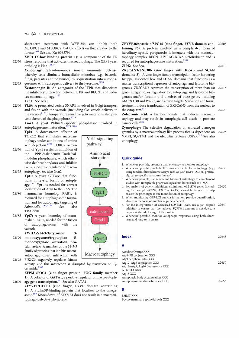
short-term treatment with WYE-354 can inhibit bothMTORC1 and MTORC2, but the effects on flux are due to theformer.341 See also Ku-0063794.XBP1 (X-box binding protein 1): A component of the ER
22550 stress response that activates macroautophagy. The XBP1 yeastortholog is Hac1.2175
Xenophagy: Cell-autonomous innate immunity defense,whereby cells eliminate intracellular microbes (e.g., bacteria,fungi, parasites and/or viruses) by sequestration into autopha-
22555 gosomes with subsequent delivery to the lysosome.2176
Xestospongin B: An antagonist of the ITPR that dissociatesthe inhibitory interaction between ITPR and BECN1 and indu-ces macroautophagy.2177
Yeh1: See Ayr1.22560 Ykt6: A prenylated vesicle SNARE involved in Golgi transport
and fusion with the vacuole (including Cvt vesicle delivery tothe vacuole2178); temperature sensitive ykt6 mutations also pre-vent closure of the phagophore.2024
Ymr1: A yeast PtdIns3P-specific phosphatase involved in22565 autophagosome maturation.2179,2180
Ypk1: A downstream effector ofTORC2 that stimulates macroau-tophagy under conditions of aminoacid depletion.2106 TORC2 activa-
22570 tion of Ypk1 results in inhibition ofthe PPP3/calcineurin-Cmd1/cal-modulin phosphatase, which other-wise dephosphorylates and inhibitsGcn2, a positive regulator of macro-
22575 autophagy. See also Gcn2.Ypt1: A yeast GTPase that func-tions in several forms of autoph-agy.1322 Ypt1 is needed for correctlocalization of Atg8 to the PAS. The
22580 mammalian homolog, RAB1, isrequired for autophagosome forma-tion and for autophagic targeting ofSalmonella.2181,2182 See alsoTRAPPIII.
22585 Ypt7: A yeast homolog of mam-malian RAB7, needed for the fusionof autophagosomes with thevacuole.YWHAZ/14-3-3/(tyrosine 3-
22590 monooxygenase/tryptophan 5-monooxygenase activation pro-tein, zeta): A member of the 14-3-3family of proteins that inhibitsmacro-autophagy; direct interaction with
22595 PIK3C3 negatively regulates kinaseactivity, and this interaction is disrupted by starvation or C2-ceramide.2183
ZFPM1/FOG1 (zinc finger protein, FOG family member1): A cofactor of GATA1, a positive regulator of macroautoph-
22600 agy gene transcription.641 See also GATA1.ZFYVE1/DFCP1 (zinc finger, FYVE domain containing1): A PtdIns3P-binding protein that localizes to the omega-some.583 Knockdown of ZFYVE1 does not result in a macroau-tophagy-defective phenotype.
22605ZFYVE26/spastizin/SPG15 (zinc finger, FYVE domain con-taining 26): A protein involved in a complicated form ofhereditary spastic paraparesis; it interacts with the macroau-tophagy complex BECN1-UVRAG-KIAA0226/Rubicon and isrequired for autosphagosome maturation.2184
22610ZIPK: See Sqa.ZKSCAN3/ZNF306 (zinc finger with KRAB and SCANdomains 3): A zinc finger family transcription factor harboringKruppel-associated box and SCAN domains that functions as amaster transcriptional repressor of autophagy and lysosome bio-
22615genesis. ZKSCAN3 represses the transcription of more than 60genes integral to, or regulatory for, autophagy and lysosome bio-genesis and/or function and a subset of these genes, includingMAP1LC3B andWIPI2, are its direct targets. Starvation and torin1treatment induce translocation of ZKSCAN3 from the nucleus to
22620the cytoplasm.643
Zoledronic acid: A bisphosphonate that induces macroau-tophagy and may result in autophagic cell death in prostatecancer cells.2185
Zymophagy: The selective degradation of activated zymogen22625granules by a macroautophagy-like process that is dependent on
VMP1, SQSTM1 and the ubiquitin protease USP9X.910 See alsocrinophagy.
Quick guide
1. Whenever possible, use more than one assay to monitor autophagy.226302. Whenever possible, include flux measurements for autophagy (e.g.,
using tandem fluorochrome assays such as RFP-EGFP-LC3 or, prefera-bly, cargo-specific variations thereof).
3. Whenever possible, use genetic inhibition of autophagy to complementstudies with nonspecific pharmacological inhibitors such as 3-MA.
226354. For analysis of genetic inhibition, a minimum of 2 ATG genes (includ-ing for example BECN1, ATG7 or ULK1) should be targeted to helpensure the phenotype is due to inhibition of autophagy.
5. When monitoring GFP-LC3 puncta formation, provide quantification,ideally in the form of number of puncta per cell.
226406. For the interpretation of decreased SQSTM1 levels, use a pan-caspaseinhibitor to ensure that the reduced SQSTM1 amount is not due to acaspase-induced cleavage of the protein.
7. Whenever possible, monitor autophagic responses using both short-term and long-term assays.
22645Index
A
Acridine Orange XXXAtg8–PE conjugation XXXAtg9 peripheral sites XXX
22650Atg12–Atg5 conjugation XXXAtg12–Atg5, Atg16 fluorescence XXXATG16L1 XXXAtg18 XXXAutophagic body accumulation XXX
22655Autophagosome characteristics XXX
B
BHMT XXXBovine mammary epithelial cells XXX
214 D. J. KLIONSKY ET. AL.

C
22660 C. elegans XXXCalcium XXXCell death XXXChicken DT40 cells XXXChlamydomonas XXX
22665 Chloroquine XXXCorrelative light and electron microscopy (CLEM) XXXCvt pathway XXX
D
Degradation assays XXX22670 Dictyostelium XXX
DQ-BSA XXXDRAM1 XXX
E
Electron microscopy XXX22675 Endosomal-microautophagy XXX
EPG proteins XXX
F
Flow cytometry XXXFluorescence microscopy XXX
22680 G
GFP-Atg8/LC3 processing XXXGFP-LC3 fluorescence microscopy XXX
H
Honeybee XXX22685 Hydra XXX
I
ICP34.5 XXXInhibitors XXX
K
22690 Keima XXX
L
Large animals XXXLate nucleophagy XXXLC3-I and LC3-II western blot XXX
22695 LC3-associated phagocytosis (LAP) XXXLipofuscin XXXLong-lived protein degradation XXXLysoTracker Red XXX
M
227003-methyladenine XXXMitophagy XXXMonodansylcadaverine (MDC) XXXMTOR activity XXX
P
22705Pexophagy XXXPhotodynamic therapy XXXPlanarians XXXPlants XXXPolyQ protein turnover XXX
22710Protists XXX
R
Rainbow trout XXXReticulophagy XXXRFP chimera processing XXX
22715Rosella XXX
S
Saponin XXXSea Urchin XXXSequestration assays XXX
22720SQSTM1 western blot XXX
T
TAKA assay XXXTandem mRFP/mCherry-GFP-LC3 XXXTrehalose XXX
22725V
Viral Bcl-2 XXXViral FLIP XXX
W
Western blot XXX22730WIPI1 XXX
WIPI2 XXXWIPI4 XXXWortmannin XXX
X
22735Xenophagy XXX
Z
Zebrafish XXXZymophagy XXX
AUTOPHAGY 215