A review of FDA-approved and cleared assays
24
Children's Mercy Kansas City Children's Mercy Kansas City SHARE @ Children's Mercy SHARE @ Children's Mercy Manuscripts, Articles, Book Chapters and Other Papers 11-2011 Molecular methods and platforms for infectious diseases testing: Molecular methods and platforms for infectious diseases testing: A review of FDA-approved and cleared assays A review of FDA-approved and cleared assays Rajyasree Emmadi University of Illinois at Chicago Jerry B. Boonyaratanakornkit Life Technologies Rangaraj Selvarangan Children's Mercy Hospital Venkatakrishna Shyamala Molecular Diagnostics and Blood Testing Barbara L. Zimmer Siemens Healthcare Diagnostics See next page for additional authors Follow this and additional works at: https://scholarlyexchange.childrensmercy.org/papers Part of the Diagnosis Commons, and the Infectious Disease Commons Recommended Citation Recommended Citation Emmadi, R., Boonyaratanakornkit, J. B., Selvarangan, R., Shyamala, V., Zimmer, B. L., Williams, L., Bryant, B., Schutzbank, T., Schoonmaker, M. M., Amos Wilson, J. A., Hall, L., Pancholi, P., Bernard, K. Molecular methods and platforms for infectious diseases testing: A review of FDA-approved and cleared assays Journal of Molecular Diagnostics 13, 583-604 (2011). This Article is brought to you for free and open access by SHARE @ Children's Mercy. It has been accepted for inclusion in Manuscripts, Articles, Book Chapters and Other Papers by an authorized administrator of SHARE @ Children's Mercy. For more information, please contact [email protected].
-
Upload
khangminh22 -
Category
Documents
-
view
3 -
download
0
Transcript of A review of FDA-approved and cleared assays

Children's Mercy Kansas City Children's Mercy Kansas City
SHARE @ Children's Mercy SHARE @ Children's Mercy
Manuscripts, Articles, Book Chapters and Other Papers
11-2011
Molecular methods and platforms for infectious diseases testing: Molecular methods and platforms for infectious diseases testing:
A review of FDA-approved and cleared assays A review of FDA-approved and cleared assays
Rajyasree Emmadi University of Illinois at Chicago
Jerry B. Boonyaratanakornkit Life Technologies
Rangaraj Selvarangan Children's Mercy Hospital
Venkatakrishna Shyamala Molecular Diagnostics and Blood Testing
Barbara L. Zimmer Siemens Healthcare Diagnostics
See next page for additional authors
Follow this and additional works at: https://scholarlyexchange.childrensmercy.org/papers
Part of the Diagnosis Commons, and the Infectious Disease Commons
Recommended Citation Recommended Citation Emmadi, R., Boonyaratanakornkit, J. B., Selvarangan, R., Shyamala, V., Zimmer, B. L., Williams, L., Bryant, B., Schutzbank, T., Schoonmaker, M. M., Amos Wilson, J. A., Hall, L., Pancholi, P., Bernard, K. Molecular methods and platforms for infectious diseases testing: A review of FDA-approved and cleared assays Journal of Molecular Diagnostics 13, 583-604 (2011).
This Article is brought to you for free and open access by SHARE @ Children's Mercy. It has been accepted for inclusion in Manuscripts, Articles, Book Chapters and Other Papers by an authorized administrator of SHARE @ Children's Mercy. For more information, please contact [email protected].

Creator(s) Creator(s) Rajyasree Emmadi, Jerry B. Boonyaratanakornkit, Rangaraj Selvarangan, Venkatakrishna Shyamala, Barbara L. Zimmer, Laurina Williams, Bonita Bryant, Ted Schutzbank, Michele M. Schoonmaker, Jean A. Amos Wilson, Leslie Hall, Preeti Pancholi, and Kathryn Bernard
This article is available at SHARE @ Children's Mercy: https://scholarlyexchange.childrensmercy.org/papers/1803

ReviewMolecular Methods and Platforms for InfectiousDiseases Testing
A Review of FDA-Approved and Cleared Assays
Rajyasree Emmadi,* Jerry B. Boonyaratanakornkit,†
Rangaraj Selvarangan,‡ Venkatakrishna Shyamala,§
Barbara L. Zimmer,¶ Laurina Williams,� Bonita Bryant,**Ted Schutzbank,†† Michele M. Schoonmaker,‡‡
Jean A. Amos Wilson,§§ Leslie Hall,¶¶ Preeti Pancholi,��
and Kathryn Bernard***From the Department of Pathology,* University of Illinois at
Chicago, Chicago, Illinois; AcroMetrix, by Life Technologies,†
Benicia, California; the University of Missouri-Kansas City School
of Medicine,‡ Children’s Mercy Hospital, Kansas City, Missouri;
Molecular Diagnostics & Blood Testing,§ North Potomac,
Maryland; Siemens Healthcare Diagnostics,¶ West Sacramento,
California; the Centers for Disease Control and Prevention,�
Atlanta, Georgia; Access Genetics, LLC,�� Eden Prairie,
Minnesota; Covance Central Laboratory Services,†† Indianapolis,
Indiana; Cepheid,‡‡ Sunnyvale, California; Berkeley HeartLab,§§
Alameda, California; the Division of Laboratory Medicine,¶¶
Mayo Clinic, Rochester, Minnesota; the Department of
Pathology,�� The Ohio State University Medical Center, Columbus,
Ohio; and the National Microbiology Laboratory,*** Public Health
Agency of Canada, Winnipeg, Manitoba, Canada
The superior sensitivity and specificity associatedwith the use of molecular assays has greatly improvedthe field of infectious disease diagnostics by provid-ing clinicians with results that are both accurate andrapidly obtained. Herein, we review molecularlybased infectious disease diagnostic tests that are Foodand Drug Administration approved or cleared andcommercially available in the United States as of De-cember 31, 2010. We describe specific assays and theirperformance, as stated in the Food and Drug Admin-istration’s Summary of Safety and Effectiveness Dataor the Office of In Vitro Diagnostic Device Evaluationand Safety’s decision summaries, product inserts, orpeer-reviewed literature. We summarize indicationsfor testing, limitations, and challenges related to im-plementation in a clinical laboratory setting for a
wide variety of common pathogens. The informationpresented in this review will be particularly useful forlaboratories that plan to implement or expand theirmolecular offerings in the near term. (J Mol Diagn
2011, 13:583–604; DOI: 10.1016/j.jmoldx.2011.05.011)
In 1986, the Food and Drug Administration (FDA) ap-proved the first nucleic acid test, the DNA probe foridentification of Legionnaires’ disease from bacterialculture, marketed by Gen-Probe Inc. (San Diego, CA).1
Seven years later, the FDA cleared the AMPLICOR CTtest (Roche Molecular Systems, Branchburg, NJ), thefirst DNA amplification-based test for detection of Chla-mydia trachomatis (CT) directly from a clinical sample.2
Since then, the field of clinical molecular testing ininfectious diseases has grown enormously; it repre-sents approximately 70% of the global molecular test-ing market.3
The FDA regulates in vitro diagnostic devices (IVDs),which include the reagents, systems, and products usedin the molecular diagnostic assays as class I, II, or IIImedical devices, with increasing regulatory oversight, toensure safety and effectiveness according to the risk
Accepted for publication May 27, 2011.
R.E. and J.B.B. contributed equally to this work.
Disclosures: R.E., Cepheid stock ownership of �$5000 (July 30, 2010);J.B.B., employee of Life Technologies; R.S., received grants for research,speaker assignments, or scientific advisory board from Eragen, Nanogen,bioMérieux, Gen-Probe Inc., Luminex, Idaho Technologies, BD, and Au-togenomics; V.S., previous employee of Chiron Corporation (Novartis)and Digene Corporation (Qiagen); B.L.Z., employee of Siemens Health-care Diagnostics; M.M.S., employee of Cepheid; and P.P., receivedgrants from Qiagen, Cepheid, Abbott, and Primera.
Disclaimer: The findings and conclusions herein are those of theauthor (L.W.) and do not necessarily represent the official position ofthe CDC.
Address reprint requests to Rajyasree Emmadi, M.D., Department ofPathology, University of Illinois at Chicago, 840 S Wood St., M/C 847,Chicago, IL 60612. E-mail: [email protected] or [email protected].
The Journal of Molecular Diagnostics, Vol. 13, No. 6, November 2011
Copyright © 2011 American Society for Investigative Pathology
and the Association for Molecular Pathology.
Published by Elsevier Inc. All rights reserved.
DOI: 10.1016/j.jmoldx.2011.05.011
583

posed to the patient if the results are incorrect. Severalspecific guidance documents regarding the classifica-tion and review criteria of these tests are available fromthe FDA Medical Devices website (http://www.fda.gov/MedicalDevices/default.htm, last accessed December 31,2010). The FDA also determines test complexity as high,moderate, or waived, with most molecular IVDs beingdesignated as high-complexity tests. The term FDAcleared is used for assays that are routed by a 510(k),submission showing substantial equivalence to any as-say already cleared by the FDA or marketed before 1976.The term FDA approved is used when an assay is routedby a premarket approval application to demonstrate itsefficacy and safety. A searchable database of FDA-ap-proved or FDA-cleared assays can be accessed (http://www.accessdata.fda.gov/scripts/cdrh/cfdocs/cfPMN/pmn.cfm, last accessed December 31, 2010), and anupdated list of FDA-cleared assays is available at theAssociation for Molecular Pathology website (http://www.amp.org/FDATable/FDATable.doc, last accessedDecember 31, 2010).
Molecular infectious disease (MID) testing offersseveral advantages, including rapid test results facili-tating detection of outbreaks and, in some cases,newly emerging strains; and sensitivity, specificity,identification of resistant organisms, and quantifiablecorrelation to disease severity, all of which contributeto timely therapeutic clinical decisions and early infec-tion control interventions.4 Multiplex methods can si-multaneously detect multiple infectious agents in a sin-gle clinical specimen. In addition, these methods areable to identify organisms that may be difficult to iso-late or have not been cultured by traditional methods.Assays that provide sequence or genotype can triggercollection of epidemiological information, track diseaseoutbreaks, provide strain resistance data and/or treat-ment prognosis, and determine the method or sourceand means of spread of infection.
This review complements a new Clinical and Labora-tory Standards Institute guideline, MM19: EstablishingMolecular Testing in Clinical Laboratory Environments,under review at the time of this article’s submission (Les-lie Hall, personal communication in June 2011). The as-says described were selected by reviews of several da-tabases, including the FDA database, the Association forMolecular Pathology website, and the PubMed databasefor publications related to MIDs. Although we have at-tempted to provide a comprehensive review of commer-cially available FDA-cleared or FDA-approved IVDs andplatforms, we do not endorse or promote any one of theseover the other. Our review is limited to FDA-cleared orFDA-approved assays available in the US market as ofDecember 31, 2010. Esoteric assays, such as those forbioterrorism agents and emergency use, are beyond thescope of this review. However, additional information canbe obtained at the CDC and FDA websites (http://www.selectagents.gov/index.html and http://www.fda.gov/MedicalDevices/Safety/EmergencySituations/ucm161496.htm, respectively; last accessed December 31, 2010).
Specific Considerations for CommerciallyAvailable MID Assays
We focus on commonly used assays and relevant infor-mation to assist the molecular laboratory director in assayselection and implementation. Assays available for diag-nosis and treatment are presented herein in four groups:sexually transmitted diseases (STDs) (Table 1), healthcare–associated infections (HAIs) and surveillance (Ta-ble 2), respiratory tract and central nervous system (CNS,Washington, DC) infections (Table 3), and other infec-tions and culture confirmations [eg, hepatitis B virus(HBV), various cultures, and ancillary assays for HAI andsurveillance] (Table 4). For each area, we highlight sev-eral MID assays; and reference to additional FDA-clearedassays is found in Tables 1–4.
During the FDA IVD review process, each MID assaydescribed in the specific product insert is approved orcleared for a specific patient population, specimen type,and extraction method. If a laboratory chooses to deviatefrom the product insert (eg, to offer a specimen type thatis not described in the product insert), the modified assayis considered off-label use and the laboratory is obligatedto perform a thorough validation to ensure that the mod-ification does not alter performance claims. We presentassay sensitivity, specificity, limit of detection, dynamicrange, and percentage positive or negative agreementwith culture results, as applicable.
It is out of the scope of this article to describe theactual process of implementation of MID testing; how-ever, there are several excellent recent references5,6 toassist the laboratory.
STD Data
HPV Data
More than 100 genotypes of human papilloma virus(HPV) (Table 1) have been identified based on DNAsequence heterogeneity, and �40 of these infect ano-genital or ororespiratory tracts. HPV genotypes havebeen divided into four oncogenic risk classes: low, inter-mediate/high, high, and unknown. The World Health Or-ganization’s International Agency for Research on Can-cer (IARC), in 2009, classified HPV-16, HPV-18, HPV-31,HPV-33, HPV-35, HPV-39, HPV-45, HPV-51, HPV-52,HPV-56, HPV-58, and HPV-59 as high-risk (hr) HPV typeswith sufficient evidence for causing cervical cancer, withspecial emphasis on HPV-16 as being the most aggres-sive type.7 Periodically, the IARC has revised its listing ofhr-HPV, and the components of the list have varied.
One prevailing viewpoint finds HPV testing differs fromother molecular assays in that analytic sensitivity for de-tection of HPV is not the prime driver of assay perfor-mance. A high analytic sensitivity can decrease the clin-ical specificity, resulting in more false referrals forcolposcopy and biopsy, decreased correlation with his-tological presence of disease, and a consequent distrustof a positive result by the treating physician.8 This ap-proach was further emphasized by Meijer et al,9 who set
584 Emmadi et alJMD November 2011, Vol. 13, No. 6

forth requirements for a candidate HPV screening molec-ular assay. A historical review of the use of HPV testing inthe screening and management of abnormal cervicalscreening results is provided in a recent article by Cox,10
and we recommend reading this as part of a molecularlaboratory director’s preparation for the introduction ofHPV testing. Although some commercially available HPVassays target both low-risk and hr-HPV types, we limit ourdiscussion to the application of assays detecting hr-HPV(Table 1 contains a complete listing).
The Digene Hybrid Capture 2 (HC2) HPV DNA Test(Qiagen, Gaithersburg, MD) has been the most widelyused molecular HPV assay in most clinical trials and hasbeen extensively reviewed.11 This assay is FDA ap-proved for triage in cases of equivocal cytology results inthe presence of atypical squamous cells of undeterminedsignificance to determine which patients should be re-ferred for a colposcopy and also as a screening test foruse in addition to cytology in women �30 years. TheNational Cancer Institute–sponsored clinical trial of Atyp-ical Squamous Cells of Undetermined Significance andLow-Grade Squamous Intraepithelial Lesion Triage Studyand the Canadian Cervical Cancer Screening Trial dem-onstrated the assay’s greater accuracy compared withcytology alone in detecting histologically confirmed cer-vical intraepithelial neoplasia 2/3 lesions of the cervix.12
However, Kitchener et al13 found that liquid-based cytol-ogy appears to be closing this gap and may have animproved sensitivity over conventional cytology.
The HC2 assay uses unlabeled single-stranded full-genomic-length RNA probes specific for all of the typesrecommended by IARC-2009 with the addition of HPV-68. The RNA:DNA hybrids are detected by a microplatechemiluminescent signal amplification method. The useof full-genome probes prevents false negatives resultingfrom gene deletions. The HC2 assay lacks an internalcontrol to evaluate sample adequacy or the presence ofinterfering substances. This assay has a false-positiverate of 7.8% for detection of hr-HPV because of the cross-reactivity with many untargeted low-risk HPV types.14 Inan attempt to demarcate these cross-reactive false pos-itives, which usually show a weak reaction/signal, severalgroups recommended a readjustment of the cutoff valueor retesting of initial borderline samples. In 2006, theconcept of an HC2 gray zone was introduced with therecommendation of retesting repeatedly borderline sam-ples (relative light unit per cutoff of 1.0 to 2.5) by adifferent HPV assay having a high analytic specificity.The manufacturer subsequently changed (only slightly)the criteria for interpretation of positive results, restrictedto samples collected in ThinPrep PreservCyt solution (Ho-logic Inc., Bedford, MA) and not applicable to thosecollected in the Digene Specimen Transport Medium(Qiagen Inc., Valencia, CA). The Digene-recommendedalgorithm for low-positive HPV results (gray or retestzone) required a retest followed by a second retest if theresult of the first retest was �1.0 relative light unit percutoff. This algorithm was evaluated by Muldrew et al,15
who showed that although retesting of an initial gray zonesample was necessary, a second retest did not offeradvantages over the first retest. Another group16 showed
that increasing the HC2 positive cutoff value to 2.0 rela-tive light unit would improve clinical specificity, with onlya minimal reduction in clinical sensitivity. However, theserecommendations are not part of the FDA approval of thisassay.
The Cervista HPV HR (Hologic, Inc., Bedford, MA)assay uses manual extraction with a single-well InvaderBiplex technology format that simultaneously detectsHPV and a Histone H2be DNA internal control in the samereaction. The assay is an isothermal signal amplificationmethod using Invader chemistry. The probe pools detect14 HPV types (IARC-2009 12 hr-HPV plus HPV-66 andHPV-68) and identify type-specific single-nucleotidepolymorphisms, effectively decreasing cross-reactivitywith low-risk types and false-positive results. The assayonly requires 2.0 mL of sample (half the requirement ofDigene HC2). The inclusion of an internal control is aquality control measure that differentiates between a truenegative and a sample with insufficient DNA present andis also a verification of the procedure. The same samplespecimen used in the Cervista HPV HR may then bereflexed to the Cervista HPV 16/18 genotyping assay,which specifically identifies the presence of HPV types 16and 18, now implicated in approximately 70% of casesprogressing to cancer.17,18
In a postapproval clinical study (SHENCCAST II, con-ducted in China) comparing the HC2 with the Cervistahr-HPV assays, the HC2 showed better sensitivity (95.6%versus 92.9%), whereas the Cervista assay demon-strated a statistically significantly higher specificity(91.1% versus 88.6%; P � 0.05).19
HPV testing is performed predominantly on liquid-based cytology samples, and sample collection is deter-mined by the method in use. The HC2 assay has beenvalidated for use with the Digene Specimen TransportMedium and the ThinPrep PreservCyt solution. Use ofother collection media (eg, SurePath liquid cytology me-dium) is considered unapproved off-label use. The Cerv-ista assay has been validated for use with the PreservCytsolution. The typical turnaround time is 1 to 3 days, de-pending on the platform and availability of automation.
In addition to molecular assays for the detection ofHPV, the FDA has also approved the Cervista HPV 16/18genotyping assay, briefly mentioned earlier (Hologic,Inc.). This assay is based on the same Invader technol-ogy as the Cervista hr-HPV detection test and, as indi-cated by its name, specifically detects and distinguishesHPV types 16 and 18. For cytology-negative, hr-HPV–positive women, HPV 16/18 genotyping can be used todetermine who should be referred for immediate colpos-copy. If the HPV 16/18 genotyping test result is negative,then cytology and hr-HPV testing are recommended tobe repeated in 12 months. The American Society forColposcopy and Cervical Pathology Consensus Confer-ence Recommendations for HPV 16/18 detection do notrecommend the use of HPV genotyping in women withatypical squamous cells of undetermined significancewho test positive for hr-HPV. Alternatively, the AmericanSociety for Colposcopy and Cervical Pathology recom-mends that these women are referred to colposcopy(American Society for Colposcopy and Cervical Pathol-
Review of MID Assays 585JMD November 2011, Vol. 13, No. 6

Table 1. STD Assays
Organism Specimen/sample type Assay/platform (FDA no.)
HPV Cervical cytology specimen in ThinPrep PreservCyt and cervicalcytology specimen in Specimen Transport Medium
Digene HC2 HPV DNA Test (P890064/S006)†
Cervical cytology specimen in ThinPrep PreservCyt and cervicalcytology specimen in Specimen Transport Medium
Digene HC2 High-Risk HPV DNA typing kit(P890064/S009)†
Cervical cytology specimen in ThinPrep PreservCyt Cervista HPV HR (P080014)¶
Cervical cytology specimen in ThinPrep PreservCyt Cervista HPV 16/18 (P080015)¶
HSV Vaginal lesion swab only (not for prenatal screening or infemales �18 years); collected in Copan UniversalTransport Medium or identical Copan- manufacturedmedia formulation; an extractable sample processingcontrol target is added to the specimen before lysis
MultiCode RTx HSV 1&2 Kit (K100336)††
MagNA Pure LC Total NA Kit and MagnaPure Instrument LightCycler 1.2‡‡
CT Endocervical (female) and urethral (male) swabs; urinespecimens (male and female); gynecological specimenscollected in BD SurePath preservative fluid
BD ProbeTec CT Qx Amplified DNA Assay(K090824, K091724, K081824)§§
Endocervical and vaginal (female) and urethral (male)swabs; urine specimens (male and female); patient-collected vaginal swab specimen is also accepted
APTIMA Assay for Chlamydia trachomatis(K043072, K053446, K061413, K063451)¶¶
Specimen that has been processed and tested positive byPACE 2 system for CT
PACE 2 CT (K920302)¶¶
Digene Cervical Sampler or Dacron swab placed in DigeneSpecimen Transport Medium
Digene HC2 CT-ID DNA Test (K990023,K010892)†
Endocervical (female) and urethral (male) swabs; urinespecimens (male and female)
COBAS AMPLICOR (CT/NG) Test (K973707,K973718, K070174, K053287)‡‡
NG Cervical and vaginal (female) and urethral (male) swabs;urine (male and female); PreservCyt specimens; Surepathspecimens
BD ProbeTec GC QX Amplified DNA Assay(K081825, K090971)§§
NG Vaginal and cervical swabs; urethral swabs (male); urine(male and female); PreservCyt specimen
APTIMA Assay for NG (K043144, K061509,K062440, K063664)¶¶
Specimen that has been processed and tested positive byPACE 2 system for NG
PACE 2 NG (K920301)¶¶
Cervical specimens collected using Digene CervicalSampler and Digene Swab Specimen Collection kit
Digene HC2 GC-ID DNA Test (K981485,K010893)†
Cervical (female) and urethral (male) swabs; urine (male) AMPLICOR CT-NG for NG (K974503,K974342, K070172, K053289)‡‡
CT-NG combined Vaginal (female) and urethral (male) swabs; urine (male andfemale); patient-collected vaginal swab specimen
RealTime CT-NG assay (K092704,K080739)***
Endocervical (female) and urethral (male) swabs; urinespecimens (male and female)
BD ProbeTec ET CT-GC (K012351)§§
CT-NG combined Endocervical (female) and urethral (male) swabs; urinespecimens (male and female)
APTIMA COMBO 2 (K003395, K022874,K032554, K043224, K060652)¶¶
Endocervical (female) and urethral (male) swab specimenscollected with the PACE Specimen Collection kits
PACE 2C CT-NG (K940979)¶¶
Cervical specimens collected using Digene HC2 DNACollection Device or Digene Female Swab SpecimenCollection Kit
Digene HC2 CT/GC Dual DNA Test(K981567, K010891)†,†††
HIV-1 Human plasma specimens (ACD-A and EDTA) RealTime HIV-1 assay (BP060002)*** andm2000 (m2000sp � 2000rt)
(table continues)
586 Emmadi et alJMD November 2011, Vol. 13, No. 6
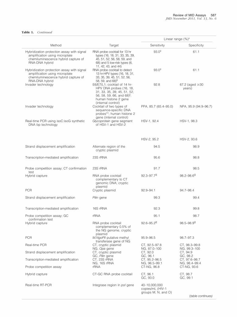
Table 1. Continued
Method Target
Linear range (%)*
Sensitivity Specificity
Hybridization protection assay with signalamplification using microplatechemiluminescence hybrid capture ofRNA-DNA hybrid
RNA probe cocktail for 13 hrtypes (16, 18, 31, 33, 35, 39,45, 51, 52, 56, 58, 59, and68) and 5 low-risk types (6,11, 42, 43, and 44)
93.0‡ 61.1
Hybridization protection assay with signalamplification using microplatechemiluminescence hybrid capture ofRNA-DNA hybrid
RNA probe cocktail to detect13 hr-HPV types (16, 18, 31,33, 35, 39, 45, 51, 52, 56,58, 59, and 68)§
93.0‡ 61.1
Invader technology E6/E7/L1; cocktail of 14 hr-HPV DNA probes (16, 18,31, 33, 35, 39, 45, 51, 52,56, 58, 59, 66, and 68)�;human histone 2 gene(internal control)
92.8 67.2 (aged �30years)
Invader technology Cocktail of two types ofsequence-specific DNAprobes**; human histone 2gene (internal control)
PPA, 85.7 (65.4–95.0) NPA, 95.9 (94.9–96.7)
Real-time PCR using isoC:isoG syntheticDNA bp technology
Glycoprotein gene segmentof HSV-1 and HSV-2
HSV-1, 92.4 HSV-1, 98.3
HSV-2, 95.2 HSV-2, 93.6
Strand displacement amplification Alternate region of thecryptic plasmid
94.5 98.9
Transcription-mediated amplification 23S rRNA 95.6 98.8
Probe competition assay; CT confirmationtest
23S rRNA 91.7 98.5
Hybrid capture RNA probe cocktailcomplementary to CTgenomic DNA; crypticplasmid
92.3–97.7�� 98.2–98.6��
PCR Cryptic plasmid 92.9–94.1 94.7–98.4
Strand displacement amplification Pilin gene 99.3 99.4
Transcription-mediated amplification 16S rRNA 92.3 99.8
Probe competition assay; GCconfirmation test
rRNA 95.1 98.7
Hybrid capture RNA probe cocktailcomplementary 0.5% ofthe NG genome, crypticplasmid
92.6–95.2�� 98.5–98.9��
PCR M.NgoPII putative methyltransferase gene of NG
95.9–96.5 98.7–97.3
Real-time PCR CT, cryptic plasmid CT, 92.5–97.8 CT, 98.3–99.8NG, Opa gene NG, 87.0–100 NG, 99.3–100
Strand displacement amplification CT, cryptic plasmid CT, 92.0 CT, 94.9GC, Pilin gene GC, 96.1 GC, 98.2
Transcription-mediated amplification CT, 23S rRNA CT, 95.2–96.5 CT, 97.6–98.7NG, 16S rRNA NG, 96.5–99.1 NG, 98.4–99.4
Probe competition assay rRNA CT-NG, 96.8 CT-NG, 93.6
Hybrid capture CT-GC RNA probe cocktail CT, 96.1 CT, 98.7GC, 93.0 GC, 99.1
Real-time RT-PCR Integrase region in pol gene 40–10,000,000copies/mL (HIV-1groups M, N, and O)
(table continues)
Review of MID Assays 587JMD November 2011, Vol. 13, No. 6

ogy, HPV Genotyping Clinical Update, http://www.asccp.org/pdfs/consensus/clinical_update_20090408.pdf,last accessed December 31, 2010).
CT and NG Data
CT and Neisseria gonorrhoeae (NG) are the most commoncause of bacterial STDs, and both can cause urogenitaltract infections ranging from acute to asymptomatic dis-ease. CT is an obligate intracellular bacterium compris-ing 15 serovars, whereas NG is a fastidious intracellulardiplococcus. Significant underreporting of disease canoccur as the result of silent infections affecting the repro-ductive age group. Identification and treatment is impor-tant to prevent the sequelae of infection, such as infertil-ity, chronic pain, and pelvic inflammatory disease.
Urogenital specimens commonly exhibit amplificationinhibition. The inhibitory substances can be removed by
including nucleic acid purification steps in the samplepreparation. The sample preparation protocols varyamong the commercially available assays, ranging fromthe use of crude lysates (AMPLICOR) to purified nucleicacids. The Roche AMPLICOR assay uses an amplifica-tion control in the sample that allows for detection ofinhibitory substances. This control consists of a plasmid-containing CT primer binding sites and a randomizedinternal sequence. The BD ProbeTec (BD Diagnostics,Sparks, MD) uses 1000 copies of a linearized NG DNAcontaining plasmid as the internal amplification control.
Commercially available assays for CT and NG (Table1) use target amplification methods, with the one excep-tion being the Digene HC2 assay, which uses a signalamplification method with an RNA probe cocktail com-plementary to approximately 39,300 bp (4%) of the Chla-mydia genomic DNA and one probe complementary to100% of the cryptic plasmid. Nucleic acid amplification
Table 1. Continued
Organism Specimen/sample type Assay/platform (FDA no.)
Plasma extracted from blood collected using EDTA orACD‡‡‡
AMPLICOR HIV-1 MONITOR test, version 1.5(BP950005/4)‡‡ and COBAS AMPLICORHIV-1 MONITOR test, version 1.5(BP950005)‡‡
Plasma separated from blood collected using EDTA COBAS AmpliPrep/COBAS TaqMan HIV-1test (BP050069/0)‡‡ and COBASAmpliPrep/COBAS TaqMan‡‡
HIV-1 Plasma extracted from blood collected using EDTA orACD‡‡‡
VERSANT HIV-1 RNA 3.0 Assay (bDNA)(BP000028/0)§§§ and VERSANT MolecularSystem§§§
Plasma or serum APTIMA HIV RNA Qualitative Assay(BL103966/5040)¶¶
Plasma or serum Procleix ULTRIO Assay¶¶
Procleix HIV-1, HCV, and/or HBVDiscriminatory Assays (BL 125113/33)¶¶
Plasma or serum COBAS AmpliScreen HIV-1 Test (BL 125059/37)‡‡
HIV-1 drugresistance¶¶¶
Plasma samples from blood collected in EDTA ViroSeq HIV-1 Genotyping System(BK030033)��� and ABI 3100/3130 capillaryelectrophoresis platform****
Plasma samples from blood collected using ACD or EDTAanticoagulants
TRUGENE HIV-1 Genotyping Kit(BK090077)§§§ and OpenGene DNASequencing System§§§
(table continues)
*Sensitivity, specificity, linear range, and percentage positive or negative agreement with culture data are sourced from FDA submission material or product inserts.†Obtained from Qiagen, Gaithersburg, MD.‡Atypical squamous cells of undetermined significance referral Papanicolaou stain population, Kaiser Study, PreservCyt solution specimens.§Cross-reacts with HPV types 40, 53, and 66.¶Obtained from Hologic, Madison, WI.�Cross-reacts with HPV types 67 and 70.**Cross-reacts with high levels of HPV type 31.††Obtained from EraGen Biosciences, Madison, WI.‡‡Obtained from Roche Molecular Diagnostics, Pleasanton, CA.§§Obtained from BD Diagnostics, Sparks, MD.¶¶Obtained from Gen-Probe, Inc., San Diego, CA.��Depending on brush or swab specimens.***Obtained from Abbott Molecular, Inc., Des Plaines, IL.†††This assay is indicated for use as an initial test and requires confirmation with the individual MID assays.‡‡‡ACD specimens will yield approximately 15% lower test results because of the dilution effect of 1.5 mL ACD in the collection tube.§§§Obtained from Siemens Healthcare Diagnostics, Deerfield, IL.¶¶¶Provides information on resistance to nucleoside and nonnucleoside reverse transcriptase and protease inhibitors.���Obtained from Celera Diagnostics, Alameda, CA.****Obtained from Applied Biosystems, Foster City, CA.ACD-A, anticoagulant citrate dextrose solution A; bDNA, branched DNA; NPA, negative predictive accuracy; PPA, positive predictive accuracy.
588 Emmadi et alJMD November 2011, Vol. 13, No. 6

testing assays typically increase sensitivity by targetingmultiple copy genes or plasmids. For CT, the targetsinclude cryptic plasmid DNA present in nearly all sero-vars (5 to 10 copies), genes such as omp1, and ribo-somal RNA (rRNA; 16S and 23S). For NG, targets includethe cytosine methyl transferase gene (M.NgoPII), the Opagene, Piv-1 genes, and 16S and 23S rRNA. The speci-men type approved for CT and NG testing is assay spe-cific (Table 1) and includes urethral swab and urine formales and endocervical/cervical samples, vaginalswabs, urine, and PreservCyt (Hologic, Inc.) specimensfor females. A male first-void urine specimen and vaginalswabs are considered optimal specimens, according tothe Association of Public Health Laboratories (http://www.aphl.org/aphlprograms/infectious/std/documents/ctgclabguidelinesmeetingreport.pdf, last accessed De-cember 31, 2010).
The AMPLICOR NG detection kit (Roche MolecularDiagnostics, Pleasanton, CA) targets the M.NgoPII gene,whereas the BD ProbeTec ET CT/GC targets the crypticplasmid of CT and the Piv-1 gene of NG. Both of theseassays have cross-reactivity with some Neisseria species.Confirmatory testing using a different gene target is anoption in such instances. Cross-reactivity has not beenreported for the Real Time CT/NG (Abbott Laboratories,Des Plaines, IL), APTIMA COMBO 2 (Gen-Probe Inc., SanDiego, CA), and PACE 2 (Gen-Probe Inc.) assays.20
Coinfection with CT and NG occurs in many patients.Simultaneous detection of both organisms in a single testis achieved by several assays (Table 1). The APTIMACOMBO 2 is a second-generation assay that uses targetcapture with transcription-mediated amplification andchemiluminescent hybridization protection. In contrast toPCR and strand displacement assays, which amplify
Table 1. Continued
Method Target
Linear range (%)*
Sensitivity Specificity
End point RT-PCR 142 bp in highly conservedregion of Gag gene
Standard, 400–750,0000 copies/mL;ultrasensitive, 50–100,000 copies/mL (HIV-1group M)
Real-time RT-PCR Gag gene 48–10,000,000 copies/mL (HIV-1 group M)
bDNA technology Pol gene 75–500,000 copies/mL (HIV-1 groups M and O)
Transcription-mediated amplification Highly conserved regions ofHIV-1 RNA
100 99.83
Transcription-mediated amplification Highly conserved regions ofHIV-1 RNA, HCV RNA,and HBV DNA
100 (ULTRIO)100 (HIV-1discriminatory)
99.5 (ULTRIO)99.7–100 (HIV-1discriminatory)
RT-PCR Gag gene 96.5–98 98.9–99.7
RT-PCR, population sequence analysis HIV-1 subtype B proteasegene and partialsequence of the reversetranscriptase regions ofthe pol gene
Validated for detection of drug-resistancemutations in 40 of 60 mutant/wild-type mixturesamples with a viral load range of2000–750,000 copies/mL
RT-PCR, population sequence analysis protease gene and part ofthe reverse transcriptaseregions
Requires samples with viral loads �1000copies/mL
Review of MID Assays 589JMD November 2011, Vol. 13, No. 6
bacterial DNA, transcription-mediated amplification am-plifies specific regions of the 23S rRNA/16S rRNA. TheAPTIMA COMBO 2 assay does not have an internal con-trol; however, it uses target capture technology, whichremoves inhibitors. The assay is a dual kinetic assay, withone signal scoring for CT and the second signal scoringfor NG. Assays that target bacterial rRNA rather thanplasmid DNA have a greater ability to detect lower con-
centrations of organisms because of the presence of upto a 1000-fold greater amount of RNA than plasmid DNAin the infected cell.
Cryptic plasmid–based detection assays could yieldfalse-negative results because CT strains without theplasmid or with deletions in the plasmid have been de-scribed (the Swedish variant, with a 377-bp fragmentdeletion). False-negative results have been reported for
Table 2. HAI Assays
OrganismSpecimen/sample
typeAssay/platform
(FDA no.) Method Target Sensitivity (%)* Specificity (%)*
MRSA/SA(screening,surveillance)
Lesion (skin/softtissue) doubleswab
Xpert MRSA/SASSTI(K080837)†
andGeneXpertSystem†
Real-timePCR
Staphylococcal proteinA (spa), the gene forMecA-mediatedoxacillin resistance(mecA), andSCCmec inserted inthe SA chromosomalattB site
MRSA % positiveagreement withculture, 93.8;SA % positiveagreement withculture, 95.7
MRSA % negativeagreement withculture, 97.3;SA % negativeagreement withculture, 89.5
Nasal swabs(surveillance)
Xpert MRSA(K070462)†
andGeneXpertSystem†
Real-timePCR
SCC inserted into theSA chromosomalattB site
% positiveagreement,80.9–90.6
% negativeagreement,92.3–96.3
Nasal swabs(surveillance)
BD GeneOhmMRSA ACP(K093346)‡
andSmartCycler†
Real-timePCR
SCCmec-orfXjunctionarea of theSCCmecadjacent tothe integration siteMREJ
92.00 94.6
Nasal swabs(presurgicalscreen)
Xpert SA NasalComplete(K100822)†
andGeneXpertSystem†
Real-timePCR
SCC inserted into theSA chromosomalattB site
% positiveagreement,80.9–90.6
% negativeagreement,92.3–96.3
Nasal swabs(surveillance)
LightCyclerMRSAAdvancedtest(K091409)§
andLightCycler2.0
Real-timePCR
Sequenceincorporating theinsertion site of theSCCmec in the SAorfX gene
% positiveagreement withdirectchromogenicculture, 95.2
% negativeagreement withdirectchromogenicculture, 96.4
Enterococcus Rectal swab(surveillance)
Xpert vanA(K092953)†
andGeneXpertSystem†
Real-timePCR
Gene sequences forVanA-encodedresistance tovancomycin-teicoplanin
98–99 81–90
C. difficile Unformed(liquid or soft)stools
Xpert C. difficile(K091109)†andGeneXpertSystem†
Real-timePCR
C. difficile tcdB 93.50 94.0
ProdesseProGastro Cd(K090239)¶
Multiplexreal-timePCR
C. difficile tcdB foundonly in toxigenicstrains
91.7 94.7
BD GeneOhmCdiff Assay(K081920)‡
Real-timePCR
C. difficile tcdB 93.8 95.5
Illumigene C.difficile(K091109)�
LAMPtechnology
C. difficile tcdA of thePaLoc
95.2 95.3
LAMP, loop-mediated amplification; PaLoc, pathogenicity locus; SCC, staphylococcal cassette chromosome.*Sensitivity, specificity, and percentage positive or negative agreement with culture data are sourced from FDA submission material or product inserts.†Obtained from Cepheid, Sunnyvale, CA.‡Obtained from BD Diagnostics-Infectious Diseases, LaJolla, CA.§Obtained from Roche Molecular Diagnostics, Pleasanton, CA.¶Obtained from Gen-Probe Prodesse, Inc., Waukesha, WI.�Obtained from Meridian Bioscience, Inc., Cincinnati, OH.
590 Emmadi et alJMD November 2011, Vol. 13, No. 6

the Roche AMPLICOR and the Abbott RealTime CT/NGassays that target the deleted region, whereas thoseassays that target outside of this region or the chromo-somal regions detect the mutant strain.21 The BD Pro-beTec assay is able to detect the CT Swedish variantbecause the cryptic plasmid target is outside the area ofdeletion. The newer Abbott RealTime CT/NG assay, FDAcleared in June 2010, includes an additional 140-bpcryptic plasmid target outside of the 377-bp deletionarea. The Abbott RealTime CT/NG assay also contains asmall fragment of noninfectious linearized DNA plasmidfor use as an internal control throughout the sample prep-aration process.
A low prevalence of STDs in a specific population mayreduce the positive predictive value of the molecular re-sult. However, the test can be repeated with a separatealiquot of the same specimen or a second specimen anda different test method and/or a different target to confirmthe positive result.20 The efficacy of this strategy isdebatable.22
HIV-1 Assay
Qualitative assays, such as the Procleix ULTRIO andDiscriminatory HIV-1/HCV/HBV assays (Gen-Probe Inc.)and the COBAS AmpliScreen HIV-1 test (Roche Molecu-lar Systems, Pleasanton), are available for donor screen-ing applications. The transcription-mediated amplifica-tion–based APTIMA HIV-1 RNA qualitative assay (Gen-Probe Inc.) can be used for diagnosing acute andprimary infections and can detect infection before sero-conversion and confirm infections in individuals whenantibody test results are positive.23,24 However, in thissegment, we will focus on the quantitative and genotyp-ing assays that are the main HIV-1–related assays per-formed in the Molecular Diagnostics Laboratory.
HIV-1 viral load assays are important for monitoringHIV-1–infected individuals, predicting the progression ofHIV disease, and monitoring antiretroviral treatment.25,26
HIV-1 is classified into three major groups (ie, M, N, andO). Group M is the most prevalent and is sub-classifiedinto seven subtypes (ie, A–D and F–H) that are geograph-ically distinct. Several commercial kits are available forquantitative determination of HIV viral load to assess pa-tient prognosis during antiretroviral therapy (Table 1).Ascertaining the viral load is a prerequisite to initiatingdrug therapy in adherence to FDA guidelines and servesto evaluate the efficacy of antiretroviral therapy (http://www.aidsinfo.nih.gov/guidelines, last accessed Decem-ber 31, 2010). In 1996, the AMPLICOR HIV-1 MONITORtest (Roche Molecular Systems) was the first FDA-ap-proved quantitative HIV end point–based RT-PCR assay.Quantification of HIV-1 RNA copy number is determinedby comparing optical density readings of the HIV-1 signalwith an external quantitation standard signal, which has aknown copy number input. However, a small dynamicrange of 400 to 750,000 copies/mL (or 50 to 100,000copies/mL for the ultrasensitive method) limits the assay.Recently, real-time RT-PCR HIV assays with options ofautomation, closed-system platform characteristics,broad dynamic range, and good specificity have become
commercially available.27 By using the COBAS AmpliP-rep/COBAS TaqMan HIV-1 Test (Roche Molecular Sys-tems), patient specimens are extracted on the COBASAmpliPrep instrument and amplification/detection occurson the COBAS TaqMan Analyzer. The assay targets theconserved region in the gag gene, which is prone to ahigh level of mutation and is only intended for the detec-tion of group M subtypes of HIV-1. Calibration is notrequired because specific calibration values and pre-defined assay control ranges are included with each kitand because uracil-N-glycosylase reduces the risk ofcarryover contamination. Initial underestimations regard-ing quantification of HIV-1 group M non-B subtypes,when compared with the COBAS AMPLICOR HIV-1MONITOR Version 1.5 and the Abbott RealTime HIV-1assay, have been reported.28–32 A second version of theCOBAS AmpliPrep/COBAS TaqMan HIV-1 Test version2.0 (Roche Molecular Systems) was recently approved.This assay improves the underquantification and subtypeinclusivity issues present with the original assay. Theversion 2.0 assay uses a two-target approach with thecombination of the new ltr primer-probe set with the orig-inal gag primer-probe set to detect the various group MHIV-1 subtypes A–D and F–H and group O. The assayhas a quantitation range of 20 to 107 RNA copies/mL.33,34
With the Abbott RealTime HIV-1 kit (Abbott Molecular,Inc., Abbott Park, IL), patient specimens are extracted onthe m2000sp instrument and detected on the m2000rtinstrument. The assay allows for flexibility in the sampleinput volumes and detects both group M and group OHIV-1 subtypes. In contrast to these real-time PCR-basedassays, the VERSANT HIV-1 RNA 3.0 Assay (SiemensHealth Care Diagnostics, Deerfield, IL) uses branchedDNA chemistry, which relies on signal amplification. Theassay has a lower risk of contamination because of the lackof amplicon production, and the VERSANT HIV-1 RNA 3.0Assay has been validated for samples containing group Msubtypes A–G (in 2002, subtype E was still believed to be atrue subtype); however, it has decreased sensitivity com-pared with target amplification assays.35,36
During treatment of HIV infections, mutant HIV-1strains emerge that are resistant to one or more drugs.37
The identification of viral resistance genotypes allowstreatment strategies to be modified.38 Retrospective andprospective intervention-based studies38–41 have pro-vided evidence supporting the clinical utility of genotypetesting for resistance, and this is recommended by theInternational AIDS Society–USA panel for selecting newregimens after treatment failure and monitoring therapyfor pregnant women. Genotype testing should also beconsidered before initiation of therapy for acute infectionsand for treatment-naïve patients with establishedinfection.38
Two genotyping systems are commercially available:TRUGENE HIV-1 Genotyping Kit (Siemens Diagnostics,Tarrytown, NY) and ViroSeq HIV-1 Genotyping Systems(Celera Diagnostics, Alameda, CA). The ViroSeq kit pro-vides reagents for viral RNA isolation from plasma, andboth kits provide reagents for RT-PCR and sequenc-ing.39,40,42 Typically, the entire protease and the 5= re-verse transcription coding regions of the pol gene are
Review of MID Assays 591JMD November 2011, Vol. 13, No. 6

Table 3. Respiratory Tract and CNS Infection Assays
Organism Specimen/sample type Platform/assay (FDA no.)
Respiratory virus panel: influenza A/H1 andA/H3; influenza B; adenovirus; RSV;metapneumovirus (hMPV); parainfluenza(hPIV) 1, 2, and 3; and rhinovirus
Nasopharyngeal swab xTAG Respiratory Viral Panel (K063765)†
Respiratory virus panel: influenza A and Band RSV
Nasopharyngeal swab, culturedclinical specimens
Verigene Respiratory Virus Nucleic Acid Test(K083088)‡ and Verigene RespiratoryVirus Nucleic Acid Test on the VerigeneSP system (K092566)‡
Nasopharyngeal swab ProFlu� (K073029, K081030, K092500)§ andSmartCycler¶
Respiratory virus panel: influenza A and Band RSV
Nasopharyngeal swab Simplexa Flu A/B and RSV (K102170)�;MagNA Pure LC Instrument and theMagNA Pure Total Nucleic Acid IsolationKit** or a Qiagen QIAamp Viral RNA MiniKit†† and 3M Integrated Cycler‡‡
Respiratory virus: influenza A/H1N1-2009 Nasopharyngeal swabs, nasalswabs, NPAs
Simplexa Influenza A H1N1 (2009)(K100148)�; MagNA Pure LC Instrumentand the MagNA Pure Total Nucleic AcidIsolation Kit** or a Qiagen QIAamp ViralRNA Mini Kit†† and 3M Integrated Cycler‡‡
Respiratory virus: influenza A/H1N1-2009 Nasopharyngeal swabs andnasal swabs
CDC Human Influenza Virus Real-Time RT-PCR Detection and Characterization Panel(K101564)§§ and Applied Biosystems 7500Fast Dx Real-Time PCR Instrument¶¶
Respiratory virus panel: seasonal influenzaA/H1, A/H3, and A/H1N1-2009
Nasopharyngeal swab ProFAST� (K101855)§ and SmartCycler¶
Respiratory virus: influenza A/H5 (Asianlineage)
Nasopharyngeal and throatswabs
JBAIDS Influenza A subtype A/H5 (Asianlineage) (K100287)�� and JBAIDSInstrument
Respiratory virus: hPIV Nasopharyngeal swab ProParaFlu� (K091053)§ and SmartCycler¶
Respiratory virus: hMPV Nasopharyngeal swab Pro hMPV� (K082688)§ and SmartCycler¶
Respiratory virus: adenovirus Nasopharyngeal swab ProAdeno� (K102952)§ and SmartCycler¶
Enterovirus CSF Xpert EV (K061062) and GeneXpert System¶
CSF NucliSENS EasyQ Enterovirus (K063261)***and NucliSENS EasyQ System
MTB Sputum and bronchialspecimens
AMPLIFIED MTD (MycobacteriumTuberculosis Direct) Test(P940034/S008)†††
(table continues)
*Sensitivity, specificity, and percentage positive or negative agreement with culture data are sourced from FDA submission material or product inserts.†Obtained from Luminex Corporation, Austin, TX.‡Obtained from Nanosphere, Inc., Northbrook, IL.§Obtained from Gen-Probe Prodesse, Inc., Waukesha, WI.¶Obtained from Cepheid, Sunnyvale, CA.�Obtained from Focus Diagnostics, Inc., Cypress, CA.**Obtained from Roche Molecular Diagnostics, Pleasanton, CA.††Obtained from Qiagen, Gaithersburg, MD.‡‡3M, St Paul, MN.§§Obtained from the CDC, Atlanta, GA.¶¶Obtained from Life Technologies Inc., Carlsbad, CA.��Obtained from the Department of Defense.***Obtained from bioMérieux Inc., Durham, NC.†††Obtained from Gen-Probe, Inc., San Diego, CA.hMPV, human metapneumovirus; hPIV, human parainfluenza virus; NASBA, nucleic acid sequence-based amplification; NPA, nasopharyngeal aspirate.
592 Emmadi et alJMD November 2011, Vol. 13, No. 6

Table 3. Continued
Method Target Sensitivity (%)* Specificity (%)*
RT-PCR, allele-specific primerextension, tag sorting
Matrix gene of influenza A, Hemagglutiningene of influenza A/H1 and A/H3
Influenza A, 96.4 Influenza A, 95.9
Influenza B, adenovirus, RSV A/Bmetapneumovirus
Influenza A/H1, 100 Influenza A/H1, 100
Parainfluenza 1, 2, and 3; and rhinovirus Influenza A/H3, 91.7 Influenza A/H3, 98.7Influenza B, 91.5 Influenza B, 96.7RSV A, 100 RSV A, 98.4RSV B, 100 RSV B, 97.4Adenovirus, 78.3 Adenovirus, 100hMPV, 96 hMPV, 98.8hPIV 1, 100 hPIV 1, 99.8hPIV 2, 100 hPIV 2, 99.8hPIV 3, 84.2 hPIV 3, 99.6Rhinovirus, 100 Rhinovirus, 91.3
Multiplex RT-PCR multiplex goldnanoparticle hybridizationtechnology Verigene System‡
Influenza A matrix gene Influenza A, 99.2 Influenza A, 90.1Influenza B NS and matrix genes Influenza B, 96.8 Influenza B, 98.5L and F genes of RSV RSV, 89.8 RSV, 91.5
Multiplex real-time RT-PCRTaqMan chemistry
Influenza A matrix gene Influenza A, 100 Influenza A, 92.6Influenza B non-structural NS1 and NS2
genesInfluenza B, 97.8 Influenza B, 98.6
RSV polymerase gene RSV, 89.5 RSV, 94.9Real-time RT-PCR Target RNA of highly conserved region of
matrix protein genes of influenza A andB and RSV
Influenza A, 100 Influenza A, 99.3Influenza B, 100 Influenza B, 99.8RSV, 98 RSV, 96.9
Real-time PCR Influenza A matrix gene and uniqueregion in Hemagglutinin gene of 2009H1N1 influenza virus
% positive agreementfor swabs, 100;NPA, 100
% negative agreementfor swabs, 92.5;NPA, 96.1
Real-time PCR Influenza A matrix gene andNucleoprotein gene specific for 2009H1N1 and Hemagglutinin gene specificfor 2009 H1N1
96 96
Real-time RT-PCR Target RNA of conserved region ofHemagglutinin gene
% positive agreementfor A/H1, 100; A/H3,100; A/H1N1-2009,95.4
% negative agreementfor A/H1, 99.0; A/H3, 99.0; A/H1N1-2009, 100
Real-time RT-PCR Two target RNA sequences 5= and 3= ofthe Hemagglutinin precursor cleavagesite within the conserved regions of theHemagglutinin gene of influenza A/H5(Asian lineage) virus
96.9–100 95.3–97.1
Multiplex real-time RT-PCRTaqMan chemistry
Conserved regions of Hemagglutinin-Neuraminidase gene of hPIV 1, 2, and 3
hPIV-1, 88.9 hPIV-1, 99.9hPIV-2, 96.3 hPIV-2, 99.8hPIV-3, 97.3 hPIV-3, 99.2
Real-time RT-PCR TaqManchemistry
hMPV: Nucleocapsid gene % positive agreement,94.1
% negativeagreement, 99.3
Multiplex real-time PCR TaqManchemistry
Adenovirus serotypes 1–51, Hexon gene 97.5 95.6
Real-time PCR Consensus region of enterovirus, 5= UTRbetween nucleotides 452 and 596
96.3–100 97.0–97.2
NASBA Enterovirus RNA 70.9–100 99.3–100
Transcription- mediatedamplification, hybridizationprotection assay
Mycobacterial 16S rRNA 96.9 100
Review of MID Assays 593JMD November 2011, Vol. 13, No. 6

amplified to generate a large amplicon that is then usedas a sequencing template for multiple primers that gen-erate a consensus sequence. Software is available forcomparing the consensus with a known reference, todetermine any mutations present, based on which treat-ment options are made available. These are kept currentby a panel of HIV experts and recommendations of theInternational AIDS Society–USA panel.37,43,44 Report-edly, genotyping assays may have limitations of sensitiv-ity in detecting a minority variant species in a patient.45 Inaddition, potential mutations may be missed at positionsnot previously characterized as resistance mutants.
HSV Data
Herpes simplex virus (HSV) is one of the most commonSTDs in the United States. Genital herpes is a chroniclife-long infection caused primarily by HSV-2, althoughthe role of HSV-1 is increasing.46,47 Most patients in-fected with genital herpes are asymptomatic, and theclinical presentation is diverse. Because of the availabilityof effective antiviral therapy, there is an increased de-mand for rapid accurate laboratory diagnosis of HSV.HSV genotyping may aid in tracing of contacts and incase evaluation.48,49 The MultiCode-RTx HSV-1&2 Kit(EraGen Biosciences, Madison, WI) is a PCR-based qual-itative IVD HSV typing assay, using vaginal swab speci-mens from symptomatic female patients. The assay is notapproved for cerebrospinal fluid (CSF) or any other gen-ital or oral lesion specimens. The assay uses fluorophore-labeled HSV-1 and HSV-2 primers that target the glyco-protein B gene. The extraction methods cleared for the testinclude the MagNA Pure LC Instrument and the MagNAPure LC Total Nucleic Acid Isolation Kit (Roche MolecularDiagnostics). The PCR amplification is performed using theLightCycler 1.2 instrument (Roche Molecular Diagnostics),after which the HSV genotypes are discriminated by meltcurve analysis. Evaluation of the appropriate specimen/le-sion and specimen collection procedures is essential be-cause lesion type and location may affect the sample qual-ity and the assay performance.
HAI Data
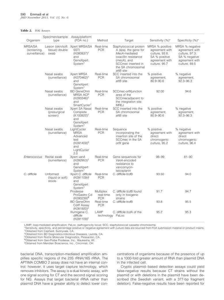
Different HAI assays have specific intended uses, suchas surveillance, presurgical, and diagnostic testing; andthe product insert should be reviewed carefully beforeimplementation.
MRSA Data
Methicillin resistance is associated with increased mor-tality in patients with staphylococcal bacteremia.50 Ap-proximately half of all Staphylococcus aureus pneumoniasin the United States are due to methicillin-resistant S.aureus (MRSA).51 MRSA ventilator-associated pneumo-nias also appear to be associated with a higher mortalityrate compared with methicillin-sensitive S. aureus venti-lator-associated pneumonias.52 Patients colonized withMRSA (nasal carriers) are also at increased risk of devel-
oping MRSA disease and can spread the infection aswell.
For direct MRSA detection from patient samples, theXpert MRSA, Xpert SA Nasal Complete assays for theGeneXpert System (Cepheid, Sunnyvale, CA), the BDGeneOhm MRSA ACP (BD Diagnostics, La Jolla, CA),and the LightCycler MRSA Advanced test (Roche Molec-ular Systems) can be used. The GeneXpert System is afully integrated and automated nucleic acid preparation,amplification, and real-time detection system. The XpertSA Nasal Complete (Cepheid) assay can be used forsurveillance of both S. aureus and MRSA carrier status,which can aid in reducing the risk of HAIs. In a recentmulticenter clinical evaluation, no statistically significantperformance differences were observed between theXpert MRSA and MRSA ACP assays compared with cul-ture.53,54 The recently approved LightCycler MRSA Ad-vanced test was similarly evaluated compared with theBD GeneOhmMRSA ACP and had a similar sensitivity(92.2% and 93.2%, respectively) but a significantly betterspecificity (98.9% and 94.2%, respectively).55 All of theseassays target the S. aureus orfX gene sequence incorpo-rating the insertion site (attBssc) of the staphylococcalcassette chromosome mec (SCCmec) for the detection ofMRSA. Targeting the mecA alone could result in falsepositivity because a large percentage of coagulase-neg-ative staphylococcus species would also test positive.False-negative results can occur as the result of novelSCCmec elements and variants resulting from recombi-nation. Targeting the orfX region alone can give false-negative results in those instances of mecA insertion intoother sites (although this is rare).56
Both the BD GeneOhm MRSA ACP and the BD Ge-neOhm StaphSR assays target the MREJ (types i to vii) ofthe SCCmec insertion into the orfX gene and the S. aureusspecies-specific nuc gene, which is distinct from the SC-Cmec cassette. Therefore, false-positive results are re-duced. However, false-positive results may still occurbecause of SCCmec variants with missing or nonfunc-tional mecA genes (empty cassette variants) and false-negative results from MREJ variants other than types ito vii.57
The LightCycler MRSA Advanced assay targets thesequence incorporating the insertion site of the SCCmecin the S. aureus orfX gene. Specifically, it targets types 2,3, and 7 of the right extremity of the SCCmec-orfX junc-tion. Thus, it may give false-negative results if the tar-geted right extremity types are not present. This assayuses the uracil-N-glycosylase enzyme before amplifica-tion to eliminate any amplicon contamination. The XpertMRSA and Xpert SA Nasal Complete assays offer ease ofuse, minimal hands-on time, and a closed-tube method oftesting. The assay can also be run either as a singleon-demand assay or in batch mode. In addition, the XpertSA Nasal Complete assay has high specificity because ofinclusion of three targets (spa, mecA, and SCCmec), re-ducing false-positive results due to empty cassette vari-ants. In this assay, all three targets must be detected forthe assay to give a positive MRSA result. Therefore, co-agulase-negative staphylococcus species, which maycontain the mecA gene but not the S. aureus–specific spa
594 Emmadi et alJMD November 2011, Vol. 13, No. 6

gene, will not render a positive MRSA result. Assays maystill test positive if there is mixed flora of both methicillin-sensitive S. aureus and coagulase-negative staphylococ-cus species in the testing sample; however, the reportedincidence of such cocolonization is low.58
Cultured material can also be tested for the presenceof methicillin-susceptible or resistant S. aureus causingsepsis (Table 4). Positive blood culture material may beanalyzed by the nonamplified peptide nucleic acid (PNA)fluorescence in situ hybridization (FISH) method (Ad-vanDx, Woburn, MA). These pathogen-specific assaysare based on positive blood culture and Gram stain re-sults for detecting S. aureus and MRSA as soon as theinstrument signal on continuously monitoring blood cul-ture systems is positive.59–61 Small quantities of positiveblood culture material can also be analyzed using themultiplex real-time PCR assays [BD GeneOhm StaphSR(BD Molecular Diagnostics) and Xpert MRSA/SA BC(Cepheid)]. The latter assay operates under a revisedCorrective Action Letter from Cepheid that instructs lab-oratories not to report an MRSA-negative result when anMRSA-negative/SA-positive result is generated on theCepheid MRSA/SA Blood Culture Assay. Instead, MRSA-indeterminate/SA-positive antimicrobial susceptibilitytesting pending is the recommendation, with further an-timicrobial susceptibility testing performed to determinethe MRSA status. The reporting of MRSA-positive/SA-positive results generated on the Cepheid MRSA/SABlood Culture Assay is not affected.
Results can be achieved by real-time PCR, closed,walk-away systems more rapidly than by more traditionalPCR assays. The commercial assays have excellent sen-sitivity and specificities when compared with culture. Inparticular, genetic excisions within the SCCmec region ofMRSA strains may also yield positive PCR results in theabsence of a functional mecA gene and may cause PCR-positive but phenotypically methicillin-susceptible S. au-reus results (empty cassette). This prevalence has dif-fered by geographical region and appears to be morecommon outside the United States.62,63 Decisions onwhich assay to implement will depend on laboratory ca-pabilities, the urgency for the result, and the impact onpatient care. It is recommended that the laboratory rou-tinely perform clinical correlation of the assay results,both positive and negative, to keep abreast of diverseand evolving MRSA strains.
VRE Data
Screening for vancomycin-resistant enterococcus (VRE)directly from perianal, perirectal, rectal, or stool speci-mens has been recommended by the CDC, Health CareInfection Control Practices Advisory Committee (http://www.cdc.gov/hicpac/mdro/mdro_0.html, last accessedDecember 31, 2010) to limit the spread of antimicrobialresistance within certain high-risk populations. For sur-veillance of VRE, the XpertvanA (Cepheid) assay can beperformed directly on rectal swab specimens from pa-tients (Table 2). Gram-positive cocci in pairs and chains–Enterococcus faecalis/other enterococci PNA FISH (Ad-vanDx) is also available to identify VRE from positive
blood culture results. Testing for VRE helps identify pa-tients colonized with resistant enterococci in approxi-mately 1 to 2 hours. Positive test results indicate thepresence of either the vanA or vanB gene, or vanA alone,which confers vancomycin resistance in E. faecalis, En-terococcus faecium, and other bacteria that may colonizethe human intestine. In general, although a positive resultdoes not imply disease caused by VRE, the presence ofvanA or vanB genes correlates with colonization and clin-ical correlation is required to determine active VRE dis-ease. PCR testing should decrease the spread of VRE byrapid identification and isolation of colonized patients;however, conventional bacterial cultures may still be re-quired to isolate VRE from clinical specimens (eg, blood)for the diagnosis of VRE infection. This approach allowsantimicrobial susceptibility testing for selection of appro-priate antimicrobial treatment and strain typing of isolatesin outbreak situations.
Clostridium difficile Infection
C. difficile infection is an important cause of diarrhea inpatients who are hospitalized, in long-term care facilitiesand receiving antibiotics, and in community settings.64
Four assays are available for the detection of toxigenicstrains of C. difficile (Table 2). The Illumigene C. difficileassay (Meridian Bioscience Inc., Cincinnati, OH) usesloop-mediated isothermal amplification technology to de-tect the pathogenicity toxin A gene (tcdA) in the patho-genicity locus of toxigenic C. difficile. The C. difficile patho-genicity locus is a gene segment present in severalknown toxigenic C. difficile strains. It codes for both tcdAand the toxin B gene (tcdB). The test includes a manualextraction step but does not require costly capital equip-ment, and results are available in approximately 1 hour.The Xpert C. difficile (Cepheid), BD GeneOhm Cdiff (BDDiagnostics), and proGastro Cd (Gen-Probe Prodesse,Inc., Waukesha, WI) assays are based on real-time PCRand target tcdB of C. difficile. A positive test result doesnot necessarily indicate the presence of viable C. difficileorganisms, but it does indicate the presence of tcdB.Specimen extraction and amplification for the Xpert C.difficile test is self-contained and automated, and the re-sults are available in approximately 45 minutes. The BDGeneOhm Cdiff assay results are available in �2 hours.Mutations or polymorphisms in primer- or probe-bindingregions may affect detection of C. difficile tcdA or tcdBvariants, resulting in false-negative results; however, vari-ant toxigenic C. difficile without tcdB or with a nonfunc-tional toxin B protein is rare. An assay may be positive fortcdB without TcdB toxin production (noncytotoxic, IX sub-type), as reported in community-associated cases inCanada.65 Because of the enhanced sensitivity of theseamplification methods, testing for C. difficile should belimited to patients with clinical symptoms of C. difficileinfection. Testing should be limited to diarrheal or loosestools (ie, those that take the shape of the container), andthe assays should also not be used for test of cure. Assayperformance is unknown for asymptomatic patients.66
Tables 2 and 4 list details of assays for HAIs.
Review of MID Assays 595JMD November 2011, Vol. 13, No. 6

Table 4. Other Organisms and Culture Confirmations
Organism Specimen/sample type Platform/assay (FDA no.)
HCV quantitative EDTA plasma or serum COBAS AmpliPrep/COBAS TaqMan HCV Test (P060030)†
Plasma or serum extracted from bloodcollected using EDTA or ACD
VERSANT HCV RNA 3.0 Assay (P020022)‡
HCV qualitative Plasma or serum APTIMA HCV RNA Qualitative Assay (P020011)§
EDTA plasma or serum AMPLICOR HCV test, version 2.0 (P000010)†
EDTA plasma or serum COBAS AMPLICOR HCV test, version 2.0 (P000012)†
HBV quantitative EDTA plasma or serum COBAS TaqMan HBV Test (P050028)†
EDTA plasma or serum RealTime HBV Assay (P080026)¶
GBS Vaginal and rectal swabs incubated inLim broth overnight
BD MAX GBS assay and BD MAX System (K090191)�
Vaginorectal swab IDI-Strep B (K022504)�
Vaginorectal swab Xpert GBS (K060540)**
Directly from vaginal and rectal swabsor from LIM broth culture
Smart GBS test (K062948)**
Vaginal and rectal swabs incubated inLim broth overnight
GBS PNA FISH (K082612)††
Vaginal and anorectal swabs in Limbroth culture
AccuProbe Group B Streptococcus Culture IdentificationKit (K974572)§
CA, GV, and TV Vaginal sample BD Affirm VPIII Microbial Identification Tests (K931374,K923133, K931151)�
Group AStreptococcus
Throat swab GASDirect Test (K924715)§
Bacterial identificationfrom culture
Cultures AccuProbe Neisseria gonorrhoeae (K895583)§
AccuProbe Listeria monocytogenes (K901397)§
Organisms causingsepsis: GPCC
GPCC-positive cultures BD GeneOhm StaphSR (K071026)�
GPCC-positive cultures Xpert MRSA/SA BC (K082140)**
GPCC-positive cultures S. aureus PNA FISH (K060099)††
S. aureus/CNS PNA FISH (K092166)††
GPCC-positive cultures AccuProbe S. aureus (K902213)§
Organisms causingsepsis: GPCPC
GPCPC-positive cultures AccuProbe Streptococcus pneumoniae (K902908)§
Organisms causingsepsis: GPCPC
GPCPC-positive cultures E. faecalis/OE PNA FISH (K083074)††
Organisms causingsepsis: smears fromGNR
GNR-positive cultures EC, PAPNA FISH (K081309, K092236)††
GNR-positive cultures EC, KP, PA PNA FISH (K101558)††
Organisms causingsepsis: yeast
Smears made directly from yeast-positive blood cultures
Candida albicans PNA FISH (K062461)††
Yeast Traffic Light PNA FISH (K080719)††
C. albicans/C. glabrata PNA FISH (K092784)††
Yeast-positive blood cultures AccuProbe Blastomyces dermatitidis (K903201)§
AccuProbe Coccidiodes immitis (K904047)§
AccuProbe Histoplasma capsulatum (K896859)§
Mycobacterialidentification fromcultures
Growth from appropriate solid mediaor broth
AccuProbe M. avium (K896494)§
AccuProbe M. avium complex (K897078)§
AccuProbe Mycobacterium gordonae (K896492)§
AccuProbe Mycobacterium intracellulare (K897077)§
AccuProbe Mycobacterium kansasii (K904463)§
AccuProbe MTB complex (K896493)§
(table continues)
596 Emmadi et alJMD November 2011, Vol. 13, No. 6

Table 4. Continued
Method Target
Linear range (%)*
Sensitivity Specificity
Real-time RT-PCR Transcript of a 244-base sequence in the highlyconserved 5= untranslated region of HCV;genotypes 1–6
43–69,000,000 IU/mL
bDNA 5=UTR and core regions of the HCV genome 3200–40,000,000 IU/mL
Transcription-mediated amplification 5=UTR of the HCV genome 91.8–100 97.8–98.5RT-PCR 5=UTR of the HCV genome 92–94 96–97RT-PCR 5=UTR of the HCV genome 92–94 96–97Real-time PCR Core-precore region of the HBV genome;
primer pairs to genotypes A–G of HBV andthe precore mutation
20–170,000,000 IU/mL
Real-time PCR Surface (S) gene of the HBV genome; primerpairs to genotypes A–G of HBV
10–1,000,000,000 IU/mL
Real-time PCR 124-bp region of the cfb gene 95 96.7
Real-time PCR SmartCyclersystem**
GBS cfb gene 94 95.9
GeneXpert System (real-timePCR)**
3= region adjacent to the cfb gene 91.1 96.0
Real-time PCR SmartCyclersystem**
DNA 3= region adjacent to the cfb gene Direct from swabs,81.6
Direct from swabs,96.4
From Lim broth, 98.7 From Lim broth, 90.4PNA-FISH 16S rRNA of Streptococcus agalactiae 89.2–100 86.8–100
Culture confirmation; hybridizationprotection assay
DNA probe that detects rRNA sequencesunique to S. agalactiae
97.5 99.8
Single-stranded DNA probes,hybridize with complementaryrRNA target sequences toform hybrids
rRNA CA, 80.6 CA, 98.2GV, 83.8 GV, 99.1TV, 92.8 TV, 99.9
Hybridization protection assay rRNA 91.7 99.3
Hybridization protection assay rRNA 100 100AccuProbe Listeria monocytogenes (K901397)§ 100 99.7
Real-time PCR SCCmec and mecA gene; nucA gene MRSA % positiveagreement, 100
MRSA % negativeagreement, 98.2–100
SA % positiveagreement, 98.8–100
SA % negativeagreement, 96.5
GeneXpert System (real-timePCR)**
Spa and mecA genes and SCCmec inserted intoS. aureus chromosomal attB insertion site
MRSA, 98.3 MRSA, 99.4SA, 100 SA, 98.6
FISH assay Species-specific 16S rRNA of S. aureus‡‡ 100 100‡‡
Hybridization protection assay rRNA 100 100Hybridization protection assay rRNA 100 100
PNA; FISH assay Species-specific 16S rRNA in E. faecalis andother enterococci§§
% positive agreementwith culture, 100
% negative agreementwith culture, 100
PNA; FISH assay Species-specific rRNA of EC and PA EC, 100 100¶¶
PA, 97.5 100PNA; FISH assay Species-specific rRNA of EC, KP, and PA EC, 100 EC, 97.5
KP, 98.7 KP, 97.5PA, 96.9 PA, 97.5
PNA FISH species-specificprobes
16S rRNA of C. albicans 100 100
Hybridization protection assay rRNA 98.1 99.798.8 100
100 100Hybridization protection assay rRNA 99.3 100
99.9 10098.8 99.7
100 10098.0 96.899.2 99.9
(table continues)
Review of MID Assays 597JMD November 2011, Vol. 13, No. 6

Respiratory Tract and CNS Infections
Detection of MTB Complex from Clinical Specimens
Data for the United States describe 11,181 cases oftuberculosis infections in 2010. Approximately one thirdof the 40 million people living with HIV/AIDS worldwideare coinfected with tuberculosis. People with HIV are upto 50 times more likely to develop tuberculosis in a givenyear than HIV-negative individuals (World Health Organi-zation, http://www.who.int/tb/challenges/hiv/en/index.html, lastaccessed December 31, 2010). Several strains of Myco-bacterium tuberculosis (MTB) are resistant to multiple an-tibiotics, and detection of these strains is critical for pa-tient treatment and public health concerns.
The AMPLIFIED MTD (Mycobacterium TuberculosisDirect) Test (Gen-Probe Inc.) (Table 3) is the only FDA-approved test available for the qualitative detection ofMTB. The assay detects MTB complex rRNA directly fromsmear-positive and smear-negative sputum, bronchialspecimens, and tracheal aspirates, with results availablein �4 hours. The sensitivity and specificity of the MTDassay are 72% and 99.3%, respectively, for smear-neg-ative patients and 96.9% and 100%, respectively, forsmear-positive patients (package insert, IN0014 revisionL, dated August 2001). Other specimen types (eg, CSF,blood, and lymph node tissue) are not FDA approved foruse with this assay. Culture of the specimen is still re-quired given the imperfect sensitivity of the MTD assayfor smear-negative specimens and for susceptibility test-ing. Non-specific inhibition was reported in 3% to 7% ofsputum specimens. Pollock et al67 have shown that dilu-tion of the processed sputum sediment by 1:10 using anMTD reaction buffer overcomes non-specific inhibitionand improves sensitivity of the MTD assay. However, thisdilution technique is not part of the FDA-approved assay.A positive MTD result in a smear-positive patient helps toinitiate antimycobacterial drug therapy much earlier thanawaiting culture results. The decision to remove patientsfrom isolation should not be based solely on a negativeMTD test result because of imperfect sensitivity, espe-cially in smear-negative patients. The limit of detection of
the AMPLIFIED MTD assay is one colony-forming unit pertest. Because of the global importance of tuberculosis,nucleic acid tests are recommended by the CDC and theAmerican Thoracic Society to improve detection andtreatment of this infection (http://www.cdcnpin.org/scripts/tb/cdc.asp, last accessed December 31, 2010).
Respiratory Tract Viral Infections
Acute respiratory tract infections are the most commoninfections in humans, and respiratory tract viruses cause80% of these infections. Respiratory tract virus infectionsrange from mild self-limiting upper respiratory tract infec-tions to severe lower respiratory tract infections. Influenzacauses 200,000 hospitalizations and 36,000 deaths in theUnited States annually, and respiratory syncytial virus(RSV) is the most common cause of severe lower respi-ratory tract disease in infants and young children world-wide (World Health Organization, http://www.who.int/vaccine_research/diseases/ari/en, last accessed Decem-ber 31, 2010). Influenza A and B and RSV account for themost serious respiratory tract diseases, with antiviral ther-apy available for treatment.
The commercially available IVDs for respiratory viralagents include single or multiple pathogen (multiplexpanel) detection and devices for the identification or typ-ing of these causative agents (Table 3). Because most ofthe respiratory viral agents cause similar symptoms, themultiplex assays provide the added value of enabling thesimultaneous detection of multiple agents with a singletest. Several multiplex panels are available, and theirapplications will be laboratory and patient populationdependent.
Several closed-tube real-time PCR systems are avail-able for the detection of respiratory tract viruses. Theseinclude the proFlu� (Gen-Probe Prodesse, Inc.) test,which simultaneously detects influenza A and B and RSV,with a platform designed for a high-throughput labora-tory. The Verigene System (Nanosphere, Inc., North-brook, IL) and the Simplexa Flu A/B and RSV (FocusDiagnostics, Inc., Cypress, CA) detect influenza A and B
Table 4. Continued
Organism Specimen/sample type Platform/assay (FDA no.)
Mycoplasma species Tissue culture Mycoplasma Tissue Culture NI (MTC-NI)Rapid Detection System (K860574)§
(table continues)
*Sensitivity, specificity, linearity range, and percentage positive or negative agreement with culture data are sourced from FDA submission material or product inserts.†Obtained from Roche Molecular Diagnostics, Pleasanton, CA.‡Obtained from Siemens Healthcare Diagnostics, Deerfield, IL.§Obtained from Gen-Probe, Inc., San Diego, CA.¶Obtained from Abbott Molecular, Inc., Des Plaines, IL.�Obtained from BD Diagnostics, Sparks, MD.**Obtained from Cepheid, Sunnyvale, CA.††Obtained from AdvanDx, Inc., Woburn, MA.‡‡False-positive results with Staphylococcus schleiferi may occur because of a single-base mismatch.§§Enterococcus moraviensis is identified as E. faecalis because of sequence identity.¶¶False-positive results may occur with Shigella species (serogroups A, B, C, or D), Escherichia albertii, and Escherichia fergusonii because of sequence similarity.ACD, anticoagulant citrate dextrose; bDNA, branched DNA; CA, Candida species; EC, Escherichia coli; GNR, Gram-negative rod; GPCC, Gram-positive
cocci in clusters; GPCPC, Gram-positive cocci in pairs and chains; GV, Gardnerella vaginalis; KP, Klebsiella pneumonia; PA, Pseudomonas aeruginosa; TV,Trichomonas vaginalis.
598 Emmadi et alJMD November 2011, Vol. 13, No. 6

and RSV. The ProParaFlu� (Gen-Probe Prodesse, Inc.)detects parainfluenza 1, 2, and 3; and the Pro hMPV�(Gen-Probe Prodesse, Inc.) detects human metapneu-movirus (Table 3). The pro hMPV� assay shows 94.1%sensitivity and 99.3% specificity against a composite ref-erence method of RT-PCR targeting the nucleocapsidand fusion genes of hMPV. The ProAdeno� (Gen-ProbeProdesse, Inc.) assay is approved for qualitative detec-tion of human adenovirus serotypes 1 to 51 in nasopha-ryngeal specimens. Compared with shell vial culture, theProAdeno� assay is 98% sensitive and 96% specific.The ProParaflu� assay is approved for qualitative detec-tion of parainfluenza 1, 2, and 3 from nasopharyngealswab specimens. The ability of the ProParaflu� assay todetect parainfluenza virus 1, 2, and 3 ranges from asensitivity of 89% to 97% and from a specificity of 99% to100% when compared with culture. The ProFAST� assayis designed to detect and differentiate influenza A/H1,influenza A/H3, and the 2009 H1N1 influenza from naso-pharyngeal specimens. Additional IVDs for influenza Aand H1N1 influenza are the Simplexa Influenza A H1N1(2009) (Focus Diagnostics, Inc.) and the CDC influenzaassay. However, the CDC assay is not commerciallyavailable. The FDA also cleared the Department of De-fense biological warfare agent detection device, theJBAIDS diagnostic system for Influenza A/H5 (avian in-fluenza) diagnosis. The JBAIDS assay is not available forgeneral commercial use.
A moderately multiplexed assay, such as the xTAGRespiratory Viral Panel (Luminex Corp, Austin, TX), de-tects a panel of 12 viruses and subtypes influenza sea-sonal H1 and H3 viruses. By using decision tree model-ing, Mahony et al68 demonstrated that the least costlystrategy to diagnose respiratory tract virus infection wasthe xTAG RVP (Luminex Corporation, Austin, TX) testalone when the prevalence of infection was �11% andDFA (Direct Fluorescent Antibody) alone when the prev-alence was �11%. The xTAG assay is relatively complex,and the assay’s open-tube format has the potential forcontamination. The assay is approved for nasopharyn-geal swab specimens. The assay cannot adequately de-tect adenovirus species C or serotypes 7a and 41, and
rhinovirus is not differentiated from enterovirus (EV). Anonsubtypable influenza A result must also be carefullyevaluated because this may be the first indication of anepidemic caused by a new influenza strain. In addition tothe potential of detecting more viral coinfections, the mul-tiplex assays, although not FDA cleared for this popula-tion, may prove useful in the evaluation of immunosup-pressed patients and in older patients in whom viral titersmay typically be lower.69
CNS Viral Infections
Viral CNS infections usually manifest as meningitis andencephalitis. Although several microorganisms are asso-ciated with CNS infections, the only MID IVD available isfor EV, the leading cause of seasonal meningitis. Nucleicacid testing aids in rapid diagnosis of viral meningitis andprevents unnecessary antibiotic use and potential repeatspinal taps, especially in children.70,71 More recently,rapid nucleic acid amplification tests, including real-timeRT-PCR, nucleic acid sequence-based amplification,and fully automated systems capable of extraction, am-plification, and detection, have replaced conventionalRT-PCR methods.72–75 CSF is the diagnostic specimenfor detection of EV in patients with aseptic meningitis.Pleocytosis and elevation of protein level in CSF are goodmarkers for CSF infection. The absence of pleocytosis inCSF may be a good predictor of a negative EV RT-PCRresult in children �2 months. However, elevation of theCSF protein level is not a good predictor of RT-PCRpositivity for EV.76
Two qualitative IVDs are available for testing CSFspecimens for EV: Xpert EV (Cepheid) and NucliSENSEasyQ Enterovirus (bioMérieux, l’Etoile, France). TheXpert EV assay is performed on a GeneXpert System(Cepheid). In a multicenter evaluation of 102 CSF spec-imens, the assay had a sensitivity of 97.1% (95% CI,84.7% to 99.9%) and a specificity of 100% (95% CI,94.6% to 100%) when compared with culture.74 Althoughthis initial study demonstrated that moderate amounts ofblood did not interfere with assay performance, a subse-quent study77 indicated that the presence of red blood
Table 4. Continued
Method Target
Linear range (%)*
Sensitivity Specificity
DNA probe rRNA of Mycoplasma and Acholeplasmaspecies
Not applicable Not applicable
Review of MID Assays 599JMD November 2011, Vol. 13, No. 6

cells could produce an invalid result in up to 8.2% of CSFspecimens. It also showed that xanthochromia and spec-imen clotting did not affect results. Specimens with anyinvalid results could be diluted at 1:5 or repeat testedafter a freeze-thaw cycle to overcome inhibition, with aminimal reduction in sensitivity (�3.6%). This is an easy-to-use assay format that affords random access capabil-ity and minimal hands-on time, with results available in�2.5 hours.
The NucliSENS EasyQ Enterovirus assay (bioMérieux),a nucleic acid sequence-based amplification, involvesnucleic acid extraction, amplification, and real-time de-tection of an internal control and EV RNA by MolecularBeacons that are labeled with two different dyes: 6-FAMfor EV and 6-ROX for the internal control. In a study of 449prospectively collected CSF specimens tested by cellculture and NucliSENS EasyQ Enterovirus, methodagreement was 86.4% (95% CI, 79.3% to 91.2%; FDA-K063261 submission data). In a premarket evaluationstudy, 73 this assay was more sensitive than culture(97.9% versus 65.6%; P � 0.001) and inhibition wasnoted in only 0.5% of CSF specimens. The GeneXpertSystem and nucleic acid sequence-based amplificationassays were compared in a recent multicenter trial fordetection of EV from CSF and had a sensitivity of 100%and 87.5%, respectively.75 Table 3 lists assays for respi-ratory tract and CNS infections.
Other Infections
HBV Data
HBV infection is a global public health problem, with 400million worldwide long-term carriers and up to 25% mor-tality.78,79 HBV is classified with A–H genotypes, andprevalence varies greatly by geography and populationsubgroups. Because there are significant differences ingenotype-dependent hepatocellular carcinoma, it is es-sential to detect and quantitate all genotypes. Typically,the conserved region in the precore/core or N-terminalportion of the S gene is targeted for target amplificationassays; and multiple probes are used to detect all geno-types. There is no role for molecular testing in the diag-nosis of acute HBV other than in the detection of asymp-tomatic patients during pretransfusion screening of bloodproducts.
There are two commercial FDA-approved assays forHBV quantification (Table 4). The COBAS TaqMan HBVTest for use with the manual High Pure System ViralNucleic Acid Kit (Roche Molecular Diagnostics) is ap-proved for serum and plasma specimen types. Quantifi-cation of HBV viral DNA is performed using the quantita-tion standard that is incorporated in each sample. Thedynamic range of the test is 29 to 110,000,000 IU/mL,with a limit of detection of 10 IU/mL. The RealTime HBVassay on the m2000 system (Abbott Molecular) has re-cently been approved to quantify HBV viral load inplasma and serum. This test has a detection capabilitythat ranges from 10 to 1 billion IU/mL and spans all known
HBV genotypes (A–H). The same quantitative test shouldbe used throughout a patient’s treatment course.
HCV Data
Hepatitis C virus (HCV) infection is the most commoncause of chronic viral hepatitis. Nearly 20% of the 4million carriers develop liver cirrhosis. Viral RNA can bedetected in HCV-infected individuals as early as 1 to 4weeks before the increase of liver enzymes, and it peaksin the first 8 to 12 weeks after infection.80 HCV infectionsare typically diagnosed by the detection of antibodiesdirected against specific HCV antigens. However, HCVserological tests demonstrate low specificity and mayrequire confirmation of positive results. Qualitative PCRassays, such as the FDA-cleared Cobas AMPLICOR HCVTest, version 2.0 (Roche Molecular Diagnostics), the AP-TIMA HCV RNA qualitative assay (Gen-Probe Inc.), or theVERSANT HCV RNA Qualitative assay (Siemens Diag-nostics), can be used to confirm serological findings.
Treatment for chronic HCV typically consists of a com-bination of pegylated interferon-� and ribavirin, and re-sponse to therapy is genotype dependent. Quantitativeviral load assays are used to establish viral load at base-line, monitor viral load during therapy, and determineresponse to treatment. Both the rapid and complete earlyvirologic responses have been used to predict if a patientwill achieve a sustained virologic response. The earlyvirologic response is defined as a �2-log10 reduction inHCV RNA levels during the first 12 weeks of therapy. Therapid and complete early virologic responses are definedas no virus detected at 4 and 12 weeks after initiation ofantiviral agent, respectively.81 A completely negative testresult for HCV RNA at week 12 (complete early virologicresponse) is a better predictor of a sustained virologicresponse than a 2-log10 reduction in HCV RNA.82 A sus-tained virologic response is achieved when �5 IU/mLHCV RNA is detected after 24-week treatment. After this,the treatment is discontinued for genotypes 2 and 3 andcontinued for an additional 24 weeks for genotypes 1 and4. Currently, there are no FDA-approved HCV genotypingtests. Therefore, the quantitative assays used to measureHCV viral load need to be sensitive and need to generateaccurate results for all genotypes and subtypes. Thesame quantitative test should be used throughout a pa-tient’s treatment course. The American Association forthe Study of Liver Diseases has published guidelines forthe use of qualitative and quantitative molecular assaysfor detection and quantification of HCV RNA in serum andplasma.83
There are two commercial FDA-approved assays forHCV quantitation. The COBAS AmpliPrep/COBAS Taq-Man System (Roche Molecular Diagnostics) is an auto-mated real-time RT-PCR that targets a highly conservedsequence in the HCV 5= untranslated region.84,85 TheVERSANT HCV RNA 3.0 Assay (branched DNA) (Sie-mens Healthcare Diagnostics) is a branched DNA signalamplification method targeting highly conserved se-quences in both the 5=UTR and the core gene.86 Theformer method has a dynamic range of detection from 25to 3.9 � 108 IU of HCV RNA/mL of plasma or serum. The
600 Emmadi et alJMD November 2011, Vol. 13, No. 6

range of the latter assay is from 615 to 7.69 � 106 IU ofHCV RNA/mL of plasma or serum.
Group B Streptococcus
Streptococcus agalactiae [group B Streptococcus (GBS)]is a leading cause of sepsis, meningitis, and deathamong newborn infants in the Western world. Between10% and 40% of healthy adult women are colonized byGBS in the genital and gastrointestinal tracts; althoughnot associated with disease in healthy women, GBS cancause disease during pregnancy and delivery.87 TheCDC, in collaboration with the American College of Ob-stetrics and Gynecology and the American Academy ofPediatrics, issued revised guidelines that included rec-ommending universal prenatal screening of all pregnantwomen between 35 and 37 weeks of gestation to deter-mine their vaginal/rectal GBS colonization status. TheCDC recommends universal antepartum GBS screeningin all pregnant women, primarily with culture from vaginal-rectal swabs, to identify candidates for intrapartum pro-phylaxis to decrease early-onset GBS-related complica-tions in the newborn.87 Use of vaginal-rectal swabsimproves GBS isolation by 40%, compared with use ofvaginal specimens alone.88 Culture requires up to 36hours of incubation and, therefore, rapid screening usingmolecular test methods may alleviate unnecessary anti-biotic treatment in patients with preterm labor. A rapidand sensitive intrapartum real-time PCR assay offers theadvantage of ascertaining the colonization status beforedelivery.
Six assays are commercially available for GBS testing(Table 4), using either direct vaginal-rectal swabs orthese swabs incubated in Lim broth (culture confirma-tion): AccuProbe Group B Streptococcus culture confir-mation test (Gen-Probe Inc.), IDI-Strep B (GeneOhm Sci-ences, San Diego), BD MAX GBS assay (BDDiagnostics), Smart GBS (Cepheid), Xpert GBS (Ceph-eid), and the GBS PNA FISH (AdvanDx) assay. The BDMAX System (BD Diagnostics) is an integrated systemthat uses probes (Molecular Beacons), whereas the Gen-eXpert (Cepheid) system uses TaqMan probes. TheXpert GBS assay is the only FDA-cleared assay for useon both intrapartum and antepartum specimens. FDA-cleared molecular methods allow for increased sensitivityand a rapid turnaround time. The GeneXpert system fa-cilitates near-point-of-care analysis in an intrapartum sce-nario. The BD GeneOhm StrepB and the Smart GBS PCRassays are not yet automated and require varying de-grees of hands-on specimen manipulation and samplepreparation. In a high-volume laboratory setting, this maynot be practical. The BD MAX system allows automationof a PCR assay that is run in a batch mode in the labo-ratory for antepartum testing. Although PCR tests haveinitially been adopted selectively in facilities with suffi-cient demand and resources, a general recommendationfor their use by the CDC and other agencies would re-quire the capacity for effective implementation in a widerange of hospital settings. Some disadvantages of intra-partum PCR testing include delays in the administrationof antibiotics, pending test results, and lack of an isolate
for susceptibility testing. Susceptibility testing is of par-ticular concern for women who are allergic to penicillin.
Fungal or Bacterial Identification from Culture
Pathogen-specific rRNA PNA FISH probes can be se-lected based on a Gram stain result and are available forbacteria and yeast. These probes have a high impact onantimicrobial stewardship and HAI.59–61,89–92 Candidaspecies are a leading cause of both community- andhospital-associated fungemia. The identification of Can-dida species in blood cultures is routinely based on pre-sumptive identification by Gram stain as yeast, followedby final identification after subculture and biochemicalanalysis. There are several PNA FISH assays availablefrom AdvanDx. The Yeast Traffic Light PNA FISH identi-fies Candida albicans/Candida parapsilosis, Candida tropi-calis, and Candida glabrata/Candida krusei and aids inappropriate drug therapy because of decreased suscep-tibility (C. glabrata) and inherent resistance (C. krusei) tofluconazole. Because these MID IVDs do not provideantimicrobial susceptibility profiles, culture should alsobe performed.
AccuProbe kits (Gen-Probe Inc.) performed on culturespecimens can identify several fungi (Coccidioides immi-tis, Histoplasma capsulatum, and Blastomyces dermatitidis)that require special handling in biological safety cabi-nets, thereby decreasing reporting times considerably.Probes for bacteria include the following: GBS, Strepto-coccus pneumoniae, S. aureus, Listeria monocytogenes,and NG. Mycobacterium kits are also available, includingprobes to identify MTB complex, Mycobacterium aviumcomplex, and species-specific M. avium, Mycobacteriumintracellulare, Mycobacterium gordonae, and Mycobacte-rium kansasii. In contrast to molecular methods, traditionalbiochemical methods to speciate Mycobacterium maytake as long as 2 months, whereas these probes cancomplete identification in �1 hour. Table 4 lists cultureconfirmation and other assays.
Summary and Conclusions
This review is intended to provide the current state-of-the-art information on FDA- cleared/approved molecularassays to diagnose infectious diseases. The technologyhas evolved rapidly over the past 25 years from a chemi-luminescent probe performed on the routine bacteriologybench top to sophisticated and automated platformswhere a patient specimen is extracted and moleculartargets are detected within short turnaround times. Al-though the application of MID tests in the diagnosis andmanagement of infectious diseases has greatly improvedhealth care, it is limited to institutions with certain capac-ities. The availability of well-characterized FDA-cleared orFDA-approved MID tests reduces the burden on labora-tories in developing or validating laboratory-developedtests, facilitates implementation of molecular testing inmore laboratories, and allows for the standardization andcomparability of tests, thus greatly improving health careoutcomes. Because of the impact on patient care and the
Review of MID Assays 601JMD November 2011, Vol. 13, No. 6

evolving technology, we can expect major changes inmolecular testing in the next few years.
Acknowledgment
We thank Dr. Haja Sittana El Mubarak (Office of in VitroDiagnostic Device Evaluation and Safety, Silver Spring, MD)for her unstinting help, extensive feedback, and generousdonation of time in the preparation of the manuscript.
References
1. Edelstein PH: Evaluation of the Gen-Probe DNA probe for the detec-tion of legionellae in culture. J Clin Microbiol 1986, 23:481–484
2. Whelen AC, Persing DH: The role of nucleic acid amplification anddetection in the clinical microbiology laboratory. Annu Rev Microbiol1996, 50:349–373
3. Renub Research: Global Molecular Diagnostic Market: Opportunitiesand Future Forecast. Rosewell, GA: 8/2009, RE-1503
4. Espy MJ, Uhl JR, Sloan LM, Buckwalter SP, Jones MF, Vetter EA, YaoJD, Wengenack NL, Rosenblatt JE, Cockerill FR 3rd, Smith TF: Real-time PCR in clinical microbiology: applications for routine laboratorytesting. Clin Microbiol Rev 2006, 19:165–256
5. Burd EM: Validation of laboratory-developed molecular assays forinfectious diseases. Clin Microbiol Rev 2010, 23:550–576
6. Jennings L, Van Deerlin VM, Gulley ML: Recommended principlesand practices for validating clinical molecular pathology tests. ArchPathol Lab Med 2009, 133:743–755
7. Bouvard V, Baan R, Straif K, Grosse Y, Secretan B, El Ghissassi F,Benbrahim-Tallaa L, Guha N, Freeman C, Galichet L, Cogliano V;WHO International Agency for Research on Cancer Monograph Work-ing Group: A review of human carcinogens–Part B: biological agents.Lancet Oncol 2009, 10:321–322
8. Stoler MH, Castle PE, Solomon D, Schiffman M: The expanded use ofHPV testing in gynecologic practice per ASCCP-guided managementrequires the use of well-validated assays. Am J Clin Pathol 2007,127:335–337
9. Meijer CJ, Berkhof J, Castle PE, Hesselink AT, Franco EL, Ronco G,Arbyn M, Bosch FX, Cuzick J, Dillner J, Heideman DA, Snijders PJ:Guidelines for human papillomavirus DNA test requirements for pri-mary cervical cancer screening in women 30 years and older. Int JCancer 2009, 124:516–520
10. Cox JT: History of the use of HPV testing in cervical screening and inthe management of abnormal cervical screening results. J Clin Virol2009, 45(Suppl 1):S3–S12
11. Cuzick J, Arbyn M, Sankaranarayanan R, Tsu V, Ronco G, MayrandMH, Dillner J, Meijer CJ: Overview of human papillomavirus-basedand other novel options for cervical cancer screening in developedand developing countries. Vaccine 2008, 26(Suppl 10):K29–K41
12. Mayrand MH, Duarte-Franco E, Rodrigues I, Walter SD, Hanley J,Ferenczy A, Ratnam S, Coutlee F, Franco EL: Human papillomavirusDNA versus Papanicolaou screening tests for cervical cancer. N EnglJ Med 2007, 357:1579–1588
13. Kitchener HC, Almonte M, Gilham C, Dowie R, Stoykova B, Sargent A,Roberts C, Desai M, Peto J: ARTISTIC: a randomised trial of humanpapillomavirus (HPV) testing in primary cervical screening. HealthTechnol Assess 2009, 13:1–150, iii-iv
14. Castle PE, Solomon D, Wheeler CM, Gravitt PE, Wacholder S, Schiff-man M: Human papillomavirus genotype specificity of hybrid capture2. J Clin Microbiol 2008, 46:2595–2604
15. Muldrew KL, Beqaj SH, Han J, Lum SH, Clinard V, Schultenover SJ,Tang YW: Evaluation of a Digene-recommended algorithm for humanpapillomavirus low-positive results present in a “retest zone.” Am JClin Pathol 2007, 127:97–102
16. Szarewski A, Ambroisine L, Cadman L, Austin J, Ho L, Terry G, LiddleS, Dina R, McCarthy J, Buckley H, Bergeron C, Soutter P, Lyons D,Cuzick J: Comparison of predictors for high-grade cervical intraepi-thelial neoplasia in women with abnormal smears. Cancer EpidemiolBiomarkers Prev 2008, 17:3033–3042
17. Munoz N, Bosch FX, de Sanjose S, Herrero R, Castellsague X, ShahKV, Snijders PJ, Meijer CJ: Epidemiologic classification of humanpapillomavirus types associated with cervical cancer. N Engl J Med2003, 348:518–527
18. Smith JS, Lindsay L, Hoots B, Keys J, Franceschi S, Winer R, CliffordGM: Human papillomavirus type distribution in invasive cervical can-cer and high-grade cervical lesions: a meta-analysis update. Int JCancer 2007, 121:621–632
19. Belinson JL, Wu R, Belinson SE, Qu X, Yang B, Du H, Wu R, Wang C,Zhang L, Zhou Y, Liu Y, Pretorius RG: A population-based clinical trialcomparing endocervical high-risk HPV testing using hybrid capture 2and Cervista from the SHENCCAST II Study. Am J Clin Pathol 2011,135:790–795
20. Johnson RE, Newhall WJ, Papp JR, Knapp JS, Black CM, Gift TL,Steece R, Markowitz LE, Devine OJ, Walsh CM, Wang S, Gunter DC,Irwin KL, DeLisle S, Berman SM: Screening tests to detect Chlamydiatrachomatis and Neisseria gonorrhoeae infections–2002. MMWR Re-comm Rep 2002, 51(RR-15):1–38; quiz CE1-4
21. Reischl U, Straube E, Unemo M: The Swedish new variant of Chla-mydia trachomatis (nvCT) remains undetected by many Europeanlaboratories as revealed in the recent PCR/NAT ring trial organised byINSTAND e.V., Germany. Euro Surveill 2009, pii: 1930214:
22. Schachter J, Chow JM, Howard H, Bolan G, Moncada J: Detection ofChlamydia trachomatis by nucleic acid amplification testing: our eval-uation suggests that CDC-recommended approaches for confirma-tory testing are ill-advised. J Clin Microbiol 2006, 44:2512–2517
23. Ethridge SF, Hart C, Hanson DL, Parker MM, Sullivan TJ, Bennett B,Stephens P, Hilliard J, Patel P: Performance of the Aptima HIV-1 RNAqualitative assay with 16- and 32-member specimen pools. J ClinMicrobiol 2010, 48:3343–3345
24. Fiebig EW, Wright DJ, Rawal BD, Garrett PE, Schumacher RT, Ped-dada L, Heldebrant C, Smith R, Conrad A, Kleinman SH, Busch MP:Dynamics of HIV viremia and antibody seroconversion in plasmadonors: implications for diagnosis and staging of primary HIV infec-tion. AIDS 2003, 17:1871–1879
25. Barre-Sinoussi F, Chermann JC, Rey F, Nugeyre MT, Chamaret S,Gruest J, Dauguet C, Axler-Blin C, Vezinet-Brun F, Rouzioux C,Rozenbaum W, Montagnier L: Isolation of a T-lymphotropic retrovirusfrom a patient at risk for acquired immune deficiency syndrome(AIDS). Science 1983, 220:868–871
26. Popovic M, Sarngadharan MG, Read E, Gallo RC: Detection, isola-tion, and continuous production of cytopathic retroviruses (HTLV-III)from patients with AIDS and pre-AIDS. Science 1984, 224:497–500
27. Sloma CR, Germer JJ, Gerads TM, Mandrekar JN, Mitchell PS, YaoJD: Comparison of the Abbott realtime human immunodeficiencyvirus type 1 (HIV-1) assay to the Cobas AmpliPrep/Cobas TaqManHIV-1 test: workflow, reliability, and direct costs. J Clin Microbiol2009, 47:889–895
28. Damond F, Roquebert B, Benard A, Collin G, Miceli M, Yeni P, Brun-Vezinet F, Descamps D: Human immunodeficiency virus type 1 (HIV-1)plasma load discrepancies between the Roche COBAS AMPLICORHIV-1 MONITOR Version 1.5 and the Roche COBAS AmpliPrep/COBASTaqMan HIV-1 assays. J Clin Microbiol 2007, 45:3436–3438
29. Gueudin M, Plantier JC, Lemee V, Schmitt MP, Chartier L, Bourlet T,Ruffault A, Damond F, Vray M, Simon F: Evaluation of the Roche CobasTaqMan and Abbott RealTime extraction-quantification systems forHIV-1 subtypes. J Acquir Immune Defic Syndr 2007, 44:500–505
30. Wirden M, Tubiana R, Marguet F, Leroy I, Simon A, Bonmarchand M,Ait-Arkoub Z, Murphy R, Marcelin AG, Katlama C, Calvez V: Impact ofdiscrepancies between the Abbott realtime and cobas TaqMan as-says for quantification of human immunodeficiency virus type 1 groupM non-B subtypes. J Clin Microbiol 2009, 47:1543–1545
31. Yao JD, Germer JJ, Damond F, Roquebert B, Descamps D: Plasmaload discrepancies between the Roche Cobas Amplicor human im-munodeficiency virus type 1 (HIV-1) Monitor version 1.5 and RocheCobas AmpliPrep/Cobas TaqMan HIV-1 assays. J Clin Microbiol2008, 46:834; author reply 834
32. Korn K, Weissbrich B, Henke-Gendo C, Heim A, Jauer CM, Taylor N,Eberle J: Single-point mutations causing more than 100-fold under-estimation of human immunodeficiency virus type 1 (HIV-1) load withthe Cobas TaqMan HIV-1 real-time PCR assay. J Clin Microbiol 2009,47:1238–1240
33. De Bel A, Marissens D, Debaisieux L, Liesnard C, Van den WijngaertS, Lauwers S, Pierard D: Correction of underquantification of human
602 Emmadi et alJMD November 2011, Vol. 13, No. 6

immunodeficiency virus type 1 load with the second version of theRoche Cobas AmpliPrep/Cobas TaqMan assay. J Clin Microbiol2010, 48:1337–1342
34. Sizmann D, Glaubitz J, Simon CO, Goedel S, Buergisser P, Drogan D,Hesse M, Kroh M, Simmler P, Dewald M, Gilsdorf M, Fuerst M,Ineichen R, Kirn A, Pasche P, Wang Z, Weisshaar S, Young K,Haberhausen G, Babiel R: Improved HIV-1 RNA quantitation by CO-BAS AmpliPrep/COBAS TaqMan HIV-1 Test, v2.0 using a novel dual-target approach. J Clin Virol 2010, 49:41–46
35. Coleman WB, Tsongalis GJ: Molecular Diagnostics: For the ClinicalLaboratorian. Totowa, Humana Press, 2006
36. Elbeik T, Alvord WG, Trichavaroj R, de Souza M, Dewar R, Brown A,Chernoff D, Michael NL, Nassos P, Hadley K, Ng VL: Comparativeanalysis of HIV-1 viral load assays on subtype quantification: bayerVersant HIV-1 RNA 3.0 versus Roche Amplicor HIV-1 Monitor version1.5. J Acquir Immune Defic Syndr 2002, 29:330–339
37. Shafer RW, Jung DR, Betts BJ, Xi Y, Gonzales MJ: Human immuno-deficiency virus reverse transcriptase and protease sequence data-base. Nucleic Acids Res 2000, 28:346–348
38. Hirsch MS, Brun-Vézinet F, D’Aquila RT, Hammer SM, Johnson VA,Kuritzkes DR, Loveday C, Mellors JW, Clotet B, Conway B, DemeterLM, Vella S, Jacobsen DM, Richman DD: Antiretroviral drug resis-tance testing in adult HIV-1 infection: recommendations of an Inter-national AIDS Society-USA Panel. JAMA 2000, 283:2417–2426
39. Baxter JD, Mayers DL, Wentworth DN, Neaton JD, Hoover ML, Win-ters MA, Mannheimer SB, Thompson MA, Abrams DI, Brizz BJ, Ioan-nidis JP, Merigan TC; CPCRA 046 Study Team for the Terry BeirnCommunity Programs for Clinical Research on AIDS: A randomizedstudy of antiretroviral management based on plasma genotypic an-tiretroviral resistance testing in patients failing therapy. AIDS 2000,14:F83–F93
40. Durant J, Clevenbergh P, Halfon P, Delgiudice P, Porsin S, Simonet P,Montagne N, Boucher CA, Schapiro JM, Dellamonica P: Drug-resis-tance genotyping in HIV-1 therapy: the VIRADAPT randomised con-trolled trial. Lancet 1999, 353:2195–2199
41. Zolopa AR, Shafer RW, Warford A, Montoya JG, Hsu P, Katzenstein D,Merigan TC, Efron B: HIV-1 genotypic resistance patterns predict re-sponse to saquinavir-ritonavir therapy in patients in whom previousprotease inhibitor therapy had failed. Ann Intern Med 1999, 131:813–821
42. Cunningham S, Ank B, Lewis D, Lu W, Wantman M, Dileanis JA,Jackson JB, Palumbo P, Krogstad P, Eshleman SH: Performance ofthe Applied Biosystems ViroSeq human immunodeficiency virus type1 (HIV-1) genotyping system for sequence-based analysis of HIV-1 inpediatric plasma samples. J Clin Microbiol 2001, 39:1254–1257
43. Hirsch MS, Gunthard HF, Schapiro JM, Brun-Vezinet F, Clotet B,Hammer SM, Johnson VA, Kuritzkes DR, Mellors JW, Pillay D, YeniPG, Jacobsen DM, Richman DD: Antiretroviral drug resistance testingin adult HIV-1 infection: 2008 recommendations of an InternationalAIDS Society-USA panel. Top HIV Med 2008, 16:266–285
44. Shafer RW, Stevenson D, Chan B: Human immunodeficiency virusreverse transcriptase and protease sequence database. Nucleic Ac-ids Res 1999, 27:348–352
45. Derdelinckx I, Van Laethem K, Maes B, Schrooten Y, De Schouwer K,De Wit S, Fransen K, Garcia Ribas S, Moutschen M, Vaira D, Zissis G,Van Ranst M, Van Wijngaerden E, Vandamme AM: Performance ofthe VERSANT HIV-1 resistance assays (LiPA) for detecting drugresistance in therapy-naive patients infected with different HIV-1 sub-types. FEMS Immunol Med Microbiol 2003, 39:119–124
46. Workowski KA, Berman SM: Sexually transmitted diseases treatmentguidelines, 2006. MMWR Recomm Rep 2006, 55:1–94
47. Xu F, Markowitz LE, Gottlieb SL, Berman SM: Seroprevalence ofherpes simplex virus types 1 and 2 in pregnant women in the UnitedStates. Am J Obstet Gynecol 2007, 43e1-43e6196:
48. Geretti AM, Brown DW: National survey of diagnostic services forgenital herpes. Sex Transm Infect 2005, 81:316–317
49. Strick LB, Wald A: Diagnostics for herpes simplex virus: is PCR thenew gold standard? Mol Diagn Ther 2006, 10:17–28
50. Cosgrove SE, Sakoulas G, Perencevich EN, Schwaber MJ, KarchmerAW, Carmeli Y: Comparison of mortality associated with methicillin-resistant and methicillin-susceptible Staphylococcus aureusbacteremia: a meta-analysis. Clin Infect Dis 2003, 36:53–59
51. American Thoracic Society; Infectious Diseases Society of America:Guidelines for the management of adults with hospital-acquired, ven-
tilator-associated, and healthcare-associated pneumonia. Am JRespir Crit Care Med 2005, 171:388–416
52. Athanassa Z, Siempos II, Falagas ME: Impact of methicillin resistanceon mortality in Staphylococcus aureus VAP: a systematic review. EurRespir J 2008, 31:625–632
53. Kelley PG, Grabsch EA, Howden BP, Gao W, Grayson ML: Compar-ison of the Xpert methicillin-resistant Staphylococcus aureus (MRSA)assay, BD GeneOhm MRSA assay, and culture for detection of nasaland cutaneous groin colonization by MRSA. J Clin Microbiol 2009,47:3769–3772
54. Wolk DM, Picton E, Johnson D, Davis T, Pancholi P, Ginocchio CC,Finegold S, Welch DF, de Boer M, Fuller D, Solomon MC, Rogers B,Mehta MS, Peterson LR: Multicenter evaluation of the Cepheid Xpertmethicillin-resistant Staphylococcus aureus (MRSA) test as a rapidscreening method for detection of MRSA in nares. J Clin Microbiol2009, 47:758–764
55. Peterson LR, Liesenfeld O, Woods CW, Allen SD, Pombo D, Patel PA,Mehta MS, Nicholson B, Fuller D, Onderdonk A: Multicenter evalua-tion of the LightCycler methicillin-resistant Staphylococcus aureus(MRSA) advanced test as a rapid method for detection of MRSA innasal surveillance swabs. J Clin Microbiol 2010, 48:1661–1666
56. Francois P, Bento M, Renzi G, Harbarth S, Pittet D, Schrenzel J:Evaluation of three molecular assays for rapid identification of meth-icillin-resistant Staphylococcus aureus. J Clin Microbiol 2007, 45:2011–2013
57. Snyder JW, Munier GK, Heckman SA, Camp P, Overman TL: Failureof the BD GeneOhm StaphSR assay for direct detection of methicillin-resistant and methicillin-susceptible Staphylococcus aureus isolatesin positive blood cultures collected in the United States. J Clin Micro-biol 2009, 47:3747–3748
58. Becker K, Pagnier I, Schuhen B, Wenzelburger F, Friedrich AW, KippF, Peters G, von Eiff C: Does nasal cocolonization by methicillin-resis-tant coagulase-negative staphylococci and methicillin-susceptibleStaphylococcus aureus strains occur frequently enough to represent arisk of false-positive methicillin-resistant S. aureus determinations bymolecular methods? J Clin Microbiol 2006, 44:229–231
59. Gonzalez V, Padilla E, Gimenez M, Vilaplana C, Perez A, FernandezG, Quesada MD, Pallares MA, Ausina V: Rapid diagnosis of Staphy-lococcus aureus bacteremia using S. aureus PNA FISH. Eur J ClinMicrobiol Infect Dis 2004, 23:396–398
60. Peters RP, Savelkoul PH, Simoons-Smit AM, Danner SA, Vandenb-roucke-Grauls CM, van Agtmael MA: Faster identification of patho-gens in positive blood cultures by fluorescence in situ hybridization inroutine practice. J Clin Microbiol 2006, 44:119–123
61. Peters RP, van Agtmael MA, Simoons-Smit AM, Danner SA, Vanden-broucke-Grauls CM, Savelkoul PH: Rapid identification of pathogensin blood cultures with a modified fluorescence in situ hybridizationassay. J Clin Microbiol 2006, 44:4186–4188
62. Donnio PY, Fevrier F, Bifani P, Dehem M, Kervegant C, Wilhelm N,Gautier-Lerestif AL, Lafforgue N, Cormier M, Le Coustumier A: Mo-lecular and epidemiological evidence for spread of multiresistantmethicillin-susceptible Staphylococcus aureus strains in hospitals.Antimicrob Agents Chemother 2007, 51:4342–4350
63. Thomas L, van Hal S, O’Sullivan M, Kyme P, Iredell J: Failure of the BDGeneOhm StaphS/R assay for identification of Australian methicillin-resistant Staphylococcus aureus strains: duplex assays as the “goldstandard” in settings of unknown SCCmec epidemiology. J Clin Mi-crobiol 2008, 46:4116–4117
64. Limbago BM, Long CM, Thompson AD, Killgore GE, Hannett GE,Havill NL, Mickelson S, Lathrop S, Jones TF, Park MM, Harriman KH,Gould LH, McDonald LC, Angulo FJ: Clostridium difficile strains fromcommunity-associated infections. J Clin Microbiol 2009, 47:3004–3007
65. MacCannell D, Louie T, Rupnik M, Krulicki W, Armstrong G, Emery J,Ward L, Lye T: Characterization of a novel TcdB-deficient NAP1variant strain of Clostridium difficile. Presented at the 46th ICAACConference, 2006 September 27–30, San Francisco, CA
66. Cohen SH, Gerding DN, Johnson S, Kelly CP, Loo VG, McDonald LC,Pepin J, Wilcox MH; Society for Healthcare Epidemiology of America;Infectious Diseases Society of America: Clinical practice guidelinesfor Clostridium difficile infection in adults: 2010 update by the Societyfor Healthcare Epidemiology of America (SHEA) and the InfectiousDiseases Society of America (IDSA). Infect Control Hosp Epidemiol2010, 31:431–455
Review of MID Assays 603JMD November 2011, Vol. 13, No. 6

67. Pollock N, Westerling J, Sloutsky A: Specimen dilution increases thediagnostic utility of the Gen-Probe Mycobacterium tuberculosis directtest. Am J Clin Pathol 2006, 126:142–147
68. Mahony JB, Blackhouse G, Babwah J, Smieja M, Buracond S, ChongS, Ciccotelli W, O’Shea T, Alnakhli D, Griffiths-Turner M, Goeree R:Cost analysis of multiplex PCR testing for diagnosing respiratory virusinfections. J Clin Microbiol 2009, 47:2812–2817
69. Petti CA, Hillyard D: Value of RVP in clinical settings: older adults.J Clin Virol 2007, 40(Suppl 1):S53–S54
70. Archimbaud C, Chambon M, Bailly JL, Petit I, Henquell C, Mirand A,Aublet-Cuvelier B, Ughetto S, Beytout J, Clavelou P, Labbe A,Philippe P, Schmidt J, Regagnon C, Traore O, Peigue-Lafeuille H:Impact of rapid enterovirus molecular diagnosis on the managementof infants, children, and adults with aseptic meningitis. J Med Virol2009, 81:42–48
71. Hamilton MS, Jackson MA, Abel D: Clinical utility of polymerase chainreaction testing for enteroviral meningitis. Pediatr Infect Dis J 1999,18:533–537
72. Capaul SE, Gorgievski-Hrisoho M: Detection of enterovirus RNA incerebrospinal fluid (CSF) using NucliSens EasyQ Enterovirus assay.J Clin Virol 2005, 32:236–240
73. Ginocchio CC, Zhang F, Malhotra A, Manji R, Sillekens P, Foolen H,Overdyk M, Peeters M: Development, technical performance, andclinical evaluation of a NucliSens basic kit application for detection ofenterovirus RNA in cerebrospinal fluid. J Clin Microbiol 2005, 43:2616–2623
74. Kost CB, Rogers B, Oberste MS, Robinson C, Eaves BL, Leos K,Danielson S, Satya M, Weir F, Nolte FS: Multicenter beta trial of theGeneXpert enterovirus assay. J Clin Microbiol 2007, 45:1081–1086
75. Marlowe EM, Novak SM, Dunn JJ, Smith A, Cumpio J, Makalintal E,Barnes D, Burchette RJ: Performance of the GeneXpert enterovirusassay for detection of enteroviral RNA in cerebrospinal fluid. J ClinVirol 2008, 43:110–113
76. Mulford WS, Buller RS, Arens MQ, Storch GA: Correlation of cerebro-spinal fluid (CSF) cell counts and elevated CSF protein levels withenterovirus reverse transcription-PCR results in pediatric and adultpatients. J Clin Microbiol 2004, 42:4199–4203
77. Sefers SE, Raymer AK, Kilby JT, Persing DH, Tang YW: Prevalenceand management of invalid GeneXpert enterovirus results obtainedwith cerebrospinal fluid samples: a 2-year study. J Clin Microbiol2009, 47:3008–3010
78. Shyamala V, Arcangel P, Cottrell J, Coit D, Medina-Selby A, McCoinC, Madriaga D, Chien D, Phelps B: Assessment of the target-capturePCR hepatitis B virus (HBV) DNA quantitative assay and comparisonwith commercial HBV DNA quantitative assays. J Clin Microbiol 2004,42:5199–5204
79. Valsamakis A: Molecular testing in the diagnosis and management ofchronic hepatitis B. Clin Microbiol Rev 2007, 20:426–439
80. Cox AL, Netski DM, Mosbruger T, Sherman SG, Strathdee S, OmpadD, Vlahov D, Chien D, Shyamala V, Ray SC, Thomas DL: Prospectiveevaluation of community-acquired acute-phase hepatitis C virus in-fection. Clin Infect Dis 2005, 40:951–958
81. Poordad FF: Review article: the role of rapid virological response indetermining treatment duration for chronic hepatitis C. Aliment Phar-macol Ther 2010, 31:1251–1267
82. Davis GL, Wong JB, McHutchison JG, Manns MP, Harvey J, AlbrechtJ: Early virologic response to treatment with peginterferon alfa-2bplus ribavirin in patients with chronic hepatitis C. Hepatology 2003,38:645–652
83. Ghany MG, Strader DB, Thomas DL, Seeff LB: Diagnosis, manage-ment, and treatment of hepatitis C: an update. Hepatology 2009,49:1335–1374
84. Forman MS, Valsamakis A: Performance characteristics of a quanti-tative hepatitis C virus RNA assay using COBAS AmpliPrep totalnucleic acid isolation and COBAS taqman hepatitis C virus analyte-specific reagent. J Mol Diagn 2008, 10:147–153
85. Han JH, Shyamala V, Richman KH, Brauer MJ, Irvine B, Urdea MS,Tekamp-Olson P, Kuo G, Choo QL, Houghton M: Characterization ofthe terminal regions of hepatitis C viral RNA: identification of con-served sequences in the 5= untranslated region and poly(A) tails atthe 3= end. Proc Natl Acad Sci U S A 1991, 88:1711–1715
86. Vermehren J, Kau A, Gartner BC, Gobel R, Zeuzem S, Sarrazin C:Differences between two real-time PCR-based hepatitis C virus (HCV)assays (RealTime HCV and Cobas AmpliPrep/Cobas TaqMan) andone signal amplification assay (Versant HCV RNA 3.0) for RNA de-tection and quantification. J Clin Microbiol 2008, 46:3880–3891
87. Verani JR, McGee L, Schrag SJ; Division of Bacterial Diseases,National Center for Immunization and Respiratory Diseases, Centersfor Disease Control and Prevention (CDC): Prevention of perinatalgroup B streptococcal disease: revised guidelines from CDC, 2010.MMWR Recomm Rep 2010, 59(RR-10):1–36
88. Bergeron MG, Ke D: New DNA-based PCR approaches for rapidreal-time detection and prevention of group B streptococcal infections innewborns and pregnant women. Expert Rev Mol Med 2001, 3:1–14
89. Forrest GN, Mehta S, Weekes E, Lincalis DP, Johnson JK, VeneziaRA: Impact of rapid in situ hybridization testing on coagulase-nega-tive staphylococci positive blood cultures. J Antimicrob Chemother2006, 58:154–158
90. Peleg AY, Tilahun Y, Fiandaca MJ, D’Agata EM, Venkataraman L,Moellering RC Jr, Eliopoulos GM: Utility of peptide nucleic acid fluores-cence in situ hybridization for rapid detection of Acinetobacter spp. andPseudomonas aeruginosa. J Clin Microbiol 2009, 47:830–832
91. Shepard JR, Addison RM, Alexander BD, Della-Latta P, Gherna M,Haase G, Hall G, Johnson JK, Merz WG, Peltroche-Llacsahuanga H,Stender H, Venezia RA, Wilson D, Procop GW, Wu F, Fiandaca MJ:Multicenter evaluation of the Candida albicans/Candida glabratapeptide nucleic acid fluorescent in situ hybridization method forsimultaneous dual-color identification of C. albicans and C. glabratadirectly from blood culture bottles. J Clin Microbiol 2008, 46:50–55
92. Wilson DA, Joyce MJ, Hall LS, Reller LB, Roberts GD, Hall GS,Alexander BD, Procop GW: Multicenter evaluation of a Candida albi-cans peptide nucleic acid fluorescent in situ hybridization probe forcharacterization of yeast isolates from blood cultures. J Clin Microbiol2005, 43:2909–2912
604 Emmadi et alJMD November 2011, Vol. 13, No. 6