The Chemistry behind Antioxidant Capacity Assays
16
REVIEWS The Chemistry behind Antioxidant Capacity Assays DEJIAN HUANG,* ,² BOXIN OU, § AND RONALD L. PRIOR # Food Science and Technology Program, Department of Chemistry, National University of Singapore, Singapore 117543, Singapore; Brunswick Laboratories, 6 Thatcher Lane, Wareham, Massachusetts 02571; and Arkansas Children’s Nutrition Center, Agricultural Research Service, U.S. Department of Agriculture, 1120 Marshall Street, Little Rock, Arkansas 72202 This review summarizes the multifaceted aspects of antioxidants and the basic kinetic models of inhibited autoxidation and analyzes the chemical principles of antioxidant capacity assays. Depending upon the reactions involved, these assays can roughly be classified into two types: assays based on hydrogen atom transfer (HAT) reactions and assays based on electron transfer (ET). The majority of HAT-based assays apply a competitive reaction scheme, in which antioxidant and substrate compete for thermally generated peroxyl radicals through the decomposition of azo compounds. These assays include inhibition of induced low-density lipoprotein autoxidation, oxygen radical absorbance capacity (ORAC), total radical trapping antioxidant parameter (TRAP), and crocin bleaching assays. ET-based assays measure the capacity of an antioxidant in the reduction of an oxidant, which changes color when reduced. The degree of color change is correlated with the sample’s antioxidant concentrations. ET-based assays include the total phenols assay by Folin-Ciocalteu reagent (FCR), Trolox equivalence antioxidant capacity (TEAC), ferric ion reducing antioxidant power (FRAP), “total antioxidant potential” assay using a Cu(II) complex as an oxidant, and DPPH. In addition, other assays intended to measure a sample’s scavenging capacity of biologically relevant oxidants such as singlet oxygen, superoxide anion, peroxynitrite, and hydroxyl radical are also summarized. On the basis of this analysis, it is suggested that the total phenols assay by FCR be used to quantify an antioxidant’s reducing capacity and the ORAC assay to quantify peroxyl radical scavenging capacity. To comprehensively study different aspects of antioxidants, validated and specific assays are needed in addition to these two commonly accepted assays. Keywords: Antioxidants; assay; hydrogen atom transfer reaction; electron-transfer reaction; free radicals; oxidants 1. INTRODUCTION Clinical trials and epidemiological studies have established an inverse correlation between the intake of fruits and vegetables and the occurrence of diseases such as inflammation, cardio- vascular disease, cancer, and aging-related disorders (1). Dietary antioxidants, including polyphenolic compounds, vitamins E and C, and carotenoids, are believed to be the effective nutrients in the prevention of these oxidative stress related diseases (2). Antioxidants have thus become a topic of increasing interest recently. A literature search revealed that the number of publications on antioxidants and oxidative stress has nearly quadrupled in the past decade (1684 in 1993; 6510 in 2003) (3). It is of great interest to the general public, medical and nutritional experts, and health and food science researchers to know the antioxidant capacity and constituents in the foods we consume. Due to the complexity of the composition of foods, separating each antioxidant compound and studying it individu- ally is costly and inefficient, notwithstanding the possible synergistic interactions among the antioxidant compounds in a food mixture. Therefore, it is very appealing to researchers to have a convenient method for the quick quantitation of antioxidant effectiveness in preventing diseases. However, such methods are yet to be developed. A total antioxidant capacity assay using one chemical reaction seems to be rather unrealistic and not easy to come by, yet there are numerous published methods claiming to measure total antioxidant capacity in vitro. Ironically, the biggest problem is the lack of a validated assay that can reliably measure the antioxidant capacity of foods and biological samples. Several reviews have been published, and the opinions vary considerably. There seems to be no consensus of opinions, most probably due to the fact that the area of antioxidants is such a complex topic. In a review by Frankel * Author to whom correspondence should be addressed (e-mail [email protected]; telephone 65-6874-8821; fax 65-6775-7895). ² National University of Singapore. § Brunswick Laboratories. # Arkansas Children’s Nutrition Center. J. Agric. Food Chem. 2005, 53, 1841-1856 1841 10.1021/jf030723c CCC: $30.25 © 2005 American Chemical Society Published on Web 02/25/2005
-
Upload
independent -
Category
Documents
-
view
0 -
download
0
Transcript of The Chemistry behind Antioxidant Capacity Assays

REVIEWS
The Chemistry behind Antioxidant Capacity AssaysDEJIAN HUANG,*,† BOXIN OU,§ AND RONALD L. PRIOR#
Food Science and Technology Program, Department of Chemistry, National University of Singapore,Singapore 117543, Singapore; Brunswick Laboratories, 6 Thatcher Lane, Wareham, Massachusetts 02571; and Arkansas
Children’s Nutrition Center, Agricultural Research Service, U.S. Department of Agriculture, 1120 Marshall Street,Little Rock, Arkansas 72202
This review summarizes the multifaceted aspects of antioxidants and the basic kinetic models ofinhibited autoxidation and analyzes the chemical principles of antioxidant capacity assays. Dependingupon the reactions involved, these assays can roughly be classified into two types: assays basedon hydrogen atom transfer (HAT) reactions and assays based on electron transfer (ET). The majorityof HAT-based assays apply a competitive reaction scheme, in which antioxidant and substrate competefor thermally generated peroxyl radicals through the decomposition of azo compounds. These assaysinclude inhibition of induced low-density lipoprotein autoxidation, oxygen radical absorbance capacity(ORAC), total radical trapping antioxidant parameter (TRAP), and crocin bleaching assays. ET-basedassays measure the capacity of an antioxidant in the reduction of an oxidant, which changes colorwhen reduced. The degree of color change is correlated with the sample’s antioxidant concentrations.ET-based assays include the total phenols assay by Folin-Ciocalteu reagent (FCR), Troloxequivalence antioxidant capacity (TEAC), ferric ion reducing antioxidant power (FRAP), “totalantioxidant potential” assay using a Cu(II) complex as an oxidant, and DPPH. In addition, other assaysintended to measure a sample’s scavenging capacity of biologically relevant oxidants such as singletoxygen, superoxide anion, peroxynitrite, and hydroxyl radical are also summarized. On the basis ofthis analysis, it is suggested that the total phenols assay by FCR be used to quantify an antioxidant’sreducing capacity and the ORAC assay to quantify peroxyl radical scavenging capacity. Tocomprehensively study different aspects of antioxidants, validated and specific assays are neededin addition to these two commonly accepted assays.
Keywords: Antioxidants; assay; hydrogen atom transfer reaction; electron-transfer reaction; free radicals;
oxidants
1. INTRODUCTION
Clinical trials and epidemiological studies have establishedan inverse correlation between the intake of fruits and vegetablesand the occurrence of diseases such as inflammation, cardio-vascular disease, cancer, and aging-related disorders (1). Dietaryantioxidants, including polyphenolic compounds, vitamins E andC, and carotenoids, are believed to be the effective nutrients inthe prevention of these oxidative stress related diseases (2).Antioxidants have thus become a topic of increasing interestrecently. A literature search revealed that the number ofpublications on antioxidants and oxidative stress has nearlyquadrupled in the past decade (1684 in 1993; 6510 in 2003)(3). It is of great interest to the general public, medical andnutritional experts, and health and food science researchers to
know the antioxidant capacity and constituents in the foods weconsume. Due to the complexity of the composition of foods,separating each antioxidant compound and studying it individu-ally is costly and inefficient, notwithstanding the possiblesynergistic interactions among the antioxidant compounds in afood mixture. Therefore, it is very appealing to researchers tohave a convenient method for the quick quantitation ofantioxidant effectiveness in preventing diseases. However, suchmethods are yet to be developed. A total antioxidant capacityassay using one chemical reaction seems to be rather unrealisticand not easy to come by, yet there are numerous publishedmethods claiming to measure total antioxidant capacity in vitro.
Ironically, the biggest problem is the lack of a validated assaythat can reliably measure the antioxidant capacity of foods andbiological samples. Several reviews have been published, andthe opinions vary considerably. There seems to be no consensusof opinions, most probably due to the fact that the area ofantioxidants is such a complex topic. In a review by Frankel
* Author to whom correspondence should be addressed ([email protected]; telephone 65-6874-8821; fax 65-6775-7895).
† National University of Singapore.§ Brunswick Laboratories.# Arkansas Children’s Nutrition Center.
J. Agric. Food Chem. 2005, 53, 1841−1856 1841
10.1021/jf030723c CCC: $30.25 © 2005 American Chemical SocietyPublished on Web 02/25/2005

and Meyer, the authors pointed out that it is problematic to useone-dimensional methods to evaluate multifunctional food andbiological antioxidants (4). The authors suggested that a generaltesting protocol should properly (a) choose a biologicallyrelevant substrate, (b) test various oxidation conditions, (c)measure both initial and secondary oxidation products, (d)compare antioxidants at the same molar concentrations of activecomponents, and (e) quantify on the basis of induction period,percent inhibition, or rates of hydroperoxide formation ordecomposition, or IC50 (antioxidant concentration to achieve50% inhibition). Rice-Evans and co-workers developed theTrolox equivalent antioxidant capacity (TEAC) assay, whichhas been broadly applied in assaying food samples (5). In herreview article, Sanchez-Moreno suggested that the 2,2-di(4-tert-octylphenyl)-1-picrylhydrazyl (DPPH) assay was an easy andaccurate method with regard to measuring the antioxidantcapacity of fruit and vegetable juices or extracts (6). The oxygenradical absorbance capacity (ORAC) assay has found evenbroader application for measuring the antioxidant capacity ofbotanical samples (7) and biological samples (8). The totalradical-trapping antioxidant parameter (TRAP) assay has alsobeen widely used (9). These assays differ from each other interms of substrates, probes, reaction conditions, and quantitationmethods. It is extremely difficult to compare the results fromdifferent assays as Frankel and co-workers have alreadyconcluded (4). In the meantime, new assays claiming to measureantioxidant capacity continue to be reported (10, 11). Table 1lists the major antioxidant capacity assays.
The complexity of the topic of antioxidants plus the confusionintroduced by improper use of questionable methods leads tothe disarray of the antioxidant research community and industry.Due to the lack of a standard assay, it is difficult to comparethe results reported from different research groups, and the foodand nutraceutical industry cannot perform strict quality controlfor antioxidant products. An open discussion of the pros andcons of various antioxidant capacity assays is needed so that
validated benchmark methods can be identified for furtherdevelopment to a standard method broadly applicable byantioxidant researchers. To achieve this goal, we review hereinthe chemistry behind the common antioxidant capacity assays.We do not intend to be comprehensive to cover all reportedassays; instead, we focus on the ones with certain degrees ofinfluence and applications.
2. MULTIFACETED NATURE OF ANTIOXIDANTSThe word “antioxidant” is increasingly popular in modern
society as it gains publicity through mass media coverage ofits health benefits. The dictionary definition of antioxidant israther straightforward but with a traditional annotation (12): “asubstance that opposes oxidation or inhibits reactions promotedby oxygen or peroxides, many of these substances (as thetocopherols) being used as preservatives in various products (asin fats, oils, food products, and soaps for retarding thedevelopment of rancidity, in gasoline and other petroleumproducts for retarding gum formation and other undesirablechanges, and in rubber for retarding aging)”. A more biologicallyrelevant definition of antioxidants is “synthetic or naturalsubstances added to products to prevent or delay their deteriora-tion by action of oxygen in air. In biochemistry and medicine,antioxidants are enzymes or other organic substances, such asvitamin E orâ-carotene, that are capable of counteracting thedamaging effects of oxidation in animal tissues” (13). Thebiologically relevant definition fits better to the concept ofantioxidants known to the general public as people are moreaware of their health than prevention of rubber autoxidation.
Depending on the scientific discipline, the scope and protec-tion targets are significantly different. In the chemical industry,antioxidants often refer to compounds that retard autoxidationof a chemical product such as rubber and plastics. The auto-xidation is caused primarily by radical chain reactions betweenoxygen and the substrates. Effective antioxidants are radicalscavengers that break down radical chain reactions. Stericallyhindered phenols and amines are often used as antioxidants inthe rubber and plastic industries. In food science, antioxidantshave a broader scope, in that they include components thatprevent fats in food from becoming rancid as well as dietaryantioxidantss“a substance in foods that significantly decreasesthe adverse effects of reactive species, such as reactive oxygenand nitrogen species, on normal physiological function inhumans” (14), as defined by the Institute of Medicine. Like theother definitions, this definition does not provide limitation onthe mechanism(s) of antioxidant action. Therefore, a dietaryantioxidant can (sacrificially) scavenge reactive oxygen/nitrogenspecies (ROS/RNS) to stop radical chain reactions, or it caninhibit the reactive oxidants from being formed in the first place(preventive). Dietary antioxidants often broadly include radicalchain reaction inhibitors, metal chelators, oxidative enzymeinhibitors, and antioxidant enzyme cofactors. Selenium is acofactor of selenoproteins (e.g., glutathione peroxidase), whichreduce peroxides to alcohols and water. Selenium per se does
Table 1. In Vitro Antioxidant Capacity Assays
assays involving hydrogenatom transfer reactions
ORAC (oxygen radical absorbance capacity)
ROO• + AH f ROOH + A• TRAP (total radical trapping antioxidant parameter)ROO• + LH f ROOH + L• Crocin bleaching assay
IOU (inhibited oxygen uptake)inhibition of linoleic acid oxidationinhibition of LDL oxidation
assays by electron-transferreaction
TEAC (Trolox equivalent antioxidant capacity)
M(n) + e (from AH) fAH•+ + M(n − 1)
FRAP (ferric ion reducing antioxidant parameter)
DPPH (diphenyl-1-picrylhydrazyl)copper(II) reduction capacitytotal phenols assay by Folin−Ciocalteu reagent
other assays TOSC (total oxidant scavenging capacity) (90)inhibition of Briggs−Rauscher oscillation reaction (91)chemiluminescence (92)electrochemiluminescence (93)
Figure 1. Broad scope of antioxidants.
1842 J. Agric. Food Chem., Vol. 53, No. 6, 2005 Reviews

not directly function as a ROS/RNS scavenger; therefore, thein vitro antioxidant capacity reported on selenium compoundsis totally irrelevant to the role of selenium in a biological system.Whereas autoxidation of a lifeless matter occurs by radical chainreactions, oxidation in a biological system is primarily mediatedby a host of redox enzymes. Nonetheless, nonenzymatic lipidautoxidation by radical chain reaction may still occur and leadto oxidative stress. Consequently, biological antioxidants includeenzymatic antioxidants (e.g., superoxide dismutase, catalase, andglutathione peroxidase) and nonenzymatic antioxidants such asoxidative enzyme (e.g., cyclooxygenase) inhibitors, antioxidantenzyme cofactors, ROS/RNS scavengers, and transition metalchelators. Halliwell defined biological antioxidants as “mol-ecules which, when present in small concentrations comparedto the biomolecules they are supposed to protect, can preventor reduce the extent of oxidative destruction of biomolecules”(15). Figure 1 outlines the scope of antioxidants in three fields.Despite the difference in scope, a radical chain reaction inhibitoris commonly regarded as an antioxidant and also the mostextensively studied.
3. ANTIOXIDANT CAPACITY ASSAYSIf one peruses the scientific papers on antioxidants, one will
find many terms used by different researchers to describeantioxidant capacity. Terms one can find include total antioxi-dant “capacity” (or efficiency, power, parameter, potential,potency, and activity). The “activity” of a chemical would bemeaningless without the context of specific reaction conditionssuch as pressure, temperature, reaction media, coreactants, andreference points. Because the “antioxidant activity” measuredby an individual assay reflects only the chemical reactivity underthe specific conditions applied in that assay, it is inappropriateand misleading to generalize the data as indicators of “totalantioxidant activity”. The other terms listed above are moreindependent of specific reactions and have similar chemicalmeanings. To be consistent in the review, we use “capacity” torefer to the results obtained by different assays. Oxidant-specificterms such as “peroxyl radical scavenging capacity”, “super-oxide scavenging capacity”, “ferric ion reducing capacity” andthe like would be more appropriate to describe the results fromspecific assays than the loosely defined terms “total antioxidantcapacity” and the like.
On the basis of the chemical reactions involved, majorantioxidant capacity assays can be roughly divided into twocategories: (1) hydrogen atom transfer (HAT) reaction basedassays and (2) single electron transfer (ET) reaction basedassays. The ET-based assays involve one redox reaction withthe oxidant (also as the probe for monitoring the reaction) asan indicator of the reaction endpoint. Most HAT-based assaysmonitor competitive reaction kinetics, and the quantitation isderived from the kinetic curves. HAT-based methods generallyare composed of a synthetic free radical generator, an oxidizablemolecular probe, and an antioxidant. HAT- and ET-based assaysare intended to measure the radical (or oxidant) scavengingcapacity, instead of the preventive antioxidant capacity of asample. Because the relative reaction rates of antioxidants (orsubstrates) against oxidants, particularly peroxyl radicals, arethe key parameters for sacrificial antioxidant capacity, we willanalyze autoxidation and its inhibition kinetics before in-depthanalysis of the individual assays.
3.1. Basic Kinetics of Autoxidation and Its Inhibition.Ingold and Denisov have independently and extensively ana-lyzed the chemistry and kinetics of inhibited autoxidation ofhydrocarbons (16). A typical autoxidation, initiated by an azocompound, and the action of its inhibitors include the following
elementary steps (assuming one antioxidant scavenges tworadicals and oxygen is in large excess; R2N2 ) azo compound;LH ) substrate; AH) antioxidant):
Under steady-state conditions, the rate of uninhibited (Run) andinhibited (Rinh) peroxide formation (or oxygen consumption) canbe expressed by the equations
wherek3, k8, andk6 denote the rate constants for propagation,termination, and inhibition, respectively. A good radical chainbreaker should (a) react much more quickly with radicals (k6
. k3), wheres (b) the antioxidant radical, A•, does not react orreacts only very slowly (rate constant, k3) with LH. Antioxi-dants can also scavenge alkoxyl radicals, which can be formedby the decomposition of peroxides through metal-catalyzedFenton-type reactions. Scavenging of alkoxide radicals can besignificant as this prevents the formation of cytotoxic com-pounds (such as 4-hydroxy-2-nonenal from linoleic acid lipidperoxide) (17). In the presence of antioxidants, lipid peroxideaccumulation should be minimal until all of the antioxidantsare sacrificed. In this sense, the reaction between antioxidantsand the radicals from decomposed lipid peroxide would beinsignificant. Hence, prevention of the primary oxidation is thekey function of sacrificial antioxidants.
Abuja and Esterbauer simulated the kinetics of peroxidationof low-density lipoproteins (LDL) in the presence or absenceof R-tocopherol using the following parameters:
initiation
R2N2 f 2R• + N2 (1)
R• + O2 f ROO• (2)
ROO• + LH f ROOH+ L• (3)
propagation
L• + O2 f LOO• (4)
LOO• + LH f LOOH + L• (5)
inhibition
LOO• + AH f LOOH + A• (6)
termination
A• + (n - 1)LOO• f nonradical products (7)
LOO• + LOO• f nonradical products (8)
Run ) {k3/(2k8)1/2}[LH] Ri
1/2 (9)
Rinh ) {k3[LH] Ri}/nk6[AH] (10)
constant radical flux rate (ROO•), Ri ) 2 × 10⁻⁶ (M‚s)-1
rate constant of hydrogen atom abstraction fromLDL by ROO•, k3 ) 3 (M‚s)-1
rate constant of hydrogen abstraction from tocopherol(TocOH),k6 ) 106 (M‚s)-1
rate constant of radical coupling betweenROO• and TocO•, k7 ) 2.5× 10⁶ (M‚s)-1
rate constant of reaction betweenTocO• and LDL, 0.07 (M‚s)-1
Reviews J. Agric. Food Chem., Vol. 53, No. 6, 2005 1843

The simulated kinetic curves of tocopherol-inhibited autox-idation of LDL show distinct lag phases (Figure 2), and thelength of the lag phase is linearly directly proportional to theconcentration of tocopherol. For a pure antioxidant compound,the most important parameter is the rate constant with peroxylradicals (k6).
3.2. Inhibited Oxygen Uptake (IOU) Method.Experimen-tally, the measurement of the rate of oxygen uptake orconjugated diene peroxide formation was applied to derive thek6 values of a pure antioxidant compound (18). Using styreneas a substrate and azoisobutrylnitrile (AIBN) as a radicalinitiator, Ingold and co-workers measured the oxygen consump-tion rates in the presence or absence of tocopherols in chlo-robenzene using a pressure transducer system under oneatmospheric pressure of oxygen. Thek6 value was calculatedby applying the equation
whereτ is the induction period, or lag time, andt is any timepoint before the acceleration phase. Thek6 values of thetocopherols were 2.35× 10⁶ (R), 1.66× 10⁶ (â), 1.59× 10⁶
(γ), and 6.5× 10⁵ (δ) M-1‚s-1. The IOU method has not foundbroad usage. The reason could be that (1) the experimental datawere collected under unrealistically high oxygen pressure; (2)accurate measurement of oxygen uptake rates may be difficult,especially at the inhibition period when the uptake rate is slow;(3) food samples normally have lower antioxidant concentra-tions, and the sensitivity of this method may not be sufficient;and (4) phase transition between inhibited oxidation anduninhibited oxidation may not be as distinct as that of thetocopherols and may lead to ambiguousτ values (19).
3.3. Inhibition of Induced Lipid Autoxidation. This methodartificially induces autoxidation of linoleic acid or LDL by eitherCu(II) or an azo initiator as reported by Pryor and co-workers(20). The progress of autoxidation is monitored by UV absor-bance at 234 nm (Amax of conjugated diene peroxides fromlinoleic acid oxidation) (21). This method is more sensitive thanthe IOU method mentioned above as it uses 10 times lessinitiator and substrate. The reaction can be carried out in micellesor in organic solvents. In micelles, reaction progress cannot be
followed directly by a UV spectrometer and sample workup isnecessary; this limits the efficiency of the method. Typically,the assay solution contains free radical initiator [2,2′-azobis(2-amidinopropane) dihydrochloride (AAPH), 4 mM), substrate(linoleic acid, 2.5 mM), antioxidant, and dissolved oxygen (airsaturated media). A typical kinetic curve of conjugated dieneformation is depicted inFigure 3. In the absence of an initiator,negligible reaction occurs (curve AB). In the presence of aradical initiator (AAPH in this case), the reaction starts andconjugated diene oxides accumulate rapidly (curve BC). Whenan antioxidant is added, the reaction slows (curve CD) untilthe antioxidant is consumed, and the reaction rate increases tothe uninhibited level (DE). The duration of the phase CD, orlag time, is dependent on the concentration and capacity of theantioxidant. The slopes of the curve CD (rate of oxidation) areinversely proportional to the concentration of antioxidants. Theantioxidant efficiency, as defined by the authors, of a sampleis obtained from the slope (S) of the curve (Figure 4).
Figure 2. Simulated kinetic curves of LDL autoxidation in the presenceof different tocopherol concentrations. (Reprinted with permission fromChem. Res. Toxicol. 1995, 8, 753−763. Copyright 1995 American ChemicalSociety.)
[O2]0 - [O2]t ) -(k3[RH]/k6[ln(1 - t/τ)]
Figure 3. Representative UV trace of the inhibited autoxidation of linoleicacid measured at 234 nm in 0.10 M SDA/0.05 M phosphate buffer (pH7.4): line AB, spontaneous autoxidation without initiator; B, initiator (ABAP)added; C, R-tocopherol (at 10-4 M) added; D, cross-point for the inhibitedand uninhibited lines; ta, time when the antioxidant is added; t4, timecorresponding to point D. The data of d[CD]uninh/dt, d[CD]inh/dt, and T arederived from this trace. (Reprinted with permission from ref 20. Copyright1993 American Chemical Society.)
Figure 4. Calculation of the AE value for R-tocopherol in 0.10 M SDS/0.05 M phosphate buffer (pH 7.4); d[CD]inh/dt(t0) is the rate of conjugateddiene formation at the time (t ) t0) when an antioxidant is added.(Reprinted with permission from ref 20. Copyright 1993 American ChemicalSociety.)
1844 J. Agric. Food Chem., Vol. 53, No. 6, 2005 Reviews

Alternatively, relative antioxidant efficiency (RAE) is definedas the ratio of the slope ofR-tocopherol to the slope of thesample.
For linoleic acid,k3 (3 M-1‚s-1) is much smaller thank6 (106
M-1‚s-1) of tocopherol. According to the literature, the majorparameters of the reactions are [LH]0 ) 2.6 mM and [AAPH]0) 1 mM. Therefore,Ri ) 2 ekd[AAPH] ) 2 × 0.5 × 3.72×10-7 × 10-3 ) 3.72× 10⁻¹⁰ M/s. Thus, the calculated slopefor tocopherol is
Apparently, the slope is extremely small, and thus a lag phasewould appear, which is consistent with the kinetic simulationsas discussed earlier. Experimentally, it could be difficult toaccurately measure such a small change in the rate of conjugateddiene oxide formation rates in the inhibited phase. In addition,many organic compounds found in food absorb light at 234 nm.This was demonstrated by Ruberto (17), who found that catecholand hydroquinone reacted with peroxyl radicals to formo-quinone andp-quinone, which absorb light at 234 nm. The useof linoleic acid as a lipid source of course bears little similarityto the lipids in a biological system. In the presence of water,linoleic acid will form micelles, which further complicate theassay as the antioxidant distribution between two phases canbe critical. Using linoleic acid or its methyl ester as an oxidationsubstrate does, however, simplify the assay, rendering it morereproducible than the assays in which lipids from a biologicalsource are used. Those lipids, although they have biologicalsimilarity, suffer from lot-to-lot variability, which is not desiredin a chemical quantitation method. It should also be noted thatconjugated dienes can be formed by only polyunsaturated fattyacids and linoleic acid can lead to only one type of conjugateddienes.
Frankel and co-workers applied Cu(II) as the initiator toinduce the oxidation of LDL. The oxidation progress wasmonitored (gas chromatograph) by the concentrations of hexanalformed in the headspace of the reaction vessels (22). Hexanalwas chosen because it was suggested to be a major secondarydecomposition product ofn-6 fatty acid peroxide. Percentinhibition (%In) of the formation of hexanal was used as aparameter to compare antioxidant capacity. It is calculatedaccording to the equation
whereC is the amount of hexanal formed in the control (noantioxidant added) andS is the amount of hexanal formed whenantioxidant was present. The sample concentration that led to50% inhibition, IC50, is used to compare the capacities ofdifferent antioxidants. Because hexanal is only the secondaryoxidation product and is only one of many other products fromLDL lipid peroxide decompositions, it is not clear if the hexanalconcentration can be an unbiased marker for the degree of LDLoxidation. In addition, hexanal has a rather high boiling point(131°C), and under ambient temperature, the vapor pressure islow and the majority of the formed hexanal will be in the liquid
phase and not be measured using this method. It was foundthat Cu(II) alone does not induce the autoxidation of lipids.Instead, the reaction was initiated by antioxidants (e.g., toco-pherols) present in the LDL. The tocopherol was first convertedto free radical by donating an electron to Cu(II) [generatingCu(I)]. The tocopherol radical then slowly induces the autoxi-dation of LDL (eqs 21-25) (23). In agreement with this finding,Ingold and co-workers observed that, in the absence of anti-oxidants, Cu(II) failed to trigger LDL peroxidation (24).Moreover, the generated Cu(I) can decompose peroxides(LOOH) by a Fenton-type reaction (eq 15) and initiate moreradical chain reactions. Overall, Cu(II) may act as a catalyst inthe presence of excessive antioxidants, and the antioxidants mayact as pro-oxidants. Thus, Cu(II) is a questionable initiator forassaying the radical chain-breaking capacity of antioxidants.
3.4. Assays Using Molecular Probes.The experimentalcomplexity and the limitations of directly monitoring reactionkinetics of the inhibited autoxidation of lipids have led to thedevelopment of more convenient methods in assessing theantioxidant capacity of a sample. Several colorimetric andfluorometric antioxidant capacity assays apply a radical reactionbut without a chain propagation step, an essential step in lipidautoxidation. It is thus debatable as to the relevance of theseapproaches to radical chain-breaking antioxidant capacity (25).In general, these assays apply a thermal radical generator togive a steady flux of peroxyl radicals in air-saturated solution.Added antioxidant competes with probes (substrates in this case)for the radicals and inhibits or retards the probe oxidation.Assays with this feature include total radical trapping antioxidantparameter (TRAP) assay, oxygen radical absorbance capacity(ORAC) assay, and crocin bleaching assay. These assays havethe following components: (a) an azo radical initiator, normallyAAPH; (b) a molecular probe (UV or fluorescence) formonitoring reaction progress; (c) antioxidant; and (d) reactionkinetic parameters collected for antioxidant capacity quantitation.
3.4.1. Basic Kinetic Considerations.Generally, if a probe(PH) competes with an antioxidant (AH) for constant flux ofperoxyl radicals (ROO•) generated from thermal decompositionof an azo compound, the elementary reactions are as follows[assuming one PH (or AH) scavenges two ROO•]
assuming the reaction is under steady state. The rate of probeoxidation can be expressed by the following equation:
RAE )SR-tocopherol
SAH
S(R-tocopherol))k3 [LH] Ri
nk6)
3 × 2.6× 10-3 × 3.72× 10-10
2 × 10⁶) 1.45) 10-18 (M2‚s-1)
%In ) [(C - S)/C] × 100
Cu(II) + AOH f Cu(I) + AO• + H+ (11)
AO• + L-H f AOH + L• (12)
L• + O2 f LOO• (13)
LOO• + L-H f LOOH + L• (14)
Cu(I) + LOOH f Cu(II) + LO• + HO- (15)
ROO• + PH f ROOH+ P• (16)
P• + ROO• f ROOP (17)
ROO• + AH f ROOH+ A• (18)
A• + ROO• f ROOA (19)
V ) -d[PH]
dt)
k16Ri[PH]
2k16[PH] + 2k18[AH]
Reviews J. Agric. Food Chem., Vol. 53, No. 6, 2005 1845
When there is no antioxidant ([AH]) 0), the uninhibitedreaction rateV0 ) 0.5Ri. Therefore
On the basis of eq 20, three scenarios would arise during thecourse of inhibited reactions:
(a) If 100k16[PH] < k18[AH], V0/V > 100 (normally in thebeginning of the reaction, then insignificant spectroscopicchanges for the probe would be observed (lag phase). This wouldoccur if either k18 . k16 (AH is an antioxidant) ork18 iscomparable to or less thank16, but [AH] is much larger than[PH] (AH is a retardant).
(b) As the reaction proceeds, antioxidant is consumed by theconstant flux of peroxyl radicals. The oxidation of the probewould progress significantly but at a slower speed thanV0. Orif PH itself is such a potent antioxidant thatk16[PH] can nolonger be neglected in comparison withk18[AH], then no lagphase will occur. The spectroscopic change is significant butat V < V0.
(c) When AH is depleted, the reaction rate isV0.The appearance and duration of three phases is dependent
on (1) the nature of the antioxidant and its concentration relativeto the probe and (2) the probe’s reactivity to the radicals. Thekinetic curves of ORAC, TRAP, and crocin bleaching assaysbear some similarity to the above kinetic model. The majordifference among these assays is the quantitation approaches.The ORAC assay applies the area under the kinetic curve (AUC)approach, the TRAP assay relies on lag time, and the crocinbleaching assay utilizes initial reaction rate.
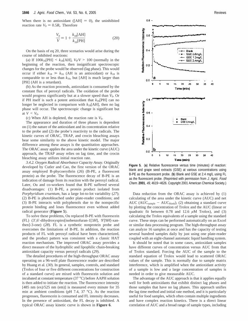
3.4.2. Oxygen Radical Absorbance Capacity Assay.Originallydeveloped by Cutler and Cao, the first version of the ORACassay employed B-phycoerythrin (26) (B-PE, a fluorescentprotein) as the probe. The fluorescence decay of B-PE is anindication of damage from its reaction with the peroxyl radical.Later, Ou and co-workers found that B-PE suffered severaldisadvantages: (1)Β-PE, a protein product isolated fromPorphyridium cruentum, has a large lot-to-lot variability (27);(2) B-PE is photobleached under plate-reader conditions; and(3) B-PE interacts with polyphenols due to the nonspecificprotein binding and loses fluorescence even without addedradical generator (Figure 5).
To solve these problems, Ou replaced B-PE with fluorescein(FL) (3′,6′-dihydroxyspiro[isobenzofuran-1[3H], 9′[9H]-xan-then]-3-one) (28). FL is a synthetic nonprotein probe andovercomes the limitations of B-PE. In addition, the reactionproducts of FL with peroxyl radical have been characterized,and the product pattern was consistent with a classic HATreaction mechanism. The improved ORAC assay provides adirect measure of the hydrophilic and lipophilic chain-breakingantioxidant capacity versus peroxyl radicals (29).
The detailed procedures of the high-throughput ORAC assayoperating on a 96-well plate fluorescence reader are describedby Huang et al. (30). In general, samples, controls, and standard(Trolox of four or five different concentrations for constructionof a standard curve) are mixed with fluorescein solution andincubated at constant temperature (37°C) before AAPH solutionis then added to initiate the reaction. The fluorescence intensity[485 nm (ex)/525 nm (em)] is measured every minute for 35min at ambient conditions (pH 7.4, 37°C). As the reactionprogresses, fluorescein is consumed and FL intensity decreases.In the presence of antioxidant, the FL decay is inhibited. Atypical ORAC assay kinetic curve is shown inFigure 6.
Data reduction from the ORAC assay is achieved by (1)calculating of the area under the kinetic curve (AUC) and netAUC (AUCsample- AUCblank), (2) obtaining a standard curveby plotting the concentration of Trolox and the AUC (linear orquadratic fit between 0.78 and 12.6µM Trolox), and (3)calculating the Trolox equivalents of a sample using the standardcurve. These steps can be performed automatically on an Excelor similar data processing program. The high-throughput assaycan analyze 16 samples at once and has the capacity of testingseveral hundred samples daily by just using one plate-readercoupled with an eight-channel automatic liquid handling system.
It should be noted that in some cases, antioxidant sampleshave different curves of concentration versus AUC from thatof Trolox standard. Forcing the AUC of the sample to thestandard equation of Trolox would lead to scattered ORACvalues of the sample. This is normally due to sample matrixinterference, which is amplified when the antioxidant activityof a sample is low and a large concentration of samples isneeded in order to give measurable AUC.
The advantage of the AUC approach is that it applies equallywell for both antioxidants that exhibit distinct lag phases andthose samples that have no lag phases. This approach unifiesthe lag time method and initial rate method, and it is particularlyuseful for food samples, which often contain multiple ingredientsand have complex reaction kinetics. There is a direct linearcorrelation of AUC and a broad range of sample types, including
V0
V) 1 +
k18[AH]
k16[PH](20)
Figure 5. (a) Relative fluorescence versus time (minutes) of reaction:blank and grape seed extracts (GSE) at various concentrations usingB-PE as the fluorescent probe. (b) Blank and GSE at 2.4 mg/L using FLas the fluorescent probe. (Reprinted with permission from J. Agric. FoodChem. 2001, 49, 4619−4626. Copyright 2001 American Chemical Society.)
1846 J. Agric. Food Chem., Vol. 53, No. 6, 2005 Reviews

raw fruit and vegetable extracts, plasma, and pure phytochemi-cals (26). Therefore, the ORAC assay has been broadly appliedin academics and the food and supplement industry as a methodof choice to quantify antioxidant capacity. In fact, an antioxidantdatabase has been generated applying the ORAC assay incombination with the total phenols assay (31, 32).
Many antioxidants are lipophilic, and it is also known thatthe antioxidant capacity of a compound is dependent uponreaction media (33-35). Therefore, an organic solvent basedORAC assay would be particularly useful for lipophilic samples.However, fluorescein is not sufficiently lipid soluble, and itsfluorescence intensity in nonpolar organic solvent is rather low.To overcome this problem, Naguib applied 4,4-difluoro-3,5-bis(4-phenyl-1,3-butadienyl)-4-bora-3a,4a-diaza-s-indacene (BO-DIPY 665/676) as a fluorescent probe and 2,2′-azobis(2,4-dimethylvaleronitrile) (AMVN) as a peroxyl radical generator.The reaction could be carried out either in liposome or on anoctane and butyronitrile mixture (36). By applying this assay,the antioxidant capacity of various carotenoids was quantified.However, this assay is 100 times less sensitive than the ORACassay, probably due to the low efficiency of the radicalgenerator, AMVN. In addition, the fluorescent quenchingmechanism of BODIPY by peroxy radical remains to beinvestigated.
3.4.3. Crocin Bleaching Assay.This assay measures theinhibition capacity of antioxidants in protecting the bleachingof crocin, a naturally occurring carotenoid derivative, by thefree radical generator AAPH (37). Ursini and co-workers later
applied this method to the analysis of plasma antioxidantcapacity (38). Experimentally, the reaction was carried out byfirst preparing a 2.0 mL phosphate buffer (0.1 M, pH 7.0)containing 10µM crocin and certain amounts of antioxidants.Next the radical initiator AAPH (50µL, 0.5 M) was added toinitiate the reaction. The progress of the reaction was monitoredby a UV-vis spectrometer at a wavelength of 443 nm, theabsorption maximum of crocin (ε ) 1.33 × 10⁵ M-1 cm-1).The bleaching rate becomes linear∼1 min after the addition ofAAPH and was monitored for 10 min. To eliminate possibleinterferences from the sample itself, blanks without crocin werealso monitored under the same wavelength. The initial crocinbleaching rates were obtained from the kinetic curves in thepresence (V) or absence (V0) of antioxidants. The relationshipbetweenV andV0 obeyed the equation (similar to eq 16)
whereka is the rate constant for the reaction of antioxidantswith ROO•, kc is the rate constant for the reaction between ROO•
and crocin, [C] is the concentration of crocin, and [AH] is theconcentration of antioxidant. A plot of [AH]/[C] versusV0/Vshould give a linear line curve with a slope ofka/kc, whichindicates the relative peroxyl radical scavenging capacity. Fora given antioxidant, theka/kc value was divided by theka/kc
value for the sample. For plasma, a linearity curve was obtainedwith a slope of 0.79. The antioxidant capacity of vitamin Cwas ranked surprisingly high at 7.7 Trolox equivalence (forcomparison, the ORAC value of vitamin C is 0.95) (24).
The crocin bleaching assay has found limited applications infood samples so far. Reaction rate constants between ROO• andphytochemicals may vary greatly, and some of them have ratescomparable to that of crocin (thus, no lag phase), whereas otherswill give a lag phase. In this case, the inhibited bleaching ratesare very small and are not sensitive to the concentration changesof antioxidants. This could be the reason vitamin C has anunusually large antioxidant capacity value. Crocin absorbs at arather short wavelength (450 nm), and many food pigments,such as carotenoids, absorb light at the same wavelength. Toavoid the interference for each sample, a sample blank (amixture containing only AAPH and food sample) must be testedat the same time. Finally, crocin is a mixture of natural pigmentsextracted from saffron and is subject to lot-to-lot variability,which limits its industrial application in a quantitative procedure.
3.4.4. Total Peroxyl Radical-Trapping Antioxidant ParameterAssay (39).Detailed accounts on the history and current stateof the TRAP assay can be found in a review by Ghiselli andco-workers (40). The TRAP assay uses R-phycoerythrin(R-PE) as a fluorescent probe (41). The reaction progress ofR-PE with AAPH was monitored fluorometrically (λex ) 495nm andλem ) 575 nm). A typical kinetic curve of fluorescencedecay is shown inFigure 7.
The antioxidant capacity of an unknown sample was ex-pressed as Trolox equivalence (X) by the equation
whereCTrolox is Trolox concentration,TTrolox is the lag time ofthe kinetic curve of R-PE in the presence of Trolox,X is theantioxidant capacity of plasma, andTplasma is the lag time ofthe kinetic curve in the presence of plasma.X is then multipliedby 2.0 (the stoichiometric factor of Trolox) and by the dilutionfactor of the sample to give the TRAP value (µmol/L). To obtainthe TTrolox from the same kinetic curve of the sample, Troloxwas added to the reaction mixture when R-PE fluorescence was
Figure 6. (a) Fluorescence decay curve of fluorescein in the presence ofR-tocopherol and AAPH. (b) Linear plot of the net AUC versusR-tocopherol concentration. (Reprinted with permission from ref 29.Copyright 2002 American Chemical Society.)
V0/V ) 1 + (ka/kc) × [AH]/[C] (21)
CTrolox/TTrolox ) X/Tplasma (22)
Reviews J. Agric. Food Chem., Vol. 53, No. 6, 2005 1847

∼50% of the initial value. The reaction was followed until thefluorescence decay rate resumed to the level before the Troloxaddition. The lag phase was then calculated by extrapolatingthe curves of maximal R-PE oxidation before and after Troloxaddition (Figure 7). Not all of the samples will yield a lag phase.Ursini and co-workers simulated effects of antioxidants on thelag phase of peroxidation and found that lag time-basedmeasurements of antioxidant capacity overestimated the anti-oxidant capacity of weaker antioxidants (34). Valkonene andco-workers modified the TRAP assay by applying dichloro-fluorescin diacetate (DCFH-DA) as the molecular probe (42).In the presence of AAPH, DCFH-DA was oxidized and also(somehow) hydrolyzed in the process, to produce highlyfluorescent dichlorofluorescein (DCF). The increase of fluo-rescence signal is an indication of oxidation progress.
4. ET-BASED ASSAYS
These assays include perhaps the most popular, but oftenmisunderstood by its name, total phenols assay by Folin-Ciocalteu reagent (FCR). In addition, also grouped into thiscategory are the Trolox equivalent antioxidant capacity (TEAC)assay, the ferric ion reducing antioxidant power (FRAP) assay,theN,N-dimethyl-p-phenylenediamine (DMPD) assay, and theCu(II) reduction capacity assay. These methods involve twocomponents in the reaction mixture, antioxidants and oxidant(also the probe). They are based on the following electron-transfer reaction:
The probe itself is an oxidant that abstracts an electron fromthe antioxidant, causing color changes of the probe. The degreeof the color change is proportional to the antioxidant concentra-tions. The reaction end point is reached when color change stops.The change of absorbance (∆A) is plotted against the antioxidantconcentration to give a linear curve. The slope of the curvereflects the antioxidant’s reducing capacity, which is expressedas Trolox equivalence (TE) or gallic acid equivalent (GAE).These assays resemble the redox titration in classical chemicalanalysis. Because there is not a competitive reaction involvedand there is no oxygen radical in the assays, it is questionable
how the assay results relate to the antioxidant capacity of asample. To make the correlation, it is assumed that antioxidantcapacity is equal to reducing capacity (43).
4.1. Total Phenols Assay by Folin-Ciocalteu Reagent.FCR was initially intended for the analysis of proteins takingadvantage of the reagent’s activity toward protein tyrosine(containing a phenol group) residue (44). Many years later,Singleton and co-workers extended this assay to the analysisof total phenols in wine; since then the assay has found manyapplications (45). The FCR-based assay gained popularity andis commonly known as the total phenols (or phenolic) assay.The FCR actually measures a sample’s reducing capacity, butthis is not reflected in the name “total phenolic assay”.Numerous publications applied the total phenols assay by FCRand an ET-based antioxidant capacity assay (e.g., FRAP, TEAC,etc.) and often found excellent linear correlations between the“total phenolic profiles” and “the antioxidant activity”. This isnot surprising if one considers the similarity of chemistrybetween the two assays. One of the assays may just beredundant. A recent report of using polyphenol oxidase forassaying total phenols in tea may be more specific to phenoliccompounds (46).
The FCR is typically made by first boiling (for 10 h) themixture of sodium tungstate (Na2WO4‚2H2O, 100 g), sodiummolybdate (Na2MoO4‚2H2O, 25 g), concentrated hydrochloricacid (100 mL), 85% phosphoric acid (50 mL), and water (700mL). After boiling, lithium sulfate (Li2SO4‚4H2O, 150 g) isadded to the mixture to give an intense yellow solutionstheFC reagent. Contamination of reductants leads to a green color,and the addition of oxidants such as bromine can restore thedesired yellow color. The exact chemical nature of the FCreagent is not known, but it is believed to contain heteropoly-phosphotunstates-molybdates. Sequences of reversible one- ortwo-electron reduction reactions lead to blue species, possibly(PMoW11O40)4-. In essence, it is believed that the molybdenumis easier to be reduced in the complex and electron-transferreaction occurs between reductants and Mo(VI):
Obviously, the FC reagent is nonspecific to phenolic compoundsas it can be reduced by many nonphenolic compounds [e.g.,vitamin C, Cu(I), etc.]. Phenolic compounds react with FCRonly under basic conditions (adjusted by a sodium carbonatesolution to pH∼10). Dissociation of a phenolic proton leads toa phenolate anion, which is capable of reducing FCR. Thissupports the notion that the reaction occurs through electron-transfer mechanism. The blue compounds formed betweenphenolate and FCR are independent of the structure of phenoliccompounds, therefore ruling out the possibility of coordinationcomplexes formed between the metal center and the phenoliccompounds.
Despite the undefined chemical nature of FCR, the totalphenols assay by FCR is convenient, simple, and reproducible.As a result, a large body of data has been accumulated, and ithas become a routine assay in studying phenolic antioxidants.
4.1. Trolox Equivalent Antioxidant Capacity Assay.TheTEAC assay was first reported by Miller and Rice-Evans in1993 (47) and later improved (48). In the improved version,ABTS•-, the oxidant, was generated by persulfate oxidation of2,2′-azinobis(3-ethylbenzothiazoline-6-sulfonic acid) (ABTS2-).Specifically, 7 mmol of ABTS ammonium was dissolved inwater and treated with 2.45 mmol of potassium persulfate, andthe mixture was then allowed to stand at room temperature for12-16 h to give a dark blue solution. This solution was diluted
Figure 7. Kinetics of R-PE oxidation initiated by 5 mM AAPH in thepresence of plasma (8 µL) before and after Trolox addition (1.8 µM finalsolution). The antioxidant capacity of each plasma sample is calculatedby comparing the two lag phases obtained in the presence and in theabsence of trolox. (Reprinted with permission from ref 38. Copyright 1998Elsevier.)
probe (oxidant)+ e (from antioxidant)freduced probe+ oxidized antioxidant
Mo(VI) + e f Mo(V)
1848 J. Agric. Food Chem., Vol. 53, No. 6, 2005 Reviews

with ethanol or buffer (pH 7.4) until the absorbance reached0.7 at 734 nm. One milliliter of the resulting solution was mixedwith 10 µL of sample. The absorbance was read at 30°C, 1, 4,and 6 min after mixing at 30°C. The difference of theabsorbance reading is plotted versus the antioxidant concentra-tions to give a straight line. The concentration of antioxidantsgiving the same percentage change of absorbance of the ABTS•-
as that of 1 mM Trolox was regarded as TEAC.Due to its operational simplicity, the TEAC assay has been
used in many research laboratories for studying antioxidantcapacity, and TEAC values of many compounds and foodsamples are reported. The TEAC values for pure antioxidantcompounds do not show clear correlation between TEAC valuesand the number of electrons an antioxidant can give away. TheTEAC values of ascorbic acid (1.05),R-tocopherol (0.97),glutathione (1.28), and uric acid (1.01) are almost the same,although glutathione can normally donate one electron (to formoxidized glutathione) whereas the others are two-electronreductants. Ferulic acid (1.90) andp-coumaric acid (2.00) havecomparable TEAC values. However, caffeic acid has a TEACvalue of 1.00 even though its structure is similar to that of ferulicacid. The TEAC value difference between quercetin (3.00) andkaempferol (1.00) is also rather surprising as they have similarchemical structures (14).
Apparently, the reaction rate differences between antioxidantsand oxidants are not reflected in the TEAC values because theTEAC assay is an end-point assay.
4.2. Ferric Ion Reducing Antioxidant Power Assay.TheFRAP assay also takes advantage of electron-transfer reactions.Herein a ferric salt, Fe(III)(TPTZ)2Cl3 (TPTZ) 2,4,6-tripyridyl-s-triazine), is used as an oxidant (49). The redox potential of
Fe(III) salt (∼0.70 V) is comparable to that of ABTS•- (0.68V). Therefore, essentially, there is not much difference betweenTEAC assay and the FRAP assay except TEAC is carried outat neutral pH and FRAP assay under acidic (pH 3.6) conditions.The FRAP assay involves the following procedures: Theoxidant in the FRAP assay is prepared by mixing TPTZ (2.5mL, 10 mM in 40 mM HCl), 25 mL of acetate buffer, and 2.5mL of FeCl3‚H2O (20 mM). The conglomerate is referred to as“FRAP reagent”. The final solution has Fe(III) of 1.67 mM andTPTZ of 0.83 mM. Therefore, the TPTZ is deficient as the idealreaction stoichiometry between Fe(III) and TPTZ is 1 to 2. Theoxidant is not just Fe(III)(TPTZ)2, it also contains other Fe(III)
species which can lead to potential problems as many metalchelators in food extract could bind Fe(III) and form complexesthat are also capable of reacting with antioxidants. To measureFRAP value, 300µL of freshly prepared FRAP reagent iswarmed to 37°C and a reagent blank reading is taken at 593nm; then 10µL of sample and 30µL of water are added.Absorbance readings are taken after 0.5 s and every 15 s until4 min. The change of absorbance (∆A ) A4min - A0min) iscalculated and related to∆A of an Fe(II) standard solution.∆Ais linearly proportional to the concentration of antioxidant. OneFRAP unit is arbitrarily defined as the reduction of 1 mol ofFe(III) to Fe(II). The FRAP values for ascorbic acid,R-toco-pherol, and uric acid are identical (2.0). The FRAP value ofbilirubin is 1-fold higher than that of ascorbic acid. These resultssuggest that 1 mol of vitamin C can reduce 2 mol of Fe(III)and that 1 mol of bilirubin can reduce 4 mol of Fe(III). This isin conflict with the fact that both vitamin C and bilirubin aretwo-electron reductants. It is known that when bilirubin isoxidized, it is transformed to beliverdin (by losing two hydrogenatoms, not just electrons), which happens to have an absorptionat 593 nm with coeffiicency (ε593 ) 1 × 10⁴) comparable withthat of Fe(II)(TPTZ)2.50
Pulido and co-workers (51) measured the FRAP values ofseveral polyphenols in water and methanol. However, theabsorption (A593) does not stop at 4 min; instead, it slowlyincreased even after several hours. Polyphenols with suchbehaviors include caffeic acid, tannic acid, ferulic acid, ascorbicacid, and quercetin. The FRAP values of these compoundscannot be obtained accurately if 4 min reaction time wasfollowed.
4.3. Total Antioxidant Potential Assay Using Cu(II) as anOxidant. There is little published information on this assay.However, an industrial laboratory is providing service using itas a measure of total antioxidant potential (52). The method isbased on reduction of Cu(II) to Cu(I) by reductants (antioxi-
dants) present in a sample. A chromogenic reagent, bathocu-proine (2,9-dimethyl-4,7-diphenyl-1,10-phenanthroline), formsa 2:1 complex with Cu(I), which has a maximum absorbanceat 490 nm (53). It was found that 1 mol ofR-tocopherol canreduce 2 mol of Cu(II) to Cu(I) (54).
More recently, Zaporozhets et al. reported a method formeasuring the antioxidant power of herbal products based onsolid-phase spectrophotometry using tetrabenzo[b,f,j,n][1,5,9,-13]tetraazacyclohexadecine-Cu(II) complex immobilized onsilica gel. The absorbance of the modified sorbent (712 nm)increases when the Cu(II) is reduced (55).
4.4. 2,2-Diphenyl-1-picrylhydrazyl Radical ScavengingCapacity Assay.DPPH is one of a few stable and commercially
Reviews J. Agric. Food Chem., Vol. 53, No. 6, 2005 1849

available organic nitrogen radicals and has a UV-vis absorptionmaximum at 515 nm. Upon reduction, the solution color fades;the reaction progress is conveniently monitored by a spectro-photometer.
The DPPH assay is typically run by the following proce-dure: DPPH solution (3.9 mL, 25 mg/L) in methanol is mixedwith sample solution (0.1 mL). The reaction progress absorbanceof the mixture is monitored at 515 nm for 30 min or until theabsorbance is stable. Upon reduction, the color of the solutionfades. The percentage of the DPPH remaining is calculated as
%DPPHrem is proportional to the antioxidant concentrations, andthe concentration that causes a decrease in the initial DPPHconcentration by 50% is defined as EC50. The time needed toreach the steady state with EC50 concentration is calculated fromthe kinetic curve and defined asTEC50. A representative kineticcurve of a DPPH assay is shown inFigure 8.
Sanchez-Moreno and co-workers classified the kinetic be-havior of the antioxidant compound as follows:<5 min (rapid),5-30 min (intermediate), and>30 min (slow). They furtherproposed a parameter, called “antiradical efficiency (AE)” (56),
to express the antioxidant capacity of a certain antioxidant. AEis calculated as
The DPPH assay is technically simple, but some disadvan-tages limit its applications. Besides the mechanistic differencefrom the HAT reaction that normally occurs between antioxi-dants and peroxyl radicals, DPPH is a long-lived nitrogenradical, which bears no similarity to the highly reactive andtransient peroxyl radicals involved in lipid peroxidation. Manyantioxidants that react quickly with peroxyl radicals may reactslowly or may even be inert to DPPH. This is evident from theTEC50 values ranging from 1.15 min (ascorbic acid) to 103 min(rutin). Consequently, the antioxidant capacity is not properlyrated. The reaction kinetics between DPPH and antioxidantsare not linear to DPPH concentrations (Figure 8). It is thusrather arbitrary to express antioxidant capacity using EC50.Finally, it was reported that the reaction of DPPH with eugenolwas reversible (57). This would result in falsely low readingsfor antioxidant capacity of samples containing eugenol and otherphenols bearing a similar structure type (o-methoxyphenol).
The DPPH assay was believed to involve hydrogen atomtransfer reaction, but a recent paper suggested otherwise. Onthe basis of the kinetic analysis of the reaction between phenolsand DPPH (58), Foti and co-workers suggested that the reactionin fact behaves like an ET reaction. The authors found that therate-determining step for this reaction consists of afastelectron-transfer process from the phenoxide anions to DPPH. Thehydrogen atom abstraction from the neutral ArOH by DPPHbecomes a marginal reaction path, because it occursVeryslowlyin strong hydrogen-bond-accepting solvents, such as methanoland ethanol. In addition, the author found that adventitious acidsor bases present in the solvent may dramatically influence theionization equilibrium of phenols and cause a reduction or anenhancement, respectively, of the measured rate constants. Thisrenders the DPPH assay much less chemically sound as a validassay for antiradical activity of measurement.
5. OVERALL CONSIDERATIONS OF ET- AND HAT-BASEDASSAYS
ET-based assays measure an antioxidant’s reducing capacity,and the HAT-based assays quantify hydrogen atom donatingcapacity. It is apparent that the hydrogen atom transfer reactionis a key step in the radical chain reaction. Therefore, the HAT-based method is more relevant to the radical chain-breakingantioxidant capacity. Pedulli and co-workers studied the anti-oxidant capacity of phenothiazine and related compounds. Thesearomatic amines exhibit antioxidant capacity because of theirlow N-H bond dissociation energies (77-80 kcal/mol). If thehydrogen atom is replaced with a methyl group, the peroxylradical scavenging capacity is lost, despite the fact that themethylated analogue has a redox potential similar to that of theparent amine. Therefore, radical trapping capacity directly relates
Figure 8. DPPH bleaching kinetics in the presence of different concentra-tions of R-carotene (a) and â-xanthophylls (b). [Reprinted with permissionfrom ref 56 (Wiley). Copyright 2000 Society of Chemical Industry.]
%DPPHrem ) 100× [DPPH]rem/[DPPH]T)0 (23)
AE ) (1/EC50)TEC50 (24)
1850 J. Agric. Food Chem., Vol. 53, No. 6, 2005 Reviews

to the hydrogen atom donating ability of a compound and isnot correlated to the redox potentials alone (59). Although thereducing capacity of a sample is not directly related to its radicalscavenging capacity, it is an important parameter of antioxidants.Some water soluble oxidants such as peroxynitrite and hy-pochlorite can be readily reduced to harmless species. In thisregard, a chemically valid ET-based assay will provide usefulinformation.
The HAT-based assay using fluorescent probes has a mecha-nistic similarity to lipid peroxidation, but under the assayconditions, the concentration of the substrate (in this case theprobe) is often smaller than the concentration of antioxidants.This is in contradiction with real situations. In food systemsthe antioxidant concentration is much smaller than that of thesubstrate (e.g., lipid). It remains to be seen if the antioxidantcapacity measured using the HAT-based assay using a molecularprobe can be translated to applications in a real food system,which is also often heterogeneous and under different oxidativestress conditions. It is known that the distribution of antioxidantin two phases has great impact on its effectiveness (60).
Mechanistically, electron transfer and hydrogen atom transferreaction can be difficult to distinguish. Apparent hydrogen atomtransfer reaction can be the result of proton-coupled electrontransfer (PCET). The detailed mechanism of the reaction isbeyond the scope of this review, but experimental (61) andtheoretical studies (62) have shown that tocopherol reaction withoxyradicals predominantly undergoes hydrogen atom transferreaction.
6. ASSAYS MEASURING OTHER ROS SCAVENGINGCAPACITY
Experimental evidence has directly or indirectly suggestedthat there are six major reactive oxygen species causingoxidative damage in the human body. These species aresuperoxide anion (O2•-), hydrogen peroxide (H2O2), peroxylradicals (ROO•), hydroxyl radical (HO•), singlet oxygen (1O2),and peroxynitrite (ONOO-). To counteract the assault of theseROS, living cells have a biological defense system composedof enzymatic antioxidants that convert ROS/RNS to harmlessspecies. For example, O2
•- is converted to oxygen and hydrogenperoxide by superoxide dismutase (SOD) or reacts with nitricoxide (NO•) to form peroxynitrite. H2O2 can be converted towater and oxygen by catalase. In contrast, no enzymatic actionis known to scavenge ROO•, HO•, 1O2, and ONOO-. Therefore,the burden of defense relies on a variety of nonenzymaticantioxidants such as vitamins C and E and many phytochemicalsthat have the property of scavenging oxidants and free radicals.To comprehensively evaluate the oxidant-scavenging capacityof a food sample, assays have to be designed to include theseROS. However, so far the majority of assays are designed tomeasure a sample’s capacity to react with one oxidant (eitherorganic radical or redox active compounds). The peroxyl radicalhas been the most frequently used ROS in the assays becauseit is a key radical in autoxidation and it can be generatedconveniently from the thermal decomposition of azo compounds.There are also limited numbers of papers describing assays forthe scavenging capacity of other ROS. In this section, we brieflydescribe some of the assays.
6.1. O2•- Scavenging Capacity Assay.Classically, the SOD
activity assay uses the competition kinetics of O2•- reduction
of cytochromec (probe) and O2•- scavenger (sample). Morerecently, the method has been adapted to a microplate format(63). Cytochromec can be reduced directly by antioxidants,which can also inhibit the xanthine oxidase. Therefore, this
method is not suitable for quantifying nonenzymatic antioxidant(54). Ewing and Janero reported a high-throughput assay usinga nonenzymatic (phenazine methosulfate/NADH/O2) O2
•- gen-erator and nitroblue tetrazolium (NBT) as a probe (64). Thisassay takes advantage of the reducing property of O2
•-. Theredox potential of O2/O2
•- is -0.2 to-0.5 V depending on themedium (vs NHE) (65). Because many dietary antioxidants canalso exhibit reducing capacity, as demonstrated in the TEACand FRAP assays, this improved method cannot be applied tononenzymatic samples. More recently, hydroethidine has beenused as the probe in measuring O2
•- scavenging capacity (66).Nonfluorescent hydroethidine is oxidized by O2
•- (generatedfrom xanthine oxidase and xanthine mixture) to form a speciesof unknown structure that exhibits a strong fluorescence signalat 586 nm. Addition of SOD inhibits the hydroethidine oxida-tion. This approach can avoid the problem of direct reductionof the probe by antioxidant, but possible inhibition of xanthineoxidase by antioxidants remains an issue.
6.2. H2O2 Scavenging Capacity Assay.H2O2 is rather inertat low concentrations. Under physiological conditions, H2O2
oxidation power is believed to be observed in combination withFe(II) (Fenton reaction). Biologically, H2O2 is converted tooxygen and water by catalase. A common assay that claims tomeasure H2O2 scavenging capacity of dietary antioxidants useshorseradish peroxidase to oxidize scopoletin to a nonfluorescentproduct. In the presence of antioxidants the oxidation isinhibited. The nature of the inhibition is ambiguous becausethere are several potential inhibition pathways. The antioxidantscan inhibit the reaction by (a) reacting directly with H2O2, (b)reacting with intermediates formed from enzyme and H2O2, or(c) inhibiting the horseradish peroxidase from binding H2O2.Therefore, it is difficult to explain the actual chemical meaningof the data (67).
6.3. Hydroxyl Radical (HO) Scavenging Assay.Biologi-cally, the hydroxyl radical is widely believed to be generatedwhen hydrogen peroxide reacts with Fe(II) (Fenton reaction).However, the Fe(II)/H2O2 mixture has disadvantages in ascavenging assay because many antioxidants are also metalchelators. When the sample is mixed with Fe(II), it may alterthe activity of Fe(II) by chelation. As a result, it is impossibleto distinguish if the antioxidants are simply good metal chelatorsor HO• scavengers. Antioxidants in food (such as vitamin C)may act as pro-oxidants by reducing Fe(III) to Fe(II) and makethe HO• generation catalytic. In fact, ascorbic acid has beenused in combination with catalytic Fe(II) and excess H2O2 togenerate a constant flux of HO• radicals. Recently, Zhu andco-workers have reported an organic Fenton reaction (68). Zhufound that a mixture of tetrachlorohydroquinone (TCHQ, amajor metabolite of the widely used biocide pentachlorophenol)and H2O2 hydroxylates salicylic acid to yield both 2,3- and 2,5-dihydroxybenzoic acid (DHBA). The hydroxylation is markedlyinhibited by hydroxyl radical scavenging agents such as dimethylsulfoxide and ethanol. The inhibited reaction was not affectedby iron chelators, such as diethylenetriaminepentaacetic acid(DTPA), bathophenanthroline disulfonic acid, phytic acid, andbathocuprione disulfonic acid. A comparison of product typeand distribution from the TCHQ/H2O2 system with that of theFe(II)/H2O2 system suggests that hydroxyl radicals are involvedin the organic Fenton reaction system. It would be interestingto see if this metal-free TCHQ/H2O2 mixture can be a hydroxylradical source for assaying the hydroxyl radical scavengingcapacity of antioxidants.
The putative hydroxyl radical is an extremely reactive andshort-lived species that can hydroxylate DNA, proteins, and
Reviews J. Agric. Food Chem., Vol. 53, No. 6, 2005 1851

lipids. Therefore, the direct scavenging of the hydroxyl radicalby dietary antioxidants in a biological system is unrealistic asthe cellular concentration of dietary antioxidants is negligiblecompared with other biological molecules. The rate constantsfor HO• reactions have been determined by pulse radiolysisthrough the deoxyribose method (69). The second-order rateconstants are>108 M-1‚s-1 for many compounds, includingunreactive compounds such as benzene (3.2× 10⁹ M-1‚s-1)and glucose (1× 10⁹ M-1‚s-1). Therefore, the ability ofantioxidants to scavenge the HO• radical is not unlikely toprovide any protection to biological molecules as the opportunityfor HO• and antioxidants to react is extremely small. On theother hand, it is possible to prevent the formation of hydroxylradicals by either deactivating free metal ions [e.g., Fe(II)]through chelation or converting H2O2 to other harmless com-pounds (such as water and oxygen). Catalase converts H2O2 toO2, and H2O and metal chelators bind metal ions so that theybecome inert toward H2O2. Thus, dietary nutrients containingmetal chelators may act as preventive antioxidants. Quantifyingthe capacity of the phytochemicals in preventing hydroxylradical formation in vitro would be more relevant and a valuableguide to antioxidant clinical research. Recently, Ou and co-workers have developed a fluorometric assay for screening themetal [Co(II)] chelating capacity of dietary antioxidants (70).The method, christened HORAC [hydroxyl (HO) radicalsaverting capacity], employs a Co(II) complex mediated Fenton-like reaction. The hydroxyl radical formation under the experi-mental conditions is indirectly confirmed by the hydroxylationof p-hydroxybenzoic acid. Fluorescein (FL) was used as theprobe. The fluorescence decay curve of FL is monitored in theabsence or presence of antioxidants, the area under thefluorescence decay curve (AUC) is then integrated, and the netAUC is calculated by subtracting the AUC of the blank fromthat of the sample antioxidant. The quantitation method is thesame as that of the ORAC assay except gallic acid is used asthe standard. This method has been rigorously validated forlinearity, precision, accuracy, and ruggedness. A wide range ofphenolic antioxidants can be analyzed. The hydroxyl radicalprevention capacity is mainly due to their metal-chelatingcapability.
6.4. Singlet Oxygen Scavenging Capacity Assay.Singletoxygen is normally generated in the presence of light andphotosensitizers. It is believed that1O2 is often responsible forUV light-dependent damage to skin (71), cataract formation inthe lens of the eyes (72), macular degeneration (73), andphotosensitivity resulting from ingestion or absorption ofphytochemicals, pharmaceuticals and pesticides that act asphotosensitizers (74). In the absence of light,1O2 productioncan be ambiguous in a biological system. It was suggested thatthe extracellular1O2 production by the spontaneous dismutationof superoxide anion has some physiological significance (75).On the other hand, chemically,1O2 can be convenientlygenerated through non-photochemical decomposition of hydro-gen peroxide by metals or hypochlorite (76, 77).
Rate constants of singlet oxygen reaction with variouscompounds have been compiled by Wilkinson and co-workers(78). Singlet oxygen can be quenched through physical meansby transferring its excitation energy to another molecule (whichis excited), or it can add to antioxidants forming endoperoxides.â-Carotene is an excellent physical quencher of1O2. Singletoxygen emits characteristic phosphorescence at 1270 nm. Thedecay rates of the light intensity were used to measure the1O2
quenching activity of a compound. Foote and co-workersreported a more sensitive method by monitoring the quenching
of singlet-oxygen-sensitized (at 703 nm) delayed fluorescence(SOSDF) of tetra-tert-butylphthalocyanine (79). The authorsreported the quenching rates ofâ-carotene,R-tocopherol, 1,4-diazabicyclo[2.2.2]octane, 2,6-di-tert-butyl-4-methylphenol, andlauric acid. The singlet oxygen quenching rates vary by 6 ordersof magnitude. Quenching of the visible SOSDF may provide ahighly sensitive method for the measurement of1O2 quenchingcapacity using commonly available apparatus or in systemswhere the 1270 nm luminescence is difficult to detect. Thismethod is not yet widely applied.
6.5. Peroxynitrite (ONOO-) Scavenging Capacity Assay.Superoxide and nitric oxide react under diffusion control rate(k > 109 M-1‚s-1) to form peroxynitrite (80). O2
•- (E° ) -0.33V) and NO (0.39 V) are not potent oxidants; its adduct, ONOO-,is not a strong oxidant either. Its protonated form, peroxynitrousacid (ONOOH), is a very strong oxidant (E° ) 2.10 V). Underphysiological pH, ONOOH (pKa ) 8.0) rearranges to form muchless oxidizing nitrate (81). At pH 7.4, the ratio of peroxyntriteand peroxynitrous acid is 4 to 1. ONOO- and ONOOH oftencause the nitration or hydroxylation of aromatic compounds,particularly tyrosine (to nitrotyrosine). Under physiologicalconditions, peroxynitrite also forms an adduct with carbondioxide dissolved in body fluid. The adduct is believed to beresponsible for the oxidative damage of proteins (82).
There are a few papers on the scavenging capacity ofantioxidants against ONOO-. Two methods are used forONOO- scavenging measurements: (1) inhibition of tyrosinenitration by ONOO- (83) and (2) inhibition of dihydro-rhodamine (DHR) 123 oxidation (84). Pannala reported theperoxynitrite quenching capacity of catechin and other polyphen-ols by measuring their inhibition capacity on reaction betweenperoxynitrite and tyrosine. The method relies on HPLC separa-tion and quantification of nitrotyrosine, and it is thus rather time-consuming. Kooy developed another method based on theinhibition of the oxidation of DHR 123 by peroxynitrite (85).The initial rate approach was used to quantify peroxynitrite-scavenging capacity. Using the same method, Chung and co-workers studied the peroxynitrite scavenging and cytoprotectivecapacity of a marine algae extract (86).
7. CONCLUSIONS
ET-Based Assay.Overall, there are a multitude of ET-basedassays for measuring the reducing capacity of antioxidants. Theassays are carried out at acidic (FRAP), neutral (TEAC), or basic(total phenols assay by FCR) conditions. The pH values havean important effect on the reducing capacity of antioxidants.At acidic conditions, the reducing capacity may be suppresseddue to protonation on antioxidant compounds, whereas in basicconditions, proton dissociation of phenolic compounds wouldenhance a sample’s reducing capacity. The oxidant in the FRAPassay has a standard redox potential comparable to that ofABTS2- (∼0.7 V), but the redox potential for Mo(VI)/Mo(V)is not known, presumably due to the complex nature of FCR.These oxidants can certainly react with common antioxidantssuch as vitamins E (E° ) 0.5 V) and C (E° ) 0.28 V) andcommon phenolic compounds. There are many more oxidantsto choose from. The question is: what are the criteria ofselecting the right oxidant? We do not have a clear guideline,but the selectivity of oxidant should be such that it does notoxidize compounds such as sugar, which is ubiquitous in foodbut normally not considered to be an antioxidant. Sugar is knownto reduce metal ions such as Cu(II) (Fehling’s reagent).
Applying multiple ET-based assays to measure the reducingcapacity of an antioxidant often leads to excellent linear
1852 J. Agric. Food Chem., Vol. 53, No. 6, 2005 Reviews

correlations between the results. Indeed, we often see, in theresearch papers on antioxidants, the findings of excellentcorrelation (R2 > 0.99) between total phenolic contents (mea-sured by FCR) and antioxidant activity (measured by FRAP,TEAC, or DPPH assays) (87). Because these assays are basedon similar redox reactions, it is therefore somewhat redundantto apply the multitude of assays in quantifying reducing capacity.It is, however, important to use one assay that is commonlyaccepted and validated. In this regard, the total phenols assayby CFR has the clear advantage over the other ET-based assays.Disregarding the chemistry principles, the total phenols assayby FCR has the following advantages:
(1) The FCR is commercially available, and the procedure israther standardized.
(2) The long-wavelength (730 nm) absorption of the chro-mophore minimizes interference from the sample matrix, whichis often colored.
(3) It is a commonly accepted assay and routinely practicedin dietary antioxidant research laboratories throughout the world.
(4) A large body of comparable data has been produced(claimed as total phenols content instead of reducing capacityof FCR).
It should be noted that the total phenols assay by FCR isconducted at rather basic conditions (pH 10, necessary forphenols to dissociate protons). Simple phenols (e.g., C6H5OH,the phenolic group in tyrosine) react with FCR, although theyare not effective radical scavenging antioxidants. Therefore,there may not necessarily be a good correlation between the“total phenols content” and the radical scavenging antioxidantcapacity of a sample. To avoid misunderstanding on the actualmeaning of “total phenolic contents”, we suggest an alternativeterm, “FCR reducing capacity”, be used.
The total phenols assay by FCR is carried out in water, anaqueous phase. For lipophilic antioxidants, this assay in itscurrent form is not applicable. In fact, we have attempted buthave been unable to measure the total phenols of the lipidsoluble fraction of bee pollen as the sample did not havesufficient water solubility. Therefore, there is an immediate needfor a modified FCR for lipophilic samples.
The reducing capacity of a sample is an important parameterreflecting one aspect of the its antioxidation property. However,it is oversimplified to refer to the result as “total antioxidantcapacity”. The latter encompasses much broader aspects includ-ing metal chelating capacity, ROS scavenging capacity, and evenoxidative enzyme inhibition capacity (e.g., polyphenol oxidaseinhibitors in preventing the browning of fruit). In addition, notall ROS share the same reaction pattern (i.e., electron transfer)toward antioxidants. ROO• abstracts a hydrogen atom fromantioxidants, whereas HO• may undergo hydrogen atom abstrac-tion or addition to an unsaturated compound (DNA bases oraromatic amino acid residue). Singlet oxygen can form endo-peroxide with dienes or aromatic compounds or be quenchedphysically through energy transfer. Superoxide anion, on theother hand, is a moderate reductant [reduces Fe(III)]. Therefore,a host of assays measuring individual ROS scavenging capacityare needed to comprehensively evaluate a sample’s ROSscavenging capacity.
HAT-Based Assay.Peroxyl radicals play a key role in theunwanted lipid oxidation in food and biological systems. Thesacrificial antioxidants, represented by vitamin E, are criticalin protecting polyunsaturated fatty acid esters in foods and incell membranes from autoxidation. Vitamin E functions througha HAT mechanism. A HAT-based assay, represented by theORAC assay, involves peroxyl radicals as the oxidant and will
provide useful information on radical chain-breaking capacity.The ORAC assay has been modified to measure lipophilicantioxidants by using a cyclodextrin derivative as a watersolubility enhancer. With interlaboratory (three laboratories)validation and industrial recognition, the ORAC assay hascurrently emerged as the assay of choice for quantifying theperoxyl radical scavenging capacity of a sample. Broadervalidations involving at least eight other laboratories are neededfor the ORAC assay to become the standard assay adopted byofficial organizations such as the AOAC International.
Antioxidants in a food system normally refer to substancesthat can inhibit fatty acid autoxidation. The major antioxidantsare metal chelators (e.g., EDTA, preventive) and chain-breakingantioxidants (e.g., BHT, sacrificial) acting as hydrogen atomdonors. It is of great importance to know if the reducing capacityof antioxidants measured by FCR and ORAC values can betranslated to its inhibition capacity of fatty acid autoxidation inan actual food system. The total phenols assay by FCR and theORAC assay are carried out in a controlled manner in ahomogeneous solution with an artificial oxidant or radicalprecursor added to initiate the reaction, whereas in a real foodlipid system, the reaction occurs without added radical initiatoror oxidant. Instead, the reaction is initiated by light, metal ions,or heat during food processing or storage. Moreover, it is oftena heterogeneous mixture (as in food emulsions), and the phasedistribution of antioxidants will be critical for its effectiveness.Therefore, the antioxidants may behave differently than theydo in the antioxidant capacity assays. Hence, more researchneeds to be carried out to study if ORAC values and (or) FCRreducing capacity can be extended to real food systems. Beforethen, one should exercise caution when using ORAC values orthe total phenols contents as a guideline for optimizing foodformulations for extension of oxidative stability.
Application of the in Vitro Assays in Clinical Research.Although the limitations of these assays have been mentionedthroughout this paper, it is necessary to emphasize that the assaysdescribed herein are strictly based on chemical reactions in vitro.They bear no similarity to biological systems. The validity ofthe data is limited to a strict chemical sense with contextinterpretation. Any claims about the bioactivity of a samplebasedsolelyon these assays such as ORAC, TEAC, and FRAPetc., would be exaggerated, unscientific, and out of context.Moreover, these assays do not measure bioavailability, in vivostability, retention of antioxidants by tissues, and reactivity insitu.
The role of dietary antioxidants is to help fight excessive ROSin our bodies. By doing so, antioxidants will be sacrificed toprotect the biomolecules from being oxidized (as theantioxidantdefinition states), and thus the antioxidant has fulfilled itsfunction. It is of importance to study whether there is acorrelation between the intake of high potent antioxidants andthe level of oxidative stress. The in vitro nature of these assaysshould not compromise their value in guiding clinical research.On the contrary, a valid in vitro assay is an invaluable tool forclinical studies if it is combined with bioavailability data andvalid oxidative stress biomarker assays. In fact, many studiesapply these assays to study the impact of antioxidant consump-tion on reducing oxidative stress markers (88). In addition tothe assay validity itself, special attention has to be paid toconfounding factors from sample matrix when biologicalsamples are measured, such as blood plasma, tissues, or urine.In this regard, valid sample processing procedure is critical. Thesynergistic effects of such a combination assays [must be a validassay or disputable results can be gathered (89)] will allow us
Reviews J. Agric. Food Chem., Vol. 53, No. 6, 2005 1853
to investigate the impact of antioxidant in reducing oxidativestress and thus the implication for disease prevention.
ACKNOWLEDGMENT
We are grateful to Nancy McHale and Kathleen Pappalardo ofBrunswick Laboratories for excellent editorial help.
LITERATURE CITED
(1) Willet, W. C.Eat, Drink, and be HealthysThe HarVard MedicalSchool Guide to Healthy Eating; Simon and Schuster: NewYork, 2001.
(2) (a) Ames, B. N.; Gold, L. S.; Willet, W. C. The causes andprevention of cancer.Proc. Natl. Acad. Sci. U.S.A.1995, 92,5258-5265. (b) Kaur, C.; Kapoor, H. C. Antioxidants in fruitsand vegetablessthe millennium’s health.Int. J. Food Sci.Technol.2001, 703-725.
(3) Based on the Web of Knowledge search on articles containingthe wordantioxidantor antioxidants.
(4) Frankel, E. N.; Meyer, A. S. The problems of using one-dimensional methods to evaluate multifunctional food andbiological antioxidants.J. Sci. Food Agric.2000, 80, 1925-1941.
(5) Re, R.; Pellegrini, N.; Proteggente, A.; Pannala, A.; Yang, M.;Rice-Evans, C. Antioxidant activity applying an improved ABTSradical cation decolorization assay.Free Radical Biol. Med. 1999,26 (9/10), 1231-1237.
(6) Sanchez-Moreno, C. Review: Methods used to evaluate the freeradical scavenging activity in foods and biological systems.FoodSci. Technol. Int. 2002, 8 (3), 121-137.
(7) Prior R. L.; Cao, G. H. Analysis of botanicals and dietarysupplements for antioxidant capacity: A review.J. AOAC Int.2000, 83 (4), 950-956.
(8) Cao, G.; Prior, R. L. Comparison of different analytical methodsfor assessing total antioxidant capacity of human serum.Clin.Chem. 1998, 44, 1309-1315.
(9) Ghiselli, A.; Serafini, M.; Natella, F.; Scaccini, C. Totalantioxidant capacity as a tool to assess redox status: criticalreview and experimental data.Free Radical Biol. Med. 2000,29, 1106-1114.
(10) Buratti, S.; Pellegrini, N.; Brenna, O. V.; Mannino, S. Rapidelectrochemical method for the evaluation of the antioxidantpower of some lipophilic food extracts.J. Agric. Food Chem.2001, 49, 5136-5141.
(11) Cervellati, R.; Ho¨ner, K.; Furrow, S. D.; Neddens, C.; Costa, S.The Briggs-Rauscher Reaction as a test to measure the activityof antioxidants.HelV. Chim. Acta2001, 84, 3533-3547.
(12) “antioxidant”. Webster’s Third New International Dictionary,Unabridged; Merriam-Webster: 2002; http://unabridged.merriam-webster.com (Nov 9, 2004).
(13) http://cancerweb.ncl.ac.uk/cgi-bin/omd?query)antioxidants.(14) Panel on Dietary Antioxidants and Related Compounds, Standing
Committee on the Scientific Evaluation of Dietary ReferenceIntakes, Food and Nutrition Board Vitamin C, Vitamin E,Selenium, andâ-Carotene and Other Carotenoids: Overview,Antioxidant Definition, and Relationship to Chronic Disease. InDietary Reference Intakes for Vitamin C, Vitamin E, Selenium,and Carotenoids; National Academy of Science: Washington,DC, 2000; pp 35-57.
(15) Halliwell, B. How to characterize a biological antioxidant.FreeRadical Res. Commun. 1990, 9, 1-32.
(16) (a) Ingold, K. U. Inhibition of the autoxidation of organicsubstances in liquid phaseChem ReV. 1961,61, 563-584. (b)Denisov, E. T.; Khudyakov, I., V. Mechanism of action andreactivities of the free radicals of inhibitors.Chem. ReV. 1987,87, 1313-1357.
(17) Petersen D. R.; Doorn, J. A. Reactions of 4-hydroxynonenal withproteins and cellular targets.Free Radical Biol. Med. 2004, 37(7), 937-945.
(18) Burton, G. W.; Ingold, K. U. Autoxidation of biologicalmolecules. 1. the autoxidation of vitamin E and related chain-breaking antioxidantsin Vitro. J. Am. Chem. Soc.1981, 103,6472-6477.
(19) Pryor, W. A.; Strickland, T.; Church, D. F. Comparison of theeffiencies of several natural and synthetic antioxidants in aqueoussodium dodecyl sulfate micelle solutions.J. Am. Chem. Soc.1988, 110, 2224-2229.
(20) Pryor, W. A.; Cornicelli, J. A.; Devall, L. J.; Tait, B.; Trivedi,B. K.; Witiak, D. T.; Wu, M. A rapid screening test to determinethe antioxidants potencies of natural and synthetic antioxidants.J. Org. Chem. 1993, 58, 3521-3532.
(21) Kleinveld, H. A.; Haklemmers, H. L. M.; Stalenhoef, A. F. H.;Demacker, P. N. M. Improved measurement of low-density-lipoprotein susceptibility to copper-induced oxidation-applicationof a short procedure for isolating low-density-lipoprotein.Clin.Chem.1992, 38 (10), 2066-2072.
(22) Meyer, A. S.; Yi, O.-S.; Pearson, D. A.; Waterhouse, A. L.;Frankel, E. N. Inhibition of human low-density lipoproteinoxidation in relation to composition of phenolic antioxidants ingrape (Vitis Vinifera). J. Agric. Food Chem. 1997, 45, 1638-1643.
(23) Abuja, P. M.; Albertini, R.; Esterbauer, H. Simulation of theinduction of oxidation of low-density lipoprotein by high copperconcentrations: evidence for a nonconstant rate of initiation.Chem. Res. Toxicol. 1997, 10, 644-651.
(24) Bowry, V. W.; Ingold, K. U. The unexpected role of vitamin E(R-tocopherol) in the peroxidation of human low-density lipo-protein.Acc. Chem. Res.1999, 32, 27-34.
(25) Antunes, F.; Barclay, R. C.; Ingold, K. U.; King, M.; Norris, J.Q.; Scaiano, J. C.; Xi, F. On the antioxidant activity of melatonin.Free Radical Biol. Med. 1999, 26, 117-128.
(26) Cao, G. H.; Alessio, H. M.; Cutler, R. G. Oxygen-radicalabsorbency capacity assay for antioxidants.Free Radical Biol.Med. 199314 (3), 303-311.
(27) Cao, G.; Prior, R. L. The measurement of oxygen radicalabsorbance capacity in biological samples.Methods Enzymol.1999, 299, 50-62.
(28) Naguib reported a carboxyl-fluorescein based ORAC assay usingfluorometer: A fluorometric method for measurement of oxygenradical scavenging activity of water soluble antioxidants.Anal.Biochem. 2000, 284, 93-98.
(29) Huang, D.; Ou, B.; Hampsch-Woodill, M.; Flanagan, J. A.;Deemer, E. K. Development and validation of oxygen radicalabsorbance capacity assay for lipophilic antioxidants usingrandomly methylated cyclodextrin as the solubility enhancer.J.Agric. Food Chem.2002, 50, 1815-1821.
(30) Huang, D.; Ou, B.; Hampsch-Woodill, M.; Flanagan, J. A.; Prior,R. L. High-throughput assay of oxygen radical absorbancecapacity (ORAC) using a multichannel liquid handling systemcoupled with a microplate fluorescence reader in 96-well formatJ. Agric. Food Chem.2002, 50, 4437-4444.
(31) (a) Caldwell, C. R. Alkylperoxyl radical scavenging activity ofred leaf lettuce (Lactuca satiVa L.) phenolics.J. Agric. FoodChem.2003, 51, 4589-4595. (b) Prior, R. L.; Hoang, H.; Gu,L.; Wu, X.; Bacchioca, M.; Howard, L.; Hampsch-Woodill, M.;Huang, D.; Ou, B.; Jacob, R. Assays for hydrophilic andlipophilic antioxidant capacity (oxygen radical absorbance capa-city (ORAC(FL)) of plasma and other biological and foodsamples.J Agric Food Chem. 2003, 51, 3273-3279.
(32) Wu, X.; Beecher, G. R.; Holden, J. M.; Haytowitz, D. B.;Gebhardt, S. E.; Prior, R. L. Lipophilic and hydrophilic anti-oxidant capacities of common foods in the United States.J.Agric. Food Chem.2004, 52, 4026-4037.
(33) Abraham, M. H.; Grellier, P. L.; Prior, D. V.; Duce, P. P.; Morris,J. J.; Taylor, P. J. Hydrogen bonding. Part 7. A scale of solutehydrogen-bond acidity based on logK values for complexationin tetrachloromethane.J. Chem. Soc., Perkin Trans. 21989,699-711.
1854 J. Agric. Food Chem., Vol. 53, No. 6, 2005 Reviews

(34) Howard, J. J.; Taylor, P. J. A.; Ingold, K. U. Inhibitedautoxidation of styrene. IV. Solvent effects.Can. J. Chem. 1964,42, 1044-1056.
(35) Foti, M.; Ruberto, G. Kinetic solvent effects on phenolicantioxidants determined by spectrophotometric measurements.J. Agric. Food Chem. 2001, 49, 342-348.
(36) Naguib, Y. M. A. Antioxidant activities of astaxanthin and relatedcarotenoids.J. Agric. Food Chem. 2000, 48, 1150-1154.
(37) Bors, W.; Michel, C.; Saran, M. Inhibition of bleaching of thecarotenoid crotin, a rapid test for quantifying antioxidant activity.Biochim. Biophys. Acta1984, 796, 312-319.
(38) Turaro, F.; Ghiselli, A.; Rapuzzi, P.; Maiorino, M.; Ursini, F.Analysis of plasma antioxidants capacity by competition kinetics.Free Radical Biol. Med. 1998, 24, 1228-1234.
(39) Wayner, D. D. M.; Burton, G. W.; Ingold, K. U.; Locke, S.Quantitative measurement of the total, per oxyl radical-trappingantioxidant capability of human blood plasma by controlledperoxidation.FEBS Lett. 1985, 187 (1), 33-37.
(40) Ghiselli, A.; Serafini, M.; Natella, F.; Scaccini, C. Totalantioxidant capacity as a tool to assess redox status: critical viewand experimental data.Free Radical Biol. Med. 2000, 29, 1106-1114.
(41) Ghiselle, A.; Serafini, M.; Maiane, G.; Azzini, E.; Ferro-Luzzi,A. A fluorescence-based method for measuring total plasmaantioxidant capacity.Free Radical Biol. Med. 1995, 18, 29-36.
(42) Valkonen, M.; Kuusi, T. Spectrophotometric assay for totalperoxyl radical-trapping antioxidant potential in human serum.J. Lipid Res.1997, 38 (4), 823-833.
(43) Benzie, I. F. F.; Strain, J. J. Ferric reducing/antioxidant powerassay: direct measure of total antioxidant activity of biologicalfluids and modified version for simultaneous measurement oftotal antioxidant power and ascorbic acid concentration.MethodsEnzymol. 1999, 299, 15-27.
(44) Folin, O.; Ciocalteu, V. Tyrosine and tryptophan determinationsproteinsJ. Biol. Chem. 1927, 73, 627.
(45) Singleton, V. L.; Orthofer, R.; Lamuela-Raventos, R. M. Analysisof total phenols and other oxidation substrates and antioxidantsby means of Folin-Ciocalteu reagent.Methods Enzymol.1999,299, 152-178.
(46) Stevanato, R.; Fabris, S.; Momo, F. Enzymatic method for thedetermination of total phenolic content in tea and wine.J. Agric.Food Chem. 2004, 52, 6287-6293.
(47) Miller, N. J.; Rice-Evans, C. A.; Davies, M. J.; Gopinathan, V.;Milner, A. A Novel method for measuring antioxidant capacityand its application to monitoring antioxidant status in prematureneonates.Clin. Sci. 1993, 84, 407-412.
(48) Re, R.; Pellegrini, N.; Proteggente, A.; Pannala, A.; Yang, M.;Rice-Evans, C. Antioxidant activity applying an improved ABTSradical cation decolorization assay.Free Radical Biol. Med. 1999,26, 1231-1237.
(49) Benzie, I. F. F.; Strain, J. J. The ferric reducing ability of plasmaas a measure of “antioxidant power”: the FRAP assay,Anal.Biochem.1996, 239, 70-76.
(50) Perkampus, H.-H.UV-Vis Atlas of Organic Compounds, 2nded.; VCH: New York, 1991; Part II, J4124, p 1139.
(51) Pulido, R.; Bravo, L.; Saura-Calixo, F. Antioxidant activity ofdietary polyphenols as determined by a modified ferric reducing/antioxidant power assay. J. Agric. Food Chem.2000, 48, 3396-3402.
(52) Oxiresearch, Inc. Colorimetric, quantitative assay for totalantioxidant potential (aqueous samples), 2002.
(53) Schilt, A. A.Analytical Application of 1,10-Phenanthroline andRelated Compounds; Pergamon Press: New York, 1966.
(54) Yamashita, N.; Murata, M.; Inoue, S.; Burkitt, M. J.; Milne, L.;Kawanishi, S.R-Tocopherol induces oxidative damage to DNAin the presence of copper(II) ions.Chem. Res. Toxicol.1998,11, 855-862.
(55) Zaporozhets, O. A.; Krushynska, O. A.; Lipkovska, N. A.;Barvinchenko, V. N. A new test method for the evaluation oftotal antioxidant activity of herbal products.J. Agric. Food Chem.2004, 52, 21-25.
(56) Jimenez-Escrig, A.; Jime´nez-Jime´nez, I.; Sa´nchez-Moreno, C.;Saura-Calixto, F. Evaluation of free radical scavenging of dietarycarotenoids by the stable radical 2,2-diphenyl-1-picrylhydrazyl.J. Sci. Food Agric. 2000, 80, 1686-1690.
(57) Bondet, V.; Brand-Williams, W.; Berset, C. Kinetics andmechanisms of antioxidant activity using the DPPH free radicalmethod.Food Sci. Technol.-Leb.1997, 30 (6), 609-615.
(58) Foti, M. C.; Daquino, C.; Geraci, C. Electron-transfer reactionof cinnamic acids and their methyl esters with the DPPH radicalin alcoholic solutions.J. Org. Chem.2004, 69, 2309-2314.
(59) Lucarini, M.; Pedrielli, P.; Pedulli, G. F.; Valginigli, L.; Gigmes,D.; Tordo, P. Bond dissociation energies of the N-H bond andrate constants for the reaction with alkyl, alkoxyl, and peroxylradicals of phenothiazines and related compounds.J. Am. Chem.Soc. 1999, 121, 11546-11553.
(60) Huang, S. W.; Frankel, E. N.; Schwarz, K.; Aeschbach, R.;German, J. B. Antioxidant activity of carnosic acid and methylcarnosate in bulk oils and oil-in-water emulsions.J. Agric. FoodChem.1996, 44, 2951-2956.
(61) Nakanishi, I.; Fukuhara, K.; Shimada, T.; Ohkubo, K.; Iizuka,Y.; Inami, K.; Mochizuki, M.; Urano, S.; Itoh, S.; Miyata, N.;Fukuzumi, S. Effects of magnesium ion on kinetic stability andspin distribution of phenoxyl radical derived from a vitamin Eanalogue: mechanistic insight into antioxidative hydrogen-transfer reaction of vitamin E.J. Chem. Soc., Perkin Trans. 22002, 1520.
(62) Wright, J. S.; Johnson, E. R.; DiLabio, G. A. Predicting theactivity of phenolic antioxidants: theoretical method, analysisof substituent effects, and application to major families ofantioxidants.J. Am. Chem. Soc. 2001, 123, 1173-1183.
(63) Quick, K. L.; Hardt, J. I.; Dugan, L. L. Rapid microplate assayfor superoxide scavenging efficiency.J. Neurosci. Methods2000,97, 138-144.
(64) Ewing, J. F.; Janero, D. R. Microplate superoxide dismutase assayemploying nonenzymatic superoxide generator.Anal. Biochem.1995, 232, 243-248.
(65) Cotton, F. A.; Wilkinson, G.; Murillo, C. A.; Bochmann, M.AdVances in Inorganic Chemistry, 6th ed.; Wiley: New York,1999; 451 pp.
(66) Zhao, H.; Kalivendi, S.; Zhang, H.; Joseph, J.; Nithipatikom,K.; Vasquez-Vivar, J.; Kalyanaraman, B. Superoxide reacts withhydroethidine but forms a fluorescent product that is distinctlydifferent from ethidium: potential implications in intracellularfluorescence detection of superoxide.Free Radical Biol. Med.2003, 34, 1359-1368.
(67) Martinez-Tome, M.; Garcia-Carmona, F.; Murcia, M. A. Com-parison of the antioxidants and pro-oxidants activities of broccoliamino acids with those of common food additives.J. Sci. FoodAgric. 2001, 81, 1019-1026.
(68) Zhu, B. Z.; Kitrosky, N.; Chevion, M. Evidence for productionof hydroxyl radicals by pentachlorophenol metabolites andhydrogen peroxide: A metal-independent organic Fenton reac-tion. Biochem. Biophys. Res. Commun. 2000, 270(3), 942-946.
(69) Halliwell, B. Antioxidant characterization, methodology andmechanism.Biochem. Pharmacol.1995, 49, 1341.
(70) Ou, B.; Hampsch-Woodill, M.; Flanagan, J.; Deemer, E. K.;Prior, R. L.; Huang, D. Novel fluorometric assay for hydroxylradical prevention capacity using fluorescein as the probe.J.Agric. Food Chem. 2002, 50, 2772-2777.
(71) Sies, H.; Stahl, W. Carotenoids and UV protection.Photochem.Photobiol. Sci. 2004, 3 (8), 749-752.
(72) Zigman, S. Lens UVA photobiology.J. Ocul. Pharmocol. Ther.2000, 16 (2), 161-165.
(73) Rozanowska, M.; Wessels, J.; Boulton, M.; Burke, J. M.;Rodgers, M. A.; Truscott, T. G.; Sarna, T. Blue light-inducedsinglet oxygen generation by retinal lipofuscin in non-polarmedia.Free Radical Biol. Med.1998, 24, 1107-1112.
(74) Ebermann, R.; Alth, G.; Kreitner, M.; Kubin, A. Natural productsderived from plants as potential drugs for the photodynamicdestruction of tumor cells.J. Photochem. Photobiol. B1996,36, 95-97.
Reviews J. Agric. Food Chem., Vol. 53, No. 6, 2005 1855
(75) Tarr, M.; Velenzeno, D. P. Singlet oxygen: the relevance ofextracellular production mechanisms to oxidative stressin ViVo.Photochem. Photobiol. Sci.2003, 2, 355-361.
(76) Martinez, G. R.; Ravanat, J. L.; Medeiros, M. H. G.; Cadet, J.;Di Mascio, P. Synthesis of a naphthalene endoperoxide as asource of O-18-labeled singlet oxygen for mechanistic studies.J. Am. Chem. Soc. 2000, 122, 10212-10213.
(77) (a) Aubry, J. M. Search for singlet oxygen in the decompositionof hydrogen peroxide by mineral compounds in aqueous solu-tions.J. Am. Chem. Soc.1985, 107, 5844-5849. (b) Busby, R.H. M.; Hamblett, C. G. I.; Gorman, A. A. Quenching of singletoxygen by Trolox C, ascorbate, and amino acids effects of pHand temperature.J. Phys. Chem. A1999, 103, 7454-7459.
(78) Wilkinson, F.; Helman, W. P.; Ross, A. B. Rate constants forthe decay and reactions of the lowest electronically excited singletstate of molecular oxygen in solution. An expanded and revisedcompilation.J. Phys. Chem. Ref. Data1995, 24, 663-1021.
(79) Fu, Y. L.; Krasnovsky, A. A.; Foote, C. S. Quenching of singletoxygen and sensitized delayed phthalocyanine fluorescence.J.Phys. Chem. A1997, 101, 2552-2554.
(80) Moncada, S.; Higgs, E. A. Molecular mechanisms and therapeuticstrategies related to NO.FASEB J.1995, 9, 1319.
(81) Pryor, W. A.; Squadrito, G. L. The chemistry of peroxynitrite:a product from the reaction of NO and O2
•-. Am. J. Physiol.1995, 268, L699.
(82) Squadrito, G. L.; Pryor, W. A. Mapping the reaction ofperoxynitrite with CO2: energetics, reactive species, and biologi-cal implications.Chem. Res. Toxicol. 2002, 15, 885-895.
(83) Pannala, A.; Razaq, R.; Halliwell, B.; Singh, S.; Rice-Evans, C.A. Inhibition of peroxynitrite dependent tyrosine nitration byhydroxycinnamates: Nitration or electron donation?Free RadicalBiol. Med.1998, 24, 594-606.
(84) Kooy, N. W. R.; J. A.; Ischiropoulos, H.; Beckman, J. S.Peroxynitrite-mediated oxidation of dihydrorhodamine 123.FreeRadical Biol. Med.1994, 20, 149-156.
(85) Paquay, J. B. G.; Haenen, G. R. M. M.; Stender, G.; Wiseman,S. A.; Tijburg, L.; Bast, A. Protection against nitric oxide toxicityby tea.J. Agric. Food Chem.2000, 48, 5768-5772.
(86) Chung, H. Y. Y. T.; Soung, D. Y.; Kye, I. S.; No, J. K.; Baek,B. S. Peroxynitrite-scavenging activity of green tea tannin.J.Agric. Food Chem.1998, 46, 4484-4486.
(87) See, for example: Connor, A. M.; Luby, J. J.; Hancock, J. F.;Berkheimer, S.; Hanson, E. J. Changes in fruit antioxidantactivity among blueberry cultivars during cold-temperaturestorage,J. Agric. Food Chem. 2002, 50, 893-898.
(88) Serafini, M.; Bugianesi, R.; Maiani, G.; Valtuena, S.; De Santis,S.; Crozier, A. Plasma antioxidants from chocolate.Nature2003,424, 1013.
(89) (a) Halliwell, B. Plasma antioxidants (communication arising):Health benefits of eating chocolate?Nature2003, 436, 787. (b)Schroeter, H.; Holt, R. R.; Orozco, T. J.; Schmitz, H. H.; Keen,C. L. Nutrition: Milk and absorption of dietary flavanols.Nature2003, 426, 787. (c) Serafini, M.; Crozier, A. Nutrition: Milkand absorption of dietary flavanols.Nature2003, 426, 788.
(90) Winston, G. W.; Regoli, F.; Dugas, A. J., Jr.; Fong, J. H.;Blanchard, K. A. A rapid gas chromatographic assay fordetermining oxyradical scavenging capacity of antioxidants andbiological fluids.Free Radical Biol. Med. 1998, 24, 480-493.
(91) Cervellati, R.; Renzulli, C.; Guerra, M. C.; Speroni, E. Evalu-tation of antioxidant activity of some natural polyphenoliccompounds using the Briggs-Rauscher reaction method.J.Agric. Food Chem. 2002, 50, 7504-7509.
(92) Ashida, S.; Okazaki, S.; Tsuzuki, W.; Suzuki, T. Chemilumi-nescent method for the evaluation of antioxidant activity usinglipi hydroperoxide-luminol.Anal. Sci. 1991, 7, 93-96.
(93) Chmura, J.; Slawinski, J. Assay of antioxidants by the quenchingof the anthracene-sensitized electrochemiluminescnce.J. Biolu-min. Chemilumin.1994, 9, 1-6.
Received for review October 24, 2003. Accepted December 15, 2004.
JF030723C
1856 J. Agric. Food Chem., Vol. 53, No. 6, 2005 Reviews