Comparison of Two Rapid Assays for the Detection of BRAF ...
16
Citation: Long-Mira, E.; Picard-Gauci, A.; Lassalle, S.; Hofman, V.; Lalvée, S.; Tanga, V.; Zahaf, K.; Bonnetaud, C.; Lespinet, V.; Camuzard, O.; et al. Comparison of Two Rapid Assays for the Detection of BRAF V600 Mutations in Metastatic Melanoma including Positive Sentinel Lymph Nodes. Diagnostics 2022, 12, 751. https:// doi.org/10.3390/diagnostics12030751 Academic Editor: Sung Chul Lim Received: 20 February 2022 Accepted: 18 March 2022 Published: 19 March 2022 Publisher’s Note: MDPI stays neutral with regard to jurisdictional claims in published maps and institutional affil- iations. Copyright: © 2022 by the authors. Licensee MDPI, Basel, Switzerland. This article is an open access article distributed under the terms and conditions of the Creative Commons Attribution (CC BY) license (https:// creativecommons.org/licenses/by/ 4.0/). diagnostics Article Comparison of Two Rapid Assays for the Detection of BRAF V600 Mutations in Metastatic Melanoma including Positive Sentinel Lymph Nodes Elodie Long-Mira 1,2,3 , Alexandra Picard-Gauci 4 , Sandra Lassalle 1,2,3 ,Véronique Hofman 1,2,3 , Salomé Lalvée 1 , Virginie Tanga 1,3 , Katia Zahaf 1 , Christelle Bonnetaud 1,3 , Virginie Lespinet 1,3 , Olivier Camuzard 5 , Henri Montaudié 4 , Gilles Poissonnet 6 , Thierry Passeron 4 , Marius Ilié 1,2,3, * and Paul Hofman 1,2,3, * 1 CHU Nice, FHU OncoAge, Laboratory of Clinical and Experimental Pathology, Pasteur Hospital, Université Côte d’Azur, 06000 Nice, France; [email protected] (E.L.-M.); [email protected] (S.L.); [email protected] (V.H.); [email protected] (S.L.); [email protected] (V.T.); [email protected] (K.Z.); [email protected] (C.B.); [email protected] (V.L.) 2 CNRS, INSERM, IRCAN, Team 4, FHU OncoAge, Centre Antoine Lacassagne, Université Côte d’Azur, 06100 Nice, France 3 CHU Nice, FHU OncoAge, Hospital-Integrated Biobank (BB-0033-00025), Université Côte d’Azur, 06000 Nice, France 4 CHU Nice, Department of Dermatology, Archet Hospital, Université Côte d’Azur, 06200 Nice, France; [email protected] (A.P.-G.); [email protected] (H.M.); [email protected] (T.P.) 5 Department of Plastic and Reconstructive Surgery, Institut Universitaire Locomoteur & Sport (iULS), Pasteur Hospital, Université Côte d’Azur, 06000 Nice, France; [email protected] 6 Centre Antoine Lacassagne, Cervico-Facial Surgery Department, 06100 Nice, France; [email protected] * Correspondence: [email protected] (M.I.); [email protected] (P.H.); Tel.: +33-492-038-749 (P.H.) Abstract: Testing for the BRAF mutation is mandatory for the management of patients with locally advanced or metastatic melanoma. Molecular analysis based on DNA sequencing remains the gold- standard method for the screening of the different BRAF mutations. These methods must be rapid, sensitive, and specific enough to allow optimal therapeutic management in daily practice and also to include patients in clinical trials. Here, we compared the Idylla BRAF Mutation Test and the anti-BRAF V600E (clone VE1) immunohistochemistry (IHC) in 90 melanoma samples, with a focus on a challenging cohort of 32 positive sentinel lymph nodes. The BRAF status was assessed with both methods independently of the percentage of tumor cells. The concordance rate was calculated excluding both non-contributory analyses and BRAF V600K/R/M mutants due to the specific V600E- IHC test design. The incidence of the BRAF V600E mutation was 33% with both BRAF Idylla and BRAF IHC. The agreement rate was 91% (72/79). Although the agreement rate was high, we suggest that the use of IHC is more suitable for rapid BRAF testing on sentinel lymph node biopsies when associated with a low percentage and scattered tumor cells, which gave a high risk of non-contributory analysis and/or false negative results with the Idylla TM BRAF Mutation Test. Keywords: metastatic melanoma; BRAF; RT-PCR; sentinel lymph node; immunohistochemistry 1. Introduction Melanoma is an aggressive skin cancer with an increasing incidence worldwide [1,2]. Histopathological examination of melanoma is the gold standard for providing prognostic information according to the American Joint Committee on Cancer (AJCC) classification [3]. However, some early-stage patients still recur or metastasize [4]. Sentinel lymph node (SLN) examination helps for better staging procedure. Therefore, SLN biopsy has to be performed as a staging procedure and treatment decision in patients with tumor thickness (Breslow) ≥1 mm or ≥0.8 mm with additional risk factors [4,5]. Moreover, this procedure is Diagnostics 2022, 12, 751. https://doi.org/10.3390/diagnostics12030751 https://www.mdpi.com/journal/diagnostics
-
Upload
khangminh22 -
Category
Documents
-
view
2 -
download
0
Transcript of Comparison of Two Rapid Assays for the Detection of BRAF ...
�����������������
Citation: Long-Mira, E.;
Picard-Gauci, A.; Lassalle, S.;
Hofman, V.; Lalvée, S.; Tanga, V.;
Zahaf, K.; Bonnetaud, C.; Lespinet, V.;
Camuzard, O.; et al. Comparison of
Two Rapid Assays for the Detection
of BRAF V600 Mutations in
Metastatic Melanoma including
Positive Sentinel Lymph Nodes.
Diagnostics 2022, 12, 751. https://
doi.org/10.3390/diagnostics12030751
Academic Editor: Sung Chul Lim
Received: 20 February 2022
Accepted: 18 March 2022
Published: 19 March 2022
Publisher’s Note: MDPI stays neutral
with regard to jurisdictional claims in
published maps and institutional affil-
iations.
Copyright: © 2022 by the authors.
Licensee MDPI, Basel, Switzerland.
This article is an open access article
distributed under the terms and
conditions of the Creative Commons
Attribution (CC BY) license (https://
creativecommons.org/licenses/by/
4.0/).
diagnostics
Article
Comparison of Two Rapid Assays for the Detection of BRAFV600 Mutations in Metastatic Melanoma including PositiveSentinel Lymph NodesElodie Long-Mira 1,2,3, Alexandra Picard-Gauci 4, Sandra Lassalle 1,2,3, Véronique Hofman 1,2,3, Salomé Lalvée 1,Virginie Tanga 1,3, Katia Zahaf 1, Christelle Bonnetaud 1,3, Virginie Lespinet 1,3, Olivier Camuzard 5,Henri Montaudié 4, Gilles Poissonnet 6, Thierry Passeron 4, Marius Ilié 1,2,3,* and Paul Hofman 1,2,3,*
1 CHU Nice, FHU OncoAge, Laboratory of Clinical and Experimental Pathology, Pasteur Hospital,Université Côte d’Azur, 06000 Nice, France; [email protected] (E.L.-M.); [email protected] (S.L.);[email protected] (V.H.); [email protected] (S.L.); [email protected] (V.T.); [email protected] (K.Z.);[email protected] (C.B.); [email protected] (V.L.)
2 CNRS, INSERM, IRCAN, Team 4, FHU OncoAge, Centre Antoine Lacassagne, Université Côte d’Azur,06100 Nice, France
3 CHU Nice, FHU OncoAge, Hospital-Integrated Biobank (BB-0033-00025), Université Côte d’Azur,06000 Nice, France
4 CHU Nice, Department of Dermatology, Archet Hospital, Université Côte d’Azur, 06200 Nice, France;[email protected] (A.P.-G.); [email protected] (H.M.); [email protected] (T.P.)
5 Department of Plastic and Reconstructive Surgery, Institut Universitaire Locomoteur & Sport (iULS),Pasteur Hospital, Université Côte d’Azur, 06000 Nice, France; [email protected]
6 Centre Antoine Lacassagne, Cervico-Facial Surgery Department, 06100 Nice, France;[email protected]
* Correspondence: [email protected] (M.I.); [email protected] (P.H.); Tel.: +33-492-038-749 (P.H.)
Abstract: Testing for the BRAF mutation is mandatory for the management of patients with locallyadvanced or metastatic melanoma. Molecular analysis based on DNA sequencing remains the gold-standard method for the screening of the different BRAF mutations. These methods must be rapid,sensitive, and specific enough to allow optimal therapeutic management in daily practice and alsoto include patients in clinical trials. Here, we compared the Idylla BRAF Mutation Test and theanti-BRAF V600E (clone VE1) immunohistochemistry (IHC) in 90 melanoma samples, with a focuson a challenging cohort of 32 positive sentinel lymph nodes. The BRAF status was assessed withboth methods independently of the percentage of tumor cells. The concordance rate was calculatedexcluding both non-contributory analyses and BRAF V600K/R/M mutants due to the specific V600E-IHC test design. The incidence of the BRAF V600E mutation was 33% with both BRAF Idylla andBRAF IHC. The agreement rate was 91% (72/79). Although the agreement rate was high, we suggestthat the use of IHC is more suitable for rapid BRAF testing on sentinel lymph node biopsies whenassociated with a low percentage and scattered tumor cells, which gave a high risk of non-contributoryanalysis and/or false negative results with the IdyllaTM BRAF Mutation Test.
Keywords: metastatic melanoma; BRAF; RT-PCR; sentinel lymph node; immunohistochemistry
1. Introduction
Melanoma is an aggressive skin cancer with an increasing incidence worldwide [1,2].Histopathological examination of melanoma is the gold standard for providing prognosticinformation according to the American Joint Committee on Cancer (AJCC) classification [3].However, some early-stage patients still recur or metastasize [4]. Sentinel lymph node(SLN) examination helps for better staging procedure. Therefore, SLN biopsy has to beperformed as a staging procedure and treatment decision in patients with tumor thickness(Breslow)≥1 mm or≥0.8 mm with additional risk factors [4,5]. Moreover, this procedure is
Diagnostics 2022, 12, 751. https://doi.org/10.3390/diagnostics12030751 https://www.mdpi.com/journal/diagnostics

Diagnostics 2022, 12, 751 2 of 16
now mandatory for the new adjuvant option, allowing the inclusion of patients in differentclinical trials [5].
In the last few years, new therapeutic options such as molecular-targeted therapies andimmunotherapy have improved the outcomes of patients with metastatic melanoma [6].In this regard, screening for predictive biomarkers is mandatory for the managementof advanced or metastatic cutaneous melanoma, but since the establishment of differentadjuvant and neoadjuvant treatments in primary tumors (pT2b-pT4) or SLN, the indicationsfor BRAF testing are increasing considerably [3,5]. In particular, there is an essentialneed to perform this testing within a short turnaround time on increasingly smaller-sized specimens.
BRAF V600 mutations are oncogenic drivers of cutaneous melanoma and are detectedin approximately 40% of cases [7,8]. The most frequent mutation is a substitution of glu-tamic acid for valine at codon 600 (p.V600E), occurring in around 75% of the cases. Variousmethods for the detection of the BRAF V600E mutation have been developed, in particulartargeted molecular methods based on the analysis of DNA or protein expression evaluatedwith immunohistochemistry (IHC) on formalin-fixed and paraffin embedded (FFPE) tis-sue sections or cytological specimens [9,10]. The BRAF V600E mutation-specific antibody(clone VE1) is a monoclonal antibody specific for the mutated V600E epitope [11,12]. Thisanti-BRAF V600E IHC can be easily set up in a pathology laboratory and used routinely inaccordance with quality control/assurance and accreditation procedures [13]. DNA-basedanalyses usually require a dedicated space to set up the equipment, to avoid contamination,and need highly qualified staff and a mastered turnaround time [14]. The IdyllaTM platform(Biocartis, Mechelen, Belgium) is a fully automated PCR-based system designed to be easyto implement and use in a pathology laboratory. It requires little space, avoids contam-ination, and necessitates only a short handling time. Both the BRAF IdyllaTM Mutationtest and the VE1 IHC are fast, sensitive, and specific methods, while requiring limitedtumor material.
We conducted a comparative study of the performance of two validated methodsset up at the Laboratory of Clinical and Experimental Pathology (LCEP) (Nice, France),the BRAF VE1 IHC (accredited according to the ISO 15189 norm: www.cofrac.fr, accessedon 19 February 2022) and the BRAF Idylla Mutation test, on 90 melanoma FFPE samples,including 32 positive sentinel lymph nodes (SLN). In addition, we reviewed the literatureon the performance of the BRAF Idylla test for melanoma.
2. Materials and Methods2.1. Samples
Fifty-eight unselected FFPE melanoma tissue samples from the LCEP, Nice UniversityHospital, were included consecutively between January 2018 and January 2019. In addition,32 positive SLN were included retrospectively. The percentage of tumor cells (%TC) inmelanoma samples was evaluated independently by four senior pathologists (ELM, MI, SL,and VH) on routine hematoxylin-eosin-safran (HES) stained tissue sections, according tothe procedures of the French association for quality assurance in pathology (AFAQAP) [15].All patients gave their informed consent, and the study was conducted according to theHelsinki guidelines.
2.2. Assessment of the BRAF Mutation Status with the Idylla TM Method
The BRAF mutation status was assessed routinely at the LCEP on the IdyllaTM plat-form (Biocartis, Mechelen, Belgium). The CE-IVD IdyllaTM BRAF Mutation Test is a fullyautomated cartridge, ready-to-use, with all reagents on-board to remove paraffin, lyse thesample, extract, and amplify DNA. The tumor area was macrodissected and transferredto the cartridge (as per the manufacturer’s instructions). A systematic HES control stainafter each macrodissection ensured that the tumor material was properly selected. After a90 min run and less than 2 min hands-on time, the final report was directly available onthe console. The value of Cq (quantification cycle) was determined by different parame-

Diagnostics 2022, 12, 751 3 of 16
ters related to the amplification curves generated by the IdyllaTM Explore software. Thedifference between the Cq of the internal control and the variant (or ∆Cq) identified thepresence or absence of a mutation with a high sensitivity, down to 1% of mutant allele(according to manufacturer’s instructions) [16,17]. The IdyllaTM BRAF Mutation Test candetect 7 mutations in the BRAF gene (Table 1). As the real time PCR uses allele specificprimers separated into two chambers, the result is given as “V600E/E2/D Mutation” or“V600K/R/M Mutation” or “Wild Type”.
Table 1. Overview of mutations detected with the Idylla BRAF Assay (IdyllaTM platform—Biocartis,Mechelen, Belgium).
Exon Results Given with Idylla Mutation Detected
15 V600E/E2/D c.1799T > Ac.1799_1800TG > AAc.1799_1800TG > ATc.1799_1800TG > AC
V600K/R/M c.1798_1799GT > AAc.1798_1799GT > AG
c.1798G > AWild Type c.1799T
2.3. Assessment of the BRAF V600E Status with Immunohistochemistry
IHC was performed on the same FFPE block, after molecular analysis, with the BRAFV600E mutation-specific antibody (mouse monoclonal, clone VE1, prediluted, 16-min incu-bation, Roche Ventana, Tucson, AZ, USA) as previously described at the LCEP [18]. Theprotocol was applied to 3 µm tissue sections with an automated immunostainer (VentanaBenchmark Ultra; Ventana Medical Systems, Tucson, AZ, USA) using the OptiView DABIHC Detection Kit (Ventana). All slides were reviewed independently of the clinicopatho-logical parameters and the results of the molecular biology by one pathologist (ELM) forthe purpose of the study, as reported previously [13].
2.4. Literature Search
An electronic search of the Medline database (using PubMed as search interface) wasperformed. We used the following medical terms and headings for the search: “BRAF”,“melanoma”, “Idylla” or “RT-PCR” and “IHC”, “V600E,” “Idylla”, or “RT-PCR”. Wescreened all studies comparing IHC with Idylla for the detection of BRAF V600E and Idyllawith other genetic analyses for melanoma patients, published up to April 2021.
3. Results3.1. Samples
We studied 90 melanoma samples, including 77 metastases and 13 advanced-stageprimary tumors. Metastatic sites included the lymph node (n = 54), the subcutaneous area(n = 14), the lung (n = 7) and miscellaneous sites (n = 2). 60% (32/54) of the lymph nodemetastases were SLN. The SLN tumor burden was evaluated according to the combinedRotterdam [19] and Dewar criteria [20]. The tumor size, evaluated according to Rotterdamcriteria, was <0.1 mm for 1/32 (3%) of cases, between 0.1–1 mm for 14/32 (43%) of cases,and greater than 1 mm for 17/32 (53%) of cases with a mean of 3.69 mm [0.55–14]. Themicroanatomical localization, evaluated according to Dewar criteria, was combined (10/32),sub capsular (8/32), multifocal (6/32), parenchymal (6/32), and extensive (2/32) (Table 2).
3.2. Molecular Testing
The Idylla molecular analysis found 57/85 (67%) wt-BRAF V600, 28/85 (33%) mutant-BRAF and 5/90 (5.5%) non-contributive cases due to insufficient DNA input (Table 2). All ofthe failed results were observed in SLN (5/32; 16%). In these cases, the metastatic involve-ment was multifocal or parenchymal according to Dewar criteria. The tumor size ranged

Diagnostics 2022, 12, 751 4 of 16
from approximately 10 TC (Rotterdam < 0.1 mm) to 3 mm (mean 1.16 mm [<0.1–3 mm])and the macrodissected surface area was from 10 to 30 mm2.
Table 2. (A) Clinical and pathological characteristics of the 90 melanoma patients included in thestudy. (B) Focus on the metastatic SLN samples.
(A)
Clinical and Pathological CharacteristicsN (%)
90 (100)
Age at diagnostic Median 63Range 10–93
Gender Male 42 (47)Female 48 (53)
Tissue sample Metastasis 77 (85)Primary 13 (15)
Metastatic site Lymph node 54 (70)Including SLN 32 (60)Sub-cutaneous 14 (18)
Pulmonary 7 (9)Other 2 (3)
Percentage of tumor cells (TC) ≤1% 6 (7)1 < TC < 10% 12 (13)
10 ≤ TC < 50% 15 (17)≥50 57 (63)
Mean percentage of tumor cell in SLN 9.3%
Molecular analysis Assessed 90 (100)Not contributive 5 (5)
Molecular status mutated BRAF 28 (33)BRAF V600E 22 (79)BRAF V600K 6 (21)
wt-BRAF 57 (67)
IHC analysis Assessed 90 (100)Not contributive 2 (2)
IHC status BRAF V600E 29 (33)wt-BRAF V600E 59 (67)
(B)
Pathological Characteristics of Metastatic SLNN (%)
90 (100)
SLN Number 32 (35)SLN tumor size <0.1 mm 1 (3)
0.1–1 mm 14 (43)>1 mm 17 (53)
SLN tumor microanatomicallocalization
Sub-capsular 8 (25)Parenchymal 6 (19)
Combined 10 (31)Multifocal 6 (19)Extensive 2 (6)
3.3. Immunohistochemistry
IHC analysis showed 29/90 (33%) BRAF V600E positive results and 59/90 (65%)negative results. All V600K/R/M mutated cases were VE1 IHC negative. VE1 IHC wasnot contributive (no detectable tumor cells) in 2/90 (2%) cases corresponding to SLN, thetumor burden of which was evaluated at less than 0.1 mm and between 0.1–1 mm. Thesetwo cases were also not contributive using the BRAF IdyllaTM Mutation Test (due to aninsufficient amount of DNA).

Diagnostics 2022, 12, 751 5 of 16
3.4. Performance Comparison of Both Methods
The overall agreement between IHC and Idylla for the assessment of the BRAF statuswas calculated on 79 cases, excluding the non-contributory cases and the BRAF V600K/R/Mmutated results due to the use of the specific BRAF V600E-IHC test (Figure 1).
Diagnostics 2022, 12, 751 13 of 18
3.2. Molecular Testing The Idylla molecular analysis found 57/85 (67%) wt-BRAF V600, 28/85 (33%) mutant-
BRAF and 5/90 (5.5%) non-contributive cases due to insufficient DNA input (Table 2). All of the failed results were observed in SLN (5/32; 16%). In these cases, the metastatic in-volvement was multifocal or parenchymal according to Dewar criteria. The tumor size ranged from approximately 10 TC (Rotterdam < 0.1 mm) to 3 mm (mean 1.16 mm [<0.1–3 mm]) and the macrodissected surface area was from 10 to 30 mm2.
3.3. Immunohistochemistry IHC analysis showed 29/90 (33%) BRAF V600E positive results and 59/90 (65%) neg-
ative results. All V600K/R/M mutated cases were VE1 IHC negative. VE1 IHC was not contributive (no detectable tumor cells) in 2/90 (2%) cases corresponding to SLN, the tu-mor burden of which was evaluated at less than 0.1 mm and between 0.1–1 mm. These two cases were also not contributive using the BRAF IdyllaTM Mutation Test (due to an insufficient amount of DNA).
3.4. Performance Comparison of Both Methods The overall agreement between IHC and Idylla for the assessment of the BRAF status
was calculated on 79 cases, excluding the non-contributory cases and the BRAF V600K/R/M mutated results due to the use of the specific BRAF V600E-IHC test (Figure 1).
Figure 1. Flowcharts of the results of testing for BRAF with IHC and molecular method.
After excluding the unpaired cases (IHC or Idylla alone) and the BRAF V600K/R/M results due to the specific BRAF V600E-IHC test design, 79 cases were included in the sta-tistical analysis.
According to these criteria, 71/79 (90%) of the cases had concordant results. Among the discordant results, we observed one Idylla BRAF V600E/E2/D with a negative VE1 IHC and seven Idylla wt-BRAF V600 with a positive VE1 IHC. All VE1 IHC were reviewed as follows: four had 5%TC and the others had equal or less than 1%TC. The VE1 IHC was repeated for these eight cases, and one sample was then reclassified as negative due to non-specific melanophage staining.
After reclassification of the discordant VE1 IHC sample, the agreement was 72/79 (91%). The concordance rate was even higher for 57/58 (98%) when only the “non-senti-nel” tissue specimens were analyzed. Interestingly, while the supplier recommended a minimum amount of 50% TC for the Idylla BRAF assay, we did not detect any false nega-tive results for the BRAF V600E mutation compared to VE1 IHC when the threshold was >10% TC.
Among the discordant samples, all the six positive VE1 IHC cases were analyzed with a molecular method on another FFPE block (metastasis or primitive), resulting in a BRAF V600 mutation in 100% of cases (Table 3).
Figure 1. Flowcharts of the results of testing for BRAF with IHC and molecular method.
After excluding the unpaired cases (IHC or Idylla alone) and the BRAF V600K/R/Mresults due to the specific BRAF V600E-IHC test design, 79 cases were included in thestatistical analysis.
According to these criteria, 71/79 (90%) of the cases had concordant results. Amongthe discordant results, we observed one Idylla BRAF V600E/E2/D with a negative VE1 IHCand seven Idylla wt-BRAF V600 with a positive VE1 IHC. All VE1 IHC were reviewed asfollows: four had 5%TC and the others had equal or less than 1%TC. The VE1 IHC wasrepeated for these eight cases, and one sample was then reclassified as negative due tonon-specific melanophage staining.
After reclassification of the discordant VE1 IHC sample, the agreement was 72/79 (91%).The concordance rate was even higher for 57/58 (98%) when only the “non-sentinel” tissuespecimens were analyzed. Interestingly, while the supplier recommended a minimumamount of 50% TC for the Idylla BRAF assay, we did not detect any false negative results forthe BRAF V600E mutation compared to VE1 IHC when the threshold was >10% TC.
Among the discordant samples, all the six positive VE1 IHC cases were analyzed witha molecular method on another FFPE block (metastasis or primitive), resulting in a BRAFV600 mutation in 100% of cases (Table 3).
Because of faint labelling of tumor cells, the discordant sample—VE1 IHC negativeand BRAF V600E/E2/D Idylla positive—was tested again with BRAF VE1 IHC using ared chromogen (UltraView Universal Alkaline Phosphatase Red Detection Kit, Roche).Interpretation remained equivocal due to persistent weak cytoplasmic staining. NGSanalysis of the same sample with the Ion AmpliSeq™ Cancer Hotspot Panel V2 usingthe Ion AmpliSeq™ Library Kit™ (Ion Genestudio™ S5 Thermo Fisher Scientific, Illkirch-Graffenstaden, France) was BRAF wild type, whereas we expected to observe a BRAFV600D mutation (not detected with the VE1 antibody). This discrepancy between thesetwo molecular biology methods may be related to a difference in the sensitivity threshold(5% sensitivity for the NGS method vs. 1% for Idylla). In addition, in this case, a newsubcutaneous metastasis collected after treatment with a BRAF inhibitor was analyzed bythe three methods (IHC, NGS, and Idylla), and did not reveal a BRAF V600E mutation. Ofnote, the BRAF V600 mutation status is usually consistent between primary melanomasand matched metastases, even after targeted therapy [21–23]. Overall, a false positive resultwith the Idylla method cannot be ruled out (Figure 2).

Diagnostics 2022, 12, 751 6 of 16
Table 3. Analysis of discordant results.
Cases Diagnostic Location Stage TC % Idylla IdyllaExplore
∆CQ(Idylla)
BRAFVE1IHC
OtherSample
AvailableTC(%)
MolecularMethods Results
PositiveIdylla/Negative
IHC#62 Metastatic
melanomaSub-
cutaneous IV 20 V600E/E2/D Amplification 6.54 Negative Metastaticmelanoma 20
Idylla andNGS and
IHCWild type
NegativeIdylla/Positive
IHC
#2 Metastaticmelanoma
Sentinellymphnode
IIIA 5 Wild type Delayed am-plification 10.07 Positive Metastatic
melanoma 50 PS ** V600E
#17 Metastaticmelanoma
Sentinellymphnode
IIIA 5 Wild type Delayed am-plification 8.69 Positive Metastatic
melanoma 80 Idylla V600E/E2/D
#22 Metastaticmelanoma
Lymphnode IIIA 5 Wild type Delayed am-
plification 11.39 Positive Metastaticmelanoma 70 NGS * V600E
#23 Metastaticmelanoma
Sentinellymphnode
IIIA ≤1 Wild type No amplifi-cation
Notappli-cable
Positive Primitivemelanoma 30 Idylla V600E/E2/D
#27 Metastaticmelanoma
Sentinellymphnode
IIIA 1 < CT< 5 Wild type Delayed am-
plification 10.08 Positive Metastaticmelanoma 50 PS ** V600E
#28 Metastaticmelanoma
Sentinellymphnode
IIIA ≤1 Wild type Delayed am-plification 9.9 Positive Metastatic
melanoma 80 PS ** V600E
Abbreviations: NGS: Next generation sequencing; PS: pyrosequencing; * NGS Ion GeneStudio™ S5 Thermo FisherScientific, Illkirch-Graffenstaden, France—Ion AmpliSeq™ Cancer Hotspot Panel V2); ** Pyrosequencing (Qiagen,Hilden, Germany—Therascreen BRAF Pyro Kit).
Diagnostics 2022, 12, 751 15 of 18
Figure 2. Example of discordant results. (a–d), a micro-metastatic sentinel lymph node that ex-pressed BRAF V600E when analyzed by IHC and was negative with the BRAF Idylla test despite delayed amplification. (e–g), a subcutaneous melanoma metastasis considered negative for IHC BRAF V600E when compared to the control. (g) (some background noise due to intracytoplasmic pigment), when stained with a red chromogen the interpretation remains equivocal (h,i) and posi-tive with the Idylla method. (a,e) HES stain ×20; (b) Melan A IHC ×20 (Clone A103, Roche Ventana); (c,f) BRAF V600E IHC ×20 (Clone VE1, Roche Ventana); (g) BRAF V600E IHC ×40 external control showing viable tumor cells and melanophages; (h) BRAF V600E IHC ×20 (Clone VE1, Roche Ven-tana—Red detection kit); (i) BRAF V600E IHC ×40 external control viable tumor cells with red chro-mogen; (d,j) Amplification curves with the Idylla Explore tool.
3.5. Interpretation of the Results According to the sensitivity threshold of the Idylla method, 6/85 (7%) of cases were
at risk of false negative results using a molecular biology approach (defined as a %TC ≤ 1%), all in the SLN cohort. Examination of these cases with the Idylla Explore tool revealed that 3/6 showed late amplification curves with delayed Cq and a ΔCq ranging from 9.9 to 10.98. In addition, 3/6 cases could be tested on other metastases or on the primary samples with the same IdyllaTM method. The results showed 2/4 mutated cases and 2/4 WT cases, i.e., a proven risk of a false negative in 50% of the cases when the percentage of tumor cells is 1% or less (Table 4).
Table 4. Contribution of the Idylla Explore tool for cases at risk of false negative results.
Cases SLN Stage % TC Idylla Result
Delayed Amplification
Idylla Explore Tool (ΔCQ) IHC BRAF Re-Test/Other Sample
#3 yes IIIA 1 Wild type No Not applicable Negative Not applicable #5 yes IIIA <1 Wild type Yes ΔCQ = 10.08 Negative Wild type (primitive)
#10 yes IIIA <1 Wild type No Not applicable Negative Wild type (metastasis) #11 yes IIIA <1 Wild type Yes ΔCQ = 10.98 Negative Not applicable #23 yes IIIA <1 Wild type No Not applicable Positive BRAF V600E (primitif) #28 yes IIIA <1 Wild type Yes ΔCQ = 9.90 Positive BRAF V600E (metastasis)
4. Discussion We compared two biomarker-screening methods, DNA- and protein-based, to assess
the BRAF mutation status in a cohort of 90 melanoma samples enriched in SLN with a low TC content. Our results showed a high agreement between these two methods, and the incidence of the BRAF V600E mutation reported in our study is representative of that pub-lished in the literature [24].
In total, 16 studies [16,17,25–38] have already reported a comparison between the Idylla method and other molecular reference methods for the assessment of the BRAF
Figure 2. Example of discordant results. (a–d), a micro-metastatic sentinel lymph node that expressedBRAF V600E when analyzed by IHC and was negative with the BRAF Idylla test despite delayedamplification. (e–g), a subcutaneous melanoma metastasis considered negative for IHC BRAF V600Ewhen compared to the control. (g) (some background noise due to intracytoplasmic pigment), whenstained with a red chromogen the interpretation remains equivocal (h,i) and positive with the Idyllamethod. (a,e) HES stain ×20; (b) Melan A IHC ×20 (Clone A103, Roche Ventana); (c,f) BRAFV600E IHC ×20 (Clone VE1, Roche Ventana); (g) BRAF V600E IHC ×40 external control showingviable tumor cells and melanophages; (h) BRAF V600E IHC ×20 (Clone VE1, Roche Ventana—Reddetection kit); (i) BRAF V600E IHC ×40 external control viable tumor cells with red chromogen;(d,j) Amplification curves with the Idylla Explore tool.
3.5. Interpretation of the Results
According to the sensitivity threshold of the Idylla method, 6/85 (7%) of cases were atrisk of false negative results using a molecular biology approach (defined as a %TC ≤ 1%),all in the SLN cohort. Examination of these cases with the Idylla Explore tool revealed that3/6 showed late amplification curves with delayed Cq and a ∆Cq ranging from 9.9 to 10.98.In addition, 3/6 cases could be tested on other metastases or on the primary samples withthe same IdyllaTM method. The results showed 2/4 mutated cases and 2/4 WT cases, i.e., a

Diagnostics 2022, 12, 751 7 of 16
proven risk of a false negative in 50% of the cases when the percentage of tumor cells is 1%or less (Table 4).
Table 4. Contribution of the Idylla Explore tool for cases at risk of false negative results.
Cases SLN Stage % TC IdyllaResult
DelayedAmplification
Idylla ExploreTool (∆CQ) IHC BRAF Re-Test/Other Sample
#3 yes IIIA 1 Wild type No Not applicable Negative Not applicable#5 yes IIIA <1 Wild type Yes ∆CQ = 10.08 Negative Wild type (primitive)#10 yes IIIA <1 Wild type No Not applicable Negative Wild type (metastasis)#11 yes IIIA <1 Wild type Yes ∆CQ = 10.98 Negative Not applicable#23 yes IIIA <1 Wild type No Not applicable Positive BRAF V600E (primitif)#28 yes IIIA <1 Wild type Yes ∆CQ = 9.90 Positive BRAF V600E (metastasis)
4. Discussion
We compared two biomarker-screening methods, DNA- and protein-based, to assessthe BRAF mutation status in a cohort of 90 melanoma samples enriched in SLN with alow TC content. Our results showed a high agreement between these two methods, andthe incidence of the BRAF V600E mutation reported in our study is representative of thatpublished in the literature [24].
In total, 16 studies [16,17,25–38] have already reported a comparison between theIdylla method and other molecular reference methods for the assessment of the BRAFV600E mutation in melanoma patients at the time of the publication (Table 5). Most of themused the CE-IVD IdyllaTM BRAF Mutation Test with a concordance rate ranging from 96.2%to 100% when compared with NGS, Sanger sequencing, pyrosequencing, or digital PCR,confirming that the test is reliable.
In addition, nine previous studies reported results using the RT-PCR Idylla methodand VE1 IHC for the diagnostic of BRAF mutations [28,29,33,35,39–43]. Among these,four focused on melanoma samples [28,29,33,35]. the others included central nervoussystem tumors, colorectal cancers, ovarian tumors, hairy cell leukemia, and salivarygland tumors [39–43] (Table 6). Among melanoma samples, the concordance rate washigh (89–100%), with good sensitivity (82.3–94%) and specificity (95–100%). The discordantcases were mostly due to inadequate preanalytical treatment (as decalcification) and reflectthe difficulties of interpretation of VE1 IHC, especially due to the use of a chromogen notsuitable for a melanocytic pathology.

Diagnostics 2022, 12, 751 8 of 16
Table 5. Literature review of studies on the performance of Idylla for BRAF detection in melanoma.
Ref. Study Gene Idylla Test CE-IVDMutationDetectedfor BRAF
SampleNumberof Sam-
ples
Type ofTumor
SampleOrigin
TATmin
ReferenceMethod
Concordancefor BRAF %
Sens%
Spe%
PPV%
NPV%
Melchioret al. 2015
[25]
Multicenterretrospective BRAF Idylla BRAF
Mutation Test CE-IVD V600E/E2/DV600K/R/M
FFPEtissue 139 Melanoma NS 90 SS, RT-PCR,
ddPCR, HRM 97.84 NS NS NS NS
Janku et al.2015 [16] Retrospective BRAF Idylla BRAF
Mutation Test NS V600E/E2/DV600K/R/M
FFPEtissue 60
Melanoma,CRC, PTCand others
NS 90 RT-PCR, NGS 97 (RT-PCR)100 (NGS) 95 97 98 92
Janku et al.2016 [26] Prospective BRAF
BRAFMutation Test
prototypeRUO Not
specified
Cell-free
DNA vsFFPE
160
CRC,Melanoma,NSCLCC
and others
Blood 90
PCR-basedmethod, massspectrometry,
NGS
88 73 98 96 85
Schiefer et al.2016 [17]
MulticenterRetrospec-
tiveBRAF Idylla BRAF
Mutation test CE-IVD V600E/E2/DV600K/R/M
FFPEtissue 419
Melanoma,PTC, CRCand others
Primaryand
Metastases90 SS, PS, NGS 96.2 (SS);
97.5 (PS) NS NS NS NS
Harlé et al.2016 [27] Retrospective BRAF Idylla BRAF
Mutation Test CE-IVD V600E/E2/DV600K/R/M
FFPEtissue 59 Melanoma NS 90
HRM,real-time PCR,
NGS, IHCNS 93.5
(NGS) 100 100 93.3
Barel et al.2018 [28] Retrospective
NRAS-BRAF-EGFR
Idylla NRAS-BRAF-EGFR
S492RMutation Test
RUO V600 E/E2/DV600 K/R
FFPEtissue 36 Melanoma
Primaryand
metastases110 NGS, IHC 97.2
(overall) NS NS NS NS
Bisschopet al. 2018
[29]Retrospective BRAF Idylla BRAF
Mutation Test CE-IVD V600E/E2/DV600K/R/M
FFPEtissue 37 Melanoma
Primaryand
metastases90 HRM, SS, IHC,
NGS97.3
(overall) 100 0.94 100 100
Long-Miraet al. 2018
[30]Prospective BRAF—
NRAS
ctNRAS-BRAF
Mutation TestRUO V600E/E2/D
V600 K/R/M
Cell-free
DNA vsFFPE
19 Melanoma Blood 90 PS, NGS 84 80 89 NS NS
Seremet et al.2018 [31]
Prospectiveshort com-munication
BRAF-NRAS NS NS NS cell-free
DNA 7 Melanoma Blood NS No NS NS NS NS NS
Serre et al.2018 [32]
Prospectiveand
retrospectiveBRAF Idylla BRAF
Mutation Test CE-IVD V600E/E2/DV600K/R/M
FFPEtissue 37 Melanoma Metastases 90 No NS NS NS NS NS

Diagnostics 2022, 12, 751 9 of 16
Table 5. Cont.
Ref. Study Gene Idylla Test CE-IVDMutationDetectedfor BRAF
SampleNumberof Sam-
ples
Type ofTumor
SampleOrigin
TATmin
ReferenceMethod
Concordancefor BRAF %
Sens%
Spe%
PPV%
NPV%
Vallée et al.2019 [33] Prospective
NRAS-BRAF-EGFR
Idylla NRAS-BRAF-EGFR
S492RMutation Test
RUO V600 E/E2/DV600 K/R
FFPEtissue 65 Melanoma
Primaryand
metastases120 IHC, ASA, SS,
ddPCR92.1
(overall) 100 100 100 100
Huang et al.2019 [34] Retrospective BRAF
IdyllaNRAS-BRAF
and IdyllaBRAF
Mutation Test
NS V600 E/E2/DV600 K/R
FFPEtissue 210
CRC,Melanoma,NSCLCC
and others
NS NS NGS, SS 100 NS NS NS NS
Bourhis et al.2019 [35] Retrospective BRAF Idylla BRAF
Mutation Test CE-IVD V600E/E2/DV600K/R/M
FFPEtissue
and de-calcified
tissue
11samples(paired)
Melanoma,Hairy cellleukemia
Metastases 90 IHC100 (exceptdecalcifiedsamples)
NS NS NS NS
Van Haeleet al. 2020
[36]Prospective BRAF Idylla BRAF
Mutation Test CE-IVD V600E/E2/DV600K/R/M
FFPEtissue
and cellblock
48Melanoma,NSCLCC,
CRCMetastases 90 NGS, Cobas 100 (NGS) NS NS NS NS
Petty et al.2020 [37] Retrospective BRAF Idylla BRAF
Mutation Test NS V600E/E2/DV600K/R/M
FFPEtissue
and cellblock
23 MelanomaPrimary
andmetastases
90 SS, ARMS 100 100 100 100 100
Colombinoet al. 2020
[38]Retrospective BRAF NS NS V600E/E2/D
V600K/R/M DNA 319 MelanomaPrimary
andmetastases
120 SS, PS, NGS 98.4(BRAF+) NS NS NS NS

Diagnostics 2022, 12, 751 10 of 16
Table 6. Overview of studies combining immunohistochemistry and Idylla for BRAF evaluation.
Reference Tumor TypeNumberof Sam-
plesType Antibody BRAF
MutationCE-IVD
ImmunostainingSystem Sample Idylla
Method
BRAFMutation
with Idylla
ConcordanceRate (%)
IHCSensitivity
(%)
IHCSpecificity
(%)
PPV(%)
NPV(%)
Durslewiczet al. 2020
[39]CNS tumor 22
Clone VE1(Ventana Medical
System)
BRAFV600E Yes
VentanaBenchMark
ULTRA stainer
FFPEtissue
Idylla BRAFmutation Test
V600E/E2/DV600K/R/M 86 NS NS NS NS
Sadlecki et al.2017 [40]
Ovariantumor 42
Clone VE1(Ventana Medical
System)
BRAFV600E Yes Ventana
BenchMark GXFFPEtissue
Idylla BRAFmutation Test
V600E/E2/DV600K/R/M 100 NS NS NS NS
Bourhis et al.2019 [35]
Metastaticmelanomaand hairy
cell leukemia
11 Clone VE1 BRAFV600E No Ventana
Benchmark XT
FFPEtissue
and de-calcified
Idylla BRAFmutation Test
V600E/E2/DV600K/R/M 100 NS NS NS NS
Bisschop et al.2018 [29]
Metastaticmelanoma 37
Clone VE1(Ventana Medical
System)
BRAFV600E Yes
VentanaBenchMark
ULTRA stainer
FFPEtissue
Idylla NRAS-BRAF-EGFR
S492RMutation Test
V600 E/E2/DV600 K/R 97.3 (overall) 94 95 NS NS
Barel et al.2018 [28]
Melanoma(metastatic
and primary)36 Clone VE1 BRAF
V600E No VentanaBenchmark XT
FFPEtissue
Idylla NRAS-BRAF-EGFR
S492RMutation Test
V600 E/E2/DV600 K/R 100 NS NS NS NS
Colling et al.2017 [41] CRC 20
Clone VE1(Ventana Medical
System)
BRAFV600E Yes
VentanaBenchmark
Immunostainer
FFPEtissue
Idylla NRAS-BRAF-EGFR
S492RMutation Test
V600 E/E2/DV600 K/R 90 NS NS NS NS
Vallée et al.2019 [33]
Melanoma(metastatic
and primary)65 Clone VE1
(Eurobio)BRAFV600E No NS FFPE
tissue
Idylla NRAS-BRAF-EGFR
S492RMutation Test
V600 E/E2/DV600 K/R 89 (overall) 82,3 100 100 93
Bodnar et al.2017 [42]
Salivarygland tumor 95
Clone VE1(Ventana Medical
System)
BRAFV600E Yes Ventana
BenchMark GXFFPEtissue
Idylla BRAFMutation Test
V600E/E2/DV600K/R/M 97 NS NS NS NS
Cardus et al.2019 [43]
Hairy cellleukemia
and B/T cellneoplasm
218Clone VE1
(Ventana MedicalSystem)
BRAFV600E Yes
VentanaBenchMark
ULTRA stainer
FFPEtissue
and de-calcified
Idylla BRAFMutation Test
V600E/E2/DV600K/R/M 100 NS NS NS NS

Diagnostics 2022, 12, 751 11 of 16
Unlike previous studies, the current study includes positive SLN biopsies with micro-scopic metastases where the estimation of TC for molecular analysis is challenging andresponsible for an increased risk of false negatives with molecular testing methods [44–47].Since new adjuvant treatments in completely resected stage II-IV melanoma have demon-strated a significant impact on relapse-free survival and overall survival in patients withBRAFV600-mutant melanoma [48,49], the early detection of a BRAF V600 mutation inprimary tumors (pT2b-pT4) or SLN is crucial, even at an early tumor stage [4]. These newtherapeutic strategies underscore the value of testing positive SLN regardless of their tumorburden. The risk of false negative could be reduced with a macrodissection step to helpminimize interobserver variation when evaluating the TC content and enrich the samplewith TC for molecular biology. However, it is difficult to achieve in SLN because the tumorsurface area is very restricted (i.e., multifocal or dispersed distribution—not compatiblewith the minimum surface area to be analyzed with Idylla). Thus, macrodissection on glassslides is most often responsible for failure due to insufficient DNA input, and macrodis-section on paraffin blocks gives a high degree of variation in the cellularity due to fleetingmicrometastases. The results, at risk of false negatives, must be reported and a supplemen-tary analysis must be performed on another FFPE sample; the only alternative for thesesmall-stage tumors remains, therefore, the analysis of the primary site. This raises the prob-lem of the tumor heterogeneity reported in different studies [50–52], although sometimesnot very comparable because of the different molecular analysis tests that are performed(NGS versus targeted sequencing, sensitivity threshold...) [53–55]. Other studies suggestthat the BRAF mutation status of the primary tumor is retained in metastases [56,57] andthat primary and/or metastatic tissue can be used for routine mutational analysis providedthat sufficient TC content is available. Thus, the consistency of BRAF mutations amongprimary and metastatic tumors is still being debated. Finally, the question of inter-tumorheterogeneity also exists in the presence of several synchronous primary tumors, whichsometimes happens in sun-exposed patients [57].
The IdyllaTM BRAF Mutation Test allows the detection of the actionable BRAF V600E/D/K/R/M mutations with a low amount of material and has good sensitivity, but is notable to make a distinction between them, which can be an obstacle to predicting thetherapeutic response [58,59]. Another limitation is the impossibility of collecting DNA fromthe cartridge for further analysis, such as NGS. Moreover, in several cases of SLN, it cannotbe performed because it cannot meet the supplier’s recommendations (especially regardingthe tumor sample size and the % of TC), although our results suggest that 10% TC isappropriate for the detection of BRAF V600E (Table 7). In the present study, the 5/90 casesthat remained unamplified with Idylla contained all had low melanin content. Melaninhas been recognized as a PCR inhibitor [60], but it seems to have little impact on the Idyllatechnology [37]. Here, the lack of amplification was related rather to an insufficient intakeof tissue.
At LCEP (Nice, France), we apply the established European Organisation for Researchand Treatment of Cancer (EORTC) protocol for SLN [61,62], which saves unstained slidesthat can be used for complementary IHC, in particular VE1 IHC. With this approach, BRAFevaluation was possible on all but two samples with VE1 IHC, without a risk of falsenegative results as long as the pre-analytical steps are mastered. The major advantage ofVE1 IHC is that it is highly suitable for small specimens, even at a single cell level, and usesminimal tissue. In addition, it is easy to implement and cost-effective. Nevertheless, itsdisadvantages lie in the fact that it only detects the BRAF V600E mutant. In addition, itcan be more easily subjected to pre-analytical variations, inducing a risk of false negativeresults and some pitfalls associated with background noise or an alteration in the extent,distribution, and intensity of the staining [35,63]. The pathologist must also be well trainedin the interpretation of VE1 IHC in order to avoid the risk of false positive results. A positiveBRAF V600E IHC is most often strong and diffuse throughout the tumor cells [11,13,64].Weak or melanophage related staining should be interpreted carefully as equivocal oruninterpretable and requires control with molecular biology. In pigmented tumors with
Diagnostics 2022, 12, 751 12 of 16
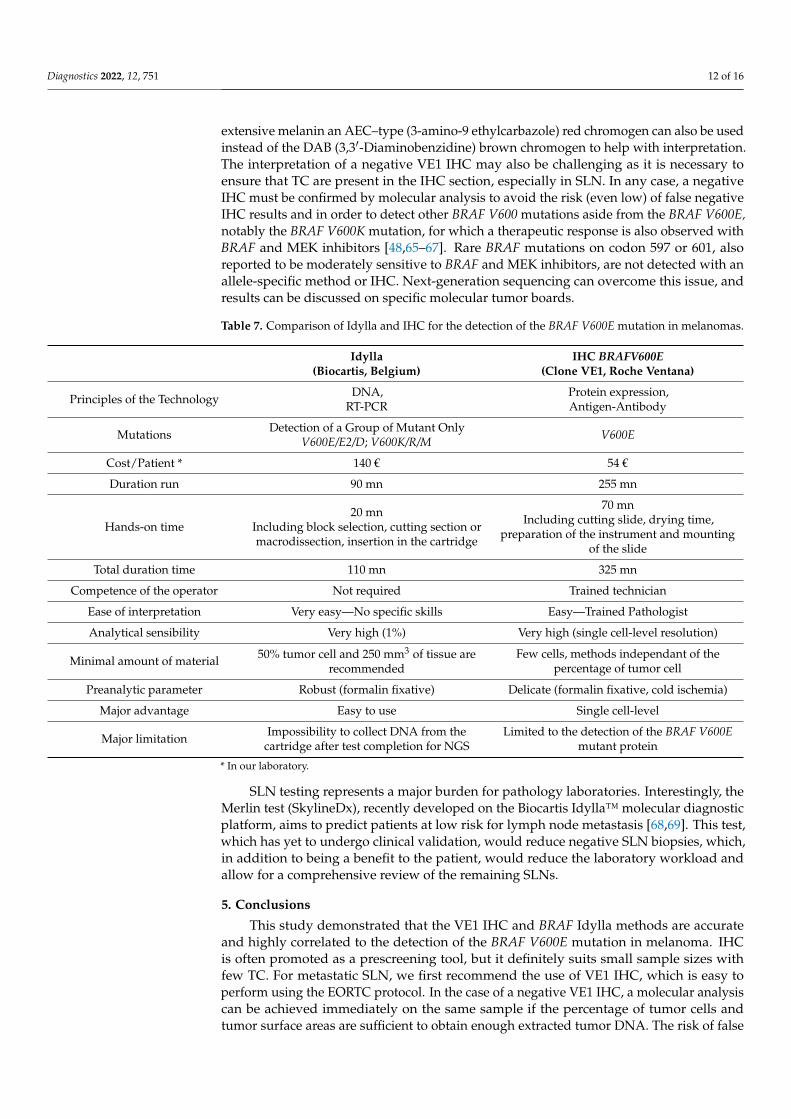
extensive melanin an AEC–type (3-amino-9 ethylcarbazole) red chromogen can also be usedinstead of the DAB (3,3′-Diaminobenzidine) brown chromogen to help with interpretation.The interpretation of a negative VE1 IHC may also be challenging as it is necessary toensure that TC are present in the IHC section, especially in SLN. In any case, a negativeIHC must be confirmed by molecular analysis to avoid the risk (even low) of false negativeIHC results and in order to detect other BRAF V600 mutations aside from the BRAF V600E,notably the BRAF V600K mutation, for which a therapeutic response is also observed withBRAF and MEK inhibitors [48,65–67]. Rare BRAF mutations on codon 597 or 601, alsoreported to be moderately sensitive to BRAF and MEK inhibitors, are not detected with anallele-specific method or IHC. Next-generation sequencing can overcome this issue, andresults can be discussed on specific molecular tumor boards.
Table 7. Comparison of Idylla and IHC for the detection of the BRAF V600E mutation in melanomas.
Idylla(Biocartis, Belgium)
IHC BRAFV600E(Clone VE1, Roche Ventana)
Principles of the Technology DNA,RT-PCR
Protein expression,Antigen-Antibody
Mutations Detection of a Group of Mutant OnlyV600E/E2/D; V600K/R/M V600E
Cost/Patient * 140 € 54 €
Duration run 90 mn 255 mn
Hands-on time20 mn
Including block selection, cutting section ormacrodissection, insertion in the cartridge
70 mnIncluding cutting slide, drying time,
preparation of the instrument and mountingof the slide
Total duration time 110 mn 325 mn
Competence of the operator Not required Trained technician
Ease of interpretation Very easy—No specific skills Easy—Trained Pathologist
Analytical sensibility Very high (1%) Very high (single cell-level resolution)
Minimal amount of material 50% tumor cell and 250 mm3 of tissue arerecommended
Few cells, methods independant of thepercentage of tumor cell
Preanalytic parameter Robust (formalin fixative) Delicate (formalin fixative, cold ischemia)
Major advantage Easy to use Single cell-level
Major limitation Impossibility to collect DNA from thecartridge after test completion for NGS
Limited to the detection of the BRAF V600Emutant protein
* In our laboratory.
SLN testing represents a major burden for pathology laboratories. Interestingly, theMerlin test (SkylineDx), recently developed on the Biocartis Idylla™ molecular diagnosticplatform, aims to predict patients at low risk for lymph node metastasis [68,69]. This test,which has yet to undergo clinical validation, would reduce negative SLN biopsies, which,in addition to being a benefit to the patient, would reduce the laboratory workload andallow for a comprehensive review of the remaining SLNs.
5. Conclusions
This study demonstrated that the VE1 IHC and BRAF Idylla methods are accurateand highly correlated to the detection of the BRAF V600E mutation in melanoma. IHCis often promoted as a prescreening tool, but it definitely suits small sample sizes withfew TC. For metastatic SLN, we first recommend the use of VE1 IHC, which is easy toperform using the EORTC protocol. In the case of a negative VE1 IHC, a molecular analysiscan be achieved immediately on the same sample if the percentage of tumor cells andtumor surface areas are sufficient to obtain enough extracted tumor DNA. The risk of false

Diagnostics 2022, 12, 751 13 of 16
negative results can be prevented with the selection of an adequate specimen (cellularity—melanin load—mastered fixative condition—decalcification) and the use of a molecularassay with high sensitivity. Alternatively, in the case of negative results due to a very lownumber of tumor cells, a liquid biopsy can now open up new promises to evaluate theBRAF status in these patients [26,30,70].
Author Contributions: Conceptualization, E.L.-M. and P.H.; Data curation, E.L.-M., A.P.-G., S.L.(Sandra Lassalle), V.H., M.I. and P.H.; Formal analysis, E.L.-M., A.P.-G., S.L. (Sandra Lassalle), V.H.,S.L. (Salomé Lalvée), C.B., M.I. and P.H.; Funding acquisition, P.H.; Investigation, E.L.-M. and P.H.;Methodology, E.L.-M., S.L. (Salomé Lalvée), V.T., K.Z., C.B., V.L., M.I. and P.H.; Project administration,M.I. and P.H.; Resources, E.L.-M., A.P.-G., S.L. (Sandra Lassalle), V.H., O.C., H.M., G.P., T.P., M.I.and P.H.; Supervision, P.H.; Validation, E.L.-M., A.P.-G., S.L. (Sandra Lassalle), V.H., M.I. and P.H.;Visualization, E.L.-M., A.P.-G., S.L. (Sandra Lassalle), V.H., M.I. and P.H.; Writing—original draft,E.L.-M., A.P.-G., S.L. (Sandra Lassalle), V.H., S.L. (Salomé Lalvée), V.T., K.Z., C.B., V.L., O.C., H.M.,G.P., T.P., M.I. and P.H.; Writing—review and editing, E.L.-M., A.P.-G., S.L. (Sandra Lassalle), V.H.,V.T., K.Z., C.B., V.L., O.C., H.M., G.P., T.P., M.I. and P.H. All authors have read and agreed to thepublished version of the manuscript.
Funding: This research received no external funding.
Institutional Review Board Statement: The study was conducted according to the guidelines of theDeclaration of Helsinki and approved by the Institutional Review Board (or Ethics Committee) ofCentre Hospitalier Universitaire de Nice (protocol code 2018-05 and May 2018).
Informed Consent Statement: Informed consent was obtained from all subjects involved in the study.
Data Availability Statement: Data are available by request to the corresponding author.
Conflicts of Interest: The authors declare no conflict of interest.
References1. Sung, H.; Ferlay, J.; Siegel, R.L.; Laversanne, M.; Soerjomataram, I.; Jemal, A.; Bray, F. Global Cancer Statistics 2020: GLOBOCAN
Estimates of Incidence and Mortality Worldwide for 36 Cancers in 185 Countries. CA Cancer J. Clin. 2021, 71, 209–249. [CrossRef][PubMed]
2. Siegel, R.L.; Miller, K.D.; Fuchs, H.E.; Jemal, A. Cancer Statistics, 2022. CA Cancer J. Clin. 2022, 72, 7–33. [CrossRef]3. Garbe, C.; Amaral, T.; Peris, K.; Hauschild, A.; Arenberger, P.; Bastholt, L.; Bataille, V.; Del Marmol, V.; Dréno, B.; Fargnoli, M.C.;
et al. European Consensus-Based Interdisciplinary Guideline for Melanoma. Part 1: Diagnostics—Update 2019. Eur. J. Cancer2020, 126, 141–158. [CrossRef] [PubMed]
4. Prieto, V.G. Sentinel Lymph Nodes in Cutaneous Melanoma. Clin. Lab. Med. 2017, 37, 417–430. [CrossRef] [PubMed]5. Garbe, C.; Amaral, T.; Peris, K.; Hauschild, A.; Arenberger, P.; Bastholt, L.; Bataille, V.; Del Marmol, V.; Dréno, B.; Fargnoli, M.C.;
et al. European Consensus-Based Interdisciplinary Guideline for Melanoma. Part 2: Treatment—Update 2019. Eur. J. Cancer 2020,126, 159–177. [CrossRef]
6. Luke, J.J.; Flaherty, K.T.; Ribas, A.; Long, G.V. Targeted Agents and Immunotherapies: Optimizing Outcomes in Melanoma. Nat.Rev. Clin. Oncol. 2017, 14, 463–482. [CrossRef]
7. Davies, H.; Bignell, G.R.; Cox, C.; Stephens, P.; Edkins, S.; Clegg, S.; Teague, J.; Woffendin, H.; Garnett, M.J.; Bottomley, W.; et al.Mutations of the BRAF Gene in Human Cancer. Nature 2002, 417, 949–954. [CrossRef]
8. Platz, A.; Egyhazi, S.; Ringborg, U.; Hansson, J. Human Cutaneous Melanoma; a Review of NRAS and BRAF Mutation Frequenciesin Relation to Histogenetic Subclass and Body Site. Mol. Oncol. 2008, 1, 395–405. [CrossRef]
9. Bergdorf, K.N.; Lee, L.A.; Weiss, V.L. BRAF Molecular Testing in Cytopathology: Implications for Diagnosis, Prognosis, andTargeted Therapeutics. Cancer Cytopathol. 2020, 128, 9–11. [CrossRef]
10. Cheng, L.; Lopez-Beltran, A.; Massari, F.; MacLennan, G.T.; Montironi, R. Molecular Testing for BRAF Mutations to InformMelanoma Treatment Decisions: A Move toward Precision Medicine. Mod. Pathol. 2018, 31, 24–38. [CrossRef]
11. Long, G.V.; Wilmott, J.S.; Capper, D.; Preusser, M.; Zhang, Y.E.; Thompson, J.F.; Kefford, R.F.; von Deimling, A.; Scolyer, R.A.Immunohistochemistry Is Highly Sensitive and Specific for the Detection of V600E BRAF Mutation in Melanoma. Am. J. Surg.Pathol. 2013, 37, 61–65. [CrossRef] [PubMed]
12. Ritterhouse, L.L.; Barletta, J.A. BRAF V600E Mutation-Specific Antibody: A Review. Semin. Diagn. Pathol. 2015, 32, 400–408.[CrossRef]
13. Long, E.; Ilie, M.; Lassalle, S.; Butori, C.; Poissonnet, G.; Washetine, K.; Mouroux, J.; Lespinet, V.; Lacour, J.P.; Taly, V.; et al. Whyand How Immunohistochemistry Should Now Be Used to Screen for the BRAFV600E Status in Metastatic Melanoma? TheExperience of a Single Institution (LCEP, Nice, France). J. Eur. Acad. Dermatol. Venereol. 2015, 29, 2436–2443. [CrossRef] [PubMed]

Diagnostics 2022, 12, 751 14 of 16
14. Long-Mira, E.; Washetine, K.; Hofman, P. Sense and Nonsense in the Process of Accreditation of a Pathology Laboratory. VirchowsArch. 2016, 468, 43–49. [CrossRef]
15. Lhermitte, B.; Egele, C.; Weingertner, N.; Ambrosetti, D.; Dadone, B.; Kubiniek, V.; Burel-Vandenbos, F.; Coyne, J.; Michiels, J.-F.;Chenard, M.-P.; et al. Adequately Defining Tumor Cell Proportion in Tissue Samples for Molecular Testing Improves InterobserverReproducibility of Its Assessment. Virchows Arch. 2017, 470, 21–27. [CrossRef] [PubMed]
16. Janku, F.; Claes, B.; Huang, H.J.; Falchook, G.S.; Devogelaere, B.; Kockx, M.; Bempt, I.V.; Reijans, M.; Naing, A.; Fu, S.; et al. BRAFMutation Testing with a Rapid, Fully Integrated Molecular Diagnostics System. Oncotarget 2015, 6, 26886–26894. [CrossRef][PubMed]
17. Schiefer, A.-I.; Parlow, L.; Gabler, L.; Mesteri, I.; Koperek, O.; von Deimling, A.; Streubel, B.; Preusser, M.; Lehmann, A.;Kellner, U.; et al. Multicenter Evaluation of a Novel Automated Rapid Detection System of BRAF Status in Formalin-Fixed,Paraffin-Embedded Tissues. J. Mol. Diagn. 2016, 18, 370–377. [CrossRef]
18. Ilie, M.I.; Lassalle, S.; Long-Mira, E.; Bonnetaud, C.; Bordone, O.; Lespinet, V.; Lamy, A.; Sabourin, J.-C.; Haudebourg, J.; Butori, C.;et al. Diagnostic Value of Immunohistochemistry for the Detection of the BRAF(V600E) Mutation in Papillary Thyroid Carcinoma:Comparative Analysis with Three DNA-Based Assays. Thyroid 2014, 24, 858–866. [CrossRef]
19. Van Akkooi, A.C.J.; De Wilt, J.H.W.; Verhoef, C.; Graveland, W.J.; Van Geel, A.N.; Kliffen, M.; Eggermont, A.M.M. High PositiveSentinel Node Identification Rate by EORTC Melanoma Group Protocol. Prognostic Indicators of Metastatic Patterns afterSentinel Node Biopsy in Melanoma. Eur. J. Cancer 2006, 42, 372–380. [CrossRef]
20. Dewar, D.J.; Newell, B.; Green, M.A.; Topping, A.P.; Powell, B.W.E.M.; Cook, M.G. The Microanatomic Location of MetastaticMelanoma in Sentinel Lymph Nodes Predicts Nonsentinel Lymph Node Involvement. J. Clin. Oncol. 2004, 22, 3345–3349.[CrossRef]
21. Lito, P.; Rosen, N.; Solit, D.B. Tumor Adaptation and Resistance to RAF Inhibitors. Nat. Med. 2013, 19, 1401–1409. [CrossRef]22. Yancovitz, M.; Litterman, A.; Yoon, J.; Ng, E.; Shapiro, R.L.; Berman, R.S.; Pavlick, A.C.; Darvishian, F.; Christos, P.; Mazumdar, M.;
et al. Intra- and Inter-Tumor Heterogeneity of BRAF(V600E)) Mutations in Primary and Metastatic Melanoma. PLoS ONE 2012,7, e29336. [CrossRef] [PubMed]
23. Satzger, I.; Marks, L.; Kerick, M.; Klages, S.; Berking, C.; Herbst, R.; Völker, B.; Schacht, V.; Timmermann, B.; Gutzmer, R. AlleleFrequencies of BRAFV600 Mutations in Primary Melanomas and Matched Metastases and Their Relevance for BRAF InhibitorTherapy in Metastatic Melanoma. Oncotarget 2015, 6, 37895–37905. [CrossRef] [PubMed]
24. Hodis, E.; Watson, I.R.; Kryukov, G.V.; Arold, S.T.; Imielinski, M.; Theurillat, J.-P.; Nickerson, E.; Auclair, D.; Li, L.; Place, C.; et al.A Landscape of Driver Mutations in Melanoma. Cell 2012, 150, 251–263. [CrossRef] [PubMed]
25. Melchior, L.; Grauslund, M.; Bellosillo, B.; Montagut, C.; Torres, E.; Moragón, E.; Micalessi, I.; Frans, J.; Noten, V.; Bourgain, C.;et al. Multi-Center Evaluation of the Novel Fully-Automated PCR-Based IdyllaTM BRAF Mutation Test on Formalin-FixedParaffin-Embedded Tissue of Malignant Melanoma. Exp. Mol. Pathol. 2015, 99, 485–491. [CrossRef]
26. Janku, F.; Huang, H.J.; Claes, B.; Falchook, G.S.; Fu, S.; Hong, D.; Ramzanali, N.M.; Nitti, G.; Cabrilo, G.; Tsimberidou, A.M.;et al. BRAF Mutation Testing in Cell-Free DNA from the Plasma of Patients with Advanced Cancers Using a Rapid, AutomatedMolecular Diagnostics System. Mol. Cancer Ther. 2016, 15, 1397–1404. [CrossRef] [PubMed]
27. Harlé, A.; Salleron, J.; Franczak, C.; Dubois, C.; Filhine-Tressarieu, P.; Leroux, A.; Merlin, J.-L. Detection of BRAF MutationsUsing a Fully Automated Platform and Comparison with High Resolution Melting, Real-Time Allele Specific Amplification,Immunohistochemistry and Next Generation Sequencing Assays, for Patients with Metastatic Melanoma. PLoS ONE 2016,11, e0153576. [CrossRef]
28. Barel, F.; Guibourg, B.; Lambros, L.; Flahec, G.; Marcorelles, P.; Uguen, A. Evaluation of a Rapid, Fully Automated Platform forDetection of BRAF and NRAS Mutations in Melanoma. Acta Derm. Venerol. 2018, 98, 44–49. [CrossRef]
29. Bisschop, C.; Ter Elst, A.; Bosman, L.J.; Platteel, I.; Jalving, M.; Van den Berg, A.; Diepstra, A.; Van Hemel, B.; Diercks, G.F.H.;Hospers, G.A.P.; et al. Rapid BRAF Mutation Tests in Patients with Advanced Melanoma: Comparison of Immunohistochemistry,Droplet Digital PCR, and the Idylla Mutation Platform. Melanoma Res. 2018, 28, 96–104. [CrossRef]
30. Long-Mira, E.; Ilie, M.; Chamorey, E.; Leduff-Blanc, F.; Montaudié, H.; Tanga, V.; Allégra, M.; Lespinet-Fabre, V.; Bordone, O.;Bonnetaud, C.; et al. Monitoring BRAF and NRAS Mutations with Cell-Free Circulating Tumor DNA from Metastatic MelanomaPatients. Oncotarget 2018, 9, 36238–36249. [CrossRef]
31. Seremet, T.; Planken, S.; Schreuer, M.; Jansen, Y.; Delaunoy, M.; El Housni, H.; Lienard, D.; Del Marmol, V.; Heimann, P.; Neyns, B.Illustrative Cases for Monitoring by Quantitative Analysis of BRAF/NRAS CtDNA Mutations in Liquid Biopsies of MetastaticMelanoma Patients Who Gained Clinical Benefits from Anti-PD1 Antibody Therapy. Melanoma Res. 2018, 28, 65–70. [CrossRef][PubMed]
32. Serre, D.; Salleron, J.; Husson, M.; Leroux, A.; Gilson, P.; Merlin, J.-L.; Geoffrois, L.; Harlé, A. Accelerated BRAF Mutation AnalysisUsing a Fully Automated PCR Platform Improves the Management of Patients with Metastatic Melanoma. Oncotarget 2018,9, 32232–32237. [CrossRef] [PubMed]
33. Vallée, A.; Denis-Musquer, M.; Herbreteau, G.; Théoleyre, S.; Bossard, C.; Denis, M.G. Prospective Evaluation of Two ScreeningMethods for Molecular Testing of Metastatic Melanoma: Diagnostic Performance of BRAF V600E Immunohistochemistry and ofa NRAS-BRAF Fully Automated Real-Time PCR-Based Assay. PLoS ONE 2019, 14, e0221123. [CrossRef] [PubMed]
34. Huang, H.; Springborn, S.; Haug, K.; Bartow, K.; Samra, H.; Menon, S.; Mackinnon, A.C. Evaluation, Validation, and Implementa-tion of the Idylla System as Rapid Molecular Testing for Precision Medicine. J. Mol. Diagn. 2019, 21, 862–872. [CrossRef]

Diagnostics 2022, 12, 751 15 of 16
35. Bourhis, A.; Le Flahec, G.; Uguen, A. Decalcification Can Cause the Failure of BRAF Molecular Analyses and Anti-BRAFV600EVE1 Immunohistochemistry. Pathol. Int. 2019, 69, 219–223. [CrossRef]
36. Van Haele, M.; Vander Borght, S.; Ceulemans, A.; Wieërs, M.; Metsu, S.; Sagaert, X.; Weynand, B. Rapid Clinical Mutational Testingof KRAS, BRAF and EGFR: A Prospective Comparative Analysis of the Idylla Technique with High-Throughput next-GenerationSequencing. J. Clin. Pathol. 2020, 73, 35–41. [CrossRef]
37. Petty, D.R.; Hassan, O.A.; Barker, C.S.; O’Neill, S.S. Rapid BRAF Mutation Testing in Pigmented Melanomas. Am. J. Dermatopathol.2020, 42, 343–348. [CrossRef]
38. Colombino, M.; Rozzo, C.; Paliogiannis, P.; Casula, M.; Manca, A.; Doneddu, V.; Fedeli, M.A.; Sini, M.C.; Palomba, G.; Pisano, M.;et al. Comparison of BRAF Mutation Screening Strategies in a Large Real-Life Series of Advanced Melanoma Patients. J. Clin.Med. 2020, 9, 2430. [CrossRef]
39. Durslewicz, J.; Klimaszewska-Wisniewska, A.; Antosik, P.; Kasperska, A.; Grzanka, D.; Szylberg, T.; Szylberg, Ł. Detection ofBRAF V600E Mutation in Ganglioglioma and Pilocytic Astrocytoma by Immunohistochemistry and Real-Time PCR-Based IdyllaTest. Dis. Markers 2020, 2020, 8880548. [CrossRef]
40. Sadlecki, P.; Walentowicz, P.; Bodnar, M.; Marszalek, A.; Grabiec, M.; Walentowicz-Sadlecka, M. Determination of BRAF V600E(VE1) Protein Expression and BRAF Gene Mutation Status in Codon 600 in Borderline and Low-Grade Ovarian Cancers. TumourBiol. 2017, 39. [CrossRef]
41. Colling, R.; Wang, L.M.; Soilleux, E. Validating a Fully Automated Real-Time PCR-Based System for Use in the MolecularDiagnostic Analysis of Colorectal Carcinoma: A Comparison with NGS and IHC. J. Clin. Pathol. 2017, 70, 610–614. [CrossRef]
42. Bodnar, M.; Burduk, P.; Antosik, P.; Jarmuz-Szymczak, M.; Wierzbicka, M.; Marszalek, A. Assessment of BRAF V600E (VE1)Protein Expression and BRAF Gene Mutation Status in Codon 600 in Benign and Malignant Salivary Gland Neoplasms. J. Oral.Pathol. Med. 2017, 46, 340–345. [CrossRef] [PubMed]
43. Cardus, B.; Colling, R.; Hamblin, A.; Soilleux, E. Comparison of Methodologies for the Detection of BRAF Mutations in BoneMarrow Trephine Specimens. J. Clin. Pathol. 2019, 72, 406–411. [CrossRef] [PubMed]
44. Viray, H.; Li, K.; Long, T.A.; Vasalos, P.; Bridge, J.A.; Jennings, L.J.; Halling, K.C.; Hameed, M.; Rimm, D.L. A Prospective,Multi-Institutional Diagnostic Trial to Determine Pathologist Accuracy in Estimation of Percentage of Malignant Cells. Arch.Pathol. Lab. Med. 2013, 137, 1545–1549. [CrossRef] [PubMed]
45. Smits, A.J.J.; Kummer, J.A.; Dde Bruin, P.C.; Bol, M.; Van den Tweel, J.G.; Seldenrijk, K.A.; Willems, S.M.; Offerhaus, G.J.A.; DeWeger, R.A.; Van Diest, P.J.; et al. The Estimation of Tumor Cell Percentage for Molecular Testing by Pathologists Is Not Accurate.Mod. Pathol. 2014, 27, 168–174. [CrossRef]
46. Chen, G.; Dudley, J.; Tseng, L.-H.; Smith, K.; Gurda, G.T.; Gocke, C.D.; Eshleman, J.R.; Lin, M.-T. Lymph Node Metastases ofMelanoma: Challenges for BRAF Mutation Detection. Hum. Pathol. 2015, 46, 113–119. [CrossRef]
47. Dudley, J.C.; Gurda, G.T.; Tseng, L.-H.; Anderson, D.A.; Chen, G.; Taube, J.M.; Gocke, C.D.; Eshleman, J.R.; Lin, M.-T. TumorCellularity as a Quality Assurance Measure for Accurate Clinical Detection of BRAF Mutations in Melanoma. Mol. Diagn. Ther.2014, 18, 409–418. [CrossRef]
48. Long, G.V.; Hauschild, A.; Santinami, M.; Atkinson, V.; Mandalà, M.; Chiarion-Sileni, V.; Larkin, J.; Nyakas, M.; Dutriaux, C.;Haydon, A.; et al. Adjuvant Dabrafenib plus Trametinib in Stage III BRAF-Mutated Melanoma. N. Engl. J. Med. 2017,377, 1813–1823. [CrossRef]
49. Maio, M.; Lewis, K.; Demidov, L.; Mandalà, M.; Bondarenko, I.; Ascierto, P.A.; Herbert, C.; Mackiewicz, A.; Rutkowski, P.;Guminski, A.; et al. Adjuvant Vemurafenib in Resected, BRAFV600 Mutation-Positive Melanoma (BRIM8): A Randomised,Double-Blind, Placebo-Controlled, Multicentre, Phase 3 Trial. Lancet Oncol. 2018, 19, 510–520. [CrossRef]
50. Varada, S.; Mahalingam, M. Mutation Stability in Primary and Metastatic Melanoma: What We Know and What We Don’t. Histol.Histopathol. 2015, 30, 763–770. [CrossRef]
51. Ito, T.; Tanaka, Y.; Murata, M.; Kaku-Ito, Y.; Furue, K.; Furue, M. BRAF Heterogeneity in Melanoma. Curr. Treat. Options Oncol.2021, 22, 20. [CrossRef] [PubMed]
52. Chang, G.A.; Wiggins, J.M.; Corless, B.C.; Syeda, M.M.; Tadepalli, J.S.; Blake, S.; Fleming, N.; Darvishian, F.; Pavlick, A.;Berman, R.; et al. TERT, BRAF, and NRAS Mutational Heterogeneity between Paired Primary and Metastatic Melanoma Tumors.J. Investig. Dermatol. 2020, 140, 1609–1618.e7. [CrossRef] [PubMed]
53. Heinzerling, L.; Baiter, M.; Kühnapfel, S.; Schuler, G.; Keikavoussi, P.; Agaimy, A.; Kiesewetter, F.; Hartmann, A.; Schneider-Stock,R. Mutation Landscape in Melanoma Patients Clinical Implications of Heterogeneity of BRAF Mutations. Br. J. Cancer 2013,109, 2833–2841. [CrossRef] [PubMed]
54. Manfredi, L.; Meyer, N.; Tournier, E.; Grand, D.; Uro-Coste, E.; Rochaix, P.; Brousset, P.; Lamant, L. Highly Concordant ResultsBetween Immunohistochemistry and Molecular Testing of Mutated V600E BRAF in Primary and Metastatic Melanoma. ActaDerm. Venereol. 2016, 96, 630–634. [CrossRef] [PubMed]
55. Guadarrama-Orozco, J.A.; Ortega-Gómez, A.; Ruiz-García, E.B.; Astudillo-de la Vega, H.; Meneses-García, A.; Lopez-Camarillo, C.Braf V600E Mutation in Melanoma: Translational Current Scenario. Clin. Transl. Oncol. 2016, 18, 863–871. [CrossRef]
56. Cormican, D.; Kennedy, C.; Murphy, S.; Werner, R.; Power, D.G.; Heffron, C.C.B.B. High Concordance of BRAF Mutational Statusin Matched Primary and Metastatic Melanoma. J. Cutan. Pathol. 2019, 46, 117–122. [CrossRef]
57. Nielsen, L.B.; Dabrosin, N.; Sloth, K.; Bønnelykke-Behrndtz, M.L.; Steiniche, T.; Lade-Keller, J. Concordance in BRAF V600EStatus over Time in Malignant Melanoma and Corresponding Metastases. Histopathology 2018, 72, 814–825. [CrossRef]

Diagnostics 2022, 12, 751 16 of 16
58. Parakh, S.; Murphy, C.; Lau, D.; Cebon, J.S.; Andrews, M.C. Response to MAPK Pathway Inhibitors in BRAF V600M-MutatedMetastatic Melanoma. J. Clin. Pharm. Ther. 2015, 40, 121–123. [CrossRef]
59. Popescu, A.; Haidar, A.; Anghel, R.M. Treating Malignant Melanoma When a Rare BRAF V600M Mutation Is Present: CaseReport and Literature Review. Rom. J. Intern. Med. 2018, 56, 122–126. [CrossRef]
60. Eckhart, L.; Bach, J.; Ban, J.; Tschachler, E. Melanin Binds Reversibly to Thermostable DNA Polymerase and Inhibits Its Activity.Biochem. Biophys. Res. Commun. 2000, 271, 726–730. [CrossRef]
61. Cook, M.G.; Green, M.A.; Anderson, B.; Eggermont, A.M.M.; Ruiter, D.J.; Spatz, A.; Kissin, M.W.; Powell, B.W.E.M.; EORTCMelanoma Group. The Development of Optimal Pathological Assessment of Sentinel Lymph Nodes for Melanoma. J. Pathol.2003, 200, 314–319. [CrossRef] [PubMed]
62. Cook, M.G.; Massi, D.; Szumera-Cieckiewicz, A.; Van den Oord, J.; Blokx, W.; Van Kempen, L.C.; Balamurugan, T.; Bosisio, F.;Koljenovic, S.; Portelli, F.; et al. An Updated European Organisation for Research and Treatment of Cancer (EORTC) Protocol forPathological Evaluation of Sentinel Lymph Nodes for Melanoma. Eur. J. Cancer 2019, 114, 1–7. [CrossRef] [PubMed]
63. Engel, K.B.; Moore, H.M. Effects of Preanalytical Variables on the Detection of Proteins by Immunohistochemistry in Formalin-Fixed, Paraffin-Embedded Tissue. Arch. Pathol. Lab. Med. 2011, 135, 537–543. [CrossRef] [PubMed]
64. Menzies, A.M.; Lum, T.; Wilmott, J.S.; Hyman, J.; Kefford, R.F.; Thompson, J.F.; O’Toole, S.; Long, G.V.; Scolyer, R.A. IntrapatientHomogeneity of BRAFV600E Expression in Melanoma. Am. J. Surg. Pathol. 2014, 38, 377–382. [CrossRef] [PubMed]
65. Dummer, R.; Hauschild, A.; Santinami, M.; Atkinson, V.; Mandalà, M.; Kirkwood, J.M.; Chiarion Sileni, V.; Larkin, J.; Nyakas, M.;Dutriaux, C.; et al. Five-Year Analysis of Adjuvant Dabrafenib plus Trametinib in Stage III Melanoma. N. Engl. J. Med. 2020,383, 1139–1148. [CrossRef]
66. Dummer, R.; Lebbé, C.; Atkinson, V.; Mandalà, M.; Nathan, P.D.; Arance, A.; Richtig, E.; Yamazaki, N.; Robert, C.; Schadendorf, D.;et al. Combined PD-1, BRAF and MEK Inhibition in Advanced BRAF-Mutant Melanoma: Safety Run-in and Biomarker Cohortsof COMBI-i. Nat. Med. 2020, 26, 1557–1563. [CrossRef]
67. Hauschild, A.; Dummer, R.; Schadendorf, D.; Santinami, M.; Atkinson, V.; Mandalà, M.; Chiarion-Sileni, V.; Larkin, J.; Nyakas, M.;Dutriaux, C.; et al. Longer Follow-Up Confirms Relapse-Free Survival Benefit with Adjuvant Dabrafenib Plus Trametinib inPatients with Resected BRAF V600-Mutant Stage III Melanoma. J. Clin. Oncol. 2018, 36, 3441–3449. [CrossRef]
68. Bellomo, D.; Arias-Mejias, S.M.; Ramana, C.; Heim, J.B.; Quattrocchi, E.; Sominidi-Damodaran, S.; Bridges, A.G.; Lehman, J.S.;Hieken, T.J.; Jakub, J.W.; et al. Model Combining Tumor Molecular and Clinicopathologic Risk Factors Predicts Sentinel LymphNode Metastasis in Primary Cutaneous Melanoma. JCO Precis. Oncol. 2020, 4, 319–334. [CrossRef]
69. Yousaf, A.; Tjien-Fooh, F.J.; Rentroia-Pacheco, B.; Quattrocchi, E.; Kobic, A.; Tempel, D.; Kolodney, M.; Meves, A. Validation ofCP-GEP (Merlin Assay) for Predicting Sentinel Lymph Node Metastasis in Primary Cutaneous Melanoma Patients: A U.S. CohortStudy. Int. J. Dermatol. 2021, 60, 851–856. [CrossRef]
70. Salvianti, F.; Massi, D.; De Giorgi, V.; Gori, A.; Pazzagli, M.; Pinzani, P. Evaluation of the Liquid Biopsy for the Detection ofBRAFV600E Mutation in Metastatic Melanoma Patients. Cancer Biomark. 2019, 26, 271–279. [CrossRef]


![RAPHIDOPHYCEAE [CHADEFAUD EX SILVA] SYSTEMATICS AND RAPID IDENTIFICATION: SEQUENCE ANALYSES AND REAL-TIME PCR ASSAYS](https://static.fdokumen.com/doc/165x107/6344f5fe6cfb3d4064097559/raphidophyceae-chadefaud-ex-silva-systematics-and-rapid-identification-sequence.jpg)