KIAA1549: BRAF Gene Fusion and FGFR1 Hotspot Mutations Are Prognostic Factors in Pilocytic...
12
ORIGINAL ARTICLE KIAA1549:BRAF Gene Fusion and FGFR1 Hotspot Mutations Are Prognostic Factors in Pilocytic Astrocytomas Aline Paixa ˜o Becker, MD, MSc, Cristovam Scapulatempo-Neto, MD, PhD, Adriana C. Carloni, MSc, Alessandra Paulino, BSc, Jamie Sheren, PhD, Dara L. Aisner, MD, PhD, Evelyn Musselwhite, MSc, Carlos Clara, MD, PhD, He ´lio R. Machado, MD, PhD, Ricardo S. Oliveira, MD, PhD, Luciano Neder, MD, PhD, Marileila Varella-Garcia, PhD, and Rui M. Reis, PhD Abstract Up to 20% of patients with pilocytic astrocytoma (PA) experience a poor outcome. BRAF alterations and Fibroblast growth factor re- ceptor 1 (FGFR1) point mutations are key molecular alterations in Pas, but their clinical implications are not established. We aimed to determine the frequency and prognostic role of these alterations in a cohort of 69 patients with PAs. We assessed KIAA1549:BRAF fusion by fluorescence in situ hybridization and BRAF (exon 15) mutations by capillary sequencing. In addition, FGFR1 expression was ana- lyzed using immunohistochemistry, and this was compared with gene amplification and hotspot mutations (exons 12 and 14) assessed by fluorescence in situ hybridization and capillary sequencing. KIAA1549:BRAF fusion was identified in almost 60% of cases. Two tumors harbored mutated BRAF. Despite high FGFR1 expression overall, no cases had FGFR1 amplifications. Three cases harbored a FGFR1 p.K656E point mutation. No correlation was observed be- tween BRAF and FGFR1 alterations. The cases were predominantly pediatric (87%), and no statistical differences were observed in mo- lecular alterationsYrelated patient ages. In summary, we confirmed the high frequency of KIAA1549:BRAF fusion in PAs and its asso- ciation with a better outcome. Oncogenic mutations of FGFR1, al- though rare, occurred in a subset of patients with worse outcome. These molecular alterations may constitute alternative targets for novel clini- cal approaches, when radical surgical resection is unachievable. Key Words: BRAF, Brain tumor, FGFR1, Glioma, Molecular diag- nosis, Pilocytic astrocytoma, Prognosis. INTRODUCTION Pilocytic astrocytomas (PAs) are the major solid neo- plasms in children and teenagers (1, 2). According to data from the Central Brain Tumor Registry of the United States, it is the main neoplasm in the 5- to 14-year-old range in the United States (3). Similarly in Brazil, PAs are the second most common neoplasm in pediatric patients after leukemias (4), accounting for almost 20% of primary brain tumors in chil- dren (5). Pilocytic astrocytomas are less frequent in adults in whom they are associated with more aggressive clinical courses (1, 6). According to the World Health Organization (WHO), PAs are grade I tumors because of their well-limited and usually indolent nature (1). The 5-year survival rate is 990% in children (1, 7), and 52% in adults (8). Despite the overall good prognosis of PAs, up to 20% of patients will have a poor outcome, with recurrence, growth of incompletely resected lesions, or dissemination through the cerebrospinal fluid, and ultimately death due to disease (1, 7). Pilocytic astrocytomas can occur throughout the neu- raxis, but the most common location of sporadic tumors is the cerebellum (1). Extracerebellar tumors, particularly those lo- cated in the cerebral hemispheres and in the optic pathways, have a known association with neurofibromatosis 1 (NF1), a familial tumor predisposition syndrome with autosomal domi- nant inheritance (1, 9). Approximately 10% of all PAs are re- lated to NF1 (NF1-PAs), and conversely, PAs are the most frequent brain tumor related to NF1 (49% of cases) (10, 11). When these PAs arise in locations where gross total resection is difficult to achieve, they usually follow a more benign course than sporadic PAs (11). Molecular studies based on the relationship between PAs and NF1 allowed the discovery of germline and somatic mutations with silencing of the tumor suppressor gene NF1 in NF1-PAs. These were recently defined as point mutations, splice mutations or nonsense mutations (germline mutations) and loss of heterozygosity and epigenetic changes, such as 1 J Neuropathol Exp Neurol Volume 74, Number 7, July 2015 J Neuropathol Exp Neurol Copyright Ó 2015 by the American Association of Neuropathologists, Inc. Vol. 74, No. 7 July 2015 pp. 00Y00 From the Molecular Oncology Research Center (APB, ACC, AP, RMR), and Department of Pathology (CSN), Barretos Cancer Hospital, Barretos, Sa ˜o Paulo, Brazil; Cancer Center (JS, DLA), and School of Medicine (EM, MVG), University of Colorado, Aurora, Colorado; Department of Neurosurgery(CC), Barretos Cancer Hospital, Barretos, Sa ˜o Paulo, Brazil; Department of Surgery (RSO), and Department of Pathology and Forensic Medicine (APB, LN), Faculty of Medicine of Ribeira ˜o Preto, University of Sa ˜o Paulo (FMRP-USP), Sa ˜o Paulo, Brazil; Life and Health Sciences Research Institute (ICVS), Health Sciences School, University of Minho (RMR), Braga, Portugal; and ICVS/3B’s Y PT Government Associate Laboratory(RMR), Braga/Guimara ˜es, Portugal. Send correspondence and reprint requests to: Rui Manuel Reis, PhD, Molecular Oncology Research Center, Barretos Cancer Hospital, Rua Antenor Duarte Vilela, 1331. Barretos-SP, Brazil; E-mail: [email protected] This study was partially supported by CNPq/Universal (475358/2011-2), and FAPESP (2012/19590-0) grants to RMR and to the NIH- P30CA046934 (CCSG Molecular Pathology/Cytogenetics) to MVG and DLA. This is an open access article distributed under the terms of the Creative Commons Attribution-NonCommercial-NoDerivatives 3.0 License, where it is permissi- ble to download and share the work provided it is properly cited. The work cannot be changed in any way or used commercially. Copyright © 2015 by the American Association of Neuropathologists, Inc. Unauthorized reproduction of this article is prohibited.
-
Upload
independent -
Category
Documents
-
view
1 -
download
0
Transcript of KIAA1549: BRAF Gene Fusion and FGFR1 Hotspot Mutations Are Prognostic Factors in Pilocytic...

ORIGINAL ARTICLE
KIAA1549:BRAF Gene Fusion and FGFR1 Hotspot Mutations ArePrognostic Factors in Pilocytic Astrocytomas
Aline Paixao Becker, MD, MSc, Cristovam Scapulatempo-Neto, MD, PhD, Adriana C. Carloni, MSc,Alessandra Paulino, BSc, Jamie Sheren, PhD, Dara L. Aisner, MD, PhD, Evelyn Musselwhite, MSc,
Carlos Clara, MD, PhD, Helio R. Machado, MD, PhD, Ricardo S. Oliveira, MD, PhD,Luciano Neder, MD, PhD, Marileila Varella-Garcia, PhD, and Rui M. Reis, PhD
AbstractUp to 20% of patients with pilocytic astrocytoma (PA) experience
a poor outcome. BRAF alterations and Fibroblast growth factor re-ceptor 1 (FGFR1) point mutations are key molecular alterations inPas, but their clinical implications are not established. We aimed todetermine the frequency and prognostic role of these alterations in acohort of 69 patients with PAs. We assessed KIAA1549:BRAF fusionby fluorescence in situ hybridization and BRAF (exon 15) mutationsby capillary sequencing. In addition, FGFR1 expression was ana-lyzed using immunohistochemistry, and this was compared withgene amplification and hotspot mutations (exons 12 and 14) assessedby fluorescence in situ hybridization and capillary sequencing.KIAA1549:BRAF fusion was identified in almost 60% of cases. Twotumors harbored mutated BRAF. Despite high FGFR1 expressionoverall, no cases had FGFR1 amplifications. Three cases harbored aFGFR1 p.K656E point mutation. No correlation was observed be-tween BRAF and FGFR1 alterations. The cases were predominantlypediatric (87%), and no statistical differences were observed in mo-lecular alterationsYrelated patient ages. In summary, we confirmedthe high frequency of KIAA1549:BRAF fusion in PAs and its asso-ciation with a better outcome. Oncogenic mutations of FGFR1, al-though rare, occurred in a subset of patients with worse outcome. These
molecular alterations may constitute alternative targets for novel clini-cal approaches, when radical surgical resection is unachievable.
Key Words: BRAF, Brain tumor, FGFR1, Glioma, Molecular diag-nosis, Pilocytic astrocytoma, Prognosis.
INTRODUCTIONPilocytic astrocytomas (PAs) are the major solid neo-
plasms in children and teenagers (1, 2). According to datafrom the Central Brain Tumor Registry of the United States, itis the main neoplasm in the 5- to 14-year-old range in theUnited States (3). Similarly in Brazil, PAs are the second mostcommon neoplasm in pediatric patients after leukemias (4),accounting for almost 20% of primary brain tumors in chil-dren (5). Pilocytic astrocytomas are less frequent in adults inwhom they are associated with more aggressive clinicalcourses (1, 6). According to the World Health Organization(WHO), PAs are grade I tumors because of their well-limitedand usually indolent nature (1). The 5-year survival rate is990% in children (1, 7), and 52% in adults (8). Despite theoverall good prognosis of PAs, up to 20% of patients willhave a poor outcome, with recurrence, growth of incompletelyresected lesions, or dissemination through the cerebrospinalfluid, and ultimately death due to disease (1, 7).
Pilocytic astrocytomas can occur throughout the neu-raxis, but the most common location of sporadic tumors is thecerebellum (1). Extracerebellar tumors, particularly those lo-cated in the cerebral hemispheres and in the optic pathways,have a known association with neurofibromatosis 1 (NF1), afamilial tumor predisposition syndrome with autosomal domi-nant inheritance (1, 9). Approximately 10% of all PAs are re-lated to NF1 (NF1-PAs), and conversely, PAs are the mostfrequent brain tumor related to NF1 (49% of cases) (10, 11).When these PAs arise in locations where gross total resection isdifficult to achieve, they usually follow a more benign coursethan sporadic PAs (11).
Molecular studies based on the relationship betweenPAs and NF1 allowed the discovery of germline and somaticmutations with silencing of the tumor suppressor gene NF1 inNF1-PAs. These were recently defined as point mutations,splice mutations or nonsense mutations (germline mutations)and loss of heterozygosity and epigenetic changes, such as
1J Neuropathol Exp Neurol � Volume 74, Number 7, July 2015
J Neuropathol Exp NeurolCopyright � 2015 by the American Association of Neuropathologists, Inc.
Vol. 74, No. 7July 2015pp. 00Y00
From the Molecular Oncology Research Center (APB, ACC, AP, RMR), andDepartment of Pathology (CSN), Barretos Cancer Hospital, Barretos, SaoPaulo, Brazil; Cancer Center (JS, DLA), and School of Medicine (EM,MVG), University of Colorado, Aurora, Colorado; Department ofNeurosurgery(CC), Barretos Cancer Hospital, Barretos, Sao Paulo, Brazil;Department of Surgery (RSO), and Department of Pathology and ForensicMedicine (APB, LN), Faculty of Medicine of Ribeirao Preto, Universityof Sao Paulo (FMRP-USP), Sao Paulo, Brazil; Life and Health SciencesResearch Institute (ICVS), Health Sciences School, University of Minho(RMR), Braga, Portugal; and ICVS/3B’s Y PT Government AssociateLaboratory(RMR), Braga/Guimaraes, Portugal.
Send correspondence and reprint requests to: Rui Manuel Reis, PhD, MolecularOncology Research Center, Barretos Cancer Hospital, Rua Antenor DuarteVilela, 1331. Barretos-SP, Brazil; E-mail: [email protected]
This study was partially supported by CNPq/Universal (475358/2011-2), andFAPESP (2012/19590-0) grants to RMR and to the NIH- P30CA046934(CCSG Molecular Pathology/Cytogenetics) to MVG and DLA.
This is an open access article distributed under the terms of the Creative CommonsAttribution-NonCommercial-NoDerivatives 3.0 License, where it is permissi-ble to download and share the work provided it is properly cited. The workcannot be changed in any way or used commercially.
Copyright © 2015 by the American Association of Neuropathologists, Inc. Unauthorized reproduction of this article is prohibited.
methylation of the gene (somatic mutations) (9). These mu-tations result in loss of expression of neurofibromin, which isa negative signaling regulator of RAS proteins; this results inan increase of activated RAS levels and further activation ofthe mitogen-activated protein kinase (MAPK) pathway (12).The constitutive activation of the MAPK pathway increasessurvival and proliferation of cancer cells in various neo-plasms (13, 14).
MAPK is a key signaling pathway in the developmentof PAs; it is altered in up to 90% of cases (7, 15, 16). Themajor alterations leading to constitutive activation of MAPKin PAs are gene fusions and point mutations involving theoncogene BRAF (7, 17Y23). Gene fusions between KIAA1549and BRAF (KIAA1549:BRAF fusion) leading to the over-expression of the fusion protein affects up to 80% of PAs.There are decreasing rates with age, varying from 79% inchildren younger than 10 years to 7% in patients older than40 years (16, 20); this is associated with a better prognosis inlow-grade gliomas, including PAs (21). Less frequent fusions,such as SRGAP3-RAF1 (24) and FAM131B-BRAF (25, 26),have also been described. Another mechanism of sustainedBRAF activation in PAs is the point mutation V600E, whichresults in an amino acid substitution at codon 600 in BRAF,from a valine (V) to a glutamic acid (E) in the majority ofcases, leading to the activation of the kinase domain of thisoncogene (7, 18, 22, 27). Nevertheless, this finding is in-frequent in PAs (approximately 6%) and may be detectedmore frequently in other brain tumor types, such as glio-blastomas (22, 28), gangliogliomas, and particularly, pleo-morphic xanthoastrocytomas (960%) (16, 22).
Recent studies have identified upstream alterations inthe MAPK pathway, mainly in the tyrosine-kinase receptorFibroblast growth factor receptor 1 (FGFR1), leading toconstitutive activation of the growth cascade in PAs (15,29). In contrast to the FGFR1 amplification frequently ob-served in breast, ovary, and lung cancer (30, 31), gene fu-sions and duplications are described at low frequencies inbrain tumors such as glioblastomas (32) and pediatric dif-fuse astrocytomas (16), respectively. In PAs, the newlydescribed alterations of FGFR1 are point mutations in thehotspot tyrosine kinase region, affecting mainly the codons546 (p.N546K Yasparagine-to-lysine substitution) and 656(p.K656E Ylysine-to-glutamate substitution) of the gene inextracerebellar PAs (15).
Despite the great improvement in the knowledge on themolecular oncogenesis of PAs in the last years, the mainestablished prognostic factors for PAs remain in the clinicalfeatures, such as patient’s age, feasibility of radical resectionof lesion (33Y35), exposure to radiation therapy (1), and thesporadic or hereditary nature of the tumor (12). The prog-nostic implications of BRAF and FGFR1 alterations have notbeen fully explored, and advances in this field might identifypotential targets for clinical treatment of PA, particularly forthe tumors located in eloquent areas where radical resection israrely achieved.
In this study, we aimed to determine the frequency ofthe molecular alterations in BRAF and FGFR1 and to evaluatethe prognostic role of these oncogenes in a series of Brazilianpatients with PAs.
MATERIALS AND METHODS
PatientsSixty-nine patients from the Barretos Cancer Hospital
(HCB) and the Hospital of Clinics of Faculty of Medicine ofRibeirao Preto (HCRP), from 1993 to 2013, were included in thisstudy. The patients were clustered according to sex, age group(e19 years old vs Q20 years old), clinical diagnosis of NF1(confirmed by standardized clinical criteria), and lesion location(cerebellar vs extracerebellar). The outcome of patients wasclassified as ‘‘favorable’’ (i.e. patients without any events and/or with Karnofsky index Q80 at follow-up) and ‘‘unfavorable’’(i.e. occurrence of some event and/or Karnofsky index e70 atfollow-up). We defined ‘‘event’’ as death, growth of a partiallyresected lesion, or the recurrence of a completely resected lesionconfirmed by immediate postsurgical computed tomography(36), detected either clinically and/or through neuroradiologicexaminations. The study was approved by both local EthicsCommittee (protocols HCB 87362 and HCRP 212.313).
The series included 38 male and 31 female patients(ratio, 1.2:1), with ages ranging from 0.3 to 53.4 years old(median, 9.1 years old). The 5-year and 10-year survival ofthe series were 9 95% and 80%, respectively. Overall, 35cases (50.7%) had unfavorable outcomes according to ourcriteria: 8 patients (11.5%) had relapsing or growing residualtumors; 23 patients (33.3%) developed moderate to severeclinical deficits (Karnofsky index, 50Y70); and 4 patients(5.8%) died of disease. The deaths occurred after 1.7, 2.6, 6.5,and 10.7 years of the diagnosis, respectively. Two deceasedpatients had cerebellar lesions with subsequent medullarydissemination of the tumor, 1 patient had a suprasellar lesion,and the other had an insular tumor. Table 1 summarizesclinical data of the patients.
Of the 69 patients included in this study, 5 had relapsedlesions analyzed, and 1 of these had yet a second relapsedlesion analyzed, totaling 75 samples. All cases were reviewedby 2 neuropathologists, according to the 2007 WHO diag-nostic criteria (1); negative immunohistochemical reaction tomutated IDH1 was found in all cases (37, 38).
We constructed 2 blocks of tissue microarray (TMA)from the formalin-fixed, paraffin-embedded (FFPE) samples,using the Beecher Instruments TMA platform, with tissue coresat 1 mm diameter for the HCB cases and 1.5 mm for the HCRPcases. Because of the histologic heterogeneity of the PAs, weobtained up to 8 cores of each case (average, 3.6 cores/case),representing the different histologic patterns of the tumors. In9 cases, adjacent nonneoplastic cerebellar tissue was availableand included in the TMA.
ImmunohistochemistryAutomated immunohistochemistry using Ventana Bench-
Mark Ultra equipment (Ventana-Roche, Tucson, AZ) wasperformed in the TMA slides with 4-Km-thick tissue sections,according to the manufacturer’s protocols. First, the sectionswere deparaffinized and dehydrated, then the antigen retrievalprocess was done with a mixed citrate/EDTA buffer (pH 6.0, at125 -C for 4 minutes and 95 -C for 25 minutes in pressurecooker). The monoclonal antibody used was anti-FGFR1 (Cell
Beckerm et al J Neuropathol Exp Neurol � Volume 74, Number 7, July 2015
� 2015 American Association of Neuropathologists, Inc.2
Copyright © 2015 by the American Association of Neuropathologists, Inc. Unauthorized reproduction of this article is prohibited.

TABLE 1. Clinicoepidemiologic Data of the PA SeriesID Origin Sex Age (years) Location NF1 Extension of Ressection Event Status Folllow-Up (months)
1 P01 HCB M 4.8 C Total Recurrence AWD 27
2 P02 HCB M 4.2 C Total No AND 27.5
3 P03 HCB M 8.4 C Total No AND 36.2
**4 P04 HCB M 8 C Partial Growth AWD 34.1
*5 P05 HCB F 15.8 C Partial Growth AND 24.4
6 P06 HCB M 20.8 C Partial No AWD 16.8
7 P07 HCB F 35.4 SC Partial No AWD 22.9
8 P08 HCB M 4.8 C Partial Growth AWD 29.3
9 P10 HCB M 10.5 C Total No AND 43
10 P12 HCB M 5.2 C Total No AND 36.5
11 P13 HCB M 5.1 CH Partial No AWD 53.5
12 P16 HCB M 53.4 C Partial Growth AWD 34.1
13 P17 HCB F 19.2 C Total No AND 60.3
14 P18 HCB F 17 CH Total No AND 65.3
15 P20 HCB M 9.2 BS Partial No AWD 63.3
16 P21 HCB M 3.5 C Total No AND 66.6
17 P23 HCB M 16.4 CH Yes Total No AND 58.2
18 P24 HCB M 21.9 CH Partial No AWD 12.9
19 P25 HCB F 2.1 C Partial No AWD 7.1
20 P26 HCB M 10.2 C Total No AND 39.2
21 P28 HCB F 7.5 C Total No AND 86.3
22 P29 HCB F 5.2 C Total No AND 8.8
23 P30 HCB M 15.3 C Partial Growth AWD 8
24 P31 HCRP F 11.3 C Total No AND 133.2
25 P32 HCRP F 18.1 C Total Recurrence D 20.6
26 P33 HCRP F 13.8 C Partial No AWD 196.8
27 P34 HCRP M 5.2 SC Partial Growth AWD 194.7
28 P35 HCRP M 12.7 C Total No AND 179.1
29 P36 HCRP F 3.8 C Total Recurrence AND 168.6
30 P37 HCRP M 11.1 SS Partial No AWD 170.6
31 P38 HCRP F 9 SC Partial Growth AWD 155
32 P39 HCRP M 12.8 CH Partial Growth AWD 144.5
33 P40 HCRP F 3.6 C Total No AND 66.1
*34 P41 HCRP M 9.6 C Partial Growth D 128.8
35 P42 HCRP M 5.9 C Total No AND 51.7
36 P43 HCRP M 3.6 C Total No AND 116.5
37 P44 HCRP F 2 C Total No AND 115.7
38 P45 HCRP M 7.1 C Total No AND 112.8
39 P46 HCRP F 9.9 BS Partial No AWD 91.6
40 P47 HCRP M 17.4 CH Total No AND 63.7
41 P48 HCRP F 2.2 C Total No AND 83.2
42 P49 HCRP F 5.8 BS Partial No AWD 83.4
*43 P50 HCRP F 5.3 C Partial Growth AND 75.1
44 P51 HCRP M 27.7 C Total No AND 59.1
45 P52 HCRP F 16.2 SS Partial Growth D 79.2
46 P53 HCRP F 11.7 C Partial No AWD 68.6
47 P54 HCRP M 4.5 OP Partial Growth AWD 19.1
48 P55 HCRP F 0.3 CH Partial Growth D 31.5
49 P56 HCRP F 4.1 SS Partial No AWD 66.3
50 P57 HCRP F 5.7 C Total Recurrence AWD 58.3
51 P58 HCRP M 3.1 SS Partial Growth AWD 54.1
52 P59 HCRP M 14.7 CH Yes Total No AWD 58.7
53 P60 HCRP M 21.9 OP Partial No AWD 18.9
54 P61 HCRP M 32.2 CH Total No AND 10.2
55 P62 HCRP F 14.4 C Total No AND 45.1
(Continued on next page)
J Neuropathol Exp Neurol � Volume 74, Number 7, July 2015 BRAF Fusion and FGFR1 Mutations in PAs
� 2015 American Association of Neuropathologists, Inc. 3
Copyright © 2015 by the American Association of Neuropathologists, Inc. Unauthorized reproduction of this article is prohibited.

Signaling, Danvers, MA, clone D8E4, dilution 1:50). As ex-ternal controls for the immunohistochemical reaction, we usedprostate epithelium and liver; internal control was the endothe-lial cytoplasmic reaction.
The cytoplasmic expression of FGFR1 was evaluated ina double-blind fashion following semiquantitative criteriabased on the intensity (0 = negative, 1 = weak, 2 = moderate,3 = strong) and extension of the reaction (0, 0% of positivecells; 1, G25% of positive cells; 2, 25%Y50% of positive cells;and 3, 950% of positive cells) (39). With the sum of theseanalyses, we achieved scores ranging from 0 to 6. Sampleswith scores 0 to 2 were considered negative; those with scores3 to 6 were considered positive (39). Tissues sections werealso evaluated for nuclear expression; Q25% nuclear stainingwas considered positive, and cases with G25% of nuclearstaining were considered negative. In the cases with more than1 tissue core, we calculated the average score.
KIAA1549-BRAF and FGFR1 Fluorescence In SituHybridization Assay
For analysis of KIAA1549-BRAF fusion, fluorescence insitu hybridization (FISH) probes were created from BACclones containing human DNA from regions homologous tothe KIAA1549 and BRAF genes on chromosome 7, as identi-fied through the Ensembl Genome Browser (GRCh37). TheBRAF DNA was validated by polymerase chain reaction(PCR) using the following sequences as primers: 5¶-CAGAGTTTGTCAGATGGTCCCTTT-3¶ (forward) and 5¶-ACCATATAATAGAAGCGCCTCCCA-3¶ (reverse). For the KIAA1549DNA, the validation sequences were 3¶-AGGTATTGTTGGAACATTGAAGGCT-3¶ (forward) and 5¶-CAGTCAAATGCTCGCAATGAATGAA-3¶ (reverse). DNA inserts were extractedfrom clone mini-cultures, purified and subjected to whole ge-nome amplification using the REPLI-g Midi Kit from Qiagen(Cat# 150045, Qiagen, Dusseldorf, Germany).
An aliquot of 1 Kg of each purified BRAF and KIAA1549DNA were labeled, respectively, with SpectrumRed and Spec-trumGreen conjugated dUTPs using the Vysis Nick Translation
Kit (Cat# 32Y801300, Abbott Molecular, Des Plaines, IL), aspreviously reported (40). Labeled DNA was coprecipitatedwith herring sperm DNA as carrier (1:50) and human Cot-1DNA (1:10) for blocking of repetitive sequences then diluted1:10 in t-DenHyb hybridization buffer (Insitus Biotechnologies,Albuquerque, NM). The labeled FISH probe mix was validatedfor chromosome mapping and quality of hybridization in in-terphase and metaphase cells prior to this study.
The FFPE slides were deparaffinized and dehydratedaccording to previously established protocols (41). The probewas applied to the selected areas, and hybridization was allowed tooccur at 37 -C for 40 to 67 hours and, finally, the chromatin wascounterstained with DAPI/anti-fade (0.3 Kg/mL in Vectashieldmounting medium, Vector Laboratories, Burlingame, CA).
Analysis was performed on an epifluorescence microscopeusing single interference filter sets for green (fluorescein isothio-cyanate), red (Texas red), and blue (DAPI), as well as dual (red/green) and triple (blue, red, and green) band pass filters. For eachinterference filter, monochromatic images were acquired andmerged using CytoVision (Leica Microsystems Inc., Wetzlar,Germany). A minimum of 50 tumor nuclei was evaluated.
The specimen was considered positive for the KIAA1549:BRAF fusion when there were doublets of red and green signalsvery close or partially overlapping observed, as opposed tosignals separated by 92 signal diameters, which characterizealleles with native status.
FGFR1 AmplificationThe FGFR1/CEP8 enumeration assay measured 2 ge-
nomic targets using 2 commercial FISH probes provided asAnalyte Specific Reagents (ASR) by Abbott Molecular (Ref. 08N21-020 and Ref. 06 J37-018, respectively): Vysis LSI FGFR1SpectrumRed FISH probe, which contains the entire FGFR1gene, labeled with SpectrumRed fluorophore, and the CEP8 (D8Z2) FISH probe, labeled with SpectrumGreen fluorophore.
The FFPE slides were processed and evaluated as pre-viously described for the KIAA1549-BRAF fusion. The deter-mination of low and high level of FGFR1 gene amplification
56 P63 HCRP F 16.7 CH Partial No AWD 47
57 P64 HCRP M 24.8 CH Partial No AWD 50.9
58 P65 HCRP F 9.1 SC Total No AND 46.1
59 P66 HCRP F 15.3 CH Yes Total No AWD 46.9
60 P67 HCRP M 7.2 BS Partial No AWD 49
61 P68 HCRP M 28.2 SC Partial No AWD 44.8
62 P69 HCRP M 4.1 SS Partial No AWD 34.7
63 P70 HCRP F 4.9 SS Partial Growth AWD 31
64 P71 HCRP M 5.7 C Partial Growth AWD 22.8
65 P72 HCRP M 17.4 CH Total Recurrence AWD 18.4
66 P73 HCRP F 6.9 OP Yes Partial No AWD 15.5
67 P75 HCRP F 11 CH Yes Partial No AWD 7.5
68 P76 HCRP F 8.5 OP Partial No AWD 7.6
*69 P77 HCRP M 12.5 C Partial Growth AWD 8.4
AND, Alive, no evidence of disease; AWD, alive, with disease; BS, brainstem; C, cerebellum; CH, cerebral hemispheres; D, death; F, female; M, male; SC, spinal cord; SS,suprasellar; OP, optic pathway. *Patients with 2 samples in the TMA. **Patients with 3 samples in the TMA.
TABLE 1. (Continued)
Beckerm et al J Neuropathol Exp Neurol � Volume 74, Number 7, July 2015
� 2015 American Association of Neuropathologists, Inc.4
Copyright © 2015 by the American Association of Neuropathologists, Inc. Unauthorized reproduction of this article is prohibited.
followed the criteria proposed by Schultheis et al (42) based onthe ratio FGFR1/CEP 8 Q 2.0, or the average number ofFGFR1 signals per nucleus Q6 copies.
BRAF and FGFR1 Point Mutation AnalysesWe first obtained serial 10-Km-unstained sections of
FFPE blocks. One adjacent hematoxylin and eosinYstainedsection was used for identification and selection of tumor areaby the pathologist. DNA was isolated from 1 or 2 unstainedsection from each specimen, depending on the size of thetissue fragment, as previously described (43). Briefly, tissueswere deparaffinized and dehydrated. Selected areas of tumorwere macrodissected using a sterile needle (18G� 12) (BectonDickinson, Curitiba, Brazil), and carefully collected into amicrotube. DNA was isolated using QIAamp DNA Micro Kit(Qiagen, Hilden, Germany), following the manufacturer’s in-structions, followed by evaluation of DNA quantity and qualityby Nanodrop 2000 (Thermo Scientific, Wilmington, DE). DNAsamples were then diluted to a final concentration of 50 ng/KLand stored at j20 -C for further molecular analysis.
The whole exons of BRAF (exon 15; codon 600) andFGFR1 (exons 12 and 14; codons 546 and 656) were ana-lyzed by PCR, followed by direct sequencing, with emphasisin the hotspot loci, as previously described (15, 28). Briefly,the PCR reaction was performed in a final volume of 15 KL,under the following conditions: 1� PCR buffer (Invitrogen,Carlsbad, CA); 2 mmol/L MgCl2 (Invitrogen); 10 mmol/LdNTPs (Invitrogen); 0.3 mmol/L of both sense and antisenseprimers (Sigma Aldrich, St. Louis, MO); 1 unit of PlatinumTaq DNA polymerase (Invitrogen); and 50 ng of DNA. TheBRAF primers used were TCATAATGCTTGCTCTGATAGGA (sense) andGGCCAAAAATTTAATCAGTGGA (antisense)(28), for FGFR1 exon 12 TCAAGTCCCAGGGAAAAGCAG(sense) and AGGCCTTGGGACTGATACCC (antisense), andfor FGFR1 exon 14 GACAAGTCGGCTAGTTGCAT (sense)and CCCACTCCTTGCTTCTCAGAT (antisense). The PCRwas performed in Veriti 96-Wll Thermal Cycler (AppliedBiosystems, Austin, TX ). The PCR products were evaluated byagarose gel electrophoresis prior to capillary sequencing.
The PCR products of each analyzed exon were firstlypurified with EXOSAP-IT (GE Technology, Cleveland, OH),then, PCR products were submitted to a sequencing reactionusing 1 KL of BigDye (Applied Biosystems), 1.5 KL of se-quencing buffer (Applied Biosystems) and 3.2 Kmol/L ofprimer. The sequencing reaction was followed by post-sequencing purification with EDTA, alcohol and sodium cit-rate. The purified products were eluted in HiDi (formamide)and incubated at 90 -C for 5 minutes and at 4 -C for at least5 minutes. Direct sequencing was carried out on a GeneticAnalyzer ABI PRISM\ 3500 (Applied Biosystems). Theanalysis of each sample was done by comparison of eletro-pherogram with Ensembl GeneBank sequence (BRAF:ENSG00000157764 and FGFR1: ENSG00000077782).
All cases with mutations were confirmed twice with anew PCR and direct sequencing starting from extracted DNA.In addition, for quality controls, a new DNA isolation andfurther mutation analyses were performed in 10% of cases.
Statistical AnalysisStatistical analyses were performed with SPSS version
20 for Windowsi (IBM, Chicago, IL) with statistically sig-nificant values of p G 0.05. Differences in molecular alter-ations of BRAF and FGFR1 between groups were verified bythe Fisher exact and the Pearson chi-square tests. Overallsurvival (OS) and event-free survival (EFS) curves were de-termined by the Kaplan-Meier method.
RESULTS
Molecular Characterization of BRAFThe FISH assay forKIAA1549:BRAF fusion detection was
successful in 64 (92.8%) of 69 primary lesions and in 4 of 5relapsing lesions, which maintained the expression pattern oftheir primary counterparts (Fig. 1A, B). Thirty-seven primarylesions (57.8 %) displayed KIAA1549:BRAF fusion, with strongpositive association with a cerebellar location (p G 0.001), andnegative association with clinical diagnosis of NF1 (p = 0.011)(Table 2). There was a tendency for this alteration to be detectedin the younger group, with 55.0% and 44.4% of patients posi-tive for KIAA1549:BRAF fusion in the pediatric and adultgroup, respectively (Table 2). Nevertheless, this difference wasnot statistically significant. In addition, there were no differ-ences between groups for sex or outcome (Table 2).
Because of compromised DNA quality in some speci-mens, we were able to obtain conclusive results of BRAFpoint mutations in 48 (69.6%) of 69 primary lesions and in 5of 5 recurrences. Two cases (4.2%) showed BRAF point mu-tations (Fig. 1C, D). One recurrent suprasellar (hypothalamic)tumor of an 11-year-old male patient (P37, Table 1) had thep.V600E mutation, but the patient had only 1 sample avail-able for molecular analysis, and this tumor was also positivefor the KIAA1549:BRAF fusion in the FISH assay. In addition,a point mutation p.V600K, with a valine-to-lysine substitutionat the codon 600 (Fig. 1D), was detected in a cerebellar PA ofan 11-year-old female patient (P31, Table 1), who remainsalive without evidence of disease after a long follow-up (11years). None of these patients with tumors harboring mutatedBRAF had the clinical diagnosis of NF1. Despite the smallnumber of BRAF-mutated cases, we performed statisticalanalysis but did not identify significant associations betweenBRAF status and patients clinical features (Table 2).
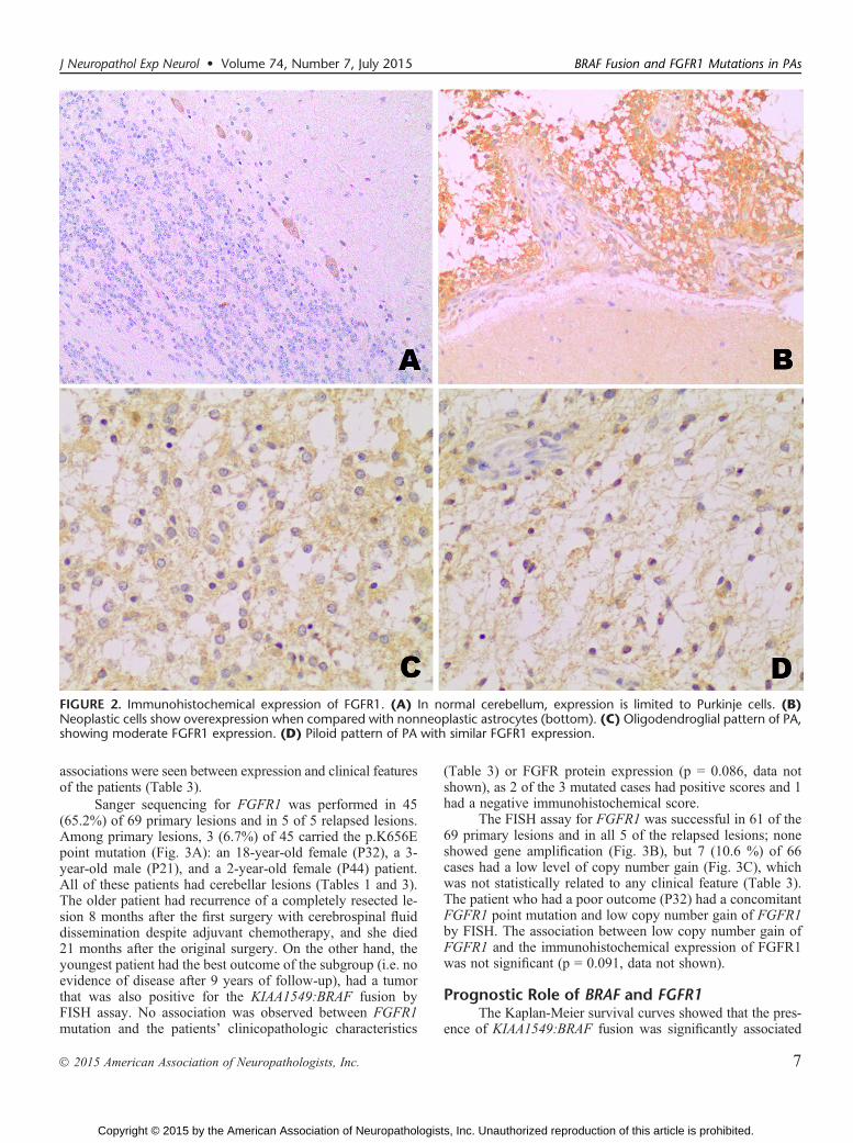
Molecular Characterization of FGFR1The immunohistochemical expression of FGFR1 was
tested in 74 of 75 samples. Nonneoplastic cerebellum showedcytoplasmic staining only in Purkinje cells and was absent orfaintly expressed in the nonneoplastic astrocytes (Fig. 2A, B).In tumor areas, cytoplasmic staining was detected in 51(73.9%) of 69 primary tumors, regardless of the histologicpattern of the PA (Fig. 2C, D) and no nuclear staining wasobserved. Forty-nine cases (71%) had scores Q3, and 19 cases(27.5%) were completely negative. No association was foundbetween FGFR1 immunohistochemical staining with the pres-ence of the KIAA1549:BRAF fusion (p = 0.272), or with BRAFpoint mutations (p = 0.456) (data not shown). No significant
J Neuropathol Exp Neurol � Volume 74, Number 7, July 2015 BRAF Fusion and FGFR1 Mutations in PAs
� 2015 American Association of Neuropathologists, Inc. 5
Copyright © 2015 by the American Association of Neuropathologists, Inc. Unauthorized reproduction of this article is prohibited.

FIGURE 1. Molecular alterations in BRAF. (A, B) FISH assay for detection of KIAA1549:BRAF fusion showing a positive (A) and anegative (B) case (white arrows). (C, D) Point mutations detected by Sanger sequencing for V600E (C) and V600K (D).
TABLE 2. Clinical Features of Patients and Their Association with BRAF ChangesKIAA1549:BRAF Fusion BRAF Point Mutation
(Total No. in the Series) Positive Negative p Wild Type Mutated p
Sex Female (31) 18 11 0.530 25 1 1.0
Male (38) 19 16 21 1
Age e19 years (60) 33 22 0.381 40 2 1.0
Q20 years (09) 4 5 6 0
NF1 Yes (05) 0 5 0.011 5 0 1.0
No (64) 37 22 41 2
Tumor location Cerebellar (36) 27 6 G0.0001 27 1 1.0
Extracerebellar (33) 10 21 19 1
Outcome Favorable (34) 22 16 0.987 26 1 1.0
Unfavorable (35) 15 11 20 1
Beckerm et al J Neuropathol Exp Neurol � Volume 74, Number 7, July 2015
� 2015 American Association of Neuropathologists, Inc.6
Copyright © 2015 by the American Association of Neuropathologists, Inc. Unauthorized reproduction of this article is prohibited.
associations were seen between expression and clinical featuresof the patients (Table 3).
Sanger sequencing for FGFR1 was performed in 45(65.2%) of 69 primary lesions and in 5 of 5 relapsed lesions.Among primary lesions, 3 (6.7%) of 45 carried the p.K656Epoint mutation (Fig. 3A): an 18-year-old female (P32), a 3-year-old male (P21), and a 2-year-old female (P44) patient.All of these patients had cerebellar lesions (Tables 1 and 3).The older patient had recurrence of a completely resected le-sion 8 months after the first surgery with cerebrospinal fluiddissemination despite adjuvant chemotherapy, and she died21 months after the original surgery. On the other hand, theyoungest patient had the best outcome of the subgroup (i.e. noevidence of disease after 9 years of follow-up), had a tumorthat was also positive for the KIAA1549:BRAF fusion byFISH assay. No association was observed between FGFR1mutation and the patients’ clinicopathologic characteristics
(Table 3) or FGFR protein expression (p = 0.086, data notshown), as 2 of the 3 mutated cases had positive scores and 1had a negative immunohistochemical score.
The FISH assay for FGFR1 was successful in 61 of the69 primary lesions and in all 5 of the relapsed lesions; noneshowed gene amplification (Fig. 3B), but 7 (10.6 %) of 66cases had a low level of copy number gain (Fig. 3C), whichwas not statistically related to any clinical feature (Table 3).The patient who had a poor outcome (P32) had a concomitantFGFR1 point mutation and low copy number gain of FGFR1by FISH. The association between low copy number gain ofFGFR1 and the immunohistochemical expression of FGFR1was not significant (p = 0.091, data not shown).
Prognostic Role of BRAF and FGFR1The Kaplan-Meier survival curves showed that the pres-
ence of KIAA1549:BRAF fusion was significantly associated
FIGURE 2. Immunohistochemical expression of FGFR1. (A) In normal cerebellum, expression is limited to Purkinje cells. (B)Neoplastic cells show overexpression when compared with nonneoplastic astrocytes (bottom). (C) Oligodendroglial pattern of PA,showing moderate FGFR1 expression. (D) Piloid pattern of PA with similar FGFR1 expression.
J Neuropathol Exp Neurol � Volume 74, Number 7, July 2015 BRAF Fusion and FGFR1 Mutations in PAs
� 2015 American Association of Neuropathologists, Inc. 7
Copyright © 2015 by the American Association of Neuropathologists, Inc. Unauthorized reproduction of this article is prohibited.

TABLE 3. Clinical Features of Patients and Their Association with FGFR1 ChangesFGFR1 Expression
p
FGFR1 lcng
p
FGFR1 Point Mutation
pPositive Negative Positive Negative Wild Type Mutated
Sex Female 22 9 0.994 3 24 1.0 23 2 1.0
Male 27 11 4 30 19 1
Age e19 years 43 17 0.758 6 47 0.922 37 3 1.0
Q20 years 6 3 1 7 5 0
NF1 Yes 4 1 1.0 0 5 1.0 5 0 1.0
No 45 19 7 49 37 3
Tumor location Cerebellar 27 9 0.446 5 26 0.425 24 3 0.264
Extracerebellar 22 11 2 28 18 0
Outcome Favorable 22 12 0.255 2 33 0.125 25 2 1.0
Unfavorable 27 8 5 21 17 1
Lcgn, low copy number gain.
FIGURE 3. Molecular alterations of FGFR1. (A) Electropherogram showing the point mutation K656E. (B, C) FISH assay displayinga normal pattern (B), and a case with low-copy number gain of the FGFR1 signal (C). The amount of FGFR1 signals (green) did notreach the cutoff value needed for the diagnosis of gene amplification.
Beckerm et al J Neuropathol Exp Neurol � Volume 74, Number 7, July 2015
� 2015 American Association of Neuropathologists, Inc.8
Copyright © 2015 by the American Association of Neuropathologists, Inc. Unauthorized reproduction of this article is prohibited.
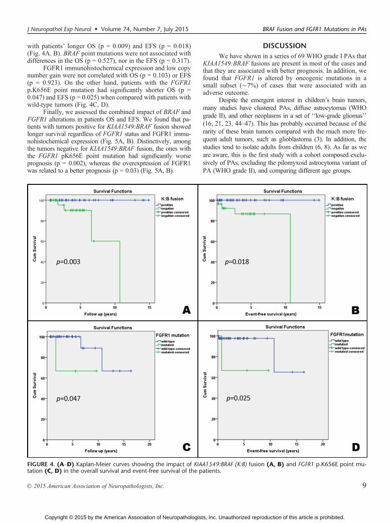
with patients’ longer OS (p = 0.009) and EFS (p = 0.018)(Fig. 4A. B). BRAF point mutations were not associated withdifferences in the OS (p = 0.527), nor in the EFS (p = 0.317).
FGFR1 immunohistochemical expression and low copynumber gain were not correlated with OS (p = 0.103) or EFS(p = 0.923). On the other hand, patients with the FGFR1p.K656E point mutation had significantly shorter OS (p =0.047) and EFS (p = 0.025) when compared with patients withwild-type tumors (Fig. 4C, D).
Finally, we assessed the combined impact of BRAF andFGFR1 alterations in patients OS and EFS. We found that pa-tients with tumors positive for KIAA1549:BRAF fusion showedlonger survival regardless of FGFR1 status and FGFR1 immu-nohistochemical expression (Fig. 5A, B). Distinctively, amongthe tumors negative for KIAA1549:BRAF fusion, the ones withthe FGFR1 pK656E point mutation had significantly worseprognosis (p = 0.002), whereas the overexpression of FGFR1was related to a better prognosis (p = 0.03) (Fig. 5A, B).
DISCUSSIONWe have shown in a series of 69 WHO grade I PAs that
KIAA1549:BRAF fusions are present in most of the cases andthat they are associated with better prognosis. In addition, wefound that FGFR1 is altered by oncogenic mutations in asmall subset (È7%) of cases that were associated with anadverse outcome.
Despite the emergent interest in children’s brain tumors,many studies have clustered PAs, diffuse astrocytomas (WHOgrade II), and other neoplasms in a set of ‘‘low-grade gliomas’’(16, 21, 23, 44Y47). This has probably occurred because of therarity of these brain tumors compared with the much more fre-quent adult tumors, such as glioblastoma (3). In addition, thestudies tend to isolate adults from children (6, 8). As far as weare aware, this is the first study with a cohort composed exclu-sively of PAs, excluding the pilomyxoid astrocytoma variant ofPA (WHO grade II), and comparing different age groups.
FIGURE 4. (AYD) Kaplan-Meier curves showing the impact of KIAA1549:BRAF (K:B) fusion (A, B) and FGFR1 p.K656E point mu-tation (C, D) in the overall survival and event-free survival of the patients.
J Neuropathol Exp Neurol � Volume 74, Number 7, July 2015 BRAF Fusion and FGFR1 Mutations in PAs
� 2015 American Association of Neuropathologists, Inc. 9
Copyright © 2015 by the American Association of Neuropathologists, Inc. Unauthorized reproduction of this article is prohibited.

Herein, we were able to evaluate gene fusions and pointmutations in 67 of 69 cases and observed that nearly 60% ofthem had alterations in BRAF and/or FGFR1, which are trig-gers of the MAPK pathway, the dominant oncogenic pathwayof PAs (15, 19). The high incidence of KIAA1549:BRAF fu-sion and its predominance in cerebellar lesions are in line withprevious studies (7, 15, 16, 24, 26), confirming it as the mostfrequent molecular change of PAs (7). In addition, 2 of 5 tumorsthat harbored BRAF or FGFR1 mutations had a coexistingKIAA1549:BRAF gene fusion. The occurrence of a concomi-tant KIAA1549:BRAF fusion and other changes in the samepathway is a rare occurrence, but it has been previouslyreported (7, 21, 26). Finally, the negative relationship betweenKIAA1549:BRAF fusions and clinical diagnosis of NF1 (thuswith alterations in the NF1 gene) has been previously reported(7). Further studies on NF1 are necessary for a better under-standing of this relationship.
The incidence of BRAF point mutations in our study(4.2%) is also similar to published data (22). The presence ofthis alteration did not show a clinical impact on prognosis,thereby confirming the previous findings of Bannykh et al,who showed that the BRAF V600E mutation did not implyhigher aggressiveness to PAs (48). The usual p.V600E pointmutation was detected only in an 11-year-old male patientwho had an unstable hypothalamic lesion, which reinforcesthe occurrence of this mutation in extracerebellar lesions (7).Moreover, that patient also harbored the KIAA1549:BRAFfusion. We also observed 1 unusual BRAF point mutation incodon 600 of a cerebellar PA. The point mutation V600K wasfound in an 11-year-old female patient who had an excellentoutcome after long follow-up (11.1 years). This mutation waspreviously described in 5% to 15% of melanomas and wasrelated to metastatic disease and worse outcome (49Y51);however, it has also been associated with a response to first-generation BRAF inhibitors (PLX4032, vemurafenib) (51).Patients with PAs have experienced adverse results after
treatment with vemurafenib (52), as opposed to the good re-sponse observed in patients with high-grade tumors (53, 54),probably because of the overall low frequency of p.V600E.Nevertheless, the subset of patients with KIAA1549:BRAFpositive tumors could potentially benefit from treatment withthe second-generation BRAF inhibitors such as PLX-PB3,which specifically target the fusion protein (52).
Most tumors in our series showed strong immunohis-tochemical FGFR1 expression. These findings are in line witha previous study in gliomas, in which FGFR1 overexpressionwas detected, although the underlying molecular mechanismwas not explained at the time (55). Our FISH assays largelyeliminated amplification as the underlying mechanism in thePAs, contrary to what is seen in a subset of breast and lungcancers (30, 31). FGFR1 low copy number gain was rare andshowed a nonstatistically significant trend toward immuno-histochemical overexpression (p = 0.094, data not shown).The mutated form of FGFR1 was also not associated withprotein overexpression (p = 1.0).
With respect to the FGFR1 mutation, we observedp.K656E point mutations at the tyrosine kinase domain in6.7% of the PAs in this series, all of which were located in thecerebellum. The oncogenic FGFR1 mutations, p.K656E andp.N456K, were recently described by Jones et al (15) as re-current events in extracerebellar PAs. Those mutations werefurther described in rosette-forming glioneuronal tumor of thefourth ventricle, but 1 of the patients in that series had anearlier extracerebellar (diencephalic) PA with pilomyxoidfeatures, which also harbored the p.K656E mutation (29).
FGFR1 is currently an attractive therapeutic target, andthe immunohistochemical assessment of FGFR1 may repre-sent a good indicator of the management of PAs. Recentstudies have related the efficacy of novel specific FGFR1 in-hibitors, such as ponatinib (AP 24534), in cases of lung cancerwith FGFR1 overexpression that were assessed by immuno-blotting and mRNA quantification (56). Besides this drug, other
FIGURE 5. Kaplan-Meier curves comparing the simultaneous impact of KIAA1549:BRAF (K:B) fusion and FGFR1 alterations on theoverall survival of patients. (A) Impact of FGFR1 p.K656E point mutation. (B) Impact of FGFR1 expression assessed by immuno-histochemistry (positive score Q3; negative score e2).
Beckerm et al J Neuropathol Exp Neurol � Volume 74, Number 7, July 2015
� 2015 American Association of Neuropathologists, Inc.10
Copyright © 2015 by the American Association of Neuropathologists, Inc. Unauthorized reproduction of this article is prohibited.

FGFR1 inhibitors, such as lucitanib (57) and CH5183284/Debio 1347 (58), may constitute future alternatives for thetreatment of inoperable PAs. Nevertheless, further preclinicaland clinical studies are needed to determine whether FGFR1expression and hotspot mutations will modulate and predictpatient response to these FGFR1-specific tyrosine inhibitors.
Concomitant KIAA1549:BRAF fusion and FGFR1 mu-tations were not referred events in the study of Jones et al(15), but this was detected in 1 of the patients of our series.We further evaluated the impact of both the aforementioned al-terations in the prognosis of the patients. The KIAA1549:BRAFfusion had a positive impact on patients’ OS and EFS and wasconfirmed as a prognostic factor, corroborating the tendency tobetter outcome of PAs, similar to what happens in the complexgroup of low-grade gliomas described by Hawkins et al (21).
On the other hand, FGFR1 mutations were significantlyrelated to PA patients’ shorter OS and EFS when comparedwith the wild-type group; however, the significance of this find-ing needs to be confirmed in larger series. To our knowledge, thisthe first study to indicate the prognostic role of FGFR1 mutationin PAs and their occurrence in cerebellar lesions.
In conclusion, we confirmed the pivotal role ofKIAA1549:BRAF fusion and, to a lesser extent, of FGFR1 inMAPK activation in PAs. More exactly, we showed the use-fulness of evaluating the KIAA1549:BRAF fusion as a prog-nostic biomarker, while FGFR1 mutation may be a relevantprognostic marker in PAs. With further investigation, themolecular changes of BRAF and FGFR1 may constitute po-tential therapeutic targets for inoperable or recurrent PAs.
ACKNOWLEDGMENTThe authors thank Nathalia Campanella for the help in
picture editing.
REFERENCES1. Scheithauer BW, Hawkins C, Tihan T, et al. Pilocytic astrocytoma. In:
David N, Louis MD, Hiroko Ohgaki PD, Otmar D, Wiestler MD,Webster K, Cavenee PD, editors.WHO Classification of Tumours of theCentral Nervous System. World Health Organization Classification ofTumours. Lyon, France: IARC Press, 2007:13Y21
2. Ward E, DeSantis C, Robbins A, et al. Childhood and adolescent cancerstatistics, 2014. CA Cancer J Clin 2014;64:83Y103
3. Dolecek TA, Propp JM, Stroup NE, et al. CBTRUS statistical report:primary brain and central nervous system tumors diagnosed in the UnitedStates in 2005Y2009. Neuro Oncol 2012;14(Suppl 5):v1Y49
4. Camargo Bd, Felipe CFP, Noronha CP, et al. Cancer na crian0a e noadolescente no Brasil dados dos registros de base populacional e demortalidade. Rio de Janeiro: Instituto Nacional do Cancer, 2008
5. Rosemberg S, Fujiwara D. Epidemiology of pediatric tumors of the ner-vous system according to the WHO 2000 classification: a report of 1,195cases from a single institution. Child Nerv Sys 2005;21:940Y4
6. Theeler BJ, Ellezam B, Sadighi ZS, et al. Adult pilocytic astrocytomas:clinical features and molecular analysis. Neuro Oncol 2014 2014;16:841Y7. doi: 10.1093/neuonc/not246
7. Jones DT, Gronych J, Lichter P, et al. MAPK pathway activation inpilocytic astrocytoma. Cell Molec Life Sci 2012;69:1799Y811
8. Johnson DR, Brown PD, Galanis E, et al. Pilocytic astrocytoma survivalin adults: analysis of the Surveillance, Epidemiology, and End ResultsProgram of the National Cancer Institute. J Neuro-oncol 2012;108:187Y93
9. Gutmann DH, McLellan MD, Hussain I, et al. Somatic neurofibromatosistype 1 (NF1) inactivation characterizes NF1-associated pilocytic astro-cytoma. Genome Res 2013;23:431Y9
10. Rodriguez FJ, Perry A, Gutmann DH, et al. Gliomas in neurofibromatosistype 1: a clinicopathologic study of 100 patients. J Neuropathol ExpNeurol 2008;67:240Y9
11. Listernick R, Charrow J, Gutmann DH. Intracranial gliomas in neurofi-bromatosis type 1. Am J Med Gen 1999;89:38Y44
12. Tada K, Kochi M, Saya H, et al. Preliminary observations on geneticalterations in pilocytic astrocytomas associated with neurofibromatosis 1.Neuro Oncol 2003;5:228Y34
13. Dhanasekaran DN, Johnson GL. MAPKs: function, regulation, role incancer and therapeutic targeting. Oncogene 2007;26:3097Y9
14. Murphy T, Hori S, Sewell J, et al. Expression and functional role ofnegative signalling regulators in tumour development and progression.Int J Cancer 2010;127:2491Y9
15. Jones DT, Hutter B, Jager N, et al. Recurrent somatic alterations of FGFR1and NTRK2 in pilocytic astrocytoma. Nature Gen 2013;45:927Y32
16. Zhang J, Wu G, Miller CP, et al. Whole-genome sequencing identifiesgenetic alterations in pediatric low-grade gliomas. Nature Gen 2013;45:602Y12
17. Bar EE, Lin A, Tihan T, et al. Frequent gains at chromosome 7q34 in-volving BRAF in pilocytic astrocytoma. J Neuropathol Exp Neurol 2008;67:878Y87
18. Capper D, Preusser M, Habel A, et al. Assessment of BRAF V600Emutation status by immunohistochemistry with a mutation-specificmonoclonal antibody. Acta Neuropathol 2011;122:11Y9
19. Forshew T, Tatevossian RG, Lawson AR, et al. Activation of theERK/MAPK pathway: a signature genetic defect in posterior fossapilocytic astrocytomas. J Pathol 2009;218:172Y81
20. Hasselblatt M, Riesmeier B, Lechtape B, et al. BRAF-KIAA1549 fusiontranscripts are less frequent in pilocytic astrocytomas diagnosed in adults.Neuropathol Appl Neurobiol 2011;37:803Y6
21. Hawkins C, Walker E, Mohamed N, et al. BRAF-KIAA1549 fusionpredicts better clinical outcome in pediatric low-grade astrocytoma. ClinCancer Res 2011;17:4790Y8
22. Schindler G, Capper D, Meyer J, et al. Analysis of BRAF V600E mu-tation in 1,320 nervous system tumors reveals high mutation frequenciesin pleomorphic xanthoastrocytoma, ganglioglioma and extra-cerebellarpilocytic astrocytoma. Acta Neuropathol 2011;121:397Y405
23. Tian Y, Rich BE, Vena N, et al. Detection of KIAA1549-BRAF fusiontranscripts in formalin-fixed paraffin-embedded pediatric low-grade gli-omas. J Molec Diag 2011;13:669Y77
24. Jones DT, Kocialkowski S, Liu L, et al. Oncogenic RAF1 rearrangementand a novel BRAF mutation as alternatives to KIAA1549:BRAF fusionin activating the MAPK pathway in pilocytic astrocytoma. Oncogene2009;28:2119Y23
25. Roth JJ, Santi M, Pollock AN, et al. Chromosome band 7q34 deletionsresulting in KIAA1549-BRAF and FAM131B-BRAF fusions in pediatriclow-grade gliomas. Brain Pathol 2015;25:182Y92
26. Cin H, Meyer C, Herr R, et al. Oncogenic FAM131B-BRAF fusionresulting from 7q34 deletion comprises an alternative mechanism ofMAPK pathway activation in pilocytic astrocytoma. Acta Neuropathol2011;121:763Y74
27. Chen YH, Gutmann DH. The molecular and cell biology of pediatriclow-grade gliomas. Oncogene 2014;33:2019Y26
28. Basto D, Trovisco V, Lopes JM, et al. Mutation analysis of B-RAF genein human gliomas. Acta Neuropathol 2005;109:207Y10
29. Gessi M, Moneim YA, Hammes J, et al. FGFR1 mutations inrosette-forming glioneuronal tumors of the fourth ventricle. J NeuropatholExp Neurol 2014;73:580Y4
30. Dutt A, Ramos AH, Hammerman PS, et al. Inhibitor-sensitive FGFR1 am-plification in human non-small cell lung cancer. PloS One 2011;6:e20351
31. Theillet C, Adelaide J, Louason G, et al. FGFRI and PLAT genes andDNA amplification at 8p12 in breast and ovarian cancers. Genes Chro-mosome Cancer 1993;7:219Y26
32. Singh D, Chan JM, Zoppoli P, et al. Transforming fusions of FGFR andTACC genes in human glioblastoma. Science 2012;337:1231Y5
33. Colin C, Padovani L, Chappe C, et al. Outcome analysis of childhoodpilocytic astrocytomas: a retrospective study of 148 cases at a single in-stitution. Neuropathol Appl Neurobiol 2013;39:693Y705
34. Fernandez C, Figarella-Branger D, Girard N, et al. Pilocytic astrocytomasin children: prognostic factorsVa retrospective study of 80 cases. Neu-rosurgery 2003;53:544Y53; discussion 54Y5
J Neuropathol Exp Neurol � Volume 74, Number 7, July 2015 BRAF Fusion and FGFR1 Mutations in PAs
� 2015 American Association of Neuropathologists, Inc. 11
Copyright © 2015 by the American Association of Neuropathologists, Inc. Unauthorized reproduction of this article is prohibited.

35. Paixao Becker A, de Oliveira RS, Saggioro FP, et al. In pursuit ofprognostic factors in children with pilocytic astrocytomas. Childs NervSyst 2010;26:19Y28
36. Schneider JH Jr, Raffel C, McComb JG. Benign cerebellar astrocytomasof childhood. Neurosurgery 1992;30:58Y62; discussion 3
37. Capper D, Weissert S, Balss J, et al. Characterization of R132Hmutation-specific IDH1 antibody binding in brain tumors. Brain Pathol2010;20:245Y54
38. Mellai M, Piazzi A, Caldera V, et al. IDH1 and IDH2 mutations, im-munohistochemistry and associations in a series of brain tumors. JNeuro-oncol 2011;105:345Y57
39. Pinto F, Pertega-Gomes N, Pereira MS, et al. T-box transcription factorbrachyury is associated with prostate cancer progression and aggres-siveness. Clinical Cancer Res 2014;20:4949Y61
40. Toschi L, Finocchiaro G, Nguyen TT, et al. Increased SOX2 gene copynumber is associated with FGFR1 and PIK3CA gene gain in nonYsmallcell lung cancer and predicts improved survival in early stage disease.PloS One 2014;9:e95303
41. Aisner DL, Nguyen TT, Paskulin DD, et al. ROS1 and ALK fusions incolorectal cancer, with evidence of intratumoral heterogeneity for mo-lecular drivers. Molec Cancer Res 2014;12:111Y8
42. Schultheis AM, Bos M, Schmitz K, et al. Fibroblast growth factor re-ceptor 1 (FGFR1) amplification is a potential therapeutic target insmall-cell lung cancer. Mod Pathol 2014;27:214Y21
43. Yamane LS, Scapulatempo-Neto C, Alvarenga L, et al. KRAS and BRAFmutations and MSI status in precursor lesions of colorectal cancerdetected by colonoscopy. Oncology Rep 2014;32:1419Y26
44. Bhattacharjee MB, Armstrong DD, Vogel H, et al. Cytogenetic analysisof 120 primary pediatric brain tumors and literature review. Cancer GenCytogen 1997;97:39Y53
45. Bigner SH, McLendon RE, Fuchs H, et al. Chromosomal characteristicsof childhood brain tumors. Cancer Gen Cytogen 1997;97:125Y34
46. Pfister S, Janzarik WG, Remke M, et al. BRAF gene duplication con-stitutes a mechanism of MAPK pathway activation in low-grade astro-cytomas. J Clin Invest 2008;118:1739Y49
47. Sievert AJ, Jackson EM, Gai X, et al. Duplication of 7q34 in pediatriclow-grade astrocytomas detected by high-density single-nucleotide
polymorphism-based genotype arrays results in a novel BRAF fusiongene. Brain Pathol 2009;19:449Y58
48. Bannykh SI, Mirocha J, Nuno M, et al. V600E BRAF mutation inpilocytic astrocytoma is associated with a more diffuse growth pattern butdoes not confer a more aggressive clinical behavior. Cli Neuropathol2014;33:388Y98
49. El-Osta H, Falchook G, Tsimberidou A, et al. BRAF mutations in advancedcancers: clinical characteristics and outcomes. PloS One 2011;6:e25806
50. Menzies AM, Haydu LE, Visintin L, et al. Distinguishing clinicopatho-logic features of patients with V600E and V600K BRAF-mutant meta-static melanoma. Clinical Cancer Res 2012;18:3242Y9
51. Rubinstein JC, Sznol M, Pavlick AC, et al. Incidence of the V600Kmutation among melanoma patients with BRAF mutations, and potentialtherapeutic response to the specific BRAF inhibitor PLX4032. J TranslMed 2010;8:67
52. Sievert AJ, Lang SS, Boucher KL, et al. Paradoxical activation and RAFinhibitor resistance of BRAF protein kinase fusions characterizing pedi-atric astrocytomas. Proc Natl Acad Sci USA 2013;110:5957Y62
53. Bautista F, Paci A, Minard-Colin V, et al. Vemurafenib in pediatric pa-tients with BRAFV600E mutated high-grade gliomas. Ped Blood Cancer2014;61:1101Y3
54. Robinson GW, Orr BA, Gajjar A. Complete clinical regression of aBRAF V600E-mutant pediatric glioblastoma multiforme after BRAFinhibitor therapy. BMC Cancer 2014;14:258
55. Ueba T, Takahashi JA, Fukumoto M, et al. Expression of fibroblastgrowth factor receptor-1 in human glioma and meningioma tissues.Neurosurgery 1994;34:221Y5; discussion 5Y6
56. Wynes MW, Hinz TK, Gao D, et al. FGFR1 mRNA and protein ex-pression, not gene copy number, predict FGFR TKI ensitivity across alllung cancer histologies. Clin Cancer Res 2014;20:3299Y309
57. Soria JC, DeBraud F, Bahleda R, et al. Phase I/IIa study evaluating thesafety, efficacy, pharmacokinetics, and pharmacodynamics of lucitanib inadvanced solid tumors. Ann Oncol 2014;25:2244Y51
58. Nakanishi Y, Akiyama N, Tsukaguchi T, et al. The fibroblast growthfactor receptor genetic status as a potential predictor of the sensitivity toCH5183284/Debio 1347, a novel selective FGFR inhibitor. Molec Can-cer Ther 2014;13:2547Y58
Beckerm et al J Neuropathol Exp Neurol � Volume 74, Number 7, July 2015
� 2015 American Association of Neuropathologists, Inc.12
Copyright © 2015 by the American Association of Neuropathologists, Inc. Unauthorized reproduction of this article is prohibited.