In pursuit of prognostic factors in children with pilocytic astrocytomas
Upload
independentCategory
view
0download
0
Duplication of 7q34 is specific to juvenile pilocytic astrocytomasand a hallmark of cerebellar and optic pathway tumours
K Jacob1, S Albrecht2, C Sollier1, D Faury1, E Sader1, A Montpetit3, D Serre3, P Hauser4, M Garami4, L Bognar5,Z Hanzely6, JL Montes7, J Atkinson7, J-P Farmer7, E Bouffet8, C Hawkins9, U Tabori8 and N Jabado*,1
1Department of Pediatrics and Human Genetics, Montreal Children’s Hospital, McGill University Health Center, Montreal, Canada; 2Department ofPathology, Montreal Children’s Hospital, McGill University Health Center, Montreal, Canada; 3McGill University and Genome Quebec Innovation Centre,Montreal, Canada; 4Second Department of Pediatrics, Faculty of Medicine, Semmelweis University, Budapest, Hungary; 5Department of Neurosurgery,Medical and Health Science Center, University of Debrecen, Debrecen, Hungary; 6Division of Neuro-Surgery, Division of Pathology, National Institute ofNeurosurgery, Budapest, Hungary; 7Division of Neurosurgery, Montreal Children’s Hospital, McGill University Health Center, Montreal, Canada;8Department of Pediatrics, Hospital for Sickkids, Toronto, Canada; 9Department of Pediatric Neuropathology, Hospital for Sickkids, Toronto, Canada
BACKGROUND: Juvenile pilocytic astrocytomas (JPA), a subgroup of low-grade astrocytomas (LGA), are common, heterogeneous andpoorly understood subset of brain tumours in children. Chromosomal 7q34 duplication leading to fusion genes formed betweenKIAA1549 and BRAF and subsequent constitutive activation of BRAF was recently identified in a proportion of LGA, and may beinvolved in their pathogenesis. Our aim was to investigate additional chromosomal unbalances in LGA and whether incidence of 7q34duplication is associated with tumour type or location.METHODS AND RESULTS: Using Illumina-Human-Hap300-Duo and 610-Quad high-resolution-SNP-based arrays and quantitative PCRon genes of interest, we investigated 84 paediatric LGA. We demonstrate that 7q34 duplication is specific to sporadic JPA (35 of53 – 66%) and does not occur in other LGA subtypes (0 of 27) or NF1-associated-JPA (0 of 4). We also establish that it is site specificas it occurs in the majority of cerebellar JPA (24 of 30 – 80%) followed by brainstem, hypothalamic/optic pathway JPA (10 of16 – 62.5%) and is rare in hemispheric JPA (1 of 7 – 14%). The MAP-kinase pathway, assessed through ERK phosphorylation, wasactive in all tumours regardless of 7q34 duplication. Gain of function studies performed on hTERT-immortalised astrocytes show thatoverexpression of wild-type BRAF does not increase cell proliferation or baseline MAPK signalling even if it sensitises cells to EGFRstimulation.CONCLUSIONS AND INTERPRETATION: Our results suggest that variants of JPA might arise from a unique site-restricted progenitor cellwhere 7q34 duplication, a hallmark of this tumour-type in association to MAPK-kinase pathway activation, potentially plays a site-specific role in their pathogenesis. Importantly, gain of function abnormalities in components of MAP-Kinase signalling are potentiallypresent in all JPA making this tumour amenable to therapeutic targeting of this pathway.British Journal of Cancer (2009) 101, 722–733. doi:10.1038/sj.bjc.6605179 www.bjcancer.comPublished online 14 July 2009& 2009 Cancer Research UK
Keywords: SNP arrays; 7q34; JPA; LGA; paediatric; BRAF
��������������������������������������������������������������������
Juvenile pilocytic astrocytomas (JPA) account for 60–80% ofpaediatric low-grade astrocytomas (LGA), the most commonpaediatric brain tumour, and thus are the most frequentlyencountered subtype of brain neoplasm in children under theage of 19 years. They are classified according to the World HealthOrganisation (WHO) as WHO grade I (Louis et al, 2007), andoccur sporadically throughout childhood or arise in up to 15– 40%of children affected with neurofibromatosis type 1 (NF1; Pollackand Mulvihill, 1997). JPA exhibit distinct features, readilydistinguishable from their other LGA counterparts, includingclinical course and molecular characteristics, and have betterprognosis in affected children (Gajjar et al, 1997; Ishii et al, 1998;
Cheng et al, 2000; Tada et al, 2003; Broniscer et al, 2007; Fisheret al, 2008).
Even though they share similar histology, there is heterogeneitybetween sporadic JPA in terms of localisation, radiologic features,histologic atypia and clinical behaviour, all of which argue for thepossibility of genetic disparity between potential JPA subgroups.Typically, JPA occur as exophytic cerebellar tumours, however,they can also arise in the brain stem or the optic pathway, wherethey behave more aggressively than NF1-associated JPA (Grill et al,2000), or, more rarely, in the cerebral hemispheres. Maximalsurgical resection is the mainstay of therapy, and failure to achieveit remains the main therapeutic concern. Although cerebellarJPA are readily amenable to complete surgical resection, in otherless anatomically accessible locations, surgery may result in thepersistence of residual disease, which can require further therapyfor tumour control, and ultimately lead to increased morbidity/mortality. In addition, some of these extracerebellar JPA seem lesscircumscribed and may exhibit atypical pathologic features,
Received 23 February 2009; revised 2 June 2009; accepted 11 June 2009;published online 14 July 2009
*Correspondence: Dr N Jabado, Montreal Children’s Hospital ResearchInstitute, 4060 Ste Catherine West, PT-239, Montreal, Qc, Canada H3Z2Z3; E-mail: [email protected]
British Journal of Cancer (2009) 101, 722 – 733
& 2009 Cancer Research UK All rights reserved 0007 – 0920/09 $32.00
www.bjcancer.com
Gen
etic
san
dG
en
om
ics

leading to pathologic misdiagnosis, including into higher gradetumours.
Until recently, the few genetic abnormalities documented inJPA mainly identified chromosomal gains of 7q and trisomy ofchromosomes 5, 7 or 8 in some tumours (White et al, 1995; Rickertand Paulus, 2004; Wemmert et al, 2006). Recently severalconsecutive papers described duplication of 7q34 in LGA includingJPA (Bar et al, 2008; Deshmukh et al, 2008; Jones et al, 2008; Pfisteret al, 2008; Sievert et al, 2008). However, the data reported areconflicting regarding the subgroup of LGA affected by this geneticevent, the size of the duplication and its anatomical localisationwithin the brain. Indeed, Deshmukh et al (2008) identifiedamplification of 7q34 in 8 of 10 cerebellar JPA at 138151200 –139456000, which included HIPK2, a potential gene of interest. Theauthors further showed using an immunohistochemical approachthat overexpression of HIPK2 was more frequent in LGA than inhigh-grade gliomas, and that it was more common in infratentorialtumours. Their silencing of HIPK2 in U87 (a glioblastoma (GBM)cell line) decreased the cells proliferation rate. Pfister et al (2008)identified duplication of 7q34 within 139186224 –140156951 in 30of 66 (45.5%) paediatric LGA. This region included BRAF, a genewhich was not in the genetic interval described in Deshmukh et al(2008). The authors confirmed the absence of previously describedBRAF oncogenic mutations in their sample set, and showed thatsilencing of BRAF in a LGA cell line decreased cell proliferationrates. However, based on lower resolution BAC arrays, they did notprecisely map the genetic region missing other genes potentiallycritical in the pathogenesis of LGA, including HIPK2. In addition,data from this report suggest that LGA other than JPA mayharbour 7q34 duplication and that this genetic alteration is morefrequent in non-cerebellar tumours.
Recent reports have indicated a central role for the mitogen-activated protein kinase (MAPK) pathway in the tumorigenesis ofpilocytic astrocytomas and showed that duplication at 7q34 leadsto a fusion between KIAA1549 and BRAF resulting in constitutiveactivation of the BRAF kinase (Jones et al, 2008; Sievert et al,2008). In particular, Jones et al (2008) focused on JPA and describea tandem duplication at 7q34 producing a transforming BRAFfusion gene in 29 of 44 tumours (66%), and V600E point mutationof BRAF in two further cases. Sievert et al indicated that 7q34duplication occurs in 17 of 22 JPA but also report it in 3 of6 diffuse astrocytomas (LGA grade II).
To determine the specificity of 7q34 duplication to a givensubgroup of LGA and identify additional genetic aberrations intumours that do not carry this duplication, we investigated 115paediatric brain tumour samples including 57 JPA, and 27 diffuseastrocytomas (Tables 1, 2a and b). Our data indicate that 7q34duplication is exclusive to JPA and a hallmark of specificanatomical localisations of these tumours within the brain. Weidentify additional genetic abnormalities in JPA that do notharbour 7q34 duplication, which may help shed light on theirpathogenesis. Data from gain of function analysis in immortalisedastrocytes further confirm that increased expression of wild-typeBRAF is unable to cause malignant transformation on its ownhowever may contribute to an increased response to exogenoustriggering of membrane receptors. Moreover, we show that theMAPK pathway is active in all JPA regardless of 7q34 duplicationand that all of these tumours may be amenable to therapeutictargeting of this pathway.
MATERIALS AND METHODS
Samples
All samples were obtained with informed consent after approvalof the Institutional Review Board of the respective hospitals theywere treated in, and independently reviewed by senior paediatric
neuropathologists (SA, CH, ZH) according to the WHO guidelines(Kleihues et al, 2002). A total of 115 paediatric brain tumours(average 9.4±4.7 years) were analysed (Tables 1, 2a and b);57 JPA (53 sporadic, 4 from NF1 Patients), 27 diffuse astrocytomas(grade II), 1 grade II ependymoma, 5 grade I gangliogliomas and25 high-grade astrocytomas (HGA) were included. There were nopylomixoid variants within the JPA included within this study. Allsamples were taken at the time of the first surgery before furthertreatment, when needed. Tissues were obtained from the London/Ontario Tumor Bank, and from collaborators in Montreal, Torontoand Hungary.
DNA extraction and hybridisation, SNP analysis
DNA from frozen tumours was extracted as described previously(Wong et al, 2006). For SNP analysis, DNA (250 ng) from 40samples was assayed with the Human Hap300-Duo (N¼ 28) andthe 610-Qad (N¼ 16) genotyping beadchips according to therecommendations of the manufacturer (Illumina, San Diego, CA,USA). These BeadChips enable whole-genome genotyping ofrespectively over 300 000 and 610 000 tagSNP markers derivedfrom the International HapMap Project (www.hapmap.org) with amean intermaker distance of 10 and 5 kb respectively. Imageintensities were extracted using Illumina’s BeadScan software.Data for each BeadChip were self-normalised using informationcontained within the array. Penn-CNV (Wang et al, 2007) andGqCNV (D Serre et al, unpublished) algorithms were applied onthe genotype data derived from the 40 LGA. Only the alterationsthat were detected by both algorithm and that contained more than5 consecutive SNPs were considered in this study. They werefurther confirmed by visualisation in the BeadStudio.
Validation of copy number changes by quantitativereal-time PCR
Quantitative real-time PCR (q-PCR) was done on an ABI-Prism7000 sequence detector (Applied Biosystems, Bedford, MA, USA)using a SYBR Green kit (Applied Biosystems). The target locusfrom each tumour DNA was normalised to the reference, Line-1 aspreviously described (Wong et al, 2006; Supplementary Table 1).
Cell lines, antibodies and transfections
hTERT-immortalised human astrocytes (kind gift of Dr A Guha,Labbatt Brain Tumour Research Centre, Ontario, Canada) weregrown and transfected with 2 mg of plasmid DNA encoding wild-type C-Myc-Braf using Fugene 6 (Roche, Mississauga, ON, Canada)as previously described (Shi et al, 2004). Unless stated otherwise, allantibodies used were obtained from Cell Signaling (Danvers, MA,USA). Transfected cells were used at 0, 48 and 72 h posttransfection.HIPK2 plasmids were generously provided by Dr Gabriella D’Orazi(Regina Elena Cancer Institute, Rome, Italy), and BRAF plasmids byDr Richard Marais (Cancer Research, London, UK).
Western blot and immunofluorescence analysis
Extracts were prepared from cell pellets and western blot analysisperformed on total lysates as previously described (Jabado et al,1998; Rajasekhar et al, 2003). Cross-reactivity was visualised byECL chemiluminescence (Amersham) on a phosphorimager. Forimmunofluorescence analysis images were acquired using a Retiga1300 digital camera (QIMAGING) and a Zeiss confocal microscope.
Immunohistochemical analysis
Immunohistochemical analyses for phospho-Erk (pErk), wereperformed and the slides scored as previously described (Fauryet al, 2007).
7q34 duplication in paediatric low-grade astrocytomas
K Jacob et al
723
British Journal of Cancer (2009) 101(4), 722 – 733& 2009 Cancer Research UK
Gen
eti
cs
an
dG
en
om
ics

[3H]Thymidine incorporation assay
DMEM (10 ml) containing 7.4 kBq (0.2 mCi) of [methyl-3H]thymidine(Amersham Pharmacia Biotech Europe, Freiburg, Germany; Batch215, 65 Ci mM) were added to each microplate well. Experiments weredone in triplicate at least three times with identical results.
RESULTS
Copy number variants in 40 LGA
To chart genomic alterations in our sample set, we first performeda high-resolution genome-wide screen of the samples using the
Table 1 Characteristics of the 53 sporadic juvenile pilocytic astrocytomas (JPA) included in the study
PatientsAge
(years) LocationHIPK2(qPCR)
BRAF(qPCR) SNP array
Amplificationof 7q34 pERK (IHC) BRAFV600E
Cerebellar JPA (n¼ 30)1 6 Cerebellar ND ND 7q34 amp Y ND ND2 6 Cerebellar ND ND 7q34 amp Y ND ND3 6 Cerebellar ND ND 7q34 amp Y ND ND4 6 Cerebellar ND ND 7q34 amp Y ND ND5 6 Cerebellar ND ND 7q34 amp Y ND ND6 6 Cerebellar ND ND 7q34 amp Y ND ND7 6 Cerebellar ND ND 7q34 amp Y ND ND8 6 Cerebellar ND ND 7q34 amp Y ND ND9 4 Cerebellar A A 7q34 amp Y Pos WT
10 4 Cerebellar A A 7q34 amp Y Pos WT11 4 Cerebellar N N N N Pos WT12 4 Cerebellar A A 7q34 amp Y Pos WT13 4 Cerebellar N N ND N Pos ND14 4 Cerebellar A A 7q34 amp Y Pos WT15 4 Cerebellar ND ND N N Pos ND16 4 Cerebellar A A 7q34 amp Y Pos WT17 3 Cerebellar N N N N Pos WT18 6 Cerebellar N N N N Pos BRAFV600E19 11 Cerebellar A A 7q34 amp Y Pos WT20 11 Cerebellar A A 7q34 amp Y Pos WT21 6 Cerebellar A A ND Y Pos WT22 11 Cerebellar A A ND Y Pos WT23 9 Cerebellar N N ND N Pos WT24 4 Cerebellar A A ND Y Pos WT25 8 Cerebellar A A ND Y Pos WT26 2 Cerebellar A A ND Y Pos WT27 9 Cerebellar A A ND Y Pos WT28 16 Cerebellar A A ND Y Pos WT29 2 Cerebellar A A ND Y Pos WT30 14 Cerebellar A A ND Y Pos WT N¼ 24/30
Brainstem, hypothalamus and optic pathway (OP) JPA (n¼ 16)31 18 Brainstem N N ND N Pos ND32 6 Brainstem N N ND N Pos ND33 3 Brainstem N N ND N Pos ND34 1 Brainstem A A 7q34 amp Y Pos WT35 9 Brainstem A A 7q34 amp Y Pos WT36 4 Brainstem A A 7q34 amp Y Pos WT37 6 Brainstem A N ND Y-HIPK2 Pos WT38 6 OP A A ND Y Pos ND39 12 OP A A ND Y Pos ND40 11 OP A A ND Y Pos WT41 6 OP ND ND N N ND ND42 6 OP ND ND 7q34 amp Y ND ND43 3 OP A A ND Y Pos WT44 7 OP A A 7q34 amp Y Pos ND45 6 OP ND ND N N ND ND46 6 OP ND ND N N ND ND N¼ 10/16
Hemispheric JPA (n¼ 7)47 6 Parietal ND ND N N ND ND48 10 Parietal N N N N Pos WT49 13 Occipital lobe N N N N Pos WT50 6 Temporal A N ND Y-HIPK2 Pos WT51 4 Occipital N N ND N Pos WT52 6 Temporal ND ND N N ND ND53 15 Ventricular ND N N N Pos ND N¼ 1/7
N¼ 35/53
N¼ negative; A¼ amplified; ND¼ not done; WT¼wild type; Pos¼ positive; qPCR¼ quantitative real-time PCR; pERK¼ phospho-ERK; IHC¼ immunohistochemistry.
7q34 duplication in paediatric low-grade astrocytomas
K Jacob et al
724
British Journal of Cancer (2009) 101(4), 722 – 733 & 2009 Cancer Research UK
Gen
etic
san
dG
en
om
ics

SNP arrays. The copy number variant (CNV) analysis of theresulting dataset generated on the Human Hap300-Duo and610-Qad arrays gave similar results for both platforms. All samples
had at least one CNV and the overall frequency of CNVs accordingto the chromosomal position showed that most tumours hadonly focal abnormalities, some of them previously reported. MostLGA did not have chromosome-wide gains or deletions, withthe exception of two JPA samples, which had gains of the wholechromosome 7 (Supplementary Table 2). Regions with loss-of-heterozygosity were rarely found in LGA. We found a singleregion showing recurrent gain of 7q34 in 20 of 40 (50%) samples(minimal common region of gain for all tumours on chromo-some 7:138380901 –140119915, NCBI Build 36.3; Figure 1). Thegain specifically corresponds to a chromosomal duplication,according to the Illumina Plots. A total copy number of 3 wasinferred based of the logR ratio plot that is characterised by anupward deflection from 0 to 0.35 and by a split in the heterozygousallele frequencies (B-allele frequency measure) into two popula-tions, one located at 0.67 (2 : 1 ratio) and the other at 0.33 (1 : 2ratio; Figure 1). This gain in 7q34 is a somatic event as it waspresent in the tumour and not in DNA from peripheral bloodtaken from the same patients (n¼ 7), thus excluding a germ-linesegmental duplication (data not shown). It was not found in a setof 1363 control DNA analysed with the Illumina Human-Hap 300Kplatform (Hakonarson et al, 2007) or in the 25 HGA included inthis study.
Table 2a Characteristics of the other low-grade gliomas included in this study
PatientsAge
(years) Location PathologyHIP2K(qPCR)
BRAF(qPCR)
SNParray
Amplificationof 7q34 pERK (IHC)
BRAFV600E
Optic pathway NF-1-associated JPA (n¼ 4)54 7 Optic pathway JPA/NF1 ND ND N N ND ND55 7 Optic pathway JPA/NF1 ND ND N N ND ND56 7 Optic pathway JPA/NF1 ND ND N N ND ND57 7 Optic pathway JPA/NF1 ND ND N N ND ND
Other low grade gliomas (n¼ 33)58 6 Parietal lobe Diffuse astrocytoma ND ND N N ND ND59 7 Posterior fossa Diffuse astrocytoma ND ND N N ND ND60 2 Posterior fossa Diffuse astrocytoma N N ND N Pos ND61 14 Temporal lobe Diffuse astrocytoma N N ND N Pos ND62 10 Temporal lobe Diffuse astrocytoma N N ND N Pos ND63 0.25 Temporal lobe Diffuse astrocytoma A N ND Y-HIP2K Pos WT64 6 Posterior fossa Diffuse astrocytoma ND ND N N ND ND65 13 Posterior fossa Diffuse astrocytoma N N ND N ND ND66 6 Temporal lobe Diffuse astrocytoma N N ND N ND ND67 5 Fourth ventricule Diffuse astrocytoma N N ND N ND ND68 2 Parietal lobe Diffuse astrocytoma N N ND N ND ND69 14 Posterior fossa Diffuse astrocytoma N N ND N ND ND70 12 Posterior fossa Diffuse astrocytoma N N ND N ND ND71 11 Temporal lobe Diffuse astrocytoma N N ND N ND ND72 10 Temporal lobe Diffuse astrocytoma N N ND N ND ND73 7 Temporal lobe Diffuse astrocytoma N N ND N ND ND74 9 Posterior fossa Diffuse astrocytoma N N ND N ND ND75 12 Parietal lobe Diffuse astrocytoma N N ND N ND ND76 15 Temporal lobe Diffuse astrocytoma N N ND N ND ND77 2 Posterior fossa Diffuse astrocytoma N N ND N ND ND78 9 Parietal lobe Diffuse astrocytoma N N ND N ND ND79 18 Temporal lobe Diffuse astrocytoma N N ND N ND ND80 5 Brainstem Diffuse astrocytoma N N ND N ND ND81 3 Brainstem Diffuse astrocytoma N N ND N ND ND82 2 Hippocampus Diffuse astrocytoma N N ND N ND ND83 11 Hipothalamus Diffuse astrocytoma N N ND N ND ND84 14 Posterior fossa Diffuse astrocytoma N N ND N ND ND85 6 Frontal lobe Ganglioglioma ND ND N N ND ND86 6 Brainstem Ganglioglioma ND ND N N ND ND87 3 Temporal lobe Ganglioglioma N N N N Pos WT88 16 Hippocampus Ganglioglioma N A N N Pos WT89 1 Hippocampus Ganglioglioma N N N N Pos M90 18 Cauda equina Ependymoma N N N N Pos WT
JPA¼ juvenile pilocytic astrocytoma; M¼V600E mutation; NF1¼ neurofibromatosis 1; SNP¼ single nucleotide polymorphism; qPCR¼ quantitative real-time PCR;pERK¼ phospho-ERK; IHC¼ immunohistochemistry; N¼ negative; A¼ amplified; ND¼ not done; WT¼wild type; Pos¼ positive; M¼mutated.
Table 2b Clinical characteristics of samples from children with high-radeastrocytomas included in this study
Number of patients 25
GenderMale 14Female 11
Median age 12 years (10–18 months)
WHO classificationAstrocytomas grade IV 18Astrocytoma grade III 7
Tumor siteSupratentorial 18Infratentorial 6Mixed 1
7q34 duplication in paediatric low-grade astrocytomas
K Jacob et al
725
British Journal of Cancer (2009) 101(4), 722 – 733& 2009 Cancer Research UK
Gen
eti
cs
an
dG
en
om
ics

7q34 duplication involves both BRAF and HIPK2 genes andis more frequent in extrahemispheric JPA
To validate amplification of the genetic interval we identified, DNAwas extracted from 17 samples analysed by SNP arrays and anadditional independent set of 55 samples including 22 JPA (Tables1, 2a and b). We performed quantitative real-time PCR (qPCR) ongenes included within (HIPK2 – homeodomain-interacting protein
kinase 2; TBXAS1 – thromboxane synthetase 1), at the edge (BRAF)and located just after (MRPS33) the interval of interest (Figure 1).The resulting profiles further confirm those obtained by SNParrays and show that 26 of 72 samples have a detectableamplification of HIPK2, TBXAS1 and BRAF, and that most sampleshave normal copy number of MRPS33, which was located justoutside of the genetic interval of interest on chromosome 7q34(Figures 1 and 2; Tables 1, 2a and b). Levels of messenger RNA for
0.0
0.2
0.4
0.6
0.8
1.0
B A
llele
Fre
q
1501401301201101009080706050403020100Base Position (Mb)
–1.00
0.00
1.000.0
0.2
0.4
0.6
0.8
1.0
B A
llele
Fre
qLo
g R
Rat
io
Figure 1 Duplication of 7q34 visualised using BeadStudio. (A) Chromosome-wide data showing duplication at 7q34 through the increase in the log Rratio values (top) and split in the B allele frequencies (bottom) plotted for each SNP for one JPA sample (Patient 10; Table 1). (B) Zoom-in showing genesincluded within the region of interest. (C) Detailed view of the 7q34 locus amplified in each of the 20 JPA samples. Genes within and outside the region ofinterest are shown on the left. Genes we further used to validate duplication within this dataset and an additional dataset of 35 tumours are bolded.
7q34 duplication in paediatric low-grade astrocytomas
K Jacob et al
726
British Journal of Cancer (2009) 101(4), 722 – 733 & 2009 Cancer Research UK
Gen
etic
san
dG
en
om
ics
BRAF and HIPK2 were also tested using quantitative RT-PCR, aspreviously described (Faury et al, 2007; Haque et al, 2007),in seven samples with 7q34 duplication, and were in the range of1.7–5 (data not shown).
Based on concordant results we considered the amplification ofHIPK2, TBXAS1 and BRAF by qPCR analysis to reflect duplicationof the 7q34 region. We thus combined SNP and qPCR data, andfurther determined the incidence of 7q34 duplication based onhistology, for example, sporadic JPA (N¼ 53), NF1-associated JPA(N¼ 4), diffuse astrocytomas (N¼ 27) and the other paediatricbrain tumours (N¼ 31). 7q34 duplication was only present insporadic JPA. Indeed, it was identified in 35 of 53 (66%) JPA, andwas absent in NF1-associated JPA and the other brain tumours(Tables 1, 2a and b; Figure 2). Remarkably, this duplication wasmore prevalent in tumours originating from specific sites withinthe brain. In this regard, we found this amplification in 24 of30 (80%) of cerebellar and 10 of 16 (62.5%) of brainstem/hypothalamic/optic-pathway JPA, whereas only 1 of 7 of hemi-spheric JPA had this duplication, which did not include BRAF(Tables 1, 2a and b; Figure 2; Po0.001). We also sequenced BRAFin 52 samples and found point mutations affecting the hot spotcodon 600 in exon 15 of BRAF, V600E (an activating mutationpreviously described in melanomas and other cancers (Wan et al,2004)) only in 1 JPA and 1 grade II ganglioglioma (Tables 1, 2a andb; Supplementary Table 3) in keeping with previous studies(Jeuken et al, 2007; Jones et al, 2008; Pfister et al, 2008).
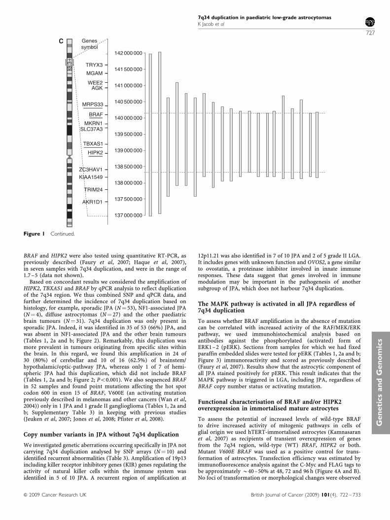
Copy number variants in JPA without 7q34 duplication
We investigated genetic aberrations occurring specifically in JPA notcarrying 7q34 duplication analysed by SNP arrays (N¼ 10) andidentified recurrent abnormalities (Table 3). Amplification of 19p13including killer receptor inhibitory genes (KIR) genes regulating theactivity of natural killer cells within the immune system wasidentified in 5 of 10 JPA. A recurrent region of amplification at
12p11.21 was also identified in 7 of 10 JPA and 2 of 5 grade II LGA.It includes genes with unknown function and OVOS2, a gene similarto ovostatin, a proteinase inhibitor involved in innate immuneresponses. These data suggest that genes involved in immunemodulation may be important in the pathogenesis of anothersubgroup of JPA, which does not harbour 7q34 duplication.
The MAPK pathway is activated in all JPA regardless of7q34 duplication
To assess whether BRAF amplification in the absence of mutationcan be correlated with increased activity of the RAF/MEK/ERKpathway, we used immunohistochemical analysis based onantibodies against the phosphorylated (activated) form ofERK1–2 (pERK). Sections from samples for which we had fixedparaffin embedded slides were tested for pERK (Tables 1, 2a and b;Figure 3) immunoreactivity and scored as previously described(Faury et al, 2007). Results show that the astrocytic component ofall JPA stained positively for pERK. This result indicates that theMAPK pathway is triggered in LGA, including JPA, regardless ofBRAF copy number status or activating mutation.
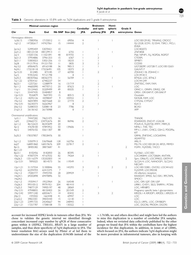
Functional characterisation of BRAF and/or HIPK2overexpression in immortalised mature astrocytes
To assess the potential of increased levels of wild-type BRAFto drive increased activity of mitogenic pathways in cells ofglial origin we used hTERT-immortalised astrocytes (Kamnasaranet al, 2007) as recipients of transient overexpression of genesfrom the 7q34 region, wild-type (WT) BRAF, HIPK2 or both.Mutant V600E BRAF was used as a positive control for trans-formation of astrocytes. Transfection efficiency was estimated byimmunofluorescence analysis against the C-Myc and FLAG tags tobe approximately B40–50% at 48, 72 and 96 h (Figure 4A and B).No foci of transformation or morphological changes were observed
137 000 000
137 500 000
138 000 000
138 500 000
139 000 000
139 500 000
140 000 000
140 500 000
141 000 000
141 500 000
142 000 000
Genessymbol
TRYX3
MGAM
WEE2AGK
MRPS33
BRAF
MKRN1SLC37A3
TRIM24
AKR1D1
TBXAS1
HIPK2
ZC3HAV1KIAA1549
333–
Figure 1 Continued.
7q34 duplication in paediatric low-grade astrocytomas
K Jacob et al
727
British Journal of Cancer (2009) 101(4), 722 – 733& 2009 Cancer Research UK
Gen
eti
cs
an
dG
en
om
ics

in cells transfected with WT constructs, whereas transformationfoci were readily distinguishable in V600E transfectant cells.Proliferation assays revealed no change in growth of cellsoverexpressing of BRAF, HIPK2 or both, compared to theirmock-transfected controls (Figure 4C). Similarly, overexpressionof BRAF had no discernible effect on the baseline level of ERKexpression or phosphorylation (Figure 4D). Indeed, in serumstarved EV and C-Myc-BRAF transfectant cells pERK was similarin intensity in both settings, in keeping with previous findings inCOS cells (Ciampi et al, 2005). Remarkably, exposure of BRAFoverexpressing cells to ligands of the epidermal growth factorreceptor (EGFR), such as transforming growth factor-a or EGFincreased pERK levels by a mean of B5.9-fold relative to 2.3-foldin empty-vector transfectants (mean of three distinct experiments,Figure 4D). These results indicate that although BRAF over-expression may not be sufficient to drive proliferative responses ofastrocytic cells it may sensitise them to exogenous growth factors.
DISCUSSION
We show that somatic duplication of 7q34 in LGA is specific toJPA. We also establish that its prevalence varies with the site of
origin within the brain of the JPA, and is more frequent incerebellar (24 of 30 – 80%) followed by brainstem and opticpathway tumours (10 of 16 – 62.5%), whereas it is rare inhemispheric JPA (1 of 7 – 14%; Tables 1, 2a and b; Figures 1and 2). Our analysis and the several validation steps we performedcharacterise this region with increased precision compared to therecently published concurrent studies, which missed either BRAFor HIPK2 (Deshmukh et al, 2008; Jones et al, 2008; Pfister et al,2008) and confirm that 7q34 amplification includes both BRAF andHIPK2 in as many as 34 of 35 JPA samples. We identify additionalgenetic regions in JPA without 7q34 duplication (Table 3) andshow that most genes within these regions are modulators of theimmune system.
Specificity of 7q34 duplication to JPA and its prevalence ininfratentorial tumours contrast with previous reports alluding that7q34 duplication may be present in non-JPA LGA (Deshmukhet al, 2008; Pfister et al, 2008; Sievert et al, 2008) and is morefrequent in non-cerebellar LGA (Pfister et al, 2008; Sievert et al,2008). Deshmukh et al showed 7q34 duplication in 8 of 10cerebellar JPA and further used increased HIPK2 expression ontissue microarray as a surrogate for marker for this genetic event.Other molecular events, distinct from 7q34 duplication, may leadto HIPK2 overexpression (Sombroek and Hofmann, 2009) and may
0
1
2
3
4
5
6C
opy
num
ber
Cerebellar Cortical Optic pathway + Brainstem Other
HIPK2
0
1
2
3
4
5
6
Cop
y nu
mbe
r
Cerebellar Cortical Optic pathway + Brainstem Other
TBXA1
0
1
2
3
4
5
6
Cop
y nu
mbe
r
Cerebellar Cortical Optic pathway + Brainstem Other
BRAF
0
1
2
3
4
5
6
Cop
y nu
mbe
r
Cerebellar Cortical Optic pathway + Brainstem Other
MRPS33
Figure 2 HIPK2, TBXAS1, BRAF and MRPS33 copy number in the brain tumours included in this study. DNA qPCR-based copy number for HIPK2,TBXAS1, BRAF and MRPS33 are plotted. The cut-off for DNA copy number was set using the mean of ±2 s.d. (plotted lines) against MRPS33, which waslocated after the genetic interval of interest (Figure 1).
7q34 duplication in paediatric low-grade astrocytomas
K Jacob et al
728
British Journal of Cancer (2009) 101(4), 722 – 733 & 2009 Cancer Research UK
Gen
etic
san
dG
en
om
ics
account for increased HIPK2 levels in tumours other than JPA. Wechose to validate the genetic interval we identified throughconcordant increased copy number by qPCR of three consecutivegenes within it (HIPK2, TBXAS1, BRAF) in a large number ofsamples, and thus show specificity of 7q34 duplication to JPA. Thelower resolution BAC-arrays used by Pfister et al led them tounderestimate the size of the duplication (0.94 Mb instead of the
B1.74 Mb, we and others describe) and might have led the authorsto miss this duplication in a number of cerebellar JPA samples.Indeed, when we revisited data collectively published by the othergroups we found that JPA within the cerebellum have the highestincidence for this duplication. In addition, in Jones et al (2008),which focused on JPA, the authors indicate 7q34 duplication mightbe more prevalent in infratentorial tumours, also in keeping with
Table 3 Genomic alterations in 10 JPA with no 7q34 duplications and 5 grade II astrocytomas
Minimal common regionCerebellar
Brainstemand optic
Hemi-spheric Grade II
Chr Start End Nb SNP Size (kb) JPA pathway JPA JPA astrocytoma Genes
Homozygous deletions1p36.13 17085956 17155012 11 69056 4 1 1 LOC100129182, TRNAN2, CROCC1q21.2 147282617 147427061 15 144444 2 FJL12528, ECM1, FJ13544, TSRC1, MCL1,
ENSA2p16.2 52995459 53070421 10 1 LOC4020722q31.2 180123158 180129913 11 6755 1 1 ZNF385B2q13 110201336 111021091 48 819755 1 1 Mall, NPHP1, FLJ, RGPD6, RGPD73p21.3 37954886 37961253 15 6367 1 2 CTDSPL3p21.1 53003023 53021256 13 18233 1 1 SFMBT13q28 191217916 191221750 11 3835 3 2 CCD504q13.2 69064675 69163188 36 98513 1 1 1 UGT2B29P, UGT2B17, LOC1001326514q34.1 173218118 173236491 105 18373 2 GALNT175p15.33 812485 873185 17 60700 1 2 ZDHHC11B, ZDHHC115q15 97053242 97121798 8 1 LOC3918135q35.3 180307066 180363775 11 56709 1 1 BTNL8, LOC, BTNL37q11.22 67054161 67482237 54 1 LOC4412498p23.1 12257261 12388979 16 131719 2 ZNF705C, FAM, FAM,9p11.2 44683090 44770712 20 87623 2 1 1 LOC11q11 55124465 55209499 49 85035 1 1 OR4C11, OR4P4, OR4S2, OR11q11 55447435 55484857 8 1 OR5I1, OR10AF1P, OR10AK1P12p13.31 9526879 9607393 15 80515 3 2 Ovostatin17p11.2 18292126 18398047 9 105922 1 1 NOS2B, FAM, LOC19q13.2 46047894 46075668 22 27774 2 CYP2A6, CYP2A719q13.41 56230373 56263967 8 1 KLK1320q13.2 52080333 52088118 23 7786 2 BCAS121q21.3 26136162 26176988 8 1 APP
Chromosomal amplifications1p31.1 74447282 74631475 16 1 TNNI3K1q43 234665731 234752676 84 86946 2 EDARADD, ENO1P, LGALS82p11.2 86152633 86343699 20 1 POLR1A, FLJ20758, IMMT, MRPL354p12 47678100 47893952 50 139606 1 NPAL1, TXK, TEC4q12 54076102 55611307 88 1 FIP1L1, LNX1, CHIC2, GSH-2, PDGFRa,
KIT5q35.3 178337857 178528476 30 1 GRM6, ZNF354C, LOC645944,
ADAMTS25q11.2 53507913 53670668 128 162756 1 ARL156q27 168091860 168319676 209 227817 2 1 MLLT4, LOC100128124, KIF25, FRMD18p12-8p11.23
38405382 38873687 41 2 FGFR1, FLJ43582, TACC1
8p23.1 8102456 8182850 26 80395 1 FLJ10661, LOC100110q11.22 47007374 47167032 82 74264 2 1 LOC340844, LOC728684, ANTXRL10q26.3 135116379 135202003 14 2 Sprn, OR6LP2, LOC399832, OR7M1P12p13.31 7895025 8014573 56 119549 1 SLC2A14, LOC, NANOGP1, SLC2A3,
NECAP112p11.21 31157554 31300846 55 143293 3 2 2 2 LOC100132881, OVOS2, LOC44163212q14.2 62294703 62415375 7 1 FLJ32949, LOC39033814q11.2 19283777 19493705 28 209929 1 All olfactory receptors14q24.1-14q24.2
69202898 69470896 32 1 KIAA0247, SFRS5, SLC10A1, RPL7AP6,SMOC1
15q11.2 19359417 19523964 26 164548 1 LOC, OR11J2P, OR11J5P15q26.3 99310512 99791921 204 481410 1 LRRK1, CHSY1, SELS, SNRPA1, PCSK617q25.3 74877129 74905197 48 28069 1 LOC, HRNBP319q13.31 47948855 48150403 26 201549 2 Pregnancy specific beta-1-glycoproteins19q13.42 59971240 60054671 14 83432 1 3 2 KIR2DL1-4, KIR3DP1, KIR3DL1, KIR2DS1-420p13 1593502 1663448 41 69947 1 LOC21q22.2 39823301 39835430 15 12130 1 LOC22q11.23 23991725 24240667 94 248943 1 IGLL3, LRP5L, LOC, CRYBB2P122q11.21 17257787 17388108 73 130322 1 LOC, DGCR6, PRODH, DGCR5
7q34 duplication in paediatric low-grade astrocytomas
K Jacob et al
729
British Journal of Cancer (2009) 101(4), 722 – 733& 2009 Cancer Research UK
Gen
eti
cs
an
dG
en
om
ics

our data. They also show that one sample was classified as a gradeII astocytoma; however, the child had prolonged disease-freesurvival more in keeping with a diagnosis of JPA. Thismisclassification might also account for the findings of 7q34duplication in some non-JPA LGA in the study by the other groups(Deshmukh et al, 2008; Pfister et al, 2008; Sievert et al, 2008). Thecentral review of samples by senior paediatric neuropathologistswe performed has been shown to increase reproducibility ofneuropathologic classification of tumours (Pollack et al, 2003) andmay account for increased accuracy in our dataset. Our findingsand these from recent studies alluding to the presence of differentmolecular signatures in sporadic JPA based on its region of origin(Sharma et al, 2007) make it tempting to speculate that, like CNSependymoma (Taylor et al, 2005), variants of JPA might arise froma unique site-restricted progenitor cell.
RAF kinases are components of a conserved signalling pathwaythat regulates cellular responses to extracellular signals (Wan et al,2004). Their role in glioma formation, and the role of the MAPKcascade they trigger, have been identified in tumour samples andfurther confirmed in mouse models (Jeuken et al, 2007; Pritchardet al, 2007; Lyustikman et al, 2008; Pfister et al, 2008). NF1 encodesfor a Ras-GTPAse and its loss of function may lead to activation ofRas/RAF signalling and coincides with formation of indolent JPAmainly within the optic pathway. Our exploration of immortalisedastrocytes show that overexpression of wild-type BRAF may not besufficient by itself to mediate the activation of the MAPK cascade.In keeping with these data, only oncogenic BRAF mutations havebeen implicated in tumours of epithelial (Rajagopalan et al, 2002)and neuroectodermal origin (Brose et al, 2002; Pollock et al, 2003).7q34 duplication in JPA was recently shown to produce novel
JPA with 7q34 duplication JPA without 7q34 duplication
P13
P45
P41
P55
P50
P57
P28
P10
P36
P14
P27
P21 P32
P33
P31
P23
P15
P11
P25
P24
P29
P16
P12
P9
Figure 3 The astrocytic component of all the brain tumours included in this study show MAPK pathway activation regardless of BRAF copy number.Immunohistochemical analyses for phosphorylated ERK (pERK), used as a surrogate marker of MAPK pathway activation were performed for the 52 samplesincluded in this study for which formalin-fixed paraffin-embedded slides were available. Sections were stained using anti-pERK followed by detection usingthe DAKO kit (red accounts for positive staining) and hematoxylin counterstaining. Staining intensity was scored as in (Faury et al, 2007; Haque et al, 2007).Full characteristics of samples are provided in Tables 1, 2a and b.
7q34 duplication in paediatric low-grade astrocytomas
K Jacob et al
730
British Journal of Cancer (2009) 101(4), 722 – 733 & 2009 Cancer Research UK
Gen
etic
san
dG
en
om
ics
DAPI
C-MYC/BRAF
EV
FLAG DAPI Overlay
FLAG/HIPK2
EV
Empty vectorBRAFHIPK2BRAF+HIPK2
EV C-MYC/BRAF EV C-MYC/BRAF
1 2.1 2.5 1 5.5 5.9
0 5 150EGF (200ng)
BRAF
pERK
5 15C-MYC
BRAF
pERK
�-actin �-actin
48 h 72 h 96 h
CP
M
C-MYC BRAF Overlay
350 000300 000250 000200 000150 000100 00050 000
0
Figure 4 Functional analysis of BRAF and HIPK2 overexpression in hTERT-immortalised astrocytes. (A) Immunofluorescence analysis of hTERT-immortalised astrocytes transfected with CMYC/BRAF or the empty vector (EV) at 48 h using an antibody recognising the C-MYC tag (red), BRAF (green)and DAPI counterstaining (blue). (B) Immunofluorescence analysis of hTERT-immortalised astrocytes transfected with FLAG/HIPK2 or the EV at 48 h usingan antibody recognising the FLAG tag (red) and DAPI counterstaining (blue). (C) Proliferation of hTERT-immortalised astrocytes was assessed at 48, 72 and96 h following transfection with mock, BRAF, HIPK2 or both genes. No difference in the rate of cell growth was observed between the transfectant cells.Results represent the median of three separate experiments performed in triplicates. (C) Total cell extracts of hTERT-immortalised astrocytes transfectedwith the empty-vector or C-Myc-tagged BRAF. Cells were serum starved overnight before protein lysate extraction. (D) Left panel: hTERT-immortalisedastrocytes were transiently transfected with CMYC/BRAF or the empty vector. Cells were serum starved overnight and total protein lysates extracted at48 h posttransfection. Western blot analysis for C-Myc, BRAF, phopshoERK (pERK) and b-actin (loading control) was performed. Right panel: Empty-vector(EV) and C-Myc/BRAF transfectant hTERT-immortalised astrocytes were stimulated with 200 ng of epidermal growth factor (EGF) for 5 and 15 min. Totalcell lysates extracted at baseline (0) and following activation (5, 15 min) with EGF were immunoblotted using antibodies against b-actin (loading control) andphosphorylated ERK (pERK), C-Myc and BRAF. Note the shift of BRAF immunoreactive band following EGF activation which indicates phosphorylation ofthe kinase and increase in its molecular weight. Importantly, increase in pERK levels relative to baseline following EGF stimulation was two fold higher inC-MYC/BRAF transfectant cells than in EV transfectants. Results represent the median of three separate experiments (Po0.001).
7q34 duplication in paediatric low-grade astrocytomas
K Jacob et al
731
British Journal of Cancer (2009) 101(4), 722 – 733& 2009 Cancer Research UK
Gen
eti
cs
an
dG
en
om
ics

oncogenic BRAF fusion genes with transforming capacity (Joneset al, 2008), similar to findings in radiation-associated papillarythyroid cancer (Ciampi et al, 2005) where an intrachromosomalinversion, and not duplication, let to this oncogenic event. Thesedata indicate that wild-type BRAF would be unlikely to inducetransformation. Tumours in which the duplication occurs withoutthis in-frame fusion or in patients with trisomy of the fullchromosome 7 require other mechanisms to activate BRAF andinduce oncogenesis. Intriguingly, our data suggest that all JPAhave active MAPK pathway regardless of 7q34 duplication(Figure 3). In keeping with the importance of the MAPK pathwayin JPA genesis, two additional alternative mechanisms resulting inMAPK activation were very recently identified in JPA (Jones et al,2009). This group studied the 10 JPA for which V600E pointmutation of BRAF (2 of 44), clinical diagnosis of NF1 (3 of 44) orthe common BRAF fusion following 7q34 duplication (29 of 44)could not be found (Jones et al, 2008). In one patient they foundtandem duplication at 3p25 producing an in-frame oncogenicfusion between SRGAP3 and RAF1, a finding corroborated by aconcurrent group on additional patients with JPA and no clinicalNF1, 7q34 duplication or BRAF mutations (Forshew et al, 2009).This genetic event bears striking resemblance to the commonKIAA1549 BRAF fusion event and the fusion protein includes theRaf1 kinase domain, and shows elevated kinase activity whencompared with wild-type Raf1. Secondly, in one patient with JPA anovel 3 bp insertion at codon 598 in BRAF mimics the hotspotV600E mutation to produce a transforming, constitutively activeBRaf kinase (Jones et al, 2009). These findings further imply thatBRAF or RAF1 constitutive activation, achieved through differentgenetic events, converge to activate the MAPK pathway and thatthis pathway could be clinically targeted in all JPA.
Gains in 7q34 have been described in other cancers includingovarian cancer. However these are amplifications and notduplications and the region involved has some but no completeoverlap with the one identified in JPA. Also, genetic analysis ofmedulloblastomas (Gilbertson and Ellison, 2008), ependymomas(Taylor et al, 2005) and high-grade astrocytomas (our data) do notidentify 7q34 duplication in these tumours making this geneticevent specific to JPA. Unscheduled activation of the MAPKpathway may lead to cellular senescence, which could functionto limit tumour development (Courtois-Cox et al, 2006). Thereforewe also postulate that oncogenic BRAF may cooperate with other,presently unknown changes, to drive formation of a subset of JPAat the same time it may drive activation of other pathways
including cellular senescence. Ultimately, this may make thistumour less aggressive, similar to what is observed in melanocyteswhere oncogenic BRAF mutations result only in naevi formation.As complete surgical removal is the rule for cerebellar JPA, studiescompiling higher numbers of JPA, occurring in other locations, areneeded to establish whether 7q34 duplication is indeed associatedwith improved progression-free survival, as well as the biologicalmechanisms controlling these variables.
In summary, our studies suggest that the 7q34 region may play asite-specific role in pathogenesis of JPA and that the involvementof an active MAP kinase signalling pathway through oncogenicBRAF and other RAF family members in this process is crucial.They further emphasise the relevance of therapeutic targeting ofthis pathway in all JPA. To avoid misdiagnosis and adapt therapies,all infratentorial LGA should be screened by immunohistochem-istry for active MAP kinase pathway (phosphorylated ERK) andfurther tested for the presence of 7q34 duplication, then, in itsabsence, 3p25 gain or BRAF mutation. Further functionalevaluation of genes included within this genetic interval, otherswe identify herein, and others involved in the MAP kinase cascadeand their correlation in larger sample sets to the site of origin, ageof the patient and progression-free survival will help shed light onthe pathogenesis of JPA.
ACKNOWLEDGEMENTS
This work was supported by the Canadian Institute of HealthResearch (CIHR) and the Brain Tumor Foundation (NJ, UT), theCole Foundation (NJ), the Hungarian Scientific Research Fund(OTKA) Contract No. T-04639 and the National Research andDevelopment Fund (NKFP) Contract No. 1A/002/2004 (PH, MG,LB, ZH). K Jacob is the recipient of studentship award from CIHRand N Jabado is the recipient of a Chercheur Boursier award fromFonds de Recherche en Sante du Quebec.
Conflict of interestThe authors declare no conflict of interest.
Supplementary Information accompanies the paper on BritishJournal of Cancer website (http://www.nature.com/bjc)
REFERENCES
Bar EE, Lin A, Tihan T, Burger PC, Eberhart CG (2008) Frequent gains atchromosome 7q34 involving BRAF in pilocytic astrocytoma. J Neuro-pathol Exp Neurol 67: 878 – 887
Broniscer A, Baker SJ, West AN, Fraser MM, Proko E, Kocak M, Dalton J,Zambetti GP, Ellison DW, Kun LE, Gajjar A, Gilbertson RJ, Fuller CE(2007) Clinical and molecular characteristics of malignant transforma-tion of low-grade glioma in children. J Clin Oncol 25: 682 – 689
Brose MS, Volpe P, Feldman M, Kumar M, Rishi I, Gerrero R, Einhorn E,Herlyn M, Minna J, Nicholson A, Roth JA, Albelda SM, Davies H, Cox C,Brignell G, Stephens P, Futreal PA, Wooster R, Stratton MR, Weber BL(2002) BRAF and RAS mutations in human lung cancer and melanoma.Cancer Res 62: 6997 – 7000
Cheng Y, Pang JC, Ng HK, Ding M, Zhang SF, Zheng J, Liu DG, Poon WS(2000) Pilocytic astrocytomas do not show most of the genetic changescommonly seen in diffuse astrocytomas. Histopathology 37: 437 – 444
Ciampi R, Knauf JA, Kerler R, Gandhi M, Zhu Z, Nikiforova MN, RabesHM, Fagin JA, Nikiforov YE (2005) Oncogenic AKAP9-BRAF fusion is anovel mechanism of MAPK pathway activation in thyroid cancer. J ClinInvest 115: 94 – 101
Courtois-Cox S, Genther Williams SM, Reczek EE, Johnson BW,McGillicuddy LT, Johannessen CM, Hollstein PE, MacCollin M,Cichowski K (2006) A negative feedback signaling network underliesoncogene-induced senescence. Cancer Cell 10: 459 – 472
Deshmukh H, Yeh TH, Yu J, Sharma MK, Perry A, Leonard JR, Watson MA,Gutmann DH, Nagarajan R (2008) High-resolution, dual-platform aCGHanalysis reveals frequent HIPK2 amplification and increased expressionin pilocytic astrocytomas. Oncogene 27: 4745 – 4751
Faury D, Nantel A, Dunn SE, Guiot MC, Haque T, Hauser P, Garami M,Bognar L, Hanzely Z, Liberski PP, Lopez-Aguilar E, Valera ET, Tone LG,Carret AS, Del Maestro RF, Gleave M, Montes JL, Pietsch T, Albrecht S,Jabado N (2007) Molecular profiling identifies prognostic subgroups ofpediatric glioblastoma and shows increased YB-1 expression in tumors.J Clin Oncol 25: 1196 – 1208
Fisher PG, Tihan T, Goldthwaite PT, Wharam MD, Carson BS, Weingart JD,Repka MX, Cohen KJ, Burger PC (2008) Outcome analysis of childhoodlow-grade astrocytomas. Pediatr Blood Cancer 51: 245 – 250
Forshew T, Tatevossian RG, Lawson AR, Ma J, Neale G, Ogunkolade BW,Jones TA, Aarum J, Dalton J, Bailey S, Chaplin T, Carter RL, Gajjar A,
7q34 duplication in paediatric low-grade astrocytomas
K Jacob et al
732
British Journal of Cancer (2009) 101(4), 722 – 733 & 2009 Cancer Research UK
Gen
etic
san
dG
en
om
ics

Broniscer A, Young BD, Ellison DW, Sheer D (2009) Activation of theERK/MAPK pathway: a signature genetic defect in posterior fossapilocytic astrocytomas. J Pathol 218: 172 – 181
Gajjar A, Sanford RA, Heideman R, Jenkins JJ, Walter A, Li Y, Langston JW,Muhlbauer M, Boyett JM, Kun LE (1997) Low-grade astrocytoma:a decade of experience at St. Jude Children0s Research Hospital. J ClinOncol 15: 2792 – 2799
Gilbertson RJ, Ellison DW (2008) The origins of medulloblastoma subtypes.Annu Rev Pathol 3: 341 – 365
Grill J, Laithier V, Rodriguez D, Raquin MA, Pierre-Kahn A, Kalifa C (2000)When do children with optic pathway tumours need treatment?An oncological perspective in 106 patients treated in a single centre.Eur J Pediatr 159: 692 – 696
Hakonarson H, Grant SF, Bradfield JP, Marchand L, Kim CE, Glessner JT,Grabs R, Casalunovo T, Taback SP, Frackelton EC, Lawson ML,Robinson LJ, Skraban R, Lu Y, Chiavacci RM, Stanley CA, Kirsch SE,Rappaport EF, Orange JS, Monos DS, Devoto M, Qu HQ, PolychronakosC (2007) A genome-wide association study identifies KIAA0350 as a type1 diabetes gene. Nature 448: 591 – 594
Haque T, Faury D, Albrecht S, Lopez-Aguilar E, Hauser P, Garami M,Hanzely Z, Bognar L, Del Maestro RF, Atkinson J, Nantel A, Jabado N(2007) Gene expression profiling from formalin-fixed paraffin-embeddedtumors of pediatric glioblastoma. Clin Cancer Res 13: 6284 – 6292
Ishii N, Sawamura Y, Tada M, Daub DM, Janzer RC, Meagher-Villemure M,de Tribolet N, Van Meir EG (1998) Absence of p53 gene mutations in atumor panel representative of pilocytic astrocytoma diversity using a p53functional assay. Int J Cancer 76: 797 – 800
Jabado N, Jauliac S, Pallier A, Bernard F, Fischer A, Hivroz C (1998) Sam68association with p120GAP in CD4+ T cells is dependent on CD4 moleculeexpression. J Immunol 161: 2798 – 2803
Jeuken J, van den Broecke C, Gijsen S, Boots-Sprenger S, Wesseling P(2007) RAS/RAF pathway activation in gliomas: the result of copynumber gains rather than activating mutations. Acta Neuropathol 114:121 – 133
Jones DT, Kocialkowski S, Liu L, Pearson DM, Backlund LM, Ichimura K,Collins VP (2008) Tandem duplication producing a novel oncogenicBRAF fusion gene defines the majority of pilocytic astrocytomas. CancerRes 68: 8673 – 8677
Jones DT, Kocialkowski S, Liu L, Pearson DM, Ichimura K, Collins VP(2009) Oncogenic RAF1 rearrangement and a novel BRAF mutation asalternatives to KIAA1549:BRAF fusion in activating the MAPK pathwayin pilocytic astrocytoma. Oncogene 28: 2119 – 2123
Kamnasaran D, Qian B, Hawkins C, Stanford WL, Guha A (2007) GATA6 isan astrocytoma tumor suppressor gene identified by gene trapping ofmouse glioma model. Proc Natl Acad Sci USA 104: 8053 – 8058
Kleihues P, Louis DN, Scheithauer BW, Rorke LB, Reifenberger G,Burger PC, Cavenee WK (2002) The WHO classification of tumors ofthe nervous system. J Neuropathol Exp Neurol 61: 215 – 225; discussion226 – 229
Louis DN, Ohgaki H, Wiestler OD, Cavenee WK, Burger PC, Jouvet A,Scheithauer BW, Kleihues P (2007) The 2007 WHO classification oftumours of the central nervous system. Acta Neuropathol 114: 97 – 109
Lyustikman Y, Momota H, Pao W, Holland EC (2008) Constitutiveactivation of Raf-1 induces glioma formation in mice. Neoplasia 10:501 – 510
Pfister S, Janzarik WG, Remke M, Ernst A, Werft W, Becker N, Toedt G,Wittmann A, Kratz C, Olbrich H, Ahmadi R, Thieme B, Joos S,Radlwimmer B, Kulozik A, Pietsch T, Herold-Mende C, Gnekow A,Reifenberger G, Korshunov A, Scheurlen W, Omran H, Lichter P(2008) BRAF gene duplication constitutes a mechanism of MAPKpathway activation in low-grade astrocytomas. J Clin Invest 118:1739 – 1749
Pollack IF, Boyett JM, Yates AJ, Burger PC, Gilles FH, Davis RL, Finlay JL(2003) The influence of central review on outcome associations in
childhood malignant gliomas: results from the CCG-945 experience.Neuro Oncol 5: 197 – 207
Pollack IF, Mulvihill JJ (1997) Neurofibromatosis 1 and 2. Brain Pathol 7:823 – 836
Pollock PM, Harper UL, Hansen KS, Yudt LM, Stark M, Robbins CM, MosesTY, Hostetter G, Wagner U, Kakareka J, Salem G, Pohida T, Heenan P,Duray P, Kallioniemi O, Hayward NK, Trent JM, Meltzer PS (2003) Highfrequency of BRAF mutations in nevi. Nat Genet 33: 19 – 20
Pritchard C, Carragher L, Aldridge V, Giblett S, Jin H, Foster C, Andreadi C,Kamata T (2007) Mouse models for BRAF-induced cancers. Biochem SocTrans 35: 1329 – 1333
Rajagopalan H, Bardelli A, Lengauer C, Kinzler KW, Vogelstein B,Velculescu VE (2002) Tumorigenesis: RAF/RAS oncogenes and mis-match-repair status. Nature 418: 934
Rajasekhar VK, Viale A, Socci ND, Wiedmann M, Hu X, Holland EC (2003)Oncogenic Ras and Akt signaling contribute to glioblastoma formationby differential recruitment of existing mRNAs to polysomes. Mol Cell 12:889 – 901
Rickert CH, Paulus W (2004) Comparative genomic hybridization in centraland peripheral nervous system tumors of childhood and adolescence.J Neuropathol Exp Neurol 63: 399 – 417
Sharma MK, Mansur DB, Reifenberger G, Perry A, Leonard JR, Aldape KD,Albin MG, Emnett RJ, Loeser S, Watson MA, Nagarajan R, Gutmann DH(2007) Distinct genetic signatures among pilocytic astrocytomas relate totheir brain region origin. Cancer Res 67: 890 – 900
Shi Q, Bao S, Maxwell JA, Reese ED, Friedman HS, Bigner DD, Wang XF,Rich JN (2004) Secreted protein acidic, rich in cysteine (SPARC),mediates cellular survival of gliomas through AKT activation. J BiolChem 279: 52200 – 52209
Sievert AJ, Jackson EM, Gai X, Hakonarson H, Judkins AR, Resnick AC,Sutton LN, Storm PB, Shaikh TH, Biegel JA (2008) Duplication of 7q34in pediatric low-grade astrocytomas detected by high-density single-nucleotide polymorphism-based genotype arrays results in a novelBRAF fusion gene. Brain Pathol, 21 October 2008 [E-pub ahead of print]
Sombroek D, Hofmann TG (2009) How cells switch HIPK2 on and off.Cell Death Differ 16: 187 – 194
Tada K, Kochi M, Saya H, Kuratsu J, Shiraishi S, Kamiryo T, Shinojima N,Ushio Y (2003) Preliminary observations on genetic alterations inpilocytic astrocytomas associated with neurofibromatosis 1. Neuro Oncol5: 228 – 234
Taylor MD, Poppleton H, Fuller C, Su X, Liu Y, Jensen P, Magdaleno S,Dalton J, Calabrese C, Board J, Macdonald T, Rutka J, Guha A, Gajjar A,Curran T, Gilbertson RJ (2005) Radial glia cells are candidate stem cellsof ependymoma. Cancer Cell 8: 323 – 335
Wan PT, Garnett MJ, Roe SM, Lee S, Niculescu-Duvaz D, Good VM, JonesCM, Marshall CJ, Springer CJ, Barford D, Marais R (2004) Mechanism ofactivation of the RAF-ERK signaling pathway by oncogenic mutations ofB-RAF. Cell 116: 855 – 867
Wang K, Li M, Hadley D, Liu R, Glessner J, Grant SF, Hakonarson H, BucanM (2007) PennCNV: an integrated hidden Markov model designed forhigh-resolution copy number variation detection in whole-genome SNPgenotyping data. Genome Res 17: 1665 – 1674
Wemmert S, Romeike BF, Ketter R, Steudel WI, Zang KD, Urbschat S(2006) Intratumoral genetic heterogeneity in pilocytic astrocytomasrevealed by CGH-analysis of microdissected tumor cells and FISH ontumor tissue sections. Int J Oncol 28: 353 – 360
White FV, Anthony DC, Yunis EJ, Tarbell NJ, Scott RM, Schofield DE(1995) Nonrandom chromosomal gains in pilocytic astrocytomas ofchildhood. Hum Pathol 26: 979 – 986
Wong KK, Tsang YT, Chang YM, Su J, Di Francesco AM, Meco D, RiccardiR, Perlaky L, Dauser RC, Adesina A, Bhattacharjee M, Chintagumpala M,Lau CC (2006) Genome-wide allelic imbalance analysis of pediatricgliomas by single nucleotide polymorphic allele array. Cancer Res 66:11172 – 11178
7q34 duplication in paediatric low-grade astrocytomas
K Jacob et al
733
British Journal of Cancer (2009) 101(4), 722 – 733& 2009 Cancer Research UK
Gen
eti
cs
an
dG
en
om
ics
Copyright © 2022 FDOKUMEN