Basic of Retailing Subject Lesson Plan - Saraswati Mahila ...
Upload
khangminh22Category
view
0download
0
Nucleic AcidsDNA & RNA
Ms. Bhoomi D. Patel
Assistant Professor
Department of Pharmaceutical Chemistry & Quality Assurance
Saraswati Institute of Pharmaceutical Sciences
Dhanap, Gandhinagar - 382355

Based on McMurry, Organic Chemistry,
Chapter 28, 6th edition, (c) 2003
2
Nucleic Acids and Heredity
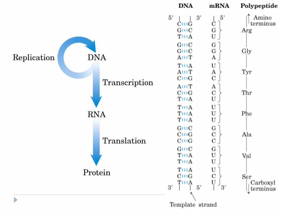
Processes in the transfer of genetic information:
Replication: identical copies of DNA are made
Transcription: genetic messages are read and carried out of the cell nucleus to the ribosomes, where protein synthesis occurs.
Translation: genetic messages are decoded to make proteins.
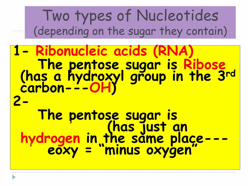
Two types of Nucleotides(depending on the sugar they contain)
1- Ribonucleic acids (RNA)The pentose sugar is Ribose
(has a hydroxyl group in the 3rd
carbon---OH)2- Deoxyribonucleic acids (DNA)
The pentose sugar is Deoxyribose (has just an hydrogen in the same place---H)Deoxy = “minus oxygen”

Definitions
Nucleic acids are polymers of nucleotides
In eukaryotic cells nucleic acids are either:
Deoxyribose nucleic acids (DNA)
Ribose nucleic acids (RNA)
Messenger RNA (mRNA)
Transfer RNA (tRNA)
Ribosomal RNA (tRNA)
Nucleotides are carbon ring structures containing nitrogen linked to a
5-carbon sugar (a ribose)
5-carbon sugar is either a ribose or a deoxy-ribose making the
nucleotide either a ribonucleotide or a deoxyribonucleotide
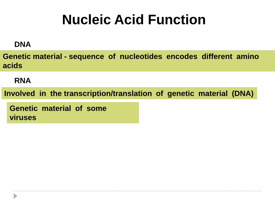
Nucleic Acid Function
DNA
Genetic material - sequence of nucleotides encodes different amino
acids
RNA
Involved in the transcription/translation of genetic material (DNA)
Genetic material of some
viruses

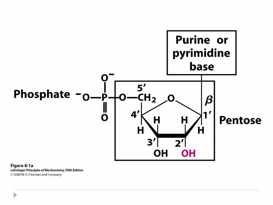
Nucleotide Structure
Despite the complexity and diversity of life the structure of DNA is
dependent on only 4 different nucleotides
Diversity is dependent on the nucleotide sequence
All nucleotides are 2 ring structures composed of:
5-carbon sugar : b-D-ribose (RNA)
b-D-deoxyribose (DNA)
Base Purine
Pyrimidine
Phosphate group A nucleotide WITHOUT a phosphate group is a
NUCLEOSIDE
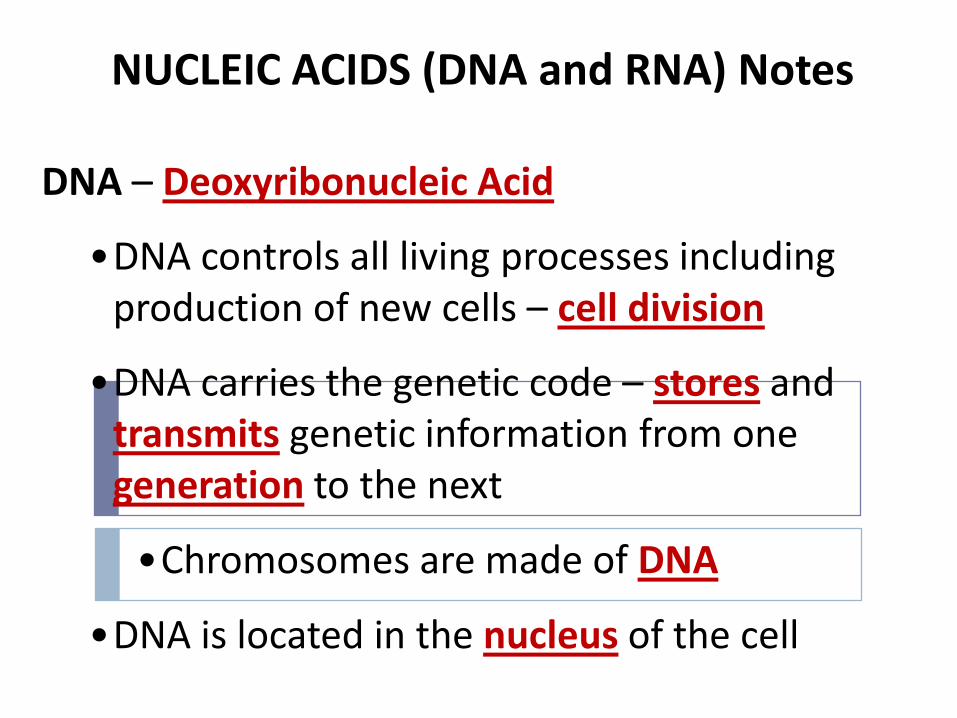
NUCLEIC ACIDS (DNA and RNA) Notes
DNA – Deoxyribonucleic Acid
•DNA controls all living processes including production of new cells – cell division
•DNA carries the genetic code – stores and transmits genetic information from one generation to the next
•Chromosomes are made of DNA
•DNA is located in the nucleus of the cell
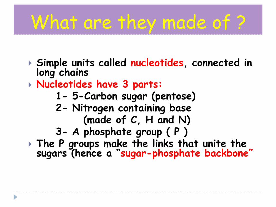
What are they made of ?
Simple units called nucleotides, connected in long chains
Nucleotides have 3 parts:1- 5-Carbon sugar (pentose)2- Nitrogen containing base
(made of C, H and N)3- A phosphate group ( P )
The P groups make the links that unite the sugars (hence a “sugar-phosphate backbone”
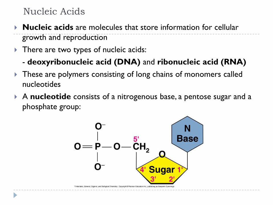
Nucleic Acids
Nucleic acids are molecules that store information for cellular
growth and reproduction
There are two types of nucleic acids:
- deoxyribonucleic acid (DNA) and ribonucleic acid (RNA)
These are polymers consisting of long chains of monomers called
nucleotides
A nucleotide consists of a nitrogenous base, a pentose sugar and a
phosphate group:

All nucleotides contain three components:
1. A nitrogen heterocyclic base
2. A pentose sugar
3. A phosphate residue
Nucleic Acids
DNA and RNA are nucleic acids, long, thread-like polymers
made up of a linear array of monomers called nucleotides
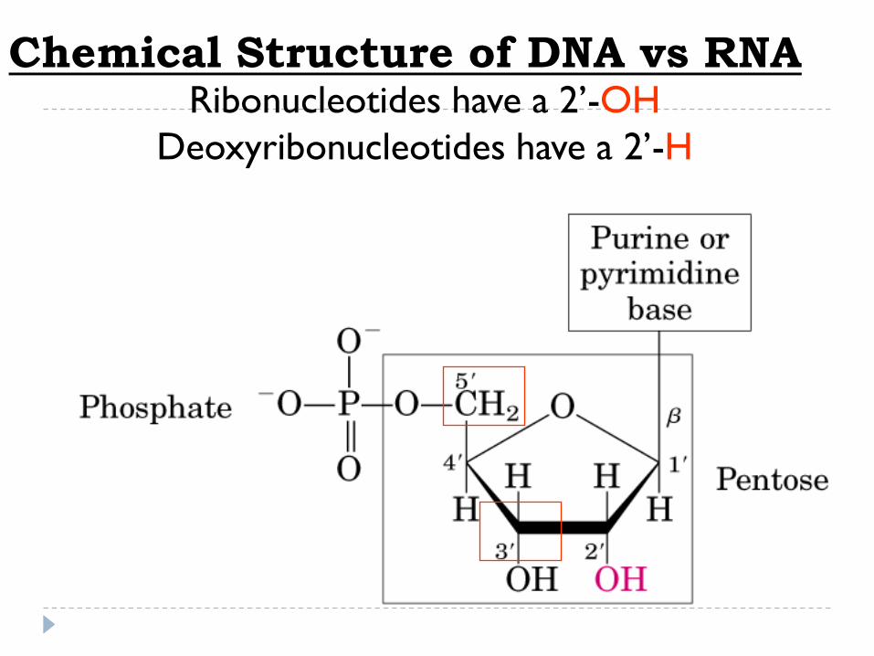
Ribonucleotides have a 2’-OH
Deoxyribonucleotides have a 2’-H
Chemical Structure of DNA vs RNA
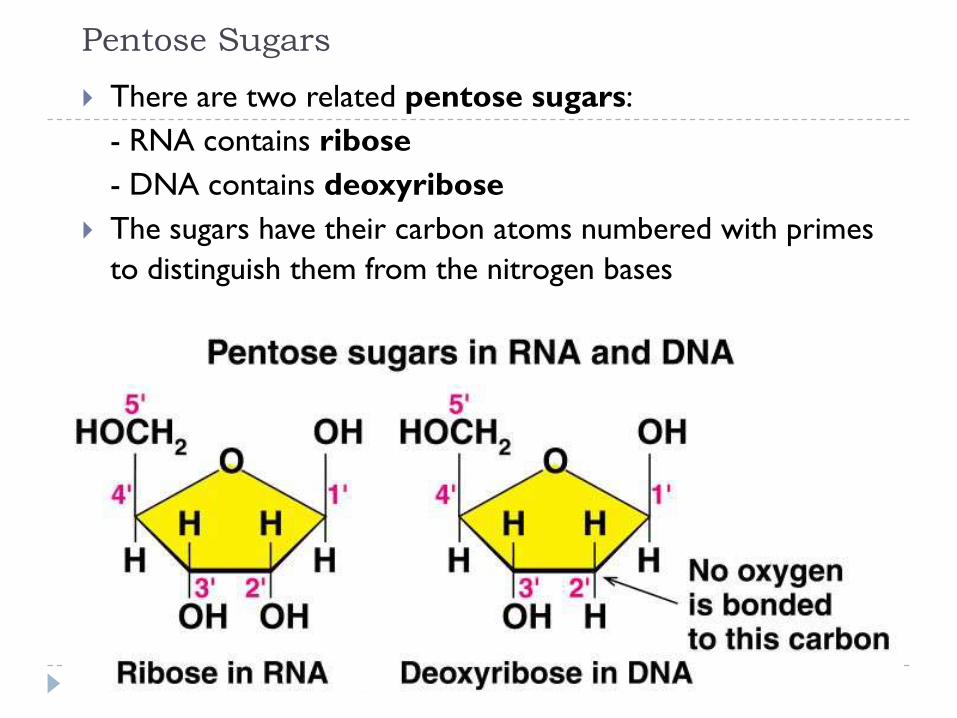
Pentose Sugars
There are two related pentose sugars:
- RNA contains ribose
- DNA contains deoxyribose
The sugars have their carbon atoms numbered with primes
to distinguish them from the nitrogen bases
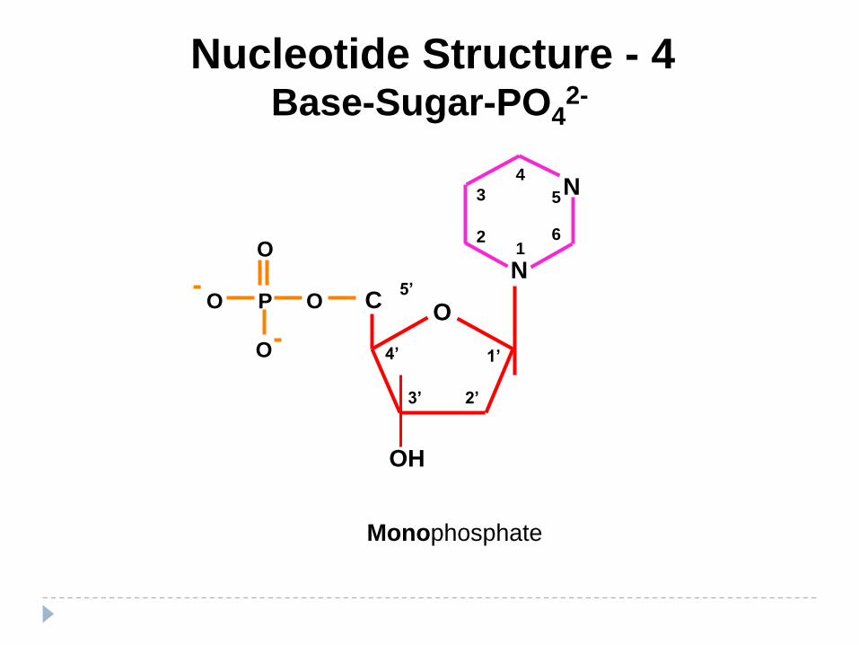
Nucleotide Structure - 4Base-Sugar-PO4
2-
P
O
O
O
O
N
N
5
61
2
3
4
OC
5’
1’4’
3’ 2’
OH
Monophosphate
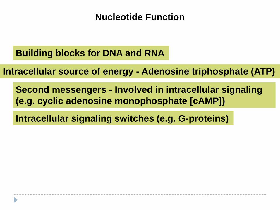
Nucleotide Function
Building blocks for DNA and RNA
Intracellular source of energy - Adenosine triphosphate (ATP)
Second messengers - Involved in intracellular signaling
(e.g. cyclic adenosine monophosphate [cAMP])
Intracellular signaling switches (e.g. G-proteins)

Nucleotide Structure - 4Phosphate Groups
Phosphate groups are what makes a nucleoside a
nucleotide
Phosphate groups are essential for nucleotide
polymerization
P
O
O
O
O X
Basic structure:

Nucleotide Structure - 4Phosphate Groups
Number of phosphate groups determines nomenclature
P
O
O
O
O CH2
P
O
O
O
P
O
O
O
O CH2
Monophosphate
e.g. AMP
Diphosphatee.g. ADP
Free = inorganic
phosphate (Pi)
Free = Pyro-
phosphate (PPi)

Triphosphate
e.g. ATP
P
O
O
O
P
O
O
O
O P
O
O
O CH2
Nucleotide Structure - 4Phosphate Groups
No Free form exists
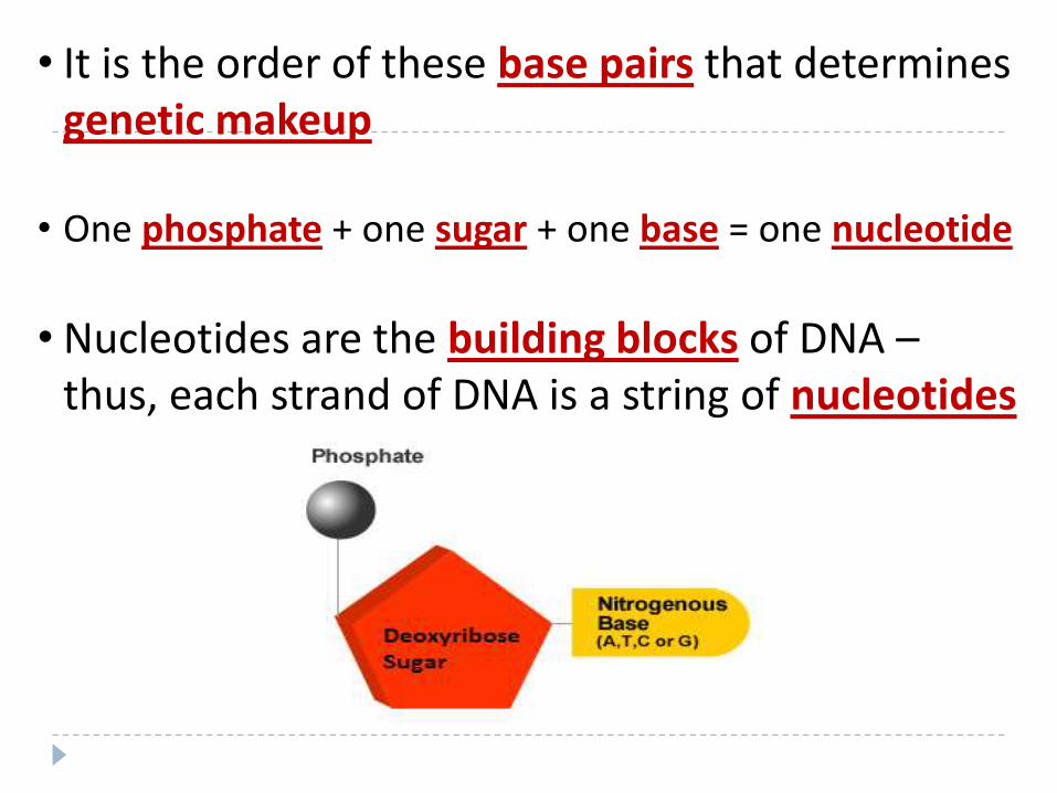
• It is the order of these base pairs that determines genetic makeup
• One phosphate + one sugar + one base = one nucleotide
• Nucleotides are the building blocks of DNA –thus, each strand of DNA is a string of nucleotides

Sanger dideoxy sequencing incorporates dideoxy
nucleotides, preventing further synthesis of the DNA
strand
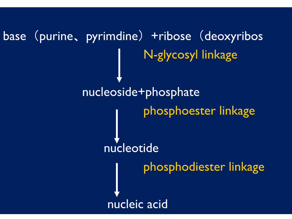
base(purine、pyrimdine)+ribose(deoxyribos
N-glycosyl linkage
nucleoside+phosphate
phosphoester linkage
nucleotide
phosphodiester linkage
nucleic acid

Nucleotide Structure - 1Sugars
OHOCH2
5’
1’4’
3’ 2’
OHOCH2
OH
OHOH
OHOCH2
OH
HOH
Ribose
Deoxyribose
Generic Ribose
Structure
N.B. Carbons are given numberings
as a prime
Based on McMurry, Organic Chemistry,
Chapter 28, 6th edition, (c) 2003
24
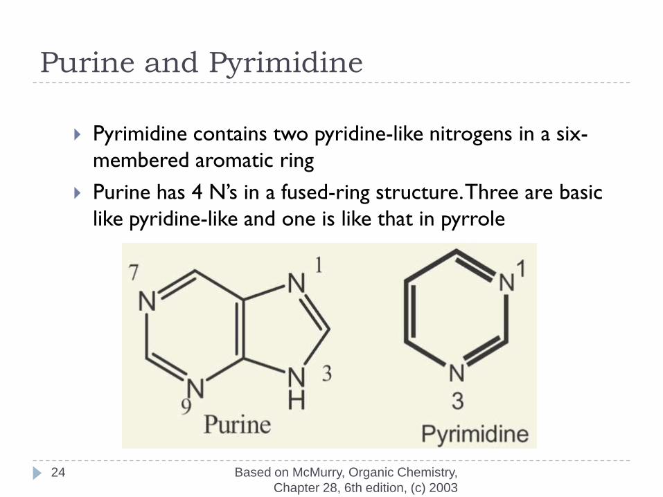
Purine and Pyrimidine
Pyrimidine contains two pyridine-like nitrogens in a six-
membered aromatic ring
Purine has 4 N’s in a fused-ring structure. Three are basic
like pyridine-like and one is like that in pyrrole

Nucleotide Structure - 2Bases - Purines
N
N
N
N
1
23
4
5
67
8
9
Adenine
Guanine
A
G
N
N
N
N
H
NH2
N
N
N
N
H
O
H
NH2

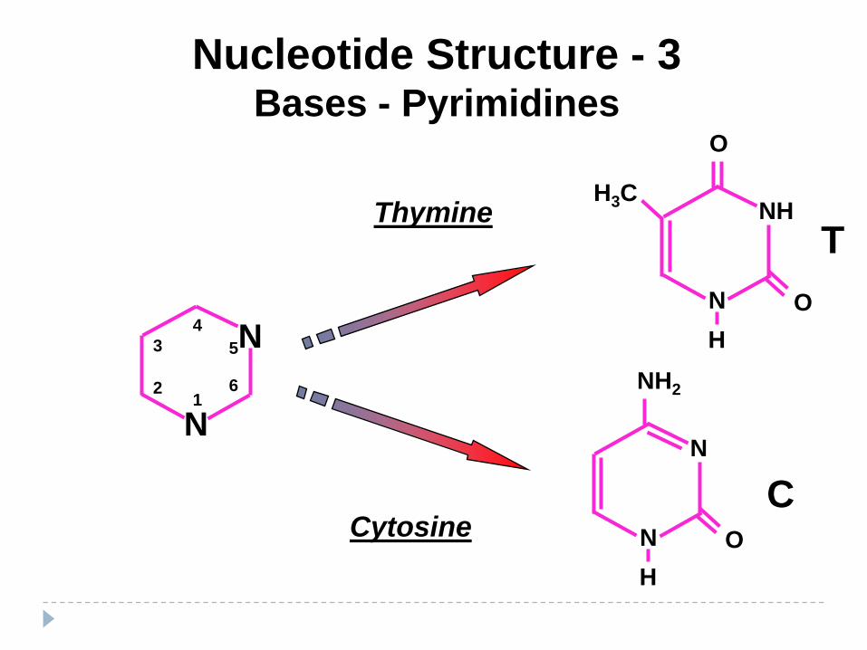
Nucleotide Structure - 3Bases - Pyrimidines
N
N
5
61
2
3
4
Thymine
Cytosine
NH
N
O
T
O
H
H3C
C
N
N
NH2
O
H

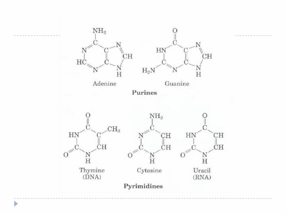
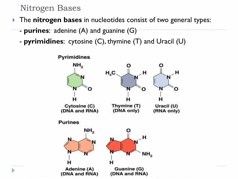
Nitrogen Bases
The nitrogen bases in nucleotides consist of two general types:
- purines: adenine (A) and guanine (G)
- pyrimidines: cytosine (C), thymine (T) and Uracil (U)
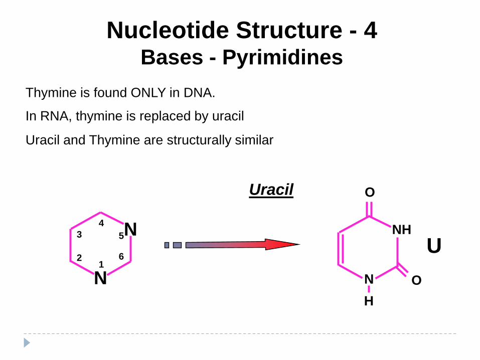
Nucleotide Structure - 4Bases - Pyrimidines
N
N
5
61
2
3
4
Uracil
NH
N
O
U
O
H
Thymine is found ONLY in DNA.
In RNA, thymine is replaced by uracil
Uracil and Thymine are structurally similar
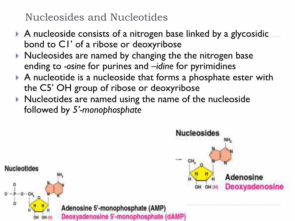
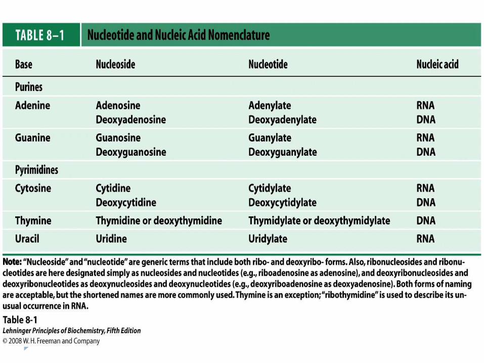
Nucleosides and Nucleotides
A nucleoside consists of a nitrogen base linked by a glycosidicbond to C1’ of a ribose or deoxyribose
Nucleosides are named by changing the the nitrogen base ending to -osine for purines and –idine for pyrimidines
A nucleotide is a nucleoside that forms a phosphate ester with the C5’ OH group of ribose or deoxyribose
Nucleotides are named using the name of the nucleoside followed by 5’-monophosphate

Names of Nucleosides and Nucleotides
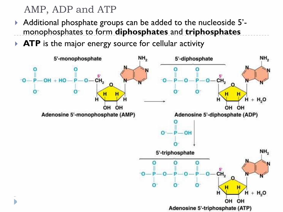
AMP, ADP and ATP
Additional phosphate groups can be added to the nucleoside 5’-monophosphates to form diphosphates and triphosphates
ATP is the major energy source for cellular activity
1

Died in
2004

DNA stands for deoxyribose nucleic acid
This chemical substance is present in the nucleus
of all cells in all living organisms
DNA controls all the chemical changes which
take place in cells
The kind of cell which is formed, (muscle, blood,
nerve etc) is controlled by DNA
DNA 2
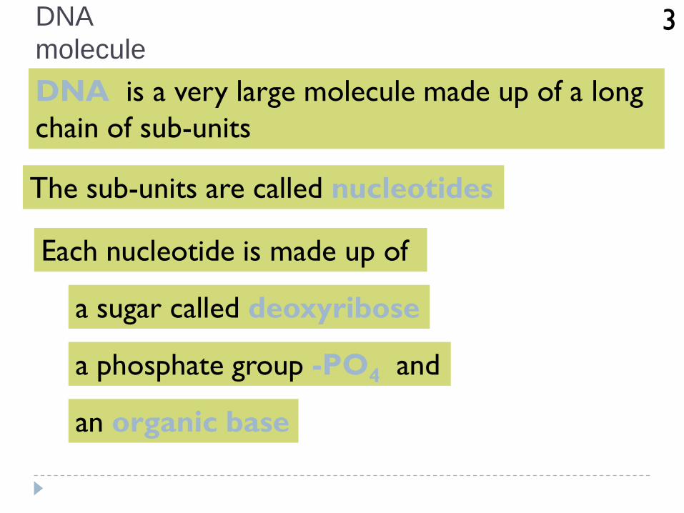
DNA is a very large molecule made up of a long
chain of sub-units
The sub-units are called nucleotides
Each nucleotide is made up of
a sugar called deoxyribose
a phosphate group -PO4 and
an organic base
DNA
molecule3
Based on McMurry, Organic Chemistry,
Chapter 28, 6th edition, (c) 2003
39
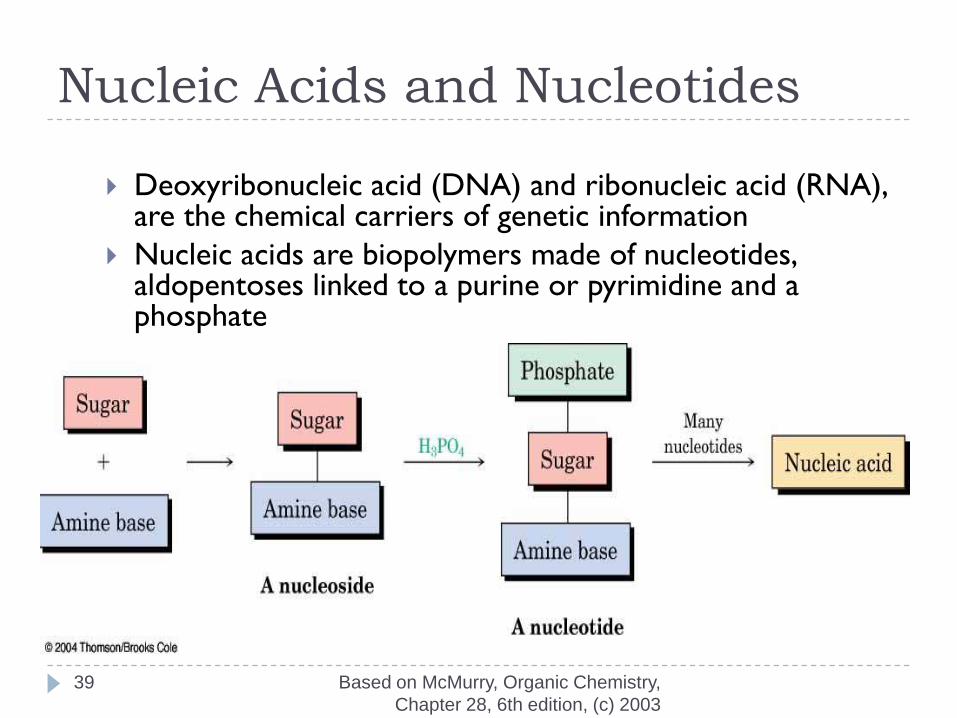
Nucleic Acids and Nucleotides
Deoxyribonucleic acid (DNA) and ribonucleic acid (RNA), are the chemical carriers of genetic information
Nucleic acids are biopolymers made of nucleotides, aldopentoses linked to a purine or pyrimidine and a phosphate
Based on McMurry, Organic Chemistry,
Chapter 28, 6th edition, (c) 2003
40
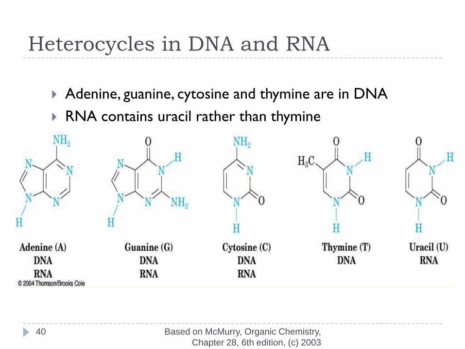
Heterocycles in DNA and RNA
Adenine, guanine, cytosine and thymine are in DNA
RNA contains uracil rather than thymine
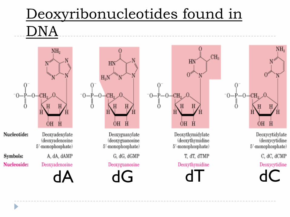
dA dG dT dC
Deoxyribonucleotides found in
DNA

42
The Deoxyribonucleotides
Hydrogen Bonding Interactions
Two bases can hydrogen bond to form a base pair
For monomers, large number of base pairs is
possible
In polynucleotide, only few possibilities exist
Watson-Crick base pairs predominate in double-
stranded DNA
A pairs with T
C pairs with G
Purine pairs with pyrimidine
一、the building block molecule of
nucleic acid--nucleotide
In RNA:
AMP、CMP、GMP、TMP
In DNA:
dAMP、dCMP、dGMP 、dUMP
Functions of
Nucleotides and Nucleic Acids
Nucleotide Functions:
Energy for metabolism (ATP)
Enzyme cofactors (NAD+)
Signal transduction (cAMP)
Nucleic Acid Functions:
Storage of genetic info (DNA)
Transmission of genetic info (mRNA)
Processing of genetic information (ribozymes)
Protein synthesis (tRNA and rRNA)

二、the linkage ----
phosphodiester bridge
3’terminal
5’terminal
Nucleotide residues
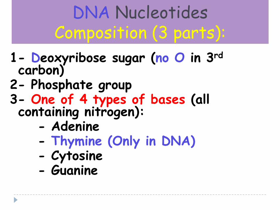
DNA NucleotidesComposition (3 parts):
1- Deoxyribose sugar (no O in 3rd
carbon)2- Phosphate group3- One of 4 types of bases (all containing nitrogen):
- Adenine- Thymine (Only in DNA)- Cytosine- Guanine
48
Base Pairing in DNA: The Watson–
Crick Model
In 1953 Watson and Crick noted that DNA consists
of two polynucleotide strands, running in opposite
directions and coiled around each other in a double
helix
Strands are held together by hydrogen bonds
between specific pairs of bases
Adenine (A) and thymine (T) form strong hydrogen
bonds to each other but not to C or G
(G) and cytosine (C) form strong hydrogen bonds to
each other but not to A or T

49
The Difference in the Strands
The strands of DNA are complementary because of H-bonding
Whenever a G occurs in one strand, a C occurs opposite it in the other strand
When an A occurs in one strand, a T occurs in the other

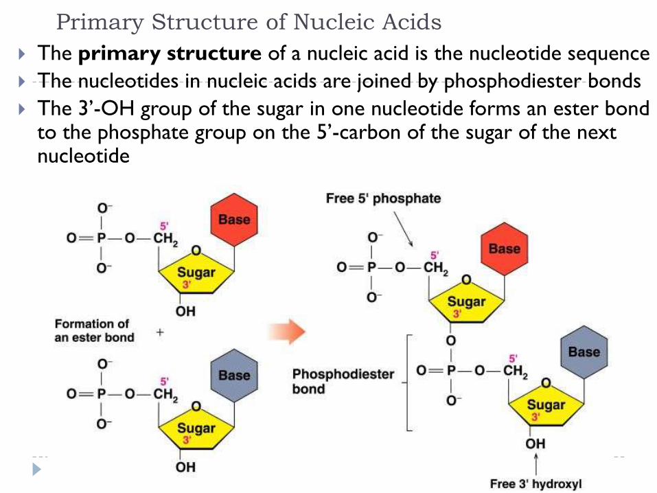
Primary Structure of Nucleic Acids
The primary structure of a nucleic acid is the nucleotide sequence
The nucleotides in nucleic acids are joined by phosphodiester bonds
The 3’-OH group of the sugar in one nucleotide forms an ester bond to the phosphate group on the 5’-carbon of the sugar of the next nucleotide

52
Generalized Structure of DNA
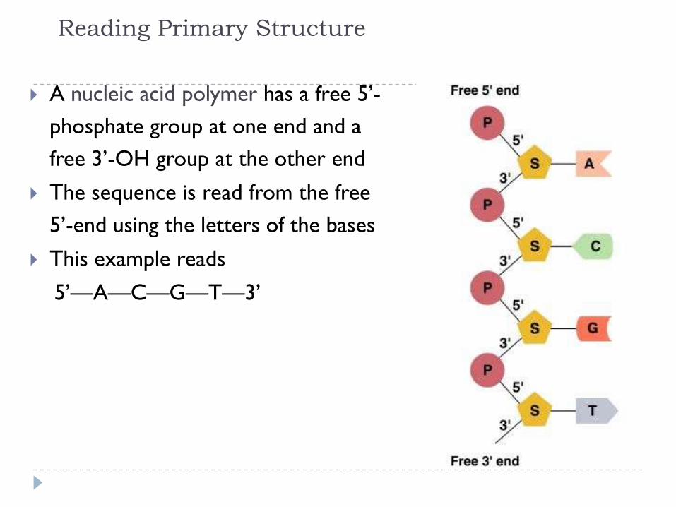
Reading Primary Structure
A nucleic acid polymer has a free 5’-
phosphate group at one end and a
free 3’-OH group at the other end
The sequence is read from the free
5’-end using the letters of the bases
This example reads
5’—A—C—G—T—3’

Example of DNA Primary Structure
In DNA, A, C, G, and T are linked by 3’-5’ ester bonds
between deoxyribose and phosphate

55
Describing a Sequence
Chain is described from 5 end,
identifying the bases in order of
occurrence, using the abbreviations A
for adenosine, G for guanosine, C for
cytidine, and T for thymine (or U for
uracil in RNA)
A typical sequence is written as
TAGGCT
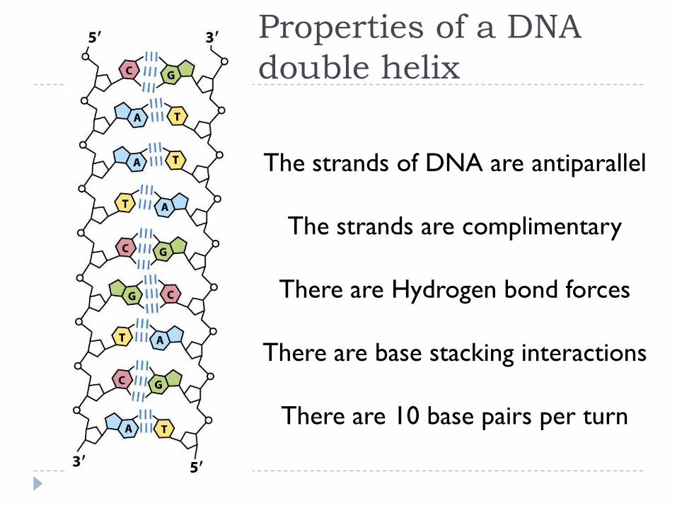
The strands of DNA are antiparallel
The strands are complimentary
There are Hydrogen bond forces
There are base stacking interactions
There are 10 base pairs per turn
Properties of a DNA
double helix

The Double Helix (DNA)Structural model:
Model proposed by Watson & Crick, 1953 Two sugar-phosphate strands, next to each other, but running in opposite directions.
Specific Hydrogen bonds occur among bases from one chain to the other:
A---T , C---GDue to this specificity, a certain base on one strand indicates a certain base in the other.
The 2 strands intertwine, forming a double-helix that winds around a central axis

Untwisted it looks like this:
• The sides of the ladder are: P = phosphateS = sugar molecule
• The steps of the ladder are C, G, T, A = nitrogenous bases(Nitrogenous means containing the element nitrogen.)
A = AdenineT = ThymineA always pairs with T in DNA
C = CytosineG = GuanineC always pairs with G in DNANucleotide
(Apples are Tasty)
(Cookies are Good)
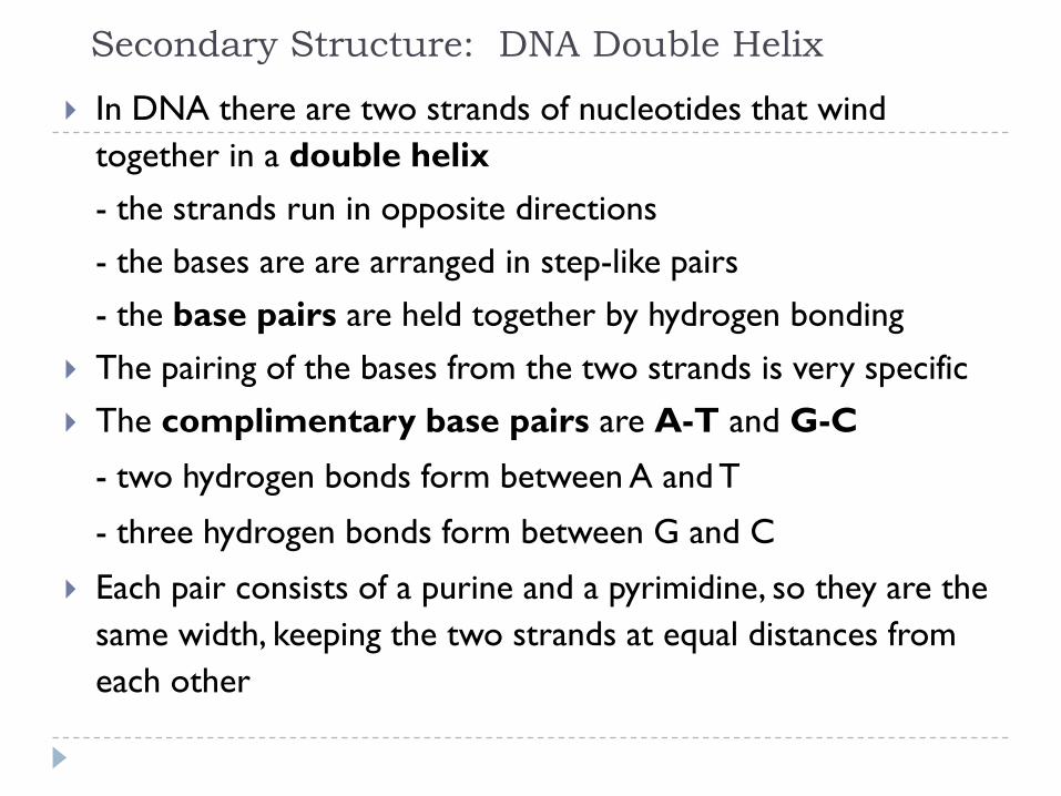
Secondary Structure: DNA Double Helix
In DNA there are two strands of nucleotides that wind
together in a double helix
- the strands run in opposite directions
- the bases are are arranged in step-like pairs
- the base pairs are held together by hydrogen bonding
The pairing of the bases from the two strands is very specific
The complimentary base pairs are A-T and G-C
- two hydrogen bonds form between A and T
- three hydrogen bonds form between G and C
Each pair consists of a purine and a pyrimidine, so they are the
same width, keeping the two strands at equal distances from
each other

Model of DNA:•The model was developed
by Watson and Crick in 1953.
•They received a nobel prizein 1962 for their work.
•The model looks like a twisted ladder – double helix.

Nucleic Acid Structure“Base Pairing”
T A AG C C
3’
T C GG TA
3’ 5’
5’
DNA base-pairing is antiparallel
i.e. 5’ - 3’ (l-r) on top : 5’ - 3’ (r-l) on
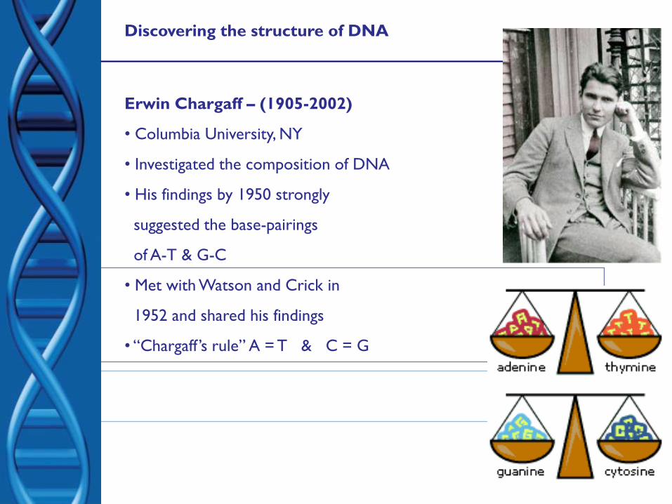
Discovering the structure of DNA
Erwin Chargaff – (1905-2002)
• Columbia University, NY
• Investigated the composition of DNA
• His findings by 1950 strongly
suggested the base-pairings
of A-T & G-C
• Met with Watson and Crick in
1952 and shared his findings
• “Chargaff ’s rule” A = T & C = G
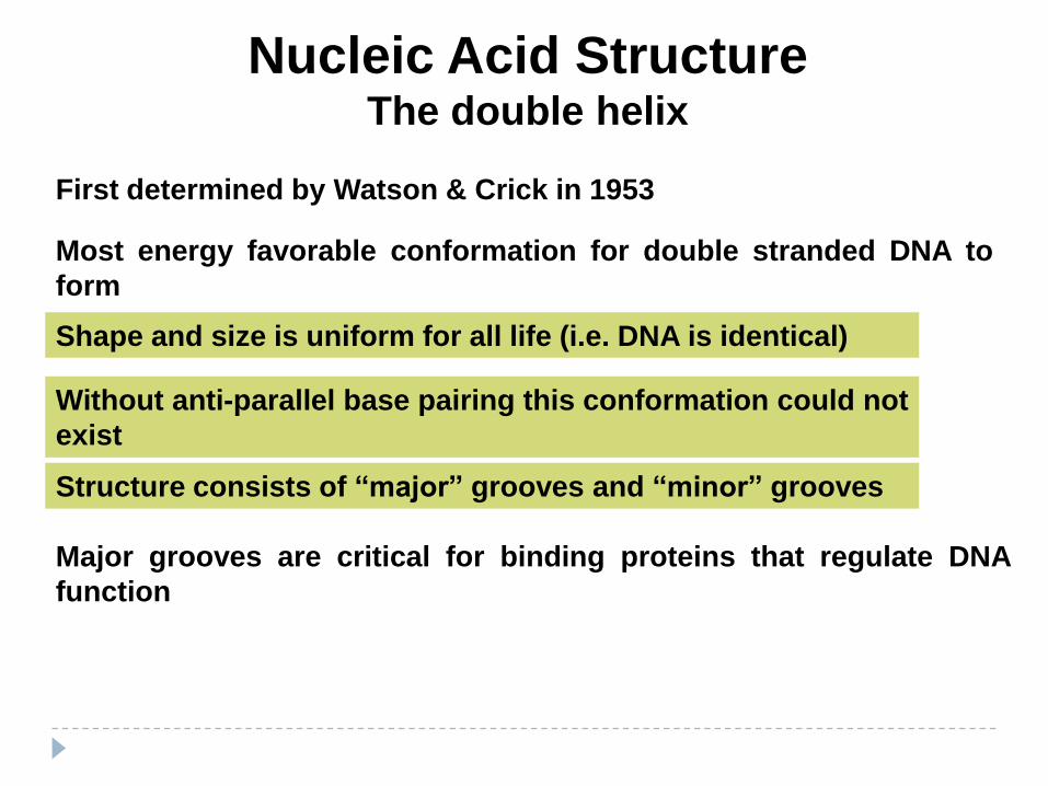
Nucleic Acid StructureThe double helix
First determined by Watson & Crick in 1953
Shape and size is uniform for all life (i.e. DNA is identical)
Most energy favorable conformation for double stranded DNA to
form
Without anti-parallel base pairing this conformation could not
exist
Structure consists of “major” grooves and “minor” grooves
Major grooves are critical for binding proteins that regulate DNA
function

Discovering the structure of DNA
• DNA = Deoxyribose nucleic acid
• Present in all living cells
• Contains all the information
• Nucleotides:
• a subunit that consists of:
• a sugar (deoxyribose)
• a phosphate
• and one nitrogen base – 4 different bases
•Adenine (A) and Thymine (T)
•Guanine (G) and Cytosine (C)

The paired strands are coiled into a spiral
called
A DOUBLE HELIX
1
3
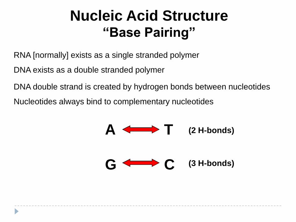
Nucleic Acid Structure“Base Pairing”
RNA [normally] exists as a single stranded polymer
DNA exists as a double stranded polymer
DNA double strand is created by hydrogen bonds between nucleotides
Nucleotides always bind to complementary nucleotides
A T
CG
(2 H-bonds)
(3 H-bonds)
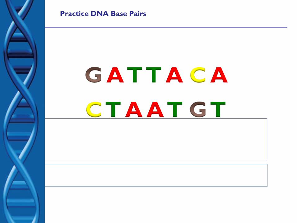
Practice DNA Base Pairs
A T T A C A
C T A A T T
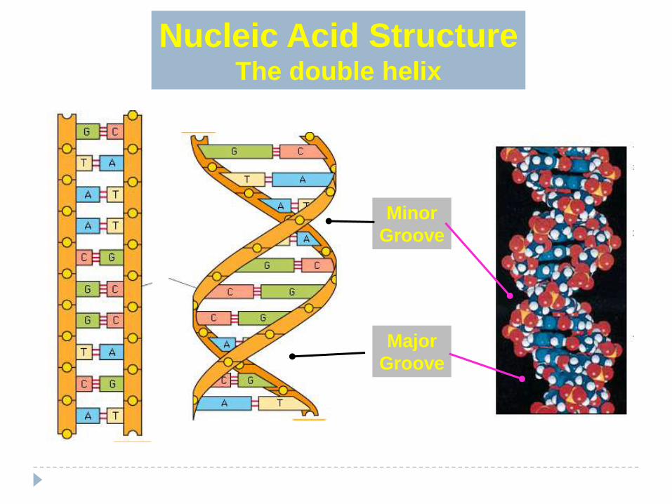
Nucleic Acid StructureThe double helix
Major
Groove
Minor
Groove

Before a cell divides, the DNA strands unwind
and separate
Each strand makes a new partner by adding
the appropriate nucleotides
The result is that there are now two
double-stranded DNA molecules in
the nucleus
So that when the cell divides, each nucleus
contains identical DNA
This process is called replication
replication 16
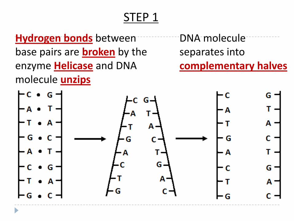
Hydrogen bonds between base pairs are broken by the enzyme Helicase and DNA molecule unzips
DNA molecule separates into complementary halves
STEP 1
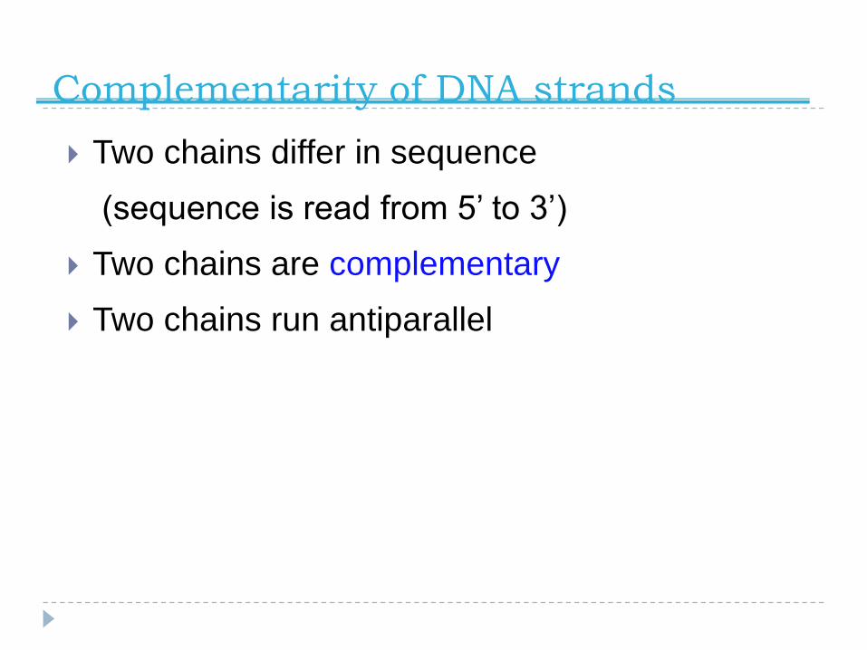
Complementarity of DNA strands
Two chains differ in sequence
(sequence is read from 5’ to 3’)
Two chains are complementary
Two chains run antiparallel
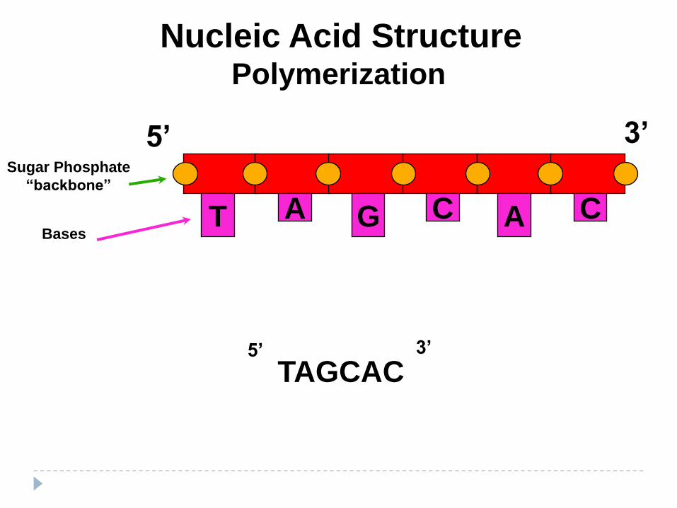
Nucleic Acid StructurePolymerization
T A AG C C
5’ 3’
TAGCAC5’ 3’
Bases
Sugar Phosphate
“backbone”

PP
(PPi)
Nucleic Acid StructurePolymerization
P P P
S
N
C
P P P
S
N
C
+
P P P
S
N
C
P
S
N
C
Phosphodiesterase
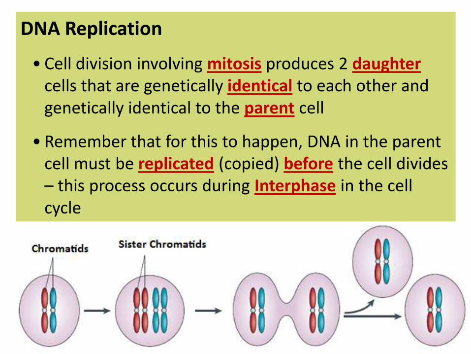
DNA Replication
• Cell division involving mitosis produces 2 daughtercells that are genetically identical to each other and genetically identical to the parent cell
• Remember that for this to happen, DNA in the parent cell must be replicated (copied) before the cell divides – this process occurs during Interphase in the cell cycle
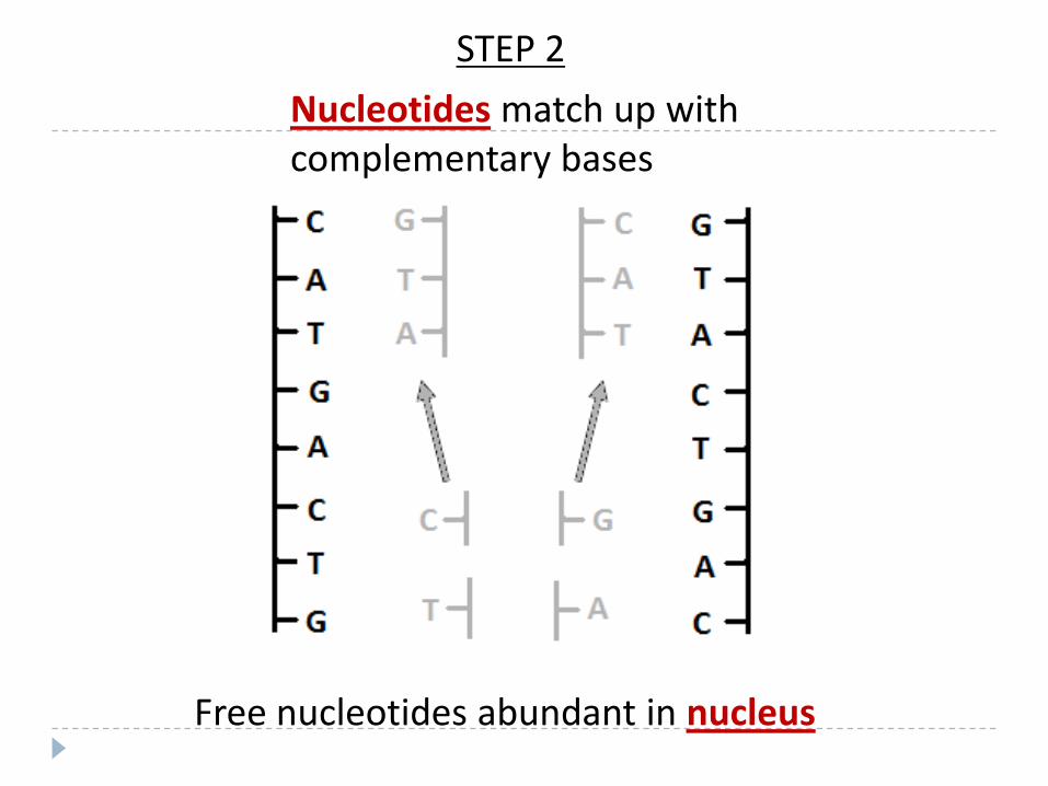
Nucleotides match up with complementary bases
Free nucleotides abundant in nucleus
STEP 2
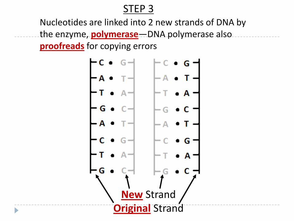
New StrandOriginal Strand
Nucleotides are linked into 2 new strands of DNA by the enzyme, polymerase—DNA polymerase also proofreads for copying errors
STEP 3

Mutations occur when copying errors cause a change in the sequence of DNA nucleotide bases

Diagram Examples of DNA Replication: (You could see DNA replication represented different ways.)
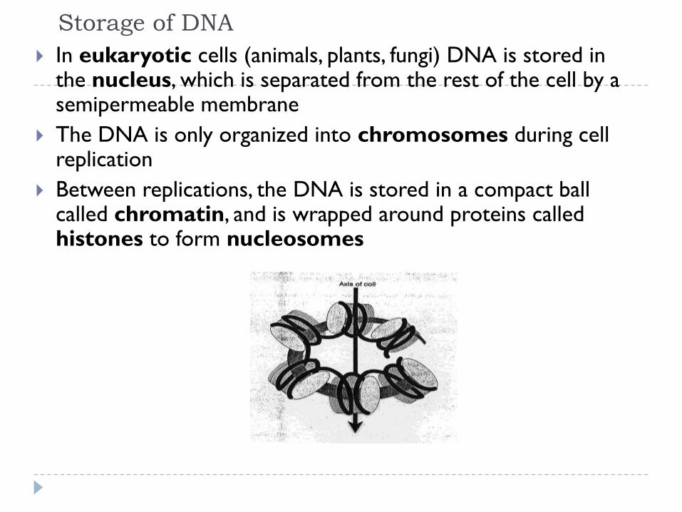
Storage of DNA
In eukaryotic cells (animals, plants, fungi) DNA is stored in the nucleus, which is separated from the rest of the cell by a semipermeable membrane
The DNA is only organized into chromosomes during cell replication
Between replications, the DNA is stored in a compact ball called chromatin, and is wrapped around proteins called histones to form nucleosomes
DNA Replication
When a eukaryotic cell divides, the process is called mitosis
- the cell splits into two identical daughter cells
- the DNA must be replicated so that each daughter cell has a copy
DNA replication involves several processes:
- first, the DNA must be unwound, separating the two strands
- the single strands then act as templates for synthesis of the new strands, which are complimentary in sequence
- bases are added one at a time until two new DNA strands that exactly duplicate the original DNA are produced
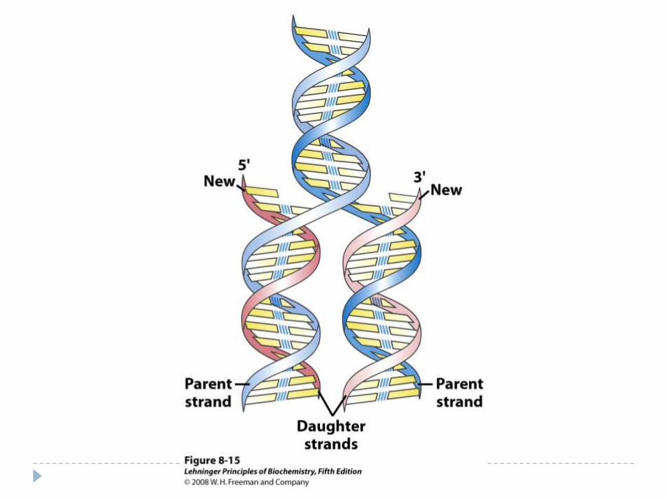
The process is called semi-conservative replication because one strand of each daughter DNA comes from the parent DNA and one strand is new
The energy for the synthesis comes from hydrolysis of phosphate groups as the phosphodiester bonds form between the bases

Direction of Replication
The enzyme helicase unwinds several sections of parent DNA
At each open DNA section, called a replication fork, DNA polymerase catalyzes the formation of 5’-3’ester bonds of the leading strand
The lagging strand, which grows in the 3’-5’ direction, is synthesized in short sections called Okazaki fragments
The Okazaki fragments are joined by DNA ligase to give a single 3’-5’ DNA strand
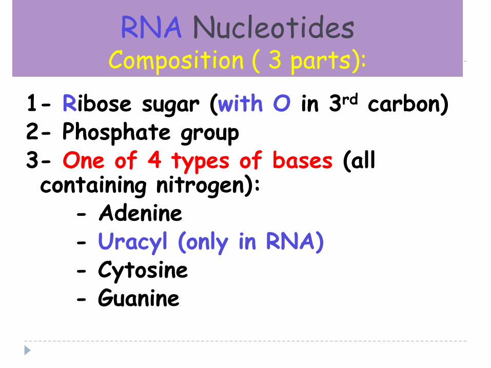
RNA NucleotidesComposition ( 3 parts):
1- Ribose sugar (with O in 3rd carbon)2- Phosphate group3- One of 4 types of bases (all containing nitrogen):
- Adenine- Uracyl (only in RNA)- Cytosine- Guanine

Ribonucleic Acid (RNA)
RNA is much more abundant than DNA
There are several important differences between RNA and
DNA:
- the pentose sugar in RNA is ribose, in DNA it’s deoxyribose
- in RNA, uracil replaces the base thymine (U pairs with A)
- RNA is single stranded while DNA is double stranded
- RNA molecules are much smaller than DNA molecules
There are three main types of RNA:
- ribosomal (rRNA), messenger (mRNA) and transfer (tRNA)
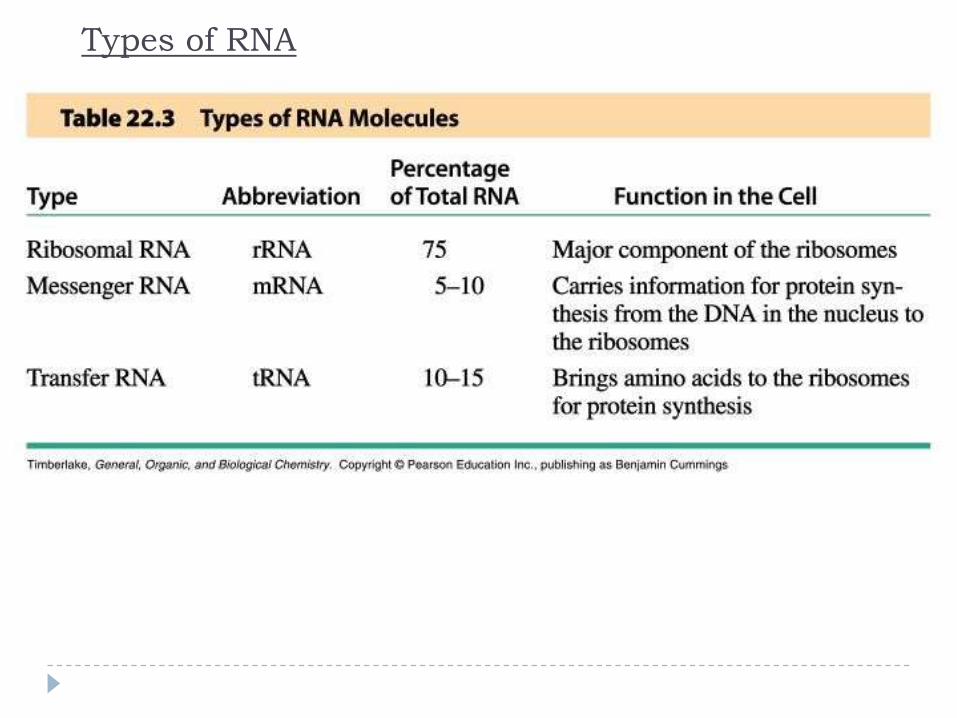
Types of RNA
87
Messenger RNA (mRNA)
Its sequence is copied from genetic DNA
It travels to ribsosomes, small granular
particles in the cytoplasm of a cell where
protein synthesis takes place
88
Ribosomal RNA (rRNA)
Ribosomes are a complex of proteins and
rRNA
The synthesis of proteins from amino
acids and ATP occurs in the ribosome
The rRNA provides both structure and
catalysis
89
Transfer RNA (tRNA)
Transports amino acids to the
ribosomes where they are joined
together to make proteins
There is a specific tRNA for each
amino acid
Recognition of the tRNA at the anti-
codon communicates which amino
acid is attached
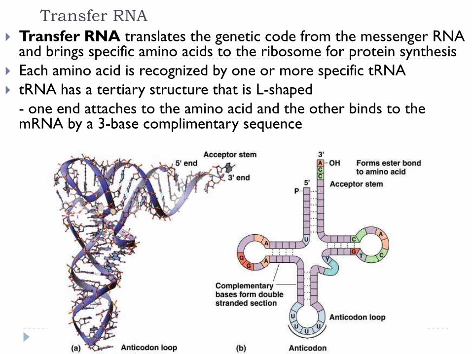
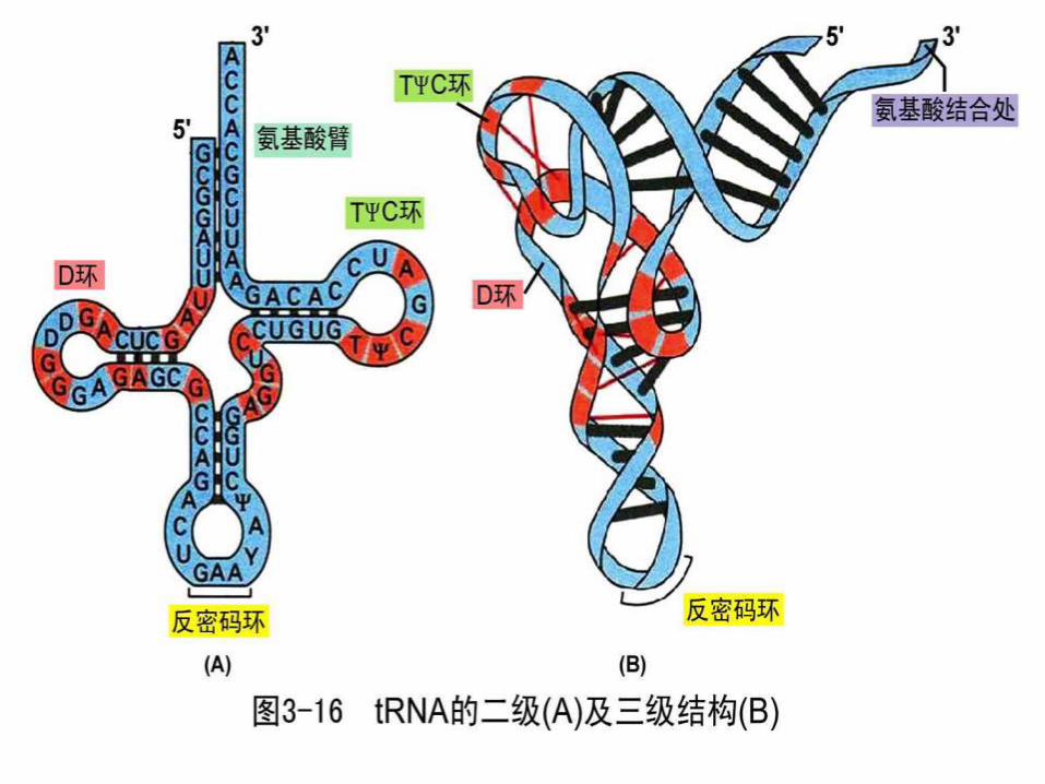
Transfer RNA
Transfer RNA translates the genetic code from the messenger RNA and brings specific amino acids to the ribosome for protein synthesis
Each amino acid is recognized by one or more specific tRNA
tRNA has a tertiary structure that is L-shaped
- one end attaches to the amino acid and the other binds to the mRNA by a 3-base complimentary sequence
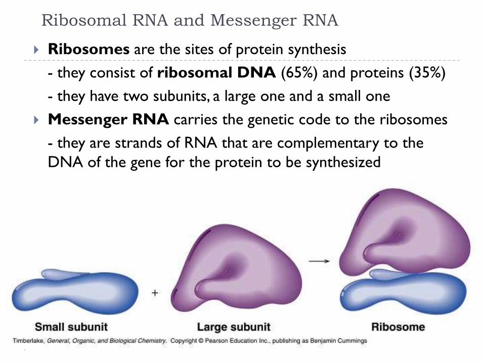
Ribosomal RNA and Messenger RNA
Ribosomes are the sites of protein synthesis
- they consist of ribosomal DNA (65%) and proteins (35%)
- they have two subunits, a large one and a small one
Messenger RNA carries the genetic code to the ribosomes
- they are strands of RNA that are complementary to the
DNA of the gene for the protein to be synthesized
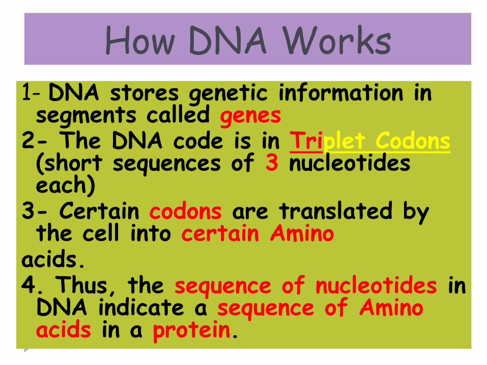
How DNA Works1- DNA stores genetic information in segments called genes
2- The DNA code is in Triplet Codons(short sequences of 3 nucleotides each)
3- Certain codons are translated by the cell into certain Amino
acids.4. Thus, the sequence of nucleotides in DNA indicate a sequence of Amino acids in a protein.

Based on McMurry, Organic Chemistry,
Chapter 28, 6th edition, (c) 2003
93
Transcription Process
Several turns of the DNA double helix unwind, exposing the bases of the two strands
Ribonucleotides line up in the proper order by hydrogen bonding to their complementary bases on DNA
Bonds form in the 5 3 direction,

RNA—Ribonucleic Acid
• RNA is a messenger that allows the instruction of DNA to be delivered to the rest of the cell
• RNA is different than DNA:
1.The sugar in RNA is ribose; the sugar in DNA is deoxyribose
2.RNA is a single strand of nucleotides; DNA is a double strand of nucleotides
3.RNA has Uracil (U) instead of Thymine (T) which is in DNA
4.RNA is found inside and outside of the nucleus; DNA is found only inside the nucleus

95
Transcription of RNA from DNA
Only one of the two DNA strands is transcribed into
mRNA
The strand that contains the gene is the coding or
sense strand
The strand that gets transcribed is the template or
antisense strand
The RNA molecule produced during transcription is
a copy of the coding strand (with U in place of T)
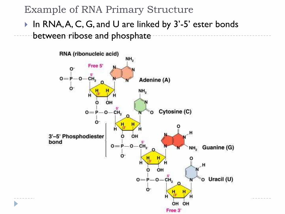
Example of RNA Primary Structure
In RNA, A, C, G, and U are linked by 3’-5’ ester bonds
between ribose and phosphate

97
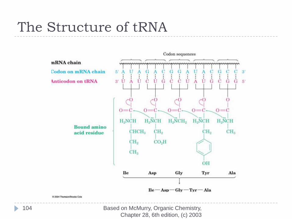
The Parts of Transfer RNA
There are 61 different tRNAs, one for each of the 61 codons that specifies an amino acid
tRNA has 70-100 ribonucleotides and is bonded to a specific amino acid by an ester linkage through the 3 hydroxyl on ribose at the 3 end of the tRNA
Each tRNA has a segment called an anticodon, a sequence of three ribonucleotides complementary to the codon sequence

Protein Synthesis
The two main processes involved in protein synthesis are
- the formation of mRNA from DNA (transcription)
- the conversion by tRNA to protein at the ribosome (translation)
Transcription takes place in the nucleus, while translation takes place in the cytoplasm
Genetic information is transcribed to form mRNA much the same way it is replicated during cell division

RNA Polymerase
During transcription, RNA polymerase moves along the DNA
template in the 3’-5’direction to synthesize the corresponding
mRNA
The mRNA is released at the termination point
Processing of mRNA
Genes in the DNA of eukaryotes contain exons that
code for proteins along with introns that do not
Because the initial mRNA, called a pre-RNA, includes the
noncoding introns, it must be processed before it can be
read by the tRNA
While the mRNA is still in the nucleus, the introns are
removed from the pre-RNA
The exons that remain are joined to form the mRNA that
leaves the nucleus with the information for the synthesis of
protein
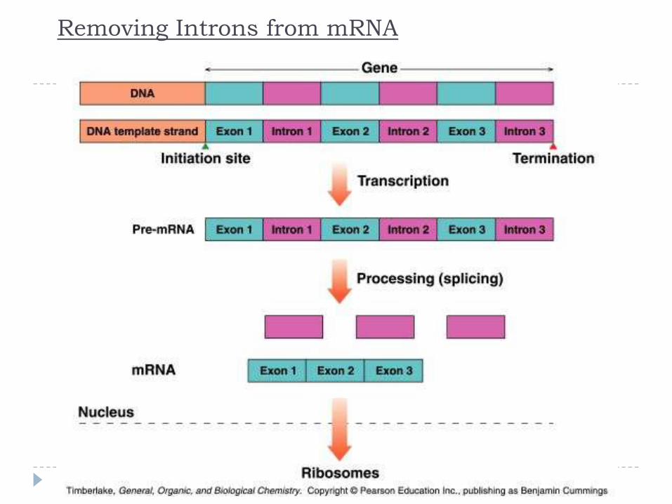
Removing Introns from mRNA

Transcription
Several steps occur during transcription:
- a section of DNA containing the gene unwinds
- one strand of DNA is copied starting at the initiation point,
which has the sequence TATAAA
- an mRNA is synthesized using complementary base pairing
with uracil (U) replacing thymine (T)
- the newly formed mRNA moves out of the nucleus to
ribosomes in the cytoplasm and the DNA re-winds
Based on McMurry, Organic Chemistry,
Chapter 28, 6th edition, (c) 2003
104
The Structure of tRNA
Regulation of Transcription
A specific mRNA is synthesized when the cell requires a
particular protein
The synthesis is regulated at the transcription level:
- feedback control, where the end products speed up or
slow the synthesis of mRNA
- enzyme induction, where a high level of a reactant
induces the transcription process to provide the necessary
enzymes for that reactant
Regulation of transcription in eukaryotes is complicated and
we will not study it here
106
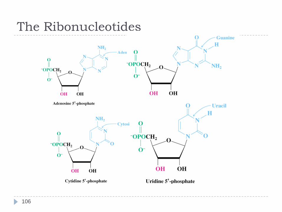
The Ribonucleotides
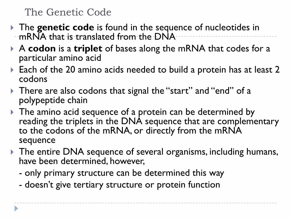
The Genetic Code
The genetic code is found in the sequence of nucleotides in mRNA that is translated from the DNA
A codon is a triplet of bases along the mRNA that codes for a particular amino acid
Each of the 20 amino acids needed to build a protein has at least 2 codons
There are also codons that signal the “start” and “end” of a polypeptide chain
The amino acid sequence of a protein can be determined by reading the triplets in the DNA sequence that are complementary to the codons of the mRNA, or directly from the mRNA sequence
The entire DNA sequence of several organisms, including humans, have been determined, however,
- only primary structure can be determined this way
- doesn’t give tertiary structure or protein function

The sequence of bases in DNA forms the
Genetic Code
A group of three bases (a triplet)
controls
the production of a particular amino
acid in
the cytoplasm of the cell
The different amino acids and the order
in which they are joined up determines
the sort of protein being produced
Genetic code 1 19

For example
Cytosine
Adenine Codes for Valine
Cytosine (C)
Guanine (G)
Adenine (A)
Codes for Alanine
Thymine
Coding 21

This is known as the triplet code
Each triplet codes for a specific amino acid
CGA - CAA - CCA - CCA - GCT - GGG - GAG - CCA -
Ala Val Gly Gly Arg Pro Leu Gly
Ala Val Gly Gly Arg Pro Leu Gly
The amino acids are joined together in the correct
sequence to make part of a protein
Triplet code 22
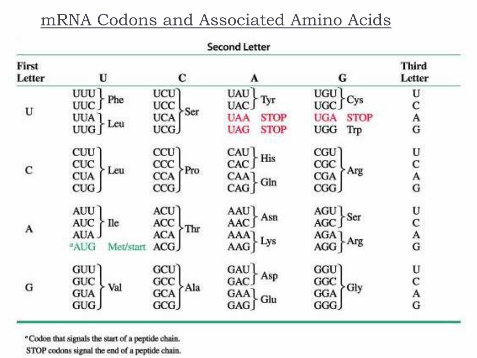
mRNA Codons and Associated Amino Acids
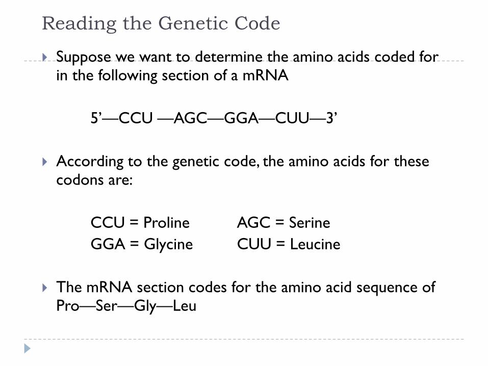
Reading the Genetic Code
Suppose we want to determine the amino acids coded for in the following section of a mRNA
5’—CCU —AGC—GGA—CUU—3’
According to the genetic code, the amino acids for these codons are:
CCU = Proline AGC = Serine
GGA = Glycine CUU = Leucine
The mRNA section codes for the amino acid sequence of Pro—Ser—Gly—Leu

Translation and tRNA Activation
Once the DNA has been
transcribed to mRNA, the
codons must be tranlated to the
amino acid sequence of the
protein
The first step in translation is
activation of the tRNA
Each tRNA has a triplet called
an anticodon that
complements a codon on
mRNA
A synthetase uses ATP hydrolysis
to attach an amino acid to a
specific tRNA

Initiation and Translocation
Initiation of protein synthesis occurs when a mRNA attaches to a ribosome
On the mRNA, the start codon (AUG) binds to a tRNA with methionine
The second codon attaches to a tRNA with the next amino acid
A peptide bond forms between the adjacent amino acids at the first and second codons
The first tRNA detaches from the ribosome and the ribosome shifts to the adjacent codon on the mRNA (this process is called translocation)
A third codon can now attach where the second one was before translocation

Termination
After a polypeptide with all the amino acids for a protein is
synthesized, the ribosome reaches the the “stop” codon:
UGA, UAA, or UAG
There is no tRNA with an anticodon for the “stop” codons
Therefore, protein synthesis ends (termination)
The polypeptide is released from the ribosome and the
protein can take on it’s 3-D structure
(some proteins begin folding while still being synthesized,
while others do not fold up until after being released from
the ribosome)

The proteins build the cell structures
They also make enzymes
The DNA controls which enzymes are made and
the enzymes determine what reactions take place
The structures and reactions in the cell determine
what sort of a cell it is and what its function is
So DNA exerts its control through the enzymes
DNA and enzymes 23
A sequence of triplets in the DNA molecule may
code for a complete protein
Such a sequence forms a gene
There may be a thousand or more bases in
one gene
Genes 24
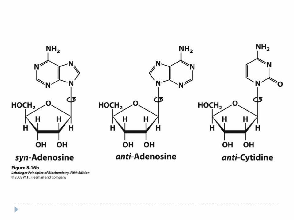
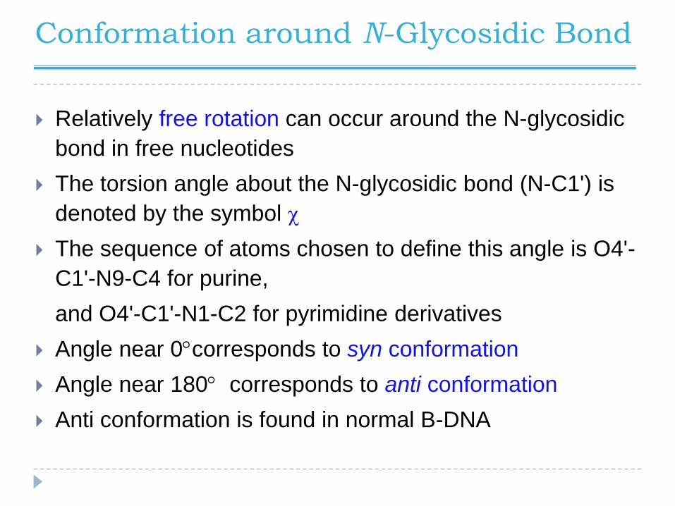
Conformation around N-Glycosidic Bond
Relatively free rotation can occur around the N-glycosidic
bond in free nucleotides
The torsion angle about the N-glycosidic bond (N-C1') is
denoted by the symbol c
The sequence of atoms chosen to define this angle is O4'-
C1'-N9-C4 for purine,
and O4'-C1'-N1-C2 for pyrimidine derivatives
Angle near 0corresponds to syn conformation
Angle near 180 corresponds to anti conformation
Anti conformation is found in normal B-DNA

Replication of Genetic Code
“It has not escaped our notice that the specific pairing
we have postulated immediately suggests a possible
copying mechanism for the genetic material”
Watson and Crick, in their Nature paper,1953
• Strand separation occurs first
• Each strand serves as a template
for the synthesis of a new strand
• Synthesis is catalyzed by enzymes
known as DNA polymerases
• Newly made DNA molecule has one
daughter strand and one parent strand.

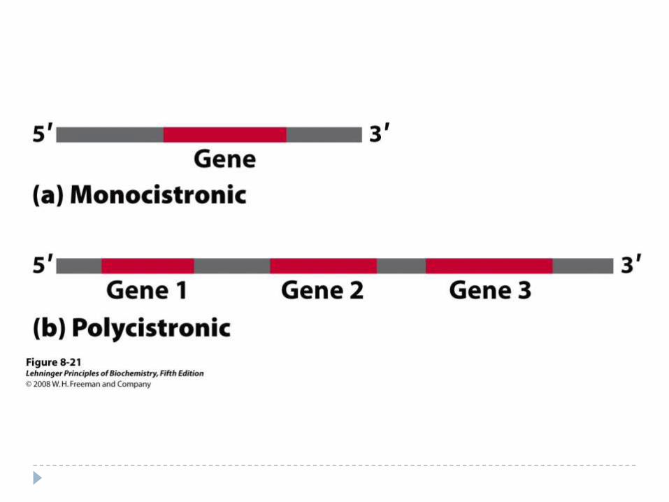
Messenger RNA:
Code Carrier for the Sequence of Proteins
• Is synthesized using DNA template
•Contains ribose instead of deoxyribose
•Contains uracil instead of thymine
•One mRNA may code for more than
one protein

Factors Affecting DNA Denaturation
The midpoint of melting (Tm) depends on base composition high CG increases Tm
Tm depends on DNA length Longer DNA has higher Tm
Important for short DNA
Tm depends on pH and ionic strength High salt increases Tm
Copyright © 2022 FDOKUMEN