Isolation and characterization of Erythrobacter sp. strains from the upper ocean
12
Abstract Seven strains of marine aerobic anoxygenic pho- totrophs belonging to the genus Erythrobacter were iso- lated. The strains were characterized regarding their phys- iological and biochemical properties, 16S rDNA and pufM gene sequences, morphological features, substrate prefer- ence, as well as pigment and lipid composition. All strains had functional type-2 reaction centers containing bacterio- chlorophyll, served by small, light-harvesting complex 1, and were photosynthetically competent. In addition, large pools of carotenoids were found, but only some of the ac- cessory pigments transfer energy to the reaction centers. All of the isolates were facultative photoheterotrophs. They required an organic carbon substrate for growth; however, they are able to supplement a significant fraction of their metabolic requirements with photosynthetically derived energy. Keywords Aerobic anoxygenic phototrophs · Aerobic photosynthetic bacteria · Bacteriochlorophyll a · Erythrobacter · Photoheterotrophy Abbreviations BChl Bacteriochlorophyll · Chl Chlorophyll · Erb. Erythrobacter · Erm. Erythromicrobium · FAMEs Fatty acid methyl esters · IRFRR Infrared fast repetition rate · LH1, LH2 Light-harvesting complex 1 and 2, respectively · Por. Porphyrobacter · PUFAs Polyunsaturated fatty acids · Rsb. Roseobacter · RubisCO Ribulose-1,5-bisphosphate carboxylase/oxygenase · µ Growth rate · σ 470 Functional cross-section of the photosynthetic unit at 470 nm Introduction Photosynthetic organisms play a crucial role in the marine environment. Chlorophyll-containing eukaryotic and prokary- otic phytoplankton utilize light as their main energy source, evolve oxygen, and convert inorganic CO 2 into organic molecules. In addition to these dominant, oxygenic organ- isms, there exists a broad spectrum of anoxygenic photo- synthetic bacteria that contain bacteriochlorophyll (BChl). These organisms evolved approximately 3 billion years ago (Des Marais 2000), long before the oxidation of Earth’s atmosphere (Rye and Holland 1998). Most of these photo- synthetic bacteria cannot sustain photoautotrophic metab- olism under oxic conditions, although some can grow het- erotrophically. In the present, oxygen-rich atmosphere, these organisms are confined to marginal ecological niches, such as anoxic zones of lakes, marine and freshwater sediments, and sulfide springs. In the late 1970s, a new kind of BChl-a-containing bacteria was discovered in Tokyo Bay, Japan (Harashima et al. 1978; Shiba et al. 1979). In contrast to their close relatives, purple non-sulfur photosynthetic bacteria, these bacteria were obligate aerobes requiring oxygen for growth Michal Koblížek · Oded Béjà · Robert R. Bidigare · Stephanie Christensen · Bryan Benitez-Nelson · Costantino Vetriani · Marcin K. Kolber · Paul G. Falkowski · Zbigniew S. Kolber Isolation and characterization of Erythrobacter sp. strains from the upper ocean Arch Microbiol (2003) 180 : 327–338 DOI 10.1007/s00203-003-0596-6 Received: 14 March 2003 / Revised: 28 July 2003 / Accepted: 6 August 2003 / Published online: 23 September 2003 ORIGINAL PAPER M. Koblížek (✉) · M. K. Kolber · P. G. Falkowski · Z. S. Kolber Environmental Biophysics and Molecular Ecology Program, Rutgers University, 71 Dudley Road, Institute of Coastal and Marine Sciences, New Brunswick, NJ 08901, USA Fax: +420-384-721246, e-mail: [email protected] O. Béjà Department of Biology, Technion-Israel Institute of Technology, 32000 Haifa, Israel R. R. Bidigare · S. Christensen · B. Benitez-Nelson Department of Oceanography, University of Hawai’i at Manoa, 1000 Pope Road, Honolulu, HI 96822, USA C. Vetriani Department of Biochemistry and Microbiology, Rutgers University, 76 Lipman Drive, New Brunswick, NJ 08901, USA P. G. Falkowski Department of Geology, Rutgers University, 610 Taylor Road, Piscataway, NJ 08854-8066, USA Present address: M. Koblížek Institute of Microbiology, Opatovický mlýn, 379 81 Třeboň, Czechia Present address: Z. S. Kolber MBARI, 7700 Sandholdt Road, Moss Landing, CA 95039, USA © Springer-Verlag 2003
Transcript of Isolation and characterization of Erythrobacter sp. strains from the upper ocean
Abstract Seven strains of marine aerobic anoxygenic pho-totrophs belonging to the genus Erythrobacter were iso-lated. The strains were characterized regarding their phys-iological and biochemical properties, 16S rDNA and pufMgene sequences, morphological features, substrate prefer-ence, as well as pigment and lipid composition. All strainshad functional type-2 reaction centers containing bacterio-chlorophyll, served by small, light-harvesting complex 1,and were photosynthetically competent. In addition, largepools of carotenoids were found, but only some of the ac-cessory pigments transfer energy to the reaction centers.All of the isolates were facultative photoheterotrophs. Theyrequired an organic carbon substrate for growth; however,they are able to supplement a significant fraction of their
metabolic requirements with photosynthetically derivedenergy.
Keywords Aerobic anoxygenic phototrophs · Aerobicphotosynthetic bacteria · Bacteriochlorophyll a · Erythrobacter · Photoheterotrophy
Abbreviations BChl Bacteriochlorophyll · Chl Chlorophyll · Erb. Erythrobacter · Erm.Erythromicrobium · FAMEs Fatty acid methyl esters · IRFRR Infrared fast repetition rate · LH1, LH2Light-harvesting complex 1 and 2, respectively · Por.Porphyrobacter · PUFAs Polyunsaturated fatty acids ·Rsb. Roseobacter · RubisCO Ribulose-1,5-bisphosphatecarboxylase/oxygenase · µ Growth rate · σ470 Functionalcross-section of the photosynthetic unit at 470 nm
Introduction
Photosynthetic organisms play a crucial role in the marineenvironment. Chlorophyll-containing eukaryotic and prokary-otic phytoplankton utilize light as their main energy source,evolve oxygen, and convert inorganic CO2 into organicmolecules. In addition to these dominant, oxygenic organ-isms, there exists a broad spectrum of anoxygenic photo-synthetic bacteria that contain bacteriochlorophyll (BChl).These organisms evolved approximately 3 billion years ago(Des Marais 2000), long before the oxidation of Earth’satmosphere (Rye and Holland 1998). Most of these photo-synthetic bacteria cannot sustain photoautotrophic metab-olism under oxic conditions, although some can grow het-erotrophically. In the present, oxygen-rich atmosphere, theseorganisms are confined to marginal ecological niches, suchas anoxic zones of lakes, marine and freshwater sediments,and sulfide springs.
In the late 1970s, a new kind of BChl-a-containingbacteria was discovered in Tokyo Bay, Japan (Harashimaet al. 1978; Shiba et al. 1979). In contrast to their closerelatives, purple non-sulfur photosynthetic bacteria, thesebacteria were obligate aerobes requiring oxygen for growth
Michal Koblížek · Oded Béjà · Robert R. Bidigare ·Stephanie Christensen · Bryan Benitez-Nelson ·Costantino Vetriani · Marcin K. Kolber ·Paul G. Falkowski · Zbigniew S. Kolber
Isolation and characterization of Erythrobacter sp. strains from the upper ocean
Arch Microbiol (2003) 180 : 327–338DOI 10.1007/s00203-003-0596-6
Received: 14 March 2003 / Revised: 28 July 2003 / Accepted: 6 August 2003 / Published online: 23 September 2003
ORIGINAL PAPER
M. Koblížek (✉) · M. K. Kolber · P. G. Falkowski · Z. S. KolberEnvironmental Biophysics and Molecular Ecology Program, Rutgers University, 71 Dudley Road, Institute of Coastal and Marine Sciences, New Brunswick, NJ 08901, USAFax: +420-384-721246,e-mail: [email protected]
O. BéjàDepartment of Biology, Technion-Israel Institute of Technology, 32000 Haifa, Israel
R. R. Bidigare · S. Christensen · B. Benitez-NelsonDepartment of Oceanography, University of Hawai’i at Manoa,1000 Pope Road, Honolulu, HI 96822, USA
C. VetrianiDepartment of Biochemistry and Microbiology, Rutgers University, 76 Lipman Drive, New Brunswick, NJ 08901, USA
P. G. FalkowskiDepartment of Geology, Rutgers University, 610 Taylor Road, Piscataway, NJ 08854-8066, USA
Present address:M. Koblížek Institute of Microbiology, Opatovický mlýn, 379 81 Třeboň, Czechia
Present address:Z. S. Kolber MBARI, 7700 Sandholdt Road, Moss Landing, CA 95039, USA
© Springer-Verlag 2003
(Shimada 1995; Yurkov and Beatty 1998; Imhoff 2001).Two of the first isolates (OCh 101 and OCh 114), laterclassified as Erythrobacter (Erb.) longus (Shiba andShimidu 1982) and Roseobacter (Rsb.) denitrificans (Shiba1991), established two major genera of marine photo-heterotrophs. Subsequently, many strains of those bacteriawere isolated from nutrient-rich coastal waters (Nishi-mura et al. 1994; Shiba et al. 1991; Yurkov et al. 1994). Atpresent, all the reported marine isolates belong to the α3and α4 Proteobacteria, forming four genera: Erythrobac-ter (Shiba and Shimidu 1982), Roseobacter (Shiba 1991),“Citromicrobium” (Yurkov et al. 1999), and Roseibium(Suzuki et al. 2000).
Until recently, aerobic anoxygenic photoheterotrophswere thought to occupy small, nutrient-rich ecologicalniches, and to be restricted to mostly heterotrophic metab-olism. The discovery of a high level of bacterial phototro-phy in the open upper ocean (Kolber et al. 2000) chal-lenged this notion. Based on kinetic fluorescence mea-surements, BChl-a-containing bacteria were suggested tocontribute 2–5% of the total photosynthetic electron trans-port fluxes in the upper ocean (Kolber et al. 2000). Mi-croscopy studies indicated that they account for up to 8%of the total bacterial community in the open ocean (Kol-ber et al. 2001). Erythrobacter strains are frequentlyfound in cultivated material (Shiba et al. 1991), and it wassuggested that they might form a major fraction of marinephotoheterotrophs (Kolber et al. 2001). However, thisview is contradicted by a recent molecular study of oceanicbacterial communities, which showed that marine photo-heterotrophs are much more diverse (Béjà et al. 2002).This is consistent with our recent culture studies in which,apart from Erythrobacter sp., also Roseobacter-like and“Citromicrobium”-like isolates have been found (Koblížek,unpublished data).
Little is known about the metabolism, physiology, andnutritional requirements of aerobic anoxygenic photohetero-trophs. Oceanic species appear to utilize dissolved organicmatter as a source of organic carbon, and light energy tosupplement heterotrophic metabolism. Their ability to fixCO2 is controversial (Shimada 1995; Yurkov and Beatty1998). To partially fill this knowledge gap, we character-ized several marine isolates belonging to the genus Eryth-robacter regarding their pigment and lipid composition,morphology, substrate preferences, vitamin requirements,antibiotic sensitivity, 16S rDNA and pufM sequences, spec-troscopic properties, and photosynthetic ability.
Material and methods
Strain isolation
About 5 µl of seawater was streaked onto 1.5% bacteriological agarplates supplemented with f/2 media (Guillard and Ryther 1962).Plates were stored for 7 days in the dark to eliminate oxygenic pho-toautotrophs, and then exposed to a natural light/dark cycle, result-ing in the production of small, pigmented colonies within 2 weeks.Under these conditions the plating yields ranged between 1 and 10colonies per 1,000 cells applied. The type strains, Erb. longus (ATCC33941) and Erb. litoralis (ATCC 700002), formed colonies under
these conditions. The colonies were resuspended in sterile f/2 sea-water medium [natural seawater enriched with 9×10⁻⁴ M NaNO3,3×10–5 M NaH2PO4, trace metals (10–5 M sodium iron (III) ethyl-enediamine tetra-acetate, 4×10⁻⁸ M CuSO4, 8×10⁻⁸ M ZnSO4, 4×10⁻⁸ M CoCl2, 9×10⁻⁷ M MnCl2, 3×10⁻⁸ M Na2MoO4) and vitamins(2×10⁻⁹ M biotin, 3.7×10⁻¹⁰ M B12)], and tested for the presence ofBChl a using an infra-red fast repetition rate (IRFRR) fluorometer(Kolber et al. 1998). Samples exhibiting the BChl a fluorescencetransient, indicative of bacterial photosynthetic electron transport,were repeatedly plated onto f/2 agar plates until uniform orange oryellow colonies were obtained. The isolates were then grown in or-ganic medium composed of f/2 medium supplemented with 0.5 gpeptone and 0.1 g yeast extract per liter.
The isolates could be maintained in liquid or on agar media at4 °C for several months. For long-term storage, the cells were re-suspended in 30% glycerol containing f/2 medium, frozen in liquidnitrogen, and stored at –75 °C.
Strain characterization
Whole-cell absorption spectra were recorded on an Aminco DW2000spectrophotometer operated in the dual beam mode. The cells wereresuspended in 70% glycerol in f/2 medium. The spectra wererecorded with a resolution of 0.33 nm, using 2-nm slit width. Be-cause of high turbidity, the absorption spectra were corrected bysubtracting the scattering profile, obtained from cells bleachedwith sodium hypochlorite. The same samples were also used forthe measurement of BChl a fluorescence excitation spectra. Thefluorescence excitation spectra were recorded using an AmincoAB2 spectrofluorometer at an emission wavelength of 875 nm,with a resolution of 1 nm, using 4 nm excitation and 16 nm emis-sion slit-widths. The emission monochromator was protected againststray light by a Schott RG830 glass filter.
Antibiotic sensitivity was tested in the organic medium with in-dividual antibiotics added at concentrations 50 mg/l if not statedotherwise. Growth was assayed after 4–5 days. For the simple organiccarbon-source growth tests, an artificial medium was derived fromthe f/2 medium. It contained: 0.4 M NaCl, 20 mM MgSO4, 10 mMCaCl2, 10 mM KCl, 20 mM MgCl2, 1.7 mM KBr, 1 mM (NH4)2SO4,10–4 M NaH2PO4, 2×10⁻⁴ M H3BO3, 10–8 M Na2SeO3, pH 8.0. Thetrace metal mix and vitamins were the same as in f/2 media. Forthe growth test, the medium was enriched with 1 mM of the indi-vidual carbon sources.
Respiration was measured using a Clark oxygen electrode placedin a temperature-stabilized measuring chamber at 23 °C. The elec-trode signal was recorded using a custom made potentiostat and ananoamperometer.
Carbon dioxide fixation was assayed at a BChl a concentrationof 30 nM, at 25 °C. The bacterial suspension was labeled with 37 kBq per ml NaH14CO3 (New England Nuclear, USA) and dividedinto 2-ml aliquots placed in glass scintillation vials. The vials wereexposed to varying light intensities produced by a white halogenlamp attenuated by a set of neutral density filters. Incubations wereterminated by addition of 30 µl 36% HCl (final pH~1), and sampleswere evaporated to dryness and resuspended in 2 ml 30 mM Trisbuffer (pH 7.7) with 15 ml scintillation cocktail (Ready Safe, Beck-man). The incorporated radioactivity was determined with a Beck-man LS6K-IC scintillation counter and corrected for blank counts.
Cells were counted using epifluorescence microscopy with acri-dine orange staining. To determine organic carbon and nitrogencontent, bacterial cells were collected onto pre-combusted glass-fiber filters, washed, dried in a dessicator, and analyzed with anautomated Perkin Elmer model 2400 CHN elemental analyzer.
Pigment analysis
BChl a concentration was determined spectroscopically in metha-nol with an Aminco DW2000 spectrophotometer using the absorp-tion coefficient ε771=54.8 mM–1cm–1 (Permentier et al. 2000). Totalcarotenoid content was determined in the same extracts, using anabsorption coefficient of ε460=128 mM–1cm–1 (Yurkov et al. 1993).
328
Pigment composition was assayed by HPLC. The cell pelletswere placed in 3 ml acetone, ground using a hand-held glass/glasstissue homogenizer, and extracted for 1–2 h (0 °C, dark). Samples(200 µl) of a mixture of 0.3 ml water plus 1.0 ml extract were in-jected onto a Varian 9012 HPLC system equipped with a Varian9300 autosampler, a Timberline column heater (26 °C), andSpherisorb 5 µm ODS2 analytical (4.6×250 mm) column and cor-responding guard cartridge. Pigments were detected with a Ther-moSeparation UV 2000 detector (λ=360 nm). A ternary solventsystem was employed for HPLC pigment analysis: solvent A(methanol:0.5 M ammonium acetate, 80/20, v/v), solvent B (ace-tonitrile:water, 85/15, v/v), and solvent C (ethyl acetate). The flowrate was held constant at 1 ml min–1. The linear gradient used forpigment separation was a modified version of the method ofWright et al. (1991): 0.0’ (100% A), 10.0’ (100% B), 11.0’ (78%B, 22% C), 27.5’ (10% B, 90% C), 29.0’ (100% B), and 30.0’(100% B). Solvents A and B contained 0.01% 2,6-di-tert-butyl-p-cresol (0.01% BHT, w/v; Sigma-Aldrich) to prevent the formationof allomerized BChl a and bacteriophaeophytin a derivatives(Bidigare and Trees 2000).
The individual peaks were identified by an absorption UV/VISdiode array detector and by comparison of the retention times withthose of authentic standards (β,β-carotene, zeaxanthin, BChl a,and BPhe a) and with extracts prepared from the type strain forErb. longus (ATCC 33941). HPLC peak identities were confirmedby “off-line” UV/VIS and LC/MS analyses of corresponding frac-tions. Mass spectra of selected carotenoid fractions were obtainedusing an Agilent Technologies 1100 series HPLC (0.7 ml methanolmin–1, operated in column by-pass mode) interfaced with modelSL quadrupole MSD (APCI, fragmentor setting of 30 V). The rel-ative pigment abundances in the extracts were estimated as per-centages of total peak area at 360 nm (%A360).
Fatty acid analysis
Cells grown on the organic medium were pelleted, combined with10 ml 5% H2SO4 in methanol, and divided equally among three 5-ml reaction vials. A total of 200 µl C19:0 fatty acid recoverystandard was added to the three vials. The sample vials were vor-texed and placed in a 100 °C dry-bath for 2 h. After cooling, thevial contents were transferred to a 20-ml vial with methanol andhexane rinses totaling 1.5 ml. An additional 1.5 ml hexane and 3 mlH2O were placed in the vial and mixed vigorously for 1 min. Fattyacid methyl esters (FAMEs) were partitioned into the hexanelayer, which was separated from the methanol/water layer via cen-trifugation for 5 min at 700×g. The hexane layer was pipetted offand two additional 1.5-ml hexane extractions were done. The hex-ane extract was evaporated to ~1 ml under a N2 stream and passedthrough a Na2SO4 column to remove residual water. The samplewas evaporated to 500 µl under a N2 stream and a 50-µl aliquotwas removed for GC/MS analysis. The GC/MS system consistedof a Hewlett Packard 6890 Plus GC interfaced to a HP 5973 MassSelective Detector. An HP 7683 Autoinjector was used to intro-duce 0.5 µl sample directly onto a J and W DB-5 column (60 m,0.32 mm I.D., 0.25 mm film thickness) through a cooled on-col-umn inlet. The initial GC oven temperature was 50 °C (1-min hold)followed by a ramp of 20 °C min–1 to 180 °C (0-min hold), a 2 °Cmin–1 ramp to 280 °C (0-min hold), and a 10 °C min–1 ramp to 320 °C (10-min hold). FAMEs were identified either through theuse of authentic standards (Alltech, Sigma-Aldrich) or by their massspectra. FAMEs were quantified using a C23 normal alkane asboth an internal standard and as part of a FAME standard mixture.
IRFRR fluorometry
The photosynthetic ability of the bacterial isolates was assessed us-ing an IRFRR fluorometer (Kolber et al. 1998, 2000) equippedwith a large-area avalanche photodiode detector (Advanced Pho-tonix, 630-70-72-631, 16-mm diameter) protected by RG695 glassand 880-nm interference (50-nm half-width) filters. The instru-ment generates a train of short (0.6 µs) blue (470 nm) or infrared
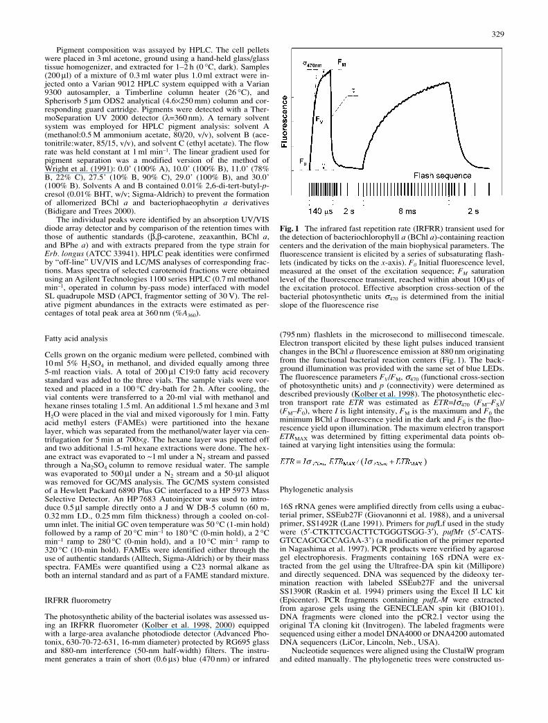
(795 nm) flashlets in the microsecond to millisecond timescale.Electron transport elicited by these light pulses induced transientchanges in the BChl a fluorescence emission at 880 nm originatingfrom the functional bacterial reaction centers (Fig. 1). The back-ground illumination was provided with the same set of blue LEDs.The fluorescence parameters FV/FM, σ470 (functional cross-sectionof photosynthetic units) and p (connectivity) were determined asdescribed previously (Kolber et al. 1998). The photosynthetic elec-tron transport rate ETR was estimated as ETR=Iσ470 (FM–FS)/(FM–F0), where I is light intensity, FM is the maximum and F0 theminimum BChl a fluorescence yield in the dark and FS is the fluo-rescence yield upon illumination. The maximum electron transportETRMAX was determined by fitting experimental data points ob-tained at varying light intensities using the formula:
Phylogenetic analysis
16S rRNA genes were amplified directly from cells using a eubac-terial primer, SSEub27F (Giovanonni et al. 1988), and a universalprimer, SS1492R (Lane 1991). Primers for pufLf used in the studywere (5′-CTKTTCGACTTCTGGGTSGG-3′), pufMr (5′-CATS-GTCCAGCGCCAGAA-3’) (a modification of the primer reportedin Nagashima et al. 1997). PCR products were verified by agarosegel electrophoresis. Fragments containing 16S rDNA were ex-tracted from the gel using the Ultrafree-DA spin kit (Millipore)and directly sequenced. DNA was sequenced by the dideoxy ter-mination reaction with labeled SSEub27F and the universalSS1390R (Raskin et al. 1994) primers using the Excel II LC kit(Epicenter). PCR fragments containing pufL-M were extractedfrom agarose gels using the GENECLEAN spin kit (BIO101).DNA fragments were cloned into the pCR2.1 vector using theoriginal TA cloning kit (Invitrogen). The labeled fragments weresequenced using either a model DNA4000 or DNA4200 automatedDNA sequencers (LiCor, Lincoln, Neb., USA).
Nucleotide sequences were aligned using the ClustalW programand edited manually. The phylogenetic trees were constructed us-
( )��� ���σ σ= +����� �������� � ���� � � ���
329
( )��� ���σ σ= +����� �������� � ���� � � ���
Fig. 1 The infrared fast repetition rate (IRFRR) transient used forthe detection of bacteriochlorophyll a (BChl a)-containing reactioncenters and the derivation of the main biophysical parameters. Thefluorescence transient is elicited by a series of subsaturating flash-lets (indicated by ticks on the x-axis). F0 Initial fluorescence level,measured at the onset of the excitation sequence; FM saturationlevel of the fluorescence transient, reached within about 100 µs ofthe excitation protocol. Effective absorption cross-section of thebacterial photosynthetic units σ470 is determined from the initialslope of the fluorescence rise
ing the nucleotide sequences and the maximum-likelihood methodusing TREE-PUZZLE package (v. 5.0, Schmidt et al. 2002). TheGenBank sequence accession numbers for the 16S rDNA are asfollows: M59062 (Erb. longus), AB013354 (Erb. litoralis),AF118020 (Erb. citreus), Y16267 (“Citromicrobium bathyomar-inum”), AB033327 (Por. neustonensis) AB013355 (Erm. ramo-sum), AB024289 (Erythromonas ursincola), AB021493 (Por. san-guineus=“Agrobacterium sanguineum”), AY326257 (MG3),AY326258 (NJ3Y), AY326259 (NAP1). The accession numbersfor the pufM sequences are as follows: D50648 (Erb. longus),AB010981 (Erb. litoralis), AB010981 (Por. neustonensis)AB010873 (Erm. ramosum), AB031016 (Blastomonas “Eryth-romonas” ursincola), AB011074 (Por. sanguineus), AY326260(NAP1), AY326261 (COL13), AY326262 (MG3), AY326263(MG22), AY326264 (NJ3Y), AY326265 (BA13).
Results
Isolation
Seven strains containing functional bacterial photosyn-thetic units were isolated from various marine environ-ments in the Atlantic, Pacific, and Indian Oceans and theMediterranean Sea (Table 1). Six of these were collectedfrom surface waters, with the exception of strain AT8,which was collected from a depth of 22 m (Chl a andBChl a maximum). Sea surface temperatures in the sam-pling locations ranged from 8 °C (MG22 isolate) to 23 °C(COL13 isolate) (Table 1). Strain NJ3Y was isolated fromcoastal waters, COL13 from water collected on a beach,and the remaining strains from open-ocean waters. Theisolation procedure yielded also other BChl-a-containingisolates related to Roseobacter and Citromicrobium gen-era. However, only Erythrobacter isolates are describedhere.
General characterization
The cells exhibited a varied morphology but were mostfrequently ovoid or rod-shaped, 0.5–0.7×1-4 µm. In or-ganic-rich liquid media, the cells tended to form irregular,rapidly sedimenting “clumps” or aggregates. All strainsformed circular, smooth orange-pink (yellow for NJ3Y)colonies on agar. Large amounts of carotenoids (Table 2)were responsible for their color. The bacteriochlorophyllcontent ranged from 10–20 to 10–19 mol BChl a per cell(Table 2), which is more than an order of magnitude lowerthan that found in purple non-sulfur bacteria (Göbel1978). Correspondingly, the cellular carbon per BChl aratios ranged from 700:1 to 1500:1 (w:w), which is more
than ten times higher than the C:Chl a ratios reported formarine phytoplankton. BChl a was synthesized exclu-sively in the dark.
330
Table 1 Locations where thebacterial isolates were col-lected
Strain Location Tempera- Dateture (°C)
AT8 NE Pacific, 48°15’N, 128°19’W, surface 16 July 21, 2000BA13 NE Pacific 48°33’N 129°53’W, 22 m 16 July 30, 2000COL13 French Mediterranean coast, 42°28’N 3°11’E, surface 23 September 04, 2000MG3 SE Atlantic/W Indian 41°06’S 19°25’E, surface 16 December 1, 2000MG22 SE Atlantic/W Indian 45°37’S 20°15’E, surface 8 November 30, 2000NAP1 NW Atlantic 39°36’N 72°27’W, surface 12 April 16, 2000NJ3Y New Jersey coast 40°N 74°W, surface 11 December 18, 1999
Table 2 Biochemical and biophysical characteristics of strainsNAP1, MG3, and NJ3Y. The strains were grown at room temper-ature in organic media under a natural light/dark cycle
Strains
NAP1 MG3 NJ3Y
Major carotenoid Erythroxanthin sulfate Zea-xanthin
BChl a/cell (10–21 mol) 30–70 25–60 5–40Carotenoids/cell (10–21 mol) 220–260 340–370 380–410C/cell (10–15 g) 25–45 50–80 25–45Growth optimum (°C) 32.5 25 30µ25°C (days–1) 4.7 2.4 3.0µmax (days–1) 6.8 2.4 4.0C:N ratio (mol/mol) 5.4–6.0 4.9–5.5 4.3–4.8FV/FM 0.80–0.85σ470 (Å2) 40–45p 0–0.3ETRMAX (electrons s–1) 60–120
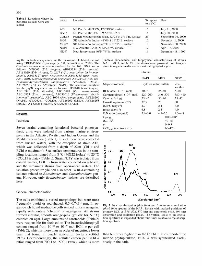
Fig. 2 In vivo absorption (thin line) and fluorescence excitation(thick line) spectra of the NAP1 isolate with marked positions ofprimary BChl a (376, 592, 870 nm) and carotenoid (463, 494 nm)absorption and excitation peaks. The vertical scale of the excita-tion spectrum is expanded about four times relative to the absorp-tion spectrum
All the isolates exhibited relatively uniform in vivo ab-sorption spectra (Fig. 2), with the infrared QY band of BChla light-harvesting 1 (LH1) antenna centered between 865and 871 nm, depending upon the specific strain (Table 3).Light-harvesting complex 2 (LH2), frequently found inpurple photosynthetic bacteria and Roseobacter species,was absent, resulting in a small functional cross-section ofthe photosynthetic units. At 470 nm, the calculated cross-sections σ470 averaged between 40 and 45Å2; this is five toseven times lower than values typically measured in oxy-genic photoautotrophs at the same wavelength (Gorbunovet al. 2000). Carotenoids were responsible for the majorabsorption bands between 350 and 600 nm, with peaks at460–464 nm and 494–497 nm. The absorption bands ofcytochromes could be distinguished in the in vivo absorp-tion spectra by means of second derivative analysis (notshown). The positions of the γ (Soret) band at 408–416nm and the α band at 550–551 nm indicated the presence
of cytochrome c type. The QX and the Soret (BY) absorp-tion bands of BChl a overlapped with carotenoids absorp-tion, and their peaks at 592 nm and 376 nm were better re-solved in the BChl a fluorescence excitation spectrum(Fig. 2).
Nutrition and growth
All isolates were capable of growth at room temperature(20 °C) in the organic medium under oxic conditions. Nogrowth was observed in anaerobiosis. Acetate, butyrate,glucose, and pyruvate were utilized as simple carbon sourcesfor growth. Utilization of other carbon sources (e.g., glyc-erol, fumarate, succinate, lactate, and glutamine) variedamong the strains (Table 4). No growth occurred in artifi-cial medium devoid of an added organic carbon source.Optimal growth was typically obtained using standard
331
Strain NAP1 COL13 NJ3Y MG3 Erb.litoralis
Erb.longus
„Cit. bathyo-marinum“
Erm.ramosum
S. sibiricus Rsb. deni-trificans
Environment Openocean
Marinecoast
Marinecoast
Openocean
Marinecoast
Marinecoast
Deep ocean Freshwater
Fresh water Marinecoast
Subclass ofProteobacteria
a-4 a-4 a-4 a-4 a-4 a-4 a-4 a-4 a-4 a-3
Color Orange Orange Yellow Orange Orange Orange Yellow Orange Yellow PinkIn vivo BChl aAbs. max.LH1 870 870 869 865 871 866 867 868 867 870LH2 – – – – – – – 798,832 – 806Chloramphenicol + + + + + + + + + +Erytromycin + + + + + + n.d. + – +Nalidixic acid – – – – – – n.d. + + +Penicillin G + + + + + + – – – +Polymyxin B – – – – – – + + – +Streptomycin – – – – – – – – + +Tetracycline + + + + + + + + – +
Table 3 Comparative characteristics of the isolates and other spe-cies of aerobic anoxygenic photoheterotrophs. Strain NAP1 is con-sidered representative for all the open ocean isolates of group 1(AT8, BA13, MG22, NAP1). + Antibiotic sensitivity, – antibiotic
resistance, LH1, LH2 light-harvesting complex 1 and 2, respec-tively. Data for “Cit. bathyomarinum”, Erm. ramosum and S. sibir-icus taken from Yurkov and Gorlenko (1992), Yurkov et al.(1992), Yurkov et al. (1999)
Table 4 Substrate utilizationamong the studied strains.None of the strains is capableof growth on arabinose, citrate,glycolate, malate, sorbitol, su-crose, or tartrate. High organicmedium corresponds to 5 gpeptone and 1 g yeast extractper liter
Nutrient Strains
AT8 BA13 COL13 MG3 MG22 NAP1 NJ3Y
Acetate ++ ++ + + ++ ++ +Butyrate ++ +++ ++ ++ +++ +++ +++Fructose + – – – + + –Fumarate – – – – – – ++Glucose +++ ++ +++ ++ ++ +++ ++Glutamate +++ +++ +++ +++ +++ +++ +Glutamine – – – – – + –Glycerol + – – ++ – – +Lactate + – – – – + –Pyruvate +++ ++ ++ + +++ +++ +++Succinate – – – – – – +++High organic medium ++ ++ ++ – +++ +++ +++
peptone+yeast extract media. All the strains, except MG3,were capable of growth on high organics medium (5 gpeptone and 1 g yeast extract per liter), but both growthand BChl a accumulation optima were frequently found atlower concentrations of the organics (not shown). Ammo-nium and urea could be utilized as nitrogen sources whilenitrite and nitrate were not utilized. No growth was de-tected under diazotrophic conditions. Glutamate, leucine,and isoleucine could serve as combined sources of bothcarbon and nitrogen. All of the isolates required vitaminB12 and biotin for growth.
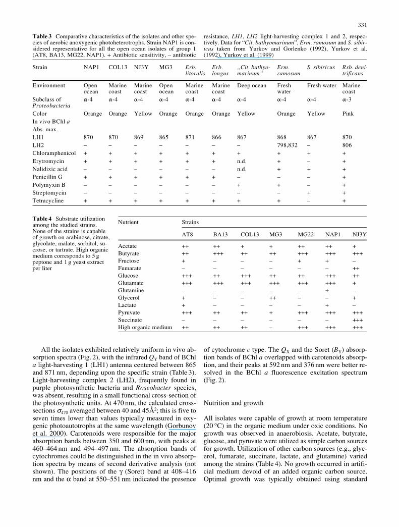
Specific growth rates were tested in the organic me-dium at 25 °C with NAP1, NJ3Y and MG3 strains and rangedfrom 2.4 days–1 for MG3 to 4.7 days–1 for NAP1 (see alsoTable 2). The optimal growth temperature for NAP1 was32.5 °C, but the synthesis of BChl a was inhibited at tem-peratures higher than 30 °C (Fig. 3); the temperature opti-mum for BChl a accumulation was 22.5 °C. Optimal growthtemperatures were lower in the NJ3Y (30 °C) and MG3isolates (25 °C), which, unlike NAP1, did not exhibit asharp temperature dependence of cellular BChl a content.The Q10 values for growth ranged from 1.8 for NJ3Y to2.6 for NAP1.
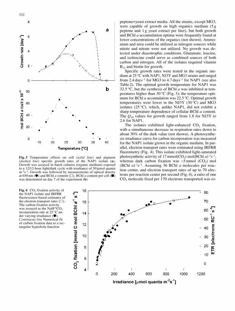
The isolates exhibited light-enhanced CO2 fixation,with a simultaneous decrease in respiration rates down toabout 30% of the dark value (not shown). A photosynthe-sis-irradiance curve for carbon incorporation was measuredfor the NAP1 isolate grown in the organic medium. In par-allel, electron transport rates were estimated using IRFRRfluorometry (Fig. 4). This isolate exhibited light-saturatedphotosynthetic activity of 17 mmol(CO2) mol(BChl a)–1s–1,whereas dark carbon fixation was ~5 mmol (CO2) mol(BChl a)–1s–1. Assuming 36 BChl a molecules per reac-tion center, and electron transport rates of up to 70 elec-trons per reaction center per second (Fig. 4), a ratio of oneCO2 molecule fixed per 170 electrons transported was es-
332
Fig. 3 Temperature effects on cell (solid line) and pigment(dashed line) specific growth rates of the NAP1 isolate (a).Growth was assayed in batch cultures (organic medium) exposedto a 12/12-hour light/dark cycle with irradiance of 50 µmol quantam–2s–1. Growth was followed by measurements of optical densityat 650 nm (E) and BChl a content (V). BChl a content per cell (K)was determined on day 7 of the experiment (b)
Fig. 4 CO2 fixation activity ofthe NAP1 isolate and IRFRRfluorescence-based estimates ofthe electron transport rates (V).The carbon fixation activitywas assayed as the NaH14CO3incorporation rate at 25 °C un-der varying irradiance (E).Continuous line Numerical fitof carbon fixation data to a rec-tangular hyperbola function
timated. This is an efficiency about one order of magni-tude less than that measured in oxygenic photoautotrophs(Falkowski and Raven 1997).
All of the isolates, as well as the type strains (Erb. longusand Erb. litoralis), were sensitive to amikacin, ampicillin,cephapirin, chloramphenicol, cloxacillin, erythromycin, fu-sidic acid, gentamycin, kanamycin, linkomycin, neomycin,novobiocin, penicillin G (20 kU/l), rifampicin (10 mg/l),spectinomycin (20 mg/l), tetracycline, and vancomycin.Isolates were resistant to amphotericine B (2.5 mg/l), cy-cloheximide, dihydrostreptomycin, geneticin, nalidixic acid,nystatin, paromomycin, polymyxin B, and streptomycin.Resistance towards streptomycin and dihydrostreptomycinseems to be typical for this bacterial group (see Table 3),and (dihydro)streptomycin-containing (50 mg/l) platesmight be used as selective media for isolation or mainte-nance of these strains.
Pigment distribution
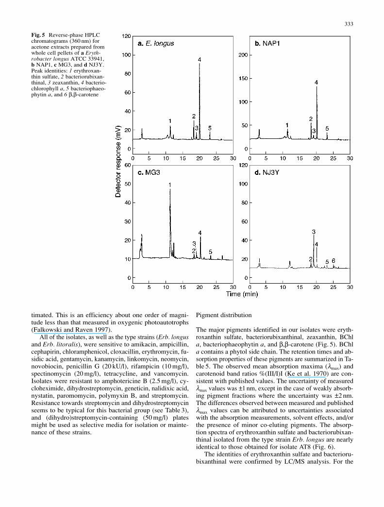
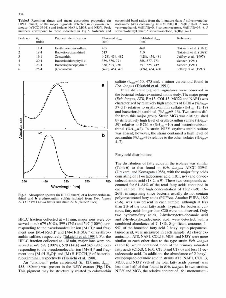
The major pigments identified in our isolates were eryth-roxanthin sulfate, bacteriorubixanthinal, zeaxanthin, BChla, bacteriophaeophytin a, and β,β-carotene (Fig. 5). BChla contains a phytol side chain. The retention times and ab-sorption properties of these pigments are summarized in Ta-ble 5. The observed mean absorption maxima (λmax) andcarotenoid band ratios %(III/I)I (Ke et al. 1970) are con-sistent with published values. The uncertainty of measuredλmax values was ±1 nm, except in the case of weakly absorb-ing pigment fractions where the uncertainty was ±2 nm.The differences observed between measured and publishedλmax values can be attributed to uncertainties associatedwith the absorption measurements, solvent effects, and/orthe presence of minor co-eluting pigments. The absorp-tion spectra of erythroxanthin sulfate and bacteriorubixan-thinal isolated from the type strain Erb. longus are nearlyidentical to those obtained for isolate AT8 (Fig. 6).
The identities of erythroxanthin sulfate and bacterioru-bixanthinal were confirmed by LC/MS analysis. For the
333
Fig. 5 Reverse-phase HPLCchromatograms (360 nm) foracetone extracts prepared fromwhole cell pellets of a Eryth-robacter longus ATCC 33941,b NAP1, c MG3, and d NJ3Y.Peak identities: 1 erythroxan-thin sulfate, 2 bacteriorubixan-thinal, 3 zeaxanthin, 4 bacterio-chlorophyll a, 5 bacteriophaeo-phytin a, and 6 β,β-carotene
HPLC fraction collected at ~11 min, major ions were ob-served at m/z 679 (50%), 599 (17%) and 597 (100%), cor-responding to the pseudomolecular ion [M+H]+ and frag-ment ions [M+H-SO3]+ and [M+H-H2SO3]+ of erythrox-anthin sulfate, respectively (Takaichi et al. 1991). For theHPLC fraction collected at ~18 min, major ions were ob-served at m/z 597 (100%), 579 (14%) and 565 (9%), cor-responding to the pseudomolecular ion [M+H]+ and frag-ment ions [M+H-H2O]+ and [M+H-HOCH3]+ of bacterio-rubixanthinal, respectively (Takaichi et al. 1988).
An “unknown” polar carotenoid (Rt=12.0 min, λmax=455, 480 nm) was present in the NJ3Y extract (Fig. 1D).This pigment may be structurally related to caloxanthin
sulfate (λmax=450, 475 nm), a minor carotenoid found inErb. longus (Takaichi et al. 1991).
Three different pigment signatures were observed inthe bacterial isolates examined in this study. The major group(Erb. longus, AT8, BA13, COL13, MG22 and NAP1) wascharacterized by relatively high amounts of BChl a (%A360=37–51) relative to erythroxanthin sulfate (%A360=12–19)and bacteriorubixanthinal (%A360=9–13). Two strains dif-fer from this major group. Strain MG3 was distinguishedby its relatively high level of erythroxanthin sulfate (%A360=59) relative to BChl a (%A360 =10) and bacteriorubixan-thinal (%A360=2). In strain NJ3Y erythroxanthin sulfatewas absent; however, the strain contained a high level ofzeaxanthin (%A360=39) relative to the other isolates (%A360=4–7).
Fatty acid distributions
The distribution of fatty acids in the isolates was similar(Table 6) to that found in Erb. longus ATCC 33941(Urakami and Komagata 1988), with the major fatty acidsconsisting of 11-octadecenoic acid (18:1, n-7) and 6,9-oc-tadecadienoic acid (18:2, n-9). These two compounds ac-counted for 61–84% of the total fatty acids contained ineach sample. The high concentration of 18:2 (n-9), 16–26%, is surprising since bacteria usually do not containpolyunsaturated fatty acids (PUFAs). Another PUFA, 18:2(n-6), was also present in each sample, although at lessthan 2% of the total fatty acids. Typical for bacterial cul-tures, fatty acids longer than C20 were not observed. Onlytwo hydroxy-fatty acids, 2-hydroxytetra-decanoic acidand 2-hydroxyhexadecanoic acid, were detected, with acombined abundance of 7–18%. Significant amounts, 1–9%, of the branched fatty acid 2-hexyl-cyclo-propaneoc-tanoic acid, were measured in each sample. At closer ex-amination, AT8, NAP1, COL13, MG3, and NJ3Y were moresimilar to each other than to the type strain Erb. longus(Table 6), which contained more of the primary saturatedfatty acids (C15:0, C16:0, C17:0 and C18:0) and less 11-oc-tadecenoic acid. In addition, the abundance of 2-hexyl-cyclopropane-octanoic acid in strains AT8, NAP1, COL13,MG3, and NJ3Y (9% of the total fatty acids present) wasless than half of that found in Erb. longus. In two strains,NJ3Y and MG3, the relative content of 16:1 monounsatu-
334
Peak no. Rt Pigment identification Observed λmax Published λmax Reference(min) (nm) (nm)
1 11.4 Erythroxanthin sulfate 465 469 Takaichi et al. (1991)2 18.4 Bacteriorubixanthinal 513 510 Takaichi et al. (1988)3 19.1 Zeaxanthin (428), 454, 482 (428), 454, 481 Jeffrey et al. (1997)4 20.4 Bacteriochlorophyll a 359, 580, 771 358, 577, 773 Scheer (1991)5 23.4 Bacteriophaeophytin a 358, 525, 750 357, 525, 749 Scheer (1991)6 25.4 β,β-carotene (426), 454, 478 (426), 454, 480 Jeffrey et al. (1997)
Table 5 Retention times and mean absorption properties (inHPLC eluant) of the major pigments detected in Erythrobacterlongus (ATCC 33941) and isolates NAP1, MG3, and NJ3Y. Peaknumbers correspond to those indicated in Fig. 5. Solvents and
carotenoid band ratios from the literature data: 1 solvent=metha-nol+water (4:1) containing 40 mM NH4OH, %(III/II)=0; 2 sol-vent=methanol, %(III/II)=0; 3 solvent=acetone, %(III/II)=33; 4, 5solvent=diethyl ether; 6 solvent=acetone, %(III/II)=21
Fig. 6 Absorption spectra (in HPLC eluant) of a bacteriorubixan-thinal and b erythroxanthin sulfate isolated from Erb. longusATCC 33941 (solid lines) and strain AT8 (dashed lines)
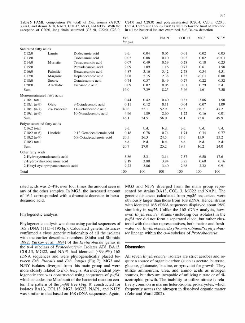
rated acids was 2–4%, over four times the amount seen inany of the other samples. In MG3, the increased amountof 16:1 corresponded with a dramatic decrease in hexa-decanoic acid.
Phylogenetic analysis
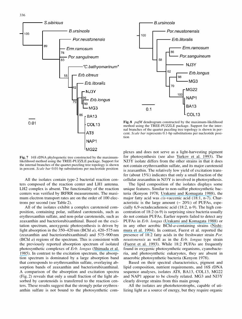
Phylogenetic analysis was done using partial sequences of16S rDNA (1115–1195 bp). Calculated genetic distancesconfirmed a close genetic relationship of all the isolateswith the earlier described members (Shiba and Shimidu1982; Yurkov et al. 1994) of the Erythrobacter genus inthe α-4 subclass of Proteobacteria. Isolates AT8, BA13,COL13, MG22, and NAP1 had identical (~99.9%) 16SrDNA sequences and were phylogenetically placed be-tween Erb. litoralis and Erb. longus (Fig. 7). MG3 andNJ3Y isolates diverged from this main group and weremore closely related to Erb. longus. An independent phy-logenetic tree was constructed using sequences of pufM,which encodes the M subunit of the bacterial reaction cen-ter. The pattern of the pufM tree (Fig. 8) constructed forisolates BA13, COL13, MG3, MG22, NAP1, and NJ3Ywas similar to that based on 16S rDNA sequences. Again,
MG3 and NJ3Y diverged from the main group repre-sented by strains BA13, COL13, MG22 and NAP1. Thegenetic distances calculated from pufM sequences wereobviously larger than those from 16S rDNA. Hence, strainswith identical 16S rDNA sequences displayed about 98%similarity in pufM. Unlike the 16S rDNA analysis, how-ever, Erythrobacter strains (including our isolates) in thepufM tree did not form a separated clade, but rather clus-tered with the other representatives, both marine and fresh-water, of Erythrobacter/Erythromicrobium/Porphyrobac-ter lineage within the α-4 subclass of Proteobacteria.
Discussion
All seven Erythrobacter isolates are strict aerobes and re-quire a source of organic carbon (such as acetate, butyrate,glucose, glutamate, leucine, or pyruvate) for growth. Theyutilize ammonium, urea, and amino acids as nitrogensources, but they are incapable of utilizing nitrate or of di-azotrophic growth. The inability to utilize nitrate is rela-tively common in marine heterotrophic prokaryotes, whichfrequently access the nitrogen in dissolved organic matter(Zehr and Ward 2002).
335
Erb. AT8 NAP1 COL13 MG3 NJ3Ylongus
Saturated fatty acidsC12:0 Lauric Dodecanoic acid b.d. 0.04 0.05 0.01 0.02 0.05C13:0 Tridecanoic acid 0.02 0.08 0.10 0.02 0.02 <0.01C14:0 Myristic Tetradecanoic acid 0.07 0.49 0.59 0.28 0.10 0.25C15:0 Pentadecanoic acid 2.09 1.09 1.16 0.77 0.61 1.58C16:0 Palmitic Hexadecanoic acid 4.87 3.16 3.42 2.78 0.34 4.51C17:0 Margaric Heptadecanoic acid 8.08 2.15 2.38 1.32 <0.01 0.80C18:0 Stearic Octadecanoic acid 0.74 0.37 0.49 0.27 0.22 0.32C20:0 Arachidic Eicosanoic acid 0.09 0.02 0.05 0.01 0.29 b.d.Sum 16.0 7.39 8.25 5.46 1.61 7.50
Monounsaturated fatty acidsC16:1 total 0.44 0.42 0.40 0.37 3.86 1.58C18:1 (n-9) Oleic 9-Octadecenoic acid 0.11 0.12 0.11 0.04 0.07 1.09C18:1 (n-7) cis-Vaccenic 11-Octadecenoic acid 40.6 52.1 52.9 59.5 68.7 47.2C19:1 (n-9) 10-Nonadecenoic acid 4.96 1.89 2.60 1.22 0.16 0.01Sum 46.1 54.5 56.0 61.1 72.8 49.9
Polyunsaturated fatty acidsC16:2 total b.d. b.d. b.d. b.d. b.d. b.d.C18:2 (n-6) Linoleic 9,12-Octadecadienoic acid 0.18 0.78 0.74 1.74 0.34 0.77C18:2 (n-9) 6,9-Octadecadienoic acid 20.5 26.3 24.5 17.6 15.9 23.2C18:3 total b.d. b.d. b.d. b.d. b.d. b.d.Sum 20.7 27.0 25.2 19.3 16.2 24.0
Other fatty acids2-Hydroxytetradecanoic acid 5.86 3.31 3.14 7.57 6.50 17.62-Hydroxyhexadecanoic acid 2.19 3.88 3.94 3.85 0.60 0.162-Hexyl-cyclopropaneoctanoic acid 9.22 3.86 3.40 2.68 2.32 0.91
Total 100 100 100 100 100 100
Table 6 FAME composition (% total) of Erb. longus (ATCC33941) and strains AT8, NAP1, COL13, MG3, and NJ3Y. With theexception of C20:0, long-chain saturated (C21:0, C22:0, C23:0,
C24:0 and C28:0) and polyunsaturated (C20:4, C20:5, C20:3,C22:4, C22:5 and C22:6) FAMEs were below the limit of detectionin all the bacterial isolates examined. b.d. Below detection
All the isolates contain type-2 bacterial reaction cen-ters composed of the reaction center and LH1 antenna.LH2 complex is absent. The functionality of the reactioncenters was verified by IRFRR measurements. The maxi-mum electron transport rates are on the order of 100 elec-trons per second (see Table 2).
All of the isolates exhibit a complex carotenoid com-position, containing polar, sulfated carotenoids, such aserythroxanthin sulfate, and non-polar carotenoids, such aszeaxanthin and bacteriorubixanthinal. Based on the exci-tation spectrum, anoxygenic photosynthesis is driven bylight absorption in the 350–420 nm (BChl a), 420–575 nm(zeaxanthin and bacteriorubixanthinal) and 575–900 nm(BChl a) regions of the spectrum. This is consistent withthe previously reported absorption spectrum of isolatedphotosynthetic complexes of Erb. longus (Shimada et al.1985). In contrast to the excitation spectrum, the absorp-tion spectrum is dominated by a large absorption bandthat corresponds to erythroxanthin sulfate, overlaying ab-sorption bands of zeaxanthin and bacteriorubixanthinal. A comparison of the absorption and excitation spectra(Fig. 2) reveals that only a small fraction of the light ab-sorbed by carotenoids is transferred to the reaction cen-ters. These results suggest that the strongly polar erythrox-anthin sulfate is not bound to the photosynthetic com-
plexes and does not serve as a light-harvesting pigmentfor photosynthesis (see also Yurkov et al. 1993). TheNJ3Y isolate differs from the other strains in that it doesnot contain erythroxanthin sulfate, and its major carotenoidis zeaxanthin. The relatively low yield of excitation trans-fer (about 15%) indicates that only a small fraction of thecellular zeaxanthin in NJ3Y is involved in photosynthesis.
The lipid composition of the isolates displays someunique features. Similar to non-sulfur photosynthetic bac-teria (Kenyon 1978; Urakami and Komagata 1988), themajor fatty acid was cis-vaccenic acid (18:1, n-7). Char-acteristic is the large amount (~ 20%) of PUFAs, espe-cially 6,9-octadecadienoic acid (18:2, n-9). The high con-centration of 18:2 (n-9) is surprising since bacteria usuallydo not contain PUFAs. Earlier reports failed to detect anyPUFAs in Erb. longus (Urakami and Komagata 1988) orin any other aerobic BChl-a-containing strains (Nishi-mura et al. 1994). In contrast, Fuerst et al. reported thepresence of 18:2 fatty acids in the freshwater strain Por.neustonensis as well as in the Erb. longus type strain(Fuerst et al. 1993). While 18:2 PUFAs are frequentlyfound in oxygenic photosynthetic organisms, cyanobacte-ria, and photosynthetic eukaryotes, they are absent inanaerobic photosynthetic bacteria (Kenyon 1978).
Based on their spectral characteristics, pigment andlipid composition, nutrient requirements, and 16S rDNAsequence analyses, isolates AT8, BA13, COL13, MG22and NAP1 appear to be closely related. MG3 and NJ3Yclearly diverge strains from this main group.
All the isolates are photoheterotrophs, capable of uti-lizing light as a source of energy, but they require organic
336
Fig. 7 16S rDNA phylogenetic tree constructed by the maximum-likelihood method using the TREE-PUZZLE package. Support forthe internal branches of the quartet puzzling tree topology is shownin percent. Scale bar 0.01-bp substitutions per nucleotide position
Fig. 8 pufM dendrogram constructed by the maximum-likelihoodmethod using the TREE-PUZZLE package. Support for the inter-nal branches of the quartet puzzling tree topology is shown in per-cent. Scale bar represents 0.1-bp substitutions per nucleotide posi-tion
carbon for growth. They display light-mediated CO2 fixa-tion, although carbon fixation activity is relatively low,more than one order of magnitude less efficient than inoxygenic photoautotrophs. The isolates lack RubisCO(R. Tabita, Ohio State University, personal communication),which suggests an alternative pathway for carbon fixa-tion. The significant (3.5-fold) enhancement of carboxyla-tion in the light (Fig. 4) indicates that photosynthesis pro-vides the major portion of energy required for carboxyla-tion.
More important is the ability of anoxygenic photo-heterotrophs to substitute respiratory carbon with photo-synthetically driven ATP production, as indicated by thestrong suppression of respiration under irradiance. As-suming an average irradiance of 200 µmol quanta m–2s–1,the photosynthetic electron transport rates approach 30 elec-trons per second per reaction center (see Fig. 4). With oneand half protons translocated across the membrane perelectron transported, and three protons required for pro-duction of one ATP, a photosynthetic flux of 15 ATP mol-ecules per reaction center per second can be maintained.Containing about 1,000 reaction centers (36 BChl a perphotosynthetic unit, Table 2) and exposed to 12 h of sun-light, the cell can derive about 10–15 mol ATP per day fromcyclic photophosphorylation.
Aerobic anoxygenic photoheterotrophs account for asignificant fraction of the marine microbial community inoligotrophic waters. Cell counts carried out in these re-gions indicate that BChl-a-containing bacteria contributeup to 8% of the total bacteria, with BChl a/Chl a valuesranging from 0.8 to 10% (Kolber et al. 2000, 2001). Thus,their relative abundance and photoheterothophic metabo-lism suggest a specific role for these bacteria in the ma-rine carbon cycle.
Acknowledgements The authors thank Maxim Gorbunov, MichaelBehrenfeld, Yoram Gerchman and Ondrej Prasil for supplying thewater samples, and Kevin Wyman for laboratory assistance. Thisresearch was supported by Rutgers University through a Post-doc-toral Research Fellowship to MK and by grants from NSF (Bio-complexity to PGF, and OCE-022095 to ZSK), EEC-9731725(RRB) and OCE-9617409 (RRB), from NASA NAG5-7171 (RRB)and NAGW-3439 (RRB) and from Czech MSMT projectsLN00A141 and MSM12310001 (MK) and GACR 206/03/P079(MK).
References
Béjà O, Suzuki MT, Heidelberg JF, Nelson WC, Preston CM,Hamada T, Eisen JA, Fraser CM, DeLong EF (2002) Unsus-pected diversity among marine aerobic anoxygenic phototrophs.Nature 415:630–633
Bidigare RR, Trees CC (2000) HPLC phytoplankton pigments:Sampling, laboratory methods, and quality assurance proce-dures. In: Mueller J, Fargion G (eds) Ocean optics protocols forsatellite ocean color sensor validation, revision 2. NASA Tech-nical Memorandum 2000–209966, pp 154–161
Des Marais, D.J. 2000. When did photosynthesis emerge on Earth?Science 289:1703–1704.
Falkowski PG, Raven JA (1997) Aquatic photosynthesis. Black-well Science, Malden, Massachusetts
Fleischman D, Kramer D (1998) Photosynthetic rhizobia. BiochimBiophys Acta 1364:17–36
Fuerst JA, Hawkins JA, Holmes A, Sly LI, Moore CJ, Stacke-brandt E (1993) Porphyrobacter neustonensis gen. nov., sp.nov., an aerobic bacteriochlorophyll-synthesizing budding bac-terium from fresh water. Int J Syst Bacteriol 43:125–34
Giovannoni SJ, DeLong EF, Olsen GJ, Pace NR (1988) Phyloge-netic group-specific oligodeoxynucleotide probes for identifi-cation of single microbial cells. J Bacteriol 170:720–726
Göbel F (1978) Quantum efficiences of growth. In: Clayton RK,Sistrom WR (eds) The photosynthetic bacteria. Plenum , NewYork, pp 907–925
Gorbunov MY, Falkowski PG, Kolber ZS (2000) Measurement ofphotosynthetic parameters in benthic organisms in situ using aSCUBA-based fast repetition rate fluorometer. Limnol Oceanogr45:242–245
Guillard RRL, Ryther JH (1962) Studies on marine planktonic di-atoms I. Cyclotella nana (Hustedt) and Detonula confervacea(Cleve) Gran. Can J Microbiol 8:229–239
Harashima K, Shiba T, Totsuka T, Simidu U, Taga N (1978) Oc-currence of bacteriochlorophyll a in a strain of an aerobic het-erotrophic bacterium. Agric Biol Chem 42:1627–1628
Imhoff JF (2001) The anoxygenic phototrophic purple bacteria. In:Boone DR, Castenholz RW, Garrity GM (eds) Bergey’s man-nual of systematic bacteriology, 2nd edn, vol 1. Springer,Berlin Heidelberg New York, pp 631–637
Jeffrey SW, Mantoura RFC, Wright SW (eds) 1997: Phytoplank-ton pigments in oceanography. Monographs on OceanographicMethodology, UNESCO
Ke B, Imsgard F, Kjøsen H, Liaaen-Jensen S (1970) Electronicspectra of carotenoids at 77°K. Biochim Biophys Acta 210:139–152
Kenyon CN (1978) Complex lipids and fatty acids of photosyn-thetic bacteria. In: Clayton RK, Sistrom WR (eds) The photo-synthetic bacteria. Plenum, New York, pp 281–313
Kolber ZS, Prasil O, Falkowski PG (1998) Measurements of vari-able chlorophyll fluorescence using fast repetition rate tech-niques: defining methodology and experimental protocols. Bio-chim Biophys Acta 1367:88–106
Kolber ZS, Van Dover CL, Niederman RA, Falkowski PG (2000)Bacterial photosynthesis in surface waters of the open ocean.Nature 407:177–179
Kolber ZS, Plumley FG, Lang AS, Beatty JT, Blankenship RE,VanDover CL, Vetriani C, Koblizek M, Rathgeber C, Fal-kowski PG (2001) Contribution of aerobic photoheterotrophicbacteria to the carbon cycle in the ocean. Science 292:2492–2495
Lane DJ (1991) 16S/23S rRNA sequencing. In Stackebrant E,Goodfellow M (eds) Nucleic acid techniques in bacterial sys-tematics. Wiley, New York
Nagashima KVP, Hiraishi A, Shimada K, Matsuura K (1997) Hori-zontal transfer of genes coding for the photosynthetic reactioncenters of purple bacteria. J Mol Evol 45:131–136
Nishimura Y, Muroga Y, Saito S, Shiba T, Takamiya KI, Shioi Y(1994) DNA relatedness and chemotaxonomic feature of aero-bic bacteriochlorophyll-containing bacteria isolated from coastof Australia. J Gen Appl Microbiol 40:287–296
Permentier HP, Schmidt KA, Kobayashi M, Akiyama M, Hager-Braun C, Neerken S, Miller M, Amesz J (2000) Compositionand optical properties of reaction centre core complexes fromthe green sulfur bacteria Prosthecochloris aestuarii andChlorobium tepidum Photosynth Res 64:27–39
Raskin L, Stromley JM, Rittmann BE, Stahl DA (1994) Group-specific 16S rRNA hybridization probes to describe naturalcommunities of methanogens. Appl Environ Microbiol 60:1232–1240
Rye R, Holland HD (1998) Paleosols and the evolution of atmo-spheric oxygen: a critical review. Am J Sci 298:621–672
Scheer H (ed) (1991) Chlorophylls. CRC, Boca Raton, FloridaSchmidt HA, Strimmer K, Vingron M, von Haeseler A (2002)
TREE-PUZZLE: maximum likelihood phylogenetic analysisusing quartets and parallel computing. Bioinformatics 18:502–504
337
Shiba T (1991) Roseobacter litoralis gen. nov., sp. nov. andRoseobacter denitrificans sp. nov., aerobic pink-pigmentedbacteria which contain bacteriochlorophyll a. Syst Appl Micro-biol 14:140–145
Shiba T, Shimidu U (1982) Erythrobacter longus gen. nov., sp.nov., an aerobic bacterium which contains bacteriochlorophylla. Int J Syst Bacteriol 32:211–217
Shiba T, Shimidu U, Taga N (1979) Distribution of aerobic bacte-ria which contain bacteriochlorophyll a. Appl Environ Micro-biol 38:43–45
Shiba T, Shioi Y, Takamiya K-I, Sutton DC, Wilkinson CR (1991)Distribution and physiology of aerobic bacteria containing bac-teriochlorophyll a on the east and west coasts of Australia.Appl Environ Microbiol 57:295–300
Shimada K (1995) Aerobic anoxygenic phototrophs. In: Blanken-ship RE, Madigan MT, Bauer CE (eds) Anoxygenic photosyn-thetic bacteria. Kluwer, Dordrecht, pp 105–122
Shimada K, Hayashi H, Tasumi M (1985) Bacteriochlorophyll-protein complexes of aerobic bacteria, Erythrobacter longusand Erythrobacter species OCh114. Arch Microbiol 143:244–247
Suzuki T, Muroga Y, Takahama M, Nishimura Y (2000) Rosei-bium denhamense gen. nov., sp. nov. and Roseibium hameli-nense sp. nov., aerobic bacteriochlorophyll-containing bacteriaisolated from the east and west coast of Australia. Int J SystBacteriol 50:2151–2156
Takaichi S, Shimada K, Ishidsu J-I (1988) Monocyclic cross-con-jugated carotenal from an aerobic photosynthetic bacteriumErythrobacter longus. Phytochemistry 27:3605–3609
Takaichi S, Furihata K, Ishidsu J-I, Shimada K (1991) Carotenoidsulphates from the aerobic photosynthetic bacterium Eryth-robacter longus. Phytochemistry 30:3411–3415
Urakami T, Komagata K (1988) Cellular fatty acid compositionwith special reference to the existence of hydroxy fatty acids,and the occurrence of squalene and sterols in species of Rho-dospirillaceae genera and Erythrobacter longus. J Gen ApplMicrobiol 34:67–84
Wright SW, Jeffrey SW, Mantoura RFC, Llewellyn CA, BjornlandT, Repeta D, Welschmeyer N (1991) Improved HPLC methodfor the analysis of chlorophylls and carotenoids from marinephytoplankton. Mar Ecol Prog Ser 77:183–196
Yurkov VV, Gorlenko VM (1992) New species of aerobic bacteriafrom the genus Erythromicrobium containing Bacteriochloro-phyll a. Mikrobiologia (english edition) 61:248–255
Yurkov VV, Beatty JT (1998) Aerobic anoxygenic phototrophicbacteria. Microbiol Mol Biol Rev 62:695–724
Yurkov VV, Gorlenko VM, Kompantseva EI (1992) A new type offreshwater aerobic orange-colored bacterium Erythromicro-bium gen. nov., containing bacteriochlorophyll a. Microbiolo-gia (english edition) 61:256–260
Yurkov VV, Gad’on N, Drews G (1993) The major part of polarcarotenoids of the aerobic bacteria Roseococcus thiosulfato-philus RB3 and Erythromicrobium ramosum E5 is not bound tothe bacteriochlorophyll a-complexes of the photosynthetic ap-paratus. Arch Microbiol 160:372–376
Yurkov VV, Stackebrandt E, Holmes A, Fuerst J, Hugenholtz P,Golecki J, Gad’on N, Gorlenko V, Kompantseva E, Drews G(1994) Phylogenetic positions of novel aerobic, bacteriochloro-phyll a-containing bacteria and description of Roseococcusthiosulfatophilus gen. nov., sp. nov., Erythromicrobium ramo-sum gen. nov., sp. nov., and Erythrobacter litoralis sp. nov. IntJ Syst Bacteriol 44:427–434
Yurkov VV, Krieger S, Stackebrandt E, Beatty JT (1999) Citromi-crobium bathyomarinum, a novel aerobic bacterium isolatedfrom deep-sea hydrothermal vent plume waters that containsphotosynthetic pigment-protein complexes. J Bacteriol 181:4517–4525
Zehr JP, Ward BB (2002) Nitrogen cycling in the oceans: Newprospectives on processes and paradigms. Appl Environ Micro-biol 68:1015–1024
338