Site-Specific Expression of Gelatinolytic Activity during Morphogenesis of the Secondary Palate in...
14
Site-Specific Expression of Gelatinolytic Activity during Morphogenesis of the Secondary Palate in the Mouse Embryo Nikolaos Gkantidis 1 , Susan Blumer 1 , Christos Katsaros 1 , Daniel Graf 2 , Matthias Chiquet 1 * 1 Department of Orthodontics and Dentofacial Orthopedics, School of Dental Medicine, University of Bern, Bern, Switzerland, 2 Institute for Oral Biology, Center for Dental Medicine, University of Zurich, Zurich, Switzerland Abstract Morphogenesis of the secondary palate in mammalian embryos involves two major events: first, reorientation of the two vertically oriented palatal shelves into a horizontal position above the tongue, and second, fusion of the two shelves at the midline. Genetic evidence in humans and mice indicates the involvement of matrix metalloproteinases (MMPs). As MMP expression patterns might differ from sites of activity, we used a recently developed highly sensitive in situ zymography technique to map gelatinolytic MMP activity in the developing mouse palate. At embryonic day 14.5 (E14.5), we detected strong gelatinolytic activity around the lateral epithelial folds of the nasopharyngeal cavity, which is generated as a consequence of palatal shelf elevation. Activity was concentrated in the basement membrane of the epithelial fold but extended into the adjacent mesenchyme, and increased in intensity with lateral outgrowth of the cavity at E15.5. Gelatinolytic activity at this site was not the consequence of epithelial fold formation, as it was also observed in Bmp7- deficient embryos where shelf elevation is delayed. In this case, gelatinolytic activity appeared in vertical shelves at the exact position where the epithelial fold will form during elevation. Mmp2 and Mmp14 (MT1-MMP), but not Mmp9 and Mmp13, mRNAs were expressed in the mesenchyme around the epithelial folds of the elevated palatal shelves; this was confirmed by immunostaining for MMP-2 and MT1-MMP. Weak gelatinolytic activity was also found at the midline of E14.5 palatal shelves, which increased during fusion at E15.5. Whereas MMPs have been implicated in palatal fusion before, this is the first report showing that gelatinases might contribute to tissue remodeling during early stages of palatal shelf elevation and formation of the nasopharynx. Citation: Gkantidis N, Blumer S, Katsaros C, Graf D, Chiquet M (2012) Site-Specific Expression of Gelatinolytic Activity during Morphogenesis of the Secondary Palate in the Mouse Embryo. PLoS ONE 7(10): e47762. doi:10.1371/journal.pone.0047762 Editor: Michael Schubert, Ecole Normale Supe ´rieure de Lyon, France Received July 20, 2012; Accepted September 20, 2012; Published October 16, 2012 Copyright: ß 2012 Gkantidis et al. This is an open-access article distributed under the terms of the Creative Commons Attribution License, which permits unrestricted use, distribution, and reproduction in any medium, provided the original author and source are credited. Funding: This work was funded in part by grant 31003A-135530 from the Swiss National Science Foundation (http://www.snf.ch/E/Pages/default.aspx). The funder had no role in study design, data collection and analysis, decision to publish, or preparation of the manuscript. No additional external funding was received for this study. Competing Interests: The authors have declared that no competing interests exist. * E-mail: [email protected] Introduction Cleft lip with or without cleft palate (CLP) and cleft palate only (CPO) are among the most frequent birth defects in humans, affecting 1–2 per thousand newborns [1,2,3,4]. About 70% of cases are considered non-syndromic, whereas the remainder is associated with various other defects [5]. A multitude of genetic as well as environmental factors are believed to contribute to the etiology of CLP and CPO [2]. However, in most of the non- syndromic cases the cause remains unknown [5]. In mouse knockout models, close to 20 genes have been identified that upon deletion result in cleft palate; many of them code for components of the TGF-b, BMP, WNT, and FGF signaling pathways [6,7]. Morphogenesis of the secondary palate is a complex process involving several consecutive events, which occur from embryonic day E13.5 to E15.5 in the mouse and between the 6th and 10th week of gestation in humans [8,9]. First, paired palatal shelves grow out vertically from the maxillary processes on both sides of the tongue. In a second step, the palatal shelves elevate into a horizontal position above the tongue. As a consequence, two opposing epithelial folds (one from each shelf) are generated comprising the lateral margins of a newly formed cavity, which will eventually give rise to the nasopharynx. The palatal shelves grow horizontally and contact each other, forming the midline epithelial seam (MES). Finally, the medial edge epithelial (MEE) cells disappear, and the two palatal shelves fuse by establishing continuity between their mesenchyme [8]. Through this morpho- genetic process, the secondary palate separates the primary oral cavity into the oropharynx and the nasopharynx, supporting vital functions such as feeding and breathing. Cleft palate can arise from either a deficit of the palatal shelves to grow to appropriate size, from disturbances in palatal shelf elevation, or from a lack of shelf fusion [7]. Experiments in mice [10,11,12,13] and genetic linkage studies in humans [14,15] indicate that, besides defects in growth factor signaling, deficiencies in certain matrix metalloproteinases (MMPs) are involved in the etiology of cleft lip and palate. MMPs comprise a family of secreted Zn 2+ -dependent proteases that cleave extracellular matrix (ECM) proteins and are known to be essential for tissue remodeling in development and disease [16]. Specific domains of MMPs provide substrate specificity [17,18]: for example, the ‘‘collagenases’’ MMP-1, 28, and 213 mainly attack PLOS ONE | www.plosone.org 1 October 2012 | Volume 7 | Issue 10 | e47762
Transcript of Site-Specific Expression of Gelatinolytic Activity during Morphogenesis of the Secondary Palate in...
Site-Specific Expression of Gelatinolytic Activity duringMorphogenesis of the Secondary Palate in the MouseEmbryoNikolaos Gkantidis1, Susan Blumer1, Christos Katsaros1, Daniel Graf2, Matthias Chiquet1*
1Department of Orthodontics and Dentofacial Orthopedics, School of Dental Medicine, University of Bern, Bern, Switzerland, 2 Institute for Oral Biology, Center for Dental
Medicine, University of Zurich, Zurich, Switzerland
Abstract
Morphogenesis of the secondary palate in mammalian embryos involves two major events: first, reorientation of the twovertically oriented palatal shelves into a horizontal position above the tongue, and second, fusion of the two shelves at themidline. Genetic evidence in humans and mice indicates the involvement of matrix metalloproteinases (MMPs). As MMPexpression patterns might differ from sites of activity, we used a recently developed highly sensitive in situ zymographytechnique to map gelatinolytic MMP activity in the developing mouse palate. At embryonic day 14.5 (E14.5), we detectedstrong gelatinolytic activity around the lateral epithelial folds of the nasopharyngeal cavity, which is generated asa consequence of palatal shelf elevation. Activity was concentrated in the basement membrane of the epithelial fold butextended into the adjacent mesenchyme, and increased in intensity with lateral outgrowth of the cavity at E15.5.Gelatinolytic activity at this site was not the consequence of epithelial fold formation, as it was also observed in Bmp7-deficient embryos where shelf elevation is delayed. In this case, gelatinolytic activity appeared in vertical shelves at the exactposition where the epithelial fold will form during elevation. Mmp2 and Mmp14 (MT1-MMP), but not Mmp9 and Mmp13,mRNAs were expressed in the mesenchyme around the epithelial folds of the elevated palatal shelves; this was confirmedby immunostaining for MMP-2 and MT1-MMP. Weak gelatinolytic activity was also found at the midline of E14.5 palatalshelves, which increased during fusion at E15.5. Whereas MMPs have been implicated in palatal fusion before, this is the firstreport showing that gelatinases might contribute to tissue remodeling during early stages of palatal shelf elevation andformation of the nasopharynx.
Citation: Gkantidis N, Blumer S, Katsaros C, Graf D, Chiquet M (2012) Site-Specific Expression of Gelatinolytic Activity during Morphogenesis of the SecondaryPalate in the Mouse Embryo. PLoS ONE 7(10): e47762. doi:10.1371/journal.pone.0047762
Editor: Michael Schubert, Ecole Normale Superieure de Lyon, France
Received July 20, 2012; Accepted September 20, 2012; Published October 16, 2012
Copyright: � 2012 Gkantidis et al. This is an open-access article distributed under the terms of the Creative Commons Attribution License, which permitsunrestricted use, distribution, and reproduction in any medium, provided the original author and source are credited.
Funding: This work was funded in part by grant 31003A-135530 from the Swiss National Science Foundation (http://www.snf.ch/E/Pages/default.aspx). Thefunder had no role in study design, data collection and analysis, decision to publish, or preparation of the manuscript. No additional external funding was receivedfor this study.
Competing Interests: The authors have declared that no competing interests exist.
* E-mail: [email protected]
Introduction
Cleft lip with or without cleft palate (CLP) and cleft palate only
(CPO) are among the most frequent birth defects in humans,
affecting 1–2 per thousand newborns [1,2,3,4]. About 70% of
cases are considered non-syndromic, whereas the remainder is
associated with various other defects [5]. A multitude of genetic as
well as environmental factors are believed to contribute to the
etiology of CLP and CPO [2]. However, in most of the non-
syndromic cases the cause remains unknown [5]. In mouse
knockout models, close to 20 genes have been identified that upon
deletion result in cleft palate; many of them code for components
of the TGF-b, BMP, WNT, and FGF signaling pathways [6,7].
Morphogenesis of the secondary palate is a complex process
involving several consecutive events, which occur from embryonic
day E13.5 to E15.5 in the mouse and between the 6th and 10th
week of gestation in humans [8,9]. First, paired palatal shelves
grow out vertically from the maxillary processes on both sides of
the tongue. In a second step, the palatal shelves elevate into
a horizontal position above the tongue. As a consequence, two
opposing epithelial folds (one from each shelf) are generated
comprising the lateral margins of a newly formed cavity, which
will eventually give rise to the nasopharynx. The palatal shelves
grow horizontally and contact each other, forming the midline
epithelial seam (MES). Finally, the medial edge epithelial (MEE)
cells disappear, and the two palatal shelves fuse by establishing
continuity between their mesenchyme [8]. Through this morpho-
genetic process, the secondary palate separates the primary oral
cavity into the oropharynx and the nasopharynx, supporting vital
functions such as feeding and breathing. Cleft palate can arise
from either a deficit of the palatal shelves to grow to appropriate
size, from disturbances in palatal shelf elevation, or from a lack of
shelf fusion [7].
Experiments in mice [10,11,12,13] and genetic linkage studies
in humans [14,15] indicate that, besides defects in growth factor
signaling, deficiencies in certain matrix metalloproteinases (MMPs)
are involved in the etiology of cleft lip and palate. MMPs comprise
a family of secreted Zn2+-dependent proteases that cleave
extracellular matrix (ECM) proteins and are known to be essential
for tissue remodeling in development and disease [16]. Specific
domains of MMPs provide substrate specificity [17,18]: for
example, the ‘‘collagenases’’ MMP-1, 28, and 213 mainly attack
PLOS ONE | www.plosone.org 1 October 2012 | Volume 7 | Issue 10 | e47762

native fibrillar collagens, whereas MMP-2 and MMP-9 are the
major ‘‘gelatinases’’ that cleave partially denatured collagen, as
well as collagen IV in basement membranes [18,19]. Most MMPs
are secreted as inactive proenzymes and their activity is regulated
in intricate ways, positively by proteolytic cleavage of the
propeptide [20] and negatively (in most cases) by tissue inhibitors
of metalloproteinases (TIMPs; [21]). Membrane-type MMPs (e.g.
MT1-MMP/MMP-14) on cell surfaces are required for activation
of major secreted MMPs (e.g. MMP-2/gelatinase B) [20].
MMPs have been implicated in the process of palatal shelf
fusion. Palatal shelves from E14.0 wild type mouse embryos failed
to fuse when cultured in close proximity in the presence of
synthetic MMP inhibitor or excess TIMP-2 [12]. In this case,
MMPs are likely to function downstream of TGF-b3, a central
regulator of palatal shelf fusion in mice and humans
[5,14,22,23,24]. Tgfb3-deficient mouse embryos expressed lower
levels of MMP-2 (gelatinase B) and MMP-13 (collagenase 3) in the
developing palate, and addition of TGF-b3 to palatal mesenchyme
cultures induced MMP-13 expression [12]. Similarly, MT6-MMP
(MMP-25), which has been linked to cleft palate in humans, is also
regulated by TGF-b3 and required for palatal shelf fusion in vitro
[25]. These results demonstrate that tissue remodeling by specific
MMPs plays an important role in the late steps of secondary palate
formation.
Compared to palatal shelf fusion, little is known yet about the
molecular mechanism of palatal shelf elevation. It has been
proposed that changes in osmotic pressure due to adsorption of
water by accumulating hyaluronic acid causes shelf elevation
[8,26,27], although direct evidence is missing. A mechanism by
which the vertical shelves simply swing up by 90u around an
imaginary ‘‘hinge’’ seems unlikely, as substantial reorganization of
mesenchymal tissue is documented during shelf elevation. The
presumptive middle edge epithelial (MEE) cells are not located
exactly at the apex of the vertical shelves, but instead on their
distal-medial aspect facing the tongue [28]. Moreover, tracking
experiments indicated that during shelf elevation, the lower half of
the vertical shelves bulges out mesially, whereas part of the upper
half retracts laterally, forming an epithelial fold [29]. Thus, like for
other morphogenetic events, coordinated cell contractions/move-
ments and remodeling of the extracellular matrix are likely
mechanisms underlying palatal shelf elevation [30]. Here, we used
a highly sensitive in situ zymography technique to detect
gelatinolytic activity in palatal shelves of mouse embryos at the
stage of their elevation and fusion. The method is based on
incubating unfixed cryosections with dye-quenched gelatin (DQ-
gelatin) whose fluorescence increases upon enzymatic cleavage
[31]. The major enzymes detected in tissues by gelatin
zymography are MMP-2 (gelatinase B) and MMP-9 (gelatinase
A); however other MMPs with weaker gelatinolytic activity might
Figure 1. Overview of craniofacial structures in the mouse embryo. Frontal sections (mid-posterior level) of (A) E13.5 and (B) E14.5 wild typemouse heads were labeled with antibody to laminin-111, which stains all basement membranes. The boxed areas illustrate the region of interest thatis presented in most of the following figures. Note that at E13.5 the palatal shelves are vertically oriented besides the tongue, while at E14.5 theshelves have elevated above the tongue to form the secondary palate. e, eye; b, forebrain; t, tongue; p, palatal shelf; tb, tooth buds. Bar, 500 mm.Drawings of E13.5 (C) and E14.5 (D) mouse heads indicate the different anteroposterior section planes through the palatal region (a, anterior; m,middle; p, posterior) that are presented in following figures. Note that at E14.5, the elevated secondary palate now separates the oral from the newlyformed nasopharyngeal cavity.doi:10.1371/journal.pone.0047762.g001
Gelatinolysis in Palate Morphogenesis
PLOS ONE | www.plosone.org 2 October 2012 | Volume 7 | Issue 10 | e47762
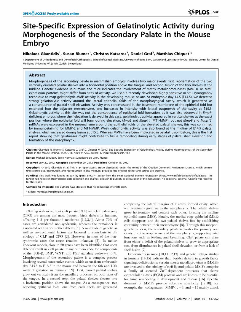
Figure 2. Double labeling for gelatinolytic activity and laminin in the palatal region of the mouse embryo. Frontal cryosections of E13.5(A–C), E14.5 (D–F), and E15.5 (G–I) wild type mouse heads were subjected to DQ-gelatin zymography (green panels), followed byimmunofluorescence labeling for laminin-111 on the same section (red panels). The images show representative sections from the anterior (A, D,G), the middle (B, E, H), and the posterior (C, F, I) level of the palate. No site with significant gelatinolytic activity, as manifested by increasedfluorescence, is detected at E13.5 prior to palatal shelf elevation. At E14.5, gelatinolytic activity is evident in the nasal cartilage and in the main palatalarteries (arrows). In addition, signs of gelatinolysis are visible at the midline epithelial seam (asterisks), whereas prominent gelatinolytic activity isdetected around the folds of the elevated palatal shelves, where the nasopharynx will form (arrowheads). A similar pattern of gelatinolysis is observedat E15.5, although activity is increased compared to E14.5. Furthermore, prominent activity was evident for maxillary bone. p, palatal shelf; t, tongue;n, nasal cartilage; m, maxillary bone. Bar, 200 mm.doi:10.1371/journal.pone.0047762.g002
Gelatinolysis in Palate Morphogenesis
PLOS ONE | www.plosone.org 3 October 2012 | Volume 7 | Issue 10 | e47762
contribute to the signal [32]. This method is able to detect
gelatinolytic activity associated with minute structures such as
distinct basement membranes in the developing mouse embryo
head [33]. Double labeling with antibodies to specific MMPs
allows to reveal the relationship between active enzyme and
protein distribution, which might differ due to the complex
regulation of MMP activity.
We provide the first evidence that gelatinases are involved not
only in the process of palatal shelf fusion but also in their elevation,
as gelatinolytic activity was detected in the basement membrane
and underlying mesenchyme of the nasopharyngeal folds that
formed when shelves reoriented from vertical to horizontal
position. In a model of delayed palatal shelf elevation, the still
vertically oriented shelves displayed gelatinolytic activity at the
exact site where the epithelial fold would normally form. This
suggested that the marked increase in gelatinolytic activity at this
position might be causally involved in the formation of the
epithelial fold during shelf elevation, rather than being a conse-
quence of the process.
Materials and Methods
Animals, embryonic tissue, and cryosectioningAnimal experiments were approved by the local veterinary
authorities (permit 98/2011, Veterinaramt Zurich) in compliance
with Swiss federal law (TSchG, TSchV) and cantonal by-laws in
full compliance with the European Guideline 86/609/EC. This
authority approval also included ethical approval. C57BL/6
wildtype mouse embryos were obtained from J.-F. Spetz at the
Friedrich-Miescher Institute for Biomedical Research (FMI) in
Basel, Switzerland. Embryos were obtained by timed mating, and
E0.5 was considered as the morning where the vaginal plug was
seen. Pregnant females were sacrificed by cervical dislocation at
the desired stage (E13.5, E14.5, and E15.5), embryos were
harvested and decapitated. The Bmp7 null allele of the Bmp7
heterozygous null mice was generated by Cre-mediated re-
combination in the germ line of a conditional Bmp7 allele (Bmp7flx),
in which exon 1 is flanked by loxP sites as described earlier [34].
Bmp7 heterozygous null mice (Bmp7+/D) were intercrossed to
obtain Bmp7D/D and control embryos from the same litter. E14.5
embryos were harvested from timed pregnant mice sacrificed by
cervical dislocation. Genotyping of embryos was carried out by
allele-specific PCR [34]. In total, 6 E13.5, 14 E14.5, and 12 E15.5
C57BL/6 wild type, as well as 4 Bmp7+/D and 4 Bmp7D/D mouse
embryos were used for the present study.
The embryo heads were washed in ice-cold PBS, soaked and
embedded in Tissue Tek (O.C.T. compound; Sakura Finetek
Europe B.V., Zoeterwoude, Netherlands), and frozen on a metal
block cooled to 280uC. All tissue was stored at 280uC before
sectioning. Serial frontal sections (10–12 mm thick) of the embryo
heads were prepared on a Cryocut E cryomicrotome (Reichert-
Jung, Leica Microsystems, Heerbrugg, Switzerland), dried at 37uCfor 1–5 min, and stored at 280uC before further use.
In situ zymographyFluorescein conjugated, dye-quenched gelatin from pig skin
(DQTM-gelatin) was obtained from Molecular Probes (Invitrogen,
Basel, Switzerland). A 1 mg/ml stock solution of DQ-gelatin was
prepared in gelatinase reaction buffer (150 mM NaCl, 5 mM
CaCl2, 0.2 mM NaN3, 50 mM Tris-HCl, pH 7.6) and stored at
4uC. The working solution for in situ zymography was made by
directly diluting DQ-gelatin stock solution in reaction buffer to
a final concentration of 20 mg/ml. Unfixed cryosections were
thawed, rounded with a wax pen, overlaid with 250 ml DQ-gelatin
working solution (for approximately half of the slide), and
incubated at 37uC in a dark wet chamber for 3 hours. After three
washes with PBS, sections were either processed for immunoflu-
orescence (see below), or mounted directly in 90% glycerol in PBS
containing 10 mg/ml propyl 3,4,5-trihydroxybenzoate (Merck,
Darmstadt, Germany) as anti-fading agent.
Differences in the extent of gelatinolytic activity during palatal
shelf elevation were quantified by measuring the mean pixel
intensity (ImageJ Software) in a rectangular area of defined size
(20 mm x 50 mm), which included the palatal epithelium and
basement membrane of the nasopharyngeal fold, at the middle
anteroposterior palatal level. Twelve such regions were measured
for each developmental stage (E13.5, E14.5, and E15.5), from
a total of 18 mouse embryo heads. Both left and right folds were
measured on each section. Background intensity, measured in an
adjacent region inside the palatal shelf, was subtracted from the
value obtained for each corresponding fold. All measurements
were performed twice and the mean value was used for analysis.
Non parametric statistics were used for analyzing the results since
data were not normally distributed (Shapiro-Wilk test). Statistical
analysis was performed through the SPPS Statistics 17.0 software.
The specificity of the ISZ protocol used here was thoroughly
tested before [33] and confirmed in the present study through
various types of control experiments. For negative controls, the
DQ-gelatin was either omitted from the gelatinase reaction buffer,
or replaced by 20 mg/ml unlabeled pig skin gelatin (Merck); in
both cases, no signal was observed. To control for the specificity of
the enzyme reaction, either of the following metalloproteinase
inhibitors was added to the DQ-gelatin working solution prior to
incubation of the slides: 10 mM ethylenediamine tetra-acetic acid
(EDTA; Merck; replacing CaCl2 in the reaction buffer); 1 mM
1,10-phenanthroline (Phen; Sigma, Buchs, Switzerland); or 50 mM(2R)-[(4-Biphenylylsulfonyl)amino]-N-hydroxy-3-phenylpropiona-
mide (BiPS; MMP-2/MMP-9 inhibitor II; Calbiochem/Merck
Chemicals, Nottingham, UK). Addition of either of these reagents
to the zymography buffer resulted in partial or complete inhibition
of fluorescence generated by DQ-gelatin cleavage. EDTA,
a general divalent cation chelator and metalloproteinase antago-
nist, and phenanthroline, a potent Zn2+ complexing agent and
MMP inhibitor, almost completely inhibited the gelatinolytic
activity. BiPS, a specific MMP-2/MMP-9 inhibitor, significantly
attenuated the reaction in situ (Fig. S1).
Immunofluorescence stainingTo combine ISZ with antibody labeling, we performed ISZ on
unfixed cryosections, before continuing with immunolabeling as
published previously [33]. Immediately after processing for ISZ,
sections were fixed with cold acetone (220uC) for 1 min, blocked
with 3% bovine serum albumin in PBS (BSA/PBS) for 10 min,
and then incubated with primary antibody diluted in BSA/PBS
for 45 min at room temperature. The following primary
antibodies were used: rabbit anti-mouse EHS laminin (1:200;
[35]); goat anti-mouse MMP-2 (1:20; R&D Systems, Abingdon,
UK); goat anti-human MT1-MMP/MMP14 (1:20; R&D Sys-
tems). After three washes in BSA/PBS, sections were covered for
30 min with the respective secondary antibody (rhodamine-
conjugated goat anti-rabbit IgG from Cappel/MP Biomedicals,
Santa Ana, CA; Cy3-conjugated mouse anti-goat IgG from
Jackson ImmunoResearch/Milan Analytica, Rheinfelden, Swit-
zerland) diluted 1:100 in BSA/PBS. After washing in DEPC-
treated H2O, slides were mounted in buffered glycerol with anti-
fading agent (see above).
Gelatinolysis in Palate Morphogenesis
PLOS ONE | www.plosone.org 4 October 2012 | Volume 7 | Issue 10 | e47762

Gene specific RNA probes and in situ hybridizationTotal RNA was isolated from E14.5 C57BL/6 wildtype mouse
embryos using an RNAeasy Mini Kit (Qiagen, Hombrechtikon,
Switzerland) and reverse transcribed to cDNA using Moloney
murine leukemia virus reverse transcriptase (Promega, Dubendorf,
Switzerland). Primers specific for the murine Mmp2, Mmp9,
Mmp13, and Mmp14 (MT1-MMP) genes were designed using
Figure 3. Close-up images of double labeling for gelatinolytic activity and laminin around medial edge epithelial cells of palatalshelves. Frontal cryosections (middle anteroposterior level) from E13.5 (A, B), E14.5 (C, D), and E15.5 (E, F) wild type mouse heads were subjected toDQ-gelatin zymography, followed by immunofluorescence labeling for laminin on the same section. (A) In situ zymography and (B) laminin labeling,respectively, in the distal-medial region of a E13.5 palatal shelf, where epithelial fusion will occur after shelf elevation. No sign of activity was detectedat that site (arrows), while gelatinolysis was evident in the tongue mesenchyme (asterisks). (C) In situ zymography and (D) laminin labeling,respectively, immediately prior to midline fusion of the palatal shelves at E14.5. Note weak gelatinolytic activity at the site where epithelial fusion willoccur (arrows). (E) In situ zymography and (F) laminin labeling, respectively, during midline fusion of the palate at E15.5. Prominent gelatinolyticactivity is evident at sites of midline epithelial fusion (arrows), and at the epithelial remnants of the palatal shelves at the midline (arrowheads). p,palatal shelf; t, tongue. Bars, 50 mm.doi:10.1371/journal.pone.0047762.g003
Gelatinolysis in Palate Morphogenesis
PLOS ONE | www.plosone.org 5 October 2012 | Volume 7 | Issue 10 | e47762
a program provided by NCBI (http://www.ncbi.nlm.nih.gov/
tools/primer-blast/index.cgi?LINK_LOC=BlastHome) and fit-
ted with BamH1 (forward primers) or HindIII (reverse primers)
at their 59 ends, respectively (Table S1). Using these primers
(Microsynth, Balgach, Switzerland) and mouse cDNA as a tem-
plate, specific products were amplified by PCR using Go Taq
polymerase (Promega), cut with respective restriction enzymes,
and cloned into pBluescript SK+ plasmid (Stratagene/Agilent,
Santa Clara, USA). Digoxygenin-labeled anti-sense and sense
RNA probes were generated with a labeling kit from Roche
Diagnostics [36]. The labeled probes were used for in situ
hybridization as published in detail elsewhere [37].
MicroscopySlides were viewed with 2x, 4x, 10x, and 40x fluorescence
objectives on a Olympus BX-51 microscope equipped with
epifluorescence optics, using mirror units U-MWIBA3 for
fluorescein (DQ-gelatin) and U-MWIGA3 for rhodamine and
Cy3 (secondary antibodies), respectively. Digital images were
recorded using a ProgRes CT3 CMOS camera and ProgRes
Capture Pro software (Jenoptik, Jena, Germany). For each
objective, all slides from one experiment were photographed at
exactly the same camera settings, and resulting images were
processed identically.
Results
Distribution of gelatinolytic activity during palatalmorphogenesis in wild type mouse embryosTo present an overview on the relevant anatomical structures,
Fig. 1 (A, B) presents frontal cryosections through the entire head
of E13.5 and E14.5 mouse embryos, respectively. The sections
were labeled with antibody to laminin-111 [35], an integral
component of embryonic basement membranes, which also
delineates the entire oral epithelium. The staining clearly
demonstrates the vertical orientation of the two palatal shelves
on either side of the tongue in the E13.5 mouse embryo head,
while one day later, at E14.5, the shelves have already elevated
above the tongue to form the secondary palate, which separates
the oral from the newly generated nasopharyngeal cavity (Fig. 1B).
The schemes in Fig. 1C, D indicate different anteroposterior
section planes through the palatal region of E13.5 and E14.5
mouse heads, respectively, which are depicted in the following
figures.
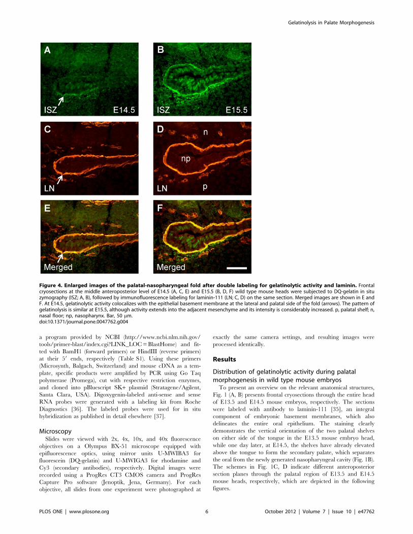
Figure 4. Enlarged images of the palatal-nasopharyngeal fold after double labeling for gelatinolytic activity and laminin. Frontalcryosections at the middle anteroposterior level of E14.5 (A, C, E) and E15.5 (B, D, F) wild type mouse heads were subjected to DQ-gelatin in situzymography (ISZ; A, B), followed by immunofluorescence labeling for laminin-111 (LN; C, D) on the same section. Merged images are shown in E andF. At E14.5, gelatinolytic activity colocalizes with the epithelial basement membrane at the lateral and palatal side of the fold (arrows). The pattern ofgelatinolysis is similar at E15.5, although activity extends into the adjacent mesenchyme and its intensity is considerably increased. p, palatal shelf; n,nasal floor; np, nasopharynx. Bar, 50 mm.doi:10.1371/journal.pone.0047762.g004
Gelatinolysis in Palate Morphogenesis
PLOS ONE | www.plosone.org 6 October 2012 | Volume 7 | Issue 10 | e47762
A double-labeling technique comprising in situ zymography
(ISZ) with dye-quenched (DQ-)gelatin, followed by brief acetone
fixation and immunofluorescence labeling, was performed to
associate gelatinolytic activity in situ with laminin-111 as a marker
for embryonic basement membranes (Fig. 2). In the region of the
developing secondary palate, no site with prominent gelatinolytic
activity was identified prior to palatal shelf elevation at E13.5 at
three anteroposterior levels (Fig. 2A–C). In contrast, after palatal
shelf elevation at E14.5, gelatinolytic activity was evident in the
nasal cartilage and in the two main palatal arteries (Fig. 2D–F).
Interestingly, at this embryonic stage signs of gelatinolysis were
evident at the midline epithelial seam (MES) formed prior to
palatal shelf fusion, and in addition prominent activity was
detected as sharp profiles surrounding distinct epithelial structures,
such as the folds of the elevated palatal shelves, which contribute
to morphogenesis of the nasal cavities in the front and the
nasopharynx more posteriorly. In the posterior region, gelatino-
lytic activity faded out at the dorsal side of the palate. This pattern
was also observed at E15.5 stage (Fig. 2G–I), although activity was
increased compared to E14.5. Furthermore, prominent activity
was evident for maxillary bone, as has been shown before for
mandibular bone [33]. The present findings by ISZ of a temporal
increase in gelatinolytic activity during embryonic development of
the craniofacial region confirm previous results obtained with
extracts from mouse embryo heads, which were analyzed for
MMP activity (by SDS-gel zymography), protein (by immuno-
blotting), and mRNA (by RT-PCR), respectively [38].
The extent of gelatinolytic activity at the nasopharyngeal fold
was quantified by measuring the integrated pixel intensity in ISZ
images of the palatal epithelium and basement membrane over
a fixed area. An approximately 5-fold increase was measured from
E13.5 to E14.5, and another 3-fold increase from E14.5 to E15.5.
Non parametric tests identified significant differences among all
groups (Kruskal-Wallis test, p,0.001) and between all pairs of
groups compared (Mann-Whitney test, p,0.001), confirming the
qualitative observations.
Higher magnification images of the midline epithelial seam
(MES) region demonstrated that the appearance of gelatinolytic
activity was tightly linked to the process of palatal shelf fusion
(Fig. 3). At E13.5, when the palatal shelves were still vertically
oriented, no sign of gelatinolysis was detected along the entire oral
epithelium, including the prospective fusion sites on the distal-
medial aspect of the shelves. Traces of gelatinolysis were evident
only within the tongue mesenchyme (Fig. 3A, B). At E14.5,
immediately prior to fusion of the palatal shelves, gelatinolytic
activity was present at the MES and was located mainly in the
pericellular space surrounding a subset of epithelial cells (Fig. 3C,
D). At E15.5 during palatal shelf fusion, prominent gelatinolytic
activity was associated mainly with remnants of basement
membranes of the disorganizing MES (Fig. 3E, F).
ISZ/laminin double labeling also revealed extensive colocaliza-
tion of gelatinolysis with the basement membranes of the
nasopharyngeal epithelial folds that are generated upon palatal
shelf elevation (Fig. 4). At stage E15.5 (Fig. 4B, D, F), gelatinolytic
activity was also associated with palatal and nasal epithelial cells,
as well as with the mesenchyme adjacent to the strongly labeled
epithelial basement membranes. Interestingly, activity was stron-
gest at the lateral bend where the oral epithelium folds back on
itself, but faded out towards more central regions of the
nasopharyngeal cavity. Gelatinolysis was asymmetric and more
prominent on the palatal aspect of the folds, in epithelial cells as
well as basement membranes and adjacent palatal mesenchyme
(Fig. 4A, B).
In situ zymography during palatal morphogenesis inBmp7 deficient miceDuring normal mouse embryogenesis, elevation of the palatal
shelves occurs very rapidly, i.e. within only a few hours on the 14th
day of development. To determine whether gelatinolytic activity
appeared on the proximal-medial aspect of the palatal shelves
already before their elevation, or only afterwards as a consequence
of formation of the nasopharyngeal fold, we studied Bmp7-deficient
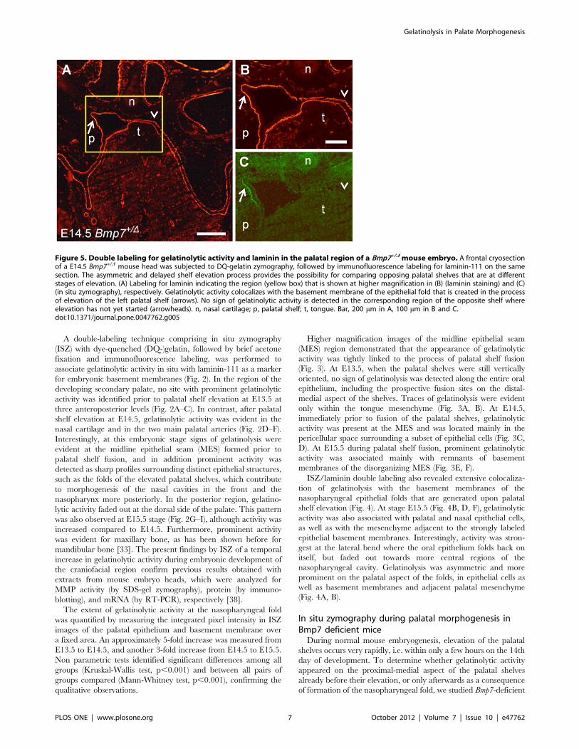
Figure 5. Double labeling for gelatinolytic activity and laminin in the palatal region of a Bmp7+/D mouse embryo. A frontal cryosectionof a E14.5 Bmp7+/D mouse head was subjected to DQ-gelatin zymography, followed by immunofluorescence labeling for laminin-111 on the samesection. The asymmetric and delayed shelf elevation process provides the possibility for comparing opposing palatal shelves that are at differentstages of elevation. (A) Labeling for laminin indicating the region (yellow box) that is shown at higher magnification in (B) (laminin staining) and (C)(in situ zymography), respectively. Gelatinolytic activity colocalizes with the basement membrane of the epithelial fold that is created in the processof elevation of the left palatal shelf (arrows). No sign of gelatinolytic activity is detected in the corresponding region of the opposite shelf whereelevation has not yet started (arrowheads). n, nasal cartilage; p, palatal shelf; t, tongue. Bar, 200 mm in A, 100 mm in B and C.doi:10.1371/journal.pone.0047762.g005
Gelatinolysis in Palate Morphogenesis
PLOS ONE | www.plosone.org 7 October 2012 | Volume 7 | Issue 10 | e47762
embryos. In the absence of Bmp7, palate shelf elevation is delayed
and asymmetrical, which eventually leads to cleft palate [39].
Because of asymmetrical palatal shelves, comparisons between
different elevation stages and orientations were possible.
In the representative example of a E14.5 Bmp7 wt/D mouse
embryo shown in Fig. 5, gelatinolysis was detected at similar sites
as in wild type embryos of the same developmental stage.
However, only one of the palatal shelves had just started to
elevate (Fig. 5A). Double staining on the same section revealed
that gelatinolytic activity codistributed perfectly with laminin-111
immunostaining at the forming epithelial fold of the elevating
shelf, although the body of this shelf was still almost vertically
oriented. In the same embryo, no sign of gelatinolysis was evident
on the opposing side where shelf elevation had not started yet
(Fig. 5B, C).
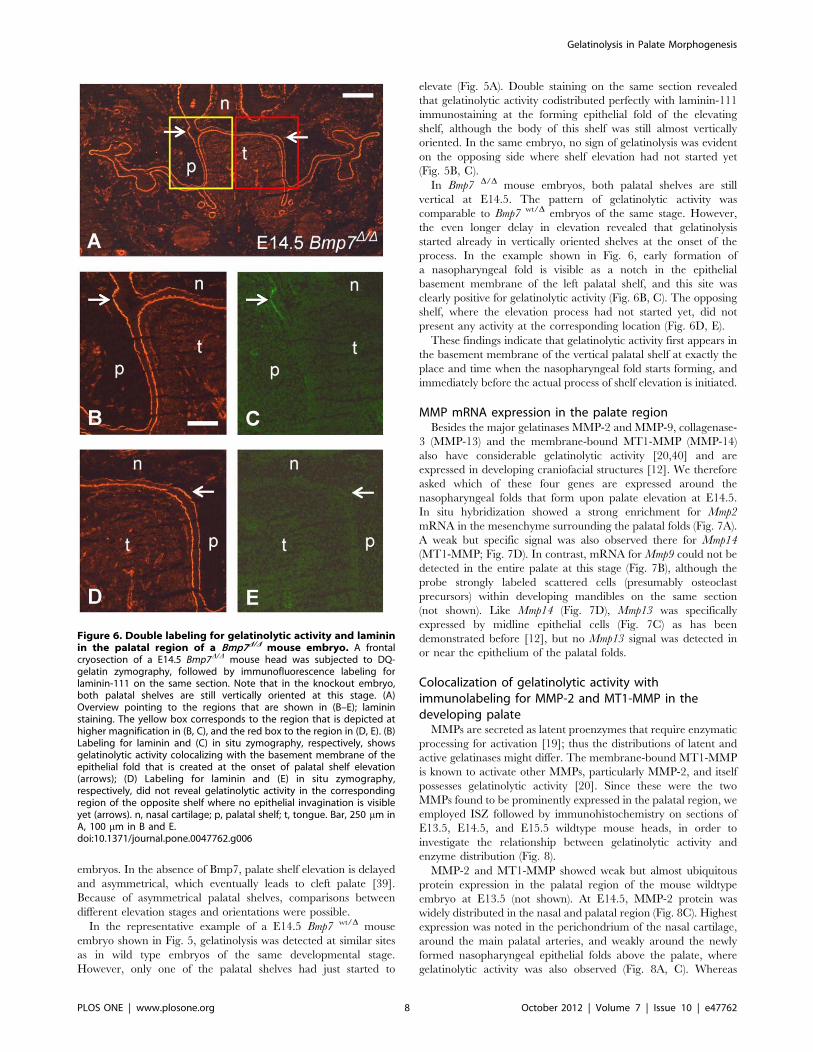
In Bmp7 D/D mouse embryos, both palatal shelves are still
vertical at E14.5. The pattern of gelatinolytic activity was
comparable to Bmp7 wt/D embryos of the same stage. However,
the even longer delay in elevation revealed that gelatinolysis
started already in vertically oriented shelves at the onset of the
process. In the example shown in Fig. 6, early formation of
a nasopharyngeal fold is visible as a notch in the epithelial
basement membrane of the left palatal shelf, and this site was
clearly positive for gelatinolytic activity (Fig. 6B, C). The opposing
shelf, where the elevation process had not started yet, did not
present any activity at the corresponding location (Fig. 6D, E).
These findings indicate that gelatinolytic activity first appears in
the basement membrane of the vertical palatal shelf at exactly the
place and time when the nasopharyngeal fold starts forming, and
immediately before the actual process of shelf elevation is initiated.
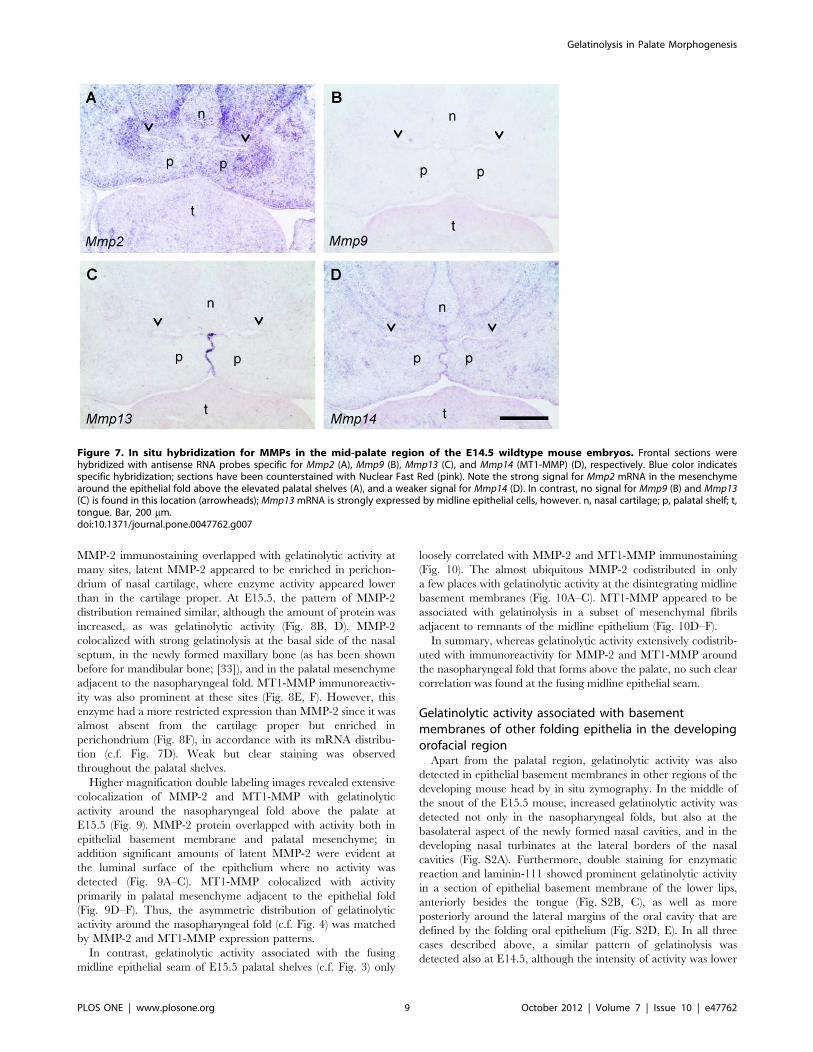
MMP mRNA expression in the palate regionBesides the major gelatinases MMP-2 and MMP-9, collagenase-
3 (MMP-13) and the membrane-bound MT1-MMP (MMP-14)
also have considerable gelatinolytic activity [20,40] and are
expressed in developing craniofacial structures [12]. We therefore
asked which of these four genes are expressed around the
nasopharyngeal folds that form upon palate elevation at E14.5.
In situ hybridization showed a strong enrichment for Mmp2
mRNA in the mesenchyme surrounding the palatal folds (Fig. 7A).
A weak but specific signal was also observed there for Mmp14
(MT1-MMP; Fig. 7D). In contrast, mRNA for Mmp9 could not be
detected in the entire palate at this stage (Fig. 7B), although the
probe strongly labeled scattered cells (presumably osteoclast
precursors) within developing mandibles on the same section
(not shown). Like Mmp14 (Fig. 7D), Mmp13 was specifically
expressed by midline epithelial cells (Fig. 7C) as has been
demonstrated before [12], but no Mmp13 signal was detected in
or near the epithelium of the palatal folds.
Colocalization of gelatinolytic activity withimmunolabeling for MMP-2 and MT1-MMP in thedeveloping palateMMPs are secreted as latent proenzymes that require enzymatic
processing for activation [19]; thus the distributions of latent and
active gelatinases might differ. The membrane-bound MT1-MMP
is known to activate other MMPs, particularly MMP-2, and itself
possesses gelatinolytic activity [20]. Since these were the two
MMPs found to be prominently expressed in the palatal region, we
employed ISZ followed by immunohistochemistry on sections of
E13.5, E14.5, and E15.5 wildtype mouse heads, in order to
investigate the relationship between gelatinolytic activity and
enzyme distribution (Fig. 8).
MMP-2 and MT1-MMP showed weak but almost ubiquitous
protein expression in the palatal region of the mouse wildtype
embryo at E13.5 (not shown). At E14.5, MMP-2 protein was
widely distributed in the nasal and palatal region (Fig. 8C). Highest
expression was noted in the perichondrium of the nasal cartilage,
around the main palatal arteries, and weakly around the newly
formed nasopharyngeal epithelial folds above the palate, where
gelatinolytic activity was also observed (Fig. 8A, C). Whereas
Figure 6. Double labeling for gelatinolytic activity and lamininin the palatal region of a Bmp7D/D mouse embryo. A frontalcryosection of a E14.5 Bmp7D/D mouse head was subjected to DQ-gelatin zymography, followed by immunofluorescence labeling forlaminin-111 on the same section. Note that in the knockout embryo,both palatal shelves are still vertically oriented at this stage. (A)Overview pointing to the regions that are shown in (B–E); lamininstaining. The yellow box corresponds to the region that is depicted athigher magnification in (B, C), and the red box to the region in (D, E). (B)Labeling for laminin and (C) in situ zymography, respectively, showsgelatinolytic activity colocalizing with the basement membrane of theepithelial fold that is created at the onset of palatal shelf elevation(arrows); (D) Labeling for laminin and (E) in situ zymography,respectively, did not reveal gelatinolytic activity in the correspondingregion of the opposite shelf where no epithelial invagination is visibleyet (arrows). n, nasal cartilage; p, palatal shelf; t, tongue. Bar, 250 mm inA, 100 mm in B and E.doi:10.1371/journal.pone.0047762.g006
Gelatinolysis in Palate Morphogenesis
PLOS ONE | www.plosone.org 8 October 2012 | Volume 7 | Issue 10 | e47762
MMP-2 immunostaining overlapped with gelatinolytic activity at
many sites, latent MMP-2 appeared to be enriched in perichon-
drium of nasal cartilage, where enzyme activity appeared lower
than in the cartilage proper. At E15.5, the pattern of MMP-2
distribution remained similar, although the amount of protein was
increased, as was gelatinolytic activity (Fig. 8B, D). MMP-2
colocalized with strong gelatinolysis at the basal side of the nasal
septum, in the newly formed maxillary bone (as has been shown
before for mandibular bone; [33]), and in the palatal mesenchyme
adjacent to the nasopharyngeal fold. MT1-MMP immunoreactiv-
ity was also prominent at these sites (Fig. 8E, F). However, this
enzyme had a more restricted expression than MMP-2 since it was
almost absent from the cartilage proper but enriched in
perichondrium (Fig. 8F), in accordance with its mRNA distribu-
tion (c.f. Fig. 7D). Weak but clear staining was observed
throughout the palatal shelves.
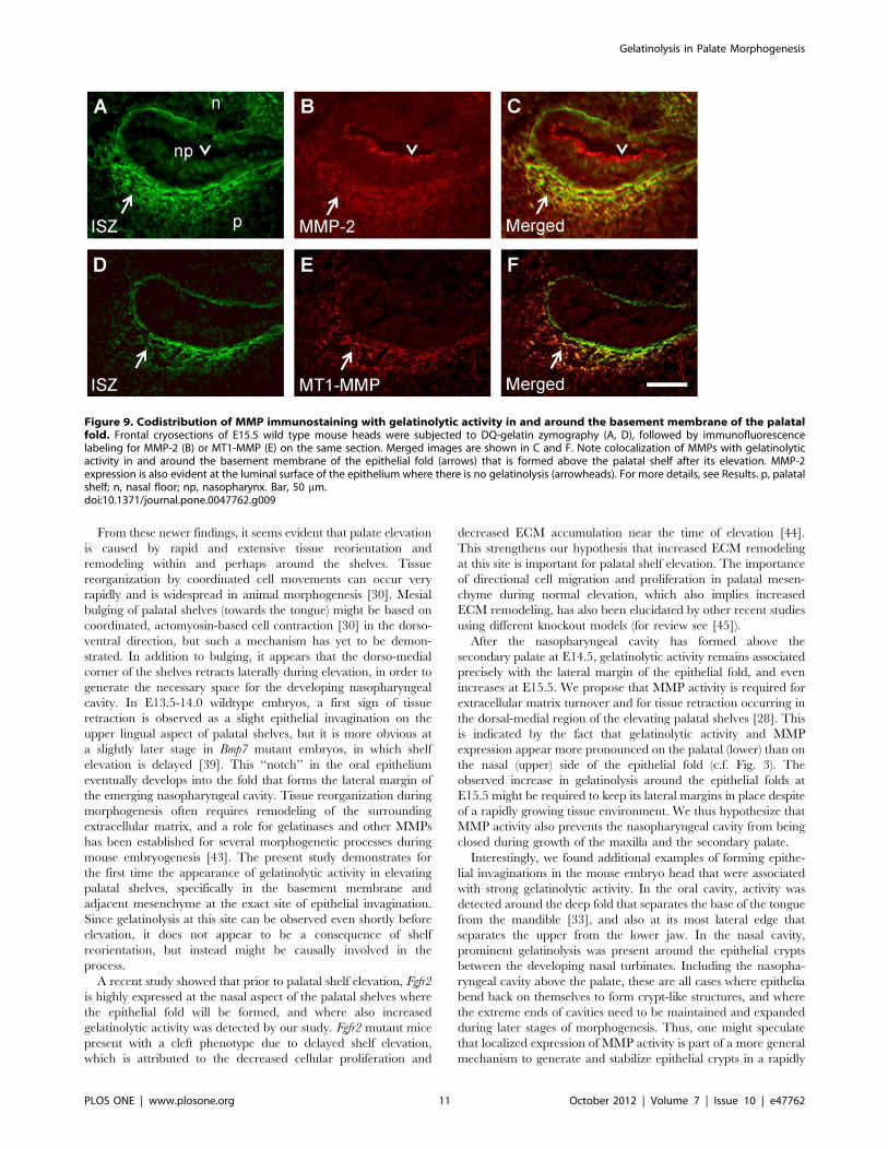
Higher magnification double labeling images revealed extensive
colocalization of MMP-2 and MT1-MMP with gelatinolytic
activity around the nasopharyngeal fold above the palate at
E15.5 (Fig. 9). MMP-2 protein overlapped with activity both in
epithelial basement membrane and palatal mesenchyme; in
addition significant amounts of latent MMP-2 were evident at
the luminal surface of the epithelium where no activity was
detected (Fig. 9A–C). MT1-MMP colocalized with activity
primarily in palatal mesenchyme adjacent to the epithelial fold
(Fig. 9D–F). Thus, the asymmetric distribution of gelatinolytic
activity around the nasopharyngeal fold (c.f. Fig. 4) was matched
by MMP-2 and MT1-MMP expression patterns.
In contrast, gelatinolytic activity associated with the fusing
midline epithelial seam of E15.5 palatal shelves (c.f. Fig. 3) only
loosely correlated with MMP-2 and MT1-MMP immunostaining
(Fig. 10). The almost ubiquitous MMP-2 codistributed in only
a few places with gelatinolytic activity at the disintegrating midline
basement membranes (Fig. 10A–C). MT1-MMP appeared to be
associated with gelatinolysis in a subset of mesenchymal fibrils
adjacent to remnants of the midline epithelium (Fig. 10D–F).
In summary, whereas gelatinolytic activity extensively codistrib-
uted with immunoreactivity for MMP-2 and MT1-MMP around
the nasopharyngeal fold that forms above the palate, no such clear
correlation was found at the fusing midline epithelial seam.
Gelatinolytic activity associated with basementmembranes of other folding epithelia in the developingorofacial regionApart from the palatal region, gelatinolytic activity was also
detected in epithelial basement membranes in other regions of the
developing mouse head by in situ zymography. In the middle of
the snout of the E15.5 mouse, increased gelatinolytic activity was
detected not only in the nasopharyngeal folds, but also at the
basolateral aspect of the newly formed nasal cavities, and in the
developing nasal turbinates at the lateral borders of the nasal
cavities (Fig. S2A). Furthermore, double staining for enzymatic
reaction and laminin-111 showed prominent gelatinolytic activity
in a section of epithelial basement membrane of the lower lips,
anteriorly besides the tongue (Fig. S2B, C), as well as more
posteriorly around the lateral margins of the oral cavity that are
defined by the folding oral epithelium (Fig. S2D, E). In all three
cases described above, a similar pattern of gelatinolysis was
detected also at E14.5, although the intensity of activity was lower
Figure 7. In situ hybridization for MMPs in the mid-palate region of the E14.5 wildtype mouse embryos. Frontal sections werehybridized with antisense RNA probes specific for Mmp2 (A), Mmp9 (B), Mmp13 (C), and Mmp14 (MT1-MMP) (D), respectively. Blue color indicatesspecific hybridization; sections have been counterstained with Nuclear Fast Red (pink). Note the strong signal for Mmp2 mRNA in the mesenchymearound the epithelial fold above the elevated palatal shelves (A), and a weaker signal for Mmp14 (D). In contrast, no signal for Mmp9 (B) and Mmp13(C) is found in this location (arrowheads); Mmp13mRNA is strongly expressed by midline epithelial cells, however. n, nasal cartilage; p, palatal shelf; t,tongue. Bar, 200 mm.doi:10.1371/journal.pone.0047762.g007
Gelatinolysis in Palate Morphogenesis
PLOS ONE | www.plosone.org 9 October 2012 | Volume 7 | Issue 10 | e47762

at the earlier stage. At E13.5, no activity was present yet at any of
these sites (not shown).
Discussion
Morphogenesis of the secondary palate in mammals involves
a sequence of essential steps, among them the elevation of the two
palatal shelves from a horizontal to a vertical position above the
tongue, followed by their fusion at the midline [7,8]. Whereas
palatal shelf fusion has been extensively studied, much less is
known about the mechanism of shelf elevation, which happens in
an amazingly short time frame [7]. In the normal mouse embryo,
the process occurs within less than a day and requires that the
tongue moves out of the way of the rising shelves. In embryos
defective for the gad67 gene, palatal shelves elevate within only
30 minutes after the tongue has been removed experimentally
[41]. It has long been speculated how and where the forces for this
rapid reorientation of palatal shelves are generated. A prominent
hypothesis proposed a rotation of the shelves in a ‘‘barndoor’’
fashion, thought to be driven by hyaluronic acid-generated
osmotic pressure [8,26,38]. However, a causative relationship
between hyaluronan accumulation and shelf elevation has never
been established, and such a mechanism might be difficult to
reconcile with the fast kinetics of the process. In fact, the simple
rotation model of palatal shelf elevation has been challenged in
recent years. Chou et al. [29] used carbon markings to produce
fate maps of fetal mouse palates during elevation in culture. From
their data, the authors concluded that palatal shelves primarily
bulge out mesially in their anterior and posterior regions during
elevation, whereas some rotation is involved in the mid-palate
region. A more recent histomorphometric study confirmed these
findings by showing that the horizontal outgrowth of vertically
oriented palatal shelves starts from the medial side of the mid-
posterior region, and palatal shelf reorientation occurs in
a dynamic, spatiotemporally coordinated manner along the AP
axis [42]. In another recent study, Jin et al. [28] demonstrated that
the prospective medial edge is not located at the very distal rim of
vertical palatal shelves, but instead more medially, on their interior
aspect facing the tongue. Using a mouse mutant (Zfhx1a2/2) in
which palatal shelf elevation is retarded by one day, they found
that genes typical for the prospective medial edge (Mmp13 for the
epithelium and Goosecoid for the mesenchyme) were expressed at
the distal-medial (lingual) aspects of the still vertically positioned
shelves, rather than at their distal tips [28].
Figure 8. Double labeling for gelatinolytic activity and MMP-2 and MT1-MMP in the palatal region of the mouse embryo. Frontalcryosections of E14.5 (A, C, E) and E15.5 (B, D, F) wild type mouse heads were subjected to DQ-gelatin zymography, followed by immunofluorescencelabeling for MMPs on the same section. Serial sections were used; only one zymography image (double labeling for MMP-2) per stage is shown (A, B).Note that despite of differences in distribution, both MMP-2 (C, D) and MT1-MMP (E, F) substantially overlap with gelatinolytic activity, most notablyin developing cartilage and bone but also around the expanding nasopharyngeal cavity (arrowheads). For more details, see Results. n, nasal cartilage;p, palatal shelf; t, tongue; m, maxillary bone. Bar, 200 mm.doi:10.1371/journal.pone.0047762.g008
Gelatinolysis in Palate Morphogenesis
PLOS ONE | www.plosone.org 10 October 2012 | Volume 7 | Issue 10 | e47762
From these newer findings, it seems evident that palate elevation
is caused by rapid and extensive tissue reorientation and
remodeling within and perhaps around the shelves. Tissue
reorganization by coordinated cell movements can occur very
rapidly and is widespread in animal morphogenesis [30]. Mesial
bulging of palatal shelves (towards the tongue) might be based on
coordinated, actomyosin-based cell contraction [30] in the dorso-
ventral direction, but such a mechanism has yet to be demon-
strated. In addition to bulging, it appears that the dorso-medial
corner of the shelves retracts laterally during elevation, in order to
generate the necessary space for the developing nasopharyngeal
cavity. In E13.5-14.0 wildtype embryos, a first sign of tissue
retraction is observed as a slight epithelial invagination on the
upper lingual aspect of palatal shelves, but it is more obvious at
a slightly later stage in Bmp7 mutant embryos, in which shelf
elevation is delayed [39]. This ‘‘notch’’ in the oral epithelium
eventually develops into the fold that forms the lateral margin of
the emerging nasopharyngeal cavity. Tissue reorganization during
morphogenesis often requires remodeling of the surrounding
extracellular matrix, and a role for gelatinases and other MMPs
has been established for several morphogenetic processes during
mouse embryogenesis [43]. The present study demonstrates for
the first time the appearance of gelatinolytic activity in elevating
palatal shelves, specifically in the basement membrane and
adjacent mesenchyme at the exact site of epithelial invagination.
Since gelatinolysis at this site can be observed even shortly before
elevation, it does not appear to be a consequence of shelf
reorientation, but instead might be causally involved in the
process.
A recent study showed that prior to palatal shelf elevation, Fgfr2
is highly expressed at the nasal aspect of the palatal shelves where
the epithelial fold will be formed, and where also increased
gelatinolytic activity was detected by our study. Fgfr2 mutant mice
present with a cleft phenotype due to delayed shelf elevation,
which is attributed to the decreased cellular proliferation and
decreased ECM accumulation near the time of elevation [44].
This strengthens our hypothesis that increased ECM remodeling
at this site is important for palatal shelf elevation. The importance
of directional cell migration and proliferation in palatal mesen-
chyme during normal elevation, which also implies increased
ECM remodeling, has also been elucidated by other recent studies
using different knockout models (for review see [45]).
After the nasopharyngeal cavity has formed above the
secondary palate at E14.5, gelatinolytic activity remains associated
precisely with the lateral margin of the epithelial fold, and even
increases at E15.5. We propose that MMP activity is required for
extracellular matrix turnover and for tissue retraction occurring in
the dorsal-medial region of the elevating palatal shelves [28]. This
is indicated by the fact that gelatinolytic activity and MMP
expression appear more pronounced on the palatal (lower) than on
the nasal (upper) side of the epithelial fold (c.f. Fig. 3). The
observed increase in gelatinolysis around the epithelial folds at
E15.5 might be required to keep its lateral margins in place despite
of a rapidly growing tissue environment. We thus hypothesize that
MMP activity also prevents the nasopharyngeal cavity from being
closed during growth of the maxilla and the secondary palate.
Interestingly, we found additional examples of forming epithe-
lial invaginations in the mouse embryo head that were associated
with strong gelatinolytic activity. In the oral cavity, activity was
detected around the deep fold that separates the base of the tongue
from the mandible [33], and also at its most lateral edge that
separates the upper from the lower jaw. In the nasal cavity,
prominent gelatinolysis was present around the epithelial crypts
between the developing nasal turbinates. Including the nasopha-
ryngeal cavity above the palate, these are all cases where epithelia
bend back on themselves to form crypt-like structures, and where
the extreme ends of cavities need to be maintained and expanded
during later stages of morphogenesis. Thus, one might speculate
that localized expression of MMP activity is part of a more general
mechanism to generate and stabilize epithelial crypts in a rapidly
Figure 9. Codistribution of MMP immunostaining with gelatinolytic activity in and around the basement membrane of the palatalfold. Frontal cryosections of E15.5 wild type mouse heads were subjected to DQ-gelatin zymography (A, D), followed by immunofluorescencelabeling for MMP-2 (B) or MT1-MMP (E) on the same section. Merged images are shown in C and F. Note colocalization of MMPs with gelatinolyticactivity in and around the basement membrane of the epithelial fold (arrows) that is formed above the palatal shelf after its elevation. MMP-2expression is also evident at the luminal surface of the epithelium where there is no gelatinolysis (arrowheads). For more details, see Results. p, palatalshelf; n, nasal floor; np, nasopharynx. Bar, 50 mm.doi:10.1371/journal.pone.0047762.g009
Gelatinolysis in Palate Morphogenesis
PLOS ONE | www.plosone.org 11 October 2012 | Volume 7 | Issue 10 | e47762
growing organism. This hypothesis is further supported by the fact
that apart from basement membranes, gelatinolytic activity
appears to be increased also above the epithelium itself at these
sites. According to recent evidence, MMP function has been
associated with epithelial cell motility or even apoptosis [46,47,48],
but this has to be further investigated.
The importance of basement membrane remodeling and the
role of MMP-2 and activating enzymes, like MT1-MMP, in this
process has been shown in various developmental and cancer
studies [49]. The contribution of MMP-2 in this process is
attributed to the degradation of type IV collagen, a major
component of the basement membrane [50]. We asked which
MMPs might be responsible for the gelatinolytic activity around
the epithelial folds of the forming nasopharyngeal cavity above the
secondary palate. In situ hybridization experiments revealed that
of the two major gelatinases, MMP-2 but not MMP-9 was strongly
and specifically expressed at these sites. In addition, MT1-MMP,
a cell membrane-bound enzyme that is required for the activation
especially of pro-MMP-2 [20], showed a weaker but similar
mRNA distribution. Immunolabeling confirmed that MMP-2 and
MT1-MMP protein colocalized extensively with the pattern of
gelatinolysis around the epithelial folds of the palate. We
previously showed codistribution of gelatinolytic activity with
these MMPs around the crypt that separates the developing
tongue from the mandibles [33]. In contrast to these epithelial
folds, however, we detected hardly any codistribution of
gelatinolytic activity with MMP-2 and MT1-MMP proteins at
the midline epithelial seam during palate fusion, although both
enzymes were detected in the adjacent palatal mesenchyme where
they partially colocalized with activity. Since antibodies also
recognize inactive proenzymes, it is to be expected that MMP
proteins and gelatinolytic activity do not overlap perfectly.
Nevertheless, we assume that other MMPs are responsible for
the observed gelatinolysis at the MES. The most likely candidate is
MMP-13 (collagenase-3), which also cleaves denatured fibrillar
collagen (gelatin) [40] and is expressed by midline epithelial cells
(Fig. 6; [12]). The expression of MMP 13 in the MES has been
implicated before in palatal shelf fusion [12]. Although Mmp13
knockout mouse do not present with a cleft phenotype [51], MMP-
13 is the main candidate responsible for the detected activity in
Figure 10. Relationship between MMP immunostaining and gelatinolytic activity at the midline epithelial seam of opposing palatalshelves during their fusion. Frontal cryosections of E15.5 wild type mouse heads were subjected to DQ-gelatin zymography (A, D), followed byimmunofluorescence labeling for MMP-2 (B) or MT1-MMP (E) on the same section. Merged images are shown in C and F. At the midline, somecolocalization with gelatinolytic activity is evident only for MMP-2 (arrows). MT1-MMP shows weak colocalization with activity at the midlineepithelium (arrows) and is more strongly colocalized with gelatinolysis in fibrillar structures adjacent to remnants of the midline epithelium(arrowheads). For more details, see Results. p, palatal shelf. Bar, 50 mm.doi:10.1371/journal.pone.0047762.g010
Gelatinolysis in Palate Morphogenesis
PLOS ONE | www.plosone.org 12 October 2012 | Volume 7 | Issue 10 | e47762
MES. This is strengthened by evidence that MMP-13 forms
complexes with MMP-2 and MT1-MMP, resulting in synergism
between these proteases [52]. Thus, the presence of all three
MMPs in and adjacent to the MES, as demonstrated by in situ
hybridization and immunohistochemistry here, might be impor-
tant for palatal shelf fusion.
In summary, our present study implies a function for MMP-
dependent gelatinolysis not only in palatal shelf fusion as has been
suggested before, but for the first time also in the process of palatal
shelf elevation/reorientation. Specifically, gelatinolytic activity was
detected in the epithelial fold and retracting mesenchyme at the
upper medial corner of palatal shelves, exactly at the time of their
reorientation. Colocalization studies were consistent with the
notion that MMP-2 is primarily responsible for gelatinolytic
activity at this site, and that MT1-MMP might also be involved
either directly, or indirectly through activation of MMP-2.
However, additional experiments are required to establish a causal
relationship.
Supporting Information
Figure S1 Effect of MMP inhibition on development offluorescent signal during in situ zymography with DQ-gelatin. Unfixed frontal cryosections of E14.5 wild type mouse
heads were incubated with DQ-gelatin (see Materials and
Methods), with or without adding inhibitors to the reaction buffer.
(A) Control without inhibitor. (B) Inclusion of a specific MMP-2/
MMP-9 inhibitor (BiPS; 50 mM; diluted in 0.2% DMSO) partially
attenuated the reaction. (C) General MMP inhibitors 1,10-
phenanthroline (1 mM) and (D) EDTA (10 mM) strongly
suppressed the gelatinolytic activity. (E) Incubation of slides with
only ISZ buffer (without DQ-gelatin) or (F) replacement of DQ-
gelatin by 20 mg/ml of unlabeled pig skin gelatin did not produce
any fluorescent signal. n, nasal cartilage; p, palatal shelf; t, tongue.
Bar, 200 mm.
(TIF)
Figure S2 Gelatinolytic activity associated with distinctepithelial structures at other sites of the E15.5 mouseembryo head. Frontal cryosections of E15.5 wild type mouse
heads were subjected to DQ-gelatin zymography, followed by
immunofluorescence labeling for laminin on the same section. (A)
In situ zymography of a section in the middle of the snout showing
increased gelatinolytic activity in the epithelial folds formed at the
lower lateral part of the nasal cavities created after palatal shelf
elevation, and in the developing nasal turbinates (arrows). (B) In
situ zymography and (C) immunofluorescence labeling for
laminin, respectively, of the lateral side of the mouth opening.
Note the presence of gelatinolytic activity at the epithelial
basement membrane of the lower lip next to the tongue (arrows).
(D) In situ zymography and (E) immunofluorescence labeling for
laminin, respectively, of the lateral limits of the oral cavity.
Prominent gelatinolytic activity is evident at the epithelial
basement membrane of the fold that is created at the lateral end
of the oral epithelium separating upper and lower jaw (arrows). n,
nasal cartilage; nc, nasal cavity; p, palatal shelf; nt, nasal
turbinates; up, upper lip; lo, lower lip; utb, upper tooth bud; ltb,
lower tooth bud. Bar, 250 mm in A, 100 mm in B–E.
(TIF)
Table S1
(DOC)
Acknowledgments
We thank Jean-Francois Spetz for providing us with wildtype mouse
embryos, and Sabrina Ruggiero for excellent technical help and advice.
Author Contributions
Conceived and designed the experiments: NG CK MC. Performed the
experiments: NG SB DG. Analyzed the data: NG SB CK DG MC.
Contributed reagents/materials/analysis tools: NG SB DG MC. Wrote the
paper: NG DG MC.
References
1. Mitchell LE (2009) Epidemiology of cleft lip and palate. In Comprehensive Cleft
Care; Losee JE, Kirschner RE, editors. Toronto: McGraw-Hill.
2. Mossey PA, Little J, Munger RG, Dixon MJ, Shaw WC (2009) Cleft lip and
palate. Lancet 374: 1773–1785.
3. Kouskoura T, Fragou N, Alexiou M, John N, Sommer L, et al. (2011) The
genetic basis of craniofacial and dental abnormalities. Schweiz Monatsschr
Zahnmed 121: 636–646.
4. Mitchell JC, Wood RJ (2000) Management of cleft lip and palate in primary
care. J Pediatr Health Care 14: 13–19.
5. Stanier P, Moore GE (2004) Genetics of cleft lip and palate: syndromic genes
contribute to the incidence of non-syndromic clefts. Hum Mol Genet 13 Spec
No 1: R73–81.
6. Gritli-Linde A (2008) The etiopathogenesis of cleft lip and cleft palate: usefulness
and caveats of mouse models. Curr Top Dev Biol 84: 37–138.
7. Meng L, Bian Z, Torensma R, Von den Hoff JW (2009) Biological mechanisms
in palatogenesis and cleft palate. J Dent Res 88: 22–33.
8. Ferguson MW (1988) Palate development. Development 103 Suppl: 41–60.
9. Gritli-Linde A (2007) Molecular control of secondary palate development. Dev
Biol 301: 309–326.
10. Miettinen PJ, Chin JR, Shum L, Slavkin HC, Shuler CF, et al. (1999) Epidermal
growth factor receptor function is necessary for normal craniofacial development
and palate closure. Nat Genet 22: 69–73.
11. Robbins JR, McGuire PG, Wehrle-Haller B, Rogers SL (1999) Diminished
matrix metalloproteinase 2 (MMP-2) in ectomesenchyme-derived tissues of the
Patch mutant mouse: regulation of MMP-2 by PDGF and effects on
mesenchymal cell migration. Dev Biol 212: 255–263.
12. Blavier L, Lazaryev A, Groffen J, Heisterkamp N, DeClerck YA, et al. (2001)
TGF-beta3-induced palatogenesis requires matrix metalloproteinases. Mol Biol
Cell 12: 1457–1466.
13. Shi J, Son M–Y, Yamada S, Szabova L, Kahan S, et al. (2008) Membrane-type
MMPs enable extracellular matrix permissiveness and mesenchymal cell
proliferation during embryogenesis. Dev Biol 313: 196–209.
14. Jugessur A, Shi M, Gjessing HK, Lie RT, Wilcox AJ, et al. (2009) Genetic
determinants of facial clefting: analysis of 357 candidate genes using two nationalcleft studies from Scandinavia. PLoS One 4: e5385.
15. Jugessur A, Skare O, Lie RT, Wilcox AJ, Christensen K, et al. (2012) X-linkedgenes and risk of orofacial clefts: evidence from two population-based studies in
scandinavia. PLoS One 7: e39240.
16. Parks WC, Wilson CL, Lopez-Boado YS (2004) Matrix metalloproteinases as
modulators of inflammation and innate immunity. Nat Rev Immunol 4: 617–629.
17. Briknarova K, Gehrmann M, Banyai L, Tordai H, Patthy L, et al. (2001)Gelatin-binding region of human matrix metalloproteinase-2: solution structure,
dynamics, and function of the COL-23 two-domain construct. J Biol Chem 276:27613–27621.
18. Overall CM (2002) Molecular determinants of metalloproteinase substratespecificity: matrix metalloproteinase substrate binding domains, modules, and
exosites. Mol Biotechnol 22: 51–86.
19. Nagase H, Woessner JF Jr (1999) Matrix metalloproteinases. J Biol Chem 274:
21491–21494.
20. Evans RD, Itoh Y (2007) Analyses of MT1-MMP activity in cells. Methods Mol
Med 135: 239–249.
21. Baker AH, Edwards DR, Murphy G (2002) Metalloproteinase inhibitors:
biological actions and therapeutic opportunities. J Cell Sci 115: 3719–3727.
22. Kaartinen V, Voncken JW, Shuler C, Warburton D, Bu D, et al. (1995)Abnormal lung development and cleft palate in mice lacking TGF-beta 3
indicates defects of epithelial-mesenchymal interaction. Nat Genet 11: 415–421.
23. Proetzel G, Pawlowski SA, Wiles MV, Yin M, Boivin GP, et al. (1995)
Transforming growth factor-beta 3 is required for secondary palate fusion. Nat
Genet 11: 409–414.
24. Ito Y, Yeo JY, Chytil A, Han J, Bringas P Jr, et al. (2003) Conditionalinactivation of Tgfbr2 in cranial neural crest causes cleft palate and calvaria
defects. Development 130: 5269–5280.
25. Brown GD, Nazarali AJ (2010) Matrix metalloproteinase-25 has a functional
role in mouse secondary palate development and is a downstream target of
TGF-beta3. BMC Dev Biol 10: 93.
Gelatinolysis in Palate Morphogenesis
PLOS ONE | www.plosone.org 13 October 2012 | Volume 7 | Issue 10 | e47762

26. Brinkley LL, Morris-Wiman J (1987) Computer-assisted analysis of hyaluronate
distribution during morphogenesis of the mouse secondary palate. Development100: 629–635.
27. Morris-Wiman J, Brinkley L (1992) An extracellular matrix infrastructure
provides support for murine secondary palatal shelf remodelling. Anat Rec 234:575–586.
28. Jin JZ, Tan M, Warner DR, Darling DS, Higashi Y, et al. (2010) Mesenchymalcell remodeling during mouse secondary palate reorientation. Dev Dyn 239:
2110–2117.
29. Chou MJ, Kosazuma T, Takigawa T, Yamada S, Takahara S, et al. (2004)Palatal shelf movement during palatogenesis: a fate map of the fetal mouse
palate cultured in vitro. Anat Embryol (Berl) 208: 19–25.30. Wozniak MA, Chen CS (2009) Mechanotransduction in development: a growing
role for contractility. Nat Rev Mol Cell Biol 10: 34–43.31. Frederiks WM, Mook OR (2004) Metabolic mapping of proteinase activity with
emphasis on in situ zymography of gelatinases: review and protocols.
J Histochem Cytochem 52: 711–722.32. Snoek-van Beurden PA, Von den Hoff JW (2005) Zymographic techniques for
the analysis of matrix metalloproteinases and their inhibitors. Biotechniques 38:73–83.
33. Gkantidis N, Katsaros C, Chiquet M (2012) Detection of gelatinolytic activity in
developing basement membranes of the mouse embryo head by combiningsensitive in situ zymography with immunolabeling. Histochem Cell Biol 138:
557–571.34. Zouvelou V, Passa O, Segklia K, Tsalavos S, Valenzuela DM, et al. (2009)
Generation and functional characterization of mice with a conditional BMP7allele. Int J Dev Biol 53: 597–603.
35. Paulsson M, Aumailley M, Deutzmann R, Timpl R, Beck K, et al. (1987)
Laminin-nidogen complex. Extraction with chelating agents and structuralcharacterization. Eur J Biochem 166: 11–19.
36. Koch M, Bohrmann B, Matthison M, Hagios C, Trueb B, et al. (1995) Largeand small splice variants of collagen XII: differential expression and ligand
binding. J Cell Biol 130: 1005–1014.
37. Fluck M, Tunc-Civelek V, Chiquet M (2000) Rapid and reciprocal regulation oftenascin-C and tenascin-Y expression by loading of skeletal muscle. J Cell Sci
113 ( Pt 20): 3583–3591.38. Morris-Wiman J, Du Y, Brinkley L (1999) Occurrence and temporal variation in
matrix metalloproteinases and their inhibitors during murine secondary palatalmorphogenesis. J Craniofac Genet Dev Biol 19: 201–212.
39. Zouvelou V, Luder HU, Mitsiadis TA, Graf D (2009) Deletion of BMP7 affects
the development of bones, teeth, and other ectodermal appendages of theorofacial complex. J Exp Zool B Mol Dev Evol 312B: 361–374.
40. Knauper V, Cowell S, Smith B, Lopez-Otin C, O’Shea M, et al. (1997) The role
of the C-terminal domain of human collagenase-3 (MMP-13) in the activation of
procollagenase-3, substrate specificity, and tissue inhibitor of metalloproteinase
interaction. J Biol Chem 272: 7608–7616.
41. Iseki S, Ishii-Suzuki M, Tsunekawa N, Yamada Y, Eto K, et al. (2007)
Experimental induction of palate shelf elevation in glutamate decarboxylase 67-
deficient mice with cleft palate due to vertically oriented palatal shelf. Birth
Defects Res A Clin Mol Teratol 79: 688–695.
42. Yu K, Ornitz DM (2011) Histomorphological study of palatal shelf elevation
during murine secondary palate formation. Dev Dyn 240: 1737–1744.
43. Chin JR, Werb Z (1997) Matrix metalloproteinases regulate morphogenesis,
migration and remodeling of epithelium, tongue skeletal muscle and cartilage in
the mandibular arch. Development 124: 1519–1530.
44. Snyder-Warwick AK, Perlyn CA, Pan J, Yu K, Zhang L, et al. (2010) Analysis of
a gain-of-function FGFR2 Crouzon mutation provides evidence of loss of
function activity in the etiology of cleft palate. Proc Natl Acad Sci U S A 107:
2515–2520.
45. Bush JO, Jiang R (2012) Palatogenesis: morphogenetic and molecular
mechanisms of secondary palate development. Development 139: 231–243.
46. Zheng G, Lyons JG, Tan TK, Wang Y, Hsu TT, et al. (2009) Disruption of E-
cadherin by matrix metalloproteinase directly mediates epithelial-mesenchymal
transition downstream of transforming growth factor-beta1 in renal tubular
epithelial cells. Am J Pathol 175: 580–591.
47. Aldonyte R, Brantly M, Block E, Patel J, Zhang J (2009) Nuclear localization of
active matrix metalloproteinase-2 in cigarette smoke-exposed apoptotic
endothelial cells. Exp Lung Res 35: 59–75.
48. Moretti RM, Mai S, Montagnani Marelli M, Rizzi F, Bettuzzi S, et al. (2011)
Molecular mechanisms of the antimetastatic activity of nuclear clusterin in
prostate cancer cells. Int J Oncol 39: 225–234.
49. Itoh Y, Seiki M (2006) MT1-MMP: a potent modifier of pericellular
microenvironment. J Cell Physiol 206: 1–8.
50. Hornebeck W, Emonard H, Monboisse J–C, Bellon G (2002) Matrix-directed
regulation of pericellular proteolysis and tumor progression. Semin Cancer Biol
12: 231–241.
51. Inada M, Wang Y, Byrne MH, Rahman MU, Miyaura C, et al. (2004) Critical
roles for collagenase-3 (Mmp13) in development of growth plate cartilage and in
endochondral ossification. Proc Natl Acad Sci U S A 101: 17192–17197.
52. Cowell S, Knauper V, Stewart ML, D’Ortho MP, Stanton H, et al. (1998)
Induction of matrix metalloproteinase activation cascades based on membrane-
type 1 matrix metalloproteinase: associated activation of gelatinase A, gelatinase
B and collagenase 3. Biochem J 331 (Pt 2): 453–458.
Gelatinolysis in Palate Morphogenesis
PLOS ONE | www.plosone.org 14 October 2012 | Volume 7 | Issue 10 | e47762