Phosphorylation of HPr by the Bifunctional HPr Kinase/P-Ser-HPr Phosphatase from Lactobacillus casei...
10
10.1128/JB.182.9.2582-2590.2000. 2000, 182(9):2582. DOI: J. Bacteriol. Anne Galinier, Gaspar Pérez-Martínez and Josef Deutscher Valérie Dossonnet, Vicente Monedero, Monique Zagorec, Inducer Expulsion Repression and Inducer Exclusion but Not Controls Catabolite Lactobacillus casei HPr Kinase/P-Ser-HPr Phosphatase from Phosphorylation of HPr by the Bifunctional http://jb.asm.org/content/182/9/2582 Updated information and services can be found at: These include: REFERENCES http://jb.asm.org/content/182/9/2582#ref-list-1 at: This article cites 56 articles, 37 of which can be accessed free CONTENT ALERTS more» articles cite this article), Receive: RSS Feeds, eTOCs, free email alerts (when new http://journals.asm.org/site/misc/reprints.xhtml Information about commercial reprint orders: http://journals.asm.org/site/subscriptions/ To subscribe to to another ASM Journal go to: on February 7, 2014 by guest http://jb.asm.org/ Downloaded from on February 7, 2014 by guest http://jb.asm.org/ Downloaded from
Transcript of Phosphorylation of HPr by the Bifunctional HPr Kinase/P-Ser-HPr Phosphatase from Lactobacillus casei...
10.1128/JB.182.9.2582-2590.2000.
2000, 182(9):2582. DOI:J. Bacteriol. Anne Galinier, Gaspar Pérez-Martínez and Josef DeutscherValérie Dossonnet, Vicente Monedero, Monique Zagorec, Inducer ExpulsionRepression and Inducer Exclusion but Not
Controls CataboliteLactobacillus caseiHPr Kinase/P-Ser-HPr Phosphatase from Phosphorylation of HPr by the Bifunctional
http://jb.asm.org/content/182/9/2582Updated information and services can be found at:
These include:
REFERENCEShttp://jb.asm.org/content/182/9/2582#ref-list-1at:
This article cites 56 articles, 37 of which can be accessed free
CONTENT ALERTS more»articles cite this article),
Receive: RSS Feeds, eTOCs, free email alerts (when new
http://journals.asm.org/site/misc/reprints.xhtmlInformation about commercial reprint orders: http://journals.asm.org/site/subscriptions/To subscribe to to another ASM Journal go to:
on February 7, 2014 by guest
http://jb.asm.org/
Dow
nloaded from
on February 7, 2014 by guest
http://jb.asm.org/
Dow
nloaded from
JOURNAL OF BACTERIOLOGY,0021-9193/00/$04.0010
May 2000, p. 2582–2590 Vol. 182, No. 9
Copyright © 2000, American Society for Microbiology. All Rights Reserved.
Phosphorylation of HPr by the Bifunctional HPr Kinase/P-Ser-HPrPhosphatase from Lactobacillus casei Controls Catabolite Repression
and Inducer Exclusion but Not Inducer ExpulsionVALERIE DOSSONNET,1 VICENTE MONEDERO,1 MONIQUE ZAGOREC,2 ANNE GALINIER,3
GASPAR PEREZ-MARTINEZ,4 AND JOSEF DEUTSCHER1*
Laboratoire de Genetique des Microorganismes, 78850 Thiverval-Grignon,1 Laboratoire de Recherche sur la Viande,78352 Jouy-en-Josas,2 and Institut de Biologie et Chimie des Proteines, 69367 Lyon,3 France, and Departamento
de Biotecnologıa, Instituto de Agroquımica y Tecnologıa de Alimentos, 46100 Burjasot, Valencia, Spain4
Received 23 November 1999/Accepted 9 February 2000
We have cloned and sequenced the Lactobacillus casei hprK gene encoding the bifunctional enzyme HPrkinase/P-Ser-HPr phosphatase (HprK/P). Purified recombinant L. casei HprK/P catalyzes the ATP-dependentphosphorylation of HPr, a phosphocarrier protein of the phosphoenolpyruvate:carbohydrate phosphotrans-ferase system at the regulatory Ser-46 as well as the dephosphorylation of seryl-phosphorylated HPr (P-Ser-HPr). The two opposing activities of HprK/P were regulated by fructose-1,6-bisphosphate, which stimulatedHPr phosphorylation, and by inorganic phosphate, which stimulated the P-Ser-HPr phosphatase activity. Amutant producing truncated HprK/P was found to be devoid of both HPr kinase and P-Ser-HPr phosphataseactivities. When hprK was inactivated, carbon catabolite repression of N-acetylglucosaminidase disappeared,and the lag phase observed during diauxic growth of the wild-type strain on media containing glucose pluseither lactose or maltose was strongly diminished. In addition, inducer exclusion exerted by the presence ofglucose on maltose transport in the wild-type strain was abolished in the hprK mutant. However, inducerexpulsion of methyl b-D-thiogalactoside triggered by rapidly metabolizable carbon sources was still operativein ptsH mutants altered at Ser-46 of HPr and the hprK mutant, suggesting that, in contrast to the modelproposed for inducer expulsion in gram-positive bacteria, P-Ser-HPr might not be involved in this regulatoryprocess.
Bacteria can respond to the presence of abundant carbonsources via a complex network of regulatory processes. Rapidlymetabolizable carbohydrates such as glucose inhibit expressionof catabolic operons encoding the enzymes necessary for theuptake and metabolism of less-favorable carbon sources (43)and also affect the synthesis of enzymes of central metabolicpathways such as glycolytic enzymes, as has been shown inLactobacillus casei (31) and Lactococcus lactis (28). In gram-positive bacteria, the metabolite-activated HPr kinase/P-Ser-HPr phosphatase (HprK/P) (2, 13, 22, 38) is the first enzyme ofthe signal transduction pathway used to control several ofthese regulatory processes. This bifunctional enzyme catalyzesthe phosphorylation of HPr at the regulatory seryl residue 46(5) as well as the dephosphorylation of seryl-phosphorylatedHPr (P-Ser-HPr) (22). HPr is a phosphocarrier protein of thephosphoenolpyruvate (PEP):carbohydrate phosphotransferasesystem (PTS). The PTS catalyzes the concomitant uptake andphosphorylation of carbohydrates by forming a protein phos-phorylation cascade. HPr becomes intermediately phosphory-lated by PEP and enzyme I at the catalytic His-15, and P-His-HPr transfers the phosphoryl group to the sugar-specificenzyme II complexes which transport and phosphorylate theircorresponding substrates (35).
Carbon catabolite repression (CCR) in gram-positive bacte-ria was found to be one of the P-Ser-HPr-controlled processes.In Bacillus subtilis and L. casei, the activity of several catabolic
enzymes was partly or completely relieved from repression byglucose or other rapidly metabolizable carbon sources whenSer-46 of HPr was replaced with an alanine (ptsH1 mutation)(6, 11, 29, 51, 59). P-Ser-HPr was found to bind with highaffinity to the catabolite control protein A (CcpA) (4, 20), amember of the LacI/GalR family of transcriptional repressors/activators (17). Binding of P-Ser-HPr allows CcpA to interactwith the cis-acting catabolite response elements (cre) (10, 11,14, 21, 29, 36), which are located in front of or within the 59region of many operons (18, 33), leading either to reducedexpression of catabolite-repressed genes (CCR) or to en-hanced expression of catabolite-activated genes (carbon catab-olite activation [CCA]) (36, 49).
B. subtilis was found to possess an HPr-like protein, Crh(catabolite repression HPr), in which the catalytic His-15 wasreplaced with a glutamine, but which contained Ser-46 (12). Asa consequence, Crh was not phosphorylated with PEP andenzyme I. By contrast, HprK/P was able to catalyze the ATP-dependent phosphorylation of Crh at Ser-46 (12, 30). Similarto P-Ser-HPr, P-Ser-Crh is implicated in CCR and CCA. In aptsH1 mutant, several genes and operons were not or onlypartly relieved from CCR or CCA. The residual CCR andCCA observed in ptsH1 mutants disappeared almost com-pletely when crh was disrupted or when the crh gene wasexchanged for the crh1 allele (replacement of Ser-46 of Crhwith an Ala) (11, 12, 29, 36, 49). Similar to P-Ser-HPr, P-Ser-Crh seems to act as corepressor for CcpA, allowing binding ofthe repressor to several cre sites (11, 29, 36).
In addition to its participation in CCR, P-Ser-HPr has beensuggested to inhibit the glucose- and lactose-specific non-PTSpermeases of Lactobacillus brevis by an inducer exclusionmechanism (56, 57) and to stimulate sugar-P phosphatases
* Corresponding author. Mailing address: Laboratoire de Genet-ique des Microorganismes, INRA-CNRS URA 1925, F-78850 Thiver-val-Grignon, France. Phone: 33-1-30815447. Fax: 33-1-30815457. E-mail: [email protected].
2582
on February 7, 2014 by guest
http://jb.asm.org/
Dow
nloaded from

presumed to be implicated in inducer expulsion in streptococci,lactococci, and enterococci (54, 55, 58). During inducer expul-sion, nonmetabolizable carbohydrates such as methyl b-D-thio-galactoside (TMG) or 2-deoxy-D-glucose, which are taken upby the PTS and accumulated in the cell as phospho derivatives,are expelled in a two-step process triggered by the presence ofa rapidly metabolizable carbon source (41, 46). The accumu-lated phosphorylated carbohydrates are first dephosphorylatedand are subsequently expelled from the cell in the unphosphor-ylated form (40). In contrast to the well-established role ofP-Ser-HPr in CCR and CCA, its participation in inducer ex-clusion and inducer expulsion was based mainly on in vitroexperiments with vesicles. L. lactis vesicles seemed to have lostmost of their HPr, whereas other enzymes, such as HprK/P orenzyme I, remained in the vesicles (55). Electroporation ofwild-type B. subtilis HPr into L. lactis vesicles preloaded withTMG-6-P was reported to stimulate glucose-mediated inducerexpulsion of TMG, whereas Ser-46-Ala mutant HPr had nosuch effect (55). B. subtilis Ser-46-Asp mutant HPr, whichstructurally resembles P-Ser-HPr (53), was found to activatestreptococcal, lactococcal, and enterococcal intracellular sug-ar-P phosphatases assumed to catalyze the first step of inducerexpulsion (54, 55, 58). The substrate-stimulated binding of B.subtilis Ser-46-Asp mutant HPr to the glucose- and lactose-specific L. brevis H1 symporters suggested that P-Ser-HPrmight also play a role in inducer exclusion (56, 57). As Ser-46-Asp, but not Ser-46-Ala, B. subtilis mutant HPr electroporatedinto L. brevis vesicles slowed the uptake of the nonmetaboliz-able lactose and glucose analogs TMG and 2-deoxy-D-glucoseand promoted their export from vesicles preloaded with thesesugar derivatives, it has been proposed that binding of P-Ser-HPr to the lactose- or glucose-specific H1 symporters of L.brevis would lead to inducer expulsion by converting the trans-porters into diffusion facilitators (57).
CcpA (24), the cre (18), HprK/P, and HPr containing theconsensus phosphorylation motif (V/G)(N/D)XKS(L/I)(M/I)(G/N)(V/L) from position 42 to 50 (13) have been detected inmany gram-positive organisms. It is therefore likely that theCCR and CCA mechanism established for B. subtilis consti-tutes the major CCR and CCA mechanism operative in lowguanine-plus-cytosine gram-positive bacteria. To test the im-portance of HprK/P in CCR in gram-positive organisms otherthan B. subtilis, we cloned the hprK gene from L. casei andconstructed an hprK mutant in which codon 208 was convertedto an amber codon. Based on in vitro results, inducer exclusion
and inducer expulsion were also suggested to be controlled byP-Ser-HPr. However, these two regulatory phenomena do notseem to exist in bacilli, but have been described for lactobacilli(3, 16, 56, 57). Therefore, the hprK208(Am) mutant allowed,for the first time, in vivo experiments aimed to determinewhether phosphorylation of HPr by HprK/P is indeed involvedin inducer exclusion and inducer expulsion.
MATERIALS AND METHODS
Strains, plasmids, and culture media. L. casei BL23 (ATCC 393 cured ofplasmid pLZ15) was used in this study. The mutant strains ccpA::erm (32), ptsH1(Ser46Ala), and ptsH2 (Ser46Thr) (51) were derived from this strain. Bacteriawere grown under static conditions at 37°C in MRS medium (Difco Laboratories,Detroit, Mich.) or MRS fermentation medium (Scharlau S.A., Barcelona,Spain). For diauxic growth experiments, L. casei strains were pregrown overnightin 1 liter of MRS basal medium containing 10 g of polypeptone, 10 g of meatextract, 5 g of yeast extract (all from Difco Laboratories), 2 g of K2HPO4 z 3H2O,5 g of sodium acetate, 2 g of dibasic ammonium citrate, 0.1 g of MgSO4, 0.05 gof MnSO4, 1 ml of Tween 80, and 5 g of glucose. The overnight culture was usedto inoculate 30 ml of fresh basal medium containing 0.05% glucose plus either0.05% lactose or 0.05% maltose at an optical density at 550 nm (OD550) of 0.05,which was subsequently incubated at 37°C. Samples of 1 ml were withdrawn atthe indicated time intervals to monitor growth by measuring the OD550.
Escherichia coli NM522 (Appligene Oncor Lifescreen, Watford, United King-dom), M15[pREP4] (QIAGEN, Chatsworth, Calif.), and ECE89 (45) weregrown with shaking at 37°C in Luria-Bertani medium. Cloning experiments werecarried out as previously described (44), and transformed bacteria were plated onsolid media containing 1.5% agar. The antibiotic concentrations for selecting E.coli transformants were 100 mg of ampicillin per ml or 25 mg of kanamycin perml and 5 mg of erythromycin per ml for the selection of L. casei integrants.
The plasmids used in this study were pBC KS1 (Stratagene, La Jolla, Calif.),pQE30 (Qiagen), pUSH1 (45), and the pBluescript SK(2)-derived integrativevector for lactobacilli, pRV300 (26).
DNA amplification by PCR. PCR aimed to obtain fragments of the L. caseihprK gene were carried out with Taq DNA polymerase (Appligene) by usingchromosomal L. casei DNA as a template and the following oligonucleotides:ohprKLc1 (59-GGNRTNGGNAARAGYGARAC-39) based on the conservedsequence GIGKSET present in most HprK/P around position 160 andohprKLc2 (59-RAARTTNCCCCANCGNCC-39) based on the conserved se-quence GRWGNF present in prolipoprotein diacylglyceryl transferases (Lgt)(37) or ohprKLc3 (59-ATAAAGCTTGARMTGACNGGNTAYTTYRAYTWYTA-39) based on the conserved sequence ELTGYFNYY present around po-sition 40 in HprK/Ps and ohprKLc4 (59-ATTGAAAAGAGCTCGGATTAAGTGCT-39). ohprKLc3 and ohprKLc4 contain restriction sites for HindIII and SacI,respectively, which are indicated in italics. Oligonucleotide ohprKLc4 corre-sponds to the sequence located 9 to 35 bp downstream from the hprK stop codon.The C at position 10 of ohprKLc4 was replaced with an A and the A in position12 was replaced with a C to allow the creation of the SacI site. To exclude errorsintroduced by PCR, each DNA fragment was amplified in at least two indepen-dent experiments, was cloned into pBC KS1 (Stratagene) (cut with EcoRV orHindIII and SacI), providing plasmids pHKLc1 and pHKLc2, respectively(Fig. 1), and was sequenced on a Perkin-Elmer Abiprism 373 automated se-
FIG. 1. Schematic presentation of the 3,139-bp-long cloned and sequenced chromosomal L. casei DNA fragment containing the hprK gene. Indicated are the fiveORFs (yvlB, yvlC, yvlD, hprK, and lgt) detected in this fragment and several restriction sites. The DNA sequence shown above this scheme represents the regionpreceding hprK and includes a putative promoter (210 and 235), a presumed ribosome binding site (SD), and the ATG start codon. The L. casei DNA fragmentspresent in the plasmids pHKLc1, pHKLc2, and pHKLc3 are aligned underneath the schematic presentation of the cloned L. casei DNA. The asterisk in pHKLc3indicates the position of the hprK208(Am) mutation.
VOL. 182, 2000 HPr KINASE/P-Ser-HPr PHOSPHATASE FROM L. CASEI 2583
on February 7, 2014 by guest
http://jb.asm.org/
Dow
nloaded from
quencer. The fragment of the hprK gene in pHKLc1 was oriented in the samedirection as the lacZ fragment.
To purify L. casei HprK/P carrying a His tag, PCR amplification was carriedout by using chromosomal L. casei DNA as a template and the two oligonucle-otides 59-GTGGGATCCATGGCAGACAGCG-39 and 59-TACGGTACCAATGAACTTCCA-39 containing a BamHI and a KpnI restriction site, respectively (initalics). The resulting 1,033-bp fragment containing the complete hprK gene wascut with BamHI and KpnI and was cloned into plasmid pQE30 (Qiagen) cut withthe same restriction enzymes to give pQEHKLc. The correct sequence of theamplified hprK was confirmed by DNA sequencing.
DNA preparation and modification. Plasmid DNA was prepared from E. colicells by using the Qiagen Miniprep kit (Qiagen). L. casei chromosomal DNA wasprepared by using the Purogene DNA isolation kit (Gentra System Inc., Min-neapolis, Minn.). DNA-modifying enzymes were used as recommended by theirmanufacturers (New England Biolabs, Beverly, Mass., or Appligene).
In vitro mutagenesis. A point mutation was introduced into L. casei hprK bycarrying out a PCR by using plasmid pHKLc2 as a template and the two oligo-nucleotides ohprKLc5 (59-CCCCTCGAGGTCGACGGTATGGATAAGCTTGA-39), which contained part of the multiple cloning site of pHKLc2 including aSalI restriction site (in italics) and a replacement of the C in position 21 by a G(underlined) destroying the ClaI site, and ohprKLc6 (59-CATGACATCGATAATGCCCTAGCCACGAATTTC-39). ohprKLc6 was based on the DNA se-quence from position 610 to 643 of L. casei hprK containing a ClaI site (in italics).In position 20 of ohprKLc6, a T is present instead of an A, changing theleucine-encoding TTG triplet 208 of L. casei hprK to the amber codon TAG(underlined, reverse complementary). The resulting 522-bp PCR fragment wasdigested with SalI and ClaI and cloned into pHKLc1 cut with the same enzymes,thus providing pHKLc3 containing the 39 part of hprK with the amber mutationand the 59 part of lgt (Fig. 1). Plasmid pHKLc3 was digested with HindIII andSacI, and the resulting 1,312-bp fragment was cloned into the integrative vectorpRV300 (26) cut with the same enzymes to give the 4.8-kb plasmidpHKLc208(Am).
Preparation of electrocompetent cells and electroporation. PlasmidpHKLc208(Am) was transformed into L. casei by electroporation by using aGene-pulser apparatus (Bio-Rad Laboratories, Richmond, Calif.) as previouslydescribed (34).
Southern-blot hybridization. Southern blot hybridization was carried out with5 mg of chromosomal DNA from L. casei which was digested with 40 U ofHindIII, separated by electrophoresis on an agarose gel, and transferred to aHybond-N membrane (Amersham Corp., Arlington Heights, Ill.). Hybridizationwas carried out overnight at 65°C by using a 590-bp internal fragment of the L.casei hprK gene as a probe. This fragment was obtained from plasmid pHKLc2by digestion with HindIII and XhoI. The probe was labeled by random primingwith [a-32P]dCTP by using the Megaprime DNA labeling kit (Amersham) ac-cording to the supplier’s suggestions.
Protein purification. In order to purify His-tagged L. casei HprK/P, E. coliM15[pREP4] (Qiagen) was transformed with plasmid pQEHKLc. A resultingtransformant was isolated and grown in 1 liter of Luria-Bertani medium (Difco)at 37°C until it reached an OD595 of about 0.7. Preparation of crude extracts andpurification of His-tagged HprK/P were carried out as previously described (12).After the last purification step, HPrK/P was dialyzed against 50 mM Tris-HClbuffer, pH 7.4, containing 0.1 mM dithiothreitol and 0.1 mM phenylmethylsul-fonyl fluoride and subsequently stored at 280°C.
His-tagged B. subtilis HPr was purified as previously described (13). B. subtilisP-Ser-HPr was prepared by using the B. subtilis HPr kinase (22). His-tagged B.subtilis HprK/P was overproduced and purified as previously described (13), andB. subtilis Ser-46-Ala mutant HPr was obtained as previously described (8). Tooverproduce HPr from L. casei, a DNA fragment carrying the L. casei ptsH genewith BamHI and HindIII restriction sites at the 59 and 39 ends, respectively, wasamplified by PCR by using plasmid pVMH1 as template (51). After verifying thecorrect sequence, the L. casei ptsH gene was inserted into the shuttle vectorpUSH1 (45). The resulting plasmid pLCHPr was transformed into E. coli ECE89for overproduction of L. casei HPr as previously described (45). Since the His-tagged HPr did not bind to Ni-nitrilotriacetic acid columns, the crude extract waskept for 10 min at 75°C, and precipitated proteins were removed by centrifuga-tion. The heat-stable HPr remained in solution, allowing almost pure HPr to beobtained from L. casei.
HPr kinase and P-Ser-HPr phosphatase assays. In order to measure HPrkinase and P-Ser-HPr phosphatase activities in crude extracts of integrants car-rying pHKLc208(Am), cells were grown in 10 ml of MRS medium, were har-vested by centrifugation, and were resuspended in 800 ml of 50 mM Tris-HClbuffer, pH 7.4, before the cells were broken by sonication (Branson sonifier 250).To demonstrate HPr kinase activity in these L. casei crude extracts, ATP-depen-dent phosphorylation assays were carried out in the presence or absence of 1.5mg of HPr(His)6 of either L. casei or B. subtilis in a total volume of 20 mlcontaining 5 ml of crude extract, 25 mM [g-32P]ATP (0.5 mCi), 10 mM MgCl2, 50mM Tris-HCl, pH 7.4, and 20 mM fructose-1,6-bisphosphate. The phosphoryla-tion reaction was stopped by adding an equal volume of sample buffer (25) to theassay mixtures before loading them onto a 15% polyacrylamide gel containing0.1% sodium dodecyl sulfate. After electrophoresis, gels were treated for 5 minwith boiling 16% trichloroacetic acid before they were dried and exposed toautoradiography (Biomax MR; Kodak).
P-Ser-HPr phosphatase assays were carried out by incubating a 20-ml assaymixture containing 10 ml of crude extract, 2.5 mg of B. subtilis P-Ser-HPr(His)6,20 mM sodium phosphate, pH 7.2, 10 mM MgCl2, and 50 mM Tris-HCl buffer,pH 7.4, for 10 min at 37°C. The dephosphorylation reaction was stopped byheating the assay mixture for 5 min at 65°C. An equal volume of sample bufferwas added before HPr and P-Ser-HPr were separated on a 12.5% nondenaturingpolyacrylamide gel.
To measure the effects of FBP and inorganic phosphate (Pi) on HPr kinaseand P-Ser-HPr phosphatase activities, phosphorylation and dephosphorylationassays were carried out in a total volume of 20 ml containing 0.02 or 0.05 mg ofpurified L. casei HprK/P(His)6, 5 mM MgCl2, and 50 mM Tris-HCl buffer, pH7.4. B. subtilis HPr(His)6 (2.5 mg), 1 mM ATP, and varying concentrations of FBPwere added for the kinase assay, whereas 2.5 mg of B. subtilis P-Ser-HPr(His)6and varying concentrations of sodium phosphate were included for the phospha-tase assay. After incubation for 5 min at 37°C, the reactions were stopped byheating the assay mixtures for 5 min at 65°C before separating HPr and P-Ser-HPr on a 12.5% nondenaturing polyacrylamide gel.
N-acetylglucosaminidase assay. Wild-type and ccpA, ptsH1, and hprK208(Am)mutant cells were grown in 10 ml of MRS fermentation medium to an OD595 ofbetween 0.7 and 0.9, were harvested by centrifugation, and were washed twicewith 10 mM sodium phosphate buffer, pH 7.2. Permeabilized L. casei cells wereobtained as previously described (3). To measure N-acetylglucosaminidase ac-tivity, a 500-ml assay mixture containing 10 ml of permeabilized cells, 10 mMsodium phosphate, pH 6.8, 1 mM MgCl2, and 5 mM p-nitrophenyl-N-acetyl-b-D-glucosaminide (Sigma) was incubated for 10 min at 37°C. The reaction wasstopped with 500 ml of 5% Na2CO3, and the OD420 was measured.
Maltose transport and consumption. Uptake of [14C]maltose and maltoseconsumption by resting L. casei wild-type and hprK208(Am) mutant cells weremeasured in the presence and absence of glucose as previously described (51).
Inducer expulsion of [14C]TMG by resting cells. L. casei cells were grown tomid-exponential phase in 250 ml of MRS fermentation medium containing 0.5%lactose. Glucose was subsequently added to a final concentration of 0.5%, andcells were grown for a further 40 min to induce the glucose/mannose-specific PTSproteins (50). Cells were harvested by centrifugation and were washed twice with50 mM sodium phosphate buffer, pH 7.2, containing 10 mM MgCl2. Glucose andTMG uptake studies with these cells were performed as previously described(40). To measure expulsion of TMG, 2.4 mg of cells (dry weight) were resus-pended in 1 ml of 50 mM Tris-maleate buffer, pH 7.2, containing 1% peptone,were prewarmed for 10 min at 37°C prior to adding [14C]TMG (0.5 mM; specificactivity 0.5 mCi/mmol) (Isotopchim, Ganagobie-Peyruis, France), and were in-cubated for an additional 10 min at 37°C. The [14C]TMG-6-P-containing cellswere collected by centrifugation and were rapidly resuspended in 1 ml of theabove buffer kept at 37°C. Ten microliters of 0.5 M glucose or mannose wassubsequently added to trigger the expulsion of TMG. Aliquots of 100 ml werewithdrawn at different time intervals, rapidly filtered through 0.45-mm-pore-sizefilters, and washed twice with 5 ml of ice-cold 50 mM Tris-maleate buffer, pH 7.2.The radioactivity retained by the cells was determined by liquid scintillationcounting.
Separation of TMG and TMG-6-P by ion exchange and thin-layer chroma-tography. To test whether L. casei accumulates TMG or TMG-6-P, cells whichhad taken up [14C]TMG were kept for 10 min in boiling water. After centrifu-gation, the supernatant was loaded on a Dowex AG1-X2 column (Bio-Rad) andunphosphorylated TMG was eluted with water before TMG-6-P was eluted with1 M LiCl (40). TMG and TMG-6-P were also separated by thin-layer chroma-tography on Silica gel 60 plates (Merck, Darmstadt, Germany) as previouslydescribed (1). By using these two methods, we also tested whether the radioac-tive galactoside taken up by L. casei cells was expelled as [14C]TMG or[14C]TMG-6-P.
Nucleotide sequence accession number. The DNA sequence of the cloned L.casei chromosomal DNA fragment has been submitted to the EMBL databaseunder accession no. Y18948.
RESULTS
Cloning of a DNA fragment encoding parts of HprK andLgt. In most low-guanine-plus-cytosine-content gram-positivebacteria, the hprK gene is followed by the prolipoprotein dia-cylglyceryl transferase-encoding lgt gene (13, 22, 37). L. caseiwas found to have the same gene order, since the two degen-erate primers ohprKLc1 and ohprKLc2 could be used to am-plify by PCR a 879-bp DNA fragment which was cloned intopBC KS1 providing plasmid pHKLc1 and was subsequentlysequenced. Analysis of the sequence data suggested that thePCR fragment encodes the 162 C-terminal amino acids ofHprK/P and the 129 N-terminal amino acids of Lgt.
To obtain part of the missing sequence of the L. casei hprK,a PCR was carried out by using L. casei BL23 DNA as atemplate and the primers ohprKLc3 and ohprKLc4. The re-
2584 DOSSONNET ET AL. J. BACTERIOL.
on February 7, 2014 by guest
http://jb.asm.org/
Dow
nloaded from
sulting 875-bp PCR fragment was digested with HindIII andSacI and was cloned into pBC KS1 cut with the same enzymes,providing plasmid pHKLc2. DNA sequencing and comparisonof the translated sequence with known HprK/P sequences sug-gested that the amplified DNA fragment encodes amino acids40 to 319 of L. casei HprK/P.
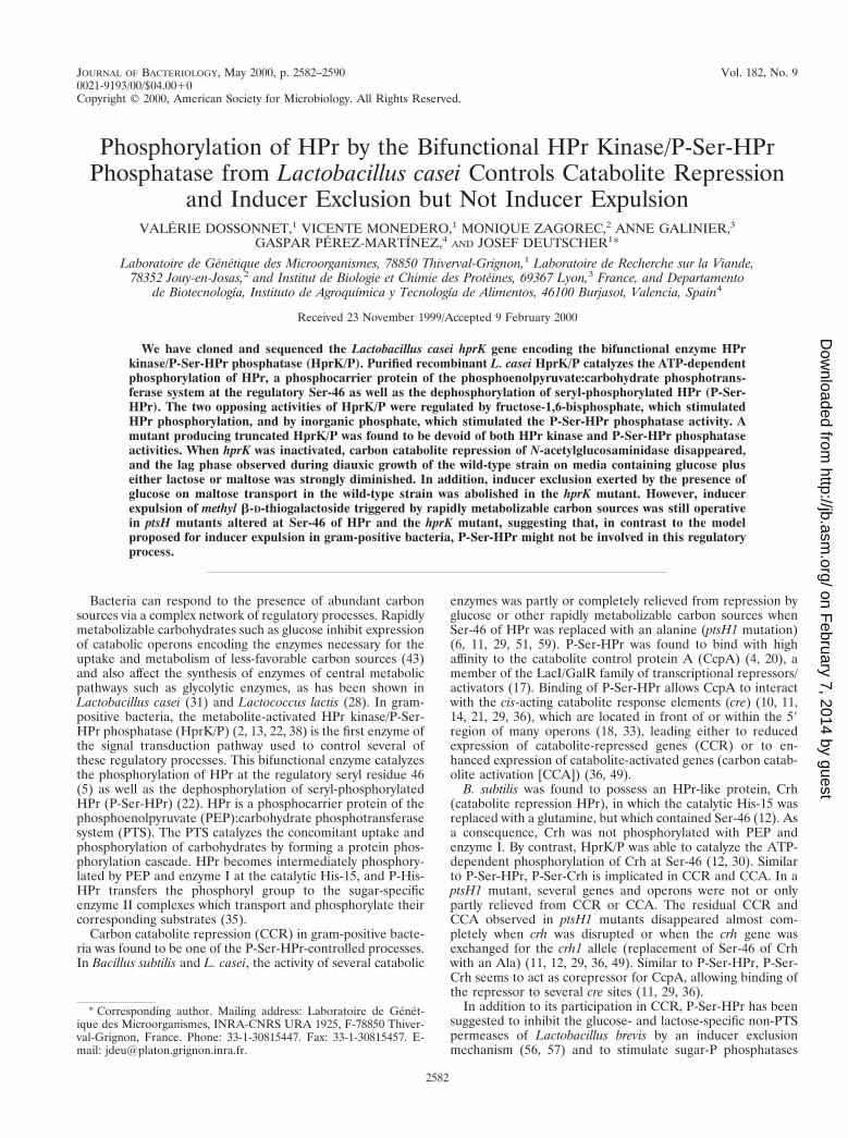
Construction of a L. casei hprK mutant and cloning of theentire hprK. In order to clone the missing 59 part of the hprKgene of L. casei and to test whether it indeed encodes thebifunctional HPr kinase/P-Ser-HPr phosphatase, a PCR wascarried out to introduce a point mutation replacing theleucine-encoding codon 208 of hprK with an amber codon. A1,312-bp DNA fragment carrying the amber mutation wascloned into the integrative vector pRV300 (26), providing plas-mid pHKLc208(Am) (see Materials and Methods). L. caseiBL23 was transformed with pHKLc208(Am), and erythromy-cin-resistant clones were obtained. In seven clones, the inte-gration of pHKLc208(Am) was tested by Southern blottingusing as a probe a 590-bp internal hprK fragment as describedin Materials and Methods. Only one HindIII fragment of 5.2kb could be detected with DNA from wild-type L. casei BL23,whereas the seven erythromycin-resistant clones gave twobands with sizes of 3.6 and 6.5 kb (data not shown), suggestingthat plasmid pHKLc208(Am), which contains a single HindIIIsite, had been integrated in the chromosome of these trans-formants. The integrants were tested for HPr kinase activity,since the Campbell-like recombination of pHKLc208(Am)with the L. casei chromosome can occur in two different waysgiving rise to two types of integrants. Four integrants exhibitedan HprK2 phenotype and three exhibited an HprK1 pheno-type. Phosphorylation experiments with HPr(His)6 from B.subtilis and L. casei and crude extracts from one of the inte-grants devoid of HPr kinase activity are shown in Fig. 2.Whereas phosphorylation of both HPr could be observed withcrude extracts of the wild-type strain (Fig. 2, lanes 1 and 3),neither of the HPrs was phosphorylated with crude extracts ofthe integrant (Fig. 2, lanes 2 and 4). Sequencing of appropriatePCR products obtained from this integrant revealed that thisstrain, which was called LcG102, contained an hprK allelecarrying the amber codon at position 208 and an incompletehprK gene lacking the 59 part together with the promoterregion, explaining why this strain was devoid of HPr kinaseactivity. Sequencing of PCR products obtained with chromo-somal DNA from one of the erythromycin-resistant hprK1
strains showed that it contained a wild-type hprK and an in-complete hprK carrying the amber mutation. Our attempts toisolate from the latter strain erythromycin-sensitive hprK mu-
tants resulting from a second recombination causing excisionof the plasmid were not successful. The second recombinationcan also occur at two different sites with respect to the intro-duced amber codon, leading to hprK1 or hprK strains. About300 erythromycin-sensitive clones obtained after growth with-out selective pressure were tested and were all found to behprK1.
Chromosomal DNA of LcG102 was isolated, digested withHindIII, religated, and transformed into E. coli NM522. Theplasmid present in one of the ampicillin-resistant clones waspurified and found to carry an insert of approximately 3.2 kb.DNA sequencing of this plasmid (pHKLcUS) revealed that theinsert contained the L. casei DNA fragment already present inpHKLc208(Am) and, in addition, the 59 part of the presumedhprK, its promoter region, and two complete and one incom-plete open reading frame (ORF) located upstream of hprK(Fig. 1). The proteins encoded by these three ORFs exhibited23, 22, and 36% sequence identity, respectively, when com-pared to the proteins encoded by the B. subtilis yvlB, yvlC, andyvlD genes (23).
The presumed L. casei hprK gene consists of 957 bp andencodes a protein of 35,349 Da composed of 319 amino acids,which exhibits 50% sequence identity when compared to B.subtilis HprK/P. As in all other known HprK/P, the A motif ofnucleotide binding proteins (GX4GKS) is present around po-sition 160. The presumed hprK gene starts with an ATG, whichis preceded by a putative ribosome binding site located 5 bpupstream of the start codon (Fig. 1). Downstream from hprKand separated from hprK by only 1 bp, the lgt gene begins. Thecloned lgt fragment encodes the first 129 amino acids of L.casei Lgt which exhibit 53% sequence identity when comparedto the corresponding N-terminal part of B. subtilis Lgt.
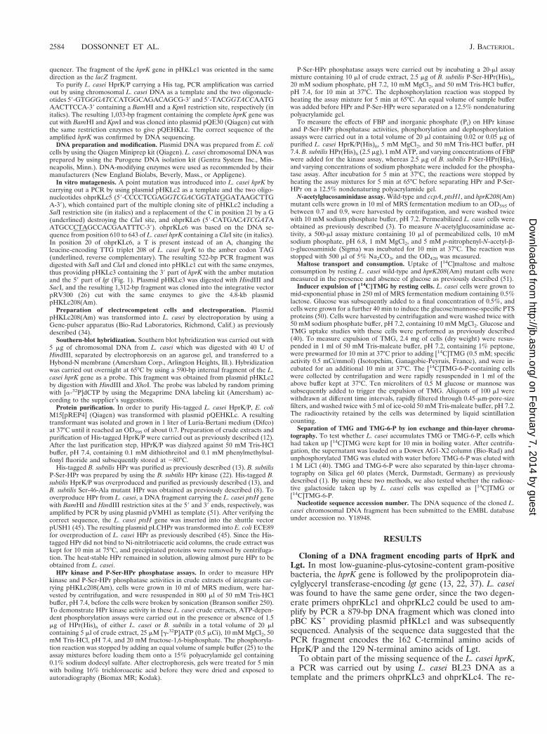
L. casei HprK/P is a bifunctional enzyme regulated by FBPand Pi. In order to confirm that the presumed hprK geneencodes L. casei HprK/P and to test whether it exhibits bothHPr kinase and P-Ser-HPr phosphatase activities similar to theB. subtilis and Enterococcus faecalis enzymes (22), His-taggedL. casei HprK/P was purified as described in Materials andMethods. The purified enzyme phosphorylated B. subtilis HPr(Fig. 3A, lane 1), and this activity was stimulated by FBP (Fig.3A, lanes 2 to 5). Phosphorylation seems to occur at Ser-46, asthe B. subtilis Ser-46-Ala mutant HPr was not phosphorylatedby the L. casei HprK/P (data not shown). HprK/P from L. caseiwas found to be bifunctional, as it also catalyzed the dephos-phorylation of P-Ser-HPr(His)6 of B. subtilis (Fig. 3B, lane 2).The P-Ser-HPr phosphatase activity was stimulated by Pi (Fig.3B, lanes 3 to 5). Although the presence of Pi inhibited theATP-dependent phosphorylation of HPr (compare Fig. 3C,lanes 1 and 2), a strong stimulation of HPr(Ser) phosphoryla-tion by FBP occurred when the HPr kinase assays were carriedout in the presence of low concentrations of Pi. When 1 mM Piwas used, almost no HPr phosphorylation could be observed inthe absence of FBP, whereas in the presence of 20 mM FBP,a strong HPr kinase activity could be detected (Fig. 3C, lanes2 and 3). By contrast, when using 8 mM Pi, FBP had almostcompletely lost its stimulating effect on HPr phosphorylation(Fig. 3C, lanes 8 and 9). FBP exerted only a weak inhibitoryeffect on Pi-stimulated P-Ser-HPr phosphatase activity (about1.5- to twofold inhibition with 10 mM FBP, data not shown).
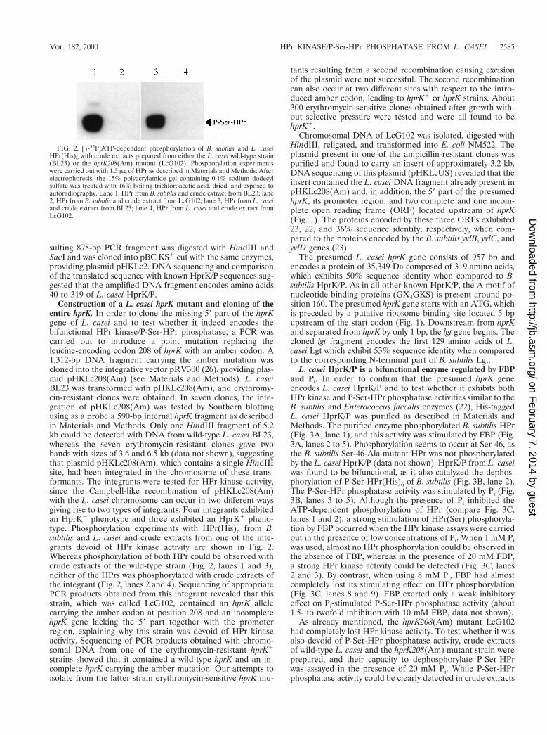
As already mentioned, the hprK208(Am) mutant LcG102had completely lost HPr kinase activity. To test whether it wasalso devoid of P-Ser-HPr phosphatase activity, crude extractsof wild-type L. casei and the hprK208(Am) mutant strain wereprepared, and their capacity to dephosphorylate P-Ser-HPrwas assayed in the presence of 20 mM Pi. While P-Ser-HPrphosphatase activity could be clearly detected in crude extracts
FIG. 2. [g-32P]ATP-dependent phosphorylation of B. subtilis and L. caseiHPr(His)6 with crude extracts prepared from either the L. casei wild-type strain(BL23) or the hprK208(Am) mutant (LcG102). Phosphorylation experimentswere carried out with 1.5 mg of HPr as described in Materials and Methods. Afterelectrophoresis, the 15% polyacrylamide gel containing 0.1% sodium dodecylsulfate was treated with 16% boiling trichloroacetic acid, dried, and exposed toautoradiography. Lane 1, HPr from B. subtilis and crude extract from BL23; lane2, HPr from B. subtilis and crude extract from LcG102; lane 3, HPr from L. caseiand crude extract from BL23; lane 4, HPr from L. casei and crude extract fromLcG102.
VOL. 182, 2000 HPr KINASE/P-Ser-HPr PHOSPHATASE FROM L. CASEI 2585
on February 7, 2014 by guest
http://jb.asm.org/
Dow
nloaded from
of the wild-type strain (Fig. 4, lanes 3 and 4), no such activitywas found in crude extracts of the hprK208(Am) mutantLcG102 (Fig. 4, lanes 1 and 2). Even increasing the incubationtime from 10 to 30 min did not allow detection of dephospho-rylated HPr (data not shown).
The hprK208(Am) mutation affects CCR. To determinewhether, similar to B. subtilis HprK/P (13, 38), L. caseiHprK/P is also involved in CCR, the repressive effect ofglucose on N-acetylglucosaminidase activity was measuredin the hprK208(Am) mutant and compared to the activityfound in the wild-type and the ccpA and ptsH1 mutant strains.
In wild-type strain BL23, N-acetylglucosaminidase activity wasrepressed 18-fold by the presence of glucose compared to cellsgrown in ribose-containing medium (Table 1). Similar to thecase in L. casei ccpA or ptsH1 mutants (32, 51), CCR of N-acetylglucosaminidase activity was strongly diminished in thehprK208(Am) mutant LcG102 (Table 1).
The hprK208(Am) mutation affects diauxic growth. Growthof the hprK208(Am) mutant LcG102 in MRS medium contain-ing 0.05% glucose plus either 0.05% lactose or 0.05% maltosewas compared to the growth behavior of the wild-type strainBL23. Wild-type L. casei grown in media containing mixturesof glucose and lactose or glucose and maltose exhibited adiauxic growth curve with two distinct growth phases separatedby a lag phase of about 8 h for cells growing in glucose/lactose-containing medium and 7 h for cells growing in glucose/mal-tose-containing medium. In the hprK208(Am) mutant LcG102,the lag phase was reduced to less than 2 h for cells grown ineither glucose-and-lactose- or glucose-and-maltose-containingmedium (data not shown).
The hprK208(Am) mutation prevents the exclusion of mal-tose by glucose. It has recently been demonstrated that re-placement of Ser-46 in L. casei HPr with alanine or threonineand replacement of Ile-47 with threonine prevents the exclu-sion of maltose by glucose (51). To test whether the effect ofthe ptsH mutations replacing Ser-46 was indeed due to theabsence of ATP-dependent phosphorylation of HPr, we stud-ied glucose-triggered maltose exclusion in the hprK208(Am)mutant strain LcG102. As recently reported (51), maltose up-take by wild-type cells was instantaneously arrested when glu-cose was added to the transport medium (Fig. 5A). By contrast,when an identical experiment was carried out with thehprK208(Am) mutant LcG102, maltose uptake was not inhib-ited but, rather, was slightly stimulated by the presence of
FIG. 3. The effect of FBP and Pi on ATP-dependent phosphorylation of B.subtilis HPr. HPr and P-Ser-HPr were separated on nondenaturing 12.5% poly-acrylamide gels. (A) Effect of FBP on the L. casei HPr kinase activity. HPrphosphorylation was carried out with 20 ng of HprK/P and the indicated con-centrations of FBP for 3 min at 37°C as described in Materials and Methods. HPrstandard (2.5 mg) was loaded on lane 6. (B) Effect of Pi on L. casei P-Ser-HPrphosphatase activity. HprK/P-catalyzed dephosphorylation of P-Ser-HPr wasperformed with 50 ng of HprK/P and the indicated Pi concentrations by incu-bating the assay mixture for 5 min at 37°C as described in Materials and Meth-ods. P-Ser-HPr standard (2.5 mg) was loaded on lane 1. (C) Effect of FBP onATP-dependent HPr phosphorylation in the presence of Pi. ATP-dependent HPrphosphorylation was carried out for 5 min at 37°C with 20 ng of HprK/P and theindicated amounts of Pi in the presence (1) or absence (2) of 20 mM FBP.Lanes 10 and 11 contain 2.5 mg of HPr and P-Ser-HPr standards, respectively.After electrophoresis, gels were stained with Coomassie blue.
FIG. 4. P-Ser-HPr phosphatase activity in crude extracts of L. casei wild-typeor hprK208(Am) mutant. P-Ser-HPr phosphatase assays were carried out with 10ml of crude extracts in the absence (2) or presence (1) of 2.5 mg of P-Ser-HPras described in Materials and Methods. P-Ser-HPr and HPr standards (2.5 mgeach) were loaded on lanes 5 and 6, respectively. Samples were separated on anondenaturing 12.5% polyacrylamide gel which was stained with Coomassieblue.
TABLE 1. Catabolite repression of N-acetylglucosaminidasein L. casei
L. casei strainN-acetylglucosaminidase activitya
RepressionfactorRibose Glucose
Wild-type 37.6 6 6.7 2.0 6 0.9 18.8hprK208(Am) 31.5 6 4.5 26.3 6 1.7 1.2ptsH1 35.3 6 7.2 26.7 6 6.5 1.3ccpA 30.6 6 4.3 19.4 6 0.7 1.6
a N-acetylglucosaminidase activity was determined by using p-nitrophenyl-N-acetyl-b-D-glucosaminide as substrate. Activity is expressed as nmoles of p-nitrophenol formed per minute and per milligram of cells (dry weight). Valuesare given 6 standard errors.
2586 DOSSONNET ET AL. J. BACTERIOL.
on February 7, 2014 by guest
http://jb.asm.org/
Dow
nloaded from
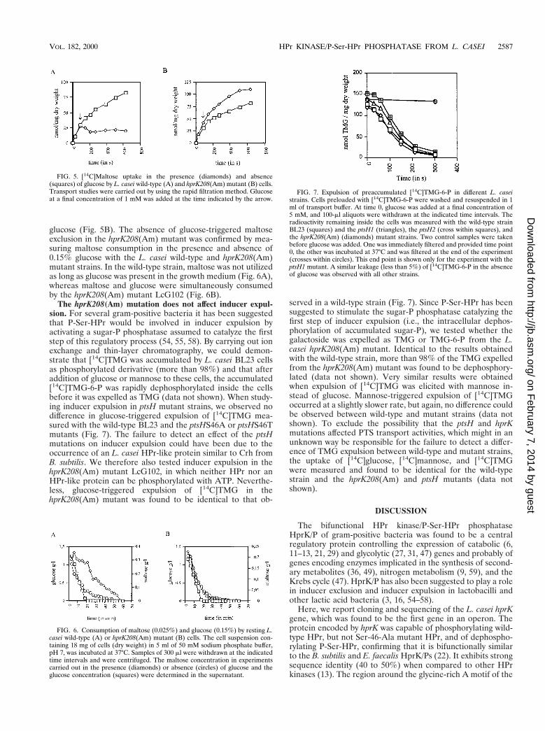
glucose (Fig. 5B). The absence of glucose-triggered maltoseexclusion in the hprK208(Am) mutant was confirmed by mea-suring maltose consumption in the presence and absence of0.15% glucose with the L. casei wild-type and hprK208(Am)mutant strains. In the wild-type strain, maltose was not utilizedas long as glucose was present in the growth medium (Fig. 6A),whereas maltose and glucose were simultaneously consumedby the hprK208(Am) mutant LcG102 (Fig. 6B).
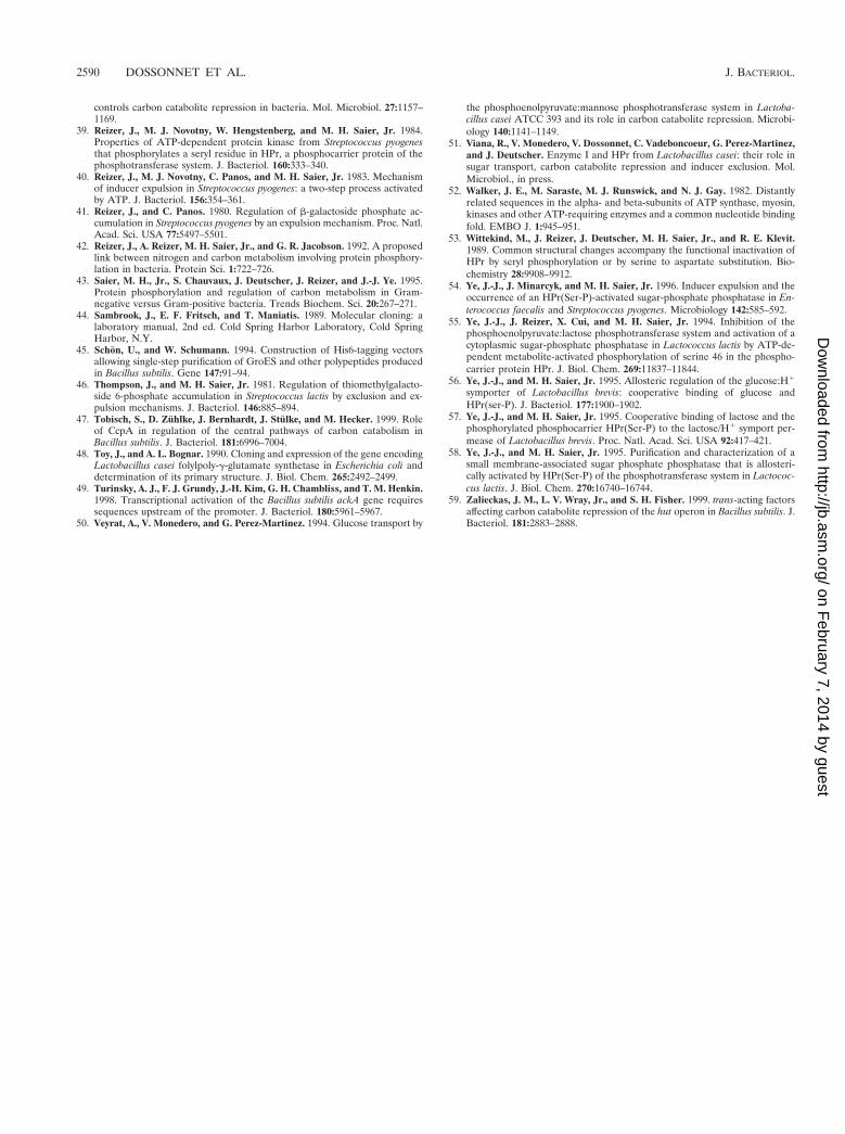
The hprK208(Am) mutation does not affect inducer expul-sion. For several gram-positive bacteria it has been suggestedthat P-Ser-HPr would be involved in inducer expulsion byactivating a sugar-P phosphatase assumed to catalyze the firststep of this regulatory process (54, 55, 58). By carrying out ionexchange and thin-layer chromatography, we could demon-strate that [14C]TMG was accumulated by L. casei BL23 cellsas phosphorylated derivative (more than 98%) and that afteraddition of glucose or mannose to these cells, the accumulated[14C]TMG-6-P was rapidly dephosphorylated inside the cellsbefore it was expelled as TMG (data not shown). When study-ing inducer expulsion in ptsH mutant strains, we observed nodifference in glucose-triggered expulsion of [14C]TMG mea-sured with the wild-type BL23 and the ptsHS46A or ptsHS46Tmutants (Fig. 7). The failure to detect an effect of the ptsHmutations on inducer expulsion could have been due to theoccurrence of an L. casei HPr-like protein similar to Crh fromB. subtilis. We therefore also tested inducer expulsion in thehprK208(Am) mutant LcG102, in which neither HPr nor anHPr-like protein can be phosphorylated with ATP. Neverthe-less, glucose-triggered expulsion of [14C]TMG in thehprK208(Am) mutant was found to be identical to that ob-
served in a wild-type strain (Fig. 7). Since P-Ser-HPr has beensuggested to stimulate the sugar-P phosphatase catalyzing thefirst step of inducer expulsion (i.e., the intracellular dephos-phorylation of accumulated sugar-P), we tested whether thegalactoside was expelled as TMG or TMG-6-P from the L.casei hprK208(Am) mutant. Identical to the results obtainedwith the wild-type strain, more than 98% of the TMG expelledfrom the hprK208(Am) mutant was found to be dephosphory-lated (data not shown). Very similar results were obtainedwhen expulsion of [14C]TMG was elicited with mannose in-stead of glucose. Mannose-triggered expulsion of [14C]TMGoccurred at a slightly slower rate, but again, no difference couldbe observed between wild-type and mutant strains (data notshown). To exclude the possibility that the ptsH and hprKmutations affected PTS transport activities, which might in anunknown way be responsible for the failure to detect a differ-ence of TMG expulsion between wild-type and mutant strains,the uptake of [14C]glucose, [14C]mannose, and [14C]TMGwere measured and found to be identical for the wild-typestrain and the hprK208(Am) and ptsH mutants (data notshown).
DISCUSSION
The bifunctional HPr kinase/P-Ser-HPr phosphataseHprK/P of gram-positive bacteria was found to be a centralregulatory protein controlling the expression of catabolic (6,11–13, 21, 29) and glycolytic (27, 31, 47) genes and probably ofgenes encoding enzymes implicated in the synthesis of second-ary metabolites (36, 49), nitrogen metabolism (9, 59), and theKrebs cycle (47). HprK/P has also been suggested to play a rolein inducer exclusion and inducer expulsion in lactobacilli andother lactic acid bacteria (3, 16, 54–58).
Here, we report cloning and sequencing of the L. casei hprKgene, which was found to be the first gene in an operon. Theprotein encoded by hprK was capable of phosphorylating wild-type HPr, but not Ser-46-Ala mutant HPr, and of dephospho-rylating P-Ser-HPr, confirming that it is bifunctionally similarto the B. subtilis and E. faecalis HprK/Ps (22). It exhibits strongsequence identity (40 to 50%) when compared to other HPrkinases (13). The region around the glycine-rich A motif of the
FIG. 5. [14C]Maltose uptake in the presence (diamonds) and absence(squares) of glucose by L. casei wild-type (A) and hprK208(Am) mutant (B) cells.Transport studies were carried out by using the rapid filtration method. Glucoseat a final concentration of 1 mM was added at the time indicated by the arrow.
FIG. 6. Consumption of maltose (0.025%) and glucose (0.15%) by resting L.casei wild-type (A) or hprK208(Am) mutant (B) cells. The cell suspension con-taining 18 mg of cells (dry weight) in 5 ml of 50 mM sodium phosphate buffer,pH 7, was incubated at 37°C. Samples of 300 ml were withdrawn at the indicatedtime intervals and were centrifuged. The maltose concentration in experimentscarried out in the presence (diamonds) or absence (circles) of glucose and theglucose concentration (squares) were determined in the supernatant.
FIG. 7. Expulsion of preaccumulated [14C]TMG-6-P in different L. caseistrains. Cells preloaded with [14C]TMG-6-P were washed and resuspended in 1ml of transport buffer. At time 0, glucose was added at a final concentration of5 mM, and 100-ml aliquots were withdrawn at the indicated time intervals. Theradioactivity remaining inside the cells was measured with the wild-type strainBL23 (squares) and the ptsH1 (triangles), the ptsH2 (cross within squares), andthe hprK208(Am) (diamonds) mutant strains. Two control samples were takenbefore glucose was added. One was immediately filtered and provided time point0, the other was incubated at 37°C and was filtered at the end of the experiment(crosses within circles). This end point is shown only for the experiment with theptsH1 mutant. A similar leakage (less than 5%) of [14C]TMG-6-P in the absenceof glucose was observed with all other strains.
VOL. 182, 2000 HPr KINASE/P-Ser-HPr PHOSPHATASE FROM L. CASEI 2587
on February 7, 2014 by guest
http://jb.asm.org/
Dow
nloaded from

nucleotide binding fold (52) is especially well conserved.HprK/Ps seem to contain an overlapping tandem repeat of theGXXXXGKS consensus sequence in the nucleotide bindingsite, leading to the GXXXXGKSXXGKS sequence, which isfully conserved in HprK/P of B. subtilis or Mycoplasma geni-talium. In most other organisms possessing HprK/P, the first Kin the above sequence was found to be replaced with a D. It isnot known whether this repetition of the nucleotide bindingmotif is of any functional significance. In L. casei and all othergram-positive bacteria (except Clostridium acetobutylicum) forwhich the corresponding DNA sequence data are available,hprK is the first gene in an operon and always followed by theprolipoprotein diacylglyceryl transferase-encoding lgt gene.This conserved gene organization could be indicative of a func-tional interaction between HprK/P and Lgt. In the gram-neg-ative bacteria Neisseria gonorrhoeae, Neisseria meningitidis, andBordetella pertussis, hprK is not followed by lgt, but is precededby a gene encoding an EIINtr-like protein (42), which seems toform an operon together with hprK. The cloned region up-stream of L. casei hprK was found to contain one incompleteand two complete ORFs which seem to be organized in anoperon. The deduced amino acid sequences exhibited similar-ity to proteins of unknown function encoded by the B. subtilisyvlB, yvlC, and yvlD genes, which are located about 11.5 kbupstream of the B. subtilis hprK gene (23).
The two opposing activities of recombinant purified L. caseiHprK/P, HPr kinase and P-Ser-HPr phosphatase, were foundto be regulated by FBP and Pi. HPr kinase from L. casei wassimilarly active with HPr from L. casei and B. subtilis (Fig. 2).However, in contrast to the HPr kinase activity of B. subtilisHprK/P, which is strongly stimulated by FBP (13, 19, 38), or ofStreptococcus salivarius HprK/P, which is inhibited by FBP (2),the HPr kinase activity of the L. casei enzyme was slightlystimulated by FBP (Fig. 3A). When the experiments werecarried out in the presence of 1 to 4 mM Pi, a stronger stim-ulatory effect of FBP on HPr phosphorylation could be ob-served (Fig. 3C). HprK/Ps from different organisms thereforeappear to respond to similar but not identical intracellularsignals, which probably reflects an adaptation to the specificphysiological changes taking place in each organism when acarbon source is rapidly metabolized. Pi strongly stimulated theP-Ser-HPr phosphatase activity, as has also been observed withthe B. subtilis and E. faecalis enzymes (13, 22). FBP and Pi donot seem to compete for the same binding site, as the Pi-stimulated P-Ser-HPr phosphatase activity associated with L.casei HprK/P was only slightly inhibited by FBP.
To study the different regulatory functions suggested forHprK/P in carbon metabolism, an hprK mutant has been con-structed, producing a protein truncated at position 208. Crudeextracts of the hprK208(Am) mutant strain LcG102 were notable to phosphorylate HPr from L. casei and B. subtilis (Fig. 2).They were also devoid of P-Ser-HPr phosphatase activity (Fig.4), confirming that the major P-Ser-HPr phosphatase activityin L. casei is associated with HprK/P. Nevertheless, a homo-logue of the B. subtilis yvoE gene, which is located in the sameoperon as hprK (13) and which encodes an enzyme exhibitinglow P-Ser-HPr phosphatase activity, is present in Lactobacillusrhamnosus (48), a very close relative of L. casei. Although thecorresponding L. rhamnosus gene is not located in the sameoperon as the hprK gene, the purified L. rhamnosus YvoEhomologue exhibited low P-Ser-HPr phosphatase activity sim-ilar to B. subtilis YvoE (V. Dossonnet and J. Deutscher, un-published results).
Similar to ptsH1 and ccpA mutants, N-acetylglucosaminidaseactivity in the hprK208(Am) mutant LcG102 was found to bealmost completely relieved from repression by glucose (Table
1). In addition, the lag phase of about 8 h observed duringdiauxic growth of the L. casei wild-type strain in media con-taining glucose and either lactose or maltose was reduced toless than 2 h in the hprK208(Am) mutant. The fact that the lagphase had not completely disappeared is probably due to thepregrowth of cells in glucose-containing medium. When thecells were pregrown in lactose- or maltose-containing medium,the lag phase was much shorter for the wild-type strain (50)and had completely disappeared for the hprK mutant (data notshown). These results established that, similar to B. subtilis,HprK/P plays an important role in CCR in L. casei, and theystrongly suggested that this CCR mechanism is operative in awide variety of gram-positive bacteria, since CcpA, cre sites,HprK/P, and HPr with a phosphorylatable Ser-46 can be foundin almost all low-guanine-plus-cytosine-content gram-positiveorganisms.
Results recently obtained with L. casei ptsH mutants, inwhich Ser-46 was replaced with other amino acids, suggestedthat ATP-dependent phosphorylation of HPr is involved inmaltose exclusion in this organism (51). The participation ofP-Ser-HPr in maltose exclusion has been confirmed by theresults obtained with the L. casei hprK208(Am) mutantLcG102. Maltose uptake or consumption by the wild-typestrain was prevented by the presence of glucose in the medium(Fig. 5A and 6A), whereas with the hprK208(Am) mutant ad-dition of glucose led to increased maltose uptake, and maltoseand glucose were found to be simultaneously consumed (Fig.5B and 6B). Glucose uptake by the hprK208(Am) mutant wasnot altered when compared to the wild-type strain. It is there-fore likely that P-Ser-HPr plays a role similar to unphosphor-ylated EIIAGlc, the regulatory protein involved in inducer ex-clusion in E. coli (35), by inhibiting certain non-PTS transportsystems when a rapidly metabolizable carbon source is takenup by gram-positive bacteria. However, our results could alsobe explained by assuming that the maltose permease is acti-vated by P-His-HPr-catalyzed phosphorylation similar to thelactose permease of Streptococcus thermophilus (15) and thatthis activation is prevented when P-Ser-HPr is formed in re-sponse to the rapid uptake and metabolism of a carbohydrate.
The ATP-dependent phosphorylation of HPr at Ser-46 byHprK/P was discovered in connection with a regulatory phe-nomenon called inducer expulsion (7, 39). In several lactic acidbacteria, including lactobacilli (3), the addition of glucose tocells preloaded with a nonmetabolizable sugar derivative takenup by the PTS and therefore accumulated as phospho com-pound was found to cause the rapid expulsion of the sugarderivative in its dephosphorylated form (41, 46). Since thesame conditions, which lead to inducer expulsion, were alsofound to trigger the in vivo phosphorylation of HPr at Ser-46,P-Ser-HPr was thought to play a role in inducer expulsion (39).However, as ptsH1 or hprK mutants were only available for B.subtilis, and since no nonmetabolizable PTS sugars submittedto inducer expulsion are known for this organism, no in vivoexperiments addressing the role of P-Ser-HPr in inducer ex-pulsion have yet been carried out. We therefore studied in-ducer expulsion in L. casei and could show that this organismaccumulates TMG as a phosphorylated derivative and thataddition of glucose to cells preloaded with [14C]TMG-6-P ledto the expulsion of unphosphorylated TMG, similar to thatobserved in Streptococcus pyogenes (39). The Ser-46-Ala andSer-46-Thr ptsH mutations, which caused relief from CCR(51), had no effect on glucose-triggered expulsion of preaccu-mulated [14C]TMG-6-P (Fig. 7). Nevertheless, it was possiblethat, similar to B. subtilis (12), L. casei might possess an HPr-like protein, which could be operative in inducer expulsion, orthe metabolite-activated HprK/P might phosphorylate an un-
2588 DOSSONNET ET AL. J. BACTERIOL.
on February 7, 2014 by guest
http://jb.asm.org/
Dow
nloaded from

known protein implicated in inducer expulsion. However, thehprK208(Am) mutant, which is devoid of HPr kinase activity,exhibited expulsion of preaccumulated [14C]TMG-6-P identi-cal to the wild-type strain, i.e., [14C]TMG-6-P was dephospho-rylated in the cells before unphosphorylated TMG was ex-pelled. These results clearly established that HPr kinaseactivity is not required for TMG expulsion in L. casei. As themechanism of TMG expulsion in L. casei strongly resemblesthe mechanism of inducer expulsion operative in streptococciand lactococci (41, 46), the results obtained in this study ques-tion the proposed implication of P-Ser-HPr in inducer expul-sion of gram-positive bacteria.
ACKNOWLEDGMENTS
We thank Andree Lepingle for technical assistance in DNA se-quencing and Sandrine Poncet, Dominique Le Coq, Stephane Aymer-ich, and Christian Vadeboncoeur for valuable discussions.
This work was supported by the CNRS, the INRA, and the INA-PG,and by European Community Biotech Program contract BIO4-CT96-0380. V. Monedero was supported by a FPU fellowship from theMinisterio de Educacion y Cultura of Spain.
REFERENCES
1. Bettenbrock, K., U. Siebers, P. Ehrenreich, and C.-A. Alpert. 1999. Lacto-bacillus casei 64H contains a phosphoenolpyruvate-dependent phospho-transferase system for uptake of galactose, as confirmed by analysis of ptsHand different gal mutants. J. Bacteriol. 181:225–230.
2. Brochu, D., and C. Vadeboncoeur. 1999. The HPr(Ser) kinase of Streptococ-cus salivarius: purification, properties, and cloning of the hprK gene. J.Bacteriol. 181:709–717.
3. Chassy, B. M., and J. Thompson. 1983. Regulation of lactose-phosphoenol-pyruvate-dependent phosphotransferase system and b-D-phosphogalactosidegalactohydrolase activities in Lactobacillus casei. J. Bacteriol. 154:1195–1203.
4. Deutscher, J., E. Kuster, U. Bergstedt, V. Charrier, and W. Hillen. 1995.Protein kinase-dependent HPr/CcpA interaction links glycolytic activity tocarbon catabolite repression in Gram-positive bacteria. Mol. Microbiol. 15:1049–1053.
5. Deutscher, J., B. Pevec, K. Beyreuther, H.-H. Kiltz, and W. Hengstenberg.1986. Streptococcal phosphoenolpyruvate-sugar phosphotransferase system:amino acid sequence and site of ATP-dependent phosphorylation of HPr.Biochemistry 25:6543–6551.
6. Deutscher, J., J. Reizer, C. Fischer, A. Galinier, M. H. Saier, Jr., and M.Steinmetz. 1994. Loss of protein kinase-catalyzed phosphorylation of HPr, aphosphocarrier protein of the phosphotransferase system, by mutation of theptsH gene confers catabolite repression resistance to several catabolic genesof Bacillus subtilis. J. Bacteriol. 176:3336–3344.
7. Deutscher, J., and M. H. Saier, Jr. 1983. ATP-dependent protein kinase-catalyzed phosphorylation of a seryl residue in HPr, a phosphate carrierprotein of the phosphotransferase system in Streptococcus pyogenes. Proc.Natl. Acad. Sci. USA 80:6790–6794.
8. Eisermann, R., J. Deutscher, G. Gonzy-Treboul, and W. Hengstenberg. 1988.Site-directed mutagenesis with the ptsH gene of Bacillus subtilis. Isolationand characterization of heat-stable proteins altered at the ATP-dependentregulatory phosphorylation site. J. Biol. Chem. 263:17050–17054.
9. Faires, N., S. Tobisch, S. Bachem, I. Martin-Verstraete, M. Hecker, and J.Stulke. 1999. The catabolite control protein CcpA controls ammonium as-similation in Bacillus subtilis. J. Mol. Microbiol. Biotechnol. 1:141–148.
10. Fujita, Y., Y. Miwa, A. Galinier, and J. Deutscher. 1995. Specific recognitionof the Bacillus subtilis gnt cis-acting catabolite-responsive element by a pro-tein complex formed between CcpA and seryl-phosphorylated HPr. Mol.Microbiol. 17:953–960.
11. Galinier, A., J. Deutscher, and I. Martin-Verstraete. 1999. Phosphorylationof either Crh or HPr mediates catabolite repression and binding of CcpA tothe cre of the Bacillus subtilis xyn operon. J. Mol. Biol. 286:307–314.
12. Galinier, A., J. Haiech, M.-C. Kilhoffer, M. Jaquinod, J. Stulke, J. Deut-scher, and I. Martin-Verstraete. 1997. The Bacillus subtilis crh gene encodesa HPr-like protein involved in carbon catabolite repression. Proc. Natl.Acad. Sci. USA 94:8439–8444.
13. Galinier, A., M. Kravanja, R. Engelmann, W. Hengstenberg, M.-C. Kilhoffer,J. Deutscher, and J. Haiech. 1998. New protein kinase and protein phos-phatase families mediate signal transduction in bacterial catabolite repres-sion. Proc. Natl. Acad. Sci. USA 95:1823–1828.
14. Gosseringer, R., E. Kuster, A. Galinier, J. Deutscher, and W. Hillen. 1997.Cooperative and non-cooperative DNA binding modes of catabolite controlprotein CcpA from Bacillus megaterium result from sensing two differentsignals. J. Mol. Biol. 266:665–676.
15. Gunnewijk, M. G. W., P. W. Postma, and B. Poolman. 1999. Phosphorylation
and functional properties of the IIA domain of the lactose transport proteinof Streptococcus thermophilus. J. Bacteriol. 181:632–641.
16. Hausman, S. Z., J. Thompson, and J. London. 1984. Futile xylitol cycle inLactobacillus casei. J. Bacteriol. 160:211–215.
17. Henkin, T. M., F. J. Grundy, W. L. Nicholson, and G. H. Chambliss. 1991.Catabolite repression of a-amylase gene expression in Bacillus subtilis in-volves a trans-acting gene product homologous to the Escherichia coli lacIand galR repressors. Mol. Microbiol. 5:575–584.
18. Hueck, C. J., W. Hillen, and M. H. Saier, Jr. 1994. Analysis of a cis-activesequence mediating catabolite repression in Gram-positive bacteria. Res.Microbiol. 145:503–518.
19. Jault, J. M., S. Fieulaine, S. Nessler-Baudet, P. Gonzalo, J. Deutscher, A. DiPietro, and A. Galinier. 2000. The HPr kinase from Bacillus subtilis is ahomo-oligomeric enzyme which exhibits strong positive cooperativity fornucleotide and fructose 1,6-bisphosphate binding. J. Biol. Chem. 275:2009–2016.
20. Jones, B. E., V. Dossonnet, E. Kuster, W. Hillen, J. Deutscher, and R. E.Klevit. 1997. Binding of the catabolite repressor protein CcpA to its DNAtarget is regulated by phosphorylation of its corepressor HPr. J. Biol. Chem.272:26530–26535.
21. Kim, J.-H., M. I. Voskuil, and G. H. Chambliss. 1998. NADP, corepressorfor the Bacillus catabolite control protein CcpA. Proc. Natl. Acad. Sci. USA95:9590–9595.
22. Kravanja, M., R. Engelmann, V. Dossonnet, M. Bluggel, H. E. Meyer, R.Frank, A. Galinier, J. Deutscher, N. Schnell, and W. Hengstenberg. 1999.The hprK gene of Enterococcus faecalis encodes a novel bifunctional enzyme:the HPr kinase/phosphatase. Mol. Microbiol. 31:59–66.
23. Kunst, F., N. Ogasawara, I. Moszer, A. M. Albertini, G. Alloni, V. Azevedo,et al. 1997. The complete genome sequence of the Gram-positive bacteriumBacillus subtilis. Nature 390:249–256.
24. Kuster, E., E. J. Luesink, W. M. de Vos, and W. Hillen. 1996. Immunologicalcrossreactivity to the catabolite control protein CcpA from Bacillus megate-rium is found in many Gram-positive bacteria. FEMS Microbiol. Lett. 139:109–115.
25. Laemmli, U. K. 1970. Cleavage of structural proteins during the assembly ofthe head of bacteriophage T4. Nature 227:680–685.
26. Leloup, L., S. D. Ehrlich, M. Zagorec, and F. Morel-Deville. 1997. Single-crossover integration in the Lactobacillus sake chromosome and insertionalinactivation of the ptsI and lacL genes. Appl. Environ. Microbiol. 63:2117–2123.
27. Luesink, E. J., M. A. Beumer, O. P. Kuipers, and W. M. de Vos. 1999.Molecular characterization of the Lactococcus lactis ptsHI operon and anal-ysis of the regulatory role of HPr. J. Bacteriol. 181:764–771.
28. Luesink, E. J., R. E. M. A. van Harpen, B. P. Grossiord, O. P. Kuipers, andW. M. de Vos. 1998. Transcriptional activation of the glycolytic las operonand catabolite repression of the gal operon in Lactococcus lactis are medi-ated by the catabolite control protein CcpA. Mol. Microbiol. 30:789–798.
29. Martin-Verstraete, I., J. Deutscher, and A. Galinier. 1999. Phosphorylationof HPr and Crh by HprK, early steps in the catabolite repression signallingpathway for the Bacillus subtilis levanase operon. J. Bacteriol. 181:2966–2969.
30. Martin-Verstraete, I., A. Galinier, E. Darbon, Y. Quentin, V. Charrier, M.-C.Kilhoffer, J. Haiech, G. Rapoport, and J. Deutscher. 1999. The Q15H mu-tation enables Crh, a Bacillus subtilis HPr-like protein, to carry out someregulatory HPr functions, but does not make it an effective phosphocarrierfor sugar transport. Microbiology 145:3195–3204.
31. Monedero, V. 1997. Elements implicated in catabolite repression in Lacto-bacillus casei. Ph.D. thesis. Universitat de Valencia, Spain.
32. Monedero, V., M. J. Gosalbes, and G. Perez-Martinez. 1997. Cataboliterepression in Lactobacillus casei ATCC 393 is mediated by CcpA. J. Bacte-riol. 179:6657–6664.
33. Nicholson, W. L., and G. H. Chambliss. 1985. Isolation and characterizationof a cis-acting mutation conferring catabolite repression resistance to a-amy-lase synthesis in Bacillus subtilis. J. Bacteriol. 161:875–881.
34. Posno, M., R. J. Leer, N. van Luijk, M. J. F. van Giezen, P. T. H. M.Heuvelmans, B. C. Lokman, and P. H. Pouwels. 1991. Incompatibility ofLactobacillus vectors with replicons derived from small cryptic Lactobacillusplasmids and segregational instability of the introduced vectors. Appl. En-viron. Microbiol. 57:1822–1828.
35. Postma, P. W., J. W. Lengeler, and G. R. Jacobson. 1993. Phosphoenolpyru-vate:carbohydrate phosphotransferase systems of bacteria. Microbiol. Rev.57:543–594.
36. Presecan-Siedel, E., A. Galinier, R. Longin, J. Deutscher, A. Danchin, P.Glaser, and I. Martin-Verstraete. 1999. The catabolite regulation of the ptagene as part of the carbon flow pathways in Bacillus subtilis. J. Bacteriol.181:6889–6897.
37. Qi, H.-Y., K. Sankaran, K. Gan, and H. C. Wu. 1995. Structure-functionrelationship of bacterial prolipoprotein diacylglyceryl transferase: function-ally significant conserved regions. J. Bacteriol. 177:6820–6824.
38. Reizer, J., C. Hoischen, F. Titgemeyer, C. Rivolta, R. Rabus, J. Stulke, D.Karamata, M. H. Saier, Jr., and W. Hillen. 1998. A novel protein kinase that
VOL. 182, 2000 HPr KINASE/P-Ser-HPr PHOSPHATASE FROM L. CASEI 2589
on February 7, 2014 by guest
http://jb.asm.org/
Dow
nloaded from
controls carbon catabolite repression in bacteria. Mol. Microbiol. 27:1157–1169.
39. Reizer, J., M. J. Novotny, W. Hengstenberg, and M. H. Saier, Jr. 1984.Properties of ATP-dependent protein kinase from Streptococcus pyogenesthat phosphorylates a seryl residue in HPr, a phosphocarrier protein of thephosphotransferase system. J. Bacteriol. 160:333–340.
40. Reizer, J., M. J. Novotny, C. Panos, and M. H. Saier, Jr. 1983. Mechanismof inducer expulsion in Streptococcus pyogenes: a two-step process activatedby ATP. J. Bacteriol. 156:354–361.
41. Reizer, J., and C. Panos. 1980. Regulation of b-galactoside phosphate ac-cumulation in Streptococcus pyogenes by an expulsion mechanism. Proc. Natl.Acad. Sci. USA 77:5497–5501.
42. Reizer, J., A. Reizer, M. H. Saier, Jr., and G. R. Jacobson. 1992. A proposedlink between nitrogen and carbon metabolism involving protein phosphory-lation in bacteria. Protein Sci. 1:722–726.
43. Saier, M. H., Jr., S. Chauvaux, J. Deutscher, J. Reizer, and J.-J. Ye. 1995.Protein phosphorylation and regulation of carbon metabolism in Gram-negative versus Gram-positive bacteria. Trends Biochem. Sci. 20:267–271.
44. Sambrook, J., E. F. Fritsch, and T. Maniatis. 1989. Molecular cloning: alaboratory manual, 2nd ed. Cold Spring Harbor Laboratory, Cold SpringHarbor, N.Y.
45. Schon, U., and W. Schumann. 1994. Construction of His6-tagging vectorsallowing single-step purification of GroES and other polypeptides producedin Bacillus subtilis. Gene 147:91–94.
46. Thompson, J., and M. H. Saier, Jr. 1981. Regulation of thiomethylgalacto-side 6-phosphate accumulation in Streptococcus lactis by exclusion and ex-pulsion mechanisms. J. Bacteriol. 146:885–894.
47. Tobisch, S., D. Zuhlke, J. Bernhardt, J. Stulke, and M. Hecker. 1999. Roleof CcpA in regulation of the central pathways of carbon catabolism inBacillus subtilis. J. Bacteriol. 181:6996–7004.
48. Toy, J., and A. L. Bognar. 1990. Cloning and expression of the gene encodingLactobacillus casei folylpoly-g-glutamate synthetase in Escherichia coli anddetermination of its primary structure. J. Biol. Chem. 265:2492–2499.
49. Turinsky, A. J., F. J. Grundy, J.-H. Kim, G. H. Chambliss, and T. M. Henkin.1998. Transcriptional activation of the Bacillus subtilis ackA gene requiressequences upstream of the promoter. J. Bacteriol. 180:5961–5967.
50. Veyrat, A., V. Monedero, and G. Perez-Martinez. 1994. Glucose transport by
the phosphoenolpyruvate:mannose phosphotransferase system in Lactoba-cillus casei ATCC 393 and its role in carbon catabolite repression. Microbi-ology 140:1141–1149.
51. Viana, R., V. Monedero, V. Dossonnet, C. Vadeboncoeur, G. Perez-Martinez,and J. Deutscher. Enzyme I and HPr from Lactobacillus casei: their role insugar transport, carbon catabolite repression and inducer exclusion. Mol.Microbiol., in press.
52. Walker, J. E., M. Saraste, M. J. Runswick, and N. J. Gay. 1982. Distantlyrelated sequences in the alpha- and beta-subunits of ATP synthase, myosin,kinases and other ATP-requiring enzymes and a common nucleotide bindingfold. EMBO J. 1:945–951.
53. Wittekind, M., J. Reizer, J. Deutscher, M. H. Saier, Jr., and R. E. Klevit.1989. Common structural changes accompany the functional inactivation ofHPr by seryl phosphorylation or by serine to aspartate substitution. Bio-chemistry 28:9908–9912.
54. Ye, J.-J., J. Minarcyk, and M. H. Saier, Jr. 1996. Inducer expulsion and theoccurrence of an HPr(Ser-P)-activated sugar-phosphate phosphatase in En-terococcus faecalis and Streptococcus pyogenes. Microbiology 142:585–592.
55. Ye, J.-J., J. Reizer, X. Cui, and M. H. Saier, Jr. 1994. Inhibition of thephosphoenolpyruvate:lactose phosphotransferase system and activation of acytoplasmic sugar-phosphate phosphatase in Lactococcus lactis by ATP-de-pendent metabolite-activated phosphorylation of serine 46 in the phospho-carrier protein HPr. J. Biol. Chem. 269:11837–11844.
56. Ye, J.-J., and M. H. Saier, Jr. 1995. Allosteric regulation of the glucose:H1
symporter of Lactobacillus brevis: cooperative binding of glucose andHPr(ser-P). J. Bacteriol. 177:1900–1902.
57. Ye, J.-J., and M. H. Saier, Jr. 1995. Cooperative binding of lactose and thephosphorylated phosphocarrier HPr(Ser-P) to the lactose/H1 symport per-mease of Lactobacillus brevis. Proc. Natl. Acad. Sci. USA 92:417–421.
58. Ye, J.-J., and M. H. Saier, Jr. 1995. Purification and characterization of asmall membrane-associated sugar phosphate phosphatase that is allosteri-cally activated by HPr(Ser-P) of the phosphotransferase system in Lactococ-cus lactis. J. Biol. Chem. 270:16740–16744.
59. Zalieckas, J. M., L. V. Wray, Jr., and S. H. Fisher. 1999. trans-acting factorsaffecting carbon catabolite repression of the hut operon in Bacillus subtilis. J.Bacteriol. 181:2883–2888.
2590 DOSSONNET ET AL. J. BACTERIOL.
on February 7, 2014 by guest
http://jb.asm.org/
Dow
nloaded from