Angiogenesis and remodelling in human thoracic aortic aneurysms
Upload
independentCategory
view
1download
0
RESEARCH PAPER
Characterization ofzofenoprilat as an inducerof functional angiogenesisthrough increased H2SavailabilityE Terzuoli1*, M Monti1*, V Vellecco2, M Bucci2, G Cirino2, M Ziche1 andL Morbidelli1
1Department of Life Sciences, University of Siena, Siena, Italy, and 2Department of Experimental
Pharmacology, University of Naples Federico II, Naples, Italy
CorrespondenceLucia Morbidelli, Department ofLife Sciences, University of Siena,Via A. Moro 2, 53100 Siena, Italy.E-mail: lucia.morbidelli@unisi.it----------------------------------------------------------------
*These authors equallycontributed to the work.----------------------------------------------------------------
Received25 July 2014Revised13 January 2015Accepted22 January 2015
BACKGROUND AND PURPOSEHydrogen sulfide (H2S), an endogenous volatile mediator with pleiotropic functions, promotes vasorelaxation, exertsanti-inflammatory actions and regulates angiogenesis. Previously, the SH-containing angiotensin-converting enzyme inhibitor(ACEI), zofenopril, was identified as being effective in preserving endothelial function and inducing angiogenesis amongACEIs. Based on the H2S donor property of its active metabolite zofenoprilat, the objective of this study was to evaluatewhether zofenoprilat-induced angiogenesis was due to increased H2S availability.
EXPERIMENTAL APPROACHHUVECs were used for in vitro studies of angiogenesis, whereas the Matrigel plug assay was used for in vivo assessments.
KEY RESULTSZofenoprilat-treated HUVECs showed an increase in all functional features of the angiogenic process in vitro. As zofenoprilatinduced the expression of CSE (cystathionine-γ-lyase) and the continuous production of H2S, CSE inhibition or silencingblocked the ability of zofenoprilat to induce angiogenesis, both in vitro and in vivo. The molecular mechanisms underlyingH2S/zofenoprilat-induced angiogenesis were dependent on Akt, eNOS and ERK1/2 cascades. ATP-sensitive potassium (KATP)channels, the molecular target that mediates part of the vascular functions of H2S, were shown to be involved in the upstreamactivation of Akt and ERK1/2. Moreover, the up-regulation of fibroblast growth factor-2 was dependent on CSE-derived H2Sresponse to H2S and KATP activation.
CONCLUSIONS AND IMPLICATIONSZofenoprilat induced a constant production of H2S that stimulated the angiogenic process through a KATP
channel/Akt/eNOS/ERK1/2 pathway. Thus, zofenopril can be considered as a pro-angiogenic drug acting through H2S releaseand production, useful in cardiovascular pathologies where vascular functions need to be re-established and functionalangiogenesis induced.
AbbreviationsACEI, angiotensin-converting enzyme inhibitor; CSE, cystathionine-γ-lyase; ECs, endothelial cells; FGF-2, fibroblastgrowth factor-2; KATP, ATP-sensitive potassium; PAG, D-L-propargylglycine
BJP British Journal ofPharmacology
DOI:10.1111/bph.13101www.brjpharmacol.org
British Journal of Pharmacology (2015) •• ••–•• 1© 2015 The British Pharmacological Society

IntroductionHydrogen sulfide (H2S) is endogenously synthesized invarious tissues by two different enzymes: cystathionineβ-synthase (CBS) and cystathionine γ-lyase (CSE), which useL-cysteine as a substrate (Kamoun, 2004). CSE is responsiblefor H2S production in the vasculature, although additionalpathways are involved. H2S is increasingly recognized as amajor player in several physiopathological conditionsrelated to the cardiovascular and nervous systems, inflam-mation and cancer (Szabo, 2007; Szabo andPapapetropoulos, 2011; Vandiver and Snyder, 2012). Endog-enous H2S is recognized as a mediator of the angiogenicresponse induced by VEGF (Papapetropoulos et al., 2009;Szabo and Papapetropoulos, 2011; Coletta et al., 2012).Recently, it has been demonstrated that H2S administrationto cultured endothelial cells (ECs) promotes proliferation,migration and tube formation as well as in vivo angiogenesis(Papapetropoulos et al., 2009; Szabo and Papapetropoulos,2011; Polhemus et al., 2013). This H2S-mediated pro-angiogenic effect is carried out both directly, by it acting onvascular endothelium through both Akt and MAPK (p38and ERK1/2) activation (Cai et al., 2007; Papapetropouloset al., 2009), and indirectly, due to the up-regulation ofVEGF and its receptors associated with the down-regulationof angiogenesis inhibitors (endostatin, angiostatin andparstatin) in vascular smooth muscle cells under hypoxicconditions (Liu et al., 2010) and during myocardial infarc-tion (Qipshidze et al., 2012).
We previously demonstrated a central role for fibroblastgrowth factor (FGF-2) and its receptor FGFR1 in mediatingthe acquisition of an angiogenic phenotype in coronarymicrovascular endothelium (Donnini et al., 2006) by aSH-containing ACEI (namely zofenoprilat), which hasbeen reported to exhibit both potent antioxidant and scav-enger effects, and an anti-inflammatory action (Cominaciniet al., 2002). Zofenoprilat, but not other ACEI bearing (i.e.
captopril) or not (i.e. enalaprilat) a SH group, was most potentand effective at promoting EC survival and ex vivo angiogen-esis (Donnini et al., 2006; 2010). In microvascular endothe-lium, zofenoprilat up-regulates eNOS, FGF-2 and telomerasemRNA, induces cell survival, restores damaged ECs and pro-motes angiogenesis (Donnini et al., 2006; 2010). Moreover,zofenoprilat protected the coronary endothelium fromdoxorubicin-induced injury through the up-regulation ofH2S/CSE (Monti et al., 2013).
Recently, H2S, spontaneously released by zofenoprilat(both the S-isomer and the R-isomer), has been demon-strated to account for the peripheral vascular effects of thedrug zofenopril, independently of ACE inhibition (Bucciet al., 2014). The active drug metabolite behaved as a H2Sdonor in a cell-free environment, slowly releasing H2S intosolution. Additionally, when the drug was administered invivo and the H2S evaluation was performed ex vivo, anincreased expression of CSE in vascular tissue (i.e. aorta) wasregistered. Moreover, increased levels of H2S were found inthe plasma of SHRs, after zofenopril treatment (Bucci et al.,2014).
Here, we investigated the contribution of H2S to the pro-angiogenic effects induced by zofenoprilat in vascularendothelium and the molecular mechanisms involved. Inour studies, zofenoprilat promoted angiogenesis in aH2S/CSE-dependent manner in both in vitro and in vivomodels, through the activation of Akt, eNOS and ERK1/2pathways downstream to ATP-sensitive potassium (KATP)channel opening. Moreover, zofenoprilat increased cell–celljunction organization and reduced the increase in endothe-lial permeability induced by VEGF, a feature dependent onCSE activity and H2S availability. Our data clearly indicatethat the ACEI zofenoprilat is a pro-angiogenic drug produc-ing functional vessels dependent on H2S release, a peculiar-ity that can explain its off-target protective effect, inaddition to its ability to inhibit ACE and control vasculartone.
Tables of Links
TARGETS
Ion channelsa Enzymesb
KATP channel Akt (PKB)
Cystathionine β-synthase (CBS)
Cystathionine-γ-lyase (CSE)
Endothelial (e) NOS
ERK1
ERK2
MEK
p38 MAPK
PI3K
LIGANDS
Captopril Nitric oxide (NO)
Enalapril Phalloidin
Enalaprilat Propargylglycine (PAG)
FGF-2 U0126
Glibenclamide VEGF
L-cysteine Zofenopril
L-NAME Zofenoprilat
NaHS
These Tables list key protein targets and ligands in this article which are hyperlinked to corresponding entries in http://www.guidetopharmacology.org, the common portal for data from the IUPHAR/BPS Guide to PHARMACOLOGY (Pawson et al., 2014) and arepermanently archived in the Concise Guide to PHARMACOLOGY 2013/14 (a,bAlexander et al., 2013a,b).
BJP E Terzuoli et al.
2 British Journal of Pharmacology (2015) •• ••–••

Methods
Cell culturesHUVECs were purchased from PromoCell (Heidelberg,Germany) and were grown following the manufacturer’sinstructions. Cells were split 1:3 twice a week and used untilpassage 10.
siRNA transfectionThe siRNA sequence of human CSE (ID: s3712) and thesilencer negative control were from Ambion (Life Technolo-gies, Paisley, UK) (Papapetropoulos et al., 2009; Suzuki et al.,2011). A total of 1.5 × 10⁵ cells were seeded in 6-well platesand then transiently transfected using Lipofectamine 2000(Life Technologies) according to the manufacturer’s instruc-tions. Cells were assayed 48 h after transfection.
H2S measurement in culture mediaH2S release in the conditioned media was measured via themethylene blue assay (Stipanuk and Beck, 1982; Yang et al.,2005; Bucci et al., 2010). Confluent cells were stimulated withzofenoprilat (10 μM, 15 min, or 2, 4, 8 and 24 h) in thepresence/absence of actinomycin D (5 μg·mL−1). Trichloro-acetic acid (10%) was added to the supernatants collected.After protein precipitation, zinc acetate (1%) was added to thesupernatants, and then N,N-dimethylphenylendiaminesulfate (20 mM) in 7.2 M HCl and FeCl3 (30 mM) in 1.2 MHCl were supplemented to the reaction mixture, and absorb-ance was measured after 20 min at a wavelength of 668 nm.
Adhesion assayHUVECs were plated (4 × 10⁴ cells per well) in 24-well plateswith test substances and incubated at 37°C for 40 min. Theculture medium was removed and adherent cells were fixedwith methanol, stained and randomly counted at 20×magnification.
Wound assayECs were seeded into 24-well plates (1 × 10⁵ cells per well) and,after confluence, were treated with test substances for 18 h.The assay was carried out as previously reported (Terzuoli et al.,2014). Results are expressed as percentage of wound area.
Proliferation assayA total of 1 × 10³ cells per well were seeded in 96 multiplatesand, after adherence (3–4 h), cells, silenced or not for CSE,were treated with zofenoprilat (1–100 μM) or NaHS (6–60 μM)in the absence or presence of D-L-propargylglycine (PAG)(3 mM) or glibenclamide (10 μM). Cells were kept in culturefor 5 consecutive days, and zofenoprilat or NaHS was freshlyadded every 2 days. In certain experiments, cells were exposedto VEGF (20 ng·mL−1) for 48 h in the absence or presence ofzofenoprilat. Cells were then fixed, stained and randomlycounted at 20× magnification (Donnini et al., 2006).
In vitro angiogenesis model on MatrigelHUVECs were silenced for CSE or pretreated with PAG (3 mM,30 min) and then treated with zofenoprilat and other ACEIs(10 μM, 18 h). Cells (7 × 10⁴ cells per well of a 24 multi-well
plate) were then plated onto a thin layer (300 μL) of base-ment membrane matrix (Matrigel; Becton Dickinson,Waltham, MA, USA). Quantification of the tubular structuresafter the staining with DY554-phalloidin (Thermo FisherScientific, Waltham, MA, USA) was performed after 18 h aspreviously published (Terzuoli et al., 2014).
Western blotCells (3 × 10⁵ per 6 cm plate), at 90% of confluence, weretreated with either zofenoprilat with or without the pretreat-ment with PAG (3 mM) or glibenclamide (10 μM) or L-NAME(200 μM) or U0126 (10 μM) or LY294002 (10 μM) for 30 min.Additionally, cells were stimulated with zofenoprilat (10 μM,2, 4 and 24 h) in the presence/absence of actinomycin D(5 μg·mL−1). ERK1/2, Akt, eNOS-Ser1177 phosphorylation andCSE or FGF-2 expression were evaluated by Western blot(Donnini et al., 2006; Monti et al., 2010).
Measurement of permeabilityCells were seeded on collagen-coated insert membranes(Corning, Sigma-Aldrich, St. Louis, MO, USA) containing ahigh density of 0.4 μm-diameter pores, and the inserts wereplaced into a 12 multi-well plate. Cells were seeded at 8 × 10⁴
cells per insert and cultured for 72 h. Confluent monolayerswere pretreated for 18 h with zofenoprilat and then VEGF(20 ng·mL−1, 8 h) was added where indicated. FITC-dextran,40 kDa, (10 μM) was used as fluorescent marker of permeabil-ity, which was evaluated after 20 min by measuring thefluorescence in a plate reader (Tecan, Mannedorf, Switzer-land), at 485 and 535 nm excitation and emission respec-tively (Terzuoli et al., 2014).
Immunofluorescence analysisVascular endothelial cadherin (VE-cadherin) and zonulaoccludens-1 (ZO-1) protein, expressed at the cell surface, werevisualized by confocal analysis. Cells, 5 × 10⁴, were seededonto 1 cm round glass coverslips. After 24 h, cells werewashed and treated with the indicated stimuli. Immunofluo-rescence analysis was performed as previously reported(Monti et al., 2014; Terzuoli et al., 2014).
In vivo Matrigel angiogenesis assayIn vivo Matrigel angiogenesis assay was performed as previ-ously described (Terzuoli et al., 2014). C57 male black mice(20–25 g, 25 animals) were kept in temperature- andhumidity-controlled rooms (22°C, 50%) with lights on from07:00 to 19:00 h, and water and food available ad libitum. Micewere s.c. injected in the dorsal midline region with 0.4 mL ofMatrigel (Becton Dickinson, Franklin Lakes, NJ, USA) alone orwith stimuli. After 15 days, mice were killed by CO2 inhalationand implants harvested. Plugs were re-suspended in 1 mL ofDrabkin’s reagent (Sigma-Aldrich) for 18 h on ice, and haemo-globin concentration was determined by absorbance at540 nm and compared with a standard curve (Sigma-Aldrich).
Materials and reagentsCell culture reagents, NaHS, PAG, L-NAME, glibenclamideand N,N-dimethylphenylendiamine sulfate, FeCl3, zincacetate and actinomycin D were from Sigma-Aldrich, whileU0126 and LY294002 were from Calbiochem (Milan, Italy).
BJPZofenoprilat induces angiogenesis through H2S
British Journal of Pharmacology (2015) •• ••–•• 3

FBS was from Hyclone (Euroclone, Milan, Italy). Hoechst33342, 40 kDa FITC-dextran, Lipofectamine 2000, scrambleand CSE silencing sequences were from Life Technologies(Carlsbad, CA, USA). Trichloroacetic acid was purchased fromCarlo Erba (Arese, Milan, Italy).
Anti-phospho-Akt (Ser473), anti-Akt, anti-phospho-ERK1/2,anti-ERK1/2 and anti-phospho-eNOS (Ser1177) antibodies wereobtained from Cell Signaling (Celbio, Milan, Italy). Anti-CSEand anti-β-actin were from Sigma-Aldrich. Anti-FGF-2 anti-body was from Upstate (Millipore, Vimodrone, Milan, Italy).Anti-ZO-1 was from BD Transduction Laboratories (Milan,Italy). Anti-VE-cadherin was from E-Bioscience (San Diego,CA, USA). VEGF was from R&D Systems (Milan, Italy). Zofeno-prilat, captopril, enalapril and R-isomer were kindly providedby Menarini Group (Florence, Italy).
Animal welfare and ethical statementsAll procedures were carried out in accordance with the Italianlaw (No. 116, 27 January 1992), which reflects the EuropeanDirective 2010/63/UE, were approved by the University ofSiena Ethical Board and the Italian Ministry of Health. Allefforts were made to minimize the number of animals usedand their suffering. All studies involving animals are reportedin accordance with the ARRIVE guidelines for reportingexperiments involving animals (Kilkenny et al., 2010;McGrath et al., 2010).
Data analysis and statistical proceduresResults are either representative or average of at least threeindependent experiments performed in triplicate. Statisticalanalysis was performed using ANOVA test, followed by Bonfer-roni’s test and Student’s t-test when appropriate (GraphPad,GraphPad Software, La Jolla, CA, USA). P < 0.05 was consid-ered statistically significant.
Results
Zofenoprilat promotes angiogenesis inhuman ECsAs has been previously demonstrated, zofenoprilat inducedangiogenesis in coronary post-capillary ECs (CVEC; Donniniet al., 2006). Exposure of HUVECs to increasing concentra-tions (1–100 μM) of zofenoprilat, consistent with thatachieved in vivo (Evangelista and Manzini, 2005), promotedcell adhesion, migration and growth, with a maximal effect at10 μM (Figure 1A–C). Analogue effects were obtained whenan exogenous source of H2S was added, administered to thecells as NaHS (Figure 1D–F). However, VEGF, in the sameexperimental conditions, was more effective (Figure 1D–F).Similar data, in the proliferation assay, were obtained withthe R-isomer, the inactive diastereoisomer of zofenoprilat onACE activity (Supporting Information Fig. S1A). In the lightof these results, we selected 10 μM as the concentration ofzofenoprilat for the next experiments.
CSE-dependent H2S production mediateszofenoprilat-induced in vitroangiogenic activityWe evaluated the expression of CSE in HUVECs, and theability of zofenoprilat or its R-isomer to modulate its expres-
sion was also assessed. As shown in Figure 2A and SupportingInformation Fig. S1B, the endothelium constitutivelyexpressed CSE, and incubation with zofenoprilat furtherincreased it in a time-dependent manner, with a 3- to 3.5-foldincrease versus control at 18 and 24 h respectively(Figure 2A). Enhanced CSE expression was confirmed whenthe R-isomer was used (Supporting Information Fig. S1B). Asreported by Monti et al. (2013), captopril did not reproducethis effect (0.125-fold increase vs. control). Therefore, aszofenoprilat has been shown to have spontaneous H2S donoractivity (Bucci et al., 2014), exposure of HUVECs to zofeno-prilat resulted in the accumulation of H2S in the culturemedium after 15 min, which was maintained for up to 8 hand further increased at 24 h (Figure 2B).
Moreover, to determine the importance of the modula-tion of CSE expression in acute and chronic treatment, meas-urement of H2S in the presence of zofenoprilat was performedin the presence and absence of the transcriptional inhibitoractinomycin D at different time points from 2 to 24 h. Asshown in Figure 2C and D, expression of CSE and accumula-tion of H2S by zofenoprilat was reduced in a time-dependentmanner in the presence of actinomycin D at 4 and 18 h andat 8 and 24 h respectively. These data document the continu-ous availability of H2S resulting from zofenoprilat initiallybehaving as a H2S donor and later through an up-regulationof CSE.
In the next experiments, the biosynthesis of H2S wasprevented through the pharmacological inhibition of CSEby PAG or CSE silencing (Papapetropoulos et al., 2009).The pre-incubation of HUVECs with PAG (3 mM) com-pletely impaired zofenoprilat-induced angiogenic effects(adhesion, migration and proliferation) (Figure 3A–C). Also,the activity of the R-isomer of zofenoprilat on cell prolif-eration was dependent on H2S formation (Supporting Infor-mation Fig. S1A). In addition, the anti-angiogenic effect ofPAG on zofenoprilat-mediated angiogenesis was evident in athree-dimensional differentiation model. When plated in athin layer of Matrigel and stimulated with zofenoprilat,HUVECs assembled into a network of capillary-like tubes(Figure 3D, panel b vs. panel a, and panel e for quantifica-tion). Pretreatment of ECs with PAG completely inhibitedall these effects (Figure 3D). Similar effects were observedwith the R-isomer of zofenoprilat (Supporting InformationFig. S1C). The effects of other ACEIs, such as captopriland enalaprilat (10 μM), on the main angiogenic function(proliferation and tube formation) were also evaluated. Bothcompounds were less effective at inducing angiogenicfeatures than zofenoprilat (Supporting Information Fig. S2Aand B). Of note, the SH-containing ACEI captopril partiallyacted through CSE-derived H2S, as shown from thepretreatment with PAG (Supporting Information Fig. S2Aand B).
To corroborate the role of endogenous H2S on angiogen-esis, we next used a siRNA approach to selectively impair H2Sproduction in ECs. The CSE mRNA silencing blunted CSEprotein expression (Figure 4A), leading to an attenuation ofthe proliferation (Figure 4B) and capillary-like formation inMatrigel (Figure 4C, panel d vs. panel b) in response tozofenoprilat treatment.
These results demonstrate that endothelial CSE contrib-utes to H2S release from zofenoprilat-treated endothelium
BJP E Terzuoli et al.
4 British Journal of Pharmacology (2015) •• ••–••

and that zofenoprilat-derived H2S is necessary to stimulateangiogenesis.
Effect of zofenoprilat-derived H2S on theactivation of angiogenic signalling pathwaysIn coronary post-capillary venules, it has been demonstratedthat zofenoprilat acts through Akt/eNOS/ERK1/2/FGF-2 acti-vation (Donnini et al., 2006; 2010). Here, we confirmed theactivation of this pathway by zofenoprilat in HUVECs. Inparticular, zofenoprilat was able to induce, in a time-dependent manner, the phosphorylation of Akt, eNOS(Ser1177) and ERK1/2, with a maximal effect at 15 min, whilethere was no activation of p38 (data not shown).
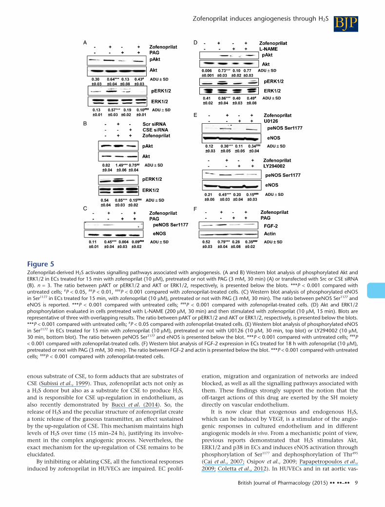
Pre-incubation with PAG (3 mM, 30 min) completelyinhibited the activation of both phospho-Akt and ERK1/2(Figure 5A), demonstrating the crucial role of zofenoprilat-induced released of H2S in the promotion of these earlysignals. Accordingly, the genetic ablation of endogenous CSEprotein significantly inhibited Akt and ERK1/2 phosphoryla-tion (Figure 5B).
As previously demonstrated in CVEC (Donnini et al.,2006; 2010), zofenoprilat promoted activation of the Akt-dependent eNOS pathway in HUVECs. eNOS phosphoryla-tion induced by zofenoprilat was under the control of CSEactivity (Figure 5C). Then, eNOS activation governed ERK1/2,but not Akt phosphorylation, as demonstrated by the treat-ment with L-NAME (200 μM, Figure 5D). This signallingpathway was confirmed by the finding that pretreatment ofcells with U0126 (10 μM), a MEK inhibitor, did not modifyeNOS Ser1177 phosphorylation (Figure 5E). Conversely, pre-treatment with LY294002 (10 μM), a PI3K inhibitor, signifi-cantly reduced eNOS Ser1177 phosphorylation induced byzofenoprilat (Figure 5E).
Finally, zofenoprilat, as previously demonstrated inmicrovascular endothelium (Donnini et al., 2010), inducedan up-regulation FGF-2, which we demonstrated here to beunder the control of CSE/H2S availability (Figure 5F).
Since the vascular functions of H2S have been associatedwith the ATP-dependent K+ channel (Zhao et al., 2001;Papapetropoulos et al., 2009), its role in the occurrence of
0 6 60 VEGF0
20
40
60
80
100
*** *****
Wou
nd a
ssay
(% a
rea
of w
ound
)
0 1 10 1000
50
100
150
***
0 1 10 1000
20
40
60
80 ** **
*
Zofenoprilat (mM)
Adh
esio
n(c
ount
ed c
ells
per
wel
l)
A
B
0 1 10 1000
20
40
60
80
100
*****
Wou
nd a
ssay
(% a
rea
of w
ound
)
C
D
E
F
0 6 60 VEGF0
50
100
150
** ***
0 6 60 VEGF0
20
40
60
80 **
NaHS (mM)
***
Adh
esio
n(c
ount
ed c
ells
per
wel
l)
Zofenoprilat (mM) NaHS (mM)
Prol
ifera
tion
(cou
nted
cel
ls p
er w
ell)
Prol
ifera
tion
(cou
nted
cel
ls p
er w
ell)
NaHS (mM)Zofenoprilat (mM)
Figure 1Effect of zofenoprilat and NaHS on EC adhesion, migration and proliferation. Evaluation of adhesion (40 min) (A and D), migration (18 h) (B andE) and proliferation (5 days) (C and F) in HUVECs in the presence of zofenoprilat (1–100 μM) (A–C) or NaHS (6–60 μM) (D–F). Data in panels (A),(C), (D) and (F) represent counted cells per well. Data in panels (B) and (E) are % area of wound, representing the complement ofhealing/migration at 18 h with respect to time 0. VEGF (20 ng·mL−1) was used as a control. n = 3. *P < 0.05, **P < 0.01, ***P < 0.001 versusuntreated cells.
BJPZofenoprilat induces angiogenesis through H2S
British Journal of Pharmacology (2015) •• ••–•• 5

angiogenesis was investigated. HUVECs, expressing KATP
channels (Papapetropoulos et al., 2009), were pretreated for30 min with glibenclamide (10 μM), and Akt and ERK1/2phosphorylation was evaluated by Western blot. Figure 6Ashows that the pharmacological block of KATP channels par-tially impaired Akt and ERK1/2 activation, suggesting theinvolvement of this transport system in H2S-induced actions.Furthermore, the involvement of KATP in FGF-2 expressionwas evaluated and the results showed that this endogenousgrowth factor is also under the partial control of this channel(Figure 6B). From a functional point of view, blockade of KATP
channels in ECs by glibenclamide reduced the proliferation ofECs induced by zofenoprilat as well as NaHS (Figure 6C).
All these data suggest that zofenoprilat induces the releaseof H2S, which, in turn, activates KATP channels, which, in part,
control Akt and ERK1/2 phosphorylation, FGF-2 expressionand endothelial angiogenic behaviour.
Zofenoprilat, via CSE activation, promotesin vivo angiogenesis and normalizesendothelial monolayersTo further study the H2S-mediated angiogenic activity ofzofenoprilat in vivo, we used the Matrigel implant assay inmice. Matrigel plugs, either with zofenoprilat alone or in thepresence of PAG, were s.c. injected in C57 black mice andharvested after 15 days. Implants containing zofenoprilatshowed several branched structures throughout the implant(Figure 7A, panel b vs panel a). Conversely, in implants inwhich zofenoprilat and PAG were concomitantly adminis-tered, total vascular volume was markedly reduced
Figure 2Zofenoprilat increases CSE protein expression and H2S production. Expression of CSE (A) in HUVECs in control condition and after incubation withzofenoprilat (10 μM) for the indicated times. The blot is representative of three with similar results. The ratio between CSE and actin is presentedbelow the blot. **P < 0.01, ***P < 0.001 compared with untreated cells. Production of H2S (B) was determined in control ECs and in response tozofenoprilat (10 μM) stimulation for 15 min, 2, 4, 8 and 24 h. n = 3. **P < 0.01 versus untreated cells. (C) Expression of CSE in HUVECs in control(Ctr) condition and in response to zofenoprilat (10 μM) in the presence and absence of actinomycin D (5 μg·mL−1) for 2 (a), 4 (b) and 18 (c) h.The blot is representative of three with similar results. The ratio between CSE and actin is presented below the blot. ***P < 0.001 compared withuntreated cells. ###P < 0.01 versus zofenoprilat-treated cells. (D) Production of H2S was determined in response to zofenoprilat stimulation (10 μM)in the presence and absence of actinomycin D (5 μg·mL−1) for 2 (a), 8 (b) and 24 (c) h. n = 3. **P < 0.01, ***P < 0.001 versus untreated cells;#P < 0.05, ##P < 0.01 versus zofenoprilat-treated cells.
BJP E Terzuoli et al.
6 British Journal of Pharmacology (2015) •• ••–••

(Figure 7A, panel d vs. panel b). Quantitative analysis of func-tioning vessels by haemoglobin determination revealed thatzofenoprilat produced a 2.3-fold increase in blood contentcompared with control, whereas administration of PAGreduced it by 42% (Figure 7A, panel e).
However, when designing a pro-angiogenic strategy, thequality and the morphology of newly formed vessels arecrucial parameters as well as the interaction of the stimuluswith known angiogenic factors. To pursue this aim, we per-formed a proliferation assay to evaluate whether zofenoprilatcould interfere with VEGF. Exposing ECs treated with zofeno-prilat (10 μM, 48 h) to a maximal concentration of VEGF,expected to be found in an angiogenic environment(20 ng·mL−1), ) did not enhance the pro-angiogenic response(Table 1).
Since VEGF stimulated vasculature is characterized byelevated permeability and leakage (Ferrara et al., 2003), thereis a strong demand for pharmacological strategies that areable to produce functional angiogenesis (Cai et al., 2011;
Anisimov et al., 2013). In our experiments, zofenoprilatinhibited VEGF-induced permeability in a H2S-dependentmanner (Figure 7B), indicating a potential beneficial role inthe normalization of vasculature. To confirm the capabilityof zofenoprilat to reduce VEGF-mediated permeability,VE-cadherin and ZO-1 protein expression and localization atthe cell–cell junction were evaluated in vitro. As shown inFigure 7C and D (panels a), confluent HUVEC monolayersexpressed VE-cadherin and ZO-1 protein mainly at the cell–cell contact. In the presence of VEGF, the contacts disap-peared (panels b), while VE-cadherin and ZO-1 labelling wasrestored by zofenoprilat (panels d of Figure 7C and D). Theprotective effect of zofenoprilat was related to the availabilityof CSE-dependent H2S, since PAG pre-incubation reversed theprotective effect of zofenoprilat, inducing cell retraction anda reduction in VE-cadherin and ZO-1 protein labelling(panels e of Figure 7C and D).
These data demonstrate that zofenoprilat has pro-angiogenic activity mediated through the production of H2S,
Figure 3Role of H2S production on in vitro angiogenesis in response to zofenoprilat. ECs were incubated with zofenoprilat (10 μM, 40 min or 5 days,respectively), pretreated or not with PAG (3 mM, 30 min), and adhesion (A) or proliferation (B) were assessed. Data represent counted cells perwell. n = 3. *P < 0.05, **P < 0.01 versus untreated cells. #P < 0.05, ##P < 0.01 versus zofenoprilat-treated cells. Cell migration (C) was evaluatedusing scratch wound healing assay on ECs treated with 0.1% FBS (a), zofenoprilat (10 μM, 18 h, b), PAG (3 mM, 30 min pretreatment, c) orzofenoprilat with PAG (d). Representative pictures of wounded EC monolayers are shown. n = 3. Quantification (e) of cell migration presented as% area of wound. ***P < 0.001 versus untreated cells; ##P < 0.01 versus zofenoprilat. (D) Pseudocapillary formation in Matrigel by ECs exposedto 0.1% FBS (a), zofenoprilat (10 μM, b), PAG (3 mM, 30 min pretreatment, c) and PAG + zofenoprilat (d) after 18 h. Cells were labelled withDY554-phalloidin, and quantification (e) of pseudocapillaries was performed by counting the numbers of complete circles per well. n = 3.**P < 0.01 versus untreated cells; ##P < 0.01 versus zofenoprilat.
BJPZofenoprilat induces angiogenesis through H2S
British Journal of Pharmacology (2015) •• ••–•• 7

which controls all the signalling pathways and functionalresponses of the angiogenic vascular endothelium to producefunctional and normalized vessels.
Discussion and conclusions
In the last decade, our attention has been focused on a clini-cally available highly lipophilic ACEI, zofenopril, whoseactive metabolite zofenoprilat bears a SH group and displaysthe unique properties, among other ACEIs, of promoting ECsurvival and angiogenesis, beyond its vasoactive properties(Donnini et al., 2006; 2010; Monti et al., 2013). Recently, ithas been demonstrated that zofenoprilat, the active metabo-lite of zofenopril, spontaneously releases H2S in solution, andthat in an in vivo pathological model, such as the SHR,zofenopril increases H2S levels in the vascular tissue andplasma (Bucci et al., 2014).
The increased availability of H2S, induced by zofenoprilat,prompted us to analyse the involvement of this gaseoustransmitter in the pro-angiogenic and protective effect of thisACEI on endothelium using in vitro and in vivo experimentalmodels associated with angiogenesis. In HUVECs, all thefunctional activities related to angiogenesis in response tozofenoprilat were analysed. HUVECs exposed to zofenoprilatshowed an increase in cell adhesion, migration and prolifera-
tion, with maximal effects at 10 μM, as previously demon-strated in coronary post-capillary endothelium (Donniniet al., 2006; 2010). H2S, given exogenously as NaHS, repro-duced all the effects seen with zofenoprilat. The zofenoprilatR-isomer, a molecule that maintains the SH group but isineffective as an ACEI, replicated these pro-angiogeniceffects. This last finding supports the hypothesis that theangiogenic effect induced by zofenoprilat is dependent uponthe presence of the SH group in its structure and its ability torelease H2S.
Experiments were carried out to further dissect the rela-tionship between zofenoprilat-released H2S and the biosyn-thetic pathway of this gaseous transmitter. The release of H2Sfrom vascular endothelium mainly involves CSE (Kamoun,2004; Kimura, 2010). HUVECs constitutively express CSE,which is further up-regulated by zofenoprilat and itsR-isomer, but not by captopril, as previously demonstrated inCVEC (Monti et al., 2013). Moreover, experiments with thetranscriptional inhibitor actinomycin D showed how theaccumulation of H2S by zofenoprilat is related to gene tran-scription as it is reduced in the presence of actinomycin D.
Another H2S donor drug has also been reported to havethis peculiar property (Ma et al., 2011). Furthermore, it hasrecently been demonstrated that exogenous H2S significantlyincreases the transcription and expression of CSE (Wanget al., 2013). Zofenoprilat reacts with L-cysteine, the endog-
Figure 4Effect of CSE silencing on in vitro angiogenesis induced by zofenoprilat. (A) Western blot analysis of CSE expression in ECs silenced for CSE andtreated for 24 h with zofenoprilat (10 μM). The ratio between CSE and actin is presented. ***P < 0.001 compared with scramble (Scr) cells. (B)Proliferation of ECs transfected with Scr or CSE siRNA and stimulated with zofenoprilat. Data are expressed as cell number counted per well. **P< 0.01 versus Scr siRNA-untreated cells; ##P < 0.01 versus Scr siRNA cells treated with zofenoprilat. (C) Network formation of ECs seeded onMatrigel after transfection with Src siRNA in 0.1% FBS (a), or plus zofenoprilat (b), or with CSE siRNA in 0.1% FBS (c), or plus zofenoprilat (d) for8 h. Cells were labelled with DY554-phalloidin, and quantification (e) of pseudocapillaries was performed by counting the number of completecircles per well. n = 3. **P < 0.01 versus Scr siRNA-untreated cells; ##P < 0.01 versus Scr siRNA zofenoprilat-treated cells.
BJP E Terzuoli et al.
8 British Journal of Pharmacology (2015) •• ••–••
enous substrate of CSE, to form adducts that are substrates ofCSE (Subissi et al., 1999). Thus, zofenoprilat acts not only asa H2S donor but also as a substrate for CSE to produce H2S,and is responsible for CSE up-regulation in endothelium, asalso recently demonstrated by Bucci et al. (2014). So, therelease of H2S and the peculiar structure of zofenoprilat createa tonic release of the gaseous transmitter, an effect sustainedby the up-regulation of CSE. This mechanism maintains highlevels of H2S over time (15 min–24 h), justifying its involve-ment in the complex angiogenic process. Nevertheless, theexact mechanism for the up-regulation of CSE remains to beelucidated.
By inhibiting or ablating CSE, all the functional responsesinduced by zofenoprilat in HUVECs are impaired. EC prolif-
eration, migration and organization of networks are indeedblocked, as well as all the signalling pathways associated withthem. These findings strongly support the notion that theoff-target actions of this drug are exerted by the SH moietydirectly on vascular endothelium.
It is now clear that exogenous and endogenous H2S,which can be induced by VEGF, is a stimulator of the angio-genic responses in cultured endothelium and in differentangiogenic models in vivo. From a mechanistic point of view,previous reports demonstrated that H2S stimulates Akt,ERK1/2 and p38 in ECs and induces eNOS activation throughphosphorylation of Ser1177 and dephosphorylation of Thr495
(Cai et al., 2007; Osipov et al., 2009; Papapetropoulos et al.,2009; Coletta et al., 2012). In HUVECs and in rat aortic vas-
Figure 5Zofenoprilat-derived H2S activates signalling pathways associated with angiogenesis. (A and B) Western blot analysis of phosphorylated Akt andERK1/2 in ECs treated for 15 min with zofenoprilat (10 μM), pretreated or not with PAG (3 mM, 30 min) (A) or transfected with Src or CSE siRNA(B). n = 3. The ratio between pAKT or pERK1/2 and AKT or ERK1/2, respectively, is presented below the blots. ***P < 0.001 compared withuntreated cells; #P < 0.05, ##P < 0.01, ###P < 0.001 compared with zofenoprilat-treated cells. (C) Western blot analysis of phosphorylated eNOSin Ser1177 in ECs treated for 15 min, with zofenoprilat (10 μM), pretreated or not with PAG (3 mM, 30 min). The ratio between peNOS Ser1177 andeNOS is reported. ***P < 0.001 compared with untreated cells; ###P < 0.001 compared with zofenoprilat-treated cells. (D) Akt and ERK1/2phosphorylation evaluated in cells pretreated with L-NAME (200 μM, 30 min) and then stimulated with zofenoprilat (10 μM, 15 min). Blots arerepresentative of three with overlapping results. The ratio between pAKT or pERK1/2 and AKT or ERK1/2, respectively, is presented below the blots.***P < 0.001 compared with untreated cells; #P < 0.05 compared with zofenoprilat-treated cells. (E) Western blot analysis of phosphorylated eNOSin Ser1177 in ECs treated for 15 min with zofenoprilat (10 μM), pretreated or not with U0126 (10 μM, 30 min, top blot) or LY294002 (10 μM,30 min, bottom blot). The ratio between peNOS Ser1177 and eNOS is presented below the blot. ***P < 0.001 compared with untreated cells; ###P< 0.001 compared with zofenoprilat-treated cells. (F) Western blot analysis of FGF-2 expression in ECs treated for 18 h with zofenoprilat (10 μM),pretreated or not with PAG (3 mM, 30 min). The ratio between FGF-2 and actin is presented below the blot. ***P < 0.001 compared with untreatedcells; ###P < 0.001 compared with zofenoprilat-treated cells.
BJPZofenoprilat induces angiogenesis through H2S
British Journal of Pharmacology (2015) •• ••–•• 9

orelaxation, the two gaseous transmitters H2S and NO coop-erate in the fine control of vascular functions (Bucci et al.,2012; 2014; Coletta et al., 2012). In our experiments, themolecular mechanisms underlying the H2S/zofenoprilat-induced angiogenic phenotype in HUVECs are dependent onAkt, eNOS-Ser1177 and ERK1/2. These results confirm datapreviously obtained in CVEC (Donnini et al., 2006; 2010),indicating that H2S released by zofenoprilat is responsiblefor the sequential activation of the Akt/eNOS/ERK1/2 signal-ling pathway, which, in turn, controls the endogenousup-regulation of FGF-2.
All the signalling cascades involved in the effect ofzofenoprilat seem to be dependent on activation of CSE, sincesilencing or pharmacological blockade of CSE blunts the acti-vation of Akt, eNOS-Ser1177 and ERK1/2 and expression ofFGF-2. Some of the vascular functions of H2S have beenattributed to the opening of the KATP channel (Zhao et al.,2001; Zanardo et al., 2006; Li et al., 2011). ECs express KATP
channels both in the plasma membrane and in the intracel-lular organelles (Katnik and Adams, 1997; Malester et al.,2007). The influence of KATP channels on the angiogenic effectof H2S is evident from the data showing that glibenclamideabolishes p38 activation and EC mobility (Papapetropouloset al., 2009; Szabo and Papapetropoulos, 2011). In our model,KATP channels were shown to be partially involved in the
upstream activation of Akt and ERK1/2 and the expression ofFGF-2, including the sulfhydration modification of cysteineresidues in the target downstream proteins, demonstrated toenhance their activity (Mustafa et al., 2009; Yang et al., 2013).
In addition to these findings, zofenoprilat was found topromote neovascularization of Matrigel plugs s.c. implantedin mice. This effect was dependent on a functioning CSEsince the presence of PAG impaired the occurrence of angio-genesis. The stimulation of an angiogenic response might,however, acquire a negative feature when it is induced withinthe tumour environment or in ischaemic diseases, resultingin disorganized and highly permeable vessels (Jain, 2003; Caiet al., 2011; Anisimov et al., 2013). We thus aimed to ascer-tain whether zofenoprilat could cooperate or synergize withangiogenic factors such as VEGF and how it could correctendothelial hyperpermeability. Zofenoprilat did not potenti-ate the EC responses in the presence of VEGF; the concentra-tion of VEGF used was as high as that usually found in anangiogenic environment. On the contrary, in the presence ofVEGF, zofenoprilat restored endothelial permeability to basallevels. Inhibition of endothelial leakage (measured as perme-ability induced by VEGF and expression/localization ofVE-cadherin and ZO-1 protein) is thus an additional positivefeature of zofenoprilat’s effect in the intervention againstcardiovascular diseases. All the related vessel normalization
Figure 6KATP channels are involved in signalling pathways and proliferation induced by zofenoprilat. (A) Western blot analysis of phosphorylated Akt andERK1/2 in HUVECs treated for 15 min with zofenoprilat (10 μM) pretreated or not with glibenclamide (10 μM, 30 min). n = 3. The ratio betweenpAKT or pERK1/2 and AKT or ERK1/2, respectively, is presented. ***P < 0.001 compared with untreated cells; #P < 0.05 compared withzofenoprilat-treated cells. (B) Western blot analysis of FGF-2 in HUVECs treated for 18 h with zofenoprilat (10 μM) pretreated or not withglibenclamide (10 μM, 30 min). n = 3. The ratio between FGF-2 and actin is presented. **P < 0.01 compared with untreated cells; ###P < 0.001compared with zofenoprilat-treated cells. (C) Proliferation of ECs treated with zofenoprilat (10 μM) or NaHS pretreated or not with glibenclamide(10 μM, 30 min). n = 3. ***P < 0.001 versus control (Ctr); ##P < 0.01, ###P < 0.001 versus zofenoprilat or H2S-treated cells.
BJP E Terzuoli et al.
10 British Journal of Pharmacology (2015) •• ••–••

parameters were dependent on H2S production, but the exactmolecular mechanisms that control the restoration oftransendothelial junctions remain to be elucidated. Recently,H2S has been demonstrated to exert a protective effect onlung vascular hyperpermeability by scavenging reactiveoxygen species and activation of Akt (Wang et al., 2012);these mechanisms cannot be excluded in our model.
In conclusion, H2S released from zofenoprilat stimulatesall the angiogenic properties of ECs through a KATP channel/Akt/eNOS/ERK1/2/FGF-2 pathway. Moreover, zofenopril/zofenoprilat induced CSE to generate H2S, a propertyexclusive to this drug with respect to other SH-containingACEI. This extra beneficial effect, beyond ACE inhibition,could be helpful in cardiovascular pathologies where ECfunctions and integrity need to be re-established and func-tional angiogenesis induced.
Figure 7Zofenoprilat/H2S promotes in vivo angiogenesis and controls endothelial hyperpermeability. (A) Representative plugs isolated after 15 days fromimplant of Matrigel containing the following stimuli: none (a), 10 μM zofenoprilat (b), 3 mM PAG (c), zofenoprilat + PAG (d). (e) Quantitativeanalysis of haemoglobin/angiogenesis in implants. For each condition (n = 6), the means ± SD are shown. **P < 0.01 versus untreated plugs; ##P< 0.01 versus zofenoprilat-treated plugs. (B) Cell permeability was evaluated in HUVECs pretreated overnight with zofenoprilat (10 μM) or PAG(3 mM), where indicated, and then treated with VEGF (20 ng·mL−1, 8 h). Results are expressed as fluorescent units ± SD. ***P < 0.001 versusuntreated cells; #P < 0.05, ###P < 0.001 versus VEGF; §P < 0.01 versus VEGF + zofenoprilat. (C and D) Immunofluorescence analysis of VE-cadherin(C) and ZO-1 protein (D) in confluent HUVECs treated with the following stimuli: none (a), 20 ng·mL−1 VEGF (b), 10 μM zofenoprilat (18 h) (c),zofenoprilat (18 h) + VEGF (8 h) (d), pretreatment for 30 min with 3 mM PAG + zofenoprilat (18 h) + VEGF for 8 h (e). Confocal images were takenat 60× magnification. Scale bar = 10 μm.
Table 1Effect of zofenoprilat on the angiogenic activation of endothelium byVEGF
VEGF alone +Zofenoprilat
Proliferation 50 ± 8 52 ± 4
HUVECs were exposed to 20 ng·mL−1 VEGF in the absence orpresence of 10 μM zofenoprilat for 48 h. Proliferation data arereported as % increase over control response, with 100% rep-resenting the cell number counted in the basal condition (n = 3).
BJPZofenoprilat induces angiogenesis through H2S
British Journal of Pharmacology (2015) •• ••–•• 11

Acknowledgements
This work was financed by Menarini Group and by the COSTAction BM1005 (ENOG: European Network on Gasotransmit-ters). Part of this work was funded by Agenzia Spaziale Ital-iana (ASI) and Istituto Toscano Tumori (ITT). M. M. was aNoxamet srl fellow. E. T. was a FIRC (Fondazione Italiana perla Ricerca sul Cancro) fellow.
We are grateful to Dr Cinzia Della Giovampaola for thesupport at confocal microscopy and Dr Valerio Ciccone fortechnical assistance.
Author contributions
E. T. and M. M. performed the research. E. T., M. M., M. Z. andL. M. designed the research study. V. V., M. B. and G. C.contributed essential reagents or tools. E. T., M. M. and L. M.analysed the data. E. T., M. M., M. Z., M. B. and L. M. wrotethe paper.
Conflict of interest
The authors declare no conflict of interest.
ReferencesAlexander SPH, Benson HE, Faccenda E, Pawson AJ, Sharman JL,Spedding M et al. (2013a). The Concise Guide to PHARMACOLOGY2013/14: Ion channels. Br J Pharmacol. 170: 1607–1651.
Alexander SPH, Benson HE, Faccenda E, Pawson AJ, Sharman JL,Spedding M et al. (2013b). The Concise Guide to PHARMACOLOGY2013/14: Enzymes. Br J Pharmacol. 170: 1797–1867.
Anisimov A, Tvorogov D, Alitalo A, Leppänen VM, An Y, Han ECet al. (2013). Vascular endothelial growth factor-angiopoietinchimera with improved properties for therapeutic angiogenesis.Circulation 127: 424–434.
Bucci M, Papapetropoulos A, Vellecco V, Zhou Z, Pyriochou A,Roussos C et al. (2010). Hydrogen sulfide is an endogenousinhibitor of phosphodiesterase activity. Arterioscler Thromb VascBiol 30: 1998–2004.
Bucci M, Papapetropoulos A, Vellecco V, Zhou Z, Zaid A,Giannogonas P et al. (2012). cGMP-dependent protein kinasecontributes to hydrogen sulfide-stimulated vasorelaxation. PLoSONE 7: e53319.
Bucci M, Vellecco V, Cantalupo A, Brancaleone V, Zhou Z,Evangelista S et al. (2014). Hydrogen sulfide accounts for theperipheral vascular effects of zofenopril independently of ACEinhibition. Cardiovasc Res 102: 138–147.
Cai J, Wu L, Qi X, Shaw L, Li Calzi S, Caballero S et al. (2011).Placenta growth factor-1 exerts time-dependent stabilization ofadherens junctions following VEGF-induced vascular permeability.PLoS ONE 6: e18076.
Cai WJ, Wang MJ, Moore PK, Jin HM, Yao T, Zhu YC (2007). Thenovel proangiogenic effect of hydrogen sulfide is dependent on Aktphosphorylation. Cardiovasc Res 76: 29–40.
Coletta C, Papapetropoulos A, Erdelyi K, Olah G, Módis K,Panopoulos P et al. (2012). Hydrogen sulfide and nitric oxide aremutually dependent in the regulation of angiogenesis andendothelium-dependent vasorelaxation. Proc Natl Acad Sci U S A109: 9161–9166.
Cominacini L, Pasini A, Garbin U, Evangelista S, Crea AE,Tagliacozzi D et al. (2002). Zofenopril inhibits the expression ofadhesion molecules on endothelial cells by reducing reactiveoxygen species. Am J Hypertens 15: 891–895.
Donnini S, Solito R, Giachetti A, Granger HJ, Ziche M, Morbidelli L(2006). Fibroblast growth factor-2 mediates angiotensin-convertingenzyme inhibitor-induced angiogenesis in coronary endothelium.J Pharmacol Exp Ther 319: 515–522.
Donnini S, Terzuoli E, Ziche M, Morbidelli L (2010). Sulfhydrylangiotensin converting enzyme inhibitor promotes endothelial cellsurvival through nitric oxide synthase, fibroblast growth factor-2and telomerase cross-talk. J Pharmacol Exp Ther 332: 776–784.
Evangelista S, Manzini S (2005). Antioxidant and cardioprotectiveproperties of the sulphydryl angiotensin-converting enzymeinhibitor zofenopril. J Int Med Res 33: 42–54.
Ferrara N, Gerber HP, Le Couter J (2003). The biology of VEGF andits receptors. Nat Med 9: 669–676.
Jain RK (2003). Molecular regulation of vessel maturation. Nat Med9: 685–693.
Kamoun P (2004). Endogenous production of hydrogen sulfide inmammals. Amino Acids 26: 243–254.
Katnik C, Adams DJ (1997). Characterization of ATP-sensitivepotassium channels in freshly dissociated rabbit aortic endothelialcells. Am J Physiol 272: H2507–H2511.
Kilkenny C, Browne W, Cuthill IC, Emerson M, Altman DG (2010).Animal research: reporting in vivo experiments: the ARRIVEguidelines. Br J Pharmacol 160: 1577–1579.
Kimura H (2010). Hydrogen sulfide: its production, release andfunctions. Amino Acids 41: 113–121.
Li L, Rose P, Moore P (2011). Hydrogen sulfide and cell signaling.Annu Rev Pharmacol Toxicol 51: 169–187.
Liu X, Pan L, Zhou Y, Gong Q, Rose P, Zhu Y (2010).Hypoxia-inducible factor-1α is involved in the pro-angiogenic effectof hydrogen sulfide under hypoxic stress. Biol Pharm Bull 33:1550–1554.
Ma K, Liu Y, Zhu Q, Liu CH, Duan JL, Tan BK et al. (2011). H2Sdonor,S-propargyl-cysteine, increases CSE in SGC-7901 andcancer-induced mice: evidence for a novel anti-cancer effect ofendogenous H2S? PLoS ONE 6: e20525.
McGrath J, Drummond G, McLachlan E, Kilkenny C, Wainwright C(2010). Guidelines for reporting experiments involving animals: theARRIVE guidelines. Br J Pharmacol 160: 1573–1576.
Malester B, Tong X, Ghiu I, Kontogeorgis A, Gutstein DE, Xu J et al.(2007). Transgenic expression of a dominant negative K(ATP)channel subunit in the mouse endothelium: effects on coronaryflow and endothelin-1 secretion. FASEB J 21: 2162–2172.
Monti M, Donnini S, Giachetti A, Mochly-Rosen D, Ziche M (2010).deltaPKC inhibition or epsilonPKC activation repairs endothelialvascular dysfunction by regulating eNOS post-translationalmodification. J Mol Cell Cardiol 48: 746–756.
Monti M, Solito R, Puccetti L, Pasotti L, Roggeri R, Monzani E et al.(2014). Protective effects of novel metal-nonoates on the cellularcomponents of the vascular system. J Pharmacol Exp Ther 351:500–509.
BJP E Terzuoli et al.
12 British Journal of Pharmacology (2015) •• ••–••

Monti M, Terzuoli E, Ziche M, Morbidelli L (2013). The sulphydrylcontaining ACE inhibitor zofenoprilat protects coronaryendothelium from doxorubicin-induced apoptosis. Pharmacol Res76: 171–181.
Mustafa AK, Gadalla MM, Sen N, Kim S, Mu W, Gazi SK et al.(2009). H2S signals through protein S-sulfhydration. Sci Signal 2:ra72.
Osipov RM, Robich MP, Feng J, Liu Y, Clements RT, Glazer HP et al.(2009). Effect of hydrogen sulfide in the porcine model ofmyocardial ischemia-reperfusion: comparison of differentadministration regimens and characterization of the cellularmechanisms of protection. J Cardiovasc Pharmacol 54: 287–297.
Papapetropoulos A, Pyriochou A, Altaany Z, Yang G, Marazioti A,Zhou Z et al. (2009). Hydrogen sulfide is an endogenous stimulatorof angiogenesis. Proc Natl Acad Sci U S A 106: 21972–21977.
Pawson AJ, Sharman JL, Benson HE, Faccenda E, Alexander SP,Buneman OP et al.; NC-IUPHAR (2014). The IUPHAR/BPS Guide toPHARMACOLOGY: an expert-driven knowledgebase of drug targetsand their ligands. Nucl. Acids Res. 42 (Database Issue): D1098–106.
Polhemus DJ, Kondo K, Bhushan S, Bir SC, Kevil CG, Murohara Tet al. (2013). Hydrogen sulfide attenuates cardiac dysfunction afterheart failure via induction of angiogenesis. Circ Heart Fail 6:1077–1086.
Qipshidze N, Metreveli N, Mishra PK, Lominadze D, Tyagi SC(2012). Hydrogen sulfide mitigates cardiac remodeling duringmyocardial infarction via improvement of angiogenesis. Int J BiolSci 8: 430–441.
Stipanuk MH, Beck PW (1982). Characterization of the enzymiccapacity for cysteine desulphhydration in liver and kidney of therat. Biochem J 206: 267–277.
Subissi A, Evangelista S, Giachetti A (1999). Preclinical profile ofzofenopril: an angiotensin converting enzyme inhibitor withpeculiar cardioprotective properties. Cardiovasc Drug Rev 2:115–133.
Suzuki K, Olah G, Modis K, Coletta C, Kulp G, Gerö D et al. (2011).Hydrogen sulfide replacement therapy protects the vascularendothelium in hyperglycemia by preserving mitochondrialfunction. Proc Natl Acad Sci U S A 108: 13829–13834.
Szabo C (2007). Hydrogen sulphide and its therapeutic potential.Nat Rev Drug Discov 6: 917–935.
Szabo C, Papapetropoulos A (2011). Hydrogen sulphide andangiogenesis: mechanisms and applications. Br J Pharmacol 164:853–865.
Terzuoli E, Meini S, Cucchi P, Catalani C, Cialdai C, Maggi CA et al.(2014). Antagonism of bradykinin B2 receptor preventsinflammatory responses in human endothelial cells by quenchingthe NF-κB pathway activation. PLoS ONE 9: e84358.
Vandiver M, Snyder SH (2012). Hydrogen sulfide: a gasotransmitterof clinical relevance. J Mol Med (Berl) 90: 255–263.
Wang M, Guo Z, Wang S (2013). The effect of certain conditions inthe regulation of cystathionine γ-lyase by exogenous hydrogensulfide in mammalian cells. Biochem Genet 51: 503–513.
Wang T, Wang L, Zaidi SR, Sammani S, Siegler J, Moreno-Vinasco Let al. (2012). Hydrogen sulfide attenuates particulate matter-inducedhuman lung endothelial barrier disruption via combined reactiveoxygen species scavenging and Akt activation. Am J Respir Cell MolBiol 47: 491–496.
Yang G, Wu L, Wang R (2005). Proapoptotic effect of endogenousH2S on human aorta smooth muscle cells. FASEB J 20: 553–555.
Yang G, Zhao K, Ju Y, Mani S, Cao Q, Puukila S et al. (2013).Hydrogen sulfide protects against cellular senescence vias-sulfhydration of Keap1 and activation of Nrf2. Antioxid RedoxSignal 18: 1906–1919.
Zanardo RC, Brancaleone V, Distrutti E, Fiorucci S, Cirino G,Wallace JL (2006). Hydrogen sulfide is an endogenous modulator ofleukocyte-mediated inflammation. FASEB J 20: 2118–2120.
Zhao W, Zhang J, Lu Y, Wang R (2001). The vasorelaxant effect ofH2S as a novel endogenous gaseous KATP channel opener. EMBO J20: 6008–6016.
Supporting information
Additional Supporting Information may be found in theonline version of this article at the publisher’s web-site:
http://dx.doi.org/10.1111/bph.13101
Figure S1 Effect of R-isomer of zofenoprilat on endothelialcell functionality. (A) Cell proliferation evaluated in HUVECstreated with zofenoprilat R-isomer (10 μM, 5 days) inpresence/absence of PAG (3 mM, 30 min). Data are expressedas counted cells/well. n = 3; **P < 0.01 versus untreated cellsand #P < 0.05 versus R-isomer. (B) CSE expression was evalu-ated in HUVECs treated with R-isomer (10 μM) for indicatedtimes. The blot is representative of 3 with similar results. Theratio between CSE over actin is reported. ***P < 0.001, com-pared to untreated cells. (C) Pseudocapillary formation inMatrigel by ECs exposed to 0.1% FBS (a), R-isomer (10 μM, b),PAG (3 mM, pretreatment for 30 min, c), and PAG + R-isomer(d) after 18 h. Quantification (e) of pseudocapillaries wasperformed by counting the numbers of complete circles/well.n = 3; ***P < 0.01 versus untreated cells, ###P < 0.01 versusR-isomer.Figure S2 Effect of captopril and enalaprilat on angiogenicparameters. (A) HUVECs were treated with captopril or enal-aprilat (10 μM) with or without PAG (3 mM, 30 min pretreat-ment). Data are expressed as counted cells/well after 5 days ofincubation. n = 3; *P < 0.01 versus untreated cells and #P <0.05 versus R-isomer. (B) Evaluation of network formation ofECs seeded on Matrigel: 0.1% FBS (a), captopril (b), PAG (c),enalaprilat (d) and captopril + PAG (e). Quantification (f) ofpseudocapillaries was performed by counting the numbers ofcomplete circles/well. n = 3; **P < 0.01 versus untreated cells.
BJPZofenoprilat induces angiogenesis through H2S
British Journal of Pharmacology (2015) •• ••–•• 13
Copyright © 2022 FDOKUMEN