Sonic Hedgehog Is a Potent Inducer of Rat Oligodendrocyte Development from Cortical Precursors in...
13
Sonic Hedgehog Is a Potent Inducer of Rat Oligodendrocyte Development from Cortical Precursors in Vitro Kerren Murray, Viviane Calaora, Catherine Rottkamp, Oivin Guicherit,* and Monique Dubois-Dalcq Unite ´ de Neurovirologie et Re ´ge ´ne ´ration du Syste `me Nerveux, Pasteur Institute, Paris, France; and *Curis Incorporated, Cambridge, Massachusetts 02136 Sonic Hedgehog (Shh) induces oligodendrocyte develop- ment in the ventral neural tube and telencephalon but its role in oligodendrocyte generation in dorsal telencepha- lon is debated. Transcripts for Shh and its receptor com- plex were detected in subventricular zone and neocortex from E17 to birth. As Shh is not yet expressed in E15 neocortex, we grew E15 cortical precursors (CP) into neu- rospheres in the presence of recombinant Octyl-Shh (O- Shh). After sphere adhesion and removal of O-Shh, en- hanced neurite outgrowth and cell migration were already observed at 3 h. Three days after O-Shh treatment, oligo- dendrocyte progenitors (OP) emerged and continued to increase in number for 7 days while the ratio of neuronal cells decreased compared to control. Shh selectively trig- gered mitosis of OP but not neuronal progenitors and enhanced growth of neonatal OP. Thus Shh in E15–17 embryonic neocortex can signal CP to adopt an oligoden- drocyte fate and favors expansion of this lineage. INTRODUCTION Sonic Hedgehog (Shh) is a potent morphogen in- volved in the development of limbs and muscles as well as the anterior/posterior organization of the neural tube in vertebrates (Goodrich and Scott, 1998). The 19- kDa aminoterminal fragment of Shh (also called Shh-N) is released by autocatalytic proteolysis and induces graded signaling of neighbour target cells (Marti et al., 1995; Briscoe and Ericson, 1999; McMahon, 2000). Co- valent linkage of cholesterol to Shh C terminus may result in Shh tethering to the cell membrane and plays an important role in long range signaling during pat- terning of chick limb and mouse digits (Zeng et al., 2001; Lewis et al., 2001). On target cells, Shh upregulates expression of Patched (Ptc), a 12 transmembrane do- main protein, which is the ligand binding subunit of the Shh receptor. Binding of Shh to Ptc releases Ptc inhibi- tion of Smoothened (Smo), a seven transmembrane do- main G protein coupled receptor. This transducing sub- unit of Shh triggers a cascade that turns on expression of Shh target genes in the nucleus (Goodrich and Scott, 1998; Ruiz i Atalba, 1999; McMahon, 2000). There is accumulating evidence that Shh is essential to oligodendrocyte development (Miller et al., 1999). In the embryonic chick and mouse spinal cord, a midline transient embryonic structure called the notochord, se- cretes Shh which, in turn, induces Shh synthesis in the floor plate and the emergence of anterior motor neurons (Ericson et al., 1996). Shortly after, oligodendrocyte pro- genitors (OP) emerge on each side of the ventral ven- tricular (VZ) zone, forming two discrete clusters above the floor plate, a pattern conserved from birds to man (Pringle et al., 1996; Poncet et al., 1996; Hajihosseini et al., 1996; Orentas et al., 1999; Richardson et al., 2000). In chick spinal cord, these oligodendrocytes are derived from progenitors expressing the transcription factor Nkx 2.2 (Soula et al., 2001). The territory of Shh expression extends to the ventral diencephalic and telencephalic regions in the medial ganglionic eminence and the zona limitans intrathal- amica which separates diencephalon from telencepha- lon (Rubenstein and Shimamura, 1997). It has been proposed that the majority of rodent telencephalic OP are generated in the ventral neuroepithelium where Shh transcripts are abundant because many cells expressing platelet-derived growth factor receptor (PDGF-R) alpha and the transcription factors Olg1 and 2 are found in this region at E14 (Rowitch et al., 2000; Tekki-Kessaris et al., 2001). Similarly, in chick, a monofocal origin of doi:10.1006/mcne.2001.1079, available online at http://www.idealibrary.com on Molecular and Cellular Neuroscience 19, 320 –332 (2002) MCN 1044-7431/02 $35.00 © 2002 Elsevier Science (USA) All rights reserved. 320
-
Upload
independent -
Category
Documents
-
view
0 -
download
0
Transcript of Sonic Hedgehog Is a Potent Inducer of Rat Oligodendrocyte Development from Cortical Precursors in...

doi:10.1006/mcne.2001.1079, available online at http://www.idealibrary.com on
Molecular and Cellular Neuroscience 19, 320–332 (2002)MCN
Sonic Hedgehog Is a Potent Inducer of RatOligodendrocyte Development from CorticalPrecursors in VitroKerren Murray, Viviane Calaora, Catherine Rottkamp,Oivin Guicherit,* and Monique Dubois-DalcqUnite de Neurovirologie et Regeneration du Systeme Nerveux, Pasteur Institute, Paris,France; and *Curis Incorporated, Cambridge, Massachusetts 02136
Sonic Hedgehog (Shh) induces oligodendrocyte develop-ment in the ventral neural tube and telencephalon but itsrole in oligodendrocyte generation in dorsal telencepha-lon is debated. Transcripts for Shh and its receptor com-plex were detected in subventricular zone and neocortexfrom E17 to birth. As Shh is not yet expressed in E15neocortex, we grew E15 cortical precursors (CP) into neu-rospheres in the presence of recombinant Octyl-Shh (O-Shh). After sphere adhesion and removal of O-Shh, en-hanced neurite outgrowth and cell migration were alreadyobserved at 3 h. Three days after O-Shh treatment, oligo-dendrocyte progenitors (OP) emerged and continued toincrease in number for 7 days while the ratio of neuronalcells decreased compared to control. Shh selectively trig-gered mitosis of OP but not neuronal progenitors andenhanced growth of neonatal OP. Thus Shh in E15–17embryonic neocortex can signal CP to adopt an oligoden-drocyte fate and favors expansion of this lineage.
INTRODUCTION
Sonic Hedgehog (Shh) is a potent morphogen in-volved in the development of limbs and muscles as wellas the anterior/posterior organization of the neuraltube in vertebrates (Goodrich and Scott, 1998). The 19-kDa aminoterminal fragment of Shh (also called Shh-N)is released by autocatalytic proteolysis and inducesgraded signaling of neighbour target cells (Marti et al.,1995; Briscoe and Ericson, 1999; McMahon, 2000). Co-valent linkage of cholesterol to Shh C terminus mayresult in Shh tethering to the cell membrane and playsan important role in long range signaling during pat-terning of chick limb and mouse digits (Zeng et al., 2001;Lewis et al., 2001). On target cells, Shh upregulates
320
main protein, which is the ligand binding subunit of theShh receptor. Binding of Shh to Ptc releases Ptc inhibi-tion of Smoothened (Smo), a seven transmembrane do-main G protein coupled receptor. This transducing sub-unit of Shh triggers a cascade that turns on expressionof Shh target genes in the nucleus (Goodrich and Scott,1998; Ruiz i Atalba, 1999; McMahon, 2000).
There is accumulating evidence that Shh is essentialto oligodendrocyte development (Miller et al., 1999). Inthe embryonic chick and mouse spinal cord, a midlinetransient embryonic structure called the notochord, se-cretes Shh which, in turn, induces Shh synthesis in thefloor plate and the emergence of anterior motor neurons(Ericson et al., 1996). Shortly after, oligodendrocyte pro-genitors (OP) emerge on each side of the ventral ven-tricular (VZ) zone, forming two discrete clusters abovethe floor plate, a pattern conserved from birds to man(Pringle et al., 1996; Poncet et al., 1996; Hajihosseini et al.,1996; Orentas et al., 1999; Richardson et al., 2000). Inchick spinal cord, these oligodendrocytes are derivedfrom progenitors expressing the transcription factorNkx 2.2 (Soula et al., 2001).
The territory of Shh expression extends to the ventraldiencephalic and telencephalic regions in the medialganglionic eminence and the zona limitans intrathal-amica which separates diencephalon from telencepha-lon (Rubenstein and Shimamura, 1997). It has beenproposed that the majority of rodent telencephalic OPare generated in the ventral neuroepithelium where Shhtranscripts are abundant because many cells expressingplatelet-derived growth factor receptor (PDGF-R) alphaand the transcription factors Olg1 and 2 are found inthis region at E14 (Rowitch et al., 2000; Tekki-Kessaris et
expression of Patched (Ptc), a 12 transmembrane do- al., 2001). Similarly, in chick, a monofocal origin of
1044-7431/02 $35.00© 2002 Elsevier Science (USA)
All rights reserved.
telencephalic oligodendrocytes in the anterior entope-duncular area has been described (Olivier et al., 2001).Yet, chick OP appear first in a ventral VZ region andlater in lateral and dorsal germinative zones close todomains of transient expression of Shh, suggesting thatinduction of OP by Shh occurs both in dorsal andventral regions (Ono et al., 1997; Davies and Miller,2001).
We have investigated the role of Shh signaling inoligodendrocyte development in rat forebrain duringlate embryonic life. Using RT/PCR, we found that Shhand its receptor complex are all expressed in SVZ fromE17 to birth (P1) at the time of gliogenesis. To determineif and when Shh signaling can occur in neocortex, weanalyzed transcript expression from E15 to P1: Ptc andSmo were expressed throughout this period while Shhtranscripts, absent at E15, emerged at E17. We thereforegrew E15 cortical precursors (CP) as neurospheres inthe presence of recombinant Shh (O-Shh) and foundthat it stimulated CP migration and neurite outgrowtha few hours after neurosphere adhesion. Remarkably,O-Shh pretreatment of these neurospheres strongly in-creased the number of oligodendrocytes, but not ofneurons, generated from CP from 3 to 10 days afteradhesion. O-Shh selectively increased mitosis of OP butnot of neuronal progenitors. We propose that Shh inE15 to 17 embryonic neocortex can directly signal CP toinduce an oligodendrocyte fate and that it selectivelyenhances cell growth in this lineage.
RESULTS
Expression of Shh Transducing Subunits PrecedesThat of Shh in the Embryonic Neocortex
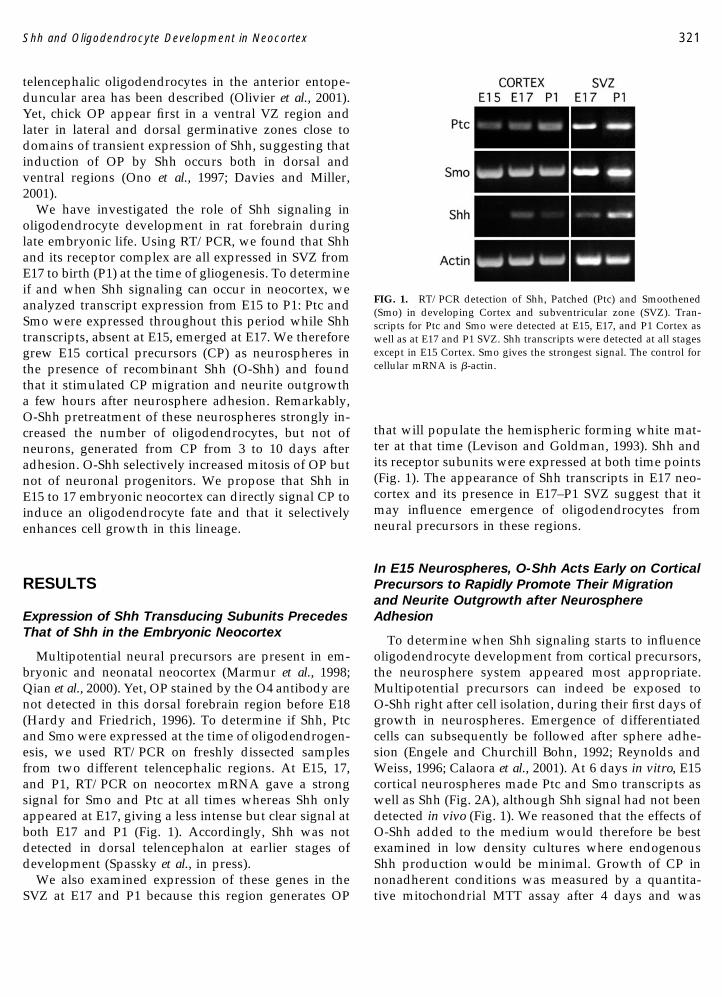
Multipotential neural precursors are present in em-bryonic and neonatal neocortex (Marmur et al., 1998;Qian et al., 2000). Yet, OP stained by the O4 antibody arenot detected in this dorsal forebrain region before E18(Hardy and Friedrich, 1996). To determine if Shh, Ptcand Smo were expressed at the time of oligodendrogen-esis, we used RT/PCR on freshly dissected samplesfrom two different telencephalic regions. At E15, 17,and P1, RT/PCR on neocortex mRNA gave a strongsignal for Smo and Ptc at all times whereas Shh onlyappeared at E17, giving a less intense but clear signal atboth E17 and P1 (Fig. 1). Accordingly, Shh was notdetected in dorsal telencephalon at earlier stages ofdevelopment (Spassky et al., in press).
We also examined expression of these genes in theSVZ at E17 and P1 because this region generates OP
that will populate the hemispheric forming white mat-ter at that time (Levison and Goldman, 1993). Shh andits receptor subunits were expressed at both time points(Fig. 1). The appearance of Shh transcripts in E17 neo-cortex and its presence in E17–P1 SVZ suggest that itmay influence emergence of oligodendrocytes fromneural precursors in these regions.
In E15 Neurospheres, O-Shh Acts Early on CorticalPrecursors to Rapidly Promote Their Migrationand Neurite Outgrowth after NeurosphereAdhesion
To determine when Shh signaling starts to influenceoligodendrocyte development from cortical precursors,the neurosphere system appeared most appropriate.Multipotential precursors can indeed be exposed toO-Shh right after cell isolation, during their first days ofgrowth in neurospheres. Emergence of differentiatedcells can subsequently be followed after sphere adhe-sion (Engele and Churchill Bohn, 1992; Reynolds andWeiss, 1996; Calaora et al., 2001). At 6 days in vitro, E15cortical neurospheres made Ptc and Smo transcripts aswell as Shh (Fig. 2A), although Shh signal had not beendetected in vivo (Fig. 1). We reasoned that the effects ofO-Shh added to the medium would therefore be bestexamined in low density cultures where endogenousShh production would be minimal. Growth of CP innonadherent conditions was measured by a quantita-tive mitochondrial MTT assay after 4 days and was
FIG. 1. RT/PCR detection of Shh, Patched (Ptc) and Smoothened(Smo) in developing Cortex and subventricular zone (SVZ). Tran-scripts for Ptc and Smo were detected at E15, E17, and P1 Cortex aswell as at E17 and P1 SVZ. Shh transcripts were detected at all stagesexcept in E15 Cortex. Smo gives the strongest signal. The control forcellular mRNA is �-actin.
321Shh and Oligodendrocyte Development in Neocortex

found to be slightly increased (about 10%) by treatmentwith 50 and 500 ng/ml of O-Shh (Fig. 2B). This suggeststhat O-Shh is not significantly stimulating CP growth.
We then examined whether exposure of these multi-potential neural precursors to O-Shh for 5 days wouldresult in a particular pattern of migration and differen-tiation after adhesion and withdrawal of O-Shh (thiswill be referred to as O-Shh “primed” neurospheres). At3 h after adhesion of spheres of identical size, manymore O-Shh treated neural precursors had already mi-grated out than in control spheres (Figs. 3A and 3B).The number of cells in the sphere outgrowth was in-creased in a dose-dependent manner, reaching a three-fold increase at 500 ng/ml (�25 nM) and a greater thanfivefold increase over control at the maximal dose of5000 ng/ml of O-Shh (Fig. 3B). As O-Shh did not sig-nificantly increase CP growth (Fig. 2B), it seemed likelythat O-Shh-treated precursors showed enhanced migra-tory properties. Moreover, there was a strong stimula-tion of neurite growth already 3 h after adhesion, witha maximal threefold increase at �25 nM (500 ng/ml) of
O-Shh, a dose 10 times less than the one inducing themost intense migratory response (Figs. 3C and 3D). Asfew neuronal progenitors (stained by TUJ1 antibody)migrated out of the sphere, most of the neurites ap-peared to grow from cells within the spheres (Fig. 3C).Thus early exposure of growing neural precursors toO-Shh triggers enhanced migration and neurite out-growth within hours after adhesion.
Priming Neurospheres with O-Shh Has a PotentBut Delayed Effect on OligodendrocyteDevelopment after Neurosphere Adhesion
The timing of oligodendrocyte development is nor-mally delayed compared to that of neuronal cells. Ac-cordingly, we did not detect O4� cells in controlspheres outgrowth at 3 h. At 3 days very few OP wereobserved (0.4 � 0.2%) even though numerous cells hadmigrated out from neurospheres (101.2 � 5 DAPI-pos-itive cells by field). A different picture emerged in theoutgrowth of O-Shh primed neurospheres after 3 days.First, the total number of cells in the sphere outgrowthwas increased 1.7-fold with O-Shh at 500 ng/ml and2-fold with O-Shh at 5000 ng/ml compared to controlneurospheres. Second, the percentage of O4� cells wasenhanced by, respectively, 12-fold with 500 ng/ml ofO-Shh (5.4 � 0.9%) and 24-fold with 5000 ng/ml ofO-Shh (8.6 � 1.3%). In contrast, the percentage of neu-ronal cells in the outgrowth was around 4% in bothcontrol and O-Shh-treated neurospheres (data notshown). This suggests a specific effect of O-Shh onoligodendrocyte emergence days after neurite out-growth stimulation occurs.
The respective ratio of oligodendrocytic and neuronalcells in neurosphere outgrowth was then measured atlater times. At 7 days postadhesion, the total number ofcells in the outgrowth was still higher after treatmentwith O-Shh (respectively, 1.5- and 2-fold with O-Shh500 ng/ml and O-Shh 5000 ng/ml in comparison withcontrols) and the percentage of highly branched multi-processed O4� oligodendrocytes (Figs. 4F–4H) was in-creased over controls by, respectively, 2.3 and 2.8 times(at 500 and 5000 ng/ml, respectively) (Figs. 4A, 4C, 4D,and 4E). At 10 days, the number of DAPI-labeled cellsper field was equivalent in all experimental conditions(data not shown), but the ratio of differentiated lookingO4� oligodendrocytes to TUJ1� neuronal cells wasover 2 times after O-Shh priming. This indicates that0-Shh priming of CP favors development of oligoden-drocytes over neurons on the long term (Fig. 4B).
As Shh is synthesized in vitro (Tekki-Kessaris et al.,2001; Fig. 2A), endogenous Shh may have taken the
FIG. 2. O-Shh induces little increase in growth of cortical neuro-spheres. (A) RT-PCR analysis preformed on RNA extracted fromneurospheres grown for 6 days showed transcripts for Shh, Ptc, andSmo with maximal intensity for Smo. (B) MTT analysis of neuralprecursors grown for 4 days in presence of different doses of O-Shhshowed a small increase in cell growth in the presence of 50 and 500ng/ml of O-Shh but not with 5000 ng/ml where growth was de-creased compared to control spheres without O-Shh. Each bar repre-sents the mean � SEM of eight wells for each condition in twodifferent experiments.
322 Murray et al.

relay after O-Shh treatment was discontinued to furthertrigger oligodendrocyte development. Nevertheless,the striking increase in O4 � cell ratio and the earlieremergence of oligodendrocytes appears clearly relatedto the initial “priming” by O-Shh of neural precursorsduring their initial growth. Thus the data indicate thatO-Shh can signal growing CP to adopt an oligodendro-cyte fate, yet OP and differentiating oligodendrocyteswill emerge several days later and continue to increasein numbers up to 10 days after O-Shh exposure wasdiscontinued.
O-Shh Increases OP Number by Triggering TheirMitosis in E17 Cortical Explants
Since the first OP are detected at E18 in dorsal neo-cortex in vivo (Hardy and Friedrich, 1996), we examinedwhether O-Shh had oligodendrogenic effects in intactE17 cortex tissue cultured as explants. When O-Shh wasadded to the medium on the first day of explantation,OP appeared in the explant outgrowth by day threecompared to day four in controls. After 5 days (Fig. 5),a large proportion of untreated explant outgrowth con-tained no or few cells labeled with antibodies to GD3, aganglioside highly expressed by OP (Fig. 5A, blackarrow). In contrast, in explants treated with O-Shh,several OP double labeled with O4 and GD3 antibodieswere found in the outgrowth and at the edge of thesphere (Fig. 5B, white arrows).
The effects of increasing doses of O-Shh given for 6days to E17 cortical explants was then studied by count-ing the ratio of OP to total cells in each explant out-growth cultures (Table 1). Surprisingly, we did not finda threshold dose effect with O-Shh as the percentage of
FIG. 3. After adhesion, neurospheres rapidly respond to O-Shhpriming by increased migration and neurite outgrowth. Neuro-spheres were grown for 5 days in the presence of two different O-Shhconcentrations and then washed and adhered for 3 h before immu-nolabeling with O4 and TUJ1 antibodies and counterstaining withDAPI. O-Shh priming of neurospheres caused enhanced migration
(A and B) and neurite outgrowth (C and D). (A) Phase contrastmicrographs: more cells migrated out of O-Shh primed neurospheres(delineated by the circle and excluded from the quantitative analysis)compared to untreated control cultures. (B) The total number of cells(counted by DAPI staining) migrating out of adherent spheres in-creased in a dose dependent manner with a peak response at themaximal dose of 5000 ng/ml. Each bar represents the mean � SEM ofcell numbers in outgrowth of 9 spheres from triplicates in threedifferent experiments. (C) O-Shh primed neurospheres showed, al-ready 3 h after adhesion, an increase in neurite growth (measured bythe TUJ1 stained area) in the absence of O4 � cells at that time. Scalebar, 50 �m. (D) The surface area of TUJI staining correspondingmainly to neurite growth and scattered cells outside the neurosphere(C) was maximal after neurosphere treatment with 500 ng/ml rShh.Each bar represents the mean � SEM of this total surface area in 9 (firstexperiment) and 15 neurospheres (second experiment) from 3 wells ineach experiment. Student test: *P � 0.05; **P � 0.01; ***P � 0.001.
323Shh and Oligodendrocyte Development in Neocortex

OP to total cells was already increased by 17-fold whenexplants were treated with only 50 ng/ml (�2.5 nM) ofO-Shh and, with 100 ng/ml of O-Shh, it reached a22-fold increase over controls. Such large OP increase isin keeping with that observed in O-Shh primed neuro-pheres 3 days after adhesion (see above). The percent-
ages of cells immunostained by the O4 antibody or forNG2, a chondroitin-sulfate proteoglycan characteristicof OP, was closely similar at each dose tested (Table 1).
To determine whether Shh enhances OP numbersthrough a mitogenic effect, BrdU was incorporatedovernight in cortical explants treated with increasing
FIG. 4. O-Shh neurosphere priming has potent oligodendrogenic effects 7–10 days later. Neurospheres were grown in the presence of O-Shh(as described in the legend of Fig. 3) before adhesion for 7 (A and C–H) or 10 days (B) to allow differentiation (after withdrawal of O-Shh) andwere triple labeled as described in the legend of Fig. 3 (A–H). The ratio of oligodendrocytes (O4 in green) and neuronal cells (TUJI in red) tothe total number of cells (blue nuclei) in the outgrowth was determined (A). (A) After 7 days of sphere adhesion, the ratio of O4�oligodendrocytes was considerably increased in neurospheres pretreated with O-Shh, whereas the ratio of neuronal cells stayed the same as incontrols. Each bar represents the mean � SEM of 30–40 random fields around 12 neurospheres in 4 wells for each condition. (B) After 10 daysof sphere adhesion, the ratio of O4 � cells was significantly lower in control adherent spheres compared to O-Shh primed neurospheres and theneuronal cell ratio was more than double in controls compared to spheres pretreated with 500 ng/ml Shh. Each bar represents the mean � SEMof 30–40 different fields per experimental condition from triplicates of two independent experiments. (C–H) Control neurospheres showednumerous red neuronal cells, some with long processes and few green oligodendrocytes (C, F). In contrast O-Shh “primed” neurospheresshowed many more green oligodendrocytes (D, E, G, H). High magnification of neurosphere outgrowth in F, G, and H showed that the processcomplexity characteristic of differentiated oligodendrocytes is observed in all experimental conditions (arrows). Scale bar, 50 �m in C; 25 �m inF. Student test: *P � 0.05; **P � 0.01; ***P � 0.001.
324 Murray et al.

doses of O-Shh for 3 days. Immunostaining for BrdUand GD3 demonstrated a significant 60% increase in OPmitosis at 100 and 1000 ng/ml of O-Shh and a 4.5-foldincrease in BrdU-labeled GD3� OP at 10000 ng/ml(Fig. 5C). In contrast, O-Shh did not produce any sig-nificant DNA synthesis in TUJ1� neuronal progenitorscompared to untreated explants. These data suggestthat O-Shh is a selective mitogen for OP. However, thismitogenic effect was maximal at doses 20-fold those
causing induction of an oligodendrocyte fate in CP (Fig.4), raising some concerns about the physiological rele-vance of the assay.
Shh Is a Selective Mitogen for OP in Cocultures ofCortical Cells with Shh-Expressing Cells
Since the cholesterol modification of Shh facilitatesinteraction with Ptc and association with the cell surface
FIG. 5. O-Shh induces emergence of oligodendrocyte progenitors (OP) from E17 cortical explants and stimulates OP mitosis. (A and B) AfterO-Shh treatment for 5 days, explants were double labeled with GD3 (red) and O4 (green) antibodies. In A, only a few bipolar GD3 (red) and noO4 � cells are seen in the untreated control. In B, the GD3-positive cells are bipolar or bearing a few processes (black arrows), whereas the O4cells (yellow due to double label) are multiprocessed and also present at the edge of the explant (white arrows). Scale bar, 25 �m. (C) Explantstreated with increasing doses of O-Shh were pulsed with BrdU for 18 h on the third day before being fixed and double labeled for BrdU, GD3,or TUJ1 to label dividing OP or neuronal progenitors. The percentage of each progenitor showing BrdU incorporation was then calculated. DNAsynthesis of OP was increased almost two fold at 100 and 1000 ng/ml and over fourfold at 10,000 ng/ml of O-Shh. There was no effect onneuronal progenitor mitosis. Total cells in the outgrowth of three explants were counted for each dose.
325Shh and Oligodendrocyte Development in Neocortex

(Porter et al., 1996; McMahon, 2000), we therefore useda more physiologically relevant assay by seeding dis-sociated E17 CP expressing nestin and/or PSA-NCAM(Figs. 6A and 6B) on QT6 quail cells which had beenstably transfected with Shh (Teillet et al., 1998; Figs. 6Cand 6D). In both cocultures with Shh transfected andcontrol quail cells, GD3� OP and TUJ1 neuronal pro-genitors were observed after 3 days. BrdU assays re-vealed a 60% increase of the ratio of BrdU-labeled GD3�OP cultured on Shh� cells compared to control quailcells, although the latter stimulated OP mitosis to somedegree (Fig. 6E). Such mitogenic response to Shh wasnot observed with TUJ1� neuronal progenitors (Fig.6E).
These results on cocultures of CP with Shh express-ing cells correlate with the explant data (Fig. 5C) andindicate that Shh signaling selectively triggers mitosisof OP but not neuronal progenitors developing fromE17 CP in vitro.
Shh Promotes Growth of NeonatalOligodendrocytes
Purified populations of OP enriched from neonatalbrain cultures also expressed Shh and its receptor com-plex when examined by RT/PCR (Fig. 7A). After aninitial day of culture with PDGF followed by 2 days ofgrowth with O-Shh (without PDGF), the mitochondrialMTT assay revealed a moderate increase in the numberof living cells at 2 days, starting at 500 ng/ml (Fig. 7B,black bars). At 4 days, a stronger increase in living cellswas observed at the highest O-Shh dose (5000 ng/ml),suggesting an effect on growth or survival (Fig. 7B,white bars).
To determine whether Shh can protect cells againstthe programmed cell death (PCD) known to occur inmature oligodendrocytes (Barres et al., 1992; Yasuda etal., 1995), we immunostained these enriched OP popu-lations for activated caspase 3 after O-Shh treatment for4 days. We found that, while the relative number of O4cells had increased in the presence of O-Shh (from66.1 � 2.2% at 50 ng/ml to 71.5 � 2.6% at 500 ng/mlversus 47.4 � 4.2% in control), a minor fraction of O4plus cells expressing activated caspase 3 had also in-creased proportionally with O-Shh (from 8.4 � 0.7% at50 ng/ml to 11.7 � 0.9% at 500 ng/ml versus 5.2 � 0.8%in control). This suggests that O-Shh enhanced OPgrowth at 4 days—as shown by the MTT assay—butthat it did not significantly modify the rate of PCD inthese cells.
DISCUSSION
We have investigated when and how Shh controlsoligodendrocyte development in rat neocortex from E15to birth. While transcripts for Shh and its receptor com-plex were detected in the SVZ and cortical regions fromE17 to P1, only Ptc and Smoothened but not Shh weredetected in E15 neocortex. When E15 dissociated CPwere cultured as neurospheres in the presence of O-Shh, we observed only a small growth increase in con-trast to a marked dose dependent increase in neuralprecursor migration and neurite outgrowth right aftersphere adhesion. Moreover, O-Shh priming of growingneurospheres strongly enhanced the number of differ-entiating oligodendrocytes, but not of neuronal cells, 1week after adhesion and withdrawal of O-Shh. Whencortical explants and dissociated cells were cultured atE17—just before emergence of OP—in the presence ofO-Shh or Shh-expressing cells, Shh selectively increasedproliferation of OP but not neuronal progenitors gen-erated from CP. Our results indicate that Shh acts earlyand directly on CP to induce oligodendrocyte commit-ment, and enhances growth of OP and/or differentiat-ing neonatal oligodendrocytes. Thus Shh signaling ap-pears to play an important role in generation ofoligodendrocytes in the dorsal telencephalic region ofrodents, in agreement with a recent study on chickmetencephalon oligodendrogliogenesis (Davies andMiller, 2001).
During, telencephalon development, there is a corre-lation between territories of Shh expression and oligo-dendrogliogenesis. In chick, intense Shh immunostain-ing is observed in ventral regions at stage 29 (Daviesand Miller, 2001). In E12.5 mice and E13.5 rats, a strong
TABLE 1
Effect of O-Shh on NG2 and O4 Cell Numbersin E17 Cortical Explants
Shh(ng/ml)
Percentage of NG2�/total number of cells
Percentage of O4�/total number of cells
0 0.50 � 0.37 0.50 � 0.3750 8.69 � 3.14 8.39 � 3.07
100 11.09 � 4.19 11.09 � 4.1910000 10.81 � 2.62 9.63 � 2.94
Note. Dose–response to human O-Shh was assayed by staining theexplants with either NG2 or O4 antibodies and calculating the per-centage of OP (stained by either antibody) of the total cells in theoutgrowth. Eight explants were counted for each dose and numbersare given with standard error of the mean. O-Shh induces a �16- to17-fold increase already at 50 ng/ml (2.5 nM), which increases to�22-fold at 100 ng/ml, an inducing effect slightly higher than thatobserved at 10,000 ng/ml.
326 Murray et al.

Shh in situ hybridization signal is detected in ganglioniceminences (Tekki-Kessaris et al., 2001). Accordingly, Ptctranscripts and cells making PDGF-Ralpha or Proteo-lipid/DM20 transcripts, genes highly expressed in sub-populations of OP, are observed in the vicinity of Shhterritory (Spassky et al., 2000; Nery et al., 2001; Tekki-Kessaris et al., 2001). In E12–13 rats, striatal precursorsshow a much higher potential to generate oligodendro-cytes than CP but this difference disappears at E16(Birling and Price, 1998). Moreover, Shh signal is alsodetected in E9.5 and 10.5 mouse rostral telencephalonwhere generation of OP expressing Proteolipid/DM20transcripts is dependent on Shh signaling (Spassky etal., 2001).
In neocortex, results on Shh expression differ be-tween chick and rat. Chick Shh protein is detected atstage 34 in dorsal regions whereas no Shh in situ hy-bridization signal is observed in E18.5 rats cortex (Da-vies and Miller, 2001; Tekki-Kessaris et al., 2001). ByRT/PCR, we detected Shh transcripts starting at E17 inneocortex but transcript abundance may have been toolow for a positive signal by in situ hybridization at thattime. It is possible that a diffusable form of Shh synthe-sized elsewhere has reached neocortical areas to up-regulate Ptc (Zeng et al., 2001). The responsiveness ofcortical cells to recombinant Shh has now been ob-served in several studies. When treated with Shh, chickdorsal explants generated more oligodendrocytes thanthe intermediate region and rat E14.5 CP rapidly up-regulated the oligodendrocyte specific transcription fac-tor Olg1 (Davies and Miller, 2001; Lu et al., 2000). Anti-Shh antibodies were recently shown to block emergenceof OP in mouse E9.5 dorsal explants after 13 days invitro (Spassky et al., in press). Moreover, when ectopicexpression of Shh was induced by retroviral transfer inE9.5 forebrain, the dorsal telencephalic region becameenlarged 5 days later and the large majority of Shhtransduced cells developed into oligodendrocytes ex-pressing myelin genes 21 days after birth (Nery et al.,2001). Taken together with our present report of strongenhancement of oligodendrocyte development from CPby O-Shh, these studies demonstrate a potent oligoden-drocyte-inducing activity of Shh in dorsal telencepha-lon of birds and rodents.
Our observation of rapid emergence of cortical cellswith a neuronal phenotype from O-Shh primed neuro-spheres is in keeping with the well known effect of Shhon neuronal development in the neural tube (Ericson etal., 1996; Briscoe and Ericson, 1999). The rapid stimula-tion of neurite outgrowth in adherent neurospheresprimed at 25 nM of O-Shh was particularly striking. Theenhanced migration of CP from the sphere induced by
O-Shh correlates with observations on chick granuleneuron migration from cerebellar explants (Dhamaneand Ruiz i Altaba, 1999). Moreover, Nkx2.1 mutants,which lack Shh expression in several telencephalic re-gions, showed defect in migration from median gangli-onic eminence explants (Nery et al., 2001). Thus, inventral regions where Shh is intensely expressed, thismorphogen could induce not only oligodendrocytespecification but also initial waves of precursor migra-tion toward lateral regions. In dorsal regions, inductionof oligodendrocyte fate and precursor migration by Shhmight be closely related events contributing to comple-tion of oligodendrogenesis (Davies and Miller, 2001).
This raises the question of the identity of the cellsproducing Shh in germinative and cortical regions atthe time of gliogenesis in the forebrain. Anti-Shh anti-bodies could partially neutralize the generation of oli-godendrocytes from neonatal SVZ explants (our un-published observations). This suggests that Shh is madeby multipotential precursors in germinative zones asalso observed in cultured neurospheres (Fig. 2A). Al-ternatively, Shh could be made by another cell type inthese regions or by differentiating neurons. Indeed, Shhmade by retinal ganglia neurons signals astrocytes inoptic nerve and Purkinje cell-derived Shh acts on cere-bellar granule neuron progenitors (Wallace and Raff,1999; Wallace, 1999; Wechsler-Reya and Scott, 1999).Although we have not identified the cells that synthe-size Shh in E17 cortex, the most likely candidates areneurons which are known to make Shh transcripts inthe postnatal and adult stage (Traiffort et al., 1998,1999). Shh is synthesized by CNS explants, dissociatedor purified OP as soon as they are cultured and cyclo-pamine, which interferes with Shh signaling, inhibitsoligodendrocyte generation in vitro (Incardona et al.,2000a; Tekki-Kessaris et al., 2001). Similarly, we foundthat neonatal OP synthesize Shh transcripts in vitrosuggesting that Shh signaling could occur between OPwithin the cultured population, resulting in enhancedgrowth.
Shh can signal survival and/or mitosis in differentneural cell types. Shh promotes the survival of dopa-minergic or GABA interneurons in midbrain andcraniofacial neural crest cells (Miao et al., 1997; Ahlgrenand Bronner-Fraser, 1999; Britto et al., 2000). Retinal andcerebellar granule precursors are stimulated to divideby Shh (Jensen and Wallace, 1997; Wechsler-Reya andScott, 1998; Dahmane and Ruiz i Altaba, 1999; Wallace,1999). Increased precursor proliferation was observedin spinal cord of transgenic mice overexpressing Shh inthe dorsal midline region, resulting in emergence ofPDGF-Ralpha� OP in this regions (Rowitch et al., 1999).
327Shh and Oligodendrocyte Development in Neocortex

Other studies showed synergism of Shh with othermitogenic factors such as FGF2 on forebrain precursors(Zhu et al., 1999). Our results show that Shh is a selec-tive mitogen for OP derived from E17 rat CP as alsoobserved recently in mouse dorsal brain where it mod-ulates cell proliferation (Dahmane et al., 2001). In addi-tion, the present study confirms the observation that
Shh is mitogenic for OP purified from developing chickbrain (Davies and Miller, 2001). In our neurosphereexperiments, it is possible that Shh first induced CPmigration and neurite outgrowth which, in turn, trig-gered specification and mitosis of OP.
The hydrophobic octyl modification of the aminoter-minus of the O-Shh used in this study considerablyincreased its potency over unmodified O-Shh althoughthe latter can also signal Ptc (Taylor et al., 2001). In chickneural tube explants, rapid receptor-mediated endocy-tosis occurs when Shh binds to Ptc on the receiving cell,creating a gradient of cells with different levels of in-ternalized Shh (Incardona et al., 2000b). Little is knownabout the respective role of short and long range sig-naling in oligodendrocyte generation in the forebrain.Shh diffusion in intact tissue is indeed difficult to assayas the potent cholesterol modified and multimeric formof Shh is undetectable by current antibodies (Zeng et al.,2001).
In conclusion, our study shows that Shh is a stronginducer of oligodendrocyte fate and development fromrat CP isolated during late embryonic life. As observedin chick CNS development (Davies and Miller, 2001),
FIG. 6. Coculture of dissociated cortical precursors with Shh ex-pressing cells also induces mitosis of oligodendrocyte but not neuro-nal progenitors. (A–D) Monolayers of QT6 quail cells expressing Shhseeded with cortical precursors for 5 h, express nestin (A, green) andPSA-NCAM (B, red) or mostly PSA-NCAM (black arrows, A, B).Control quail cells (C) and Shh-expressing quail cells (D) were labeledwith anti-Shh antibody 5E1 (green), which specifically stained thesurface of Shh transfected cells in D. Scale bar, 25 �m. (E) Three daysafter coculture with Shh expressing cells, the number of corticalGD3 � cells in DNA synthesis is significantly increased, while noincrease in mitosis is observed in TUJ1 � neuronal progenitors. Fortyrandom fields were counted per condition from three independentexperiments, bars represent SEM.
FIG. 7. O-Shh increases growth of neonatal OP expressing the Shhreceptor complex. (A) RT-PCR analysis detected transcripts for Ptc,Smo, and Shh in RNA from oligodendrocyte progenitors (OP) puri-fied from mixed neonatal primary cultures. Control is �Actin. (B)MTT analysis of postnatal OP grown for 2 or 4 days in presence ofincreasing doses of O-Shh showed a significant increase in cell growthin the presence of 500 and 5000 ng/ml of O-Shh at 2 days. By day 4,there was a fourfold increase in cell numbers over controls at 5000ng/ml of O-Shh. Each bar represents the mean � SEM of 6 wells.
328 Murray et al.

there is an important delay between Shh exposure andthe onset of rat oligodendrocyte development. The fulldifferentation of CNS myelin-forming cells clearly re-quires other factors to inhibit specific signaling systemsand stop mitosis before setting into motion axon en-wrapment and myelination (Barres and Raff, 1999; Ro-gister et al., 1999; Calaora et al., 2001).
EXPERIMENTAL METHODS
RT-PCR Analysis
Cortex or SVZ tissue was dissected from E17 and P1Sprague–Dawley rats euthanized in agreement withanimal regulations at the Pasteur Institute. The mRNAwas extracted using the Qiagen RNeasy Mini-Prep kitand reverse transcription performed using SuperScriptII reverse transcriptase (Gibco/BRL). Primers were de-signed from rat sequences for Shh (Tekki-Kessaris et al.,2001) and Smo and from human sequences for Ptc. Thefollowing primer sets and annealing conditions wereused through 35 cycles.
Shh: 5�-GTGATGAACCAGTGGCCTGG-3� and 5�-GCCGCCACGGAGTTCTCTGC-3�, 64°C.
Ptc: 5�-CGCTGTCTTCCTTCTGAACC-3� and 5�-AT-CAGCACTCCCAGCAGAGT-3�, 60°C.
Smo: 5�-TGGAGTTGCCCAGCCGTACC-3� and 5�-CGCCCCGAAGGCTGCGATGT-3�, 64°C.
The recombinant human Shh protein. Shh had itsactive N-terminal peptide modified by addition of ahydrophobic eight-carbon octyl chain to the N terminalcysteine (N-octylmaleimide in Table 1 of Taylor et al.,2001) and is called octyl-Shh (O-Shh). This O-Shh was40 times more active than the unmodified form in theassay of osteoblast formation from a fibroblastic cell line(Taylor et al., 2001). In organotypic assays, concentra-tions of 100 to 500 ng/ml (�5 to 25 nM) showed induc-ing activities although the skin-punch assay may re-quire 2–5 �g/ml (Guicherit, personal communication).In neural cells, different Octyl-Shh batches showedvariations in biological activity, perhaps because mostneural cell assays were run in the absence of serum thatstabilizes the Shh protein. Each batch of O-Shh was 1mg/ml in 1 part of A buffer (5 mM NaPO4, pH 5.5, 150mM NaCl, 0.5 mM dithiotreitol) and 9 parts of PBS. Itwas stored at minus 80°C in small aliquots until use andaddition to the medium at concentrations between 50and 10000 ng/ml every 48 h. Of note is that some Shhlots were in limited supplies, which accounts for differ-
ences in the range and number of concentrations thatwere used in our experiments. Preliminary assays weregenerally performed with each batch to determine theoptimal range to work with in experiments shown inthis paper.
Neurosphere Cultures from E15 Cortex
The neocortex of E15 rat embryos were removed asfor explant culture and mechanically dissociated beforeplating cells as previously described (Calaora et al.,2001). Briefly, 2 � 105 viable cells/ml were seeded inuntreated 96-well and 24-well culture plates (respec-tively Sarstetd, Orsay, France and Falcon, Plymouth,UK). Culture medium consisted of DMEM/F12 (1:1,v/v) supplemented with B27 (both from Life Technol-ogy, Eragny, France) and 20 ng/ml of EGF (Sigma, SaintLouis, MO) with or without O-Shh. After 2 days, two-thirds of the media was replaced with the same freshmedia.
Adhesion of neurospheres. After 5 days in culture,O-Shh and EGF were removed by neurosphere centrif-ugation and washing in serum free medium. Threeneurospheres of the same size (80 �m in diameter) wereplated onto 12-mm coated coverslips (poly-d-lysine/fibronectin; 10 �g/ml each; Sigma) and 2 ml of serumfree medium was then added to each well.
Cultures of Cortical Explant
Cortices from E17.5 rat were separated from the in-ternal hemispheric and periventricular zone tissues inice cold MEM/Hepes medium. After dissection the me-dium was removed and the tissue was chopped intofiner explants with a scalpel. The explants were platedon 25 �g/ml poly-l-lysine or polylysine � 10 �g/mlfibronectin coated coverslips in 50 �l of N1 medium (50�g/ml transferrin, 100 �M putrescine, 20 nM proges-terone, 30 nM selenium, 5 �g/ml insulin in DMEM)supplemented with 30 nM 3,3�,5-triiodo-l-thyronine so-dium salt and 10 ng/ml d-biotin. The explants wereincubated overnight (10% CO2/37°C) and then floodedwith the same medium. All media were from GIBCOand reagents from Sigma.
Culture of Dissociated CNS Cells and Quail Cells
E17 cortex tissues were enzymatically dissociated in0.025% trypsin/EDTA at 37°C for 15 min. The suspen-sion was triturated in the presence of 0.25 mg/mlDNAse in HBSS. 50,000 cells were then plated in 100 �ldrops on 25 �g/ml poly-l-lysine-coated coverslips and
329Shh and Oligodendrocyte Development in Neocortex

incubated for 30 min. After adhesion, we added 400 �lof N1 medium (supplemented with 10 ng/ml of IGF-1from R&D Systems and 0.5% fetal bovine serum FBS).
Monolayers of QT6 quail fibroblasts (a gift of N.Pringle, University College London) and Shh trans-fected quail fibroblasts (a gift of D. Duprez, Institutd’Embryologie Cellulaire et Moleculaire, Nogent-sur-Marne, France; Teillet et al., 1998) were prepared in24-well trays in 10% FBS to be 50% confluent on the dayof plating dissociated E17 cortex cells. Cultures wererinsed twice in N1 medium before plating 25 or 50,000cortex cells per well. Medium was supplemented asabove with T3, Biotin, and B27 (1/50 GIBCO).
Oligodendrocyte progenitors were prepared frommixed primary cultures of P1 rat telencephalon, treatedat 7–9 days with 5 mM leucine methylester for 15 min at37°C then shaken overnight as described (McCarthyand DeVellis, 1980). Cells were plated in polyornithinecoated 96-well trays at 15,000 cells/well for 3 h in 5%FBS before switching to N2 (GIBCO) supplementedwith biotin and T3 (as above), 0.5% FBS, and 10 ng/mlPDGFAA (R&D Systems) for 24 h (McKinnon et al.,1993). PDGF was then washed out and replaced with0-Shh for 2 or 4 days when proliferation was assayed byMTT (see below).
Immunocytochemistry of Cell Cultures
Explants and cell cultures were fixed at 4–6 days afterexplantation and/or Shh treatment in 4% formaldehydein phosphate-buffered saline solution (PBS; pH 7.4) for15–20 min at room temperature. Neurospheres were fixedat different times after adhesion (3 h, 3, 7, and 10 days) for30 min at room temperature. Nonspecific binding siteswere blocked with 5% normal goat serum in MEM/Hepesmedium for 30 min at room temperature and were thenincubated in appropriate primary antibodies for either 1 hat room temperature or overnight at 4°C. The monoclonalmouse 04 IgM (Boehringer Mannheim at 1:5) and GD3IgG3 (gifts of Drs. J. Goldmann, Columbia University, NY,L. J. Old, Ludwig Institute for Cancer Research, NY, andG. Wolswijk, Institute of Brain Research, The Netherlands)recognize oligodendrocyte progenitors (OP) and wereused as described (Pfeiffer et al., 1993; Ben Hur et al., 1998).The NG2 antibody (a gift of Joel Levine, SUNY, NY) is amouse monoclonal IgG recognizing an epitope close to theN Terminal of the NG2 chondroitin sulfate proteoglycanand it labels OP in the developing and adult brain (Levineet al., 2001). The TUJI antibody recognizes neuronal pro-genitors and is a mouse IgG2a monoclonal antibodyagainst neuron specific Class III �-Tubulin (BAbCO, Rich-mond, CA; 1:500). The polyclonal rabbit antibody to
caspase-3 recognizes CPP32 cleaved product 17–20 kDa(Cell signaling Tech, MA). The nuclear marker DAPI (0.1�g/ml) was from Sigma. The cells were then rinsed in PBSand incubated in the appropriate secondary antibodies for30 min and with DAPI at room temperature (Ben Hur etal., 1998; Calaora et al., 2001). The coverslips weremounted in DABCO (1,4-diazobicyclo-(2,2,2) octane,SIGMA) and preparations were viewed and photo-graphed using a DMRB Leica fluorescence microscopeequipped with a Coolsnap Photometrics camera (Prince-ton Instrument, Evry, France) and an Axiovert 135 in-verted Zeiss microscope equipped with a Zeiss Axiocamcamera (Zeiss, Le Pecq, France).
BrdU Mitogen Assay
Bromodeoxyuridine (BrdU) was added to cell cul-tures at 10 �M for 24 h before fixation in 4% parafor-maldehyde. Cells were then immunolabeled as de-scribed (Ben Hur et al., 1998).
MTT Assay
To test the effect of exogenous Shh on NP growth,MTT reduction assay was used as previously described(Calaora et al., 2001). Two and 4-day cultures wereincubated with 1 �g/ml of 3-(4,5-Dimethylthiazol-2-yl)-2,5-diphenyl tetrazolium bromide (MTT, Sigma) for 4 hat 37°C. One hundred microliters of dissolving solutionwas added to the wells before measurement of theoptical density (O.D.) at both 570 and 630 nm. Back-ground absorbance of medium in the absence of cellswas subtracted.
Quantitative Analysis
Neurospheres. Quantitation was done with the Ax-ioscan camera and the Metamorph software imaging sys-tem (Version 4.6, Universal Imaging Corporation, Down-ingtown, PA). To study early migration at 3 h afterneurosphere adhesion, we recorded images of TUJI andDAPI staining of the entire outgrowth of neurospheres foreach O-Shh concentration and counted the nuclei andmeasured the surface area of the TUJI outgrowth. Tostudy differentiation in adherent neurospheres at 3, 7, and10 days, we recorded images of 30–40 randomly chosenfields at 40� magnification for each experimental condi-tion and at 20� magnification for 3 days in adhesion. Wethen counted the total number of TUJI and O4 positivecells as well as the number of DAPI positive cells percondition and calculated the ratio of differentiated cellsper field as mean � SEM.
330 Murray et al.

Explants. In the O-Shh assays, total cells and all OPwere counted around eight explants of each experimen-tal condition and the ratio of OP per total cell wascalculated for each explant as mean plus SEM. For theBrdU assays, we counted the number of GD3 (OP) orTUJ1 (NP) positive cells with BrdU-labeled nuclei andthe total cell number surrounding each of 6–12 theexplants. We then calculated the percentage of dividingprogenitors in the presence or absence of O-Shh.
Dissociated cells/cocultures. For each of three exper-iments, 10 random fields were counted on 4 coverslips ofeach condition and the percentage of GD3 or TUJ1 labeledcells which incorporated BrdU was calculated.
Wherever appropriate, the differences between con-trol and experimental populations were subjected to theone tailed Student’s t test of significance. A value of P �0.05 is considered statistically significant (*P � 0.05;**P � 0.01; ***P � 0.001).
ACKNOWLEDGMENTS
This work was supported by Grant 0480/I 16298 from the MultipleSclerosis Society of Great Britain and Northern Ireland to M. Dubois-Dalcq and Viviane Calaora and by the European Community contractQLG3-CT-200-00911. Catherine Rottkamp had a Fulbright fellowshipduring her tenure at the Pasteur Institute and is presently at theInstitute of Pathology, Case Western Reserve University, 2085 Adel-bert Road, Cleveland, Ohio, 44106. We thank Roberto Bruzzone forcritical reading of the manuscript.
REFERENCES
Ahlgren, S. C., and Bronner-Fraser, M. (1999). Inhibition of sonichedgehog signaling in vivo results incraniofacial neural crest celldeath. Curr Biol. 9: 1304–1314.
Barres, B. A., Hart, I. K., Coles, H. S., Burne, J. F., Voyvodic, J. T.,Richardson, W. D., and Raff, M. C. (1992). Cell death and control ofcell survival in the oligodendrocyte lineage. Cell 70: 31–46.
Barres, B. A., and Raff, M. C. (1999). Axonal control of oligodendro-cyte development. J. Cell Biol. 147: 1123–1128.
Ben-Hur, T., Rogister, B., Murray, K., Rougon, G., and Dubois-Dalcq,M. (1998). Growth and fate of PSA-NCAM� precursors of thepostnatal brain. J. Neurosci. 18: 5777–5788.
Birling, M. C., and Price, J. (1998). A study of the potential of theembryonic rat telencephalon to generate oligodendrocytes. Dev.Biol. 193: 100–113.
Briscoe, J., and Ericson, J. (1999). The specification of neuronal identityby graded Sonic Hedgehog signalling. Semin. Cell Dev. Biol. 10:353–362.
Britto, J. M., Tannahill, D., and Keynes, R. J. (2000). Life, death andSonic hedgehog. Bioessays 22: 499–502.
Calaora, V., Rogister, B., Bismuth, K., Murray, K., Brandt, H.,Leprince, P., Marchionni, M., and Dubois-Dalcq, M. (2001). Neu-regulin signaling regulates neural precursor growth and the gen-eration of oligodendrocytes in vitro. J. Neurosci. 21: 4740–4751.
Dahmane, N., and Ruiz-i-Altaba, A. (1999). Sonic hedgehog regulates
the growth and patterning of the cerebellum. Development 126:3089–3100.
Dahmane, N., Sanchez, P., Palma, V., Sun, T., Beyna, M., Weiner, H., andRuiz i Altaba, A. (2001). The sonic hedgehog-Gli pathway regulatesdorsal brain growth and tumorigenesis. Development 128: 5201–5212.
Davies, J. E., and Miller, R. H. (2001). Local sonic hedgehog signalingregulates oligodendrocyte precursor appearance in multiple ven-tricular zone domains in the chick metencephalon. Dev. Biol. 233:513–525.
Engele, J., and Churchill Bohn, M. (1992). Effects of acidic and basicfibroblast Growth factors (aFGF, bFGF) on glial precursor prolifer-ation: Age dependency and brain region specificity. Dev. Biol. 152:363–372.
Ericson, J., Morton, S., Kawakami, A., Roelink, H., and Jessell, T. M.(1996). Two critical periods of Sonic Hedgehog signaling requiredfor the specification of motor neuron identity. Cell 87: 661–673.
Goodrich, L. V., and Scott, M. P. (1998). Hedgehog and patched inneural development and disease. Neuron 21: 1243–1257.
Hajihosseini, M., Tham, T. N., and Dubois-Dalcq, M. (1996). Origin ofoligodendrocytes within the human spinal cord. J. Neurosci. 16:7981–7994.
Hardy, R. J., and Friedrich, V. L. (1996). Oligodendrocyte progenitorsare generated throughout the embryonic mouse brain, but differ-entiate in restricted foci. Development 122: 2059–2069.
Incardona, J. P., Gaffield, W., Lange, Y., Cooney, A., Pentchev, P. G.,Liu, S., Watson, J. A., Kapur, R. P., and Roelink, H. (2000a). Cyclo-pamine inhibition of Sonic Hedgehog signal transduction is notmediated through effects on cholesterol transport. Dev. Biol. 224:440–452.
Incardona, J. P., Lee, J. H., Robertson, C. P., Enga, K., Kapur, R. P., andRoelink, H. (2000b). Receptor-mediated endocytosis of soluble andmembrane-tethered Sonic hedgehog by Patched-1. Proc. Natl. Acad.Sci. USA 97: 12044–12049.
Jensen, A. M., and Wallace, V. A. (1997). Expression of Sonic hedge-hog and its putative role as a precursor cell mitogen in the devel-oping mouse retina. Mech Dev. 124: 363–371.
Levine, J. M., Reynolds, R., and Fawcett, J. W. (2001). The oligodendro-cyte precursor cell in health and disease. Trends Neurosci. 24: 39–47.
Levison, S. W., and Goldman, J. E. (1993). Both oligodendrocytes andastrocytes develop from progenitors in the subventricular zone ofpostnatal rat forebrain. Neuron 10: 201–212.
Lewis, P. M., Dunn, M. P., McMahon, J. A., Logan, M., Martin, J. F.,St-Jacques, B., and McMahon, A. P. (2001). Cholesterol modificationof sonic hedgehog is required for long-range signaling activity andeffective modulation of signaling by Ptc1. Cell 105: 599–612.
Lu, Q. R., Yuk, D., Alberta, J. A., Zhu, Z., Pawlitzky, I., Chan, J.,McMahon, A. P., Stiles, C. D., and Rowitch, D. H. (2000). Sonichedgehog—Regulated oligodendrocyte lineage genes encodingbHLH proteins in the mammalian central nervous system. Neuron25: 317–329.
Marmur, R., Kessler, J. A., Zhu, G., Gokhan, S., and Mehler, M. F.(1998). Differentiation of oligodendroglial progenitors derived fromcortical multipotent cells requires extrinsic signals including acti-vation of gp130/LIFbeta receptors. J. Neurosci. 18: 9800–9811.
Marti, E., Bumcrot, D. A., Takada, R., and McMahon, A. P. (1995).Requirement of 19K form of Sonic hedgehog for induction of dis-tinct ventral cell types in CNS explants. Nature 375: 322–325.
McCarthy, K., and De Vellis, J. (1980). Preparation of separate astro-glial and oligodendroglia cell cultures from rat cerebral tissue.J. Cell Biol. 85: 890–902.
McKinnon, R. D., Smith, C., Behar, T., Smith, T., and Dubois-Dalcq, M.
331Shh and Oligodendrocyte Development in Neocortex
(1993). Distinct effects of bFGF and PDGF on oligodendrocyteprogenitor cells. Glia 7: 245–254.
McMahon, A. P. (2000). More surprises in the Hedgehog signalingpathway. Cell 100: 185–188.
Miao, N., Wang, M., Ott, J. A., D’Alessandro, J. S., Woolf, T. M.,Bumcrot, D. A., Mahanthappa, N. K., and Pang, K. (1997). Sonichedgehog promotes the survival of specific CNS neuron popula-tions and protects these cells from toxic insult in vitro. J. Neurosci.17: 5891–5899.
Miller, R. H., Hayes, J. E., Dyer, K. L., and Sussman, C. R. (1999).Mechanisms of oligodendrocyte commitment in the vertebrateCNS. Int. J. Dev. Neurosci. 17: 753–763.
Nery, S., Wichterle, H., and Fishell, G. (2000). Sonic hedgehog con-tributes to oligodentrocyte specification in the mammalien fore-brain. Development 128: 527–540.
Olivier, C., Cobos, I., Perez Villegas, E. M., Spassky, N., Zalc, B.,Martinez, S., and Thomas, J.-L. (2001). Monofocal origin of telence-phalic oligodendrocytes in the anterior entopeduncular area of thechick embryo. Development 128: 1757–1769.
Ono, K., Yasui, Y., Rutishauser, U., and Miller, R. H. (1997). Focalventricular origin and migration of oligodendrocyte precursors intothe chick optic nerve. Neuron 19: 283–292.
Orentas, D. M., Hayes, J. E., Dyer, K. L., and Miller, R. H. (1999). Sonichedgehog signaling is required during the appearance of spinalcord oligodendrocyte precursors. Development 126: 2419–2429.
Pfeiffer, S. E., Warrington, A. E., and Bansal, R. (1993). The oligoden-trocyte and its many cellular processes. Trends Cell. Biol. 3: 191–197.
Poncet, C., Soula, C., Trousse, F., Kan, P., Hirsinger, E., Pourquie, O.,Duprat, A. M., and Cochard, P. (1996). Induction of oligodendro-cyte progenitors in the trunk neural tube by ventralizing signals:Effects of notochord and floor plate grafts, and of sonic hedgehog.Mech. Dev. 60: 13–32.
Porter, J. A., Young, K. E., and Beachy, P. A. (1996). Cholesterolmodification of hedgehog signaling proteins in animal develop-ment. Science 274: 255–259.
Pringle, N. P., Yu, W. P., Guthrie, S., Roelink, H., Lumsden, A.,Peterson, A. C., and Richardson, W. D. (1996). Determination ofneuroepithelial cell fate: Induction of the oligodendrocyte lineageby ventral midline cells and sonic hedgehog. Dev. Biol. 177: 30–42.
Qian, X., Shen, Q., Goderie, S. K., He, W., Capela, A., Davis, A. A., andTemple, S. (2000). Timing of CNS cell generation: A programmedsequence of neuron and glial cell production from isolated murinecortical stem cells. Neuron 28: 69–80.
Reynolds, B. A., and Weiss, S. (1996). Clonal and population analysesdemonstrate that an EGF-responsive mammalian embryonic CNSprecursor is a stem cell. Dev. Biol. 175: 1–13.
Richardson, W. D., Smith, H. K., Sun, T., Pringle, N. P., Hall, A., andWoodruff, R. (2000). Oligodendrocyte lineage and the motor neu-ron connection. Glia 12: 136–142.
Rogister, B., Ben-Hur, T., and Dubois-Dalcq, M. (1999). From neuralstem cells to myelinating oligodendrocytes. Mol. Cell. Neurosci. 14:287–300.
Rowitch, D. H., S-Jacques, B., Lee, S. M., Flax, J. D., Snyder, E. Y., andMcMahon, A. P. (1999). Sonic hedgehog regulates proliferation andinhibits differentiation of CNS precursor cells. J. Neurosci. 19: 8954–8965.
Rubenstein, J. L., and Shimamura, K. (1997). Regulation of patterningand differentiation in the embryonic vertebrate forebrain. In Molecular
and Cellular Approaches to Neural Development (Cowan, W. M., Jessell,T. M., and Zipursky, S. L., Eds.), pp. 356–390. Oxford Univ. Press, NY.
Ruiz i Altaba, A. (1999). Gli proteins and Hedgehog signaling: devel-opment and cancer. Trends Genet. 15: 418–425.
Soula, C., Danesin, C., Kan, P., Grob, M., Poncet, C., and Cochard, P.(2001). Distinct sites of origin of oligodendrocytes and somaticmotoneurons in the chick spinal cord: Oligodendrocytes arise fromNkx2.2-expressing progenitors by a Shh-dependent mechanism.Development 128: 1369–1379.
Spassky, N., Olivier, C., Perez-Villegas, E., Goujet-Zalc, C., Martinez,S., Thomas, Jl., and Zalc, B. (2000). Single or multiple oligodendro-glial lineages: A controversy. Glia 29: 143–148.
Spassky, N., Heydon, K., Mangatal, A., Jankovski, A., Olivier, C., Quer-aud-Lesaux, F., Goujet-Zalc, C., Thomas, J.-L., and Zalc, B. (2001).Sonic hedgehog dependent emergence of oligodendrocytes in thetelencephalon: Evidence for a source of oligodendrocytes in the olfac-tory bulb independent on PDGFR� signaling. Development 128: 4993–5004.
Taylor, F. R., Wen, D., Garber, E. A., Carmillo, A. N., Baker, D. P.,Arduini, R. M., Williams, K. P., Weinreb, P. H., Rayhorn, P., Hro-nowski, X., Whitty, A., Day, E. S., Boriack-Sjodin, A., Shapiro, R. I.,Galdes, A., and Pepinsky, R. B. (2001). Enhanced potency of humanSonic hedgehog by hydrophobic modification. Biochemistry 40:4359–4371.
Teillet, M., Watanabe, Y., Jeffs, P., Duprez, D., Lapointe, F., and LeDouarin, N. M. (1998). Sonic hedgehog is required for survival ofboth myogenic and chondrogenic somitic lineages. Development125: 2019–2030.
Tekki-Kessaris, N., Wookruff, R., Hall, A. C., Gaffield, W., Kiura, S.,Stiles, C. D., Rowitch, D. H., and Richardson, W. D. (2001). Hedge-hog-dependent oligodendrocyte lineage specification in the telen-cephalon. Development 128: 2545–2554.
Traiffort, E., Charytoniuk, D., Watroba, L., Faure, H., Sales, N., andRuat, M. (1999). Discrete localizations of hedgehog signalling com-ponents in the developing and adult rat nervous system. Eur.J. Neurosci. 11: 3199–3214.
Traiffort, E., Charytoniuk, D. A., Faure, H., and Ruat, M. (1998).Regional distribution of Sonic Hedgehog, patched, and smooth-ened mRNA in the adult rat brain. J. Neurochem. 70: 1327–1330.
Wallace, V. A., and Raff, M. C. (1999). A role for Sonic hedgehog inaxon-to-astrocyte signalling in the rodent optic nerve. Development126: 2901–2909.
Wallace, V. A. (1999). Purkinje-cell-derived Sonic hedgehog regulatesgranule neuron precursor cell proliferation in the developingmouse cerebellum. Curr. Biol. 9: 445–448.
Wechsler-Reya, R. J., and Scott, M. P. (1999). Control of neuronalprecursor proliferation in the cerebellum by Sonic Hedgehog. Neu-ron 22: 103–114.
Yasuda, T., Grinspan, J., Stern, J., Franceschini, B., Bannerman, P., andPleasure, D. (1995). Apoptosis occurs in the oligodendroglial lin-eage, and is prevented by basic fibroblast growth factor. J. Neurosci.Res. 40: 306–317.
Zeng, X., Goetz, J. A., Suber, L. M., Scott, W. J., Jr., Schreiner, C. M.,and Robbins, D. J. (2001). A freely diffusible form of Sonic hedge-hog mediates long-range signalling. Nature 411: 716–720.
Zhu, G., Mehler, M. F., Zhao, J., Yu Yung, S., and Kessler, J. A. (1999).Sonic hedgehog and BMP2 exert opposing actions on proliferationand differentiation of embryonic neural progenitor cells. Dev. Biol.215: 118–129.
Received August 17, 2001Revised November 2, 2001
Accepted November 16, 2001
332 Murray et al.