GLI3-dependent repression of DR4 mediates hedgehog antagonism of TRAIL-induced apoptosis
23
GLI3-dependent repression of DR4 mediates hedgehog antagonism of TRAIL-induced apoptosis S Kurita 1 , JL Mott 1 , LL Almada 1,2 , SF Bronk 1 , NW Werneburg 1 , S-Y Sun 3 , LR Roberts 1 , ME Fernandez-Zapico 1,2 , and GJ Gores 1 1 Division of Gastroenterology and Hepatology, Mayo Clinic, College of Medicine, Rochester, MN, USA 2 Schulze Center for Novel Therapeutics, Division of Oncology Research, Mayo Clinic, Rochester, MN, USA 3 Departments of Hematology and Medical Oncology, Winship Cancer Institute, Emory University School of Medicine, Atlanta, GA, USA Abstract Tumor necrosis factor-related apoptosis-inducing ligand (TRAIL) induces apoptosis through its cognate receptors death receptor 4 (DR4) and death receptor 5 (DR5), preferentially in malignant cells. However, many malignant cells remain resistant to TRAIL cytotoxicity by poorly characterized mechanisms. Here, using cholangiocarcinoma cells, as a model for TRAIL resistance, we identified a role for the oncogenic Hedgehog (Hh)-GLI pathway in the regulation of TRAIL cytotoxicity. Blockade of Hh using pharmacological and genetic tools sensitizes the cells to TRAIL cytotoxicity. Restoration of apoptosis sensitivity coincided with upregulation of DR4 expression, while expression of other death effector proteins remained unaltered. Knockdown of DR4 mimics Hh-mediated resistance to TRAIL cytotoxicity. Hh regulates the expression of DR4 by modulating the activity of its promoter. Luciferase, chromatin immunoprecipitation and expression assays show that the transcription factor GLI3 binds to the DR4 promoter and Hh requires an intact GLI3-repression activity to silence DR4 expression. Finally, small interfering RNA (siRNA)-targeted knockdown of GLI3, but not GLI1 or GLI2, restores DR4 expression and TRAIL sensitivity, indicating that the Hh effect is exclusively mediated by this transcription factor. In conclusion, these data provide evidence of a regulatory mechanism, which modulates TRAIL signaling in cancer cells and suggest new therapeutic approaches for TRAIL-resistant neoplasms. Keywords apoptosis; cholangiocarcinoma; GLI transcription factors; TRAIL © 2010 Macmillan Publishers Limited All rights reserved Correspondence: Dr GJ Gores, Division of Gastroenterology and Hepatology, Mayo Clinic, College of Medicine, 200 First Street SW, Rochester, MN 55905, USA. [email protected]. Conflict of interest The authors declare no conflict of interest. Supplementary Information accompanies the paper on the Oncogene website (http://www.nature.com/onc) NIH Public Access Author Manuscript Oncogene. Author manuscript; available in PMC 2010 August 27. Published in final edited form as: Oncogene. 2010 August 26; 29(34): 4848–4858. doi:10.1038/onc.2010.235. NIH-PA Author Manuscript NIH-PA Author Manuscript NIH-PA Author Manuscript
-
Upload
mayoclinic -
Category
Documents
-
view
0 -
download
0
Transcript of GLI3-dependent repression of DR4 mediates hedgehog antagonism of TRAIL-induced apoptosis

GLI3-dependent repression of DR4 mediates hedgehogantagonism of TRAIL-induced apoptosis
S Kurita1, JL Mott1, LL Almada1,2, SF Bronk1, NW Werneburg1, S-Y Sun3, LR Roberts1, MEFernandez-Zapico1,2, and GJ Gores11Division of Gastroenterology and Hepatology, Mayo Clinic, College of Medicine, Rochester, MN,USA2Schulze Center for Novel Therapeutics, Division of Oncology Research, Mayo Clinic, Rochester,MN, USA3Departments of Hematology and Medical Oncology, Winship Cancer Institute, Emory UniversitySchool of Medicine, Atlanta, GA, USA
AbstractTumor necrosis factor-related apoptosis-inducing ligand (TRAIL) induces apoptosis through itscognate receptors death receptor 4 (DR4) and death receptor 5 (DR5), preferentially in malignantcells. However, many malignant cells remain resistant to TRAIL cytotoxicity by poorly characterizedmechanisms. Here, using cholangiocarcinoma cells, as a model for TRAIL resistance, we identifieda role for the oncogenic Hedgehog (Hh)-GLI pathway in the regulation of TRAIL cytotoxicity.Blockade of Hh using pharmacological and genetic tools sensitizes the cells to TRAIL cytotoxicity.Restoration of apoptosis sensitivity coincided with upregulation of DR4 expression, while expressionof other death effector proteins remained unaltered. Knockdown of DR4 mimics Hh-mediatedresistance to TRAIL cytotoxicity. Hh regulates the expression of DR4 by modulating the activity ofits promoter. Luciferase, chromatin immunoprecipitation and expression assays show that thetranscription factor GLI3 binds to the DR4 promoter and Hh requires an intact GLI3-repressionactivity to silence DR4 expression. Finally, small interfering RNA (siRNA)-targeted knockdown ofGLI3, but not GLI1 or GLI2, restores DR4 expression and TRAIL sensitivity, indicating that the Hheffect is exclusively mediated by this transcription factor. In conclusion, these data provide evidenceof a regulatory mechanism, which modulates TRAIL signaling in cancer cells and suggest newtherapeutic approaches for TRAIL-resistant neoplasms.
Keywordsapoptosis; cholangiocarcinoma; GLI transcription factors; TRAIL
© 2010 Macmillan Publishers Limited All rights reservedCorrespondence: Dr GJ Gores, Division of Gastroenterology and Hepatology, Mayo Clinic, College of Medicine, 200 First Street SW,Rochester, MN 55905, USA. [email protected] of interestThe authors declare no conflict of interest.Supplementary Information accompanies the paper on the Oncogene website (http://www.nature.com/onc)
NIH Public AccessAuthor ManuscriptOncogene. Author manuscript; available in PMC 2010 August 27.
Published in final edited form as:Oncogene. 2010 August 26; 29(34): 4848–4858. doi:10.1038/onc.2010.235.
NIH
-PA Author Manuscript
NIH
-PA Author Manuscript
NIH
-PA Author Manuscript

IntroductionTumor necrosis factor–related apoptosis-inducing ligand or TRAIL (TNSF10) is a ligand thatinitiates signaling cascades by binding to two cognate receptors termed death receptor 4, DR4(TNSF10A, also referred to as TRAIL receptor 1), and death receptor 5, DR5 (TNSF10B, alsoreferred to as TRAIL receptor 2/KILLER/TRICK-2) (Johnstone et al., 2008). TRAIL cansignal both apoptosis, especially in transformed cells (Johnstone et al., 2008), and non-apoptotic signaling cascades (Ishimura et al., 2006). Pro-apoptotic signaling by TRAIL andits receptors has been best characterized. Clustering and oligomerization of DR4 and DR5 byTRAIL results in conformational changes of the death domains within their cytoplasmic tails,facilitating recruitment and activation of caspases 8 and 10 within a death-inducing signalingcomplex (Kischkel et al., 1995; Ashkenazi and Dixit, 1999; Falschlehner et al., 2007). Ifactivation of these initiator caspases is sufficiently robust, they directly activate caspase 3,which in turn results in cellular demise (Daniel et al., 2001; Eggert et al., 2001). This is referredto as type I death receptor signaling. However, if the magnitude of caspase 8 and 10 activationis not sufficient to directly activate caspase 3, TRAIL may still induce apoptosis by a pathwayinvolving mitochondrial dysfunction, type II death receptor signaling (Daniel et al., 2001). Inthis pathway, caspase 8 and 10 result in the cleavage of the pro-apoptotic BH3-only proteinBid to generate a truncated Bid (tBID). This pro-apoptotic protein triggers the mitochondrialdeath pathway, a pathway of apoptosis, which is regulated by members of the mitochondrialB-cell leukemia 2 protein family (Liu et al., 1996; Kluck et al., 1997). Although these aspectsof the cascade including its mitochondrial inhibitory mechanism are well characterized, thenon-mitochondrial regulatory events modulating the response to TRAIL are still poorlyunderstood.
Here, we used cholangiocarcinoma cells as a model to study TRAIL resistance. Given theemerging data implicating Hedgehog (Hh) signaling in gastrointestinal tumor biology (Leeset al., 2005; Parkin and Ingham, 2008), we explored Hh signaling as a mechanism contributingto TRAIL resistance by cancer cells. The data support a regulatory mechanism independent ofmitochondria modulating cell-death response to TRAIL. We show that activation of theoncogenic Hh-GLI pathway represses the expression of DR4 receptor through regulation ofits promoter. Mechanistically, we have shown that silencing of DR4 by Hh requires an intactGLI3 activity. Consistent with these data, the inhibition of Hh pathway sensitizescholangiocarcinoma cells to TRAIL-induced apoptosis. Thus, our data define a mechanism bywhich oncogenic signaling could prevent TRAIL-induced cell death in cancer cells.
ResultsBlockade of the Hh pathway sensitizes cholangiocarcinoma cells to TRAIL-inducedapoptosis by upregulating DR4
We first ascertained whether inhibiting Smoothened, the signaling component of the Hhreceptor complex, using cyclopamine (Taipale et al., 2000; Chen et al., 2002), sensitizescholangiocarcinoma cells to TRAIL-induced cytotoxicity. Neither TRAIL nor cyclopaminealone induced significant apoptosis in cholangiocarcinoma cells such as KMCH, HuCCT-1and Mz-ChA-1. However, cyclopamine sensitized all the three human cholangiocarcinoma celllines to TRAIL cytotoxicity (Figures 1a and b). This effect of cyclopamine was concentration-and time-dependent with sensitization as low as 500 nm, a maximal effect at 5 µM and utmosttoxicity by 8 h of incubating KMCH cells with cyclopamine plus TRAIL (SupplementaryFigure 1). In contrast, cyclopamine did not sensitize the TRAIL-resistant humanhepatoblastoma cell line, human hepatocellular liver carcinoma cell line or the non-malignantnormal rat cholangiocyte cell line to TRAIL cyctotoxicity (Supplementary Figure 2).Therefore, sensitization to TRAIL-mediated apoptosis by cyclopamine was concentration- andcell-specific.
Kurita et al. Page 2
Oncogene. Author manuscript; available in PMC 2010 August 27.
NIH
-PA Author Manuscript
NIH
-PA Author Manuscript
NIH
-PA Author Manuscript

Validation using short hairpin RNA (shRNA)-targeted knockdown of Smoothened alsosensitizes KMCH cells to TRAIL cytotoxicity (Figures 1c and e), showing that the relevanteffect of cyclopamine is on Smoothened inhibition. Similarly, overexpression of aconstitutively active, cyclopamine-insensitive, Smoothened was able to rescue thecyclopamine effect and restore the resistance to TRAIL cytotoxicity in KMCH cells (Figures1f and g). Next, we sought to identify potential changes in expression of apoptotic mediatorsin cyclopamine treated cells, which could explain this sensitization to TRAIL-mediatedapoptosis. After 24 h of cyclopamine treatment, DR4, but not DR5 protein and mRNA wereupregulated in KMCH cells, whereas protein levels of other apoptotic mediators including Fas,TRAIL decoy receptors 1 and 2 (DcR1, DcR2), cellular FLICE-inhibitory protein, caspase 8and members of the B-cell leukemia 2 protein family were unaltered (Figures 2a and c). Inaddition, validation using shRNA-targeted knockdown of Smoothened also upregulated DR4expression at both mRNA and protein level (Figures 2d and e). This increase in total cellularDR4 protein levels was also associated with an increase in cell surface expression of the deathreceptor (Figure 2f). Increased DR4 mRNA and protein expression on cyclopamine treatmentor knockdown of Smoothened suggested that sensitization to TRAIL cytotoxicity could bemediated specifically through DR4. Consistent with this interpretation, a DR4-specificagonistic antibody, but not a DR5-specific agonistic antibody, triggered apoptosis incyclopamine-treated cells (Figure 3a). In addition, cells stably expressing shRNA-targetingDR4 were not sensitized to TRAIL cytotoxicity by cyclopamine (Figure 3b). Finally, DR4overexpression sensitized the cells to a TRAIL-mediated cell death (Figure 3c). Taken together,these observations implicate DR4 upregulation as a likely mechanism responsible for TRAILcytotoxicity in cyclopamine-treated cholangiocarcinoma cells.
Cholangiocarcinoma cells have canonical Hh pathwaySensitization to TRAIL-mediated apoptosis by cyclopamine suggests cholangiocarcinomacells express and respond to Hh signaling ligands. To confirm this hypothesis, expression ofthe mediators of Hh-GLI signaling pathway was examined by quantitative RT–PCR (Figure4). All three Hh ligands (Sonic, Indian and Desert) could be identified at the mRNA level inthe three human cholangiocarcinoma cell lines. However, both Sonic Hedgehog (Shh) andIndian Hedgehog (Ihh) were predominantly expressed by KMCH cells whereas IndianHedgehog was predominantly expressed only by HuCCT-1 and Mz-ChA-1 cells (Figure 4a).As acceptable commercial anti-sera for Hh ligands at the protein level are not available, proteinexpression for the ligands could not be confirmed. Expression for the cell surface proteins,Smoothened and Patched1, was confirmed by both RT–PCR and immunoblot analysis (Figure4b); the specificity of the anti-sera for Smoothened was confirmed by using appropriate positiveand negative controls (Supplementary Figure 3). The cell lines expressed the transcriptionfactors regulated by Hh, GLI1, GLI2 and GLI3. (Figure 4c). Finally, in all the 47 humancholangiocarcinoma samples (containing cancer plus admixed stromal cells), mRNAtranscripts were also identified for all the Hh pathway mediators (Supplementary Figure 4),thus supporting the biological relevance of our findings. To address whether the componentsof the Hh cascade are signaling through the canonical pathway, the cholangiocarcinoma cellswere treated with cyclopamine, and the mRNA expression for GLI1 and Patched1, establishedtranscriptional targets of canonical Hh pathway, were assayed by RT–PCR. GLI1 and Patched1were downregulated by cyclopamine treatment as well as shRNA targeting the knockdown ofSmoothened (Figure 4d). Conversely, both GLI1 and Patched1 were upregulated in KMCHcell treated with recombinant Shh ligand (Figure 4e). Collectively, these data suggest that theHh signaling is constitutively active and acts through the canonical ligand activated pathwayin human cholangiocarcinoma cells. Interestingly, cyclopamine alone did not alter cellproliferation or survival over 96 h (data not shown), suggesting the major effect of Hh inhibitionis sensitization to TRAIL cytotoxicity.
Kurita et al. Page 3
Oncogene. Author manuscript; available in PMC 2010 August 27.
NIH
-PA Author Manuscript
NIH
-PA Author Manuscript
NIH
-PA Author Manuscript

Hh silencing of the DR4 promoter requires intact GLI-binding sitesTo determine if DR4 is transcriptionally regulated by GLI, we searched the 8 kb 5′-flankingregion of the human and mouse DR4 gene for the 5′-GACCACCCAXXXXG-3′ GLI consensus-binding motif using the Enhancer Element Locator software(http://www.cs.helsinki.fi/u/kpalin/EEL/). In all, four candidate-binding sites (Figure 5a) wereidentified; all with the same sequence 5′-GGCCTCCCAAAGTG-3′, each had two mismatches(underlined) as compared with the consensus sequence. Using an earlier described luciferasereporter driven by the DR4-promoter fragment (−586/+63; Guan et al., 2002), we inserted atandem repeat (five copies) of the DR4 candidate GLI-binding sites. To confirm whether GLIsites are required to modulate the DR4-promoter activity, we performed a luciferase-basedassay using either the DR4-promoter construct alone (parental) or the DR4 promoter plus GLI-enhancer sequences. The parental construct was not responsive to cyclopamine, consistent witha lack of identified GLI-binding sites. However, when GLI-binding sites were inserted, theconstruct was Hh-responsive, as luciferase activity was increased by cyclopamine treatment(Figure 5b). In contrast, when mutant GLI-binding sites (four tandem copies) were incorporatedinto the reporter gene construct instead, luciferase activity was not enhanced by cyclopaminetreatment. Consistent with these data, reporter gene activity was actually enhanced in the DR4-promoter construct containing mutated GLI-enhancer sequences compared with thatcontaining intact GLI-binding sites, presumably because of the release from tonic GLI-mediated suppression (Figure 5b). Additionally, we assayed promoter function using anunrelated Hh-responsive construct containing eight tandem GLI-binding sites (Chen et al.,2009). This additional promoter was also stimulated by the cyclopamine treatment (Figure 5b).These data suggest that Hh may regulate DR4 gene expression through GLI-binding sites inthe DR4 promoter.
GLI3 knockdown restores DR4 levels and sensitizes cholangiocarcinoma cells to TRAIL-mediated apoptosis
Canonical Hh pathway signaling uses the GLI1, GLI2 and GLI3 proteins as transcriptionaleffectors of its cellular functions. To ascertain which of these GLI proteins likely suppressesthe expression of DR4; GLI1, GLI2 and GLI3 were selectively knocked down by a smallinterfering RNA (siRNA) approach in KMCH cells (Figure 6a). DR4 expression was increasedby gene silencing of GLI3, while gene silencing of GLI1 or GLI2 did not alter DR4 expression(Figures 6b and c). In contrast, gene silencing of GLI1, GLI2 or GLI3 did not alter the DR5expression. These data suggest that the transcriptional factor, GLI3, downregulates DR4expression in human cholangiocarcinoma cells by actively repressing its promoter. If thisinterpretation is correct, knockdown of GLI3 should sensitize cells to TRAIL cytotoxicity.Indeed, KMCH cells stably expressing shRNA-targeting GLI3 were sensitized to TRAIL-mediated apoptosis, while cells co-transfected with shRNA-targeting GLI3 plus an shRNA-resistant GLI3 were resistant to TRAIL cytotoxicity (Figure 6d). A parsimonious interpretationof these data is that GLI3 represses DR4 expression. Based on this assessment, we used theluciferase reporter assay to measure the effect of GLI3 silencing or overexpression on DR4-promoter function. Similar to cyclopamine, GLI3 gene silencing increased luciferase activity(Figure 6e). Consistent with repression by GLI3, DR4-reporter activity was decreased in GLI3overexpressing cells (Figure 6e), as was the activity of an earlier described GLI-responsivepromoter (Supplementary Figure 5). However, the reporter containing mutant GLI-bindingsites did not have increased activity on GLI3 silencing, nor decreased function when GLI3 wasoverexpressed (Figure 6e). Finally, chromatin immunoprecipaitation assay showed that onlyGLI3 was found to bind consistently to the DR4 promoter (Figure 6f). We note that on occasionGLI2 binding was observed, suggesting that Gli dimers may participate in suppression of DR4expression, but this observation could not be reliably replicated. Given that Gli3 siRNA alonerelieved Hh repression of DR4, and the consistent results of the chromatin immunoprecipitationassay, we can only confidently implicate Gli3 in DR4 regulation.
Kurita et al. Page 4
Oncogene. Author manuscript; available in PMC 2010 August 27.
NIH
-PA Author Manuscript
NIH
-PA Author Manuscript
NIH
-PA Author Manuscript

DiscussionOur study is consistent with previous reports that Hh signaling components are constitutivelyexpressed by cholangiocarcinoma cells (Berman et al., 2003). However, this study extendsthese observations by showing that the Hh pathway imparts survival signals by inhibitingTRAIL cytotoxicity. This observation is quite relevant to human cholangiocarcinoma whichparadoxically expresses TRAIL in vivo (Ishimura et al., 2006), and is, therefore, reliant onmechanisms to prevent TRAIL death signaling for survival. Given the role of immunesurveillance in cancer biology by natural killer and natural killer T cells which eradicate targetcells by TRAIL-mediated apoptosis (Johnstone et al., 2008), this function of Hh signaling incancer biology could be pertinent to other cancers as well as cholangiocarcinoma.
The Hh signaling has been shown previously to regulate apoptotic pathways. For example, inbasal cell carcinoma and central nervous system tumors, Hh regulates expression of the anti-apoptotic protein B-cell leukemia 2 and cellular FLICE-inhibitory protein (Bigelow et al.,2004; Regl et al., 2004; Kump et al., 2008). Despite its ability to promote apoptosis,cyclopamine treatment did not alter the B-cell leukemia 2or myeloid cell leukemia 1 proteinexpression in human cholangiocarcinoma cells. This was unexpected given that B-cellleukemia 2 family proteins, especially myeloid cell leukemia 1, are pivotal in inhibiting deathligand-induced apoptosis in cholangiocarcinoma cells (Taniai et al., 2004; Kobayashi et al.,2005; Mott et al., 2007). Cyclopamine did, however, result in enhanced DR4 expression. Theupregulation of DR4 appears to be the mechanism for cyclopamine sensitization to TRAIL ascells in which DR4 expression was suppressed by targeted-shRNA, retained TRAIL resistancedespite cyclopamine treatment. Inhibition of Hh signaling with enhanced DR expression alsoappears to overcome myeloid cell leukemia 1-mediated resistance to TRAIL cytotoxicity incholangiocarcinoma cells. This observation suggests cyclopamine switches TRAIL signalingfrom a Type II pathway (dependent on mitochondrial dysfunction for apoptosis) to a Type Ipathway (mitochondrial-independent pathway of apoptosis). Further studies will be requiredto examine this concept.
Why DR5, which is expressed by these cells, does not compensate for low-level DR4expression in TRAIL killing is unclear and requires further investigation. TRAIL receptorsundergo post-translational modification by O-glycosylation and perhaps other processesmodulating their cytotoxicity (Wagner et al., 2007). Selective inactivation of DR5 throughsuch modifications is a possible mechanism for this finding.
Previous studies reported that DNA-damaging agents, such as VP-16 and doxorubicin, induceDR4 expression by either p53-dependent or –independent mechanisms (Guan et al., 2001).There are few reports dealing with the issue of p53-independent regulation of DR4 expression.Nuclear factor-κB and activator protein-1 were recently reported to regulate DR4 expressionat the transcriptional level (Guan et al., 2002; Mendoza et al., 2008). In this study, we haveshown that gene silencing of GLI3 increased the expression of DR4, indicating that GLI3suppresses DR4 expression. The inhibition of DR4 expression by GLI3 appears to be direct,as multiple GLI-binding elements are present in the promoter, and mutation of these elementsincreases the activity of the DR4 promoter. In addition, our data show a physical interactionof GLI3 protein with the endogenous DR4 promoter, which further supports regulation throughdirect binding. As our studies were performed in KMCH cells with mutant p53 (Wehbe etal., 2006), our observations provide mechanistic insight into the p53-independent regulationof this death receptor gene. Tonic suppression by GLI3 may be an important mechanism forits regulation.
In conclusion, our studies implicate Hh suppression of DR4 expression as a mechanism forTRAIL resistance in human cholangiocarcinoma cells. As these cancers express TRAIL in
Kurita et al. Page 5
Oncogene. Author manuscript; available in PMC 2010 August 27.
NIH
-PA Author Manuscript
NIH
-PA Author Manuscript
NIH
-PA Author Manuscript

vivo, pharmacological inhibition of Hh signaling may promote autocrine and/or paracrinecholangiocarcinoma autonomous cytotoxicity. Collectively, these concepts suggest Hhpathway inhibitors, which are under clinical development (Rubin and de Sauvage, 2006; Rudinet al., 2009), could have therapeutic efficacy in the treatment of human cholangiocarcinoma.
Materials and methodsCell culture
The human cholangiocarcinoma cell lines such as KMCH, HuCCT-1 and Mz-ChA-1 werecultured in Dulbecco’s modified Eagle’s medium supplemented with 10% fetal bovine serum,100 000 U/l penicillin, 100 mg/l streptomycin and 100 mg/l gentamycin as previously described(Kobayashi et al., 2005; Isomoto et al., 2007).
Quantitative RT–PCRTotal RNA was isolated from cells using TRIzol reagent (Invitrogen, Carlsbad, CA, USA),and complementary DNA was prepared using random primers and moloney murine leukemiavirus-reverse transcriptase as previously described in detail (Higuchi et al., 2001). Thecomplementary DNA product was amplified by polymerase chain reaction with Taq DNApolymerase using standard protocols. Polymerase chain reaction primers are depicted inSupplementary Table 1. Primers for 18S ribosomal RNA (rRNA) were purchased from Ambion(Austin, TX, USA). RT–PCR was performed with the Roche LightCycler using SYBR greenas the fluorophore (Invitrogen) as previously described (Kobayashi et al., 2005). The copynumber of the target mRNA in each sample was normalized as a ratio using the copy numberfor 18S rRNA in the denominator.
Immunoblot analysisWe obtained whole-cell lysates as previously described (Isomoto et al., 2007). SDS–PAGEresolved protein samples, transferred to nitrocellulose membranes and blotted with theindicated primary antibodies at a dilution of 1:500 to 1:1000. Peroxidase-conjugated secondaryantibodies (Biosource International, Camarillo, CA, USA) were incubated at a dilution of1:3000 to 1:5000. Bound antibodies were visualized using enhanced chemiluminescencereagents (Amersham, Arlington Heights, IL, USA) and Kodak X-OMAT film (Eastman Kodak,Rochester, NY, USA). Primary anti-sera taken, were those raised to Smoothened (Santa CruzBiotechnology, Santa Cruz, CA, USA: H-300), patched-1 (Cell Signaling, Danvers, MA,USA), Bcl-2 and Mcl-1 (Santa Cruz Biotechnology: B19 for Bcl-2, S19 for Mcl-1), Bak(Upstate, Charrlotteville, VA, USA), Bax (Santa Cruz Biotechnology: C-12), Bid (R&DSystems, Minneapolis, MN, USA), Bim (BD Biosciences, San Jose, CA, USA), Bik and Bad(Santa Cruz Biotechnology: N-19), Puma (Abcam, Cambridge, MA, USA), Noxa (ProSci,Poway, CA, USA), caspase 8 (BD Biosciences), Fas (Santa Cruz Biotechnology: C-20), DR4(Alexis, San Diego, CA, USA), DR5 and cFLIP (ProSci), DcR1 and DcR2 (Enzo Life Science,Plymouth Meeting, PA, USA), and actin (Santa Cruz Biotechnology: C-11).
ApoptosisApoptosis was quantified morphologically by staining the nuclei with 4′,6-diamino-2-phenylindole dihydrochloride and assessing the characteristic nuclear changes of apoptosis(that is, chromatin condensation and nuclear fragmentation) by fluorescence microscopy usingexcitation and emission wavelengths of 380 and 430 nm, respectively. Caspase-3/7 activity incell cultures was used to assess apoptosis biochemically and was measured by quantifyingrhodamine release from the caspase-3/7 substrate rhodamine-110, bis (N-CBZ-L-aspartyl-L-glutamyl-L-valyl-L-aspartic acid amide), using the Apo-ONE homogeneous caspase-3/7 kit(Promega, Madison, WI, USA) following the manufacturer’s instructions.
Kurita et al. Page 6
Oncogene. Author manuscript; available in PMC 2010 August 27.
NIH
-PA Author Manuscript
NIH
-PA Author Manuscript
NIH
-PA Author Manuscript

RNA interferenceA siRNA was employed to silence GLI1, -2 or -3 expression. All siRNAs were designed andsynthesized using the software available at http://www.ambion.com and the Silencer siRNAConstruction kit from Ambion (Austin, TX, USA). The siRNA sequences used to inhibit GLI1,-2 and -3 expression were as follows: GLI1, 5′-AACTTTGATCCTTACCTCCCACCTGTCTC-3′; GLI2, 5′-AACTCTTGAGGTCTCTAACATCCTGTCTC-3′; GLI3, 5′-AAGGCATAATGTTGTCACAGACCTGTCTC-3′ (partial T7-promoter sequence is bold).As control, cells were transfected with a scrambled RNA duplex with the following sequence:5′-AACGTGATTTATGTCACCAGA-3′. Inhibition of target mRNA expression was assessedafter transient transfection of cells with siRNA by RT–PCR. Cells grown in six-well disheswere transiently transfected with 20 nm siRNA using 2.5 µl/ml LipofectAMINE 2000(Invitrogen) in a total transfection volume of 2 ml of OPTI-MEM I (Gibco-Invitrogen,Carlsbad, CA, USA). After incubation at 37 °C and 5% CO2 for 4 h, the medium was exchangedwith 2 ml of medium.
The shRNA for DR4 was from Sigma-Aldrich (St Louis, MO, USA) (MISSION short hairpinRNA lentiviral plasmid, targeting nucleotides 1499–1519, Genebank accession # NM 003844for DR4-shRNA). To generate the GLI3-targeted shRNA construct, the pSSH1 plasmidcontaining the human H1 promoter for the expression of shRNA was used. A double-strandedDNA template (5′-GATCCCCGCAATAGGCTTTAGGAAAATTCAAGAGATTTTCCTAAAGCCTATTGCTTTTTGGAAA-3′) was inserted into the pSSH1 plasmid after the H1 RNA promoter. TheDNA insert contains both sense and anti-sense sequences (bold type) for the GLI3 mRNA anda 9-nucleotide linker sequence, yielding transcription of an shRNA-targeting GLI3. For stabletransfection, KMCH cells were transfected using OPTI-MEM I (Gibco-Invitrogen) containing5 µl/ml LipofectAMINE 2000 (Invitrogen) and 3 µg/ml plasmid DNA. Forty-eight hours aftertransfection, fresh medium containing 1 µg/ml puromycin or 1.2 g/l G418 was added. Survivingclones were separated using cloning rings and individually cultured. lentiviral particlescontaining shRNA to Smoothened were constructed using the BLOCK-iT Inducible H1Lentiviral RNAi System (Invitrogen), following the manufacturer’s protocol. Sequences(inserted into pLenti4/BLOCK-iT-DEST) are depicted in Supplementary Table 2. Cells weretransduced with lentiviral particles containing pLenti6/TR 1-day before transduction with theinducible constructs as described above by following manufacturer’s protocol and theexpression was induced with 1 mg/ml tetracycline (Sigma-Aldrich). After an additional 48 h,cells were used for the experiments. Target suppression was assessed by immunoblot analysis(DR4 and Smoothened) or by RT–PCR (GLI3).
DR4-surface expressionThe KMCH cells were cultured on glass cover slip and allowed to grow to ~50% confluency.Following treatment with vehicle or cyclopamine, cells were fixed with 4% paraformaldehydein phosphate-buffered saline containing 0.1 M PIPES, 1 mM ethylene glycol tetra acetic acidand 3 mM magnesium sulfate. Cells were then blocked for 1 h at room temperature withphosphate-buffered saline containing 5% bovine serum albumin, 5% glycerol and 0.04%sodium azide. After incubation with polyclonal rabbit anti-DR4 (ProSci) in blocking buffer at4 °C overnight, the cells were washed three times with phosphate-buffered saline , incubatedwith Alexa Flour 488-conjugated chicken anti-rabbit IgG (Molecular Probes, Carlsbad, CA,USA) at a dilution of 1:500 in blocking buffer for 1 h at 37 °C. Cells were then washed threetimes in phosphate-buffered saline and three times in H2O, mounted onto slides using ProLongAntifade Kit (Molecular Probes), and imaged by confocal microscopy, with excitation andemission wavelengths of 488 and 507 nm, respectively. Cell surface fluorescence was
Kurita et al. Page 7
Oncogene. Author manuscript; available in PMC 2010 August 27.
NIH
-PA Author Manuscript
NIH
-PA Author Manuscript
NIH
-PA Author Manuscript

quantified using the LSM210 imaging software (Carl Zeiss Microimaging Inc., Thornwood,NJ, USA).
DR4 overexpressionThe KMCH cells were transfected with green fluorescent protein (pEGFP-N1) or DR4-GFP(Akazawa et al., 2009) using FuGene HD. Twenty-four hours after transfection, fresh mediumwas added (untreated) or cells were treated with TRAIL (5 ng/ml) for 6 h, followed by 4′,6-diamino-2-phenylindole dihydrochloride staining. Green fluorescent protein - positive cellswere then analyzed for apoptotic nuclear morphology.
Expression constructs for GLI3 and SmoothenedThe GLI3 expression construct included the GLI3 cDNA in a pRK5 backbone, and was a kindgift from Dr Steven Yan Cheng (University of California, San Francisco, CA, USA). Cellswere transfected using OPTI-MEM I (Gibco-Invitrogen) containing 5 µl/ml LipofectAMINE2000 (Invitrogen) and 3 µg/ml plasmid DNA. Transfected cells were used for experiments 72h after transfection. The coding sequence for Smoothened containing the M2 activatingmutation and HSV glycoprotein D-amino terminal epitope tag (CA-SMO) was subcloned frompRK7 (Xie et al., 1998) into pCDNA 3.1 + by digestion at HindIII and EcoRI using standardrecombinant DNA methodology. Cells were transfected using OPTI-MEM I (Gibco-Invitrogen) containing 5 µl/ml LipofectAMINE 2000 (Invitrogen), and 3 µg/ml plasmid DNA.Forty-eight hours after transfection, fresh medium containing 1.2 g/l G418 was added.Surviving clones were separated using cloning rings and individually cultured.
Reporter constructs and DR4 promoter-reporter assayThe luciferase reporter plasmid containing 0.65 kb DR4 5′-flanking region, pGL3 (−586/+63),has been described previously (Guan et al., 2002). Double-stranded oligonucleotidescorresponding to the candidate GLI-binding sites in the DR4 promoter region were synthesized(Mayo DNA Synthesis Core Facility, Rochester, MN, USA) and ligated in tandem (five copies)at the BamHI site of the pGL3 (−586/+63) vector. As a negative control, double-strandedoligonucleotides with two C-to-G mutations in the DR4 GLI-binding site (Figure 5a) weresynthesized and ligated (four tandem copies) at the BamHI site of the pGL3 (−586/+63) vector.The mutations were positioned within the consensus-binding region to disrupt GLI-binding.The vectors containing the correct and mutant inserts were sequenced to confirm that the GLI-binding site was as expected and that the luciferase gene did not contain cloning artifacts. Asa positive control to determine GLI activity, a reporter containing eight consecutive GLI-binding sites downstream of the luciferase gene (8×-GLI) was included in the experimentalparadigm (Sasaki et al., 1997). The 8×-GLI reporter was kindly provided by Dr Chi-chung Hui(Research Institute, Toronto, ON, Canada). The plasmid with the TRAIL-R1/DR4 (luciferase+ DR4 promoter), the TRAIL-R1/DR4 GLI-binding site (luciferase + DR4 promoter + GLI-binding sites), the mutant binding site (luciferase + DR4 promoter + mutant GLI-binding sites)or 8×-GLI (luciferase + 8× GLI-binding sites) were transfected into KMCH cells using FuGeneHD (Roche Diagnosis, Basel, Switzerland). Cells were co-transfected with 25 ng of a plasmidexpressing Renilla luciferase (pRL-CMV) (Promega). Twenty-four hours after transfection,cell lysates were prepared, and both firefly and Renilla luciferase activities were quantifiedusing the Dual-Luciferase Reporter Assay System (Promega) according to the manufacturer’sinstructions. Firefly luciferase activity was normalized to Renilla luciferase activity to controlfor transfection efficiency and cell number.
Chromatin immunoprecipitation assayThe KMCH cells were treated with 1% formaldehyde for 15 min at room temperature,harvested in lysis buffer (R&D Systems) and sheared to fragment DNA of ~500 bp. Samples
Kurita et al. Page 8
Oncogene. Author manuscript; available in PMC 2010 August 27.
NIH
-PA Author Manuscript
NIH
-PA Author Manuscript
NIH
-PA Author Manuscript

were then immunoprecipitated using GLI1, GLI2 or GLI3 anti-sera (R&D Systems) or beadsalone at 4 °C overnight. Following immunoprecipitation, samples were washed and elutedusing the chromatin immunoprecipitation kit (R&D systems) following manufacturer’sinstructions. Cross-links were removed at 65 °C for 4 h and immunoprecipitated DNA waspurified (Qiagen, Valencia, CA, USA) and subsequently amplified by polymerase chainreaction using a primer set for an area containing potential GLI-biding site I in DR4 promotersequence: forward 5′-AGCGCAATGGCTCCATCTCGGCTC-3′, reverse 5′-AGTCACGGTCCTGCCTGCGAAGAA-3′. Polymerase chain reaction products werevisualized by ethidium bromide staining after electrophoresis in a 2% agarose gel.
Supplementary MaterialRefer to Web version on PubMed Central for supplementary material.
AcknowledgmentsWe thank Erin Nystuen-Bungum for her excellent secretarial assistance. This work was supported by NIH R01 GrantsDK59427 (GJG), CA136526, (MEF-Z), CA100882 (LRR), the Clinical and Optical Microscopy Cores for P30 DK84567, Mayo Clinic Pancreatic SPORE P50 CA102701 (MEF-Z), and the Mayo Foundation.
ReferencesAkazawa Y, Mott JL, Bronk SF, Werneburg NW, Kahraman A, Guicciardi ME, et al. Death receptor 5
internalization is required for lysosomal permeabilization by TRAIL in malignant liver cell lines.Gastroenterology 2009;136:2365–2376. e2361–e2367. [PubMed: 19272388]
Ashkenazi A, Dixit VM. Apoptosis control by death and decoy receptors. Curr Opin Cell Biol1999;11:255–260. [PubMed: 10209153]
Berman DM, Karhadkar SS, Maitra A, Montes De Oca R, Gerstenblith MR, Briggs K, et al. Widespreadrequirement for Hedgehog ligand stimulation in growth of digestive tract tumours. Nature2003;425:846–851. [PubMed: 14520411]
Bigelow RL, Chari NS, Unden AB, Spurgers KB, Lee S, Roop DR, et al. Transcriptional regulation ofbcl-2 mediated by the sonic hedgehog signaling pathway through gli-1. J Biol Chem 2004;279:1197–1205. [PubMed: 14555646]
Chen JK, Taipale J, Cooper MK, Beachy PA. Inhibition of Hedgehog signaling by direct binding ofcyclopamine to Smoothened. Genes Dev 2002;16:2743–2748. [PubMed: 12414725]
Chen MH, Wilson CW, Li YJ, Law KK, Lu CS, Gacayan R, et al. Cilium-independent regulation of Gliprotein function by Sufu in Hedgehog signaling is evolutionarily conserved. Genes Dev 2009;23:1910–1928. [PubMed: 19684112]
Daniel PT, Wieder T, Sturm I, Schulze-Osthoff K. The kiss of death: promises and failures of deathreceptors and ligands in cancer therapy. Leukemia 2001;15:1022–1032. [PubMed: 11455969]
Eggert A, Grotzer MA, Zuzak TJ, Wiewrodt BR, Ho R, Ikegaki N, et al. Resistance to tumor necrosisfactor-related apoptosis-inducing ligand (TRAIL)-induced apoptosis in neuroblastoma cells correlateswith a loss of caspase-8 expression. Cancer Res 2001;61:1314–1319. [PubMed: 11245427]
Falschlehner C, Emmerich CH, Gerlach B, Walczak H. TRAIL signalling: decisions between life anddeath. Int J Biochem Cell Biol 2007;39:1462–1475. [PubMed: 17403612]
Guan B, Yue P, Clayman GL, Sun SY. Evidence that the death receptor DR4 is a DNA damage-inducible,p53-regulated gene. J Cell Physiol 2001;188:98–105. [PubMed: 11382926]
Guan B, Yue P, Lotan R, Sun SY. Evidence that the human death receptor 4 is regulated by activatorprotein 1. Oncogene 2002;21:3121–3129. [PubMed: 12082627]
Higuchi H, Bronk SF, Takikawa Y, Werneburg N, Takimoto R, El-Deiry W, et al. The bile acidglycochenodeoxycholate induces trail-receptor 2/DR5 expression and apoptosis. J Biol Chem2001;276:38610–38618. [PubMed: 11507096]
Kurita et al. Page 9
Oncogene. Author manuscript; available in PMC 2010 August 27.
NIH
-PA Author Manuscript
NIH
-PA Author Manuscript
NIH
-PA Author Manuscript

Ishimura N, Isomoto H, Bronk SF, Gores GJ. Trail induces cell migration and invasion in apoptosis-resistant cholangiocarcinoma cells. Am J Physiol Gastrointest Liver Physiol 2006;290:G129–G136.[PubMed: 16166346]
Isomoto H, Mott JL, Kobayashi S, Werneburg NW, Bronk SF, Haan S, et al. Sustained IL-6/STAT-3signaling in cholangiocarcinoma cells due to SOCS-3 epigenetic silencing. Gastroenterology2007;132:384–396. [PubMed: 17241887]
Johnstone RW, Frew AJ, Smyth MJ. The TRAIL apoptotic pathway in cancer onset, progression andtherapy. Nat Rev Cancer 2008;8:782–798. [PubMed: 18813321]
Kischkel FC, Hellbardt S, Behrmann I, Germer M, Pawlita M, Krammer PH, et al. Cytotoxicity-dependentAPO-1 (Fas/CD95)-associated proteins form a death-inducing signaling complex (DISC) with thereceptor. EMBO J 1995;14:5579–5588. [PubMed: 8521815]
Kluck RM, Bossy-Wetzel E, Green DR, Newmeyer DD. The release of cytochrome c from mitochondria:a primary site for Bcl-2 regulation of apoptosis. Science 1997;275:1132–1136. [PubMed: 9027315]
Kobayashi S, Werneburg NW, Bronk SF, Kaufmann SH, Gores GJ. Interleukin-6 contributes to Mcl-1up-regulation and TRAIL resistance via an Akt-signaling pathway in cholangiocarcinoma cells.Gastroenterology 2005;128:2054–2065. [PubMed: 15940637]
Kump E, Ji J, Wernli M, Hausermann P, Erb P. Gli2 upregulates cFlip and renders basal cell carcinomacells resistant to death ligand-mediated apoptosis. Oncogene 2008;27:3856–3864. [PubMed:18264131]
Lees C, Howie S, Sartor RB, Satsangi J. The hedgehog signalling pathway in the gastrointestinal tract:implications for development, homeostasis, and disease. Gastroenterology 2005;129:1696–1710.[PubMed: 16285967]
Liu X, Kim CN, Yang J, Jemmerson R, Wang X. Induction of apoptotic program in cell-free extracts:requirement for dATP and cytochrome c. Cell 1996;86:147–157. [PubMed: 8689682]
Mendoza FJ, Ishdorj G, Hu X, Gibson SB. Death receptor-4 (DR4) expression is regulated by transcriptionfactor NF-kappaB in response to etoposide treatment. Apoptosis 2008;13:756–770. [PubMed:18421578]
Mott JL, Kobayashi S, Bronk SF, Gores GJ. mir-29 regulates Mcl-1 protein expression and apoptosis.Oncogene 2007;26:6133–6140. [PubMed: 17404574]
Parkin CA, Ingham PW. The adventures of Sonic Hedgehog in development and repair. I. Hedgehogsignaling in gastrointestinal development and disease. Am J Physiol Gastrointest Liver Physiol2008;294:G363–G367. [PubMed: 18063705]
Regl G, Kasper M, Schnidar H, Eichberger T, Neill GW, Philpott MP, et al. Activation of the BCL2promoter in response to Hedgehog/GLI signal transduction is predominantly mediated by GLI2.Cancer Res 2004;64:7724–7731. [PubMed: 15520176]
Rubin LL, de Sauvage FJ. Targeting the Hedgehog pathway in cancer. Nat Rev Drug Discov2006;5:1026–1033. [PubMed: 17139287]
Rudin CM, Hann CL, Laterra J, Yauch RL, Callahan CA, Fu L, et al. Treatment of Medulloblastomawith Hedgehog Pathway Inhibitor GDC-0449. N Engl J Med 2009;361:1173–1178. [PubMed:19726761]
Sasaki H, Hui C, Nakafuku M, Kondoh H. A binding site for Gli proteins is essential for HNF-3beta floorplate enhancer activity in transgenics and can respond to Shh in vitro. Development 1997;124:1313–1322. [PubMed: 9118802]
Taipale J, Chen JK, Cooper MK, Wang B, Mann RK, Milenkovic L, et al. Effects of oncogenic mutationsin Smoothened and Patched can be reversed by cyclopamine. Nature 2000;406:1005–1009. [PubMed:10984056]
Taniai M, Grambihler A, Higuchi H, Werneburg N, Bronk SF, Farrugia DJ, et al. Mcl-1 mediates tumornecrosis factor-related apoptosis-inducing ligand resistance in human cholangiocarcinoma cells.Cancer Res 2004;64:3517–3524. [PubMed: 15150106]
Wagner KW, Punnoose EA, Januario T, Lawrence DA, Pitti RM, Lancaster K, et al. Death-receptor O-glycosylation controls tumor-cell sensitivity to the proapoptotic ligand Apo2L/TRAIL. Nat Med2007;13:1070–1077. [PubMed: 17767167]
Kurita et al. Page 10
Oncogene. Author manuscript; available in PMC 2010 August 27.
NIH
-PA Author Manuscript
NIH
-PA Author Manuscript
NIH
-PA Author Manuscript

Wehbe H, Henson R, Lang M, Meng F, Patel T. Pifithrin-alpha enhances chemosensitivity by a p38mitogen-activated protein kinase-dependent modulation of the eukaryotic initiation factor 4E inmalignant cholangiocytes. J Pharmacol Exp Ther 2006;319:1153–1161. [PubMed: 16982703]
Xie J, Murone M, Luoh SM, Ryan A, Gu Q, Zhang C, et al. Activating Smoothened mutations in sporadicbasal-cell carcinoma. Nature 1998;391:90–92. [PubMed: 9422511]
Kurita et al. Page 11
Oncogene. Author manuscript; available in PMC 2010 August 27.
NIH
-PA Author Manuscript
NIH
-PA Author Manuscript
NIH
-PA Author Manuscript

Figure 1.Cyclopamine sensitizes cholangiocarcinoma cells to Tumor necrosis factor-related apoptosis-inducing ligand (TRAIL)-mediated cytotoxicity. (a) Cells were pretreated with vehicle(ethanol, 0.01% v/v) or cyclopamine (5 µM) for 24 h. After pretreatment, human recombinantTRAIL was added where indicated at 5 ng/ml in fresh medium and the cells were incubatedfor an additional 5 h. Cells were then stained with 4′,6-diamino-2-phenylindole dihydrochloride(DAPI) and cells with apoptotic morphology were counted. Mean ± s.e.m., *P<0.001. (b) Inparallel, cells were pretreated with vehicle or cyclopamine and treated with TRAIL as in panel(a) but after 6 h caspase-3/7 activity was measured. Mean ± s.e.m., *P<0.001. (c) KMCH cellswere transfected with either of two independent short hairpin RNA (shRNA) expression vectorstargeting Smoothened (shSMO 1 and shSMO 2) or a scrambled shRNA vector, followed byquantitative RT–PCR for Smoothened using total RNA. Results expressed as copies of eachcomplementary DNA. Mean ± s.e.m., *P<0.001. (d) Cells transfected as in panel (c) were thentreated with TRAIL (5 ng/ml) for 6 h, then stained with DAPI and cells with apoptoticmorphology were counted. (e) Caspase-3/7 activity was measured 6 h after TRAIL treatment
Kurita et al. Page 12
Oncogene. Author manuscript; available in PMC 2010 August 27.
NIH
-PA Author Manuscript
NIH
-PA Author Manuscript
NIH
-PA Author Manuscript

of cells transfected as in panel (c). Mean ± s.e.m., *P<0.001. (f, g) KMCH cells were pretreatedwith vehicle or cyclopamine as above, or transfected with a constitutively active Smoothenedvector (CA-SMO). Cells were then stained with DAPI and cells with apoptotic morphologywere counted (f) or caspase-3/7 activity was measured 6 h after TRAIL treatment (g). Mean ±s.e.m., *P<0.001.
Kurita et al. Page 13
Oncogene. Author manuscript; available in PMC 2010 August 27.
NIH
-PA Author Manuscript
NIH
-PA Author Manuscript
NIH
-PA Author Manuscript

Figure 2.Cyclopamine upregulates death receptor 4 (DR4) expression. (a) Immunoblotting of celllysates from KMCH cells treated with vehicle or cyclopamine (5 µM) for Fas, TRAIL receptorsDR4, DR5, DcR1 and DcR2, and proximal signaling proteins caspase 8 and cellular FLICE-inhibitory protein. Actin was used as a loading control. (b) Quantitative RT–PCR for DR4 andDR5 using total RNA from KMCH cells after treatment with cyclopamine for the indicatedtimes. Expression was normalized to 18S rRNA levels and presented as fold increase over time.Mean ± s.e.m., *P<0.01. (c) Whole-cell lysates of KMCH cells treated with vehicle orcyclopamine (5 µM) were prepared for immunoblotting for B-cell leukemia 2family proteins,including BH3-only proteins. (d) KMCH cells transfected with scrambled control short hairpinRNA (shRNA) or two shRNA sequences targeting Smoothened (SMO shRNA 1 and SMOshRNA 2) were treated with tetracycline (1 mg/ml) to induce shRNA expression and whole-cell lysates prepared for immunoblotting. (e) In parallel, total RNA was prepared from KMCH
Kurita et al. Page 14
Oncogene. Author manuscript; available in PMC 2010 August 27.
NIH
-PA Author Manuscript
NIH
-PA Author Manuscript
NIH
-PA Author Manuscript

cells transfected as indicated and RT–PCR for DR4 or DR5 was performed. Mean ± s.e.m.,presented as fold increase over scrambled. *P<0.01. (f) DR4 surface expression was performedby using confocal microscopy. KMCH cells treated with cyclopamine (5 µM) for 24 h.Quantification of DR4-green fluorescent protein (GFP) localization was accomplished usingthe LSM210 imaging software. Fluorescence intensity for the green channel was quantified.Mean ± s.e.m. *P<0.05.
Kurita et al. Page 15
Oncogene. Author manuscript; available in PMC 2010 August 27.
NIH
-PA Author Manuscript
NIH
-PA Author Manuscript
NIH
-PA Author Manuscript

Figure 3.Death receptor 4 (DR4) receptor activation is necessary and sufficient for apoptosis incyclopamine-sensitized cells. (a) Cells were pretreated with vehicle or cyclopamine (5 µM) for24 h. DR4 agonist (0.1 µg/ml) or DR5 agonist (1 µg/ml) was then added in fresh medium andthe cells were further incubated for the indicated time-intervals. Cells were then stained with4′,6-diamino-2-phenylindole dihydrochloride (DAPI) and cells with apoptotic morphologywere counted. Mean ± s.e.m., *P<0.001. (b) KMCH cells stably transfectred with DR4-shRNAand parent KMCH cells were incubated with vehicle or 5 µM cyclopamine for 24 h. Upper,Immunoblotting of whole cell lysates was performed using antibody against DR4. Actin wasused as a loading control. Lower, Cells were pretreated where indicated with cyclopamine then
Kurita et al. Page 16
Oncogene. Author manuscript; available in PMC 2010 August 27.
NIH
-PA Author Manuscript
NIH
-PA Author Manuscript
NIH
-PA Author Manuscript

treated with TRAIL (5 ng/ml) for 5 h followed by DAPI staining. Cells with apoptoticmorphology were counted. Mean ± s.e.m., *P<0.001 compared with parent cells treated withTRAIL and cyclopamine. (c) KMCH cells transiently transfected with green fluroscent protein(GFP) (control) or DR4-GFP were treated where indicated with TRAIL (5 ng/ml) for 6 h. Thenumber of GFP-positive cells with apoptotic nuclear morphology is presented as a percent oftotal GFP-positive cells. Data are mean ± standard error, *P<0.05; **P<0.01.
Kurita et al. Page 17
Oncogene. Author manuscript; available in PMC 2010 August 27.
NIH
-PA Author Manuscript
NIH
-PA Author Manuscript
NIH
-PA Author Manuscript

Figure 4.Cholangiocarcinoma cell lines expressed Hedgehog pathway mediators and respond toHedgehog pathway activation and inhibition. (a) Quantitative RT–PCR for Sonic Hedgehog(Shh), Indian Hedgehog (Ihh), and Desert Hedgehog (Dhh) using total RNA from KMCH,HuCCT-1 and Mz-ChA-1 cells. Results are expressed as copies of each complementary DNA(cDNA) (Mean ± s.e.m.). (b) Upper, Quantitative RT–PCR for Smoothened and Patched-1using total RNA from KMCH, HuCCT-1 and Mz-ChA-1 cells. Results are expressed as copiesof each cDNA (Mean ± s.e.m.). Lower, Whole cell lysates from KMCH, HuCCT-1 and Mz-ChA-1 cells were probed using a Smoothened or a Patched-1 antibody. Actin was used as aloading control. (c) Quantitative RT–PCR for Hedgehog transcription factors, GLI1, -2 and
Kurita et al. Page 18
Oncogene. Author manuscript; available in PMC 2010 August 27.
NIH
-PA Author Manuscript
NIH
-PA Author Manuscript
NIH
-PA Author Manuscript

-3, using total RNA from KMCH, HuCCT-1 and Mz-ChA-1 cells. Results expressed as copiesof each cDNA (Mean ± s.e.m.). (d) Quantitative RT–PCR for Smoothened, Patched-1 andGLI1 using total RNA from KMCH cells after treatment with cyclopamine or gene silencingof Smoothened by a tetracycline-inducible shRNA technique. Results are expressed as copiesof each cDNA (Mean ± s.e.m.). Cells were treated with cyclopamine 5 µM for 24 h beforeisolating mRNA or were transfected with Smoothened shRNA for 5 days before isolatingmRNA. *P<0.001. (e) Quantitative RT–PCR for Patched-1, and GLI1 using total RNA fromKMCH cells after treatment with recombinant sonic hedgehog ligand (rSHh). Results werenormalized to 18S and expressed as fold increase over vehicle. Mean ± s.e.m., *P<0.01.
Kurita et al. Page 19
Oncogene. Author manuscript; available in PMC 2010 August 27.
NIH
-PA Author Manuscript
NIH
-PA Author Manuscript
NIH
-PA Author Manuscript

Figure 5.GLI3 directly repressed death receptor 4 (DR4) promoter function. (a) Putative GLI-bindingsites in the DR4 promoter region. Nucleotide positions (I, II, III and IV) were counted fromthe transcription start site (TSS). Potential binding sites were observed in both the forward andthe reverse direction; (+, sense; −, anti-sense) as indicated also by arrow on the schematic.(b) A DR4 promoter fragment (−586/+63) in pGL3, the same promoter plus five copies of theGLI-binding sequence (DR4 (−586/+63) + 5 × GBS), and a reporter construct containing 8 ×GLI-binding sites (8 × -GLI) were used to evaluate the promoter activity on cyclopaminetreatment. KMCH cells were co-transfected with 25 ng of pRL-CMV and 0.5 µg of indicatedpGL3-based DR4 promoter reporter plasmids or 8 × -GLI reporter plasmids. Twenty-four hours
Kurita et al. Page 20
Oncogene. Author manuscript; available in PMC 2010 August 27.
NIH
-PA Author Manuscript
NIH
-PA Author Manuscript
NIH
-PA Author Manuscript

after transfection, medium or cyclopamine (5 µM) was added where indicated for 24 h. Bothfirefly and Renilla luciferase activities were quantified and data (firefly/Renilla luciferaseactivity) are expressed as fold increase over DR4 promoter-reporter constructs containing 5 ×GLI-binding sites treated with medium. Mean ± s.e.m., *P<0.001, **P<0.05.
Kurita et al. Page 21
Oncogene. Author manuscript; available in PMC 2010 August 27.
NIH
-PA Author Manuscript
NIH
-PA Author Manuscript
NIH
-PA Author Manuscript
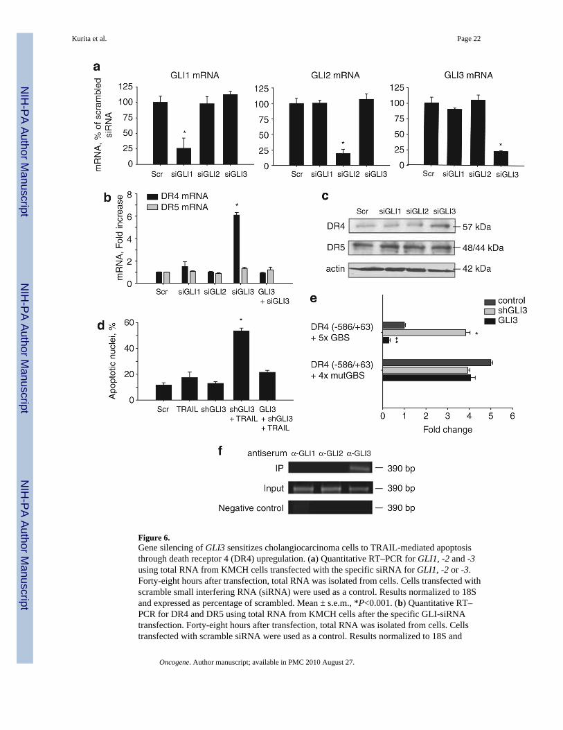
Figure 6.Gene silencing of GLI3 sensitizes cholangiocarcinoma cells to TRAIL-mediated apoptosisthrough death receptor 4 (DR4) upregulation. (a) Quantitative RT–PCR for GLI1, -2 and -3using total RNA from KMCH cells transfected with the specific siRNA for GLI1, -2 or -3.Forty-eight hours after transfection, total RNA was isolated from cells. Cells transfected withscramble small interfering RNA (siRNA) were used as a control. Results normalized to 18Sand expressed as percentage of scrambled. Mean ± s.e.m., *P<0.001. (b) Quantitative RT–PCR for DR4 and DR5 using total RNA from KMCH cells after the specific GLI-siRNAtransfection. Forty-eight hours after transfection, total RNA was isolated from cells. Cellstransfected with scramble siRNA were used as a control. Results normalized to 18S and
Kurita et al. Page 22
Oncogene. Author manuscript; available in PMC 2010 August 27.
NIH
-PA Author Manuscript
NIH
-PA Author Manuscript
NIH
-PA Author Manuscript

expressed as fold increase over scramble. GLI3 silencing was rescued where indicated by co-transfection with a GLI3 expression plasmid resistant to the siRNA. Mean ± s.e.m., *P<0.001.(c) Immunoblotting of whole cell lysates obtained from KMCH cells transfected with thespecific siRNA targeting GLI1, -2, or -3. Seventy-two hours after transfection, total cellularprotein was isolated from the cells. Membranes were probed using a DR4 and a DR5 antibody.Actin was used as a loading control. (d) KMCH cells transfectred with GLI3 shRNA or co-transfected with shGLI3 plus a GLI3-expression-plasmid resistant to the shRNA wereincubated with 5 ng/ml human recombinant TRAIL for 5 h. Cells were then stained with 4′,6-diamino-2-phenylindole dihydrochloride (DAPI) and cells with apoptotic morphology werecounted. Mean ± s.e.m., *P<0.001 compared with vehicle. (e) DR4 promoter constructs (−586/+63) with a tandem repeat of the candidate GLI-binding site (DR4 (−586/+63) + 5 × GBS) ora tandem repeat of a mutated GLI-binding site (DR4 (−586/+63) + 4 × mutGBS) were assayedin KMCH cells. Cells were co-transfected with shGLI3 or a GLI3-expression construct asindicated. Mean ± s.e.m. *P<0.001 compared with medium; **P<0.005 compared withmedium. (f) Chromatin immunoprecipitation was performed on KMCH cells using anti-GLI1,-GLI2 or –GLI3 anti-serum and the pull down used for DR4 promoter polymerase chainreaction using primers flanking the candidate GLI-binding site I. The polymerase chainreaction amplicon was visualized by ethidium bromide staining after agarose electrophoresis.
Kurita et al. Page 23
Oncogene. Author manuscript; available in PMC 2010 August 27.
NIH
-PA Author Manuscript
NIH
-PA Author Manuscript
NIH
-PA Author Manuscript