Mitochondrial genomes are retained by selective constraints ...
8
Mitochondrial genomes are retained by selective constraints on protein targeting Patrik Björkholm a,1 , Ajith Harish a,1 , Erik Hagström a , Andreas M. Ernst b , and Siv G. E. Andersson a,2 a Department of Molecular Evolution, Cell, and Molecular Biology, Uppsala University, 752 36 Uppsala, Sweden; and b Department of Cell Biology, Yale University School of Medicine, New Haven, CT 06520 Edited by Patrick J. Keeling, University of British Columbia, Vancouver, BC, Canada, and accepted by the Editorial Board June 29, 2015 (received for review December 16, 2014) Mitochondria are energy-producing organelles in eukaryotic cells considered to be of bacterial origin. The mitochondrial genome has evolved under selection for minimization of gene content, yet it is not known why not all mitochondrial genes have been transferred to the nuclear genome. Here, we predict that hydrophobic membrane pro- teins encoded by the mitochondrial genomes would be recognized by the signal recognition particle and targeted to the endoplasmic retic- ulum if they were nuclear-encoded and translated in the cytoplasm. Expression of the mitochondrially encoded proteins Cytochrome oxi- dase subunit 1, Apocytochrome b, and ATP synthase subunit 6 in the cytoplasm of HeLa cells confirms export to the endoplasmic reticulum. To examine the extent to which the mitochondrial proteome is driven by selective constraints within the eukaryotic cell, we investigated the occurrence of mitochondrial protein domains in bacteria and eukary- otes. The accessory protein domains of the oxidative phosphorylation system are unique to mitochondria, indicating the evolution of new protein folds. Most of the identified domains in the accessory pro- teins of the ribosome are also found in eukaryotic proteins of other functions and locations. Overall, one-third of the protein domains identified in mitochondrial proteins are only rarely found in bacteria. We conclude that the mitochondrial genome has been maintained to ensure the correct localization of highly hydrophobic membrane pro- teins. Taken together, the results suggest that selective constraints on the eukaryotic cell have played a major role in modulating the evo- lution of the mitochondrial genome and proteome. mitochondria | protein domain | evolution | endoplasmatic reticulum | signal recognition particle M itochondria are organelles that produce energy by oxida- tive phosphorylation (OXPHOS), channeling electrons through the respiratory chain complexes to generate ATP, the energy currency of the cell. A unique feature of mitochondria is that they possess a distinct genome of their own. Mitochondrial (mt) genomes vary dramatically in size, but gene content is limited and remarkably similar in different organisms (1– 4). The human mitochondrial ge- nome contains only 13 protein coding genes, whereas the jakobid mitochondrial genomes contain up to 70 genes, mostly coding for subunits of the OXPHOS system complexes and ribosomal proteins (5, 6). Having two separate genomes is a costly arrangement for the cell. Approximately 250 proteins encoded in the nuclear (nu) genome are needed just to maintain and express the few remaining mito- chondrial genes (7). Mitochondrial sequences are frequently copied to the nuclear genomes (8– 10), confirming that mechanisms for the transfer of mitochondrial genes to the nuclear genome are in place. A major, unresolved question in evolutionary biology is why not all mitochondrial genes have been transferred to the nuclear genome, and thus why the mitochondrial genome has been retained. Over the years, many competing hypotheses have been put forward to explain the retention of organelle genomes. Some argue that gene transfer is an on-going process and that all mi- tochondrial genes will eventually end up in the nuclear genome (11). Strictly speaking, organelles such as mitosomes and hy- drogenosomes have lost their genomes entirely, as has also a plastid from a nonphotosynthetic species of the genus Polytomella (3). This does not however explain the universal retention of a genome in aerobic mitochondria and photosynthetic chloroplasts. Others suggest that the transfer of genes have been halted, either because of barriers against gene transfer or because the mitochondrial ge- nome confers benefits. One such proposed barrier to functional gene transfers is codon reassignments (12). This explanation has been dismissed because it is not applicable to all mitochondrial genomes. Another suggested barrier is the extreme hydrophobicity of the mitochondrial proteins, which has been claimed to prevent their import from the cytosol (13). However, this hypothesis has also been rejected because hydrophobic proteins can be imported across the mitochondrial membrane and because some membrane- spanning proteins are nu-encoded, such as the Lhca and Lhcb proteins in chloroplasts (ct) (14). Instead, models based on bene- ficial functions, such as the colocation for redox regulation hy- pothesis (15, 16), have gained popularity during the past decade. However, no mitochondrial genes involved in redox regulation have been identified (17). Thus, all hypotheses that have been put for- ward are controversial and none have been experimentally verified. In striking contrast to the few proteins encoded by the mito- chondrial genome, it is estimated that more than 1,000 mito- chondrial proteins are encoded by the nuclear genome (18). Many of these have no bacterial homologs. For example, the mi- tochondrial ATP/ADP translocase that exports the ATP produced in the mitochondrion to the cytoplasm shows no sequence simi- larity to the bacterial type of ATP/ADP translocase. Rather, the mitochondrial ATP/ADP translocase has evolved from a family of eukaryotic phosphate transporters (19). Studies of the yeast mito- chondrial proteome have indicated that about 40% of all mito- chondrial proteins have no homologos in bacteria, and might thus have originated within the eukaryotic genome (20, 21). Specifically, it has been suggested that new proteins have been added to the OXPHOS complex and the mito-ribosome before the diversifica- tion of the eukaryotic lineages (22, 23). This pattern contrasts with the core components of the mitochondrial OXPHOS system and the mito-ribosome, which are highly conserved and show strong sequence similarity to their bacterial homologs. In this study, we revisit a hypothesis proposed 30 years ago but since long forgotten: namely that a mitochondrial genome is This paper results from the Arthur M. Sackler Colloquium of the National Academy of Sciences, “Symbioses Becoming Permanent: The Origins and Evolutionary Trajectories of Organelles,” held October 15–17, 2014, at the Arnold and Mabel Beckman Center of the National Academies of Sciences and Engineering in Irvine, CA. The complete program and video recordings of most presentations are available on the NAS website at www.nasonline.org/Symbioses. Author contributions: P.B. and S.G.E.A. designed research; P.B., A.H., and A.M.E. per- formed research; P.B., A.H., E.H., and S.G.E.A. analyzed data; and P.B., A.H., E.H., and S.G.E.A. wrote the paper. The authors declare no conflict of interest. This article is a PNAS Direct Submission. P.J.K. is a guest editor invited by the Editorial Board. 1 P.B. and A.H. contributed equally to this work. 2 To whom correspondence should be addressed. Email: [email protected]. This article contains supporting information online at www.pnas.org/lookup/suppl/doi:10. 1073/pnas.1421372112/-/DCSupplemental. 10154–10161 | PNAS | August 18, 2015 | vol. 112 | no. 33 www.pnas.org/cgi/doi/10.1073/pnas.1421372112 Downloaded by guest on January 8, 2022
-
Upload
khangminh22 -
Category
Documents
-
view
1 -
download
0
Transcript of Mitochondrial genomes are retained by selective constraints ...

Mitochondrial genomes are retained by selectiveconstraints on protein targetingPatrik Björkholma,1, Ajith Harisha,1, Erik Hagströma, Andreas M. Ernstb, and Siv G. E. Anderssona,2
aDepartment of Molecular Evolution, Cell, and Molecular Biology, Uppsala University, 752 36 Uppsala, Sweden; and bDepartment of Cell Biology, YaleUniversity School of Medicine, New Haven, CT 06520
Edited by Patrick J. Keeling, University of British Columbia, Vancouver, BC, Canada, and accepted by the Editorial Board June 29, 2015 (received for reviewDecember 16, 2014)
Mitochondria are energy-producing organelles in eukaryotic cellsconsidered to be of bacterial origin. The mitochondrial genome hasevolved under selection for minimization of gene content, yet it is notknown why not all mitochondrial genes have been transferred to thenuclear genome. Here, we predict that hydrophobic membrane pro-teins encoded by the mitochondrial genomes would be recognized bythe signal recognition particle and targeted to the endoplasmic retic-ulum if they were nuclear-encoded and translated in the cytoplasm.Expression of the mitochondrially encoded proteins Cytochrome oxi-dase subunit 1, Apocytochrome b, and ATP synthase subunit 6 in thecytoplasm of HeLa cells confirms export to the endoplasmic reticulum.To examine the extent to which the mitochondrial proteome is drivenby selective constraints within the eukaryotic cell, we investigated theoccurrence of mitochondrial protein domains in bacteria and eukary-otes. The accessory protein domains of the oxidative phosphorylationsystem are unique to mitochondria, indicating the evolution of newprotein folds. Most of the identified domains in the accessory pro-teins of the ribosome are also found in eukaryotic proteins of otherfunctions and locations. Overall, one-third of the protein domainsidentified in mitochondrial proteins are only rarely found in bacteria.We conclude that the mitochondrial genome has been maintained toensure the correct localization of highly hydrophobic membrane pro-teins. Taken together, the results suggest that selective constraints onthe eukaryotic cell have played a major role in modulating the evo-lution of the mitochondrial genome and proteome.
mitochondria | protein domain | evolution | endoplasmatic reticulum |signal recognition particle
Mitochondria are organelles that produce energy by oxida-tive phosphorylation (OXPHOS), channeling electrons
through the respiratory chain complexes to generate ATP, the energycurrency of the cell. A unique feature of mitochondria is that theypossess a distinct genome of their own. Mitochondrial (mt) genomesvary dramatically in size, but gene content is limited and remarkablysimilar in different organisms (1–4). The human mitochondrial ge-nome contains only 13 protein coding genes, whereas the jakobidmitochondrial genomes contain up to 70 genes, mostly coding forsubunits of the OXPHOS system complexes and ribosomal proteins(5, 6). Having two separate genomes is a costly arrangement for thecell. Approximately 250 proteins encoded in the nuclear (nu) genomeare needed just to maintain and express the few remaining mito-chondrial genes (7). Mitochondrial sequences are frequently copiedto the nuclear genomes (8–10), confirming that mechanisms for thetransfer of mitochondrial genes to the nuclear genome are in place. Amajor, unresolved question in evolutionary biology is why not allmitochondrial genes have been transferred to the nuclear genome,and thus why the mitochondrial genome has been retained.Over the years, many competing hypotheses have been put
forward to explain the retention of organelle genomes. Someargue that gene transfer is an on-going process and that all mi-tochondrial genes will eventually end up in the nuclear genome(11). Strictly speaking, organelles such as mitosomes and hy-drogenosomes have lost their genomes entirely, as has also a plastidfrom a nonphotosynthetic species of the genus Polytomella (3). This
does not however explain the universal retention of a genome inaerobic mitochondria and photosynthetic chloroplasts. Otherssuggest that the transfer of genes have been halted, either becauseof barriers against gene transfer or because the mitochondrial ge-nome confers benefits. One such proposed barrier to functionalgene transfers is codon reassignments (12). This explanation hasbeen dismissed because it is not applicable to all mitochondrialgenomes. Another suggested barrier is the extreme hydrophobicityof the mitochondrial proteins, which has been claimed to preventtheir import from the cytosol (13). However, this hypothesis hasalso been rejected because hydrophobic proteins can be importedacross the mitochondrial membrane and because some membrane-spanning proteins are nu-encoded, such as the Lhca and Lhcbproteins in chloroplasts (ct) (14). Instead, models based on bene-ficial functions, such as the colocation for redox regulation hy-pothesis (15, 16), have gained popularity during the past decade.However, no mitochondrial genes involved in redox regulation havebeen identified (17). Thus, all hypotheses that have been put for-ward are controversial and none have been experimentally verified.In striking contrast to the few proteins encoded by the mito-
chondrial genome, it is estimated that more than 1,000 mito-chondrial proteins are encoded by the nuclear genome (18).Many of these have no bacterial homologs. For example, the mi-tochondrial ATP/ADP translocase that exports the ATP producedin the mitochondrion to the cytoplasm shows no sequence simi-larity to the bacterial type of ATP/ADP translocase. Rather, themitochondrial ATP/ADP translocase has evolved from a family ofeukaryotic phosphate transporters (19). Studies of the yeast mito-chondrial proteome have indicated that about 40% of all mito-chondrial proteins have no homologos in bacteria, and might thushave originated within the eukaryotic genome (20, 21). Specifically,it has been suggested that new proteins have been added to theOXPHOS complex and the mito-ribosome before the diversifica-tion of the eukaryotic lineages (22, 23). This pattern contrasts withthe core components of the mitochondrial OXPHOS system andthe mito-ribosome, which are highly conserved and show strongsequence similarity to their bacterial homologs.In this study, we revisit a hypothesis proposed 30 years ago but
since long forgotten: namely that a mitochondrial genome is
This paper results from the Arthur M. Sackler Colloquium of the National Academy of Sciences,“Symbioses Becoming Permanent: The Origins and Evolutionary Trajectories of Organelles,”held October 15–17, 2014, at the Arnold andMabel Beckman Center of the National Academiesof Sciences and Engineering in Irvine, CA. The complete program and video recordings of mostpresentations are available on the NAS website at www.nasonline.org/Symbioses.
Author contributions: P.B. and S.G.E.A. designed research; P.B., A.H., and A.M.E. per-formed research; P.B., A.H., E.H., and S.G.E.A. analyzed data; and P.B., A.H., E.H., andS.G.E.A. wrote the paper.
The authors declare no conflict of interest.
This article is a PNAS Direct Submission. P.J.K. is a guest editor invited by the EditorialBoard.1P.B. and A.H. contributed equally to this work.2To whom correspondence should be addressed. Email: [email protected].
This article contains supporting information online at www.pnas.org/lookup/suppl/doi:10.1073/pnas.1421372112/-/DCSupplemental.
10154–10161 | PNAS | August 18, 2015 | vol. 112 | no. 33 www.pnas.org/cgi/doi/10.1073/pnas.1421372112
Dow
nloa
ded
by g
uest
on
Janu
ary
8, 2
022

needed to prevent the export of highly hydrophobic mitochondrialmembrane proteins to the endoplasmic reticulum (ER) (24). Weuse a combination of bioinformatics methods to predict proteinlocalization and cell biological methods to verify our predictionsexperimentally. In addition, we analyze the nu-encoded fractionof the mitochondrial proteome for which structure, and hencefunction, can be assigned. Taking these data together, we are ableto gain a comprehensive insight into the role that the two differentgenomes play in orchestrating mitochondrial functions.
ResultsMitochondrial Proteins are Potential Targets for Recognition by SignalRecognition Particle. Revisiting the hydrophobicity hypothesis in itsoriginal formulation is motivated by recent insights into themechanism whereby the signal recognition particle (SRP) exportsproteins to the ER. This is a cotranslational pathway, where SRPbinds to a hydrophobic domain, either in the form of a trans-membrane domain (TMD) or a signal sequence, which leads toarrest of the nascent peptide chain. Importantly, it is the hydro-phobicity rather than the signal sequence per se that is recognizedby SRP. Next, the ribosome with the arrested peptide chain istransported to the ER (25). Soluble proteins shorter than 100–120amino acids are missed by SRP, whereas proteins of 120–160amino acids can be captured, although quite inefficiently (26).Thus, the hydrophobicity of the TMD, as well as the length of theC-terminal tail following the first hydrophobic domain, is criticalfor protein recognition by SRP (27).To predict the potential of nu- and organelle-encoded mitochon-
drial proteins to be targets for recognition by SRP, we calculated thefree insertion energy (ΔG, kcal/mol) of the TMDs (28), and cate-gorized the TMDs as either hydrophobic or marginally hydrophobic.The proteins were then classified as arrested by SRP if they con-tained a hydrophobic TMD and a tail that, together with the TMD,was longer than 120 amino acids. Proteins with only marginally hy-drophobic TMDs were considered to avoid recognition by SRP.The human mitochondrial OXPHOS system consists of five
membrane-spanning complexes composed of 96 proteins in total, ofwhich 13 are encoded by the mitochondrial genome and 83 by thenuclear genome (SI Appendix, Table S1). We classified 10 of the 13mt-encoded proteins as targets for SRP, whereas only 1 of the 83nu-encoded proteins was predicted to be an SRP target (Fisher’sExact χ2 test, P < 0.01) (Fig. 1A). Similarly, only 2 of the 51 nu-encoded mitochondrial membrane proteins with known structureswere classified as arrested by SRP (SI Appendix, Table S2). Con-sistent with these results, 251 of 281 nu-encoded membrane pro-teins localized to mitochondria with the aid of green fluorescentprotein tags in yeast and humans (29) were predicted to avoidrecognition by SRP (SI Appendix, Fig. S1 and Table S3).We also estimated the potential of chloroplast proteins in the
photosynthetic apparatus of Arabidopsis thaliana, (SI Appendix,Table S4), to be recognized by SRP. Of the 14 ct-encoded pro-teins longer than 100 amino acids, 11 would be arrested by SRP,compared with only 1 of the 24 nu-encoded proteins with a lengthof more than 100 amino acids (Fisher’s Exact χ2 test, P < 0.01)(Fig. 1B). Importantly, the nu-encoded light-harvesting (Lh) pro-teins Lhca and Lhcb would not be recognized by SRP accordingto our calculations. As much as one-third of the 30 ct-encodedmembrane proteins are shorter than 50 amino acids (SI Appendix,Fig. S2). We suggest that these extremely short single-spanningmembrane proteins, which integrate randomly into the thylakoidmembrane in plants (30), would be difficult to transport to thethylakoid membrane if not encoded in the chloroplast genome.
Mistargeting of Mitochondrial Proteins to the ER. To experimentallytest our predictions, mt-encoded proteins Cytochrome oxidasesubunit 1 (Cox1), Apocytchrome b (Cytb), and ATP synthasesubunit 6 (ATP6) were expressed in the cytoplasm of HeLa cells(Fig. 2). The proteins were FLAG-tagged at the N terminus and
visualized by immunofluorescence. The FLAG-tag was placed atthe N terminus rather than at the C terminus to avoid extendingthe length of the C-terminal sequence following the first hydro-phobic domain. We used FLAG-tagged ERLIN1 and OMP25-GFP constructs as positive localization controls (for ER and mi-tochondria, respectively) and immunostained with antibodiesmarking the ER (calnexin) and mitochondria (TOM20) (Fig. 2A).As predicted, Cox1, Cytb, and ATP6 colocalized with the ER (Fig.2). Mistargeting of these mt-encoded proteins to the ER resultedin the formation of aberrant honeycomb structures, as previouslyobserved during viral infections (31). This finding suggests thatmistargeting of mitochondrial proteins to the ER affects themorphology of the cell. We conclude that genes for hydrophobicmembrane proteins of more than 120 amino acids are likelyretained in distinct organelle genomes to ensure a correct locali-zation of these proteins and avoid transport to the ER.
Phyletic Distribution Patterns of Mitochondrial Protein Folds inEukaryotes. It is clear that the mitochondrial genome and thesupporting genetic apparatus, whether encoded by the mito-chondrial or nuclear genome, are dedicated to synthesize andassemble the OXPHOS complex, which is central to energymetabolism. It is less clear to what extent selective constraintsacting on the eukaryotic cell, like targeting proteins to theircorrect locations or regulating the activities of the two genomes,have influenced the mitochondrial proteome. To learn moreabout the evolutionary pressures acting on mitochondrial pro-teins, we surveyed a broad taxonomic range of organisms for thepresence or absence patterns of proteins that are critical to mi-tochondrial functions. Because of the difficulty of assigningproteins to clusters of orthologous groups for highly divergentproteins and multidomain proteins (32), we used a protein do-main-centric approach for this analysis. In brief, we assigned
dG s
core
, kca
l/m
oldG
sco
re, k
cal/
mol
Tail Length, amino acids
Tail Length, amino acids
A
B
Fig. 1. Biophysical characteristics of membrane proteins involved in aerobicrespiration and photosynthesis. The figure shows the insertion-free energy(ΔG, dG) and the length of the TMD and the following C-terminal tail forproteins involved in (A) the OXPHOS system complexes, and (B) the photo-synthetic apparatus. The insertion-free energy (kcal/mol) was estimated foreither the first TMD with a calculated ΔG value below zero or if no suchsegment was found, the most hydropohobic segment in the protein. Red dotscorrespond to proteins encoded by the organelle genome, and blue dots toproteins encoded by the nuclear genome. Characteristic features of proteinsthat are putative targets for recognition by SRP are shown in the gray area.
Björkholm et al. PNAS | August 18, 2015 | vol. 112 | no. 33 | 10155
EVOLU
TION
COLLOQUIUM
PAPE
R
Dow
nloa
ded
by g
uest
on
Janu
ary
8, 2
022

protein domains at the superfamily (SF) level of the SCOP(Structural Classification of Proteins) hierarchy (33) usinghidden Markov model libraries of protein domains in theSUPERFAMILY database (E < 0.0001) (34). SCOP-SF domainsare inferred to be homologous based on similarity of sequence,structure, and function.The obvious start for such a survey was the OXPHOS system
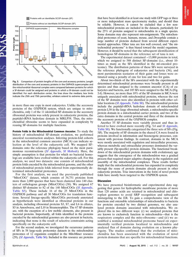
itself. Thus, we predicted protein folds in the OXPHOS systemin human mitochondria, which consists of a central set of 14proteins that are conserved in both eukaryotes and bacteria, andan accessory set of 30 protein subunits that have solely beenidentified in mitochondria (35). In total, we assigned 39 distinctSF-domains to the central set of proteins and another 20 distinctSF-domains to the accessory proteins. A length comparisonrevealed a marked difference between the central and the ac-cessory proteins in the mitochondrial OXPHOS system, withmedian lengths of about 300 and 100 amino acids, respectively(Fig. 3). The size difference was observed irrespectively ofwhether the proteins contained recognizable domains or not.The phyletic distribution of the identified SFs was examined in
43 eukaryotes from 7 major eukaryotic lineages for which apublished multigene phylogeny is available that shows their in-ternal relationships (36). In the phylogeny, 13 bacterial specieswere used as outgroups, 6 of which are from the Alphaproteo-bacteria. The analysis showed that of the 39 SF-domains in thecentral set of proteins, 34 were also present in bacteria (Fig. 4).The remaining five SF-domains represent novel protein exten-sions located in proteins for which no homologs are present inbacteria. These additional domains include a transmembraneanchor protein domain in two proteins of the cytochrome re-ductase and oxidase complexes, respectively, and a nonglobularα/β subunit solely identified in human mitochondria.All of the 20 SF-domains identified in the accessory proteins,
one-third of which are single transmembrane-spanning domains(STMD), were markedly absent from bacteria. The functions ofthe accessory proteins are largely unknown, although a few havebeen implicated in the biogenesis of the OXPHOS complex (35,37). Some of the short STMDs are quite hydrophobic, and assuch might help stabilize the respiratory protein complexes in themembrane. The most prominent candidates are UQCR10 andCOX6A, which have a high hydrophobicity and a number ofpositive charges flanking this segment, making them well an-chored in the membrane. Our phyletic analysis showed a highlyscattered distribution pattern of these domains in the eukaryoticlineages and most domains were also not associated with pro-teins of other functions. This finding suggests that the identifieddomains represent cases of de novo evolution of protein folds.However, these domains are all located in membrane-associ-
ated proteins. For comparison, we also examined protein folds ofthe human mitochondrial ribosome, which is composed of acentral set of soluble proteins, 21 and 32 for the small and largeribosomal subunit, respectively, plus 12 and 18 soluble accessoryproteins that are unique to mitochondria. We predicted 34 SFsfor the 53 central proteins and 10 SFs for the 30 accessoryproteins in the mitochondrial ribosome. Unlike the accessoryproteins of the OXPHOS complex, the accessory proteins of theribosome are of similar sizes as the central proteins (Fig. 3). Astudy of the phyletic distribution patterns of the protein folds inthe accessory ribosomal proteins showed that they are broadlypresent in our reference set of bacteria and eukaryotes (SI Ap-pendix, Fig. S3). The protein domains in the central mitochon-drial ribosomal proteins were also identified in the cytosolicribosomal proteins, which explain why these domains are present
-CALNEXIN mergeFLAG-COX1
-TOM20 mergeFLAG-COX1
-CALNEXIN mergeFLAG-ATP6
-TOM20 mergeFLAG-ATP6
-CALNEXIN mergeFLAG-CYTB
-TOM20 mergeFLAG-CYTB
-CALNEXIN mergeFLAG-ERLIN1
-TOM20 mergeFLAG-ERLIN1
-CALNEXIN mergeOMP25-GFP
-TOM20 mergeOMP25-GFP
Fig. 2. Mitochondrial membrane proteins atypically expressed in thecytoplasm localize to the ER. Amino acid sequences of COX1, ATP6, andCYB were reverse-translated to nuclear codon use and FLAG-tagged attheir amino termini. HeLa cells were transfected with plasmids containingthese synthetic genes and subjected to immunofluorescence analysis.From the top, FLAG-tagged ERLIN1 and OMP25-GFP constructs were usedas positive localization controls and immunostained with antibodies markingthe ER (calnexin) or mitochondria (TOM20). Below this, localization analyses
of the synthetic mitochondrial membrane proteins COX1, ATP6, and CYB.(Scale bars, 5 μm.)
10156 | www.pnas.org/cgi/doi/10.1073/pnas.1421372112 Björkholm et al.
Dow
nloa
ded
by g
uest
on
Janu
ary
8, 2
022
in more than one copy in most eukaryotes. Unlike the accessoryproteins of the OXPHOS system, which are unique to mito-chondria, only 1 of the 11 identified SF-domains in the accessoryribosomal proteins was solely present in eukaryotic proteins, thepeptidyl-tRNA hydrolase domain in MRLP58. Thus, the mito-chondrial ribosome seems to have expanded in complexity byreusing SF-domains for multiple functions.
Protein Folds in the Mitochondrial Common Ancestor. To study thehistory of mitochondrial SF-domain evolution, we performedancestral reconstruction analyses. Inferring protein-fold cohortsin the mitochondrial common ancestor (MCA) can indicate se-lection at the level of the eukaryotic cell. We mapped SF-domains onto the reference phylogeny based on the most parsi-monious reconstructions (SI Appendix, SI Methods). Here, thesimple assumption is that proteins for which no bacterial homo-logs are available have evolved within the eukaryotic cell. For thisanalysis, we used two datasets: one consists of mitochondrialprotein folds encoded by the mitochondrial genome, and the otherof mitochondrial protein folds inferred from experimentally de-termined mitochondrial proteomes.For the first analysis, we used the previously published
“MitoCOG” dataset, which consists of 34,751 proteins frommore than 2,000 species that have been clustered into 140 clus-ters of orthologous groups (MitoCOGs) (38). We assigned 67distinct SF-domains to 82 of the 140 MitoCOGs (SI Appendix,Table S5). These include 16 of the 25 MitoCOGs in theOXPHOS pathway and all 30 MitoCOGs for translation func-tions. Several lineage-specific MitoCOGs previously annotatedas hypotheticals were identified as ribosomal proteins in ouranalysis, including ribosomal proteins S3, S7, and L6 in ciliates,S3 in Amoebozoa, and L10 in Stramenophiles. The 67 SF-domainsare, with the exception of a few mobile elements, also present inbacterial proteins. Importantly, all folds identified in the proteinsencoded by the mitochondrial genomes are also present in bacteria,indicating that none is the result of selective constraints actingspecifically on the eukaryotic cell.For the second analysis, we investigated the occurrence patterns
of SFs in 58 large-scale proteomics datasets in the mitochondrialproteomes of 12 organisms compiled in the MitoMiner resource(29), (SI Appendix, Table S6). Included in this resource are proteins
that have been identified in at least one study with GFP tags or threeor more independent mass spectrometry studies, and should thusbe reliable. However, it cannot be excluded that a few non-mitochondrial proteins are included in the datasets, particularly forthe 25% of proteins assigned to mitochondria in a single species.Some domains may also represent mis-assignments. The mitochon-drial proteomes of yeast, humans, mouse, and Drosophila contain alarge number of proteins deduced from several different experi-mental approaches. Other taxa are less well covered, and “the mi-tochondrial proteome” is thus biased toward the model organisms.However, it should be noted that the subsequent identification ofhomologous SF-domains in the reference set of taxa is not.The experimental dataset contained ∼13,000 proteins in total,
which we assigned to 588 distinct SF-domains (i.e., about 10-times as many as the SFs identified in the mt-encoded pro-teome). The distribution patterns of these SFs were surveyed inthe reference phylogeny of eukaryotes and bacteria (36), andmost parsimonious scenarios of their gains and losses were es-timated using a penalty of one for loss and two for gains.About two-thirds of the SFs associated with the experimentally
determined mitochondrial proteomes were broadly present in allspecies and thus assigned to the common ancestor (CA) of eu-karyotes and bacteria, and 105 SFs were assigned to the MCA (Fig.5A). However, no more than 10% of the SF-domains in MCA wereuniquely associated with mitochondrial proteins, whereas the restof the SF domains were also identified in proteins with other cel-lular locations (SI Appendix, Table S8). The mitochondrial proteinsinclude the peptidyl-tRNA hydrolase domain of mitochondrialprotein L58 in the large ribosomal subunit, a mitochondria-specificprotein domain in a mitochondrial carrier protein, and two of theextra domains in the central proteins and three of the domains inthe accessory proteins of the OXPHOS complex.Another 55 SF-domains were variably present and thus in-
ferred to have evolved more recently (Fig. 5A and SI Appendix,Table S8). We functionally categorized the three sets of SFs (Fig.5B). The majority of SF-domains in the shared CA were found inproteins involved in information and bioenergetic processes, con-sistent with previous findings. In contrast, domains in the MCAwere mostly associated with regulation and intracellular processes,whereas metabolic and extracellular processes dominated the var-iably present (Sp-specific) protein domains. The functional break-down of the mitochondrial protein domains with evolutionary agesuggests that the emergence of the mitochondrion was a gradualprocess that required major adaptive changes in the regulation andassembly of the mitochondrial complexes. These results furtherimply that the mitochondrial proteome has expanded in complexitythrough the reuse of protein domains already present in othereukaryotic proteins. True innovations in the form of novel proteinfolds have mostly been targeted to the OXPHOS system.
DiscussionWe have presented bioinformatic and experimental data sug-gesting that genes for hydrophobic membrane proteins of morethan 120 amino acids are retained in mitochondrial and chlo-roplast genomes to avoid SRP recognition and subsequentmistargeting to the ER. To gain a comprehensive view of thefunctions and ostensible relationships of mitochondria to bacteriafor proteins encoded by two distinct genomes, we also ana-lyzed protein domains associated with mitochondria. We ex-plored this in two different ways: (i) we analyzed proteins thatare known to exclusively function in mitochondria—that is therespiratory complex and the mito-ribosome—and (ii) we as-sembled a nominal mitochondrial proteome based on exper-imentally verified proteins localized to mitochondria andanalyzed flux of domains during evolution on a known phy-logeny. The studies confirmed that the evolution of mito-chondria has been associated with the evolution of novelprotein domains that are specific for mitochondrial functions.
0
100
200
300
400
500
600
700
800
Prot
ein
leng
th
Central
Accessory
Accessory-STMD
SSU-Central
LSU-central
SSU-Accessory
LSU-Accessory
Proteins with an identifiable SCOP-domain (SF)
Proteins without an identifiable SCOP-domain (SF)
OXPHOS supercomplex Mito-Ribosome complex
Fig. 3. Comparison of protein lengths of the core and accessory proteins. Lengthdistribution of the core and accessory proteins in the OXPHOS supercomplex andthemitochondrial ribosomal complexwere compared between proteins for whicha SF-domain could be assigned and proteins in which a SF-domain could not beidentified. For each distribution center, the lines show the medians; box limitsindicate the 25th and 75th percentiles; whiskers extend to extreme data points.
Björkholm et al. PNAS | August 18, 2015 | vol. 112 | no. 33 | 10157
EVOLU
TION
COLLOQUIUM
PAPE
R
Dow
nloa
ded
by g
uest
on
Janu
ary
8, 2
022

The prime examples are the accessory proteins in theOXPHOS system, which have been suggested to be involvedin the organization, regulation, and biogenesis of the complex(35, 37, 39).The evolution of the mitochondrial ribosome has been studied
in great detail previously (40), and it has been inferred that 19accessory ribosomal proteins were present already in the mito-chondrial ancestor (41). We identified SF-domains for 6 of these19 proteins, all of which were associated with generalized func-tions present in many bacterial and cytosolic proteins. For ex-ample, the Nudix domain in MRPL46 is also found in decappingenzymes, ADP ribose diphosphatase, and similar proteins, and
the double-stranded (ds) RNA-binding protein domain found inMRPL44 and MRPS5 is a generic RNA-binding domain foundin diverse enzymes, such as RNA helicases, dsRNA-dependentprotein kinase, RNase III, and so forth. The accessory proteins,which are situated on the surface of the ribosome, have beensuggested to be involved in cotranslational processes and pro-grammed cell death (42, 43), providing examples of how theeukaryotic cell controls the mitochondrial processes.Overall, one-third of the SFs identified in mitochondrial proteins
are mainly found in eukaryotic genomes and only rarely found inbacterial genomes. About 25% of the protein domains assigned tothe mitochondrial ancestor was associated with regulatory functions,
......
Mag
neto
spiri
llum
......
......
....P
elag
ibac
ter
......
......
.....T
oxop
lasm
a
........
........
..Aur
eoco
ccus
......
......
..Try
pano
som
a
......
......
......
......
......
Pich
ia
......
......
...Bu
rkho
lder
ia
......
.....C
aeno
rhab
ditis
......
......
......
....T
heile
ria
......
......
..Des
ulfo
vibrio
......
......
......
......
.....H
omo
......
......
......
.....U
stila
go
......
......
.Dict
yost
eliu
m
......
.....A
grob
acte
rium
......
......
......
..Geo
bact
er
......
......
......
....Y
arro
wia
......
......
......
..Nae
gler
ia
......
......
Bran
chio
stom
a
......
......
......
......
....B
rugi
a
......
......
....T
hala
ssio
sira
........
........
..Sch
istos
oma
......
......
......
...M
onos
iga
......
......
......
......
..Vol
vox
........
........
........
...Bab
esia
......
......
......
.Lei
shm
ania
......
Batra
choc
hytri
um
........
........
........
..Lac
caria
........
........
........
........
....Ap
is
......
......
....P
aram
eciu
m
......
......
.....M
icrom
onas
......
......
......
...So
libac
ter
....S
trong
yloce
ntro
tus
......
......
......
.Sel
agin
ella
......
......
......
....R
icket
tsia
......
....P
unice
ispiri
llum
Schiz
osac
char
omyc
es
......
......
......
.Dro
soph
ila
......
......
......
.Asp
ergi
llus
......
....P
haeo
dact
ylum
........
........
....Es
cher
ichia
......
......
......
......
.....O
ryza
......
......
......
..Tric
hopl
ax
......
......
......
....C
hlor
ella
........
........
...Phy
com
yces
......
......
......
Arab
idop
sis
......
......
Phys
com
itrel
la
......
......
......
......
....Ix
odes
........
........
.Ostr
eoco
ccus
......
.....A
cant
ham
oeba
......
......
...Pl
ancto
myc
es
......
.Pol
ysph
ondy
lium
........
........
...Myx
ococ
cus
........
.....S
acch
arom
yces
......
......
.....P
lasm
odiu
m
..Rho
dops
eudo
mon
as
......
......
..Tet
rahy
men
a
1 2 4 8 16 32 64 12825
651
2
Number of domains
Phyletic distribution of OXPHOS SF-domains
Accessory
Accessory - STMD
CentralC1,C2|d.15.4C1,C5|c.37.1C1|c.2.1C1|a.28.1C1|d.58.1C1|c.81.1C1|e.18.1
C1|e.19.1C1|c.142.1C1|d.15.13C1|a.29.12C2|c.3.1C2|a.7.3C2|d.168.1C2|a.1.2C2|f.21.2C3|a.3.1C3|f.23.11C3|f.32.1C3|f.21.1C3|d.185.1C3|b.33.1C3|f.23.12C3|d.184.1C4|f.24.1C4|b.6.1C4|f.17.2C4|f.25.1C5|a.69.1C5|b.49.1C5|c.49.2
C5|b.93.1C5|a.137.8C5|f.52.1C5|f.17.1
C5|f.18.1
C1|c.47.1C3|f.27.1C3|f.28.1C4|a.118.11C4|g.41.5C4|a.51.1C5|f.53.1C5|f.45.1C5|h.4.8
C3|f.23.14C3|f.23.15C3|f.23.13C4|f.23.1C4|f.23.2C4|f.23.3C4|f.23.4C4|f.23.5C4|f.23.6C4|f.23.7C5|a.118.3
Comple
x
SF
airetcaBsetoyrakuE
Fig. 4. Phyletic distribution of SF-domains of the human mitochondrial OXPHOS complex. The number of central and accessory SF-domains identified in eachspecies of eukaryotes and bacteria are compared along the phylogeny. Columns correspond to the species in the phylogenetic tree and rows are individual SF-domains of the OXPHOS supercomplex. The colors indicate the number of domains in each species if present and absences are shown as white. Descriptions ofeach row include the complex in which a SF-domain is identified in, followed by the SCOP classification of the SF-domain. The corresponding SF-domainsdescriptions are listed in SI Appendix, Table S7. SF-domains encoded in the nuclear genome are shown for all species. The SFs encoded by the human mi-tochondrial genome are indicated in bold. The number of domains in eukaryotes only include nu-encoded SFs.
10158 | www.pnas.org/cgi/doi/10.1073/pnas.1421372112 Björkholm et al.
Dow
nloa
ded
by g
uest
on
Janu
ary
8, 2
022

whereas metabolic functions dominate among the clade and species-specific mitochondrial protein domains. A large majority of thesefolds are generic and present in proteins with many functions anddestinations within the eukaryotic cell. One of the few nu-encodedmitochondrial proteins, which was classified as arrested by SRP inour analysis, is mistargeted to the ER in the absence of a strongmitochondrial target peptide (44). Thus, some of the nu-encodedproteins with characteristics that are typical of proteins recognizedby SRP might in fact be targeted to both mitochondria and the ER.Taken together, these results are fully consistent with previous gene-based studies, which have shown that about one-third of the mito-chondrial proteins, mostly associated with regulatory, transport, ormembrane functions, have evolved in response to the specific con-straints presented by the compartmentalized eukaryotic cell (20–23).Our results also provide an explanation for several rare cases of
gene transfers of otherwise universal mitochondrial genes to thenuclear genome. As predicted by our hypothesis, the mitochon-drial proteins show a reduced hydrophobicity of the first TMDs inthe few species in which these genes have been successfullytransferred in nature (45–47). For example, the first TMD of thenu-encoded Cox2 protein in legumes is less hydrophobic than thefirst TMD of the mt-encoded homolog (48). Experimental studieshave confirmed that the mitochondrial variant of Cox2 could notbe imported into mitochondria unless the first TMD was removedor if the sequence was changed to that of the less hydrophobic nu-encoded homolog (48) and when an exceptionally strong mito-chondrial targeting peptide of 130 amino acids was added (49).Simlarly, all three TMDs of the mt-encoded SdhC protein
in Reclinomonas americana are highly hydrophobic, whereasthe TMDs of the nu-encoded homologs in yeast, humans, andacanthamoeba are only marginally or not at all hydrophobic. Ourfindings also explain why the 3′-end of the cox2 gene couldsuccessfully be transferred to the nuclear genome in some greenalgae, whereas the 5′-end of the gene encoding the hydrophobicTMD remains situated in the mitochondrial genome.Thus, successful transfers of mitochondrial genes for hydrophobic
proteins have occurred in nature and have been achieved in thelaboratory by modifications, such as: (i) reducing the hydrophobicityof the first TMD; (ii) reducing the length of the C-terminal se-quence of the hydrophobic TMD; (iii) moving the hydrophobicTMD toward the C-terminal end; or (iv) by splitting the gene intotwo, leaving the 5′-end, which codes for the hydrophobic TMD inthe mitochondrial genome. We suggest that the effect of thesemodifications is a reduced potential for recognition by SRP, en-abling the protein to reach the mitochondrial destination.However, arrest by SRP and subsequent mistargeting to the
ER is unlikely to explain all of the peculiarities of mitochondrialgenomes in individual species. For example, genes for SdhD,ATP1, ATP3, and ATP9 in the R. americana and other jakobidprotists are too short, do not contain hydrophobic TMDs, or theC-terminal tail following the TMD segment is too short to allowarrest by SRP. These mitochondrial genomes also encode severalribosomal proteins, which serve key functions in ribosomal assemblyand initial rRNA binding (50). Interestingly, a similar set of ribo-somal proteins is also encoded by chloroplast genomes, indicative ofconvergent evolution in response to similar selective constraints. Thisfinding suggests that there is a hierarchical order of mitochondrial
A
B
Fig. 5. Ancestral reconstruction analysis of SF-domains in the mitochondrialproteome. (A) Flux of SF-domains in the mitochondrial proteome duringevolution of eukaryotes and bacteria. Parsimonious reconstruction of do-main flux is mapped onto a eukaryote phylogeny rooted with bacterialoutgroup. Numbers at nodes show the number of SF-domains inferred tohave been gained (green), lost (red), and the total number present at eachnode or branch (black) along the phylogeny. Only the major internal nodes
are shown for clarity. Colored nodes represent the Common Ancestor ofeukaryotes and bacteria (CA) (brown); Mitochondrial Common Ancestor(MCA) (blue); Bacterial Common Ancestor (BCA) (orange). Because thenumber of gains and losses varies between species, only a range from aminimum to a maximum in each clade is shown here. Details of flux for allspecies are shown in SI Appendix, Fig. S4. (B) A comparison of the functionsassociated to SF-domains estimated to be in the CA of eukaryotes and bacteria,MCA, and species specific SF-domains (Sp-specific). Bars represent the percentof SF-domains associated with a functional category.
Björkholm et al. PNAS | August 18, 2015 | vol. 112 | no. 33 | 10159
EVOLU
TION
COLLOQUIUM
PAPE
R
Dow
nloa
ded
by g
uest
on
Janu
ary
8, 2
022

gene loss and that the genes that encode hydrophobic TMDs aresubjected to the strongest selective constraints and therefore the lastto be lost or transferred to the nuclear genomes (48).Chloroplasts have several membranes, making the import
of membrane proteins in the photosynthetic apparatus to thechloroplast a more complex process than targeting membraneproteins to mitochondria. Like mitochondria, chloroplasts have adouble-membrane envelope, but additionally chloroplasts con-tain internal membrane structures (thylakoids) where the twophotosystems are embedded. To accommodate this extra layer ofcomplexity, higher plants contain three different SRP targetingsystems (51). One is the cytosolic SRP system for transport to theER, whereas the other two SRP systems specifically mediateprotein targeting to the thylakoids. One of the latter is a co-translational SRP system that inserts plastid-encoded membraneproteins with hydrophobic TMDs in the thylakoid membrane(51). The other SRP system acts posttranslationally (51). Thus,neither of the plastid SRP systems would be able to transportproteins with hydrophobic TMDs to the thylakoid membrane ifthe genes were located in the nuclear genomes.Secondary plastids found in photosynthetic eukaryotes other
than plants, like algae, are even more complicated in theirmembrane structures. Compared with primary plastids that havea double-membrane envelope, secondary plastids are boundby up to four membranes, further complicating targeting andtransport of proteins. However, the gene content in the sec-ondary plastids of photosynthetic algae is largely the same as inthe primary plastids, although dinoflagellates have exceptionallyfew chloroplast genes (52). In addition to 2 rRNA genes, di-noflagellates contain 12 genes for key proteins in the photosyn-thetic apparatus, located on plasmid-like minicircles of 2–4 kb.Consistent with our predictions, 8 of these 12 proteins are targetsfor recognition by the cytosolic SRP. The mitochondrial ge-nomes of the dinoflagellates are also highly reduced and encodeCytochrome b and Cytochrome oxidase subunits, similarly primetargets for recognition by the cytosolic SRP.Soluble nu-encoded chloroplast proteins are transported into
the secondary plastids through the ER with the aid of proteintranslocases, like the TIC-TOC (53, 54). However, bulky hy-drophobic proteins are unlikely to fold properly in a solubleenvironment and cannot be transported across several layers ofmembranes in their nonnative state, even with the aid of a seriesof translocases. Furthermore, when unfolded or misfolded theseproteins would be extremely prone to degradation by proteasesand, even worse, are likely to aggregate in deleterious mannerfor the cell. Transporting membrane proteins with multiple hy-drophobic TMDs to the thylakoid membranes of the secondaryplastids would be a major obstacle if nu-encoded. The retentionof genes for membrane proteins with highly hydrophobic TMDsin the extremely reduced mitochondrial and chloroplast genomesof dinoflagellates is thus fully consistent with our hypothesis.An alternative hypothesis is that chloroplasts need a genome
to facilitate stoichiometry adjustments and ensure similar tran-scription rates in all chloroplasts within the cell (15, 55). A two-component regulatory system encoded by the nuclear genomehas been identified that regulates the expression of plastid-encodedproteins of photosystems (PS) I and II (56). Here, a sensor kinase(CSK) phosphorylates itself as well as sigma factor 1 (SIG1) inresponse to increased oxidation levels, thereby repressing tran-scription form the PSI promoter, but leaving the promoter of PSIIunaffected. A more reduced state of the plastoquinone pool in-activates CSK, after which SIG1 becomes dephosphorylated andthe repression of PS1 gene transcription is removed.Thus, selection against mistargeting to the ER and selection
for redox regulation may both pose strong selective constraintson the retention of the chloroplast genome. The two theories arenonexclusive and there are ample of support for both in plastids.The evolution of specific targeting systems for plastid-encoded
proteins and the extra layer of membranes in organisms withsecondary plastids provide strong barriers against gene transfersto the nuclear genomes of these organisms. Currently, we canonly speculate about which of these forces was the original driverfor the maintenance of the chloroplast genomes. Suffice it toconclude that many selective constraints are operating on or-ganelle genomes in modern organisms, including recentlyevolved regulatory circuits and transport systems, which havelimited the loss and transfer of organelle genes.However, other types organelles derived from aerobic mito-
chondria and photosynthetic chloroplasts have been identifiedin anaerobic and heterotrophic eukaryotic lineages (3). The mt-derived organelles belong to five different classes, of which aerobicmitochondria, anaerobic mitochondria, and hydrogen-producingmitochondria contain a genome. The genomes of anaerobic mi-tochondria contain genes for NADH dehydrogenase subunits, afew of which are on our list of potential targets for SRP, whichcould explain the retention of their genomes. Mitosomes andmost hydrogenosomes lack a genome; this shows that the mito-chondrial genome can be lost when the OXPHOS system is nolonger required. Similarly, the photosynthetic apparatus hasbeen lost independently in many nonphotosynthetic species, andthese losses are often associated with the evolution of parasitism.The plastid genomes in these organisms are severely affected bythe change in lifestyle, showing signs of rearrangements, losses,and pseudogenization. Notably, the nonphotosynthetic greenalgal Polytomella (57) and the parasitic flowering plant Rafflesia(58) seem to have lost their genomes. This finding suggests thatthe plastid genome can also be lost in the absence of selectionon the photosynthetic apparatus.On the clinical side, an important goal has been to develop
methods to cure human mitochondrial genetic diseases. Becauseno genetic system is available for the manipulation of mamma-lian mitochondrial genomes, gene therapy by allotopic expres-sion of mt-encoded proteins is an attractive alternative. How-ever, mitochondrial proteins, such as Cox1 and Cytb, could notbe imported to the mitochondrion, despite the insertion of verystrong mt-targeting peptides (59, 60). To date, no study hasprovided unequivocal evidence for functional protein import andintegration of these two proteins into mitochondria when theirgenes have been transferred to the nuclear genome (61). Theresults presented in this study suggest that SRP protein targetingpresents a major barrier in allotopic expression of mitochondrialproteins. This insight can explain previous studies of allotopicgene expression and have important implications for curing mi-tochondrial genetic diseases.In conclusion, highly hydrophobic membrane proteins in mi-
tochondria are recognized by SRP according to our bionformaticpredictions, and exported to the ER when expressed in the cy-toplasm, as verified by our experimental studies. These resultsresolve the long-standing question about why aerobic mitochon-dria and photosynthetic chloroplasts need a distinct compart-mental genome, by and large, although other factors may also beinvolved. En passant, we note that the results also imply that theSRP targeting system for transport of proteins to the ER waslikely in place before the evolution of mitochondria.
MethodsSRP-prediction. To predict if a protein is a target of SRP,weuse a programcalledDGpred that calculates the free insertion energy (ΔG, kcal/mol) of a TMD (28). Ifa TMD gets a score of zero or less the TMD is considered to be hydrophobicand if it has a higher score it is considered to be marginally hydrophobic. If oneTMD is considered to be hydrophobic and the length of that TMD and its tail islonger than 120 amino acids we predict it to be arrested by SRP.
Experimental Analyses. Synthetic genes were constructed by LifeTechnologiesbased on the amino acid sequences of mitochondrial proteins following reverse-translation to nuclear codon usage. The synthetic genes were cloned into aplasmid after the addition of restriction sites, FLAG tags, and linker sequences.
10160 | www.pnas.org/cgi/doi/10.1073/pnas.1421372112 Björkholm et al.
Dow
nloa
ded
by g
uest
on
Janu
ary
8, 2
022

HeLa cells were transfected with the plasmids and subjected to immunoflu-orescence staining with antibodies.
Bioinformatic Analyses. Transmembrane domains were predicted and theirhydrophobicity was calculated (28). SCOP SF-domain assignments wereobtained from the SUPERFAMILY (ver 1.75) database (32). Proteins for whichSUPERFAMILY assignments were unavailable, SF-domain assignments werecarried out as described in (32). To estimate ancestral domain content and to
analyze domain flux during evolution, the most parsimonious scenarios ofmitochondrial SF-domain content evolution were reconstructed by usinggeneralized parsimony based ancestral-state reconstruction methods.
ACKNOWLEDGMENTS. We thank Karl Dyrhage for assisting with data ana-lyses. This work was supported by Swedish Research Council Grant 621-2011-4669 (to S.G.E.A.) and the Knut and Alice Wallenberg FoundationGrant 2011.148 (to S.G.E.A.).
1. Gray MW, Burger G, Lang BF (1999) Mitochondrial evolution. Science 283(5407):1476–1481.2. Gray MW (2000) Mitochondrial genes on the move. Nature 408(6810):302–303, 305.3. Smith DR, Keeling PJ (2015) Mitochondrial and plastid genome architecture: Re-
curring themes, but significant differences at the extremes. Proc Natl Acad Sci USA112:10177–10184.
4. Gray MW (2015) Mosaic nature of the mitochondrial proteome: Implications for theorigin and evolution of mitochondria. Proc Natl Acad Sci USA 112:10133–10138.
5. Lang BF, et al. (1997) An ancestral mitochondrial DNA resembling a eubacterial ge-nome in miniature. Nature 387(6632):493–497.
6. Burger G, Gray MW, Forget L, Lang BF (2013) Strikingly bacteria-like and gene-richmitochondrial genomes throughout jakobid protists. Genome Biol Evol 5(2):418–438.
7. Park CB, Larsson NG (2011) Mitochondrial DNA mutations in disease and aging. J CellBiol 193(5):809–818.
8. Adams KL, Daley DO, Qiu YL, Whelan J, Palmer JD (2000) Repeated, recent and diversetransfers of a mitochondrial gene to the nucleus in flowering plants. Nature408(6810):354–357.
9. Stupar RM, et al. (2001) Complex mtDNA constitutes an approximate 620-kb insertionon Arabidopsis thaliana chromosome 2: Implication of potential sequencing errorscaused by large-unit repeats. Proc Natl Acad Sci USA 98(9):5099–5103.
10. Hazkani-Covo E, Zeller RM, Martin W (2010) Molecular poltergeists: MitochondrialDNA copies (numts) in sequenced nuclear genomes. PLoS Genet 6(2):e1000834.
11. Palmer JD (1997) Organelle genomes: Going, going, gone! Science 275(5301):790–791.12. Jacobs HT (1991) Structural similarities between a mitochondrially encoded poly-
peptide and a family of prokaryotic respiratory toxins involved in plasmid mainte-nance suggest a novel mechanism for the evolutionary maintenance of mitochondrialDNA. J Mol Evol 32(4):333–339.
13. Popot JL, de Vitry C (1990) On the microassembly of integral membrane proteins.Annu Rev Biophys Biophys Chem 19:369–403.
14. Allen JF (2003) Why chloroplasts and mitochondria contain genomes. Comp FunctGenomics 4(1):31–36.
15. Allen JF (1993) Control of gene expression by redox potential and the requirement forchloroplast and mitochondrial genomes. J Theor Biol 165(4):609–631.
16. Race HL, Herrmann RG, Martin W (1999) Why have organelles retained genomes?Trends Genet 15(9):364–370.
17. de Grey ADJN (2005) Forces maintaining organellar genomes: Is any as strong asgenetic code disparity or hydrophobicity? BioEssays 27(4):436–446.
18. Uhlén M, et al. (2015) Proteomics. Tissue-based map of the human proteome. Science347(6220):1260419.
19. Amiri H, Karlberg O, Andersson SGE (2003) Deep origin of plastid/parasite ATP/ADPtranslocases. J Mol Evol 56(2):137–150.
20. Kurland CG, Andersson SGE (2000) Origin and evolution of the mitochondrial pro-teome. Microbiol Mol Biol Rev 64(4):786–820.
21. Karlberg O, Canbäck B, Kurland CG, Andersson SGE (2000) The dual origin of theyeast mitochondrial proteome. Yeast 17(3):170–187.
22. Gabaldón T, Huynen MA (2003) Reconstruction of the proto-mitochondrial metabo-lism. Science 301(5633):609.
23. Huynen MA, Duarte I, Szklarczyk R (2013) Loss, replacement and gain of proteins atthe origin of the mitochondria. Biochim Biophys Acta 1827(2):224–231.
24. von Heijne G (1986) Why mitochondria need a genome. FEBS Lett 198(1):1–4.25. Noriega TR, Chen J, Walter P, Puglisi JD (2014) Real-time observation of signal rec-
ognition particle binding to actively translating ribosomes. eLife 3:1–12.26. Lakkaraju AK, et al. (2012) Efficient secretion of small proteins in mammalian cells relies
on Sec62-dependent posttranslational translocation. Mol Biol Cell 23(14):2712–2722.27. Reithinger JH, et al. (2013) A short C-terminal tail prevents mis-targeting of hydro-
phobic mitochondrial membrane proteins to the ER. FEBS Lett 587(21):3480–3486.28. Hessa T, et al. (2007) Molecular code for transmembrane-helix recognition by the
Sec61 translocon. Nature 450(7172):1026–1030.29. Smith AC, Blackshaw JA, Robinson AJ (2012) MitoMiner: A data warehouse for mi-
tochondrial proteomics data. Nucleic Acids Res 40(Database issue):D1160–D1167.30. Aldridge C, Cain P, Robinson C (2009) Protein transport in organelles: Protein trans-
port into and across the thylakoid membrane. FEBS J 276(5):1177–1186.31. Moffat K, et al. (2005) Effects of foot-and-mouth disease virus nonstructural proteins
on the structure and function of the early secretory pathway: 2BC but not 3A blocksendoplasmic reticulum-to-Golgi transport. J Virol 79(7):4382–4395.
32. Gabaldón T, Koonin EV (2013) Functional and evolutionary implications of gene or-thology. Nat Rev Genet 14(5):360–366.
33. Murzin AG, Brenner SE, Hubbard T, Chothia C (1995) SCOP: A structural classification ofproteins database for the investigation of sequences and structures. JMol Biol 247(4):536–540.
34. Gough J, Karplus K, Hughey R, Chothia C (2001) Assignment of homology to genomesequences using a library of hidden Markov models that represent all proteins ofknown structure. J Mol Biol 313(4):903–919.
35. Kmita K, Zickermann V (2013) Accessory subunits of mitochondrial complex I. Bio-chem Soc Trans 41(5):1272–1279.
36. He D, et al. (2014) An alternative root for the eukaryote tree of life. Curr Biol 24(4):465–470.37. Kmita K, et al. (2015) Accessory NUMM (NDUFS6) subunit harbors a Zn-binding site
and is essential for biogenesis of mitochondrial complex I. Proc Natl Acad Sci USA112(18):5685–5690.
38. Kannan S, Rogozin IB, Koonin EV (2014) MitoCOGs: Clusters of orthologous genesfrom mitochondria and implications for the evolution of eukaryotes. BMC Evol Biol14:237.
39. Zickermann V, Angerer H, Ding MG, Nübel E, Brandt U (2010) Small single trans-membrane domain (STMD) proteins organize the hydrophobic subunits of largemembrane protein complexes. FEBS Lett 584(12):2516–2525.
40. Smits P, Smeitink JAM, van den Heuvel LP, Huynen MA, Ettema TJG (2007) Re-constructing the evolution of the mitochondrial ribosomal proteome. Nucleic AcidsRes 35(14):4686–4703.
41. Desmond E, Brochier-Armanet C, Forterre P, Gribaldo S (2011) On the last commonancestor and early evolution of eukaryotes: Reconstructing the history of mito-chondrial ribosomes. Res Microbiol 162(1):53–70.
42. Cavdar Koc E, et al. (2001) A new face on apoptosis: Death-associated protein 3 andPDCD9 are mitochondrial ribosomal proteins. FEBS Lett 492(1-2):166–170.
43. O’Brien TW, O’Brien BJ, Norman RA (2005) Nuclear MRP genes and mitochondrialdisease. Gene 354:147–151.
44. Miyazaki E, Kida Y, Mihara K, Sakaguchi M (2005) Switching the sorting mode ofmembrane proteins from cotranslational endoplasmic reticulum targeting to post-translational mitochondrial import. Mol Biol Cell 16(4):1788–1799.
45. Funes S, et al. (2002) The typically mitochondrial DNA-encoded ATP6 subunit of theF1F0-ATPase is encoded by a nuclear gene in Chlamydomonas reinhardtii. J Biol Chem277(8):6051–6058.
46. Déquard-Chablat M, et al. (2011) Two nuclear life cycle-regulated genes encode in-terchangeable subunits c of mitochondrial ATP synthase in Podospora anserina. MolBiol Evol 28(7):2063–2075.
47. Bietenhader M, et al. (2012) Experimental relocation of the mitochondrial ATP9 geneto the nucleus reveals forces underlying mitochondrial genome evolution. PLoS Genet8(8):e1002876.
48. Daley DO, Clifton R, Whelan J (2002) Intracellular gene transfer: Reduced hydro-phobicity facilitates gene transfer for subunit 2 of cytochrome c oxidase. Proc NatlAcad Sci USA 99(16):10510–10515.
49. Daley DO, et al. (2002) Gene transfer from mitochondrion to nucleus: Novel mecha-nisms for gene activation from Cox2. Plant J 30(1):11–21.
50. Maier U-G, et al. (2013) Massively convergent evolution for ribosomal proteingene content in plastid and mitochondrial genomes. Genome Biol Evol 5(12):2318–2329.
51. Träger C, et al. (2012) Evolution from the prokaryotic to the higher plant chloroplastsignal recognition particle: The signal recognition particle RNA is conserved in plastidsof a wide range of photosynthetic organisms. Plant Cell 24(12):4819–4836.
52. Koumandou VL, Nisbet RE, Barbrook AC, Howe CJ (2004) Dinoflagellate chloroplasts—Where have all the genes gone? Trends Genet 20(5):261–267.
53. Kunau W-H, Agne B, Girzalsky W (2001) The diversity of organelle protein importmechanisms. Trends Cell Biol 11(9):358–361.
54. Chaal BK, Green BR (2005) Protein import pathways in ‘complex’ chloroplasts derivedfrom secondary endosymbiosis involving a red algal ancestor. Plant Mol Biol 57(3):333–342.
55. Allen JF, de Paula WB, Puthiyaveetil S, Nield J (2011) A structural phylogenetic mapfor chloroplast photosynthesis. Trends Plant Sci 16(12):645–655.
56. Puthiyaveetil S, Ibrahim IM, Allen JF (2013) Evolutionary rewiring: A modified pro-karyotic gene-regulatory pathway in chloroplasts. Philos Trans R Soc Lond B Biol Sci368(1622):20120260.
57. Smith DR, Lee RW (2014) A plastid without a genome: Evidence from thenonphotosynthetic green algal genus Polytomella. Plant Physiol 164(4):1812–1819.
58. Molina J, et al. (2014) Possible loss of the chloroplast genome in the parasitic flow-ering plant Rafflesia lagascae (Rafflesiaceae). Mol Biol Evol 31(4):793–803.
59. Claros MG, et al. (1995) Limitations to in vivo import of hydrophobic proteins intoyeast mitochondria. The case of a cytoplasmically synthesized apocytochrome b. Eur JBiochem 228(3):762–771.
60. Oca-Cossio J, Kenyon L, Hao H, Moraes CT (2003) Limitations of allotopic expression ofmitochondrial genes in mammalian cells. Genetics 165(2):707–720.
61. Perales-Clemente E, Fernández-Silva P, Acín-Pérez R, Pérez-Martos A, Enríquez JA (2011)Allotopic expression of mitochondrial-encoded genes in mammals: achieved goal, un-demonstrated mechanism or impossible task? Nucleic Acids Res 39(1):225–234.
Björkholm et al. PNAS | August 18, 2015 | vol. 112 | no. 33 | 10161
EVOLU
TION
COLLOQUIUM
PAPE
R
Dow
nloa
ded
by g
uest
on
Janu
ary
8, 2
022