Anchored FRET sensors detect local caspase activation prior to neuronal degeneration
11
RESEARCH ARTICLE Open Access Anchored FRET sensors detect local caspase activation prior to neuronal degeneration Ricardo A Figueroa 1,2† , Veronica Ramberg 1† , Tom Gatsinzi 1 , Malin Samuelsson 1 , Mu Zhang 1 , Kerstin Iverfeldt 1* and Einar Hallberg 1* Abstract Background: Recent studies indicate local caspase activation in dendrites or axons during development and in neurodegenerative disorders such as Alzheimer’s disease (AD). Emerging evidences point to soluble oligomeric amyloid-b peptide as a causative agent in AD. Results: Here we describe the design of fluorescence resonance energy transfer (FRET)-based caspase sensors, fused to the microtubule associated protein tau. Specific caspase sensors preferentially cleaved by caspase-3, -6 or -9 were expressed in differentiated human neuroblastoma SH-SY5Y cells. The anchoring of the sensors resulted in high FRET signals both in extended neurites and soma and made analysis of spatiotemporal signal propagation possible. Caspase activation was detected as loss of FRET after exposure to different stimuli. Interestingly, after staurosporine treatment caspase-6 activation was significantly delayed in neurites compared to cell bodies. In addition, we show that exposure to oligomer-enriched amyloid-b peptide resulted in loss of FRET in cells expressing sensors for caspase-3 and -6, but not -9, in both soma and neurites before neurite degeneration was observed. Conclusions: Taken together, the results show that by using anchored FRET sensors it is possible to detect stimuli- dependent differential activation of caspases and to distinguish local from global caspase activation in live neuronal cells. Furthermore, in these cells oligomer-enriched amyloid-b peptide induces a global, rather than local activation of caspase-3 and -6, which subsequently leads to neuronal cell death. Keywords: Amyloid-β, Caspases, FRET, Live Cell Imaging, Neurite degeneration, Neurodegeneration, Spatiotemporal analysis Introduction In comparison with current knowledge about axon growth and guidance, relatively little is known about the intracellular mechanisms of neurite (axon or dendrite) elimination and its relation to apoptosis is still not clear. It has been suggested that axonal degeneration may occur through a local self-destructive program, which is distinct from the proteolytic program that mediates apoptosis [1]. On the other hand removal of dendrites from sensory neurons during pruning in Drosophila melanogaster is directed by local caspase-like activity [2]. In addition, there is evidence for the involvement of the non-classical executioner caspase-6 in local axonal degeneration induced by trophic deprivation of cultured primary sensory neurons [3]. In these neurons, localiza- tion of inactive procaspase-6 was observed in both the axons and cell bodies, whereas procaspase-3 was primar- ily detected in cell bodies [3]. Caspase-6 activation, as detected by immunostaining of active caspase-6, has been reported to occur in hippocampus and cerebral cortex of mild, moderate, severe and very severe spora- dic Alzheimer’ s disease (AD) [4]. In human ischemia caspase-6 translocates to the nuclei [5] where it plays a role in nuclear lamin cleavage and subsequent chroma- tin condensation [6]. In contrast, in AD brains immu- nostaining of active caspase-6 was concentrated in neurites [5], indicating a local effect in neurites. * Correspondence: [email protected]; [email protected] † Contributed equally 1 Department of Neurochemistry, Stockholm University, SE-106 91 Stockholm, Sweden Full list of author information is available at the end of the article Figueroa et al. Molecular Neurodegeneration 2011, 6:35 http://www.molecularneurodegeneration.com/content/6/1/35 © 2011 Figueroa et al; licensee BioMed Central Ltd. This is an Open Access article distributed under the terms of the Creative Commons Attribution License (http://creativecommons.org/licenses/by/2.0), which permits unrestricted use, distribution, and reproduction in any medium, provided the original work is properly cited.
Transcript of Anchored FRET sensors detect local caspase activation prior to neuronal degeneration

RESEARCH ARTICLE Open Access
Anchored FRET sensors detect local caspaseactivation prior to neuronal degenerationRicardo A Figueroa1,2†, Veronica Ramberg1†, Tom Gatsinzi1, Malin Samuelsson1, Mu Zhang1, Kerstin Iverfeldt1* andEinar Hallberg1*
Abstract
Background: Recent studies indicate local caspase activation in dendrites or axons during development and inneurodegenerative disorders such as Alzheimer’s disease (AD). Emerging evidences point to soluble oligomericamyloid-b peptide as a causative agent in AD.
Results: Here we describe the design of fluorescence resonance energy transfer (FRET)-based caspase sensors,fused to the microtubule associated protein tau. Specific caspase sensors preferentially cleaved by caspase-3, -6 or-9 were expressed in differentiated human neuroblastoma SH-SY5Y cells. The anchoring of the sensors resulted inhigh FRET signals both in extended neurites and soma and made analysis of spatiotemporal signal propagationpossible. Caspase activation was detected as loss of FRET after exposure to different stimuli. Interestingly, afterstaurosporine treatment caspase-6 activation was significantly delayed in neurites compared to cell bodies. Inaddition, we show that exposure to oligomer-enriched amyloid-b peptide resulted in loss of FRET in cellsexpressing sensors for caspase-3 and -6, but not -9, in both soma and neurites before neurite degeneration wasobserved.
Conclusions: Taken together, the results show that by using anchored FRET sensors it is possible to detect stimuli-dependent differential activation of caspases and to distinguish local from global caspase activation in liveneuronal cells. Furthermore, in these cells oligomer-enriched amyloid-b peptide induces a global, rather than localactivation of caspase-3 and -6, which subsequently leads to neuronal cell death.
Keywords: Amyloid-β, Caspases, FRET, Live Cell Imaging, Neurite degeneration, Neurodegeneration, Spatiotemporalanalysis
IntroductionIn comparison with current knowledge about axongrowth and guidance, relatively little is known about theintracellular mechanisms of neurite (axon or dendrite)elimination and its relation to apoptosis is still not clear.It has been suggested that axonal degeneration mayoccur through a local self-destructive program, which isdistinct from the proteolytic program that mediatesapoptosis [1]. On the other hand removal of dendritesfrom sensory neurons during pruning in Drosophilamelanogaster is directed by local caspase-like activity[2]. In addition, there is evidence for the involvement of
the non-classical executioner caspase-6 in local axonaldegeneration induced by trophic deprivation of culturedprimary sensory neurons [3]. In these neurons, localiza-tion of inactive procaspase-6 was observed in both theaxons and cell bodies, whereas procaspase-3 was primar-ily detected in cell bodies [3]. Caspase-6 activation, asdetected by immunostaining of active caspase-6, hasbeen reported to occur in hippocampus and cerebralcortex of mild, moderate, severe and very severe spora-dic Alzheimer’s disease (AD) [4]. In human ischemiacaspase-6 translocates to the nuclei [5] where it plays arole in nuclear lamin cleavage and subsequent chroma-tin condensation [6]. In contrast, in AD brains immu-nostaining of active caspase-6 was concentrated inneurites [5], indicating a local effect in neurites.
* Correspondence: [email protected]; [email protected]† Contributed equally1Department of Neurochemistry, Stockholm University, SE-106 91 Stockholm,SwedenFull list of author information is available at the end of the article
Figueroa et al. Molecular Neurodegeneration 2011, 6:35http://www.molecularneurodegeneration.com/content/6/1/35
© 2011 Figueroa et al; licensee BioMed Central Ltd. This is an Open Access article distributed under the terms of the CreativeCommons Attribution License (http://creativecommons.org/licenses/by/2.0), which permits unrestricted use, distribution, andreproduction in any medium, provided the original work is properly cited.
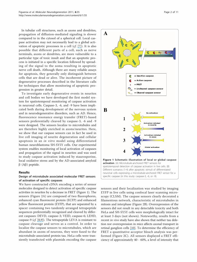
In tubular cell structures, such as axons and dendrites,propagation of diffusion-mediated signaling is slowercompared to in the cytosol of a spherical cell. Local cas-pase activation may not necessarily lead to a global acti-vation of apoptotic processes in a cell (cf. [7]). It is alsopossible that different parts of a cell, such as nerveterminals, axons or dendrites, are more vulnerable to aparticular type of toxic insult and that an apoptotic pro-cess is initiated in a specific location followed by spread-ing of the signal to the soma resulting in apoptoticnerve cell death. Although there are many reliable assaysfor apoptosis, they generally only distinguish betweencells that are dead or alive. The incoherent picture ofdegenerative processes described in the literature callsfor techniques that allow monitoring of apoptotic pro-gression in greater detail.To investigate early degenerative events in neurites
and cell bodies we have developed the first model sys-tem for spatiotemporal monitoring of caspase activationin neuronal cells. Caspase-3, -6, and -9 have been impli-cated both during development of the nervous systemand in neurodegenerative disorders, such as AD. Hence,fluorescence resonance energy transfer (FRET)-basedsensors preferentially cleaved by caspase-3, -6 and -9were designed. The sensors localize to microtubules andare therefore highly enriched in axons/neurites. Here,we show that our caspase sensors can in fact be used inlive cell imaging of neurite degeneration and cellularapoptosis in an in vitro model using differentiatedhuman neuroblastoma SH-SY5Y cells. Our experimentalsystem enables monitoring of local activation of caspasesand propagation of the signal in neurites and was usedto study caspase activation induced by staurosporine,local oxidative stress and by the AD-associated amyloidb (Ab) peptide.
ResultsDesign of microtubule associated molecular FRET sensorsof activation of specific caspasesWe have constructed cDNA encoding a series of sensormolecules designed to detect activation of specific caspaseactivities in neurites by a decrease in FRET (Figure 1). Thesensors (Figure 2A) are composed of two fluorophores,enhanced cyan fluorescent protein (ECFP) and enhancedyellow fluorescent protein (EYFP), that are separated by aspacer containing two tandemly arranged tetrapeptidesequences preferentially recognized and cleaved by differ-ent caspases: DEVD, caspase-3; VEID, caspase-6; LEHD,caspase-9 (cf. [8,9]). The tetrapeptide LEVA is resistant tocaspase cleavage and serves as a control. In order tolocalize the caspase sensors to microtubules, which areabundant in axons of neurons, they were fused to themicrotubule-associated protein tau. HeLa cells were tran-siently transfected with plasmids encoding the caspase
sensors and their localization was studied by imagingEYFP in live cells using confocal laser scanning micro-scopy (CLSM). The caspase sensors clearly decorated afilamentous network, characteristic of microtubules inmitosis and interphase (Figure 2B). Overexpression of thesensors did not result in any detectable toxicity and bothHeLa and SH-SY5Y cells were morphologically intact forat least 3 days (not shown). Noteworthy, results from arecent in vivo study have also shown that neither tau dele-tion nor overexpression in mice affects axonal transport inretinal ganglion cells [10]. To determine the efficiency ofFRET a quantitative acceptor bleach analysis was per-formed (Figure 2C). All sensors displayed a FRET effi-ciency of approximately 40 - 60%, a level of intensity that
Figure 1 Schematic illustration of local or global caspaseactivation. (A) Microtubule-anchored FRET sensors forspatiotemporal detection of caspase activation in live cells. (B)Different scenarios (1-4) after apoptotic stimuli of differentiatedneuronal cells expressing a microtubule-anchored FRET sensor for aspecific caspase (in this study: caspase-3, -6, or -9).
Figueroa et al. Molecular Neurodegeneration 2011, 6:35http://www.molecularneurodegeneration.com/content/6/1/35
Page 2 of 11

is readily detectable via ratiometric imaging of FRET/ECFP. Thus, the sensor molecules gave strong and stableFRET signals prior to induction of apoptosis and caspaseactivation. Moreover, staurosporine treated U2OS cellsexpressing the caspase-6 (VEID) sensor, clearly showedmaintained localization of EYFP fluorescence to microtu-bules after loss of FRET (Figure 2D). In contrast, ECFPfluorescence displayed a diffuse distribution in the cell.This clearly demonstrates that loss of FRET is a conse-quence of VEID-specific cleavage and is not due to anyother proteolytic event.
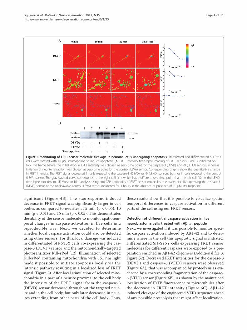
Loss of FRET and proteolytic cleavage of sensormolecules in differentiated neuroblastoma cellsundergoing apoptosisDifferentiated human neuroblastoma SH-SY5Y cellswere transfected with plasmids encoding the differentFRET sensor molecules. The experiments were startedby addition of staurosporine, one of the best inducers ofapoptosis in many different cell types, after which thecells were imaged by FRET microscopy for 8 hours inorder to monitor changes in FRET. In cells expressingsensors for caspase-3 (DEVD) a drop in FRET wasdetectable before retraction of neurites and the FRETsignal continued to decrease for 20 min (Figure 3A).The decrease in FRET signal from the caspase-9(LEHD) sensor was completed in 10 min (Figure 3A).Cells expressing the control FRET sensor, containingthe engineered non-cleavable tetra-amino acid motif(LEVA), did not show any alteration in FRET signalwithin this time frame (Figure 3A) although massiveapoptotic morphological changes were evident (notshown).Western blot analysis, using anti-GFP antibodies, of
extracts of SH-SY5Y cells expressing the caspase-3(DEVD) or control (LEVA) FRET sensors displayed a spe-cific band with an apparent molecular weight of 130 kDa(Figure 3B). After 3 hours of incubation with 10 μM staur-osporine a specific band at approximately 100 kDaappeared in cells expressing the caspase-3 (DEVD) sensor.EYFP and ECFP both have an expected size of 27 kDa,whereas the 441 amino acid long tau isoform in humanbrain tissue has been reported to migrate with an apparentmolecular weight of approximately 74 kDa [11]. Thus, the~100 kDa cleavage product is consistent with the expectedmigration of the EYFP-Tau fragment (~27 kDa + ~74kDa) from caspase-dependent cleavage of the DEVDmotif. In support of this is the fact that the ~100 kDaband was absent from staurosporine treated cells expres-sing the control (LEVA) sensor. In addition, the presenceof caspase inhibitors prevented staurosporine-inducedappearance of the ~100 kDa band (Additional file 1, FigureS1). The results demonstrate that decreased FRET inten-sity in apoptotic SH-SY5Y cells expressing the FRET sen-sor was correlated to caspase-dependent proteolysis of thesensor molecule.
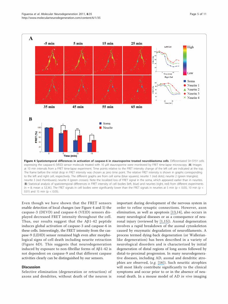
Detection of local caspase activation in liveneuroblastoma cellsWhen we monitored the caspase activity in differentiatedneuroblastoma cells expressing the caspase-6 (VEID) sen-sor treated with staurosporine we observed that theFRET intensity first decreased in the cell body and only5-10 min later decreased in the neurites (Figure 4A andAdditional file 2, Movie). Statistical analysis of a largerdata set showed that the spatiotemporal difference was
Figure 2 Design of FRET sensor molecules for detection oflocal activation of specific caspases. (A) Schematic outline ofFRET sensor molecules. ECFP and EYFP are connected with differentspacer sequences containing two tandemly arranged tetra-aminoacid cleavage sequences preferentially cleaved by different caspases(as specified) or a non-cleavable control tetra-amino acid LEVAsequence. The microtubule binding Tau protein is fused to the C-terminus of EYFP to target the sensors to microtubules. (B)Distribution of the caspase-3 (DEVD) sensor to microtubules ofmitotic (left) and interphase (right) HeLa cells. (C) Quantitativeacceptor bleaching experiments in differentiated neuroblastoma SH-SY5Y cells expressing different caspase sensors, showingapproximately 50% increase in ECFP fluorescence after bleaching ofEYFP. (D) U2OS cells expressing FRET sensors for caspase-6 (VEID)treated with 10 μM staurosporine. The apoptotic cell (star) showedmaintained localization of EYFP fluorescence to microtubules (rightcell), in contrast to ECFP fluorescence, after loss of FRET.
Figueroa et al. Molecular Neurodegeneration 2011, 6:35http://www.molecularneurodegeneration.com/content/6/1/35
Page 3 of 11
significant (Figure 4B). The staurosporine-induceddecrease in FRET signal was significantly larger in cellbodies as compared to neurites at 5 min (p < 0,05), 10min (p < 0.01) and 15 min (p < 0.05). This demonstratesthe ability of the sensor molecule to monitor spatiotem-poral changes in caspase activation in live cells in areproducible way. Next, we decided to determinewhether local caspase activation could also be detectedusing other sensors. For this, local damage was inducedin differentiated SH-SY5Y cells co-expressing the cas-pase-3 (DEVD) sensor and the mitochondrially-targetedphotosensitizer KillerRed [12]. Illumination of selectedKillerRed containing mitochondria with 561 nm lightmade it possible to initiate apoptosis locally via theintrinsic pathway resulting in a localized loss of FRETsignal (Figure 5). After local stimulation of selected mito-chondria in a part of a neurite proximal to the cell bodythe intensity of the FRET signal from the caspase-3(DEVD) sensor decreased throughout the targeted neur-ite and in the cell body, but only later decreased in neur-ites extending from other parts of the cell body. Thus,
these results show that it is possible to visualize spatio-temporal differences in caspase activation in differentparts of the cell using our FRET sensors.
Detection of differential caspase activation in liveneuroblastoma cells treated with Ab1-42 peptideNext, we investigated if it was possible to monitor speci-fic caspase activation induced by Ab1-42 and to deter-mine where in the cell this apoptotic signal is initiated.Differentiated SH-SY5Y cells expressing FRET sensormolecules for different caspases were exposed to a pre-paration enriched in Ab1-42 oligomers (Additional file 3,Figure S2). Decreased FRET intensities for the caspase-3(DEVD) and caspase-6 (VEID) sensors were observed(Figure 6A), that was accompanied by proteolysis as evi-denced by a corresponding fragmentation of the caspase-6 (VEID) sensor (Figure 6B). As shown by the maintainedlocalization of EYFP fluorescence to microtubules afterthe decrease in FRET intensity (Figure 6C), Ab1-42induced cleavage of the engineered VEID sequence aheadof any possible proteolysis that might affect localization.
Figure 3 Monitoring of FRET sensor molecule cleavage in neuronal cells undergoing apopotosis. Transfected and differentiated SH-SY5Ycells were treated with 10 μM staurosporine to induce apoptosis. (A) FRET intensity time-lapse imaging of FRET sensors. Time is indicated ontop. The frame before the initial drop in FRET intensity was chosen as zero time point for the caspase-3 (DEVD) and -9 (LEHD) sensors, whereasinitiation of neurite retraction was chosen as zero time point for the control (LEVA) sensor. Corresponding graphs show the quantitative changein FRET intensity. The FRET signal decreased in cells expressing the caspase-3 (DEVD), or -9 (LEHD) sensors, but not in cells expressing the control(LEVA) sensor. The gray dashed curve corresponds to the right cell (#1), which has a different zero time point than the left cell (#2) in the LEHDtime-lapse experiment. (B) Western blot analysis using anti-GFP antibodies of FRET sensor molecules in extracts of cells expressing the caspase-3(DEVD) sensor or the uncleavable control (LEVA) sensor incubated for 3 hours in the absence or presence of 10 μM staurosporine.
Figueroa et al. Molecular Neurodegeneration 2011, 6:35http://www.molecularneurodegeneration.com/content/6/1/35
Page 4 of 11
Even though we have shown that the FRET sensorsenable detection of local changes (see Figure 4 and 5) thecaspase-3 (DEVD) and caspase-6 (VEID) sensors dis-played decreased FRET intensity throughout the cell.Thus, our results suggest that the Ab1-42 peptideinduces global activation of caspase-3 and caspase-6 inthese cells. Interestingly, the FRET intensity from the cas-pase-9 (LEHD) sensor remained high even after morpho-logical signs of cell death including neurite retraction(Figure 6D). This suggests that neurodegenerationinduced by exposure to non-fibrillar forms of Ab1-42 isnot dependent on caspase-9 and that different caspaseactivities clearly can be distinguished by our sensors.
DiscussionSelective elimination (degeneration or retraction) ofaxons and dendrites, without death of the neuron is
important during development of the nervous system inorder to refine synaptic connections. However, axonelimination, as well as apoptosis [13,14], also occurs inmany neurological diseases or as a consequence of neu-ronal injury (reviewed by [1,15]). Axonal degenerationinvolves a rapid breakdown of the axonal cytoskeletoncaused by enzymatic degradation of neurofilaments. Aprocess termed dying-back degeneration (or Wallerian-like degeneration) has been described in a variety ofneurological disorders and is characterized by initialdegeneration of distal regions of long axons followed bydistal-to-proximal progression. In many neurodegenera-tive diseases, including AD, axonal and dendritic atro-phies are observed, (e.g. [16]). Such neuritic atrophieswill most likely contribute significantly to the clinicalsymptoms and occur prior to or in the absence of neu-ronal death. In a mouse model of AD in vivo imaging
Figure 4 Spatiotemporal differences in activation of caspase-6 in staurosporine treated neuroblastoma cells. Differentiated SH-SY5Y cellsexpressing the caspase-6 (VEID) sensor molecule treated with 10 μM staurosporine were monitored by FRET time-lapse microscopy. (A) Imagesat 10 min intervals from a FRET time-lapse experiment. Time points relative to the FRET intensity change of the left cell are indicated at the top.The frame before the initial drop in FRET intensity was chosen as zero time point. The relative FRET intensity is shown in graphs correspondingto the left and right cell, respectively. The different graphs are from cell soma (blue squares); neurite 1 (red dots); neurite 2 (green triangles);neurite 3 (red rhombuses); neurite 4 (green crosses). Note the localized loss of FRET signal in the soma, which appeared earlier than in neurites.(B) Statistical analysis of spatiotemporal differences in FRET intensity of cell bodies (left, blue) and neurites (right, red) from different experiments(n = 8; mean ± S.E.M.). The FRET signals in cell bodies were significantly lower than the FRET signals in neurites at 5 min (p < 0.05), 10 min (p <0.01) and 15 min (p < 0.05).
Figueroa et al. Molecular Neurodegeneration 2011, 6:35http://www.molecularneurodegeneration.com/content/6/1/35
Page 5 of 11

studies indicated that the Ab deposits, typically observedin AD brain, can indeed induce breakage of axonal anddendritic branches [17].Here, we describe the development and characteriza-
tion of new FRET based sensors for spatiotemporal ima-ging and analysis of caspase activation in relation toneurite degeneration and neuronal cell death. A full-length tau moiety was added to the FRET sensors bothto mediate association to microtubules in neurites andthus increase signal intensity as well as to restrict diffu-sion of the sensors to allow monitoring progression ofthe apoptotic signal in a spatiotemporal manner (cf.Figure 1). As expected our caspase sensors localize tothe cytoskeleton when expressed in non-neuronal(HeLa) cells and also to the microtubule of the neuriteswhen expressed in differentiated neuronal cells. FRETstudies in differentiated SH-SY5Y cells exposed to staur-osporine verify the sensors as early markers of apoptosisoccurring prior to neurite retraction and any other mor-phological changes could be detected. Caspase activationwas measured as loss of FRET and it was confirmed, bywestern blot analysis of sensor fragmentation, to corre-spond to cleavage between EYFP and ECFP where thecaspase cleavage sites are localized.In this study we focus on three different caspases; cas-
pase-3, -6, and -9. Caspase-9 is the initiator caspase asso-ciated with the mitochondrial or intrinsic apoptoticpathway [18]. Proapoptotic signals stimulating release of
cytochrome c from mitochondria that promotes oligo-merization of apoptotic peptidase activating factor 1(Apaf-1), will lead to recruitment and activation of cas-pase-9. Caspase-9 null mice die perinatally and show asimilar phenotype as mice with deletion of Apaf-1 or cas-pase-3 genes, including enlarged and deformed brains[19,20]. This clearly indicates the importance of theintrinsic apoptotic pathway during development of thenervous system. Caspase-9 has also been proposed to beactivated by an alternative pathway: It has been shownthat endoplasmic reticulum (ER) stress can activate cas-pase-9 in the absence of cytochrome c and Apaf-1[21,22]. Interestingly, Ab peptides have been shown toinduce apoptosis mediated by ER stress [23]. Further-more, increased levels of active caspase-9 have beendetected in AD brain [15,24]. Also, in a recently pub-lished study [25] caspase-9 and the intrinsic apoptoticpathway was suggested to be involved in non-apoptoticcaspase-3 activity in dendritic spines in Tg2576-APPswemice. This activity correlated with the onset of memorydeficits in these mice. Therefore, caspase-9 has been pro-posed to be involved not only in developmental apoptosisof neuronal cells but also in neurodegenerative disorderssuch as AD. Caspase-3 is the major executioner caspaseand can be activated once an initiator caspase, like cas-pase-9, is activated. Caspase-6 is also often classified asan executioner caspase since it cleaves the nuclear laminproteins during apoptosis [6]. However, proteomics
Figure 5 Detection of caspase activity induced by local photo activation of mitochondrial KillerRed. Differentiated neuroblastoma cellscoexpressing mitochondrially targeted KillerRed and the caspase-3 (DEVD) sensor molecule. (A) Images of mitochondrial KillerRed fluorescence,with corresponding phase contrast image, before (circled) and after photo-activation in selected mitochondria. (B) Corresponding FRET intensitytime-lapse images. Time points (see Figure 3) relative to the FRET intensity change are indicated at the top. The frame before the initial drop inFRET intensity was chosen as zero time point. The graph displays relative FRET intensity in the cell body (blue squares), neurite 1 (red dots),neurite 2 (green triangles) and neurite 3 (magenta rhombuses).
Figueroa et al. Molecular Neurodegeneration 2011, 6:35http://www.molecularneurodegeneration.com/content/6/1/35
Page 6 of 11
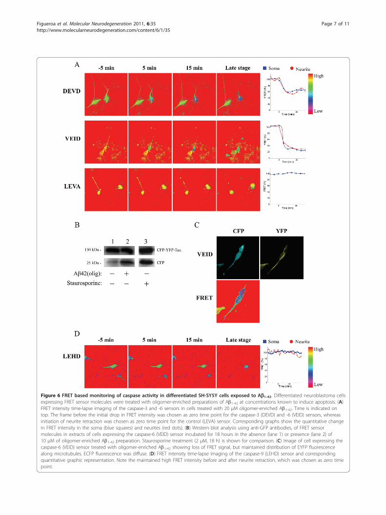
Figure 6 FRET based monitoring of caspase activity in differentiated SH-SY5Y cells exposed to Ab1-42. Differentiated neuroblastoma cellsexpressing FRET sensor molecules were treated with oligomer-enriched preparations of Ab1-42 at concentrations known to induce apoptosis. (A)FRET intensity time-lapse imaging of the caspase-3 and -6 sensors in cells treated with 20 μM oligomer-enriched Ab1-42. Time is indicated ontop. The frame before the initial drop in FRET intensity was chosen as zero time point for the caspase-3 (DEVD) and -6 (VEID) sensors, whereasinitiation of neurite retraction was chosen as zero time point for the control (LEVA) sensor. Corresponding graphs show the quantitative changein FRET intensity in the soma (blue squares) and neurites (red dots). (B) Western blot analysis using anti-GFP antibodies, of FRET sensormolecules in extracts of cells expressing the caspase-6 (VEID) sensor incubated for 18 hours in the absence (lane 1) or presence (lane 2) of10 μM of oligomer-enriched Ab1-42 preparation. Staurosporine treatment (2 μM, 18 h) is shown for comparison. (C) Image of cell expressing thecaspase-6 (VEID) sensor treated with oligomer-enriched Ab1-42 showing loss of FRET signal, but maintained distribution of EYFP fluorescencealong microtubules. ECFP fluorescence was diffuse. (D) FRET intensity time-lapse imaging of the caspase-9 (LEHD) sensor and correspondingquantitative graphic representation. Note the maintained high FRET intensity before and after neurite retraction, which was chosen as zero timepoint.
Figueroa et al. Molecular Neurodegeneration 2011, 6:35http://www.molecularneurodegeneration.com/content/6/1/35
Page 7 of 11
analysis showed that nearly half of identified neuronalcaspase-6 targets were cytoskeleton-associated, indicatingthat caspase-6 most likely can function also before deathsignals reach the nucleus [26]. In addition, recent studieshave implicated caspase-6 in axonal degeneration notlinked to apoptotic cell death [3].We evaluated the performance of the caspase sensors in
differentiated neuroblastoma cells treated with staurospor-ine. We observed a significant decrease in FRET signal incells expressing caspase-3 and -9 sensors, but not in cellsexpressing the non-cleavable control (Figure 3). Stauros-porine is a non-specific inhibitor of protein kinases and isone of the best inducers of apoptosis in different types ofcells, including neuroblastoma [27,28]. Although themechanism by which staurosporine induces apoptosis isnot well known, the mitochondrial pathway has beenshown to be involved [29,30], consistent with our results.For caspase-3 and -9, we could not detect any time differ-ence in the loss of FRET in neurites and soma. This indi-cates that staurosporine may induce a global activation ofthese two caspases in neuronal cells. However, when theloss of FRET in cells expressing the caspase-6 sensor wasanalyzed we observed a clear and significant delay in theneurites compared to the cell bodies (Figure 4). This indi-cates a difference in staurosporine-induced apoptotic sig-naling in axons/dendrites compared to the cell bodies. Aprevious study has indicated a spatiotemporal difference incaspase-3 compared to caspase-6 activation [31]. In undif-ferentiated SH-SY5Y cells exposed to staurosporine cas-pase-6 activation, monitored by the appearance of a laminB1 fragment in western blot analysis, was considerablydelayed compared to cleavage of caspase-3 substrates [31].Thus, it may be speculated that staurosporine induces adelayed activation of caspase-6 that is initiated in the cellbody and thereafter is spread to the neurites.The ability to detect caspase activation at sub-cellular
resolution was further established using a co-expressedmitochondrial photosensitizer (Killer red) [12], whichenabled induction of apoptosis locally in the cell via theintrinsic pathway. We observed that if caspase-3 activationwas initiated in one of the neurites the signal was quicklytransferred to the cell body but not to other neurites(Figure 5). The other neurites degenerated as well after thecell had been exposed to the death stimuli in spite of thedelayed caspase-3 activation.Finally, we used differentiated neuroblastoma cells
expressing caspase sensors to analyze the effect of the Abpeptide on apoptotic signaling. Apoptotic signaling isbelieved to play an important role in a number of neuro-logical disorders, including AD [32]. If caspase activationis crucial for the progression of AD pathology it is impor-tant to learn how the activation occurs, where in the neu-ron this process takes place, and whether or not it willresult in neuronal cell death. Ab is a hydrophobic peptide
generated by sequential proteolytic cleavage of amyloidprecursor protein (APP) by b- and g-secretase and is theleading candidate for activation of apoptotic mechanismsin AD [7,33]. Ab is highly prone to aggregate and in thebrain it is found in different forms ranging from mono-mers and non-fibrillar aggregates, termed oligomers, to ahighly fibrillar form. Recent evidences suggest that it isthe diffusible Ab oligomers that have the most toxicproperties [34,35]. Ab oligomers have also been shown tobind to synaptic sites [36], which may be important forearly synaptic failure observed in AD (cf. [37]). Further-more, Ab has been proposed to have a local effect onneurites causing degeneration and neuritic apoptosisusing compartmented cultures of hippocampal neurons[38]. In the present study we have exposed the cells to apreparation enriched in oligomer-enriched forms of Abwith non-detectable levels of fibrils. Ab exposure resultedin activation of caspase-3 and -6, but we could not detectany specific spatiotemporal effect of the peptide. Thus,our results suggest that in these neuronally differentiatedcells there is no part of the cell that is more vulnerable toinsult and that Ab rather induces global caspase activa-tion. Because the caspase-9 sensor resisted proteolysis,the intrinsic pathway may play a less important role inAb1-42 induced apoptosis of these cells. Most interest-ingly, it was recently shown in a mouse model that cas-pase activation precedes and leads to tangles (i.e., acharacteristic neuropathological feature in AD).Evidences also indicated that caspase-3 and -6 were acti-vated in the tangle positive cells [39].In conclusion, we show that the anchored caspase sen-
sors localize to the cytoskeleton, that they are differentiallycleaved depending on stimuli and that they can be used todetect local caspase activation and to follow the spreadingof caspase activation from one part of the cell to another.Under the conditions used in this study we could notdetect any specific spatiotemporal effect of Ab on caspaseactivation. Caspase-3 and -6, but not -9, were activatedboth in soma and in neurites. It will be interesting to usethe anchored FRET sensors in experiments designed tocause retraction and loss of synaptic contacts withoutcausing cell death. This is indeed a promising new tool inresearch on AD and other neurodegenerative conditions.
Materials and methodsGene constructionTo construct the localized caspase sensors, EYFP wasamplified from the EYFP-C1 plasmid (BD Biosciences)using the following primers: forward, 5 ’-TATTA-GATCTCATGGTGAGCAAGGGCGA-G-3’, reverse,5’-ACGTGAATTCCTTGTACAGCTCGTCCATGC-3’.The resulting PCR product was digested with BglII-EcoRI, gel-purified and ligated into the BglII-EcoRI ofthe ECFP-C1 vector (BD Biosciences). The longest
Figueroa et al. Molecular Neurodegeneration 2011, 6:35http://www.molecularneurodegeneration.com/content/6/1/35
Page 8 of 11
isoform of human tau (441 amino acids) was amplifiedfrom the pBThtau40 plasmid (kindly provided by Dr.Jacek Biernat, Max-Planck-Gesellschaft) using the fol-lowing primers: forward, 5’-ATTAGAATTCATGGCT-GAGCCCCGCCAG-3’, reverse, 5’-ATGCGGATCCGTGATCACAAAC-3’. The resulting PCR product wasdigested with EcoRI-BamHI, gel-purified and ligatedinto the EcoRI-BamHI of the ECFP-EYFP vector. Tocreate cleavage sites for caspase-3, -6 and -9 plus anun-cleavable control sequence single stranded sense32-mers; 5’-CCGGAGCAGATGAGGTCGATGCC-GATGAGGTC-3’ (caspase-3, DEVDx2), 5’-CCGGAG-CAGTCGAGATAGATGCCGTAGAGATC-3’ (caspase-6, VEIDx2), 5’-CCGGAGCACTGGAGCACGATGCCCTGGAGCAC-3’ (caspase-9, LEHDx2) and 5’-CCGGAGCACTGGAGGTCGCCCTGGAGGTCGCC-3’ (con-trol, LEVAx2) were annealed to complementary anti-sense 32-mers; 5’-GATCGACCTCATCGGCATCGACCTCATCTGCT-3’ (caspase-3), 5’-GATCGATCTC-TACGGCATCTATCTCGACTGCT-3’ (caspase-6), 5’-GATCGTGCTCCAGGGCATCGTGCTCCAGTGCT-3’(caspase-9) and 5 ’-GATCGGCGACCTCCAGGGC-GACCTCCAGTGCT-3’ (control) generating doublestranded DNA with caspase cleavage sites in tandemand BspEI-BglII sticky ends that were ligated into theBspEI-BglII of the ECFP-EYPP-Tau vector. One basewas changed in the BglII site in the linker region tofacilitate restriction analysis.
Cell culture and transfectionHeLa (CCL-2, American Type culture Collection) andU2OS cells (ATCC-HTB-96, LGC standards) were cul-tured in Dulbecco’s Modified Eagle Medium (DMEM, Invi-trogen) supplemented with 10% fetal bovine serum (FBS,Invitrogen), 100 U/ml penicillin and 100 μg/ml streptomy-cin (PEST, Invitrogen). Human neuroblastoma SH-SY5Ycells (CRL-2266, ATCC) were cultured in DMEM/F12 sup-plemented with 10% FBS, 100 U/ml PEST. The cells weremaintained in a humidified 5% CO2 atmosphere at 37°C.Cells were transfected using FuGENE HD (Roche). In livecell imaging experiments cells were seeded on to glasscover slips prior to differentiation. The neuroblastoma cellswere differentiated with 10 μM retinoic acid (RA) for threedays before transfection after witch cells were incubatedfor one day prior to the experiment. For western blot ana-lysis cells were seeded at a density of 25.000 cells/cm2 intoPetri dishes (Nunc, Denmark). The following day the cellswere treated with 10 μM RA for 72 h. After 24 h, the cellswere transfected with plasmids encoding different caspasesensors and incubated for 48 h prior to the experiment.
Preparation of Ab1-42 enriched in oligomersAb1-42 (American peptide) was dissolved in hexafluoroi-sopropanol (HFP) to remove any preexisting structures
in the lyophilized stocks and was then stored at -70°C.The peptide was prepared to obtain a solution enrichedin oligomeric, but devoid of fibrillar, forms of the peptide,cf. [40]. The oligomer-enriched Ab1-42 was preparedfreshly before each experiment. HFP was allowed to eva-porate from the tube using speedvac system for 5 min atroom temperature. Thereafter, Ab1-42 was dissolved indimethyl sulfoxide (DMSO) to a concentration of 5 mM.Subsequently, phosphate bovine saline (PBS) was addedto give a final Ab1-42 concentration of 100 μM. Finally,the preparation was vigorously vortexed at room tem-perature for 10 min, whereafter Ab1-42 was allowed toaggregate by incubation for 24 h at 4°C at 200 rpm. Ana-lysis of the Ab preparation was made by western blot andThioflavin T assay (Additional file 2, Figure S2).
ImagingImaging was performed on a Leica TCS-SP laser scanningconfocal microscope (Leica, Heidelberg, Germany) with a63.0 × 1.32 oil immersion objective using a 442 nm10 mW HeCd laser, 488 nm laser line of a 40 mW Ar-laser and 561 nm laser line of a yellow diode pumpedsolid-state 50 mW laser. Emission was collected between455-505 nm (CFP), 515-575 nm (YFP and FRET), and580-650 nm (KillerRed) using a long pass dicroic mirror(LP) 450 for CFP and FRET, LP500 for YFP and tripledicroic 488/561/633 for KillerRed. The laser lines werescanned sequentially to minimize bleed through. Inductionof KillerRed phototoxic effect was achieved by scanning aregion of the cells using the 561 nm laser line. Live cellimaging was performed by mounting cells in sealed cham-bers with serum free media buffered with HEPES. Inexperiments with staurosporine and Ab, the agent wasadded to the media when mounting the cells. Temperaturewas maintained at 37°C using the Cube and Box micro-scope incubator system (Live Imaging Services). Projec-tions of image stacks were used for quantification. Imageswere processed and quantified using ImageJ (Rasband, W.S., Image J, U. S. National Institutes of Health, Bethesda,Maryland, USA, http://rsb.info.nih.gov/ij/, 1997-2009). Toacquire the ratio-metric images the intensity in the FRETchannel was divided by the intensity in the CFP channel.A pseudo-color was applied and adjusted to visualize thechanges in FRET. For graphic representation, images weresegmented in a semi-automatic process rendering somaand neurite regions of interest. Values were normalized inrelation to the mean value prior to changes in FRET. Thelast frame before decreased FRET could be detected ineach individual experiment was chosen as zero time pointto enable alignment of graphs from different experiments.At least three individual experiments were performed.Spatiotemporal studies of FRET signals in neurites andsoma were statistically analyzed using unpaired two-tailedStudent’ s T-test.
Figueroa et al. Molecular Neurodegeneration 2011, 6:35http://www.molecularneurodegeneration.com/content/6/1/35
Page 9 of 11

Western blot analysisFor western blot analysis of caspase sensor fragmentationhuman neuroblastoma SH-SY5Y cells treated with Ab1-42 or staurosporine were harvested as follows: First, thecells were washed twice with PBS containing 0.7 μg/mlaprotinin (from bovine) and 0.8 mM PMSF. Thereafter,the cells were lysed in lysis buffer (150 mM NaCl,50 mM TrisCl, 1 mM EDTA, 1% NP-40), containingcomplete protease inhibitor cocktail, for 20 min at 4°C.In order to remove cell debris the lysate was centrifugedat 10 000 g for 5 min at 4°C. The supernatant was thenused for western blot analysis. 30 μg protein from eachseparate sample was separated by electrophoresis on 8%SDS-polyacrylamide gels and subsequently transferred topolyvinyldine difluoride membranes at 350 mA for 3 h.The membrane was blocked in PBS, containing 5% drymilk (w/v) and 0.1% Tween, for 1.5 h, followed by overnight incubation at 4°C with primary rabbit polyclonal ormouse monoclonal anti-GFP antibody (1:1000) (R&Dsystems). Finally, secondary horseradish peroxidise-coupled donkey anti-rabbit or sheep anti-mouse antibo-dies (1:5000) were incubated for 1 h at room temperatureand the detected proteins were visualised by chemilumi-nescence using Western LightingTM-PLUS-ECL kit (Per-kin Elmer, Netherlands). All western blot reagents werefrom GE Healthcare or Bio-Rad Laboratories.
Additional material
Additional file 1: Figure S1 Inhibition of staurosporine-inducedFRET-sensor cleavage by caspase inhibitors. western blot analysisusing anti-GFP antibodies of FRET sensor molecules in cells expressingthe VEID sensor in the presence or absence (1 h pretreatment) of thecaspase inhibitors Z-VAD-FMK (Promega) or Z-Asp-CH2-DCB (PeptaNova)followed by 1 h staurosporine treatment.
Additional file 2: Movie. Spatiotemporal difference in activation ofcaspase-6 in staurosporine treated neuroblastoma cells.Differentiated SH-SY5Y cells expressing the caspase-6 (VEID) sensormolecule treated with staurosporine were monitored by FRET time-lapsemicroscopy. One frame every 5 min.
Additional file 3: Figure S2 Analysis of oligomer- and fibril-enrichedpreparations of Ab1-42. (A) Representative western blot showing highlevels of fibrilar Ab in the oligomeric/fibrillar preparation (lane 1) and theabsence of Ab fibrils in the oligomeric preparation (lane 2). The Absamples were separated by SDS-PAGE and both stacking- and separationgel was analyzed by western blot using 6E10 antibodies (SignetLaboratories). (B) Statistical analysis of Thioflavin T (T3516, Sigma)fluorescence in oligomeric/fibril-enriched Ab preparations (fAb; open bar)and oligomer-enriched Ab preparations (oAb; filled bar). Ab samples wereanalyzed in phenol red-free medium and according to the manufacturer’sinstructions. The data represent mean ± SEM for three independentexperiments. A.U., arbitrary units. *p < 0.05 significantly different from fAb.
AcknowledgementsWe thank Konstantin Lukyanov for the kind gift of KillerRed plasmid, Dr.Jacek Biernat for the kind gift of pBThtau40 plasmid, and Dr. Marie Beckmanfor valuable discussion. This work was supported by grants from theSwedish Research Council to EH (#2006-5977, #2006-3468 and #2010-4481)and to KI (#2007-2798 and #2010-4505).
Author details1Department of Neurochemistry, Stockholm University, SE-106 91 Stockholm,Sweden. 2Department of Biochemistry and Biophysics, Stockholm University,SE-106 91 Stockholm, Sweden.
Authors’ contributionsThe original design of this study was made by RAF, VR, KI and EH. cDNAconstruction was made by VR, MZ and MS. Cell culture, transfection,treatments and microscopy was performed by RAF, VR and TG and imageanalysis was performed by RAF. TG performed western blot analysis. RAFand VR coordinated the experiments. RAF, VR, TG, KI and EH wrote themanuscript. All authors have read and approved the final version of themanuscript.
Competing interestsThe authors declare that they have no competing interests.
Received: 5 October 2010 Accepted: 23 May 2011Published: 23 May 2011
References1. MC Raff, AV Whitmore, JT Finn, Axonal self-destruction and
neurodegeneration. Science. 296, 868–871 (2002). doi:10.1126/science.10686132. DW Williams, S Kondo, A Krzyzanowska, Y Hiromi, JW Truman, Local caspase
activity directs engulfment of dendrites during pruning. Nat Neurosci. 9,1234–1236 (2006). doi:10.1038/nn1774
3. A Nikolaev, T McLaughlin, DD O’Leary, M Tessier-Lavigne, APP binds DR6 totrigger axon pruning and neuron death via distinct caspases. Nature. 457,981–989 (2009). doi:10.1038/nature07767
4. S Albrecht, N Bogdanovic, B Ghetti, B Winblad, AC LeBlanc, Caspase-6activation in familial alzheimer disease brains carrying amyloid precursorprotein or presenilin i or presenilin II mutations. J Neuropathol Exp Neurol.68, 1282–1293 (2009). doi:10.1097/NEN.0b013e3181c1da10
5. H Guo, S Albrecht, M Bourdeau, T Petzke, C Bergeron, AC LeBlanc, Activecaspase-6 and caspase-6-cleaved tau in neuropil threads, neuritic plaques,and neurofibrillary tangles of Alzheimer’s disease. Am J Pathol. 165,523–531 (2004). doi:10.1016/S0002-9440(10)63317-2
6. YA Lazebnik, A Takahashi, RD Moir, RD Goldman, GG Poirier, SH Kaufmann,WC Earnshaw, Studies of the lamin proteinase reveal multiple parallelbiochemical pathways during apoptotic execution. Proc Natl Acad Sci USA.92, 9042–9046 (1995). doi:10.1073/pnas.92.20.9042
7. MP Mattson, Pathways towards and away from Alzheimer’s disease. Nature.430, 631–639 (2004). doi:10.1038/nature02621
8. A Takahashi, ES Alnemri, YA Lazebnik, T Fernandes-Alnemri, G Litwack, RDMoir, RD Goldman, GG Poirier, SH Kaufmann, WC Earnshaw, Cleavage oflamin A by Mch2 alpha but not CPP32: multiple interleukin 1 beta-converting enzyme-related proteases with distinct substrate recognitionproperties are active in apoptosis. Proc Natl Acad Sci USA. 93, 8395–8400(1996). doi:10.1073/pnas.93.16.8395
9. K Takemoto, T Nagai, A Miyawaki, M Miura, Spatio-temporal activation ofcaspase revealed by indicator that is insensitive to environmental effects. JCell Biol. 160, 235–243 (2003). doi:10.1083/jcb.200207111
10. A Yuan, A Kumar, C Peterhoff, K Duff, RA Nixon, Axonal transport rates invivo are unaffected by tau deletion or overexpression in mice. J Neurosci.28, 1682–1687 (2008). doi:10.1523/JNEUROSCI.5242-07.2008
11. L Buee, T Bussiere, V Buee-Scherrer, A Delacourte, PR Hof, Tau proteinisoforms, phosphorylation and role in neurodegenerative disorders. BrainRes Brain Res Rev. 33, 95–130 (2000)
12. ME Bulina, DM Chudakov, OV Britanova, YG Yanushevich, DB Staroverov, TVChepurnykh, EM Merzlyak, MA Shkrob, S Lukyanov, KA Lukyanov, Agenetically encoded photosensitizer. Nat Biotechnol. 24, 95–99 (2006).doi:10.1038/nbt1175
13. JF Kerr, AH Wyllie, AR Currie, Apoptosis: a basic biological phenomenonwith wide-ranging implications in tissue kinetics. Br J Cancer. 26, 239–257(1972). doi:10.1038/bjc.1972.33
14. RC Taylor, SP Cullen, SJ Martin, Apoptosis: controlled demolition at thecellular level. Nat Rev Mol Cell Biol. 9, 231–241 (2008)
15. DC Lu, S Rabizadeh, S Chandra, RF Shayya, LM Ellerby, X Ye, GS Salvesen,EH Koo, DE Bredesen, A second cytotoxic proteolytic peptide derived fromamyloid beta-protein precursor. Nat Med. 6, 397–404 (2000). doi:10.1038/74656
Figueroa et al. Molecular Neurodegeneration 2011, 6:35http://www.molecularneurodegeneration.com/content/6/1/35
Page 10 of 11

16. TC Dickson, CE King, GH McCormack, JC Vickers, Neurochemical diversity ofdystrophic neurites in the early and late stages of Alzheimer’s disease. ExpNeurol. 156, 100–110 (1999). doi:10.1006/exnr.1998.7010
17. J Tsai, J Grutzendler, K Duff, WB Gan, Fibrillar amyloid deposition leads tolocal synaptic abnormalities and breakage of neuronal branches. NatNeurosci. 7, 1181–1183 (2004). doi:10.1038/nn1335
18. P Li, D Nijhawan, I Budihardjo, SM Srinivasula, M Ahmad, ES Alnemri, XWang, Cytochrome c and dATP-dependent formation of Apaf-1/caspase-9complex initiates an apoptotic protease cascade. Cell. 91, 479–489 (1997).doi:10.1016/S0092-8674(00)80434-1
19. R Hakem, A Hakem, GS Duncan, JT Henderson, M Woo, MS Soengas, A Elia,JL de la Pompa, D Kagi, W Khoo., et al, Differential requirement for caspase9 in apoptotic pathways in vivo. Cell. 94, 339–352 (1998). doi:10.1016/S0092-8674(00)81477-4
20. K Kuida, TF Haydar, CY Kuan, Y Gu, C Taya, H Karasuyama, MS Su, P Rakic,RA Flavell, Reduced apoptosis and cytochrome c-mediated caspaseactivation in mice lacking caspase 9. Cell. 94, 325–337 (1998). doi:10.1016/S0092-8674(00)81476-2
21. N Morishima, K Nakanishi, H Takenouchi, T Shibata, Y Yasuhiko, Anendoplasmic reticulum stress-specific caspase cascade in apoptosis.Cytochrome c-independent activation of caspase-9 by caspase-12. J BiolChem. 277, 34287–34294 (2002). doi:10.1074/jbc.M204973200
22. RV Rao, S Castro-Obregon, H Frankowski, M Schuler, V Stoka, G del Rio, DEBredesen, HM Ellerby, Coupling endoplasmic reticulum stress to the celldeath program. An Apaf-1-independent intrinsic pathway. J Biol Chem. 277,21836–21842 (2002). doi:10.1074/jbc.M202726200
23. T Nakagawa, H Zhu, N Morishima, E Li, J Xu, BA Yankner, J Yuan, Caspase-12 mediates endoplasmic-reticulum-specific apoptosis and cytotoxicity byamyloid-beta. Nature. 403, 98–103 (2000). doi:10.1038/47513
24. TT Rohn, RA Rissman, MC Davis, YE Kim, CW Cotman, E Head, Caspase-9activation and caspase cleavage of tau in the Alzheimer’s disease brain.Neurobiol Dis. 11, 341–354 (2002). doi:10.1006/nbdi.2002.0549
25. M D’Amelio, V Cavallucci, S Middei, C Marchetti, S Pacioni, A Ferri, ADiamantini, D De Zio, P Carrara, L Battistini, S Moreno, A Bacci, MAmmassari-Teule, H Marie, F Cecconi, Caspase-3 triggers early synapticdysfunction in a mouse model of Alzheimer’s disease. Nat Neurosci. 14,69–76 (2011). doi:10.1038/nn.2709
26. G Klaiman, TL Petzke, J Hammond, AC Leblanc, Targets of caspase-6 activityin human neurons and Alzheimer disease. Mol Cell Proteomics. 7,1541–1555 (2008). doi:10.1074/mcp.M800007-MCP200
27. J Boix, N Llecha, VJ Yuste, JX Comella, Characterization of the cell deathprocess induced by staurosporine in human neuroblastoma cell lines.Neuropharmacology. 36, 811–821 (1997). doi:10.1016/S0028-3908(97)00030-0
28. M Deshmukh, EM Johnson Jr, Staurosporine-induced neuronal death:multiple mechanisms and methodological implications. Cell Death Differ. 7,250–261 (2000). doi:10.1038/sj.cdd.4400641
29. K Li, Y Li, JM Shelton, JA Richardson, E Spencer, ZJ Chen, X Wang, RSWilliams, Cytochrome c deficiency causes embryonic lethality andattenuates stress-induced apoptosis. Cell. 101, 389–399 (2000). doi:10.1016/S0092-8674(00)80849-1
30. MC Wei, WX Zong, EH Cheng, T Lindsten, V Panoutsakopoulou, AJ Ross, KARoth, GR MacGregor, CB Thompson, SJ Korsmeyer, Proapoptotic BAX andBAK: a requisite gateway to mitochondrial dysfunction and death. Science.292, 727–730 (2001). doi:10.1126/science.1059108
31. VJ Yuste, I Sanchez-Lopez, C Sole, M Encinas, JR Bayascas, J Boix, JXComella, The prevention of the staurosporine-induced apoptosis by Bcl-X(L),but not by Bcl-2 or caspase inhibitors, allows the extensive differentiationof human neuroblastoma cells. J Neurochem. 80, 126–139 (2002).doi:10.1046/j.0022-3042.2001.00695.x
32. CW Cotman, AJ Anderson, A potential role for apoptosis inneurodegeneration and Alzheimer’s disease. Mol Neurobiol. 10, 19–45(1995). doi:10.1007/BF02740836
33. SC Biswas, Y Shi, JP Vonsattel, CL Leung, CM Troy, LA Greene, Bim iselevated in Alzheimer’s disease neurons and is required for beta-amyloid-induced neuronal apoptosis. J Neurosci. 27, 893–900 (2007). doi:10.1523/JNEUROSCI.3524-06.2007
34. Y Gong, L Chang, KL Viola, PN Lacor, MP Lambert, CE Finch, GA Krafft, WLKlein, Alzheimer’s disease-affected brain: presence of oligomeric A betaligands (ADDLs) suggests a molecular basis for reversible memory loss. ProcNatl Acad Sci USA. 100, 10417–10422 (2003). doi:10.1073/pnas.1834302100
35. C Haass, DJ Selkoe, Soluble protein oligomers in neurodegeneration:lessons from the Alzheimer’s amyloid beta-peptide. Nat Rev Mol Cell Biol. 8,101–112 (2007). doi:10.1038/nrm2101
36. PN Lacor, MC Buniel, L Chang, SJ Fernandez, Y Gong, KL Viola, MP Lambert,PT Velasco, EH Bigio, CE Finch., et al, Synaptic targeting by Alzheimer’s-related amyloid beta oligomers. J Neurosci. 24, 10191–10200 (2004).doi:10.1523/JNEUROSCI.3432-04.2004
37. DM Walsh, DJ Selkoe, A beta oligomers - a decade of discovery. JNeurochem. 101, 1172–1184 (2007). doi:10.1111/j.1471-4159.2006.04426.x
38. KJ Ivins, ET Bui, CW Cotman, Beta-amyloid induces local neuritedegeneration in cultured hippocampal neurons: evidence for neuriticapoptosis. Neurobiol Dis. 5, 365–378 (1998). doi:10.1006/nbdi.1998.0228
39. A de Calignon, LM Fox, R Pitstick, GA Carlson, BJ Bacskai, TL Spires-Jones, BTHyman, Caspase activation precedes and leads to tangles. Nature. 464,1201–1204 (2010). doi:10.1038/nature08890
40. WB Stine Jr, KN Dahlgren, GA Krafft, MJ LaDu, In vitro characterization ofconditions for amyloid-beta peptide oligomerization and fibrillogenesis. JBiol Chem. 278, 11612–11622 (2003). doi:10.1074/jbc.M210207200
doi:10.1186/1750-1326-6-35Cite this article as: Figueroa et al.: Anchored FRET sensors detect localcaspase activation prior to neuronal degeneration. MolecularNeurodegeneration 2011 6:35.
Submit your next manuscript to BioMed Centraland take full advantage of:
• Convenient online submission
• Thorough peer review
• No space constraints or color figure charges
• Immediate publication on acceptance
• Inclusion in PubMed, CAS, Scopus and Google Scholar
• Research which is freely available for redistribution
Submit your manuscript at www.biomedcentral.com/submit
Figueroa et al. Molecular Neurodegeneration 2011, 6:35http://www.molecularneurodegeneration.com/content/6/1/35
Page 11 of 11