Strategic analysis of the Technoscientific Network on Mediterranean Biological Diversity
Upload
khangminh22Category
view
0download
0
Fungal Diversity in the Mediterranean Area
Printed Edition of the Special Issue Published in Diversity
www.mdpi.com/journal/diversity
Giuseppe VenturellaEdited by
Fungal Diversity in the Mediterranean Area • Giuseppe Venturella

Fungal Diversity in theMediterranean Area


Fungal Diversity in theMediterranean Area
Editor
Giuseppe Venturella
MDPI • Basel • Beijing • Wuhan • Barcelona • Belgrade • Manchester • Tokyo • Cluj • Tianjin

Editor
Giuseppe Venturella
University of Palermo
Italy
Editorial Office
MDPI
St. Alban-Anlage 66
4052 Basel, Switzerland
This is a reprint of articles from the Special Issue published online in the open access journal
Diversity (ISSN 1424-2818) (available at: https://www.mdpi.com/journal/diversity/special issues/
fungal diversity).
For citation purposes, cite each article independently as indicated on the article page online and as
indicated below:
LastName, A.A.; LastName, B.B.; LastName, C.C. Article Title. Journal Name Year, Article Number,
Page Range.
ISBN 978-3-03936-978-2 (Hbk) ISBN 978-3-03936-979-9 (PDF)
c© 2020 by the authors. Articles in this book are Open Access and distributed under the Creative
Commons Attribution (CC BY) license, which allows users to download, copy and build upon
published articles, as long as the author and publisher are properly credited, which ensures maximum
dissemination and a wider impact of our publications.
The book as a whole is distributed by MDPI under the terms and conditions of the Creative Commons
license CC BY-NC-ND.

Contents
About the Editor . . . . . . . . . . . . . . . . . . . . . . . . . . . . . . . . . . . . . . . . . . . . . . vii
Giuseppe Venturella
Fungal Diversity in the Mediterranean AreaReprinted from: Diversity 2020, 12, 253, doi:10.3390/d12060253 . . . . . . . . . . . . . . . . . . . . 1
Elias Polemis, Vassiliki Fryssouli, Vassileios Daskalopoulos and Georgios I. Zervakis
Basidiomycetes Associated with Alnus glutinosa Habitats in Andros Island (Cyclades, Greece)Reprinted from: Diversity 2020, 12, 232, doi:10.3390/d12060232 . . . . . . . . . . . . . . . . . . . . 5
Beatrice Belfiori, Valentina D’Angelo, Claudia Riccioni, Marco Leonardi, Francesco Paolocci,
Giovanni Pacioni and Andrea Rubini
Genetic Structure and Phylogeography of Tuber magnatum PopulationsReprinted from: Diversity 2020, 12, 44, doi:10.3390/d12020044 . . . . . . . . . . . . . . . . . . . . 27
Maria Letizia Gargano, Georgios I. Zervakis, Omoanghe S. Isikhuemhen,
Giuseppe Venturella, Roberta Calvo, Anna Giammanco, Teresa Fasciana and Valeria Ferraro
Ecology, Phylogeny, and Potential Nutritional and Medicinal Value of a Rare White “Maitake”Collected in a Mediterranean ForestReprinted from: Diversity 2020, 12, 230, doi:10.3390/d12060230 . . . . . . . . . . . . . . . . . . . . 41
Carolina Elena Girometta, Annarosa Bernicchia, Rebecca Michela Baiguera,
Francesco Bracco, Simone Buratti, Marco Cartabia, Anna Maria Picco and Elena Savino
An Italian Research Culture Collection of Wood Decay FungiReprinted from: Diversity 2020, 12, 58, doi:10.3390/d12020058 . . . . . . . . . . . . . . . . . . . . 53
Neji Mahmoudi, Teresa Dias, Mosbah Mahdhi, Cristina Cruz, Mohamed Mars and
Maria F. Caeiro
Does Arbuscular Mycorrhiza Determine Soil Microbial Functionality in Nutrient-LimitedMediterranean Arid Ecosystems?Reprinted from: Diversity 2020, 12, 234, doi:10.3390/d12060234 . . . . . . . . . . . . . . . . . . . . 73
Jelena Lazarevic and Audrius Menkis
Fungal Diversity in the Phyllosphere of Pinus heldreichii H. Christ—An Endemic andHigh-Altitude Pine of the Mediterranean RegionReprinted from: Diversity 2020, 12, 172, doi:10.3390/d12050172 . . . . . . . . . . . . . . . . . . . . 89
Beata Zimowska, Sylwia Okon, Andrea Becchimanzi, Ewa Dorota Krol and Rosario Nicoletti
Phylogenetic Characterization of Botryosphaeria Strains Associated with Asphondylia Galls onSpecies of LamiaceaeReprinted from: Diversity 2020, 12, 41, doi:10.3390/d12020041 . . . . . . . . . . . . . . . . . . . . 105
Anna Poli, Elena Bovio, Lucrezia Ranieri, Giovanna Cristina Varese and Valeria Prigione
News from the Sea: A New Genus and Seven New Species in the Pleosporalean FamiliesRoussoellaceae and ThyridariaceaeReprinted from: Diversity 2020, 12, 144, doi:10.3390/d12040144 . . . . . . . . . . . . . . . . . . . . 115
v


About the Editor
Giuseppe Venturella, Prof., Full Professor of Forest Botany and Mycology at the Department of
Agricultural, Food and Forestry Sciences (SAAF) of the University of Palermo, he is also the President
of the Italian Medicinal Mushrooms Society (SIFM) and a member of the Mycology Interest Group
of the Italian Botanical Society (SBI). He is an Italian representative in the International Society
of Medicinal Mushrooms, a member of the Editorial Board of the International Journal of Medicinal
Mushrooms, as well as the President of the Ninth International Medicinal Mushrooms Conference,
held in Palermo in 2017. Finally, he is the author of 108 scientific publications indexed on SCOPUS,
and numerous other publications in national and international journals, as well as monographs on
mushrooms and truffles.
vii


diversity
Editorial
Fungal Diversity in the Mediterranean Area
Giuseppe Venturella
Department of Agricultural, Food and Forest Sciences, University of Palermo, Viale delle Scienze, Bldg. 5,I-90128 Palermo, Italy; [email protected]; Tel.: +39-09123891234
Received: 19 June 2020; Accepted: 19 June 2020; Published: 21 June 2020
Abstract: The Special Issue entitled “Fungal Diversity in the Mediterranean Area” aimed at highlightingthe role of various organisms in the Mediterranean habitat. The role of fungi at the root and phyllospherelevel; the biodiversity in small island territories and the sea; rare forms of fungi never previouslyfound; the commercial, food, and therapeutic value of some ascomycetes and basidiomycetes; thediversity related to fungi associated with galls on plants; and the important role of culture collectionfor the ex situ conservation of fungal biodiversity are the topics dealt with in this Special Issue.
Keywords: fungal diversity; mycorrhiza; Mediterranean forest; medicinal mushroom; bioprospecting;marine fungi; phylogenetics; galls; basidiomycetes; ascomycetes; culture collection
Fungi are extremely heterogeneous organisms characterized by high levels of species diversityand are widespread in all environments. Research on fungal diversity cannot be considered exhaustive,given the continuous discovery of new species and the variability of environments where fungi can beharvested, including the seabed. The fields of application are also varied and range from agriculture,forestry, food, medical, and pharmaceutical sectors. If compared to the central and northern Europeanregions, the Mediterranean environment is a reservoir of continuous discoveries which, in additionto having a taxonomic, environmental, and biogeographical interest, allow researchers to highlightpeculiar contents of nutritive elements and uncommon therapeutic applications. This Special Issueincludes eight research articles dealing with the fungal biodiversity of the Mediterranean area fromvarious points of view.
Mahmoudi et al. compare samples of roots and rhizospheric soils from arid areas of Tunisiacharacterized by intensive grazing [1]. The mycorrhizal frequency and the intensity and density ofspores varies between plants at the same site and, for each plant, between sites.; Mahmoudi et al.have shown a positive effect of mycorrhizal plants on the microbial activity of the soil. The authorsconclude that Arbuscular Mycorrhizal Fungi (AMF) improves soil biological properties, supportingthe hypothesis that mycorrhiza and grazing compete for plant photosynthates. Besides, under aridconditions, mycorrhizal symbiosis plays a decisive role concerning soil functionality.
The importance of mycorrhizae is even more evident in the case of species of high historical,gastronomic, and commercial value. Tuber magnatum Pico, the most prized truffle in the world, hasbeen studied by Belfiori et al. who examined white truffles from Italy, Hungary, Serbia, Romania,Bulgaria, and Greece and characterized them from a genetic point of view. This study is of fundamentalimportance for application purposes and to allow the better traceability of white truffles for commercialuse and also to prevent the erosion of the biodiversity of white truffles [2].
The biodiversity of macromycetes in Mediterranean forests is the theme of the scientificcontributions of Polemis et al. and Gargano et al. In the first article, the authors analyze thefungal diversity of the basidiomycetes associated with Alnus glutinosa L. in a restricted environmentsuch as the island of Andros in the Cyclades (Greece). In a long term study, the authors analyze from amorphological, ecological and genetic point of view several macromycetes, of which 21 species are firstnational records and 68 are reported for the first time from Greek Alnus glutinosa forests, includingsome rare species [3].
Diversity 2020, 12, 253; doi:10.3390/d12060253 www.mdpi.com/journal/diversity1

Diversity 2020, 12, 253
Gargano et al. investigated a rare species of albino maitake (Grifola frondosa (Dicks.) Gray)collected for the first time in a forest ecosystem of Sicily (southern Italy) [4]. The article highlightsthe potential application of the albino maitake concerning its nutritional value, particularly high incertain mineral elements and vitamins, and medical value about the ability of its extracts to reduce theproduction of biofilm by Staphylococcus aureus ATCC 43300.
Lazarevic and Menkis also highlight how the phyllosphere is expressive of high species diversity.In the case study of the phyllosphere of the endemic forest tree Pinus heldreichii H.Christ., a huge numberof fungal species were isolated, and mainly constituted Ascomycota [5]. The variability of the fungalcommunity detected at different study sites and altitudes highlights the influence of environmentalconditions on the presence/absence of fungal species. There is also a significant correlation betweenthe presence of pathogenic fungi on the leaves, exalted by biotic and abiotic stress factors, and thecomposition of the fungal community.
The Special Issue also includes an investigation into the diversity of marine fungi by Poli et al.These authors reported the presence of new genera and species isolated from seagrass and algae of theMediterranean Sea and highlighted how the families Roussoellaceae and Thyridariaceae, until nowassociated with terrestrial plants, are well represented also in the marine environment [6].
Zimowska et al. contributed to a particular aspect of fungal diversity related to fungi associatedwith galls on plants of the family Lamiaceae. The results showed full identity with Botryosphaeriadothidea (Moug.) Ces. & De Not. of isolates from galls collected from Lamiaceae, while a possibleseparation from this species should be verified for isolates recovered from Acacia in Australia andSouth Africa [7].
Finally, an interesting contribution to the ex situ conservation of wood decay fungi has beenpublished by Girometta et al. The strains, kept in the MicUNIPV Research Culture Collection ofthe University of Pavia (Italy), include some species of environmental and medicinal interest closelyrelated to the Mediterranean environment sensu stricto, together with others typical of environmentscharacterized by continental temperate climates [8].
The articles published in this Special Issue reaffirm the importance and role of fungi in differentecosystems. The characterization of fungal biodiversity is of fundamental importance both from anenvironmental and applicative point of view. Further studies should be conducted in the future tohighlight the importance of the in situ and ex situ conservation of fungal diversity for future generations.
Funding: This research received no external funding.
Conflicts of Interest: The author declares no conflict of interest.
References
1. Mahmoudi, N.; Dias, T.; Mahdhi, M.; Cruz, C.; Mars, M.; Caeiro, M.F. Does Arbuscular Mycorrhiza DetermineSoil Microbial Functionality in Nutrient-Limited Mediterranean Arid Ecosystems? Diversity 2020, 12, 234.[CrossRef]
2. Belfiori, B.; D’Angelo, V.; Riccioni, C.; Leonardi, M.; Paolocci, F.; Pacioni, G.; Rubini, A. Genetic Structure andPhylogeography of Tuber magnatum Populations. Diversity 2020, 12, 44. [CrossRef]
3. Polemis, E.; Fryssouli, V.; Daskalopoulos, V.; Zervakis, G.I. Basidiomycetes Associated with Alnus glutinosaHabitats in Andros Island (Cyclades, Greece). Diversity 2020, 12, 232. [CrossRef]
4. Gargano, M.L.; Zervakis, G.I.; Isikhuemhen, O.S.; Venturella, G.; Calvo, R.; Giammanco, A.; Fasciana, T.;Ferraro, V. Ecology, Phylogeny, and Potential Nutritional and Medicinal Value of a Rare White “Maitake”Collected in a Mediterranean Forest. Diversity 2020, 12, 230. [CrossRef]
5. Lazarevic, J.; Menkis, A. Fungal Diversity in the Phyllosphere of Pinus heldreichii H. Christ—An Endemicand High-Altitude Pine of the Mediterranean Region. Diversity 2020, 12, 172. [CrossRef]
6. Poli, A.; Bovio, E.; Ranieri, L.; Varese, G.C.; Prigione, V. News from the Sea: A New Genus and Seven NewSpecies in the Pleosporalean Families Roussoellaceae and Thyridariaceae. Diversity 2020, 12, 144. [CrossRef]
7. Zimowska, B.; Oko, S.; Becchimanzi, A.; Krol, E.D.; Nicoletti, R. Phylogenetic Characterization of BotryosphaeriaStrains Associated with Asphondylia Galls on Species of Lamiaceae. Diversity 2020, 12, 41. [CrossRef]
2

Diversity 2020, 12, 253
8. Girometta, C.E.; Bernicchia, A.; Baiguera, R.M.; Bracco, F.; Buratti, S.; Cartabia, M.; Picco, A.M.; Savino, E.An Italian Research Culture Collection of Wood Decay Fungi. Diversity 2020, 12, 58. [CrossRef]
© 2020 by the author. Licensee MDPI, Basel, Switzerland. This article is an open accessarticle distributed under the terms and conditions of the Creative Commons Attribution(CC BY) license (http://creativecommons.org/licenses/by/4.0/).
3


diversity
Article
Basidiomycetes Associated with Alnus glutinosaHabitats in Andros Island (Cyclades, Greece)
Elias Polemis, Vassiliki Fryssouli, Vassileios Daskalopoulos and Georgios I. Zervakis *
Laboratory of General and Agricultural Microbiology, Agricultural University of Athens, 11855 Athens, Greece;[email protected] (E.P.); [email protected] (V.F.); [email protected] (V.D.)* Correspondence: [email protected]; Tel.: +30-210-5294341
Received: 15 May 2020; Accepted: 7 June 2020; Published: 9 June 2020
Abstract: Alluvial forests dominated by black alder (Alnus glutinosa) are widespread in Europe alongriver banks and watercourses forming a habitat of renowned ecological/conservation importance.Despite the considerable interest this habitat has attracted in terms of the associated fungal diversity,very few pertinent data are available from the eastern Mediterranean. Andros island (Aegean Sea,Greece) hosts the southernmost population of A. glutinosa in the Balkan Peninsula; such stands havebeen systematically inventoried for several years in respect to macrofungi. In total, 187 specimens werecollected and studied by examining morphoanatomic features and by evaluating (when necessary)the outcome of sequencing the internal transcribed spacer (ITS) region of nuclear ribosomal DNA(nrDNA) to elucidate their identity and obtain an insight into phylogenetic relationships. As a result,106 species were recorded, 92 are saprotrophic and 14 form ectomycorrhizae (ECM) with alders.Twenty-one species are first national records, while 68 other species are reported for the first time fromthis habitat in Greece. Several findings of particular interest due to their rarity, ecological preferencesand/or taxonomic status are presented in detail and discussed, e.g., six Alnicola taxa, Cortinariusamericanus, Lactarius obscuratus, Paxillus olivellus and Russula pumila (among the ECMs), and thesaprotrophs Entoloma uranochroum, Gymnopilus arenophilus, Hyphoderma nemorale, Lepiota ochraceofulva,Phanerochaete livescens and Psathyrella hellebosensis.
Keywords: macrofungi; Basidiomycota; mushroom diversity; ectomycorrhiza; saprotroph; alder;Aegean Sea; Mediterranean; Alnicola
1. Introduction
Alluvial forests with Alnus glutinosa Gaertn. and Fraxinus excelsior L. (priority habitat 91E0*;Annex I, Directive 92/43/EEC) are distributed throughout Europe, but they are generally rare andthreatened since only remnants exist, mainly in central and northern Europe [1]. Alder stands areconsiderably less frequent in the Mediterranean region, where the repercussions of changes in thehydrological cycle caused by global warming and climate destabilization are much more evident [2].The southernmost limit of the priority habitat 91E0* in the Balkan Peninsula is located in Andros island(Figure 1), i.e., the northernmost in the Cyclades and situated at a transition zone between continentalGreece and other islands of the Aegean Archipelago. From the geomorphological point of view,it is characterized by a remarkably intense relief and by many rivulets and streams of constant flow,which are unique among most of Central and South Aegean islands. A. glutinosa trees demonstratea patchy distribution in Andros, predominantly occurring along the main streams within the Site ofCommunity Importance (SCI) GR4220001 and in altitudes ranging from sea level to as high as 850 mabove sea level (a.s.l.), very close to the highest peaks of the island. In many cases black alders aremixed with Platanus orientalis L., Fraxinus ornus L. and/or Nerium oleander L. (in lower altitudes), whilethey also form pure stands, as it is the case at the estuaries of the Vori stream in NE Andros.
Diversity 2020, 12, 232; doi:10.3390/d12060232 www.mdpi.com/journal/diversity5

Diversity 2020, 12, 232
Figure 1. Map presenting Natura 2000 sites, which include the priority habitat 91E0* in continentalEurope (in green) and in islands (in blue); Andros island is indicated by the red arrow. Data fromhttps://www.eea.europa.eu/data-and-maps/data/natura-6.
Alder trees are known to form symbiotic relationships with nitrogen-fixing actinomycetes of thegenus Frankia Brunchorst [3,4], with arbuscular mycorrhizal fungi (AM) of Glomeromycota [5,6] andwith various ectomycorrhizal (ECM) fungi of Ascomycota and Basidiomycota [7–9]. European alderstands have been relatively well-studied in terms of both macro- and microfungal communities,and approx. 1000 species of saprotrophic and ECM macrofungi were reported [10–15]. In addition,mycocoenological studies from Europe and North America suggested that ECM fungi of Alnus spp.exhibit a remarkably high degree of host specificity compared to other tree species [8,16], while theanalysis of both sporophores and ectomycorrhizae evidenced that alders have a low number (<50) ofECM symbionts worldwide [17–19].
Limited knowledge is available on the diversity of fungi associated with alders in Greece, and onlypreliminary data are reported in the few pertinent publications [20,21]. On the other hand, Andros isthe only island of the Aegean Archipelago where a systematic inventory of macrofungi is in progressfor more than 20 years. Biotopes characterized by river banks, springs and alluvial forests, whereA. glutinosa is often the dominant tree species, were forayed in the past and 37 mushroom specieswere reported from this particular habitat in Andros, including ECM symbionts as well as xylotrophic,litter and/or humus saprotrophs [22–24]. Among the latter, Entoloma alnicola Noordel. & Polemis wasdescribed asnew species for science and it is still known from the type locality only [25].
Since 2017, mycodiversity studies in alder stands of Andros were intensified in the frame ofa LIFE Nature project (LIFE16-NAT_GR_000606), which -among others- aims at the conservationand restoration of the priority habitat 91E0* in the island. Hence, during the last few years, new
6

Diversity 2020, 12, 232
sites with alder stands were repeatedly forayed (in addition to those previously investigated), and alarge number of new collections were made. These, together with previously sampled—but stillunidentified—specimens, were subjected to detailed morphoanatomical examination in conjunctionwith sequencing and phylogenetic analyses (where judged necessary) in order to assess their identity.Moreover, in several occasions, past relevant reports on recorded taxa were revised/re-evaluatedaccording to the latest respective taxonomic and phylogenetic concepts. Hence, this work presents anupdated compilation of available data on the diversity of macrofungi in a habitat of significant interestoccurring at the limits of its distribution in Europe.
2. Materials and Methods
2.1. Sampling of Biological Material
Data presented in this inventory are based on specimens collected from 10 sampling sites coveringalmost the entire area of A. glutinosa distribution in Andros island, which appears mainly within(or marginally out) the SCI GR4220001, extending from sea-level to an altitude of ca. 850 m a.s.l.(Figure 2; Table S1).The biological material examined for the purpose of this work was sampled in38 forays performed during the last 25 years from late October to April; more than half of those (#23)were conducted in the period from 2017 to 2020. In total, 187 specimens found exclusively under aldertrees or directly on their wood, woody residues or leaf-litter were collected, and voucher specimensare deposited in the Fungarium of the Laboratory of General and Agricultural University of Athens(ACAM).
Figure 2. Sampling sites (in yellow marking) in the Alnus glutinosa habitat and relative position/size ofthe area under investigation within Andros island (map in upper right corner).
7

Diversity 2020, 12, 232
2.2. Morpho-Anatomical Features in Basidiomes
The morphological study included in situ recording of macroscopic features of taxonomic interest,while ex-situ examination involved observations of morphoanatomical characters in dried specimens.Sections were mounted and observed in KOH 3–5% (w/v), in Melzer’s reagent, in cotton-blue,in cresyl-blue and in sulfovaniline solution. Observations were performed with the use of aZeiss AxioImager A2 microscope under bright field and differential interference contrast (DIC);microphotographs were taken with the aid of a mounted digital camera (Axiocam). For all examinedspecimens a minimum of 30 mature basidiospores were measured and the resulting measurements aswell as additional observations of other essential microscopical features (hymenial cystidia, pileipellisetc.) were used for determination of the species examined in accordance to pertinent identificationkeys and monographs (e.g., [26–36]).
2.3. DNA Extraction, Amplification and Sequencing
When deemed necessary, DNA sequencing and phylogenetic analyses were performed.Total genomic DNA was obtained from dried basidiomes and DNA extraction was performedthrough the use of the Nucleospin Plant II DNA kit (Macherey and Nagel, Düren, Germany) byfollowing the manufacturer’s protocol. The internal transcribed spacer (ITS; ITS1, 5.8S, ITS2) regionwithin the nuclear ribosomal RNA gene cluster was examined by using the primers ITS1/ITS4 [37].Polymerase chain reactions (PCR) were performed in 50 μL containing 50 ng DNA template, 0.25 μMof each primer, 0.2 mM of each dNTP, 1 ×HiFi Buffer (Takara BIO INC., Shiga, Japan) and 1 U HiFi TaqDNA polymerase (Takara BIO INC., Shiga, Japan). PCR reactions were performed as follows: 94 ◦C for5 min, followed by 35 cycles of 94 ◦C for 30 s, 50 ◦C for 30 s and 72 ◦C for 1 min, and a final extension at72 ◦C for 10 min. PCR products were run in 1% agarose gels and purified using Invitrogen PureLink kit(Thermo Fisher Scientific, Seoul, S. Korea), and were submitted for sequencing to CeMIA SA (Larissa,Greece). The same PCR primers were used for sequencing. Chromatograms were checked with the aidof BioEdit v. 7.2.5 software [38]. Then sequences were examined against GenBank built-in search toolsfor obtaining information which could confer at identifying the material under study. A total of 61validated sequences generated in this work were deposited in GenBank and the accession numbersMT458502 to MT458562 were obtained.
2.4. Phylogenetic Analysis of Sequence Data
A total of 42, 29 and 22 ITS sequences corresponding to selected species of the genera AlnicolaKühner (and Naucoria (Fr.) P. Kumm.), Lactarius Pers. and Paxillus Fr. (including 12, 5 and 4 sequencesgenerated in this work), respectively, were subjected to phylogenetic analysis. In addition, species ofthe same or other genera were used as outgroups in each case. Multiple sequence alignment of eachITS rDNA dataset was conducted using the Q-INS-I algorithm as implemented in the online version ofMAFFT v. 7 [39]. Alignments were reviewed, manually adjusted at misaligned sites and trimmed atthe same position through MEGA X [40] before being used for further analysis.
Phylogenetic relationships of taxa for each alignment were inferred by using maximum likelihood(ML) and Bayesian inference (BI) through the CIPRES web portal (www.phylo.org; Miller et al. 2010).ML analyses were conducted by RAxML BlackBox online server (http://phylobench.vital-it.ch/raxml-bb/) [41] using default parameters and calculating bootstrap statistics according to the programrecommendations for the best-scoring ML tree. BI analyses were performed by MrBayes v. 3.2.1 [42].The best-fit substitution model for each dataset was selected according to the corrected Akaikeinformation criterion (cAIC), as implemented in jModeltest v.2 [43]. The TPM2uf+G, TPM1uf+G andSYM+G models were selected for the Alnicola, Lactarius and Paxillus datasets, respectively. To estimateposterior probabilities, Markov chain Monte Carlo (MCMC) simulation was implemented in two parallelindependent runs of four chains, one cold and three heated, with trees sampled every 1000 generationsuntil the standard deviation of split frequencies is below 0.05; the first 25% of trees were omitted as
8

Diversity 2020, 12, 232
burn-in. A 50% majority rule consensus tree was built and visualized with iTOL [44]. Clades with MLbootstrap support (MLB) ≥ 65% and Bayesian posterior probability (BPP) ≥ 95% were considered assignificantly supported.
3. Results and Discussion
The study of 187 specimens of macrofungi associated with the A. glutinosa priority habitat inAndros led to the identification of 106 species (74 genera) of basidiomycetes. Among them, 14 (13%)are ECM species (Table 1) strictly associated with alders [18,19]. The other 92 (87%) are saprotrophic;70 (66%) saproxylic and 22 (21%) saprotrophic on soil, humus or leaf-litter (Table 2). Interestingly,10 ECM and 11 saprotrophic species are first national records, while other 68 are reported for the firsttime from this habitat in Greece. Identification of specimens to species was performed by examiningtheir morphoanatomic features and by evaluating (when necessary) the outcome of ITS sequencingand phylogenetic analysis; in the latter case, the respective GenBank accession numbers are provided(Tables 1 and 2). Selected findings of particular interest are presented (and discussed) by providingbrief descriptions and comments on characters of potentially diagnostic value.
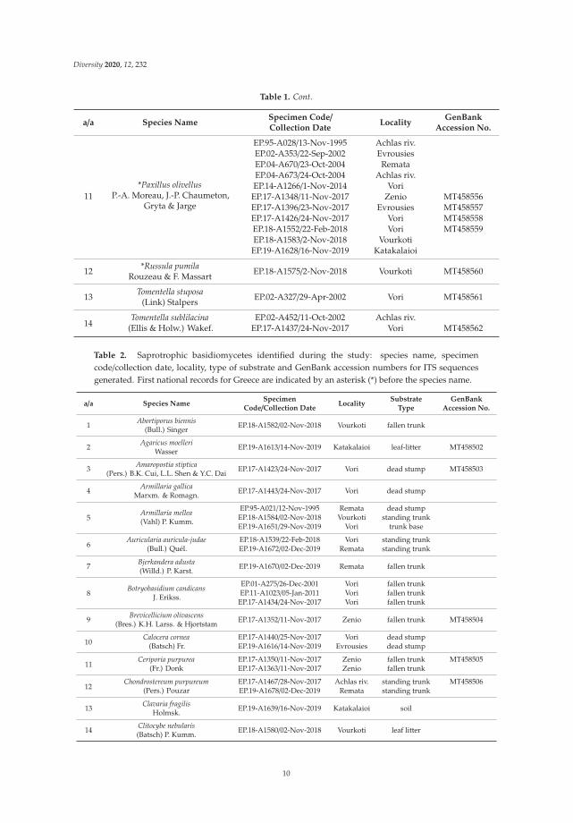
Table 1. Ectomycorrhizal (ECM) fungi identified during the study: species name, specimencode/collection date, locality and GenBank accession numbers for ITS sequences generated. Firstnational records for Greece are indicated by an asterisk (*) before the species name.
a/a Species NameSpecimen Code/Collection Date
LocalityGenBank
Accession No.
1*Alnicola escharoides
(Fr.) Romagn.
EP.17-A1344/11-Nov-2017 ZenioEP.17-A1420/24-Nov-2017 VoriEP.18-A1548/22-Feb-2018 Vori MT458538EP.18-A1561/1-Nov-2018 Zenio MT458539EP.18-A1571/2-Nov-2018 Vourkoti MT458540EP.19-A1636/16 Nov 2019 Katakalaioi
2 *Alnicola inculta(Peck) Singer EP.17-A1346/11 Nov 2017 Zenio MT458541
3 *Alnicola luteolofibrillosaKühner EP.17-A1430/24-Nov-2017 Vori MT458542
4*Alnicola subconspersa
(Kühner ex P.D. Orton) BonEP.17-A1421/24-Nov-2017 Vori MT458543EP.19-A1637/16-Nov-2019 Katakalaioi MT458544
5Alnicola striatula
(P.D. Orton) Romagn.EP.04-A679/15-Nov-2004 Evrousies
EP.19-A1614/14-Nov-2019 Evrousies MT458545
6*Alnicola umbrina(R. Maire) Kühner
EP.04-A678/15-Nov-2004 EvrousiesEP.17-A1377/2-Nov-2017 Lezina MT458546EP.18-A1572/2-Nov-2018 Vourkoti MT458547EP.19-A1607/12 Nov 2019 Zenio MT458548EP.19-A1638/16-Nov-2019 KatakalaioiEP.19-A1646/17-Nov-2019 Achlas riv.EP.19-A1666/2-Dec-2019 Remata MT458549
7 *Cortinarius americanusA.H. Sm. EP.19-A1622/15-Nov-2019 Vourkoti
8Gyrodon lividus
(Bull.) Sacc.EP.14-A1263/1-Nov-2014 VoriEP.17-A1428/24-Nov-2017 Vori
9 *Inocybe calosporaQuél. EP.18-A1570/2-Nov-2018 Vourkoti MT458550
10*Lactarius obscuratus
(Lasch) Fr.
EP.17-A1347/11-Oct-2017 Zenio MT458551EP.17-A1566/1-Nov-2018 Zenio MT458552EP.17-A1576/2-Nov-2018 Vourkoti MT458553EP.19-A1645/17-Nov-2019 Achlas riv. MT458554EP.19-A1664/30-Nov-2019 Remata MT458555
9
Diversity 2020, 12, 232
Table 1. Cont.
a/a Species NameSpecimen Code/Collection Date
LocalityGenBank
Accession No.
11*Paxillus olivellus
P.-A. Moreau, J.-P. Chaumeton,Gryta & Jarge
EP.95-A028/13-Nov-1995 Achlas riv.EP.02-A353/22-Sep-2002 EvrousiesEP.04-A670/23-Oct-2004 RemataEP.04-A673/24-Oct-2004 Achlas riv.EP.14-A1266/1-Nov-2014 Vori
EP.17-A1348/11-Nov-2017 Zenio MT458556EP.17-A1396/23-Nov-2017 Evrousies MT458557EP.17-A1426/24-Nov-2017 Vori MT458558EP.18-A1552/22-Feb-2018 Vori MT458559EP.18-A1583/2-Nov-2018 Vourkoti
EP.19-A1628/16-Nov-2019 Katakalaioi
12 *Russula pumilaRouzeau & F. Massart EP.18-A1575/2-Nov-2018 Vourkoti MT458560
13 Tomentella stuposa(Link) Stalpers EP.02-A327/29-Apr-2002 Vori MT458561
14Tomentella sublilacina
(Ellis & Holw.) Wakef.EP.02-A452/11-Oct-2002 Achlas riv.
EP.17-A1437/24-Nov-2017 Vori MT458562
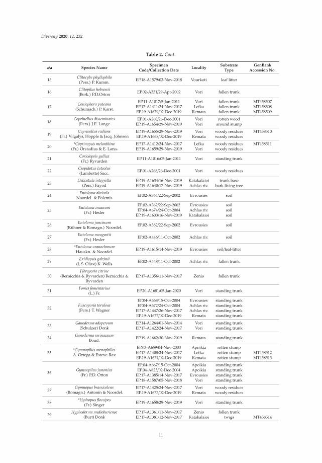
Table 2. Saprotrophic basidiomycetes identified during the study: species name, specimencode/collection date, locality, type of substrate and GenBank accession numbers for ITS sequencesgenerated. First national records for Greece are indicated by an asterisk (*) before the species name.
a/a Species NameSpecimen
Code/Collection DateLocality
SubstrateType
GenBankAccession No.
1 Abortiporus biennis(Bull.) Singer EP.18-A1582/02-Nov-2018 Vourkoti fallen trunk
2 Agaricus moelleriWasser EP.19-A1613/14-Nov-2019 Katakalaioi leaf-litter MT458502
3 Amaropostia stiptica(Pers.) B.K. Cui, L.L. Shen & Y.C. Dai EP.17-A1423/24-Nov-2017 Vori dead stump MT458503
4 Armillaria gallicaMarxm. & Romagn. EP.17-A1443/24-Nov-2017 Vori dead stump
5Armillaria mellea(Vahl) P. Kumm.
EP.95-A021/12-Nov-1995 Remata dead stumpEP.18-A1584/02-Nov-2018 Vourkoti standing trunkEP.19-A1651/29-Nov-2019 Vori trunk base
6Auricularia auricula-judae
(Bull.) Quél.EP.18-A1539/22-Feb-2018 Vori standing trunkEP.19-A1672/02-Dec-2019 Remata standing trunk
7 Bjerkandera adusta(Willd.) P. Karst. EP.19-A1670/02-Dec-2019 Remata fallen trunk
8Botryobasidium candicans
J. Erikss.
EP.01-A275/26-Dec-2001 Vori fallen trunkEP.11-A1023/05-Jan-2011 Vori fallen trunk
EP.17-A1434/24-Nov-2017 Vori fallen trunk
9 Brevicellicium olivascens(Bres.) K.H. Larss. & Hjortstam EP.17-A1352/11-Nov-2017 Zenio fallen trunk MT458504
10Calocera cornea
(Batsch) Fr.EP.17-A1440/25-Nov-2017 Vori dead stumpEP.19-A1616/14-Nov-2019 Evrousies dead stump
11Ceriporia purpurea
(Fr.) DonkEP.17-A1350/11-Nov-2017 Zenio fallen trunk MT458505EP.17-A1363/11-Nov-2017 Zenio fallen trunk
12Chondrostereum purpureum
(Pers.) PouzarEP.17-A1467/28-Nov-2017 Achlas riv. standing trunk MT458506EP.19-A1678/02-Dec-2019 Remata standing trunk
13 Clavaria fragilisHolmsk. EP.19-A1639/16-Nov-2019 Katakalaioi soil
14 Clitocybe nebularis(Batsch) P. Kumm. EP.18-A1580/02-Nov-2018 Vourkoti leaf litter
10
Diversity 2020, 12, 232
Table 2. Cont.
a/a Species NameSpecimen
Code/Collection DateLocality
SubstrateType
GenBankAccession No.
15 Clitocybe phyllophila(Pers.) P. Kumm. EP.18-A1579/02-Nov-2018 Vourkoti leaf litter
16 Clitopilus hobsonii(Berk.) P.D.Orton EP.02-A331/29-Apr-2002 Vori fallen trunk
17Coniophora puteana
(Schumach.) P. Karst.
EP.11-A1017/5-Jan-2011 Vori fallen trunk MT458507EP.17-A1411/24-Nov-2017 Lefka fallen trunk MT458508EP.19-A1679/02-Dec-2019 Remata fallen trunk MT458509
18Coprinellus disseminates
(Pers.) J.E. LangeEP.01-A260/26-Dec-2001 Vori rotten wood
EP.19-A1654/29-Nov-2019 Vori around stump
19Coprinellus radians
(Fr.) Vilgalys, Hopple & Jacq. JohnsonEP.19-A1655/29-Nov-2019 Vori woody residues MT458510EP.19-A1668/02-Dec-2019 Remata woody residues
20*Coprinopsis melanthina
(Fr.) Örstadius & E. Larss.EP.17-A1412/24-Nov-2017 Lefka woody residues MT458511EP.19-A1659/29-Nov-2019 Vori woody residues
21 Coriolopsis gallica(Fr.) Ryvarden EP.11-A1016/05-Jan-2011 Vori standing trunk
22 Crepidotus luteolus(Lambotte) Sacc. EP.01-A268/26-Dec-2001 Vori woody residues
23Delicatula integrella
(Pers.) FayodEP.19-A1634/16-Nov-2019 Katakalaioi trunk baseEP.19-A1640/17-Nov-2019 Achlas riv. bark living tree
24 Entoloma alnicolaNoordel. & Polemis EP.02-A364/22-Sep-2002 Evrousies soil
25Entoloma incanum
(Fr.) Hesler
EP.02-A362/22-Sep-2002 Evrousies soilEP.04-A674/24-Oct-2004 Achlas riv. soil
EP.19-A1633/16-Nov-2019 Katakalaioi soil
26 Entoloma juncinum(Kühner & Romagn.) Noordel. EP.02-A362/22-Sep-2002 Evrousies soil
27 Entoloma mougeotii(Fr.) Hesler EP.02-A446/11-Oct-2002 Achlas riv. soil
28 *Entoloma uranochroumHauskn. & Noordel. EP.19-A1615/14-Nov-2019 Evrousies soil/leaf-litter
29 Exidiopsis galzinii(L.S. Olive) K. Wells EP.02-A448/11-Oct-2002 Achlas riv. fallen trunk
30Fibroporia citrine
(Bernicchia & Ryvarden) Bernicchia &Ryvarden
EP.17-A1356/11-Nov-2017 Zenio fallen trunk
31 Fomes fomentarius(L.) Fr. EP.20-A1681/05-Jan-2020 Vori standing trunk
32Fuscoporia torulosa(Pers.) T. Wagner
EP.04-A668/15-Oct-2004 Evrousies standing trunkEP.04-A672/24-Oct-2004 Achlas riv. standing trunk
EP.17-A1447/26-Nov-2017 Achlas riv. standing trunkEP.19-A1677/02-Dec-2019 Remata standing trunk
33Ganoderma adspersum
(Schulzer) DonkEP.14-A1264/01-Nov-2014 Vori standing trunkEP.17-A1422/24-Nov-2017 Vori standing trunk
34 Ganoderma resinaceumBoud. EP.19-A1662/30-Nov-2019 Remata standing trunk
35*Gymnopilus arenophilusA. Ortega & Esteve-Rav.
EP.03-A659/04-Nov-2003 Apoikia rotten stumpEP.17-A1408/24-Nov-2017 Lefka rotten stump MT458512EP.19-A1674/02-Dec-2019 Remata rotten stump MT458513
36Gymnopilus junonius
(Fr.) P.D. Orton
EP.04-A667/15-Oct-2004 Apoikia standing trunkEP.04-A825/02-Dec-2004 Apoikia standing trunk
EP.17-A1385/14-Nov-2017 Evrousies standing trunkEP.18-A1587/05-Nov-2018 Vori standing trunk
37Gymnopus brassicolens
(Romagn.) Antonín & Noordel.EP.17-A1425/24-Nov-2017 Vori woody residuesEP.19-A1673/02-Dec-2019 Remata woody residues
38 *Hydropus floccipes(Fr.) Singer EP.19-A1658/29-Nov-2019 Vori standing trunk
39Hyphoderma medioburiense
(Burt) DonkEP.17-A1361/11-Nov-2017 Zenio fallen trunkEP.17-A1381/12-Nov-2017 Katakalaioi twigs MT458514
11

Diversity 2020, 12, 232
Table 2. Cont.
a/a Species NameSpecimen
Code/Collection DateLocality
SubstrateType
GenBankAccession No.
40 *Hyphoderma nemoraleK.H. Larss. EP.02-A326/29-Apr-2002 Vori fallen trunk MT458515
41Hyphoderma setigerum
(Fr.) DonkEP.02-A332/29-Apr-2002 Vori fallen trunk
EP.17-A1357/11-Nov-2017 Zenio fallen trunk MT458516
42 *Hyphodermella corrugate(Fr.) J. Erikss. & Ryvarden EP.17-A1398/23-Nov-2017 Evrousies fallen branch MT458517
43 *Lepiota ochraceofulvaP.D. Orton EP.19-A1627/15-Nov-2019 Vourkoti leaf-litter MT458518
44 * Lepista ovispora(J.E. Lange) Gulden EP.19-A1608/12-Nov-2019 Zenio leaf-litter
45 Lepista nuda(Bull.) Cooke EP.18-A1564/01-Nov-2018 Zenio leaf-litter
46 Leucoagaricus melanotrichus(Malençon & Bertault) Trimbach EP.19-A1604/12-Nov-2019 Zenio leaf-litter
47 Leucopaxillus gentianeus(Quél.) Kotl. EP.18-A1577/01-Nov-2018 Zenio leaf-litter
48 Marasmius rotula(Scop.) Fr. EP.02-A360/22-Sep-2002 Evrousies twigs
49 Melanoleuca exscissa(Fr.) Singer EP.17-A1415/24-Nov-2017 Vori soil MT458519
50 Merulius tremellosusSchrad.
EP.04-A680/15-Nov-2004 Evrousies fallen trunkEP.02-A323/29-Apr-2002 Vori fallen trunk
51Mycena galericulata
(Scop.) Gray
EP.17-A1349/11-Nov-2017 Zenio around trunkEP.19-A1625/15-Nov-2019 Vourkoti dead stump MT458520EP.19-A1629/16-Nov-2019 Katakalaioi around trunk MT458521EP.19-A1668/02-Dec-2019 Remata dead stump
52 Mycena haematopus(Pers.) P. Kumm. EP.01-A262/26-Dec-2001 Vori rotten trunk
53 Mycena pseudocorticolaKühner
EP.17-A1449/26-Nov-2017 Achlas riv. bark living treeEP.19-A1667/02-Dec-2019 Remata bark living tree
54 Mycena sanguinolenta(Alb. & Schwein.) P. Kumm EP.19-A1642/17-Nov-2019 Achlas riv. twigs
55 Mycetinis scorodonius(Fr.) A.W. Wilson & Desjardin EP.19-A1644/17-Nov-2019 Achlas riv. twigs
56Mycoacia aurea
(Fr.) J. Erikss. & RyvardenEP.02-A325/29-Apr-2002 Vori fallen trunkEP.11-A1026/05-Jan-2011 Vori fallen trunk
57 Mycoacia uda(Fr.) Donk EP.17-A1351/11-Nov-2017 Zenio fallen trunk
58Mycoaciella bispora
(Stalpers) Erikss. & RyvardenEP.01-A272/26-Dec-2001 Vori fallen trunkEP.02-A330/29-Apr-2002 Vori fallen trunk
59Paralepista flaccida(Sowerby) Vizzini
EP.18-A1563/01-Nov-2018 Zenio leaf-litterEP.18-A1581/02-Nov-2018 Vourkoti leaf-litter
60Parasola kuehneri
(Uljé & Bas) Redhead, Vilgalys &Hopple
EP.18-A1546/22-Feb-2018 Vori soil
61 Peniophora tamaricicolaBoidin & Malenç. EP.02-A329/29-Apr-2002 Vori fallen trunk
62Peniophorella praetermissa
(P. Karst.) K.H. Larss.EP.01-A276/26-Dec-2001 Vori fallen trunk
EP.17-A1355/11-Nov-2017 Zenio fallen trunk MT458522
63 Perenniporia ochroleuca(Berk.) Ryvarden EP.02-A461/11-Oct-2002 Achlas riv. dead wood
64*Phanerochaete livescens
(P. Karst.) Volobuev & SpirinEP.01-A273/26-Dec-2001 Vori fallen trunkEP.18-A1541/22-Feb-2018 Vori fallen trunk MT458523
65 Phellinus lundelliiNiemelä EP.17-A1342/13-Apr-2017 Zenio standing trunk
66Phlebia rufa
(Pers.) M.P. Christ.EP.17-A1354/11-Nov-2017 Zenio fallen trunk MT458524EP.20-A1682/05-Jan-2020 Vori fallen trunk MT458525
12

Diversity 2020, 12, 232
Table 2. Cont.
a/a Species NameSpecimen
Code/Collection DateLocality
SubstrateType
GenBankAccession No.
67 Phlebiopsis ravenelii(Cooke) Hjortstam EP.11-A1018/05-Jan-2011 Vori fallen trunk
68 Phloeomana alba(Bres.) Redhead EP.19-A1641/17-Nov-2019 Achlas riv. bark living tree MT458526
69 Phloeomana speirea(Fr.) Redhead EP.17-A1378/12-Nov-2017 Lezina twigs MT458527
70 Physisporinus vitreus(Pers.) P. Karst. EP.20-A1684/05-Jan-2020 Vori fallen trunk
71 Pilatotrama ljubarskyi(Pilát) Zmitrovich EP.18-A1551/22-Feb-2018 Vori fallen trunk MT458528
72Pleurotus ostreatus(Jacq.) P. Kumm.
EP.01-A261/26-Dec-2001 Vori standing trunkEP.18-A1586/05-Nov-2018 Vori standing trunk
73Pluteus cervinus
(Schaeff.) P. Kumm.
EP.01-A321/29-Apr-2002 Vori dead wood MT458529EP.19-A1623/15-Nov-2019 Vourkoti dead wood MT458530EP.19-A1676/02-Dec-2019 Remata fallen trunk
74 Pluteus nanus(Pers.) P. Kumm. EP.19-A1653/29-Nov-2019 Vori woody residues
75 Pluteus salicinus(Pers.) P. Kumm. EP.19-A1657/29-Nov-2019 Vori fallen branch
76 *Pluteus podospileusSacc. & Cub. EP.19-A1643/17-Nov-2019 Achlas riv. fallen twigs
77 Postia balsamea(Peck) Jülich EP.19-A1663/30-Nov-2019 Remata fallen trunk
78Psathyrella candolleana
(Fr.) MaireEP.02-A336/04-Jun-2002 Achlas riv. woody residues
EP.19-A1624/15-Nov-2019 Vourkoti woody residues MT458531
79 Psathyrella corrugis(Pers.) Konrad & Maubl. EP.19-A1601/16-Oct-2019 Vourkoti soil/buried
wood MT458532
80 *Psathyrella hellebosensisDeschuyteneer & A. Melzer EP.17-A1409/24-Nov-2017 Lefka woody residues MT458533
81 Psathyrella microrhiza(Lasch) Konrad & Maubl. EP.19-A1603/12-Nov-2019 Vourkoti woody residues MT458534
82 Psathyrella prona(Fr.) Gill. EP.02-A363/22-Sep-2002 Evrousies soil
83 Psathyrella vinosofulvaP.D. Orton EP.17-A1431/24-Nov-2017 Vori soil MT458535
84 Radulomyces confluens(Fr.) M.P. Christ. EP.11-A1022/05-Jan-2011 Vori fallen trunk
85 Steccherinum ochraceum(Pers. ex J.F. Gmel.) Gray EP.18-A1540/22-Feb-2018 Vori fallen trunk
86Stereum hirsutum
(Willd.) Pers.
EP.17-A1397/23-Nov-2017 Evrousies trunk/branchEP.17-A1463/28-Nov-2017 Achlas riv. trunk/branchEP.18-A1544/22-Feb-2018 Vori trunkEP.20-A1691/25-Jan-2020 Lefka branch
87Trechispora nivea
(Pers.) K.H. Larss.EP.02-A328/29-Apr-2002 Vori fallen trunkEP.20-A1683/05-Jan-2020 Vori fallen trunk MT458536
88Trametes versicolor
(L.) LloydEP.11-A1019/05-Jan-2011 Vori fallen trunkEP.18-A1542/22-Feb-2018 Vori standing trunk
89 Tubaria furfuracea(Pers.) Gillet EP.19-A1652/29-Nov-2019 Vori woody residues
90 Tulostoma fimbriatumFr. EP.17-A1431/24-Nov-2017 Vori soil
91Vitreoporus dichrous
(Fr.) Zmitr.
EP.02-A445/11-Oct-2002 Achlas riv. fallen trunkEP.17-A1439/25-Nov-2017 Vori fallen branchEP.18-A1538/22-Feb-2017 Vori fallen branch
92Xylodon raduloides
Riebesehl & Langer
EP.11-A1025/05-Jan-2011 Vori fallen trunkEP.18-A1543/22-Feb-2018 Vori fallen trunk MT458538EP.19-A1671/02-Dec-2019 Remata fallen trunk
13

Diversity 2020, 12, 232
3.1. The ECM Element
Among the ECM macrofungi recorded (Table 1), the genus Alnicola is represented by six species(Figure 3); five of them form part of the sect. Alnicola sensu Moreau [45], which is characterizedby urticoid cheilocystidia, and one of the sect. Submelinoideae Singer with clavate or capitatecheilocystidia [46].
Figure 3. Species of the genus Alnicola recorded in Andros alder stands: A. escharoides basidiomes,basidiospores and cheilocystidia (a–c); A. umbrina basidiomes, basidiospores and cheilocystidia (d–f);A. striatula basidiomes, basidiospores and cheilocystidia (g–i); A. subconspersa basidiomes, basidiosporesand cheilocystidia (j–l); A. luteolofibrillosa basidiomes, basidiospores and cheilocystidia (m–o); A. incultabasidiomes, basidiospores, basidia and cheilocystidia (p–s). Bars: basidiomes, 1 mm; basidiosporesand basidia, 10 μm; cheilocystidia, 20 μm.
14

Diversity 2020, 12, 232
Different opinions exist regarding the genus name in pertinent literature since some Europeanauthors as well as the Index Fungorum prefer to conserve the name Naucoria (Fr.) P. Kumm., whereasMoreau, in his nomenclatural revision, rejected this name in favour of Alnicola Kühner [45]; the latterapproach is accepted by other European mycologists, the Mycobank, and is also adopted in thiswork. Moreover, the taxonomy of species of the sect. Alnicola remains problematic and, consequently,a phylogenetic analysis was performed to deal with this issue.
The most often found Alnicola species in our study were A. umbrina (R. Maire) Kühner andA. escharoides (Fr.) Romagn., i.e., two of the most common taxa associated with alders in Europe; bothconstitute new national records for Greece. Particularly A. escharoides (syn. A. citrinella Moreau &A. de Haan [47]) is distinguished from all other (more or less brownish) species found in Androsby its pale yellowish-buff non striate pileus, the amygdaliform to navicular spores, with prominentornamentation, measuring 9.9–11.8 × 5.3–5.9 μm, Q = 1.9–2.1 (Figure 3a–c). Following phylogeneticanalysis, our specimens are positioned in a distinct group (albeit not adequately supported) togetherwith other sequences from material identified as A. escharoides and A. citrinella (Figure 4).
Figure 4. Phylogeny of Alnicola species derived from rDNA ITS sequences through ML analysis.Branches are labelled when MLB > 65% and BPP > 0.95. Hebeloma species (H. louiseae, H. pallidolabiatum,H. crustuliniforme) were used as outgroups. Boxes include sequences from specimens recorded in theAlnus glutinosa habitat.
On the other hand, A. umbrina (Figure 3d–f) is hereby considered as a species complex following thenomenclatural concept of Moreau [45] and the outcome of the phylogenetic study by Rochet et al. [19].According to our observations, A. umbrina shows a rather large morphological variability with darkbrown hygrophanous pilei bearing prominent striations up to their centre when wet, becoming muchlighter and indistinctly striate only at margin when dry. Basidiospores are variable in size and shape,
15

Diversity 2020, 12, 232
often somewhat elongated fusiform, weakly to moderately verrucose, measuring 10.7–13.6× 5.2–6.1μm,Q = 1.9–2.4. Sequences generated in this work clustered together with material identified as A. umbrina,N. scolecina (Fr.) Quél., A. striatula (P.D. Orton) Romagn. and A. subconspersa (Kühner ex P.D. Orton)Bon into a group that was not adequately supported (Figure 4). However, the morphological features ofspecimens identified as N. scolecina in Europe are very similar to descriptions of A. umbrina [22,33,48,49].Therefore, N. scolecina and A. umbrina form part of the same complex and the question whether theyconstitute different entities or not remains open and in need of further research.
One collection representing another closely related taxon, previously reported as N. striatula P.D.Orton (Figure 3g–i) from alder stands in Andros [22], derived from the same site during our recentforays. According to Moreau (2005), A. striatula might merely correspond to a pale form of A. umbrina,but our morphological studies revealed some noteworthy differences when compared to specimenshereby named A. umbrina, i.e., pileus always very prominently striate, smooth and shiny, and (mostimportantly) significantly smaller basidiospores measuring 8.2–10.0 × 4.5–5.6 μm, Q = 1.7–1.9; thesefeatures are in accordance to previous descriptions of N. striatula [33,48,50]. As evidenced fromour phylogenetic analysis (Figure 4), this particular collection forms part of the A. umbrina complex(together with the other two A. striatula sequences included in the tree) by using ITS alone; however,since it is morphologically distinct and fits to the widely accepted taxonomic concept of A. striatula,we provisionally retain it in this inventory as a separate taxon, until a future multigene approachshows otherwise.
A similar looking species to A. umbrina—but less common in Andros—is A. subconspersa (Figure 3j–l).The most prominent distinguishing features versus our A. umbrina specimens are the non (or veryfaintly) striate pileus as well as the size and shape of spores, being wider, amygdaliform to navicularand more prominently ornamented, measuring 10.9–12.9 × 6.0–6.8 μm, Q = 1.7–2.0. It is noteworthythat A. subconspersa forms a well-supported phylogenetic group including sequences labelled asA. scolecina (Fr.) Romagn. (Figure 4), which is indicative of the morphological affinity of these taxa thathad apparently led to the development of ambiguous species concepts.
Another collection representing a member of the sect. Alnicola was recorded in the alluvial littoralforest of Vori; it corresponds to A. luteolofibrillosa Kühner and constitutes the first report of this speciesin Greece (Figure 3m–o). It is morphologically characterized by non-striate, pale buff, fibrillose totomentose pilei, with abundant whitish veil remnants on stipe and pileal margin; the respectivesequence falls within a highly-supported terminal subgroup corresponding to this species (Figure 4).Lastly, A. inculta (Peck) Singer (Figure 3p–s) was recorded only at Zenio (i.e., the site with the highestaltitude among those of this study, 850 m) and is reported for the first time in Greece. It formspart of the sect. Submelinoideae, and, according to Moreau [45] is conspecific to the taxon widelyreferred as N. celluloderma P.D. Orton, as it is also evidenced by our phylogenetic analysis (Figure 4).Morphologically, this species is easily distinguished from all aforementioned taxa thanks to the clavateto capitate cheilocystidia characterizing members of sect. Submelinoideae and the 2-spored basidia.
The most common ECM mushroom in alder stands of Andros belongs to the genus Paxillus; it wasthe first recorded Alnus-specific symbiont in the island 25 years ago, and was later repeatedly foundin this particular habitat (Figure 5a–d). It was initially identified as P. rubicundulus P.D. Orton [22];however, sequencing of recent collections revealed that it forms part of the newly described taxonP. olivellus Moreau P-A, Chaumeton J-P, Gryta H, Jargeat P [51]. Although clearly separated bymolecular approaches, P. olivellus can be hardly distinguished from P. rubicundulus and P. adelphusChaumeton JP, Gryta H, Jargeat P, Moreau P-A on the basis of morphology alone, i.e., only by theolivaceous tinges of the young basidiomes and the basidiospores shape, which are ovoid to ellipsoid inP. olivellus, cylindrical in P. rubicundulus and short cylindrical in P. adelphus [51]. Such features wereobserved in our specimens since olivaceous tints were always evident in young basidiomes, and sporeswere ovoid to ellipsoid measuring 6.7–8.1× 4.5–5.2 μm, Q = 1.39–1.66. In addition, ITS sequences fromour material originating from various sites in the habitat under study were very similar or identical tothose corresponding to P. olivellus (including the type), and formed a terminal subgroup with high
16

Diversity 2020, 12, 232
support (Figure 6). Therefore, this particular species seems to be the only representative of the genusPaxillus in the black alder stands of Andros island.
Figure 5. Alder-associated ECM fungi recorded in Andros: Paxillus olivellus basidiomes (a; bar 1 mm),basidiospores (b; bar 10 μm), section of lamella (c; bar 20 μm), hymenial cystidium and basidia(d; bar 20 μm); Lactarius obscuratus basidiomes (e, bar 1 mm), basidiospores (f, bar 10 μm), pileipellis(g, bar 20 μm); Russula pumila basidiomes (h, bar 1 mm), basidiospores (i, bar 10 μm), pileipellis(j, bar 20 μm); Cortinarius americanus basidiomes (k, bar 1 mm); Inocybe calospora basidiospores,basidium and pleurocystidium (l, bar 10 μm).
17

Diversity 2020, 12, 232
Figure 6. Phylogeny of Paxillus species derived from rDNA ITS sequences through ML analysis.Branches are labelled when MLB > 65%, and BPP > 0.95. P. cuprinus and P. obscurosporus were used asoutgroups. The coloured box includes sequences from specimens recorded in the Alnus glutinosa habitat.
Lactarius obscuratus (Lasch) Fr. is one the few Alnus-specific ECM symbionts of this particulargenus; it was found in several inland collection sites dominated by A. glutinosa, but not in the alluvial(littoral) forest of Vori (Figure 5e–g). Phylogenetic analysis of our sequences derived from severalcollections confirmed that they belong to this particular species (Figure 7). However, the respectiveterminal subgroup in our phylogenetic tree is composed from sequences named either L. obscuratus orL. cyathuliformis Bon, which is due to the different interpretations existing about this taxon (J. Nuytinck,pers. comm.). In the study of Rochet et al. [19], it is referred as L. cyathuliformis, whereas the correctname for the same group is L. obscuratus according to Wisitrassameewong et al. [52]. Since thebasidiospores average size in our collections (measuring 7.6–8.5 × 6.1–6.3 μm) is in agreement with theconcept of L. obscuratus (sensu Heilmann-Clausen et al. [31]; according to the same authors, spores ofL. cyathuliformis have an average size of 8.3–9.9 × 7.0–7.7 μm), we adopt this name for the specimensincluded in this work. The genus Russula Pers. is represented by R. pumila Rouzeau & F. Massart(Figure 5h–j) detected in one site only (Vourkoti). Although R. pumila is synonymous to R. alnetorumRomagn. according to both Index Fungorum and Mycobank, there are different opinions about thesynonymy of these two taxa and to the best of our knowledge this issue has not been resolved yet(S. Adamcik, pers. comm.). R. pumila is reported to occur mainly in lowlands with A. glutinosa,as opposed to R. alnetorum, which is mostly recorded in subalpine habitats with A. viridis [53,54];therefore, we adopt the use of the former name.
Tomentella sublilacina (Ellis & Holw.) Wakef. and T. stuposa (Link) Stalpers were previously recordedin Andros (in the alluvial alder forest of Vori; [20]) and identified on the basis of their morphology.These names are provisionally retained here due to the absence of precise taxonomic informationconcerning alder-specific Tomentella species. It should be noted that the ITS sequences generated inthe frame of this work represent phylogenetically distinct taxa corresponding to entities named “aff.sublilacina” and “aff. stuposa” in previous studies referring to material originating from alder hostsonly [18,55].
It is noteworthy that the first ever report of an alniphilous Cortinarius species in Greece derivesfrom a single collection of C. americanus A.H. Sm. (Figure 5k), which forms part of a small group of
18

Diversity 2020, 12, 232
species within the subgenus Telamonia (Fr.) Trog known to be associated with A. glutinosa and A. incanain Europe [33,56]. C. americanus is characterized by the minute size, pileus not exceeding 2 cm indiameter, with dark violet to blackish colour, and spores smaller than 10 × 6 μm [33,56]; our collectionhas spores measuring 7.6–8.4 × 4.9–5.6 μm.
Last, Inocybe calospora Quél., recorded for the first time in Greece, was collected in Vourkoti only;it is an easily identified species thanks to its unique star-shaped spiny spores (Figure 5). Among allECM species included in this inventory it is the only one which is not considered to be exclusivelyassociated with alders, and reported from diverse damp deciduous forests of Europe [33,56].
Figure 7. Phylogeny of Lactarius species derived from rDNA ITS sequences through ML analysis.Branches are labelled when MLB > 65%, and BPP > 0.95. L. torminosus, L. scrobiculatus andL. pseudoscrobiculatus were used as outgroup. The colored box includes sequences from specimensrecorded in the Alnus glutinosa habitat.
3.2. The Saproxylic Element
By far the highest number of species recorded in this inventory correspond to white-rot andbrown-rot basidiomycetes found on various wood parts of A. glutinosa (Table 2). This is quite anticipatedsince alder trees have a life-span which rarely exceeds 100 years; therefore, they produce large amountsof dead wood. Moreover, in contrast to ECM species, wood-rotting fungi do not show any specificity toalders, with only few exceptions including the common in northern Europe plant-pathogenic polyporeInonotus radiatus (Sowerby) P. Karst. [15,57–60], which, however, was not among our findings. Most ofthe recorded species are wood rotting basidiomycetes that are quite common throughout Europe ondeciduous tree species including alders [13,61,62]. The best represented genera of saproxylic fungiwere Hyphoderma Wallr. (three spp.), Mycena (Pers.) Roussel (four spp.), Pluteus Fr. (four spp.) andPsathyrella (Fr.) Quél. (four spp. on woody residues or buried wood). In total, seven species recordedon dead wood or bark of living alder trees are recorded for the first time in Greece and are presented inmore detail below.
19

Diversity 2020, 12, 232
Among corticioid basidiomycetes, Hyphoderma nemorale K.H. Larss. is a distinct, widely distributedbut rare species in Europe [35], which was identified by re-examining an old collection from Vorialluvial forest and further confirmed by ITS sequencing. The presence of thick-walled subicularhyphae and of two types of hymenial cystidia (i.e., short ventricose and subcapitate, and more seldomlong tubular with characteristic constrictions, sometimes moniliform, which was the case in ourspecimen) are the main diagnostic features of this species [63]. Hyphodermella corrugata (Fr.) J. Erikss.& Ryvarden (Figure 8a) is fairly common and widespread in Europe [35]. It is easily identifiedthanks to its characteristic cystidioid hyphal ends appearing in bundles that are heavily incrusted [24].Phanerochaete livescens (P. Karst.) Volobuev & Spirin (Figure 8b) is a species closely related to Ph. sordida(P. Karst.) J. Erikss. & Ryvarden which was recently described by using both morphological andphylogenetic criteria [64]. In accordance to the pertinent description, our specimens possessed cystidiawith thickened walls to the acute apex, densely covered by crystals, as opposed to the accidentallyencrusted, obtuse and thin-walled towards the apex cystidia of Ph. sordida. Moreover, the identity ofour specimen was confirmed by the respective ITS sequence which was identical to those of Ph. livescensas determined by Volobuev et al. [64].
Figure 8. Saproxylic species recorded on Alnus glutinosa wood and litter in Andros: Hyphodermellacorrugata (a) Phanerochaete livescens (b) Pluteus podospileus (c) Delicatula integrella (d) Coprinopsismelanthina (e) Gymnopilus arenophilus (f) Hydropus floccipes (g) Lepiota ochraceofulva (h) Lepistaovispora (i) Psathyrella hellebosensis (j) Entoloma uranochroum (k) Bar: 1 mm.
20

Diversity 2020, 12, 232
Among the agaricoid wood-inhabiting fungi, the genus Pluteus Fr. is hereby represented byfour species, of which P. podospileus Sacc. & Cub. (Figure 8c) is reported for the first time in Greece.It belongs to the sect. Celluloderma Fay. subsection Mixtini Sing. ex Sing, and possesses a pileipellismade up of both fusiform and broadly clavate elements. P. thomsonii (Berk. & Broome) Dennisis very similar morphologically but it differs in the absence of pleurocystidia and the shape ofcheilocystidia, which are characteristically rostrate [65]. The record of Delicatula integrella (Pers.) Rat.(Figure 8d) is worth mentioning since it was reported only once before in Greece, in the content ofa regional field-guide [66]. D. integrella forms whitish-mycenoid mushrooms of minute size withpileus diameter measuring (in our specimens) 0.4–0.6 cm, reduced, almost vein-like lamellae andnon-amyloid, amygdaliform-fusoid spores. It is considered widespread and common in Europe and N.America, and grows on decaying wood and wood debris of deciduous trees [34]. Coprinopsis melanthina(Fr.) Örstadius & E. Larss. (Figure 8e) is a striking-looking psathyrelloid species, easily identifieddue to the relatively large basidiomes with wooly to squamulose pileus (measuring 2–6 cm in diam.)and stipe (up to 6.0 × 0.8 cm), absence of pleurocystidia, almost colourless basidiospores, devoidof germ-pore, measuring 9.8–11.8(13.5) × 5.6–6.5 μm in our collections. This is a rather uncommonEuropean species growing on and around rotten stumps in humid deciduous forests [33]; the onlyother record of this species in Greece derives from Crete (G. Konstandinidis, pers. comm.).
Gymnopilus arenophilus A. Ortega & Esteve-Rav. (Figure 8f) was described from continental areasof Spain [67] and from maritime dunes in France, under or near Mediterranean pines, on sandy soil bybeing attached to wood debris or wood, often burnt or buried in the sand [68]. Two of our collectionsfrom the alluvial A. glutinosa habitat at Lefka were sequenced and found to correspond to this species.Apparently, no native pines exist in Andros while both specimens were growing on rotten alder stumps,a fact that largely expands the so far known ecological and geographical range of this Mediterraneanspecies. Morphologically, our specimens possessed features that fit well to the taxonomic conceptof G. arenophilus i.e., smooth to fibrillose pileal surface, bitter taste, ellipsoid to subamygdaliform,moderately verrucose spores, measuring 8–10 × 5.5–6.5 μm, lageniform cheilocysidia often withsubcapitate apex, 25–45 × 5–8 μm and absence of pleurocystidia. On the other hand, the size ofbasidiomes was significantly larger, with pilei up to 10 cm in diam. and a sturdy stipe often thickerthan 1 cm. One previous collection of ours also found on rotten alder stump, and originally identifiedas G. picreus (Pers.) P. Karst. [22], is now re-assessed as G. arenophilus.
Hydropus floccipes (Fr.) Singer (Figure 8g) is a rare mycenoid species generally found to grow ondecayed trunks of deciduous trees in damp forests, and is characterized by non-amyloid, subglobosespores, not blackening basidiomes, and typically scabrous stipe with grey-brown spots [33,69].In addition, our specimens possessed yellowish stipe, previously reported for H. floccipes var. luteipesOrtega & Zea described from Spain [70]; the latter is otherwise microscopically identical and ofunknown phylogenetic status. Two unpublished reports of H. floccipes exist from Greece (D. Sofronisand G. Konstandinidis, pers. comm.).
3.3. Litter and Other Terrestrial Decomposers
Apart ECM and saproxylic fungi, several other mushroom species were recorded under black aldertrees, and therefore constitute a part of the fungal diversity of the A. glutinosa priority habitat in Andros.Needless to say, none of these species is specifically linked to alders; instead they are considered‘generalists’ to be found in both deciduous and coniferous forests. Among them, the following fourspecies are recorded for the first time in Greece. Lepiota ochraceofulva P.D. Orton (Figure 8h) is arather rare (but widespread) species in Europe, reported from Fagus and other deciduous trees onhumus-rich, loamy soil [71,72]; it forms highly toxic mushrooms containing amanitins. Our singlecollection consisted of few basidiomes growing on a thick layer of leaf-litter under A. glutinosa. They arecharacterized by pilei of up to 7 cm in diam., with orange-brown scales; lamellae forming a distinctcollarium and reddish-orange in maturity; spores ellipsoid to oblong, dextrinoid, not metachromatic incresyl-blue, measuring 5.4–7.5(8.1) × 3.5–4(4.5) μm; basidia (2)4-spored, clamped; cheilocystidia short
21

Diversity 2020, 12, 232
clavate to cylindrical, rarely papilate, often in chains; pileipellis, a hymeniderm, composed of more orless clavate elements up to 50 μm long.
Lepista ovispora (J.E. Lange) Gulden (Figure 8i) is an uncommon (albeit widespread) Europeanspecies recorded only once during this study on leaf litter under alders. Typical diagnostic featuresare the densely caespitose habit, the relatively fleshy basidiomes, the brown hygrophanous pileuswith pruinose surface [73]. In addition, our specimens possessed spores ovoid to broadly ellipsoid,finely punctate, 4.7–6.8 × 3.8–4.4 μm, clamped basidia and no cystidia. Psathyrella hellebosensisD. Deschuyteneer, A. Melzer (Figure 8j) was recently described from Belgium [74] and was laterreported from riparian alder habitats in Italy [75]. The morphological features of our collection arein agreement with the morphology of Belgian and Italian basidiomes, but since it corresponds toa rarely reported species, a detailed description of our material is hereby provided: pileus up to3 cm in diam., hygrophanous from dark reddish-brown to greyish-beige, with scanty remains of veil;lamellae subdistant with whitish edge; stipe 2–4 × 0.2–0.3 cm, not rooting; spores 7.3–8.7 × 4.5–5.7 μm,Q = 1.43–1.73, ovoid to angular in face-view and not or weakly phaseoliform in side-view, not opaque;lamellae edge sterile composed exclusively of sphaeropendunculate paracystidia (no pleurocystidioidparacystidia were observed); pleurocystidia 34–48 × 11–17 μm, utriform. The material was collectedfrom wet soil by the alluvial stream banks. This species shows high phylogenetic affinity to P. thujinaA. H. Sm. by using ITS sequences only; however, it is clearly separated when the tef-1α marker is addedin the phylogenetic analysis, while it is also distinguished by its distinctly larger and prominentlyphaseoliform spores [75].
Previous studies on the mycodiversity of Andros island reported the occurrence of four Entolomaspecies, one of them was new to science, i.e., E. alnicola Noordel. & Polemis [22,25]. Our recent fieldwork resulted in other interesting collections of Entoloma spp. for which the identity and phylogeneticrelationships to closely allied taxa are still under investigation. However, by using morphology alone,the presence of a rare European species was confirmed, namely E. uranochroum Hauskn. & Noordel.(Figure 8k) recorded for the first time in an alder habitat. This beautiful dark blue-violet mushroomwas so far reported from subalpine meadows on calcareous soil in Austria (type locality) and theFrench Alps. Moreover, its striking microscopical features, e.g., the large fusiform cheilocystidida withgranular yellowish-brown content, place it in the distinct section Ramphocystotae (Largent) Noordel.,together with only one other European representative, namely E. rhynchocystidiatum Noordel. &Liiv [76].
4. Conclusions
A long-term study of the diversity of macrofungi in alder stands of Andros resulted in an inventoryconsisting of 106 species of basidiomycetes, including 21 taxa recorded for the first time in Greece.The majority of findings corresponded to saprotrophs (#92, mainly wood-rotting fungi) and the restwere ECM species. Considering the limited size of the area under study in a small Aegean island,the outcome of this work in terms of the number of taxa and variability is indicative of the wealth of theA. glutinosa priority habitat. However, the black alder stands in Andros have suffered considerably fromfloods in the past (as a consequence of fires that destroyed vegetation in the surrounding mountainswhich acted as a physical barrier protecting from downhill water runoffs) and their regeneration ishindered due to grazing by feral goats. The importance of fungi in the conservation/restoration ofsuch natural habitats was demonstrated in the past [77,78], and recent activities focus at improving thestatus of the degenerated alder stands by exploiting indigenous ECM fungi as inoculants to youngalder seedlings prior to their transplantation on site. Moreover, new knowledge about mushroomdiversity and the ecological role of this group of organisms seems to enhance considerably people’sperception and awareness, and hence facilitates implementation of conservations actions which arecurrently under way in selected alder stands of Andros.
22

Diversity 2020, 12, 232
Supplementary Materials: The following is available online at http://www.mdpi.com/1424-2818/12/6/232/s1,Table S1: Details of the 10 sampling sites in Andros island from where basidiomes were collected: locality name,coordinates, altitude (m a.s.l.) and surface of the study area (m2).
Author Contributions: Conceptualization, E.P. and G.Z.; methodology, E.P., G.I.Z., V.D. and V.F.; validation, E.P.,V.D. and V.F.; formal analysis, E.P., G.I.Z. and V.F.; investigation, E.P., G.I.Z., V.D. and V.F.; data curation, E.P., G.I.Z.and V.F.; writing—original draft preparation, E.P.; writing—final draft, G.I.Z.; review and editing—final draft, E.P.,G.I.Z., V.D. and V.F.; supervision, G.I.Z.; project administration, G.I.Z.; and funding acquisition, G.I.Z. All authorshave read and agreed to the published version of the manuscript.
Funding: This study was funded by the project titled “Conservation of priority species and habitats ofAndros Island protected area integrating socioeconomic considerations” (European Commission – LIFE-Nature,LIFE16 NAT/GR/000606).
Acknowledgments: We would like to thank V. Goritsas for the preparation of the map figures included in thiswork, and S. Adamcik, B. Dima, M. Noordeloos and J. Nuytinck for helpful discussions on some of the findings ofthis study.
Conflicts of Interest: The authors declare no conflict of interest.
References
1. Kajba, D.; Gracan, J. EuFORGEN Technical Guidelines for Genetic Conservation and Use for Black Alder(Alnus glutinosa); Bioversity International: Rome, Italy, 2003; pp. 1–6.
2. Karl, T.R.; Trenberth, K.E. Modern Global Climate Change. Science 2003, 302, 1719–1723. [CrossRef] [PubMed]3. McEwan, N.R.; Wilkinson, T.; Girdwood, S.E.; Snelling, T.J.; Collins, T.; Dougal, K.; Jones, D.L.; Godbold, D.L.
Evaluation of the microbiome of decaying alder nodules by next generation sequencing. Endocyt. Cell Res.2017, 28, 14–19.
4. Roy, M.; Pozzi, A.C.; Gareil, R.; Nagati, M.; Manzi, S.; Nouioui, I.; Sharikadze, N.; Jargeat, P.; Gryta, H.;Moreau, P.-A.; et al. Alder and the Golden Fleece: High diversity of Frankia and ectomycorrhizal fungirevealed from Alnus glutinosa subsp. barbata roots close to a Tertiary and glacial refugium. PeerJ 2017, 5, e3479.[CrossRef] [PubMed]
5. Orfanoudakis, M.Z.; Hooker, J.E.; Wheeler-Jones, C.T. Early interactions between arbuscular mycorrhizalfungi and Frankia during colonisation and root nodulation of Alnus glutinosa. Symbiosis 2004, 36, 69–82.
6. Põlme, S.; Öpik, M.; Moora, M.; Zobel, M.; Kohout, P.; Oja, J.; Kõljalg, U.; Tedersoo, L. Arbuscular mycorrhizalfungi associating with roots of Alnus and Rubus in Europe and the Middle East. Fungal Ecol. 2016, 24, 27–34.[CrossRef]
7. Harley, J.L.; Smith, S.E. Mycorrhizal Symbiosis; Academic Press: London, UK, 1983; pp. 1–483.8. Pritsch, K.; Munch, J.C.; Buscot, F. Characterization and identification of black alder ectomycorrhizas by
PCR/RFLP analyses of the rDNA internal described spacer (ITS). New Phytol. 1997, 137, 357–369. [CrossRef]9. Pritsch, K.; Munch, J.C.; Buscot, F. Morphological and anatomical characterisation of black alder Alnus glutinosa
(L.) Gaertn. ectomycorrhizas. Mycorrhiza 1997, 7, 201–216. [CrossRef]10. Boyle, H. Aspekte der Macromycetenflora dreier Erlenbrücher Norddeutschlands und vergleichende
PCR/RFLP- Analyse ausgewählter ectomycorrhizaler Mycobionten. EcoSys Suppl. 1996, 10, 1–106.11. Brunner, I.; Horak, E. Mycoecological analysis of Alnus associated macrofungi in the region of the Swiss
National Park as recorded by J. Favre (1960). Mycol. Helv. 1990, 4, 111–139.12. Bujakiewicz, A.M. Macrofungi in the alder and alluvial forests in various parts of Europe and North America.
Opera Bot. 1989, 100, 29–41.13. Kunttu, P.; Kotiranta, H.; Kulju, M.; Pasanen, H.; Kouki, J. Occurrence patterns, diversity and ecology of
aphyllophoroid fungi on the black alder (Alnus glutinosa) in an archipelago in the Baltic Sea. Ann. Bot. Fenn.2016, 53, 173–193. [CrossRef]
14. Senn-Irlet, B.; Mürner, R.; Martini, E.; Küffer, N.; de Marchi, R.; Bieri, G. Saprobic fungi on wood and litter ofAlnus alnobetula in the Swiss Alps. Mycotaxon 2012, 120, 506.
15. Strid, Å. Wood-inhabiting fungi of alder forests in north-central Scandinavia 1. Aphylloporales (Basidiomycetes).Taxonomy, ecology and distribution. Wahlenbergia 1975, 1, 1–237.
16. Griesser, B. Mykosoziologie der Grauerlen-und Sanddorn-Auen (Alnetum incanae, Hippophaëtum) amHinterrhein (Domleschg, Graubünden, Schweiz). Ver. Geobot. Inst. ETH 1992, 109, 1–235.
17. Molina, R. Ectomycorrhizal specificity in the genus Alnus. Can. J. Bot. 1981, 59, 325–334. [CrossRef]
23

Diversity 2020, 12, 232
18. Tedersoo, L.; Suvi, T.; Jairus, T.; Ostonen, I.; Põlme, S. Revisiting ectomycorrhizal fungi of the genus Alnus:Differential host specificity, diversity and determinants of the fungal community. New Phytol. 2009, 182,727–735. [CrossRef]
19. Rochet, J.; Moreau, P.-A.; Manzi, S.; Gardes, M. Comparative phylogenies and host specialization in the alderectomycorrhizal fungi Alnicola, Alpova and Lactarius (Basidiomycota) in Europe. BMC Evol. Biol. 2011, 11, 40.[CrossRef]
20. Dimou, D.M.; Polemis, E.; Zervakis, G.I. Macromycetes associated with Alnus glutinosa in Greece.Phytopathol. Mediterr. 2006, 45, 78.
21. Dimou, D.M.; Zervakis, G.I.; Polemis, E. Mycodiversity studies in selected ecosystems of Greece: IV.Macrofungi from Abies cephalonica forests and other intermixed tree species. (Oxya mountain, central Greece).Mycotaxon 2008, 104, 39–42.
22. Polemis, E.; Dimou, D.M.; Tzanoudakis, D.; Zervakis, G.I. Diversity of Basidiomycota (subclassAgaricomycetidae) in the island of Andros (Cyclades, Greece). Nova Hedwig. 2012, 95, 25–58. [CrossRef]
23. Polemis, E.; Dimou, D.; Zervakis, G.I. The family Hymenochaetaceae (Agaricomycetes, Basidiomycota) in theislands of the Aegean Archipelago (Greece). Plant Biosyst. 2013, 147, 306–314. [CrossRef]
24. Polemis, E.; Roberts, P.; Dimou, D.M.; Zervakis, G.I. Heterobasidiomycetous fungi from Aegean Islands(Greece): New annotated records for a neglected group. Plant Biosyst. 2016, 150, 295–303. [CrossRef]
25. Noordeloos, M.; Polemis, E. Studies in the genus Entoloma (Basidiomycetes, Agaricales) from the Kiklades(C. Aegean, Greece). Mycotaxon 2008, 105, 301–312.
26. Eriksson, J.; Ryvarden, L. The Corticiaceae of North Europe, Vol. 4: Hyphodermella—Mycoacia; Fungiflora: Oslo,Norway, 1976; pp. 549–886.
27. Bas, C.; Kuyper, T.W.; Noordeloos, M.E.; Vellinga, E.C. (Eds.) Flora Agaricina Neerlandica; A. A. Balkema:Rotterdam, The Netherlands, 1990; Volume 2, pp. 1–137.
28. Bas, C.; Kuyper, T.W.; Noordeloos, M.E.; Vellinga, E.C. (Eds.) Flora Agaricina Neerlandica; A. A. Balkema:Rotterdam, The Netherlands, 1995; Volume 3, pp. 1–183.
29. Bas, C.; Kuyper, T.W.; Noordeloos, M.E.; Vellinga, E.C. (Eds.) Flora Agaricina Neerlandica; A. A. Balkema:Rotterdam, The Netherlands, 1999; Volume 4, pp. 1–191.
30. Noordeloos, M.E.; Kuyper, T.W.; Vellinga, E.C. (Eds.) Flora Agaricina Neerlandica; A. A. Balkema: Rotterdam,The Netherlands, 2001; Volume 5, pp. 1–170.
31. Heilmann-Clausen, J.; Verbeken, A.; Vesterholt, J. The Genus Lactarius. Fungi of Northern Europe; DanishMycological Society, Svampetryk: Copenhagen, Denmark, 1998; Volume 2, pp. 1–287.
32. Bernicchia, A. Polyporaceae S.L.; Candusso: Alassio, Italy, 2005; pp. 1–808.33. Knudsen, H.; Vesterholt, J. (Eds.) Funga Nordica. Agaricoid, Boletoid and Cypheloid Genera; Nordsvamp:
Copenhagen, Denmark, 2008; pp. 1–965.34. Antonín, V.; Noordeloos, M.E. A Monograph of Hemimycena, Delicatula, Fayodia, Gamundia, Myxomphalia,
Resinomycena, Richenella and Pseudomphalina (Tribus Mycenae Sensu Singer, Mycena Excluded); IHW Verlag:Eching, Germany, 2004; pp. 1–279.
35. Bernicchia, A.; Gorjón, S.P. Corticiaceae S.L.; Candusso: Alassio, Italy, 2010; pp. 1–1008.36. Aronsen, A.; Læssøe, T. The Genus Mycena. Fungi of Northern Europe; Svampetryk: Copenhagen, Denmark,
2016; Volume 5, pp. 1–373.37. White, T.J.; Bruns, T.; Lee, S.; Taylor, J.W. Amplification and direct sequencing of fungal ribosomal RNA
genes for phylogenetics. In PCR Protocols: A Guide to Methods and Applications; Innis, M.A., Gelfand, D.H.,Sninsky, J.J., White, T.J., Eds.; Academic Press Inc.: New York, NY, USA, 1990; pp. 315–322.
38. Hall, T.A. BioEdit: A User-Friendly Biological Sequence Alignment Editor and Analysis Program forWindows 95/98/NT. Nucleic Acids Symp. Ser. 1999, 41, 95–98.
39. Katoh, K.; Rozewicki, J.; Yamada, K.D. MAFFT online service: Multiple sequence alignment, interactivesequence choice and visualization. Brief Bioinform. 2019, 20, 1160–1166. [CrossRef]
40. Kumar, S.; Stecher, G.; Li, M.; Knyaz, C.; Tamura, K. MEGA X: Molecular Evolutionary Genetics Analysisacross computing platforms. Mol. Biol. Evol. 2018, 35, 1547–1549. [CrossRef] [PubMed]
41. Stamatakis, A.; Hoover, P.; Rougemont, J. A rapid bootstrap algorithm for the RAxML Web servers. Syst. Biol.2008, 57, 758–771. [CrossRef]
24

Diversity 2020, 12, 232
42. Ronquist, F.; Teslenko, M.; van der Mark, P.; Ayres, D.L.; Darling, A.; Höhna, S.; Larget, B.; Liu, L.;Suchard, M.A.; Huelsenbeck, J.P. MrBayes 3.2: Efficient Bayesian phylogenetic inference and model choiceacross a large model space. Syst. Biol. 2012, 61, 539–542. [CrossRef]
43. Darriba, D.; Taboada, G.L.; Doallo, R.; Posada, D. jModelTest 2: More models, new heuristics and parallelcomputing. Nat. Methods 2012, 9, 772. [CrossRef] [PubMed]
44. Letunic, I.; Bork, P. Interactive tree of life (iTOL) v3: An online tool for the display and annotation ofphylogenetic and other trees. Nucleic Acids Res. 2016, 44, W242–W245. [CrossRef] [PubMed]
45. Moreau, P.-A. A nomenclatural revision of the genus Alnicola (Cortinariaceae). Fungal Divers. 2005, 20,121–155.
46. Moreau, P.-A.; Peintner, U.; Gardes, M. Phylogeny of the ectomycorrhizal mushroom genus Alnicola(Basidiomycota, Cortinariaceae) based on rDNA sequences with special emphasis on host specificity andmorphological characters. Mol. Phylogen. Evol. 2006, 38, 794–807. [CrossRef]
47. De Haan, A.; Moreau, P.-A. Waarnemingen in het genus Alnicola (Zompzwam) in Vlaanderen (3). Steerbeckia2012, 31, 3–15.
48. Moser, M. Keys to Agarics and Boleti; R. Phillips: Tonbridge, UK, 1983; pp. 1–535.49. Horak, E. Röhrlinge und Blätterpilze in Europa—Unter der Mitarbeit von Anton Hausknecht (Bolbitiaceae) und P.A.
Moreau (Alnicola); Elsevier Spektrum Akademischer: Heidelberg, Germany, 2005; pp. 1–557.50. Henrici, A. Keys to Naucoria in Britain. Field Mycol. 2009, 9, 55–62. [CrossRef]51. Jargeat, P.; Moreau, P.-A.; Gryta, H.; Chaumeton, J.P.; Gardes, M. Paxillus rubicundulus (Boletales, Paxillaceae)
and two new alder-specific ectomycorrhizal species, Paxillus olivellus and Paxillus adelphus, from Europe andNorth Africa. Fungal Biol. 2016, 120, 711–728. [CrossRef]
52. Wisitrassameewong, K.; Looney, B.P.; Le, H.T.; De Crop, E.; Das, K.; Van de Putte, K.; Eberhardt, U.; Jiayu, G.;Stubbe, D.; Hyde, K.D.; et al. Lactarius subgenus Russularia (Basidiomycota, Russulales): Novel Asianspecies, worldwide phylogeny and evolutionary relationships. Fungal Biol. 2016, 120, 1554–1581. [CrossRef][PubMed]
53. Galli, R. Le Russule. Atlante Pratico—Monografico per la Determinazione delle Russule; Dalla Natura: Milano,Italy, 2003; pp. 1–480.
54. Floriani, M.; Partacini, G. Sull’identità di Russula puellaris var. leprosa. Boll. Gruppo Micol. G. Bres. 1998, 40,213–218.
55. Roy, M.; Rochet, J.; Manzi, S.; Jargeat, P.; Gryta, H.; Moreau, P.-A.; Gardes, M. What determinesAlnus-associated ectomycorrhizal community diversity and specificity? A comparison of host and habitateffects at a regional scale. New Phytol. 2013, 198, 1228–1238. [CrossRef] [PubMed]
56. Breitenbach, J.; Kranzlin, F. Fungi of Switzerland; Verlag Mykologia: Lucerne, Switzerland, 2000; Volumes 1–5,pp. 1–342.
57. Yurchenko, E.O. Natural substrata for corticioid fungi. Acta Mycol. 2006, 41, 113–124. [CrossRef]58. Ainsworth, M. Some British alder-associated wood- inhabiting fungi. Field Mycol. 2010, 11, 10–15. [CrossRef]59. Kotiranta, H.; Saarenoksa, R.; Kytövuori, I. Aphyllophoroid fungi of Finland. A check-list with ecology,
distribution, and threat categories. Norrlinia 2009, 19, 1–223.60. Niemelä, T.; Kotiranta, H. Polypore survey of Finland 3. The genera Coltricia, Inonotopsis, Inonotus and Onnia.
Karstenia 1983, 23, 15–25. [CrossRef]61. Küffer, N.; Senn-Irlet, B. Diversity and ecology of corticioid basidiomycetes in green alder stands in
Switzerland. Nova Hedwig. 2000, 71, 131–143.62. Pietka, J.; Grzywacz, A. Grzyby wielkoowocnikowe stwierdzone na olszy czarnej Alnus glutinosa (L.) Gaertn.
w drzewostanach olszowych wykazujacych objawy zamierania. Sylwan 2018, 162, 22–31.63. Larsson, K.-H. Two new species in Hyphoderma. Nord. J. Bot. 1998, 18, 121–127. [CrossRef]64. Volobuev, S.; Okun, M.; Ordynets, A.; Spirin, V. The Phanerochaete sordida group (Polyporales, Basidiomycota)
in temperate Eurasia, with a note on Phanerochaete pallida. Mycol. Prog. 2015, 14, 1–13. [CrossRef]65. Vellinga, E.C. Pluteaceae Kolt. & P. In Flora Agaricina Neerlandica; Bas, C., Noordeloos, M.E., Kuyper, T.W.,
Vellinga, E.C., Eds.; A. A. Balkema: Rotterdam, The Netherlands, 1990; Volume 2, pp. 31–64.66. Konstantinidis, G. 1000 Mushrooms of Western Makedonia; The Mushroom Friends Society of Western
Makedonia: Kastoria, Greece, 2006; pp. 1–523. (In Greek)67. Ortega, A.; Esteve-Raventós, F. A new species of Gymnopilus (Cortinariaceae) from sandy soils in Pinus
forests. Persoonia 2005, 18, 505–510.
25

Diversity 2020, 12, 232
68. Guzman-Davalos, L.; Ortega, A.; Contu, M.; Vizzini, A.; Rodriguez, A.; Villalobos-Arambula, A.R.; Santerre, A.Gymnopilus maritimus (Basidiomycota, Agaricales), a new species from coastal psammophilous plantcommunities of northern Sardinia, Italy, and notes on G. arenophilus. Mycol. Prog. 2009, 8, 195–205. [CrossRef]
69. Bas, C. Hydropus (Kühner) ex Singer. In Flora Agaricina Neerlandica; Bas, C., Kuyper, T.W., Noordeloos, M.E.,Vellinga, E.C., Eds.; A. A. Balkema: Rotterdam, The Netherlands, 1999; Volume 4, pp. 166–172.
70. Ortega, A.; Zea, M. Hydropus floccipes var. luteipes Ortega & Zea var. nov. en España meridional. Bol. Soc.Micol. Madr. 1991, 15, 189–191.
71. Vellinga, E.C. Lepiota (Pers.: Fr.) S.F. Gray. In Flora Agaricina Neerlandica; Noordeloos, M.E., Kuyper, T.W.,Vellinga, E.C., Eds.; A. A. Balkema: Rotterdam, The Netherlands, 2001; Volume 5, pp. 109–151.
72. Gierczyk, B.; Kujawa, A.; Szczepkowski, A.; Chachuła, P. Rare species of Lepiota and related genera. Acta Mycol.2011, 46, 137–178. [CrossRef]
73. Noordeloos, M.E.; Bas, C. Flora Agaricina Neerlandica; Bas, C., Kuyper, T.W., Noordeloos, M.E., Vellinga, E.C.,Eds.; A. A. Balkema: Rotterdam, The Netherlands, 1995; Volume 3, pp. 67–75.
74. Deschuyteneer, D.; Melzer, A. Psathyrella hellebosensis, a new species from Belgium. Bull. AMFB 2017, 10,3–10.
75. Voto, P.; Dovana, F.; Garbelotto, M. A revision of the genus Psathyrella, with a focus on subsectionSpadiceogriseae. FUSE 2019, 4, 97–170. [CrossRef] [PubMed]
76. Noordeloos, M.E. Entoloma s.l. Supplemento. Fungi Europei Vol. 5a; Candusso: Alassio, Italy, 2004; pp. 1–760.77. Glassman, S.; Levine, C.; DiRocco, A.; Battles, J.J.; Bruns, T.D. Ectomycorrhizal fungal spore bank recovery
after a severe forest fire: Some like it hot. ISME J. 2016, 10, 1228–1239. [CrossRef] [PubMed]78. Zotti, M.; Persiani, A.M.; Ambrosio, E.; Vizzini, A.; Venturella, G.; Donnini, D.; Angelini, P.; Di Piazza, S.;
Pavarino, M.; Lunghini, D.; et al. Macrofungi as ecosystem resources: Conservation versus exploitation.Plant Biosyst. 2013, 147, 219–225. [CrossRef]
© 2020 by the authors. Licensee MDPI, Basel, Switzerland. This article is an open accessarticle distributed under the terms and conditions of the Creative Commons Attribution(CC BY) license (http://creativecommons.org/licenses/by/4.0/).
26

diversity
Article
Genetic Structure and Phylogeography ofTuber magnatum Populations
Beatrice Belfiori 1 , Valentina D’Angelo 2, Claudia Riccioni 1, Marco Leonardi 2 ,
Francesco Paolocci 1 , Giovanni Pacioni 2 and Andrea Rubini 1,*
1 National Research Council, Institute of Biosciences and Bioresources–Perugia Division,Via Madonna Alta n. 130, 06128 Perugia, Italy; [email protected] (B.B);[email protected] (C.R.); [email protected] (F.P.)
2 Department of Life, Health & Environmental Sciences, University of L’Aquila, 67010 Coppito-L’Aquila, Italy;[email protected] (V.D.); [email protected] (M.L.);[email protected] (G.P.)
* Correspondence: [email protected]
Received: 31 December 2019; Accepted: 22 January 2020; Published: 24 January 2020
Abstract: The ectomycorrhizal fungus Tuber magnatum produces the white truffle appreciatedworldwide for its unique aroma. With respect to other Tuber spp. of economic interest, T. magnatumpresents a narrower geographical range. This species has, in fact, long been considered endemicto Italy. However, over the last few decades several reports have documented the presence ofwhite truffles in different Mediterranean countries and in particular in various areas of south-eastEurope. In this study, samples from several Pannonian and Balkan countries such as Hungary, Serbia,Romania, Bulgaria and Greece have been collected and genotyped with microsatellite markers andthe data merged with those available for Italian populations. Our objectives were to test whetherItalian and south-east European populations are differentiated and to evaluate the genetic diversity ofT. magnatum all over its distributional range. We show the genetic structure of T. magnatum populationswith the differentiation of four main groups: northern Italy, central-northern Italy, southern Italyand the Balkan/Pannonian region. The present study allowed us to refine the evolutionary historyof T. magnatum and track the possible post-glacial expansion route of this species. The assessmentof T. magnatum’s genetic structure is not only of scientific relevance, but it is also important for theconservation and market traceability of this prestigious fungus.
Keywords: microsatellite; SSR; white truffle; genetic diversity
1. Introduction
Species of the genus Tuber (Ascomycota, Pezizales, Tuberaceae) establish symbiosis with theroots of several tree and shrub species by forming structures for nutrients exchange, known asectomycorrhiza [1]. In virtue of this mutualistic relationship these fungi produce hypogeous fruitingbodies (ascomata) known as truffles, that produce their spores sequestered within the surroundingtissues [2]. Tuber spp. thus rely on mycophagists for spore dispersal. Truffles produced by several Tuberspp. hold distinctive aromatic properties, which make them appreciated and marketed worldwideas food delicacies. Among edible Tuber spp. the black truffles harvested in Europe (T. melanosporumVittad. and T. brumale Vittad.), the black summer truffle T. aestivum Vittad., the whitish truffle T. borchiiVittad. and the white truffle T. magnatum Pico are of particular relevance.
The evaluation of the intraspecific genetic diversity and population genetic structure of a speciesis crucial to understand its biology and ascertain its origin, history and evolution. By using molecularmarkers and performing a wide geographical sampling, a fine assessment of the population geneticstructure of T. melanosporum, T. brumale, T. indicum, T. aestivum and T. magnatum has been performed [3–9].
Diversity 2020, 12, 44; doi:10.3390/d12020044 www.mdpi.com/journal/diversity27

Diversity 2020, 12, 44
These studies highlighted the presence of geographically structured populations and phylogeographicpatterns in these species.
Regarding T. magnatum, its genetic variability was initially investigated over a low number ofspecimens collected in Italy [10,11] or Italy and Croatia [12], using RAPD (Random Amplification ofPolymorphic DNA), ITS-RFLP (Restriction Fragment Length Polymorphism of the Internal TranscribedSpacer of rDNA) and SNPs (Single Nucleotide Polymorphism) either in the ITS region, in the ß-tubulingene or in a SCAR (Sequence Characterized Amplified Region). All these studies showed a verylimited intraspecific polymorphism [13]. A broader investigation was carried out by Frizzi et al. [14]who analyzed the polymorphism of eleven isoenzymes on 139 specimens from 13 Italian populations.The low genetic variability across populations and the lack of an interpretable evolutionary trajectorywere thought to be in agreement with a self-reproductive system, a restricted species endemism anda relatively recent differentiation of this taxon [14]. A few years later, by employing seven polymorphicsimple sequence repeats (SSR) loci over 316 specimens from Italy and the Istrian peninsula (Croatiaand Slovenia), Rubini and colleagues [4] disclosed for the first time an isolation by distance pattern anda phylogeographic structure in T. magnatum, with central Italy that likely represented a refugium forthis species during the last ice age. In addition, this study has been instrumental for a deep reevaluationof the life cycle and the reproductive strategies of all Tuber spp. and to prove that truffle ascocarps aremainly made of the haploid, maternal tissue [15].
Around the late 90s of the last century, it became clear that T. magnatum can also be found inthe Balkan peninsula and countries nearby [16] and, although more sporadically, in the south-east ofFrance [17] and Switzerland [18]. The recent discovery of natural T. magnatum populations in areasranging from Greece until Hungary and Romania ([13], and references therein), calls now for studiesbased on a larger sampling area than before. On these premises, here we employed SSR markers andan extensive sampling on most of the T. magnatum distributional range to shed more light on the geneticstructure and phylogeography of this species. In particular, we aimed at evaluating whether Italian andsouth-east European populations are genetically differentiated and tracking the post-glacial expansionpattern of this species. The Balkan peninsula, like the Italian one, in fact could have representeda glacial refugia for T. magnatum during the last glaciation.
The assessment of the genetic diversity distribution could reveal important findings for aspectsspanning from ecology, conservation and marketing of this prestigious fungus.
2. Materials and Methods
2.1. Sample Source and DNA Analysis
Tuber magnatum samples were collected in 2015–2016 with the help of local pickers. Samplinglocations were mainly in Pannonian and Balkan countries and more marginally in central and southernItaly (Table 1). Genomic DNA was isolated from freeze-dried and fresh ascocarps (1–5 mg) according toPaolocci et al. [19]. DNA quantity and quality were evaluated using the spectrophotometer Nanodrop(MySpec, Wilmington, DE, USA). The isolated DNA was diluted to 20 ng/μL and stored at −20 ◦C.All samples were genotyped using eight microsatellite loci previously characterized in this species:the loci MA4, MA7, MA14, MA12, MA19 and MA13 derived from Rubini et al. [20] and the loci MA2-1and MA5-1 derived from Rubini et al. [4]. The SSR loci were PCR-amplified using multiplex PCR [19].To this purpose, two panels, each consisting of four loci, were defined (Table S1). PCR conditions werethose reported in Rubini et al. [20]. Moreover, the locus MA13 was analyzed in all samples consideredby Rubini et al. [4].
The SSR amplicons were analyzed by capillary electrophoresis using an ABI 3130 Genetic Analyzerin presence of the Genescan 500 LIZ size standard (Applied Biosystems, Foster City, CA, USA). Sizingof amplicons and allele scoring were performed using GeneMapper software version 3.7 (AppliedBiosystems, Foster City, CA, USA).
28

Diversity 2020, 12, 44
Ta
ble
1.
Tube
rm
agna
tum
sam
ples
cons
ider
edin
this
stud
y.
Po
pu
lati
on
No
.P
op
ula
tio
nN
am
e(S
am
pli
ng
Lo
cati
on
)L
oca
lity
Sam
ple
Siz
e*
Mean
No
.o
fA
llele
s**
Exp
ect
ed
Hete
rozy
go
sity
**A
lleli
cR
ich
ness
**
1V
alle
delC
rati
Ital
y(C
alab
ria)
31.
5±0
.19
0.22±0
.08
ND++
2Po
tenz
aIt
aly
(Bas
ilica
ta)
14(1
2)2.
9±0
.35
0.37±0
.06
1.81±0
.15
3Be
neve
nto,
Ave
llino
Ital
y(C
ampa
nia)
(5)
2.3±0
.37
0.39±0
.09
2.05±0
.29
4Is
erni
a,C
ampo
bass
oIt
aly
(Mol
ise)
(16)
3.4±0
.53
0.56±0
.06
2.27±0
.18
5A
gnon
eIt
aly
(Mol
ise)
(10)
3.4±0
.73
0.46±0
.11
2.21±0
.33
6C
aste
ldel
Giu
dice
Ital
y(M
olis
e)(1
6)2.
9±0
.48
0.42±0
.09
1.95±0
.23
7A
tele
ta,S
P.A
vella
naIt
aly
(Abr
uzzo
-Mol
ise)
36(1
8)4.
6±0
.89
0.50±0
.10
2.17±0
.26
8R
oio
delS
angr
oIt
aly
(Abr
uzzo
-Mol
ise)
(10)
2.9±0
.48
0.41±0
.09
1.98±0
.23
9Q
uadr
i,R
osel
loIt
aly
(Abr
uzzo
)(1
4)2.
9±0
.58
0.44±0
.11
2.04±0
.27
10V
alle
Rov
eto
Ital
y(A
bruz
zo)
(7)
2.3±0
.41
0.34±0
.09
1.83±0
.25
11C
hiet
iIt
aly
(Abr
uzzo
)(5
)2.
3±0
.41
0.37±0
.12
2.08±0
.35
12A
scol
iPic
eno
Ital
y(M
arch
e)(1
0)2.
8±0
.56
0.37±0
.11
1.92±0
.30
13M
onte
cast
rilli
Ital
y(U
mbr
ia)
(9)
2.8±0
.59
0.42±0
.10
2.06±0
.29
14Fa
bro
Ital
y(U
mbr
ia)
(11)
2.8±0
.41
0.42±0
.07
1.96±0
.21
15A
ppen
nino
umbr
oIt
aly
(Um
bria
)(9
)2.
4±0
.50
0.36±0
.10
1.85±0
.27
16G
ubbi
oIt
aly
(Um
bria
)(2
5)3.
8±0
.75
0.45±0
.09
1.97±0
.22
17C
ittà
diC
aste
lloIt
aly
(Um
bria
)(1
2)2.
9±0
.69
0.39±0
.10
1.93±0
.28
18M
onte
mag
gior
eIt
aly
(Um
bria
)(1
2)2.
3±0
.53
0.29±0
.11
1.68±0
.27
19Pi
etra
lung
aIt
aly
(Um
bria
)(7
)2.
4±0
.42
0.36±0
.10
1.92±0
.28
20Fi
renz
eIt
aly
(Tos
cana
)(3
2)4.
1±0
.90
0.47±0
.08
2.04±0
.21
21V
aldi
Zen
aIt
aly
(Em
ilia-
Rom
agna
)(1
0)2.
9±0
.58
0.43±0
.10
2.02±0
.27
22Pi
anor
oIt
aly
(Em
ilia-
Rom
agna
)(9
)2.
5±0
.53
0.38±0
.11
1.94±0
.20
23A
lba
Ital
y(P
iem
onte
)(7
)2.
8±0
.49
0.42±0
.09
2.12±0
.29
24A
sti
Ital
y(P
iem
onte
)(9
)2.
5±0
.42
0.40±0
.10
1.98±0
.27
25La
nghe
Ital
y(P
iem
onte
)(1
5)3.
0±0
.76
0.42±0
.11
2.01±0
.30
26Pa
via
Ital
y(L
omba
rdia
)(1
1)2.
4±0
.42
0.36±0
.10
1.85±0
.26
27Is
tria
Cro
atia
-Slo
veni
a(1
7)3.
0±0
.60
0.41±0
.10
1.93±0
.25
28So
uthe
rnH
unga
ryH
unga
ry14
3.1±0
.74
0.37±0
.11
1.91±0
.30
29Ša
bac
Serb
ia6
2.1±0
.44
0.28±0
.11
1.78±0
.31
30V
alje
voSe
rbia
72.
0±0
.33
0.33± 0
.10
1.66±0
.27
31A
nina
Rom
ania
31.
4±0
.18
0.17±0
.08
ND++
32C
raio
vaR
oman
ia22
3.6±0
.84
0.44±0
.12
2.04±0
.31
33O
ryah
ovo
Bulg
aria
62.
0±0
.27
0.39±0
.09
1.89±0
.22
34R
azgr
adBu
lgar
ia6
1.9±0
.40
0.29±0
.11
1.75±0
.32
35K
asto
ria
Gre
ece
183.
6±0
.71
0.47±0
.11
2.13±0
.28
36El
atoc
hori
Gre
ece
62.
5±0
.46
0.40±0
.10
2.15±0
.33
Mea
nIt
aly
-12
.46±1
.52
2.84±0
.12
0.40±0
.01
1.99±0
.03
Mea
nPa
nnon
ia/B
alka
ns-
10.5
0±2
.09
2.52±0
.24
0.36±0
.03
1.92±0
.05
Mea
nTo
tal
-11
.92±1
.23
2.75±0
.11
0.39±0
.01
1.97±0
.02
*Sa
mpl
esde
rive
dfr
omth
eda
tase
tofR
ubin
ieta
l.[4
]are
indi
cate
din
pare
nthe
ses.
**St
anda
rder
rors
ofth
em
eans
are
repo
rted
.ND++
:Not
dete
rmin
eddu
eto
the
smal
lsam
ple
size
and
pres
ence
ofm
issi
ngda
ta.
29

Diversity 2020, 12, 44
2.2. Genetic Diversity Data and Population Structure Analyses
The mean number of alleles (Na) and expected heterozygosity (He) for each locus and for eachpopulation were evaluated using GenAlEx v. 6.501 [21]. Allelic richness (Ar) was calculated withthe software ADZE [22] using the rarefaction method [23] to correct for differences in sample size.The Ar was weighted to four individuals by excluding calculation for Population 1 and 31, which havea very small sample size. To avoid losing most of the information due to the small sample size of somepopulations, Ar was also calculated by sorting individuals into eight geographical groups according totheir proximity (Figure S1). In this case, it was possible to consider a larger sample size, up to n = 15.
To estimate the degree of differentiation among populations, the analysis of molecular variance(AMOVA) and calculation of Fst and Rst values were performed using Arlequin software, version3.5.1.2 [24]. These analyses were carried out both by comparing all 36 populations (sampling locations)and two regional groups of populations: Italy and Balkans/Pannonia.
Multilocus version 1.3b [25] was used to calculate the number of multilocus genotypes (MLGs)and genotypic diversity (i.e., the probability that two individuals taken at random have differentgenotypes). To evaluate if samples sharing the same MLG were true clones or resulted from randommating, the Psex (the probability of obtaining the same MLG from different sexual events) valuesand their significance levels were calculated with MLGsim software [26]. To test for the presence ofan isolation by distance pattern, correlation between genetic and geographic distances was evaluatedby performing a Mantel test according to Rousset [27] and using the software GenAlEx. The geographicdistance matrix, consisting of the natural logarithm of the pairwise distance among populations, wascalculated with GenAlEx considering the average geographic coordinates of each population. A geneticdistance matrix consisting of pairwise Rst/(1−Rst) values was calculated using the software SPAGeDiversion 1.5 [28].
The genetic structure of T. magnatum populations was evaluated by Bayesian analysis usingthe software STRUCTURE version 2.3.4 [29] and TESS version 2.3 [30,31]. Five independent runs ofSTRUCTURE for K (max number of estimated clusters) ranging from 2 to 10 were conducted. For eachK, 2000,000 MCMC (Markov Chain Monte Carlo) and a burn-in of 200,000 iterations were performed,respectively. The admixture and no admixture models were tested considering correlated anduncorrelated allele frequencies. The optimal K was determined by comparing both the log-likelihoodvalues and Evanno’s ΔK [32] using the software Structure Harvester [33]. TESS analysis was performedboth under the no admixture and the CAR admixture models by conducing 50 runs for each K rangingfrom 2 to 10 with 50,000 total MCMC steps and a burn-in of 10,000 sweeps. The spatial interactionparameter was set to the default value of 0.6 and the degree of trend to linear. To estimate the best K,the Deviance Information Criterion (DIC) was averaged across runs for each K. The smallest valuebefore reaching a plateau was selected as the best K. Burn-in length and number of MCMC interactionsfor STRUCTURE and TESS were established by checking the convergence of summary statistics,and by evaluating consistence among runs of different lengths, following the recommendations in thesoftware manuals.
STRUCTURE and TESS results were processed with the software CLUMPP [34] using the Greedyalgorithm, random input order of runs and 1000 repeats. CLUMPP results were used to generate bargraphs using DISTRUCT [35]. The ancestry coefficients calculated with STRUCTURE were also plottedinto a geographic map using the “POPSutilities” R script [36] according to the interpolation proceduredescribed by Francois [37]. Interpolate values of ancestry coefficients among each pair of samples werecalculated with R using the CLUMPP Q-matrix and a spatial grid obtained from the raster map of thearea. For each sample, transition between one group (K) to another was displayed with a progressivechange in the color intensity. The starting colors are those assigned to the different K.
30

Diversity 2020, 12, 44
3. Results
3.1. Genetic Diversity of T. magnatum Populations
SSR data were generated for 111 T. magnatum samples. Most of the samples were from theBalkan/Pannonian region (9 populations, 88 samples) and a few from central (Abruzzo-Molise,18 samples) and southern Italy (Basilicata and Calabria, 5 samples) (Table S2). SSR analysis alwaysshowed the presence of a single allele per locus as expected for haploid organisms. In total, 49 alleleswere detected and among them and 12 were new with respect to the alleles previously identified [4](Table S2). The data obtained in this study were then merged with the data from Rubini et al. [4], whichwere relative to samples mainly collected in Italy, to end up with a dataset of 429 samples grouped into36 populations covering almost all of the known T. magnatum distributional area (Table 1 and Figure 1).
Figure 1. Map showing the geographical location of the Tuber magnatum samples analyzed. Populationsare indicated with circles and numbered as in Table 1.
Considering this merged dataset, the number of alleles was 77 with a minimum of 3 alleles for thelocus MA13 and a maximum of 19 for the locus MA4. The expected heterozygosity (He) ranged from0.12 (MA13) to 0.84 (MA51) (Table S3). In each population the mean number of alleles ranged from 1.4to 4.6 and the expected heterozygosity from 0.17 to 0.56 (Table 1). Comparison of allele distributionbetween samples from the Italian and Balkan/Pannonian regions revealed 10 private alleles specific toItaly and 28 to the Balkans/Pannonia. Some of these 38 alleles were also private for single populations,but their frequency was no higher than 0.041 (Table S4). The remaining 39 alleles were shared betweenthe two regions, the most of them without relevant differences. Only a few showed a biased frequency,this was the case of the alleles 162 and 172 at MA4 and MA14 loci, respectively, being markedly morefrequent among the Balkan/Pannonian samples, and the allele 121 at locus MA21 more frequent amongItalian samples (Table S4). By combining the allelic profiles at the eight SSR loci, 362 MLGs wereidentified. Among these MLGs, 48 were shared at least between two individuals, but only a few(9) turned out to be clones as they had Psex values that were significantly lower, thus rejecting thehypothesis of their origin by sexual reproduction (Table S5). These putative clones were detected onlywithin a population. Some MLGs were also shared among both close and distant populations butnone of them showed significant Psex values, suggesting that they were generated by chance because
31

Diversity 2020, 12, 44
of random mating (Table S5). The overall genotypic diversity was 0.998. When plotted against thenumber of loci, the genotypic diversity reached a value higher than 0.99 at six loci and also slightlyincreased up to eight loci (Figure S2).
The Ar for each single population ranged from 1.66 to 2.27. The highest values were observed forAbruzzo-Molise (Populations 4, 5 and 7) among the Italian populations and for Greece (Populations35 and 36) among the Balkan/Pannonian populations (Table 1). Grouping individuals at a largegeographical scale (i.e., 8 groups) allowed us to calculate the Ar by means of a rarefaction analysisbased on a larger sample size (i.e., n = 15) and to show that the individuals from central-south Italy(Group 2) and those from the southern Balkans (Group 8) had the highest Ar values: 3.50 and 3.66,respectively (Figure S1).
3.2. The Balkan/Pannonian and Italian Populations Were Genetically Differentiated
The presence of a genetic structure was revealed by AMOVA, which showed a marked andsignificant genetic differentiation between the 36 populations (Fst 0.169, p < 0.001; Rst = 0.422,p < 0.001). A higher significant differentiation (Fst 0.212, p < 0.001; Rst = 0.427, p < 0.001) was detectedwhen two regional groups of populations (Italian and Balkan/Pannonian) were considered (Table S6).Furthermore, the Mantel test showed that the among-populations differentiation significantly increasedwith geographic distance (R2 = 0.0232, p < 0.005; Figure 2).
Figure 2. Mantel correlation between the genetic and geographic distances.
To better evaluate the genetic structure of T. magnatum populations, an admixture analysis wasperformed. Bayesian clustering using the software STRUCTURE, showed that the most probablenumber of clusters (K) was four (Figure S3A,B).
Three of these clusters were primarily associated with different geographical areas of sampleacquisition, with one grouping most of the individuals from southern Italy, one those fromcentral-northern Italy and Istria, and the third those from Balkans/Pannonia. Conversely, neithera specific nor a prevalent geographical provenance emerged within individuals of the fourth cluster(Figure 3A). We also noted that some individuals of Balkans/Pannonia shared common ancestrywith those of central-northern Italy, Istria and with Population 1 from southern Italy. Plotting theancestry coefficients obtained with STRUCTURE into the geographical map produced a better picture
32

Diversity 2020, 12, 44
of the geographical distribution of individuals belonging to the four genetic clusters since they clearlymatched the four distinct geographical areas: south Italy, central Italy, central-northern Italy and Istriaas well as the Balkans/Pannonia (Figure 3B). No further relevant differentiation among individualsemerged when a higher value of K was considered (Figure 3A). Similar results were obtained usingthe no-admixture model considering either correlated or uncorrelated allele frequencies (data notshown). When STRUCTURE was run to analyze the Balkan/Pannonian regional group only, a furthersub-structuration, with the differentiation of Greek Populations 35 and 36 starting from K = 3, emerged(Figure S4). Conversely, the same analysis on samples from Italy and Istria did not show any furthersub-structuration (data not shown).
Figure 3. Bayesian analysis of genetic structure. (A) Plot of the ancestry coefficients of each singleindividual obtained with STRUCTURE based on admixture model and correlated allele frequencies.Each K is represented by a different color. Populations are indicated below the figure and theirgeographic origin above. (B) Spatial interpolation of population structure inferred for K = 4. Black dotsrepresent the samples locations. The four K groups are represented by green, yellow, red and bluecolor gradients.
33

Diversity 2020, 12, 44
The genetic structure of populations was further explored using TESS. Unlike STRUCTURE, thissoftware estimates the number of genetic clusters (K) by taking into account geographical coordinatesof individuals to detect discontinuities in allele frequencies. Running TESS, under the admixturemodel there were three clusters matching; basically, the three main clusters found by STUCTURE(data not shown). The no-admixture model was more informative since, according to the DIC values,the most probable number of clusters was five (Figure 4A). In agreement with STRUCTURE, TESSevidenced a clear differentiation of southern Italian and Balkan/Pannonian populations. Moreover,the same analysis showed a further differentiation between populations of southern-central Italy andthose of central and northern Italy. A fifth group was represented by the Balkan Population 35 fromGreece and Population 1 from Calabria, which showed partial common ancestry (Figure 4B).
Figure 4. TESS analysis considering a no-admixture model. (A) Values of DIC in relation to the valuesof K. (B) Bar plots. Each individual is represented by vertical columns and the proportions of each Kare represented by different colors. Populations and their geographic origin are given below and abovethe figure, respectively.
4. Discussion
When T. magnatum is evoked, what comes to mind varies according to people: According tothe most gourmets and consumers, T. magnatum is the premium truffle, whereas for mycologists itrepresents the Tuber species that, among those of economic relevance, has the narrowest distributionalrange and the least understood autecology [13]. Previous studies revealed the presence of a geneticstructure of T. magnatum Italian populations with southernmost and north-westernmost populationsgenetically differentiated from those of central Italy and Istria [4]. In these studies, only few samplesother than those coming from Italy were considered; but, differently from what has been believed in thepast, the T. magnatum distributional range is not indeed confined to the Italian peninsula only. Rather,it adds to the trans-Adriatic species as an increasing number of works have recently reported on therecovery of this fungus in different Balkan countries [16]. Thus, here we took advantage of T. magnatumspecimens harvested from these new hotspots of white truffle production, spanning from Hungary,the northernmost, to Bulgaria, the easternmost, and down to Greece, to assess the genetic variability ofthis species and to ascertain whether genetic differentiation exists between populations of the two mainregions of the T. magnatum distributional range: Italy and the Balkans/Pannonia. The analysis of morethan 400 individuals, distributed into 36 populations located in Italy and the Balkan/Pannonian regionand genotyped with eight SSR loci, unveils the presence of a high genetic variability and a clear geneticstructure of T. magnatum populations.
4.1. New Insights into Genetic Diversity of White Truffle
The study by Rubini and colleagues [4] conducted over more than 300 T. magnatum samplesharvested in Italy and Istria and genotyped by means of a few SSR markers was the first to show thepresence of genetically structured populations and the occurrence of an extensive gene flow within
34
Diversity 2020, 12, 44
and among T. magnatum populations, likely thanks to spore dispersal by mycophagist mammals. Withrespect to previous studies, not only the higher sample size but also the use of the more informativemarkers, that is, polymorphic SSRs vs. isoenzymes, RAPD or SNPs on highly conserved loci (i.e., βtubulin), were therefore crucial to these authors to gain first evidence on the abundance and distributionof the genetic variability in this truffle species. Yet, the use of a handful of polymorphic SSR loci coupledto a sampling of T. melanosporum all over the distributional range was sufficient to Riccioni et al. [5]to confute the thesis of a trifling genetic polymorphism in this species [38] and map its possiblepost glacial recolonization pattern. According to this observation, here we tested the hypothesisthat even a relatively small number of polymorphic SSR loci would be sufficient to shed light intoT. magnatum genetic diversity, if samples representative of the entire species distributional range areanalyzed. Following this rationale, the first goal of the present study was to broaden the T. magnatumsampling areas. Thus, despite the widely known and hard to counteract secrecy of truffle hunters,an extensive sampling of T. magnatum fruitbodies from Bulgaria, Hungary, Romania, Serbia and Greecehas been performed. Eighty-eight samples from these countries and a few samples from south Italyhave been genotyped for eight SSR loci and these data merged with the SSR profiles retrieved fromRubini et al. [4]. By doing so, the number of alleles detected among the 429 specimens increased up to77, with 12 new alleles being found, and with the number of alleles per locus (from 3 up to 19) in thesame range found for other truffle species, such as T. melanosporum (2–18) and T aestivum (4–15) [39,40].
The value of the He over loci of 0.54 (Table S4) or 0.38, if the average value among populationsis considered (Table 1), is much higher than that reported for this species (0.16) when sampling wasconfined to southern Italy only [41]. This high value of He suggests that at large geographical scale thelevel of genetic diversity in T. magnatum is comparable with that of other European species with a widergeographical distribution, such as T. melanosporum and T. aestivum, which showed He values of 0.41and 0.50, respectively [7,39]. The high level of genetic diversity is also confirmed by the high numberof MLGs: 332 over 429 fruit bodies analyzed and the overall genotypic diversity of 0.99, a value similarto that found in T. melanosporum and T. aestivum [7,39]. Moreover, the evidence that genotypic diversityreaches a plateau value of 0.99 with just six loci indicates that the number of loci used in this study isadequate to evaluate the genetic variability of T. magnatum.
If the entire sample set is split into two main regions, the Italian and the Balkan/Pannonian ones,then the presence of many private alleles specific to a single region emerges, although with a verylow frequency. It is worth mentioning that a few of the shared alleles show a quite different frequencybetween the two regions. In sum, enlarging the sample size with individuals from the easternmost andsouthernmost T. magnatum distributional areas has allowed us to disclose new and private alleles andgain a closer look into the population genetics of this species.
4.2. Large Scale Sampling Reveals a Phylogeographic Structure in T. magnatum
The AMOVA analysis of the SSR data obtained from the 429 samples proves the presence ofa genetic structure among populations. The values of Fst and Rst, in fact, are higher in the presentresearch vs. the previous work by Rubini et al. [4], suggesting that the new populations considered,mainly from the Balkans/Pannonia, are genetically differentiated from those of Italy and Istria. This isconfirmed by the Fst and Rst values that not only are significant but also increase when two regionalgroups of populations (Balkan/Pannonia and Italy) are compared. In keeping with this, the Mantel testdepicts an isolation by distance pattern (Figure 2).
The presence of a genetic structure has been evaluated more in detail by performing Bayesiananalysis. The STRUCTURE algorithm clearly reveals that Balkan/Pannonian and Italian populationsare genetically differentiated. Moreover, Italian populations are split into tree genetic clusters: southern,central and central-northern Italy, in agreement with results of Rubini et al. [4].
Historical population expansions and restrictions resulting from climate changes have beenreported for plant species, including truffle host species, which experienced population bottlenecksas a consequence of glaciations [42]. In concert with this, the geographical distribution of European
35

Diversity 2020, 12, 44
truffle species has followed the population expansion and restriction processes of their hosts [38,43].For example, within the black truffle clade, T. melanosporum survived in refugia located in the Iberianand Italian peninsulas, as inferred by ITS, ISSR and SSR markers [3,5,44]. Conversely the survivingpattern of T. brumale aggr., resulting from the phylogeny of ITS, LSU and PKC loci, was more complex:Within the T. brumale clade A, populations of haplotype I survived the last glaciation in Western Europe,those of haploytpe II in Eastern Europe whereas those of clade B in the Carpathian basin and Balkanregion [6]. This latter clade was later proposed as representing a cryptic species, T. cryptobrumale [45].Concerning the T. aestivum clade, according to the distribution and relatedness of the ITS haplotypesof samples all over Europe and Turkey, it has been suggested that this species survived in Turkeywhereas European populations likely experienced a population bottleneck during the last glaciation [9].The geographic structure of T. magnatum populations from the Italian peninsula, as per SSR analyses,was consistent with the occurrence a glacial refugium in central Italy from which the northernmost andsouthernmost populations originated [4]. Thanks to the large sampling performed, this hypothesis ishere reinforced as among Italian populations, those from the central-southern area exhibit the highestlevels of allelic richness. Our study also suggests that the Balkan peninsula may have representeda T. magnatum glacial refugium as well. Our inference stems from the following considerations:(i) Balkan/Pannonian populations, with a few exceptions, belong to a different genetic cluster withrespect to Italian specimens, suggesting an independent evolutionary history; (ii) STRUCTURE analysisperformed in Balkan/Pannonian populations shows that the individuals from the southernmostpopulations (Greece) tend to differentiate from the others and the He allelic richness is higher inthese populations, a situation frequently expected in correspondence to putative glacial refugia [46];and (iii) many truffle host plant species, including those that host T. magnatum (i.e., beech and hornbeamspp.) survived the last glaciation in three main Mediterranean peninsulas: the Iberian, Italian andBalkan ones [47–49].
It is noteworthy that, although a general phylogeographic pattern emerged from the STRUCTUREanalysis, some individual from the Balkan/Pannonian region (e.g., individuals from Populations 30, 32and 34) share common ancestry with individual from Italian populations. Moreover, analyses that takein account the geographical information (i.e., TESS algorithm), confirm clustering into four groups butalso show that the individuals from Population 1 (southern Italy) share partial ancestry with thosefrom Population 35, one of the southernmost populations from the Balkan area.
The finding that individuals across the two shores of the Adriatic sea partially share a commonancestry poses the question whether strain migration occurred from one side of the Adriatic shoreto the other. In fact, the hypothesis that, in the past, strains from southern-central Italy moved tothe Balkan region or vice versa, cannot be ruled out. Many Mediterranean taxa present disjunctdistributions between the west and east Mediterranean, and these disjunct biogeographical patternsare the results of the complex paleogeographic history of the present Mediterranean region [50]. Landbridges between the Italian and Balkan shores of the Adriatic sea occurred in the Neogene through theformation of the Apulo-Dalmatic Realm [51] and likely during the Pleistocene glaciations [52]. Thus, itis conceivable that by enabling the migration of mycophagist animals these geological events mayhave favored truffle spore dispersal between the two peninsulas. Along the same reasoning, as thetwo shores of the Adriatic sea shared basically the same climate and soil (calcareous soil of cretaceousorigin) conditions [53], it is more than conceivable that whatever the truffle migration direction was,the newly introduced truffle strains encountered host species and pedoclimatic conditions that havefavored their settlement. Mycophagist-mediated spore dispersal across the Alps also appears to bethe most conceivable explanation of the higher relatedness of Istrian specimens to those from Italyrather than to those from the Balkans. A more extensive sampling of specimens from both sides of theAdriatic sea covering, in particular, the latitudes spanning from 40◦ to 42◦ N, coupled with the use ofphylogenetically informative functional markers, would help us to further test the “bridge” hypothesis.
36
Diversity 2020, 12, 44
4.3. Genetic Structure and Conservation Implications
Unearthing T. magnatum population genetic structure may have important implications forconservation of its biodiversity. Differently from black truffle species, T. magnatum cultivation is notyet established [13]; thus, propagation of this species is almost exclusively natural. In vitro isolation ofmycelium strains is also very challenging as this species shows a very slow growth rate and optimalnutritional requirements have not been identified yet. Thus, the preservation of T. magnatum strains exsitu in genetic banks is currently unfeasible. Rather, the most affordable strategy for the conservationof T. magnatum biodiversity relies on the preservation of its natural habitats. On these premises, resultsof the present study may be of relevance to identify and preserve populations and strains specific andadapted to different environments.
5. Conclusions
Here we have shown that T. magnatum genetic diversity is higher than hitherto thought andgeographically structured across the Italian peninsula and Balkan/Pannonia region. Our findingsare of relevance to make inferences about the phylogeographic history of this species but also formarketing and conservation purposes. The price of white truffles is traditionally dictated by theirgeographic provenance; thus, by increasing the number of genetic markers it would be possible, in thenear future, to trace the origin of these truffles. This is a prerequisite to promote and sustain local whitetruffle-linked ecosystem services and economies but also to evaluate if and to what extent geneticdeterminants concur to shape the aroma variability across white truffles of different provenance. Froma conservation point-of-view, the presence of a phylogeographic structure led us to hypothesize thatT. magnatum strains of different geographic areas might exhibit different adaptation traits. In the lightof the difficulties in its cultivation/propagation and of a global warming scenario, preservation ofT. magnatum natural habitats from both Italy and the Balkan/Pannonian countries is therefore crucial toprevent the erosion of its biodiversity.
Supplementary Materials: The following are available online at http://www.mdpi.com/1424-2818/12/2/44/s1,Table S1: Panels of SSR loci used, Table S2: List of the samples considered in this study, Table S3: Polymorphismlevels of the 8 SSRs over the entire sample set, Table S4: Allele distribution and frequency in samples from Italian(I) and Balkan/Pannonian regions (B), Table S5: List of MLG and results of MLGsim analysis. Populations areindicated when identical MLG are found, Table S6: AMOVA analysis among all populations and consideringtwo regional groups of populations (Italy, and Balkans/Pannonia), Figure S1: Allelic richness in eight geographicalgroups. The geographical groups are indicated below the figure. Each geographical group includes all individualsfrom populations (numbered as in Table 1) reported in parentheses, Figure S2: Average genotypic diversity infunction of the number of loci, Figure S3: Estimation of the most probable k. (a) Mean log likelihood over 5runs (error bars = standard deviations) and (b) ΔK, the second order rate of change in the likelihood at each K,Figure S4: STRUCTURE analysis performed on Balcan/Pannonian populations only, based on admixture modeland correlated allele frequencies. Each K is represented by a different color. Populations are indicated below thefigure and their geographic origin above.
Author Contributions: Conceptualization, A.R., G.P. and F.P.; methodology, A.R., B.B. and C.R.; investigation,data curation, B.B., V.D., M.L. and C.R.; formal analysis, A.R. and B.B.; resources, G.P. and M.L.; writing—originaldraft preparation, A.R., G.P., F.P.; writing—review and editing, B.B., V.D., C.R., M.L., G.P., F.P. and A.R. All authorshave read and agree to the published version of the manuscript.
Funding: This research received no external funding.
Acknowledgments: We are grateful to Pavlina Kladopoulou, Giorgio Konstanidis, Kiro Prodan, Kenan Kyose,Iordan Taralanski, Dejan Polic, Oszkar Fekete, Istvan Bagi, Silvio Guardiani, Gianni Miglietta, Mario Marchione,Marilena Oddis, Simona Ascione, Domenico Puntillo for providing us with fresh specimens of Tuber magnatumfrom the Balkans and Hungary and some areas of the Italian peninsula.
Conflicts of Interest: The authors declare no conflict of interest.
37
Diversity 2020, 12, 44
References
1. Smith, S.E.; Read, D.J. Mycorrhizal Symbioses, 3rd ed.; Academic Press: Cambridge, MA, USA, 2008.2. Trappe, J.M.; Molina, R.; Luoma, D.L.; Cázares, E.; Pilz, D.; Smith, J.E.; Castellano, M.A.; Miller, S.L.;
Trappe, M.J. Diversity, Ecology and Conservation of Truffle Fungi in Forests of the Pacific Northwest; Gen. Tech.Rep. PNW-GTR-772; USDA Forest Service, Pacific Northwest Research Station: Portland, OR, USA, 2009.
3. Murat, C.; Díez, J.; Luis, P.; Delaruelle, C.; Dupré, C.; Chevalier, G.; Bonfante, P.; Martin, F. Polymorphism atthe ribosomal DNA ITS and its relation to postglacial re-colonization routes of the Perigord truffle Tubermelanosporum. New Phytol. 2004, 164, 401–411. [CrossRef]
4. Rubini, A.; Paolocci, F.; Riccioni, C.; Vendramin, G.G.; Arcioni, S. Genetic and phylogeographic structures ofthe symbiotic fungus Tuber magnatum. Appl. Environ. Microbiol. 2005, 71, 6584–6589. [CrossRef] [PubMed]
5. Riccioni, C.; Belfiori, B.; Rubini, A.; Passeri, V.; Arcioni, S.; Paolocci, F. Tuber melanosporum outcrosses:Analysis of the genetic diversity within and among its natural populations under this new scenario. NewPhytol. 2008, 180, 466–478. [CrossRef] [PubMed]
6. Merényi, Z.; Varga, T.; Geml, J.; Orczán, Á.K.; Chevalier, G.; Bratek, Z. Phylogeny and phylogeography ofthe Tuber brumale aggr. Mycorrhiza 2014, 24, 101–113. [CrossRef] [PubMed]
7. Molinier, V.; Murat, C.; Peter, M.; Gollotte, A.; De la Varga, H.; Meier, B.; Egli, S.; Belfiori, B.; Paolocci, F.;Wipf, D. SSR-based identification of genetic groups within European populations of Tuber aestivum Vittad.Mycorrhiza 2016, 26, 99–110. [CrossRef] [PubMed]
8. Qiao, P.; Tian, W.; Liu, P.; Yu, F.; Chen, J.; Deng, X.; Wan, S.; Wang, R.; Wang, Y.; Guo, H. Phylogeography andpopulation genetic analyses reveal the speciation of the Tuber indicum complex. Fungal Genet. Biol. 2018, 113,14–23. [CrossRef]
9. Riccioni, C.; Rubini, A.; Türkoglu, A.; Belfiori, B.; Paolocci, F. Ribosomal DNA polymorphisms reveal geneticstructure and a phylogeographic pattern in the Burgundy truffle Tuber aestivum Vittad. Mycologia 2019, 111,26–39. [CrossRef]
10. Gandeboeuf, D.; Dupré, C.; Roeckel-Drévet, P.; Nicolas, P.; Chevalier, G. Grouping and identification of Tuberspecies using RAPD markers. Can. J. Bot. 1997, 75, 36–45. [CrossRef]
11. Mello, A.; Fontana, A.; Meotto, F.; Comandini, O.; Bonfante, P. Molecular and morphological characterizationof T. magnatum mycorrhizas in a long-term survey. Microbiol. Res. 2001, 155, 279–284. [CrossRef]
12. Mello, A.; Murat, C.; Vizzini, A.; Gavazza, V.; Bonfante, P. Tuber magnatum Pico, a species of limitedgeographical distribution: Its genetic diversity inside and outside a truffle ground. Environ. Microbiol. 2005,7, 55–65. [CrossRef]
13. Riccioni, C.; Rubini, A.; Belfiori, B.; Gregori, G.; Paolocci, F. Tuber magnatum: The Special One. What MakesIt so Different from the Other Tuber spp. In True Truffle (Tuber spp.) in the World: Soil Ecology, Systematicsand Biochemistry; Zambonelli, A., Iotti, M., Murat, C., Eds.; Springer: Cham, Switzerland, 2016; pp. 87–103.[CrossRef]
14. Frizzi, G.; Lalli, G.; Miranda, M.; Pacioni, G. Intraspecific isozyme variability in Italian populations of thewhite truffle Tuber magnatum. Mycol. Res. 2001, 105, 365–369. [CrossRef]
15. Paolocci, F.; Rubini, A.; Riccioni, C.; Arcioni, S. Reevaluation of the life cycle of Tuber magnatum. Appl. Environ.Microbiol. 2006, 72, 2390–2393. [CrossRef] [PubMed]
16. Marjanovic, Ž.; Grebenc, T.; Markovic, M.; Glišic, A.; Milenkovic, M. Ecological specificities and moleculardiversity of truffles (genus Tuber) originating from mid-west of the Balkan Peninsula. Sydowia 2010, 62,67–87.
17. Tabouret, P. Exclusif! Description d’un site français producteur de truffe blanche d’Italie. Le Trufficulteur2011, 78, 18.
18. Tabouret, P. Constitution d’une fédération Suisse de la truffe et de la trufficulture. Le Trufficulteur 2012, 81, 25.19. Paolocci, F.; Rubini, A.; Granetti, B.; Arcioni, S. Rapid molecular approach for a reliable identification of
Tuber spp. ectomycorrhizae. FEMS Microbiol. Ecol. 1999, 28, 23–30. [CrossRef]20. Rubini, A.; Topini, F.; Riccioni, C.; Paolocci, F.; Arcioni, S. Isolation and characterization of polymorphic
microsatellite loci in white truffle (Tuber magnatum). Mol. Ecol. Notes 2004, 4, 116–118. [CrossRef]21. Peakall, R.; Smouse, P.E. GenAlEx 6.5: Genetic analysis in Excel. Population genetic software for teaching
and research—An update. Bioinformatics 2012, 28, 2537–2539. [CrossRef]
38
Diversity 2020, 12, 44
22. Szpiech, Z.A.; Jakobsson, M.; Rosenberg, N.A. ADZE: A rarefaction approach for counting alleles private tocombinations of populations. Bioinformatics 2008, 24, 2498–2504. [CrossRef]
23. El Mousadik, A.; Petit, R.J. High level of genetic differentiation for allelic richness among populations of theargan tree [Argania spinosa (L.) Skeels] endemic to Morocco. Theor. Appl. Genet. 1996, 92, 832–839. [CrossRef]
24. Excoffier, L.; Lischer, H.E. Arlequin suite ver 3.5: A new series of programs to perform population geneticsanalyses under Linux and Windows. Mol. Ecol. Resour. 2010, 10, 564–567. [CrossRef] [PubMed]
25. Agapow, P.M.; Burt, A. Indices of multilocus linkage disequilibrium. Mol. Ecol. Notes 2011, 1, 101–102.[CrossRef]
26. Stenberg, P.; Lundmark, M.; Saura, A. MLGsim: A program for detecting clones using a simulation approach.Mol. Ecol. Resour. 2003, 3, 329–331. [CrossRef]
27. Rousset, F. Genetic differentiation and estimation of gene flow from F-statistics under isolation by distance.Genetics 1997, 145, 1219–1228. [PubMed]
28. Hardy, O.J.; Vekemans, X. SPAGEDI: A versatile computer program to analyze spatial genetic structure atthe individual or population level. Mol. Ecol. Notes 2002, 2, 618–620. [CrossRef]
29. Pritchard, J.K.; Stephens, M.; Donnelly, P. Inference of population structure using multilocus genotype data.Genetics 2000, 155, 945–959.
30. Chen, C.; Durand, E.; Forbes, F.; Francois, O. Bayesian clustering algorithms ascertaining spatial populationstructure: A new computer program and a comparison study. Mol. Ecol. Notes 2007, 7, 747–756. [CrossRef]
31. Durand, E.; Jay, F.; Gaggiotti, O.E.; François, O. Spatial inference of admixture proportions and secondarycontact zones. Mol. Biol. Evol. 2009, 26, 1963–1973. [CrossRef]
32. Evanno, G.; Regnaut, S.; Goudet, J. Detecting the number of clusters of individuals using the softwareSTRUCTURE: A simulation study. Mol. Ecol. 2005, 14, 2611–2620. [CrossRef]
33. Dent, A.E.; von Holdt, B.M. STRUCTURE HARVESTER: A website and program for visualizing STRUCTUREoutput and implementing the Evanno method. Conserv. Genet. Resour. 2012, 4, 359–361.
34. Jakobsson, M.; Rosenberg, N.A. CLUMPP: A cluster matching and permutation program for dealing withlabel switching and multimodality in analysis of population structure. Bioinformatics 2007, 23, 1801–1806.[CrossRef] [PubMed]
35. Rosenberg, N.A. DISTRUCT: A program for the graphical display of population structure. Mol. Ecol. Notes2004, 4, 137–138. [CrossRef]
36. Jay, F.; Manel, S.; Alvarez, N.; Durand, E.Y.; Thuiller, W.; Holderegger, R.; Taberlet, P.; François, O. Forecastingchanges in population genetic structure of alpine plants in response to global warming. Mol. Ecol. 2012, 21,2354–2368. [CrossRef] [PubMed]
37. How to Display Ancestry Coefficients (Q-matrix) on a Geographic Map. Available online: http://membres-timc.imag.fr/Olivier.Francois/TESS_Plot.html (accessed on 4 November 2019).
38. Bertault, G.; Raymond, M.; Berthomieu, A.; Callot, G.; Fernandez, D. Trifling variation in truffles. Nature1998, 394, 734. [CrossRef]
39. Murat, C.; Riccioni, C.; Belfiori, B.; Cichocki, N.; Labbé, J.; Morin, E.; Tisserant, E.; Paolocci, F.; Rubini, A.;Martin, F. Distribution and localization of microsatellites in the Perigord black truffle genome and identificationof new molecular markers. Fungal Genet. Biol. 2011, 48, 592–601. [CrossRef]
40. Molinier, V.; Murat, C.; Morin, E.; Gollotte, A.; Wipf, D.; Martin, F. First identification of polymorphicmicrosatellite markers in the Burgundy truffle, Tuber aestivum (Tuberaceae). Appl. Plant. Sci. 2013, 1, 1200220.[CrossRef]
41. Pomarico, M.; Figliuolo, G.; Rana, G.L. Tuber spp. Biodiversity in one of the southernmost Europeandistribution areas. Biodivers. Conserv. 2007, 16, 3447–3461. [CrossRef]
42. Petit, R.J.; Brewer, S.; Bordács, S.; Burg, K.; Cheddadi, R.; Coart, E.; Cottrel, J.; Csaikl, U.M.; Van Dam, B.;Deans, J.D.; et al. Identification of refugia and post-glacial colonisation routes of European white oaks basedon chloroplast DNA and fossil pollen evidence. For Ecol. Manag. 2002, 156, 49–74. [CrossRef]
43. Bertault, G.; Rousset, F.; Fernandez, D.; Berthomieu, A.; Hochberg, M.E.; Callot, G.; Raymond, M. Populationgenetics and dynamics of the black truffle in a man-made truffle field. Heredity 2001, 86, 451–458. [CrossRef]
44. García-Cunchillos, I.; Sánchez, S.; Barriuso, J.J.; Pérez-Collazos, E. Population genetics of the westernmostdistribution of the glaciations-surviving black truffle Tuber melanosporum. Mycorrhiza 2014, 24, 89–100.[CrossRef]
39

Diversity 2020, 12, 44
45. Merényi, Z.; Varga, T.; Hubai, A.G.; Pitlik, P.; Eros, Á.; Trappe, J.M.; Bratek, Z. Challenges in the delimitationof morphologically similar species: A case study of Tuber brumale agg. (Ascomycota, Pezizales). Mycol. Prog.2017, 16, 613. [CrossRef]
46. Widmer, A.; Lexer, C. Glacial refugia: Sanctuaries for allelic richness, but not for gene diversity. Trends Ecol.Evol. 2001, 16, 267–269. [CrossRef]
47. Taberlet, P.; Fumagalli, L.; Wust-Saucy, A.G.; Cosson, J.F. Comparative phylogeography and postglacialcolonisation routes in Europe. Mol. Ecol. 1998, 7, 453–464. [CrossRef] [PubMed]
48. Grivet, D.; Petit, R.J. Chloroplast DNA phylogeography of the hornbeam in Europe: Evidence for a bottleneckat the outset of postglacial colonization. Conserv. Genet. 2003, 4, 47–56. [CrossRef]
49. Magri, D. Patterns of post-glacial spread and the extent of glacial refugia of European beech (Fagus sylvatica).J. Biogeogr. 2008, 35, 450–463. [CrossRef]
50. Sanmartín, I. Dispersal vs vicariance in the mediterranean: Historical biogeography of the PalearticPachydeminae (Coleoptera, Scarabeodidea). J. Biogeog. 2003, 30, 1883–1897. [CrossRef]
51. De Giuli, C.; Masini, F.; Torre, D. Island endemism in the Eastern Mediterranean mammalian paleofaunaspatterns in the Gargano paleo-arcipelago. Atti Accad. Naz. Lin. 1990, 85, 247–262.
52. Kaiser, K. The climate of Europe during the Quaternary Ice Age. In Quaternary Geology and Climate;Wright, H.E., Jr., Ed.; National Academy of Sciences: Washington, DC, USA, 1969.
53. Gridelli, E. IL problema delle specie a diffusione attuale transadriatica con particolare riguardo ai Coleotteri.Ital. J. Zool. 1950, 17, 421–441.
© 2020 by the authors. Licensee MDPI, Basel, Switzerland. This article is an open accessarticle distributed under the terms and conditions of the Creative Commons Attribution(CC BY) license (http://creativecommons.org/licenses/by/4.0/).
40
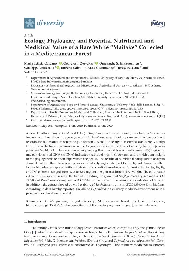
diversity
Article
Ecology, Phylogeny, and Potential Nutritional andMedicinal Value of a Rare White “Maitake” Collectedin a Mediterranean Forest
Maria Letizia Gargano 1 , Georgios I. Zervakis 2 , Omoanghe S. Isikhuemhen 3,
Giuseppe Venturella 4 , Roberta Calvo 4,*, Anna Giammanco 5, Teresa Fasciana 5 and
Valeria Ferraro 4
1 Department of Agricultural and Environmental Science, University of Bari Aldo Moro, Via Amendola 165/A,I-70126 Bari, Italy; [email protected]
2 Laboratory of General and Agricultural Microbiology, Agricultural University of Athens, 11855 Athens,Greece; [email protected]
3 Mushroom Biology and Fungal Biotechnology Laboratory, Department of Natural Resource &Environmental Design, North Carolina A&T State University, Greensboro, NC 27411, USA;[email protected]
4 Department of Agricultural, Food and Forest Sciences, University of Palermo, Viale delle Scienze, Bldg. 5,I-90128 Palermo, Italy; [email protected] (G.V.); [email protected] (V.F.)
5 Department of Health Promotion, Mother and Child Care, Internal Medicine and Medical Specialities,University of Palermo, 90127 Palermo, Italy; [email protected] (A.G.); [email protected] (T.F.)
* Correspondence: [email protected]; Tel.: +39-349-090-6252
Received: 4 May 2020; Accepted: 4 June 2020; Published: 8 June 2020
Abstract: Albino Grifola frondosa (Dicks.) Gray “maitake” mushrooms (described as G. albicansImazeki and then placed in synonymy with G. frondosa) are particularly rare, and the few pertinentrecords are not treated in scientific publications. A field investigation carried out in Sicily (Italy)led to the collection of an unusual white Grifola specimen at the base of a living tree of Quercuspubescens Willd. s.l. The outcome of sequencing the internal transcribed spacer (ITS) region ofnuclear ribosomal DNA (nrDNA) indicated that it belongs to G. frondosa and provided an insightto the phylogenetic relationships within the genus. The results of nutritional composition analysisshowed that the albino basidioma possesses relatively high contents of Ca, Fe, K, and Cu and is ratherlow in Na when compared with literature data on edible mushrooms. Vitamin (B1, B2, B3, B5, B9,and D2) contents ranged from 0.15 to 3.89 mg per 100 g of mushroom dry weight. The cold-waterextract of this specimen was effective at inhibiting the growth of Staphylococcus epidermidis ATCC12228 and Pseudomonas aeruginosa ATCC 15442 at the maximum screening concentration of 50% v/v.In addition, the extract slowed down the ability of Staphylococcus aureus ATCC 43300 to form biofilms.According to data hereby reported, the albino G. frondosa is a culinary-medicinal mushroom with apromising exploitation potential.
Keywords: Grifola frondosa; fungal diversity; Mediterranean forest; medicinal mushroom;bioprospecting; ITS rDNA; phylogenetics; basidiomycete; polypore fungus; Quercus pubescens
1. Introduction
The family Grifolaceae Jülich (Polyporales, Basidiomycota) comprises only the genus GrifolaGray [1], which consists of nine species according to Index Fungorum. Grifola frondosa (Dicks) Grayincludes several forms and varieties, such as G. frondosa f. frondosa (Dicks.) Gray, G. frondosa f.intybacea (Fr.) Pilát, G. frondosa var. frondosa (Dicks.) Gray, and G. frondosa var. intybacea (Fr.) Cetto,while G. intybacea (Fr.) Imazeki is considered as a synonym. The culinary-medicinal mushroom
Diversity 2020, 12, 230; doi:10.3390/d12060230 www.mdpi.com/journal/diversity41

Diversity 2020, 12, 230
G. frondosa, widely known as “maitake”, is a white-rot polypore associated primarily with deciduoustrees of different genera (Quercus L., Acer L., Carpinus L., Castanea Mill., Fagus L., Ulmus L.) while itappears more rarely on conifers. This species is distributed in temperate regions of North America,Europe, and Asia (Japan and China) [2]. Albino maitake mushrooms (described as G. albicans byImazeki in 1943 [3] and then placed in synonymy with G. frondosa) are quite rare and the few availablepertinent records do not appear in scientific publications. Only recently, Kawaguchi et al. [4,5] reportedon melanin biosynthesis in G. frondosa and suggested that the albino mutation is caused by a singlebase deletion in the coding region of the tyrosinase 2 (tyr2) gene. It is noteworthy that white strains ofG. frondosa are much sought after since they can be used for culinary purposes without the unwanteddark brown pigment resulting from processing the common form of this mushroom [4].
Edible mushrooms are widely acknowledged for their nutritional and medicinal properties [6];however, their potential market is still far from being fully developed considering that there is ahigh consumer demand. Consequently, the private sector is eager to generate new relevant products.In the frame of a research project aiming at collecting mushrooms with bioprospecting potential,a field investigation carried out in Sicily (Italy) led to the collection of an unusual Grifola basidiomaof white color and considerable weight (ca. 7 kg). The objective of the present work was to providemorphological, molecular/phylogenetic, and ecological data about this rare albino Grifola specimen.Information on the nutritional value and antimicrobial activity is also reported.
2. Materials and Methods
2.1. Sample Collection, Habitat Details, and Evaluation of Morphological Characters
Research carried out in forest ecosystems in Sicily (Italy) led to the collection of a single albinoGrifola basidioma found at the base of a monumental downy oak tree (Quercus pubescens Willd. s.l.),10 January 2016, Castelbuono, Madonie Regional Park (province of Palermo, Sicily), 800 m a.s.l.,37◦54′48” N, 14◦04′41” E. In Castelbuono, summers are rather brief, hot, and dry, with clear skies andcondensation often appearing in the late afternoon, while winters are long, cold, rainy, and windy.Annual temperature ranges from 7 to 29 ◦C (rarely below 4 ◦C or above 32 ◦C) while the averagevalue is 15.8 ◦C. The average annual rainfall is 515 mm. In January, the month when the specimenunder study was harvested, the average temperature is 8.9 ◦C, and the respective maximum andminimum values are 11.5 and 6.3 ◦C; the rainfall is 67 mm. The forest is characterized by evergreenand deciduous oak vegetation with a prevalence of Quercus ilex L. and Q. pubescens Willd. s.l. In theupper part of the mountain, the vegetation is characterized by mixed woods of oaks, Fagus sylvatica L.and Ilex aquifolium L. The specimen was transferred to the Department of Agricultural, Food, andForest Sciences (SAAF) of the University of Palermo, and stored at <4 ◦C for up to 24 h prior tomorphological examination, which was carried out according to Bernicchia [7]. Observations onmacromorphological characters (pileus, cuticle, pores, stipe, context, etc.) were performed on freshmaterial, while microscopic characters (hyphal system, generative hyphae, basidia, sterigmata, cystidia,cystidioles, and basidiospores) were studied by using 3% potassium hydroxide and ammoniacalRed Congo. In addition, the reaction of the flesh to iodine-potassium iodide, potassium hydroxide,and iron salts was also evaluated.
2.2. Establishment of Pure Cultures and Mushroom Cultivation Trials
The collected basidioma was dried and then deposited in the Herbarium of the Department ofAgricultural, Food, and Forest Sciences (SAAF 450). Prior to this, a piece of pseudo-tissue was removedfrom the fresh specimen, placed on potato dextrose agar (PDA) in Petri dishes under aseptic conditions,and incubated for 15 days at 25 ± 2 ◦C. The established pure culture was stored in the Mycothecaof the Herbarium SAF (SAF 323), and it was subsequently used for the inoculation of mushroomcultivation substrates composed of either holm oak (Quercus ilex L.) or chestnut (Castanea sativa Miller)wood residues. The former (holm oak wood shavings) was filled in plastic bottles (volume: 1 L each),
42

Diversity 2020, 12, 230
while the latter (chestnut wood chips of ca. 2–3 cm and sawdust) was filled in plastic bags (volume: 6 Leach) after their moisture content was adjusted to 50–60%; in both cases, non-hydrophilic cotton filterswere used to allow air exchange. Sterilization was performed twice at 120 ◦C, 1.1 Atm for 20 min witha 24-h interval between each cycle. After the final cooling, substrates were inoculated using activelygrowing mycelium and incubation was carried out at 25 ◦C in the dark.
2.3. DNA Extraction, PCR Amplification, and Sequencing
Total genomic DNA was extracted from the herbarium specimen using the Nucleospin Plant IIDNA kit (Macherey and Nagel, Düren, Germany) following the manufacturer’s protocol. The internaltranscribed spacer (ITS) region of nuclear ribosomal DNA (nrDNA) was amplified using the primercombination ITS1/ITS4 [8]. Polymerase chain reactions (PCRs) were performed in 50 μL containing50 ng DNA template, 0.25 μM of each primer, 0.2 mM of each dNTP, 1 × HiFi Buffer (Takara BIO INC.,Shiga, Japan), and 1 U HiFi Taq DNA polymerase (Takara BIO INC., Shiga, Japan). Conditions for PCRamplification were as follows: 94 ◦C for 5 min, followed by 35 cycles of 94 ◦C for 30 s, 50 ◦C for 30 sand 72 ◦C for 1 min, and a final extension at 72 ◦C for 10 min.
PCR products were purified using an Invitrogen PureLink kit (Thermo Fisher Scientific,Seoul, Korea) and were submitted for sequencing to CeMIA S.A. (Larissa, Greece). The resultingchromatograms were proofread, and the sequence generated was deposited in GenBank under theaccession number MN944407.
2.4. Phylogenetic Analysis of Sequence Data
In addition to the biological material examined in this study, 53 additional ITS sequences wereincluded in the phylogenetic analysis; 38 of them representing other Grifola Spp., i.e., all availablesequences of G. colensoi (Berk.) G. Cunn., G. gargal Singer, and G. sordulenta (Mont.) Singer, andthe rest corresponding to two other species used as outgroups, i.e., Polyporus umbellatus (Pers.) Fr.(syn. Grifola umbellata (Pers.) Pilát) and Rhodonia placenta (Fr.) Niemelä, K.H. Larss. & Schigel (syn.Oligoporus placentus (Fr.) Gilb. & Ryvarden). Sequence alignment was performed through the aid of theClustal Omega software (https://www.ebi.ac.uk/Tools/msa/clustalo/); alignments were inspected andmanually adjusted at misaligned sites using MEGA X (Pennsylvania State University, State College,PA, USA) [9].
Phylogenetic relationships were inferred by using the maximum likelihood (ML) method based onthe Kimura 2-parameter model [10]. Initial tree(s) for the heuristic search were obtained automaticallyby applying the Neighbor-Join and BioNJ algorithms to a matrix of pairwise distances estimated usingthe maximum composite likelihood (MCL) approach, and then selecting the topology with the superiorlog likelihood value. Phylogenetic analysis was conducted in MEGA X [9].
2.5. Evaluation of Nutritional Value and Determination of Antimicrobial Activity
The powder resulting after freeze-drying part of the collected basidioma was also subjected toanalysis for the evaluation of the nutritional value (proximate composition, and content in elementsand vitamins) according to standard methodologies reported by Palazzolo et al. [11], AOAC [12],Thompson et al. [13], and Loewus [14].
With regard to the antibacterial properties, the study initially included the evaluation of the activityof the lyophilized powder deriving from the basidioma against four Gram-positive and Gram-negativebacteria strains: Staphylococcus aureus ATCC 25923, S. epidermidis ATCC 12228, Pseudomonas aeruginosaATCC 15442, and Escherichia coli ATCC 25922. Tryptic soy broth (Sigma-Aldrich, Darmstadt, Germany)containing glucose (2% w/v) or Mueller Hinton (Sigma-Aldrich, Darmstadt, Germany) was used asgrowth media. The lyophilized sample (3 g) was placed in a beaker with 200 mL of demineralizedwater, and then stored at −20 ◦C.
The minimum inhibitory concentration (MIC) was determined in a Minisart syringe with a 2.5-μmcellulose acetate filter by preparing solutions in vitro at increasing concentrations, incubating them
43
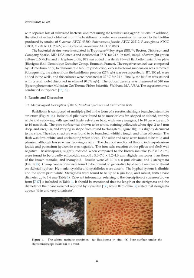
Diversity 2020, 12, 230
with separate lots of cultivated bacteria, and measuring the results using agar dilutions. In addition,the effect of extract obtained from the basidioma powder was examined in respect to the biofilmproduced by strains of S. aureus ATCC 43300, Enterococcus faecalis ATCC 29212, P. aeruginosa ATCC27853, E. coli ATCC 25922, and Klebsiella pneumoniae ATCC 700603.
The bacterial strains were inoculated in Trypticase™ Soy Agar (BBL™; Becton, Dickinson andCompany, Sparks, MD, USA) media and incubated at 37 ◦C for 24 h. In total, 100 μL of overnight grownculture (0.5 McFarland in tryptose broth, BT) was added in a sterile 96-well flat bottom microtiter plate(Biosigma S.r.l. Dominique Dutscher Group, Brumath, France). The negative control was composedby BT medium only; to demonstrate biofilm production, excess bacterial suspension was removed.Subsequently, the extract from the basidioma powder (25% v/v) was re-suspended in BT, 100 μL wereadded in the wells, and the cultures were incubated at 37 ◦C for 24 h. Finally, the biofilm was stainedwith crystal violet dissolved in ethanol (0.5% w/v). The optical density was measured at 540 nm(Spectrophotometer Multiskan Go; Thermo Fisher Scientific, Waltham, MA, USA). The experiment wasconducted in triplicate [15,16].
3. Results and Discussion
3.1. Morphological Description of the G. frondosa Specimen and Cultivation Tests
Basidioma is composed of multiple pilei in the form of a rosette, sharing a branched stem-likestructure (Figure 1a). Individual pilei were found to be more or less fan-shaped or deltoid, entirelywhite and yellowing with age, and finely velvety or bald, with wavy margins, 4 to 10 cm wide and 5to 10 mm thick. The pore surface was shown to be white, staining yellowish when ripe, 2 to 3 mmdeep, and irregular, and varying in shape from round to elongated (Figure 1b); it is slightly decurrentto the stipe. The stipe structure was found to be branched, whitish, tough, and often off-center. Theflesh was firm, white, and unchanging when sliced. The odor and taste were found to be mild andpleasant, although less so when decaying or acrid. The chemical reaction of flesh to iodine-potassiumiodide and potassium hydroxide was negative. The iron salts reaction on the pileus and flesh wasnegative. Basidiospores, slightly reduced when compared to the brown maitake (5–7 × 3.2 μm),were found to be broadly ellipsoidal, smooth, 5.0–7.0 × 3.2–4.0 μm, slightly narrower than thoseof the brown maitake, and inamyloid. Basidia were 25–30 × 6–8 μm; clavate; and 4-sterigmata(Figure 2a). Clamp connections were found to be present on generative hyphae but are rare or absenton skeletal hyphae. Hymenial cystidia and cystidioles were absent. The hyphal system is dimitic,and the spore print white. Sterigmata were found to be up to 6 μm long, and robust, with a basediameter up to 1.6 μm (Table 1). Relevant information referring to the description of common brownform [7,17] is included in Table 1. It should be mentioned that the length of the sterigmata and thediameter of their base were not reported by Ryvarden [17], while Bernicchia [7] stated that sterigmataappear “thin and very divaricate”.
Figure 1. The albino maitake specimen: (a) Basidioma in situ; (b) Pore surface under thestereomicroscope (scale bar = 1 mm).
44

Diversity 2020, 12, 230
Figure 2. (a) Microscopic features (basidia and spores) of albino maitake. (b) Pure culture isolated fromfresh basidioma.
Table 1. Main macro- and micromorphological features of brown [7,17] and albino (this study) formsof maitake.
Descriptive Characters Brown Form Albino Form
Habit Cluster of pilei Cluster of pilei
Pileus Irregular, wrinkled, flat and uneven Fan-shaped or deltoidStipe Whitish Whitish
Tubules Short, thick, whitish Short, thick, whitishPores Very small, then angular and wider Round to elongated, slightly decurrent to the stipe
StemWhitish and fleshy, with numerous
flattened branches and bifurcations thatend in a fan-shaped pileus
Structure branched, whitish, tough, often off-center
FleshWhite, immutable, tenacious especially
towards the base, fragile towardsthe pileus
Firm, white, unchanging when sliced
Smell Penetrating, intense then nauseating Mild and pleasant, unpleasant or acridwhen decaying
Taste Pleasant and delicate Mild and pleasant, unpleasant or acridwhen decaying
Basidiospores Ellipsoidal, 5.0–6.5 × 3.5–5.0 μm Broadly ellipsoidal, smooth, 5.0–7.0 × 3.2–4.0 μmBasidia 23–30 × 5–8 μm; clavate; 4-sterigmata 25–30 × 6–8 μm; clavate; 4-sterigmata
Hyphal system Dimitic with clamp connections Dimitic with clamp connections
Habitat At the base of hardwoods, particularlychestnut and oak trees At the base of oak trees
Period of fructification Late August to October December to January
Mycelium growth was very slow in the pure cultures established, and the surface of the Petri dish(diameter 90 mm) was completely overgrown in 30 days. The colony was found to be slightlyfloccose and flat, with dense white hyphae without any zonation and with a filiform margin(Figure 2b). Regarding the cultivation tests performed either in holm oak or in chestnut-basedsubstrates, colonization was slow and ca. two months elapsed before they were completely overgrownby mycelium. At this time, a few primordia were formed on the substrate surface, which, however,failed to produce fully developed basidiomata.
3.2. Phylogenetic Analysis
The main objective of the molecular analysis was to identify the biological material underexamination (albino maitake). Following BLAST, the sequence (MN944407) from our specimen showedhigh identity (>99%) to several other sequences deposited as G. frondosa in GenBank. The generatedML trees had similar topologies, and only the tree with the highest log likelihood (−7662.14) wasselected to be presented (Figure 3).
45

Diversity 2020, 12, 230
Figure 3. Maximum likelihood tree produced on the basis of ITS (internal transcribed spacer) sequenceanalysis presenting the phylogenetic position of the albino G. frondosa specimen (underlined in redand marked with an asterisk) among members of the genus Grifola; GenBank accession numbers arequoted together with information of their geographic origin (when available). Sequences of Polyporusumbellatus (syn. G. umbellata) and Rhodonia placenta were used as outgroups. The percentage of trees inwhich the associated taxa clustered together is shown next to the branches when ≥70% (ML bootstrapsupport values derived from a total of 1000 replicates). The tree is drawn to scale, with branch lengthsmeasured as the number of substitutions per site.
46

Diversity 2020, 12, 230
To the best of our knowledge, the sequence obtained in this study is the only one available froman albino Grifola specimen. The outcome of the phylogenetic analysis revealed that the albino maitakeclusters within the G. frondosa terminal clade, which receives high bootstrap support (100%), and formsa subgroup (84%) together with other sequences from material collected in Europe; the rest of theG. frondosa sequences derive almost exclusively from east Asia (China, Japan, South Korea, Taiwan)and the USA. Although this subgroup could represent a distinct taxonomic entity, the informationgenerated from our work (through the examination of one basidioma and the use of a single markeronly) does not allow such inferences, at least before additional specimens are included and analyzed inthe frame of a multigene approach. However, it is noteworthy that G. frondosa sequences from Asianand eastern North American isolates were separated through the use of a partial beta-tubulin gene andITS [18]. G. frondosa forms a sister group to G. sordulenta (94%). The latter is highly supported (99%)and consists of three sequences under this name and one representing G. gargal (AB539700). Hence,the distinct taxonomic position of the latter is dubious by examining the ITS marker alone. Of interestwas also the position of the sole G. colensoi sequence available (MH856542; this species is known so farfrom Australia and New Zealand only), which appears to be quite distant from the rest of the materialplaced in the genus Grifola. Lastly, sequences deposited either as G. umbellata or P. umbelatus forma distinct highly supported group (100%), which is well separated from the clade corresponding toGrifola Spp. To the best of our knowledge, the sequence generated in the frame of this work is the firstone deriving from an albino G. frondosa specimen.
3.3. Mushroom Proximate Composition and Content in Elements and Vitamins
In the frame of this work, it was not possible to analyze (and hence directly compare to ouralbino specimen) the composition of wild basidiomata of brown maitake since they were not availableduring the period of the study, this species being very rare in Sicily. On the other hand, only limitedliterature data are available referring to the chemical composition and vitamin contents in cultivatedG. frondosa [19,20]; the respective values reported—apart from a few exceptions—are considerablylower than those found in the white maitake. However, such information should be evaluated with caresince it is known that the nature of the cultivation substrate could considerably affect the mushroomcontent [21,22]. Similarly, metal accumulation in wild edible mushrooms was found to be influencedby various factors, including the species, the available concentration of elements in soil substrates,the substrates’ properties (e.g., pH, organic matter content), the antagonistic and/or synergistic effectsamong elements, and the concentration of other elements in mushrooms [23–26].
The results of the proximate composition analysis of the albino maitake showed rather low valuesof protein, fat, and carbohydrate contents (Table 2), while the calorie content was 35.84 kcal·100 g−1.In addition, element analysis showed that this specimen presents relatively high contents in Ca, Fe, K,and Cu, and is rather low in Na (Table 2). The Ca content is higher than in some baby foods, suchas oatmeal and whole milk, as well as ham, cream, spread cheese, white chocolate, and others [27].The content in Fe is considerably higher than in most other foods; it is noteworthy that dried thyme(i.e., one of the richest foods in Fe) contains about 50 mg less Fe per 100 g when compared to the albinoG. frondosa. The K content in albino maitake is lower only to that of cremor tartar and much higherthan that of dried spirulina algae [27], while the Na content is lower than that of miso soup and bakingpowder [27]. The Cu content of albino maitake is higher than most foods, including dark chocolate,oysters, liver, and lobster [27].
47

Diversity 2020, 12, 230
Table 2. Outcome of proximate composition analysis (% for nitrogen, proteins, fats, carbohydrates,and ash), and content in elements and vitamins (mg·100 g−1) of the albino maitake specimen. Values areexpressed as means ± standard error, n = 3.
Composition/Content Albino Maitake
Nitrogen 2.18 ± 0.01Proteins 13.65 ± 0.00
Fats 1.02 ± 0.04Carbohydrates 6.78 ± 0.11
Ash 1.06 ± 0.10Ca 245.30 ± 0.17Fe 178.40 ± 0.15Mg 455.90 ± 0.19K 11,785.60 ± 0.36
Na 3433.40 ± 0.34P2O5 2399.61 ± 0.02Cu 2.81 ± 0.02Mn 2.41 ± 0.03Zn 10.33 ± 0.02Se 4.00 ± 0.00Pb 2.10 ± 0.00
Thiamine (B1) 0.15 ± 0.03Riboflavin (B2) 3.89 ± 0.06
Niacin (B3) 0.36 ± 0.03Pantothenic Acid (B5) 0.68 ± 0.02
Folic acid (B9) 0.38 ± 0.01D2 0.41 ± 0.02
The content in vitamins was: B1, 0.15 mg 100 g−1; B2, 0.36 mg·100 g−1; B3, 3.89 mg·100 g−1; B5,0.68 mg 100 g−1; B9, 0.38 mg 100 g−1; and D2, 0.41 mg 100 g−1 (Table 2). Noteworthy is the value ofvitamin B9, or folic acid, since the daily requirement is about 0.2 mg, which doubles for pregnantwomen [28].
3.4. Antibacterial and Antibiofilm Activity
The minimal inhibitory concentration (MIC) of the extract deriving from the albino maitake wasdetermined by a micro-method, i.e., by diluting the protein extract in the range from 25% to 0.025% v/vas previously described [29].
The antibacterial activity test carried out on albino maitake showed that the cold water extract waseffective in inhibiting the growth of two bacterial strains, i.e., S. epidermidis ATCC 12228 and P. aeruginosaATCC 15442, at the maximum screening concentration of 50% v/v. S. epidermidis is a Gram-positivebacterium typically present on the skin, which could be particularly dangerous in surgical procedures,while P. aeruginosa, is a ubiquitous bacterium and opportunistic pathogen in humans. As regards theeffect of the water extract from the albino G. frondosa on biofilm formation, the results revealed that itdecreased the biofilm produced by S. aureus ATCC43300 [30], whereas it slightly enhanced biofilmformation by E. faecalis ATCC 29212, E. coli ATCC 25922, K. pneumoniae ATCC 700603, and P. aeruginosaATCC 15442 (Figure 4).
48

Diversity 2020, 12, 230
Figure 4. Effect of the cold water extract from the albino Grifola frondosa basidioma on biofilm production(expressed as optical density, OD) by five bacterial species. BT: biofilm produced in the absence of thefungal extract; BT+GF: biofilm produced in the presence of the fungal extract. The experiment wasconducted in triplicate.
3.5. Ecological and Conservation Issues
The characterization of mushrooms growing in the Mediterranean area is one of the main targetsof studies carried out by our research group. The principal objective is to provide reliable/robustscientific data amid the considerable confusion existing in the commercial exploitation of pertinentmaterial in Italy and elsewhere, since accurate identification and/or origin of products placed on themarket are often missing or of ambiguous validity. Previous results have highlighted the importanceof mushrooms of the genus Pleurotus in human nutrition and in the feeding of farm animals [31–35].Particular attention is also drawn to edible mushroom species that are infrequent/rare or linked toparticular habitats (e.g., Pleurotus nebrodensis and P. opuntiae) [36,37], and for which only few or nostudies exist regarding their nutritional and medicinal properties.
Although G. frondosa is widely distributed in the northern hemisphere, it is considered very rarein Italy. Particularly in Sicily, the only reports available concern the brown maitake found in thenortheastern part of the island and date back to the end of the 19th century [38]; more recent informalrecords by amateur mycological groups derive from the same wider region (however, no pertinentsamples are available for further study). Hence, the occurrence of an albino maitake is of apparentimportance in a biogeographical, ecological, and applied context. In addition, the presence of thisfungus on a monumental Q. pubescens tree of high significance points out the potential risks forhost plants in the area because G. frondosa demonstrates a parasitic action resulting in root and buttdecay [39,40]. At the same time, it is necessary to ensure the necessary conditions for its ex situconservation due to the exploitation potential it exhibits as food and source of bioactive compoundsas it was reported for wild mushrooms in general [41]. We have therefore adopted well-establishedprotocols for its long-term maintenance [42] since our investigation showed that fructification ofthe albino maitake is not constant over time and the ability of the strain to be preserved underlaboratory conditions—without significant degeneration of its properties—is lower than in othermushroom species.
4. Conclusions
An albino maitake specimen was reported for the first time in Italy. The examined basidiomadiffers morphologically from the common brown/gray form, with the most significant differences beingthe color and shape of the pileus, the type of pores, the shape of the stipe, and the period of fructification.Phylogenetic analysis evidenced that the specimen under study groups among other European isolateswithin a larger terminal clade corresponding to G. frondosa. Moreover, the nutritional composition
49

Diversity 2020, 12, 230
and the potential of the albino maitake to serve as source of functional compounds is of interest,and further investigation is in progress on the characterization of polysaccharides and at evaluatingthe antitumor activity against several types of cancer cells. The outcome of tests performed using themushroom water extract against human pathogenic bacteria demonstrated its efficacy at inhibiting thegrowth of S. epidermidis and P. aeruginosa strains, while it also hindered biofilm formation by S. aureus,i.e., the causal agent of serious infections in immunocompromised patients. The antibacterial agentspresent in the albino maitake are promising candidates for dealing with pathogens that show resistanceto a large spectrum of antibiotics and are widespread in hospitals.
Author Contributions: Conceptualization, G.V. and M.L.G.; methodology, G.V., G.I.Z., M.L.G. and O.S.I.;validation, R.C. and V.F.; formal analysis, G.I.Z., M.L.G. and O.S.I.; investigation, G.V., G.I.Z., M.L.G. and O.S.I.;data curation, A.G. and T.F.; biofilm analysis, R.C. and V.F.; writing—original draft preparation, G.V. and M.L.G.;writing—review and editing, G.V. and G.I.Z.; visualization, G.V.; supervision, G.V. All authors have read andagreed to the published version of the manuscript.
Funding: This research received no external funding.
Conflicts of Interest: The authors declare no conflicts of interest.
References
1. Justo, A.; Miettinen, O.; Floudas, D.; Ortiz-Santana, B.; Sjökvist, E.; Lindner, D.L.; Nakasone, K.; Niemelä, T.;Larsson, K.-H.; Ryvarden, L.; et al. A revised family-level classification of the Polyporales (Basidiomycota).Fungal Biol. 2017, 121, 798–824. [CrossRef] [PubMed]
2. Chen, A.W.; Stamets, P.; Cooper, R.B.; Huang, N.L.; Han, S.-H. Ecology, morphology, and morphogenesis in natureof edible and medicinal mushroom Grifola frondosa (Dicks.: Fr.) S.F. Gray—Maitake (Aphyllophoromycetideae).Int. J. Med. Mushrooms 2000, 2, 8. [CrossRef]
3. Imazeki, R. Genera of Polyporaceae of Nippon. Bull. Nat. Sci. Mus. Tokyo 1943, 6, 1–111.4. Kawaguchi, N.; Hayashi, M.; Chen, F.-C.; Shimomura, N.; Yamaguchi, T.; Aimi, T. Genetic analyses of causal
genes of albinism (white fruiting body) in Grifola frondosa. J. Wood Sci. 2019, 65, 32. [CrossRef]5. Kawaguchi, N.; Hayashi, M.; Nakano, S.; Shimomura, N.; Yamaguchi, T.; Aimi, T. Expression of
tyrosinase genes associated with fruiting body formation and pigmentation in Grifola frondosa. Mycoscience2019, 60, 262–269. [CrossRef]
6. Gargano, M.L.; Van Griensven, L.J.L.D.; Isikhuemhen, O.S.; Lindequist, U.; Venturella, G.; Wasser, S.P.;Zervakis, G.I. Medicinal mushrooms: Valuable biological resources of high exploitation potential. Plant Biosyst.2017, 151, 548–565. [CrossRef]
7. Bernicchia, A. Polyporaceae s.l.; Edizioni Candusso: Alassio, Italy, 2005; p. 808.8. White, T.J.; Bruns, T.; Lee, S.; Taylor, J. Amplification and Direct Sequencing of Fungal Ribosomal RNA Genes
for Phylogenetics; Academic Press: Cambridge, MA, USA, 1990; pp. 315–322.9. Kumar, S.; Stecher, G.; Li, M.; Knyaz, C.; Tamura, K. MEGA X: Molecular evolutionary genetics analysis
across computing platforms. Mol. Biol. Evol. 2018, 35, 1547–1549. [CrossRef]10. Kimura, M. A simple method for estimating evolutionary rates of base substitutions through comparative
studies of nucleotide sequences. J. Mol. Evol. 1980, 16, 111–120. [CrossRef]11. Palazzolo, E.; Gargano, M.L.; Venturella, G. The nutritional composition of selected wild edible mushrooms
from Sicily (southern Italy). Int. J. Food Sci. Nutr. 2011, 63, 79–83. [CrossRef]12. AOAC. Official Methods of Analysis of the AOAC. Anal. Chem. 1980, 52, 148A. [CrossRef]13. Thompson, T.E.; Senter, S.D.; Grauke, L. Lipid content and fatty acids of pecan pollen. HortScience
1993, 28, 1191–1193. [CrossRef]14. Loewus, F.A. Improvement in anthrone method for determination of carbohydrates. Anal. Chem. 1952, 24, 219.
[CrossRef]15. Calà, C.; Amodio, E.; Di Carlo, E.; Virruso, R.; Fasciana, T.; Giammanco, A. Biofilm production in Staphylococcus
epidermidis strains, isolated from the skin of hospitalized patients: Genetic and phenotypic characteristics.New Microbiol. 2015, 38, 521–529. [PubMed]
50

Diversity 2020, 12, 230
16. Fasciana, T.; Gentile, B.; Aquilina, M.; Ciammaruconi, A.; Mascarella, C.; Anselmo, A.; Fortunato, A.; Fillo, S.;Petralito, G.; Lista, F.; et al. Co-existence of virulence factors and antibiotic resistance in new Klebsiellapneumoniae clones emerging in south of Italy. BMC Infect. Dis. 2019, 19, 928. [CrossRef]
17. Ryvarden, L.; Gilbertson, R.L. European Polypores. Part 1. Synopsis Fungorum 1993, 6, 1–387.18. Shen, Q.; Geiser, D.M.; Royse, D.J. Molecular Phylogenetic Analysis of Grifola frondosa (Maitake) reveals
a species partition separating Eastern North American and Asian Isolates. Mycologia 2002, 94, 472–482.[CrossRef]
19. Stamets, P. Notes on Nutritional properties of culinary-medicinal mushrooms. Int. J. Med. Mushrooms2005, 7, 103–110. [CrossRef]
20. Niedzielski, P.; Mleczek, M.; Budka, A.; Rzymski, P.; Siwulski, M.; Jasinska, A.; Gasecka, M.; Budzynska, S.A screening study of elemental composition in 12 marketable mushroom species accessible in Poland.Eur. Food Res. Technol. 2017, 243, 1759–1771. [CrossRef]
21. Koutrotsios, G.; Mountzouris, K.; Chatzipavlidis, I.; Zervakis, G.I. Bioconversion of lignocellulosic residuesby Agrocybe cylindracea and Pleurotus ostreatus mushroom fungi—Assessment of their effect on the finalproduct and spent substrate properties. Food Chem. 2014, 161, 127–135. [CrossRef]
22. Sakellari, A.; Karavoltsos, S.; Tagkouli, D.; Rizou, C.; Sinanoglou, V.J.; Zoumpoulakis, P.; Koutrotsios, G.;Zervakis, G.I.; Kalogeropoulos, N. Trace elements in Pleurotus ostreatus, P. eryngii, and P. nebrodensisMushrooms Cultivated on various agricultural by-products. Anal. Lett. 2019, 52, 2692–2709. [CrossRef]
23. Garcia, E.M.; Siegert, I.G.; Suarez, P. Toxicity assays and naphthalene utilization by natural bacteria selectedin marine environments. Bull. Environ. Contam. Toxicol. 1998, 61, 370–377. [CrossRef] [PubMed]
24. Mendil, D.; Özgür, D.U.; Tuzen, M.; Hasdemir, E.; Sarı, H.; Sari, H. Trace metal levels in mushroom samplesfrom Ordu, Turkey. Food Chem. 2005, 91, 463–467. [CrossRef]
25. Venturella, G.; Gargano, M.L.; Compagno, R.; Saitta, A.; Alaimo, M.G. The mineral contents of someboletaceae species from Sicily (Southern Italy). J. AOAC Int. 2014, 97, 612–623. [CrossRef]
26. Kokkoris, V.; Massas, I.; Polemis, E.; Koutrotsios, G.; Zervakis, G.I. Accumulation of heavy metals by wildedible mushrooms with respect to soil substrates in the Athens metropolitan area (Greece). Sci. Total. Environ.2019, 685, 280–296. [CrossRef] [PubMed]
27. Fidanza, F. Alimenti e Tabelle di Composizione. In Nutrizione Umana; Fidanza, F., Liguori, G., Eds.; Idelson:Napoli, Italy, 1984; pp. 221–238.
28. Barua, S.; Kuizon, S.; Junaid, M.A. Folic acid supplementation in pregnancy and implications in healthand disease. J. Biomed. Sci. 2014, 21, 77. [CrossRef] [PubMed]
29. Schillaci, D.; Petruso, S.; Sciortino, V. 3,4,5,3′,5′-Pentabromo-2-(2′-hydroxybenzoyl) pyrrole: A potentiallead compound as anti-Gram-positive and anti-biofilm agent. Int. J. Antimicrob. Agents 2005, 25, 338–340.[CrossRef] [PubMed]
30. Giammanco, A.; Galia, E.; Fasciana, T.; Sciortino, M.; Cannella, S.; Arrigo, I.; Tricoli, M.R.; Di Paola, L.;Venturella, G.; Gargano, M.L. The Anti-Biofilm Potential of Pleurotus eryngii var. elaeoselini and White formof Grifola frondosa Extracts Against Staphylococcus aureus methicillin Resistance. In Proceedings of the10th International Medicinal Mushrooms Conference, Nantong, China, 19–22 September 2019; InternationalSociety for Medicinal Mushrooms & Jiangsu Provincial Department of Agriculture and Rural Affairs:Nantong, China, 2019; pp. 184–185.
31. La Guardia, M.; Venturella, G.; Venturella, F. On the chemical composition and nutritional value of Pleurotustaxa growing on Umbelliferous plants (Apiaceae). J. Agric. Food Chem. 2005, 53, 5997–6002. [CrossRef][PubMed]
32. Koutrotsios, G.; Kalogeropoulos, N.; Stathopoulos, P.; Kaliora, A.C.; Zervakis, G.I. Bioactive compounds andantioxidant activity exhibit high intraspecific variability in Pleurotus ostreatus mushrooms and correlate wellwith cultivation performance parameters. World J. Microbiol. Biotechnol. 2017, 33, 98. [CrossRef]
33. Koutrotsios, G.; Kalogeropoulos, N.; Kaliora, A.C.; Zervakis, G.I. Toward an Increased functionalityin oyster (Pleurotus) mushrooms produced on Grape Marc or Olive Mill Wastes Serving as sources ofbioactive compounds. J. Agric. Food Chem. 2018, 66, 5971–5983. [CrossRef]
34. Gaglio, R.; Guarcello, R.; Venturella, G.; Palazzolo, E.; Francesca, N.; Moschetti, G.; Settanni, L.; Saporita, P.;Gargano, M.L. Microbiological, chemical and sensory aspects of bread supplemented with differentpercentages of the culinary mushroom Pleurotus eryngii in powder form. Int. J. Food Sci. Technol.2018, 54, 1197–1205. [CrossRef]
51

Diversity 2020, 12, 230
35. Bonanno, A.; Di Grigoli, A.; Vitale, F.; Di Miceli, G.; Todaro, M.; Alabiso, M.; Gargano, M.L.; Venturella, G.;Anike, F.N.; Isikhuemhen, O.S. Effects of diets supplemented with medicinal mushroom myceliated grainson some production, health, and oxidation traits of dairy ewes. Int. J. Med. Mushrooms 2019, 21, 89–103.[CrossRef]
36. Zervakis, G.I.; Ntougias, S.; Gargano, M.L.; Besi, M.I.; Polemis, E.; Typas, M.A.; Venturella, G. A reappraisalof the Pleurotus eryngii complex—New species and taxonomic combinations based on the application of apolyphasic approach, and an identification key to Pleurotus taxa associated with Apiaceae plants. Fungal Biol.2014, 118, 814–834. [CrossRef] [PubMed]
37. Zervakis, G.I.; Venturella, G.; Fryssouli, V.; Inglese, P.; Polemis, E.; Gargano, M.L. Pleurotus opuntiaerevisited—An insight to the phylogeny of dimitic Pleurotus species with emphasis on the P. djamor complex.Fungal Biol. 2019, 123, 188–199. [CrossRef] [PubMed]
38. Venturella, G. A check-list of Sicilian fungi. Bocconea 1991, 2, 1–221.39. Antonio, A.; Valeria, B.; Marcello, D.; Caterina Patrizia Di, F.; Di Gesaro, M.; Emanuele, S. Monumental trees
and their existence value: The case study of an Italian natural park. J. For. Sci. 2016, 61, 55–61. [CrossRef]40. Schwarze, F.W.M.R.; Engels, J.; Mattheck, C. Fungal Strategies of Wood Decay in Trees; Springer:
Berlin/Heidelberg, Germany, 2000.41. Zotti, M.; Persiani, A.M.; Ambrosio, E.; Vizzini, A.; Venturella, G.; Donnini, D.; Angelini, P.; Di Piazza, S.;
Pavarino, M.; Lunghini, D.; et al. Macrofungi as ecosystem resources: Conservation versus exploitation.Plant Biosyst. 2013, 147, 219–225. [CrossRef]
42. Moore, D.; Nauta, M.M.; Evans, S.E.; Rotheroe, M. Fungal Conservation. Issues and Solutions; CambridgeUniversity Press: Cambridge, UK, 2001; p. 262.
© 2020 by the authors. Licensee MDPI, Basel, Switzerland. This article is an open accessarticle distributed under the terms and conditions of the Creative Commons Attribution(CC BY) license (http://creativecommons.org/licenses/by/4.0/).
52

diversity
Article
An Italian Research Culture Collection of WoodDecay Fungi
Carolina Elena Girometta 1,* , Annarosa Bernicchia 2, Rebecca Michela Baiguera 1,
Francesco Bracco 1 , Simone Buratti 1, Marco Cartabia 1,3, Anna Maria Picco 1 and Elena Savino 1
1 Department of Earth and Environmental Sciences (DSTA), University of Pavia, 27100 Pavia, Italy;[email protected] (R.M.B.); [email protected] (F.B.);[email protected] (S.B.); [email protected] (M.C.);[email protected] (A.M.P.); [email protected] (E.S.)
2 School of Agriculture and Veterinary Medicine, University of Bolognavia, Guidotti 39, 40134 Bologna, Italy;[email protected]
3 MOGU S.r.l. Via San Francesco d’Assisi 62, 21020 Inarzo (VA), Italy* Correspondence: [email protected]
Received: 29 December 2019; Accepted: 30 January 2020; Published: 1 February 2020
Abstract: One of the main aims of the University of Pavia mycology laboratory was to collect wooddecay fungal (WDF) strains in order to deepen taxonomic studies, species distribution, officinalproperties or to investigate potential applications such as biocomposite material production basedon fungi. The Italian Alps, Apennines and wood plains were investigated to collect Basidiomycotabasidiomata from living or dead trees. The purpose of this study was to investigate the wood decaystrains of the Mediterranean area, selecting sampling sites in North and Central Italy, includingforests near the Ligurian and Adriatic seas, or near the Lombardy lakes. The isolation of mycelia inpure culture was performed according to the current methodology and the identity of the strainswas confirmed by molecular analyses. The strains are maintained in the Research Culture CollectionMicUNIPV of Pavia University (Italy). Among the 500 WDF strains in the collection, the mostinteresting isolates from the Mediterranean area are: Dichomitus squalens (basidioma collected fromPinus pinea), Hericium erinaceus (medicinal mushroom), Inocutis tamaricis (white-rot agent on Tamarixtrees), Perenniporia meridionalis (wood degrader through Mn peroxidase) and P. ochroleuca. In addition,strains of species related to the Mediterranean climate (e.g., Fomitiporia mediterranea and Cellulariellawarnieri) were obtained from sites with a continental-temperate climate.
Keywords: wood decay fungi (WDF); culture collection; fungal strain; host; Italy; morphological andmolecular identification
1. Introduction
Wood decay fungi provide an extraordinary model both for pure and applied research, as well asa food or medicinal mushroom resource.
From an ecological point of view, wood decay fungi have a fundamental role, since they areimportant degraders of lignocelluloses. Heterogeneity in degradation strategies consists in differentenzymatic pools, conditions for secretion and catalysis, alternative non-enzymatic pathways andstrategy-switch depending on environmental conditions [1,2]. Consistently, wood decay fungi shiftfrom necrotrophism to pure saprotrophism, sometimes at an intraspecific level.
Systematic revisions based on multi-locus or genomic approach have revealed an extremelycomplex scenario concerning both biochemical features and morphology. Similar degradation strategiesand similar morphologies are widespread, even among phylogenetically distant taxa, whereas thesame taxonomic group may include species displaying different strategies and different morphologies.
Diversity 2020, 12, 58; doi:10.3390/d12020058 www.mdpi.com/journal/diversity53

Diversity 2020, 12, 58
As a whole, wood decayers appear to be a pivotal model in the study of the evolutionary relationshipsof both Dikarya and extra-Dikarya taxa [3–5]. Consistently, the incipient molecular-based biogeographyof wood decayers seems to display distribution patterns strongly affected by preferred host species(trees or shrubs), which are apparently followed throughout [6].
Wood decay fungi include several edible species as well as species reported to be the source ofbioactive compounds, related to either primary (e.g., β-glucans) or secondary metabolism (terpenoids,phenolics, acids, superior alcohols, etc.). Structural diversity, occurrence and distribution among taxa,synthesis stimulation factors and correlation to growth stage, bioactivity pathways, standardization ofproducts and crude extracts are the main current topics under investigation [7–9].
Due to their relatively easy reproduction in culture, several other applications are being developedon wood decay fungi: degradation of organic pollutants and bioremediation [10]; pretreatment ofbiomasses for production of sugars and bioethanol [11,12]; production of enzymes for industrialpurposes, namely Mn peroxidases, laccases, cellulases and hemicellulases [13]; and bioadsorption andbioaccumulation of metal ions either in living or dead biomass [14–16].
Last but not least, necrotrophic wood decay means a loss of harvest in forestry and woody cultures,whereas in public and private green areas it means destabilization and consequently risk for peopleand objects [17].
Culture collections are an important reference, since availability and exchange of authenticated,quality-guaranteed pure cultures are increasingly needed by researchers at an international level [18,19].Above all, tests on different species and strains (at intraspecific level) are required since biochemicaldifferences are often not negligible [20–22].
Actually, only a few research centers can afford to structure their culture collection in conformityto international guidelines provided by the World Federation for Culture Collections (WFCC) [23,24].The strains maintained in many universities or research centers can be considered an important sourceof experimental material, even without a WFCC certification. Since small uncertified collectors aregeographically widespread, their contribution may be significantly representative of local ecosystemsand biodiversity [25].
The Mycology Laboratory at the Botanical Garden of Pavia University (Italy) has a long traditionof the isolation, identification and preservation of fungal strains in various areas of mycology. This issupported by the numerous publications from the middle of the last century [26] up to now. Currently,the fungal strains collection is named MicUNIPV and each working group preserves and enrichesthe collection.
Although the definition of ecotypes is usually hard, a remarkable intraspecific variability is welldocumented in several fungal species and it may be particularly true for rare species, whose populationsare supposed to be more isolated [27]. This highlights the value of Italian territory for fungal biodiversityand the great potentiality for research [28].
Italy has a wide variety of climates and morphologies, both due to its remarkable latitudinal range(about 13◦) and structural-topographic complexity, including the presence of four different seas andtwo main mountain chains. According to the official maps of MATTM (Ministero dell’Ambiente e dellaTutela del Territorio e del Mare), 28 different phytoclimatic classes are recognized, five of which arespecifically referred to as Mediterranean [29], also taking into account the biogeographic reference mapsuggested by Rivas-Martínez [30]. Nevertheless, the pluri-millennial stratification of human impacthas made it difficult to distinguish between actual and potential ecosystem features. As a consequence,the classification and mapping of either Italian ecoregions or phytoclimates provide a tool for thecomprehension of biodiversity instead of a strict map of biodiversity itself [31].
The present article reports the results obtained by the researchers of the Laboratory of Mycology inDSTA-University of Pavia (Italy) who continuously collect new cultures of wood decay fungi, focusingon fungal biodiversity of species related to the Mediterranean area and climates.
54

Diversity 2020, 12, 58
2. Materials and Methods
2.1. Sampling Sites and Field Work
Basidiomata were mostly (but not exclusively) collected in North and Central Italy. Samplingstratification was selectively applied, i.e., specific areas have been more frequently and strictly examinedthan others and sampling effort was not equal among different species [32]. The different environmentsexamined are resumed hence:
(a) highly-fragmented marginal woodlands and shrublands placed into an agricultural landscape,particularly referring to vegetation surrounding the hydrographic network, including major lakes;
(b) mountain continuous woodlands and shrublands, both managed and unmanaged;(c) woody cultures (e.g., poplar plantations and vineyards), tree rows and hedges in
agricultural landscape;(d) urban and suburban environments (tree rows, parks, private and public gardens).
Environments a, c, and d are mostly related to basal altitudinal belt and upper hill altitudinal beltin Po Plain, Apennines and Prealps (lower mountain thermal belt), as well as Adriatic, Tirrenian andLigurian coasts; Environment b is mostly related to the lower and upper montane belt in the Northand Central Apennines.
The basidiomata were completely or partially harvested by knife, gently brushed to eliminatedebris and stored in paper bags until laboratory operations. The collecting sites were geolocalized,and the host species and general features were detected.
2.2. Experimental Procedures
Basidiomata identification was carried out by macro and micro-morphological analysis [6,33,34];stereo and light microscopy were performed by Zeiss Axioplan and Zeiss Stemi 2000-C.
According to Stalpers [35] and Gams et al. [36], as well as Stamets [37], isolation of mycelia in pureculture was obtained in sterile conditions by inoculating small portions of the basidioma context intoPetri dishes containing MEA medium and antibiotic (malt extract 2% + agar 1.5% + cloramphenicol50 ppm). The incubation was carried out at 24 ◦C in the dark and each strain growth was checkedconstantly for a month. Based on the above, all the mentioned strains are to be regarded as dikaryotic.
Besides the morphological checks, molecular identifications of isolates were carried out onmycelia cultured in liquid medium (malt extract 2%). DNA was extracted from lyophilized mycelia byNucleospin Plant II kit (Macherey-Nagel). Amplification by Polymerase Chain Reaction (PCR) usedthe primer pair ITS1 (19bp) and ITS4 (20bp)—that is, Internal Transcribed Spacer of ribosomal DNA;this region has been widely used for different fungal taxa [38,39]. PCR protocol exploited Dream TaqMastermix (Promega) and was performed in a thermocycler, as reported in Table 1.
Table 1. Thermocycling protocol for PCR.
Step Aim T (◦C) Duration Cycle Repetitions
I Denaturation 95 5 min
IIDenaturation 95 30 s
35Annealing 50 45 sElongation 72 1 min
III Final elongation 72 10 min
The qualitative checking of DNA (5 μL/sample) was performed both after extraction andamplification by DNA run (30 min, 100 V) on electrophoretic gel (1% agarose). SYBR Safe-DNA GelStain (Invitrogen) was used as an intercalant; GeneRuler 1kb (Thermo Scientific, Waltham - USA) wasused as a ladder; BlueJuice (Invitrogen) was used as a gel loading buffer. The imaging was performedby Gel Doc (Biorad, Berkeley, CA, USA).
55

Diversity 2020, 12, 58
ExoSAP-IT (Applied Biosystems, Foster City, CA, USA) was used for the purification ofamplification products. According to the suggested protocol, the sample/ExoSAP ratio was 5:2 μL;the reaction was carried out in a thermocycler in two steps—15 min at 37 ◦C and 15 min at 80 ◦C.
The sequencing was ordered to Macrogen (The Netherlands). Sequence analysis was performedby Sequencher 5.0 Demo. The sequences were finally matched with the ones available in the molecularidentification facility of Mycobank [40].
Strains in pure culture were stored by different methods:
(a) on malt extract agar (MEA) in a aPetri plate at 3 ◦C;(b) in a glass tube corked with cotton at room temperature;(c) colonized paper discs in demineralized water at 4 ◦C;(d) at −80 ◦C in glycerol (selected strains only).
Periodic checking and refreshment of cultures was performed to avoid contaminationand devitalization.
The strains are maintained in the Fungal Research Culture Collection (MicUNIPV) of Departmentof Earth and Environmental Sciences of University of Pavia (Italy); each strain is included in a privatedatabase with all the information regarding sampling sites, data of collection and ecological notes.
3. Results and Discussion
MicUNIPV includes species related to plant pathology, soil, extreme environments, fresh andmarine water, monuments and cultural heritage. As previously mentioned, different working groupswithin the Laboratory of Mycology (DSTA-University of Pavia) are engaged in the management,preservation and improvement of each MicUNIPV section. The section regarding wood decay specieshas up to now achieved 500 strains belonging to 110 different species [41–43]. The broad focus on wooddecay led us to include in this section species related to different applications such as nutraceuticals,forest pathology, wood degradation and biocomposite materials.
The distribution of most species exceeds the Mediterranean area; nevertheless, several of themalso display wide spatial gaps among stations and clear heterogeneity in host preference depending onthe geographic location of the population.
Here, we present the species that have a distribution strongly related to the Mediterraneanregion and/or Southern Europe and/or warm climates, according to Ryvarden and Melo [6] andBernicchia [33,34,44]. The species related to the Mediterranean diversity are reported in Table 2 andthe most peculiar are discussed below.
56

Diversity 2020, 12, 58
Ta
ble
2.
Sele
cted
Ital
ian
stra
ins
from
Mic
UN
IPV
rela
ted
toth
eM
edit
erra
nean
area
.Phy
tocl
imat
ecl
ass
asin
[29]
.
Mic
UN
IPV
IDS
pe
cie
sA
uth
ors
Lo
cali
tyM
un
icip
ali
tyH
ost
Ph
yto
clim
ate
Cla
ss
D.c
on.1
Dae
dale
opsi
sco
nfra
gosa
(Bol
ton)
J.Sc
hröt
.D
orm
elle
tto
Dor
mel
lett
o(N
O)
Uni
dent
ified
broa
dlea
fm
esot
empe
rate/h
umid
supr
atem
pera
te
D.c
on.2
Dae
dale
opsi
sco
nfra
gosa
(Bol
ton)
J.Sc
hröt
.Pi
anPo
rcin
oBa
gno
diR
omag
na(F
C)
Uni
dent
ified
broa
dlea
fhy
perh
umid
supr
atem
pera
te/u
ltra
hype
rhum
id
D.q
.1D
aeda
lea
quer
cina
(L.)
Pers
.R
.N.B
osco
Giu
sepp
eN
egri
Pavi
a(P
V)
Que
rcus
robu
rhu
mid
supr
atem
pera
te/s
ubhu
mid
D.q
.2D
aeda
lea
quer
cina
(L.)
Pers
.C
ono
diVo
loM
alpe
nsa
Gal
lara
te(V
A)
Que
rcus
rubr
am
esot
empe
rate/h
umid
supr
atem
pera
te
D.q
.3D
aeda
lea
quer
cina
(L.)
Pers
.Fo
sso
dell’
Oca
Rov
esca
la(P
V)
Que
rcus
petr
aea
hum
idsu
prat
empe
rate/s
ubhu
mid
D.s
q.1
Dic
hom
itus
squa
lens
(P.K
arst
.)D
.A.R
eid
Pine
tadi
San
Vit
ale
Rav
enna
(RA
)Pi
nus
pine
asu
prat
empe
rate/h
umid
-sub
hum
idm
esot
empe
rate
D.s
q.2
Dic
hom
itus
squa
lens
(P.K
arst
.)D
.A.R
eid
Ispr
a,Lu
ngol
ago
Ispr
a(V
A)
Ced
rus
sp.
mes
otem
pera
te/h
umid
supr
atem
pera
te
D.tr
ic.1
Dae
dale
opsi
str
icol
or(B
ull.)
Bond
arts
evan
dSi
nger
Rio
Bard
onez
zaSa
nta
Mar
iade
llaVe
rsa
(PV
)Pr
unus
aviu
mhu
mid
supr
atem
pera
te/s
ubhu
mid
Des
.t.1
Des
arm
illar
iata
besc
ens
(Sco
p.)
R.A
.Koc
han
dA
ime
RN
ISBo
sco
Siro
Neg
riZ
erbo
lò(P
V)
Que
rcus
robu
rhu
mid
supr
atem
pera
te/s
ubhu
mid
Fm.i.
1Fo
mito
psis
iber
ica
Mel
oan
dR
yvar
den
Via
Mon
tello
Var
ese
(VA
)C
oryl
usav
ella
nahu
mid
supr
atem
pera
te/h
yper
hum
id
Fm.i.
2Fo
mito
psis
iber
ica
Mel
oan
dR
yvar
den
Vill
aBa
ragi
ola
Var
ese
(VA
)A
bies
alba
hum
idsu
prat
empe
rate/h
yper
hum
id
Fm.i.
3Fo
mito
psis
iber
ica
Mel
oan
dR
yvar
den
Via
Tass
oV
ares
e(V
A)
Ced
rus
deod
ara
hum
idsu
prat
empe
rate/h
yper
hum
id
Fm.i.
4Fo
mito
psis
iber
ica
Mel
oan
dR
yvar
den
Via
S.Fr
ance
sco
Inar
zo(V
A)
Betu
lape
ndul
ahu
mid
supr
atem
pera
te/h
yper
hum
id
Fm.i.
5Fo
mito
psis
iber
ica
Mel
oan
dR
yvar
den
Vill
aTo
eplit
zV
ares
e(V
A)
Fagu
ssy
lvat
ica
hum
idsu
prat
empe
rate/h
yper
hum
id
Fm.i.
6Fo
mito
psis
iber
ica
Mel
oan
dR
yvar
den
Vill
aM
yliu
sV
ares
e(V
A)
Fagu
ssy
lvat
ica
hum
idsu
prat
empe
rate/h
yper
hum
id
Fm.m
.1Fo
miti
pori
am
edite
rran
eaM
.Fis
ch.
RN
ISBo
sco
Siro
Neg
riZ
erbo
lò(P
V)
Que
rcus
robu
rhu
mid
supr
atem
pera
te/s
ubhu
mid
Fm.m
.2Fo
miti
pori
am
edite
rran
eaM
.Fis
ch.
RN
ISBo
sco
Siro
Neg
riZ
erbo
lò(P
V)
Que
rcus
robu
rhu
mid
supr
atem
pera
te/s
ubhu
mid
Fm.m
.3Fo
miti
pori
am
edite
rran
eaM
.Fis
ch.
RN
ISBo
sco
Siro
Neg
riZ
erbo
lò(P
V)
Hed
era
helix
hum
idsu
prat
empe
rate/s
ubhu
mid
Fm.m
.4Fo
miti
pori
am
edite
rran
eaM
.Fis
ch.
Rio
Bard
onez
zaSa
nta
Mar
iade
llaVe
rsa
(PV
)R
obin
iaps
eudo
acac
iahu
mid
supr
atem
pera
te/s
ubhu
mid
Fm.m
.5Fo
miti
pori
am
edite
rran
eaM
.Fis
ch.
Com
iso
Rag
usa
(RG
)C
istu
ssp
.m
esom
edit
erra
nean/s
ubhu
mid
-dry
ther
mom
edit
erra
nean
Fm.m
.6Fo
miti
pori
am
edite
rran
eaM
.Fis
ch.
Con
odi
Volo
Mal
pens
aG
alla
rate
(VA
)Q
uerc
usru
bra
mes
otem
pera
te-h
umid
supr
atem
pera
te
Fm.m
.7Fo
miti
pori
am
edite
rran
eaM
.Fis
ch.
R.N
.Tor
bier
ede
lSeb
ino
Prov
aglio
d’Is
eo(B
S)C
oryl
usav
ella
nam
esot
empe
rate
-hum
idsu
prat
empe
rate
Fm.m
.8Fo
miti
pori
am
edite
rran
eaM
.Fis
ch.
Vill
aA
ugus
taV
ares
e(V
A)
Fagu
ssy
lvat
ica
hum
idsu
prat
empe
rate
-hyp
erhu
mid
Fm.m
.9Fo
miti
pori
am
edite
rran
eaM
.Fis
ch.
Olg
iate
Com
asco
Olg
iate
Com
asco
(CO
)A
ctin
idia
chin
ensi
sm
esot
empe
rate
-hum
idsu
prat
empe
rate
57

Diversity 2020, 12, 58
Ta
ble
2.
Con
t.
Mic
UN
IPV
IDS
pe
cie
sA
uth
ors
Lo
cali
tyM
un
icip
ali
tyH
ost
Ph
yto
clim
ate
Cla
ss
Fm.m
.10
Fom
itipo
ria
med
iterr
anea
M.F
isch
.Pr
adon
eno
rdR
oves
cala
(PV
)V
itis
vini
fera
hum
idsu
prat
empe
rate
-sub
hum
id
Fm.m
.11
Fom
itipo
ria
med
iterr
anea
M.F
isch
.Pr
adon
eno
rdR
oves
cala
(PV
)V
itis
vini
fera
hum
idsu
prat
empe
rate
-sub
hum
id
Fm.m
.12
Fom
itipo
ria
med
iterr
anea
M.F
isch
.Pr
adon
eno
rdR
oves
cala
(PV
)V
itis
vini
fera
hum
idsu
prat
empe
rate
-sub
hum
id
Fm.m
.13
Fom
itipo
ria
med
iterr
anea
M.F
isch
.Pr
adon
eno
rdR
oves
cala
(PV
)V
itis
vini
fera
hum
idsu
prat
empe
rate
-sub
hum
id
Fm.m
.14
Fom
itipo
ria
med
iterr
anea
M.F
isch
.Pr
adon
eno
rdR
oves
cala
(PV
)V
itis
vini
fera
hum
idsu
prat
empe
rate
-sub
hum
id
Fm.m
.15
Fom
itipo
ria
med
iterr
anea
M.F
isch
.Pr
adon
eno
rdR
oves
cala
(PV
)V
itis
vini
fera
hum
idsu
prat
empe
rate
-sub
hum
id
Fm.m
.16
Fom
itipo
ria
med
iterr
anea
M.F
isch
.Pr
adon
eno
rdR
oves
cala
(PV
)V
itis
vini
fera
hum
idsu
prat
empe
rate
-sub
hum
id
Fm.m
.17
Fom
itipo
ria
med
iterr
anea
M.F
isch
.U
nipv
_pol
osc
ient
ifico
via
Ferr
ata
Pavi
a(P
V)
Salix
alba
hum
idsu
prat
empe
rate
-sub
hum
id
G.a
dsp.
1G
anod
erm
aad
sper
sum
(Sch
ulze
r)D
onk
Ort
oBo
tani
coPa
via
(PV
)Q
uerc
ussp
.hu
mid
supr
atem
pera
te-s
ubhu
mid
G.a
dsp.
2G
anod
erm
aad
sper
sum
(Sch
ulze
r)D
onk
R.N
.Bos
coG
iuse
ppe
Neg
riPa
via
(PV
)Po
pulu
sni
gra
hum
idsu
prat
empe
rate
-sub
hum
id
G.a
dsp.
3G
anod
erm
aad
sper
sum
(Sch
ulze
r)D
onk
R.N
.Bos
coG
iuse
ppe
Neg
riPa
via
(PV
)U
nide
ntifi
edbr
oadl
eaf
hum
idsu
prat
empe
rate
-sub
hum
id
G.a
dsp.
4G
anod
erm
aad
sper
sum
(Sch
ulze
r)D
onk
Parc
ode
llaVe
rnav
ola
Pavi
a(P
V)
Aln
usgl
utin
osa
hum
idsu
prat
empe
rate
-sub
hum
id
G.a
dsp.
5G
anod
erm
aad
sper
sum
(Sch
ulze
r)D
onk
RN
ISBo
sco
Siro
Neg
riZ
erbo
lò(P
V)
Uni
dent
ified
broa
dlea
fhu
mid
supr
atem
pera
te-s
ubhu
mid
G.a
dsp.
6G
anod
erm
aad
sper
sum
(Sch
ulze
r)D
onk
Rio
Bard
onez
zaR
oves
cala
(PV
)Q
uerc
usro
bur
hum
idsu
prat
empe
rate
-sub
hum
id
G.a
dsp.
7G
anod
erm
aad
sper
sum
(Sch
ulze
r)D
onk
Mad
onna
delB
occo
Sant
aM
argh
erit
aSt
affor
a(P
V)
Que
rcus
cerr
issu
prat
empe
rate
-hum
idm
esot
empe
rate
G.a
dsp.
8G
anod
erm
aad
sper
sum
(Sch
ulze
r)D
onk
Mor
ina
Rov
esca
laQ
uerc
uspe
trae
ahu
mid
supr
atem
pera
te-s
ubhu
mid
G.a
dsp.
9G
anod
erm
aad
sper
sum
(Sch
ulze
r)D
onk
RN
ISBo
sco
Siro
Neg
riZ
erbo
lò(P
V)
Que
rcus
robu
rhu
mid
supr
atem
pera
te-s
ubhu
mid
G.a
dsp.
10G
anod
erm
aad
sper
sum
(Sch
ulze
r)D
onk
RN
ISBo
sco
Siro
Neg
riZ
erbo
lò(P
V)
Popu
lus
nigr
ahu
mid
supr
atem
pera
te-s
ubhu
mid
G.a
dsp.
11G
anod
erm
aad
sper
sum
(Sch
ulze
r)D
onk
RN
ISBo
sco
Siro
Neg
riZ
erbo
lò(P
V)
Que
rcus
robu
rhu
mid
supr
atem
pera
te-s
ubhu
mid
G.a
dsp.
12G
anod
erm
aad
sper
sum
(Sch
ulze
r)D
onk
Ca’
delB
osco
Nib
bian
oV
alTi
done
(PC
)Q
uerc
usce
rris
hum
idsu
prat
empe
rate
-hyp
erhu
mid
G.a
dsp.
13G
anod
erm
aad
sper
sum
(Sch
ulze
r)D
onk
Mon
tebo
lone
Pavi
a(P
V)
Ced
rus
atla
ntic
ahu
mid
supr
atem
pera
te-s
ubhu
mid
G.a
dsp.
14G
anod
erm
aad
sper
sum
(Sch
ulze
r)D
onk
Cas
cina
Scov
aPa
via
(PV
)U
nide
ntifi
edbr
oadl
eaf
hum
idsu
prat
empe
rate
-sub
hum
id
G.a
dsp.
15G
anod
erm
aad
sper
sum
(Sch
ulze
r)D
onk
Tici
noTo
rre
d’Is
ola
(PV
)Q
uerc
ussp
.hu
mid
supr
atem
pera
te-s
ubhu
mid
G.a
dsp.
17G
anod
erm
aad
sper
sum
(Sch
ulze
r)D
onk
unkn
own
Bolo
gna
(BO
)U
nide
ntifi
edbr
oadl
eaf
supr
atem
pera
te/h
umid
mes
otem
pera
te-s
ubhu
mid
G.c
ar.1
Gan
oder
ma
carn
osum
Pat.
Fore
ste
Cas
enti
nesi
Popp
i(A
R)
Abi
esal
bahy
perh
umid
supr
atem
pera
te/u
ltra
hype
rhum
id
58

Diversity 2020, 12, 58
Ta
ble
2.
Con
t.
Mic
UN
IPV
IDS
pe
cie
sA
uth
ors
Lo
cali
tyM
un
icip
ali
tyH
ost
Ph
yto
clim
ate
Cla
ss
G.p
f.1G
anod
erm
apf
eiff
eri
Bres
.Pr
atid
iTiv
oPi
etra
cam
ela
(TE)
Fagu
ssy
lvat
ica
supr
atem
pera
te/h
yper
hum
idm
esot
empe
rate/h
umid
H.e
.1H
eric
ium
erin
aceu
s(B
ull.)
Pers
.C
olle
Ciu
piSi
ena
(SI)
Que
rcus
ilex
subh
umid
mes
otem
pera
te/h
umid
H.e
.2H
eric
ium
erin
aceu
s(B
ull.)
Pers
.C
aste
llodi
Belc
aro
Sien
a(S
I)Q
uerc
usile
xsu
bhum
idm
esot
empe
rate/h
umid
H.e
.3H
eric
ium
erin
aceu
s(B
ull.)
Pers
.St
rada
per
Cas
tello
diBe
lcar
oSi
ena
(SI)
Que
rcus
ilex
subh
umid
mes
otem
pera
te/h
umid
H.e
.4H
eric
ium
erin
aceu
s(B
ull.)
Pers
.St
rada
per
Cas
tello
diBe
lcar
oSi
ena
(SI)
Que
rcus
ilex
subh
umid
mes
otem
pera
te/h
umid
H.e
.5H
eric
ium
erin
aceu
s(B
ull.)
Pers
.C
olle
Val
d’El
saC
olle
Val
d’El
sa(S
I)Q
uerc
usile
xsu
bhum
idm
esot
empe
rate/h
umid
I.t.1
Inoc
utis
tam
aric
is(P
at.)
Fias
son
and
Nie
mel
äA
pani
Brin
disi
(BR
)Ta
mar
ixga
llica
ther
mom
edit
erra
nean/m
esom
edit
erra
nean/d
ryin
fram
edit
erra
nean/s
ubhu
mid
I.t.2
Inoc
utis
tam
aric
is(P
at.)
Fias
son
and
Nie
mel
äO
stia
Lido
Rom
a(R
M)
Tam
arix
galli
casu
bhum
idm
esom
edit
erra
nean
L.s.
1La
etip
orus
sulp
hure
us(B
ull.)
Mur
rill
RN
ISBo
sco
Siro
Neg
riZ
erbo
lò(P
V)
Que
rcus
robu
rhu
mid
supr
atem
pera
te-s
ubhu
mid
L.s.
2La
etip
orus
sulp
hure
us(B
ull.)
Mur
rill
RN
ISBo
sco
Siro
Neg
riZ
erbo
lò(P
V)
Que
rcus
robu
rhu
mid
supr
atem
pera
te-s
ubhu
mid
L.s.
3La
etip
orus
sulp
hure
us(B
ull.)
Mur
rill
RN
ISBo
sco
Siro
Neg
riZ
erbo
lò(P
V)
Que
rcus
robu
rhu
mid
supr
atem
pera
te-s
ubhu
mid
L.s.
4La
etip
orus
sulp
hure
us(B
ull.)
Mur
rill
RN
ISBo
sco
Siro
Neg
riZ
erbo
lò(P
V)
Que
rcus
robu
rhu
mid
supr
atem
pera
te-s
ubhu
mid
L.s.
5La
etip
orus
sulp
hure
us(B
ull.)
Mur
rill
Piet
raga
vina
Var
zi(P
V)
Cas
tane
asa
tiva
supr
atem
pera
te-h
yper
hum
idm
esot
empe
rate
-hu
mid
L.s.
6La
etip
orus
sulp
hure
us(B
ull.)
Mur
rill
Con
odi
Volo
Mal
pens
aG
alla
rate
(VA
)Q
uerc
ussp
.m
esot
empe
rate
-hum
idsu
prat
empe
rate
L.w
.1C
ellu
lari
ella
war
nier
i(D
urie
uan
dM
ont.)
Zm
itr.
and
V.M
alys
heva
R.N
.Bos
coG
iuse
ppe
Neg
riPa
via
(PV
)Q
uerc
usro
bur
hum
idsu
prat
empe
rate
-sub
hum
id
L.w
.2C
ellu
lari
ella
war
nier
i(D
urie
uan
dM
ont.)
Zm
itr.
and
V.M
alys
heva
Bosc
ode
lCec
coSa
nta
Mar
iade
llaVe
rsa
(PV
)U
lmus
min
orhu
mid
supr
atem
pera
te-s
ubhu
mid
L.w
.3C
ellu
lari
ella
war
nier
i(D
urie
uan
dM
ont.)
Zm
itr.
and
V.M
alys
heva
Rio
Mar
sino
la-F
raci
onR
oves
cala
(PV
)Po
pulu
sni
gra
hum
idsu
prat
empe
rate
-sub
hum
id
L.w
.4C
ellu
lari
ella
war
nier
i(D
urie
uan
dM
ont.)
Zm
itr.
and
V.M
alys
heva
RN
ISBo
sco
Siro
Neg
riZ
erbo
lò(P
V)
Que
rcus
robu
rhu
mid
supr
atem
pera
te-s
ubhu
mid
L.w
.5C
ellu
lari
ella
war
nier
i(D
urie
uan
dM
ont.)
Zm
itr.
and
V.M
alys
heva
RN
ISBo
sco
Siro
Neg
riZ
erbo
lò(P
V)
Rob
inia
pseu
doac
acia
hum
idsu
prat
empe
rate
-sub
hum
id
L.w
.6C
ellu
lari
ella
war
nier
i(D
urie
uan
dM
ont.)
Zm
itr.
and
V.M
alys
heva
Bosc
oSf
orza
nord
-Rio
Bard
onez
zaZ
iano
Piac
enti
no(P
C)
Ulm
usm
inor
hum
idsu
prat
empe
rate
-sub
hum
id
P.f.1
Pere
nnip
oria
frax
inea
(Bul
l.)R
yv.
R.N
.Bos
coG
iuse
ppe
Neg
riPa
via
(PV
)Po
pulu
sni
gra
hum
idsu
prat
empe
rate
-sub
hum
id
P.f.2
Pere
nnip
oria
frax
inea
(Bul
l.)R
yv.
Via
Scal
aPa
via
(PV
)C
eltis
aust
ralis
hum
idsu
prat
empe
rate
-sub
hum
id
P.f.3
Pere
nnip
oria
frax
inea
(Bul
l.)R
yv.
Rio
Bard
onez
zaR
oves
cala
(PV
)Sa
lixal
bahu
mid
supr
atem
pera
te-s
ubhu
mid
59
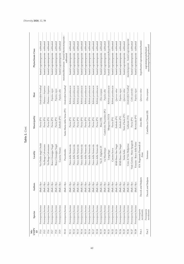
Diversity 2020, 12, 58
Ta
ble
2.
Con
t.
Mic
UN
IPV
IDS
pe
cie
sA
uth
ors
Lo
cali
tyM
un
icip
ali
tyH
ost
Ph
yto
clim
ate
Cla
ss
P.f.4
Pere
nnip
oria
frax
inea
(Bul
l.)R
yv.
Via
Uba
ldo
degl
iUba
ldi
Pavi
a(P
V)
Uni
dent
ified
broa
dlea
fhu
mid
supr
atem
pera
te-s
ubhu
mid
P.f.5
Pere
nnip
oria
frax
inea
(Bul
l.)R
yv.
Via
Borg
oC
alve
nzan
oPa
via
(PV
)Pl
atan
usx
hisp
anic
ahu
mid
supr
atem
pera
te-s
ubhu
mid
P.f.6
Pere
nnip
oria
frax
inea
(Bul
l.)R
yv.
Via
Borg
oC
alve
nzan
oPa
via
(PV
)Pl
atan
usx
hisp
anic
ahu
mid
supr
atem
pera
te-s
ubhu
mid
P.f.7
Pere
nnip
oria
frax
inea
(Bul
l.)R
yv.
Bosc
oG
iuse
ppe
Neg
riPa
via
(PV
)Po
pulu
sni
gra
hum
idsu
prat
empe
rate
-sub
hum
id
P.f.8
Pere
nnip
oria
frax
inea
(Bul
l.)R
yv.
Bosc
oG
iuse
ppe
Neg
riPa
via
(PV
)Po
pulu
sni
gra
hum
idsu
prat
empe
rate
-sub
hum
id
P.f.9
Pere
nnip
oria
frax
inea
(Bul
l.)R
yv.
Cas
cina
Vena
raZ
erbo
lò(P
V)
Popu
lus
alba
hum
idsu
prat
empe
rate
-sub
hum
id
P.f.1
0Pe
renn
ipor
iafr
axin
ea(B
ull.)
Ryv
.Pi
zzof
redd
oSa
nta
Mar
iade
llaVe
rsa
(PV
)U
nide
ntifi
edbr
oadl
eaf
mes
omed
iter
rane
an-h
umid
ther
mot
empe
rate
-su
bhum
id
P.f.1
1Pe
renn
ipor
iafr
axin
ea(B
ull.)
Ryv
.Pa
rco
della
Vern
avol
aPa
via
(PV
)R
obin
iaps
eudo
acac
iahu
mid
supr
atem
pera
te-s
ubhu
mid
P.f.1
2Pe
renn
ipor
iafr
axin
ea(B
ull.)
Ryv
.Pa
rco
della
Vern
avol
aPa
via
(PV
)R
obin
iaps
eudo
acac
iahu
mid
supr
atem
pera
te-s
ubhu
mid
P.f.1
3Pe
renn
ipor
iafr
axin
ea(B
ull.)
Ryv
.Pa
rco
della
Vern
avol
aPa
via
(PV
)R
obin
iaps
eudo
acac
iahu
mid
supr
atem
pera
te-s
ubhu
mid
P.f.1
4Pe
renn
ipor
iafr
axin
ea(B
ull.)
Ryv
.Pa
rco
della
Vern
avol
aPa
via
(PV
)R
obin
iaps
eudo
acac
iahu
mid
supr
atem
pera
te-s
ubhu
mid
P.f.1
5Pe
renn
ipor
iafr
axin
ea(B
ull.)
Ryv
.Pa
rco
della
Vern
avol
aPa
via
(PV
)R
obin
iaps
eudo
acac
iahu
mid
supr
atem
pera
te-s
ubhu
mid
P.f.1
6Pe
renn
ipor
iafr
axin
ea(B
ull.)
Ryv
.Pa
rco
della
Vern
avol
aPa
via
(PV
)R
obin
iaps
eudo
acac
iahu
mid
supr
atem
pera
te-s
ubhu
mid
P.f.1
7Pe
renn
ipor
iafr
axin
ea(B
ull.)
Ryv
.Pa
rco
della
Vern
avol
aPa
via
(PV
)R
obin
iaps
eudo
acac
iahu
mid
supr
atem
pera
te-s
ubhu
mid
P.f.1
8Pe
renn
ipor
iafr
axin
ea(B
ull.)
Ryv
.Pa
rco
della
Vern
avol
aPa
via
(PV
)R
obin
iaps
eudo
acac
iahu
mid
supr
atem
pera
te-s
ubhu
mid
P.f.1
9Pe
renn
ipor
iafr
axin
ea(B
ull.)
Ryv
.V
iaD
.Alig
hier
i25
Illa
si(V
R)
Ole
aeu
ropa
eahu
mid
supr
atem
pera
te-s
ubhu
mid
P.f.2
0Pe
renn
ipor
iafr
axin
ea(B
ull.)
Ryv
.vi
aM
asch
erpa
Cas
telv
etro
Piac
enti
no(P
C)
Popu
lus
alba
hum
idsu
prat
empe
rate
-sub
hum
id
P.f.2
1Pe
renn
ipor
iafr
axin
ea(B
ull.)
Ryv
.Lu
ngol
ago
Mer
gozz
o(V
CO
)R
obin
iaps
eudo
acac
iahu
mid
supr
atem
pera
te/h
yper
hum
id
P.f.2
2Pe
renn
ipor
iafr
axin
ea(B
ull.)
Ryv
.C
asci
naSc
ova
Pavi
a(P
V)
Rob
inia
pseu
doac
acia
hum
idsu
prat
empe
rate
-sub
hum
id
P.f.2
3Pe
renn
ipor
iafr
axin
ea(B
ull.)
Ryv
.R
NIS
Bosc
oSi
roN
egri
Zer
bolò
(PV
)Q
uerc
usro
bur
hum
idsu
prat
empe
rate
-sub
hum
id
P.f.2
4Pe
renn
ipor
iafr
axin
ea(B
ull.)
Ryv
.R
NIS
Bosc
oSi
roN
egri
Zer
bolò
(PV
)Po
pulu
sni
gra
hum
idsu
prat
empe
rate
-sub
hum
id
P.f.2
5Pe
renn
ipor
iafr
axin
ea(B
ull.)
Ryv
.Sa
nta
Sofia
Torr
ed’
Isol
a(P
V)
Que
rcus
robu
rhu
mid
supr
atem
pera
te-s
ubhu
mid
P.f.2
6Pe
renn
ipor
iafr
axin
ea(B
ull.)
Ryv
.C
ono
diVo
loM
alpe
nsa
Gal
lara
te(V
A)
Uni
dent
ified
broa
dlea
fm
esot
empe
rate
-hum
idsu
prat
empe
rate
P.f.2
7Pe
renn
ipor
iafr
axin
ea(B
ull.)
Ryv
.V
iale
Gor
izia
-Mur
aSp
agno
lePa
via
(PV
)C
eltis
aust
ralis
hum
idsu
prat
empe
rate
-sub
hum
id
P.f.2
8Pe
renn
ipor
iafr
axin
ea(B
ull.)
Ryv
.Fo
sson
e-B
osco
della
Fam
eR
oves
cala
(PV
)Po
pulu
sni
gra
hum
idsu
prat
empe
rate
-sub
hum
id
P.m
.1Pe
renn
ipor
iam
erid
iona
lisD
ecoc
kan
dSt
alpe
rsR
.N.R
egio
nale
Pira
mid
idi
Zon
eZ
one
(BS)
Que
rcus
robu
rhy
perh
umid
supr
atem
pera
te/h
umid
P.m
.2Pe
renn
ipor
iam
erid
iona
lisD
ecoc
kan
dSt
alpe
rsSo
rman
oC
aste
llina
inC
hian
ti(S
I)O
lea
euro
paea
supr
atem
pera
te/h
umid
mes
otem
pera
te/h
yper
hum
id
60

Diversity 2020, 12, 58
Ta
ble
2.
Con
t.
Mic
UN
IPV
IDS
pe
cie
sA
uth
ors
Lo
cali
tyM
un
icip
ali
tyH
ost
Ph
yto
clim
ate
Cla
ss
P.oc
h.1
Pere
nnip
oria
ochr
oleu
ca(B
erk.
)R
yvar
den
Belc
aro
Sien
a(S
I)Q
uerc
usile
xsu
bhum
idm
esot
empe
rate/h
umid
P.oc
h.2
Pere
nnip
oria
ochr
oleu
ca(B
erk.
)R
yvar
den
LeM
anie
Savo
na(S
V)
Que
rcus
ilex
mes
omed
iter
rane
an/d
ry-s
ubhu
mid
ther
mom
edit
erra
nean
Ph.c
.1Ph
ellin
usco
ntig
uus
(Per
s.)
Pat.
Rio
Bard
onez
zaZ
iano
Piac
enti
no(P
C)
Rob
inia
pseu
doac
acia
hum
idsu
prat
empe
rate
-sub
hum
id
Ph.to
r.1Ph
ellin
usto
rulo
sus
(Per
s.)
Bour
dota
ndG
alzi
nTi
cino
Torr
ed’
Isol
a(P
V)
Prun
usav
ium
hum
idsu
prat
empe
rate
-sub
hum
id
Pl.e
.1Pl
euro
tus
eryn
gii
(DC
.)Q
uél.
Aid
omag
gior
eA
idom
aggi
ore
(OR
)un
iden
tified
subh
umid
mes
omed
iter
rane
an
Pl.e
.2Pl
euro
tus
eryn
gii
(DC
.)Q
uél.
Spad
afor
aSp
adaf
ora
(ME)
unid
entifi
edth
erm
omed
iter
rane
an/s
ubhu
mid
mes
omed
iter
rane
an
Punc
.s.1
Punc
tula
ria
stri
goso
zona
ta(S
chw
ein.
)P.
H.B
.Tal
bot
Bosc
odi
Baul
i’Pa
lazz
olo
Acr
eide
(SR
)Q
uerc
ussp
.m
esom
edit
erra
nean/d
ryth
erm
omed
iter
rane
an-s
ubhu
mid
Sp.p
.1Sp
ongi
pelli
spa
chyo
don
(Per
s.)
Kot
l.an
dPo
uzar
Rio
Mar
sino
la-F
raci
onR
oves
cala
(PV
)Pr
unus
aviu
mhu
mid
supr
atem
pera
te-s
ubhu
mid
Sp.p
.2Sp
ongi
pelli
spa
chyo
don
(Per
s.)
Kot
l.an
dPo
uzar
Civ
ezza
Impe
ria
(IM
)Q
uerc
uspu
besc
ens
mes
omed
iter
rane
an/d
ryth
erm
omed
iter
rane
an-s
ubhu
mid
61

Diversity 2020, 12, 58
The species reported in Table 2 represent about one in five of the comprehensive collection of wooddecay species in MicUNIPV. The temperate region and Mediterranean region in Italy are reciprocallyintersected and several phytoclimates are represented based on both thermal–pluviometrical parametersand floristic–vegetational ones. According to our field observations, this has the consequence thatseveral species can be found in different phytoclimates and on different hosts, whereas a minor fractionis strictly related to one or few hosts.
Daedaleopsis confragosa and D. tricolor are easily distinguished by morphology; nevertheless,ITS sequences are important to discriminate the species strain. D. tricolor seems more common inCentral and Southern Europe; in Italy, it has been reported in seven out of 20 regions [45]. Our strain(MicUNIPV D.tric.1) comes from the lower Apennines in Pavia Province; other field observationssuggest that Prunus avium is the favourite host of D. tricolor in North and Central Italy. The strain hasnot been characterized yet, although pharmacological effects have been reported [46].
Daedalea quercina has been reported in 11/20 Italian regions [45]; as expected, all MicUNIPV strainswere isolated from Quercus spp. Nevertheless, strain MicUNIPV D.q.1 efficiently colonized poplarwood chips and confirmed that this species is a typical brown rot agent [47].
Despite being apparently cosmopolitan, Desarmillaria tabescens is strictly related to Quercus inwarm climates, where it behaves as a secondary pathogen [48]. D. tabescens has been reported in 16/20Italian Regions [45]. Accordingly, our strain was isolated from roots of Q. robur in RNIS Bosco SiroNegri (Pavia, Italy), which is a significant, unmanaged residue of typical forest of the western Po Plain.
Fomitopsis iberica is a rare species, reported in three Italian regions [45]. All the strains in MicUNIPVwere isolated close to Varese lakes, either on broadleaves or conifers.
Ganoderma is represented in MicUNIPV by seven species: G. adspersum, G. applanatum, G. carnosum,G. pfeifferi, G. lucidum, G. resinaceum and G. valesiacum. This genus has been intensely studied dueto its wide range of secondary metabolites, including several bioactive compounds [49]. Accordingto Ryvarden and Melo [6], Ganoderma is one of the most difficult genera to identify at species level.As reported in Table 2, we obtained strains of G. adspersum, confirming that is a southern speciesin Europe [50]. G. carnosum is usually located in the G. lucidum complex due to its morphologicalsimilarity, despite it showing clear differences in host relationship. Our strain was isolated from itstype-locality in Italy, i.e., a forest of Abies alba, that is likely to be its preferred host in South Europe [33].Molecular identification by ITS region met difficulties in discriminating G. carnosum from G. tsugaeand G. oregonensis; this topic presents questions about the real interspecific diversity within thisconifer-related species in Ganoderma. Strains in pure culture will thus help us to investigate boththe molecular and morphological nested diversity in this complex. An analogous problem concernsG. pfeifferi, as it partially shares its trophic niche with G. lucidum and G. resinaceum.
L. sulphureus is particularly related to Quercus according to our observations in North Italy,although Castanea sativa is also represented. Interestingly, C. sativa is also one of the favourite hosts ofPhellinus torulosus, according to field observations, despite strain MicUNIPV Ph.tor.1 being isolatedfrom P. avium.
Genus Perenniporia is represented in MicUNIPV by P. fraxinea, P. meridionalis and P. ochroleuca.According to our field observation, P. fraxinea is more common and widespread than expected,particularly in urban areas. We have focused our attention in indentifying strains, which, to date,number 27. Some of the isolated strains have been used for population studies and tests on heavymetal bioaccumulation [16,51].
Pleurotus eryngii is a typically Mediterranean species as well as its herbaceous hosts in Apiaceae.Consistently, the MicUNIPV Pl.e.1 and Pl.e.2 strains were isolated in properly Mediterranean areas(Sardinia and Sicily).
Punctularia strigosozonata is a rare, poorly studied species, typically related to the Mediterraneanarea; its resupinate morphology increases the difficulty in achieving pure isolates.
Spongipellis pachyodon has a mainly central–southern distribution in Europe; according to Onofriet al. [45], in Italy it is known in five out of 20 Regions, not including either Lombardy or Liguria.
62

Diversity 2020, 12, 58
This species is reported as uncommon but locally abundant; regarding this, our field observationssuggest that the population in the Pavia-Piacenza Apennines is particularly related to P. avium.
Further species listed in Table 2 are reported below in more detail owing to their taxonomiccontroversy or potential applications.
3.1. Cellulariella Warnieri (Durieu and Mont.) Zmitr. and V. Malysheva
3.1.1. Background
As detailed in Table 2, the basidiomata of some species were collected in the Mediterranean areaand others that are known to prefer warm environment, even if they were collected in continental ortemperate zones. An example is Cellulariella warnieri, a poorly investigated species related to warmclimates, according to Bernicchia and Gorjón [44]; despite not strictly being related to the Mediterraneanregion, Ryvarden and Melo [6] reported it as a southern and rare species.
The notable scarcity of data about this species has probably contributed to its uncertain systematicand taxonomic status. Currently it is reported as: Lenzites warnieri Durieu and Mont. by Mycobank [40],C. warnieri by Index Fungorum [52] and Trametes warnieri (Durieu and Mont.) Zmitr., Wasser andEzhov by Ryvarden and Melo [6]. The latter indication is suggested also by Justo and Hibbett [53]based on a five marker-based phylogenetic classification of Trametes. Significantly, only 108 records forthis species have been reported by the GBIF (Global Biodiversity Information Facility) [54] and only 10sequences are available in GenBank, almost half of them being critical as they are reported from SouthEast Asia [55]. Further analyses on a more representative number of strains are thus needed to clarifythe position of C. warnieri.
Strain MicUNIPV L.w.1 was tested for the evaluation of lignocellulolytic activity and resultedin a very low production of Mn peroxydase and lack of lignin peroxydase, whereas cellulase andhemicellulase had the highest presentation among the species under examination [47]. This was alsoconfirmed when testing the effect of its colonization on Medicago sativa for pre-treatment, as celluloseand hemicellulose were preferentially removed [56].
3.1.2. MicUNIPV WDF Strains Results
According to our field observations, localities are distributed as small local clusters which arevery scattered in turn. Thus, the strains MicUNIPV L.w.1, L.w.4 and L.w.5 were collected from Po plainareas (Pavia and RNIS Bosco Siro Negri), whereas strains the MicUNIPV L.w.2, L.w.3 and L.w.6 werecollected from hill area (Oltrepo Pavese).
The six strains of MicUNIPV were collected from Quercus, Ulmus, Populus and Robinia; interestingly,our field observations pointed out some preference for Ulmus, which was not previously reported asa host in Italy. As expected, all the strains were isolated from individuals behaving as saprotrophs.It should be noted that, since C. warnieri develops basidiomata in late autumn but it releases spores inspring, the basidioma itself remains vital even at low temperatures and under the snow.
3.2. Dichomitus squalens (P. Karst.) D.A. Reid
3.2.1. Background
Dichomitus squalens is a model species for studies about the selectivity of white rot and itsenzymatic basis [57–60]. Despite being reported all throughout the boreal emisphere, it appearsscattered and is commonly found in the northern parts of Europe, North America and Asia [61];the GBIF [54] places the wide majority of records in the Fennoscandian region. The host relationship isapparently controversial and surprising: Ryvarden and Melo [6] assumed Pinus as the only Europeanhost species, whereas Bernicchia and Gorjón [44] recorded Picea abies for the Italian sample andNiemelä [62] assigned most samples to Pinus and a smaller fraction to Picea abies in Białowieza Forest(Poland / Belarus). Nevertheless, it should be noted that American samples have been reported on six
63

Diversity 2020, 12, 58
different genera in Pinaceae. Furthermore, young basidiomata of D. squalens are easily misidentifieddue to the close morphological resemblance with Neoantrodia serialis (Fr.) Audet and related species.Consistently, a remarkable intraspecific variability in growth and enzyme profiles was revealed bytesting different monokarya strains [61]. This is also consistent with the numerous mating typesderiving from tetrapolarity [61].
3.2.2. MicUNIPV WDF Strains Results
Strains MicUNIPV D.sq.1 and MicUNIPV D.sq.2 were recovered from Pinus pinea and Cedrus sp.respectively near the Adriatic Sea and Varese Lake. The Italian strains have not yet been investigatedfor their enzymatic properties, so they may provide an additional tool to explore the diversity indegradation potential of this selective decayer.
3.3. Hericium Erinaceus (Bull.) Pers.
3.3.1. Background
Hericium erinaceus (Bull.) Pers. is one of the most famous cultivated medicinal species in theworld; a wide range of peculiar compounds, both related to primary (e.g., β-glucans) and secondarymetabolism (e.g., erinacines and hericenones) have been up to now characterized and screened forbioactivity [7,63–66]. GBIF [54] places the wide majority of H. erinaceus sites in Europe, North Americaand North Eastern Asia. According to the phylogenetic study by Cesaroni et al. [67], a subcladecontaining European and American ITS sequences is well distinguished from the Asian clade. Despiterelying on ITS region only, these data suggest the possibility to differentiate H. erinaceus strains alsoby the phylogeographic structure. Notwithstanding the scarcity of available data for Asian samples,H. erinaceus apparently has a quite broad trophic niche including several host species in Fagaceae,and Aceraceae to lesser extent, and particularly showing a preference for Quercus all throughout itsdistribution area [37]. Consistently, European samples have mainly been recovered from Quercusand Fagus, the former likely being the exclusive host in Italy and the only known host in NorthAfrica [34,68]. Strain MicUNIPV H.e.2 was analyzed for the production of erinacine A and hericenes(presumably A, B, C, D). Thus, a complete quali-quantitative comparison of these selected metaboliteswas provided throughout different growth stages but within the same strain, which is a powerful toolfor the standardization of bioactive products [69].
Strain MicUNIPV H.e.1 was selected to test the effect of oral supplementation on mice memory.The results indicate an improvement in recognition memory and induction of hyppocampal andcerebellar neurogenesis during aging. This strain has therefore contributed to pointing out whichareas are directly involved in the neuroactivity of H. erinaceus compounds, highlighting which type ofmemory is increased [70].
3.3.2. MicUNIPV WDF Strains Results
Accordingly, all four strains in the MicUNIPV collection were recovered in the municipalityof Siena from Q. ilex, that is, a featuring species in the flora of Mediterranean area often forminghomogeneous woodlands. It is noteworthy that the Mediterranean basin hosts a great variety ofQuercus species, whose phylogenetic and systematic relationships are still controversial, with particularconcern to the Q. ilex group [71–73].
3.4. Inocutis Tamaricis (Pat.) Fiasson and Niemelä
3.4.1. Background
The relationship of Inocutis tamaricis with Tamarix is apparently so strict to be regarded asa discriminant character in identification [6,33,44]. Although the genus Tamarix consists of 72 acceptedspecies in Europe, Asia and Africa [74], I. tamaricis is restricted to the Mediterranean basin and
64

Diversity 2020, 12, 58
Macaronesia. Here, it grows on different Tamarix species according to their availability but showsa preference for T. gallica [75,76]. Consistently, strains MicUNIPV I.t.1 and I.t.2 were both isolated fromT. gallica. As a whole, the intra-familiar phylogeny of Hymenochaetaceae is still to be clarified; multiplerevisions have tried to point out nested diversity within polyphyletic taxa, such as Inonotus [77,78].Thus, the genus Inocutis is nowadays accepted to be distinct from Inonotus itself, as formerly suggestedby Fiasson and Niemelä [79]. Interestingly, the type-species for Inocutis is I. rheades (Pers.) Fiassonand Niemelä, which is morphologically very similar to I. tamaricis and is mostly distinguished by hostand distribution [6]. Thus, I. tamaricis may be regarded as the Mediterranean counterpart of I. rheades.As a whole, only 12 sequences have been up to now deposited in GenBank [55] as belonging to thisspecies, some of which are lacking data to assess their effective reliability. Further sequences fromthe Mediterranean area, equipped with information about host and geographic origin, are needed tosupport studies about intrageneric diversity in Inocutis in the light of biogeographic patterns.
3.4.2. MicUNIPV WDF Strains Results
Strains MicUNIPV I.t.1 and I.t.2 were both isolated from T. gallica, forming in both cases ornamentalrows along the sea coast.
3.5. Fomitiporia Mediterranea M. Fisch.
3.5.1. Background
As mentioned for Inonotus, the genus Phellinus is increasingly revealing its hidden diversity;recognized as being polyphyletic, several species have been distributed into other genera, such asFomitiporia Murrill. Fomitiporia mediterranea is a peculiar example due to its morphology, being actuallyindistinguishable from P. punctatus. According to Fischer [80], these two species also show differencesin growth rate at selected temperatures and mating behaviour. The same study provides strongevidence for dichotomy in host selection by F. mediterranea depending on biogeography, i.e., this speciesgrows on several tree species in Italy [81,82], whereas north of the Alps it apparently grows on Vitisvinifera exclusively [83,84].
It should be considered that misidentification with P. punctatus has probably led to theunderestimation of F. mediterranea in the Mediterranean area [85]. Analogously, Polemis et al. [86]suggested that the relationship with P. pseudopunctatus A. David, Dequatre and Fiasson shouldbe reconsidered as well, enclosing the latter in F. mediterranea clade. It may be observed that theP. pseudopunctatus is apparently more related to the South Mediterranean region and climates [87,88].Further analyses on strains from different geographic origins and hosts are thus needed to clarifyboth phylogenetic relationships and biogeographic patterns. As a whole, this species complex ischaracterized by intense necrotrophic white rot; F. mediterranea in particular is regarded as one of themain agents responsible for wood rot in V. vinifera, Corylus avellana and Olea europaea [82,83,85].
3.5.2. MicUNIPV WDF Strains Results
All of the 17 Italian strains up to now attained by the Laboratory of Mycology DSTA–Universityof Pavia were assigned to F. mediterranea instead of P. punctatus and recovered from different substratesin North Italy. Even within one province (Pavia), F. mediterranea was located on five hosts, namelyQ. robur, Hedera helix (State Natural Strict Reserve Bosco Siro Negri), Salix alba (University of Paviacourtyard), R. pseudocacacia and V. vinifera (Oltrepo Pavese hills).
The identified strains thus provide a tool to deepen pathology dynamics and different susceptibilitydepending on host species and cultivar.
65

Diversity 2020, 12, 58
3.6. Perenniporia meridionalis Decock and Stalpers
3.6.1. Background
Genus Perenniporia Murrill sensu lato is large, cosmopolitan and supposed to be polyphyletic,and thus is in need of further phylogenetic analysis based on sequences from different species [6].In turn, an example of intrageneric complexity is provided by P. meridionalis, within P. medulla-panis(Jacq.) Donk group. Actually, the complete revision by Decock and Stalpers [89] arose the doubtthat several records, as well as specimens in herbaria, are to be referred to P. meridionalis instead ofP. medulla-panis (or closely related species), particularly when coming from the Mediterranean area.Up to now, only a partial and fragmentary investigation into the intra-generic diversity in Perenniporiahas been carried out from a molecular and phylogenetic point of view [90]. It is noteworthy that nosequences at all are available in GenBank by the name of P. meridionalis, whereas 40 sequences arereferred to P. medulla-panis [55]. Strains from culture collections are thus needed as basic material forthis purpose. According to the indications by both Bernicchia and Gorjón [44], as well as Ryvardenand Melo [6], P. meridionalis is particularly related to Quercus (more than P. medulla-panis), the holotypehaving been isolated from Q. ilex in Sardinia [62,89].
3.6.2. MicUNIPV WDF Strains Results
Strains MicUNIPV P.m.1 and P.m.2 strains were respectively isolated from Q. robur (North Italy,near a lake) and Q. ilex (Central Italy). Besides considerations on biodiversity, P. meridionalis hasa great applicative potential. MicUNIPV P.m.1 showed remarkable selectivity as a white rot agentand versatility when inoculated onto unusual substrates such as grass. The selective removal oflignin by this species contemporarily relies on high activity for Mn peroxidases and very low for onecellulase; the final delignification in the substrate is clear both in thermogravimetric analysis and FTIRspectroscopy [12,47].
3.7. Perenniporia ochroleuca (Berk.) Ryvarden
3.7.1. Background
Perenniporia ochroleuca is another example of the unsolved intra-generic diversity withinPerenniporia. This species is suspected to hide a complex, and transfer to Truncospora Pilát ex Piláthas thus been suggested [91]. According to the same authors, the Iberian/Macaronesian clade givesT. atlantica Spirin and Vlasák, whereas the status of Australian samples is more uncertain, which wouldmainly belong to T. ochroleuca. Nevertheless, the new taxonomy has not yet been fully accepted, neitherby Mycobank [40] nor by Index Fungorum [52]. These hypotheses therefore need to be supportedby entering further sequences into the phylogenetic analyses from an exhaustive geographic range.P. ochroleuca was reported by Bernicchia and Gorjón [44] and Ryvarden and Melo [6] as tropical andgrowing on several hosts, whereas in Europe it is particularly related to the Mediterranean area.Nevertheless, Bernicchia and Gorjón [44] report a range of typically Mediterranean hosts, whereasRyvarden and Melo [6] also include host plants whose distribution exceeds the Mediterranean area toinclude samples from the coasts of South England and Wales. Further phylogenetic analyses focusedon the Mediterranean region versus the adjacent Atlantic ones are needed to test the monophyly of theproposed T. atlantica.
3.7.2. MicUNIPV WDF Strains Results
Both strains MicUNIPV P.och.1 and P.och.2 were isolated from Q. ilex in Central Italy and theLigurian west coast, respectively.
66

Diversity 2020, 12, 58
4. Conclusions
At the moment, MicUNIPV, the fungal research culture collection of University of Pavia (Italy),maintains 500 strains from wood decay species. Examples particularly correlated to the Mediterraneanarea were discussed and their roles in accomplished research were mentioned in this study.
Culture collections of wood decay fungi are an important tool both for systematic and appliedstudies. Strains in pure culture are more easily and reliably identified and analyzed for metabolicactivities and competitivity. The environmental features of the strain origin place have often beenunderestimated; nevertheless, the diversity of wood decay fungi strongly depends on biogeographyand is related to host distribution. This also highlights the need for an investigation including a widerconcept of the Mediterranean region than one strictly limited by climate or phytoclimate classification,i.e., even continental regions surrounding the Mediterranean area contribute to the explanation ofMediterranean diversity.
The Laboratory of Mycology DSTA–University of Pavia (Italy) has up to now successfullycollaborated with both researchers from other universities and amateurs in order to increase thediversity richness and geographic origin range of strains, as well as to enter these strains in originalpure and applied research such as MATER and CE4WE (grants from Cariplo Foundation and RegioneLombardia).
Author Contributions: Conceptualization, E.S. and C.E.G.; Methodology, E.S., A.B., A.M.P., C.E.G., R.M.B. andM.C.; Investigation, E.S., A.B., C.E.G., R.M.B., S.B. and M.C. Resources, E.S., A.M.P. and F.B.; Data Curation, E.S.,A.B. and C.E.G.; Writing – Original Draft Preparation, C.E.G., S.B.; Writing – Review and Editing, C.E.G., E.S.,A.B., A.M.P.; Supervision, E.S., A.M.P.; Project Administration, E.S.; Funding Acquisition, E.S., F.B. All authorshave read and agreed to the published version of the manuscript.
Funding: This project has been funded by: Fondo Ricerca e Giovani dell’Università degli Studi di Pavia (Savino2019); Fondazione Cariplo, grant n◦ 2018-1765 entitled “Myco-advanced leather materials (MATER)”.
Acknowledgments: The State Natural Strict Reserve (RNIS) Bosco Siro Negri – Zerbolò for support andauthorization for sampling; the Botanical Garden of Pavia (Orto Botanico di Pavia) for authorization for sampling.The authors are also grateful to C. Perini, University of Siena (Italy), Valentina Cesaroni and Fabio Savino forproviding some original specimens.
Conflicts of Interest: The authors declare no conflict of interest.
References
1. Liers, C.; Arnstadt, T.; Ullrich, R.; Hofrichter, M. Patterns of lignin degradation and oxidative enzymesecretion by different wood-and litter-colonizing basidiomycetes and ascomycetes grown on beech-wood.FEMS Microbiol. Ecol. 2011, 78, 91–102. [CrossRef] [PubMed]
2. Van den Brink, J.; de Vries, R.P. Fungal enzyme sets for plant polysaccharide degradation. Appl. Microbiol.Biotechnol. 2011, 91, 1477. [CrossRef] [PubMed]
3. Riley, R.; Salamov A., A.; Brown, D.W.; Nagy, L.G.; Floudas, D.; Held, B.W.; Levasseur, A.; Lombard, V.;Morin, E.; Otillar, R.; et al. Extensive sampling of basidiomycete genomes demonstrates inadequacy ofthe white-rot/brown-rot paradigm for wood decay fungi. Proc. Natl. Acad. Sci. USA 2014, 111, 9923–9928.[CrossRef]
4. Hibbett, D.S.; Bauer, R.; Binder, M.; Giachini, A.J.; Hosaka, K.; Justo, A.; Larsson, E.; Larsson, K.H.;Lawrey, J.D.; Miettinen, O.; et al. Agaricomycetes. In the Mycota, 2nd ed.; McLaughlin, D.J., Spatafora, J.W.,Eds.; Springer: Berlin, Germany, 2014; Volume 7, pp. 373–429. [CrossRef]
5. Nagy, L.G.; Riley, R.; Tritt, A.; Adam, C.; Daum, C.; Floudas, D.; Sun, H.; Yadav, J.S.; Pangilinan, J.;Larsson, K.H.; et al. Comparative genomics of early-diverging mushroom-forming fungi provides insightsinto the origins of lignocellulose decay capabilities. Mol. Biol. Evol. 2015, 33, 959–970. [CrossRef] [PubMed]
6. Ryvarden, L.; Melo, I. Poroid Fungi of Europe, 2nd ed.; Fungiflora: Oslo, Norway, 2017.7. Ma, B.J.; Shen, J.W.; Yu, H.Y.; Ruan, Y.; Wu, T.T.; Zhao, X. Hericenones and erinacines: Stimulators of nerve
growth factor (NGF) biosynthesis in Hericium erinaceus. Mycology 2010, 1, 92–98. [CrossRef]
67

Diversity 2020, 12, 58
8. Dresch, P.; Rosam, K.; Grienke, U.; Rollinger, J.M.; Peintner, U. Fungal strain matters: Colony growth andbioactivity of the European medicinal polypores Fomes fomentarius, Fomitopsis pinicola and Piptoporusbetulinus. AMB Express 2015, 5, 4. [CrossRef]
9. Angelini, P.; Girometta, C.; Tirillini, B.; Moretti, S.; Covino, S.; Cipriani, M.; D’Ellena, E.; Angeles, G.;Federici, E.; Savino, E.; et al. A comparative study of the antimicrobial and antioxidant activities of Inonotushispidus fruit and their mycelia extracts. Int. J. Food Prop. 2019, 22, 768–783. [CrossRef]
10. Pozdnyakova, N.N. Involvement of the ligninolytic system of white-rot and litter-decomposing fungi in thedegradation of polycyclic aromatic hydrocarbons. Biotechnol. Res. Int. 2012, 2012. [CrossRef]
11. Giles, R.L.; Galloway, E.R.; Zackeru, J.C.; Naithani, V.; Parrow, M.W. Two stage fungal biopulping solubilizeslignocellulosic carbohydrates without supplemental enzymatic hydrolysis. Int. Biodeter. Biodegradation 2014,86, 265–271. [CrossRef]
12. Girometta, C.; Zeffiro, A.; Malagodi, M.; Savino, E.; Doria, E.; Nielsen, E.; Buttafava, A.; Dondi, D. Pretreatmentof alfalfa stems by wood decay fungus Perenniporia meridionalis improves cellulose degradation andminimizes the use of chemicals. Cellulose 2017, 24, 3803–3813. [CrossRef]
13. Mendonça Maciel, M.J.; Ribeiro, H.C.T. Industrial and biotechnological applications of ligninolytic enzymesof the basidiomycota: A review. Electron. J. Biotechnol. 2010, 13, 14–15. [CrossRef]
14. Gadd, G.M. Biosorption: Critical review of scientific rationale, environmental importance and significancefor pollution treatment. J. Chem. Technol. Biot. 2009, 84, 13–28. [CrossRef]
15. Javaid, A.M.N.A.; Bajwa, R.; Manzoor, T. Biosorption of heavy metals by pretreated biomass of Aspergillusniger. Pak. J. Bot. 2011, 43, 419–425.
16. Sturini, M.; Girometta, C.; Maraschi, F.; Savino, E.; Profumo, A. A Preliminary Investigation on MetalBioaccumulation by Perenniporia fraxinea. B Environ. Contam. Tox 2017, 98, 508–512. [CrossRef]
17. Arciniegas, A.; Prieto, F.; Brancheriau, L.; Lasaygues, P. Literature review of acoustic and ultrasonictomography in standing trees. Trees 2014, 28, 1559–1567. [CrossRef]
18. CBS-KNAW Collections. Available online: www.cbs.knaw.nl (accessed on 29 December 2019).19. All Russian Collection of Microorganisms—VKM. Available online: www.vkm.ru (accessed on 29 December
2019).20. Baldrian, P.; Gabriel, J. Intraspecific variability in growth response to cadmium of the wood-rotting fungus
Piptoporus betulinus. Mycologia 2002, 94, 428–436. [CrossRef]21. Pawlik, A.; Janusz, G.; Debska, I.; Siwulski, M.; Frac, M.; Rogalski, J. Genetic and metabolic intraspecific
biodiversity of Ganoderma lucidum. BioMed Res. Int. 2015, 2015. [CrossRef]22. Zeng, Z.; Sun, H.; Vainio, E.J.; Raffaello, T.; Kovalchuk, A.; Morin, E.; Duplessis, S.; Asiegbu, F.O. Intraspecific
comparative genomics of isolates of the Norway spruce pathogen (Heterobasidion parviporum) andidentification of its potential virulence factors. BMC Genom. 2018, 19, 220. [CrossRef]
23. WFCC—World Federation for Culture Collections. Available online: www.wfcc.info (accessed on 29December 2019).
24. MIRRI—Microbial Resorurce Research Infrastructure. Available online: www.mirri.org (accessed on 29December 2019).
25. Gargano, M.L. Mycotheca of edible and medicinal mushrooms at herbarium SAF as a potential source ofnutraceuticals and cultivated mushrooms. Int. J. Med. Mushrooms 2018, 20, 405–409. [CrossRef]
26. Caretta, G. Micologo a Pavia. In Raffaele Ciferri Scienziato Versatile e Critico, Cisalpino; Istituto EditorialeUniversitario: Milano, Italy, 2000; pp. 51–54.
27. Lowry, D.B. Ecotypes and the controversy over stages in the formation of new species. Biol. J. Linn. Soc.2012, 106, 241–257. [CrossRef]
28. Angelini, P.; Compagno, R.; Arcangeli, A.; Bistocchi, G.; Gargano, M.L.; Venanzoni, R.; Venturella, G.Macrofungal diversity and ecology in two Mediterranean forest ecosystems. Plant Biosyst. 2016, 150, 540–549.[CrossRef]
29. Ministero Dell’ambiente e Della Tutela del Territorio e del Mare. Geoportale Nazionale—NuovoVisualizzatore. Available online: http://www.pcn.minambiente.it/viewer/ (accessed on 28 December 2019).
30. Rivas-Martínez, S.; Penas, Á.; Díaz, T.E. Bioclimatic and Biogeographic Maps of Europe. 1:16.000.000University of León, E-24071, Spain. 2004. Available online: http://www.globalbioclimatics.org/form/bg_mEd.htm (accessed on 28 December 2019).
68

Diversity 2020, 12, 58
31. Blasi, C.; Capotorti, G.; Copiz, R.; Guida, D.; Mollo, B.; Smiraglia, D.; Zavattero, L. Classification and mappingof the ecoregions of Italy. Plant Biosyst. 2014, 148, 1255–1345. [CrossRef]
32. Sutherland, W.J. Ecological Census Techniques: A Handbook, 2nd ed.; Cambridge University Press: Cambridge,UK, 2006.
33. Bernicchia, A. Polyporaceae sl.; Candusso: Alassio (SV), Italy, 2005.34. Bernicchia, A.; Gorjón, S.P. Corticiaceae sl.; Candusso: Alassio (SV), Italy, 2010.35. Stalpers, J.A. Identification of wood-inhabiting fungi in pure culture. Stud. Mycol. 1978, 16, 1–248.36. Gams, W.; Hoekstra, E.S.; Aptroot, A. CBS Course of Mycology; Centraalbureau voor Schimmelcultures Baarn:
Delft, NL, USA, 1998.37. Tamets, P. Growing Gourmet and Medicinal Mushrooms; Ten Speed Press: Berkeley, CA, USA, 2011.38. Toju, H.; Tanabe, A.S.; Yamamoto, S.; Sato, H. High-coverage ITS primers for the DNA-based identification
of ascomycetes and basidiomycetes in environmental samples. PLoS ONE 2012, 7, e40863. [CrossRef]39. Nilsson, R.H.; Hyde, K.D.; Pawlowska, J.; Ryberg, M.; Tedersoo, L.; Aas, A.B.; Alias, S.A.; Alves, A.;
Anderson, C.L.; Antonelli, A.; et al. Improving ITS sequence data for identification of plant pathogenic fungi.Fungal Divers 2014, 67, 11–19. [CrossRef]
40. Mycobank. Available online: www.mycobank.org (accessed on 19 December 2019).41. Altobelli, E.; Bernicchia, A.; Pecoraro, L.; Savino, E. Raccolta, isolamento e coltivazione di funghi poliporoidi
con proprietà medicinali. Micol. Ital. 2012, 41, 3–10.42. Savino, E.; Girometta, C.; Chinaglia, S.; Guglielminetti, M.; Rodolfi, M.; Bernicchia, A.; Perini, C.; Salerni, E.;
Picco, A.M. Medicinal mushrooms in Italy and their ex situ conservation through culture collection.In Proceedings of the 8th International Conference on Mushroom Biology and Mushroom Products,New Delhi, India, 19–22 Novemver 2014; pp. 50–54.
43. Savino, E.; Girometta, C.; Miteva-Staleva, J.; Kostadinova, A.; Krumova, E. Wood decay macrofungi: Straincollection and studies about antioxidant properties. Comptes Rendus l’Académie Bulg. Sci. 2016, 69, 747–755.
44. Bernicchia, A.; Gorjón, S.P. Polypores of Mediterranean Region; S.P. Publishers: Bologna, Italy, 2019; in press.45. Onofri, S.; Bernicchia, A.; Filipello Marchisio, V.; Padovan, F.; Perini, C.; Ripa, C.; Salerni, E.; Savino, E.;
Venturella, G.; Vizzini, A.; et al. Checklist dei Funghi Italiani Basidiomycetes, Basidiomycota, 1st ed.; Carlo DelfinoEditore: Sassari, Italy, 2005; pp. 1–380.
46. Bernicchia, A.; Fugazzola, M.A.; Gemelli, V.; Mantovani, B.; Lucchetti, A.; Cesari, M.; Speroni, E.DNA recovered and sequenced from an almost 7000 y-old Neolithic polypore, Daedaleopsis tricolor.Mycol. Res. 2006, 110, 14–17. [CrossRef]
47. Doria, E.; Altobelli, E.; Girometta, C.; Nielsen, E.; Zhang, T.; Savino, E. Evaluation of lignocellulolytic activitiesof ten fungal species able to degrade poplar wood. Int. Biodeter Biodegrad. 2014, 94, 160–166. [CrossRef]
48. Koch, R.A.; Wilson, A.W.; Séné, O.; Henkel, T.W.; Aime, M.C. Resolved phylogeny and biogeography of theroot pathogen Armillaria and its gasteroid relative, Guyanagaster. BMC Evol. Biol. 2017, 17, 33. [CrossRef]
49. Lindequist, U.; Jülich, W.D.; Witt, S. Ganoderma pfeifferi—A European relative of Ganoderma lucidum.Phytochemistry 2015, 114, 102–108. [CrossRef] [PubMed]
50. Karadelev, M.; Rusevska, K.; Kajevska, I. Distribution and ecology of Genus Ganoderma (Ganodermataceae) inthe Republic of Macedonia. In Proceedings of the International Conference on Biological and EnveronmentalSciences, Tirana, Albania, 26–28 September 2008; pp. 320–326.
51. Sillo, F.; Savino, E.; Giordano, L.; Girometta, C.; Astegiano, D.; Picco, A.M.; Gonthier, P. Analysis of genotypicdiversity provides a first glimpse on the patterns of spread of the wood decay fungus Perenniporla fraxineain an urban park in northern Italy. J. Plant Pathol. 2016, 98, 617–624.
52. Index Fungorum. Available online: www.indexfungorum (accessed on 19 December 2019).53. Justo, A.; Hibbett, D.S. Phylogenetic classification of Trametes (Basidiomycota, Polyporales) based on
a five-marker dataset. Taxon 2011, 60, 1567–1583. [CrossRef]54. GBIF—Global Biodiversity Information Facility. Available online: www.gbif.org (accessed on 28 December
2019).55. GenBank- NCBI (National Center for Biotechnology Information). Available online: https://www.ncbi.nlm.
nih.gov (accessed on 19 December 2019).
69

Diversity 2020, 12, 58
56. Zeffiro, A.; Dondi, D.; Marconi, R.P.; Malagodi, M.; Girometta, C.; Bentivoglio, A.; Lazzaroni, S.; Savino, E.;Nielsen, E.; Buttafava, A. Sugar Production for bioethanol from alfalfa stems. Results, and comparative studywith application of lignocellulolytic activities of novel fungal species. In Atti del Convegno, Proceedingsof the VI Workshop nazionale AICIng “Molecules and materials: Chemistry for engineering”, Roma, Italy,22–23 June 2015.
57. Floudas, D.; Binder, M.; Riley, R.; Barry, K.; Blanchette, R.A.; Henrissat, B.; Martínez, A.T.; Otillar, R.;Spatafora, J.W. The Paleozoic origin of enzymatic lignin decomposition reconstructed from 31 fungalgenomes. Science 2012, 336, 1715–1719. [CrossRef] [PubMed]
58. Rytioja, J.; Hildén, K.; Di Falco, M.; Zhou, M.; Aguilar-Pontes, M.V.; Sietiö, O.M.; Tsang, A.; de Vries, R.P.;Mäkelä, M.R. The molecular response of the white-rot fungus Dichomitus squalens to wood and non-woodybiomass as examined by transcriptome and exoproteome analyses. Environ. Microbiol. 2017, 19, 1237–1250.[CrossRef] [PubMed]
59. Daly, P.; López, S.C.; Peng, M.; Lancefield, C.S.; Purvine, S.O.; Kim, Y.M.; Zink, E.M.; Dohnalkova, A.;Singan, V.R.; Lipzen, A.; et al. Dichomitus squalens partially tailors its molecular responses to the compositionof solid wood. Environ. Microbiol. 2018, 20, 4141–4156. [CrossRef]
60. López, S.C.; Peng, M.; Issak, T.Y.; Daly, P.; de Vries, R.P.; Mäkelä, M.R. Induction of genes encoding plant cellwall-degrading carbohydrate-active enzymes by lignocellulose-derived monosaccharides and cellobiose inthe white-rot fungus Dichomitus squalens. Appl. Environ. Microbiol. 2018, 84, e00403-18. [CrossRef]
61. L’opez, S.C.; Theelen, B.; Manserra, S.; Issak, T.Y.; Rytioja, J.; Mäkelä, M.R.; de Vries, R.P. Functional diversityin Dichomitus squalens monokaryons. IMA Fungus 2017, 8, 17. [CrossRef]
62. Niemelä, T. Polypores of the Białowieza Forest; Białowieski Park Narodowy: Białowieza, Poland, 2013.63. Kawagishi, H.; Shimada, A.; Shirai, R.; Okamoto, K.; Ojima, F.; Sakamoto, H.; Ishiguro, Y.; Furukawa, S.
Erinacines A, B and C, strong stimulators of nerve growth factor (NGF)-synthesis, from the mycelia ofHericium erinaceum. Tetrahedron Lett. 1994, 35, 1569–1572. [CrossRef]
64. Friedman, M. Chemistry, nutrition, and health-promoting properties of Hericium erinaceus (lion’s mane)mushroom fruiting bodies and mycelia and their bioactive compounds. J. Agric. Food Chem 2015, 63,7108–7123. [CrossRef]
65. Brandalise, F.; Cesaroni, V.; Gregori, A.; Repetti, M.; Romano, C.; Orrù, G.; Botta, L.; Girometta, C.;Guglielminetti, M.L.; Savino, E.; et al. Dietary supplementation of Hericium erinaceus increases mossyfiber-CA3 hippocampal neurotransmission and recognition memory in wild-type mice. Evid-Based Compl.Alt. 2017. [CrossRef] [PubMed]
66. Rossi, P.; Cesaroni, V.; Brandalise, F.; Occhinegro, A.; Ratto, D.; Perrucci, F.; Lanaia, V.; Girometta, C.;Orrù, G.; Savino, E. Dietary supplementation of lion’s mane medicinal mushroom, Hericium erinaceus(Agaricomycetes), and spatial memory in wild-type mice. Int. J. Med. Mushrooms 2018, 20, 485–494.[CrossRef] [PubMed]
67. Cesaroni, V.; Brusoni, M.; Cusaro, C.M.; Girometta, C.; Perini, C.; Picco, A.M.; Rossi, P.; Salerni, E.; Savino, E.Phylogenetic Comparison between Italian and Worldwide Hericium Species (Agaricomycetes). Int. J. Med.Mushrooms 2019, 21, 943–954. [CrossRef]
68. Ouali, Z.; Sbissi, I.; Boudagga, S.; Rhaiem, A.; Hamdi, C.; Venturella, G.; Saporita, P.; Jaouani, A.; Gargano, M.L.First report of the rare tooth fungus Hericium erinaceus in North African temperate forests. Plant Biosyst.2018, 154, 24–28. [CrossRef]
69. Corana, F.; Cesaroni, V.; Mannucci, B.; Baiguera, R.M.; Picco, A.M.; Savino, E.; Ratto, D.; Perini, C.;Kawagishi, H.; Girometta, C.E.; et al. Array of Metabolites in Italian Hericium erinaceus Mycelium,Primordium, and Sporophore. Molecules 2019, 24, 3511. [CrossRef] [PubMed]
70. Ratto, D.; Corana, F.; Mannucci, B.; Priori, E.C.; Cobelli, F.; Roda, E.; Ferrari, B.; Occhinegro, A.; Di Iorio, C.;De Luca, F.; et al. Hericium erinaceus Improves recognition memory and induces hippocampal and cerebellarneurogenesis in frail mice during aging. Nutrients 2019, 11, 715. [CrossRef]
71. Manos, P.S.; Doyle, J.J.; Nixon, K.C. Phylogeny, biogeography, and processes of molecular differentiation inQuercus subgenus Quercus (Fagaceae). Mol. Phylogenet. Evol. 1999, 12, 333–349. [CrossRef]
72. Simeone, M.C.; Piredda, R.; Papini, A.; Vessella, F.; Schirone, B. Application of plastid and nuclear markers toDNA barcoding of Euro-Mediterranean oaks (Quercus, Fagaceae): Problems, prospects and phylogeneticimplications. Bot. J. Linn. Soc. 2013, 172, 478–499. [CrossRef]
70

Diversity 2020, 12, 58
73. Hubert, F.; Grimm, G.W.; Jousselin, E.; Berry, V.; Franc, A.; Kremer, A. Multiple nuclear genes stabilize thephylogenetic backbone of the genus Quercus. Syst. Biodivers. 2014, 12, 405–423. [CrossRef]
74. Kew Science—Plants of the World Online. Available online: http://www.plantsoftheworldonline.org/(accessed on 19 December 2019).
75. Loizides, M. Diversity of wood-inhabiting aphyllophoraceous basidiomycetes on the island of Cyprus.Mycotaxon 2017, 132, 985–986.
76. Gargano, M.L. Nuovi ospiti di Inonotus tamaricis (Hymenochaetaceae) in Sicilia. Inf. Bot. Ital. 2010, 42,319–321.
77. Wagner, T.; Fischer, M. Natural groups and a revised system for the European poroid Hymenochaetales(Basidiomycota) supported by nLSU rDNA sequence data. Mycol. Res. 2001, 105, 773–782. [CrossRef]
78. Wagner, T.; Fischer, M. Proceedings towards a natural classification of the worldwide taxa Phellinus s.l.and Inonotus s.l., and phylogenetic relationships of allied genera. Mycologia 2002, 94, 998–1016. [CrossRef][PubMed]
79. Fiasson, J.L.; Niemelä, T. The Hymenochaetales: A revision of the European poroid taxa. Karstenia 1984, 24,14–28. [CrossRef]
80. Fischer, M. A new wood-decaying basidiomycete species associated with esca of grapevine: Fomitiporiamediterranea (Hymenochaetales). Mycol. Prog. 2002, 1, 315–324. [CrossRef]
81. Ciccarone, C.; Graniti, A.; Schiaffino, A.; Marras, F. Molecular analysis of Fomitiporia mediterranea isolatesfrom esca-affected grapevines in southern Italy. Phytopathol. Mediterr. 2004, 43, 268–272.
82. Pilotti, M.; Tizzani, L.; Brunetti, A.; Gervasi, F.; Di Lernia, G.; Lumia, V. Molecular identification of Fomitiporiamediterranea on declining and decayed hazelnut. JPP 2010, 92, 115–129.
83. Fischer, M. Biodiversity and geographic distribution of basidiomycetes causing esca-associated white rot ingrapevine: A worldwide perspective. Phytopathol. Mediterr. 2006, 45, 30–42.
84. Kovács, C.; Sándor, E. The increasing importance of grapevine trunk diseases. Int. J. Hortic. Sci. 2016, 22,21–30. [CrossRef]
85. Markakis, E.A.; Ligoxigakis, E.K.; Roussos, P.A.; Sergentani, C.K.; Kavroulakis, N.; Roditakis, E.N.;Koubouris, G.C. Differential susceptibility responses of Greek olive cultivars to Fomitiporia mediterranea.Eur. J. Plant Pathol. 2019, 153, 1055–1066. [CrossRef]
86. Polemis, E.; Dimou, D.M.; Fryssouli, V.; Zervakis, G.I. Diversity of saproxylic basidiomycetes in Quercus ilexwoodlands of central and insular Greece. Plant Biosyst. 2019, 153, 385–397. [CrossRef]
87. Saitta, A.; Venturella, G. On the presence of Diplomitoporus lindbladii and Phellinus pseudopunctatus inSicily (Southern Italy). Bocconea 2009, 23, 273–276.
88. Karadelev, M.; Rusevska, K.; Kost, G.; Kopanja, D.M. Checklist of macrofungal species from the phylumBasidiomycota of the Republic of Macedonia. Acta Musei Maced. Sci. Nat. 2018, 21, 23–112.
89. Decock, C.; Stalpers, J.A. Studies in Perenniporia: Polyporus unitus, Boletus medulla-panis, the nomenclatureof Perenniporia, Poria and Physisporus, and a note on European Perenniporia with a resupinate basidiome.Taxon 2006, 55, 759–778. [CrossRef]
90. Zhao, C.L.; Cui, B.K.; Dai, Y.C. New species and phylogeny of Perenniporia based on morphological andmolecular characters. Fungal Divers. 2013, 58, 47–60. [CrossRef]
91. Spirin, V.; Kout, J.; Vlasák, J. Studies in the Truncospora ohiensis–T. ochroleuca group (Polyporales,Basidiomycota). Nova Hedwig. 2015, 100, 159–175. [CrossRef]
© 2020 by the authors. Licensee MDPI, Basel, Switzerland. This article is an open accessarticle distributed under the terms and conditions of the Creative Commons Attribution(CC BY) license (http://creativecommons.org/licenses/by/4.0/).
71


diversity
Article
Does Arbuscular Mycorrhiza Determine SoilMicrobial Functionality in Nutrient-LimitedMediterranean Arid Ecosystems?
Neji Mahmoudi 1, Teresa Dias 2 , Mosbah Mahdhi 3 , Cristina Cruz 2, Mohamed Mars 1
and Maria F. Caeiro 4,*
1 Unité de Recherche Biodiversité et Valorisation des Bio-ressources en Zones Arides (BVBZA),Faculté des Sciences de Gabès, Cité Erriadh Zrig 6072, Tunisie; [email protected] (N.M.);[email protected] (M.M.)
2 Centre for Ecology, Evolution and Environmental Changes (cE3c), Faculdade de Ciências da Universidadede Lisboa, Edifício C2, Piso 5, Sala 2.5.03 Campo Grande, 1749-016 Lisboa, Portugal; [email protected] (T.D.);[email protected] (C.C.)
3 Center for Environmental Research and Studies, Jazan University, Jazan-P.O. Box 114, Jazan 82817,Saudi Arabia; [email protected]
4 Centro de Estudos do Ambiente e do Mar (CESAM), Faculdade de Ciências da Universidade de Lisboa,Edifício C2, Piso 2, Campo Grande, 1749-016 Lisboa, Portugal
* Correspondence: [email protected]
Received: 5 May 2020; Accepted: 4 June 2020; Published: 10 June 2020
Abstract: Arbuscular mycorrhizal fungi (AMF) are determinant for the performance of plantcommunities and for the functionality of terrestrial ecosystems. In natural ecosystems, grazing canhave a major impact on mycorrhizal fungi and consequently on plant growth. The objective of thisstudy was to evaluate the statements referred above in Mediterranean arid areas in Tunisia. Rootsamples and rhizosphere soils of five dominant herbaceous plants were studied at six distinct aridsites differing on soil proprieties and grazing intensity. At each site, chemical and dynamic propertiesof the soil were characterized as well as the AMF colonization intensity and the soil functionality.Results showed that the mycorrhizal frequency and intensity and spore density, varied between plantsin the same site and, for each plant, between sites and evidenced a positive effect of mycorrhizedplants on soil microbial activity. Grazing and soil properties strongly affected AMF compositionand the soil microbial and biochemical dynamics, which presented the lowest values at the siteswith the highest grazing intensities. In conclusion, these results demonstrate that AMF improvesoil biological properties, supporting the hypothesis that mycorrhiza and grazing compete for plantphotosynthates, and highlight the importance of mycorrhizal symbiosis towards soil functionalityunder arid conditions.
Keywords: arbuscular mycorrhizal fungi; arid areas; biological properties; conserved areas;grazing; mycorrhiza
1. Introduction
Arid and semi-arid regions of the world are considered as being particularly vulnerable to climatechange [1]. They are already climatically stressed with high temperatures, low rainfall and long dryseasons. These ecosystems are highly dynamic, with bursts of productivity in the wet season of someyears, and very low productivity in dry years. The arid and semi-arid regions of the world have beensubjected to accelerated desertification due to increasing grazing intensity, decreased rainfall, highertemperatures and prolonged periods of drought [2,3]. These pressures, associated with increasedanthropogenic impacts and climate changes, caused the decline of forests, regression and extinction
Diversity 2020, 12, 234; doi:10.3390/d12060234 www.mdpi.com/journal/diversity73
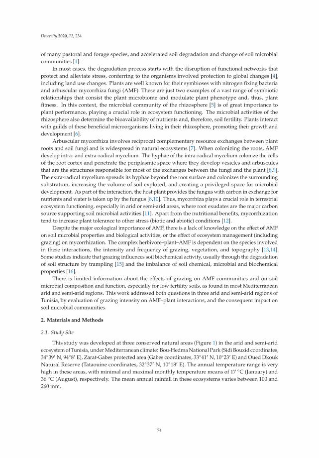
Diversity 2020, 12, 234
of many pastoral and forage species, and accelerated soil degradation and change of soil microbialcommunities [1].
In most cases, the degradation process starts with the disruption of functional networks thatprotect and alleviate stress, conferring to the organisms involved protection to global changes [4],including land use changes. Plants are well known for their symbioses with nitrogen fixing bacteriaand arbuscular mycorrhiza fungi (AMF). These are just two examples of a vast range of symbioticrelationships that consist the plant microbiome and modulate plant phenotype and, thus, plantfitness. In this context, the microbial community of the rhizosphere [5] is of great importance toplant performance, playing a crucial role in ecosystem functioning. The microbial activities of therhizosphere also determine the bioavailability of nutrients and, therefore, soil fertility. Plants interactwith guilds of these beneficial microorganisms living in their rhizosphere, promoting their growth anddevelopment [6].
Arbuscular mycorrhiza involves reciprocal complementary resource exchanges between plantroots and soil fungi and is widespread in natural ecosystems [7]. When colonizing the roots, AMFdevelop intra- and extra-radical mycelium. The hyphae of the intra-radical mycelium colonize the cellsof the root cortex and penetrate the periplasmic space where they develop vesicles and arbusculesthat are the structures responsible for most of the exchanges between the fungi and the plant [8,9].The extra-radical mycelium spreads its hyphae beyond the root surface and colonizes the surroundingsubstratum, increasing the volume of soil explored, and creating a privileged space for microbialdevelopment. As part of the interaction, the host plant provides the fungus with carbon in exchange fornutrients and water is taken up by the fungus [8,10]. Thus, mycorrhiza plays a crucial role in terrestrialecosystem functioning, especially in arid or semi-arid areas, where root exudates are the major carbonsource supporting soil microbial activities [11]. Apart from the nutritional benefits, mycorrhizationtend to increase plant tolerance to other stress (biotic and abiotic) conditions [12].
Despite the major ecological importance of AMF, there is a lack of knowledge on the effect of AMFon soil microbial properties and biological activities, or the effect of ecosystem management (includinggrazing) on mycorrhization. The complex herbivore–plant–AMF is dependent on the species involvedin these interactions, the intensity and frequency of grazing, vegetation, and topography [13,14].Some studies indicate that grazing influences soil biochemical activity, usually through the degradationof soil structure by trampling [15] and the imbalance of soil chemical, microbial and biochemicalproperties [16].
There is limited information about the effects of grazing on AMF communities and on soilmicrobial composition and function, especially for low fertility soils, as found in most Mediterraneanarid and semi-arid regions. This work addressed both questions in three arid and semi-arid regions ofTunisia, by evaluation of grazing intensity on AMF–plant interactions, and the consequent impact onsoil microbial communities.
2. Materials and Methods
2.1. Study Site
This study was developed at three conserved natural areas (Figure 1) in the arid and semi-aridecosystem of Tunisia, under Mediterranean climate: Bou-Hedma National Park (Sidi Bouzid coordinates,34◦39′ N, 94◦8′ E), Zarat-Gabes protected area (Gabes coordinates, 33◦41′ N, 10◦23′ E) and Oued DkoukNatural Reserve (Tataouine coordinates, 32◦37′ N, 10◦18′ E). The annual temperature range is veryhigh in these areas, with minimal and maximal monthly temperature means of 17 ◦C (January) and36 ◦C (August), respectively. The mean annual rainfall in these ecosystems varies between 100 and260 mm.
74

Diversity 2020, 12, 234
Figure 1. Location of the three conserved natural areas in the arid Mediterranean ecosystem of Tunisia.
Experiments were carried out in three natural protected areas of Tunisia (Bou-Hedma, Zarat, andOued Dkouk) (Table 1). At each natural park samples were taken in two sites, one inside and theother outside the protected area. The studied sites differed in grazing intensity and soil type (Table 1).The three sites inside the conserved areas were subjected to a light grazing, while the other three sitesoutside were subjected to more intensive grazing by domestic herds of sheep, goats and camels.
Table 1. Details of the six sampling sites at the three conserved natural areas.
Conserved Areas Site Vegetation Type Grazing Intensity
Bou-Hedmainside 1 Acacia spp. and spontaneous herbaceous plants Light
outside 2 Cultivated and spontaneous herbaceous plants Intensive
Zaratinside 3 Acacia spp. and spontaneous herbaceous plants Light
outside 4 Cultivated and spontaneous herbaceous plants Intensive
Oued Dkoukinside 5 Spontaneous herbaceous plants Light
outside 6 Spontaneous herbaceous plants Intensive
2.2. Roots and Soil Sampling
Five plant species (common to all study sites) and three plants per species per site were analyzedfor AMF colonization. The herbaceous plants collected were: Lotus creticus (Fabaceae), Medicagotruncatula (Fabaceae), Astragalus corrugatus (Fabaceae), Malva aegyptiaca (Malvaceae), Diplotaxis simplex(Cruciferacea).
Plant roots were carefully collected in order to access the fine active roots where mycorrhizacolonization occurs. Simultaneously, rhizosphere soil of each plant was also collected. For each site, apooled soil sample (composed of 5 soil cores with 10 cm diameter and 20 cm length) was collectedin an area without vegetation and used as a control (bulk soil) for the influence of the plant in thedynamic soil characteristics. Soils were sieved (2 mm) to remove the remains of plants, gravel andearthworms, and stored at 4 ◦C for further analysis. Soil samples were analyzed in triplicates.
2.3. AMF Colonization Status
According to the methods of Phillips and Hayman [17], AMF colonization was evaluated afterobservation of 30 root fragments per plant. Of each plant species, root segments (1–2 cm length)were submerged in 10% potassium hydroxide (KOH) at 90 ◦C for 45 min. After bleaching and
75

Diversity 2020, 12, 234
acidification steps with hydrogen peroxide (H2O2) and hydrogen chloride (HCl), respectively, rootpieces were colored with 1% Trypan Blue solution. The duration of each step varied among plantspecies, according to the respective root diameter and surface root characteristics. For each plantspecies from each site, 30 root fragments were placed on slides and preserved with lactoglycerol.In all the stained roots, the presence of hyphae, vesicles, and arbuscules inside the root wereviewed through a microscope (Nikon, Tokyo, Japan) at 400× magnification. The frequency (F%):number of colonized roots/total number of observed roots) and intensity of mycorrhization (M%):proportion, in percentage, of the root colonized by AMF) were calculated using the Mycocalc program(http://www2.dijon.inra.fr/mychintec/Mycocalcprg/download.html)
2.4. Quantification of AMF Spore Density
Using the wet sieving method from Gerdemann and Nicolson [18], AMF spores occurring insoil samples were extracted and quantified. Three nested sieves of 1000, 100, and 32 μm were used.For each soil sample, quantities of 100 g were submerged in 1 L of tap water. After a stirring step, thesupernatant was sieved through the nested sieves. The spores that hold on to the two sieves of 100 and32 μm were recovered in 5 mL centrifuge tubes. After a step of centrifugation on a viscosity gradient(sucrose solution at 60%), the supernatant retained was rinsed with distilled water to remove thesucrose solution. Retrieved AMF spores of each soil sample were counted under a stereomicroscope(40×magnification) and average numbers were calculated per 100 g of dry soil.
2.5. Soil Analysis: Physical and Chemical Properties
Soil pH and electrical conductivity were determined in a 1:1 (v/w) water: bulk soil suspensionusing a pH meter (Matest, Treviolo, Italy) and a conductivity meter (Bibby Scientific, Bibby Scientific,Staffordshire, UK), respectively [19]. Soil texture, which represents the granulometric distributionof its constituents (the proportion between small particles: clay, silt and sand), was calculated usingthe Robinson’s pipette method [20]. The other main physicochemical soil characteristics (organicmatter, total nitrogen, total phosphorus and water content) were determined by conventional analysesperformed by the Soil Analysis Laboratory in the Regional Commissariat for Agricultural Development,in Gabes. The determination of the organic matter was carried out indirectly, starting from thedetermination of the organic carbon content of soil. The determination of total phosphorus wasevaluated by a degradation acid reaction step followed by a dosing step carried out in an automatedspectrophotometer (Shimadzu, Kyoto, Japan). The total nitrogen was determined following theKjeldahl method.
2.6. Microbiological and Biochemical Properties
For the impact of AMF on microbiological parameters, the carbon of the microbial biomass (Cmic)present in the plant rhizosphere and bulk soil were evaluated following the “fumigation–extraction”technique [21]. This method is based on three essential steps: a fumigation step with chloroform,incubation in a 10-days fumigation period, and an extraction step with ninhydrin-N reactive andpotassium chloride (KCl). This technique has been used to provide rapid and accurate measurementsof soil biomass -C and -N.
To evaluate the effect of AMF on biochemical properties, alkaline phosphatase and β-glucosidaseactivities were calculated and evaluated according to the method of Caravaca et al. [22] in aspectrophotometer (Shimadzu, Japan) at 398 nm. The dehydrogenase activity was determinedas described by Garcia et al. [23] in a spectrophotometer (Shimadzu, Japan) at 490 nm.
2.7. Statistical Analyses
Analyses of variance (ANOVA) for repeated measures using the XLSTAT (v2010.5.04) software(Addinsoft, New York, NY, USA) were ascertained to test the effect and the significant difference
76

Diversity 2020, 12, 234
between the studied parameters. Least significant difference values at the 5% levels of significance(p ≤ 0.05) were calculated to assess differences between different values.
To evaluate the effects of the grazing parameter (explanatory variable) on mycorrhizal properties(F%, M% and number of spores) (response variables), ANOVA and Canonical Correlation Analysis(CCorA) were applied.
To evaluate the relationships between soil physical and chemical parameters and mycorrhizalproperties (F%, M% and number of spores), a principal-component analysis (PCA) was applied.
Pearson’s correlation was used to determine relationships between variables: mycorrhizalproperties and microbiological and biochemical parameters.
To model the relationships between mycorrhizal properties and parameters of soil microbial andbiochemical activities by linear regression, XLSTAT (v2010.5.04) software was used.
3. Results
3.1. Physical and Chemical Properties of Soils
The more resilient soil characteristics such as soil texture (Table 2) and pH did not show bigdiscrepancies among sites inside and outside the protected areas. However, differences were obtainedfor the more responsive soil dynamic characteristics: electrical conductivity (E.c), total nitrogen (T.N),total phosphorus (T.P), organic matter (Org. Mat), and water content (Wat. Con). In general, thestudied sites had an alkaline pH ranging from 8.0 to 8.4. The highest percentages of soil organic matterwere observed in Site 1 (2.6%) as well as inside the other protected areas. Identical patterns wereobserved for the total nitrogen, the highest level (194 ppm) also being observed in Site 1, followed bySites 3 and 5 (inside the protected areas). For the total phosphorus content, it was the contrary: thehighest values were registered in the sites outside the protected areas (15.3 ppm was the highest value,registered in Site 6). Water content (varying between 1.3 to 3.3%) and electrical conductivity (rangingfrom 1.3 to 2.5 s·m−1) always presented the highest values in the sites inside each protected area.
Table 2. Physical and chemical properties of the six sampling sites from the three conserved ecosystems.
Parameters
Bou-Hedma Zarat Oued Dkouk
Inside Outside Inside Outside Inside Outside
Site 1 Site 2 Site 3 Site 4 Site 5 Site 6
Clay (%) 11.0 ± 0.2 b 12.8 ± 0.1 a 9.2 ± 0.2 d 10.2 ± 0.1 c 6.5 ± 0.2 f 7.0 ± 0.1 e
Silt (%) 23.5 ± 1.1 d 26.1 ± 0.1 b 25.1 ± 0.2 c 34.7 ± 0.3 a 16.4 ± 0.7 e 15.3 ± 0.5 f
Sand (%) 65.8 ± 0.2 c 60.0 ± 1.2 e 64.7 ± 1.1 d 55.0 ± 1.4 f 77.0 ± 1.1 b 78.6 ± 1.1 a
pH 8.1 ± 0.1 b 8.1 ± 0.1 b 8.0 ± 0.1 b 8.1 ± 0.1 b 8.4 ± 0.1 a 8.4 ± 0.1 a
E.c (s·m−1) 2.5 ± 0.3 a 1.8 ± 0.2 c 2.4 ± 0.3 a 1.6 ± 0.2 d 2.0 ± 0.1 b 1.3 ± 0.1 e
T.N (ppm) 191.0 ± 23 a 150.0 ± 10 d 174.0 ± 15 b 130.0 ± 10 e 160.0 ± 10 c 90.0 ± 5 f
T.P (ppm) 7.0 ± 0.1 f 10.6 ± 0.2 c 8.2 ± 0.2 e 12.4 ± 0.2 b 10.0 ± 0.2 d 15.3 ± 0.6 a
Org. Mat (%) 2.6 ± 0.2 a 1.4 ± 0.1 d 2.0 ± 0.1 b 1.2 ± 0.1 e 1.6 ± 0.3 c 0.8 ± 0.1 f
Wa. Cont (%) 3.3 ± 0.1 a 2.3 ± 0.1 d 2.9 ± 0.2 b 2.0 ± 0.1 e 2.5 ± 0.1 c 1.4 ± 0.3 f
E.c: electrical conductivity; T.N: total nitrogen; T.P: total phosphorus; Org. Mat: organic matter; Wat. Con: watercontent. Letters a–f: significant differences (p < 0.05); mean and standard error values (n = 3).
3.2. AMF Colonization of Plant Roots
Three plants and 30 root fragments of each one of the five plant species were analyzed per site.Direct observation of the root samples under the microscope showed that the roots of all studiedplants, except those of Diplotaxis simplex, were colonized by AMF. All the structures characteristicof root colonization by AMF (intracellular aseptate hyphae, vesicles and arbuscules) were observed.The highest mycorrhizal frequency (F%)) was observed for M. truncatula in all the studied sites. At eachsite, the mycorrhizal frequency (F%)) varied among the plant species (Figure 2). The plants withthe highest AMF root colonization belong to the Fabaceae family (M. truncatula, A. corrugatus andL. creticus) in all the studied sites, always with higher values in the site inside each conserved area.
77

Diversity 2020, 12, 234
There were significant differences (p < 0.001) in the mycorrhizal intensity (M%) among sites, againwith highest M% for M. truncatula as well as, for each plant species, for the plants in the sites inside theconserved areas (Figure 2).
Figure 2. Mycorrhizal frequency (A) and Mycorrhizal intensity (B) of the five herbaceous plants in thedifferent studied sites. Data are reported as mean (±SE) of three replicates per sample.
3.3. Densities of Spore Populations in the Studied Soils
The density of AMF spores isolated from the rhizosphere of the sampled plants varied between856 (M. truncatula) in Site 1 and 81 spores/100 g of soil (D. simplex) in Site 6 (Figure 3). Therefore, sporedensity varied significantly (p < 0.001) among the studied sites; the maximum values were recorded inthe rhizosphere of plants from sites 1, 3 and 5 (inside the conserved areas and lightly grazed) and theminimum values in sites 2, 4 and 6 (outside the conserved areas and intensively grazed). Bare areas (bulksoil), followed by D. simplex rhizosphere soil, always presented the lowest values for the six sites (Figure 3).
Figure 3. Distribution of AMF spores in the different rhizosphere soils. Data are reported as mean(±SE) of three replicates per sample.
78

Diversity 2020, 12, 234
There was a clear relationship between the intensity of root cortex AMF colonization (M%),mycorrhizal frequency (F%), and the density of AMF spores in the rhizospheres (Figures 2 and 3).Furthermore, the highest spore density was registered in the rhizosphere of the plant species withhigher mycorrhiza frequency and intensity (M. truncatula).
3.4. Effect of the Grazing Intensity on the Different AMF Parameters
The grazing intensity strongly affected the mycorrhizal colonization and density of spores (Table 3),in accordance with the lowest values always registered, either for the plant mycorrhizal status orfor AMF spore populations, in samples from sites outside the protected areas (Figures 2 and 3).This explains the negative effect of the grazing intensity on the different mycorrhizal parameters(Figure 4).
Table 3. Two-factor ANOVA analysis of the impact of grazing intensity on mycorrhizal parameters.
FactorGrazing Intensity
F-Ratio p-Value
Mycorrhizal frequency (F%) 3.52 0.07 ns
Mycorrhizal intensity (M%) 11.26 0.001 ***AMF spore density 10.55 0.002 **
*** Significant at p < 0.001, ** Significant at p < 0.01, ns: non-significant.
Figure 4. Results of a Canonical Correlation Analysis (CCorA) for the relationships between grazingintensity and AMF properties. Sp. num: number of spores; F%: mycorrhizal frequency; M%:mycorrhizal intensity.
3.5. Effect of Soil Properties on the Different AMF Parameters
According to the results of the Principal-component analysis (PCA) (Figure 5), the highest valuesof AMF colonization and number of spores were found in Site 1 at Bou-Hedma followed by Site 3at Zarat. The major physical and chemical parameters of these sites (Org. Mat, E.c, Wat. Con, T.N)were in a strongly positive correlation with the different mycorrhizal properties. In contrast, the totalphosphorus (T.P) available negatively affected all the mycorrhizal parameters and was considered alimited factor to these parameters (Figure 5).
79

Diversity 2020, 12, 234
Figure 5. Results of a principal-component analysis (PCA) showing the relationships between soilproperties and AMF parameters. E.c: electrical conductivity; T.N: total nitrogen; T.P: total phosphorus;Org. Mat: organic matter; Wat. Con: water content; Sp. num: number of spores; F%: mycorrhizalfrequency; M%: mycorrhizal intensity.
3.6. Microbiological and Biochemical Properties of the Soils and Impact of AMF on Soil Microbial Communities
We found significant effects (p < 0.001) of the mycorrhizal plants on Cmic (Figure 6), this meaningthat plants are affecting soil microbial biomass and consequently potential soil dynamics. The Cmicvalues were lower in bare areas (control soil) than in rhizosphere soils, the maximum values beingrecorded in the rhizospheres of the three Fabaceae plants M. truncatula, A. corrugatus and L. creticus.Cmic also markedly decreased in the intensively grazed sites (outside the protected areas), followingthe same trend of mycorrhizal frequency and intensity in root samples and of number of spores inrhizosphere soils (Figures 2, 3, 6 and 7).
Figure 6. Microbial biomass carbon (Cmic) in the different sampled soils. Data are reported as mean(±SE) of three replicates per sample.
80

Diversity 2020, 12, 234
Figure 7. Phosphatase (A), dehydrogenase (B), β-glucosidase (C) activities in the studied soils. PNP: pnitrophenol, INTF: iodonitrotetrazolium formazan. Data are reported as mean (±SE) of three replicatesper sample.
The highest activities of the three enzymes evaluated (phosphatase, dehydrogenase andβ-glucosidase) (Figure 7) were observed in the rhizospheres of the three Fabaceae plants M. truncatula,A. corrugatus and L. halophilus, following the same trend of Cmic for each plant species and eachsite (Figure 6) as well as the values of mycorrhizal frequency and intensity and number of spores(Figures 2, 3 and 7), evidencing activities consistently greater in lightly grazed sites in comparison withthose that were intensively grazed (Figures 2, 3, 7 and 8). The ANOVA analysis evidenced significantdifferences in these values.
81

Diversity 2020, 12, 234
Figure 8. Schematic representation of the AMF effects on soil fertility and ecosystem stability underlightly grazed sites (inside the protected areas) versus intensively grazed sites (outside the protectedareas), for arid Mediterranean ecosystems.
A clear relationship was observed between the AMF parameters (frequency and intensity ofcolonization of roots and number of AMF spores in the rhizosphere) and microbial activity (microbialbiomass and enzyme activities) (Table 4, Figures S1 and S2). Based on the Pearson correlation coefficient,positive correlations with a highly significant value (p< 0.001) were observed between all AMF parameters(F%, M% and number of spores) and microbial activity (microbial biomass and enzyme activities:dehydrogenase, phosphatase and the β-glucosidase). A lower significant value (p < 0.05) was onlyobserved in Site 6 (outside Oued Dkouk Natural Reserve) between the frequency of AMF colonizationand two microbial parameters: microbial biomass and activity of β-glucosidase (Table 4).
Table 4. Pearson correlation coefficient between frequency of mycorrhization (F%), intensity ofmycorrhization (M%) and density of AMF spores (Sp. num) and microbial activity expressed bymicrobial biomass (Cmic), phosphatase activity, dehydrogenase activity, and β-glucosidase activity.
Conserved Areas Title Cmic Phosphatase Deydrogenase β-Glucosidade
Bou Hedma
Site 1
F% 0.93 *** 0.97 *** 0.95 *** 0.96 ***M% 0.99 *** 0.99 *** 0.99 *** 0.98 ***
Sp. num 0.93 *** 0.97 *** 0.95 *** 0.96 ***
Site 2
F% 0.93 *** 0.97 *** 0.96 *** 0.96 ***M% 0.98 *** 0.98 *** 0.99 *** 0.99 ***
Sp. num 0.98 *** 0.96 *** 0.98 *** 0.98 ***
Zarat
Site 3
F% 0.95 *** 0.97 *** 0.96 *** 0.97 ***M% 0.99 *** 0.98 *** 0.99 *** 0.98 ***
Sp. num 0.95 *** 0.97 *** 0.96 *** 0.97 ***
Site 4
F% 0.92 *** 0.97 *** 0.97 *** 0.96 ***M% 0.96 *** 0.97 *** 0.99 *** 0.98 ***
Sp. num 0.97 *** 0.96 *** 0.97 *** 0.95 ***
Oued Dkouk
Site 5
F% 0.96 *** 0.97 *** 0.97 *** 0.97 ***M% 0.99 *** 0.99 *** 0.98 *** 0.97 ***
Sp. num 0.98 *** 0.97 *** 0.99 *** 0.98 ***
Site 6
F% 0.90 * 0.94 *** 0.95 *** 0.91 *M% 0.98 *** 0.98 *** 0.99 *** 0.98 ***
Sp. num 0.98 *** 0.99 *** 0.97 *** 0.95 ***
*** Significant at p < 0.001, * Significant at p < 0.05.
82

Diversity 2020, 12, 234
The results shown above, concerning the effect of grazing on AMF colonization (Table 3, Figure 4)and the impact of AMF on microbial communities and activities (Table 4, Figures S1 and S2), aresummarized in Figure 8, which evidences the importance of AMF on soil functionality under aridMediterranean ecosystems. Significant mycorrhizal colonization of roots was registered in lightlygrazed sites, leading to high levels of microbial communities expressed by high values of biochemicalactivities. All these necessarily enhances soil fertility, better-adaptation and growth of plants andfinally, in the stability of the ecosystem (Figure 8).
4. Discussion
Mediterranean arid and semi-arid ecosystems are characterized by high temperatures and droughtfor most of the year. These conditions limit plant establishment and growth and accelerate soildegradation and change microbial communities [1]. Mycorrhiza form communication pathwaysbetween plants and soil, influencing plant nutrient cycling, and restoring and maintaining soil fertility,thus influencing the microbial communities of the rhizosphere and extending the influence of plantsto the soil. Several works emphasized the role of AMF in sustaining plant cover in semi-arid andarid ecosystems [4,8,10] as is the case of the following Mediterranean conserved areas in Tunisia:Bou-Hedma National Park, Zarat protected area and Oued Dkouk Natural Reserve.
Under natural conditions, about 90% of the terrestrial plants are mycorrhized, and AMF arefound in all climates and ecosystems [10]. Due to their role on plant nutrition and defense, andto the importance of the extra-radical mycelium in the establishment of biological networks, AMFare determinant for the establishment and sustainability of plant communities and environmentfunctioning [7]. However, the diversity of the AMF community and the intensity of arbuscularmycorrhizal colonization of natural vegetation is dependent on the availability of AMF sporesand the mycorrhizal dependency of the plant species [24,25], as well as on the soil structure andmanagement [26]. All these statements agree with the results of the present study, which evidencedcolonization by AMF for all the herbaceous species, except for the Cruciferaceae Diplotaxis simplex(Figure 2). This was not surprising since Cruciferaceae are usually recognized as non-mycorrhizalplants [27]. However, distinct AMF colonization rates and intensities were observed for each plantspecies, which may be related to different levels of mycorrhizal dependence [28], and/or the availabilityof AMF spores. Higher mycorrhization rates were observed in the legume species M. truncatula,A. corrugatus and L. creticus, which presented high levels of mycorrhizal intensity, independent ofthe sampled site (Figure 2). This may highlight their mycorrhizal dependency and high demand forphosphorus (P) in comparison with plants from other families such as Poaceae [29].
In general, and particularly in semi-arid and arid ecosystem, AMF vary greatly with soilcharacteristics. Several biotic and abiotic factors may contribute to the distinct mycorrhizal intensitiesobserved for the same plant species [30,31]. One abiotic factor that severely interferes withmycorrhization is the concentration of available phosphorus [10,32,33]. In the present study, soilphosphorus concentrations varied between sites, ranging from 7 to 15.3 ppm, and could explainthe distinct mycorrhizal intensities observed for the same species in distinct sites (Figures 2 and 5),supporting that its availability is a crucial driver of mycorrhizal communities and activities [34].
Another factor frequently cited as responsible for lower levels of mycorrhiza formation is theavailability of AMF spores, which is known to greatly vary in the ecosystems [35]. Sporulation inAMF occurs when the development of the mycelium begins to be limited by nutrients and is a highlycarbon demanding process. This may explain why the number of AMF spores in the rhizosphere variesamong plant species and, for the same plant species, among sites [36–38]. Apart from the rate of sporeformation, the number of AMF spores in a soil also depends on the rates of spore germination anddegradation [39]. As AMF are obligate biotrophs, the number of spores and propagules tends to behigher in the rhizosphere than in the bulk soil [12,40], and higher in the rhizosphere of plants with ahigher intensity of AMF colonization (Figure 3). These differences are particularly evident in arid andsemi-arid soils with high organic matter turnover rates and low organic matter content [41].
83

Diversity 2020, 12, 234
In low fertility soils, mycorrhiza are highways for nutrient and water transport, expandingthe plant root system and the volume of soil exploited by the plant. However, similarly to roots,hyphae are leaky and lose nutrients into the hyphosphere, which will promote the development ofselected microorganisms. In this context, it is expected that AMF play a crucial role in the biologicalcharacteristics of the rhizosphere [22,42] (Figure 6). The high values of Cmic observed in the rhizospheresoil of mycorrhizal plants imply that mycorrhiza contribute to improve the availability of carbonsubstrates to the microbial community of the rhizosphere [43,44]. These results support the hypothesisthat AMF establish unique interactions with plant roots, conferring special characteristics to therhizosphere [45], where several by product-based symbiosis and microbial loops may be assembled,contributing to improved carbon use efficiency. In this particular respect, the importance of AMF inpromoting the development of the microbial community is confirmed by the low and comparablelevels of microbial carbon observed in the soils without plant cover (control) and in the rhizospheresoil of the non-mycorrhizal plant D. simplex (Figure 6).
AMF increase the diversity of the carbon sources available to the microorganisms in therhizosphere [46]—which is partly due to their nutritional mode; the excretion of catabolic enzymesto the surrounding medium, and to the direct access by the AMF to the plant carbon. Phosphataseand β-glucosidase are two of those catabolic enzymes, and their activities were in fact higher in therhizosphere of the mycorrhizal plants than in that of the non-mycorrhizal plant studied or in the bulksoil (Figure 5). The importance of the soil microbial activity in association with several enzymes’activities was highlighted by the similarity in the activity patterns of the two hydrolytic enzymes(phosphatase and β-glucosidase) as well as of dehydrogenase, indicators of microbial activity [47],which can be inferred as being decreased under high grazing intensity (Figures S1 and S2).
At this point, it is clear that plant and AMF species are important modulators of rhizospherecharacteristics (Figures 6–8). What is not clear is why biological indicators of soil characteristics are sodistinct in the three sites outside the conserved areas. One key factor common to these sites may belimiting and related to mycorrhizal colonization. Grazing may affect mycorrhization (Table 3, Figure 4)directly in nutrient-limited ecosystems through a direct competition for carbon (Figure 8). Grazingwas consistently associated with lower soil organic matter and increased P concentrations showing astrong impact on soil chemical properties, able to negatively influence mycorrhization in a direct waythrough the increase of P bioavailability. But in nutrient-limited ecosystems grazing per se may alsoaffect mycorrhization (Table 3, Figure 4).
Plants are the primary producers of ecosystems, obtaining their biomass and energy from thecarbon fixed by photosynthesis (Figure 8). Depending on the plant species and on the growth conditions,20–50% of the newly photosynthetically fixed carbon may be transported to the roots and lost asrhizodeposition. In arid soils, this carbon is the main source of energy and biomass building material forthe microbial community. Therefore, increasing grazing intensity decreases the carbon availability fromplants and, consequently, also decreases the carbon available for mycorrhization [13,14]. An increasein grazing intensity has impacts on soil biological characteristics, with a significant decrease in themycorrhizal intensity, as evidenced in the present study, for the three mycorrhizal plant species sampledin the sites outside the conserved areas (Table 3, Figure 5; Figure 8).
5. Conclusions
AMF is of vital importance in the arid Tunisian ecosystem, where soils are generally poor inorganic matter and nutrients. High mycorrhization levels and high spore densities were revealedacross the different sites, with particular incidence in the sites inside the conserved areas. Therefore,grazing intensity affected the potential beneficial influence of AMF on soil microbial processes. At thispoint, the importance of AMF to ecosystem dynamics is clear. What is not clear is how AMF areadapted to grazing intensity to allow ecosystem stability, particularly for situations that may berepresented by the three sites outside the conserved areas. Therefore, future research should be focusedon determining the ways through which AMF adapt to grazing intensity. This may be a key feature of
84

Diversity 2020, 12, 234
ecosystem management, considering that AMF could be an important alternative for sustaining soilquality, and could be exploited as potential inoculants for rehabilitation and restoration programs inMediterranean ecosystems.
Supplementary Materials: The following are available online at http://www.mdpi.com/1424-2818/12/6/234/s1,Figure S1: Linear regression to model the correlations between mycorrhizal proprieties (mycorrhizal frequency,mycorrhizal intensity, and number of spores) and the microbiological parameter microbial biomass carbon.The analyses included all available data: from the rhizospheres of the four plants and from bulk soil, Figure S2:Linear regression to model the correlations between mycorrhizal proprieties (mycorrhizal frequency, mycorrhizalintensity, and number of spores) and biochemical activities: phosphatase (in μg PNP. g−1 soil·h−1), dehydrogenase(μg INTF. g−1 soil·h−1) and β-glucosidase (μg PNP. g−1 soil·h−1). The analyses included all available data: fromthe rhizospheres of the four plants and from bulk soil.
Author Contributions: Conceptualization, M.M. (Mohamed Mars); methodology, N.M.; validation, M.M.(Mosbah Mahdhi) and T.D.; formal analysis, N.M.; investigation, N.M.; data curation, N.M.; writing—originaldraft preparation, N.M.; writing—review and editing, C.C. and M.F.C.; visualization, C.C. and M.F.C.; projectadministration, M.M. (Mohamed Mars); funding acquisition, C.C. and M.F.C. All authors have read and agreed tothe published version of the manuscript.
Funding: This work was supported by the Ministry of High Education and Research Development-Tunisiaand by FCT/MCTES for the financial support to cE3c (project UIDB/00329/2020) and CESAM (projectsUIDP/50017/2020+UIDB/50017/2020), through Portuguese national funds.
Acknowledgments: We thank the personal staff of Bou-Hedma National Park, of Zarat-Gabes Protected area andof the Natural reserve of Oued Dkouk, for facilities and support for root and soil sampling. Thanks, are also dueto Francisco Caeiro for providing helpful comments and English revision.
Conflicts of Interest: The authors declare no conflict of interest.
References
1. Martínez-García, L.; Miranda, J.; Pugnaire, F.I. Impacts of changing rainfall patterns on mycorrhizal status ofa shrub from arid environments. Eur. J. Soil Biol. 2011, 50, 64–67. [CrossRef]
2. Mahmoudi, N.; Cruz, C.; Mahdhi, M.; Mars, M.; Caeiro, M.F. Arbuscular mycorrhizal fungi in soil, roots andrhizosphere of Medicago truncatula: Diversity and heterogeneity under semi-arid conditions. PeerJ 2019.[CrossRef] [PubMed]
3. Mosbah, M.; Philippe, D.L.; Mohamed, M. Molecular identification of arbuscular mycorrhizal fungal sporesassociated to the rhizosphere of Retama raetam in Tunisia. Soil Sci. Plant. Nutr. 2018, 64, 335–341. [CrossRef]
4. Brundrett, M. Mycorrhizal associations and other means of nutrition of vascular plants: Understandingthe global diversity of host plants by resolving conflicting information and developing reliable means ofdiagnosis. Plant Soil 2009, 320, 37–77. [CrossRef]
5. Van der Heijden, M.G.A.; Martin, F.M.; Selosse, M.A.; Sanders, J.R. Mycorrhizal ecology and evolution:The past, the present, and the future. New Phytologist. 2015, 204, 1406–1423. [CrossRef] [PubMed]
6. Barea, J.M.; Azcón, R.; Azcón-Aguilar, C. Mycorrhizosphere interactions to improve plant fitness and soilquality. Antonie van Leeuwenhoek. Inter. J. Gen. Mol. Microb. 2002, 81, 343–351.
7. Entry, J.A.; Rygiewicz, P.T.; Watrud, L.S.; Donnelly, P.K. Influence of adverse soil conditions on the formationand function of arbuscular mycorrhizas. Adv. Environ. Res. 2002, 7, 123–138. [CrossRef]
8. Smith, S.E.; Read, D.J. Mycorrhizal Symbiosis; Academic Press: San Diego, CA, USA, 1997.9. Van Aarle, I.M.; Cavagnaro, T.R.; Smith, S.E.; Smith, F.A.; Dickson, S. Metabolic activity of Glomus intraradices
in Arumand Paris-type arbuscular mycorrhizal colonization. New Phytologist. 2005, 166, 611–618. [CrossRef][PubMed]
10. Smith, S.E.; Read, D.J. Mycorrhizal Symbiosis; Academic Press: San Diego, CA, USA, 2008.11. Bago, B.; Vierheilig, H.; Piché, Y.; Ázcon-Aguilar, C. Nitrate depletion and pH changes induced by the
extraradical mycelium of the arbuscular mycorrhizal fungus Glomus intraradices grown in monoxenicculture. New Phytol. 1996, 133, 273–280. [CrossRef] [PubMed]
12. Azcón-Aguilar, C.; Palenzuela, E.J.; Roldan, A.; Bautista, S.; Vallejo, R.; Barea, J.M. Analysis of the mycorrhizalpotential in the rhizosphere of representative plant species from desertification-threatened Mediterraneanshrub lands. Appl. Soil Ecol. 2003, 22, 29–37. [CrossRef]
85

Diversity 2020, 12, 234
13. Barto, E.K.; Rilling, M.C. Does herbivory really suppress mycorrhiza? A meta-analysis. J. Ecol. 2010, 98,745–753. [CrossRef]
14. Mendoza, R.; Cabello, M.; Anchorena, J.; García, M.L. Soil parameters and host plants associated witharbuscular mycorrhizae in the grazed Magellanic steppe of Tierra del Fuego. Agric. Ecosyst. Environ. 2011,140, 411–418. [CrossRef]
15. Raiesi, F.; Asadi, E. Soil microbial activity and litter turnover in native grazed and ungrazed rangelands in asemiarid ecosystem. Biol. Fertil. Soil 2006, 43, 76–82. [CrossRef]
16. Liao, J.D.; Boutton, T.W. Soil microbial biomass response to woody plant invasion of grassland. Soil Biol.Biochem. 2008, 40, 1207–1216. [CrossRef]
17. Phillips, J.M.; Hayman, D.S. Improved procedure for clearing roots and staining parasitic and vesiculararbuscular mycorrhizal fungi for rapid assessment of infection. Trans. Br. Mycol. Soc. 1970, 55, 158–161.[CrossRef]
18. Gerdemann, J.W.; Nicolson, T.H. Spores of mycorrhizal Endogone species extracted by wet sieving anddecanting. Trans. Br. Mycol. Soc. 1963, 46, 235–244. [CrossRef]
19. AFNOR. Recueil de normes françaises, qualité des sols, méthodes d’analyses, 1st ed.; Association française denormalisation (Afnor): Paris, France, 1987.
20. Naanaa, W.; Susini, J. Méthodes d’analyse physique et chimique des sols. ES 252, Direction des Sols; Ministère del’Agriculture: Tunis, Tunisie, 1988.
21. Amato, M.; Ladd, J.N. Assay for microbial biomass based on ninhydrin-reactive nitrogen in extracts offumigated soils. Soil Biol. Biochem. 1988, 20, 107–114. [CrossRef]
22. Caravaca, F.; Alguacil, M.; Torres, P.; Roldán, A. Plant type mediates rhizospheric microbial activities andsoil aggregation in a semiarid Mediterranean salt marsh. Geoderma 2005, 12, 338–375. [CrossRef]
23. Garcia, C.; Hernandez, T.; Costa, F. Potential use of dehydrogenase activity as an index of microbial activityin degraded soils. Commun. Soil Sci. Plan. 1997, 28, 123–134. [CrossRef]
24. Zhu, Y.G.; Laidlaw, A.S.; Christie, P.; Hammond, M.E. The specificity of arbuscular mycorrhizal fungi inperennial ryegrass, white clover pasture. Agric. Ecosyst. Environ. 2000, 77, 211–278. [CrossRef]
25. Duponnois, R.; Founoune, H.; Masse, D.; Pontanier, R. Inoculation of Acacia holosericea with ectomycorrhizalfungi in a semi-arid site in Senegal: Growth response and influences on the mycorrhizal soil infectivity after2 years plantation. For. Ecol. Manag. 2005, 207, 351–362. [CrossRef]
26. Lekberg, Y.; Koide, R.T.; Rohr, J.R.; Aldrich-Wolfe, L.; Morton, J.B. Role of niche restrictions and dispersal inthe composition of arbuscular mycorrhizal fungal communities. J. Ecol. 2007, 95, 95–105. [CrossRef]
27. Muchovej, R.M. Importance of Mycorrhizae for Agricultural Crops, SS-AGR-170; Agronomy Department, FloridaCooperative Extension Service; Institute of Food and Agricultural Sciences, University of Florida: Gainesville,FL, USA, 2004.
28. Strullu, D.G. Les mycorhizes des arbres et des plantes cultivées; Techniques et Documentation Lavoisier: Paris,France, 1991.
29. Bagayogo, M. Site Specific Effects of Cereals/Legume Rotations in West Africa: Soil Mineral Nitrogen, Mycorrhizaeand Nematodes; Verlag Graner: Stuttgard, Germany, 1999.
30. Henriques, R.P.; Hay, J.D. The plant communities of a foredune in southeastern Brazil. Can. J. Bot. 1998, 76,1323–1330.
31. Mahesh, V.; Selvaraj, T. Occurrence and distribution VA mycorrhizal fungi in the soils polluted with TanneryEffluent. Adv. Biotech. 2008, 8, 34–36.
32. Dickson, S.; Smith, S.E.; Smith, F.A. Characterization of two arbuscular mycorrhiza fungi in symbiosis withAllium porum: Colonization, plant growth and phosphate uptake. New Phytol. 1999, 144, 163–172. [CrossRef]
33. Bouamri, B.; Dalpé, Y.; Serrhini, M.N.; Bennani, A. Arbuscular Mycorrhizal fungi species associated withrhizosphere of Phoenix dactylifera L. in Morocco. Afr. J. Boitech. 2006, 5, 510–516.
34. Liu, W.; Zhang, Y.; Jiang, S.; Deng, Y.; Christic, P.; Murray, P.J.; Li, X.; Zhang, J. Arbuscular mycorrhizal fungiin soil and roots respond differently to phosphorus inputs in an intensively managed calcareous agriculturalSoil. Sci. Rep. 2016, 6, 1–11. [CrossRef] [PubMed]
35. Li, L.; Yang, A.; Zhao, Z. Seasonality of arbuscular mycorrhizal symbiosis and dark septate endophytes ingrassland site in Southwest China. Microbial. Ecol. 2005, 54, 367–373.
36. Houngnandan, P.; Yemadje, R.G.; Kane, A.; Boeckx, P.; Van Cleemput, O. Les glomales indigènes de la forêtclaire à Isoberliniadoka (Craib et Stapf) à Wari-Maro au centre du Bénin. Tropicultura 2009, 27, 83–87.
86

Diversity 2020, 12, 234
37. Sghir, F.; Chliyeh, M.; Kachkouch, W.; Khouader, M.; OuazzaniTouhami, A.; Benkirane, R.; Douira, A.Mycorrhizal status of Olea europaea ssp. Oleaster in Morocco. J. Appl. Biosci. 2013, 61, 4478–4489. [CrossRef]
38. Al-Areqi, A.N.; Chliyeh, M.; Sghir, F.; Ouazzani, A.; Benkirane, R.T.; Douira, A. Diversity of Arbuscularmycorrhizal fungi in the rhizosphere of Coffea Arabica in the Republic of Yemen. J. Appl. Biosci. 2013, 64,4888–4901. [CrossRef]
39. Gemma, J.N.; Koske, R.E.; Carreiro, M. Seasonal variation in spore abundance and dormancy ofGigasporagigantea in Mycorrhizal inoculum potential of a dune Soil. Mycologia 1989, 80, 211–216. [CrossRef]
40. Lovelock, C.E.; Andersen, K.; Morton, J.B. Arbuscular mycorrhizal communities in tropical forests areaffected by host tree species and environment. Oecologia 2003, 135, 268–297. [CrossRef] [PubMed]
41. Mohammad, M.J.; Hamad, S.R.; Malkawi, H.I. Population of arbuscular mycorrhizal fungi in semi-aridenvironment of Jordan as influenced by biotic and abiotic factors. J. Arid Environ. 2003, 53, 409–417.[CrossRef]
42. Cruz, R.E.; Garcia, M.U. Nitrogen fixation and mycorrhizae in acacias on degraded grasslands. In TropicalAcacias in East Asia and the Pacific; Kamiset, A., Taylor, D.A., Eds.; Winrock Intenational Institute for AgricultureResearch: Bangkok, Thailand, 1992; pp. 59–71.
43. Böhme, L.; Langer, U.; Böhme, F. Microbial biomass, enzyme activities and microbial community structure intwo European long-term field experiments. Agric. Ecosyst. Environ. 2005, 109, 141–152. [CrossRef]
44. Flieβbach, A.; Oberholzer, H.R.; Gunst, L.; Mäder, P. Soil organic matter and biological soil quality indicatorsafter 21 years of organic and conventional farming. Agric. Ecosyst. Environ. 2007, 118, 273–274. [CrossRef]
45. Dennis, P.G.; Miller, A.J.; Hirsch, P.R. Are root exudates more important than other sources of rhizodepositsin structuring rhizosphere bacterial communities? Microbial. Ecol. 2010, 72, 313–327. [CrossRef] [PubMed]
46. Rillig, M.C. Arbuscular mycorrhizae, glomalin and soil quality. Can. J. Soil Sci. 2004, 84, 355–363. [CrossRef]47. Nannipieri, P.; Kandeler, E.; Ruggiero, P. Enzyme activities and microbiological and biochemical processes in
Soil. In Enzymes in the Environment; Activity, Ecology and Applications; Burns, R.G., Dick, R.P., Eds.; MarcelDekker: New York, NY, USA, 2002; pp. 1–33.
© 2020 by the authors. Licensee MDPI, Basel, Switzerland. This article is an open accessarticle distributed under the terms and conditions of the Creative Commons Attribution(CC BY) license (http://creativecommons.org/licenses/by/4.0/).
87


diversity
Article
Fungal Diversity in the Phyllosphere ofPinus heldreichii H. Christ—An Endemic andHigh-Altitude Pine of the Mediterranean Region
Jelena Lazarevic 1,* and Audrius Menkis 2
1 Biotechnical faculty, University of Montenegro, Mihaila Lalica 15, 81 000 Podgorica, Montenegro2 Department of Forest Mycology and Plant Pathology, Uppsala BioCenter,
Swedish University of Agricultural Sciences, P.O. Box 7026, SE-75007 Uppsala, Sweden;[email protected]
* Correspondence: [email protected]
Received: 30 March 2020; Accepted: 21 April 2020; Published: 28 April 2020
Abstract: Pinus heldreichii is a high-altitude coniferous tree species naturaly occurring in small anddisjuncted populations in the Balkans and southern Italy. The aim of this study was to assess diversityand composition of fungal communities in living needles of P. heldreichii specifically focusing onfungal pathogens. Sampling was carried out at six different sites in Montenegro, where 2–4 year-oldliving needles of P. heldreichii were collected. Following DNA isolation, it was amplified using ITS2rDNA as a marker and subjected to high-throughput sequencing. Sequencing resulted in 31,831 highquality reads, which after assembly were found to represent 375 fungal taxa. The detected fungiwere 295 (78.7%) Ascomycota, 79 (21.0%) Basidiomycota and 1 (0.2%) Mortierellomycotina. The mostcommon fungi were Lophodermium pinastri (12.5% of all high-quality sequences), L. conigenum (10.9%),Sydowia polyspora (8.8%), Cyclaneusma niveum (5.5%), Unidentified sp. 2814_1 (5.4%) and Phaeosphaeriapunctiformis (4.4%). The community composition varied among different sites, but in this respecttwo sites at higher altitudes (harsh growing conditions) were separated from three sites at loweraltitudes (milder growing conditions), suggesting that environmental conditions were among majordeterminants of fungal communities associated with needles of P. heldreichii. Trees on one study sitewere attacked by bark beetles, leading to discolouration and frequent dieback of needles, therebystrongly affecting the fungal community structure. Among all functional groups of fungi, pathogensappeared to be an important component of fungal communities in the phyllosphere of P. heldreichii,especially in those trees under strong abiotic and biotic stress.
Keywords: needle pathogens; high altitude forests; DNA metabarcoding; Montenegro
1. Introduction
Pinus heldreichii is a high-altitude (grows at ca. 1200–2000 m) conifer tree species with a discontinuousand restricted distribution in the Mediterranean region. Its forests are naturaly regenerated and consistsof small and disjuncted populations located in high mountain areas influenced by the Mediterraneanclimate in the Balkans and southern Italy. Although in the past P. heldreichii formed a continuousforest belt in the Balkans, currently its forests are scattered and largely isolated. Growing primary onshallow calcareous soils, it inhabits typical tree line habitats, often on steep ridges, mountain sidesand screens. Such habitats are nutrient poor, exposed, dry and cold during the winter [1,2]. LargerP. heldreichii forests can still be found on mountain plateaus or in valleys situated at altitudes of ca.1200–1300 m that are characterised by more developed soils (cambisols). Those forests are consideredto be climazonal. i.e., characterised by permanent and stable vegetation forests of P. heldreichii, that aregrowing at ecological optimum for the species [3]. Being a tertiary relic, P. heldreichii evolved to survive
Diversity 2020, 12, 172; doi:10.3390/d12050172 www.mdpi.com/journal/diversity89

Diversity 2020, 12, 172
severe winter frosts, and short, dry and warm summers with intensive sun radiation. Due to the shortgrowing season, P. heldreichii grows very slowly, reaching ca. 15 m height after about 150 years [1].It develops thick and branched roots, which penetrate deep in cracks of calcareous stones [2]. In thelast few centuries, P. heldreichii forests were affected by intensive livestock grazing, exploitation andforest fires [4]. Intensive grazing, as well as the short and dry growing season have also resulted invery limited natural regeneration [2–4], though recent observations show some regeneration in theabandoned mountain areas [5].
Pinus heldreichii is a protected species both in Balkan countries and in Italy due to the key importanceof nature conservation, protection against gravitational natural hazards, landscape conservation andrecreation. Hence, P. heldreichii requires special attention, i.e., the development and application ofconservation measures [1–4,6].
Fungi represent the largest microbial component associated with forest trees. They play keyroles in forest ecosystems, especially in pines forest that are obligatory mutualistic, and are importantcontributors to the primary production and carbon, nutrient and water cycling [7–9]. Pathogenic fungimay negatively affect health and growth of forest trees [10,11], while fungal endophytes and epiphytessupport ecological adaptations of host plants and constitute an important component of microbialbiodiversity [12,13]. However, information about fungal communities associated with P. heldreichii islimited. Previous studies on needle pathogens of P. heldreichii have focused on either those that wereaffecting natural regeneration [14–16], or potentially invasive ones such as Dothistroma septosporum [17].Information about the occurrence of D. septosporum in stands of P. heldreichii was not known untilrecently [17], what indicated the need for a wider assessment of fungal diversity, including fungalpathogens associated with the phyllosphere of this tree species.
The aim of this study was to assess the diversity and composition of fungal communities in livingneedles of P. heldreichii specifically focusing on fungal pathogens. Needles were sampled across thenatural distribution range of P. heldreichii in Montenegro, including sites under different environmentalconditions. This was expected to demonstrate potential site-specific effects of environmental conditionson fungal diversity and community composition. By using high-throughput sequencing of fungal ITS2rDNA, we examined fungal communities in 2–4 year-old living needles of P. heldreichii from six sitessituated in four mountain areas in Montenegro.
2. Materials and Methods
2.1. Study Sites
Samples were taken at six sites that were situated in four mountain areas, representing theP. heldreichii distribution range in Montenegro (Figure 1). The sampling sites were Orjen (ORJ),Prekornica (PRE), Kucka Korita (KKO), Kucka korita North (KKN), Kuci MT (KMT) and Prokletije(PRO) (Figure 1, Table 1).
The sampling sites differed in altitude, morphology of terrain and soil properties (Table 1,Figure 2). Sites at ORJ and KMT were on altitudes of ca. 1800 m, with trees growing on stone ridges,high inclinations, and lithosols. Sites at PRE, PRO and KKO were on an altitude of ca. 1250 m, on flatterrain and more developed soil that was leptic cambisol. A site at KKN was in close proximity tothe KKO, but it was on a slope with a thinner soil layer (molic leptosol). Trees at the KKN were ingroups heavily infested by Tomicus sp. bark beetles (Scolitinae). Trees at ORJ and KMT were growingat the upper tree line, and thus, these sites were considered as sites with harsh growing conditions,while other sites had moderate growing conditions.
90

Diversity 2020, 12, 172
Figure 1. A map showing (a) distribution of Pinus heldreichii in the Mediterranean region; a grey areawas added and represents an additional P. heldreichii site; the map is adapted from [18] and (b) samplingsites in Montenegro (ORJ-Orjen; PRE-Prekornica; KKO-Kuci Mt, Kucka korita; KKN-Kuci Mt., Kuckakorita North, KMT- Kuci mountain, Momonjevo, PRO- Prokletije); a map was adapted from [19].
Table 1. Characteristics of Pinus heldreichii sampling sites.
Site GPS Coordinates Altitude (m) Soil Type a Climate b Health c
Orjen, Reovacka greda (ORJ) 42◦35′ N18◦35′ E 1700 Lithosol Cfsb Healthy-looking
Kuci Mt., Momonjevo (KMT) 42◦36′ N19◦32′ E 1800 Lithosol Cfb Healthy-looking
Prekornica, Studeno (PRE) 42◦38′ N19◦12′ E 1200 Leptic cambisol Cfsb Healthy-looking
Prokletije, Ropojana (PRO) 42◦29′ N19◦48′ E 1250 Leptic cambisol Cfb Healthy-looking
Kucka korita (KKO) 42◦29′ N19◦30′ E 1300 Leptic cambisol Cfs”b Healthy-looking
Kucka korita North (KKN) 42◦30′ N19◦32′ E 1450 Molic leptosol Cfs”b Attacked by insects
a WRB soil clasification system [20,21], b Koopen climate classification [22,23], c Health status of the trees.
Figure 2. (a) Pinus heldreichii shoots with needles; trees growing at (b) Kuci Mountains, Momonjevo(KMT), and (c) Kuci Mountains, Kucka korita (KKO) sites.
91
Diversity 2020, 12, 172
Climate at the study sites is characterised as a humid warm temperate climate type (Cf), which isrepresented by subtypes Cfsb, Cfs”b and Cfb [22,23]. Summers are short, dry and chilly, and wintersare cold and windy. At different study sites, the mean annual air temperature is between 1 ◦C (at PRO)and 6 ◦C (at ORJ). The mean daily summer maximum is between 7 and 11 ◦C, and the mean dailywinter minimum is between −5 and 0 ◦C. The absolute minimum is in winter at ca. −30 ◦C and themaximum is in summer, at ca. 30 ◦C. The mean annual precipitation is highest at ORJ with ca. 3800 mm,and lowest at PRO with ca. 2000 mm. The rainfall reaches maximum in late autumn and early winter,while the minimum is during the summer months that is often followed by 40–70 day-long periods ofdroughts. The mean summer precipitation is 300 mm at ORJ and ca. 220 mm on other sites, making ca.10% of the total annual precipitation [22].
The bedrock at the study sites is solid chalk limestone, which contains a small proportion ofinsoluble residues. In general, soils are poorly developed, very water porous, skeletal leptosols(rendzina), having only A horizon, characterized by accumulation of humus (A-R profile). Accordingto the classification of texture, the soil is a sandy loam and the structure is powdery. Soil is faintlydifferentiated down in the soil profile. Soil is rich in humus content (10–25%), poor in calcium-carbonateand has a weak acid reaction (pH ca. 6). On more flat terrains, leptosol develops into the lepticcambisol, which contains initial B horizon. Brown B horizon improves the water retention capacity ofsoil [20,24].
2.2. Experimental Design and Sampling
At the sampling sites, stands of P. heldreichii were healthy-looking (Figure 2a). An exception wasthe KKN site, where trees were damaged by Tomicus sp. bark beetles, resulting in discolouration andfungal infection of needles. At all sites, the shoot dieback was occasional, but with higher rates at theKKN site.
Sampling was carried out in May 2015, i.e., before the beginning of the growing season. At eachsite, experimental design included sampling of five twigs with needles from five different trees. Hence,at each site, five mature P. heldreichii trees situated at a distance of ca. 50 m from each other were selectedand five twigs of up to 15 cm long and up to 2 m from the ground were sampled from different parts ofthe crown using secateurs. This sampling approach was used in order (i) to get a joint representativesample per each site (all samples per site were amplified with the same barcode, see below); (ii) tocompare fungal communities among different sites, but not within the same site. Collected twigs wereplaced in plastic bags and transported to the laboratory.
In the laboratory, twigs and needles were assessed for the presence of disease symptoms.The current-year needles were typically green and without disease symptoms. Symptomatic needleswere generally two years old or older and symptoms included changed colour that was yellow, orange,pale green or yellowish with red bands. Other symptoms were dying needle tips and a necrotic base ofneedles. In the case of insect attack (KKN site), defoliation and necrotic needles were common. In orderto sample the potentially entire fungal community associated with needles of P. heldreichii, both healthylooking needles and needles with disease symptoms were selected. No surface sterilization was carriedout. Following morphological examination, up to 5 representative needles that were 2–4 years old,were selected per twig. Selected needles were cut into 0.5–1 cm long segments, containing a randommixture of both asymptomatic and symptomatic parts of needles, placed in 2 mL screw cap tubes,and stored frozen at −20 ◦C before DNA extraction.
2.3. DNA Isolation, Amplificationand Sequencing
DNA extractions were done from 150 samples (6 sites × 5 trees × 5 needle samples),which previously were freeze-dried for 48 h. For isolation of total DNA, needles were homogenisedin a Fastprep machine (Precellys, Montigny-le-Bretonneux, France). The extraction was completedusing CTAB protocol [25]. After extraction, DNA samples were purified using a JetQuick DNApurification kit (Genomed GmbH, Leinfelden, Germany). The DNA concentration of each sample
92

Diversity 2020, 12, 172
was determined using a NanoDrop™ One spectrophotometer (Thermo Scientific, Rodchester, NY,USA) and adjusted to 1–10 ng/μL. Amplification by PCR of the ITS2 rDNA region was done usingbarcoded fungal-specific primer gITS7 [26] and barcoded universal primer ITS4 [27]. All 25 samplesfrom the same site were amplified using primers with the same barcode, resulting in six differentbarcodes representing each site. Amplification of several samples with the same barcode was done toget a broader representativeness of fungal communities per site. Amplifications were performed usingthe Applied Biosystems 2720 thermal cycler (Foster City, CA, USA). An initial denaturation step startedat 95 ◦C for 2 min, followed by 27 amplification cycles of denaturation at 95 ◦C for 30 s, annealing at55 ◦C for 30 s, and extension at 72 ◦C for 60 s. The thermal cycling was ended by a final extensionstep at 72 ◦C for 7 min. The PCR products were analyzed using gel electrophoresis on 1% agarose gelsstained with Nancy-520 (Sigma-Aldrich, Sweden). PCR products were purified using a sodium acetateprotocol [28]. Purified PCR products were quantified using a Qubit fluorometer 4.0 (Thermo FisherScientific, Waltham, MA, USA), and an equimolar mix of all PCR products with six barcodes (one pereach site) was used for high-throughput sequencing using a Pacific Biosciences platform (Menlo Park,CA, USA) at the SciLifeLab (Uppsala, Sweden).
2.4. Bioinformatics
Principles of bioinformatics followed that specified in [29]. The sequences obtained from the sixsamples that represented six sampling sites (Table 2), were subjected to quality control and clusteringin the SCATA NGS sequencing pipeline [30]. The initial procedure started with quality filteringof the sequences that included the removal of sequences shorter than 200 bps, sequences with lowread quality, primer dimers and homopolymers, which were collapsed to 3 bps before clustering.Only sequences containing a barcode and primer were retained. Then, the primer and samplebarcodes were removed from the sequence, but information on the sample and sequence associationwas stored as meta-data. A single-linkage clustering based on 98% similarity was used to clustersequences into different taxa. For each cluster, the sequence of the most common genotype wasused for taxonomic identification. For clusters containing only two sequences, a consensus sequencewas produced. The taxa were taxonomically identified using the GenBank database and the Blastnalgorithm [31]. The following criteria were used for identification: sequence coverage >80%; 94–97%similarity to genus level and >98% similarity to species level. Sequences deviating from these criteriawere identified only to a high taxonomic rank and were given unique names as shown in Table 3and Table S1. Representative sequences of fungal nonsingletons are available from GenBank underaccession numbers MT241905–MT242268.
Table 2. Generated high-quality ITS2 rDNA fungal sequences and detected diversity of fungal taxa atdifferent sampling sites of Pinus heldreichii. Within the column No. of Fungal Taxa, values followed bythe same letter in chi-square test do not differ significantly at p > 0.05.
Site No. of Fungal Sequences No. of Fungal Taxa Shannon Diversity Index
Orjen –ORJ 4830 139 a 3.4Kuci Mt.-KMT 4910 119 ab 2.9
Prekornica-PRE 5828 192 a 3.1Prokletije -PRO 3679 127 a 3.2
Kucka korita -KKO 8748 167 b 2.9Kucka korita North –KKN 3834 104 ab 2.7
All 31,829 375 -
93

Diversity 2020, 12, 172
Ta
ble
3.
Rel
ativ
eab
unda
nce
ofth
e25
mos
tcom
mon
fung
alta
xade
tect
edin
need
les
ofPi
nus
held
reic
hiia
tsix
site
sin
Mon
tene
gro.
Site
sar
eas
inTa
ble
1.
Fu
ng
al
Ta
xo
nP
hy
lum
*G
en
Ba
nk
Acc
ess
ion
No
.C
om
pa
red
(bp
)S
imil
ari
ty(%
)S
ite
sA
llO
RJ
KM
TP
RE
PR
OK
KO
KK
N
Loph
oder
miu
mpi
nast
riA
KC
6080
4936
1/36
110
04.
722
.114
.314
.214
.90.
412
.5Lo
phod
erm
ium
coni
genu
mA
HM
0606
5045
3/45
310
0-
0.1
32.4
5.5
14.5
2.8
10.9
Sydo
wia
poly
spor
aA
KU
5165
9147
3/47
310
06.
816
.02.
18.
20.
432
.28.
8C
ycla
neus
ma
nive
umA
AF0
1322
344
2/44
299
1.3
1.0
4.5
5.7
12.9
0.9
5.5
Uni
dent
ified
sp.2
814_
1A
MF9
7665
623
8/24
597
20.2
-0.
5-
8.2
-5.
4Ph
aeos
phae
ria
pont
iform
isA
KT0
0014
444
2/44
298
--
3.7
11.1
8.9
-4.
4U
nide
ntifi
edsp
.281
4_11
AK
U06
2806
203/
252
81-
-1.
39.
010
.3-
4.1
Uni
dent
ified
sp.2
814_
10A
KX
2202
6725
7/25
710
03.
3-
1.5
4.1
5.4
5.4
3.4
Phae
omon
iella
sp.2
814_
15A
GQ
1531
8725
7/25
999
7.2
5.9
0.2
1.2
2.9
0.5
3.0
Neo
cate
nulo
stro
ma
germ
anic
umA
KR
9951
0024
2/24
210
01.
3-
0.4
1.0
3.4
7.9
2.3
Mic
rosp
haer
opsi
sol
ivac
eaA
MH
8719
6924
9/24
910
00.
70.
10.
30.
1-
16.8
2.2
Alla
ntop
hom
opsi
ella
pseu
dots
ugae
AM
H85
7222
240/
240
100
6.4
7.1
--
--
2.1
Lach
nellu
laca
lyci
form
isA
MH
8587
7123
9/23
910
04.
78.
1-
--
-2.
0U
nide
ntifi
edsp
.281
4_23
AK
F983
527
209/
244
861.
4-
1.5
3.7
0.3
4.3
1.5
Ram
ocon
idio
phor
aeu
phor
biae
AM
G59
2740
232/
239
973.
26.
0-
--
0.6
1.5
Gea
stru
mia
sp.2
814_
26A
FJ43
8389
220/
235
942.
72.
60.
40.
60.
52.
31.
4U
nide
ntifi
edsp
.281
4_16
AK
P892
077
240/
242
994.
80.
10.
61.
90.
30.
51.
2U
nide
ntifi
edsp
.281
4_27
AK
T244
857
209/
248
84-
-3.
55.
0-
-1.
2C
ollo
phor
asp
.281
4_30
AN
R_1
3772
622
9/24
195
3.2
2.2
0.4
--
0.3
0.9
Uni
dent
ified
sp.2
814_
29A
MF9
7613
920
9/24
087
1.4
-0.
44.
10.
4-
0.9
Rhy
tism
atac
eae
sp.2
814_
22A
KR
2664
4623
4/23
799
0.4
-1.
01.
41.
6-
0.8
Dot
hide
omyc
etes
sp.2
814_
13A
KP9
9148
425
3/25
798
1.1
3.5
-0.
1-
0.4
0.8
Mol
lisia
ligni
AM
F161
301
237/
241
98-
4.7
--
--
0.7
Cen
angi
umac
uum
AM
G59
7445
239/
239
100
--
3.9
--
-0.
7A
thel
iaac
rosp
ora
BK
P814
375
296/
299
99-
-1.
80.
11.
3-
0.7
All
of25
taxa
74.9
79.9
74.6
76.6
86.4
75.4
79.0
*A
—A
scom
ycot
a,B—
Basi
diom
ycot
a.
94

Diversity 2020, 12, 172
2.5. Statistical Analyses
Rarefaction analysis was carried out using Analytical Rarefaction v.1.3 [32]. Differences in richnessof fungal taxa in different study sites of P. heldreichii were compared by nonparametric chi-squaretesting [33]. As each of the datasets was subjected to multiple comparisons, confidence limits forp-values of chi-square tests were reduced the corresponding number of times as required by theBonferroni correction [34]. The Shannon diversity index, qualitative Sorensen similarity index andnonmetric multidimensional scaling (NMDS) in Canoco 5 [33,35,36] were used to characterize thediversity and composition of fungal communities.
3. Results
The phyllosphere fungi of P. heldreichii were examined from four different mountain regions,represented by six sampling sites across the distribution range of P. heldreichii in Montenegro (Figure 1).Amplification of fungal ITS2 rDNA from 150 needle samples, PacBio sequencing and quality filteringresulted in 31,829 high quality reads. Sequence assembly and BLASTn analyses showed that the fungalcommunity in the phyllosphere of P. heldreichii was composed of 375 fungal taxa (Table 2, Table S1).Nonfungal taxa and singletons were excluded. The detected fungi were 295 (78.7%) Ascomycota,79 (21.0%) Basidiomycota and 1 (0.2%) Mortierellomycotina. Identification at least to genus levelwas successful for 254 (67.7%) out of 375 fungal taxa (Table S1), and those represented 78.5% of allfungal sequences.
In different study sites, the number of fungal taxa varied between 104 and 192 (Table 2).The chi-square test showed that the largest difference in richness of fungal taxa was between KKO andthe remaining sites (Table 2). Rarefaction showed that fungal taxa detected in all sites did not reach thespecies saturation (Figure 3).
Figure 3. Rarefaction curves showing the relationship between the cumulative number of fungal taxaand the number of ITS2 rDNA sequences from needles of Pinus heldreichii from six different sites.
Information on the 25 most common fungal taxa representing 79.0% of all fungal sequences is inTable 3. The most common fungi in the phyllosphere of P. heldreichii were Lophodermium pinastri (12.5%of all fungal sequences), Lophodermium conigenum (10.9%), Sydowia polyspora (8.8%), Cyclaneusma niveum(5.5%) and Unidentified sp. 2814_1 (5.4%) (Table 3). The most common fungal pathogens of pine needleswere S. polyspora (8.9%), C. niveum (5.5%), Neocatenulostroma germanicum (2.3%), Allantophomopsiellapseudotsugae (2.1%) and Cenangium acuum (0.7%) (Table 3). A pine needle pathogen, Dothistromaseptosporum (0.25%) was also detected, but at lower relative abundance (Table S1). The other detectedpathogenic species that are mainly known as wound pathogens of deciduous trees and/or agricultural
95
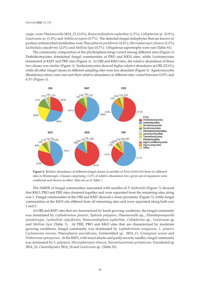
Diversity 2020, 12, 172
crops, were Phaeomoniella 2814_15 (3.0%), Ramoconidiophora euphorbiae (1.5%), Collophorina sp. (0.9%),Geastrumia sp. (1.4%) and Athelia acrospora (0.7%). The detected fungal endophytes that are known toproduce antimicrobial metabolites were Phaeosphaeria pontiformis (4.4%), Microsphaeropsis olivacea (2.2%),Lachnellula calyciformis (2.0%) and Mollisia ligni (0.7%). Ubiquitous saprotrophs were rare (Table S1).
The community composition of the phyllosphere fungi varied among different sites (Figure 4).Dothideomycetes dominated fungal communities at PRO and KKN sites, while Leotiomycetesdominated at KMT and PRE sites (Figure 4). At ORJ and KKO sites, the relative abundance of thesetwo classes was similar (Figure 4). Sordariomycetes showed higher relative abundance at ORJ (22.6%),while all other fungal classes in different sampling sites were less abundant (Figure 4). Agaricomycetes(Basidiomycotina) were rare and their relative abundance at different sites varied between 0.03% and6.3% (Figure 4).
Figure 4. Relative abundance of different fungal classes in needles of Pinus heldreichii from six differentsites in Montenegro. Classes comprising <1.0% of relative abundance for a given set of sequences werecombined and shown as other. Sites are as in Table 1.
The NMDS of fungal communities associated with needles of P. heldreichii (Figure 5) showedthat KKO, PRO and PRE sites clustered together and were separated from the remaining sites alongaxis 1. Fungal communities at the ORJ and KMT showed a closer proximity (Figure 5), while fungalcommunities at the KKN site differed from all remaining sites and were separated along both axis1 and 2.
At ORJ and KMT sites that are characterized by harsh growing conditions, the fungal communitywas dominated by Lophodermium pinastri, Sydowia polyspora, Phaeomoniella sp., Allanthopomopsiellapseudotsugae, Lachnellula calyciformis, Ramoconidiophora euphorbiae, Collophorina sp., Geastrumia sp.and Mollisia ligni (Table 3). At PRE, PRO and KKO sites that are characterized by moderategrowing conditions, fungal community was dominated by Lophodermium conigenum, L. pinastri,Cyclaneusma niveum, Phaeosphaeria punctiformis, Unidentified sp. 2814_11, Cenangium acuum andDothistroma septosporum. At the KKN, with insect attacks and partly necrotic needles, fungal communitywas dominated by S. polyspora, Microsphaeropsis olivacea, Neocatelunostroma germanicum, Uncultured sp.2814_10, Chaetothyriales 2814_18 and Geastrumia sp. (Table S1).
96

Diversity 2020, 12, 172
Figure 5. Ordination diagram based on nonmetric multidimensional scaling (NMDS) of fungalcommunities associated with needles of Pinus heldreichii from six different sites in Montenegro, with56.5% variation on Axis 1 and 28.2% on Axis 2. Each point in the diagram represents a single site andthe size of the point reflects richness of the fungal taxa as in Table 2. Sites are as in Table 1.
The Shannon diversity index of fungal communities in different study sites was between 2.7 and3.4 (Table 2). Among different sites, Sørensen similarity index of fungal communities was low tomoderate as it ranged between 0.28 and 0.54 (Table S2).
4. Discussion
A recent development of high-throughput sequencing methods provides powerful tools to explorefungal diversity. Such tools enable identification of complex fungal communities and individualcommunity components directly from environmental samples. Besides, while providing detailed andsemi-quantitative information, these tools enable studying the effects of different factors on fungaldiversity and community composition [29,37,38]. By using PacBio sequencing, we detected nearly400 fungal taxa, which were associated with needles of P. heldreichii including fungi present in very smallabundances (Table S1). Many fungal taxa remained unidentified, which remains a major challengein fungal taxonomy [39]. However, while using high-throughput sequencing, we need be aware ofpotential limitations that can include methodological biases, limitations of markers and bioinformaticschallenges [40,41].
The observed diversity of the phyllosphere fungi can be considered being high and comparablewith similar studies on different Pinus species. For example, there were 446 and 260 fungal taxa recordedduring studies of needle-associated fungi of Pinus sylvestris in Sweden and Poland, respectively [39,42].Furthermore, by using fungal culturing, there were 118 fungal taxa associated with needles of Pinus taedain USA [43], 35 taxa associated with needles of Pinus halepensis [44] and P. sylvestris in Spain [45].According to [45], the species diversity of fungal endophytes varied from 21 in Pinus monticola to 49 inPinus nigra. At different study sites, the growing conditions of P. heldreichii had in general a minor effectof the absolute richness of fungal taxa, which was similar among different sites (Table 2). However,with respect to the number of sequences obtained from samples of each site, lower richness of fungaltaxa was at the KKO (Table 2), what was likely due to the dominance of several fungal taxa such as,e.g., Lophodermium species, Cylaneusma niveum, Unidentified sp. 2814_11, Phaeosphaeria pontiformisand Unidentified sp. 2814_1. Several fungal taxa of the present study were recorded for the first timein the Balkan region such as, e.g., pathogens Neocatelunostroma germanicum and Allantophomopsiellapseudotsugae. The study also revealed a number of fungal pathogens that were previously known
97

Diversity 2020, 12, 172
from agricultural crops as, e.g., Phaeomoniella, Ramoconidiophora, Geastrumia and Athelia. These couldhave been overlooked in other studies due to latent occurrence [46]. As P. heldreichii belongs to agroup of forest trees that grows under harsh enviromental conditions, the possibility should not beexcluded that both the host and the specific environment influence the composition of associatedfungal communities. Studies on plant–endophyte associations in high stress habitats have revealedthat at least some fungal endophytes can contribute to stress tolerance of host plants [12]. Indeed,the results of this study revealed site-specific differences in fungal communities associated with needlesof P. heldreichii. For example, fungal communities were more similar among sites situated at loweraltitudes, i.e., with moderate growing conditions (PRE, PRO, KKO) as compared to those sites athigher altitudes, i.e., with harsh growing conditions (ORJ, KMT) (Table 1, Figure 5). Further, the fungalcommunity at the KKN site was largely different from the remaining sites, what was likely due to insectdamage to trees, resulting in mainly symptomatic and partly necrotic needles. These observationsdemonstrate that the composition of fungal communities and their succession in needles of P. heldreichiican be determined by different abiotic and biotic factors. Different environmental factors have earlierbeen shown to play an important role in shaping fungal communities both in the phyllosphere and inthe rhizosphere of forest trees [5,42,47,48].
Fungal pathogens represent an important biotic factor that may negatively affect health andgrowth of forest trees [11]. In the present study, a fungal pathogen Sydowia polyspora was amongthe most dominant fungi (Table 3). Sydowia polyspora has a wide geographical range [48] and iscommon in Europe. The pathogenicity of S. polyspora to young conifers (genera Thuja, Abies, Tsuga,Larix, Picea and Pinus) was previously reported [49]. In the Balkan region, it has been reported inforest plantations of P. nigra and P. sylvestris in Serbia, occurring on needles damaged by drought orfrost [50]. It was also detected on needles of P. halepensis in Italy and Spain [44,51], P. sylvestris in Polandand Lithuania [29,39] and on P. ponderosa in North America [52]. Sydowia polyspora has recently beenreported as a pathogen that dominates fungal communities vectored by bark beetles associated withP. radiata, P. nigra subsp. salzmannii and P. sylvestris in Spain [53] and Pinus yunnanensis in China [54].Sydowia symptoms include needle necrosis and shoot dieback. The fungus is favoured by a warmclimate, especially if the host is stressed by summer drought or insect or mite attack [53]. Indeed,in the present study the highest relative abundance of S. polyspora was at the KKN (Table 2), the sitethat was subjected to insect attack. Besides, it appears that S. polyspora had a negative effect on therelative abundance of L. pinastri (Table 2), which is in agreement with Behnke-Borowczyk et al. [39],who have observed the similar pattern on P. sylvestris. Cyclaneusma niveum was also among thedominant fungi (Table 3). It is one of the two fungal pathogens causing Cyclaneusma needle cast, whichis an important needle disease reported from many pine species including P. nigra and P. sylvestrisin the Balkan region and in Crimea [50,55,56], and reported from P. halepensis in Spain [44]. It wassuggested that C. niveum is more frequently associated with P. nigra under warmer climate conitions,while Cyclaneusma minus is favoured by wet, humid, above-freezing conditions, and thus, morecommonly infects P. sylvestris grown in central and northern Europe [39,50,57]. Our study is inagreement, as on P. heldreichii, C. niveum dominated the fungal community, while C. minus was recordedat a low frequency (Table S1). Interestingly, C. niveum was more common on sites with prevailingmoderate than harsh enviromental conditions. Neocatenulostroma germanicum is a recently identifiedfungal pathogen causing needle blight on P. mugo, P. sylvestris and P. nigra subsp. pallsiana in Lithuania,Ukraine and Poland, where it was found to be commonly associated with Dothistroma, Lecanostictaacicola and Cyclaneusma needle cast infections [39,58]. In this study, it was present in the needles ofP. heldreichii from five sites, with considerably higher relative abundance on symptomatic needles atthe KKN site and the neighbouring KKO site as compared to the remaining sites. Allantophomopsiellapseudotsugae (syn. Phomopsis pseudotsugae) was a pathogen commonly detected on needles of P. heldreichiigrown on sites under harsh growth conditions (ORJ and KMT) (Table 3). While being a pathogen ofconifers, that is, mainly infecting pines, it develops on young shoots [59] and has been reported inseveral European countries [60]. The present finding of the fungus represents a new record for this part
98
Diversity 2020, 12, 172
of Europe, indicating that A. pseudotsugae could be among important pathogens of P. heldreichii growingunder harsh conditions. Cenangium accum was recorded on P. heldreichii needles at the PRE site (Table 3).Previously it was demonstrated as a pathogen of weakened P. nigra and P. sylvestris in the Balkanregion [50,55]. It has also been shown to be associated with Cyclaneusma needle cast in P. sylvestrisin western Poland [39]. Cenangium accum develops predominantly on needles damaged by frostsor drought and its development is favoured by a high humidity. Similarly, Dothistroma septosporum,was recorded at a low relative abundance and only on the PRE (Table S1). Investigation of Dothistromaseptosporum accomplished using PCR and species-specific primers, has demonstrated the presenceof this potentially invasive pathogen across the P. heldreichii distribution range in Montenegro [17].It was suggested that enviromental conditions present at P. heldreichii sites suppress the development ofDothistroma needle blight and that P. heldreichii is only slightly susceptible to D. septosporum. However,the infection level by D. septosporum may vary in different years, especially after rainy periods [10,17].
Among the other fungi detected, there were fungal endophytes that are known to be commonlyassociated with different tissues of forest trees [61,62]. The relationship between endophytic fungiand plants is not clearly understood and may change depending on the health status of the plant.However, for at least part of their life, they colonise plant tissues asymptomatically [12,63,64]. In thepresent study, L. pinastri and L. conigenum were the most commonly detected fungi on P. heldreichiineedles. They are globaly distributed and commonly associated with pines [42,65]. It was shownrecently that L. pinastri colonises healthy needles latently as an endophyte, initiates active growthat the begining of needle senescence and sporulates after needle fall. It is a dominant coloniser ofdying needles and a saprotroph contributing to a needle decomposition [39]. In the Balkan region,L. pinastri was frequently reported from P. nigra and P. sylvestris grown in forests, forest nurseriesand plantations [50,55]. Lophodermium conigenum is known as a coloniser of damaged needles andcan also form fruitbodies on broken branches [65]. In comparison to L. pinastri, it is less frequentlyreported on both P. nigra and P. sylvestris in Serbia [50] and on P. sylvestris in Spain [45] and the UK [66].In the present study, L. pinastri was present on all investigated sites, while L. conigenum showeda higher relative abundance on sites characterised by moderate growth conditions (PRO, KKO andPRE). The relative abundance of L. pinastri was at least 10 times lower on the KKN site than on anyother site, what was likely due to its exclusion by pathogenic fungi which dominated on this site.
Some fungal endophytes of conifers can produce bioactive secondary metabolites, supportingecological adaptations of host plants owing strong antimicrobial activities [63,67]. Microsphaeropsisolivacea (syn. Coniothyrium olivaceum) is a fungal endophyte, which was shown to producebioactive compounds that are considered as promising antibacterial and antifungal agents [68].Microsphaeropsis olivacea was among the most abundant species on the KKN, where Sydowia polysporaand Neocatenulostroma germanicum were also abundant. Mollisia ligni has been shown to producemollisin, a compound that is known for a strong fungicidal activity against Sydowia polyspora [69].A high frequency of Mollisia ligni may result in decreased frequency of S. polyspora on P. sylvestris [39].On P. heldreichii, N. germanicum was absent in the cases where M. ligni was present (KMT).
The study has also detected a number of fungi previously known as pathogens of deciduoustrees and/or of agricultural crops including Phaeomoniella 2814_15, Ramoconidiophora euphorbiae andGeastrumia sp., which were for the first time recorded on conifers, possibly as latent endophytes. Fungifrom genus Phaeomoniella are important pathogens of grapevines, causing grapevine trunk disease [70],Ramoconidiophora and Collophorina species have been reported from necrotic and symptomless woodand leaves of Prunus, Castanea, Vitis, and from roots of Calluna [71], while Geastrumia sp. is known as oneof the species involved in disease complex of sooty blotch and flyspeck of apple (Malus domestica) [72].Phaeomoniella sp., Ramoconidiophora, Collophorina and Geastrumia were more abundant on sites with harshgrowth conditions (ORJ and KMT). Among other pathogens, Lachnellula calyciformis was abundanton sites under harsh growing conditions, while Phaeosphaeria pontiformis was on sites characterisedby moderate growing conditions. Corticoid fungus from genus Athelia, which typically occurs assaprotroph [73,74] was present on sites with moderate growth conditions.
99

Diversity 2020, 12, 172
Fungi may modulate stress tolerance, enhance growth and increase reproduction [7,47,75]. Habitatssubjected to a high abiotic stress can be inhabited by a species-specific fungal community [76,77].Our results demonstrated that needles of P. heldreichii constitute a habitat for a species-rich communityof fungi, the composition of which was found to be largely driven by environmental conditions and/orhealth status of host trees. Indeed, similar patterns were also observed for fungal communities in rootsof P. heldreichii [5], indicating that without the habitat-adapted fungal symbionts, plants are hardlycapable of surviving in high stress habitats.
Fungal communities evolve together with host plants [12], suggesting that relic and endemic pinespecies such as P. heldreichii [5,54], can be associated with a specific fungal community. For exampleamong the 50 mushroom species, which were recorded in P. heldreichii forests to date [78–81], many wererarely observed and exclusively recorded in P. heldreichii forests. Hence, Chalciporus ammarelus, Geastrumminimum, Hygroporus gliocyclus, Hygroporus hypothejus, Rhizopogon roseolus and Morchella esculentas.l. are listed on the preliminary red list [79], and thus, are protected [82,83]. Furthermore, recentlydescribed species Erioscyphella curvispora [84] was discovered in P. heldreichii needle litter and a newlydescribed genus Perzia, accommodated by a type species Perzia triseptata, gen. nov. was discoveredon the xeric bark of P. heldreichii [85]. Other findings include newly described species Velutarinabertinscensis [86], Peziza montrivicola [87], Cenangiopsis ragvanii, C. junipericola [88] and rare speciesTrichophaea flavobrunea [89] that were determined on other tree species growing on P. heldreichii sites.Those findings represents a special value to science, and together with overall recorded fungaldiversity demonstrate a high value of P. heldreichii habitats as being unique and biodiversity hotspotsin high-altitude mountain areas.
In summary, the results demonstrated that needles of P. heldreichii were associated witha species-rich community of fungi, the composition of which was found to be largely dependenton environmental conditions and/or health status of host trees. Needle pathogens appeared to bean important component of fungal communities associated with needles of P. heldreichii, but causedlimited damage likely due to interaction with other fungal species and/or due to plant defencemechanisms. In order not to become a threat to the health and growth of forest trees, the present andpotentially new invasive pathogens should be monitored regularly. Further research is also needed tounderstand patterns of different fungal species coexistence.
Supplementary Materials: The following are available online at http://www.mdpi.com/1424-2818/12/5/172/s1,Table S1: Relative abundance of fungal taxa detected in needles of Pinus heldreichii at six different sites inMontenegro, Table S2: Sørensen similarity index of the phyllosphere fungal communities among the six Pinusheldreichii sampling sites.
Author Contributions: Conceptualisation, J.L. and A.M.; methodology, J.L. and A.M.; formal analysis, J.L. andA.M.; resources, J.L.; data curation, J.L. and A.M.; writing—original draft preparation, J.L.; writing—review andediting, J.L. and A.M.; funding acquisition, J.L. All authors have read and agreed to the published version ofthe manuscript.
Funding: This research was funded by the Ministry of Science of Montenegro through the grant INVO HERIC No:01-646. A.M. was supported by the Swedish Research Council Formas (grant no. 2019-00597).
Conflicts of Interest: The authors declare no conflict of interest.
References
1. Jankovic, M.M. Betrachtungen uber gegenseitigen Bezichungen der Molika (Pinus peuce) und Panzerkiefer(Pinus heldreichii) sowie auch Uber ihre okologishen Eigenschaften, besonders in Bezug auf ihre geologisheGrundlage. Bull. L’Inst. Jard. Bot. L’univ. Beogr. 1960, 1, 141–181.
2. Jovanovic, B. Dendrology, 6th ed.; Faculty of Forestry, University of Belgrade: Belgrade, Serbia, 2007; p. 536.3. Blecic, V.; Lakušic, R. Forests of Pinius heldreichii Christ at Štitovo and Bjelasica in Montenegro. Bull. Repub.
Inst. Prot. Nat. Mus. Nat. Hist. Titogr. 1969, 4, 5–10.4. Stevanovic, V.; Jovanovic, S.; Lakušic, D. Diversity of vegetation of Yugoslavia. In Biodiversity of Yugoslavia
with List of Species of Special Importance; Radovic, I., Angelus, J., Eds.; Yugoslavia: Ecolibri, Beograd, 1995;pp. 219–241.
100

Diversity 2020, 12, 172
5. Lazarevic, J.; Menkis, A. Fungi inhabiting fine roots of Pinus heldreichii in the Montenegrin montane forests.Symbiosis 2018, 74, 189–197. [CrossRef]
6. Vendramin, G.G.; Fineschi, S.; Fady, B. EUFORGEN Technical Guidelines for Genetic Conservation and Use forBosnian Pine (Pinus heldreichii); Bioversity International: Rome, Italy, 2008; ISBN 978-92-9043-789-5.
7. Baldrian, P. Forest microbiome: Diversity, complexity and dynamics. Fems Mycrobiol. Rev. 2017, 41, 109–130.[CrossRef]
8. Stewart, J.E.; Kim, M.S.; Klopfenstein, N.B. Molecular genetic approaches toward understandingforest-associated fungi and their interactive roles within forest ecosystems. Curr. For. Rep. 2018, 4,72–84. [CrossRef]
9. Zanne, A.E.; Abarenkov, K.; Afkhami, M.E.; Aguilar-Trigueros, C.A.; Bates, S.; Bhatnagar, J.M.; Busby, P.E.;Christian, N.; Cornwell, W.K.; Crowther, T.W.; et al. Fungal functional ecology: Bringing a trait-basedapproach to plant-associated fungi. Biol. Rev. 2020, 95, 409–433. [CrossRef]
10. Drenkhan, R.; Tomešová-Haataja, V.; Fraser, S.; Bradshaw, R.; Vahalik, P.; Mullett, M.; Martín-García, J.;Bulman, L.; Wingfield, M.; Kirisits, T.; et al. Global geographic distribution and host range of Dothistromaspecies: A comprehensive review. For. Pathol. 2016, 46, 408–442. [CrossRef]
11. Elvira-Recuenco, M.; Cacciola, S.O.; Sanz-Ros, A.V.; Garbelotto, M.; Aguayo, J.; Solla, A.; Mullett, M.;Drenkhan, T.; Oskay, F.; Aday Kaya, A.G.; et al. Potential Interactions between Invasive Fusarium circinatumand Other Pine Pathogens in Europe. Forests 2020, 11, 7. [CrossRef]
12. Rodriguez, R.J.; Redman, R.S. More than 400 million years of evolution and some plants still can’t make it ontheir own: Plant stress tolerance via fungal symbiosis. J. Exp. Bot. 2008, 59, 1109–1114. [CrossRef]
13. Terhonen, E.; Blumenstein, K.; Kovalchuk, A.; Asiegbu, F.O. Forest tree microbiomes and associated fungalendophytes: Functional roles and impact on forest health. Forests 2019, 10, 42. [CrossRef]
14. Lazarevic, J. Contribution to the study of physiological characteristics of the pathogenic fungusHerpotrichia juniperi. Agric. For. 2003, 49, 95–109.
15. Lazarevic, J. Pinus heldreichii as the host of brown felt blight (Herpotrichia juniperi) in Montenegro.Mycol. Monten. 2004, 7, 77–89.
16. Lazarevic, J. Contribution to the study of te morphological characteristics of the pathogenic fungusHerpotrichia juniperi. Mycol. Monten. 2005, 8, 137–147.
17. Lazarevic, J.; Davydenko, K.; Millberg, H. Dothistroma Needle Blight on High Altitude Pine Forests inMontenegro. Balt. For. 2017, 23, 294–302.
18. Caudullo, G.; Welk, E.; San-Miguel-Ayanz, J. Chorological maps for the main European woody species.Data Brief 2017, 12, 662–666. [CrossRef]
19. Dinaric_Alps_map-fr.svg: Sémhur, Relief Map of Montenegro svg. Available online: https://commons.wikimedia.org/wiki/File:Relief_Map_of_Montenegro.svg (accessed on 10 January 2020).
20. Fuštic, B.; Đuretic, G. Soils of Montenegro; Biotechnical Institute, University of Montenegro:Podgorica, Montenegro, 2000; pp. 156–172.
21. IUSS Working Group WRB. Descriptions, distribution, use and management of Reference Soil Groups.In World Reference Base for Soil Resources. International Soil Classification System for Naming Soils and CreatingLegends for Soil Maps, Udate 2015; World Soil Resource Reports No. 106; Food and Agriculture Organizationof the United Nations: Rome, Italy, 2015; pp. 144–180.
22. Buric, M.; Micev, B.; Mitrovic, L. Atlas of Climate of Montenegro; Montenegrin Academy of Science and Arts:Podgorica, Montenegro, 2012; pp. 18–62, 86–102, 126–129.
23. Buric, D.; Ducic, V.; Mihajlovic, J. The climate of Montenegro: Modificators and types-part two.Bull. Serb. Geogr. Soc. 2014, 94, 73–90. [CrossRef]
24. Jankovic, M.M.; Stefanovic, K. Ecological relations between relict (sub)endemic pine species Pinus heldreichiiand the character of geological substrate and soil in Jugoslavia. Ekologija 1971, 6, 49–61.
25. Menkis, A.; Ihrmark, K.; Stenlid, J.; Vasaitis, R. Root-associated fungi of Rosa rugosa grown on the frontaldunes of the Baltic Sea coast in Lithuania. Microb. Ecol. 2014, 67, 769–774. [CrossRef]
26. Ihrmark, K.; Bodeker, I.T.M.; Cruz-Martinez, K.; Friberg, H.; Kubartova, A.; Schenck, J.; Strid, Y.; Stenlid, J.;Brandstrom-Durling, M.; Clemmensen, K.E.; et al. New primers to amplify the fungal ITS2 region—Evaluationby 454-sequencing of artificial and natural communities. FEMS Microbiol. Ecol. 2012, 82, 666–677. [CrossRef]
101

Diversity 2020, 12, 172
27. White, T.J.; Bruns, T.; Lee, S.; Taylor, J. Amplification and direct sequencing of fungal ribosomal RNAgenes for phylogenetics. In PCR Protocols: A Guide to Methods and Applications; Innis, M.A., Gelfand, D.H.,Sninsky, J.J., White, T.J., Eds.; Academic Press, Inc.: San Diego, CA, USA, 1990; pp. 315–322.
28. Menkis, A.; Marciulynas, A.; Gedminas, A.; Lynikiene, J.; Povilaitiene, A. High-throughput sequencingreveals drastic changes in fungal communities in the phyllosphere of Norway spruce (Picea abies) followinginvasion of the spruce bud scale (Physokermes piceae). Microb. Ecol. 2015, 70, 904–911. [CrossRef]
29. Lynikiene, J.; Marciulyniene, D.; Marciulynas, A.; Gedminas, A.; Vaiciukyne, M.; Menkis, A. Managed andunmanaged Pinus sylvestris forest stands harbour similar diversity and composition of the phyllosphere andsoil fungi. Microorganisms 2020, 8, 259. [CrossRef]
30. Brandström Durling, M.; Clemmensen, K.; Stenlid, J.; Lindahl, B. SCATA—An Efficient Bioinformatic Pipelinefor Species Identification and Quantification after High-Throughput Sequencing of Tagged Amplicons(Submitted). 2011. Available online: https://www.scata.mykopat.slu.se (accessed on 12 November 2019).
31. Altschul, S.F.; Madden, T.L.; Schäffer, A.A.; Zhang, J.; Zhang, Z.; Miller, W.; Lipman, D.J. Gapped BLAST andPSI-BLAST: A new generation of protein database search programs. Nucleic Acids Res. 1997, 25, 3389–3402.[PubMed]
32. Analytic Rarefaction, UGA Stratigraphy Lab. Available online: http://www.uga.edu/strata/software/index.html (accessed on 10 January 2020).
33. Magurran, A.E. Ecological Diversity and Its Measurement; Princeton University Press: Princeton, NJ, USA,1988; p. 192.
34. Sokal, R.R.; Rohlf, F.J. Biometry: The Principles and Practice of Statistics in Biological Research, 4th ed.;H. Freeman & Company: New York, NY, USA, 2005; p. 896.
35. Shannon, C.E. A mathematical theory of communication. Bell Syst. Tech. J. 1948, 27, 379–423.36. Ter Braak, C.J.F.; Smilauer, P. Canoco Reference Manual and User’s Guide to Canoco for Windows: Software for
Canonical Community Ordination, Version 4; Microcomputer Power: Ithaca, NY, USA, 1998; p. 351.37. Bringel, F.; Couee, I. Pivotal roles of phyllosphere microorganisms at the interface between plant functioning
and atmospheric trace gas dynamics. Front. Microbiol. 2015, 6, 486. [CrossRef] [PubMed]38. Porazinska, D.L.; Sung, W.; Giblin-Davis, R.M.; Thomas, W.K. Reproducibility of read numbers in
high-throughput sequencing analysis of nematode community composition and structure. Mol. Ecol.Resour. 2010, 10, 666–676. [CrossRef] [PubMed]
39. Behnke-Borowczyk, J.; Kwasna, H.; Kulawinek, B. Fungi associated with Cyclaneusma needle cast in Scotspine in the west of Poland. For. Pathol. 2019, 49, e12487. [CrossRef]
40. Lindahl, B.D.; Nilsson, R.H.; Tedersoo, L.; Abarenkov, K.; Carlsen, T.; Kjøller, R.; Kõljalg, U.; Pennanen, T.;Rosendahl, S.; Stenlid, J.; et al. Fungal community analysis by high-throughput sequencing of amplifiedmarkers—A user’s guide. New Phytol. 2013, 199, 288–299.
41. Tedersoo, L.; Tooming-Klunderud, A.; Anslan, S. PacBio metabarcoding of Fungi and other eukaryotes:Errors, biases and perspectives. New Phytol. 2018, 217, 1370–1385.
42. Millberg, H.; Boberg, J.; Stenid, J. Changes in fungal community of Scots pine (Pinus sylvestris) needles alonga latitudinal gradient in Sweden. Fungal Ecol. 2015, 17, 126–139. [CrossRef]
43. Oono, R.; Lefèvre, E.; Simha, A.; Lutzoni, F. A comparison of the community diversity of foliar fungalendophytes between seedling and adult loblolly pines (Pinus taeda). Fungal Biol. 2015, 119, 917–928.[CrossRef]
44. Botella, L.; Santamaria, O.; Diez, J.J. Fungi associated with decline of Pinus halepensis in Spain. Fungal Divers.2010, 40, 1–11. [CrossRef]
45. Sanz-Ros, A.V.; Muller, M.M.; San Martin, R.; Diez, J.J. Fungal endophytic communities on twigs of fast andslow growing Scots pine (Pinus sylvestris L.) in northern Spain. Fungal Biol. 2015, 119, 870–883. [CrossRef]
46. Parfitt, D.; Hunt, J.; Dockrell, D.; Rogers, H.J.; Boddy, L. Do all trees carry the seeds of their own destruction?PCR reveals numerous wood decay fungi latently present in sapwood of a wide range of angiosperm trees.Fungal Ecol. 2010, 3, 338–346. [CrossRef]
47. Bowman, E.A.; Arnold, A.E. Distribution of ectomicorrhizal and foliar endophytic fungal communitiesassociated with Pinus ponderosa along a spatially constrained elevation gradient. Am. J. Bot. 2018, 105,687–699. [CrossRef]
48. Botella, L.; Diez, J.J. Phylogenetic diversity of fungal endophytes in Spanish stands of Pinus halepensis. FungalDivers. 2011, 47, 9–18.
102
Diversity 2020, 12, 172
49. Talgo, V.; Chastanger, G.; Thomsen, I.M.; Cech, T.; Riley, K.; Lang, K.; Klemsdal, S.S. Sydowia polysporaassociated with current season needle necrosis (CSNN) on true fir (Abies spp.). Fungal Biol. 2010, 114, 545.[CrossRef] [PubMed]
50. Karadžic, D.; Milijaševic, T. The most important parasitic and saprophytic fungi in Austrian pine and Scotspine plantations in Serbia. Bull. Fac. For. 2008, 97, 147–170.
51. Tinivella, F.; Dani, E.; Minuto, G.; Minuto, A. First Report of Sydowia polyspora on Aleppo Pine (Pinus halepensis)in Italy. Plant Dis. 2014, 98, 2. [CrossRef]
52. Ridout, M.; Newcombe, G. Sydowia polyspora is both a Foliar Endophyte and a Preemergent Seed Pathogen inPinus Ponderosa. Plant Dis. 2018, 118, 3. [CrossRef]
53. Muñoz-Adalia, E.J.; Sanz-Ros, A.V.; Flores-Pacheco, J.A.; Hantula, J.; Diez, J.J.; Vainio, E.J.; Fernández, M.Sydowia polyspora Dominates Fungal Communities Carried by Two Tomicus Species in Pine PlantationsThreatened by Fusarium circinatum. Forests 2017, 8, 127. [CrossRef]
54. Pan, Y.; Ye, H.; Lu, J.; Chen, P.; Zhou, X.D.; Qiao, M.; Yu, Z.-F. Isolation and identification of Sydowiapolyspora and its pathogenicity on Pinus yunnanensis in Southwestern China. J. Phytopatol. 2018, 166, 386–395.[CrossRef]
55. Dobreva, M.; Georgieva, M.; Dermedzhiev, P.; Nachev, R.; Velinov, V.; Terziev, P.; Georgiev, G. Fungalpathogens associated with Pinus species in the region of forest protection station Plovdiv in the period2013–2016. For. Sci. 2016, 1–2.
56. Minter, D.W.; Dudka, I.O. Fungi of Ukraine. A Preliminary Checklist; International Mycological Institute:Egham, Surrey, UK; M.G. Kholodny Institute of Botany: Kiev, Ukraine, 1996; pp. 1–361.
57. Drenkhan, R.; Hanso, M. Recent invasion of foliage fungi of pines (Pinus spp.) to the Northern Baltics.For. Stud. 2009, 51, 49–64. [CrossRef]
58. Markovskaja, S.; Kacergius, A.; Davydenko, K.; Fraser, S. First record of Neocatenulostroma germanicum onpines in Lithuania and Ukraine and its co- occurrence with Dothistroma spp. and other pathogens. Path2016, 46, 522–533. [CrossRef]
59. Wajihi, A.H.; Lee, S.; Das, K.; Eom, A.; Jung, H. First Report of Allantophomopsiella pseudotsugae Isolated fromSoil in Korea. Korean J. Mycol. 2019, 47, 29–34. [CrossRef]
60. Crous, P.W.; Quaedvlieg, W.; Hansen, K.; Hawksworth, D.L.; Groenewald, J.Z. Phacidium and Ceuthospora(Phacidiaceae) are congeneric: Taxonomic and nomenclatural implications. Ima Fungus 2014, 5, 173–193.[CrossRef]
61. Sieber, T.N. Endophytic fungi in forest trees: Are they mutualists? Fungal Biol. Rev. 2007, 21, 75–89.[CrossRef]
62. Kogel, K.-H.; Franken, P.; Huckelhoven, R. Endophyte or parasite-what decides. Curr. Opin. Plant Biol.2006, 9, 358–369. [CrossRef]
63. Soltani, J.; Hosseyni Moghaddam, M.S. Fungal Endophyte Diversity and Bioactivity in the MediterraneanCypress Cupressus Sempervirens. Curr. Microbiol. 2015, 70, 580–586.
64. Jeewon, R.; Yeung, Q.S.; Wannasinghe, D.N.; Rampadarath, S.; Puchooa, D.; Wang, H.K.; Hyde, K.D.Hidden mycota of pine needles: Molecular signatures from PCRDGGE and Ribosomal DNA phylogeneticcharacterization of novel phylotypes. Sci. Rep. 2018, 8, 18053. [CrossRef]
65. Minter, D.W.; Staley, J.M.; Millar, C.S. Four species of Lophodermium on Pinus sylvestris. Trans. Br. Mycol. Soc.1978, 71, 295–301.
66. Reignoux, S.A.; Green, S.; Ennos, R.A. Molecular identification and relative abundance of cryptic Lophodermiumspecies in natural populations of Scots pine, Pinus sylvestris L. Fungal Biol. 2014, 118, 835–845. [CrossRef][PubMed]
67. Stierle, A.A.; Stierle, D.B. Bioactive secondary metabolites produced by fungal Endophytes of conifers.Nat. Prod. Commun. 2015, 10, 1671–1682. [PubMed]
68. Wang, L.W.; Xu, B.G.; Wang, J.Y.; Su, Z.Z.; Lin, F.C.; Zhang, C.L.; Kubicek, C.P. Bioactive metabolites fromPhoma species, an endophytic fungus from the Chinese medicinal plant Arisaema erubescens. Appl. Microbiol.Biotechnol. 2012, 93, 1231–1239. [CrossRef] [PubMed]
69. Johne, S.; Watzke, R.; Meusel, W.; Mollmann, V.; Harti, A.; Dahse, H.M.; Matthes, B.; Seifert, K. Biotechnologicalproduction and bioactivities of mollisin and two new, structuraly related fungal naphthequinone metabolites.Chem. Biodivers. 2005, 2, 1109–1115. [PubMed]
103

Diversity 2020, 12, 172
70. Bertscha, C.; Ramırez-Sueroa, M.; Magnin-Robertb, M.; Larignonc, P.; Chonga, J.; Abou-Mansourd, E.;Spagnolob, A.; Clementb, C.; Fontaineb, F. Grapevine trunk diseases: Complex and still poorly understood.Plant Pathol. 2013, 62, 243–265. [CrossRef]
71. Bien, S.; Kraus, C.; Damm, U. Novel Collophorina-like genera and species from Prunus trees and vineyards inGermany. Persoonia 2020, 45, 46–67. [CrossRef]
72. Piperkova, N.; Yonkova, I. Symptoms, Etiology and Control of Sooty Blotch and Flyspeck in Bulgaria. Turk. J.Agric. Nat. Sci. 2014, 1, 817–822.
73. Epidemiology of the Fungus Athelia arachnoidea in Epiphytic Communities of Broadleaved Forests underStrong Anthropogenic Impact. Available online: http://www.elibrary.lt/resursai/LMA/Ekologija/0504_07_Eko.pdf (accessed on 5 March 2020).
74. Adams, G.C.; Kropp, B.R. Athelia arachnoidea, the sexual state of Rhizoctonia carotae, a pathogen of carrot incold storage. Mycologia 1996, 88, 459–472. [CrossRef]
75. Menkis, A.; Vasiliauskas, R.; Taylor, A.F.S.; Stenlid, J.; Finlay, R. Fungal communities in mycorrhizal roots ofconifer seedlings in forest nurseries under different cultivation systems, assessed by morphotyping, directsequencing and mycelial isolation. Mycorrhiza 2005, 16, 33–41.
76. Gehring, C.A.; Theimer, T.C.; Whitham, G.; Keim, P. Ectomycorrhizal fungal community structure of pinyonpines growing in two environmental extremes. Ecology 1998, 79, 1562–1572.
77. Rosa, L.H.; Vaz, A.B.M.; Caligiorne, R.B.; Campolina, S.; Rosa, C.A. Endophytic fungi associaed withAntarctic grass Deschampsia antarctica Devs (Poaceae). Polar Biol. 2009, 32, 161–167. [CrossRef]
78. Peric, B.; Peric, O. Diversity of macromycetes in Montenegro. MASA 1997, 11, 45–142.79. Peric, B.; Peric, O. Preliminary red list of Macromycetes of Montenegro 2. Mycol. Monten. 2004, 7, 7–33.80. Lazarevic, J.; Peric, O.; Peric, B. Ectomycorrhizal fungi in Montenegro—Diversity and distribution.
Mycol. Monten. 2011, 14, 85–115.81. Lazarevic, J. Forests and biodiversity of Kuci mountains. In Katun of the Kuci Mountains; Lakovic, I., Ed.;
University of Montenegro: Podgorica, Montenegro, 2017; pp. 64–78.82. Official Gazette of Montenegro, No 054/16, 15.08.2016. Law of Nature Protection of Montenegro.
Available online: http://www.sluzbenilist.me/pregled-dokumenta/?id=\protect\T1\textbraceleft0C659042-9DD7-43FB-8E8C-B7DF445298A1\protect\T1\textbraceright (accessed on 23 March 2020).
83. Official Gazette of Montenegro, No 76/06, 12.12.2006, Act on Protection of Certain Plant andAnimal Species. 2006. Available online: http://www.sluzbenilist.me/pregled-dokumenta/?id=\protect\T1\textbraceleft631C3E5D-4129-4985-B55B-CE4D3703CA2E\protect\T1\textbraceright (accessed on 23March 2020).
84. Peric, B.; Baral, H.-O. Erioscyphella curvispora, spec. nov. from Montenegro. Mycol. Monten. 2015, 17, 89–104.85. Baral, H.-O.; Peric, B. Perzia triseptata Gen. et sp. nov. (Ascomycota, Insertae sedis) from xeric bark in France
and Montenegro. Mycol. Monten. 2016, 19, 7–20.86. Baral, H.-O.; Peric, B. Velutarina bertiscensis and V. alpestris spp. nov., with a redescription of V. rufoolivacea
and discussion of its synonyms. Mycol. Monten. 2015, 17, 17–52.87. Peric, B.; Grebenc, T. Une espèce nouvelle du genre Peziza (Pezizales): P. ontirivicola spec. Nov. Ascomycete. Org.
2015, 7, 347–356.88. Peric, B.; Baral, H.-O.; Partel, K. Cenangiopsis raghavanii and C. junipericola spp. nov (Cenangiaceae, Helotiales)
collected in Montenegro, with redescription of a recent collection of C. quercicola. Mycol. Monten. 2015, 17,7–40.
89. Peric, B.; van Vooren, N.; Healy, R.; Lazarevic, J. Une Trichophaea rare récoltée en France et au Monténégro: T.Flavobrunnea Comb. Nov. (Pezizales). Mycol. Monten. 2014, 16, 65–87.
© 2020 by the authors. Licensee MDPI, Basel, Switzerland. This article is an open accessarticle distributed under the terms and conditions of the Creative Commons Attribution(CC BY) license (http://creativecommons.org/licenses/by/4.0/).
104

diversity
Article
Phylogenetic Characterization of BotryosphaeriaStrains Associated with Asphondylia Galls on Speciesof Lamiaceae
Beata Zimowska 1, Sylwia Okon 2 , Andrea Becchimanzi 3 , Ewa Dorota Krol 1 and
Rosario Nicoletti 3,4,*
1 Department of Plant Protection, University of Life Sciences, 20-068 Lublin, Poland;[email protected] (B.Z.); [email protected] (E.D.K.)
2 Institute of Plant Genetics, Breeding and Biotechnology, University of Life Sciences, 20-068 Lublin, Poland;[email protected]
3 Department of Agricultural Sciences, University of Naples Federico II, 80055 Portici, Italy;[email protected]
4 Council for Agricultural Research and Economics, Research Centre for Olive, Citrus and Tree Fruit,81100 Caserta, Italy
* Correspondence: [email protected]
Received: 20 December 2019; Accepted: 20 January 2020; Published: 21 January 2020
Abstract: In the last decade, Botryosphaeria dothidea has been steadily reported as an associate of gallmidges (Diptera, Cecidomyiidae) in a variety of host plants and ecological settings. This cosmopolitanfungus is well-known for its ability to colonize many plant species, as both a pathogen and anendophyte. Thus, the shift from this general habit to a lifestyle involving a strict symbiotic relationshipwith an insect introduces expectancy for possible strain specialization which could reflect separatedphylogenetic lineages. Considering the recent taxonomic revision concerning species of Botryosphaeria,we evaluated the phylogenetic relationships among strains recovered from Asphondylia galls collectedon several species of Lamiaceae in Poland and in Italy, and all the currently accepted species in thisgenus. A number of strains previously characterized from gall samples from Australia and SouthAfrica, whose genetic marker sequences are deposited in GenBank, were also included in the analysis.As a result, full identity as B. dothidea is confirmed for our isolates, while strains from the southernhemisphere grouped separately, indicating the existence of genetic variation related to the geographicorigin in the association with gall midges.
Keywords: Asphondylia; Botryosphaeria; B. dothidea; DNA sequencing; gall-associated fungi; Lamiaceae;phylogenetic relationships; symbiosis
1. Introduction
Although the nature of their symbiotic relationship has not been clearly ascertained, the occurrenceof Botryosphaeria dothidea as an associate of many gall midge species (Diptera, Cecidomyiidae) is steadilyreported, regardless of host plants and ecological contexts. For a long time, the identity of thefungal symbiont has been controversial, by reason of inherent difficulties in the isolation procedure,and of several taxonomic reassessments. In fact, several other fungi, such as Cladosporium spp.and Alternaria spp., have been frequently reported as gall associates, basically in connection withtheir saprophytic aptitude, which occasionally makes them conceal the real symbiont during theisolation procedure [1–4]. On the other hand, nomenclatural inconsistency, which only recently hasbeen resolved after the epitypification of B. dothidea [5], may account for some previous incorrectreports referring to Macrophoma, Diplodia, and Dothiorella [2,3,6,7]. Indeed, taxonomists recommend acareful interpretation of past literature concerning this fungus [8,9].
Diversity 2020, 12, 41; doi:10.3390/d12020041 www.mdpi.com/journal/diversity105

Diversity 2020, 12, 41
Like several species in the Botryosphaeriaceae, B. dothidea is well-known for its cosmopolitandistribution and ability to colonize a high number of plants, either as a pathogen or as anendophyte [8–10]. The involvement in cecidomyid-gall formation on a variety of plants confirms it asa very adaptive species. Although the shift from association with plants to a lifestyle characterized bya strict symbiotic relationship with an insect suggests possible strain specialization, which could reflectseparated phylogenetic lineages, data resulting from previous studies did not provide evidence forthis hypothesis [1,11]. However, in the last decade, the evolution in fungal taxonomy boosted by theapplication of DNA sequencing has provided a remarkable contribution in view of a better resolution ofthe Botryosphaeria species aggregate. Six species presenting a Fusicoccum anamorph, namely B. corticis,B. dothidea, B. fabicerciana, B. fusispora, B. ramosa, and B. scharifii, were recognized based on a detailedphylogenetic analysis; another species, B. agaves, is included in this group, although its anamorphicstage has never been described so far [8]. In addition to this basic set, several new species have beenmore recently identified, mostly based on isolations from tree plants in China (Table 1).
Species of Lamiaceae are widespread in the Mediterranean region, where they seem to represent adiversity hotspot for gall midges. In fact, two new species of the genus Asphondylia have beenrecently described from galls collected on host plants such as Coridothymus capitatus [12] andClinopodium nepeta [4], and two more are in course of characterization from Micromeria graeca andClinopodium vulgare (Viggiani, personal communication). However, their distribution appears toreach Central Europe, following the geographical spread of some hosts, as documented in the case ofA. serpylli and A. hornigi, respectively associated with Thymus spp. and Origanum vulgare [3,13]. In thecourse of our investigations on a number of species of Lamiaceae, we had the opportunity to collectisolates from galls of Asphondylia spp. in two European countries with different climatic conditions.Despite a certain variation in biometric characteristics, sequence homology of internal transcribedspacers of ribosomal DNA (rDNA-ITS) confirmed B. dothidea as the fungal symbiont of gall midgesin both contexts. However, the relative unreliability of data available in GenBank for this species,which is to be taken into account after the recent rearrangements within Botryosphaeria, prompted us toundertake a more accurate study of the phylogenetic relationships with the new taxa and other isolatesfrom Asphondylia galls from other plant species/countries, in order to assess whether or not strainsadapted to this particular symbiotic relationship are homogeneous in taxonomic terms.
Table 1. Botryosphaeria species with Fusicoccum anamorph described after 2013.
Species Main Hosts Country Reference
B. auasmontanum Acacia Namibia [14]B. kawatsukai Malus, Pyrus China [15]
B. minutispermatia dead wood China [16]B. pseudoramosa Eucalyptus, Melastoma China [17]B. qinlingensis Quercus China [18]
B. qingyuanensis Eucalyptus China [17]B. rosaceae Amygdalus, Malus, Pyrus China [19]B. sinensia Juglans, Malus, Morus, Populus China [20]
B. wangensis Cedrus China [17]
2. Materials and Methods
2.1. Isolation and Morphological Observations
Isolations of fungal associates of gall midges were carried out on potato dextrose agar (PDA)amended with 85% lactic acid (1 mL·L−1) in 90 mm diameter Petri dishes. After removing the outerresidues of the flower calyx, fragments from gall walls were cut and transferred onto the agar medium.Plates were incubated in darkness at 25 ◦C. Hyphal tips from the emerging fungal colonies of thebotryosphaeriaceous morphotype were transferred to fresh PDA plates, for morphological observationsand storage of pure cultures at 4 ◦C. Production of pycnidia was induced in cultures prepared inplates containing 2% water agar (WA), topped with sterilized pine needles, which were kept at room
106

Diversity 2020, 12, 41
temperature under near-UV illumination [21]. Observations were carried out under a Motic BA210 microscope (Xiamen, China), and images were taken through a 1 MP Motic camera and Scopelmage9.0 software. Minimum, maximum, mean values, and the length/width (L/W) ratios of 50 conidiafrom each isolate were measured for a comparison with previously annotated reference sizes forBotryosphaeria species [8,17].
2.2. DNA Sequencing and Phylogenetic Analysis
Selected strains recovered from galls on several species of Lamiaceae collected in Italy andPoland (Table 2) were sampled from the surface of PDA cultures with a scalpel. The mycelial matterwas transferred to 1.5 mL Eppendorf tubes, for DNA extraction. DNA isolation was performed byusing a DNA easy plant and fungi isolation kit (EurX, Gdansk, Poland), according to manufacturer’sprotocol. DNA concentration was estimated on 1.5% agarose gel, compared with GeneRulerTMDNA Ladder Plus (Thermo Scientific, Waltham, MA, USA), and measured by using a NanoDrop2000 spectrophotometer (Thermo Scientific, Waltham, MA, USA). DNA samples were diluted to aconcentration of 20 ng·μL−1 and stored at 20 ◦C. Amplification of loci currently considered in taxonomyof Botryosphaeria [8] was carried out, using primers ITS1 and ITS4 for the rDNA-ITS region [22],and primers EF1-728F and EF1-986R for the translation elongation factor 1-alpha (TEF1) region [23].PCR reaction mixtures contained 20 ng of genomic DNA, 0.2 mM of dNTP, 0.2 mM of each primer,1 × Taq buffer mM buffer (10 mM of Tris-HCl, 1.5 mM of MgCl2, and 50 mM of KCl), and 1 U ofTaq polymerase, and were adjusted to a final volume of 25 μL with sterile distilled water. PCR wasconducted in a Biometra T1 thermocycler (Analytik Jena, Jena, Germany). The following reactionprofile was applied: 95 ◦C—5 min, 35 cycles (95 ◦C—45 s, 52 ◦C—45 s, and 72 ◦C—45 s), with finalelongation at 72 ◦C—5 min. PCR products were separated in 1.5% agarose gels containing EtBr in TBEbuffer, at 140 V, for 1 h.
Table 2. Isolates of B. dothidea from Asphondylia galls collected on Lamiaceae used in this study.
Number Host LocationGenBank Accession
ITS TEF1
SG3 Clinopodium nepeta San Giorgio a Cremano, Italy MN731265 MN737437AcE3 C. nepeta Astroni Nature Reserve, Italy MN731266 MN737438
AcAs2 C. nepeta Astroni Nature Reserve, Italy MN731267 MN737439AcSe1 C. nepeta Serino, Italy MN731268 MN737440CLRi2 Clinopodium vulgare Rivello, Italy MN731272 MN737444Mp26j Mentha piperita Konopnica, Poland MN731273 MN737445Mp30p M. piperita Konopnica, Poland MN731274 MN737446MgBt1 Micromeria graeca Boscotrecase, Italy MN731269 MN737441MgPC6 M. graeca Palma Campania, Italy MN731270 MN737442MgPC7 M. graeca Palma Campania, Italy MN731271 MN737443OvdF3e Origanum vulgare Fajsławice, Poland MN731275 MN737447OvFs/g O. vulgare Fajsławice, Poland MN731276 MN737448
Th/g2017 Thymus vulgaris Fajsławice, Poland MN731277 MN737449ThgI/10 T. vulgaris Fajsławice, Poland MN731278 MN737450
ITS: internal transcribed spacer; TEF1: translation elongation factor 1-alpha.
After checking and determining the size of the resulting PCR products, we submitted samplesto Genomed (Warsaw, Poland), for sequencing. The obtained nucleotide sequences were comparedwith reference strains of Botryosphaeria spp. from GenBank. All sequences were checked andmanually edited by using CLC Main Workbench 8.1.2 software (QIAGEN, Aarhus, Denmark) wherenecessary. Besides our original sequences, additional sequences of Botryosphaeria isolates fromAsphondylia galls were searched in GenBank for inclusion in the phylogenetic analysis, where astrain of the species Botryobambusa fusicoccum was used as outgroup (Table 3). The combinedand single ITS and TEF1 sequences were aligned by using Muscle [24] and manually adjustedwith AliView software [25], where necessary. The phylogenetic analysis was conformed to arecent protocol [26]. Congruence between the different datasets was tested by using the partitionhomogeneity test in PAUP software version 4.0b10 [27]. Gaps were treated as missing characters.
107

Diversity 2020, 12, 41
Phylogenetic analyses of the concatenated and single-sequence data for maximum likelihood (ML)were performed by using RAxML software version 8.2.12 [28] with GTR+G model of nucleotidesubstitution and 1000 bootstrap replications. Concatenated sequences were also analyzed for maximumparsimony (MP) by using PAUP, under the heuristic search parameters with tree bisection reconnectionbranch swapping, 100 random sequence additions, maxtrees set up to 1000, and 1000 bootstrap.Posterior probabilities of the concatenated dataset were determined by Markov Chain Monte Carlo(MCMC) sampling in MrBayes version 3.0b4 [29]. MCMC chains were run for 4000,000 generations,sampling every 100, with a 25% burn-in discarded. Phylogenetic trees were drawn by using FigTreesoftware [30]. Both the alignments and the trees of concatenated dataset were deposited in TreeBase(http://purl.org/phylo/treebase/phylows/study/TB2:S25558).
Table 3. Reference strains used in the phylogenetic analysis.
Species NumberGenBank Accession
ITS TEF1
Botryobambusa fusicoccum MFLUCC 11-0143 JX646792 JX646857
Botryosphaeria agaves MFLUCC 10-0051 JX646790 JX646855MFLUCC 11-0125 JX646791 JX646856
Botryosphaeria auasmontanumCBS 121769 KF766167 EU101348
MFLUCC 15-0923 MF398858 MF398910MFLUCC 17-1071 MF398863 MF398915
Botryosphaeria corticis CBS 119047 DQ299245 EU017539ATCC 22927 DQ299247 EU673291
Botryosphaeria dothidea
CBS 110302 AY259092 AY573218CBS 115476 AY236949 AY236898
3161 EF614924 EF6149403179 EF614917 EF6149333241 EF614920 EF6149373242 EF614916 EF6149363247 EF614923 EF6149413253 EF614921 EF6149383261 EF614926 EF6149433275 EF614925 EF6149423278 EF614919 EF6149343279 EF614918 EF614935
Botryosphaeria fabicerciana CMW 27094 HQ332197 HQ332213CMW 27108 HQ332200 HQ332216
Botryosphaeria fusispora MFLUCC 10-0098 JX646789 JX646854MFLUCC 11-0507 JX646788 JX646853
Botryosphaeria kawatsukai PGZH18 MG637267 MG637243PGZH19 MG637266 MG637242
Botryosphaeria minutispermatia GZCC 16-0013 KX447675 KX447678GZCC 16-0014 KX447676 KX447679
Botryosphaeria pseudoramosa CERC 2001 KX277989 KX278094CERC 3455 KX277997 KX278102
Botryosphaeria qinlingensis CFCC 52984 MK434301 MK425020CFCC 52985 MK434302 MK425021
Botryosphaeria qingyuanensis CERC 2946 KX278000 KX278105CERC 2947 KX278001 KX278106
Botryosphaeria ramosa CBS 122069 EU144055 EU144070
Botryosphaeria rosaceae CFCC 82350 KX197079 KX197097DZP B KX197076 KX197096
Botryosphaeria scharifii IRAN1529C JQ772020 JQ772057IRAN1543C JQ772019 JQ772056
Botryosphaeria sinensia BJFU DZP141005-06 KT343254 KU221233BJFU DZP141111-10 KT343256 KU221234
Botryosphaeria wangensis CERC 2298 KX278002 KX278107CERC 2300 KX278004 KX278109
108

Diversity 2020, 12, 41
3. Results
Cultures on PDA of Botryosphaeria isolates recovered from Asphondylia galls on Lamiaceaedisplayed a sparse to moderately dense aerial mycelium, with diverse colors, from white-cream togray to olivaceous-black, darkening with age, occasionally with narrow or wider columns of mycelium(Figure 1A). Pycnidial conidiomata developed after 10–14 days on PDA, or 8–12 days on pine needlesin WA (Figure 1B). These fruiting bodies released buff, and, respectively, black (Figure 1C) or creammasses of spores containing typical Fusicoccum conidia, one-celled or with one septum (Figure 1D).They were smooth, hyaline, mostly with granular content, fusiform or irregularly fusiform, wider inthe middle to upper third, base-truncate or subtruncate with rounded apex, and quite variable in size(Table 4). Muriform conidia referable to the synanamorphic stage Dichomera were never observed,unlike what previously resulted in subcultures of strains from galls collected on T. vulgaris directlyprepared from the isolation plates [3]. This failure was assumed to possibly derive from prolongedstorage at 4 ◦C of the stock cultures of our strains. Likewise, no isolate produced ascomata throughoutthe observation period.
Figure 1. (A) Variable morphology of isolates from Asphondylia galls collected in our study.(B,C) Production of pycnidia, respectively, on pine needles on WA (water agar) and on PDA (potatodextrose agar). (D) Fusicoccum conidia.
109

Diversity 2020, 12, 41
Table 4. Morphological features of B. dothidea isolates from Lamiaceae examined in this study.
Strain NumberConidial Dimensions (Length ×Width, μm)
Range (50 Conidia) Mean Length/Width Ratio
SG3 (14–)16.8–21.8 × (3.5–)4.2–5.6 18.9 × 4.9 3.2–5.2AcAs2 15.9–21 × (3.9–)4.3-5.8(–6.6) 18.7 × 5.2 2.6–4.7AcE3 (14.2–)16.4–21.6 × (3.4–)4.6–5.4 19.4 × 5.1 3.1–5.4AcSe1 (13.4–)15.3–21(–22.9) × 3.8–5.7 18.2 × 5.1 2.3–5.1CLRi2 14.9–21.8 × (3.9–)4.3–5.8(–6.6) 18.7 × 5.2 2.6–5.4Mp26j (17.5–)18.5–29.7 × 3.7–7(–7.5) 22.3 × 5.1 3.5–6.1Mp30p (16.5–)19.0–28.7 × (3.2–)4.0–7.3 22.3 × 5.1 3.7–6.1MgBt1 (13–)16.8-21.2 × 4.6–5.5(–6.2) 17.3 × 5.2 2.4–4.6MgPC6 (12.8–)15.3–21 × 3.8–5.7 15.6 × 4.9 2.1–5.5MgPC7 (13.4–)15.5–21(–22.9) × 3.3–5.7 18.9 × 4.4 2.3–6.0OvdF3e (17–)18.5–25.9 × (3.0–)3.5–7.4 24.3 × 5.3 3.0–6.1OvFs/g (17.5–)18.5–28.7 × (2.8–)3.7–7.4 23.5 × 5.1 3.5–6.0
Th/g2017 (15.2–)16.5–25.7 × (2.7–)3.9–6.8 20.3 × 4.8 2.9–5.5ThgI/10 (16.0–)18.5–28.4 × (3.1–)3.5–7.0 22.0 × 5.1 3.4–6.0
Because of the above unreliability of morphological characters, DNA sequence homology wasfundamental for an accurate taxonomic identification. PCR products of approximately 560 bp for ITSregion and 300 bp for TEF1 region were amplified and successfully sequenced in 14 isolates fromAsphondylia galls considered in this study. All the nucleotide sequences obtained were deposited inGenBank (Table 2), and blasted against the ex-epitype strain of B. dothidea (CMW8000/CBS115476) [5].Identity was 99.79% for all strains but one (MgBt1, 98.97%) for ITS sequences, while for TEF1 sequenceidentity was 100% except four isolates (Th/g2017, OvFs/g, OvdF3e, and again MgBt1) matchingat 99.16%.
This identity was highlighted in the subsequent phylogenetic analysis considering reference strainsof all the recognized Botryosphaeria spp. producing Fusicoccum conidia, along with previously identifiedstrains of B. dothidea collected from Asphondylia galls, in other contexts, worldwide. Although severalcontributions have been published in recent years on the subject of gall midges and associated fungi,a search in GenBank showed that sequences of both ITS and TEF1 are only available for some isolatesfrom Acacia spp., collected in Australia and South Africa, which were the subject of a previouslymentioned study [1].
The trimmed and manually adjusted alignment of concatenated locus contained 58 strains(including the outgroup) and consisted of 490 and 255 bp for ITS and TEF1, respectively. The bestscoring RAxML tree (Figure 2) had a final likelihood value of −1961.235515. The matrix had 172 distinctalignment patterns, with 5.74% of undetermined characters or gaps. The ML tree of ITS alone showedpoor resolution compared to ML trees based on TEF1 and concatenated sequences (Figure S1). ML treesof TEF1 and ITS + TEF1 showed almost the same topology, except for B. qingyuanensis, which isincluded in the same clade of B. wangensis-B. sinensis-B. qinlingensis in the phylogram based on TEF1alone (Figure S2). Parsimony analysis yielded 1000 equally parsimonious trees (tree length = 174 steps;consistency index = 0.902; retention index = 0.915; relative consistency index = 0.826; homoplasyindex = 0.098). Of the 745 characters used, 76 were parsimony-informative, 65 were variable andparsimony-uninformative, and 604 were constant. The same clades were supported in MP and MLanalyses, except for B. minutispermatia, which resulted in being less closely related to the Australianisolates in the MP tree (Figure S3).
110

Diversity 2020, 12, 41
Figure 2. Phylogenetic tree based on maximum likelihood (ML) analyses of concatenated ITS andTEF1 sequences from strains considered in this study. Original isolates from Lamiaceae are in bold.Bootstrap support values ≥ 60% for ML and maximum parsimony (MP) are presented above branchesas follows: ML/MP, bootstrap support values < 60% are marked with ‘-’. Branches in bold are supportedby Bayesian analysis (posterior probability ≥ 90%). Botryobambusa fusicoccum MFLUCC 11-0143 wasused as the outgroup reference.
4. Discussion
The wide morphological variation observed in our study is in line with recent findingsthat biometric data of Botryosphaeria species may overlap and are no longer relevant for theidentification of B. dothidea [8,9,17]. Even Dichomera conidia are infrequently observed in thisspecies [8,14,21], indicating that this character cannot be reliably taken into account for taxonomicpurposes. Hence, reports from Asphondylia galls primarily referring to this anamorphic stage [1] shouldbe considered with caution, because of the common co-occurrence of saprophytic Alternaria producingsimilar phaeodictyospores.
Therefore, nucleotide sequencing has become the primary identification method for B. dothideathrough the assessment of homologies with the ex-epitype strain (CMW8000/CBS115476) [5],particularly considering the loci of ITS and TEF1. In fact, although ITS was shown to clearlydistinguish B. dothidea from its closest relatives, the recent discovery of several cryptic species within theBotryosphaeriaceae makes it necessary to combine with TEF1 for a more reliable identification [8,31,32].
Phylogenetic analysis disclosed a clear identity with B. dothidea of our heterogeneous strain samplefrom Lamiaceae, regardless of their origin from two different climatic contexts. In fact, both Polish andItalian isolates grouped together with the type strains of this species. Conversely, Australian and SouthAfrican strains formed two distinct groupings, which indicated a divergence from the most recentcommon ancestor. Particularly, the former was associated to type strains of the species B. minutispermatiain a more comprehensive clade, while the latter exhibited a slightly higher phylogenetic distance.Considering that both Australian and South African isolates were collected in association with gallmidges on Acacia spp., this finding could be interpreted as being in agreement with a previous analysis
111
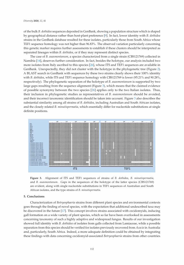
Diversity 2020, 12, 41
of the bulk B. dothidea sequences deposited in GenBank, showing a population structure which is shapedby geographical distance rather than host-plant preference [9]. In fact, lower identity with B. dothideastrains in the GenBank database resulted for these isolates, particularly those from South Africa whoseTEF1 sequence homology was not higher than 96.83%. The observed variation particularly concerningthis genetic marker requires further assessments to establish if these clusters should be interpreted asseparated lineages within B. dothidea, or if they may represent distinct species.
The case of B. auasmontanum, a species characterized from a single strain (CBS121769) collected inNamibia [14], deserves further consideration. In fact, besides the holotype, our analysis included twomore isolates from Italy ascribed to this species [26], whose ITS and TEF1 sequences are available inGenBank. Unexpectedly, they did not cluster with the holotype in the phylogenetic tree (Figure 2).A BLAST search in GenBank with sequences by these two strains clearly shows their 100% identitywith B. dothidea, while ITS and TEF1 sequence homology with CBS121769 is lower (95.21% and 90.28%,respectively). The phylogenetic separation of the holotype of B. auasmontanum is supported by twolarge gaps resulting from the sequence alignment (Figure 3), which means that the claimed evidenceof possible synonymy between the two species [26] applies only to the two Italian isolates. Thus,their inclusion in phylogenetic studies as representatives of B. auasmontanum should be avoided,and their incorrect taxonomic identification should be taken into account. Figure 3 also describes thesubstantial similarity among all strains of B. dothidea, including Australian and South African isolates,and the closely related B. minutispermatia, which essentially differ for nucleotide substitutions at singledefinite positions.
Figure 3. Alignment of ITS and TEF1 sequences of strains of B. dothidea, B. minutispermatia,and B. auasmontanum. Gaps in the sequences of the holotype of the latter species (CBS121769)are evident, along with single nucleotide substitutions in TEF1 sequences of Australian and SouthAfrican isolates, and the type strains of B. minutispermatia.
5. Conclusions
Characterization of Botryosphaeria strains from different plant species and environmental contextsgoes through the finding of novel species, with the expectation that additional undescribed taxa maybe discovered in the future [17]. This concept involves strains associated with cecidomyids, inducinggall formation on a wide variety of plant species, which so far have been overlooked in assessmentsconcerning taxonomy of such a highly adaptive and widespread fungus. Results of our investigationshowed full identity with B. dothidea of isolates from galls collected from Lamiaceae, while a possibleseparation from this species should be verified for isolates previously recovered from Acacia in Australiaand, particularly, South Africa. Indeed, a more adequate definition could be obtained by integratingthese findings with data concerning cecidomyid-associated Botryosphaeria strains from other countries.
112

Diversity 2020, 12, 41
Therefore, a more active cooperation among researchers currently working on this topic worldwide isto be encouraged, in order to shed further light on this unique biological association.
Supplementary Materials: The following are available online at http://www.mdpi.com/1424-2818/12/2/41/s1.Figure S1: Maximum-likelihood tree of ITS sequence. Figure S2: Maximum-likelihood tree of EF sequence.Figure S3: One of the 1000 most parsimonious trees resulting from the analysis of concatenated ITS andTEF1 sequences.
Author Contributions: Conceptualization, B.Z. and R.N.; methodology, A.B., B.Z., E.D.K., and S.O.; formal analysis,A.B. and S.O.; writing—original draft preparation, B.Z., R.N., and S.O.; writing—review and editing, B.Z., E.D.K.,and R.N.; funding acquisition, E.D.K. All authors have read and agreed to the published version of the manuscript.
Funding: This research received no external funding.
Acknowledgments: Authors thank Sarah Lucchesi (University of Southern Maine, Portland, USA) for revisingthe English style of this paper.
Conflicts of Interest: The authors declare no conflict of interest.
References
1. Adair, R.J.; Burgess, T.; Serdani, M.; Barber, P. Fungal associations in Asphondylia (Diptera: Cecidomyiidae)galls from Australia and South Africa: Implications for biological control of invasive acacias. Fungal Ecol.2009, 2, 121–134. [CrossRef]
2. Lebel, T.; Peele, C.; Veenstra, A. Fungi associated with Asphondylia (Diptera: Cecidomyiidae) galls onSarcocornia quinqueflora and Tecticornia arbuscula (Chenopodiaceae). Fungal Divers. 2012, 55, 143–154.[CrossRef]
3. Zimowska, B.; Viggiani, G.; Nicoletti, R.; Furmanczyk, A.; Becchimanzi, A.; Kot, I. First report of the gallmidge Asphondylia serpylli on thyme (Thymus vulgaris), and identification of the associated fungal symbiont.Ann. Appl. Biol. 2017, 171, 89–94. [CrossRef]
4. Bernardo, U.; Nugnes, F.; Gualtieri, L.; Nicoletti, R.; Varricchio, P.; Sasso, R.; Viggiani, G. A new gall midgespecies of Asphondylia (Diptera: Cecidomyiidae) inducing flower galls on Clinopodium nepeta (Lamiaceae)from Europe, its phenology, and associated fungi. Environ. Entomol. 2018, 47, 609–622. [CrossRef] [PubMed]
5. Slippers, B.; Crous, P.W.; Denman, S.; Coutinho, T.A.; Wingfield, B.D.; Wingfield, M.J. Combinedmultiple gene genealogies and phenotypic characters differentiate several species previously identified asBotryosphaeria dothidea. Mycologia 2004, 96, 83–101. [CrossRef]
6. Denman, S.; Crous, P.W.; Taylor, J.E.; Kang, J.-C.; Pascoe, I.; Wingfield, M.J. An overview of the taxonomichistory of Botryosphaeria, and a re-evaluation of its anamorphs based on morphology and ITS rDNA phylogeny.Stud. Mycol. 2000, 45, 129–140.
7. Park, I.; Sanogo, S.; Hanson, S.F.; Thompson, D.C. Molecular identification of Botryosphaeria dothidea as afungal associate of the gall midge Asphondylia prosopidis on mesquite in the United States. BioControl 2019,64, 209–219. [CrossRef]
8. Phillips, A.J.L.; Alves, A.; Abdollahzadeh, J.; Slippers, B.; Wingfield, M.J.; Groenewald, J.Z.; Crous, P.W. TheBotryosphaeriaceae: genera and species known from culture. Stud. Mycol. 2013, 76, 51–167. [CrossRef]
9. Marsberg, A.; Kemler, M.; Jami, F.; Nagel, J.H.; Postma-Smidt, A.; Naidoo, S.; Wingfield, M.J.; Crous, P.W.;Spatafora, J.W.; Hesse, C.N.; et al. Botryosphaeria dothidea: a latent pathogen of global importance to woodyplant health. Mol. Plant Pathol. 2017, 18, 477–488. [CrossRef]
10. Slippers, B.; Wingfield, M.J. Botryosphaeriaceae as endophytes and latent pathogens of woody plants:diversity, ecology and impact. Fungal Biol. Rev. 2007, 21, 90–106. [CrossRef]
11. Janson, E.M.; Peeden, E.R.; Stireman, J.O.; Abbot, P. Symbiont-mediated phenotypic variation withoutco-evolution in an insect–fungus association. J. Evol. Biol. 2010, 23, 2212–2228. [CrossRef] [PubMed]
12. Malagaris, P. Biology and ecology of Asphondylia coridothymi (Diptera: Cecidomyiidae) inducing galls onCoridothymus capitatus on the island of Samos, Greece. Acta Soc. Zool. Bohemicae 2011, 75, 239–251.
13. Viggiani, G.; Stinca, A. Infestations on oregano by the gall midge Asphondylia hornigi Wachtl. Prot. Colture2018, 4, 29–31.
14. Slippers, B.; Roux, J.; Wingfield, M.J.; van der Walt, F.J.J.; Jami, F.; Mehl, J.W.M.; Marais, G.J. Confronting theconstraints of morphological taxonomy in the Botryosphaeriales. Persoonia 2014, 33, 155–168. [CrossRef]
113

Diversity 2020, 12, 41
15. Xu, C.; Wang, C.S.; Ju, L.L.; Zhang, R.; Biggs, A.R.; Tanaka, E.; Li, B.; Sun, G.Y. Multiple locus genealogies andphenotypic characters reappraise the causal agent of apple ring rot in China. Fungal Divers. 2015, 71, 215–231.[CrossRef]
16. Ariyawansa, H.A.; Hyde, K.D.; Liu, J.K.; Wu, S.P.; Liu, Z.Y. Additions to Karst Fungi 1:Botryosphaeria minutispermatia sp. nov., from Guizhou Province, China. Phytotaxa 2016, 275, 35–44. [CrossRef]
17. Li, G.Q.; Liu, F.F.; Li, J.Q.; Liu, Q.L.; Chen, S.F. Botryosphaeriaceae from Eucalyptus plantations and adjacentplants in China. Persoonia 2018, 40, 63–95. [CrossRef]
18. Liang, L.Y.; Jiang, N.; Chen, W.Y.; Liang, Y.M.; Tian, C.M. Botryosphaeria qinlingensis sp. nov. causing oakfrogeye leaf spot in China. Mycotaxon 2019, 134, 463–473. [CrossRef]
19. Zhou, Y.P.; Zhang, M.; Dou, Z.P.; Zhang, Y. Botryosphaeria rosaceae sp. nov. and B. ramosa,new botryosphaeriaceous taxa from China. Mycosphere 2017, 8, 162–171. [CrossRef]
20. Zhou, Y.; Dou, Z.; He, W.; Zhang, X.; Zhang, Y. Botryosphaeria sinensia sp nov., a new species from China.Phytotaxa 2016, 245, 43–50. [CrossRef]
21. Crous, P.W.; Slippers, B.; Wingfield, M.J.; Rheeder, J.; Marasas, W.F.; Philips, A.J.; Alves, A.; Burgess, T.;Barber, P.; Groenewald, J.Z. Phylogenetic lineages in the Botryosphaeriaceae. Stud. Mycol. 2006, 55, 235–253.[CrossRef] [PubMed]
22. White, T.J.; Bruns, T.; Lee, S.J.W.T.; Taylor, J. Amplification and direct sequencing of fungal ribosomal RNAgenes for phylogenetic. In PCR Protocols, A Guide to Methods and Applications; Innis, M.A., Gelfand, D.H.,Sninsky, J.J., White, T.J., Eds.; Academic Press: New York, NY, USA, 1990; Volume 18, pp. 315–322.
23. Carbone, I.; Kohn, L.M. A method for designing primer sets for speciation studies in filamentous ascomycetes.Mycologia 1999, 91, 553–556. [CrossRef]
24. Edgar, R.C. MUSCLE: Multiple sequence alignment with high accuracy and high throughput.Nucleic Acids Res. 2004, 32, 1792–1797. [CrossRef] [PubMed]
25. Larsson, A. AliView: A fast and lightweight alignment viewer and editor for large datasets. Bioinformatics2014, 30, 3276–3278. [CrossRef]
26. Dissanayake, A.J.; Camporesi, E.; Hyde, K.D.; Yan, J.Y.; Li, X.H. Saprobic Botryosphaeriaceae, includingDothiorella italica sp. nov., associated with urban and forest trees in Italy. Mycosphere 2017, 8, 1157–1176.[CrossRef]
27. Wilgenbusch, J.C.; Swofford, D. Inferring evolutionary trees with PAUP*. Curr. Prot. Bioinformatics 2003,1, 6.4.1–6.4.28. [CrossRef]
28. Stamatakis, A. RAxML-VI-HPC: maximum likelihood-based phylogenetic analyses with thousands of taxaand mixed models. Bioinformatics 2006, 22, 2688–2690. [CrossRef]
29. Huelsenbeck, J.P.; Ronquist, F. MRBAYES: Bayesian inference of phylogenetic trees. Bioinformatics 2001,17, 754–755. [CrossRef]
30. Molecular Evolution, Phylogenetics and Epidemiology. Available online: http://tree.bio.ed.ac.uk/software/figtree/ (accessed on 25 November 2018).
31. Chen, S.; Pavlic, D.; Roux, J.; Slippers, B.; Xie, Y.; Wingfield, M.J.; Zhou, X.D. Characterization ofBotryosphaeriaceae from plantation-grown Eucalyptus species in South China. Plant Pathol. 2011, 60, 739–751.[CrossRef]
32. Abdollahzadeh, J.; Zare, R.; Phillips, A.J. Phylogeny and taxonomy of Botryosphaeria and Neofusicoccumspecies in Iran, with description of Botryosphaeria scharifii sp. nov. Mycologia 2013, 105, 210–220. [CrossRef]
© 2020 by the authors. Licensee MDPI, Basel, Switzerland. This article is an open accessarticle distributed under the terms and conditions of the Creative Commons Attribution(CC BY) license (http://creativecommons.org/licenses/by/4.0/).
114

diversity
Article
News from the Sea: A New Genus and Seven NewSpecies in the Pleosporalean Families Roussoellaceaeand Thyridariaceae
Anna Poli, Elena Bovio, Lucrezia Ranieri, Giovanna Cristina Varese * and Valeria Prigione
Mycotheca Universitatis Taurinensis, Department of Life Sciences and Systems Biology, University of Torino,Viale Mattioli 25, 10125 Torino, Italy; [email protected] (A.P.); [email protected] (E.B.);[email protected] (L.R.); [email protected] (V.P.)* Correspondence: [email protected]; Tel.: +39-011-670-5964
Received: 19 March 2020; Accepted: 3 April 2020; Published: 6 April 2020
Abstract: Nineteen fungal strains associated with the seagrass Posidonia oceanica, with thegreen alga Flabellia petiolata, and the brown alga Padina pavonica were collected in theMediterranean Sea. These strains were previously identified at the family level and hypothesised tobe undescribed species. Strains were examined by deep multi-loci phylogenetic and morphologicalanalyses. Maximum-likelihood and Bayesian phylogenies proved that Parathyridariella gen. nov.is a distinct genus in the family Thyriadriaceae. Analyses based on five genetic markers revealedseven new species: Neoroussoella lignicola sp. nov., Roussoella margidorensis sp. nov., R. mediterraneasp. nov., and R. padinae sp. nov. within the family Roussellaceae, and Parathyridaria flabelliaesp. nov., P. tyrrhenica sp. nov., and Parathyridariella dematiacea gen. nov. et sp. nov. within thefamily Thyridariaceae.
Keywords: marine fungi; new taxa; phylogeny; lignicolous fungi
1. Introduction
Marine fungi are a relevant and active component of the microbial communities that inhabit theoceans [1]. Fungi in the marine environment live as mutualists, parasites, pathogens and saprobes,and are pivotal to marine food webs because of the recycling of recalcitrant substrata [2]; besides, thesewidely dispersed organisms are a source of novel bioactive compounds [3].
Marine fungi have been recovered worldwide from a broad range of biotic and abiotic substrata,such as driftwood algae, sponges, corals, sediments, etc. [4,5]. Following the definition of Pang et al [6]that considered “a marine fungus” to be any fungus retrieved repeatedly from marine environmentand that reproduces in the marine environment, Jones et al. [7] listed 1680 fungal species belongingto 693 genera, 223 families, 87 orders, 21 classes and six phyla. However, considering that the totalnumber of marine fungi has been estimated to exceed 10,000 taxa [8], fungal diversity remains largelyundescribed. With more than 900 species [9], the Ascomycota are the dominant fungal phylum inthe sea; the most represented lineages include the order Pleosporales (class Dothideomycetes) with36 families, 95 genera and 194 species described to date (www.marinefungi.org).
In recent surveys aimed to uncover the underwater fungal diversity, 19 unidentified Roussoellaceaewere isolated from several substrates, as follows: 12 from the brown alga Padina pavonica (L.)Thivy [10], 4 from the green alga Flabellia petiolata (Turra) Nizamuddin [11], 2 from the seagrassPosidonia oceanica (L.) [12] Delile, and 1 from the Atlantic sponge Dysidea fragilis (Montagu) [13].The Roussoellaceae is a well-resolved family in the Pleosporales [14]. Others [15] have treated thefamily Roussoellaceae as a synonym of Thyridariaceae, based on phylogenetic affinities. However,following the discovery of new genera in this group, delineated by high resolution multi-locus
Diversity 2020, 12, 144; doi:10.3390/d12040144 www.mdpi.com/journal/diversity115
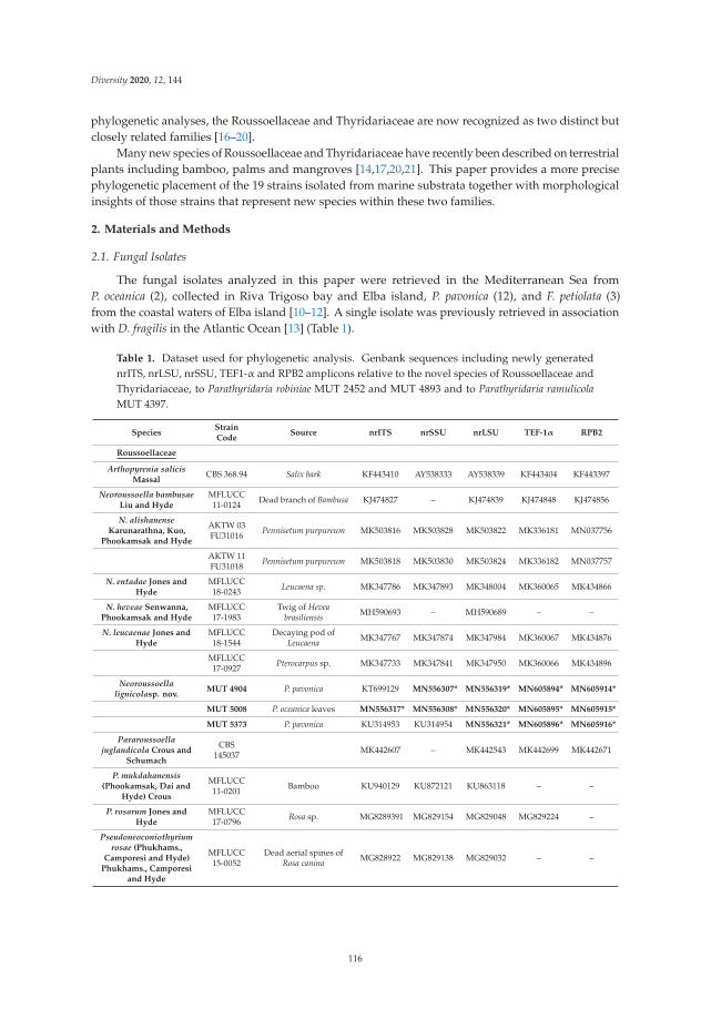
Diversity 2020, 12, 144
phylogenetic analyses, the Roussoellaceae and Thyridariaceae are now recognized as two distinct butclosely related families [16–20].
Many new species of Roussoellaceae and Thyridariaceae have recently been described on terrestrialplants including bamboo, palms and mangroves [14,17,20,21]. This paper provides a more precisephylogenetic placement of the 19 strains isolated from marine substrata together with morphologicalinsights of those strains that represent new species within these two families.
2. Materials and Methods
2.1. Fungal Isolates
The fungal isolates analyzed in this paper were retrieved in the Mediterranean Sea fromP. oceanica (2), collected in Riva Trigoso bay and Elba island, P. pavonica (12), and F. petiolata (3)from the coastal waters of Elba island [10–12]. A single isolate was previously retrieved in associationwith D. fragilis in the Atlantic Ocean [13] (Table 1).
Table 1. Dataset used for phylogenetic analysis. Genbank sequences including newly generatednrITS, nrLSU, nrSSU, TEF1-α and RPB2 amplicons relative to the novel species of Roussoellaceae andThyridariaceae, to Parathyridaria robiniae MUT 2452 and MUT 4893 and to Parathyridaria ramulicolaMUT 4397.
SpeciesStrainCode
Source nrITS nrSSU nrLSU TEF-1α RPB2
Roussoellaceae
Arthopyrenia salicisMassal
CBS 368.94 Salix bark KF443410 AY538333 AY538339 KF443404 KF443397
Neoroussoella bambusaeLiu and Hyde
MFLUCC11-0124 Dead branch of Bambusa KJ474827 – KJ474839 KJ474848 KJ474856
N. alishanenseKarunarathna, Kuo,
Phookamsak and Hyde
AKTW 03FU31016 Pennisetum purpureum MK503816 MK503828 MK503822 MK336181 MN037756
AKTW 11FU31018 Pennisetum purpureum MK503818 MK503830 MK503824 MK336182 MN037757
N. entadae Jones andHyde
MFLUCC18-0243 Leucaena sp. MK347786 MK347893 MK348004 MK360065 MK434866
N. heveae Senwanna,Phookamsak and Hyde
MFLUCC17-1983
Twig of Heveabrasiliensis MH590693 – MH590689 – –
N. leucaenae Jones andHyde
MFLUCC18-1544
Decaying pod ofLeucaena MK347767 MK347874 MK347984 MK360067 MK434876
MFLUCC17-0927 Pterocarpus sp. MK347733 MK347841 MK347950 MK360066 MK434896
Neoroussoellalignicolasp. nov.
MUT 4904 P. pavonica KT699129 MN556307* MN556319* MN605894* MN605914*
MUT 5008 P. oceanica leaves MN556317* MN556308* MN556320* MN605895* MN605915*
MUT 5373 P. pavonica KU314953 KU314954 MN556321* MN605896* MN605916*
Pararoussoellajuglandicola Crous and
Schumach
CBS145037 MK442607 – MK442543 MK442699 MK442671
P. mukdahanensis(Phookamsak, Dai and
Hyde) Crous
MFLUCC11-0201 Bamboo KU940129 KU872121 KU863118 – –
P. rosarum Jones andHyde
MFLUCC17-0796 Rosa sp. MG8289391 MG829154 MG829048 MG829224 –
Pseudoneoconiothyriumrosae (Phukhams.,
Camporesi and Hyde)Phukhams., Camporesi
and Hyde
MFLUCC15-0052
Dead aerial spines ofRosa canina MG828922 MG829138 MG829032 – –
116

Diversity 2020, 12, 144
Table 1. Cont.
SpeciesStrainCode
Source nrITS nrSSU nrLSU TEF-1α RPB2
Roussoella chiangrainaPhookamsak, Liu and
Hyde
MFLUCC10-0556 Dead branch of bamboo KJ474828 – KJ474840 KJ474849 KJ474857
R. doimaesalongensisThambug. and Hyde
MFLUCC14–0584 Dead branch of bamboo KY026584 – KY000659 KY651249 KY678394
R. elaeicola Konta. andHyde
MFLUCC15-0276a
Dead petiole of Elaeisguineensis MH742329 – MH742326 – –
MFLUCC15-0276b
Dead petiole of Elaeisguineensis MH742330 – MH742327 – –
R. euonymi Crous andAkulov
CBS143426
Fallen branches ofEuonymus europaeus MH107915 – MH107961 – MH108007
R. hysterioides (Ces.)Höhn.
CBS 546.94 Phyllostachys KF443405 AY642528 KF443381 KF443399 KF443392
R. intermedia Ju, Rogersand Huhndorf
CBS 170.96 Bamboo KF443407 KF443390 KF443382 KF443398 KF443394
R. japanensisKaz.Tanaka, Liu and Hyde
MAFF239636 Twigs of Sasa veitchii KJ474829 AB524480 AB524621 AB539114 AB539101
R. kunmingensis Jiang,Phookamsak and Hyde
KUMCC18-0128 Dead bamboo MH453491 – MH453487 MH453480 MH453484
R. mangrovei Phukhams.and Hyde
MFLUCC16-0424
Dead branches ofRhizophora MH025951 – MH023318 MH028246 MH028250
Roussoellamargidorensis sp. nov.
MUT 5329 P. pavonica KU314944 MN556309* MN556322* MN605897* MN605917*
Roussoella mediterraneasp. nov.
MUT 5306 P. pavonica KU255054 MN556310* MN556323* MN605898* MN605918*
MUT 5369 P. pavonica KU314947 KU314948 MN556324* MN605899* MN605919*
R. mexicana Crous andYáñez-Mor.
CPC 25355 Leaf spots of Coffeaarabica KT950848 – KT950862 – –
R. neopustulans Dai, Liuand Hyde
MFLUCC11-0609 Bamboo KJ474833 – KJ474841 KJ474850
MFLUCC12-0003 Bamboo KU940130 KU872122 KU863119 – –
R. nitidula Sacc. andPaol.
MFLUCC11-0182 Bamboo KJ474835 – KJ474843 KJ474852 KJ474859
MFLUCC11-0634 Bamboo KJ474834 – KJ474842 KJ474851 KJ474858
Roussoella padinae sp.nov.
MUT 5341 P. pavonica KU158153 KU158176 MN556325* MN605900* MN605920*
MUT 5365 P. pavonica KU158170 KU158179 MN556326* MN605901* MN605921*
MUT 5503 P. pavonica KU158170 MN556312* MN556327* MN605902* MN605922*
R. pseudohysterioidesDai and Hyde
MFLUCC13-0852 Bamboo KU940131 KU872123 KU863120 KU940198 –
R. pustulans (Ellis andEverh.) Ju, Rogers and
HuhndorfKT 1709 Culms of Sasa kurilensis KJ474830 AB524482 AB524623 AB539116 AB539103
R. scabrispora (Höhn.)Aptroot
MFLUCC11-0624 Bamboo KJ474836 – KJ474844 KJ474853 KJ474860
RSC Bamboo KX650566 – KX650566 KX650537 –
R. siamensisPhookamsak, Liu and
Hyde
MFLUCC11-0149 Bamboo KJ474837 KU872125 KJ474845 KJ474854 KJ474861
R. thailandica Dai, Liuand Hyde
MFLUCC11-0621 Bamboo KJ474838 – KJ474846 –
R. tuberculata Dai andHyde
MFLUCC13-0854 Bamboo KU940132 KU872124 KU863121 KU940199
R. verrucispora Kaz.Tanaka, Liu and Hyde
CBS125434 Sasa kurilensis KJ474832 AB52448 AB524622 AB539115 –
117

Diversity 2020, 12, 144
Table 1. Cont.
SpeciesStrainCode
Source nrITS nrSSU nrLSU TEF-1α RPB2
R. yunnanensis Jiang,Phookamsak and Hyde
KUMCC18-0115 Dead bamboo MH453492 – MH453488 MH453481 –
Roussoellopsismacrospora (Hino and
Katum.) HinoandKatum
MFLUCC12-0005 Bamboo KJ739604 KJ739608 KJ474847 KJ474855 KJ474862
Ro. tosaensis (Hino andKatum.) Hino and
KatumKT 1659 Culms of bamboo – AB524484 AB524625 AB539117 AB539104
Thyridariaceae
Cycasicola goaensisJones and Hyde
MFLUCC17-0754 Cycas sp. MG828885 MG829112 MG829001 MG829198 –
C. leucaenae Jones andHyde
MFLUCC17-0914 Leucaena leucocephala MK34772 MK347833 MK347942 MK360046 MK434900
Liua muriformisPhookamsak, Jiang and
Hyde
KUMCC18-0177
Dead hanging branchesof Lonicera maackii MK433599 MK433595 MK433598 MK426798 MK426799
Parathyridariapercutanea (Ahmed,
Stevens, van de Sandeand de Hoog) Jaklitsch
and Voglmayr
CBS 868.95 Human KF322118 KF366451 KF366449 KF407987 KF366452
CBS128203 Human KF322117 KF366450 KF366448 KF407988 KF366453
P. ramulicola Jaklitsch,Fourn and Voglmayr
CBS141479 Twigs of Ribes rubrum NR_147657 KX650514 KX650565 KX650536 KX650584
MUT 4397 P. oceanica KC339235 MN556311* KF636775 MN605913* MN605933*
P. robiniaeMapook,Camporesi and Hyde
MFLUCC14-1119
Dead branch of Robiniapseudoacacia KY511142 – KY511141 KY549682 –
MUT 2452 Dysidea fragilis MG813183 MN556312* MG816491 MN605903* MN605923*
MUT 4893 P. pavonica KM355998 KM355993 MN556328* MN605904* MN605924*
Parathyridaria flabelliaesp. nov.
MUT 4859 F. petiolata KR014355 KT587315 KP671716 MN605909* MN605929*
MUT 4886 F. petiolata KR014358 KT587317 KP671720 MN605910* MN605930*
Parathyridariatyrrhenica sp. nov.
MUT 4966 F. petiolata, KR014366 KT587309 KP671740 MN605911* MN605931*
MUT 5371 P. pavonica KU314951 KU314952 MN556329* MN605912* MN605932*
Parathyridarielladematiacea sp. nov.
MUT 4419 P. oceanica rhizomes KC339245 MN556313* KF636786 MN605905* MN605925*
MUT 4884 F. petiolata MN556317* KT587329 KP671726 MN605906* MN605926*
MUT 5310 P. pavonica KU255057 MN556314* MN556330* MN605907* MN605927*
MUT 5381 P. pavonica KU314959 KU314960 MN556331* MN605908* MN605928*
Thyridaria acaciae(Crous and Wingf.)
Jaklitsch and Voglmayr
CBS138873 Leaves of Acacia tortilis KP004469 – KP004497 – –
T. broussonetiae (Sacc.)Traverso
TB Hippocrepis emerus KX650567 – KX650567 KX650538 KX650585
TB1 Amorpha fruticosa KX650568 KX650515 KX650568 KX650539 KX650586
Thyridariellamahakoshae Devadatha,
Sarma, Wanas., Hydeand Jones
NFCCI4215
Decaying woodAvicennia marina MG020435 MG020441 MG020438 MG023140 MG020446
Th. mangroveiDevadatha, Sarma,
Hyde, Wanas. and Jones
NFCCI4213
Decaying woodAvicennia marina MG020434 MG020440 MG020437 MG020443 MG020445
NFCCI4214
Decaying woodAvicennia marina MG020436 MG020442 MG020439 MG020444 MG020447
118

Diversity 2020, 12, 144
Table 1. Cont.
SpeciesStrainCode
Source nrITS nrSSU nrLSU TEF-1α RPB2
Occultibambusaceae
Occultibambusabambusae Dai and Hyde
MFLUCC11-0394 Bamboo KU940124 – KU863113 KU940194 KU940171
MFLUCC13-0855 Bamboo KU940123 KU872116 KU863112 KU940193 KU940170
Ohleriaceae
Ohleria modesta Fuckel MGC KX650562 – KX650562 KX650533 KX650582
OM Branches ofChamaecytisus proliferus KX650563 KX650513 KX650563 KX650534 KX650583
Torulaceae
Dendryphion europaeumCrous and Schumacher
CPC 22943 Heracleum sphondylium KJ869146 – KJ869203 – –
Torula herbarum (Pers.)Link
CBS111855 n.a. KF443409 KF443391 KF443386 KF443403 KF443396
CBS 595.96 KF443408 KF443387 KF443385 KF443402 KF443395
Torula hollandica Crous CBS 220.69 Delphiniumdead stem KF443406 – MH877717 – –
* = newly generated sequences; n.a. = not available.
The strains investigated were originally isolated on Corn Meal Agar medium supplementedwith sea salts (CMASS; 3.5% w/v sea salt mix, Sigma-Aldrich, Saint Louis, USA, in ddH2O) and arepreserved at the Mycotheca Universitatis Taurinensis (MUT), Italy.
2.2. Morphological Analysis
All isolates were pre-grown on Malt Extract Agar-sea water (MEASW; 20 g malt extract, 20 g glucose,2 g peptone, 20 g agar in 1 L of sea water) for one month at 24 ◦C prior to inoculation in triplicate ontonew Petri dishes (9 cm Ø) containing (i) MEASW, (ii) Oatmeal Agar-sea water (OASW; 30 g oatmeal,20 g agar in 1 L of sea water), or iii) Potato Dextrose Agar-sea water (PDASW; 4 g potato extract, 20 gdextrose, 20 g agar in 1 L of sea water). Petri dishes were incubated at 15 and/or 24 ◦C. The colonygrowth was monitored periodically for 28 days. Macroscopic and microscopic traits, were assessed forstrains grown on MEASW at the end of the incubation period.
In an attempt to induce sporulation, sterile pieces of Quercus ruber cork and Pinus pinaster wood(species autochthonous to the Mediterranean area) were placed on 3 week old fungal colonies grown onMEASW ([22], modified). Petri dishes were further incubated for 4 weeks at 24 ◦C. Subsequently, corkand wood pieces were transferred to 50 mL tubes containing 20 mL of sterile sea water. Samples wereincubated at 24 ◦C for one month. In parallel, the strains were also plated on Syntetic Nutrient Agar-seawater (SNASW; 1 g KH2PO4, 1 g KNO3, 0.5 g MgSO4 • 7H2O, 0.5 g KCl, 0.2 g glucose, 0.2 g sucrose,20 g agar in 1 L of sea water) supplemented with sterile pine needles. Petri dishes were incubated at24 ◦C for one month.
Morphological structures were observed, and images captured using an optical microscope(Leica DM4500B, Leica microsystems GmbH, Wetzlar) equipped with a camera (Leica DFC320,Leica microsystems GmbH, Wetzlar). Macro- and microscopic features were compared with theavailable description of Roussoellaceae and Thyridariaceae [14,15,17,18,20].
2.3. DNA Extraction, PCR Amplification, and Data Assembling
Genomic DNA was extracted from about 100 mg of fresh mycelium grown on MEASW plates.Mycelium was disrupted by the mean of a MM400 tissue lyzer (Retsch GmbH, Haan, Germany) andDNA extracted using a NucleoSpin kit (Macherey Nagel GmbH, Duren, DE, USA) following themanufacturer’s instructions. The quality and quantity of DNA were measured spectrophotometrically(Infinite 200 PRO NanoQuant; TECAN, Männedorf); DNA was stored at −20 ◦C.
119

Diversity 2020, 12, 144
The partial sequences of five genetic markers were amplified by PCR. Primer pairs ITS1/ITS4 [23],LR0R/LR7 [24], NS1/NS4 [23] were used to amplify the internal transcribed spacers, including the 5.8SrDNA gene (nrITS), 28S large ribosomal subunit (nrLSU) and 18S small ribosomal subunit (nrSSU).The translation elongation factor (TEF1α) and RNA polymerase II subunit (RPB2) were amplified byusing primer pairs EF1-1018F/EF1-1620R [25] and fRPB2-5F/fPB2-7R [26].
Amplifications were run in a T100 Thermal Cycler (Bio-Rad, Hercules, CA, USA) programmed asdescribed in Table 2. Reaction mixtures consisted of 20–40 ng DNA template, 10× PCR Buffer (15 mMMgCl2, 500 mM KCl, 100 mM Tris-HCl, pH 8.3), 200 μM each dNTP, 1 μM each primer, 2.5 U Taq DNAPolymerase (Qiagen, Chatsworth, CA, USA), in 50 μL final volume. For problematic cases, additionalMgCl2 and/or 2.5% DMSO facilitated the reaction.
Table 2. Primers and PCR conditions used to amplify specific gene marker.
Forward and Reverse Primers Thermocycler Conditions References
ITS ITS1- ITS4 95 ◦C: 5 min, (95 ◦C: 40 s, 55 ◦C: 50 s,72 ◦C: 50 sec) × 35 cycles; 72 ◦C: 8 min; 4 ◦C:∞ [23]
LSU LR0R-LR7 95 ◦C: 5 min, (95 ◦C: 1 min, 50 ◦C: 1 min,72 ◦C: 2 min) × 35 cycles; 72 ◦C: 10 min; 4 ◦C:∞ [24]
SSU NS1-NS4 95 ◦C: 5 min, (95 ◦C: 1 min, 50 ◦C: 1 min,72 ◦C: 2 min) × 35 cycles; 72 ◦C: 10 min; 4 ◦C:∞ [23]
TEF-1α 1018F/1620R 95 ◦C: 5 min, (95 ◦C: 1 min, 50 ◦C: 1 min;72 ◦C: 2 min) × 40 cycles, 72 ◦C: 10 min; 4 ◦C:∞ [25]
RPB2 fRPB2-5F/fPB2-7cR 94 ◦C: 3 min, (94 ◦C: 30 s; 55 ◦C: 30 s; 72 ◦C: 1min) × 40 cycles, 72 ◦C: 10 min; 4 ◦C:∞ [26]
Amplicons, together with a GelPilot 1 kb plus DNA Ladder, were visualized on a 1.5% agarosegel stained with 5 mL 100 mL−1 ethidium bromide; PCR products were purified and sequenced at theMacrogen Europe Laboratory (Madrid, Span). The resulting Applied Biosystem (ABI) chromatogramswere inspected, trimmed and assembled to obtain consensus sequences using Sequencer 5.0 (GeneCodesCorporation, Ann Arbor, Michigan, USA http://www.genecodes.com). Newly generated sequenceswere deposited in GenBank (Table 1).
2.4. Sequence Alignment and Phylogenetic Analysis
A dataset consisting of nrSSU, nrITS, nrLSU, TEF1α and RPB2 was assembled on the basis ofBLASTn results and of recent phylogenetic studies focused on Roussoellaceae and Thyridariaceae [18,20].Reference sequences were retrieved from GenBank (Table 1).
Sequences were aligned using MUSCLE (default conditions for gap openings and gap extensionpenalties), implemented in MEGA v. 7.0 (Molecular Evolutionary Genetics Analysis), visually inspectedand trimmed by TrimAl v. 1.2 (http://trimal.cgenomics.org) to delimit and discard ambiguously alignedregions. Since no incongruence was observed among single-loci phylogenetic trees, alignments wereconcatenated into a single data matrix with SequenceMatrix [27]. The best evolutionary model underthe Akaike Information Criterion (AIC) was determined with jModelTest 2 [28].
Phylogenetic inference was estimated using Maximum Likehood (ML) and Bayesian Inference (BI)criteria. The ML analysis was generated using RAxML v. 8.1.2 [29] under GTR + I + G evolutionarymodel and 1000 bootstrap replicates. Support values from bootstrapping runs (MLB) were mappedon the globally best tree using the “-f a” option of RAxML and “-x 12345” as a random seed toinvoke the novel rapid bootstrapping algorithm. BI was performed with MrBayes 3.2.2 [30] with thesame substitution model (GTR + I + G). The alignment was run for 10 million generations with twoindependent runs each containing four Markov Chains Monte Carlo (MCMC) and sampling every100 iterations. The first 25% of generated trees were discarded as “burn-in”. A consensus tree wasgenerated using the “sumt” function of MrBayes and Bayesian posterior probabilities (BPP) werecalculated. Consensus trees were visualized in FigTree v. 1.4.2 (http://tree.bio.ed.ac.uk/software/figtree).
120

Diversity 2020, 12, 144
Two strains of Occutibambusa bambusae (Occultibambusaceae) were used to root the tree. Due totopological similarity of the two resulting trees, only ML analysis with MLB and BPP values wasreported (Figure 1).
Figure 1. Phylogram generated from RAxML analysis based on a combined dataset of nrITS,nrSSU, nrLSU, TEF1α and RPB2 partial sequences. The tree is rooted to Occultibambusa bambusae.Branch numbers indicate BYPP/MLB values; Bar = expected changes per site (0.06).
DNA diagnostic characters were visually identified by the presence of heterozygous bases.For each locus, aligned sequences of the individual clusters containing new species, were inspected.Nucleotide diversities of the novel species were annotated when occurred (Tables S1–S18).
Sequence alignments and phylogenetic trees were deposited in TreeBASE (http://www.treebase.org,submission number S24773).
121

Diversity 2020, 12, 144
Following phylogenetic tree inspection, isolates that clustered in the same group and that derivedfrom the same substrate were subjected to PCR-fingerprinting by using the micro- and mini-satelliteprimers (GTG)5 and M13 [31,32] to exclude duplicates from further analysis. DNA fingerprintswere visualized with 1.5% agarose gel stained with 5 mL 100 mL−1 ethidium bromide while aGelPilot 1 kb plusDNA Ladder was used as a reference. Images were acquired with a Gel Doc1000System (Bio-Rad, Hercules, CA, USA) and fingerprints analyzed using Bionumerics v 7.6 (http://www.applied-maths.com).
3. Results
3.1. Phylogenetic Inference
Preliminary analyses carried out individually with nrITS, nrSSU, nrLSU, TEF1α and RPB2 denotedno incongruence in the topology of the single-locus trees. The combined five-markers dataset—builton the basis of BLASTn results and of recent phylogenetic studies [18,20]—consisted of 81 taxa(including MUT isolates) that represented 16 genera and 56 species (Table 1). A total of 63 sequences(2 nrITS, 8 nrSSU, 13 nrLSU, 20 TEF1α and 20 RPB2) were newly generated while 261 were retrievedfrom GenBank.
The combined dataset had an aligned length of 3390 characters, of which 1683 were constant,657 were parsimony-uninformative and 1050 parsimony informative (TL = 218, CI = 0.422018,RI = 0.825243, RC = 0.348267, HI = 0.877952).
Strains MUT 4893 and MUT 2452 were identified as Parathyridaria robiniae, the rest of the strainsrepresented seven new species and one new genus (Figure 1). Parathyridaria tyrrhenica sp. nov.(MUT 5371 and MUT 4966) formed a sister clade to Parathyridaria flabelliae sp. nov. (MUT 4859 andMUT 4886) with high statistical support (BYPP = 1.00; MLB = 100%); these two novel species are closelyrelated to P. ramulicola (BYPP = 1.00; MLB = 100%) and clustered with other Parathyridaria speciesin the Thyridariaeae family. Within this family, four isolates (MUT 5310, MUT 5381, MUT 4419 andMUT 4884) clustered together with the genera Thyridariella, Liua and Cycasicola, and formed a stronglysupported monophyletic lineage (BYPP = 1.00; MLB = 100%). Therefore, we have introduced the novelgenus Parathyridariella, typified by the new species Parathyridariella dematiacea sp. nov.
The three strains, MUT 4904, MUT 5373 and MUT 5008, represented a novel species Neoroussoellalignicola sp. nov. and formed an independent and robust clade (BYPP = 1.00; MLB = 100%), within theNeoroussoella group in the Roussoellaceae.
Two sister clades within the Roussoella group were represented by the new species Roussoellapadinae sp. nov. (MUT 5503, MUT 5341 and MUT 5365) and Roussoella mediterranea sp. nov. (MUT 5306and MUT 5369). Finally, MUT 5329 Roussoella margidorensis sp. nov. clustered together with R. nitidula,R. pseudohysterioides, R. thailandica and R. tubercolata (BYPP = 0.99; MLB = 71%) but was phylogeneticallydistant from these species.
Nucleotide divergence between each novel species and members of the same clusters wereannotated for each locus, when occurred (Tables S1–S18).
3.2. Taxonomy
Parathyridariella gen. nov. V. Prigione, A. Poli, E. Bovio and G.C. VareseMYCOBANK: MB 832836Type species. Parathyridariella dematiacea sp. nov.Etymology. In reference to the phylogenetic proximity to the genus Thyridariella.Phylogenetic placement. Thyridariaceae, Sordariomycetes, Ascomycota. The genus Parathyridariellagen. nov. clusters together with genera Cycasicola, Liua and Thyridariella (Figure 1).
Parathyridariella dematiacea sp. nov. V. Prigione, A. Poli, E. Bovio and G.C. VareseMYCOBANK: MB 832837Figure 2
122

Diversity 2020, 12, 144
Type. Italy, Tuscany, Mediterranean Sea, Elba Island (LI), Ghiaie ISL, 14–15 m depth, 42◦49’04”N,10◦19’20”E, form the green alga Flabellia petiolata, 20 March 2010, R. Mussat-Sartor and N. Nurra,MUT 4884 holotype, living culture permanently preserved in metabolically inactive state bydeep-freezing at Mycotheca Universitatis Taurinensis (MUT).
Additional material examined. Italy, Ligury, Mediterranean Sea, Riva Trigoso, Punta Manara (GE),5–21 m depth, 44◦15′08.62”N 9◦24′17.64”E, from the seagrass Posidonia oceanica, March 2008, MUT 4419.
Etymology. In reference to the color of the colony on culture media.Description. Growing actively on Pinus pinaster and Quercus ruber cork. Showing a floccose
growth mainly on Pinus pinaster. Hyphae 2.8–4.8 μm wide, septate, hyaline to lightly pigmented.Chlamydospores numerous, mostly in chain, intercalary or solitary, globose to subglobose, from brownishto dark brown, 7–10 × 6–8 μm diameter.
Sexual morph not observed. Asexual morph with differentiated conidiogenesis not observed.Colony description. Colonies on MEASW attaining 28–34 mm diam after 28 days at 24 ◦C,
mycelium from dark grey/black to dark green, dense with radial grooves and concentric rings,submerged edges; reverse dark green. Brown exudate present above the concentric rings. Growth onOASW reaching 40–54 mm diam at 24 ◦C and 21–29 mm diam at 15 ◦C; colonies on PDA attaining36–49 mm diam and 15.5–22.5 mm diam at 24 ◦C and 15 ◦C, respectively.
Figure 2. Parathyridariella dematiacea sp. nov. 28-days-old colony at 21 ◦C on MEASW (A) andreverse (B); solitary (C) and in chain (D) chlamydospores. Scale bars: 10 μm (C, D).
Parathyridaria tyrrhenica sp. nov. A. Poli, V. Prigione, E. Bovio and G.C. VareseMYCOBANK: MB 832838Figure 3
123

Diversity 2020, 12, 144
Type. Italy, Tuscany, Mediterranean Sea, Elba Island (LI), Ghiaie ISL, 14–15 m depth, 42◦49’04”N,10◦19’20”E, from the brown alga Padina pavonica, March 2010, R. Mussat-Sartor and N. Nurra, MUT5371 holotype, living culture permanently preserved in metabolically inactive state by deep-freezingat MUT.
Additional material examined. Italy, Tuscany, Mediterranean Sea, Elba Island (LI), Ghiaie ISL,14–15 m depth, 42◦49’04”N, 10◦19’20”E, from the green alga Flabellia petiolata, March 2010,R. Mussat-Sartor and N. Nurra, MUT 4966.
Etymology. In reference to Tyrrhenian Sea.Description. Growing actively on Pinus pinaster wood and Quercus ruber cork. Hyphae 5 μm
diameter, septate, hyaline to brownish, sometimes wavy or swollen, forming hyphal strands.Sexual morph not observed. Asexual morph with differentiated conidiogenesis: not observed.Colony description. Colonies growing on MEASW, reaching 10 mm diam after 28 days, at 21 ◦C,
mycelium funiculose, yellowish, lightly ochre at the edges; reverse light yellow, lighter at the edges.Growth on OASW reaching 48–50 mm diam at 24 ◦C and 26–29 mm diam at 15 ◦C; colonies on PDAattaining 31–46 mm diam and 16–19 mm diam at 24 ◦C and 15 ◦C, respectively.
Figure 3. Parathyridaria tyrrhenica sp. nov. 28-days-old colony at 21 ◦C on MEASW (A) and reverse (B);mycelium (C), black and white arrows indicate hyphal strands and wavy hyphae, respectively. Scale bar:10 μm.
Parathyridaria flabelliae sp. nov. E. Bovio, A. Poli, V. Prigione and G.C. VareseMYCOBANK: MB 832839
124

Diversity 2020, 12, 144
Figure 4Type. Italy, Tuscany, Mediterranean Sea, Elba Island (LI), Ghiaie ISL, 14–15 m depth, 42◦49’04”N,
10◦19’20”E, from the green alga Flabellia petiolata, March 2010, R. Mussat-Sartor and N. Nurra, MUT4859 holotype, living culture permanently preserved in metabolically inactive state by deep-freezingat MUT.
Additional material examined. Italy, Tuscany, Mediterranean Sea, Elba Island (LI), Ghiaie ISL,14–15 m depth, 42◦49’04”N, 10◦19’20”E, from the green alga Flabellia petiolata, March 2010, R.Mussat-Sartor and N. Nurra, MUT 4886.
Etymology. In reference to the original substratum, the green alga Flabellia petiolata.Description. Growing actively on Pinus pinaster and on Quercus ruber cork. Hyphae 2.6−5 μm
wide, septate and hyaline. Chlamydospores numerous, globose or subglobose, from light to dark brown,unicellular (4 × 5 μm diameter) and multicellular (up to four-celled; 8 × 12 μm diameter).
Sexual morph not observed. Asexual morph with differentiated conidiogenesis not observed.Colony description. Colonies growing on MEASW, reaching 37–44 mm diam after 28 days at
21 ◦C, funiculose, whitish with submerged edges; reverse brown in the middle, lighter at edges.Growth on OASW reaching 60 mm diam at 24 ◦C and 33–35 mm diam at 15 ◦C; colonies on PDAattaining 53–64 mm diam and 23–24 mm diam at 24 ◦C and 15 ◦C, respectively.
Figure 4. Parathyridaria flabelliae sp. nov. 28-days-old colony at 21 ◦C on MEASW (A) and reverse (B);unicellular and multicellular chlamydospores (C). Scale bar: 10 μm.
Neoroussoella lignicola sp. nov. A. Poli, E. Bovio, V. Prigione and G.C. VareseMYCOBANK: MB 832840
125

Diversity 2020, 12, 144
Figure 5Type. Italy, Tuscany, Mediterranean Sea, Elba Island (LI), Margidore ISL, 14–15 m depth,
UTM WGS84 42◦45’29”N, 10◦18’24”E, from the brown alga Padina pavonica, March 2010, R. Mussat-Sartorand N. Nurra, MUT 5373 holotype, living culture permanently preserved in metabolically inactivestate by deep-freezing at MUT.
Additional material examined. Italy, Tuscany, Mediterranean Sea, Elba Island (LI), Margidore ISL,14–15 m depth, UTM WGS84 42◦45’29”N, 10◦18’24”E, from the brown alga Padina pavonica, March2010, R. Mussat-Sartor and N. Nurra, MUT 4904.
Italy, Tuscany, Mediterranean Sea, Elba Island (LI), Margidore ISL, 14–15 m depth, UTM WGS8442◦45’29”N, 10◦18’24”E, from the seagrass Posidonia oceanica, March 2010, R. Mussat-Sartor andN. Nurra, MUT 5008.
Etymology. In reference to the lignicolous behavior.Description. Growing efficiently on Pinus pinaster wood. Hyphae 2–4.4 μm wide, septate,
hyaline, assuming toruloid aspect when growing into wood vessels and forming chains of two-celledchlamydospores which, at maturity, protrude from the vessels. Chlamydospores 7.4 × 5.2 μm, from lightto dark brown, globose or subglobose.
Sexual morph not observed. Asexual morph with differentiated conidiogenesis not observed.Colony description. Colonies growing on MEASW, reaching 28–29 mm diam after 28 days at
21◦ C, from grey to dark green, floccose with irregular edges, reverse dark grey. Clear exudate oftenpresent. Growth on OASW reaching 27–40 mm diam at 24 ◦C and 14.5–26 mm diam at 15 ◦C; colonieson PDA attaining 38–45 mm diam and 19–29 mm diam at 24 ◦C and 15 ◦C, respectively.
Figure 5. Neoroussoella lignicola sp. nov. 28-days-old colony at 21 ◦C on MEASW (A) and reverse (B);two-celled chlamydospores inside wood vessels (C). Scale bar: 10 μm.
Roussoella margidorensis sp. nov. E. Bovio, V. Prigione, A. Poli and G.C. VareseMYCOBANK: MB 832841Figure 6
126

Diversity 2020, 12, 144
Type. Italy, Tuscany, Mediterranean Sea, Elba Island (LI), Margidore ISL, 14–15 m depth,UTM WGS84 42◦45’29”N, 10◦18’24”E, from the brown alga Padina pavonica, March 2010, R. Mussat-Sartorand N. Nurra, MUT 5329 holotype, living culture permanently preserved in metabolically inactivestate by deep-freezing at MUT.
Etymology. In reference to the area of origin, Margidore.Description. Growing actively on Pinus pinaster wood. Hyphae approx. 2 μm wide, septate,
brownish.Sexual morph not observed. Asexual morph and differentiated conidiogenesis not observed.Colony description. Colonies growing on MEASW, attaining 33–34 mm diam after 28 days at
21 ◦C; whitish, lighter to the edge, umbonate in the middle, reverse ochre. Caramel diffusible pigmentproduced. Growth on OASW reaching 45 mm diam at 24 ◦C and 27 mm diam at 15 ◦C; colonies onPDA attaining 45 mm diam and 23 mm diam at 24 ◦C and 15 ◦C, respectively.
Figure 6. Roussoella margidorensis sp. nov. 28-days-old colony at 21 ◦C on MEASW (A) and reverse (B);chlamydospores (C). Scale bar: 10 μm.
Roussoella mediterranea sp. nov. A. Poli, E. Bovio, V. Prigione, and G.C. VareseMYCOBANK: MB 832842Figure 7Type. Italy, Tuscany, Mediterranean Sea, Elba Island (LI), Margidore ISL, 14–15 m depth,
UTM WGS84 42◦45’29”N, 10◦18’24”E, from the brown alga Padina pavonica, March 2010, R. Mussat-Sartorand N. Nurra, MUT 5369 holotype, living culture permanently preserved in metabolically inactivestate by deep-freezing at MUT.
Additional material examined. Italy, Tuscany, Mediterranean Sea, Elba Island (LI), Margidore ISL,14–15 m depth, UTM WGS84 42◦45’29”N, 10◦18’24”E, from the brown alga Padina pavonica, March
127

Diversity 2020, 12, 144
2010, R. Mussat-Sartor and N. Nurra, MUT 5306 (identical to MUT 5306 on the basis of micro- andminisatellite analyses)
Etymology. In reference to the geographical origin, Mediterranean Sea.Description in culture. Growing actively on Pinus pinaster wood and poorly colonizing Quercus
ruber cork. Hyphae 2.4 μm wide, septate, dematiaceous. Chlamydospores 4.5 × 5.7 μm, from unicellularto 4-celled; branched chains of light to dark brown chlamydospores often present.
Sexual morph not observed. Asexual morph with differentiated conidiogenesis not observed.Colony description. Colonies growing on MEASW, reaching 55 mm diam after 28 days at
21 ◦C, light grey, floccose, with umbonate area in the middle, reverse brown with lighter edges.Dark exudate present. Growth on OASW reaching 67–72 mm diam at 24 ◦C and 33–38 mm diam at15 ◦C; colonies on PDA attaining 69–76 mm diam and 32.5–39 mm diam at 24 ◦C and 15 ◦C, respectively.
Figure 7. Roussoella mediterranea sp. nov. 28-days-old colony at 21 ◦C on MEASW (A) and reverse (B);unicellular and multicellular chlamydosporesn indicated by a black arrow (C). Scale bar: 10 μm.
Roussoella padinae sp. nov. V. Prigione, E. Bovio, A. Poli and G.C. VareseMYCOBANK: MB 832843Figure 8Type. Italy, Tuscany, Mediterranean Sea, Elba Island (LI), Margidore ISL, 14–15 m depth,
UTM WGS84 42◦45’29”N, 10◦18’24”E, from the brown alga Padina pavonica, March 2010, R. Mussat-Sartorand N. Nurra, MUT 5503 holotype, living culture permanently preserved in metabolically inactivestate by deep-freezing at MUT.
128

Diversity 2020, 12, 144
Additional material examined. Italy, Tuscany, Mediterranean Sea, Elba Island (LI), Margidore ISL,14–15 m depth, UTM WGS84 42◦45’29”N, 10◦18’24”E, from the brown alga Padina pavonica, March 2010,R. Mussat-Sartor and N. Nurra, MUT 5341 and MUT 5345 (identical to MUT 5503 on the basis of micro-and minisatellite analyses)
Etymology. In reference to the original substratum, Padina pavonica.Description in culture. Growing efficiently on Quercus ruber cork and poorly colonizing Pinus
pinaster wood. Hyphae 3 μm wide, septate, brownish, assuming toruloid aspect when growing intowood vessels and forming chains of two-celled chlamydospores which, at maturity, protrude fromthe vessels. Chlamydospores 5–7 × 4 μm, from light to dark brown, subglobose, ellipsoidal or cylindrical.
Sexual morph not observed. Asexual morph n with differentiated conidiogenesis not observed.Colony description. Colonies growing on MEASW, reaching 53 mm diam after 28 days at 21 ◦C,
from grey to dark green, floccose in the middle, with radial grooves, fimbriate edges, reverse brown.Growth on OASW reaching 57.5–65 mm diam at 24 ◦C and 30–35 mm diam at 15 ◦C; colonies on PDAattaining 60–69 mm diam and 30–34 mm diam at 24 ◦C and 15 ◦C, respectively.
Figure 8. Roussoella padinae sp. nov. 28-days-old colony at 21 ◦C on MEASW (A) and reverse (B);toruloid hyphae (C) and two-celled chlamydospores (D) inside wood vessels. Scale bars: 10 μm.
4. Discussion
The description of these new taxa was particularly challenging because neither asexual nor sexualreproductive structures developed in axenic conditions. Therefore, we were unable to describe therange of anatomical variations and diagnostic features among these newly recognized phylogeneticlineages. Indeed, strictly vegetative growth without sporulation is a common feature of many marinefungal strains [10,11,33]. Possibly, these organisms rely on hyphal fragmentation for their dispersal,or alternatively, the differentiation of reproductive structures may be obligatorily dependent onthe peculiar environmental conditions under which they live (e.g., wet-dry cycles, high salinity,
129

Diversity 2020, 12, 144
low temperature, high pressure, etc.). During the study of these fungi, we tried to mimic the salineenvironment by using different culture media supplemented with natural sea water or sea salts.Although these culture methods were applied to induce sporulation, we observed that only mediasupplemented with sea water supported a measurable growth of vegetative mycelium (data notshown). The method introduced by Panebianco et al. [22] to induce sporulation by placing woodand cork specimens on the colony surface with their subsequent transfer into sea water, was onlypartially successful: out of seven species, three (P. dematiacea, P. flabelliae, R. mediterranea) developedchlamydospores in the mycelium above the wood surface, two (N. lignicola, R. padinae) gave rise toresting spores inside wood vessels. Most of the strains preferred to colonize P. pinaster wood ratherthan Q. ruber cork. These structures were interpreted as “chlamydospores” instead of “conidia” for thefollowing reasons: (i) They were characterized by a very thick cell wall, a typical feature of restingspores; (ii) conidiogenous cells were never observed. Additional efforts to force the development ofreproductive structures by using SNASW and pine needles, were also unsuccessful.
Both R. padinae and N. lignicola displayed a similar lignicolous behavior, growing and producingchlamydospores inside wooden vessels, although of different size and shape. The ability to formhyphae and to grow inside the wood vessels has been reported for a number of dark septate endophytefungi in terrestrial environment [34] and, recently, for Posidoniomyces atricolor Vohník and Réblová, amarine endophyte that lives in association with the roots of P. oceanica [35]. By definition, endophyteslive inside living plant tissues. To induce sporulation, sterilized specimens of dead wood wereemployed, therefore R. padinae and N. lignicola were inferred to be “lignicolous fungi” rather than“endophytes”. The observation of this growth characteristic in two different genera, may find its reasonin an evolutionary adaptation to marine life in association with lignocellulosic matrices. Therefore, wecan hypothesize their ecological role as saprobes involved in degrading organic matter.
Notwithstanding the lack of exhaustive descriptions of morphological features, the stronglysupported phylogenetic and molecular analysis, conducted with five different genetic markers (nrSSU,nrITS, nrLSU, TEF1α and RPB2) undoubtedly pointed out the differences among these species andtheir belonging to new taxa. This is also supported by the DNA diagnostic characters identified inthe individual loci (Tables S1–S18). In particular, the present study introduces four new species ofRoussoellaceae and three new species of Thyridariaceae. Indeed, only MUT 2452 and MUT 4893 wereascribable to the previously described P. robiniae (Figure 1). In the case of MUT 4884, the holotypeof P. dematiacea, a novel genus was proposed since it formed a defined cluster with MUT 5310 andMUT 4419, well separated by the genera Cycasicola, Liua and Thyridariella.
Most of the Roussoellaceae and Thyridariaceae described to date are associated withterrestrial plants, especially bamboo and palm species [15,16]. In fact, only two species, R. mangroveiand R. nitidula have been retrieved from the marine environment (www.marinefungi.org). However,considering the present study, we can infer that these families are well represented in the sea, thusimproving our knowledge on the largely unexplored fungal marine biodiversity.
Supplementary Materials: The following are available online at http://www.mdpi.com/1424-2818/12/4/144/s1,Table S1: The eight variable sites detected in the nrITS region among P. dematiacea and its neighbor species, Table S2:The single variable site detected in the nrLSU region among P. dematiacea and its neighbor species, Table S3: Thefive variable sites detected in the nrSSU region among P. dematiacea and its neighbor species, Table S4: The sixvariable sites detected in the TEF1α partial gene among P. dematiacea and its neighbor species, Table S5: The sixvariable sites detected in the nrITS region among P. tyrrhenica, P. flabelliae and their neighbor species, Table S6: Theeight variable sites detected in the nrLSU region among P. tyrrhenica, P. flabelliae and their neighbor species, Table S7:The eight variable sites detected in the TEF1α partial gene among P. tyrrhenica, P. flabelliae and their neighborspecies, Table S8: The 33 variable sites detected in the RPB2 partial gene among P. tyrrhenica, P. flabelliae and theirneighbor species, Table S9: The two variable sites detected in nrITS region among R. mediterranea, R. padinae andthe neighbor species, Table S10: The single variable site detected in nrLSU region among R. mediterranea, R. padinae,and the neighbor species, Table S11: The six sites detected in the TEF1α partial gene among R. mediterranea, R.padinae and the neighbor species, Table S12: The six sites detected in the RPB2 partial gene among R. mediterranea,R. padinae and the neighbor species, Table S13: The eight variable sites detected in the nrITS region among N.lignicola and its neighbor species, Table S14: The three variable sites detected in the nrLSU region among N.lignicola and its neighbor species, Table S15: The eight variable sites detected in the nrSSU region among N. lignicolaand its neighbor species, Table S16: The ten sites detected in the TEF1α partial gene among N. lignicola and its
130

Diversity 2020, 12, 144
neighbor species, Table S17: The three variable sites detected in the nrITS region among R. margidoriensis and itsneighbor species, Table S18: The 29 variable sites detected in the TEF1α partial gene among R. margidoriensis andits neighbor species
Author Contributions: Conceptualization, A.P., E.B., V.P., G.C.V.; methodology, A.P., E.B., V.P., G.C.V.; software,A.P.; validation, A.P., E.B., V.P., L.R., G.C.V.; formal analysis, A.P., E.B., V.P., L.R.; investigation, A.P., E.B., V.P.,L.R., G.C.V.; resources, V.P., G.C.V.; data curation, A.P., V.P.; writing—original draft preparation, A.P., V.P.;writing—review and editing, A.P., E.B., V.P., G.C.V.; visualization, A.P., V.P.; supervision, V.P., G.C.V.; projectadministration, A.P., E.B., V.P., G.C.V.; funding acquisition, G.C.V. All authors have read and agreed to thepublished version of the manuscript.
Funding: This research was funded by Fondazione CRT, Torino, Italy and by the University of Torino (ex 60%).
Acknowledgments: The authors are grateful to Pelagosphera s.c.r.l. for harvesting algal and seagrass samples.
Conflicts of Interest: The authors declare no conflict of interest.
References
1. Richards, T.A.; Jones, M.D.; Leonard, G.; Bass, D. Marine fungi: Their ecology and molecular diversity. Ann.Rev. Mar. Sci. 2012, 4, 495–522. [CrossRef] [PubMed]
2. Amend, A.; Burgaud, G.; Cunliffe, M.; Edgcomb, V.P.; Ettinger, C.L.; Gutierrez, M.H.; Heitman, J.; Hom, E.F.Y.;Ianiri, G.; Jones, A.C.; et al. Fungi in the marine environment: Open questions and unsolved problems. Mbio2019, 10, 15. [CrossRef] [PubMed]
3. Carroll, A.R.; Copp, B.R.; Davis, R.A.; Keyzers, R.A.; Prinsep, M.R. Marine natural products. Nat. Prod. Rep.2019, 36, 122–173. [CrossRef] [PubMed]
4. Jones, E.G.; Pang, K.-L. Marine fungi and fungal-like organisms; Jones, E.G., Pang, K.-L., Eds.; Walter de Gruyter:Berlin, Germany, 2012.
5. Raghukumar, S. Fungi in Coastal and Oceanic Marine Ecosystems: Marine Fungi; Springer: Cham, Switzerland, 2017.6. Pang, K.L.; Overy, D.P.; Jones, E.B.G.; Calado, M.D.; Burgaud, G.; Walker, A.K.; Johnson, J.A.; Kerr, R.G.;
Cha, H.J.; Bills, G.F. ‘Marine fungi’ and ‘marine-derived fungi’ in natural product chemistry research: Towarda new consensual definition. Fungal Biol. Rev. 2016, 30, 163–175. [CrossRef]
7. Jones, E.B.G.; Pang, K.-L.; Abdel-Wahab, M.A.; Scholz, B.; Hyde, K.D.; Boekhout, T.; Ebel, R.; Rateb, M.E.;Henderson, L.; Sakayaroj, J.; et al. An online resource for marine fungi. Fungal Divers. 2019, 96, 347–433. [CrossRef]
8. Jones, E.B.G. Fifty years of marine mycology. Fungal Divers. 2011, 50, 73–112. [CrossRef]9. Jones, E.B.G.; Suetrong, S.; Sakayaroj, J.; Bahkali, A.H.; Abdel-Wahab, M.A.; Boekhout, T.; Pang, K.L.
Classification of marine Ascomycota, Basidiomycota, Blastocladiomycota and Chytridiomycota. FungalDivers. 2015, 73, 1–72. [CrossRef]
10. Garzoli, L.; Poli, A.; Prigione, V.; Gnavi, G.; Varese, G.C. Peacock’s tail with a fungal cocktail: First assessmentof the mycobiota associated with the brown alga Padina pavonica. Fungal Ecol. 2018, 35, 87–97. [CrossRef]
11. Gnavi, G.; Garzoli, L.; Polil, A.; Prigione, V.; Burgaud, G.; Varese, G.C. The culturable mycobiota of Flabellia petiolata:First survey of marine fungi associated to a Mediterranean green alga. PLoS ONE 2017, 12. [CrossRef]
12. Panno, L.; Bruno, M.; Voyron, S.; Anastasi, A.; Gnavi, G.; Miserere, L.; Varese, G.C. Diversity, ecological roleand potential biotechnological applications of marine fungi associated to the seagrass Posidonia oceanica. NewBiotechnol. 2013, 30, 685–694. [CrossRef]
13. Bovio, E.; Garzoli, L.; Poli, A.; Prigione, V.; Firsova, D.; McCormack, G.; Varese, G. The culturable mycobiotaassociated with three Atlantic sponges, including two new species: Thelebolus balaustiformis and T. spongiae.Fungal Syst. Evol. 2018, 1, 141–167. [CrossRef]
14. Liu, J.-K.; Phookamsak, R.; Dai, D.-Q.; Tanaka, K.; Jones, E.; Xu, J.-C.; Chukeatirote, E.; Hyde, K.D.Roussoellaceae, a new pleosporalean family to accommodate the genera Neoroussoella gen. nov., Roussoellaand Roussoellopsis. Phytotaxa 2014, 181, 1–33. [CrossRef]
15. Jaklitsch, W.M.; Voglmayr, H. Hidden diversity in Thyridaria and a new circumscription of the Thyridariaceae.Stud. Mycol. 2016, 85, 35–64. [CrossRef]
16. Tibpromma, S.; Hyde, K.D.; Jeewon, R.; Maharachchikumbura, S.S.N.; Liu, J.K.; Bhat, D.J.; Jones, E.B.G.;McKenzie, E.H.C.; Camporesi, E.; Bulgakov, T.S.; et al. Fungal diversity notes 491–602: Taxonomic andphylogenetic contributions to fungal taxa. Fungal Divers. 2017, 83, 1–261. [CrossRef]
131

Diversity 2020, 12, 144
17. Devadatha, B.; Sarma, V.V.; Jeewon, R.; Wanasinghe, D.N.; Hyde, K.D.; Jones, E.B.G. Thyridariella, a novelmarine fungal genus from India: Morphological characterization and phylogeny inferred from multigeneDNA sequence analyses. Mycol. Prog. 2018, 17, 791–804. [CrossRef]
18. Jayasiri, S.; Hyde, K.; Jones, E.; McKenzie, E.; Jeewon, R.; Phillips, A.; Bhat, D.; Wanasinghe, D.; Liu, J.;Lu, Y.; et al. Diversity, morphology and molecular phylogeny of Dothideomycetes on decaying wild seedpods and fruits. Mycosphere 2019, 10, 1–186. [CrossRef]
19. Phookamsak, R.; Hyde, K.D.; Jeewon, R.; Bhat, D.J.; Jones, E.B.G.; Maharachchikumbura, S.S.N.; Raspe, O.;Karunarathna, S.C.; Wanasinghe, D.N.; Hongsanan, S.; et al. Fungal diversity notes 929–1035: Taxonomicand phylogenetic contributions on genera and species of fungi. Fungal Divers. 2019, 95, 1–273. [CrossRef]
20. Jiang, H.B.; Hyde, K.D.; Jayawardena, R.S.; Doilom, M.; Xu, J.C.; Phookamsak, R. Taxonomic and phylogeneticcharacterizations reveal two new species and two new records of Roussoella (Roussoellaceae, Pleosporales)from Yunnan, China. Mycol. Prog. 2019, 18, 577–591. [CrossRef]
21. Dai, D.Q.; Phookamsak, R.; Wijayawardene, N.N.; Li, W.J.; Bhat, D.J.; Xu, J.C.; Taylor, J.E.; Hyde, K.D.;Chukeatirote, E. Bambusicolous fungi. Fungal Divers. 2017, 82, 1–105. [CrossRef]
22. Panebianco, C.; Tam, W.Y.; Jones, E.B.G. The effect of pre-inoculation of balsa wood by selected marine fungiand their effect on subsequent colonisation in the sea. Fungal Divers. 2002, 10, 77–88.
23. White, T.J.; Bruns, T.; Lee, S.; Taylor, J. Amplification and direct sequencing of fungal ribosomal RNA genesfor phylogenetics. In PCR protocols: A guide to methods and applications; Academic Press: New York, NY, USA,1990; Volume 18, pp. 315–322.
24. Vilgalys, R.; Hester, M. Rapid genetic identification and mapping of enzymatically amplified ribosomal DNAfrom several Cryptococcus species. J. Bacteriol. 1990, 172, 4238–4246. [CrossRef]
25. Stielow, J.; Lévesque, C.; Seifert, K.; Meyer, W.; Iriny, L.; Smits, D.; Renfurm, R.; Verkley, G.; Groenewald, M.;Chaduli, D. One fungus, which genes? Development and assessment of universal primers for potentialsecondary fungal DNA barcodes. Persoonia 2015, 35, 242–263. [CrossRef]
26. Liu, Y.J.; Whelen, S.; Hall, B.D. Phylogenetic relationships among ascomycetes: Evidence from an RNApolymerse II subunit. Mol. Biol. Evol. 1999, 16, 1799–1808. [CrossRef]
27. Vaidya, G.; Lohman, D.J.; Meier, R. SequenceMatrix: Concatenation software for the fast assembly ofmulti-gene datasets with character set and codon information. Cladistics 2011, 27, 171–180. [CrossRef]
28. Darriba, D.; Taboada, G.L.; Doallo, R.; Posada, D. jModelTest 2: More models, new heuristics and parallelcomputing. Nat. Methods 2012, 9, 772. [CrossRef]
29. Stamatakis, A. RAxML version 8: A tool for phylogenetic analysis and post-analysis of large phylogenies.Bioinformatics 2014, 30, 1312–1313. [CrossRef]
30. Ronquist, F.; Teslenko, M.; van der Mark, P.; Ayres, D.L.; Darling, A.; Hohna, S.; Larget, B.; Liu, L.;Suchard, M.A.; Huelsenbeck, J.P. MrBayes 3.2: Efficient Bayesian Phylogenetic Inference and Model ChoiceAcross a Large Model Space. Syst. Biol. 2012, 61, 539–542. [CrossRef]
31. Lieckfeldt, E.; Meyer, W.; Borner, T. Rapid identification and differentiation of yeasts by DNA and PCRfingerprinting. J. Basic Microbiol. 1993, 33, 413–426. [CrossRef]
32. Poli, A.; Lazzari, A.; Prigione, V.; Voyron, S.; Spadaro, D.; Varese, G.C. Influence of plant genotype on the cultivablefungi associated to tomato rhizosphere and roots in different soils. Fungal Biol. 2016, 120, 862–872. [CrossRef]
33. Poli, A.; Vizzini, A.; Prigione, V.; Varese, G.C. Basidiomycota isolated from the Mediterranean Sea-Phylogenyand putative ecological roles. Fungal Ecol. 2018, 36, 51–62. [CrossRef]
34. Oses, R.; Valenzuela, S.; Freer, J.; Sanfuentes, E.; Rodriguez, J. Fungal endophytes in xylem of healthy Chileantrees and their possible role in early wood decay. Fungal Divers. 2008, 33, 77–86.
35. Vohnik, M.; Borovce, O.; Kolarikova, Z.; Sudova, R.; Reblova, M. Extensive sampling and high-throughputsequencing reveal Posidoniomyces atricolor gen. et sp. nov. (Aigialaceae, Pleosporales) as the dominant rootmycobiont of the dominant Mediterranean seagrass Posidoniaoceanica. Mycokeys 2019, 55, 59–86. [CrossRef][PubMed]
© 2020 by the authors. Licensee MDPI, Basel, Switzerland. This article is an open accessarticle distributed under the terms and conditions of the Creative Commons Attribution(CC BY) license (http://creativecommons.org/licenses/by/4.0/).
132

MDPISt. Alban-Anlage 66
4052 BaselSwitzerland
Tel. +41 61 683 77 34Fax +41 61 302 89 18
www.mdpi.com
Diversity Editorial OfficeE-mail: [email protected]
www.mdpi.com/journal/diversity


MDPI St. Alban-Anlage 66 4052 Basel Switzerland
Tel: +41 61 683 77 34 Fax: +41 61 302 89 18
www.mdpi.com ISBN 978-3-03936-978-2
Copyright © 2022 FDOKUMEN