Assessment of fungal diversity in deep-sea sediments by multiple primer approach
20
1 Author version: World J. Microbiol. Biotechnol., vol.28; 2012; 659-667 Assessment of fungal diversity in deep-sea sediments by multiple primer approach Purnima Singh 1 , Chandralata Raghukumar 1 *, Pankaj Verma 2 and Yogesh Shouche 2 1 National Institute of Oceanography (Council for Scientific and Industrial Research), Dona Paula, Goa 403 004, India 2 National Centre for Cell Sciences, Lab-III, Pune University Campus, Ganeshkhind, Pune 411 007, India * Corresponding author’s Email: [email protected] ; Fax: +91 832 2450606 Abstract Increasing evidence of the fungal diversity in deep-sea sediments has come from amplification of environmental DNA with fungal specific or eukaryote primer sets. In order to assess the fungal diversity in deep-sea sediments of the Central Indian Basin (CIB) at ~5,000 m depth, we amplified sediment DNA with four different primer sets. These were fungal-specific primer pair ITS1F/ITS4 (internal transcribed spacers), universal 18S rDNA primers NS1/NS2, Euk18S-42F/Euk18S-1492R and Euk18S-555F/Euk18S-1269R. One environmental library was constructed with each of the primer pairs, and 48 clones were sequenced per library. These sequences resulted in 8 fungal Operational Taxonomic Units (OTUs) with ITS and 19 OTUs with 18S rDNA primer sets respectively by taking into account the 2% sequence divergence cut-off for species delineation. These OTUs belonged to 20 distinct fungal genera of the phyla Ascomycota and Basidiomycota. Seven sequences were found to be divergent by 79-97 % from the known sequences of the existing database and may be novel. A majority of the sequences clustered with known sequences of the existing taxa. The phylogenetic affiliation of a few fungal sequences with known environmental sequences from marine and hypersaline habitat suggests their autochthonous nature or adaptation to marine habitat. The amplification of sequences belonging to Exobasidiomycetes and Cystobasidiomycetes from deep-sea is being reported for the first time in this study. Amplification of fungal sequences with eukaryotic as well as fungal specific primers indicates that among eukaryotes, fungi appear to be a dominant group in the sampling site of the CIB. Key words: Uncultured fungi, 18S rRNA gene, ITS primers, deep-sea sediments, Central Indian Basin.
-
Upload
independent -
Category
Documents
-
view
1 -
download
0
Transcript of Assessment of fungal diversity in deep-sea sediments by multiple primer approach
1
Author version: World J. Microbiol. Biotechnol., vol.28; 2012; 659-667
Assessment of fungal diversity in deep-sea sediments by multiple primer approach
Purnima Singh 1, Chandralata Raghukumar 1*, Pankaj Verma 2 and Yogesh Shouche 2
1National Institute of Oceanography (Council for Scientific and Industrial Research), Dona Paula, Goa 403 004, India
2National Centre for Cell Sciences, Lab-III, Pune University Campus, Ganeshkhind, Pune 411 007, India
* Corresponding author’s Email: [email protected]; Fax: +91 832 2450606
Abstract
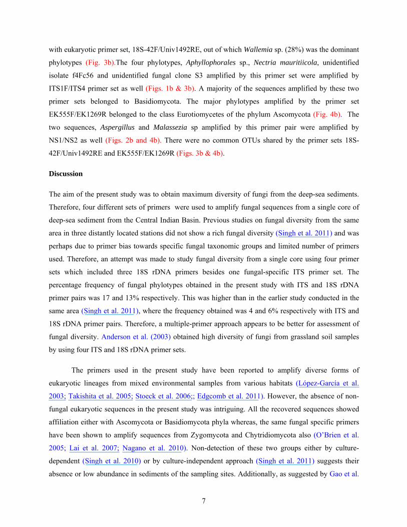
Increasing evidence of the fungal diversity in deep-sea sediments has come from amplification of environmental DNA with fungal specific or eukaryote primer sets. In order to assess the fungal diversity in deep-sea sediments of the Central Indian Basin (CIB) at ~5,000 m depth, we amplified sediment DNA with four different primer sets. These were fungal-specific primer pair ITS1F/ITS4 (internal transcribed spacers), universal 18S rDNA primers NS1/NS2, Euk18S-42F/Euk18S-1492R and Euk18S-555F/Euk18S-1269R. One environmental library was constructed with each of the primer pairs, and 48 clones were sequenced per library. These sequences resulted in 8 fungal Operational Taxonomic Units (OTUs) with ITS and 19 OTUs with 18S rDNA primer sets respectively by taking into account the 2% sequence divergence cut-off for species delineation. These OTUs belonged to 20 distinct fungal genera of the phyla Ascomycota and Basidiomycota. Seven sequences were found to be divergent by 79-97 % from the known sequences of the existing database and may be novel. A majority of the sequences clustered with known sequences of the existing taxa. The phylogenetic affiliation of a few fungal sequences with known environmental sequences from marine and hypersaline habitat suggests their autochthonous nature or adaptation to marine habitat. The amplification of sequences belonging to Exobasidiomycetes and Cystobasidiomycetes from deep-sea is being reported for the first time in this study. Amplification of fungal sequences with eukaryotic as well as fungal specific primers indicates that among eukaryotes, fungi appear to be a dominant group in the sampling site of the CIB.
Key words: Uncultured fungi, 18S rRNA gene, ITS primers, deep-sea sediments, Central
Indian Basin.

2
Introduction
Microbial communities existing in deep-sea oceanic environments account for a total cellular carbon
content of ~3x10¹⁷ g (Whitman et al. 1998). These deep-sea microbial communities, mainly of
bacteria, archaea, protists and fungi play an important role in the recycling of the nutrients (Snelgrove
et al. 1997). Among these, bacteria and archaea have been studied in detail (Stackebrandt et al. 1993;
Urakawa et al. 1999; Li et al. 1999; Takai and Horikoshi 1999; DeLong and Pace. 2001; Sogin et al.
2006; Hongxiang et al. 2008; Luna et al. 2009). A few recent studies have reported eukaryotic
diversity from extreme environments such as hydrothermal vents (López-García et al. 2003, 2007),
anoxic environments (Stoeck and Epstein 2003, Stoeck et al. 2003, 2006; Jebaraj et al. 2010) and
deep-sea sediments (Bass et al. 2007; Edgcomb et al. 2011).
The eukaryotic diversity studies have been executed by molecular survey of ribosomal genes
using culture-independent approach in most of the recent reports. An unexpected high diversity of
eukaryotic rDNA sequences was reported from the deep-sea (López-García et al. 2001). Using
eukaryotic specific primers, several studies have reported only a small fraction of total rDNA
sequences affiliating with fungi in comparison with other eukaryotic lineages (López-García et al.
2001; Stoeck et al. 2003, 2006). However, Edgcomb et al. (2011), while using eukaryotic specific
primers, reported fungi to be dominant in marine deep-sea subsurface. The use of multiple primer
approach for studying diversity has revealed recovery of diverse fungal forms from oxygen depleted
marine environments (Jebaraj et al. 2010).
Using culture-dependent approach, fungi were isolated and identified from the deep-sea sediments of
the CIB (Raghukumar et al. 2004; Damare et al. 2006; Singh et al. 2010). Further Singh et al. (2011)
analyzed the fungal diversity in deep-sea sediments using culture-independent approach by targeting
universal 18S as well as fungal specific and universal ITS (internal transcribed spacers) regions of
rRNA genes from three locations in the CIB. It is known that some of the primer pairs designed for
amplification of fungal sequences may co-amplify non-fungal templates from environmental samples
leading to inaccurate estimation of fungal diversity (Borneman and Hartin, 2000). In addition, the
specific primers designed for amplification of fungal 18S rDNA and ITS regions may be biased
towards certain fungal taxonomic groups. In order to overcome these problems, we used a multiple-
primer approach to study fungal diversity in the present work. The efficiency of primers for
amplification of fungal as well as other eukaryotic sequences from mixed environmental samples was
evaluated by targeting ITS and 18S rDNA regions. The primer set used for ITS region was fungal
specific, whereas all the three 18S rDNA primer sets used were universal. Fungal and eukaryotic 18S

3
rDNA region share some common sequences resulting in cross amplification by the primers designed
for this region (Anderson et al. 2003). Such primers may vary in their specificity in amplifying
different fungal taxonomic groups. Therefore, we hypothesized that using several 18S rDNA primer
sets may help in estimating fungal diversity with greater accuracy.
Materials and methods
Sampling
Sediment sample was collected on the cruise ABP-26 on board the Russian research vessel Academic
Boris Petrov in December 2006 from the station SVBC-33 at a depth of ~5,000 m. The seafloor at this
site is characterized by the presence of homogenous, soft to slightly compact, dark brown coloured
sediments with yellowish bands at shallower and mottling of dusky brown bands at deeper depths. The
sediment was of siliceous nature. The sampling procedure described by Raghukumar et al. (2004) and
Damare et al. (2006) was followed. Sediment was collected with an USNEL-type box corer of 50 cm3
size. The sample thus collected was undisturbed and compact. Sub-cores of sediment were collected
using a sterile PVC cylinder of 5 cm inner diameter. The length of the sediment core obtained was 30
cm. Subsections of 2 cm down to a depth of 10 cm and thereafter every 5 cm length down to 30 cm
depth were cut from the sediment core and directly introduced into sterile plastic bags to avoid
exposure to aerial contamination. The sediment was stored at -20°C immediately after sampling. In
order to monitor the contamination by air-borne fungi, fungal media plates were exposed to the air for
10 min on the deck of the research vessel where the cores were received and in the microbiology
laboratory on board the research vessel.
DNA extraction, environmental PCR and clone library analyses
Environmental DNA was isolated from each frozen sub-section of the sediment cores under
sterile conditions to avoid cross contamination. DNA was isolated from 0.5 g of the sediment sample
from each subsection of the core using the Q-Bio gene Soil DNA extraction kit (MP Biomedicals, OH,
USA) according to the manufacturer’s instructions. The DNA samples from all the subsections were
pooled together and was amplified using fungal-specific primer pair, ITS1F/ITS4 as well as universal
18S rDNA primers, NS1/NS2, Euk18S-42F/Euk18S-1492RE and Euk18S-555F/Euk18S-1269R
(Table 1). The conditions for PCR included an initial hot start incubation (5 min at 94°C) followed by
34 cycles of denaturation at 94oC for 30 s, annealing at 55oC for 30 s and extension at 72°C for 1 min,
followed by a final extension at 72°C for 15 min. The PCR reaction mixture (50 μl) consisted of 50 μg
bovine serum albumin (New England Biolab), 0.6 U Taq DNA polymerase (Bangalore Genei, India),

4
1.5 mM MgCl2, dNTPs (0.2 mM each), primers (0.5 μM each), and 1 x PCR buffer (Roche,
Switzerland). Reaction mixture without template DNA was used as a negative control, and sediments
spiked with fungal DNA was used as a positive control. Amplified products were gel-purified and
ligated with pGEM-T easy vector (Promega, USA) and transformed into E. coli cells (Invitrogen,
Carlsbad, CA), following the manufacturer’s instructions. Transformants were grown overnight at
37°C on Luria Bertani agar containing 100 μg ml-1 of ampicillin. White colonies were screened for the
presence of insert by single colony lysis PCR with M13 forward and reverse primers. PCR protocol
included an initial hot start incubation (5 min at 94oC) followed by 34 cycles of denaturation at 94oC
for 30 s, annealing at 55oC for 30 s and extension at 72oC for 1 min followed by a final extension at
72oC for 5 min. A total of 4 environmental gene libraries, one each from the four primer sets was
obtained. Forty eight clones were screened from each library. Clones containing positive insert were
further processed for plasmid isolation and purification using Millipore plasmid preparation kit
(Millipore, USA). Sequencing of the plasmids was done at the National Centre for Cell Sciences,
Pune, India, using the Big Dye Terminator cycle sequencing kit (V3.1, Applied Biosystems, USA)
according to the manufacturer’s protocol and analyzed in a DNA Analyzer (3730 DNA Analyzer,
Applied Biosystems, USA).
Phylogenetic analyses
Forward and reverse sequences were assembled using Chromas Pro version 1.34. Sequences
obtained with ITS and 18S rDNA primers were analyzed separately. All the sequences were checked
with Ribosomal Database Project for the presence of chimeras (Cole et al. 2004). These chimeric
sequences were eliminated from subsequent analyses. Pairwise alignment of the sequences was carried
out using Clustal W2 software (Thompson et al. 1994). Conserved motifs were identified, and
sequences were trimmed manually. Clone sequences were grouped into operational taxonomic units
(OTUs) by using sequence similarity cut-off value of 98% (O’Brien et al. 2005) by using MOTHUR
software (Schloss et al. 2009). A representative sequence from each OTU was queried against NCBI-
GenBank BLASTn search. Details are shown for the phylogenetic affiliation of the representative
sequence from each OUT (Table 2). Multiple alignments were done for all the sequences along with
their closest match in Clustal W. Gaps and ambiguously aligned sequences were removed from the
further analyses. A phylogenetic analysis was conducted using distance setting maximum likelihood
(ML) in MEGA 5.03 (Kumar et al, 2008) with 1000 bootstrap replicates. Individual phylogenetic trees
were constructed with sequences obtained with ITS and each of the 18S rDNA primer set.

5
Nucleotide sequence accession numbers
The GenBank accession numbers of the nucleotide sequences determined in this study are
presented in Table 2.
Results
Physical parameters of the sampling site
The sediment sample collected for the present study was characterized by the presence of polymetallic
nodules. The nodules abundance was found to be 0.4 Kg m-2. The approximate salinity, temperature
and pH were found to be 35 psu (practical salinity units), 3°C and 7.0 respectively. Among other
parameters, the total proteins, carbohydrates and lipids were approximately 1.5, 1.0 and 1.0 mg g-1
sediment respectively. Total bacterial counts (Acridine Orange Direct Counts) in this area ranged from
107-109 cells g-1 dry sediment. Organic carbon ranged from 0.3-0.4 mg g-1 dry sediment (Sharma
2008).
Analysis of libraries
No growth of aerial mycoflora was observed on media plates that were exposed on the deck
and in the microbiology laboratory on board the research vessel. A total of 48 and 144 clones (3 x 48)
from ITS and 18S primer pairs respectively, were sequenced from the environmental libraries. This
resulted in 27 fungal OTUs, eight with ITS (17% relative frequency) and nineteen with 18S rDNA
primer pairs (13 % relative frequency) after clustering on a basis of 98% sequence identity criterion. A
total of 20 distinct fungal species were obtained from these 27 OTUs. The results are presented as
OTU_01 to OTU_08 obtained with fungal-specific ITS primer pair, OTU_9 and OTU_10 with
universal 18S rDNA primer pairs, NS1/NS2, OTU_11 to OTU_20 with 18S rDNA primer pair
Euk18S-42F/ Euk18S-1492RE and OTU_21 to OTU_27 with 18S rDNA primer pairs Euk18S-555F/
Euk18S-1269R (Table 2).
None of the 18S rDNA primers amplified non-fungal eukaryotic sequences.. Out of the 48 and
144 total clones for ITS and 18S rDNA primer sets, 23 (47.9 %) and 39 (27.1 %) respectively were
bacterial or chimeric in nature and thus were excluded from the analysis. The fungal OTUs mostly
belonged to the phyla Ascomycota and Basidiomycota (Table 2). The number of singletons i.e.
detected only once were four and ten respectively for the ITS and 18S rDNA primer sets (marked with
an *asterisk in Table 2). The remaining OTUs were present ≥2 times in different libraries.

6
Diversity observed with different primer sets
The fungal specific primer set ITS1F/ITS4 amplified eight OTUs belonging to seven different
classes of Ascomycota and Basidiomycota. Of these, the majority belonged to Basidiomycota (Fig
1a). A larger proportion of the sequences affiliated with the unidentified and uncultured fungal clones
(Fig. 1a, Table 2). Among Basidiomycota, OTU_02 clustered with an uncultured clone belonging to
the class Wallemiomycetes with 79 % similarity (Table 2). Two basidiomycetes,. OTU_05 and
OTU_07 affiliated with the sequences (Table 2) which have been isolated from sea water in the
previous studies (NCBI data base).
Universal fungal 18S rDNA primer pair NS1/NS2 amplified sequences showing 99% similarity
to two phylotypes i.e. uncultured Malassezia and Aspergillus clone, belonging to the class
Exobasidiomycetes and Eurotiomycetes respectively (Table 2, Fig 2a).
The universal eukaryotic primer set 18S-42F/Univ1492RE, recovered fungal sequences
belonging to ten different phylotypes and these belonged to six different classes of the phyla
Ascomycota and Basidiomycota (Table 2). Three OTUs (OTU_15, OTU_17 and OTU_20) showed
very low similarity (79, 80 and 94 %) with their closest relative in NCBI database and thus may be
novel phylotypes. Most of the sequences amplified by this primer set clustered with uncultured fungal
clones. OTU_15 and OTU_17 branched out forming a separate cluster within Basidiomycota (Fig. 3a).
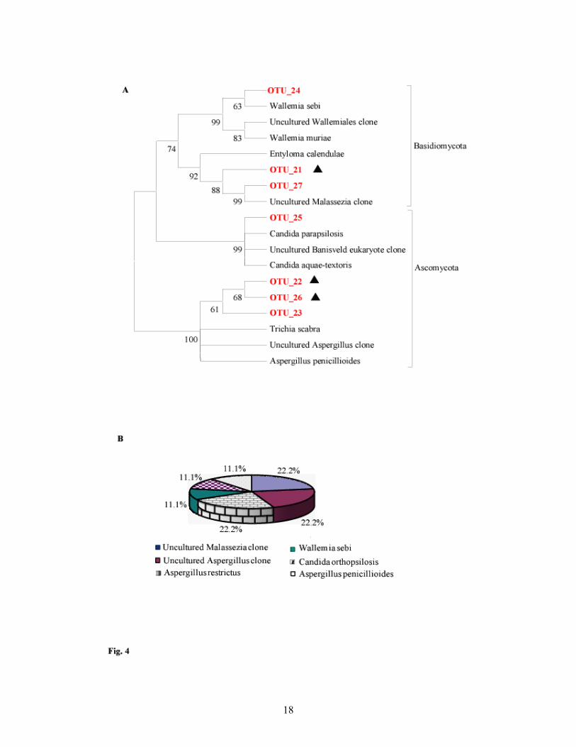
The other universal eukaryotic primer set EK555F/EK1269R, amplified seven fungal
phylotypes clustering with four different classes of Ascomycota and Basidiomycota (Fig. 4a). Three
phylotypes OTU_21, OTU_22 and OTU_26 showed < 97% similarity with existing sequences in the
database and thus may be new (Table 2). Out of these three, OTU_21 and OTU_22 affiliated with the
sequences which were also amplified by NS1/NS2 primer set but with a similarity of 99% with
sequences in the public database (Table 2). These three OTUs clustered separately from the
sequences of their closest relative, with OTU_26 showing a maximum divergence (Fig. 4a).
Proportional distribution of different taxonomic groups in various clone libraries
The proportional distribution (shown in percentage) of fungal taxonomic groups varied in
different clone libraries (Figs. 1b to 4b). Trichosporon asahii (32 %) formed a major portion of
sequences amplified by the ITS primer set (Fig. 1b). Two sequences, namely uncultured clones of
Aspergillus and Malassezia sp. amplified with 18S rDNA primers set NS1/NS2, showed distribution
of 75 and 25% respectively (Fig. 2b). Maximum number of distinct fungal sequences was obtained
7
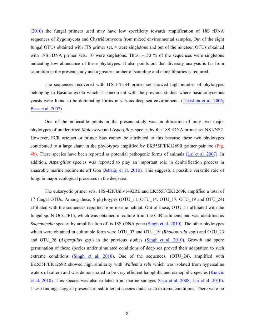
with eukaryotic primer set, 18S-42F/Univ1492RE, out of which Wallemia sp. (28%) was the dominant
phylotypes (Fig. 3b).The four phylotypes, Aphyllophorales sp., Nectria mauritiicola, unidentified
isolate f4Fc56 and unidentified fungal clone S3 amplified by this primer set were amplified by
ITS1F/ITS4 primer set as well (Figs. 1b & 3b). A majority of the sequences amplified by these two
primer sets belonged to Basidiomycota. The major phylotypes amplified by the primer set
EK555F/EK1269R belonged to the class Eurotiomycetes of the phylum Ascomycota (Fig. 4b). The
two sequences, Aspergillus and Malassezia sp amplified by this primer pair were amplified by
NS1/NS2 as well (Figs. 2b and 4b). There were no common OTUs shared by the primer sets 18S-
42F/Univ1492RE and EK555F/EK1269R (Figs. 3b & 4b).
Discussion
The aim of the present study was to obtain maximum diversity of fungi from the deep-sea sediments.
Therefore, four different sets of primers were used to amplify fungal sequences from a single core of
deep-sea sediment from the Central Indian Basin. Previous studies on fungal diversity from the same
area in three distantly located stations did not show a rich fungal diversity (Singh et al. 2011) and was
perhaps due to primer bias towards specific fungal taxonomic groups and limited number of primers
used. Therefore, an attempt was made to study fungal diversity from a single core using four primer
sets which included three 18S rDNA primers besides one fungal-specific ITS primer set. The
percentage frequency of fungal phylotypes obtained in the present study with ITS and 18S rDNA
primer pairs was 17 and 13% respectively. This was higher than in the earlier study conducted in the
same area (Singh et al. 2011), where the frequency obtained was 4 and 6% respectively with ITS and
18S rDNA primer pairs. Therefore, a multiple-primer approach appears to be better for assessment of
fungal diversity. Anderson et al. (2003) obtained high diversity of fungi from grassland soil samples
by using four ITS and 18S rDNA primer sets.
The primers used in the present study have been reported to amplify diverse forms of
eukaryotic lineages from mixed environmental samples from various habitats (López-García et al.
2003; Takishita et al. 2005; Stoeck et al. 2006;; Edgcomb et al. 2011). However, the absence of non-
fungal eukaryotic sequences in the present study was intriguing. All the recovered sequences showed
affiliation either with Ascomycota or Basidiomycota phyla whereas, the same fungal specific primers
have been shown to amplify sequences from Zygomycota and Chytridiomycota also (O’Brien et al.
2005; Lai et al. 2007; Nagano et al. 2010). Non-detection of these two groups either by culture-
dependent (Singh et al. 2010) or by culture-independent approach (Singh et al. 2011) suggests their
absence or low abundance in sediments of the sampling sites. Additionally, as suggested by Gao et al.
8
(2010) the fungal primers used may have low specificity towards amplification of 18S rDNA
sequences of Zygomycota and Chytridiomycota from mixed environmental samples. Out of the eight
fungal OTUs obtained with ITS primer set, 4 were singletons and out of the nineteen OTUs obtained
with 18S rDNA primer sets, 10 were singletons. Thus, ~ 50 % of the sequences were singletons
indicating low abundance of these phylotypes. It also points out that diversity analysis is far from
saturation in the present study and a greater number of sampling and clone libraries is required.
The sequences recovered with ITS1F/ITS4 primer set showed high number of phylotypes
belonging to Basidiomycota which is concordant with the previous studies where basidiomycetous
yeasts were found to be dominating forms in various deep-sea environments (Takishita et al. 2006;
Bass et al. 2007).
One of the noticeable points in the present study was amplification of only two major
phylotypes of unidentified Malassezia and Aspergillus species by the 18S rDNA primer set NS1/NS2.
However, PCR artefact or primer bias cannot be attributed to this because these two phylotypes
contributed to a large share in the phylotypes amplified by EK555F/EK1269R primer pair too (Fig.
4b). These species have been reported as potential pathogenic forms of animals (Lai et al. 2007). In
addition, Aspergillus species was reported to play an important role in denitrification process in
anaerobic marine sediments off Goa (Jebaraj et al. 2010). This suggests a possible versatile role of
fungi in major ecological processes in the deep-sea.
The eukaryotic primer sets, 18S-42F/Univ1492RE and EK555F/EK1269R amplified a total of
17 fungal OTUs. Among these, 5 phylotypes (OTU_11, OTU_14, OTU_17, OTU_19 and OTU_24)
affiliated with the sequences reported from marine habitat. Out of these, OTU_11 affiliated with the
fungal sp. NIOCC#F15, which was obtained in culture from the CIB sediments and was identified as
Sagenomella species by amplification of its 18S rDNA gene (Singh et al. 2010). The other phylotypes
which were obtained in culturable form were OTU_07 and OTU_19 (Rhodotorula spp.) and OTU_23
and OTU_26 (Aspergillus spp.) in the previous studies (Singh et al. 2010). Growth and spore
germination of these species under simulated conditions of deep sea proved their adaptation to such
extreme conditions (Singh et al. 2010). One of the sequences, (OTU_24), amplified with
EK555F/EK1269R showed high similarity with Wallemia sebi which was isolated from hypersaline
waters of saltern and was demonstrated to be very efficient halophilic and osmophilic species (Kunčič
et al. 2010). This species was also isolated from marine sponges (Gao et al. 2008; Liu et al. 2010).
These findings suggest presence of salt tolerant species under such extreme conditions. There were no

9
overlapping sequences amplified by these two universal eukaryotic primer sets suggesting that each
primer pair amplified specific 18S rRNA gene sequences.
None of the fungal phylotypes except Trichosporon asahii, obtained during the present study
matched with the phylotypes obtained from the cores at different locations in the CIB, during the same
sampling period (Singh et al. 2011). This suggests a spatial variation in distribution of fungal
phylotypes, even in not so dynamic habitat such as deep-sea at ~5,000 m depth. The sequences
belonging to Exobasidiomycetes amplified by all the four primer sets is being reported here for the
first time from the deep-sea environment. Its presence in anoxic sediments off Goa (Jebaraj et al.
2010) and in coastal Hawaiian waters (Gao et al. 2010) was reported and is known to consist of
species which are plant pathogens (Begerow et al. 2006). Rhodotorula sp. belonging to the subphylum
Cystobasidiomycetes, reported in the present study is known to be a mycoparasite (Bauer and
Oberwinkler 1991).
Proportional distribution of fungal sequences recovered with different primer sets varied (Figs.
1b to 4b). Among the four primer sets, the universal eukaryotic primer set 18S-42F/Univ1492RE
amplified maximum number of diverse fungal phylotypes. These results are in contrast to the previous
studies (López-García et al. 2003; Stoeck et al. 2006) where only a few fungal OTUs were obtained.
Only a few sequences overlapped between different primer sets resulting in detection of high diversity
of fungal phylotypes by multiple-primer approach (Fig 1b to 4b).
A total of seven OTUs affiliating with a percentage similarity of <97% with the existing
sequences reported in the database were obtained in the present study. This indicates the efficient
nature of the primers used to amplify such novel forms present in deep-sea sediments. These novel
forms comprised mostly of the sequences matching with Aspergillus, Malassezia and unidentified
fungal clones suggesting their possibility of being novel marine variants of the existing species.
Jebaraj et al. (2010) also reported recovery of novel fungal sequences by using multiple primer
approach.
The polymetallic nodules reported in the CIB are rich in Ba, Co, Cu, Fe, Mg and Mn (Nath et
al, 1989). The recovery of fungi from this area suggests their tolerance to metals. In fact, Connell et al.
(2009) have reported metal-tolerant yeasts producing siderophores from deep-sea basalt rocks in
Vailulu’u Seamount, Samoa. These findings indicate active role of fungi in biogeochemical cycles in
the deep sea.
10
In conclusion, the use of multiple primer approach enabled the recovery of diverse fungal
phylotypes from mixed environmental sediment sample of the Central Indian Basin. The high diversity
obtained in spite of less number of clones screened suggested the abundance of fungi under such
extreme conditions. Some of the fungal sequences obtained in the present work have been earlier
reported from marine environment. This supports their presence and ecological role in various
biological processes in this environment. The presence of halophilic forms like Wallemia sp. from
culture-independent study should be proved by culture-dependent approach using various culturing
techniques which may enhance our understanding of salt tolerance in deep-sea fungi. Fungi such as
Sagenomella species obtained by culture-dependent as well as culture-independent approach could be
used as a model organism to understand pressure tolerance in fungi.
Acknowledgements
The first author is thankful to University Grants Commission for the award of Senior Research
Fellowship and Dr. Rahul Sharma, NIO, Goa for the facilities extended during the research cruise. The
crew members of the Russian research vessel Academic Borris Petrov are acknowledged for their
support. The first two authors are grateful to the Director, NIO for the support extended. This is NIO’s
contribution No.
References
Anderson IC, Campbell CD, Prosser JI (2003) Potential bias of fungal 18S rDNA and internal transcribed spacer polymerase chain reaction primers for estimating fungal biodiversity in soil. Environ Microbiol 5: 36-47
Bass D, Howe A, Brown N, Barton H, Demidova M, Michelle H, Li L, Sander H, Watkinson SC, Willcock S, Richards TA (2007) Yeast forms dominate fungal diversity in the deep oceans. Proc R Soc B 274: 3069-3077
Bauer R, Oberwinkler F (1991) The colacosomes: new structures at the host-parasite interface of a mycoparasitic basidiomycete. Bot Acta 104: 53-57
Begerow D, Stoll M, Bauer RA (2006) phylogenetic hypothesis of Ustilaginomycotina based on multiple gene analyses and morphological data. Mycologia 98:906-916
Borneman J, Hartin RJ (2000) PCR primers that amplify fungal rRNA genes from environmental samples. Appl Environ Microbiol 66: 4356–4360
Cole JR, Chai B, Farris RJ, Wang Q, Kulam SA, McGarrell DM, Garrity GM, Tiedje JM (2004) The Ribosomal Database Project (RDP-II): sequences and tools for high-throughput rRNA analysis. Nucleic Acids Res 33: D294–D296
11
Connell L, Barrett A, Templeton A, Staudigel H (2009) Fungal diversity associated with an active deep-sea volcano: Vailulu’u Seamount, Samoa. Geomicrobiology J 26: 597-605
Damare S, Raghukumar C, Raghukumar S (2006) Fungi in deep-sea sediments of the Central Indian Basin. Deep-Sea Res PT -1 53:14-27
DeLong EF, Pace NR (2001) Environmental Diversity of Bacteria and Archaea Syst Biol 50:470-478
Edgcomb VP, Beaudoin D, Gast R, Biddle JF, Teske A (2011) Marine subsurface eukaryotes: the fungal majority. Environ Microbiol 13: 172-183
Gao Z, Johnson ZI, Wang G (2010) Molecular characterization of the spatial diversity and novel lineages of mycoplankton in Hawaiian coastal waters. ISME J 4: 111-120
Gao Z, Li B, Zheng C, Wang G (2008) Molecular Detection of Fungal Communities in the Hawaiian Marine Sponges Suberites zeteki and Mycale armata. Appl Environ Microbiol 74:6091–6101
Hongxiang X, Min W, Xiaogu W, Junyi Y, Chunsheng W (2008) Bacterial diversity in deep-sea sediment from north eastern Pacific Ocean Acta Ecol Sinica 28:479-485
Jebaraj CS, Raghukumar C, Behnke A, Stoeck T (2010) Fungal diversity in oxygen-depleted regions of the Arabian Sea revealed by targeted environmental sequencing combined with cultivation. FEMS Microb Ecol 71:99-412
Kumar S, Nei M, Dudley J, Tamura K (2008) MEGA: A biologist-centric software for evolutionary analysis of DNA and protein sequences. Brief Bioinform 9: 299-306
Kunčič MK, Kogej T, Drobne D, Gunde-Cimerman N (2010) Morphological response of the halophilic fungal genus Wallemia to high salinity. Appl Environ Microbiol 76:329-337
Lai X, Cao L, Tan H, Fang S, Huang Y, Zhou S (2007) Fungal communities from methane hydrate-bearing deep-sea marine sediments in South China Sea. ISME J 1:756–762
Li L, Kato C, Horikoshi K (1999) Bacterial diversity in deep-sea sediments from different depths. Biodivers Conserv 8:659-677
Liu WC, Li CQ, Zhu P, Yang JL, Cheng KD (2010) Phylogenetic diversity of culturable fungi associated with two marine sponges: Haliclona simulans and Gelliodes carnosa, collected from the Hainan Island coastal waters of the South China Sea. Fungal Divers 42:1-15
López-García P, Philippe H, Gail F, Moreira D (2003) Autochthonous eukaryotic diversity in hydrothermal sediment and experimental microcolonizers at the mid-Atlantic Ridge. Proc Natl Acad Sci USA 2:697-702
López-García P, Rodriguez-Valera F, Pedros-Alio C, Moreira D (2001) Unexpected diversity of small eukaryotes in deep-sea Antarctic plankton. Nature 409:603–607
12
López-García P, Vereshchaka A, Moreira D (2007) Eukaryotic diversity associated with carbonates and fluid-seawater interface in Lost City hydrothermal field. Environ Microbiol 9:546-554
Luna GM, Stumm K, Pusceddu A, Danovaro R (2009) Archaeal diversity in deep-sea sediments estimated by means of different terminal-restriction fragment length polymorphisms (T-RFLP) Protocols. Curr Microbiol 59:356-361
Nagano Y, Nagahama T, Hatada Y, Nunoura T, Takami H, Miyazaki J, Takai K, Horikoshi K (2010) Fungal diversity in deep-sea sediments-the presence of novel fungal groups. Fungal Ecol 3:316-325
Nath BN, Rao VP, Becker KP (1989) Geochemical evidence of terrigenous influence in deep-sea sediments up to 8°S in the in the Central Indian Basin. Marine Geol 87: 301-313
O’Brien H, Parrent JL, Jackson JA, Moncalvo J, Vilgalys R (2005) Fungal community analysis by large-scale sequencing of environmental samples. Appl Environ Microbiol 71:5544-5550
Raghukumar C, Raghukumar S, Sheelu G, Gupta SM, Nath BN, Rao BR (2004) Buried in time: culturable fungi in a deep-sea sediment core from the Chagos Trench, Indian Ocean. Deep-Sea Res PT 1 51:1759-1768
Schloss PD, Westcott SL, Ryabin T, Hall JR, Hartmann M, Hollister EB, Lesniewski RA, Oakley BB, Parks DH, Robinson CJ, Sahl JW, Stres W, Thallinger GG, Horn DJV, Weber CF (2009) Introducing mothur: open source, platform-independent, community-supported software for describing and comparing microbial communities. Appl Environ Microbiol 75:7537-7541
Sharma R (2008) Project report: Benthic environmental variability in the Central Indian Basin-III, Cruise #ABP26, National Institute of Oceanography, Goa, India, pp. 1-170
Singh P, Raghukumar C, Verma P, Shouche Y (2010) Phylogenetic diversity of culturable fungi from the deep-sea sediments of the Central Indian Basin and their growth characteristics. Fungal Divers 40:89-102
Singh P, Raghukumar C, Verma P, Shouche Y (2011) Fungal community analysis in the deep-sea sediments of the Central Indian Basin by culture-independent approach. Microb Ecol 61:507-517
Snelgrove PVR, Blackburn TH, Hutchings P, Alongi D, Grassle JF, Hummel H, King G, Koike I, Lambshead PJD, Ramsing NBV, Solis-Weiss, Freckman DW (1997) The importance of marine sediment biodiversity in ecosystem processes. Ambio 26: 578-582
Sogin M, Morrison HG, Huber JA, Welch DM, Huse SM, Neal PR, Arrieta JM, Herndl GJ (2006) Microbial diversity in the deep sea and underexplored “rare biosphere”. Proc Natl Acad Sci USA 103:12115–12120
Stackebrandt E, Liesack W, Goebel BM (1993) Bacterial diversity in a soil sample from a subtropical Australian environment as determined by 16S rDNA analysis. J FASEB 7:232-236

13
Stoeck T, Epstein S (2003) Novel eukaryotic lineages inferred from small-subunit rRNA analyses of oxygen depleted marine environments. Appl Environ Microbiol 69:2657–2663
Stoeck T, Hayward B, Taylor GT, Varela R, Epstein SS (2006) A multiple PCR-primer approach to access the microeukaryotic diversity in environmental samples. Protist 157:31–43
Stoeck T, Taylor GT, Epstein SS (2003) Novel eukaryotes from the permanently anoxic Cariaco Basin (Caribbean Sea). Appl Environ Microbiol 69:5656–5663
Takai K, Horikoshi K (1999) Genetic diversity of Archaea in deep-sea hydrothermal vent environments. Genetics 152:1285-1297
Takishita K, Miyake H, Kawato M, Maruyama T (2005) Genetic diversity of microbial eukaryotes in anoxic sediment around fumaroles on a submarine caldera floor based on the small-subunit rDNA phylogeny. Extremophiles 9:185–196
Takishita K, Tsuchiya M, Reimer JD, Maruyama T (2006) Molecular evidence demonstrating the basidiomycetous fungus Cryptococcus curvatus is the dominant microbial eukaryote in sediment at the Kuroshima Knoll methane seep. Extremophiles 10:165–169
Thompson JD, Higgins DG, Gibson TJ (1994) CLUSTAL W: improving the sensitivity of progressive multiple sequence alignment through sequence weighting, positions-specific gap penalties and weight matrix choice. Nucleic Acids Res 22:4673-4680
Urakawa H, Kita-Tsukamoto K, Ohwada K (1999) Microbial diversity in marine sediments from Sagami Bay and Tokyo Bay, Japan, as determined by 16S rRNA gene analysis. Microbiol 145:3305-3315
Whitman WB, Coleman DC, Wiebe WJ (1998) Prokaryotes: The unseen majority. Proc Acad Natl Sci USA 95:6578-6583
14
Legends to the figures
Fig. 1A Maximum likelihood (ML) phylogenetic tree for the fungal OTUs constructed based on fungal
ITS gene sequences obtained using fungal specific primer set ITS1F/ITS4. Topology was built using
Mega v.5.03 from a ClustalW 1.83 alignment. Numbers below branches indicate bootstrap values (>50
%) from 1,000 replicates. New sequence types are marked with triangle.
1B. Proportional distribution of different fungal taxa in the clone library constructed with fungal
specific primer set ITS1F/ITS4.
Fig. 2A ML phylogenetic tree for the fungal OTUs constructed based on fungal 18S rDNA gene
sequences obtained using universal fungal 18S rDNA primer set, NS1/NS2. Topology was built using
Mega v.5.03 from a ClustalW 1.83 alignment. Numbers below branches indicate bootstrap values (>50
%) from 1,000 replicates. New sequence types are marked with triangle.
2B. Proportional distribution of different fungal taxa in the clone library constructed with 18S rDNA
primer set NS1/NS2.
Fig. 3A ML phylogenetic tree for the fungal OTUs constructed based on 18S rDNA gene sequences
obtained using universal eukaryotic 18S rDNA primer set, Euk18S-42F/Euk18S-1492RE. Topology
was built using Mega v.5.03 from a ClustalW 1.83 alignment. Numbers below branches indicate
bootstrap values (>50 %) from 1,000 replicates. New sequence types are marked with triangle.
3B. Proportional distribution of different fungal taxa in the clone library constructed with 18S rDNA
primer set Euk18S-42F/Euk18S-1492RE .
Fig. 4A ML phylogenetic tree for the fungal OTUs constructed based on 18S rDNA gene sequences
obtained using universal eukaryotic 18S rDNA primer set, Euk18S-555F/Euk18S-1269R. Topology
was built using Mega v.5.03 from a ClustalW 1.83 alignment. Numbers below branches indicate
bootstrap values (>50 %) from 1,000 replicates. New sequence types are marked with triangle.
4B. Proportional distribution of different fungal taxa in the clone library constructed with 18S rDNA
primer set Euk18S-555F/Euk18S-1269R Uncultured Malassezia clone (22.2%) includes OTU_21
(11.1%) and OTU_27 (11.1%).

15

16

17
18
19
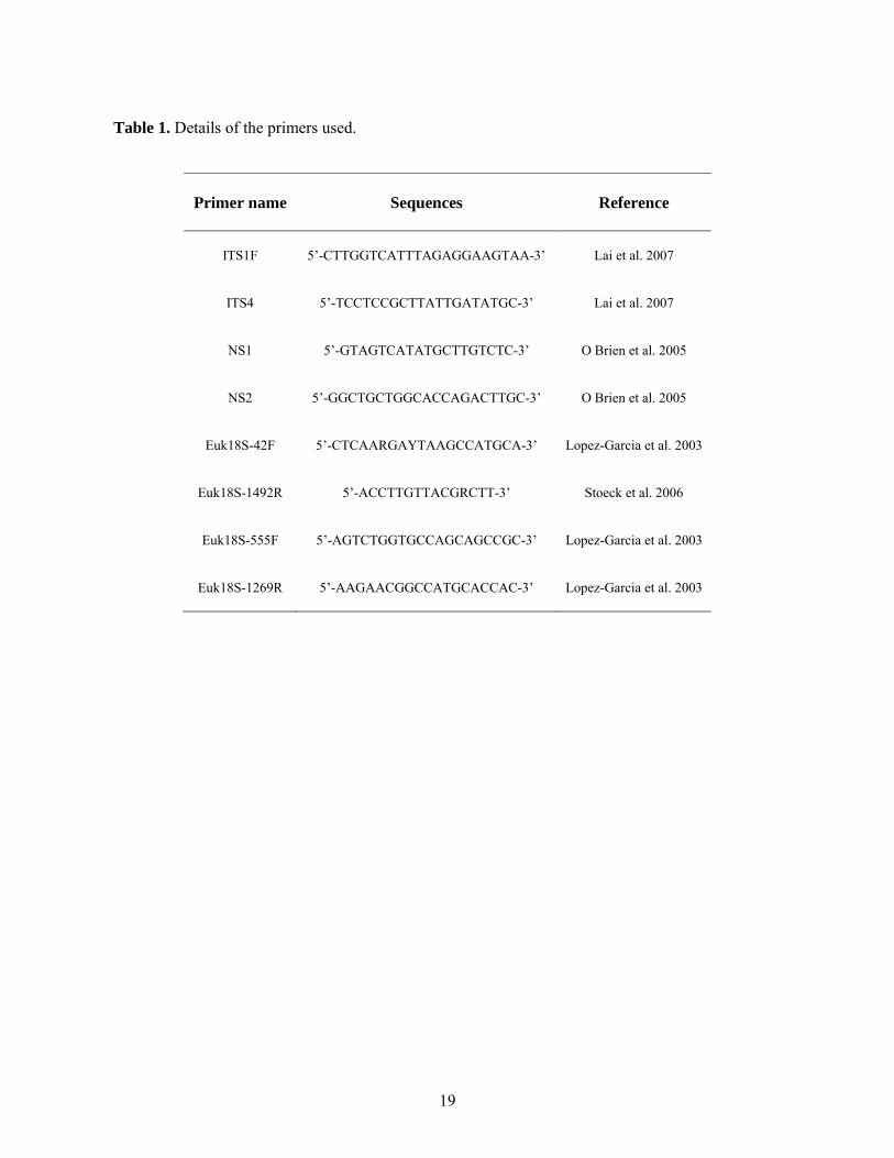
Table 1. Details of the primers used.
Primer name
Sequences
Reference
ITS1F
5’-CTTGGTCATTTAGAGGAAGTAA-3’
Lai et al. 2007
ITS4
5’-TCCTCCGCTTATTGATATGC-3’
Lai et al. 2007
NS1
5’-GTAGTCATATGCTTGTCTC-3’
O Brien et al. 2005
NS2
5’-GGCTGCTGGCACCAGACTTGC-3’
O Brien et al. 2005
Euk18S-42F
5’-CTCAARGAYTAAGCCATGCA-3’
Lopez-Garcia et al. 2003
Euk18S-1492R
5’-ACCTTGTTACGRCTT-3’
Stoeck et al. 2006
Euk18S-555F
5’-AGTCTGGTGCCAGCAGCCGC-3’
Lopez-Garcia et al. 2003
Euk18S-1269R
5’-AAGAACGGCCATGCACCAC-3’
Lopez-Garcia et al. 2003
20
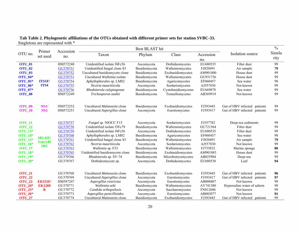
Tab Table 2. Phylogenetic affiliations of the OTUs obtained with different primer sets for station SVBC-33. Singletons are represented with *
OTU no. Primer set used
Accession no.
Best BLAST hit Isolation source
% Simila
rity Taxon Phylum Class Accession
no. OTU_01
ITS1F/ ITS4
HM572248 Unidentified isolate f4Fc56 Ascomycota Dothideomycetes EU680535 Filter dust 99 OTU_02 GU370751 Unidentified fungal clone S3 Basidiomycota Wallemiomycetes FJ820491 Air sample 79 OTU_03 GU370752 Uncultured basidiomycete clone Basidiomycota Exobasidiomycetes AM901800 House dust 99
OTU_04* GU370753 Uncultured Wallemia isolate Basidiomycota Wallemiomycetes GU931736 House dust 99OTU_05* GU370754 Aphyllophorales sp. LM82 Basidiomycota Agaricomycetes EF060457 Sea water 98 OTU_06* GU370755 Nectria mauritiicola Ascomycota Sordariomycetes AJ557830 Not known 99 OTU_07* GU370756 Rhodotorula calyptogenae Basidiomycota Cystobasidiomycetes EU669878 Sea water 99 OTU_08
HM572249 Trichosporon asahii Basidiomycota Tremellomycetes AB369919 Not known 99
OTU_09 NS1/ NS2
HM572252 Uncultured Malassezia clone Basidiomycota Exobasidiomycetes FJ393445 Gut of HBV infected patients 99 OTU_10
HM572251 Uncultured Aspergillus clone Ascomycota Eurotiomycetes FJ393417 Gut of HBV infected patients 99
OTU_11
18S-42F/ Univ149
2RE
GU370757 Fungal sp. NIOCC F15 Ascomycota Sordariomycetes FJ357782 Deep-sea sediments 99 OTU_12 GU370758 Unidentified isolate f3Fc79 Basidiomycota Wallemiomycetes GU721564 Filter dust 99
OTU_13* GU370759 Unidentified isolate f4Fc56 Ascomycota Dothideomycetes EU680535 Filter dust 99 OTU_14* GU370760 Aphyllophorales sp. LM82 Basidiomycota Agaricomycetes EF060457 Sea water 98 OTU_15* GU370761 Unidentified fungal clone S3 Basidiomycota Wallemiomycetes FJ820491 Air sample 79 OTU_16* GU370762 Nectria mauritiicola Ascomycota Sordariomycetes AJ557830 Not known 99 OTU_17 GU370763 Wallemia sp. F53 Basidiomycota Wallemiomycetes FJ755832 Marine sponge 80
OTU_18* GU370765 Unidentified basidiomycete clone Basidiomycota Exobasidiomycetes AM901883 House dust 99 OTU_19* GU370766 Rhodotorula sp. SY-74 Basidiomycota Microbotryomycetes AB025984 Deep-sea 99 OTU_20*
GU370767 Dothideomycete sp. Ascomycota Dothideomycetes EU680530 Leaf 94
OTU_21
EK555F/ EK1269
R
GU370768 Uncultured Malassezia clone Basidiomycota Exobasidiomycetes FJ393445 Gut of HBV infected patients 96 OTU_22 GU370769 Uncultured Aspergillus clone Ascomycota Eurotiomycetes FJ393417 Gut of HBV infected patients 97 OTU_23 HM587247 Aspergillus restrictus Ascomycota Eurotiomycetes AB008407 Not known 99
OTU_24* GU370771 Wallemia sebi Basidiomycota Wallemiomycetes AY741380 Hypersaline water of saltern 98 OTU_25* GU370772 Candida orthopsilosis Ascomycota Saccharomycetes FN812686 Not known 99 OTU_26* GU370773 Aspergillus penicillioides Ascomycota Eurotiomycetes AB002077 Not known 91 OTU_27 GU370774 Uncultured Malassezia clone Basidiomycota Exobasidiomycetes FJ393445 Gut of HBV infected patients 99