Characterization of unusual families of ATG8-like proteins and ATG12 in the protozoan parasite...
29
Characterisation of unusual families of ATG8-like proteins and ATG12 in the protozoan parasite Leishmania major Roderick A.M. Williams 1 , Kerry L. Woods 2 , Luiz Juliano 3 , Jeremy C. Mottram 2 , and Graham H. Coombs 1 1 Strathclyde Institute of Pharmacy and Biomedical Sciences, University of Strathclyde, Glasgow, G4 0NR, UK 2 Wellcome Centre for Molecular Parasitology and Division of Infection & Immunity, Faculty of Biomedical and Life Sciences, University of Glasgow, Glasgow, G12 8TA, UK. 3 Department of Biophysics, Universidade Federal de São Paulo, Escola Paulista de Medicina, Rua Três de Maio 100, 04044-020, São Paulo, Brazil. Abstract Leishmania major possesses, apparently uniquely, four families of ATG8-like genes, designated ATG8, ATG8A, ATG8B and ATG8C, and 25 genes in total. L. major ATG8 and examples from the ATG8A, ATG8B and ATG8C families are able to complement a Saccharomyces cerevisiae ATG8-deficient strain, indicating functional conservation. Whereas ATG8 has been shown to form putative autophagosomes during differentiation and starvation of L. major, ATG8A primarily form puncta in response to starvation - indicating a role for ATG8A in starvation-induced autophagy. Recombinant ATG8A was processed at the scissile glycine by recombinant ATG4.2 but not ATG4.1 cysteine peptidases of L. major and, consistent with this, ATG4.2-deficient L. major mutants were unable to process ATG8A and were less able to withstand starvation than wild type cells. GFP-ATG8-containing puncta were less abundant in ATG4.2 over-expression lines, in which unlipidated ATG8 predominated, which is consistent with ATG4.2 being an ATG8- deconjugating enzyme as well as an ATG8A-processing enzyme. In contrast, recombinant ATG8, ATG8B and ATG8C were all processed by ATG4.1, but not by ATG4.2. ATG8B and ATG8C both have a distinct subcellular location close to the flagellar pocket, but the occurrence of the GFP-labelled puncta suggest that they do not have a role in autophagy. L. major genes encoding possible ATG5, ATG10 and ATG12 homologues were found to complement their respective S. cerevisiae mutants, and ATG12 localised in part to ATG8-containing puncta, suggestive of a functional ATG5-ATG12 conjugation pathway in the parasite. L. major ATG12 is unusual as it requires C-terminal processing by an as yet unidentified peptidase. Keywords autophagy; Leishmania; protozoan parasite; ATG4; ATG8; ATG12 Introduction Leishmania is a protozoan parasite that occurs as different morphological forms in its two hosts (mammals and sandflies) and greatly remodels its life cycle forms during differentiation. Within the alimentary tract of the sandfly vector Leishmania exists in two forms of promastigotes, the procyclic multiplicative form and the metacyclic, non- Correspondence to: Graham H. Coombs ([email protected]). Europe PMC Funders Group Author Manuscript Autophagy. Author manuscript; available in PMC 2009 February 14. Published in final edited form as: Autophagy. 2009 February ; 5(2): 159–172. Europe PMC Funders Author Manuscripts Europe PMC Funders Author Manuscripts
-
Upload
independent -
Category
Documents
-
view
1 -
download
0
Transcript of Characterization of unusual families of ATG8-like proteins and ATG12 in the protozoan parasite...

Characterisation of unusual families of ATG8-like proteins andATG12 in the protozoan parasite Leishmania major
Roderick A.M. Williams1, Kerry L. Woods2, Luiz Juliano3, Jeremy C. Mottram2, and GrahamH. Coombs1
1Strathclyde Institute of Pharmacy and Biomedical Sciences, University of Strathclyde, Glasgow,G4 0NR, UK2Wellcome Centre for Molecular Parasitology and Division of Infection & Immunity, Faculty ofBiomedical and Life Sciences, University of Glasgow, Glasgow, G12 8TA, UK.3Department of Biophysics, Universidade Federal de São Paulo, Escola Paulista de Medicina,Rua Três de Maio 100, 04044-020, São Paulo, Brazil.
AbstractLeishmania major possesses, apparently uniquely, four families of ATG8-like genes, designatedATG8, ATG8A, ATG8B and ATG8C, and 25 genes in total. L. major ATG8 and examples fromthe ATG8A, ATG8B and ATG8C families are able to complement a Saccharomyces cerevisiaeATG8-deficient strain, indicating functional conservation. Whereas ATG8 has been shown toform putative autophagosomes during differentiation and starvation of L. major, ATG8A primarilyform puncta in response to starvation - indicating a role for ATG8A in starvation-inducedautophagy. Recombinant ATG8A was processed at the scissile glycine by recombinant ATG4.2but not ATG4.1 cysteine peptidases of L. major and, consistent with this, ATG4.2-deficient L.major mutants were unable to process ATG8A and were less able to withstand starvation than wildtype cells. GFP-ATG8-containing puncta were less abundant in ATG4.2 over-expression lines, inwhich unlipidated ATG8 predominated, which is consistent with ATG4.2 being an ATG8-deconjugating enzyme as well as an ATG8A-processing enzyme. In contrast, recombinant ATG8,ATG8B and ATG8C were all processed by ATG4.1, but not by ATG4.2. ATG8B and ATG8Cboth have a distinct subcellular location close to the flagellar pocket, but the occurrence of theGFP-labelled puncta suggest that they do not have a role in autophagy. L. major genes encodingpossible ATG5, ATG10 and ATG12 homologues were found to complement their respective S.cerevisiae mutants, and ATG12 localised in part to ATG8-containing puncta, suggestive of afunctional ATG5-ATG12 conjugation pathway in the parasite. L. major ATG12 is unusual as itrequires C-terminal processing by an as yet unidentified peptidase.
Keywordsautophagy; Leishmania; protozoan parasite; ATG4; ATG8; ATG12
IntroductionLeishmania is a protozoan parasite that occurs as different morphological forms in its twohosts (mammals and sandflies) and greatly remodels its life cycle forms duringdifferentiation. Within the alimentary tract of the sandfly vector Leishmania exists in twoforms of promastigotes, the procyclic multiplicative form and the metacyclic, non-
Correspondence to: Graham H. Coombs ([email protected]).
Europe PMC Funders GroupAuthor ManuscriptAutophagy. Author manuscript; available in PMC 2009 February 14.
Published in final edited form as:Autophagy. 2009 February ; 5(2): 159–172.
Europe PM
C Funders A
uthor Manuscripts
Europe PM
C Funders A
uthor Manuscripts

multiplicative mammal-infective form. Transformation to the smaller metacyclic form isknown as metacyclogenesis. Within mammals Leishmania live in macrophages as smallmultiplicative, non-motile amastigotes (devoid of an external flagellum). Two parasitesrelated to Leishmania, Trypanosoma brucei, the causative agent of sleeping sickness in manand nagana in cattle, and T. cruzi, responsible for Chagas disease, also have complicated lifecycles involving a variety of morphological forms living in the mammalian and respectiveinsect vectors; each undergoes extensive remodelling during differentiation to thesubsequent form in the life cycle.
Autophagy is a conserved mechanism in eukaryotes whereby cytosolic proteins aretransported in autophagosomes to the lysosomal network for degradation in response tonutrient deprivation. The amino acids generated are recycled and used for protein synthesis,thereby helping to maintain cellular homeostasis.1 In addition, autophagy plays roles inremodelling during cellular differentiation and development.2-4 Defects in autophagy inmammalian cells has been associated with a variety of disease states including cancer5-7and neurodegenerative diseases,8,9 as well as susceptibility to bacterial and viral infections.10,11 Studies on trypanosomatids have shown that autophagy is crucial for differentiationbetween different life cycle forms and provision of nutrients under starvationconditions12-15 and it has been suggested that blocking autophagy could be a new strategyfor fighting parasitic disease.12
The biogenesis of the dynamic membrane required for autophagosome formation inmammals and yeast requires the functioning of two conjugation pathways; those involving,respectively, ATG8 and ATG12/ATG5.16-20 ATG8 exists as multiple copies inmammals21-26 and plants,27 whereas yeast has just one; in all these cases, however, theATG8s are of one type. ATG8 is synthesized as an inactive precursor, then it is activated viaremoval of a C-terminal polypeptide segment, by the clan CA, family C54 cysteinepeptidase ATG4, to leave a C-terminal glycine.22,28,29 The exposed glycine is activated bythe E1-like enzyme ATG7, transferred to the E2-like enzyme ATG3, and finally conjugatedwith the phospholipid, phosphatidylethanolamine (PE).30-33 PE anchors ATG8 to theautophagosomal membrane and adjacent ATG8 molecules tether together and thus mediatethe formation of an autophagosome.34 The ATG8s are deconjugated from the PE before theautophagosomes enter the endosomal system in mammalian cells or the yeast vacuole,ATG4 also mediating this deconjugation.34-36 Thus ATG4 functions at two stages duringautophagosome biogenesis and degradation. The absence of ATG8 does not negate thefunction of the core machinery proteins,37 and small autophagosome-like structures occur inyeast ATG8 null cells (designated atg8Δ).38 However, autophagosome size and the level ofautophagy are regulated by the amount of Atg8 in yeast cells.36
Many ubiquitin-like modifier proteins similar to ATG8 have been identified, namelyubiquitin, neural precursor cell-expressed developmentally down-regulated 8 (Nedd8), smallubiquitin-related modifier (SUMO), Hub1, and ubiquitin-related modifier 1 (Urm1).39 Theyare similar to ATG8 in that they become conjugated to a primary amine to form an amide orisopeptide bond and subsequently are deconjugated to provide mechanisms that regulatecellular activities such as transcription, the cell cycle and autophagy. Of these, only ATG8and ubiquitin have as yet been characterized in trypanosomatids 12,15,39 and the evidenceis that ATG8 behaves similarly as in yeast and mammals.12,15 However, Leishmania isunusual in apparently having, based on predictions from genome mining, this type of ATG8plus three others.15 One objective of this study was to experimentally test the hypothesisthat all four groups indeed have the characteristics of ATG8s and to gain insights into theirroles.
Williams et al. Page 2
Autophagy. Author manuscript; available in PMC 2009 February 14.
Europe PM
C Funders A
uthor Manuscripts
Europe PM
C Funders A
uthor Manuscripts

There is a multiplicity of ATG4s in higher eukaryotes. Mammals have four isoforms,autophagin-1, -2, -3 and -4, and their activities are thought to differ as only autophagin-1and -3 can restore autophagy to Atg4Δ mutant yeast.40 Both ATG4s of Arabidopsis thalianacan cleave all its ATG8 copies and study of mutants defective in one or both of the AtATG4genes (designated Δatg4a, Δatg4b or Δatg4a4b-1) suggest that redundancy exists betweenthe AtAtg4s.41,42 Saccharomyces cerevisiae atg4Δ mutants are incapable of formingautophagosomes, and the expression in these mutants of ScAtg8 lacking residues beyond thescissile glycine residue resulted in autophagosome accumulation – indicating the role ofScAtg4 at the late stage of the autophagic pathway.35,43 Leishmania also has two ATG4sand, interestingly, mutants deficient in one of them (Δatg4.2) also accumulate putativeautophagosomes.13 Thus a second aim of this study was to investigate whether the twoATG4s of Leishmania act differently towards the variety of Leishmania ATG8s and whetherthis may account for functional differences between them.
The second conjugation pathway necessary for autophagy in higher eukaryotes involvesATG12 and ATG5. ATG12 is activated by ATG7 and transferred to an E2-like enzyme,ATG10, whereupon it is conjugated to ATG5.44 The ATG12-ATG5 conjugate subsequentlybinds with ATG16 forming a multimeric complex of 350-800 kDa in size.45,46 The E3-likeactivity of the ATG12-ATG5 conjugate potentiates the formation of ATG8-PE; making theautophagic pathway analogous to ubiquitin-like pathways where an E3 conjugation ligase isrequired.47,48 The ATG12-ATG5 complex is absent from fully formed autophagosomes.
It has been reported, again based on genome mining, that these proteins are absent fromtrypanosomatids and so it was concluded that the ATG12/ATG5 pathway does not occur.12,49,50 The ATG12/ATG5 pathway is absent from organisms where microautophagypredominates51 and so its absence from T. brucei would correlate with evidence that thedegradation of glycosomes in this parasites is via microautophagy.14 However, our studieshave shown that macroautophagy occurs in Leishmania.13,15 Moreover, our in silicoanalyses led us to hypothesise that the proteins necessary for the ATG12/ATG5 pathwaymay indeed be encoded in the Leishmania genome, but they are significantly divergent fromtheir yeast counterparts.15 Thus another aim of this study was to determine experimentallywhether the proteins encoded by these putative genes of the ATG12/ATG5 pathway indeedfunction as postulated.
ResultsThe ATG4 and ATG8 proteins of L. major
Leishmania major has two ATG4 cysteine peptidases, designated ATG4.1 and ATG4.2. (seeref.13; http://www.genedb.org). The ORFs of the two ATG4 genes comprise 1185 bp and1167 bp, encoding 394 and 388 amino acids with calculated molecular masses of 43.8 and42.5 kDa, for ATG4.1 and ATG4.2, respectively. The predicted proteins are aligned withthose of T. brucei, yeast, human and Arabidopsis in Figure S1A. The leishmanial proteinsare typical of ATG4s in possessing an inhibitory loop over the active site pocket (FigureS1A; triple lines) and the conserved catalytic triad within the pocket (comprising C73, H241
and D239 in ATG4.1 and C92, H267 and D265 in ATG4.2; Fig. S1A, asterisks) typical of theproteins belonging to family C54 of Clan CA of cysteine peptidases.52,53 The residueadjacent to the catalytic triad that, based on evidence for the human enzyme HsAtg4B,52,53is required for hydrolysis (Y54, marked with Δ in Figure S1A) is also conserved in bothenzymes, whereas those implicated in recognition of the C-terminal region of the substrateATG8 (W142, R229 and S316, marked with II in Fig. S1A) are present in ATG4.2, but aresubstituted with L159, V209, C296 in ATG4.1; suggesting perhaps some difference insubstrate specificities.
Williams et al. Page 3
Autophagy. Author manuscript; available in PMC 2009 February 14.
Europe PM
C Funders A
uthor Manuscripts
Europe PM
C Funders A
uthor Manuscripts
Twenty five ORFs encode ATG8-like proteins in the L. major genome.15 The paraloguedesignated ATG8 (LmjF19.1630) has counterparts in the other sequenced genomes ofLeishmania, L. infantum (LinJ19_V3.1660) and L. braziliensis (LbrM19_V2.1890). All areencoded by single copy genes with 35-46% identity with orthologues from yeast and highereukaryotes. LmjF22.1300, another ATG8-like gene identified in L. major’s gene content,has the carboxyl-terminus scissile glycine that is exposed by the hydrolytic action of ATG4prior to the activation and conjugation to PE by actions of E1-like and E2-like ATG proteins(* in Figure S2A(i)). Further, the LmjF22.1300 has amino acids C-terminal to the scissileglycine that is typical of ATG8. However, an insertion (of 58 amino acids) somewhatsimilar to that typical of ATG12s is also present in LmjF22.1300 (Figure S2A(ii). Thusbased on sequence analyses alone LmjF22.1300 could be predicted to be either an ATG8 oran ATG12. Functional studies however, point to an ATG12 function for LmjF22.1300 (seebelow), so henceforth in this paper LmjF22.1300 is defined as ATG12.
The proteins designated as the ATG8A, ATG8B and ATG8C families are encoded by genearrays, those encoding ATG8A and ATG8B being interspersed, and appear to be unique toLeishmania. The scissile glycine is conserved in all the paralogues (Figure S2A, markedwith *). Phylogenetic analysis suggests that the ATG8A, ATG8B and ATG8C families aredistinct clades (Figure S2B). While in this analysis ATG8 clusters with orthologues fromother organisms (Figure S2B), the L. major ATG12 occupies an intermediate positionbetween the ATG8 and ATG12 clades.
L. major ATG8 proteins are selectively cleaved by the ATG4 cysteine peptidasesThe specific cleavage of the C-terminus of Atg8 by Atg4 in S. cerevisiae involves thecomplex interaction of residues adjacent to the active site of the peptidase with therecognition site in ScAtg8 comprising the residues Tyr49, Leu50, Phe77 and Phe79.54 Threeof these residues are conserved in Leishmania ATG8 (being Leu54, Phe81 and Phe83, withthe Tyr being replaced by Phe53), whereas none appear to be conserved in the other proteingroups in which these residues are substituted with Leu42, Ala43, Ala70 and Ala72; Leu44,Ala45, Thr72 and Ala74, and Ser42, Ala43, Ala71 and Thr73, respectively, in all ATG8A,ATG8B and ATG8C members (Figure S2A). Thus the functionality of these three groups ofproteins as ATG8s was uncertain. To investigate if the four families of ATG8 are substratesfor the ATG4s, a representative from each class (LmjF19.1630 for ATG8; LmjF19.0844 forATG8A; LmjF19.0850 for ATG8B; LmjF09.0150 for ATG8C) were produced asrecombinant proteins with His and HA tags at the N- and C-termini, respectively. His-ATG8-HA, His-ATG8A-HA, His-ATG8B-HA and His-ATG8C-HA encode proteins ofmolecular mass 17.2, 18.0, 15.8, and 17.0 kDa, respectively. The use of the HA tag allowedthe parent proteins and the products of hydrolysis by Leishmania ATG4 to be distinguished.The study involved co-expression of individual His- and HA-tagged ATG8s and ATG4s inE. coli, with subsequent cell lysis and incubation to allow hydrolysis and then detection ofthe His-tagged proteins by Western analysis. Under these conditions only His-ATG8B-HAwas susceptible to degradation or cleavage by E. coli peptidases such that a smaller proteinwas generated (Figure 1A, lane1). However, co-expression of the representative proteins ofeach family of ATG8 with either ATG4.1 or ATG4.2 followed by incubation of the lysateresulted in a variety of banding patterns (Figure 1A). The results indicate that ATG4.1 hasproteolytic activity towards His-ATG8-HA (Figure 1A1, lane 2), His-ATG8B-HA (Figure1A3, lane 2) and His-ATG8C-HA (Figure 1A4, lane 2), whereas ATG4.2 has proteolyticactivity only towards His-ATG8A-HA (Figure 1A2, lane 3). With His-ATG8B-HA, theproteolytic cleavage by ATG4.1 was rapid and the cleavage product had a differentmolecular mass from the product generated by E. coli proteolysis alone (Figure 1A3, lane 1).Prolonged incubation of ATG4.2 with His-ATG8-HA (8-24 h) produced a limitedhydrolysis, whereas ATG4.1 degraded all His-ATG8C-HA if the incubation period was
Williams et al. Page 4
Autophagy. Author manuscript; available in PMC 2009 February 14.
Europe PM
C Funders A
uthor Manuscripts
Europe PM
C Funders A
uthor Manuscripts

greater than 1 h (data not shown). A similar assay carried out using His-ATG12-HA showedthat the ATG4s have no hydrolytic action against this protein under the in vitro assayconditions (Figure 1A5).
Using His-ATG8-HA as a substrate, it was demonstrated that the proteolytic action byATG4.1 was: (a) inhibited by 0.1 mM N-ethylmaleimide and 0.1 mM iodoacetamide (Figure1B1, lanes 2 and 6, respectively) and unaffected by 1 mM E64, 10 μM pepstatin A, and 10μM PMSF (Figure 1B1, lanes 3-5); and (b) time-dependent (Figure 1B2).
L. major His-ATG8-HA is cleaved by the ATG4 cysteine peptidases at the predicted scissileglycine residue
To determine the precise cleavage site of ATG4.1 on His-ATG8-HA, we carried out partialhydrolysis and purified the parent (His-ATG8-HA) and hydrolysed (His-ATG8*) proteinsusing affinity chromatography. Analysis of the eluant using SDS-PAGE showed thepresence of just two detectable proteins, of sizes similar to those predicted for His-ATG8-HA and His-ATG8* (17189.79 and 15613.27 Da, respectively) (Figure 1C2). MALDI-TOF-MS analysis of this sample revealed three main peaks, each with a second peak of 36 Dagreater mass (Figure 1C1). The three main peaks were designated His-ATG8*, His-ATG8+
and His-ATG8-HA, with the 36 Da partners marked with (∼). The mass of His-ATG8-HAwas 131 Da lower than predicted, which is consistent with the removal of the initiatorformyl t-RNA methionine during the translation of His-ATG8-HA (as reported previously.55,56 The mass difference between His-ATG8* and His-ATG8-HA is consistent withcleavage occurring between Gly121 and Gly122, towards the C-terminus of His-ATG8-HA.His-ATG8+ had 178 Da greater mass than His-ATG8*. This is likely to be due to N-gluconoylation of the exposed glycine on His-ATG8*, as previously reported to occur.57The 36 Da partner peaks observed are consistent with chlorination of proteins and so thegeneration of second protein species, probably because the samples were maintained at pH5.5 to prevent aggregation prior to analysis.58
Activity of L. major ATG4 towards the synthetic Fluorescence Resonance Energy Transfer(FRET) peptides Abz-peptidyl-Q-EDDnp
ATG4 peptidases cleave after a Gly residue, but what determines and is required for thisspecificity has not been analysed in detail. To investigate this we synthesised FRET peptidesbased on the amino acid residues surrounding the scissile Gly in the known and putativeATG8s (Figure S2A, underlined). In the case of LmjF19.0850 and LmjF19.0860, designatedATG8B.4 and ATG8B.5 respectively, which have three Gly residues in close proximity inthe region of the likely cleavage, substrates including each of the glycine residues (Abz-A-M-G-G-Q-EDDnp and Abz-I-A-G-L-Q-EDDnp) were synthesised. We also synthesisedanalogues in which the putative scissile Gly was replaced by Ala. Using Abz-T-F-G-M-Q-EDDnp as substrate, it was shown that the activities were optimal at pH 7.5 and dependentupon reducing agents (10 mM DTT or 40 mM β-mercaptoethanol) (data not shown). Thusthese conditions were used for the analyses.
Recombinant ATG4.1 was active towards all the substrates tested, whilst ATG4.2 was activetowards many of them (Table 1A). Notably, the two peptidases displayed differentpreferences for the various peptidyl substrates, as judged by specific activity. Mostinterestingly, activity was not dependent upon the presence of Gly; indeed peptidylsubstrates in which Gly was replaced by Ala were hydrolysed at a greater rate than theparent substrate (Table 1A). Detailed analyses (Table 2) confirmed that Abz-A-M-A-A-EDDnp was better than Abz-A-M-G-G-EDDnp as a substrate for ATG4.1. These analysesrevealed kcat/Km values of between 6 and 28 µM−1sec−1 with the various substrates usingATG4.1 (Table 2).
Williams et al. Page 5
Autophagy. Author manuscript; available in PMC 2009 February 14.
Europe PM
C Funders A
uthor Manuscripts
Europe PM
C Funders A
uthor Manuscripts
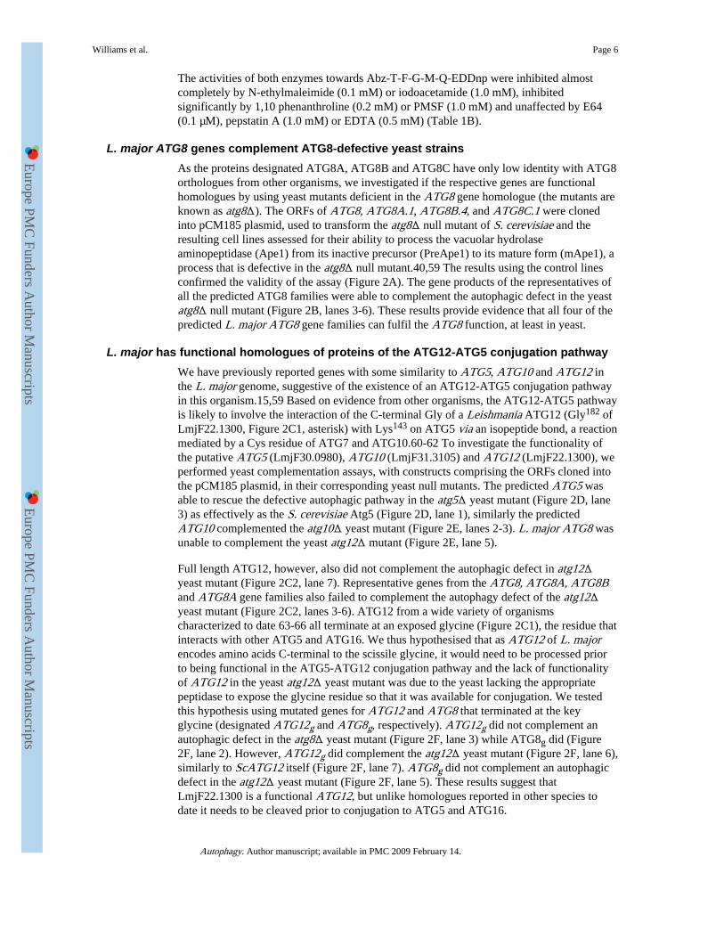
The activities of both enzymes towards Abz-T-F-G-M-Q-EDDnp were inhibited almostcompletely by N-ethylmaleimide (0.1 mM) or iodoacetamide (1.0 mM), inhibitedsignificantly by 1,10 phenanthroline (0.2 mM) or PMSF (1.0 mM) and unaffected by E64(0.1 µM), pepstatin A (1.0 mM) or EDTA (0.5 mM) (Table 1B).
L. major ATG8 genes complement ATG8-defective yeast strainsAs the proteins designated ATG8A, ATG8B and ATG8C have only low identity with ATG8orthologues from other organisms, we investigated if the respective genes are functionalhomologues by using yeast mutants deficient in the ATG8 gene homologue (the mutants areknown as atg8Δ). The ORFs of ATG8, ATG8A.1, ATG8B.4, and ATG8C.1 were clonedinto pCM185 plasmid, used to transform the atg8Δ null mutant of S. cerevisiae and theresulting cell lines assessed for their ability to process the vacuolar hydrolaseaminopeptidase (Ape1) from its inactive precursor (PreApe1) to its mature form (mApe1), aprocess that is defective in the atg8Δ null mutant.40,59 The results using the control linesconfirmed the validity of the assay (Figure 2A). The gene products of the representatives ofall the predicted ATG8 families were able to complement the autophagic defect in the yeastatg8Δ null mutant (Figure 2B, lanes 3-6). These results provide evidence that all four of thepredicted L. major ATG8 gene families can fulfil the ATG8 function, at least in yeast.
L. major has functional homologues of proteins of the ATG12-ATG5 conjugation pathwayWe have previously reported genes with some similarity to ATG5, ATG10 and ATG12 inthe L. major genome, suggestive of the existence of an ATG12-ATG5 conjugation pathwayin this organism.15,59 Based on evidence from other organisms, the ATG12-ATG5 pathwayis likely to involve the interaction of the C-terminal Gly of a Leishmania ATG12 (Gly182 ofLmjF22.1300, Figure 2C1, asterisk) with Lys143 on ATG5 via an isopeptide bond, a reactionmediated by a Cys residue of ATG7 and ATG10.60-62 To investigate the functionality ofthe putative ATG5 (LmjF30.0980), ATG10 (LmjF31.3105) and ATG12 (LmjF22.1300), weperformed yeast complementation assays, with constructs comprising the ORFs cloned intothe pCM185 plasmid, in their corresponding yeast null mutants. The predicted ATG5 wasable to rescue the defective autophagic pathway in the atg5Δ yeast mutant (Figure 2D, lane3) as effectively as the S. cerevisiae Atg5 (Figure 2D, lane 1), similarly the predictedATG10 complemented the atg10Δ yeast mutant (Figure 2E, lanes 2-3). L. major ATG8 wasunable to complement the yeast atg12Δ mutant (Figure 2E, lane 5).
Full length ATG12, however, also did not complement the autophagic defect in atg12Δyeast mutant (Figure 2C2, lane 7). Representative genes from the ATG8, ATG8A, ATG8Band ATG8A gene families also failed to complement the autophagy defect of the atg12Δyeast mutant (Figure 2C2, lanes 3-6). ATG12 from a wide variety of organismscharacterized to date 63-66 all terminate at an exposed glycine (Figure 2C1), the residue thatinteracts with other ATG5 and ATG16. We thus hypothesised that as ATG12 of L. majorencodes amino acids C-terminal to the scissile glycine, it would need to be processed priorto being functional in the ATG5-ATG12 conjugation pathway and the lack of functionalityof ATG12 in the yeast atg12Δ yeast mutant was due to the yeast lacking the appropriatepeptidase to expose the glycine residue so that it was available for conjugation. We testedthis hypothesis using mutated genes for ATG12 and ATG8 that terminated at the keyglycine (designated ATG12g and ATG8g, respectively). ATG12g did not complement anautophagic defect in the atg8Δ yeast mutant (Figure 2F, lane 3) while ATG8g did (Figure2F, lane 2). However, ATG12g did complement the atg12Δ yeast mutant (Figure 2F, lane 6),similarly to ScATG12 itself (Figure 2F, lane 7). ATG8g did not complement an autophagicdefect in the atg12Δ yeast mutant (Figure 2F, lane 5). These results suggest thatLmjF22.1300 is a functional ATG12, but unlike homologues reported in other species todate it needs to be cleaved prior to conjugation to ATG5 and ATG16.
Williams et al. Page 6
Autophagy. Author manuscript; available in PMC 2009 February 14.
Europe PM
C Funders A
uthor Manuscripts
Europe PM
C Funders A
uthor Manuscripts

ATG12 forms punctate structures in L. major promastigotesIt is known that ATG8 N-terminally tagged with GFP becomes associated with punctatestructures (putative autophagosomes) in Leishmania promastigotes, especially under thestarvation conditions and during differentiation between its life cycle forms.13 To discernthe functional similarity and differences between ATG8 and ATG12, we compared thelocalisation of ATG12 N-terminally tagged to a red fluorescent protein (and so designatedRFP-ATG12) and GFP-ATG8-containing puncta in the cytoplasm of L. majorpromastigotes. In nutrient-rich medium, RFP-ATG12 had a diffuse cytosolic distribution(data not shown). Under starvation conditions, most cells redistributed RFP-ATG12 into apunctate structure ∼500 nm wide (Figure 3A1). Promastigotes co-expressing both RFP-ATG12 and GFP-ATG8 produced, under starvation, some punctate structures displaying redand green fluorescence. This is consistent with both proteins being involved inautophagosome formation (Figure 3A1). As RFP-ATG12 produced structures thatoverlapped with GFP-ATG8-containing puncta, we investigated whether each proteinbecame lipidated. Two GFP-ATG8 proteins were detected in starved promastigotes, the non-lipidated GFP-ATG8* (∼ 40 kDa) and the rapidly migrating lipidated GFP-ATG8-PE(Figure 3B1). However, for ATG12 only one major protein was detected at 50 kDa, which isconsistent with the predicted mass of the fusion protein (Figure 3B2). On longer exposure atrace of higher molecular mass GFP-ATG12 could be detected (not shown), which could bean ATG12-ATG5 conjugate. These findings are consistent with ATG12 having a role inautophagosome biogenesis, and one that is different from that of ATG8.
L. major ATG8 paralogues form puncta in promastigotesWe recently demonstrated that GFP-ATG8 can be used as a marker for tracking putativeautophagosomes in L. major.13, 15 As the four families of ATG8 in L. major cancomplement a yeast ATG8 gene-deletion mutant and are substrates for ATG4s, we predictedthat they would associate with the putative autophagosomes in L. major in vivo. GFP taggedATG8A, ATG8B and ATG8C were expressed in L. major promastigotes (designatedWT(pN-ATG8A), WT(pN-ATG8B), WT(pN-ATG8C)) and characterised as describedpreviously.13 As predicted, GFP-ATG8A, GFP-ATG8B and GFP-ATG8C did formpunctate structures under certain conditions (Figure 4A). Interestingly, the single punctum inWT(pN-ATG8B) and WT(pN-ATG8C) was always located close to the flagellar pocket(Figure 4A3 and 4A4), the only site of endocytosis and exocytosis in the cell. Most early logphase promastigotes of WT(pN-ATG8A), WT(pN-ATG8B) and WT(pN-ATG8C) innutrient-rich medium had the GFP-tagged proteins evenly distributed throughout thecytoplasm (similar to the diffuse pattern described previously for GFP-ATG8 transgenic celllines at logarithmic phase of growth,13 although 2-10% had a single GFP-labelled punctumin the cytosol (Figure 4B, 0 h). However, in contrast to the pattern described previously forGFP-ATG8, no peak in punctate structures was observed for GFP-ATG8A, GFP-ATG8B orGFP-ATG8C during metacyclogenesis, and the proportion of cells containing punctaremained consistently low during growth in nutrient rich medium (data not shown).Suspension of the promastigotes of WT(pN-ATG8A), WT(pN-ATG8B and WT(pN-ATG8C) in nutrient-deprived medium (PBS) resulted in the production of puncta in mostWT(pN-ATG8A) cells by the end of the 4 h incubation (Figure 4A.1 and 4B), with therebeing 5-8 puncta per cell (Figure 4A1). In contrast, there was no significant increase in theoccurrence of puncta in WT(pN-ATG8B) and WT(pN-ATG8C) upon starvation (Figure4B). These data suggest that ATG8 and ATG8A are associated with starvation-inducedpuncta (putative autophagosomes), but that ATG8B and ATG8C are associated with otherstructures in the cell.
To analyse the possible involvement of Leishmania ATG4.2 in the formation of these punctain vivo, we investigated the formation of structures containing GFP-ATG8A, GFP-ATG8B
Williams et al. Page 7
Autophagy. Author manuscript; available in PMC 2009 February 14.
Europe PM
C Funders A
uthor Manuscripts
Europe PM
C Funders A
uthor Manuscripts

and GFP-ATG8C in a L. major mutant lacking ATG4.2 (Δatg4.2, 13). Δatg4.2promastigotes transfected with the GFP-ATG8A, GFP-ATG8B and GPF-ATG8C constructs(designated Δatg4.2[pN-ATG8A], Δatg4.2[pN-ATG8B], Δatg4.2[pN-ATG8C]) wereassessed for the formation of puncta formation under starvation conditions. Fluorescencemicroscopic analysis of the distribution of these ATG8 homologues in the Δatg4.2 cell linein nutrient-rich medium prior to their transfer to starvation medium was similar to that of thewild type cell lines transfected with the same construct, in that they were diffuselydistributed throughout the cytosol with just a small proportion of cells containing a singlepunctum (data not shown). This is in contrast to the greater number of ATG8-containingstructures that were present in Δatg4.2[pN-ATG8] than in WT(pN-ATG8) under similarconditions (Besteiro et al., 2006). As with WT(pN-ATG8B) and WT(pN-ATG8C),suspension of Δatg4.2[pN-ATG8B] and Δatg4.2[pN-ATG8C] promastigotes in nutrient-deprived medium did not result in a significant increase in puncta. Interestingly, however,starvation of Δatg4.2[pN-ATG8A] did not result in the increased occurrence of puncta asoccurred with WT[pN-ATG8A] (Figure 4C1 and comparison with Figure 4A2).Δatg4.2[pN-ATG8] cells on the other hand retained the enhanced number of puncta alsounder these conditions (Figure 4C2 and comparison with Figure 4A1). These results areconsistent with and support the biochemical data reported above that ATG4.2 is able tohydrolyse ATG8A rapidly and suggest that this processing could be involved inautophagosome formation in vivo.
ATG4.2 has a post-lipidation role in L. major autophagic pathway involving ATG8The enhanced number of GFP-ATG8-containing puncta in Δatg4.2 relative to wild typecells (see ref. 13 and Figure 4C2) led us to hypothesise that ATG4.2 is a deconjugatingenzyme for lipidated ATG8. We have previously shown by Western blot analysis thatextracts from the Δatg4.2 cell line over-expressing GFP-ATG8 contained relatively moreGFP-ATG8 conjugated to PE than wild type cells similarly expressing GFP-ATG8.13 Toinvestigate this further, we developed an in vitro deconjugation assay that involved theincubation of purified recombinant ATG4.2 with lysate from Δatg4.2[pN-ATG8] for 3 h.Western blot analysis carried out on aliquots collected at 90 min intervals showed areduction in GFP-ATG8-PE (Figure 5A). Next, ATG4.2 was over-expressed in WT [pN-GFP-ATG8] to produce the WT [pN-ATG4.2/pN-GFP-ATG8] line and assessed for itsability to form putative autophagosomes. Microscopic analysis of the cell line revealed thatGFP-ATG8-containing puncta did not become more abundant as the line progressed fromlog to early stationary phase of growth (Figure 5B1). Western blot analysis of cell extractsfrom early stationary phase promastigotes from both cell lines showed that the WT [pN-ATG4.2/pN-GFP-ATG8] had predominantly the unlipidated form of GFP-ATG8 (Figure5B2, lane 1), consistent with the rapid cleavage of ATG8 from PE on the nascent structures(putative autophagosomes) preventing them becoming fully formed. One predictedconsequence of this is that WT [pN-ATG4.2/pN-GFP-ATG8] promastigotes would be lessable to withstand starvation, a process that requires a functional autophagic pathway. Thiswas found to be the case (Figure 5C). Our studies have shown that the enzyme has only lowactivity towards ATG8 itself (Figure 1A1), but presumably it has better activity towardsATG8 bound to PE. These data together suggest that in L. major ATG4.2 is a deconjugatingenzyme for ATG8 and that the increased amount of ATG4.2 in the transgenic line results inthe deconjugation of PE from ATG8 before the putative autophagosomes can be properlyformed.
DiscussionThe occurrence of autophagy in a wide variety of organisms ranging from protozoa tohumans is indicative of its indispensable contribution to the life of eukaryotes.7,67 Much is
Williams et al. Page 8
Autophagy. Author manuscript; available in PMC 2009 February 14.
Europe PM
C Funders A
uthor Manuscripts
Europe PM
C Funders A
uthor Manuscripts

known of the molecular mechanisms of autophagy in mammals and yeast,67 but far lessabout the process in lower eukaryotes such as Leishmania. We have previously shown thatautophagy has a role in the transformation of Leishmania, 13,15 but predictions based onbioinformatic analyses suggested differences in the autophagic machinery from thatunderpinning autophagy in mammals; most notably the occurrence of multiple ATG8-likegenes and the uncertainty about the existence of the two conjugation pathways described forhigher eukaryotes.15,49,50,68 Thus, the major aim of this study was to verifyexperimentally whether the multiple ATG8s encoded in the Leishmania genome function asATG8s and whether or not they have different roles and also to verify whether the genesidentified as potential components of the ATG12-ATG5 conjugation pathway in the L.major genome15 are indeed functional homologues.
The yeast complementation approach has provided compelling evidence that L. majorATG8, ATG8A, ATG8B and ATG8C can indeed carry out the function of the yeast Atg8(Figure 2). Moreover, examples of the three families of ATG8-like proteins were found toform GFP-labelled puncta in promastigotes transfected with GFP-tagged ATG8s (Figure 4).However, the puncta occurrence, location and number differed between the four families.GFP-ATG8B and GFP-ATG8C occur as punctate structures in only a low proportion of cellsduring growth in normal medium and, unlike GFP-ATG8,13 no peak in puncta formationwas observed during metacyclogenesis or in response to starvation. In addition, ATG8B andATG8C do not co-localise with ATG8-labelled puncta (data not shown). These data suggestthat while ATG8B and ATG8C are likely to have ATG8-like role, this appears to be relatednot to the putative autophagosomes but to another as yet unidentified vesicle. Interestingly,GFP-ATG8B and ATG8C-containing puncta were always located close to the flagellarpocket, perhaps pointing to a potential role in endocytic trafficking or recycling. Thus thesedata suggest that ATG8B and ATG8C do not have a role in macroautophagy which is incontrast to the mammalian situation where the three ATG8 homologues (GATE-16,GABARAP and LC3) are more similar (all are in the ATG8 clade, Figure S2B) and all areinvolved in autophagy.32,33,69
ATG8A of L. major differs from each of the other families of ATG8s. It rarely was observedin punctate structures during growth in nutrient rich medium, but a strong induction ofATG8A labelled puncta was observed in response to nutrient deprivation - indicating apotential role in starvation-induced autophagy. Its role in autophagy was reinforced by thefinding that it often co-localised with ATG8-labelled puncta (Figure 4). However, itsdistribution, even when in puncta, and occurrence, the puncta did not increase duringmetacyclogenesis, indicate clearly that ATG8A differs in its detailed role from that of ATG8itself.
One common feature between yeast and higher eukaryotes is the existence of just one typeof ATG8, albeit there are multiple copies in the latter but just one copy in yeast.27,69-72Thus the multiple families of ATG8s in Leishmania is most unusual, and indeed it has noknown parallel (not occurring even in other trypanosomatid genera such as Trypanosoma).The ATG8 families are found in different Leishmania species that cause diverse diseases(for example L. infantum, L. braziliensis and L. mexicana), so they are likely to haveconserved functions in the parasite. We have now shown that they are all ATG8-like inaction but that there are clear differences. Discovering the parts that each plays is the nextgoal, but as ATG8A, ATG8B and ATG8C occur as multicopy gene families it will beproblematic to investigate this through generation of ATG8 family-specific knockout lines.
L. major also has two homologues of ATG4 (ATG4.1 and ATG4.2)13 and we hypothesisedthat part of the selective functioning of the ATG8s could be mediated through differentialcleavage by the differing ATG4s. We investigated the specificity of the two ATG4s towards
Williams et al. Page 9
Autophagy. Author manuscript; available in PMC 2009 February 14.
Europe PM
C Funders A
uthor Manuscripts
Europe PM
C Funders A
uthor Manuscripts

ATG8 and the 3 families of ATG8-like proteins using an in vitro cleavage assay. The results(Figure 1) indicate clear specificity of cleavage between different ATG4.1 and ATG4.2 andthe four ATG8 families. The mechanism determining this specificity remains uncertain,however, the two ATG4s showed some similarity in their ability to cleave short peptidefluorogenic substrates (designed on the basis of the cleavage sequence in each class ofATG8; Table 2) and so the specificity of the ATG4s towards the ATG8s must be controlledby other residues than the amino acids close to the cleavage site. Interestingly, the results ofthis approach using short peptide fluorogenic substrates show that the scissile glycineresidue is not a requirement for hydrolysis; previously it has also been shown that in theaction of HsAtg4b on a 9-mer the scissile glycine plays no role in the hydrolysis ofATG8.53 Thus residues other than this glycine and the adjacent amino acids must beimportant in determining the substrate binding of the enzyme, as previously postulated.35,73
Our investigation has provided firm evidence for the existence in L. major of functionalequivalents of proteins of the ATG12-ATG5 pathway. LmjF22.1300 has moderate levels ofidentity to both ATG8 and ATG12 and it failed to reconstitute the autophagic defect inatg12Δ yeast mutant lines. However, the gene encoding a truncated form not encodingresidues beyond the scissile glycine fully restored autophagy to atg12Δ yeast (Figure 2F). Inaddition, LmjF22.1300 was unlike ATG8 in that under starvation stress it located to a singlelarge punctum in most cells with as few as 13% having multiple structures and we couldfind no evidence that it was lipidated. Thus we have tentatively designated this as ATG12.Nevertheless it is an unusual ATG12 in that it requires cleavage prior to conjugation toATG5, a situation that does not occur in yeast or mammalian cells. In vitro assays wouldsuggest that neither ATG4.1 nor ATG4.2 can process ATG12, so it must be that a differentpeptidase cleaves ATG12 in vivo. A syntenic homologue to LmjF22.1300 is present in othertrypanosomatids; T. cruzi (designated TcATG8.2) and T. brucei (Tb927.7.3320). Ourfindings with L. major ATG12 are similar to the reported findings for TcATG8.2 in that theprotein forms punctate structures in T. cruzi, but less so than TcATG8.1, and this apparentlydoes not involve conjugation to PE.12 It is possible, therefore, that TcATG8.2 is also afunctional ATG12. The yeast functional assays used also provided support for the predictedgene identification for LmjF30.0980 (ATG5) and LmjF31.3105 (ATG10), as they couldrestore autophagy to yeast mutants lacking the ATG5 and ATG10 yeast genes. LmjF30.0980and LmjF31.3105 also have syntenic homologues in T. cruzi and T. brucei, so our resultssuggest that L. major, T. brucei and T. cruzi are similar to other eukaryotes studied in havingtwo conjugation pathways involved in autophagosome biogenesis.
Findings with mammalian cells and yeast are all in accordance with ATG4s having adeconjugation role. A down-regulation of HsAtg4b in HEK293 cells can increaseendogenous LC3-PE and GABARAP-PE levels.33 A mutant ATG8 construct lackingresidues beyond the scissile glycine expressed in atg4Δ S. cerevisiae, which is incapable offorming autophagosomes, resulted in a build up of autophagosomes that cannot be deliveredto the vacuole – indicating the importance of the role of ATG4 at the late stage of theautophagic pathway.36,74 Our previous study suggested that L. major ATG4.2 is involvedin deconjugating ATG8 from putative autophagosomes.13 We have now shown that L.major ATG4.2 has delipidation activity and can directly delipidate ATG8-PE to ATG8(Figure 5A). Moreover, over-expression of ATG4.2 greatly reduced GFP-ATG8 puncta inwild type lines (Figure 5B). This is consistent with a greatly increased rate of deconjugationof ATG8 from the surface of the putative autophagosomes before they are fully formed andthus their enhanced clearance from the cytosol, as has been described in other systems.33,34,43 Thus our findings on the effects of the absence or over-expression of ATG4.2 onthe location and form of GFP-ATG8 in Leishmania accords well with the roles of ATG4s inmammals. The enhanced susceptibility of these L. major lines to the stress of starvation (seeref. 13 and Figure 5C) is also indicative that both lines lack fully functional autophagic
Williams et al. Page 10
Autophagy. Author manuscript; available in PMC 2009 February 14.
Europe PM
C Funders A
uthor Manuscripts
Europe PM
C Funders A
uthor Manuscripts

machinery. Thus although our data show that ATG4.1 and ATG4.2 in Leishmania havedifferences in specificity towards substrates and are of different importance in certain roles,there appears to be redundancy between the two ATG4s. Further evidence for this isprovided by the finding that ATG4.2 is capable of processing ATG8 during prolongedincubation in vitro (this study) and that Δatg4.1-deficient lines expressing GFP-ATG8 formputative autophagosomes in vivo (Williams, Mottram and Coombs, unpublished).
Materials and MethodsIdentification, cloning, expression and purification of genes involved in autophagy
Genes potentially involved in autophagy in L. major have been reported.15 The openreading frames (ORFs) of ATG4.1 (LmjF32.3890) and ATG4.2 (LmjF30.0270), ATG8(LmjF19.1630), ATG8A (LmjF19.0840), ATG8B (LmjF19.0850) and ATG8C(LmjF09.0150) were amplified by PCR experiments using the Expand High Fidelity PCRsystem (Roche Molecular Biochemicals) from L. major genomic DNA, isolated as describedby (75), using gene-specific primers modified with appropriate restriction sites and epitopetags (to facilitate cloning into their respective expression vectors and resolution of cleavageproducts, respectively) as detailed in Table S1. All PCR assays were carried out in aGeneAmp 9600 PCR system (PerkinElmer Life Sciences) for 30 cycles of denaturation(94°C, 15 s), annealing (65°C, 15 s) and extension (72°C, 2 min). Each ORF was verified bynucleotide sequencing (MBSU, University of Glasgow) and cloned into the pET expressionvectors (Invitrogen) pre-digested with appropriate restriction enzymes to produce theplasmids detailed in Table S1(i). BL21(DE3) (Invitrogen) were transformed with plasmidsto provide the strains listed in Table S2 (ii). Protein expression was carried out overnight at15°C after induction with 1- 2 mM isopropyl-β-D-thiogalactopyranoside (IPTG).Recombinant proteins were purified using an affinity chromatography column (bioCAD®700E work station) and eluants of the ATG4s and ATG8s were dialysed against 50 mMTris-HCl, pH 7.9, 125 mM NaCl, 10% glycerol at 4°C overnight and stored at 4°C with 1mM DDT for ATG4 and 1% urea for ATG8. In some instances, storage buffer for ATG8was acidified to pH 5.5 with 1 mM HCl to prevent aggregation.
Assay of cleavage of ATG8s in vitroPellets of E. coli co-expressing each ATG8 homologue with either ATG4.1 or ATG4.2(Table S2(ii)) were lysed in reaction buffer (containing 25 mM Tris-HCl, pH 7.4, 50 mMKCl, 10 mM β-mercaptoethanol). The supernatant protein obtained after centrifugation at13,000 × g for 30 min at 4°C was adjusted to 5 mg ml−1 and incubated at 30°C for a fixedtime (specified by the particular experiment). The action of different peptidase inhibitors onthe hydrolytic action of ATG4 on ATG8 was determined by adding the inhibitors prior todisruption of the cell membrane. In the deconjugation assay, Δatg4.2 promastigotesexpressing GFP-ATG8-PE were lysed in reaction buffer and the supernatant was incubatedwith recombinant ATG4.2 for up to 180 min. Reactions were stopped with SDS-PAGEloading buffer (1 mM Tris-HCl pH 6.8, 20% glycerol, 10% β-mercaptoethanol and 40%bromophenol blue). The hydrolytic actions of the recombinant ATG4.1 and ATG4.2 on His-ATG8-HA, His-ATG8A-HA, His-ATG8B-HA and His-ATG8C-HA and recombinantATG4.2 on GFP-ATG8-PE were determined by Western blot analysis with α-His-tag andα-GFP antibodies (Pierce). Supernatants containing each ATG8 homologue and GFP-ATG8-PE but lacking ATG4 were used as controls.
MALDI-TOF MS analysisElectronspray ionization (ESI)–MS/MS was carried out on a hybrid quadrupole time-of-flight mass spectrometer, Q.STAR (Applied Biosystems). Solution containing partially-cleaved ATG8 treated with 1% formic acid was loaded into a borosilicate nanoflow
Williams et al. Page 11
Autophagy. Author manuscript; available in PMC 2009 February 14.
Europe PM
C Funders A
uthor Manuscripts
Europe PM
C Funders A
uthor Manuscripts

(micromass), and sent into the ESI source. MS/MS data were processed by Analyst QSsoftware, which is capable of deconvoluting the peaks of varying charge state, to obtainprotein masses.
Activity analyses of ATG4sEnzymatic activities of purified recombinant ATG4.1 and ATG4.2 towards FluorescenceResonance Energy Transfer (FRET) peptides (Abz-X-X-X-X-Q-EDDnp) as substrates,synthesized by LJ, were analysed in an assay (1 ml) comprising substrate and 10 ng ofrecombinant protein in 50 mM Tris/HCl, pH 7.9, 125 mM NaCl, 10 mM dithiothreitol.Peptide hydrolysis was measured using the LS 50-B PerkinElmer spectrofluorometer(λexcitation, 320 nm; λemission, 420 nm) at 30°C for 15 min after initiation with the enzyme.Specific activities were determined using 2.5 mM substrate. Kinetics parameters (Km, Vmaxand kcat/Km) were determined using substrate concentrations ranging from 0.5-10 mM andcalculated using the Grafit software. For inhibition experiments, the reaction mixture waspre-incubated for 30 min at 20°C with inhibitors and residual activities determined.
Yeast complementation studiesThe ORFs of ATG5 (LmjF32.3890), ATG12 (LmjF22.1300) and ATG10 (LmjF19.1630)were amplified by PCR experiments using the Expand High Fidelity PCR system (RocheMolecular Biochemicals) from L. major genomic DNA isolated as described by Medina-Acosta et al 75 using gene-specific primers modified with appropriate restriction sites (tofacilitate cloning into their respective expression vector) as detailed in Table S1. All PCRassays were carried out in a GeneAmp 9600 PCR system (PerkinElmer Life Sciences) for 30cycles of denaturation (94°C, 15 s), annealing (65°C, 15 s) and extension (72°C, 2 min).Each ORF was verified by nucleotide sequencing (MBSU, University of Glasgow) andcloned into the pCM185 expression vectors (EUROSCARF) pre-digested with appropriaterestriction enzymes to produce the plasmids detailed in Table S1(iii). Each plasmid wassubsequently used to transform their respective mutant strain of S. cerevisiae(EUROSCARF) by the lithium acetate method.76 The resultant transformed lines and wildtype lines (Table S2(iv) and (iii)) were grown in YNB medium containing 0.5% ammoniumsulphate and amino acids but lacking uracil until the optical density at 600 nm (OD600) was1.0, whereupon they were split into two aliquots. One aliquot was washed twice with SD(-N) and incubated in the same medium for up to 16 h at 30°C to induce starvation. Thesecond aliquot was washed and resuspended in fresh growth medium and incubated underthe same conditions as a control. The cells were then harvested by centrifugation (3,000 × g,4°C, 5 min), suspended in Laemmli’s sample buffer and the cells walls disrupted with lysisbuffer (containing 0.1% lyticase). Yeast proteins were resolved by SDS-PAGE, transferredto a nitrocellulose membrane and analysed by Western blotting with α-(pro)aminopeptidaseI primary antibodies (a gift from Prof D. Klionsky) and horseradish peroxidise-conjugatedanti-rabbit secondary antibodies. The bands were visualized with the ECLTM detectionsystem (Amersham Biosciences).
Occurrence in L. major transgenic lines of puncta associated with ATG proteinsL. major (MHOM/JL/80/Friedlin) promastigotes were used throughout this study. TheΔatg4.2 null mutant (in which both alleles of the gene encoding the cysteine peptidaseATG4.2 had been deleted) was generated as described previously.77 Generation of the GFP-ATG8 fusion construct is also described in Besteiro et al.13 To generate GFP-ATG8A,GFP-ATG8B, GFP-ATG8C and RFP-ATG12, their respective ORFs amplified with primersmodified with the BgIII/XhoI restriction sites (Table S1(ii)) were sub-cloned into GFP-containing pNUS-GFPnH and RFP-containing pNUS-RFPnH vectors, respectively,78yielding the plasmids detailed in Table S1(ii). These constructs were used to transfect L.major wild type and Δatg4.2 lines as previously described.79 All lines generated (Table
Williams et al. Page 12
Autophagy. Author manuscript; available in PMC 2009 February 14.
Europe PM
C Funders A
uthor Manuscripts
Europe PM
C Funders A
uthor Manuscripts

S2(i)) were grown in complete medium (modified Eagle’s Medium [designated HOMEM]with 10% (v/v) heat-inactivated foetal calf serum (HiFCS)) at 25 °C as described inWilliams et al.80 In vitro procyclic promastigote-metacyclic promastigote differentiationwas monitored by growing promastigotes in complete medium to stationary phase of growth(1-3 × 10⁷ cells ml−1) whereupon the proportion of metacyclic promastigotes wasdetermined as described.81,82 Autophagy was induced in promastigotes expressing ATG8homologues by incubation in PBS at 2 × 10⁷ cells ml−1 for up to 5 h as previously described.15 Puncta biogenesis was monitored in these starved promastigotes by fluorescencemicroscopy using Zeiss Axioplan2 imaging and Axiophot2 microscope equipped with FITCor Rhodamine filter sets, respectively; images were captured at intervals and along the Z-axis and processed using VELOCITY (Improvision). The presence and number of GFP-ATG8s- or RFP-ATG12-containing vesicles within these cells was assessed by observing atleast 100 cells from 3 independent experiments. Unless otherwise stated, early log and latelog/early stationary phase cells of all cell lines corresponds to 1-5 ×10⁶ and 5-9 ×10⁶
parasites ml−1, respectively.
Parasite viabilityViability of the L. major wild type and Δatg4.2 lines subjected to starvation conditions wasmeasured by the 3-(4,5-dimethylthiazol-2-yl)-2,5-diphenyl tetrazolium bromide (MTT)assay as described previously.15 Briefly, 4 × 10⁶ stationary phase promastigotes werefurther incubated for 45 min at 37°C with MTT to a final concentration of 1 mg ml−1,whereupon absorbance at 620 nm was measured using a microtitre plate reader.
Antibodies and immunoblottingWestern blot analysis was performed on lysates from L. major or E. coli lysates separated onstandard 12.5% Laemmli separating gels (SDS-PAGE) containing 6 M urea (Urea-SDS-PAGE) at pH 9.2. The primary α-GFP antibody (Abcam Ltd) was used at 1:2000 dilution,whereas α-His antibodies (Pierce), α-(pro)aminopeptidase 1 antibodies (a gift from Prof D.Klionsky) and the secondary anti-rabbit IgG antibody were used at a 1:5000 dilution. TheWest-Pico chemiluminescence detection system (Pierce) was used to visualize the bands ofantigens.
Supplementary MaterialRefer to Web version on PubMed Central for supplementary material.
AcknowledgmentsWe thank Richard J. Burchmore of the Sir Henry Wellcome Functional Genomics Facility, Faculty of Biomedicaland Life Sciences, University of Glasgow for the mass spectrometry analyses. This study was funded by theMedical Research Council [grant numbers G9722968, G0000508, G0700127]. We thank the EuropeanSaccharomyces cerevisiae Archive for Functional analysis (EUROSCARF), Frankfurt, Germany for the yeastmutant lines and yeast expression plasmids and Prof. D. J. Klionsky, University of Michigan, USA, for the α-(pro)aminopeptidase antibodies.
Abbreviations
MTT 3-(4,5-dimethylthiazol-2-yl)-2,5-diphenyl tetrazolium bromide
Ape vacuolar hydrolase aminopeptidase
WT wild type
GFP green fluorescence protein
Williams et al. Page 13
Autophagy. Author manuscript; available in PMC 2009 February 14.
Europe PM
C Funders A
uthor Manuscripts
Europe PM
C Funders A
uthor Manuscripts

RFP red fluorescence protein
ORF open reading frame
DTT Dithiothreitol
PBS phosphate-buffered saline
PE phosphotidylethanolamine
MALDI-TOF Matrix Assisted Laser Desorption Ionization-Time Of Flight
Abz 2-aminobenzoyl
EDDnp ethylenediamine-2,4-dinitrophenyl
kDa kilodaltons
LC3 microtubule associate protein light chain-3
GATE-16 Golgi-associated ATPase enhancer of 16 kDa
GABARAP GABA receptor-associated protein.
References1. Yoshimori T. New insights into molecular mechanisms and roles of autophagy. Cell Struct and
Funct. 2004; 29(supplement):13.
2. Boland B, Nixon RA. Neuronal macroautophagy: from development to degeneration. Mol AspectsMed. 2006; 27:503–19. [PubMed: 16999991]
3. Levine B, Klionsky DJ. Development by self-digestion: Molecular mechanisms and biologicalfunctions of autophagy. Dev Cell. 2004; 6:463–77. [PubMed: 15068787]
4. Penaloza C, Orlanski S, Ye Y, Entezari-Zaher T, Javdan M, Zakeri Z. Cell death in mammaliandevelopment. Current Pharmaceutical Design. 2008; 14:184–96. [PubMed: 18220829]
5. Kuma A, Hatano M, Matsui M, Yamamoto A, Nakaya H, Yoshimori T, et al. The role of autophagyduring the early neonatal starvation period. Nature. 2004; 432:1032–6. [PubMed: 15525940]
6. Levine B, Kroemer G. Autophagy in the pathogenesis of disease. Cell. 2008; 132:27–42. [PubMed:18191218]
7. Mizushima N, Levine B, Cuervo AM, Klionsky DJ. Autophagy fights disease through cellular self-digestion. Nature. 2008; 451:1069–75. [PubMed: 18305538]
8. Kondo Y, Kanzawa T, Sawaya R, Kondo S. The role of autophagy in cancer development andresponse to therapy. Nat Rev Cancer. 2005; 5:726–34. [PubMed: 16148885]
9. Rajawat YS, Bossis I. Autophagy in aging and in neurodegenerative disorders. Hormones (Athens).2008; 7:46–61. [PubMed: 18359744]
10. Koike M, Shibata M, Waguri S, Yoshimura K, Tanida I, Kominami E, et al. Participation ofautophagy in storage of lysosomes in neurons from mouse models of neuronal ceroid-lipofuscinoses (Batten disease). Am J Pathol. 2005; 167:1713–28. [PubMed: 16314482]
11. Lee HK, Iwasaki A. Autophagy and antiviral immunity. Curr Opin Immunol. 2008; 20:23–9.[PubMed: 18262399]
12. Alvarez VE, Kosec G, Sant'Anna C, Turk V, Cazzulo JJ, Turk B. Autophagy is involved innutritional stress response and differentiation in Trypanosoma cruzi. J Biol Chem. 2008;283:3454–64. [PubMed: 18039653]
13. Besteiro S, Williams RAM, Morrison LS, Coombs GH, Mottram JC. Endosome sorting andautophagy are essential for differentiation and virulence of Leishmania major. J Biol Chem. 2006;281:11384–96. [PubMed: 16497676]
14. Herman M, Perez-Morga D, Schtickzelle N, Michels PA. Turnover of glycosomes during life-cycledifferentiation of Trypanosoma brucei. Autophagy. 2008; 4:294–308. [PubMed: 18365344]
Williams et al. Page 14
Autophagy. Author manuscript; available in PMC 2009 February 14.
Europe PM
C Funders A
uthor Manuscripts
Europe PM
C Funders A
uthor Manuscripts

15. Williams RA, Tetley L, Mottram JC, Coombs GH. Cysteine peptidases CPA and CPB are vital forautophagy and differentiation in Leishmania mexicana. Mol Microbiol. 2006; 61:655–74.[PubMed: 16803590]
16. Dunn WA Jr. Autophagy and related mechanisms of lysosome-mediated protein degradation.Trends Cell Biol. 1994; 4:139–43. [PubMed: 14731737]
17. Klionsky DJ, Cregg JM, Dunn WA Jr. Emr SD, Sakai Y, Sandoval IV, et al. A unifiednomenclature for yeast autophagy-related genes. Dev Cell. 2003; 5:539–45. [PubMed: 14536056]
18. Levine B, Yuan J. Autophagy in cell death: an innocent convict? J Clin Invest. 2005; 115:2679–88.[PubMed: 16200202]
19. Thumm M, Egner R, Koch B, Schlumpberger M, Straub M, Veenhuis M, et al. Isolation ofautophagocytosis mutants of Saccharomyces cerevisiae. FEBS Lett. 1994; 349:275–80. [PubMed:8050581]
20. Tsukada M, Ohsumi Y. Isolation and characterization of autophagy-defective mutants ofSaccharomyces cerevisiae. FEBS Lett. 1993; 333:169–74. [PubMed: 8224160]
21. Hemelaar J, Lelyveld VS, Kessler BM, Ploegh HL. A single protease, Apg4B, is specific for theautophagy-related ubiquitin-like proteins GATE-16, MAP1-LC3, GABARAP, and Apg8L. J BiolChem. 2003; 278:51841–50. [PubMed: 14530254]
22. Kabeya Y, Mizushima N, Ueno T, Yamamoto A, Kirisako T, Noda T, et al. LC3, a mammalianhomologue of yeast Apg8p, is localized in autophagosome membranes after processing. EMBO J.2000; 19:5720–8. [PubMed: 11060023]
23. Mann SS, Hammarback JA. Molecular characterization of light chain 3. A microtubule bindingsubunit of MAP1A and MAP1B. J Biol Chem. 1994; 269:11492–7. [PubMed: 7908909]
24. Mann SS, Hammarback JA. Gene localization and developmental expression of light chain 3: acommon subunit of microtubule-associated protein 1A(MAP1A) and MAP1B. J Neurosci Res.1996; 43:535–44. [PubMed: 8833088]
25. Sagiv Y, Legesse-Miller A, Porat A, Elazar Z. GATE-16, a membrane transport modulator,interacts with NSF and the Golgi v-SNARE GOS-28. EMBO J. 2000; 19:1494–504. [PubMed:10747018]
26. Wang CW, Klionsky DJ. The molecular mechanism of autophagy. Mol Med. 2003; 9:65–76.[PubMed: 12865942]
27. Ketelaar T, Voss C, Dimmock SA, Thumm M, Hussey PJ. Arabidopsis homologues of theautophagy protein Atg8 are a novel family of microtubule binding proteins. FEBS Lett. 2004;567:302–6. [PubMed: 15178341]
28. Scherz-Shouval R, Sagiv Y, Shorer H, Elazar Z. The COOH terminus of GATE-16, an intra-Golgitransport modulator, is cleaved by the human cysteine protease HsApg4A. J Biol Chem. 2003;278:14053–8. [PubMed: 12473658]
29. Tanida I, Komatsu M, Ueno T, Kominami E. GATE-16 and GABARAP are authentic modifiersmediated by Apg7 and Apg3. Biochem Biophys Res Commun. 2003; 300:637–44. [PubMed:12507496]
30. Ichimura Y, Kirisako T, Takao T, Satomi Y, Shimonishi Y, Ishihara N, et al. A ubiquitin-likesystem mediates protein lipidation. Nature. 2000; 408:488–92. [PubMed: 11100732]
31. Ichimura Y, Imamura Y, Emoto K, Umeda M, Noda T, Ohsumi Y. In vivo and in vitroreconstitution of Atg8 conjugation essential for autophagy. J Biol Chem. 2004; 279:40584–92.[PubMed: 15277523]
32. Sou YS, Tanida I, Komatsu M, Ueno T, Kominami E. Phosphatidylserine in addition tophosphatidylethanolamine is an in vitro target of the mammalian Atg8 modifiers, LC3,GABARAP, and GATE-16. J Biol Chem. 2006; 281:3017–24. [PubMed: 16303767]
33. Tanida I, Ueno T, Kominami E. LC3 conjugation system in mammalian autophagy. Int J BiochemCell Biol. 2004; 36:2503–18. [PubMed: 15325588]
34. Nakatogawa H, Ichimura Y, Ohsumi Y. Atg8, a ubiquitin-like protein required for autophagosomeformation, mediates membrane tethering and hemifusion. Cell. 2007; 130:165–78. [PubMed:17632063]
Williams et al. Page 15
Autophagy. Author manuscript; available in PMC 2009 February 14.
Europe PM
C Funders A
uthor Manuscripts
Europe PM
C Funders A
uthor Manuscripts

35. Tanida I, Ueno T, Kominami E. Human light chain 3/MAP1LC3B is cleaved at its carboxyl-terminal Met121 to expose Gly120 for lipidation and targeting to autophagosomal membranes. JBiol Chem. 2004; 279:47704–10. [PubMed: 15355958]
36. Xie Z, Nair U, Klionsky DJ. Atg8 Controls phagophore expansion during autophagosomeformation. Mol Biol Cell. 2008 Epub May 28.
37. Suzuki K, Ohsumi Y. Molecular machinery of autophagosome formation in yeast, Saccharomycescerevisiae. FEBS Lett. 2007; 581:2156–61. [PubMed: 17382324]
38. Abeliovich H, Dunn WA Jr. Kim J, Klionsky DJ. Dissection of autophagosome biogenesis intodistinct nucleation and expansion steps. J Cell Biol. 2000; 151:1025–34. [PubMed: 11086004]
39. Ponder EL, Bogyo M. Ubiquitin-like modifiers and their deconjugating enzymes in medicallyimportant parasitic protozoa. Eukaryot Cell. 2007; 6:1943–52. [PubMed: 17905920]
40. Marino G, Uria JA, Puente XS, Quesada V, Bordallo J, Lopez-Otin C. Human autophagins, afamily of cysteine proteinases potentially implicated in cell degradation by autophagy. J BiolChem. 2003; 278:3671–8. [PubMed: 12446702]
41. Yoshimoto K, Hanaoka H, Sato S, Kato T, Tabata S, Noda T, et al. Processing of ATG8s,ubiquitin-like proteins, and their deconjugation by ATG4s are essential for plant autophagy. PlantCell. 2004; 16:2967–83. [PubMed: 15494556]
42. Yoshimoto K. Recent advances in plant autophagy research: plant atg mutants. TanpakushitsuKakusan Koso. 2006; 51:1532–6. [PubMed: 16922433]
43. Kirisako T, Ichimura Y, Okada H, Kabeya Y, Mizushima N, Yoshimori T, et al. The reversiblemodification regulates the membrane-binding state of Apg8/Aut7 essential for autophagy and thecytoplasm to vacuole targeting pathway. J Cell Biol. 2000; 151:263–76. [PubMed: 11038174]
44. Fujioka Y, Noda NN, Fujii K, Yoshimoto K, Ohsumi Y, Inagaki F. In vitro reconstitution of plantAtg8 and Atg12 conjugation systems essential for autophagy. J Biol Chem. 2008; 283:1921–8.[PubMed: 18039664]
45. Kuma A, Mizushima N, Ishihara N, Ohsumi Y. Formation of the approximately 350-kDa Apg12-Apg5.Apg16 multimeric complex, mediated by Apg16 oligomerization, is essential for autophagyin yeast. J Biol Chem. 2002; 277:18619–25. [PubMed: 11897782]
46. Mizushima N, Kuma A, Kobayashi Y, Yamamoto A, Matsubae M, Takao T, et al. Mouse Apg16L,a novel WD-repeat protein, targets to the autophagic isolation membrane with the Apg12-Apg5conjugate. J Cell Sci. 2003; 116:1679–88. [PubMed: 12665549]
47. Hanada T, Noda NN, Satomi Y, Ichimura Y, Fujioka Y, Takao T, et al. The Atg12-Atg5 conjugatehas a novel E3-like activity for protein lipidation in autophagy. J Biol Chem. 2007; 282:37298–302. [PubMed: 17986448]
48. Huang DT, Miller DW, Mathew R, Cassell R, Holton JM, Roussel MF, et al. A unique E1-E2interaction required for optimal conjugation of the ubiquitin-like protein NEDD8. Nat Struct MolBiol. 2004; 11:927–35. [PubMed: 15361859]
49. Herman M, Gillies S, Michels PA, Rigden DL. Autophagy and related processes intrypanosomatids - Insights from genomic and bioinformatic analyses. Autophagy. 2006; 2:107–18.[PubMed: 16874069]
50. Rigden DJ, Herman M, Gillies S, Michels PA. Implications of a genomic search for autophagy-related genes in trypanosomatids. Biochem Soc Trans. 2005; 33:972–4. [PubMed: 16246023]
51. Mukaiyama H, Baba M, Osumi M, Aoyagi S, Kato N, Ohsumi Y, et al. Modification of aubiquitin-like protein Paz2 conducted micropexophagy through formation of a novel membranestructure. Mol Biol Cell. 2004; 15:58–70. [PubMed: 13679515]
52. Kumanomidou T, Mizushima T, Komatsu M, Suzuki A, Tanida I, Sou YS, et al. The crystalstructure of human Atg4b, a processing and de-conjugating enzyme for autophagosome-formingmodifiers. J Mol Biol. 2006; 355:612–8. [PubMed: 16325851]
53. Sugawara K, Suzuki NN, Fujioka Y, Mizushima N, Ohsumi Y, Inagaki F. Structural basis for thespecificity and catalysis of human Atg4B responsible for mammalian autophagy. J Biol Chem.2005; 280:40058–65. [PubMed: 16183633]
54. Fass E, Amar N, Elazar Z. Identification of essential residues for the C-terminal cleavage of themammalian LC3: a lesson from yeast Atg8. Autophagy. 2007; 3:48–50. [PubMed: 17102583]
Williams et al. Page 16
Autophagy. Author manuscript; available in PMC 2009 February 14.
Europe PM
C Funders A
uthor Manuscripts
Europe PM
C Funders A
uthor Manuscripts

55. Mazel D, Pochet S, Marliere P. Genetic characterization of polypeptide deformylase, a distinctiveenzyme of eubacterial translation. EMBO J. 1994; 13:914–23. [PubMed: 8112305]
56. Tang J, Hernandez G, LeMaster DM. Increased peptide deformylase activity for N-formylmethionine processing of proteins overexpressed in Escherichia coli: application tohomogeneous rubredoxin production. Protein Expr Purif. 2004; 36:100–5. [PubMed: 15177290]
57. Geoghegan KF, Dixon HB, Rosner PJ, Hoth LR, Lanzetti AJ, Borzilleri KA, et al. Spontaneousalpha-N-6-phosphogluconoylation of a “His tag” in Escherichia coli: the cause of extra mass of258 or 178 Da in fusion proteins. Anal Biochem. 1999; 267:169–84. [PubMed: 9918669]
58. Chae YK, Im H, Zhao Q, Doelling JH, Vierstra RD, Markley JL. Prevention of aggregation afterrefolding by balanced stabilization-destabilization: production of the Arabidopsis thaliana proteinAPG8a (At4g21980) for NMR structure determination. Protein Expr Purif. 2004; 34:280–3.[PubMed: 15003262]
59. Ohsumi Y. Molecular mechanism of autophagy in yeast, Saccharomyces cerevisiae. Philos Trans RSoc Lond B Biol Sci. 1999; 354:1577–80. [PubMed: 10582243]
60. Tanida I, Mizushima N, Kiyooka M, Ohsumi M, Ueno T, Ohsumi Y, et al. Apg7p/Cvt2p: A novelprotein-activating enzyme essential for autophagy. Mol Biol Cell. 1999; 10:1367–79. [PubMed:10233150]
61. Shintani T, Mizushima N, Ogawa Y, Matsuura A, Noda T, Ohsumi Y. Apg10p, a novel protein-conjugating enzyme essential for autophagy in yeast. EMBO J. 1999; 18:5234–41. [PubMed:10508157]
62. Mizushima N, Sugita H, Yoshimori T, Ohsumi Y. A new protein conjugation system in human.The counterpart of the yeast Apg12p conjugation system essential for autophagy. J Biol Chem.1998; 273:33889–92. [PubMed: 9852036]
63. Hanada T, Ohsumi Y. Structure-function relationship of Atg12, a ubiquitin-like modifier essentialfor autophagy. Autophagy. 2005; 1:110–8. [PubMed: 16874032]
64. Suzuki NN, Yoshimoto K, Fujioka Y, Ohsumi Y, Inagaki F. The crystal structure of plant ATG12and its biological implication in autophagy. Autophagy. 2005; 1:119–26. [PubMed: 16874047]
65. Umemiya R, Matsuo T, Hatta T, Sakakibara S, Boldbaatar D, Fujisaki K. Cloning andcharacterization of an autophagy-related gene, ATG12, from the three-host tick Haemaphysalislongicornis. Insect Biochem Mol Biol. 2007; 37:975–84. [PubMed: 17681237]
66. Umemiya R, Matsuo T, Hatta T, Sakakibara S, Boldbaatar D, Fujisaki K. Autophagy-related genesfrom a tick, Haemaphysalis longicornis. Autophagy. 2008; 4:79–81. [PubMed: 17938584]
67. Xie Z, Klionsky DJ. Autophagosome formation: core machinery and adaptations. Nat Cell Biol.2007; 9:1102–9. [PubMed: 17909521]
68. Muthuvijayan V, Marten MR. In silico reconstruction of nutrient-sensing signal transductionpathways in Aspergillus nidulans. In Silico Biol. 2004; 4:605–31. [PubMed: 15752076]
69. Kabeya Y, Mizushima N, Yamamoto A, Oshitani-Okamoto S, Ohsumi Y, Yoshimori T. LC3,GABARAP and GATE16 localize to autophagosomal membrane depending on form-II formation.J Cell Sci. 2004; 117:2805–12. [PubMed: 15169837]
70. Hanaoka H, Noda T, Shirano Y, Kato T, Hayashi H, Shibata D, et al. Leaf senescence andstarvation-induced chlorosis are accelerated by the disruption of an Arabidopsis autophagy gene.Plant Physiol. 2002; 129:1181–93. [PubMed: 12114572]
71. Tanida I, Wakabayashi M, Kanematsu T, Minematsu-Ikeguchi N, Sou YS, Hirata M, et al.Lysosomal turnover of GABARAP-phospholipid conjugate is activated during differentiation ofC2C12 cells to myotubes without inactivation of the mTor kinase-signaling pathway. Autophagy.2006; 2:264–71. [PubMed: 16874098]
72. Wu J, Dang Y, Su W, Liu C, Ma H, Shan Y, et al. Molecular cloning and characterization of ratLC3A and LC3B--two novel markers of autophagosome. Biochem Biophys Res Commun. 2006;339:437–42. [PubMed: 16300744]
73. He H, Dang Y, Dai F, Guo Z, Wu J, She X, et al. Post-translational modifications of threemembers of the human MAP1LC3 family and detection of a novel type of modification forMAP1LC3B. J Biol Chem. 2003; 278:29278–87. [PubMed: 12740394]
Williams et al. Page 17
Autophagy. Author manuscript; available in PMC 2009 February 14.
Europe PM
C Funders A
uthor Manuscripts
Europe PM
C Funders A
uthor Manuscripts

74. Kirisako T, Ichimura Y, Okada H, Kabeya Y, Mizushima N, Yoshimori T, et al. The reversiblemodification regulates the membrane-binding state of Apg8/Aut7 essential for autophagy and thecytoplasm to vacuole targeting pathway. J Cell Biol. 2000; 151:263–76. [PubMed: 11038174]
75. Medina-Acosta E, Cross GA. Rapid isolation of DNA from trypanosomatid protozoa using asimple 'mini-prep' procedure. Mol Biochem Parasitol. 1993; 59:327–9. [PubMed: 8341329]
76. Ausubel, F.; Moore, DD.; Seidman, JG.; Struhe, K. Current Protocols in Molecular Biology. JohnWiley & Sons Inc.; New York: 1989.
77. Mottram JC, Souza AE, Hutchison JE, Carter R, Frame MJ, Coombs GH. Evidence fromdisruption of the lmcpb gene array of Leishmania mexicana that cysteine proteinases are virulencefactors. Proc Natl Acad Sci USA. 1996; 93:6008–13. [PubMed: 8650210]
78. Tetaud E, Lecuix I, Sheldrake T, Baltz T, Fairlamb AH. A new expression vector for Crithidiafasciculata and Leishmania. Mol Biochem Parasitol. 2002; 120:195–204. [PubMed: 11897125]
79. Robinson KA, Beverley SM. Improvements in transfection efficiency and tests of RNAinterference (RNAi) approaches in the protozoan parasite Leishmania. Mol Biochem Parasitol.2003; 128:217–228. [PubMed: 12742588]
80. Williams RAM, Kelly SM, Mottram JC, Coombs GH. 3-mercaptopyruvate sulfurtransferase ofLeishmania contains an unusual C-terminal extension and is involved in thioredoxin andantioxidant metabolism. J Biol Chem. 2003; 278:1480–6. [PubMed: 12419809]
81. Leon LL, Temporal RM, Soares MJ, Grimaldi JG. Proteinase activities during temperature-inducedstage differentiation of species complexes of Leishmania. Acta Trop. 1994; 56:289–98. [PubMed:8023752]
82. Bates PA, Tetley L. Leishmania mexicana: induction of metacyclogenesis by cultivation ofpromastigotes at acidic pH. Exp Parasitol. 1993; 76:412–23. [PubMed: 8513879]
Williams et al. Page 18
Autophagy. Author manuscript; available in PMC 2009 February 14.
Europe PM
C Funders A
uthor Manuscripts
Europe PM
C Funders A
uthor Manuscripts

Figure 1. Hydrolysis of L. major ATG8s by L. major ATG4s in vitroA. Soluble fractions of E. coli expressing His-ATG8-HA (row 1), His-ATG8A-HA (row 2),His-ATG8B-HA (row 3), His-ATG8C-HA (row 4) and His-ATG12-HA (row 5) alone (lane1) or co-expressed with either ATG4.1 (lane 2) or ATG4.2 (lane 3) were incubated for 30min at 30°C in 50 mM Tris-HCl, pH 8.0, 125 mM NaCl and 40 mM β-mercaptoethanol andthen analysed by Western blot with α-His antibody. The cleaved ATG8 bands are labelledwith an asterisk.B. Hydrolysis of His-ATG8-HA by ATG4.1.(B1) The hydrolysis was inhibited by 1 mM NEM (lane 2) and 1 mM iodoacetamide (lane 6)but unaffected by 1 mM PMSF (lane 3), 1 mM pepstatin (lane 4) and 10 mM 1,10-phenanthroline (lane 5). Lane 1 shows the control (no inhibitor added). Incubation was asdescribed in A.(B2) Hydrolysis of His-ATG8-HA by ATG4.1 was progressive over 60 min.
Williams et al. Page 19
Autophagy. Author manuscript; available in PMC 2009 February 14.
Europe PM
C Funders A
uthor Manuscripts
Europe PM
C Funders A
uthor Manuscripts

C. Analysis of the product of the cleavage of His-ATG8-HA by ATG4.1. Hydrolysis wasover 30 min as described in A.(C2) The product was purified by Ni+-affinity chromatography and analysed by SDS-PAGE,two main proteins were apparent.(C1) The cleaved products were analysed by MALDI TOF. Six molecules were detected andtheir molecular masses are given.
Williams et al. Page 20
Autophagy. Author manuscript; available in PMC 2009 February 14.
Europe PM
C Funders A
uthor Manuscripts
Europe PM
C Funders A
uthor Manuscripts

Figure 2. Complementation studies with L. major ATG genes in autophagy-defectiveSaccharomyces cerevisiaeA. Western blot analysis of aminopeptidase I (Ape1) in yeast mutant atg8Δ (lane 1), wildtype (lane 2) and yeast mutant atg12Δ (lane 3) S. cerevisiae. Cells were incubated understarvation conditions in SD(-N) medium for 16 h, lysed, and API processing was analyzedusing rabbit α-Ape1 antibodies. ProApe1 and mature Ape1 (mApe1) are indicated.B. Western blot analysis of Ape1 in atg8Δ yeast transformed with pCM185 containing L.major ATG8 homologues: ATG8 (lane 3), ATG8A (lane 4), ATG8B (lane 5), ATG8C (lane2) or transformed with pCM185 only (lane 6). Wild type S. cerevisiae are shown in lane 1.The cells were cultured under starvation conditions in SD(-N) medium for 4 h and Ape1
Williams et al. Page 21
Autophagy. Author manuscript; available in PMC 2009 February 14.
Europe PM
C Funders A
uthor Manuscripts
Europe PM
C Funders A
uthor Manuscripts
processing was analyzed using rabbit α-API antibodies. ProApe1 and mature Ape1 (mApe1)are indicated.C1. Amino acid sequence alignment of the C-terminal end of L. major ATG12 with those ofT. brucei, S. cerevisiae, human and A. thaliana. Identical and conserved amino acids areshaded in black and grey, respectively. Shade of grey depends upon number of residuesconserved. The carboxyl-terminal glycine identified in A. thaliana ATG12 (Hanada et al.,2005) and the two hydrophobic residues (Y149 and F154) required for conjugation to ATG10(Suzuki et al., 2006; Hanada et al., 2006) and essential for the formation of the ATG12-ATG5 conjugate are marked with an asterisk. L. major, LmjF22.1300; T. brucei,Tb927.7.3320; S. cerevisiae, P38316; human, AAH11033; A. thaliana, AAM70187. Thenumbers in parenthesis represent the amino acid number of the first residue in the alignment.C2. Western blot analysis of Ape1 in atg12Δ S. cerevisiae transformed with pCM185containing L. major ATG8 homologues: ATG8 (lane 3), ATG8A (lane 4), ATG8B (lane 5),ATG8C (lane 6), the putative L. major ATG12 (lane 7) or transformed with pCM185 only(lane 2). Wild type S. cerevisiae were used as the control (lane 1). The S. cerevisiae werecultured under starvation condition for 16 h and Ape1 processing was analyzed using arabbit α-API antibodies. ProApe1 and mature Ape1 (mApe1) are indicated.D. Western blot analysis of Ape1 in atg5Δ S. cerevisiae transformed with pCM185containing L. major ATG5 (lane 3) and pCM185 only (lane 2) and treated as described inC2. Wild type S. cerevisiae was included as a positive control (lane 1).E. Western blot analysis of Ape1 in atg10Δ S. cerevisiae transformed with pCM185containing the L. major ATG10 homologue (lanes 2-3), or transformed with pCM185 only(lane 1) or ATG8 (lane 5) and treated as described in C2. Wild type S. cerevisiae wasincluded as positive control (lane 4).F. Western blot analysis of ApeI in atg8Δ S. cerevisiae (lanes 2-4) and atg12Δ S. cerevisiae(lanes 5-7) transformed with pCM185 containing ATG8g (lanes 2, 5), ATG12g (lanes 3, 6)and S. cerevisiae ATG12 (lanes 4, 7), respectively, and treated as described in C2. Wild typeS. cerevisiae was included as positive control (lane 1).
Williams et al. Page 22
Autophagy. Author manuscript; available in PMC 2009 February 14.
Europe PM
C Funders A
uthor Manuscripts
Europe PM
C Funders A
uthor Manuscripts

Figure 3. Localization of ATG12 in L. major promastigotesA. Occurrence of RFP-ATG12-containing structures (A2) and GFP-ATG8-containingputative autophagosomes (A1) in wild type L. major promastigotes expressing both proteins.The position of the kinetoplast is shown (blue) using DAPI staining. L. major procyclicpromastigotes were suspended in nutrient-deprived medium for 120 min and observed byfluorescence microscopy. The merge of the RFP-ATG12 and GFP-ATG8 images, the co-labelled structure is enlarged at the bottom left, and the DIC image of the promastigote areshown in A3 and A4, respectively. Scale bar, 10 μm.B. Western blot analysis using α-GFP (B1) and α-RFP (B2) antibodies on L. majorprocyclic promastigote cell extracts (5-8 × 10⁶ cells ml−1) transfected with GFP-ATG8 andRFP-ATG12. The α-GFP antibody detected a faster migrating lipidated ATG8 (labelledGFP-ATG8-PE) and a higher molecular mass unlipidated ATG8 (labelled GFP-ATG8*),resolved in a urea SDS-PAGE gel (B1). The anti-RFP antibody showed just a single protein(labelled RFP-ATG12) consistent with the predicted size of the fusion protein (B2).
Williams et al. Page 23
Autophagy. Author manuscript; available in PMC 2009 February 14.
Europe PM
C Funders A
uthor Manuscripts
Europe PM
C Funders A
uthor Manuscripts
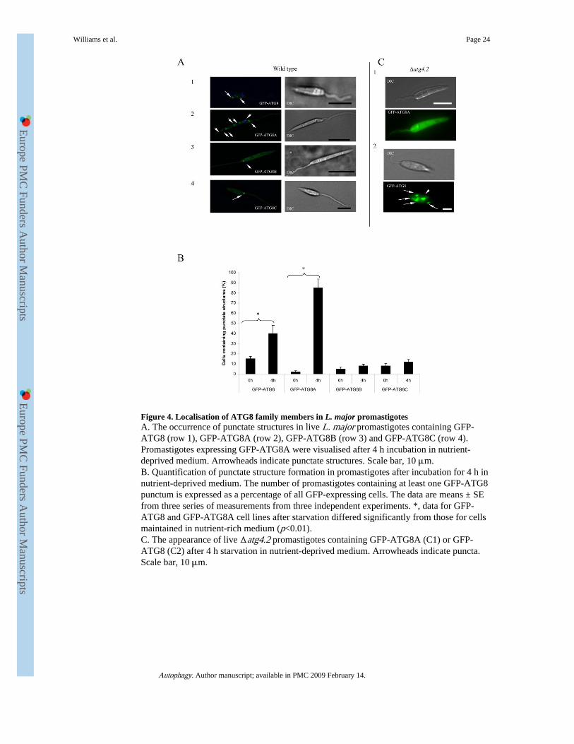
Figure 4. Localisation of ATG8 family members in L. major promastigotesA. The occurrence of punctate structures in live L. major promastigotes containing GFP-ATG8 (row 1), GFP-ATG8A (row 2), GFP-ATG8B (row 3) and GFP-ATG8C (row 4).Promastigotes expressing GFP-ATG8A were visualised after 4 h incubation in nutrient-deprived medium. Arrowheads indicate punctate structures. Scale bar, 10 μm.B. Quantification of punctate structure formation in promastigotes after incubation for 4 h innutrient-deprived medium. The number of promastigotes containing at least one GFP-ATG8punctum is expressed as a percentage of all GFP-expressing cells. The data are means ± SEfrom three series of measurements from three independent experiments. *, data for GFP-ATG8 and GFP-ATG8A cell lines after starvation differed significantly from those for cellsmaintained in nutrient-rich medium (p<0.01).C. The appearance of live Δatg4.2 promastigotes containing GFP-ATG8A (C1) or GFP-ATG8 (C2) after 4 h starvation in nutrient-deprived medium. Arrowheads indicate puncta.Scale bar, 10 μm.
Williams et al. Page 24
Autophagy. Author manuscript; available in PMC 2009 February 14.
Europe PM
C Funders A
uthor Manuscripts
Europe PM
C Funders A
uthor Manuscripts

Figure 5. A role for ATG4.2 in processing lipidated ATG8 in L. majorA. Western blot analysis using α-GFP antibody on Δatg4.2 [pN-ATG8] promastigote cellextracts (5-8 × 10⁶ cells ml−1) show an abundance of lipidated ATG8 (labelled GFP-ATG8-PE) (lane 0). Incubation of the cell extract with recombinant ATG4.2 for 90 and 180 min at30°C resulted in a decrease in GFP-ATG8-PE (lanes 2 and 3). Transketolase was used as aninternal loading control.B1. WT[pN-ATG8] (open squares) and WT[pN-ATG4.2/ pN-GFP-ATG8] (closed circles)promastigotes were compared for the occurrence of putative autophagosomes during growthin vitro in normal medium. Data are means ± SD from 3 independent experiments.
Williams et al. Page 25
Autophagy. Author manuscript; available in PMC 2009 February 14.
Europe PM
C Funders A
uthor Manuscripts
Europe PM
C Funders A
uthor Manuscripts

B2. Western blot analysis of extracts from early stationary phase WT[pN-ATG4.2/pN-GFP-ATG8] (lane 1) and WT[pN-GFP-ATG8] (lane 2) promastigotes separated by SDS-PAGEcontaining 6 M urea and detected with α-GFP antibody. Cleaved ATG8 (GFP-ATG8*) andlipidated ATG8 (GFP-ATG8-PE) are indicated. EF1α was used as an internal loadingcontrol.C. Sensitivity to starvation of WT[pN-ATG8] (open squares) and WT[pN-ATG4.2/pN-GFPATG8] (closed circles) L. major promastigotes. Cells were incubated in PBS and theirviability assessed by the MTT assay. Data are means ± SD from four replicates. *: data forWT[pN-ATG4.2/pN-GFP-ATG8] and WT[pN-GFP-ATG8] promastigotes differedsignificantly (P < 0.05).
Williams et al. Page 26
Autophagy. Author manuscript; available in PMC 2009 February 14.
Europe PM
C Funders A
uthor Manuscripts
Europe PM
C Funders A
uthor Manuscripts

Europe PM
C Funders A
uthor Manuscripts
Europe PM
C Funders A
uthor Manuscripts
Williams et al. Page 27
Table 1A
Enzymatic activity of recombinant ATG4.1 and ATG4.2 towards fluorogenic Abz-X-X-X-X-Q-EDDnpsubstrates (25 μM). Values are the means ± SE.
ATG8 homologuewith sequence
Substrates Specific Activities(nmoles.min−1 mg protein−1)
ATG4.1 ATG4.2
Hs-MAP-LC3 Abz-T-F-G-M-Q-EDDnp 99.4 ± 9.2 59.0 ± 3.2
ATG8 Abz-T-Y-G-G-Q-EDDnp 23.5 ± 7.7 <5
Abz-T-Y-A-G-Q-EDDnp 111.6 ± 7.9 <5
ATG8A Abz-S-M-G-A-Q-EDDnp 47.0 ± 9.2 <5
ATG8B Abz-A-M-G-A-Q-EDDnp 311. 6 ± 8.2 144.5 ± 3.4
ATG8B Abz-A-M-G-G-Q-EDDnp 28.1 ± 3.4 <5
Abz-A-M-A-A-Q-EDDnp 149.7 ± 3.8 22.3 ± 3.5
ATG8B Abz-I-A-G-L-Q-EDDnp 163.4 ± 4.2 83.7 ± 5.8
Abz-I-A-A-L-Q-EDDnp 152.0 ± 6.2 83.5 ± 7.3
ATG8C Abz-C-M-G-A-Q-EDDnp 128.9 ± 4.8 65.3 ± 8.4
Autophagy. Author manuscript; available in PMC 2009 February 14.

Europe PM
C Funders A
uthor Manuscripts
Europe PM
C Funders A
uthor Manuscripts
Williams et al. Page 28
Table 1B
Inhibitory activity of peptidase inhibitors against recombinant ATG4.1 and ATG4.2. ATG4 (0.5 μg) wasincubated with the inhibitors at room temperature for 15 min, and residual activity against 25 μM Abz-T-F-G-M-Q-EDDnp analysed. Values are the means ± SE.
Inhibitors % Inhibition
ATG4.1 ATG4.2
0.1 mM N-Ethylmaleimide (NEM) 99 ± 1.4 99 ± 1.9
1.0 mM Iodoacetamide 99 ± 2.1 97 ± 3.2
0.2 mM 1,10 phenanthroline 50 ± 9.3 43 ± 4.2
0.2 mM Phenylmethylsulfonyl fluoride (PMSF) 22 ± 8.4 17 ± 5.7
1.0μM E64 <1.0 ± 0.0 < 0.1 ± 0.0
1.0 mM Pepstatin A <1.0 ± 0.0 <0.1 ± 0.0
0.5 mM EDTA <1.0 ± 0.0 <0.1 ± 0.0
Autophagy. Author manuscript; available in PMC 2009 February 14.

Europe PM
C Funders A
uthor Manuscripts
Europe PM
C Funders A
uthor Manuscripts
Williams et al. Page 29
Table 2
Kinetics parameters of recombinant ATG4.1 activity towards Fluorescence Resonance Energy Transfer(FRET) peptides Abz-peptidyl-Q-EDDnp.
Substrate Km (μM) kcat (sec−1) kcat/Km (μM−1 sec−1)
Abz-T-F-G-M-Q-EDDnp 0.6 ± 0.05 4.4 ±0.5 7.3
Abz-T-Y-G-G-Q-EDDnp 0.1 ± 0.05 1.1 ±0.4 10.7
Abz-T-Y-A-G-Q-EDDnp 0.6 ± 0.07 4.5 ±0.3 7.6
Abz-S-M-G-M-A-EDDnp 0.2 ± 0.05 1.6 ±0.5 7.8
Abz-A-M-G-A-Q-EDDnp 0.5 ± 0.05 8.8 ±0.0 17.7
Abz-A-M-G-G-Q-EDDnp 0.2 ± 0.06 1.4 ±0.2 6.6
Abz-A-M-A-A-Q-EDDnp 0.2 ± 0.07 5.7 ±0.3 28.4
Abz-I-A-G-L-Q-EDDnp 0.5 ± 0.04 4.9 ±0.3 9.9
Abz-I-A-A-L-Q-EDDnp 0.2 ± 0.09 5.4 ±0.2 26.8
Abz-C-M-G-A-Q-EDDnp 0.5 ± 0.08 6.2 ±0.3 12.3
Autophagy. Author manuscript; available in PMC 2009 February 14.