Capillaria sp. from Cryptotis parva - The Keep Eastern Illinois ...
21
Eastern Illinois University e Keep Masters eses Student eses & Publications 1978 Capillaria sp. from Cryptotis parva Joseph n'Dong Eastern Illinois University is research is a product of the graduate program in Zoology at Eastern Illinois University. Find out more about the program. is is brought to you for free and open access by the Student eses & Publications at e Keep. It has been accepted for inclusion in Masters eses by an authorized administrator of e Keep. For more information, please contact [email protected]. Recommended Citation n'Dong, Joseph, "Capillaria sp. from Cryptotis parva" (1978). Masters eses. 3249. hps://thekeep.eiu.edu/theses/3249
-
Upload
khangminh22 -
Category
Documents
-
view
0 -
download
0
Transcript of Capillaria sp. from Cryptotis parva - The Keep Eastern Illinois ...

Eastern Illinois UniversityThe Keep
Masters Theses Student Theses & Publications
1978
Capillaria sp. from Cryptotis parvaJoseph n'DongEastern Illinois UniversityThis research is a product of the graduate program in Zoology at Eastern Illinois University. Find out moreabout the program.
This is brought to you for free and open access by the Student Theses & Publications at The Keep. It has been accepted for inclusion in Masters Thesesby an authorized administrator of The Keep. For more information, please contact [email protected].
Recommended Citationn'Dong, Joseph, "Capillaria sp. from Cryptotis parva" (1978). Masters Theses. 3249.https://thekeep.eiu.edu/theses/3249
PAPER CERTIFICATE #2
TO: Graduate Degree Candidates who have written formal theses.
SUBJECT: Permission to reproduce theses.
The University Library is receiving a number of requests from other institutions asking permission to reproduce dissertations for inclusion in their library holdings. Although no copyright laws are involved, we feel that professional courtesy demands that permission be obtained from the author before we allow theses to be copied.
Please sign one of the following statements:
Booth Library of Eastern Illinois University has my permission to lend my thesis to a reputable college or university for the purpose of copying it for inclusion in that institution's library or research holdings.
Date Author
I respectfully request Booth Library of .Eastern Illinois University not allow my thesis be reproduced because ----------------
Date Author
pdm

CAPILLARIA SP.
FROM CRYPTOTIS PARVA {TITLE)
BY
JOSEPH n 1 DONG -..-
THESIS
SUBMITTED IN PARTIAL FULFILLMENT OF THE REQUIREMENTS
FOR THE DEGREE OF
Master of Zoology
IN THE GRADUATE SCHOOL, EASTERN ILLINOIS UNIVERSITY
CHARLESTON, ILLINOIS
I HEREBY RECOMMEND THIS THESIS BE ACCEPTED AS FULFILLING
THIS PART OF THE GRADUATE DEGREE CITED ABOVE
~2- /J~. 111 r DATE ADVISER , - "- I
v\ DEPARTMENT 1HEAD

The undersigned, appointed by the Chairman of the Department of Zoology,
have examined a thesis entitled
Capillaria sp. from C~yptotis parva
Presented by
n Joseph 'Dong
a candicate for the degree o·i Master of Zoology
and hereby certify that in their opinion it is acceptable.
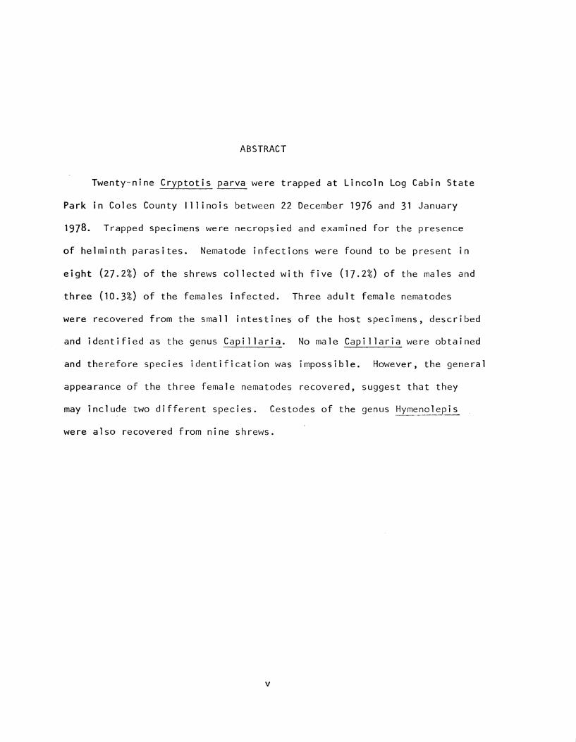
ABSTRACT
Twenty-nine Cryptotis parva were trapped at Lincoln Log Cabin State
Park in Coles County Illinois between 22 December 1976 and 31 January
1978. Trapped specimens were necropsied and examined for the presence
of helminth parasites. Nematode infections were found to be present in
eight (27.2%) of the shrews collected with five (17.2%) of the males and
three (10.3%) of the females infected. Three adult female nematodes
were recovered from the small intestines of the host specimens, described
and identified as the genus Capillaria. No male Capillaria were obtained
and therefore species identification was impossible. However, the general
appearance of the three female nematodes recovered, suggest that they
may include two different species. Cestodes of the genus Hymenolepis
were also recovered from nine shrews.
V

ACKNOWLEDGMENTS
I wish to sincerely thank Dr. B. T. Ridgeway (advisor) for his
continuing guidance and support which have made the production of this
thesis possible. Thanks also go to Dr. Richard Andrews for providing
traps and offering valuable advice on collecting methods and selection
of trapping sites. Appreciation is extended to Dr. Jaime Maya for con
firming the identification and sex of host shrews. In addition to the
above, I wish to express my gratitude to Dr. Richard Funk, Dr. Verne
Kniskern, Dr. Patrick Docter and Dr. Garland Reigel for assisting in the
editing of this paper.
ii
ACKNOWLEDGMENTS .
LIST OF FIGURES.
ABSTRACT . . .
INTRODUCTION
MATERIALS AND METHODS
RESULTS ..
DISCUSSION
LITERATURE CI TED
TABLE OF CONTENTS
iii
Page
ii
iv
V
1
2
4
8
11

LI ST OF FI GU RES
Figure Page
I. Posterior End Capi 1 laria sp. #2 7
II. Posterior End Capi 1 laria sp. #1 7
111. Anterior End Capillaria sp. #2 7
IV. Anterior End Cap i 11 aria sp. #1 . . . . . 7
V. Vu 1 var Region Capillaria sp. #2 7
VI. Vulvar Region Capillaria sp. #1 7
VII. Egg from Uterine Tract of Capillaria sp. #1 7
VI 11. Egg from Uterine Tract of Capillaria sp. #2 7
iv

INTRODUCTION
The genus Capillaria (subfamily Capillariinae, family Trichuridae)
was established in 1800 by Zeder on material recovered from poultry.
The type species is Capillaria anatis (Schran~ 1790). Since then Capil
laria have been reported from numerous vertebrate hosts throughout the
world. Yamaguti (1961) listed 37 species in fish, 13 in amphibians,
13 in reptiles, 104 in birds and 88 in mammals. Few infections have
been reported in humans (MacArthur, 1924; Morishita and Tani, 1960).
Twenty-two species of Capillaria are reported from insectivores (Lopez
Neyra, 1947; York and Maplestone, 1926; Travassos, 1915; Teixeira De
Freitas and Lent, 1936; Skryabin, Shikhobalova, and I. V. Orlov, 1957;
and Yamaguti, 1961). Eighteen species have been described from Soricidae,
particularly the genera Sorex, Blarina, Crocidura and Suncus.
Two authors in North America have worked with Capillaria of Soricidae.
Ogren (1953), described Capillaria blarinae from the esophagus of Blarind
brevicauda in Illinois and Read (1949), in his studies on North American
species of Capillaria, described Capillaria taushi from the small intes
tine of Sorex cinereus at Madison, Wisconsin. No Capillaria have been
reported in Cryptotis parva.
2
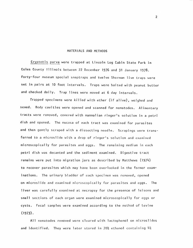
MATERIALS AND METHODS
Cryptotis parva were trapped at Lincoln Log Cabin State Park in
Coles County Illinois between 22 December 1976 and 31 January 1978.
Forty-four museum special snaptraps and twelve Sherman live traps were
set in pairs at 10 foot intervals. Traps were baited with peanut butter
and checked daily. Trap lines were moved at 6 day intervals.
Trapped specimens were killed with ether (if alive), weighed and
sexed. Body cavities were opened and scanned for nematodes. Alimentary
tracts were removed, covered with mammalian ringer's solution in a petri
dish and opened. The mucosa of each tract was examined for parasites
and then gently scraped with a dissecting needle. Scrapings were trans
ferred to a microslide with a drop of ringer's solution and examined
microscopically for parasites and eggs. The remaining medium in each
petri dish was decanted and the sediment examined. Digestive tract
remains were put into migration jars as described by Matthews (1974)
to recover paras.ites which may have been overlooked in the former exam
inations. The urinary bladder of each specimen was removed, opened
on micros! ide and examined microscopically for parasites and eggs. The
liver was carefully examined at necropsy for the presence of leisons and
small sections of each organ were examined microscopically for eggs or
cysts. Fecal samples were examined according to the method of Levine
(1973).
All nematodes removed were cleared with lactophenol on microsl ides
and identified. They were later stored in 70% ethanol containing 4%

3
glycerine. Cestodes were fixed in A.F.A. (alcohol-formol-acetic fixative)
and stained by Cable's (1958) technique. Identification of cestodes
was based on Yamaguti (1959). Nematodes were identified using Levine
(1968), Schrabin, et al (1957), and Yamaguti (1961).
4
RESULTS
Twenty-nine Cryptotis parva were trapped. Seventeen were males
and 12 were females. All specimens appeared to be mature. Their weights
ranged between 3.65 grams and 5.85 grams with the average for males
being 4.42 grams and that for females being 4.58 grams. Nematode infec
tions were present in eight (27.6%) of the shrews collected with five
(17.2%) of the males and three (10.3%) of the females infected.
Only three adult females of the genus Capillaria were encountered.
Two were recovered from the small intestine of one male shrew and appeared
to be of different species. A worm similar in size and appearance to
one of those collected from the male was removed from a female host.
No male nematodes were seen. Capillarid eggs were observed in the feces
of four male shrews and two female shrews. However, necropsy revealed
no worms.
Description of the Two Kinds of Capillaria
Capillaria sp. #1
Female: Body slender, tapering gradually towards the anterior end.
Mouth is simple and slightly subterminal. The tail is curved and blunt.
Bacillary band absent, cuticle with fine transverse striations and rectum
muscular opening at a terminal anus. The sticocytes line the entire
esophageal region, have centrally placed nuclei, and are rectangular with
crenulated outer margins. The nerve ring could not be detected.

The body is 18.44 mm long and 0.01 mm wide at the head. Greatest
diameter is 0.1 mm at the posterior half 5.73 mm from the caudal end.
The esophgus is 4.68 mm long and at its base the worm measures 0.07 mm
in width. The sticocytes averaged 0.11 mm in length and 0.0028 mm in
breath. There is a single ovary and uterus which is filled with eggs
at various stages of development. In the posterior portion, the eggs
are pressed together, transverse to the tube or oblique, only in the
5
area closest to the vagina are eggs parallel with the tube wall. The
vulva is located on a slight prominence 0.08 mm in diameter and 4.72 mm
from the anterior end. Just posterior to the vulva and also just anterior
to it, the width is 0.07 mm. The rectum measures 0.07 mm in length.
The oval, smooth walled, double pluged eggs in the uterus are brownish
orange in appearance and unsegmented. The three eggs average 0.059 mm
in total length and 0.0028 mm in diameter. The length of plugs average
0.006 mm. The egg shells measure from 0.0013 mm to 0.0018 in thickness.
Capillaria sp. #2
Female: The worm is threadlike with the esophageal region more
attenuated than the rest of the body. The oral cavity is simple and
terminal. The posterior extremity is curved and blunt. No bacillary
band has been observed, cuticle is finely striated transversely and
rectum opens at the anus terminally. The sticocytes flank the entire
esophagus, have central nuclei which are quite distinct for the most
part, and are rectangular with crenulated outer margins. The nerve
ring could not be detected.
The body is 18.25 mm in length and 0.07 mm wide at its greatest
diameter 3.38 mm from the posterior end. The width of the head is
0.01 mm and that of the anal region is 0.03 mm. The indistinct esophagus
measures 4.67 mm in length and at its base, the width of the worm is
0.05 mm. The sticocytes averaged 0.092 mm long and 0.0022 mm wide.
6
The vulva with its unsalient lips, is located 4.75 mm from the anterior
end and situated on a slight prominence 0.62 mm in diameter. The single
uterus contains eggs at various stages of shell formation, many of which
are arranged in single file along its entire proximal portion. The length
of the rectum could not be clearly determined.
The eggs in utero are orange in appearance and unsegmented. Six
eggs measured average 0.058 mm in total length and 0.027 mm in width.
The egg plugs average 0.0056 mm in length while the shells range from
0.0013 mm to 0.0015 mm in thickness.
The cestode proglottids appeared to be those of Hymenolepis. However,
further study of entire worms is necessary for definite identification.
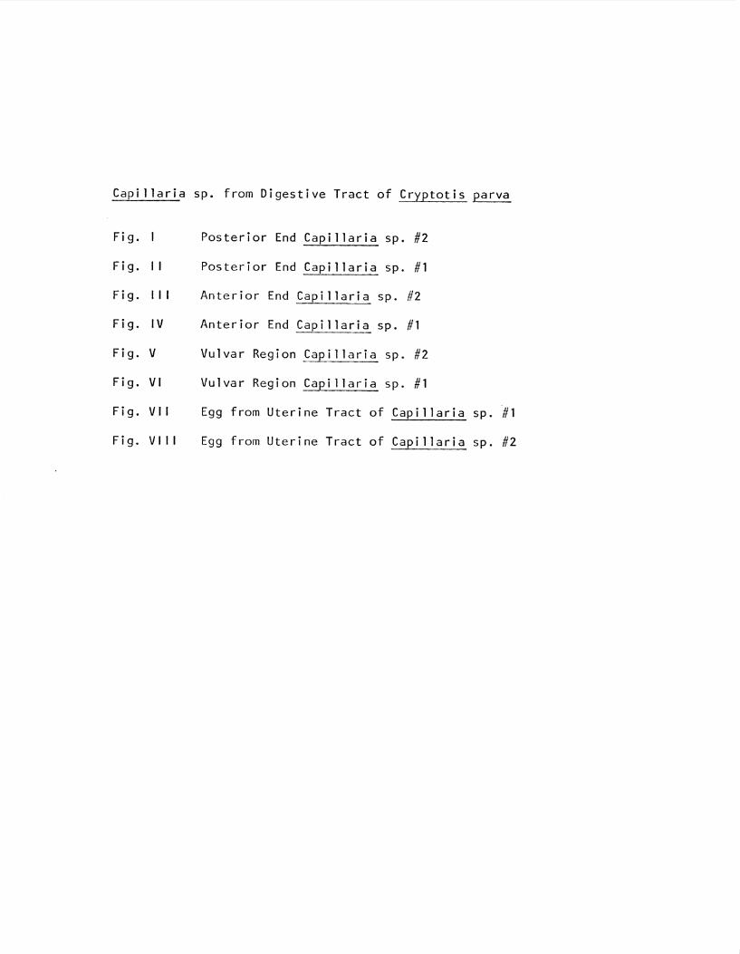
Capillaria sp. from Digestive Tract of Cryptotis parva
Fig. Posterior End Capi 1 laria sp. #2
Fig. 11 Posterior End Capillaria sp. #1
Fig. 111 Anterior End Capillaria sp. #2
Fig. IV Anterior End Capillaria sp. #1
Fig. V Vu 1 var Region Capillaria sp. #2
Fig. VI Vu 1 var Region Capillaria sp. #1
Fig. VI I Egg from Uterine Tract of Capillaria sp. #1
Fig. VI 11 Egg from Uterine Tract of Capillaria sp. #2

7
0·00MM.
DISCUSSION
Cryptotis parva were difficult to collect. Over a period of 13
months (December 1976 - January 1977), 28 specimens were caught. One
additional shrew was captured in January 1978. No specimens were col
lected after the heavy snow storms arrived late February 1977 and 1978
leaving heavy snow cover on the ground for several weeks. The severe
weather may have caused a mass winter kill both years. Considering
8
their high metabolic rate, and the need to feed constantly, bad weather
for an extended period of time may produce widespread starvation.
Another possibility may be that many were either drowned in their holes
or flushed out as the snow rapidly melted. With the vegetation no longer
offering adequate cover or protection, flushed out animals may have been
heavily preyed upon by owls and other potential predators.
Only three adult female Capillaria were recovered during the necropsies.
This is a low adult recovery rate when compared to other hosts. A number
of reasons for this may be cited. Read (1949) indicated that species
inhabiting the digestive tract are often found when the mucosa is thoroughly
scraped. However; the small size of the specimens in question and their
fragile nature may have allowed some to be overlooked. Since no fragments
were observed, it is probably that the above is not the case. The fact
that eggs were present did not necessarily mean presence of adults.
Levine (1968) mentions that if a specimen has eaten an infected host,
eggs from helminths of prey may be found in the predator feces. But,
in this investigation, the eggs recovered from hosts in which there
9
were no worms, appeared to be the same as those examined from the hosts
with worms and as those found in the uterine tract of egg bearing female
worms. It is therefore concluded that the level of infection in these
host specimens is low.
The fact that no males were recovered in this study is not uncommon.
Levine (1968) and Hesse (1923) mention several instances where males of
described Capillaria species are unknown. Considering the fact that
females outnumber males in host populations by almost 3 to 1 on the average
and the males are much smaller, it is not surprising that males are less
often observed than females.
Although Capillaria hepatica has been reported from the livers of
seven orders and 35 species of mammals (Teixeira De Freitas and Lent,
1936; Lubinsky, 1956; Freeman and Wright, 1960; Solomon and Handley,
1971), no evidence of liver inhabiting forms were found in this study.
The method by which many capillarid infections are acquired is
unknown. Hyman (1951) stated that some species in this genus require
earthworms as intermediate hosts. Others may have direct life cycles.
The Capillaria described here appear to be of two different species.
Several points of difference may be summarized. (1) The oral cavity of
Capillaria #1 opens subtermanilly while that of Capillaria #2 is terminal.
(2) Their sticocytes are of different average size. (3) The egg arrange
ment in the uterus is different and so are their average egg sizes.
(4) The prominence in the vulva region is more marked in one than in
the other. (S) The slight decrease in width posteriorly from the region
of greatest diameter is more readily observable in one than in the other.
On the other hand, they could be the same species based on the fact
that (1) the morphology of their sticocytes are similar, (2) the ratios

10
of their esophagi and vulvas to their body lengths are the same, (3) the
nature of their cuticles is similar, and (4) they both lack bacillary
bands.
Until male specimens can be obtained, the determination of species
will be reserved.

11
LITERATURE CI TED
Cable, R. M. 1958. An illustrated laboratory manual of parasitology. Burgess Publishing Company, Minneapolis. 165 pp.
Freeman, R. S. and K. A. Wright. 1960. Factors concerned with the epizootiology of Capi l laria hepatica (Bancroft, 1893) (Nematoda) in a population of Peromyscus maniculatus in Algonquin Park, Canada. J. Parasit. 46: 373-383.
Hesse, A. J. 1923. Description of ~illaria leucisci, n. sp., found in the intestine of Leuciscus phoxinus Linn. Vol. I. J. Helminthology, 65-70.
Hyman, L. H. 1951. and Entoprocta. 572 pp.
The invertebrates: Acanthocephala, Aschelminthes, Vol. 3. McGraw-Hill Book Company, Inc., New York.
Levine, N. D. 1968. Nematode parasites of domestic animals and man. Burgess Publishing Company, Minneapolis. 600 pp.
1973. Protozoan parasites of domestic animals and of man. 2d ed. Burgess Publishing Company, Minneapolis. 406 pp.
Lopez-Neyra, C. R. capi l lari i nae.
1947. Generos y especies neuvas o mal conocidas de Rev. lberica Parasitol. 7:191-238.
Lubinsky, G. 1956. On the probably presence of parasitic liver cirrhosis in Canada. Can. J. Comp. Med. 20:457-465.
MacArthur, W. P .. 1924. A case of infection of the human liver with Hepaticola hepatica (Bancroft, 1893) Hall, 1916. Proc. Roy. Soc. Med . 1 7 : 8 3- 8 4 .
Matthews, J. W. 1974. A survey of blood and intestinal parasites of Peromyscus leucopus and Microtus ochrogaster in Coles County, Illinois. Thesis (M.S.) Eastern Illinois University. 20 pp.
Motishita, K. and T. Tani. 1960. A case of Capillaria infection causing cutaneous creeping eruption in man. J. Parasit. 46:79-83.
Ogren, R. E. 1953. Capillaria blarina, n. sp. (Nematoda: Trichuridae) from the esophagus of the short tail shrew, Blarina brevicauda (Say). J. Parasit. 39:135-138.
Read, C. P. 1949. Studies on North American helminths of the genus Capillaria Zeder, 1800 (Nematoda): I. Capillarids from mammals. J. Parasit. 35:223-230.
Skryabin, K. I., N. P. Shikhobalova and I. V. Orlov. 1957. Trikhotsephalidy i Kapillyariidy Zhivotnykl i Cheloveka: Vyzyaemye imi
12
Zabolevaniya. Vol. VI. in K. I. Skryabin, ed., Osnovy nematodo-logi i. lzdat. Akad. Nauk SSSR, Moskva. 587 pp.
Solomon, G. B. and C. 0. Handley, Jr. 1971. Capi l laria hepatica (Bancroft, 1893) in Appalachian mammals. J. Parasit. 57:1142-1144.
Teixeira De Freitas, J. F. and Lent, J. F. 1936. Estudo sobre os Capillariinae parasitos de mammiferos. Mem. Inst. Osw. Ctuz. 31:85-160.
Travassos, L. 1915. Contribuicoes para o conhecimento da fauna helmintholojica brasileira. V. Sobre as especies brasileiras do genero Capillaria Zeder, 1800. Mem. Inst. Osw. Cruz. 7:146-172.
Yarnaguti, S. 1959, Systema Helminthum. vertebrates. lnterscience, New York.
Vol. 2. The cestodes of 860 pp.
1961. Systema Helminthum. Vol. 3. The nematodes of vertebrates. Parts 1 & 2. lnterscience, New York. 1261 pp.
York, W. and P.A. Maplestone. 1926. The nematode parasites of verte-brates. London. 536 pp.