F-Actin Is an Evolutionarily Conserved Damage-Associated Molecular Pattern Recognized by DNGR-1, a...
11
Immunity Article F-Actin Is an Evolutionarily Conserved Damage-Associated Molecular Pattern Recognized by DNGR-1, a Receptor for Dead Cells Susan Ahrens, 1 Santiago Zelenay, 1 David Sancho, 7 Pavel Han c, 1 Svend Kjær, 2 Christoph Feest, 3 Georgina Fletcher, 4 Charlotte Durkin, 5 Antonio Postigo, 5 Mark Skehel, 6 Facundo Batista, 3 Barry Thompson, 4 Michael Way, 5 Caetano Reis e Sousa, 1,8, * and Oliver Schulz 1,8 1 Immunobiology Laboratory 2 Protein Purification Facility 3 Lymphocyte Interaction Laboratory 4 Epithelial Biology Laboratory 5 Cell Motility Laboratory 6 Protein Analysis and Proteomics Laboratory Cancer Research UK, London Research Institute, Lincoln’s Inn Fields Laboratories, 44 Lincoln’s Inn Fields, London WC2A 3LY, UK 7 Department of Vascular Biology and Inflammation, CNIC - Centro Nacional de Investigaciones Cardiovasculares ‘‘Carlos III,’’ Melchor Ferna ´ ndez Almagro 3, 28029 Madrid, Spain 8 These authors contributed equally to this work *Correspondence: [email protected] DOI 10.1016/j.immuni.2012.03.008 SUMMARY Sterile inflammation can be initiated by innate immune recognition of markers of tissue injury termed damage-associated molecular patterns (DAMPs). DAMP recognition by dendritic cells (DCs) has also been postulated to lead to T cell responses to foreign antigens in tumors or allografts. Many DAMPs represent intracellular contents that are released upon cell damage, notably after necrosis. In this regard, we have previously described DNGR- 1 (CLEC9A) as a DC-restricted receptor specific for an unidentified DAMP that is exposed by necrotic cells and is necessary for efficient priming of cyto- toxic T cells against dead cell-associated antigens. Here, we have shown that the DNGR-1 ligand is preserved from yeast to man and corresponds to the F-actin component of the cellular cytoskeleton. The identification of F-actin as a DNGR-1 ligand suggests that cytoskeletal exposure is a universal sign of cell damage that can be targeted by the innate immune system to initiate immunity. INTRODUCTION Recognition of pathogen-associated molecular patterns (PAMPs) by innate immune receptors triggers inflammation and, when coupled to antigen encounter, favors the initiation of adaptive immunity by dendritic cells (DCs) (Iwasaki and Medzhi- tov, 2010). However, inflammation can also be driven by sterile injury to tissues in the presumed absence of PAMPs (Chen and Nun ˜ ez, 2010; Kono and Rock, 2008; Rock and Kono, 2008; Rock et al., 2010). In this case, damaged cells may act as a source of sterile proinflammatory signals, which, by analogy to PAMPs, have been termed ‘‘damage-associated molecular patterns’’ (DAMPs) (Seong and Matzinger, 2004). Many DAMPs are thought to be intracellular components that play a house- keeping role in healthy cells but are leaked or exposed by damaged cells, most notably after loss of membrane integrity associated with primary or secondary necrosis (Chen and Nun ˜ ez, 2010; Kono and Rock, 2008; Rock and Kono, 2008; Rock et al., 2010). Exposure of DAMPs by dead or damaged cells in most instances appears designed to initiate a controlled inflammatory response that induces tissue repair (Chen and Nun ˜ ez, 2010; Rock et al., 2010). However, in some cases, DAMPs might substitute for PAMPs in activating DCs and inducing adaptive immunity to dead cell-associated antigens (Matzinger, 1994). In this scenario, DAMPs would act as an alternative to PAMPs and help provide an explanation for PAMP-independent immunity such as observed in response to tumors or to allografts (Matzinger, 1994, 2002). We have recently identified DNGR-1, also known as CLEC9A, as a DAMP receptor that responds to dead cells (Sancho et al., 2009). Interestingly, expression of DNGR-1 is highly restricted to a subtype of cross-presenting DCs in both mouse and human, suggesting that the receptor may play a prominent role in the regulation of CD8 + T cell responses (Caminschi et al., 2008; Huysamen et al., 2008; Poulin et al., 2010; Sancho et al., 2008). DNGR-1 signals via a hemi immunoreceptor tyrosine- based activation motif (hemITAM) in its intracellular portion that permits recruitment of Syk kinase (Huysamen et al., 2008; Sancho et al., 2009). HemITAM-dependent and -independent DNGR-1 signals collectively regulate endocytic traffic of dead cell cargo and promote processing and cross-presentation of dead cell-associated antigens (Zelenay et al., 2012). As a con- sequence, DNGR-1 is nonredundant in mice for efficient cross- priming of cytotoxic T cells against dead cell-associated antigens, making it a key innate immune receptor in bridging DAMP sensing to the induction of T cell immunity (Sancho et al., 2009). Immunity 36, 635–645, April 20, 2012 ª2012 Elsevier Inc. 635
Transcript of F-Actin Is an Evolutionarily Conserved Damage-Associated Molecular Pattern Recognized by DNGR-1, a...
Immunity
Article
F-Actin Is an Evolutionarily ConservedDamage-Associated Molecular Pattern Recognizedby DNGR-1, a Receptor for Dead CellsSusan Ahrens,1 Santiago Zelenay,1 David Sancho,7 Pavel Han�c,1 Svend Kjær,2 Christoph Feest,3 Georgina Fletcher,4
Charlotte Durkin,5 Antonio Postigo,5 Mark Skehel,6 Facundo Batista,3 Barry Thompson,4 Michael Way,5
Caetano Reis e Sousa,1,8,* and Oliver Schulz1,81Immunobiology Laboratory2Protein Purification Facility3Lymphocyte Interaction Laboratory4Epithelial Biology Laboratory5Cell Motility Laboratory6Protein Analysis and Proteomics Laboratory
Cancer Research UK, London Research Institute, Lincoln’s Inn Fields Laboratories, 44 Lincoln’s Inn Fields, London WC2A 3LY, UK7Department of Vascular Biology and Inflammation, CNIC - Centro Nacional de Investigaciones Cardiovasculares ‘‘Carlos III,’’ Melchor
Fernandez Almagro 3, 28029 Madrid, Spain8These authors contributed equally to this work
*Correspondence: [email protected]
DOI 10.1016/j.immuni.2012.03.008
SUMMARY
Sterile inflammation can be initiated by innateimmune recognition of markers of tissue injurytermed damage-associated molecular patterns(DAMPs). DAMP recognition by dendritic cells (DCs)has also been postulated to lead to T cell responsesto foreign antigens in tumors or allografts. ManyDAMPs represent intracellular contents that arereleased upon cell damage, notably after necrosis.In this regard, we have previously described DNGR-1 (CLEC9A) as a DC-restricted receptor specific foran unidentified DAMP that is exposed by necroticcells and is necessary for efficient priming of cyto-toxic T cells against dead cell-associated antigens.Here, we have shown that the DNGR-1 ligand ispreserved from yeast to man and corresponds tothe F-actin component of the cellular cytoskeleton.The identification of F-actin as a DNGR-1 ligandsuggests that cytoskeletal exposure is a universalsign of cell damage that can be targeted by the innateimmune system to initiate immunity.
INTRODUCTION
Recognition of pathogen-associated molecular patterns
(PAMPs) by innate immune receptors triggers inflammation
and, when coupled to antigen encounter, favors the initiation of
adaptive immunity by dendritic cells (DCs) (Iwasaki and Medzhi-
tov, 2010). However, inflammation can also be driven by sterile
injury to tissues in the presumed absence of PAMPs (Chen and
Nunez, 2010; Kono and Rock, 2008; Rock and Kono, 2008;
Rock et al., 2010). In this case, damaged cells may act as
a source of sterile proinflammatory signals, which, by analogy
to PAMPs, have been termed ‘‘damage-associated molecular
patterns’’ (DAMPs) (Seong and Matzinger, 2004). Many DAMPs
are thought to be intracellular components that play a house-
keeping role in healthy cells but are leaked or exposed by
damaged cells, most notably after loss of membrane integrity
associated with primary or secondary necrosis (Chen and
Nunez, 2010; Kono and Rock, 2008; Rock and Kono, 2008;
Rock et al., 2010). Exposure of DAMPs by dead or damaged
cells in most instances appears designed to initiate a controlled
inflammatory response that induces tissue repair (Chen and
Nunez, 2010; Rock et al., 2010). However, in some cases,
DAMPs might substitute for PAMPs in activating DCs and
inducing adaptive immunity to dead cell-associated antigens
(Matzinger, 1994). In this scenario, DAMPs would act as an
alternative to PAMPs and help provide an explanation for
PAMP-independent immunity such as observed in response to
tumors or to allografts (Matzinger, 1994, 2002).
We have recently identified DNGR-1, also known as CLEC9A,
as a DAMP receptor that responds to dead cells (Sancho et al.,
2009). Interestingly, expression of DNGR-1 is highly restricted to
a subtype of cross-presenting DCs in both mouse and human,
suggesting that the receptor may play a prominent role in the
regulation of CD8+ T cell responses (Caminschi et al., 2008;
Huysamen et al., 2008; Poulin et al., 2010; Sancho et al.,
2008). DNGR-1 signals via a hemi immunoreceptor tyrosine-
based activation motif (hemITAM) in its intracellular portion that
permits recruitment of Syk kinase (Huysamen et al., 2008;
Sancho et al., 2009). HemITAM-dependent and -independent
DNGR-1 signals collectively regulate endocytic traffic of dead
cell cargo and promote processing and cross-presentation of
dead cell-associated antigens (Zelenay et al., 2012). As a con-
sequence, DNGR-1 is nonredundant in mice for efficient cross-
priming of cytotoxic T cells against dead cell-associated
antigens, making it a key innate immune receptor in bridging
DAMP sensing to the induction of T cell immunity (Sancho
et al., 2009).
Immunity 36, 635–645, April 20, 2012 ª2012 Elsevier Inc. 635

Figure 1. DNGR-1 Recognizes a Ligand in Lysates ofMetazoanCells
Lysates of human cells (HeLa; A), trout and turkey tissues (B), or insect cells
(Sf9, S2; C) were serially diluted as indicated and spotted onto membranes via
a dot blot apparatus. Membranes were probed with mDNGR-1 ECD or
hDNGR-1-Fc or the respective control reagents (mDectin-1 CTLD or hDC-
SIGN-Fc), as indicated. Data are from one representative experiment of at
least 17 (HeLa), 2 (trout and turkey), and 9 (insect) cells. See also Figure S1.
Immunity
F-Actin Is a Ligand for DNGR-1
DNGR-1 is a member of the C-type lectin superfamily and its
C-type lectin-like domain (CTLD) can be used as a soluble probe
to detect ligand in cell staining experiments (Sancho et al., 2009).
Such experiments show that the ligand for DNGR-1 is a pre-
formed component of healthy cells that can be revealed inde-
pendently of cell death by cell fixation and permeabilization
(Sancho et al., 2009). Further experimentation demonstrates
that the ligand is protease sensitive, as well as acid and heat
636 Immunity 36, 635–645, April 20, 2012 ª2012 Elsevier Inc.
labile, and present throughout the cytoplasm but excluded
from the nucleus (Sancho et al., 2009). However, further dissec-
tion of the role of DNGR-1 in DC function and in regulation of
immune responses has been hampered by the failure to identify
the DNGR-1 ligand. Here, we have used a combination of
biochemical and imaging approaches to isolate cell-derived
DNGR-1 ligands.We report that DNGR-1 specifically recognized
F-actin in all cells tested, from yeast to man. The identification of
F-actin as an evolutionarily conserved DAMP and a ligand for
DNGR-1 has important consequences for our understanding of
sterile immunity and inflammation.
RESULTS
DNGR-1 Binds to a Ubiquitous Evolutionarily ConservedLigandIn order to choose an appropriate reagent for purifying the
DNGR-1 ligand, we tested different versions of the extracellular
domain of DNGR-1. Similar to the CTLD originally used to
demonstrate DNGR-1 binding to dead cells (Sancho et al.,
2009), a more stable full-length extracellular domain (ECD) of
mouse DNGR-1 with an N-terminal FLAG tag specifically stained
irradiated cells that had lost membrane integrity and become
permeable to TO-PRO-3 dye (Figure S1A available online). A
human DNGR-1-Fc fusion protein was also able to specifically
stain dead cells, unlike a control human dendritic cell-specific
intercellular adhesion molecule-3-grabbing non-integrin (DC-
SIGN)-Fc reagent (Figure S1B). Both the mDNGR-1 ECD and
the hDNGR-1-Fc were used for subsequent studies.
The DNGR-1 ligand has previously been detected exclusively
on dead cell corpses or in fixed and permeabilized cells (Sancho
et al., 2009). We therefore first tested whether the ligand could
be solubilized from cell extracts. By dot blot analysis, ligand
could be detected in lysates of HeLa cells via either the
mDNGR-1 ECD or the hDNGR-1-Fc, but not the control
reagents, mDectin-1 (CTLD) or hDC-SIGN-Fc (Figure 1A). To
determine the evolutionary conservation of DNGR-1 ligand, we
tested additional species, including fish, birds, and insects.
Notably, specific signal was detected in lysates of trout and tur-
key tissue (Figure 1B) and in lysates of Sf9 cells from Spodoptera
frugiperda and of S2 cells from Drosophila melanogaster (Fig-
ure 1C). There was no signal when mDectin-1 was used as
a control (Figures 1B and 1C), although the same reagent could
detect ligand in crude yeast lysates (not shown), consistent with
the ability of Dectin-1 to recognize fungal b-glucans (Brown and
Gordon, 2001). We conclude that DNGR-1 probably recognizes
a ligand expressed in most cell types, from insects to man, that
can be solubilized from cell extracts for the purpose of biochem-
ical characterization.
The DNGR-1 Ligand Is a Large Protein-Based Structurethat Requires Purification under Native ConditionsWe utilized the dot blot assay to assess the biochemical proper-
ties of the ligand and design an appropriate purification scheme.
Consistent with our previous conclusions derived from staining
of cell corpses (Sancho et al., 2009), the ligand was protein
based as shown by the fact that its detection was markedly
reduced by treatment of HeLa cell lysates with trypsin or papain
(Figure S2A). However, attempts to separate cell lysates by

Figure 2. Affinity Purification of DNGR-1 Ligand
Enriches for Components of the Actin Cytoskel-
eton
(A and B) The indicated volumes of lysate of HeLa (A) or S2
(B) cells were incubated with FLAG-mDNGR-1 ECD or
FLAG-mDectin-1 preabsorbed onto anti-FLAG beads.
After centrifugation, post-pull-down supernatants (SN)
containing unbound material were serially diluted and
analyzed for the presence of DNGR-1 ligand by dot blot
(left). Input lysates before pull-down are shown for
comparison (top row in left panels). Proteins recovered
after elution of beadswith FLAG-peptidewere analyzed by
SDS-PAGE (right). Position of the FLAG-tagged proteins is
indicated by arrows. Data shown are from one of four
experiments with HeLa cells and one of two experiments
with S2 cells.
(C) Table representing pooled results of mass spectro-
metric analysis of DNGR-1 pull-downs for HeLa cells (left)
or S2 cells (right).
See also Figure S2.
Immunity
F-Actin Is a Ligand for DNGR-1
SDS-PAGE and to detect the ligand by immunoblotting with
mDNGR-1 ECD all failed, suggesting that structural integrity of
the ligand is important for detection (data not shown). Consistent
with that notion, detection of the ligand was abrogated or mark-
edly decreased by boiling or addition of SDS (Figure S2B).
Attempts at separation by native PAGE on 4%–20% gradient
gels followed by blotting with the mDNGR-1 ECD revealed that
the ligand failed to enter the gel and remained trapped in the
well (Figure S2C). These results suggested that the DNGR-1
ligand is a high-molecular-weight structure that cannot be
resolved by gel electrophoresis under native conditions. This
was confirmed by gel filtration analysis, which showed that
the ligand elutes primarily in the void volume and in the first
few fractions, which correspond to molecular weights greater
Immunity 36
than 450 kDa (Figure S2D). We conclude that
the DNGR-1 ligand is likely to be a very large
protein complex that is destroyed by denatur-
ation and heat.
Affinity Purification of DNGR-1 LigandReveals Proteins Associated with theActin CytoskeletonIn an attempt to characterize the ligand by
enrichment, we tested whether it could be
affinity isolated from mammalian and insect
cell lysates by mDNGR-1 ECD. This approach
was successful, as measured by depletion of
ligand from the postincubation supernatant, as
long as the amount of mDNGR-1 ECD was
matched to the amount of input lysate (Figures
2A and 2B). When the bound material was
subsequently eluted via an excess of FLAG
peptide and resolved by SDS-PAGE, a number
of discrete bands, in addition to a large band
corresponding to the mDNGR-1 ECD itself
(marked), could be revealed by gel staining
with SYPRO ruby dye (Figures 2A and 2B).
Notably, several proteins appeared similar be-
tween pull-downs from HeLa and S2 cells but were absent in
eluates from control pull-downs carried out with the mDectin-1
reagent (Figures 2A and 2B).
Mass spectrometry analysis of pull-downs from four indepen-
dent experiments with HeLa cells revealed that multiple actin-
associated proteins were consistently found in the DNGR-1
but not the Dectin-1 precipitates. Similarly, enrichment for
actin-associated proteins was observed upon mass spectrom-
etry analysis of the gel-filtration fractions containing the
DNGR-1 ligand (data not shown). The identified proteins
included cytolinkers such as plectin, actin filament crosslinkers
such as spectrin, actin motor proteins such as myosins, as
well as actin itself (Figure 2C). Proteins associated with microtu-
bules were not especially prevalent in the DNGR-1 precipitates,
, 635–645, April 20, 2012 ª2012 Elsevier Inc. 637

Figure 3. DNGR-1 Binds to the Actin Cytoskeleton
(A and B) Double-staining with DAPI (DNA stain; blue) and
FLAG-mDNGR-1 ECD (red) of apical (A, left) and baso-
lateral (A, right) surfaces of Drosophila imaginal wing
discs, as well as cells undergoing mitosis in the same
tissue (B). Arrows in (B) indicate contractile rings and
cleavage furrow of mitotic cells.
(C and D) Double-staining with mDNGR-1 ECD (red) and
A488-phalloidin (green) of Drosophila ovaries (C) or HeLa
cells (D).
(E) HeLa cells infected with wild-type vaccinia virus (top)
or A36R-YdF mutant virus (bottom) were stained with
mDNGR-1 ECD (red), A488-phalloidin (green), and DRAQ5
(DNA stain; blue). Insets represent enlarged areas of the
composite images indicated by the white squares. Arrows
indicate actin tails and arrowheads show viral DNA in viral
factories.
All images were obtained by laser scanning microscopy
and are representative of five (HeLa cells), three (infected
HeLa cells), and three (Drosophila tissues) analyses. Scale
bars represent 10 mm. See also Figure S3.
Immunity
F-Actin Is a Ligand for DNGR-1
indicating selectivity for the actin cytoskeleton (Figure 2C and
data not shown). We selected the proteins with the highest
number of unique peptide hits across experiments and tested
their putative identity as DNGR-1 ligands. The validation
638 Immunity 36, 635–645, April 20, 2012 ª2012 Elsevier Inc.
included carrying out loss-of-function experi-
ments such as siRNA-mediated knockdown in
HeLa cells followed by probing for DNGR-1
ligand by dot blot, as well as gain-of-function
experiments such as procuring the protein in
question and assessing its binding to DNGR-1
(data not shown). All of those experiments failed
to identify a ligand for DNGR-1 (data not shown).
Furthermore, some of the top candidates found
in the HeLa cell pull-downs (e.g., plectin, spec-
trin) were not identified in pull-downs from S2
cells even though the latter still contained
predominantly actin-associated proteins (Fig-
ure 2C). We therefore contemplated the
possibility that the enrichment for actin cyto-
skeleton-associated proteins in the DNGR-1
pull-downs might be because DNGR-1 binds
to the actin cytoskeleton itself.
The DNGR-1 ECD Labels the ActinCytoskeleton across SpeciesTo test the above hypothesis, we performed
detailed analysis of the intracellular localization
of DNGR-1 ligand in different cells via confocal
fluorescence microscopy. Staining of various
cells and tissues with mDNGR-1 ECD revealed
a remarkably discrete pattern. DNGR-1 ligand
showed apical polarization in the pseudo-
stratified columnar epithelium of Drosophila
imaginal wing discs (Figure 3A) and, in that
tissue, could additionally be seen on contractile
rings around the cleavage furrow of mitotic cells
undergoing cytokinesis (Figure 3B, arrows). In
Drosophila ovaries, DNGR-1 ligand colocalized
with striations in the surrounding muscle sheet (Figure 3C) In
HeLa cells, DNGR-1 ligand was prominently detected on stress
fibers and lamellipodia (Figure 3D). Notably, in HeLa cells in-
fected with wild-type vaccinia virus, mDNGR-1 ECD stained
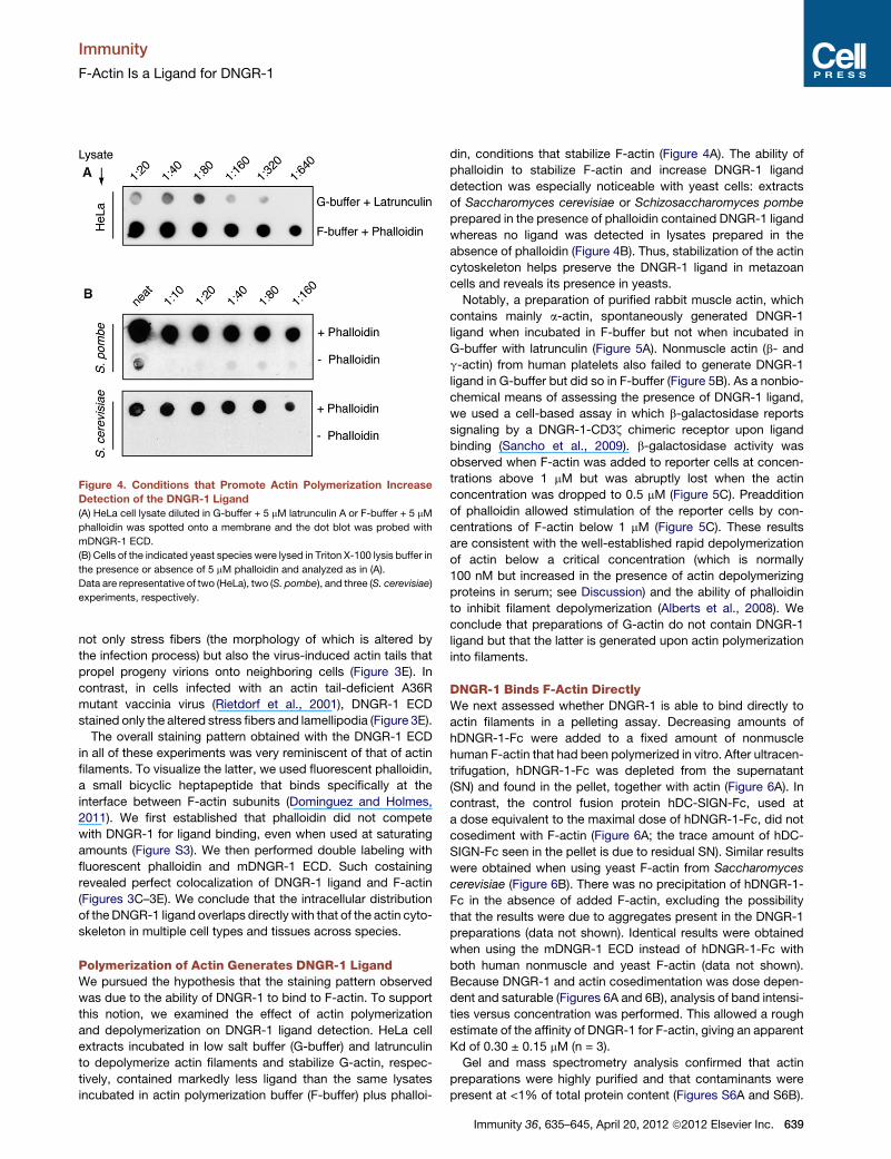
Figure 4. Conditions that Promote Actin Polymerization Increase
Detection of the DNGR-1 Ligand
(A) HeLa cell lysate diluted in G-buffer + 5 mM latrunculin A or F-buffer + 5 mM
phalloidin was spotted onto a membrane and the dot blot was probed with
mDNGR-1 ECD.
(B) Cells of the indicated yeast species were lysed in Triton X-100 lysis buffer in
the presence or absence of 5 mM phalloidin and analyzed as in (A).
Data are representative of two (HeLa), two (S. pombe), and three (S. cerevisiae)
experiments, respectively.
Immunity
F-Actin Is a Ligand for DNGR-1
not only stress fibers (the morphology of which is altered by
the infection process) but also the virus-induced actin tails that
propel progeny virions onto neighboring cells (Figure 3E). In
contrast, in cells infected with an actin tail-deficient A36R
mutant vaccinia virus (Rietdorf et al., 2001), DNGR-1 ECD
stained only the altered stress fibers and lamellipodia (Figure 3E).
The overall staining pattern obtained with the DNGR-1 ECD
in all of these experiments was very reminiscent of that of actin
filaments. To visualize the latter, we used fluorescent phalloidin,
a small bicyclic heptapeptide that binds specifically at the
interface between F-actin subunits (Dominguez and Holmes,
2011). We first established that phalloidin did not compete
with DNGR-1 for ligand binding, even when used at saturating
amounts (Figure S3). We then performed double labeling with
fluorescent phalloidin and mDNGR-1 ECD. Such costaining
revealed perfect colocalization of DNGR-1 ligand and F-actin
(Figures 3C–3E). We conclude that the intracellular distribution
of the DNGR-1 ligand overlaps directly with that of the actin cyto-
skeleton in multiple cell types and tissues across species.
Polymerization of Actin Generates DNGR-1 LigandWe pursued the hypothesis that the staining pattern observed
was due to the ability of DNGR-1 to bind to F-actin. To support
this notion, we examined the effect of actin polymerization
and depolymerization on DNGR-1 ligand detection. HeLa cell
extracts incubated in low salt buffer (G-buffer) and latrunculin
to depolymerize actin filaments and stabilize G-actin, respec-
tively, contained markedly less ligand than the same lysates
incubated in actin polymerization buffer (F-buffer) plus phalloi-
din, conditions that stabilize F-actin (Figure 4A). The ability of
phalloidin to stabilize F-actin and increase DNGR-1 ligand
detection was especially noticeable with yeast cells: extracts
of Saccharomyces cerevisiae or Schizosaccharomyces pombe
prepared in the presence of phalloidin contained DNGR-1 ligand
whereas no ligand was detected in lysates prepared in the
absence of phalloidin (Figure 4B). Thus, stabilization of the actin
cytoskeleton helps preserve the DNGR-1 ligand in metazoan
cells and reveals its presence in yeasts.
Notably, a preparation of purified rabbit muscle actin, which
contains mainly a-actin, spontaneously generated DNGR-1
ligand when incubated in F-buffer but not when incubated in
G-buffer with latrunculin (Figure 5A). Nonmuscle actin (b- and
g-actin) from human platelets also failed to generate DNGR-1
ligand in G-buffer but did so in F-buffer (Figure 5B). As a nonbio-
chemical means of assessing the presence of DNGR-1 ligand,
we used a cell-based assay in which b-galactosidase reports
signaling by a DNGR-1-CD3z chimeric receptor upon ligand
binding (Sancho et al., 2009). b-galactosidase activity was
observed when F-actin was added to reporter cells at concen-
trations above 1 mM but was abruptly lost when the actin
concentration was dropped to 0.5 mM (Figure 5C). Preaddition
of phalloidin allowed stimulation of the reporter cells by con-
centrations of F-actin below 1 mM (Figure 5C). These results
are consistent with the well-established rapid depolymerization
of actin below a critical concentration (which is normally
100 nM but increased in the presence of actin depolymerizing
proteins in serum; see Discussion) and the ability of phalloidin
to inhibit filament depolymerization (Alberts et al., 2008). We
conclude that preparations of G-actin do not contain DNGR-1
ligand but that the latter is generated upon actin polymerization
into filaments.
DNGR-1 Binds F-Actin DirectlyWe next assessed whether DNGR-1 is able to bind directly to
actin filaments in a pelleting assay. Decreasing amounts of
hDNGR-1-Fc were added to a fixed amount of nonmuscle
human F-actin that had been polymerized in vitro. After ultracen-
trifugation, hDNGR-1-Fc was depleted from the supernatant
(SN) and found in the pellet, together with actin (Figure 6A). In
contrast, the control fusion protein hDC-SIGN-Fc, used at
a dose equivalent to the maximal dose of hDNGR-1-Fc, did not
cosediment with F-actin (Figure 6A; the trace amount of hDC-
SIGN-Fc seen in the pellet is due to residual SN). Similar results
were obtained when using yeast F-actin from Saccharomyces
cerevisiae (Figure 6B). There was no precipitation of hDNGR-1-
Fc in the absence of added F-actin, excluding the possibility
that the results were due to aggregates present in the DNGR-1
preparations (data not shown). Identical results were obtained
when using the mDNGR-1 ECD instead of hDNGR-1-Fc with
both human nonmuscle and yeast F-actin (data not shown).
Because DNGR-1 and actin cosedimentation was dose depen-
dent and saturable (Figures 6A and 6B), analysis of band intensi-
ties versus concentration was performed. This allowed a rough
estimate of the affinity of DNGR-1 for F-actin, giving an apparent
Kd of 0.30 ± 0.15 mM (n = 3).
Gel and mass spectrometry analysis confirmed that actin
preparations were highly purified and that contaminants were
present at <1% of total protein content (Figures S6A and S6B).
Immunity 36, 635–645, April 20, 2012 ª2012 Elsevier Inc. 639

Figure 5. Polymerization of Purified Actin Is Sufficient to Generate
DNGR-1 Ligand
(A and B) Dot blots of rabbit muscle (A) or human nonmuscle (B) actin probed
with FLAG-mDNGR-1 ECD. 12.5 mM muscle actin and 0.6 mM nonmuscle
actin, reconstituted in G-buffer, were diluted in G- or F-buffer in the presence
of latrunculin A or phalloidin as indicated.
(C) Nonmuscle (NM) F-actin was added to BWZ-mDNGR-1-z cells at the de-
picted final concentration in the presence or absence of phalloidin. F-buffer
with and without phalloidin served as negative controls. Histogram shows
absorbance after addition of b-galactosidase substrate to lysed cells. Bars
represent the average of duplicate wells.
Data in (A), (B), and (C) are representative of six, two, and three experiments,
respectively. See also Figure S6.
Immunity
F-Actin Is a Ligand for DNGR-1
We tested whether such contaminants might contribute to
DNGR-1 binding by deliberately adding actin-associated
proteins to F-actin prior to ligand detection. We noted that addi-
tion of a-actinin, as well as some other actin-binding proteins, to
in vitro polymerized F-actin increased the extent of DNGR-1
ligand detection even though a-actinin itself did not act as
a ligand (Figure S6C). However, a-actinin did not alter the
apparent affinity of DNGR-1 for F-actin in pelleting assays (Fig-
ure S6D), arguing that it is not a component of the DNGR-1
binding site (see Discussion).
Finally, we measured DNGR-1 binding to F-actin by total
internal reflection fluorescence (TIRF) microscopy. Nonmuscle
640 Immunity 36, 635–645, April 20, 2012 ª2012 Elsevier Inc.
or muscle actin was polymerized in vitro with F-buffer together
with fluorescent phalloidin to stabilize F-actin and permit
filament visualization and was labeled with mDNGR-1 or
mDectin-1. As shown in Figure 6C, mDNGR-1 but not Dectin-1
decorated nonmuscle actin filaments along their entire length.
Pixel intensity analysis confirmed the exact overlap of the phal-
loidin and DNGR-1 ligand signals (Figure 6C). Identical results
were obtained when using the preparation of rabbit muscle actin
(Figure 6D) or the hDNGR-1-Fc versus the hDC-SIGN-Fc
reagents (Figure S4). We conclude that DNGR-1 specifically
and directly binds to actin filaments.
F-Actin in Dead Cells Triggers DNGR-1 Signaling via SykThe actin cytoskeleton is highly regulated andmight therefore be
expected to undergo rapid depolymerization upon cell death.
Given that DNGR-1 can specifically detect dead cells, we
assessed whether sufficient F-actin is retained in cell corpses
to account for DNGR-1 recognition. Notably, we found that
dead cells still contain F-actin. Thus, UV-irradiated HeLa cells
that had become secondarily necrotic after overnight culture
showed the expected loss of membrane integrity as denoted
by staining with the viability dye TO-PRO-3, yet displayed abun-
dant phalloidin staining, which correlated with DNGR-1 ligand
presence (Figure 7A). Examination of the corpses by confocal
microscopy confirmed that the actin cytoskeleton had under-
gone marked disassembly (e.g., compare with Figure 3D) but
remained morphoplogically identifiable when stained with either
phalloidin or DNGR-1 ECD (Figure 7B). Thus, even late stages of
cell death such as secondary necrosis do not lead to complete
loss of F-actin from cell corpses.
Finally, we tested whether F-actin by itself or within cell corp-
ses can not only act as a ligand for DNGR-1 but can also trigger
DNGR-1 signaling via Syk. We used cells expressing full-length
mDNGR-1 and Syk that specifically respond to DNGR-1
agonists by activating an NFAT-driven b-galactosidase reporter
(Sancho et al., 2009). As depicted in Figure 7C, addition of non-
muscle human F-actin at concentrations R1 mM to these cells
resulted in reporter activity. Lower F-actin concentrations could
also induce reporter activity provided phalloidin was added as a
stabilizer (Figure S5A). As expected, reporter activity depended
on DNGR-1 signaling via the hemITAM as shown by the fact
that no signal was observed in reporter cells expressing a Y7F
hemITAMmutant DNGR-1 that cannot engage Syk (Figure S5B).
Importantly, the reporter cells expressing Syk and wild-type
DNGR-1 also responded to the addition of necrotic (freeze-
thawed) but not live (mouse embryonic fibroblasts [MEFs])
cells (Figure 7D). To examine the effect of F-actin content on
DNGR-1 signaling, we pretreated MEFs with actin-stabilizing
or -destabilizing drugs prior to freeze-thawing. Pretreatment
with latrunculin B, an actin-depolymerizing agent, led to loss of
the reporter signal (Figure 7D). In contrast, signal was markedly
potentiated by MEF pretreatment with jasplakinolide, a phalloi-
din-like cell-permeable F-actin stabilizing drug (Figure 7D). In
all cases, activity remained dependent on DNGR-1 signaling as
shown by the fact that it was lost in reporter cells expressing
the Y7F DNGR-1 mutant (Figure S5B). We conclude that F-actin
is a bona fide agonist ligand for DNGR-1 and that the ability of
necrotic cells to trigger DNGR-1 signaling via Syk correlates
with their F-actin content.

Figure 6. mDNGR-1 ECD Binds Directly to
F-Actin
(A and B) Cosedimentation of DNGR-1 and F-actin
in a pelleting assay. 1 mg nonmuscle (NM) (A) or
3 mg yeast (B) F-actin was incubated alone (–) or
with 2 mg of hDC-SIGN-Fc or a 2-fold dilution
series of hDNGR-1-Fc (indicated by the wedge)
starting at (A) 2 mg or (B) 4 mg. After ultracentrifu-
gation, samples were analyzed for the presence of
hDNGR-1-Fc or hDC-SIGN-Fc, as well as actin, in
the supernatant and pellet fractions.
(C and D) TIRF microscopy images of (C) non-
muscle and (D) muscle F-actin labeled either with
DNGR-1 ECD or control Dectin-1 CTLD followed
by Cy3-conjugated FLAG antibody, as indicated.
A488-phalloidin was added as a counterstain to
reveal actin filaments. Data were acquired as
monochrome images and converted into color
images with ImageJ. Composite images are
shown on the right. Graphs depict pixel intensity of
the two channels along the white line indicated on
the micrographs.
See also Figure S4. Data in (A), (C), and (D) are
representative of three experiments and data in (B)
are representative of two experiments.
Immunity
F-Actin Is a Ligand for DNGR-1
DISCUSSION
DAMPs and their receptors provide a possible alternative to
PAMP-based mechanisms for initiation of inflammation and
adaptive immunity. DNGR-1 is a dedicated DAMP receptor
selectively expressed in DCs that controls cross-presentation
of dead cell-associated antigens leading to priming of cytotoxic
T cells (Sancho et al., 2009). Identification of the DNGR-1 ligand
has proven elusive and has hampered research into the mecha-
nism of action of this receptor and into the connection between
DAMP release and adaptive immunity. Here, we identify F-actin
Immunity 36, 635–6
as a universal DNGR-1 ligand that is
evolutionarily conserved from yeast to
mammals.
The conclusion that F-actin acts as the
ligand for DNGR-1 rests on three main
lines of evidence. First, the notion that
DNGR-1 directly binds F-actin can
account for all the data we have gath-
ered on the properties of the DNGR-1
ligand, such as its large mass and
susceptibility to denaturation. Further-
more, it explains the abundance of
actin-binding proteins in the mass spec-
trometry analysis of DNGR-1 affinity
isolates. Second, the intracellular distri-
bution of DNGR-1 ligand overlaps
precisely with that of the actin cytoskel-
eton in all cell types tested, from
Drosophila to man. Although many
actin-associated proteins will give a
staining pattern that overlaps with the
actin cytoskeleton in any given cell,
none of these proteins are present in
every single one of the structures labeled by DNGR-1 (muscle,
apical surface of Drosophila wing disc epithelium, contractile
rings, stress fibers, and vaccinia actin tails). For example,
although muscle F-actin is decorated with myosin, there is no
myosin in vaccinia virus actin tails. Similarly, of the proteins
we identified by mass spectrometry, spectrin is confined to
the cortical cytoskeleton and is not found in contractile rings
or vaccinia virus actin tails and plectin is not found on stress
fibers. As such, the only common protein to all the structures
visualized by DNGR-1 staining is actin itself. Third, we show
that incubation of purified G-actin under conditions that favor
45, April 20, 2012 ª2012 Elsevier Inc. 641

Figure 7. F-Actin Preservation in Dead Cells Trig-
gers DNGR-1 Signaling via Syk
(A) Dot plots showing UV-irradiated HeLa cells labeled
with DNGR-1 ECD and either TO-PRO-3 (left) or Acti-stain
670 phalloidin (right). Numbers inside oval gates represent
frequency of double-positive events.
(B) Series of confocal images showing double-stainingwith
phalloidin (left) and DNGR-1 ligand (middle) in UV-treated
cells. Necrotic cell nuclei (right) were revealedwithDRAQ5.
(C and D) F-actin acts as an agonist for DNGR-1.
(C) Nonmuscle (NM) F-actin was added to B3Z-Syk-
mDNGR-1 cells at the indicated final concentrations.
Complete RPMI medium served as a control.
(D) MEFs were either left untreated or were pretreated
with jasplakinolide or latrunculin B, freeze-thawed, and
added to B3Z-Syk-mDNGR-1 cells at a ratio of 1:5.
Untreated live MEFs and R10 medium served as controls.
(C and D) Histograms show absorbance after addition of
b-galactosidase substrate to lysed cells and plotted are
the means of duplicate wells.
Data in (A) and (B) and in (C) and (D) are representative of
three and two independent experiments, respectively.
See also Figure S5.
Immunity
F-Actin Is a Ligand for DNGR-1
polymerization is both necessary and sufficient to generate
ligand and we reveal that F-actin polymers assembled in vitro
cosediment with DNGR-1 and can be labeled with the receptor
along their entire length.
642 Immunity 36, 635–645, April 20, 2012 ª2012 Elsevier Inc.
It may be argued that, short of obtaining an
actual structure of DNGR-1 directly bound to
F-actin, we cannot formally exclude the possi-
bility that DNGR-1 binds to an F-actin-associ-
ated protein. We believe this to be exceedingly
improbable for several reasons. The actin prep-
arations used in this study were highly purified
and contaminants present in trace amounts
would not therefore be able to account stoichio-
metrically for the extent of DNGR-1 binding
along the entire length of the filaments, as
observed by TIRF microscopy. Furthermore,
mass spectrometry analysis of all three sources
of actin used in this study revealed no common
contaminant that might act as an alternative
candidate DNGR-1 ligand. Finally, many of the
F-actin binding proteins in metazoan organisms
are not found in yeast, in which the actin cyto-
skeleton does not play a structural role (Bennett
and Baines, 2001). For example, of the proteins
originally found in our HeLa and S2 cell pull-
downs, plectin, dystonin, spectrin, filamin,
adducin, dynactin, tropomodulin, and talin do
not exist in Saccharomyces cerevisiae. As
such, the simplest interpretation of our data is
that DNGR-1 binds directly to F-actin rather
than to an evolutionarily conserved F-actin-
associated protein that is present yet undetect-
able in pure actin preparations from bakers’
yeast, rabbit, and human.
The above considerations do not negate the
possibility that F-actin binding proteins do,
nevertheless, contribute to ligand detection by DNGR-1. Single
actin filaments are 5–8 nm in diameter but have a tendency to
self-assemble into thicker bundles (Pollard and Cooper, 2009).
The structures visualized by TIRF microscopy are 200–300 nm

Immunity
F-Actin Is a Ligand for DNGR-1
in diameter. Because this corresponds to the diffraction limit of
light microscopy, it cannot be ascertained whether they repre-
sent single filaments that cannot be resolved apart or fibers
composed of multiple filaments. Supporting the latter, the
images clearly show heterogeneous staining with areas of
greater staining intensity that could correspond to filament
bundles. Actin filament bundling and network formation could
facilitate DNGR-1 binding if the DNGR-1 dimer can dock on
two adjacent binding sites on separate actin filaments, a model
that will need to be validated by mapping of the binding sites
and modeling of the interaction. Bundling or network formation
is promoted by the action of F-actin crosslinking proteins such
as a-actinin and fimbrin or filamin and spectrin, respectively.
These proteins are ubiquitous in metazoan cells and most
F-actin in such cells is therefore present in the form of either
bundles (e.g., stress fibers) or networks (e.g., lamellipodia,
cortical actin) rather than as single filaments. Thus, actin cross-
linking proteins can be expected to contribute to DNGR-1 detec-
tion simply by their ability to create higher-order actin filament
assemblies. Consistent with that notion, a-actinin facilitated
ligand detection even though it did not impact the binding
affinity. This means that even trace amounts of F-actin crosslink-
ing or bundling proteins in actin preparations may contribute
to ligand detection upon actin polymerization in vitro. By the
same token, contaminating actin in purified preparations of
actin-binding proteins could give the misleading impression
that those proteins themselves act as DNGR-1 ligands (data
not shown). Of course, filament crosslinking need not be the
only way in which F-actin binding proteins modulate DNGR-1
binding: others could distort the binding site, increasing or
decreasing the affinity of the receptor for its ligand.
The identification of F-actin as a ligand for DNGR-1 opens the
door to further studies on the role of this interaction in the
regulation of immunity. Although DNGR-1 is necessary for
cross-priming cytotoxic T cells against antigens borne by dead
cells, the exact mechanism involved remains unclear. DNGR-
1-Dectin-1 chimeras have been used to analyze the potential
of DNGR-1 to signal for myeloid cell activation in response to
b-glucans, with mixed results (Huysamen et al., 2008; Zelenay
et al., 2012). In our hands, DNGR-1 does not act as an activating
receptor in myeloid cells even though it can signal for NFAT
activation and cytokine induction in lymphoid reporter cell lines
such as used here (Zelenay et al., 2012). Consistent with those
data, the addition of F-actin to DNGR-1+ DCs did not induce
any measurable signs of activation at the level of cytokine
production or upregulation of maturation markers (not shown).
Rather, we have found that DNGR-1 signals via Syk in DCs to
divert necrotic cell cargo to a recycling endosomal compartment
propitious for antigen escape into the cytosol and subsequent
cross-presentation (Zelenay et al., 2012). The identification of
a DNGR-1 ligand will now permit dissection of this pathway in
greater detail. Future efforts will aim to elucidate in which DC
subcellular compartment the receptor encounters its ligand
and how F-actin recognition by DNGR-1 and receptor signaling
affect endosomal maturation and ultimately result in antigen
extraction from corpses and translocation into the cytosol. In
addition, mapping of the binding sites on DNGR-1 and on F-actin
may lead to new strategies to target, block, or stimulate the
receptor, which could have applications in regulation of cross-
priming. Finally, the identification of the DNGR-1 ligand as
F-actin raises fascinating questions as to the maintenance of
cytoskeletal integrity in cells undergoing demise and how this
interfaces with immunity. Contrary to our own expectations, we
found that cell death did not lead to abrupt loss of cytoskeletal
integrity and we have demonstrated that cell corpses retain
polymerized actin and stimulate DNGR-1 signaling even after
membrane impermeability has been lost. Nonetheless, some
loss of F-actin did occur upon cell death, which could be pre-
vented in part by pretreatment of cells with actin-stabilizing
drugs. To what extent cytoskeletal integrity might affect cross-
priming to dead cell-associated antigens in vivo needs to be
investigated but it raises the interesting notion that manipulating
F-actin could be used to regulate that process.
Actin is an ancient protein that is highly conserved because its
evolution has been constrained by the vast number of interac-
tions that it needs to maintain with actin-binding proteins (Erick-
son, 2007; Pollard and Cooper, 2009). S. pombe actin is 91%
similar to human b- and g-actin (Erickson, 2007) and therefore
it is not surprising that DNGR-1 is able to recognize F-actin
from yeast. The extreme evolutionary conservation, as well as
the abundance and relative stability of F-actin in all cells, makes
it an ideal DAMP. It is therefore tempting to speculate that F-actin
release may act more generally as a universal sign of cell
damage, engaging DAMP receptors other than DNGR-1. Indeed,
actin release fromdead cells has long been known to be amarker
of tissue damage that correlates with extent of injury (Dahl et al.,
2003; Lee and Galbraith, 1992). Plasma contains two abundant
actin-binding proteins, Gc protein (also known as vitamin
D-binding protein) and gelsolin, that act to sever F-actin (gelsolin)
and sequester G-actin (gelsolin and Gc protein) (Lee and Gal-
braith, 1992). Although it is generally assumed that the function
of these proteins is to prevent actin polymerization from leading
to blood vessel occlusion (Lee and Galbraith, 1992), it may be
that an additional purpose is to dampen any proinflammatory
effects of innate immune F-actin recognition. Consistent with
that possibility, the gelsolin and Gc protein actin-scavenger
system can be overwhelmed by massive cell injury, a condition
that often leads to a sepsis-like syndrome in patients suffering
from severe trauma (Dahl et al., 2003; Lee and Galbraith,
1992). The identification of F-actin as the ligand for DNGR-1
may therefore open the door for future studies on amore general
role of this DAMP in inflammation and immunity from insects
to man.
EXPERIMENTAL PROCEDURES
Reagents
Purified rabbit muscle actin was a kind gift fromR. Treisman (LondonResearch
Institute). Nonmuscle actin, a-actinin, and Acti-stain 670 phalloidin were
purchased from Cytoskeleton Inc. Unlabeled and Alexa 488 (A488)-conju-
gated phalloidin were from Invitrogen. Latrunculin A was from Invitrogen
and latrunculin B and jasplakinolide were from Calbiochem. Recombinant
hFc-tagged hDNGR-1 and hDC-SIGN were from R&D Systems. Rat-
anti-mDNGR-1 antibody (7H11) and monomeric FLAG-mDectin-1 CTLD
have been described previously (Sancho et al., 2008, 2009). 7H11 was
conjugated to A488 with the Alexa Fluor 488 Monoclonal Antibody Labeling
Kit from Invitrogen. cDNAs encoding mDNGR-1 CTLD or the CTLD plus
the neck region of the long form of mDNGR-1 (mDNGR-1-ECD) were gener-
ated by PCR amplification and cloned in-frame into the p3XFLAG-CMV-9
expression vector (Sigma). Forward primer sequences for the two constructs
Immunity 36, 635–645, April 20, 2012 ª2012 Elsevier Inc. 643

Immunity
F-Actin Is a Ligand for DNGR-1
were: mDNGR-1-CTLD, 50-TTTCCCGCGGCCGCGCCTTGTC; mDNGR-1-
ECD, 50-TTTCCCGCGGCCGCGAAGTTCT; the reverse primer sequence 50-CCCTTTTCTAGATCAGATGCAG was used for both constructs. Constructs
were expressed in 293-F cells and supernatant containing FLAG-tagged
mDNGR-1 proteins was used directly or after affinity purification via anti-
FLAG (M2) beads (Sigma) and elution with an excess of 3xFLAG peptide
(Sigma).
Cells
HeLa cells were grown in MEM low bicarbonate medium containing 10% FCS
and 2 mM glutamine and split every 2–3 days. Sf9 cells were grown in Sf-900 II
medium (Invitrogen), supplemented with 100 units/ml penicillin, 100 mg/ml
streptomycin, 50 mg/ml gentamycin (Sigma), and 0.25 mg/ml amphotericin B
(Invitrogen) in roller bottles on an orbital shaker (250 rpm) at 27�C–28�C under
atmospheric CO2 pressure. S2 cells (kind gift fromN. Tapon, London Research
Institute) were grown in Schneider’s Drosophila medium (Invitrogen) at 25�Cunder atmospheric CO2 pressure. 293-F cells were grown in protein-free
FreeStyle 293 Expression medium (Invitrogen) as per manufacturer’s instruc-
tions. All media and medium supplements were from Invitrogen except MEM
(London Research Institute) and FCS (Source Bioscience). For details of
DNGR-1-expressing reporter cell lines, see Supplemental Information.
Solubilization and Precipitation of DNGR-1 Ligand
15 3 106 cells were lysed in 1 ml Triton-X lysis buffer (0.5% Triton X-100 in
50 mM Tris-buffered saline [TBS] buffer containing protease inhibitor mix
[Roche]) for 30 min on ice. In a few experiments, lysis was performed with
1% Digitonin (Dig) and 1% n-dodecyl-b-D-maltoside (DDM) in TBS. Lysates
were centrifuged, insoluble material was discarded, and the supernatant
was used immediately for downstream applications such as dot blot or pull-
downs. In some instances, cells were ‘‘pre-extracted’’ for 1min in 0.05%Triton
X-100 in 60 mMPIPES, 25 mMHEPES, 10 mM EGTA, 1mMMgAc, 5 mMphal-
loidin; pre-extracted cells were pelleted by centrifugation and then subjected
to the lysis protocol detailed above. Analogous results were obtained with or
without pre-extraction (data not shown). For trout and turkey, 0.5 cm2 pieces
of fillet (Sainsbury’s) were excised and homogenized in 1 ml Triton-X lysis
buffer with the TissueLyser II (QIAGEN) set to 3 min 30 frequency/s and
processed as detailed above. Saccharomyces cerevisiae (kind gift from
F. Uhlmann, London Research Institute) or Schizosaccharomyces pombe
(kind gift from T. Toda, London Research Institute) cells at exponential growth
phase were pelleted by centrifugation (3,000 rpm, 5 min, 4�C) and resus-
pended in 0.5 ml Triton-X lysis buffer (with or without 5 mM Phalloidin). The
yeast cells were disrupted with acid-washed glass beads (425–600 mm
diameter, Sigma) in a FastPrep ribolyser (Anachem). Supernatants were
cleared by centrifugation.
Analysis of DNGR-1 ligand by native PAGE and gel chromoatography is
described in the Supplemental Information. For DNGR-1 ligand pull-down,
different volumes (0.1–0.75 ml) of cell lysate (corresponding to �30–200 mg
of total protein in the case of HeLa cells) were added to 20–40 ml of packed
anti-FLAG (M2) beads (Sigma) preincubated with FLAG-mDNGR-1 ECD or
FLAG-mDectin-1 CTLD. Unbound material (post pull-down supernatant) was
separated fromboundmaterial by centrifugation and stored for further analysis
by dot blot to check for depletion of DNGR-1 ligand. Beads were washed
six times for 5 min with lysis buffer and eluted in 40 ml of buffer containing
400 mg/ml 33FLAG peptide. Eluted material was separated by SDS-PAGE,
and protein bands were visualized by SYPRO Ruby (Invitrogen) staining and
analyzed by mass spectrometry as described in Supplemental Information.
Detection of DNGR-1 Ligand
The reporter cell assay for DNGR-1 ligand and DNGR-1 agonist has been
described previously (Sancho et al., 2009; Supplemental Information). For
analysis of DNGR-1 ligand by dot blot, cell lysates, fractions from size
exclusion chromatography, mDNGR-1 ECD pull-downs, or actin samples
were spotted onto nitrocellulose membranes (Whatman) presoaked in PBS
or G-buffer via a Bio-Dot microfiltration apparatus (Bio-Rad) according to
the manufacturer’s instructions. Membranes were left in blocking buffer
(PBS + 5% milk + 0.05% Tween 20) for at least 1 hr. DNGR-1 ligand was re-
vealed after sequential probing of the dot blot with either FLAG-mDNGR-1
ECD (supernatant from transfected 293-F cells diluted 1:10 in blocking buffer)
644 Immunity 36, 635–645, April 20, 2012 ª2012 Elsevier Inc.
or hDNGR-1-Fc (1 mg/ml) or the respective control reagents, followed by HRP-
conjugated mouse anti-FLAG (M2) antibody (Sigma) or HRP-conjugated
rabbit-anti-hFc antibody (Stratech Scientific). After each step, membranes
were washed six times for 5 min with washing buffer (PBS + 0.05% Tween
20). Signal was revealed with the SuperSignal West Pico Chemiluminescent
substrate kit (Thermo Scientific).
Polymerization of Actin
Rabbit muscle G-actin (kind gift from R. Treisman, London Research Institute)
was reconstituted in G-buffer (5 mM Tris HCl [pH 8.0] + 0.2 mM CaCl2) at a
concentration of 5 mM. Human nonmuscle actin was reconstituted in G-buffer
as per manufacturer’s instructions to give a G-actin stock concentration of
23 mM. For dot blot experiments, muscle actin in G-buffer was either incubated
with 100 mM latrunculin B or polymerized in F-buffer (10mMTris-HCl [pH 7.5] +
50 mM KCl + 2 mM MgCl2 and 1 mM ATP) in the presence of 5 mM phalloidin.
Nonmuscle actin in G-buffer was left untreated or polymerized in F-actin buffer
without phalloidin for use in dot blot experiments and reporter cell assays. For
destabilization of actin filaments, latrunculin B (25 mM) was added to 0.6 mM
nonmuscle F-actin; a-actinin was added to nonmuscle F-actin for bundle
formation.
F-Actin Pelleting Assay
1 mg human nonmuscle actin or 3 mg S. cerevisiae actin (kind gift from
R. Karlsson) polymerized in F-buffer as described above was incubated for
30 min at room temperature with different amounts of hDNGR-1-Fc (2-fold
dilution series starting at 2 mg or 4 mg). 2 mg of hDC-SIGN-Fc was used as a
control. For sedimentation of crosslinked F-actin filaments, 250 ng of a-actinin
was added to F-actin and incubated for 30 min at room temperature prior to
the addition of hDNGR-1-Fc. Actin filaments were pelleted by ultracentrifuga-
tion at 150,0003 g for 1.5 hr at room temperature. Material in the supernatant
and pellet was separated by SDS-PAGE and analyzed for the presence of
hDNGR-1-Fc or hDC-SIGN-Fc by protein immunoblot with HRP-conjugated
rabbit-anti-hFc antibody (Stratech Scientific). Membranes were reprobed
with either mouse-anti-b-actin C4 antibody (Santa Cruz) to reveal human
nonmuscle actin or with rabbit anti-actin 20-33 antibody (Sigma) to reveal
S. cerevisiae actin followed by HRP-conjugated goat-anti-mouse IgG
(Invitrogen) or HRP-conjugated goat-anti-rabbit (Southern Biotech) antibody,
respectively. Signal was revealed with the SuperSignal West Pico Chemilumi-
nescent substrate. Analysis of band intensities was performed with ImageJ.
Microscopy
Confocal imaging of vaccinia virus-infected HeLa cells and of Drosophila
tissues was performed with standard techniques as described in more detail
in Supplemental Information. For TIRF microscopy, 0.06 mM F-actin, polymer-
ized as described above, was colabeled with A488-labeled phalloidin
and FLAG-mDNGR-1 ECD or FLAG-mDectin-1-CTLD or hDNGR-1-Fc or
hDC-SIGN-Fc in F-actin buffer. Filaments were incubated for 1 hr at room
temperature, labeled with Cy3-conjugated mouse-anti-FLAG (Sigma) or
Cy5-conjugated rabbit anti-human Fc (Stratech Scientific) antibody, and
spun down by ultracentrifugation (100,0003 g, 30min, 4�C). Stained filaments
were resuspended in 200 ml F-actin buffer to a final concentration of 0.06 mM
actin and allowed to adsorb for 1 hr onto poly-D-lysine-coated glass bottom
microwell dishes (MaTek corporation). TIRF images were acquired with
a Cell^R (Olympus) system equipped with 1503 NA 1.45 TIRFM objective
(Olympus). Image analysis was performed with ImageJ. Background was
subtracted in each case as a function of the minimum intensity in the image.
Flow Cytometry
HeLa cells were UV irradiated (UVC, 240 mJ/cm2) and cultured for 12–24 hr to
induce secondary necrosis. UV-treated HeLa cells were stained with FLAG-
mDNGR-1-CTLD, FLAG-mDNGR-1 ECD, or Fc-hDNGR-1 (2 mg/ml) followed
by A488-conjugated 7H11 (10 mg/ml) or Cy3-conjugated mouse-anti-FLAG
antibody (1:200) or Cy5-conjugated rabbit-anti-human Fc (Stratech Scientific;
10 mg/ml), as appropriate. Samples were counterstained with TO-PRO-3 to
distinguish live and dead cells. In some cases, samples were additionally
stained with Acti-stain 670 phalloidin.
To test for competition between phalloidin and DNGR-1, HeLa cells were
fixed in 4% paraformaldehyde in PBS, permeabilized in 0.1% Triton X-100 in

Immunity
F-Actin Is a Ligand for DNGR-1
PBS, and divided into samples of 1 3 105 cells. Samples were incubated for
30 min with increasing concentrations of unlabeled phalloidin before addition
of 100 nM Acti-stain 670 phalloidin. Half an hour later, cells were washed and
stained with mDNGR-1 ECD followed by Cy3-conjugated mouse-anti-FLAG
antibody. Samples were run on a FACS Calibur (Becton Dickinson) and data
were analyzed with FlowJo software (Treestar).
SUPPLEMENTAL INFORMATION
Supplemental Information includes Supplemental Experimental Procedures
and six figures and can be found with this article online at doi:10.1016/
j.immuni.2012.03.008.
ACKNOWLEDGMENTS
We thank R. Karlsson for providing yeast actin. We are grateful to L. Collinson,
H. Armer, M. Howell, R. Mori, L. Lopez-Serra, A. Fennell, and I. Jourdain for
assistance with experiments. We thank R. Treisman, G. Superti-Furga, A.
Pichlmair, K. Bennett, and members of the Immunobiology Laboratory for
helpful discussions and suggestions. Work at the London Research Institute
is funded by Cancer Research UK. C.R.S. acknowledges additional support
in the form of a prize from Fondation Bettencourt-Schueller and a grant from
the European Research Council (ERC Advanced Researcher Grant AdG-
2010-268670).
Received: October 14, 2011
Revised: February 27, 2012
Accepted: March 15, 2012
Published online: April 5, 2012
REFERENCES
Alberts, B., Johnson, A., Lewis, J., Raff, M., Roberts, K., and Walter, P.
(2008). Molecular Biology of the Cell, Fourth Edition (New York: Garland
Publishing Inc).
Bennett, V., and Baines, A.J. (2001). Spectrin and ankyrin-based pathways:
metazoan inventions for integrating cells into tissues. Physiol. Rev. 81,
1353–1392.
Brown, G.D., and Gordon, S. (2001). Immune recognition. A new receptor for
beta-glucans. Nature 413, 36–37.
Caminschi, I., Proietto, A.I., Ahmet, F., Kitsoulis, S., Shin Teh, J., Lo, J.C.,
Rizzitelli, A., Wu, L., Vremec, D., van Dommelen, S.L., et al. (2008). The
dendritic cell subtype-restricted C-type lectin Clec9A is a target for vaccine
enhancement. Blood 112, 3264–3273.
Chen, G.Y., andNunez, G. (2010). Sterile inflammation: sensing and reacting to
damage. Nat. Rev. Immunol. 10, 826–837.
Dahl, B., Schiødt, F.V., Ott, P., Wians, F., Lee, W.M., Balko, J., and O’Keefe,
G.E. (2003). Plasma concentration of Gc-globulin is associated with organ
dysfunction and sepsis after injury. Crit. Care Med. 31, 152–156.
Dominguez, R., and Holmes, K.C. (2011). Actin structure and function. Annu.
Rev. Biophys. 40, 169–186.
Erickson, H.P. (2007). Evolution of the cytoskeleton. Bioessays 29, 668–677.
Huysamen, C., Willment, J.A., Dennehy, K.M., and Brown, G.D. (2008).
CLEC9A is a novel activation C-type lectin-like receptor expressed on
BDCA3+ dendritic cells and a subset of monocytes. J. Biol. Chem. 283,
16693–16701.
Iwasaki, A., and Medzhitov, R. (2010). Regulation of adaptive immunity by the
innate immune system. Science 327, 291–295.
Kono, H., and Rock, K.L. (2008). How dying cells alert the immune system to
danger. Nat. Rev. Immunol. 8, 279–289.
Lee, W.M., and Galbraith, R.M. (1992). The extracellular actin-scavenger
system and actin toxicity. N. Engl. J. Med. 326, 1335–1341.
Matzinger, P. (1994). Tolerance, danger, and the extended family. Annu. Rev.
Immunol. 12, 991–1045.
Matzinger, P. (2002). The danger model: a renewed sense of self. Science 296,
301–305.
Pollard, T.D., and Cooper, J.A. (2009). Actin, a central player in cell shape and
movement. Science 326, 1208–1212.
Poulin, L.F., Salio, M., Griessinger, E., Anjos-Afonso, F., Craciun, L., Chen,
J.-L., Keller, A.M., Joffre, O., Zelenay, S., Nye, E., et al. (2010).
Characterization of human DNGR-1+ BDCA3+ leukocytes as putative equiva-
lents of mouse CD8alpha+ dendritic cells. J. Exp. Med. 207, 1261–1271.
Rietdorf, J., Ploubidou, A., Reckmann, I., Holmstrom, A., Frischknecht, F.,
Zettl, M., Zimmermann, T., andWay, M. (2001). Kinesin-dependent movement
on microtubules precedes actin-basedmotility of vaccinia virus. Nat. Cell Biol.
3, 992–1000.
Rock, K.L., and Kono, H. (2008). The inflammatory response to cell death.
Annu. Rev. Pathol. 3, 99–126.
Rock, K.L., Latz, E., Ontiveros, F., and Kono, H. (2010). The sterile inflamma-
tory response. Annu. Rev. Immunol. 28, 321–342.
Sancho, D., Mourao-Sa, D., Joffre, O.P., Schulz, O., Rogers, N.C., Pennington,
D.J., Carlyle, J.R., and Reis e Sousa, C. (2008). Tumor therapy in mice via
antigen targeting to a novel, DC-restricted C-type lectin. J. Clin. Invest. 118,
2098–2110.
Sancho, D., Joffre, O.P., Keller, A.M., Rogers, N.C., Martınez, D., Hernanz-
Falcon, P., Rosewell, I., andReis e Sousa, C. (2009). Identification of a dendritic
cell receptor that couples sensing of necrosis to immunity. Nature 458,
899–903.
Seong, S.-Y., and Matzinger, P. (2004). Hydrophobicity: an ancient damage-
associated molecular pattern that initiates innate immune responses. Nat.
Rev. Immunol. 4, 469–478.
Zelenay, S., Keller, A.M., Whitney, P.G., Schraml, B.U., Deddouche, S.,
Rogers, N.C., Schulz, O., Sancho, D., and Reis e Sousa, C. (2012). The DC
receptor DNGR-1 controls endocytic handling of dead cell antigens to favor
cross-priming of CTL in virus-infected mice. J. Clin. Invest., in press.
Immunity 36, 635–645, April 20, 2012 ª2012 Elsevier Inc. 645