The cycling and distribution of PER-like antigen in relation to neurons recognized by the antisera...
12
Insect Biochemistry and Molecular Biology 33 (2003) 1227–1238 www.elsevier.com/locate/ibmb The cycling and distribution of PER-like antigen in relation to neurons recognized by the antisera to PTTH and EH in Thermobia domestica Radka Za ´vodska ´ a , Ivo Sauman b , Frantis ˇek Sehnal b,∗ a Faculty of Pedagogy, University of South Bohemia, Jerony ´mova 10, 37115 C ˇ eske ´ Bude ˇjovice, Czech Republic b Institute of Entomology, Czech Academy of Sciences, Faculty of Biological Sciences, University of South Bohemia, Branis ˇovska ´ 31, 37005 C ˇ eske ´ Bude ˇjovice, Czech Republic Received 10 February 2003; received in revised form 10 June 2003; accepted 28 June 2003 Abstract The cephalic nervous system of the firebrat contains antigens recognized by antisera to the clock protein period (PER), the prothoracicotropic hormone (PTTH) and the eclosion hormone (EH). The content of the 115 kDa PER-like antigen visualized on the western blots fluctuates in diurnal rhythm with a maximum in the night. The oscillations entrained in a 12:12 h light/dark (LD) cycle persist in the darkness and disappear in continuous light. They are detected by immunostaining in 14 pairs of the protocerebral neurons and are extreme in four suboesophageal neurons and two cells in each corpus cardiacum that contain PER only during the night phase. No circadian fluctuations occur in three lightly stained perikarya of the optic lobe. Five cell bodies located in each brain hemisphere between the deuto-and the tritocerebrum retain weak immunoreactivity under constant illumination. In all cells, the staining is confined to the cytoplasm and never occurs in the cell nuclei. The cells containing PER-like material do not react with the anti-PTTH and anti-EH antisera, which recognize antigens of about 50 and 20 kDa, respectively. The anti-PTTH antiserum stains in each brain hemisphere seven neurons in the protocerebrum, eight in the optic lobe, and 3–5 in the posterior region of the deutocerebrum. The antiserum to EH reacts in each hemisphere with just two cells located medially to the mushroom bodies. No cycling of the PTTH-like and EH-like antigens was detected. 2003 Elsevier Ltd. All rights reserved. Keywords: Circadian clock; Period; PTTH; Eclosion hormone; Neurosecretion; Thermobia 1. Introduction Most processes in eukaryotic organisms exhibit cir- cadian rhythms that are characterized by cyclicity with a period close to 24 h and persistence in constant darkness, temperature compensation within the physiological tem- perature range, and entrainment to environmental stimuli such as daily changes of light intensity (Eskin, 1979). The isolation of period (per) mutants in Drosophila mel- anogaster (Konopka and Benzer, 1971) revealed that a single gene controls the circadian behavior in adult eclo- sion and locomotory activity. The fruit fly per gene was cloned in 1984 (Bargiello and Young, 1984; Reddy et ∗ Corresponding author. Tel.: +420-38-531-0350; fax: +420-38- 531-0354. E-mail address: [email protected] (F. Sehnal). 0965-1748/$ - see front matter 2003 Elsevier Ltd. All rights reserved. doi:10.1016/j.ibmb.2003.06.009 al., 1984) and later found to be expressed in dozens of neurons and hundreds of glial cells in the brain of D. melanogaster (Liu et al., 1988; Siwicki et al., 1988). The PER protein contents in the brain neurons, photorecep- tors, and many peripheral tissues showed prominent daily fluctuations (Zerr et al., 1990). PER was detected in the cytoplasm during early night and in the nuclei late at night and early in the day. The levels of per mRNA also cycled, reaching a peak several hours earlier than the PER protein rose to its maximum (Hardin et al., 1990). Further studies in the fruit fly led to cloning of several additional clock genes and to the formulation of a paradigm explaining the cycling mechanism (reviewed by Giebultowicz, 2000; Williams and Sehgal, 2001). The diurnal changes in per expression were found to depend on the translocation of PER from cytoplasm into the nucleus.
-
Upload
independent -
Category
Documents
-
view
0 -
download
0
Transcript of The cycling and distribution of PER-like antigen in relation to neurons recognized by the antisera...

Insect Biochemistry and Molecular Biology 33 (2003) 1227–1238www.elsevier.com/locate/ibmb
The cycling and distribution of PER-like antigen in relation toneurons recognized by the antisera to PTTH and EH inThermobia
domestica
Radka Za´vodskaa, Ivo Saumanb, Frantisek Sehnalb,∗
a Faculty of Pedagogy, University of South Bohemia, Jerony´mova 10, 37115 Cˇ eskeBudejovice, Czech Republicb Institute of Entomology, Czech Academy of Sciences, Faculty of Biological Sciences, University of South Bohemia, Branisˇovska31,
37005 CeskeBudejovice, Czech Republic
Received 10 February 2003; received in revised form 10 June 2003; accepted 28 June 2003
Abstract
The cephalic nervous system of the firebrat contains antigens recognized by antisera to the clock protein period (PER), theprothoracicotropic hormone (PTTH) and the eclosion hormone (EH). The content of the 115 kDa PER-like antigen visualized onthe western blots fluctuates in diurnal rhythm with a maximum in the night. The oscillations entrained in a 12:12 h light/dark (LD)cycle persist in the darkness and disappear in continuous light. They are detected by immunostaining in 14 pairs of the protocerebralneurons and are extreme in four suboesophageal neurons and two cells in each corpus cardiacum that contain PER only during thenight phase. No circadian fluctuations occur in three lightly stained perikarya of the optic lobe. Five cell bodies located in eachbrain hemisphere between the deuto-and the tritocerebrum retain weak immunoreactivity under constant illumination. In all cells,the staining is confined to the cytoplasm and never occurs in the cell nuclei. The cells containing PER-like material do not reactwith the anti-PTTH and anti-EH antisera, which recognize antigens of about 50 and 20 kDa, respectively. The anti-PTTH antiserumstains in each brain hemisphere seven neurons in the protocerebrum, eight in the optic lobe, and 3–5 in the posterior region of thedeutocerebrum. The antiserum to EH reacts in each hemisphere with just two cells located medially to the mushroom bodies. Nocycling of the PTTH-like and EH-like antigens was detected. 2003 Elsevier Ltd. All rights reserved.
Keywords:Circadian clock; Period; PTTH; Eclosion hormone; Neurosecretion;Thermobia
1. Introduction
Most processes in eukaryotic organisms exhibit cir-cadian rhythms that are characterized by cyclicity with aperiod close to 24 h and persistence in constant darkness,temperature compensation within the physiological tem-perature range, and entrainment to environmental stimulisuch as daily changes of light intensity (Eskin, 1979).The isolation ofperiod (per) mutants inDrosophila mel-anogaster(Konopka and Benzer, 1971) revealed that asingle gene controls the circadian behavior in adult eclo-sion and locomotory activity. The fruit flyper gene wascloned in 1984 (Bargiello and Young, 1984; Reddy et
∗ Corresponding author. Tel.:+420-38-531-0350; fax:+420-38-531-0354.
E-mail address:[email protected] (F. Sehnal).
0965-1748/$ - see front matter 2003 Elsevier Ltd. All rights reserved.doi:10.1016/j.ibmb.2003.06.009
al., 1984) and later found to be expressed in dozens ofneurons and hundreds of glial cells in the brain ofD.melanogaster(Liu et al., 1988; Siwicki et al., 1988). ThePER protein contents in the brain neurons, photorecep-tors, and many peripheral tissues showed prominentdaily fluctuations (Zerr et al., 1990). PER was detectedin the cytoplasm during early night and in the nuclei lateat night and early in the day. The levels ofper mRNAalso cycled, reaching a peak several hours earlier thanthe PER protein rose to its maximum (Hardin et al.,1990). Further studies in the fruit fly led to cloning ofseveral additional clock genes and to the formulation ofa paradigm explaining the cycling mechanism (reviewedby Giebultowicz, 2000; Williams and Sehgal, 2001). Thediurnal changes inper expression were found to dependon the translocation of PER from cytoplasm into thenucleus.

1228 R. Zavodska et al. / Insect Biochemistry and Molecular Biology 33 (2003) 1227–1238
The first ortholog of per gene in an insect other thanflies was cloned from the giant silkmoth Antheraea per-nyi (Reppert et al., 1994). Similar to the fruit fly, PERprotein was found to cycle in the silkmoth eyes withpronounced nuclear translocation in the photoreceptorcells. Unlike the fruit fly brain, however, PER wasexpressed only in eight large neurosecretory cells in thecentral brain of the silkmoth and was confined to theircytoplasm, never being detected in the nuclei (Saumanand Reppert, 1996a). Since the cells also contained ananti-per RNA, it was suggested that its interaction withper mRNA regulates the oscillations of PER at trans-lation level. However, a later study made this expla-nation unlikely by demonstrating that the anti-per tran-script occurs only in silkmoth females (Gotter et al.,1999). The molecular clock mechanism operating in thebrain of A. pernyi is certainly different from that in D.melanogaster but its gear remains to be elucidated.
Examinations performed on the representatives ofinsect orders Blattaria (Reppert et al., 1994), Heteroptera(Syrova and Sauman, unpublished), Diptera (cf. Piccinet al., 2000), Lepidoptera (Reppert et al., 1994), andHymenoptera (Toma et al., 2000) proved that the pergene is highly conserved. However, the distribution ofcells that express the gene, the rhythm of expression,and the regulation of this rhythm are species specific.For example, the contents of per mRNA and the PERprotein cycle in a diurnal rhythm in the photoreceptorcells of D. melanogaster and A. pernyi (Siwicki et al.,1988; Sauman and Reppert, 1996a), but are maintainedat high levels throughout the light/dark cycle in the pho-toreceptor cells of the hawkmoth Manduca sexta (Wiseet al., 2002). D. melanogaster and M. sexta exhibit PERcycling and nuclear translocation in various glial cells(Ewer et al., 1992; Wise et al., 2002). PER localizationin the glial cells was also reported for the cricket Teleog-ryllus commodus (Honegger et al., 1991) and the beetlePachymorpha sexguttata (Frisch et al., 1996), but wasnot found in A. pernyi (Sauman and Reppert, 1996a).The cytoplasm/nuclear PER cycling in certain neuronsat the base of optic lobes, which is typical for D. mel-anogaster (Siwicki et al., 1988), has not been detectedin other insects. On the other hand, Drosophila lacksPER expression in the neurosecretory protocerebral cellsthat were identified in T. commodus (Honegger et al.,1991), P. sexguttata (Frisch et al., 1996), A. pernyi(Sauman and Reppert, 1996a), and M. sexta (Wise etal., 2002).
Complex organisms possess a central circadian clockthat includes an input transduction pathway transmittingthe environmental and other signals to the core oscil-lator, and one or more output pathways that relay thecircadian pace of the oscillator to the effectors withinand outside the cell (Carre and Kay, 1996). In insects,the output pathways must entail hormonal control overthe molting process and especially its terminal phase, the
ecdysis, which is often associated with a particular timeof the day (Truman, 1972). The molting process isinitiated by increased titer of ecdysteroids secreted fromthe prothoracic glands or their homologues (Gilbert andKing, 1973). The glands are regulated by brain neuro-hormones, of which the prothoracicotropic hormone(PTTH) of Lepidoptera received most attention. PTTHis derived from just four brain cells and triggers ecdys-teroid secretion from the prothoracic glands (Agui et al.,1979). The structure of PTTH was first elucidated inBombyx mori (Kawakami et al., 1990). The antisera toB. mori PTTH were found to react with homologousneurons in other lepidopterans such as M. sexta (Dai etal., 1994) and this encouraged search for PTTH orthol-ogs. They were identified in the silkmoths Samia cynthiaricini (Kataoka and Suzuki, 1995), A. pernyi (Saumanand Reppert, 1996b), and Hyalophora cecropia (Sehnalet al., 2002), and in the hawkmoth M. sexta (Shionoyaet al., 2003). In M. sexta, it has been shown that themode of action of recombinant PTTH corresponds to theprothoracicotropic effects of the brain extracts, provingthat the isolated PTTH is the genuine stimulator of theprothoracic glands (Gilbert et al., 2000).
The terminal step of the molting process, the ecdysis,is associated with specific behavior that is controlled bya hormonal cascade terminated with the eclosion hor-mone, EH (Gammie and Truman, 1999). Nearly identicalEH structures were found in the lepidopterans M. sexta(Kataoka et al., 1987; Marti et al., 1987) and B. mori(Kono et al., 1987), and 58% identical EH ortholog inD. melanogaster (Horodyski et al., 1993). Two pairs ofventrolateral protocerebral neurons were identified as thesource of EH in several lepidopterans (Kono et al., 1990;Hewes and Truman, 1991; Naya et al., 1994) but in A.pernyi, EH was immunocytochemically localized in themedial part of the protocerebrum (Sauman andReppert, 1996a).
Consistently with the diurnal rhythm of old cuticleshedding, the secretions of PTTH in B. mori (Mizoguchiet al., 2002), of an unidentified prothoracicotropin in thekissing bug Rhodnius prolixus (Vafopoulou and Steel,2001), and of the EH in M. sexta (Reynolds and Truman,1983), exhibit distinct circadian fluctuations. It is likelythat such fluctuations are linked to the central brain clockresiding in the PER-expressing neurons. The proximityof PER-containing axons to the PTTH-secreting cells inA. pernyi (Sauman and Reppert, 1996b) and M. sexta(Wise et al., 2002) suggests that PER may regulate theproduction of neurohormones via nervous circuits.
All studies on the central circadian clock and its poss-ible output pathway in the insects were done on Pteryg-ota that diverged from their apterygote ancestors 400million years ago (Labandeira, 1998). Firebrats(Zygentoma or Thysanura sensu stricto) are the apteryg-ote sister group of pterygotes that retained the capacityof molting throughout the life. In the adult firebrats,
1229R. Zavodska et al. / Insect Biochemistry and Molecular Biology 33 (2003) 1227–1238
ecdysteroids and juvenile hormone orchestrate alter-nations of moltings with the mating and egg deposition(Rousset and Bitsch, 1993; Bitsch and Bitsch, 1988).The moltings occur in intervals of 10–12 days and areelicited by ecdysteroid peaks that also stimulate previtel-logenesis in the ovaries. Vitellogenesis is accomplishedin the post-ecdysial period at a low titer of ecdysteroidsand a peak of juvenile hormone. The completion of eggformation is associated with a juvenile hormone declinethat continues until the next ecdysis. Oviposition takesplace in about middle of the intermolt period and is fol-lowed by ecdysteroid increase evoking the next molt-ing process.
The locomotory activity of firebrats exhibits circadianoscillations with maxima at the beginning of the darkphase in a 12:12 h photoperiod cycle (Dolezal and Sau-man, unpublished). Elucidation of the clock mechanismdriving this diurnal rhythm would facilitate comprehen-sion of the differences found in clock organizationamong the pterygote insects and would help us to dis-tinguish basic clock properties from the apomorphieslimited to certain pterygote clades. In this study, wepresent evidence for a cycling PER homologue in thefirebrat and examine its nuclear/cytoplasm translocation.The precise synchronization of the molting and repro-ductive cycles implies existence of a central coordinationof the hormone secretion in the firebrat. In pterygotes,PTTH is crucial for induction of the molting process andEH elicits the ecdysis. A detection of proteins with simi-lar antigenic properties in the firebrat would provide alead for finding possible ancestral molecules of theseneurohormones. The pattern of locomotory activity indi-cates that the molting and egg-laying in the firebrat islinked to certain time of the day and this encouraged usto examine association of the PTTH- and EH-like anti-gens with the PER-expressing cells.
2. Materials and methods
2.1. Animals
Culture of the firebrat Thermobia domestica(Packard), which represents apterygote insects of theorder Zygentoma, was maintained in constant darkness(Rohdendorf, 1966) but the adults selected for our analy-ses were kept under specific illumination conditions:
1. Twelve days at 12:12 LD cycle (12 h light alternatingwith 12 h darkness); brains were dissected in 4 hintervals on days 11 and 12.
2. Ten days in LD 12:12, after the last lights-off in con-stant darkness (DD) for 2 days, samples collected onthe second DD day in 4 or 6 h intervals.
3. Ten days in LD 12:12, during lights-on transferred toconstant light (LL), brains dissected at ZT4 and ZT16
on days 1, 3, 5, and 7, and in 4 h intervals on day15 after the LL exposure.
The circadian or “zeitgeber” time (ZT) was countedfrom the lights-on moment (=ZT0). Dim red light of660–670 nm was used when the insects were handledduring the dark phase. All insects were kept at optimaltemperature of 36 °C.
2.2. Antibodies
Polyclonal rabbit antibodies were used. The antiserumto M. sexta EH was obtained from J. Truman, Universityof Seattle. The remaining antisera came from our stock.The dilutions of antibodies for western blot analysis andimmunocytochemistry are summarized in Table 1. As anadditional control for binding specificity, the anti-PTTHantibody was preincubated with 100 M excess of theoriginal antigen prior to immunological staining. No sig-nificant signal was observed above background. Formore information about the anti-PTTH antibody pro-duction and affinity purification, see Sauman andReppert (1996b).
2.3. Western blot analysis
Samples of 30 trimmed heads (the antennae, themouth parts, and the scales were removed) of adult fire-brats were homogenized in a single-detergent lysisbuffer (50 mM Tris–HCl, pH 8.0, 150 mM NaCl, 0.02%sodium azide, 1.0% Nonidet P-40, 100 µg/ml PMSF, 2µg/ml Aprotinin). The homogenate was centrifuged(12,000 g, 5 min at 4 °C) to eliminate cellular debris, anda 12 µl aliquot of the supernatant was taken for proteinquantification with the Bradford assay (Bio-Rad). Theremainder was mixed with equal volume of SDS-PAGEgel-loading buffer, boiled for 5 min, and loaded on 10%SDS—polyacrylamide gel. Equal amounts of total pro-tein per lane were applied. High molecular weight stan-dard mixture 30–200 kDa and prestained SDS-PAGEStandards 18–106 kDa (Sigma-Aldrich) were used toestimate the molecular weights of proteins. After electro-phoresis, the proteins were transferred by semidry elec-troblotting onto a polyvinylidine difluoride membrane(Sigma-Aldrich). The strip with markers was stainedwith 0.1% Coomassie Blue R-250 in 50% methanol. Theremaining membrane was wetted with distilled water,blocked for 1 h at room temperature (RT) with 5% non-fat dry milk in phosphate-buffered saline (PBS), andthen subjected to immunostaining. Incubation with pri-mary antibody (for dilutions in PBST, see Table 1) wascarried out overnight at 4 °C under gentle agitation.After washing in PBST (3 × 10 min at RT), the mem-branes were incubated with goat anti-rabbit IgG-HRP-conjugated antibody (Jackson ImmunoResearch), whichwas diluted 1:10,000 in PBST, for 1 h at RT, and washed

1230 R. Zavodska et al. / Insect Biochemistry and Molecular Biology 33 (2003) 1227–1238
Table 1The antisera (all raised in rabbits) and their dilutions for cytochemistry and western blot analysis. See text for the species and antigen abbreviations
Antigen Antibody and author(s) Antiserum dilution
Cytochemistry Western blots
A. pernyi PER 57/10w; Sauman and Reppert, 1996a 1:200 1:10,000A. pernyi PER 58/10w; Sauman and Reppert, 1996a 1:200 1:10,000A. pernyi PTTH 274/IV-A; Sauman and Reppert, 1996b 1:500 1:10,000M. sexta EH J. Truman, University of Seattle 1:2000 1:20,000
five times for 10 min with PBST. The enzymatic activityof horseradish peroxidase (HRP) was visualized bychemiluminiscent reaction using ECL western blottingdetection reagent (Bio-Rad) and X-ray film. Relativeamount of the antigen was assessed from the intensityof immunostaining.
2.4. Immunocytochemistry
The brain-suboesophageal ganglion complex was dis-sected in sterile insect saline and immediately fixed inBouin–Hollande solution without acetic acid but sup-plemented with 0.7% mercuric chloride (Watson et al.,1993). Overnight fixation at 4 °C was followed by dehy-dration in ethanol series (three times 30 min in 70%,twice 30 min in 95%, and twice 20 min in absoluteethanol), saturation with chloroform (twice 20 min), andembedding in paraplast. Sections 4–10 µm thick attachedto microscopic slides were deparaffinized in xylene(twice 10 min) and brought to distilled water through anethanol series. The slides were treated with Lugol’s iod-ine followed by 5% sodium thiosulfate to removeresidual heavy metal ions, and then washed in distilledwater and phosphate-buffered saline supplemented with0.3% Tween 20 (PBST). To prevent unspecific immuno-reaction, the sections were bathed in 10% normal goatserum in PBST (30 min at RT) prior to overnight incu-bation with primary antibody (diluted appropriately inPBST) in a humidified chamber at 4 °C. After rinsingwith PBST (three times 10 min at RT), the samples wereincubated for 1 h at RT with goat anti-rabbit IgG second-ary antibody conjugated to horseradish peroxidase(Jackson ImmunoResearch, diluted 1:1000 in PBST).Following washings in PBST (three times 10 min at RT)and once in 0.05 M Tris–HCL (pH 7.4, 10 min at RT),the HRP enzymatic activity was visualized with hydro-gen peroxide (0.005%) and 3,3�-diaminobenzidinetetrahydrochloride (0.25 mM in 0.05 M Tris–HCl, pH7.4). Stained sections were dehydrated and mounted inDPX—mounting medium (Fluka). The mounted sectionswere viewed and photographed under Zeiss Axioplane2 microscope equipped with Nomarski (DIC) optics andCCD camera.
The levels of staining were subjectively scored in ran-
domized slides with an intensity scale ranging from +(traces) to ++++ (maximum staining), absence of immu-noreactivity was marked as –. Ten animals were exam-ined and evaluated at each time point. Preimmune sera,and in the case of PTTH also antisera saturated with 100M excess of the respective antigen, were used in thecontrol reactions that were always negative.
3. Results
3.1. Antisera to PER recognize a specific protein thatundergoes circadian fluctuations
Both antisera to PER stained specifically a protein ofabout 115 kDa on the western blots of protein extractsprepared from the firebrat heads (Fig. 1). The expressionlevel of this protein at different times was examined withthe antiserum 57/10w. In insects kept in a 12:12 LDcycle, the content of the PER-like protein fluctuated indiurnal rhythm, being maximal during scotophase andminimal during the photophase (Fig. 1, LD). The fluctu-ations were maintained with the same periodicity in fir-ebrats transferred to constant darkness (Fig. 1, DD). Itshould be mentioned that such a persistence of circadianoscillations upon transfer to constant darkness is one ofthe features characterizing biological clocks.
Western analysis of the brain-suboesophageal gang-lion extracts was also performed at ZT4 and ZT16 ininsects kept for 8, 11, 13, and 15 days in constant light.The results demonstrated dramatic suppression of the115 kDa antigen (Fig. 1, LL). The circadian oscillationswere abolished and the protein became undetectable.
3.2. PER-like immunoreactivity in specific neurons
Both antisera to PER reacted with about 50 neuronsin the brain-suboesophageal ganglion complex of thefirebrat (Fig. 2A,H). No differences were found betweenmales and females. One superior and one more internalpair of large neurons were stained in the pars intercer-ebralis (Fig. 2B). Short but distinct axonal projectionscould be traced from these cells. Each hemispherefurther contained three cells in the dorsolateral protocer-

1231R. Zavodska et al. / Insect Biochemistry and Molecular Biology 33 (2003) 1227–1238
Fig. 1. Western blot analysis of the PER-like antigen in the headextracts of firebrats kept under 12:12 h light/dark cycle (LD) for 12days or transferred on day 10 either to constant darkness (DD, analyses1–2 days later) or constant light (LL, analyses on indicated days). Anti-serum 57/10w was used and the times of analyses (ZT) are given inhours after the actual or the entrained lights-on point. Band C showsan aliquot of the ZT22 sample stained with the secondary antiserumalone. Arrows indicate the specific PER-immunoreactive band corre-sponding to about 115 kDa, and arrowheads mark a band reacting non-specifically with the secondary antiserum.
ebrum (Fig. 2C) and three weakly stained cells in deepoptic lobe layer between the lobula and the medulla.About 14 small cells occurred at the base of the protocer-ebrum (Fig. 2D) and five larger cells were located at theborder between the deuto- and the tritocerebrum (Fig.2E). At specific zeitgeber or circadian times (see below),four PER-immunoreactive cells could be detected in theposterior-ventral region of the suboesophageal ganglion(SOG; Fig. 2F) and two cells in each corpus cardiacum(Fig. 2G). In all these cells, PER staining was confinedto the cytoplasm and was never detected in the nuclei.
Dispersed staining indicative of fine fibers occurred inthe dorsolateral protocerebrum and between the medullaand the lamina of the optic lobes. A narrow band offibers seemed to traverse protocerebrum at the level ofthe central body, and another band seemed to run underthe lateral brain surface into a distinctly stained fiberarborization in the suboesophageal ganglion.
3.3. Immunocytochemical data confirm cycling of thecytoplasmic PER-like antigen
PER immunoreactivity was examined in insects keptin the 12:12 h light/dark (LD) cycle. Ten animals wereanalyzed every 4 h around the clock. The intensity ofPER-staining was found to undergo profound circadian
fluctuations, being consistently higher during the nightthan during the light period (Table 2; Fig. 2A,H). Themost notable changes occurred in specific neuronslocated in the pars intercerebralis (Fig. 2Ba,Bc), the dor-solateral protocerebrum (Fig. 2Ca,Cc), and the base ofprotocerebrum (Fig. 2Da,Dc). The intensity of stainingin these perikarya was weak (+) to moderate (++) atZT0–ZT8, considerable (+++) at ZT12 and ZT20, andstrong (++++) at ZT16. The oscillations of stainingintensity in axonal projections correlated with those inthe corresponding perikarya. No circadian oscillationwas detected in the staining of PER-positive cellsbetween the deuto- and the tritocerebrum. Three cells ineach optic lobe were also weakly stained at all timepoints (Table 2). The PER-positive cells in SOG and thecorpora cardiaca were consistently detectable at ZT16,occasionally also at ZT12 and ZT20 (Table 2; Fig.2Fc,Gc) but no PER labeling was detected in these cellsduring the light period of the day (Table 2 Fig. 2Fa,Ga).Fine arborization of stained fibers was observed in theSOG throughout the LD cycle (Fig. 2Fa,Fc).
3.4. The cytoplasmic oscillations of PER-like antigenare free-running
To verify the endogenous character of the detecteddaily rhythm in PER expression, we examined PERimmunoreactivity in 4 h intervals in a DD (constantdarkness) 24 h cycle. We found that the neuron-specificpatterns of circadian changes in the staining intensitywere maintained (Table 3). Prominent oscillations con-tinued in the neurons of protocerebrum and in the SOG(Fig. 2A,H), with the same phase and amplitude duringsubjective day and subjective night as under the preced-ing LD conditions (cf. Table 2). Staining intensities ofthe PER-positive perikarya in the pars intercerebralis(Fig. 2Bb,Bd), the dorsolateral protocerebrum (Fig.2Cb,Cd), and at the base of protocerebrum (Fig.2Db,Dd) were weak to moderate at ZT0–ZT8 and strongfrom ZT12 to ZT20. The staining was maximal at ZT16.Also similar to the LD conditions, the intensity of axonalstaining within the central brain correlated with oscil-lations in the cytoplasmic PER staining in the corre-sponding perikarya. The PER-positive cells in the deuto-and the tritocerebrum and in the optic lobes did not exhi-bit any significant oscillations in their immunostaining(Fig. 2Eb,Ed). The group of PER-positive cells in theSOG and the pair of cells in each corpus cardiacum weredetected only during the subjective night, and showedmaximal staining at ZT20 (Table 3; Fig. 2Fd,2Gd). NoPER-immunoreactive cells were found in the SOG andin the corpora cardiaca during the subjective day (Table3; Fig. 2Fb,Gb).

1232 R. Zavodska et al. / Insect Biochemistry and Molecular Biology 33 (2003) 1227–1238

1233R. Zavodska et al. / Insect Biochemistry and Molecular Biology 33 (2003) 1227–1238
Table 2Numbers of perikarya and the intensities of their staining with antibody to PER in the firebrats kept under 12:12 h light/dark cycle for 12 days
Perikarya location C ZT0 ZT4 ZT8 ZT12 ZT16 ZT20
Pars intercerebralis 4 + ++ ++ +++ ++++ +++Lateral protocerebrum 3 ++ ++ ++ +++ ++++ +++Base of protocerebrum 7 ++ ++ ++ +++ ++++ +++Deuto-/tritocerebrum 5 +++ +++ +++ +++ +++ +++Optic lobe 3 + + + + + +Suboesophageal ganglion 2 � � � + +++ ++Corpus cardiacum 2 � � � ++ +++ +
Circadian zeitgeber time (ZT) is given in hours after lights-on (analysis was done in 4 h intervals). C indicates number of pairs of the immunostainedneurons. Their staining intensity was quantified subjectively as absent (�), weak (+), moderate (++), considerable (+++), and strong (++++).
Table 3Numbers of perikarya and the intensities of their staining with antibody to PER in the firebrats transferred from the 12:12 h light/dark cycle toconstant darkness on day 10 and analyzed 1–2 days later
Perikarya location C ZT0 ZT4 ZT8 ZT12 ZT16 ZT20
Pars intercerebralis 4 + ++ ++ +++ ++++ +++Lateral protocerebrum 3 ++ ++ ++ +++ ++++ +++Base of protocerebrum 7 ++ ++ ++ +++ ++++ +++Deuto-/tritocerebrum 5 +++ +++ +++ +++ +++ +++Optic lobe 3 + + + + + +Suboesophageal ganglion 2 � � � + ++ +++Corpus cardiacum 2 � � � + ++ +++
For explanation, see Table 2.
3.5. The cytoplasmic oscillations of PER-like antigenare suppressed by constant light
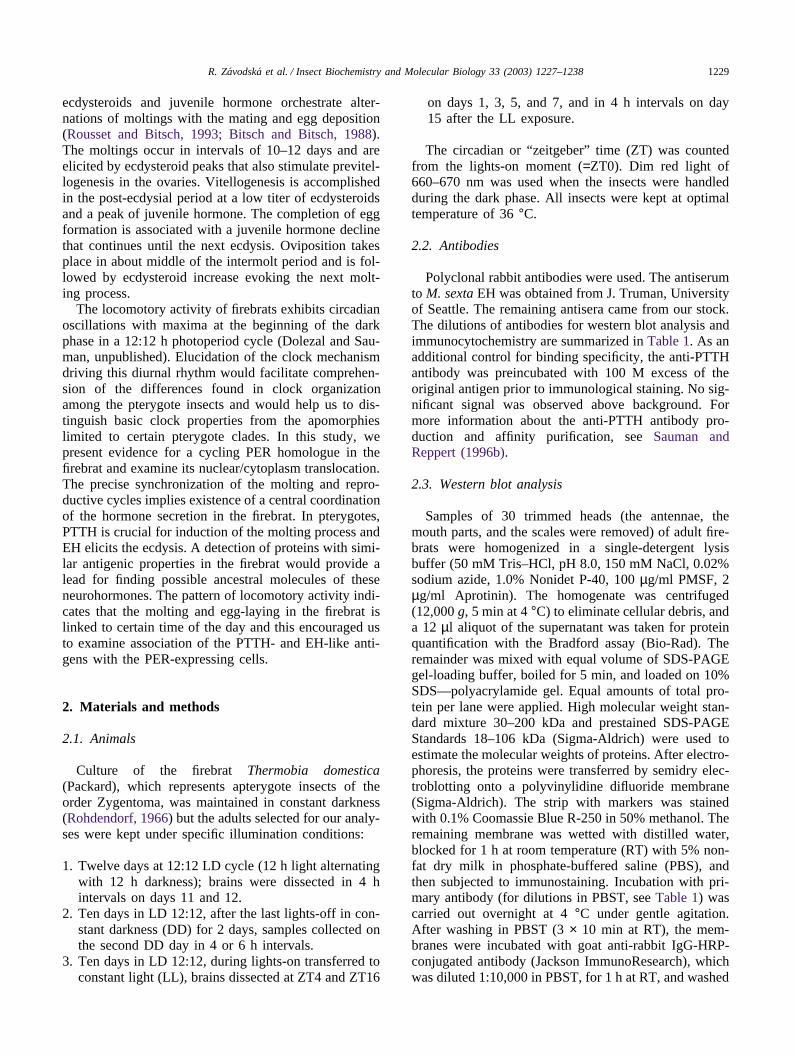
Immunocytochemical search for PER-like materialwas also done in the firebrats exposed to constant lightfor 2 weeks (Fig. 3A). Very few and only slightly stainedcells were found (Table 4). No PER-like material wasdetected in the protocerebrum (Fig. 3B–D), the SOG(Fig. 3F), and the corpora cardiaca in any of the timepoints. Weak cytoplasmic staining persisted in a groupof cells at the border between the deuto- and the tritocer-
Fig. 2. The localization of cells reacting with the 57/10w antiserum.Insects were kept in a 12:12 h light/dark cycle (LD) for 12 days ortransferred to constant darkness (DD) on day 10 and analyzed 1–2days later at 4 h (ZT4) and 16 h (ZT16) after the real or subjectivelights-on point. Schematic diagrams (A, H) show the positions and themaximal staining intensities (indicated by the thickness of drawinglines) of the PER-positive perikarya located in pars intercerebralis (1),deeper protocerebral region (2), lateral protocerebrum (3), optic loberegion between the lobula and the medulla (4), basal protocerebrum(5), between the deuto- and the tritocerebrum (6) and in the suboeso-phageal ganglion (7). Photos of the immunopositive cells (numberedas in diagrams A and H) are shown in sections through pars intercer-ebralis (B), dorsolateral protocerebrum (C), the base of protocerebrum(D), region between the deuto- and the tritocerebrum (E), suboeso-phageal ganglion (F), and corpus cardiacum (G). Magnification B–G, 160×.
ebrum (Fig. 3E). Dispersed staining indicative of finefibers also occurred in this region around the clock.
3.6. The PTTH- and EH-like immunoreactivities
The anti-PTTH-antibody stained about 35 perikaryain the firebrat brain (Fig. 3G). In each hemisphere, twocells were detected in a deep layer of the dorsolateralregion (Fig. 3H), about five cells at the base of protocer-ebrum (Fig. 3K), and 3–5 cells posteriorly at the borderbetween the deuto- and the tritocerebrum (Fig. 3L). FourPTTH-positive cells occurred ventroanteriorly at thebase of each optic lobe (Fig. 3I) and four cells dorsoan-teriorly of the lobula (Fig. 3J). No PTTH-positive axonswere detected and only dispersed staining indicated con-nections between the left and the right protocerebralgroups of PTTH-positive neurons. The two groups ofPTTH-positive cells in the optic lobe seemed to belinked with one another and also with the PTTH-positiveneurons in the ventral and ventroposterior brain regions.No significant staining of any cell or fiber was detectedwith the antibody that had been preincubated with 100M excess of the original PTTH antigen.
The antibody to EH stained just two cells locatedanteriorly to the corpora pedunculata in each brain hemi-sphere (Fig. 3G,M). The EH-like antigen was confined
1234 R. Zavodska et al. / Insect Biochemistry and Molecular Biology 33 (2003) 1227–1238
to the cytoplasm of these cells and no staining was foundoutside the perikarya. We did not have EH antigen tosaturate the antibody for verifying the specificity ofimmunostaining. However, its intensity and sharp local-ization, and the detection of a single antigen on the west-ern blots suggest that it is not coincidental.
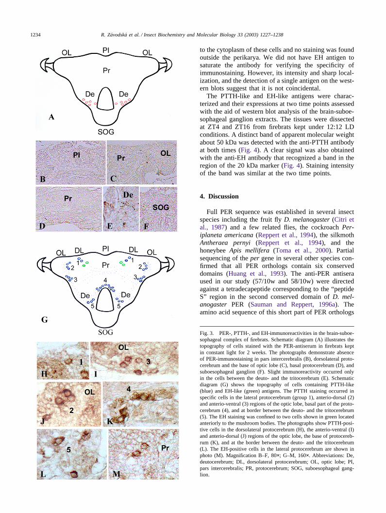
The PTTH-like and EH-like antigens were charac-terized and their expressions at two time points assessedwith the aid of western blot analysis of the brain-suboe-sophageal ganglion extracts. The tissues were dissectedat ZT4 and ZT16 from firebrats kept under 12:12 LDconditions. A distinct band of apparent molecular weightabout 50 kDa was detected with the anti-PTTH antibodyat both times (Fig. 4). A clear signal was also obtainedwith the anti-EH antibody that recognized a band in theregion of the 20 kDa marker (Fig. 4). Staining intensityof the band was similar at the two time points.
4. Discussion
Full PER sequence was established in several insectspecies including the fruit fly D. melanogaster (Citri etal., 1987) and a few related flies, the cockroach Per-iplaneta americana (Reppert et al., 1994), the silkmothAntheraea pernyi (Reppert et al., 1994), and thehoneybee Apis mellifera (Toma et al., 2000). Partialsequencing of the per gene in several other species con-firmed that all PER orthologs contain six conserveddomains (Huang et al., 1993). The anti-PER antiseraused in our study (57/10w and 58/10w) were directedagainst a tetradecapeptide corresponding to the “peptideS” region in the second conserved domain of D. mel-anogaster PER (Sauman and Reppert, 1996a). Theamino acid sequence of this short part of PER orthologs
Fig. 3. PER-, PTTH-, and EH-immunoreactivities in the brain-suboe-sophageal complex of firebrats. Schematic diagram (A) illustrates thetopography of cells stained with the PER-antiserum in firebrats keptin constant light for 2 weeks. The photographs demonstrate absenceof PER-immunostaining in pars intercerebralis (B), dorsolateral proto-cerebrum and the base of optic lobe (C), basal protocerebrum (D), andsuboesophageal ganglion (F). Slight immunoreactivity occurred onlyin the cells between the deuto- and the tritocerebrum (E). Schematicdiagram (G) shows the topography of cells containing PTTH-like(blue) and EH-like (green) antigens. The PTTH staining occurred inspecific cells in the lateral protocerebrum (group 1), anterio-dorsal (2)and anterio-ventral (3) regions of the optic lobe, basal part of the proto-cerebrum (4), and at border between the deuto- and the tritocerebrum(5). The EH staining was confined to two cells shown in green locatedanteriorly to the mushroom bodies. The photographs show PTTH-posi-tive cells in the dorsolateral protocerebrum (H), the anterio-ventral (I)and anterio-dorsal (J) regions of the optic lobe, the base of protocereb-rum (K), and at the border between the deuto- and the tritocerebrum(L). The EH-positive cells in the lateral protocerebrum are shown inphoto (M). Magnification B–F, 80×; G–M, 160×. Abbreviations: De,deutocerebrum; DL, dorsolateral protocerebrum; OL, optic lobe; PI,pars intercerebralis; PR, protocerebrum; SOG, suboesophageal gang-lion.

1235R. Zavodska et al. / Insect Biochemistry and Molecular Biology 33 (2003) 1227–1238
Table 4Numbers of perikarya and the intensities of their staining with antibody to PER in the firebrats transferred from the 12:12 h light/dark cycle tocontinuous light on day 10 and analyzed 15 days later
Perikarya location C ZT0 ZT4 ZT8 ZT12 ZT16 ZT20
Pars intercerebralis 4 � � � � � �Lateral protocerebrum 3 � � � � � �Base of protocerebrum 7 � � � � � �Deuto-/tritocerebrum 5 + + + + + +Optic lobe 3 � � � � � �Suboesophageal ganglion 2 � � � � � �Corpus cardiacum 2 � � � � � �
For explanation, see Table 2.
Fig. 4. Detection of PTTH-like and EH-like antigens on the westernblots of proteins extracted at 4 h (ZT4) and 16 h (ZT16) after lights-on from the firebrats kept under a 12:12 h photoperiod for 12 days.The anti-PTTH antiserum recognized a single specific band of about50 kDa (arrow) and the anti-EH antiserum a band of about 20 kDa(arrowhead) at both time points. Staining of one or two high molecularcomponents (asterisk) was non-specific, as revealed by analyses perfor-med without the primary antisera (Control).
of the fruit fly, cockroach, silkmoth, and the honeybee is97% identical. The specificity of the 57/10w and 58/10wantisera was confirmed in the tests on A. pernyi (Saumanand Reppert, 1996a), P. americana (Sehadova, personalcommunication), and the linden bug Pyrrhocoris apterus(Syrova and Sauman, unpublished). In all these species,the antisera reacted exclusively in neurons in whichexpression of the endogenous per gene was proved byin situ hybridization.
The western blot analysis of the firebrat head extractsdisclosed PER-immunoreactivity in a protein band ofabout 115 kDa (Fig. 1). This size is consistent with the95–135 kDa range of PER orthologs as deduced fromthe per cDNAs identified in different insect species (Citriet al., 1987; Reppert et al., 1994; Toma et al., 2000).The immunocytochemical examinations revealed PERprotein expression in about 50 cells that are located inthe central brain, the optic lobes, the suboesophagealganglion, and the corpora cardiaca of the firebrat (Fig.2). The total number of PER-positive cells found in T.domestica is considerably lower than in the cephalic gan-glia of D. melanogaster (Siwicki et al., 1988; Ewer et
al., 1992). It is higher, however, than in most insectswhich typically contain from 6 to about 60 PER-immun-opositive cells in each half of the brain-suboesophagealganglion complex (Zavodska et al., 2003). Some PER-positive cells are located in the dorsal protocerebrum innearly all species, and location in the optic lobes is alsocommon (Zavodska et al., 2003). The occurrence ofPER-positive neurons in other parts of the cephalic ner-vous system, as seen in T. domestica, is rather unusual.
The firebrat PER protein exhibited circadian oscil-lation in its abundance, which is a basic property of trueclock elements (Aronson et al., 1994; Hall, 1995), sug-gesting that the 115 kDa protein is a genuine PER ortho-log. In the 12:12 h LD cycle, the PER levels were unde-tectable or low during the light period and increased toa peak during the night (Fig. 1). These oscillations inthe abundance of the PER protein were confirmed byimmunocytochemistry (Fig. 2). They were clearly seenin the protocerebral neurons and were extreme in theneurons of the suboesophageal ganglion and the corporacardiaca, in which PER was detectable only in the nightperiod (Table 2). The daily pattern of PER oscillationsin the protocerebral neurons of T.domestica is similar tothat described for the eight PER-positive protocerebralneurons in the silkmoth A. pernyi (Sauman and Reppert,1996a). The complete disappearance of the PER antigenin the corpora cardiaca and the suboesophageal ganglionof T. domestica during the light phase may have acounterpart in the honeybee Apis mellifera, in which cer-tain protocerebral neurons are visualized with the anti-PER antiserum only under certain conditions (G. Bloch,personal communication).
It has been well established that the entrained circad-ian rhythms persist in constant darkness, whereas con-tinuous light disrupts oscillations of the molecularcomponents of the circadian clock and the overt circad-ian rhythm is abolished (Dunlap, 1999). Consistentlywith this general rule, the circadian oscillation of thefirebrat PER protein continued in constant darkness(Table 3; columns DD in Fig. 2), but no PER proteinwas detectable in the firebrats kept under constant light

1236 R. Zavodska et al. / Insect Biochemistry and Molecular Biology 33 (2003) 1227–1238
(Fig. 1). Slight immunostaining persisted in the non-cyc-ling cells located between the deuto- and the tritocereb-rum but was lacking in all other neurons (Fig. 3;Table 4).
A model of the molecular clock mechanism was elab-orated for D. melanogaster (reviewed by Giebultowicz,2000; Williams and Sehgal, 2001). It postulates PERbinding to another clock protein called timeless (TIM),and translocation of the resulting dimer into the cellnucleus. For several cell types in D. melanogaster andfor the photoreceptor cells in A. pernyi, it has beenshown that the circadian translocation of PER from thecytoplasm to the nucleus is associated with circadianchanges in the intensity of the cytoplasmic PER staining.These circadian changes are free-running in the darknessand disappear in continuous light. The 115 kDa PER-like protein of T. domestica exhibits diurnal oscillationsin its cytoplasmic contents but it was never found inthe cell nuclei. Similar cytoplasmic oscillations withouta nuclear translocation of PER were detected in the pro-tocerebral neurons of the silkmoth A. pernyi (Saumanand Reppert, 1996a), the hornworm Manduca sexta(Wise et al., 2002), the cockroach P. americana(Sehadova, personal communication), and the bug P.apterus (Syrova and Sauman, unpublished). There aretwo possible explanations for the lack of nuclear PERstaining in these insects. The PER binding to a partnerprotein in the nucleus prevents PER recognition by anti-bodies such as 57/10w and 58/10w(1), or the PER nevermoves into the nucleus and the clock motion is basedon a very different molecular mechanism than in Droso-phila (2). We cannot distinguish between these twopossibilities.
In addition to the perikarya, clear oscillations in thestaining intensity were detected in the processes of theprotocerebral PER-positive cells under both LD and DDconditions. Axonal transport of the PER protein to otherbrain regions and to the corpora cardiaca was demon-strated in A. pernyi (Sauman and Reppert, 1996a). Con-sistently with this observation, the PER-producing cellsin M. sexta protocerebrum were identified as neurosecre-tory cells of group Ia1 that send axons to the ipsilateralcorpora cardiaca (Wise et al., 2002). PER-positive fiberswere also detected in the corpora cardiaca of the mayflySiphlonurus armatus and the stonefly Perla burmeisteri-ana (Zavodska et al., 2003). Strong PER-immunoreac-tivity was found in the corpora cardiaca of the cockroachP. americana (Sehadova, personal communication).These results suggest that PER may impose circadianregulation on the release of neurohormones that are lib-erated from the retrocerebral complex corpora cardiaca–corpora allata. The endogenous PER-positive cells in thecorpora cardiaca, which have so far been found in thefirebrat only, may play a similar role.
Consistent detection of a 50 kDa protein with the anti-PTTH antiserum and of a 20 kDa protein with the anti-
EH antiserum suggests that both antisera recognize inT. domestica very specific antigens. Their uniqueness isemphasized by their localizations in just a few neurons.The anti-PTTH and anti-EH antisera react in small, sep-arate sets of brain neurons in the representatives of manyinsect orders (Zavodska et al., 2003). The PTTH-likeimmunoreactivity typically occurs in the dorsolateralprotocerebrum, where two immunopositive cells werefound in the firebrat. However, the firebrat seems to beunique by having some PTTH-immunoreactive cells atthe base of optic lobes. The EH-immunopositive cellswere detected in about two-thirds of the examined insectorders (Zavodska et al., 2003). They are always low innumber and in a few species are located in the dorsolat-eral protocerebrum as in the firebrat.
The antigens detectable in the firebrat with the anti-PTTH and anti-EH antisera probably bear structuralsimilarities to the respective hormones but differ fromthem in size. The PTTH orthologs known from five lepi-dopteran species consist each of slightly more than 100amino acids, and their size is about four-fold less thancan be deduced from the apparent 50 kDa size of thePER-like antigen in T. domestica. Similarly, all knownEH orthologs contain a little more than 60 amino acidswhereas the EH-like antigen found in T. domesticaseems to be about three times larger. These size differ-ences should not be surprising in view of considerablestructural diversification of PTTH just within Lepidop-tera (see Shionoya et al., 2003; Sehnal et al., 2002) andof EH between the holometabolous orders Lepidopteraand Diptera (cf. Horodyski et al., 1993). We regard thedetection of a PTTH-like and EH-like antigens in thefirebrat not as an indication of the existence of hormonehomologues in this species but as a cue to investigationson the phylogenic origin of these hormones. Our findingof an apparent PER ortholog in the firebrat confirmswidespread occurrence of this clock component in theeukaryotes and suggests that PER translocation fromcytoplasm to the nucleus, which has been demonstratedin certain cells of some Holometabola, evolved late inpterygote phylogeny.
Acknowledgements
We are thankful to Dr. G. Bloch for the informationon PER expression in the honeybee and to Dr. J.W. Tru-man for the gift of the anti-EH antiserum. Our study waspart of the research project AV0Z5007907 and was inpart supported by grant 204/01/0404 from the GrantAgency of the Czech Republic.
References
Agui, N., Granger, N.A., Gilbert, L.I., Bollenbacher, W.E., 1979.Cellular localization of the prothoracicotropic hormone: in vitro

1237R. Zavodska et al. / Insect Biochemistry and Molecular Biology 33 (2003) 1227–1238
assay of a single neurosecretory cell. Proc. Natl. Acad. Sci. USA76, 5694–5698.
Aronson, B.D., Johnson, K.L., Loros, J.J., Dunlap, J.C., 1994. Negativefeedback defining a circadian clock: autoregulation of the clockgene frequency. Science 263, 1578–1584.
Bargiello, T.A., Young, M.W., 1984. Molecular genetics of a biologi-cal clock in Drosophila. Proc. Natl. Acad. Sci. USA 81, 2142–2146.
Bitsch, C., Bitsch, J., 1988. 20-Hydroxyecdysone and ovarian matu-ration in the firebrat Thermobia domestica (Thysanura,Lepismatidae). Arch. Insect Biochem. Physiol. 7, 281–293.
Carre, I.A., Kay, S.A., 1996. Mechanisms of input and output in circad-ian transduction pathways. In: Verma, D.P.S. (Ed.), Signal trans-duction in plant development. Springer Verlag, Wien, New York,pp. 231–247.
Citri, Y., Colot, H.V., Jacquier, A.C., Yu, Q., Hall, J.C., Baltimore,D., Rosbash, M., 1987. A family of unusually spliced and biologi-cally active transcripts encoded by Drosophila clock gene. Nature326, 42–47.
Dai, J.D., Mizoguchi, A., Gilbert, L.I., 1994. Immunoreactivity of neu-rosecretory granules in the brain-retrocerebral complex of Manducasexta to heterologous antibodies against Bombyx prothoracicotropichormone and bombyxin. Invertebr. Reprod. Dev. 26, 187–196.
Dunlap, J.C., 1999. Molecular bases for circadian clocks. Cell 96,271–290.
Eskin, A., 1979. Identification and physiology of circadian pace-makers. Fed. Proc. 38, 2570–2572.
Ewer, J., Frish, B., Hamblen-Coyle, M.J., Rosbash, M., Hall, J., 1992.Expression of the period clock gene within different cell types inthe brain of Drosophila adults and mosaic analysis of these cellsinfluence on circadian behavioral rhythms. J. Neurosci. 12, 555–570.
Frisch, B., Fleissner, G., Fleissner, G., Brandes, C., Hall, J.C., 1996.Staining in the brain of Pachymorpha sexguttata mediated by anantibody against a Drosophila clock-gene product: labeling of cellswith possible importance for the beetle’s circadian rhythms. CellTissue Res. 286, 411–429.
Gammie, S.C., Truman, J.W., 1999. Eclosion hormone provides a linkbetween ecdysis-triggering hormone and crustacean cardioactivepeptide in the neuroendocrine cascade that controls ecdysis. J. Exp.Biol. 202, 343–352.
Giebultowicz, J.M., 2000. Molecular mechanism and cellular distri-bution of insect circadian clocks. Annu. Rev. Entomol. 45, 769–793.
Gilbert, L.I., King, D.S., 1973. Physiology of growth and developmentendocrine aspects. In: Rockstein, M. (Ed.), Physiology of Insecta,vol. 1. Academic Press, New York, pp. 249–370.
Gilbert, L.I., Rybczynski, R., Song, Q., Mizoguchi, A., Morreale, R.,Smith, W.A., Matubayashi, H., Shionoya, M., Nagata, S., Kataoka,H., 2000. Dynamic regulation of prothoracic gland ecdysteroidog-enesis: Manduca sexta recombinant prothoracicotropic hormoneand brain extracts have identical effects. Insect Biochem. Mol. Biol.30, 1079–1089.
Gotter, A.L., Levine, J.D., Reppert, S.M., 1999. Sex-linked periodgenes in the silkmoth, Antheraea pernyi: implications for circadianclock regulation and the evolution of sex chromosomes. Neuron24, 953–965.
Hall, J.C., 1995. Tripping along the trail to the moecular mechanismsof biological clocks. Trends Neurosci 18, 230–240.
Hardin, P.E., Hall, J.C., Rosbash, M., 1990. Feedback of the Droso-phila period gene product on circadian cycling of its messengerRNA. Nature 343, 536–540.
Hewes, R.S., Truman, J.W., 1991. The roles of central and peripheraleclosion hormone release in the control of ecdysis behavior in Man-duca sexta. J. Comp. Physiol. 168, 697–707.
Honegger, H.W., Leser, W., Loher, W., Siwicki, K.K., 1991. Labelling
of cells in the CNS of the cricket Teleogryllus commodus by anantibody to Drosophila per-protein. Sci. Neurosci. Abstr. 17, 1239.
Horodyski, F.M., Ewer, J., Riddiford, L.M., Truman, J.W., 1993. Iso-lation, characterization and expression of the eclosion hormonegene of Drosophila melanogaster. Eur. J. Biochem. 215, 221–228.
Huang, Z.J., Edery, I., Rosbash, M., 1993. PAS is a dimerizationdomain common to Drosophila period and several transcriptionfactors. Nature 364, 259–262.
Kataoka, H., Suzuki, A., 1995. Prothoracicotropic hormone of lepidop-teran insects. In: Konopinska, H. (Ed.), Insects: Chemical, Physio-logical, and Environmental Aspects. Wroclaw University Press,Wroclaw, Poland, pp. 70–77.
Kataoka, H., Troetschler, R.G., Kramer, S.J., Cesarin, B.J., Schooley,D.A., 1987. Isolation and primary structure of the eclosion hormoneof the tobacco hornworm, Manduca sexta. Biochem. Biophys. Res.Commun. 146, 746–750.
Kawakami, A., Kataoka, H., Oka, T., Mizoguchi, A., Kimura-Kawak-ami, M., Adachi, T., Iwami, M., Nagasawa, H., Suzuki, A., Ishi-zaki, H., 1990. Molecular cloning of the Bombyx mori protho-racicotropic hormone. Science 247, 1333–1335.
Kono, T., Nagasawa, H., Isogai, A., Fugo, H., Suzuki, A., 1987. Aminoacid sequence of eclosion hormone of the silkworm, Bombyx mori.Agric. Biol. Chem. 51, 2307–2308.
Kono, T., Nagasawa, H., Isogai, A., Fugo, H., Suzuki, A., 1990. Amonoclonal antibody against a synthetic carboxy-terminal fragmentof the eclosion hormone of the silkworm, Bombyx mori: charac-terization and application to immunohistochemistry and affinitychromatography. Zool. Sci. 7, 47–54.
Konopka, R.J., Benzer, S., 1971. Clock mutants of Drosophila mel-anogaster. Proc. Natl. Acad. Sci. USA 68, 2112–2116.
Labandeira, C.C., 1998. Early history of arthropod and vascular plantassociations. Annu. Rev. Earth Planet. Sci. 26, 329–377.
Liu, X., Lorenz, L., Yu, Q., Hall, J.C., Rosbash, M., 1988. Spatial andtemporal expression of the period gene in Drosophila melanogas-ter. Genes Dev. 2, 228–238.
Marti, T., Takio, K., Walsh, K.A., Terzi, G., Truman, J.W., 1987.Microanalysis of the amino acid sequence of the eclosion hormonefrom the tobacco hornworm Manduca sexta. FEBS Lett. 219,415–418.
Mizoguchi, A., Dedos, S.G., Fugo, H., Kataoka, H., 2002. Basic pat-tern of fluctuation in haemolymph PTTH titers during larval–pupaland pupal–adult development of the silkworm, Bombyx mori. Gen.Comp. Endocrinol. 127, 181–189.
Naya, S., Suzuki, K., Fugo, H., Sehnal, F., 1994. Eclosion hormone-like immunoreactivity in the nervous system of Bombyx mori(Lepidoptera: Bombycidae) and Antheraea yamamai (Lepidoptera:Saturniidae) before and after hatching. Eur. J. Entomol. 91, 89–196.
Piccin, A., Couchman, M., Clayton, J.D., Chalmers, D., Costa, R., Kyr-iacou, C.P., 2000. The clock gene period of the housefly, Muscadomestica, rescues behavioral rhythmicity in Drosophila mel-anogaster: evidence for intermolecular coevolution? Genetics 154,747–758.
Reddy, P., Zehring, W.A., Wheeler, D.A., Pirrotta, V., Hadfield, C.,Rosbash, M., Hall, J.C., 1984. Molecular analysis of the periodlocus in Drosophila melanogaster and identification of a transcriptinvolved in biological rhythms. Cell 38, 701–710.
Reppert, S.M., Tsai, T., Roca, A.L., Sauman, I., 1994. Cloning of astructural homolog of the circadian clock gene period from thegiant silkmoth Antheraea pernyi. Neuron 13, 1167–1176.
Reynolds, S.E., Truman, J.W., 1983. Eclosion hormone. In: Downer,R.G.H., Laufer, H. (Eds.), Endocrinology of Insects, vol. 1. AlanR. Liss, New York, pp. 217–233.
Rohdendorf, E.B., 1966. Der Einfluss der Allatektomie auf adultWeibchen von Thermobia domestica Packard (Lepismatidae,Thysanura). Zool. Jb. (Physiol.) 71, 685–693.
Rousset, A., Bitsch, C., 1993. Comparison between endogenous andexogenous yolk proteins along an ovarian cycle in the firebrat Ther-

1238 R. Zavodska et al. / Insect Biochemistry and Molecular Biology 33 (2003) 1227–1238
mobia domestica (Insecta, Thysanura). Comp. Biochem. Physiol.B 104, 33–44.
Sauman, I., Reppert, S.M., 1996a. Circadian clock neurons in the silk-moth Antheraea pernyi: novel mechanisms of period protein regu-lation. Neuron 17, 889–900.
Sauman, I., Reppert, S.M., 1996b. Molecular characterization of pro-thoracicotropic hormone (PTTH) from the giant silkmothAntheraea pernyi: developmental appearance of PTTH-expressingcells and relationship to circadian clock cells in central brain. Dev.Biol. 178, 418–429.
Sehnal, F., Hansen, I., Scheller, K., 2002. The cDNA-structure of theprothoracicotropic hormone (PTTH) of the silkmoth Hyalophoracecropia. Insect Biochem. Mol. Biol. 32, 233–237.
Shionoya, M., Matsubayashi, H., Asahina, M., Kuniyashi, H., Nagata,S., Riddiford, L.M., Kataoka, H., 2003. Molecular cloning of theprothoracicotropic hormone from the tobacco hornworm, Manducasexta. Insect Biochem Mol. Biol. 33, 795–801.
Siwicki, K.K., Eastman, C., Petersen, G., Rosbash, M., Hall, J.C.,1988. Antibodies to the period gene product of Drosophila revealdiverse tissue distribution and rhythmic changes in the visual sys-tem. Neuron 1, 141–150.
Toma, D.P., Bloch, G., Moore, D., Robinson, G.E., 2000. Changes inperiod mRNA levels in the brain and division of labor in honeybee colonies. Proc. Natl. Acad. Sci. USA 97, 6914–6919.
Truman, J.W., 1972. Physiology of insect rhythms. II. The silkmothbrain as the location of the biological clock controlling eclosion.J. Comp. Physiol. 81, 99–114.
Vafopoulou, X., Steel, C.G.H., 2001. Induction of rhythmicity in pro-thoracicotropic hormone and ecdysteroids in Rhodnius prolixus:roles of photic and neuroendocrine Zeitgebers. J. Insect Physiol.47, 935–941.
Watson, C.M., Sauman, I., Berry, S.J., 1993. Actin is a major structuraland functional element of the egg cortex of giant silkmoth duringoogenesis. Dev. Biol. 155, 315–323.
Williams, J.A., Sehgal, A., 2001. Molecular components of the circad-ian system in Drosophila. Annu. Rev. Physiol. 63, 729–755.
Wise, S., Davis, N.T., Tyndale, E., Noveral, J., Folwell, M.G., Bedian,V., Emery, I.F., Siwicki, K.K., 2002. Neuroanatomical studies ofperiod gene expression in the hawkmoth, Manduca sexta. J. Comp.Neurol. 447, 366–380.
Zavodska, R., Sauman, I., Sehnal, F., 2003. Distribution of PER pro-tein, pigment-dispersing hormone, prothoracicotropic hormone, andeclosion hormone in the cephalic nervous system of insects. J. Biol.Rhythms 18, 106–122.
Zerr, D.M., Hall, J.C., Rosbash, M., Siwicki, K.K., 1990. Circadianfluctuations of period protein immunoreactivity in the CNS and thevisual system of Drosophila. J. Neurosci. 10, 2749–2762.