Genetic diversity of the genus Malus and implications for linkage mapping with SNPs
Upload
independentCategory
view
3download
0
Cell wall structures leading to cultivar differencesin softening rates develop early during apple(Malus x domestica) fruit growthNg et al.
Ng et al. BMC Plant Biology 2013, 13:183http://www.biomedcentral.com/1471-2229/13/183
Ng et al. BMC Plant Biology 2013, 13:183http://www.biomedcentral.com/1471-2229/13/183
RESEARCH ARTICLE Open Access
Cell wall structures leading to cultivar differencesin softening rates develop early during apple(Malus x domestica) fruit growthJovyn KT Ng1,2,3*, Roswitha Schröder2, Paul W Sutherland2, Ian C Hallett2, Miriam I Hall2, Roneel Prakash2,Bronwen G Smith1, Laurence D Melton1 and Jason W Johnston4
Abstract
Background: There is a paucity of information regarding development of fruit tissue microstructure and changes inthe cell walls during fruit growth, and how these developmental processes differ between cultivars with contrastingsoftening behaviour. In this study we compare two apple cultivars that show different softening rates during fruitdevelopment and ripening. We investigate whether these different softening behaviours manifest themselves lateduring ethylene-induced softening in the ripening phase, or early during fruit expansion and maturation.
Results: ‘Scifresh’ (slow softening) and ‘Royal Gala’ (rapid softening) apples show differences in cortical microstructureand cell adhesion as early as the cell expansion phase. ‘Scifresh’ apples showed reduced loss of firmness and greater drymatter accumulation compared with ‘Royal Gala’ during early fruit development, suggesting differences in resourceallocation that influence tissue structural properties. Tricellular junctions in ‘Scifresh’ were rich in highly-esterified pectin,contributing to stronger cell adhesion and an increased resistance to the development of large airspaces during cellexpansion. Consequently, mature fruit of ‘Scifresh’ showed larger, more angular shaped cells than ‘Royal Gala’, withless airspaces and denser tissue. Stronger cell adhesion in ripe ‘Scifresh’ resulted in tissue fracture by cell rupturerather than by cell-to-cell-separation as seen in ‘Royal Gala’. CDTA-soluble pectin differed in both cultivars duringdevelopment, implicating its involvement in cell adhesion. Low pectin methylesterase activity during early stages of fruitdevelopment coupled with the lack of immuno-detectable PG was associated with increased cell adhesion in ‘Scifresh’.
Conclusions: Our results indicate that cell wall structures leading to differences in softening rates of apple fruit developearly during fruit growth and well before the induction of the ripening process.
Keywords: Apple, Cell adhesion, Cell wall, Fruit firmness, Immunofluorescence labelling, Microstructure, Pectin
BackgroundApple cultivars exhibit variable rates of softening duringripening and can vary in firmness once mature [1]. Ouraim was to determine when these differences in softeningbehaviour in apple fruit manifest themselves; in the earlystages of fruit development, after cell division ceasesand cell expansion starts, giving rise to the development ofthe complex three-dimensional cortical tissue; or later indevelopment, when the fruit approaches full size and
* Correspondence: [email protected] Science, School of Chemical Sciences, The University of Auckland,Private Bag 92019, Auckland, New Zealand2The New Zealand Institute for Plant & Food Research Limited, Mount AlbertResearch Centre, Private Bag 92169, Auckland 1142, New ZealandFull list of author information is available at the end of the article
© 2013 Ng et al.; licensee BioMed Central Ltd.Commons Attribution License (http://creativecreproduction in any medium, provided the or
begins to initiate ripening. In this study, we explore therole of cortical microstructure in the softening behaviourof apples throughout development and ripening.Apples undergo two distinct phases during growth:
a phase of intensive cell division which lasts typically3-5 weeks after full bloom, followed by a phase of cellexpansion when cell division ceases [2]. As the fruitgrows and increases in size, mass is gained mainlythrough water uptake and increase in parenchyma cellvolume [3]. Firmness declines during fruit expansion,which coincides with reduced density of cell packingand increased cell volume and air spaces [4]. Thus, thecell wall not only has to maintain structural integrityduring growth, but also allow expansion of cell size and
This is an open access article distributed under the terms of the Creativeommons.org/licenses/by/2.0), which permits unrestricted use, distribution, andiginal work is properly cited.
Ng et al. BMC Plant Biology 2013, 13:183 Page 2 of 16http://www.biomedcentral.com/1471-2229/13/183
associated extracellular air spaces. There is currently apaucity of information regarding development of tissuemicrostructure and changes in the cell walls during fruitgrowth, let alone how these developmental processes differbetween cultivars with contrasting softening behaviour. In-stead, considerable attention has been paid to changes incell wall chemistry and microstructure [5,6] duringripening, however this has only limited applicabilityfor understanding when important microstructuralfeatures develop during growth.Assessment of tissue microstructure is complex, as it
is influenced by many different cellular components, andmultiple, complementary approaches are needed to developa robust view of how cultivars differ. Studies using applemapping populations to investigate the relationshipbetween textural properties and cell size and shape haveproduced inconsistent results, with one study failing todetect quantitative trait loci (QTL) for cell size despitedetecting QTLs for textural properties [7]. In contrast, asecond study showed a significant correlation between cellsize and textural properties [8]. Further studies using ripecommercial apple cultivars have reinforced the associationbetween cell size and texture, where increased sensoryjuiciness was associated with larger cell sizes and moredensely packed tissue [9].Another important aspect of tissue microstructure is
the interconnections between adjacent cells, and how theycontribute to a three-dimensional structure. Cell-to-celladhesion is important, as it affects the fracture path acrosstissues and contributes to the mode of tissue failure. Theexamination of fracture surfaces for apples, and a broadersurvey across different types of fruit have shown two maintypes of tissue failure: 1) cell rupture that results in therelease of cellular contents; and 2) cell-to-cell separationwhere adjacent cells separate without cell rupture [10-12].It has also been shown that cell rupture can be classifiedas equatorial, or as a top or bottom fracture which mayaffect the rate at which cell contents are released [9].These results reinforce the need to understand thechemistry of the cell wall in terms of dissolution of themiddle lamella and the separation of adjacent cells,while taking into consideration the spatial distributionof different types of pectin in the cell junction zonesduring softening.Homogalacturonan (HG) pectin is believed to play a
major role in intercellular adhesion, as it is commonlyfound in the middle lamella region of the cell wallwhere two adjacent cells adjoin [13]. Here, stretches ofun-esterified galacturonic acid residues of HG are thoughtto provide the main bonding between adjacent cellsthrough calcium cross-links [14]. Enzymes that are likelyto play a role in modulating cell adhesion properties arepectin methylesterase (PME) and endo-polygalacturonase(PG), where PG is thought to depolymerise HG within
stretches of un-esterified galacturonic acid residues createdby PME [15].A number of studies have reported on the occurrence
and activity of PG in apples [16-19] but the low abundanceof PG protein and in most cases undetectable enzymaticactivity has shifted research towards a transgenic approach[20-22]. Expression of the ripening-related MdPG1 genein ‘Royal Gala’ is induced during cold storage [23], and itsdown-regulation in ‘Royal Gala’ increased cell adhesionand reduced softening [21], whereas over-expression ofthe same gene led to increased intercellular separation in‘Royal Gala’ leaves [20], demonstrating a role for PG in theloss of intercellular adhesion. PME protein is found inmost plant tissues and exists in multiple isoforms. Inapple, PME activity has been found to increase duringgrowth and decrease during ripening-related softening[24], but its role is less clear than that of PG. Apart frompectic-related changes, xyloglucan and enzymes suchas xyloglucan endotransglucosylase/hydrolase (XTH)also play important roles in the development of apple fruittexture and softening [8,25]. Recent work in apple hasshown an increase in XTH gene expression induced byethylene, thereby emphasizing the role of XTH in xylo-glucan modification during apple fruit softening [26].In this paper, we determine the microstructural proper-
ties throughout fruit development and softening of‘Scifresh’ (commercially marketed as Jazz™), an applecultivar that loses firmness slowly during ripening despitehigh ethylene production, and compare it with ‘Royal Gala’,a parent of ‘Scifresh’, that softens more rapidly duringripening but has a similarly high ethylene production.The advantage of this approach is that the ripeningphenotype is not confounded by differences in ethyleneproduction, enabling a more robust assessment of therelative contribution of structural features towards soften-ing. By using a combination of different techniques, weinvestigate differences between the cultivars in cell sizeand cell packing, fracture pattern, tensile strength andcell-to-cell adhesion. Immunolocalisation, cell wall frac-tionation and size exclusion chromatography are usedto examine differences in pectin between adjacent cellsand in zones where extracellular air spaces develop duringgrowth and ripening. The involvement of pectin-modifyingenzymes pectin methylesterase (PME) and polygalacturo-nase (PG) are also investigated.
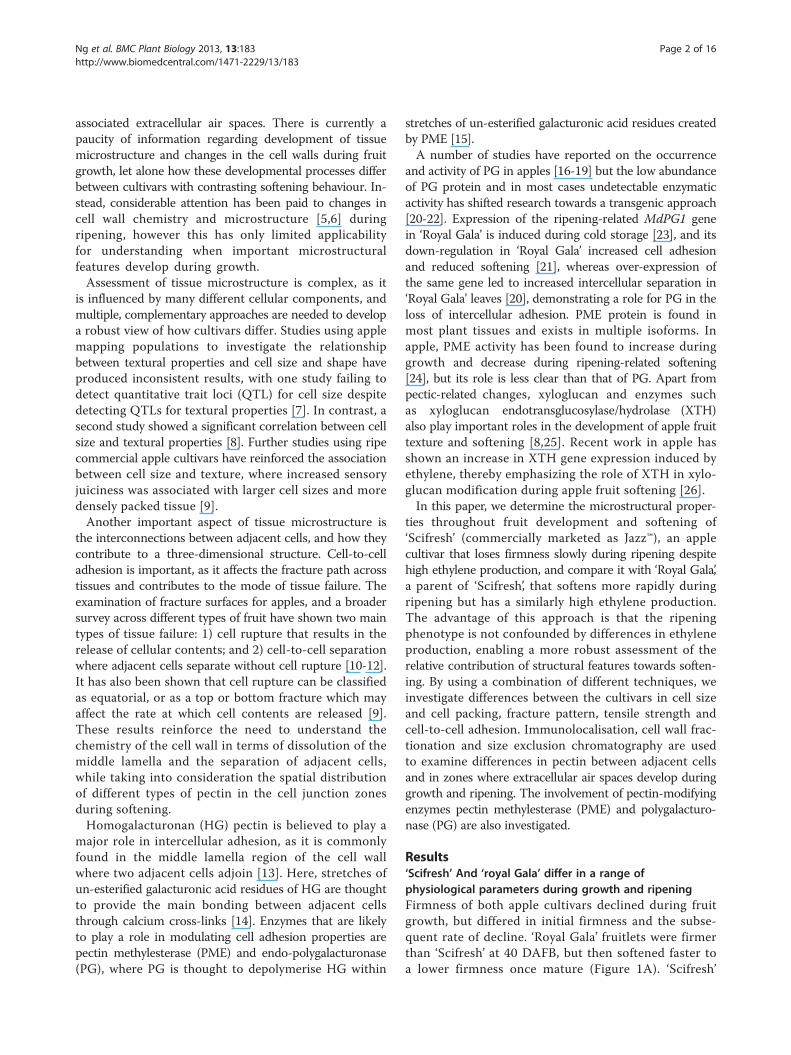
Results‘Scifresh’ And ‘royal Gala’ differ in a range ofphysiological parameters during growth and ripeningFirmness of both apple cultivars declined during fruitgrowth, but differed in initial firmness and the subse-quent rate of decline. ‘Royal Gala’ fruitlets were firmerthan ‘Scifresh’ at 40 DAFB, but then softened faster toa lower firmness once mature (Figure 1A). ‘Scifresh’
0
100
200
A
B
0.0
0.5
1.0
0 40 80 120 160
D
C
5
15
25
Days after full bloom
E
0
10
20
40-70 70-100 100-120 120-140
Incr
ease
in fr
uit w
eigh
t
(% d
ay-1
)
Firm
ness
(N)
Wei
ght
(g)
Eth
ylen
e
(µLL
-1)
Dry
mat
ter
(%)
0
25
50 ‘Scifresh’‘Royal Gala’
Figure 1 Physiological parameters during growth and maturationof ‘Royal Gala’ and ‘Scifresh’. Flesh firmness (A), fruit weight (B), drymatter concentration (C), internal ethylene production (D), and percentincrease in fruit weight (E) during development of ‘Royal Gala’ and‘Scifresh’ apples (n= 20 ± SE). Probe size for measuring firmness was5 mm. The percentage increase in fruit weight (E) was calculated as thechange in mean fruit weight relative to the weight at the start of eachperiod per day.
Ng et al. BMC Plant Biology 2013, 13:183 Page 3 of 16http://www.biomedcentral.com/1471-2229/13/183
apples had a lag phase with minimal loss of firmnessbetween 40 and 70 DAFB. Both cultivars had a similarincrease of fruit weight (Figure 1B), which coincidedwith the decline in firmness. However, firmness declinewas not exclusively due to fruit growth, as the firmnesslag phase for ‘Scifresh’ was not accompanied by slowergrowth. Dry weight accumulation was similar for bothcultivars, with the only difference occurring 70 DAFB when‘Scifresh’ accumulated more dry matter than ‘Royal Gala’(Figure 1C). For ‘Scifresh’, this peak in dry matter accu-mulation coincided with the end of the lag phase forloss of firmness (Figure 1A) and the rapid growth phase(Figure 1E), suggesting cultivar differences in dry weightassimilation and partitioning into structural features duringfruit growth. Fruit internal ethylene concentrations werecomparable between the two cultivars during fruit growthand maturation (Figure 1D), with the climacteric rise inethylene occurring between 100 and 120 DAFB. The twocultivars also produced similar concentrations of ethyleneduring ripening, but had substantial differences in the rateof softening (Figure 2). ‘Royal Gala’ declined in firmness byca. 35% during ripening, while ‘Scifresh’ effectively did notchange in firmness over the same period (Figure 2A).
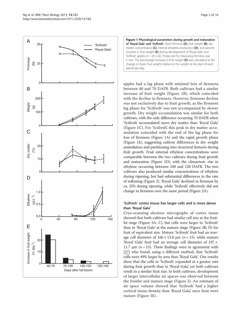
‘Scifresh’ cortex tissue has larger cells and is more densethan ‘Royal Gala’Cryo-scanning electron micrographs of cortex tissueshowed that both cultivars had similar cell size at the fruit-let stage (Figure 3A, C), but cells were larger in ‘Scifresh’than in ‘Royal Gala’ at the mature stage (Figure 3B, D) forfruit of equivalent size. Mature ‘Scifresh’ fruit had an aver-age cell diameter of 166 ± 13.8 μm (n = 15), while mature‘Royal Gala’ fruit had an average cell diameter of 107 ±11.7 μm (n = 15). These findings were in agreement with[27] who found, using a different method, that ‘Scifresh’cells were 49% larger by area than ‘Royal Gala’. Our resultsshow that the cells in ‘Scifresh’ expanded at a greater rateduring fruit growth than in ‘Royal Gala’, yet both cultivarsresult in a similar fruit size. In both cultivars, developmentof larger intercellular air spaces was observed betweenthe fruitlet and mature stage (Figure 3). An estimate ofair space volume showed that ‘Scifresh’ had a highercortical tissue density than ‘Royal Gala’, once fruit weremature (Figure 3E).

A
Inte
rnal
eth
ylen
e (µ
L-1L)
F
irmne
ss (
N)
Ripening (weeks)
B0
50
100
0.1
1
10
100
1000
0 5 10 15 20
‘Scifresh’
‘Royal Gala’
Figure 2 Firmness loss and ethylene production of ‘Royal Gala’and ‘Scifresh’ fruit during ripening. Flesh firmness (A) and internalethylene production (B) during ripening at 0.5°C (n = 20 ± SE).Note: probe size for measuring firmness was 11 mm.
Ng et al. BMC Plant Biology 2013, 13:183 Page 4 of 16http://www.biomedcentral.com/1471-2229/13/183
Fracture surfaces of ripe tissue shows more cell ruptureand greater cell adhesion in ‘Scifresh’Using conventional scanning electron microscopy, distinctdifferences in the fracture pattern were observed inripe tissue (Figure 4A–D). ‘Royal Gala’ tissue primarilyfractured between cells resulting in minimal cell rupture(Figure 4A, C), while fracturing of ‘Scifresh’ tissue occurredmore by cell rupture with minimal evidence of cell sepa-ration between adjacent cells (Figure 4B, D). Tensile testswere used to quantify the force required to pull cortextissue apart. At earlier stages of development (100 DAFBand mature) the tensile properties of the two cultivars weresimilar, with both having loss of tensile strength during thefinal stages of fruit growth (100 DAFB to maturity). Onceripe, ‘Royal Gala’ required 50% less force than ‘Scifresh’to fracture the tissue (Figure 4E). These tensile propertiessupport the fracture surface images (Figure 4A–D) byshowing that cell separation is associated with weakadhesion forces between adjacent cells in ‘Royal Gala’.
Cellular junctions are filled with highly esterified pectin in‘Scifresh’ throughout development, but not in ‘Royal Gala’The monoclonal antibody LM19, specific for non- orlow methyl-esterified HG regions of pectin, labelled cellwalls in tissue of both cultivars in two distinct patterns,in the middle lamella region and in the corners of tricellularjunctions (Figure 5; pink labelling). The labelling patternswere similar in both cultivars and were most pronouncedin fruitlet and expanding fruits, and weakest in matureand ripe fruit. In ‘Scifresh’, intercellular spaces seemedsmaller than in ‘Royal Gala’ at all developmental stages,supporting density measurements (Figure 3E). The inter-cellular spaces, particularly its corners in ‘Scifresh’ atthe fruitlet and expanding stage were filled with non-or low-esterified pectin, whereas ‘Royal Gala’ seemedto have less of this epitope filling the intercellularspaces (Figure 5). Co-staining of sections with calco-fluor (Figure 5; blue labelling) showed cellulose stainingaround the inside of the cell walls towards the cell lumenin both cultivars. Additional file 1: Figure S1 shows theLM19 antibody-labelling in green fluorescence at lowermagnification.LM20, specific for more highly methyl-esterified HG
regions, labelled ‘Royal Gala’ fruitlet and expanding fruitintensely in corners of tricellular junctions, whereas inthe more tightly-packed cells of ‘Scifresh’, this epitopeoccurred more throughout junction zones and was notjust restricted to the corners (Figure 6, pink labelling). In‘Scifresh’, the middle lamella region also labelled moreintensely, indicating more esterified HG in these areascompared to ‘Royal Gala’. In ‘Royal Gala’, intercellularspaces enlarged during fruit growth, whereas in ‘Scifresh’,they remained similar to the fruitlet stage. As with theLM19, the intercellular spaces of ‘Scifresh’ fruitlet andexpanding fruit were filled with esterified pectin (Figure 6).Additionally, this intense labelling of esterified pectinin tricellular junctions remained up to maturation in‘Scifresh’ fruit, whereas mature ‘Royal Gala’ fruit dis-played weaker labelling. In ripe fruit of both cultivars,labelling became weaker, although still concentratedat the middle lamella region, but absent from theintercellular space which filled tricellular junctions.Co-staining of sections with calcofluor (Figure 6; bluelabelling) showed cellulose staining around the insideof the cell walls towards the cell lumen in both culti-vars. Additional file 2: Figure S2 and Additional file3: Figure S3 show LM20 antibody-labelling in greenfluorescence.HG regions with sufficiently low degree of methyl-
esterification (<40%) and with at least nine consecutivenon-esterified galacturonic acid residues can interact withdivalent cations like calcium [28]. The monoclonal antibody2F4 is specific to these calcium cross-linked HG epitopes.Labelling of cell walls with 2F4 was detected in mature

0.80
0.85
0.90
Cor
tical
den
sity
(g m
L-1)
E
‘Scifresh’ ‘Royal Gala’
Figure 3 Size and density of cortical cells of ‘Royal Gala’ and‘Scifresh’ at the fruitlet and mature stage. Cryo-scanning electronmicrographs of fruitlet (A, C) and mature fruit (B, D) of ‘Royal Gala’(A, B) and ‘Scifresh’ (C, D), and cortical density of cells at the maturefruit stage (E). Bars = 200 μm for all micrographs. Apples were matchedfor size at each stage of development. Values in (E) are the mean of15 measurements ± SE.
Ng et al. BMC Plant Biology 2013, 13:183 Page 5 of 16http://www.biomedcentral.com/1471-2229/13/183
fruit of both cultivars, but disappeared during softeningin ‘Royal Gala’ and remained in ‘Scifresh’ (Figure 7). Inboth cultivars labelling was restricted to junction zones,particularly tricellular junctions. No labelling was foundthroughout the cell walls and very little was detected inthe middle lamella, confirming that calcium cross-linkedHG epitopes are restricted to the middle lamellae at cellcorners and pit fields [29]. No labelling was detected infruitlets of both cultivars (not shown), while expandingfruit were not examined.
Cell wall yield and composition differed most betweencultivars during early stages of fruit growthThe amount of cell wall material on a fresh weight basiswas highest in fruitlet and decreased during growth, mat-uration and ripening in both cultivars (Table 1). Apart fromthe fruitlet stage, where the yield of cell wall material wasconsiderably higher in ‘Royal Gala’ than in ‘Scifresh’, yieldswere comparable in both cultivars. The sum of the pectin-related sugars uronic acid, rhamnose, arabinose and galac-tose indicated a substantial amount of pectin of over 80%of the total amount of non-cellulosic sugars (Table 1).The greatest difference between the two cultivars was
observed at the fruitlet stage, where the firmer ‘RoyalGala’ fruitlet had about twice the uronic acid content of‘Scifresh’ but a lower neutral sugar content (Table 1). Al-though ‘Scifresh’ began with less uronic acid in fruitlet,its relative uronic acid content declined more slowlyduring growth and maturation than ‘Royal Gala’, up tothe point when in cell walls of ripe fruit, ‘Scifresh’ had ahigher uronic acid content. In general, during fruit growthand ripening, amounts of all neutral sugars and uronicacid decreased in both cultivars, especially galactose andarabinose.As there were major changes in the fruit physiology
beginning at 70 DAFB in terms of firmness, dry matterand growth rate (Figures 1A, D, E), the percent growthrate (Figure 1E) was compared with the percent changein cell wall material, to determine if the yield of cell wallmaterial was reflected by growth. To make this comparison,net cell wall content was calculated and presented asAdditional file 4: Figure S4 showing the difference betweenpercent growth rate and percent loss of cell wall materialduring growth. This approach showed that the depo-sition of net cell wall content strongly mirrored growth,

‘Roy
al G
ala’
‘Sci
fres
h’‘R
oyal
Gal
a’‘S
cifr
esh’
0
10
20
30
Tens
ile s
tren
gth
(N)
100 DAFBMatureRipe
E
‘Scifresh’ ‘Royal Gala’
Figure 4 Fracture pattern and tensile strength of cortical tissueof ‘Royal Gala’ and ‘Scifresh’ fruit during ripening. Scanningelectron micrographs of ripe ‘Royal Gala’ (A, C) and ‘Scifresh’ fruit(B, D) (20 weeks at 0.5°C), showing a different fracture patternbetween cells with the appearance of more intact cells in ‘Royal Gala’and more broken open cells in ‘Scifresh’. (E) Tensile tests to quantifythe force required to pull cortex tissue apart (expanding fruit 100DAFB, mature and ripe fruit). Bars A, B = 500 μm; bars C, D = 100 μm.Values in (E) are the mean of 15 measurements ± SE.
Ng et al. BMC Plant Biology 2013, 13:183 Page 6 of 16http://www.biomedcentral.com/1471-2229/13/183
in that the rapid growth phase (between 40 and 70 DAFB)was also the phase where net cell wall content was great-est. The two cultivars had a similar pattern of net cellwall content throughout development, with some evi-dence for greater deposition in ‘Royal Gala’ between 40and 70 DAFB. Deposition of net cell wall content slowedin both cultivars between 70 and 100 DAFB, and be-came negligible once the fruit approached maturity.
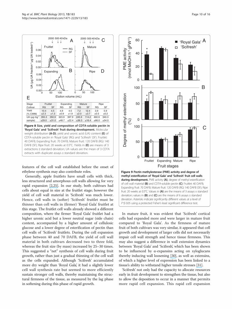
Yield, composition and molecular weight distribution ofCDTA-soluble pectin differed markedly during growthand ripeningThe molecular weight distribution of the CDTA-solublepectin differed most between the cultivars during growthand maturation, whereas the composition and yield differedmost during the ripening phase (Figure 8). The molecularweight distribution of the CDTA-extracted polyuronateswas very similar in fruitlets of both cultivars, but thendecreased more in ‘Scifresh’ than ‘Royal Gala’ while fruitexpanded (Figure 8A, B). This decrease was accompaniedby an increase in yield of CDTA-soluble pectin in ‘Scifresh’but not in ‘Royal Gala’ (Figure 8E). As the fruit matured,the molecular weight distribution of CDTA-extractablepolyuronates increased considerably in ‘Scifresh’ to besimilar to ‘Royal Gala’. In ‘Scifresh’, this increase wasaccompanied by an increase in yield of CDTA-solublepectin and a decrease in its uronic acid content (Figure 8E).During ripening, the molecular weight distribution broad-ened and remained comparable between the mature andripe stage in both cultivars (Figure 8C, D). In both cultivars,yields decreased by a third, whereas the uronic acid contentincreased per mg polysaccharide (Figure 8E).
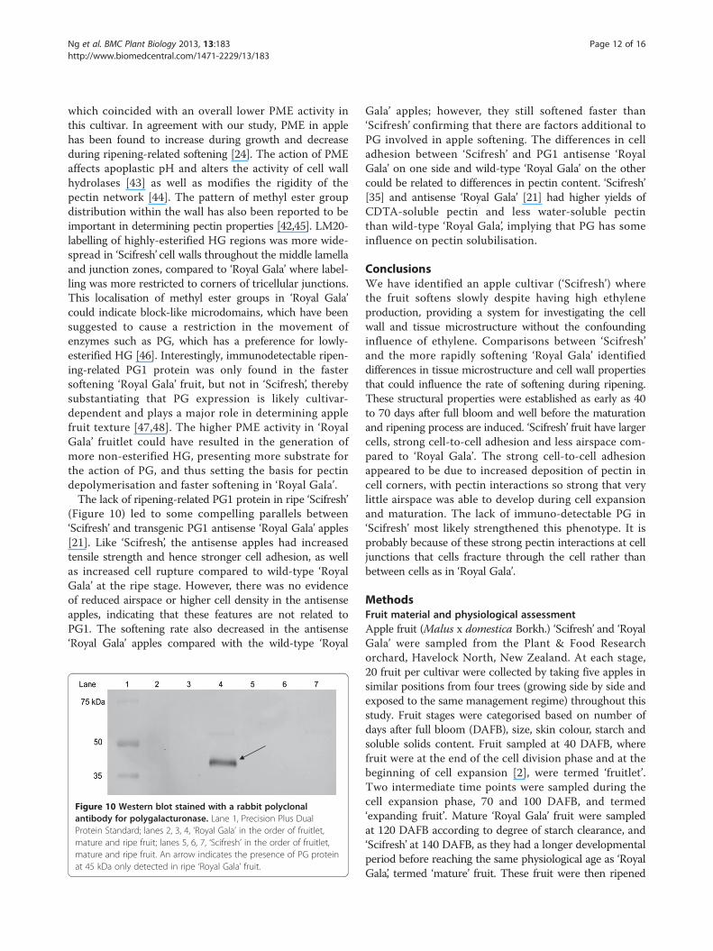
Differences in pectin methylesterase activity early in fruitdevelopment may impact the degree of methyl-esterificationof cell walls of both cultivars later on in developmentSignificant difference in pectin methylesterase (PME)activity was observed at the fruitlet stage, where ‘Scifresh’showed about a quarter the activity of ‘Royal Gala’(Figure 9A). However, while ‘Scifresh’ fruit were expand-ing, PME activity increased to the same level of activityin ‘Royal Gala’. After the fruitlet stage, PME activitycontinuously decreased and was similar between thetwo cultivars.
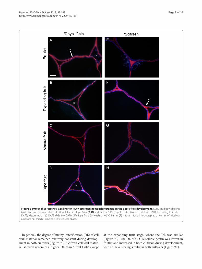
Figure 5 Immunofluorescence labelling for lowly-esterified homogalacturonan during apple fruit development. LM19 antibody labelling(pink) and anti-cellulose stain calcofluor (blue) in ‘Royal Gala’ (A-D) and ‘Scifresh’ (E-H) apple cortex tissue. Fruitlet: 40 DAFB; Expanding fruit: 70DAFB; Mature fruit: 120 DAFB (RG) 140 DAFB (SF); Ripe fruit: 20 weeks at 0.5°C. Bar in (A) = 10 μm for all micrographs. cr, corner of tricellularjunction; ml, middle lamella; is intercellular space.
Ng et al. BMC Plant Biology 2013, 13:183 Page 7 of 16http://www.biomedcentral.com/1471-2229/13/183
In general, the degree of methyl-esterification (DE) of cellwall material remained relatively constant during develop-ment in both cultivars (Figure 9B). ‘Scifresh’ cell wall mater-ial showed generally a higher DE than ‘Royal Gala’ except
at the expanding fruit stage, where the DE was similar(Figure 9B). The DE of CDTA-soluble pectin was lowest infruitlet and increased in both cultivars during development,with DE levels being similar in both cultivars (Figure 9C).
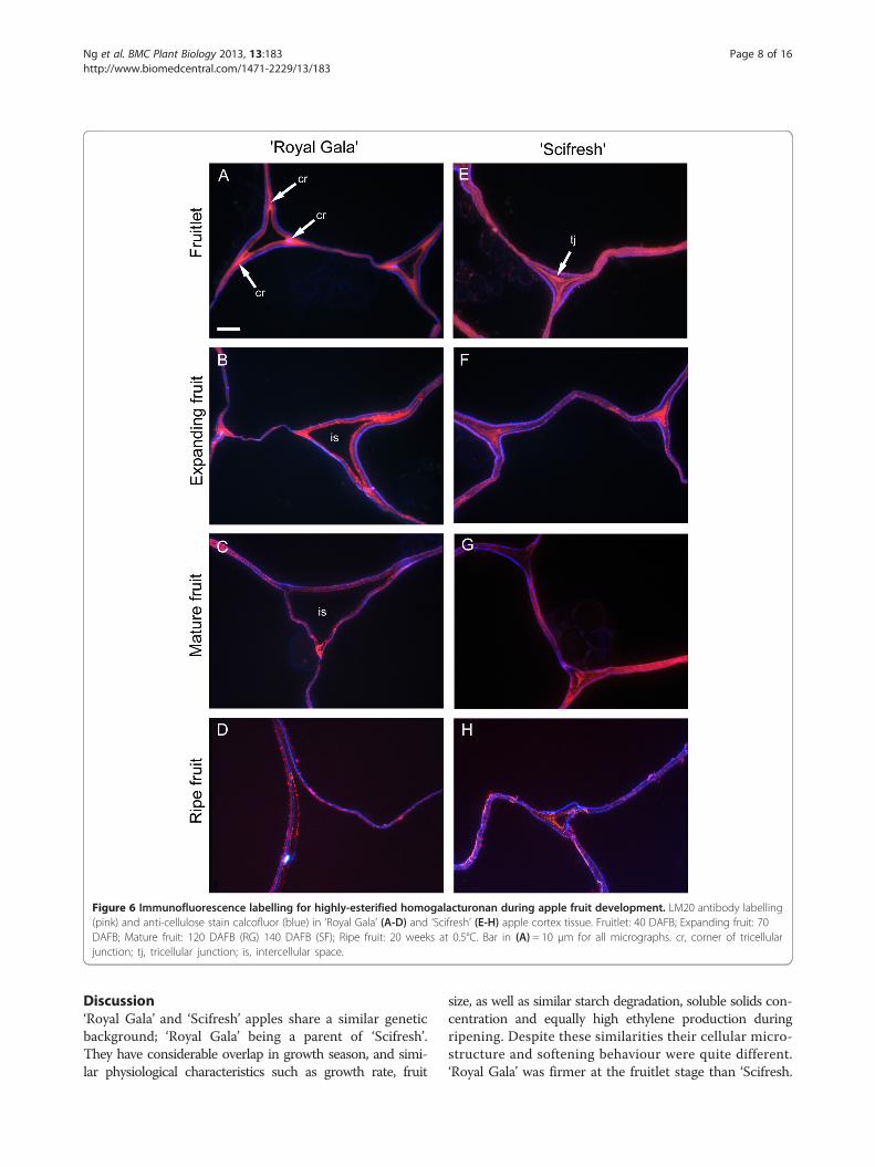
Figure 6 Immunofluorescence labelling for highly-esterified homogalacturonan during apple fruit development. LM20 antibody labelling(pink) and anti-cellulose stain calcofluor (blue) in ‘Royal Gala’ (A-D) and ‘Scifresh’ (E-H) apple cortex tissue. Fruitlet: 40 DAFB; Expanding fruit: 70DAFB; Mature fruit: 120 DAFB (RG) 140 DAFB (SF); Ripe fruit: 20 weeks at 0.5°C. Bar in (A) = 10 μm for all micrographs. cr, corner of tricellularjunction; tj, tricellular junction; is, intercellular space.
Ng et al. BMC Plant Biology 2013, 13:183 Page 8 of 16http://www.biomedcentral.com/1471-2229/13/183
Discussion‘Royal Gala’ and ‘Scifresh’ apples share a similar geneticbackground; ‘Royal Gala’ being a parent of ‘Scifresh’.They have considerable overlap in growth season, and simi-lar physiological characteristics such as growth rate, fruit
size, as well as similar starch degradation, soluble solids con-centration and equally high ethylene production duringripening. Despite these similarities their cellular micro-structure and softening behaviour were quite different.‘Royal Gala’ was firmer at the fruitlet stage than ‘Scifresh.
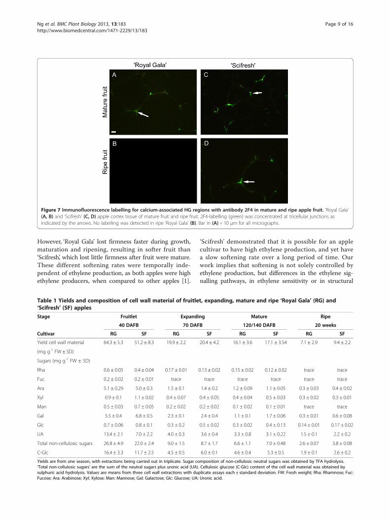
Figure 7 Immunofluorescence labelling for calcium-associated HG regions with antibody 2F4 in mature and ripe apple fruit. ‘Royal Gala’(A, B) and ‘Scifresh’ (C, D) apple cortex tissue of mature fruit and ripe fruit. 2F4-labelling (green) was concentrated at tricellular junctions asindicated by the arrows. No labelling was detected in ripe ‘Royal Gala’ (B). Bar in (A) = 10 μm for all micrographs.
Ng et al. BMC Plant Biology 2013, 13:183 Page 9 of 16http://www.biomedcentral.com/1471-2229/13/183
However, ‘Royal Gala’ lost firmness faster during growth,maturation and ripening, resulting in softer fruit than‘Scifresh’, which lost little firmness after fruit were mature.These different softening rates were temporally inde-pendent of ethylene production, as both apples were highethylene producers, when compared to other apples [1].
Table 1 Yields and composition of cell wall material of fruitle‘Scifresh’ (SF) apples
Stage Fruitlet Expandi
40 DAFB 70 DAF
Cultivar RG SF RG
Yield cell wall material 64.3 ± 5.3 51.2 ± 8.3 19.9 ± 2.2
(mg g-1 FW ± SD)
Sugars (mg g-1 FW ± SD)
Rha 0.6 ± 0.05 0.4 ± 0.04 0.17 ± 0.01
Fuc 0.2 ± 0.02 0.2 ± 0.01 trace
Ara 5.1 ± 0.29 5.0 ± 0.3 1.5 ± 0.1
Xyl 0.9 ± 0.1 1.1 ± 0.02 0.4 ± 0.07
Man 0.5 ± 0.03 0.7 ± 0.05 0.2 ± 0.02
Gal 5.5 ± 0.4 6.8 ± 0.5 2.3 ± 0.1
Glc 0.7 ± 0.06 0.8 ± 0.1 0.3 ± 0.2
UA 13.4 ± 2.1 7.0 ± 2.2 4.0 ± 0.3
Total non-cellulosic sugars 26.8 ± 4.9 22.0 ± 2.4 9.0 ± 1.5
C-Glc 16.4 ± 3.3 11.7 ± 2.5 4.5 ± 0.5
Yields are from one season, with extractions being carried out in triplicate. Sugar co‘Total non-cellulosic sugars’ are the sum of the neutral sugars plus uronic acid (UA).sulphuric acid hydrolysis. Values are means from three cell wall extractions with duFucose; Ara: Arabinose; Xyl: Xylose; Man: Mannose; Gal: Galactose; Glc: Glucose; UA:
‘Scifresh’ demonstrated that it is possible for an applecultivar to have high ethylene production, and yet havea slow softening rate over a long period of time. Ourwork implies that softening is not solely controlled byethylene production, but differences in the ethylene sig-nalling pathways, in ethylene sensitivity or in structural
t, expanding, mature and ripe ‘Royal Gala’ (RG) and
ng Mature Ripe
B 120/140 DAFB 20 weeks
SF RG SF RG SF
20.4 ± 4.2 16.1 ± 3.6 17.1 ± 3.54 7.1 ± 2.9 9.4 ± 2.2
0.13 ± 0.02 0.15 ± 0.02 0.12 ± 0.02 trace trace
trace trace trace trace trace
1.4 ± 0.2 1.2 ± 0.09 1.1 ± 0.05 0.3 ± 0.03 0.4 ± 0.02
0.4 ± 0.05 0.4 ± 0.04 0.5 ± 0.03 0.3 ± 0.02 0.3 ± 0.01
0.2 ± 0.02 0.1 ± 0.02 0.1 ± 0.01 trace trace
2.4 ± 0.4 1.1 ± 0.1 1.7 ± 0.06 0.3 ± 0.01 0.6 ± 0.08
0.5 ± 0.02 0.3 ± 0.02 0.4 ± 0.13 0.14 ± 0.01 0.17 ± 0.02
3.6 ± 0.4 3.3 ± 0.8 3.1 ± 0.22 1.5 ± 0.1 2.2 ± 0.2
8.7 ± 1.7 6.6 ± 1.1 7.0 ± 0.48 2.6 ± 0.07 3.8 ± 0.08
6.0 ± 0.1 4.6 ± 0.4 5.3 ± 0.5 1.9 ± 0.1 2.6 ± 0.2
mposition of non-cellulosic neutral sugars was obtained by TFA hydrolysis.Cellulosic glucose (C-Glc) content of the cell wall material was obtained byplicate assays each ± standard deviation. FW: Fresh weight; Rha: Rhamnose; Fuc:Uronic acid.
0.00
0.02
0.04
0.06
0 50 100
0.00
0.02
0.04
0.06RoyalGala
Scifresh
UA
(µg
UA
frac
tion-
1 µg
UA
load
ed-1
)
0 50 100
2000 500 40 kDaA
B
C
D
Fruitlet
Expanding
Mature
Ripe
RGSF
2000 500 40 kDa
Stage Fruitlet Expanding Mature RipeCultivar RG SF RG SF RG SF RG SFYield (% CWM)
10.4±2.3
4.5±1.3
4.9±0.9
4.5±1.9
7.2±2.0
12.4±3.7
3.9±0.5
5.9±0.2
UA (µg mg-1
sample) 355.3±29.0
350.9±31.0
323.9±48.7
397.9±22.4
245.9±36.3
114.2±16.4
460.9±66.0
340.3±54.0
E Elution volume (mL)
Figure 8 Size, yield and composition of CDTA-soluble pectin in‘Royal Gala’ and ‘Scifresh’ fruit during development. Molecularweight distribution (A-D), yield and uronic acid (UA) content (E) ofCDTA-soluble pectin in ‘Royal Gala’ (RG) and ‘Scifresh’ (SF). Fruitlet:40 DAFB; Expanding fruit: 70 DAFB; Mature fruit: 120 DAFB (RG) 140DAFB (SF); Ripe fruit: 20 weeks at 0.5°C. Yields in (E) are means of 3extractions ± standard deviation; UA values are the mean of 3 CDTAextracts with duplicate assays ± standard deviation.
0
50
100
0
50
100
Fruitlet Expanding Mature Ripe
Fruit stages
Deg
ree
of m
ethy
l est
erifi
catio
n (%
)C
B
0
20
40
60
PM
E a
ctiv
ity(n
mol
MeO
H h
-1 g
FW
-1)
A
*
*
‘Royal Gala’‘Scifresh’
Figure 9 Pectin methylesterase (PME) activity and degree ofmethyl esterification of ‘Royal Gala’ and ‘Scifresh’ fruit cell wallsduring development. PME activity (A), degree of methyl esterificationof cell wall material (B) and CDTA-soluble pectin (C). Fruitlet: 40 DAFB;Expanding fruit: 70 DAFB; Mature fruit: 120 DAFB (RG) 140 DAFB (SF); Ripefruit: 20 weeks at 0.5°C. Values in (A) are the means of 3 assays ± standarddeviation; values in (B) and (C) are the means of 6 assays ± standarddeviation. Asterisks indicate significantly different values at a level ofP≤ 0.05 using a protected Fisher’s least significant difference test.
Ng et al. BMC Plant Biology 2013, 13:183 Page 10 of 16http://www.biomedcentral.com/1471-2229/13/183
features of the cell wall established before the onset ofethylene synthesis may also contribute roles.Generally, apple fruitlets have small cells with thick,
less structured and amorphous cell walls allowing for veryrapid expansion [2,25]. In our study, both cultivars hadcells about equal in size at the fruitlet stage; however theyield of cell wall material in ‘Scifresh’ was much lower.Hence, cell walls in (softer) ‘Scifresh’ fruitlet must bethinner than cell walls in (firmer) ‘Royal Gala’ fruitlet atthis stage. The fruitlet cell walls already showed a differentcomposition, where the firmer ‘Royal Gala’ fruitlet had ahigher uronic acid but a lower neutral sugar (side chain)content, accompanied by a higher amount of cellulosicglucose and a lower degree of esterification of pectin thancell walls of ‘Scifresh’ fruitlets. During the cell expansionphase between 40 and 70 DAFB, the yield of cell wallmaterial in both cultivars decreased two to three fold,whereas the fruit size (by mass) increased by 25–30 times.This suggested a “net” synthesis of cell walls during fruitgrowth, rather than just a gradual thinning of the cell wallas the cells expanded. Although ‘Scifresh’ accumulatedmore dry weight than ‘Royal Gala’, it had a slightly lowercell wall synthesis rate but seemed to more efficientlysustain stronger cell walls, thereby maintaining the struc-tural firmness of the tissue, as measured by the lag phasein softening during this phase of rapid growth.
In mature fruit, it was evident that ‘Scifresh’ corticalcells had expanded more and were larger in mature fruitcompared to ‘Royal Gala’. As the firmness of maturefruit of both cultivars was very similar, it appeared that cellgrowth and development of larger cells did not necessarilyimpair cell wall strength and hence tissue firmness. Thismay also suggest a difference in wall extension dynamicsbetween ‘Royal Gala’ and ‘Scifresh’, which has been shownto be influenced by α-expansins acting on xyloglucansthereby inducing wall loosening [30], as well as extensins,of which a higher level of expression has been linked to atissue’s ability to withstand higher tensile stresses [31].‘Scifresh’ not only had the capacity to allocate resources
early in fruit development to strengthen the tissue, but alsoto allow the deposition to occur in a manner that permitsmore rapid cell expansion. This rapid cell expansion

Ng et al. BMC Plant Biology 2013, 13:183 Page 11 of 16http://www.biomedcentral.com/1471-2229/13/183
phase has also been previously identified as being a highenergy requirement phase where numerous genes aredifferentially regulated for sugar metabolism and accumu-lation [32]; and an increase in carbohydrate availabilityby reduction of fruit load enhanced cell production duringearly fruit growth [33]. It could be possible that in expand-ing fruit of ‘Scifresh’ (showing higher dry matter accumu-lation than ‘Royal Gala’ at this stage), carbon sources inthe form of sucrose, fructose or sorbitol are preferentiallyoptimized for structural wall building instead of othercellular processes. Additionally, this could also be indi-cative of a difference in turnover between metabolized andnewly-synthesized sugars in expanding fruit of ‘Royal Gala’and ‘Scifresh’. Gene expression levels of key enzymes suchas expansins, other cell wall-related genes, as well as starchmetabolism genes which are differentially regulated acrossdifferent cultivars of apples [34], warrant more investiga-tions in future studies.Extraction of cell wall material [35] showed that ‘Scifresh’
fruitlet already had a higher proportion of more tightlybound pectin and hemicelluloses than ‘Royal Gala’, possiblymaking stronger cellulose-pectin and/or cellulose- hemicel-lulose networks in ‘Scifresh’ compared to ‘Royal Gala’, cellwall features that continued to exist up to the ripe stage.The observation that ‘Scifresh’ apples can accommodatecell expansion while not compromising structural hardnesswas in agreement with [36] and [37] who reported thatapple cultivars with larger cells have better keeping qualitiesand ripen slower, presumably due to a lower respiratoryrate compared to higher respiration in smaller cells.Apples with more angular cells have been reported to be
firmer [27]. Hexagonal-shaped cells with a higher numberof facets have larger areas for adherence compared tospherical-shaped cells [38]. Cells in mature fruit of‘Scifresh’ were larger and more angular than ‘RoyalGala’, and had a higher cortical density indicating lessairspace and therefore greater cell-to-cell contact area.This may also be indicative of a higher turgor pressure,which contributes to higher firmness in apple fruit [5].Turgor pressure provides non-woody plant tissues withmechanical rigidity and is the driving force for growth,but at the same time it generates large forces tendingto separate cells. Therefore, cell adhesion depends on thestrength of reinforcing zones located precisely at the pointsof maximum stress, the cell junctions where adjacent cellsmeet [39]. At these cell junctions, ‘Scifresh’ was filled withhighly esterified pectin as shown by immunolabelling withLM20 from the fruitlet stage throughout development. In‘Royal Gala’ fruitlet, this labelling was far less intense thanin ‘Scifresh’, and perhaps it is the lower presence of pectinin these tricellular junctions that explains the cell sizedifference observed between the two cultivars.Strong cell adhesion, less airspace and higher cell density
was maintained in ‘Scifresh’ from fruitlet to ripe fruit,
whereas fracture surfaces of ripe ‘Royal Gala’ tissueshowed less cell rupture and more separation betweencells compared to ‘Scifresh’. Cell rupture is a major factorin perception of juiciness, and for this to occur, cell-to-celladhesion must be strong. Less cell-to-cell contact andmore intercellular air spaces have been related to a soft,drier perception of ripe apples [9], which was evident inripe ‘Royal Gala’ but not in ‘Scifresh’. Moreover, tensilestrength or the force to pull tissue apart (which had beenthe same up to the mature stage) had decreased consider-ably during ripening of ‘Royal Gala’, but not in ‘Scifresh’.Tensile strength depends on the strength of cell adhesion,but also on the overall strength of the cell wall, i.e. itsresistance to fracture or its elasticity [40]. Perhaps oneof the driving forces to maintain strong cell adhesion in‘Scifresh’ even in the ripe stage was the presence of calciumpectate in the corners of tricellular junctions, which wasobserved by immunolabelling with 2F4 at the mature stagein both fruit, but at the ripe stage was undetectable in‘Royal Gala’.The CDTA-soluble fraction of the cell wall contains
pectin that is considered an important linking componentbetween adjacent cells, with composition, degree of esteri-fication, and polymer length determining the strength ofthat ‘glue’. The degree of methyl-esterification (DE) of theCDTA-soluble pectin was very similar between the twocultivars, and slowly increased over development. Theincrease in DE in the CDTA-soluble fractions did howevernot necessarily imply biosynthesis as suggested by [41], butrather more highly esterified pectin becomes increasinglyextractable with ripening. According to [42], HG with DEhigher than 60% (notably present in CDTA-soluble pectinin both cultivars at the mature and ripe stages) can formgels via hydrophobic interactions and hydrogen bondsbetween methyl-esterified blocks of homogalacturonan.The longer these blocks are, the stronger the gel formed.Although the DE of CDTA-soluble pectin in both cultivarswas similar throughout development, and the molecularweight distribution (with the exception of the expandingfruit stage) remained large, yields were different betweenthe cultivars and correlated with fruit firmness throughoutgrowth and ripening, as higher yields were generally foundin firmer fruit. Interestingly, in expanding fruit themolecular weight distribution of the CDTA-soluble pectindecreased in both cultivars, but more extensively in‘Scifresh’ than in ‘Royal Gala’. One could speculate thatthis decrease in molecular weight distribution maycontribute to cell expansion, as the shorter length of thesepectin molecules may result in reduced cohesive strength.As pectin molecules were considerably shorter in ‘Scifresh’,‘Scifresh’ cells may be able to expand more, giving rise tolarger cell size compared to ‘Royal Gala’.In cell wall material, the DE was overall higher in
‘Scifresh’ than in ‘Royal Gala’ throughout development,
Ng et al. BMC Plant Biology 2013, 13:183 Page 12 of 16http://www.biomedcentral.com/1471-2229/13/183
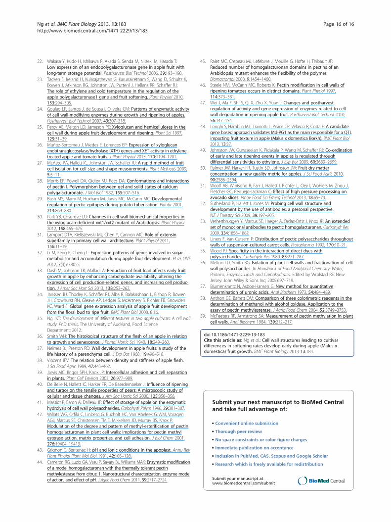
which coincided with an overall lower PME activity inthis cultivar. In agreement with our study, PME in applehas been found to increase during growth and decreaseduring ripening-related softening [24]. The action of PMEaffects apoplastic pH and alters the activity of cell wallhydrolases [43] as well as modifies the rigidity of thepectin network [44]. The pattern of methyl ester groupdistribution within the wall has also been reported to beimportant in determining pectin properties [42,45]. LM20-labelling of highly-esterified HG regions was more wide-spread in ‘Scifresh’ cell walls throughout the middle lamellaand junction zones, compared to ‘Royal Gala’ where label-ling was more restricted to corners of tricellular junctions.This localisation of methyl ester groups in ‘Royal Gala’could indicate block-like microdomains, which have beensuggested to cause a restriction in the movement ofenzymes such as PG, which has a preference for lowly-esterified HG [46]. Interestingly, immunodetectable ripen-ing-related PG1 protein was only found in the fastersoftening ‘Royal Gala’ fruit, but not in ‘Scifresh’, therebysubstantiating that PG expression is likely cultivar-dependent and plays a major role in determining applefruit texture [47,48]. The higher PME activity in ‘RoyalGala’ fruitlet could have resulted in the generation ofmore non-esterified HG, presenting more substrate forthe action of PG, and thus setting the basis for pectindepolymerisation and faster softening in ‘Royal Gala’.The lack of ripening-related PG1 protein in ripe ‘Scifresh’
(Figure 10) led to some compelling parallels between‘Scifresh’ and transgenic PG1 antisense ‘Royal Gala’ apples[21]. Like ‘Scifresh’, the antisense apples had increasedtensile strength and hence stronger cell adhesion, as wellas increased cell rupture compared to wild-type ‘RoyalGala’ at the ripe stage. However, there was no evidenceof reduced airspace or higher cell density in the antisenseapples, indicating that these features are not related toPG1. The softening rate also decreased in the antisense‘Royal Gala’ apples compared with the wild-type ‘Royal
Figure 10 Western blot stained with a rabbit polyclonalantibody for polygalacturonase. Lane 1, Precision Plus DualProtein Standard; lanes 2, 3, 4, ‘Royal Gala’ in the order of fruitlet,mature and ripe fruit; lanes 5, 6, 7, ‘Scifresh’ in the order of fruitlet,mature and ripe fruit. An arrow indicates the presence of PG proteinat 45 kDa only detected in ripe ‘Royal Gala’ fruit.
Gala’ apples; however, they still softened faster than‘Scifresh’ confirming that there are factors additional toPG involved in apple softening. The differences in celladhesion between ‘Scifresh’ and PG1 antisense ‘RoyalGala’ on one side and wild-type ‘Royal Gala’ on the othercould be related to differences in pectin content. ‘Scifresh’[35] and antisense ‘Royal Gala’ [21] had higher yields ofCDTA-soluble pectin and less water-soluble pectinthan wild-type ‘Royal Gala’, implying that PG has someinfluence on pectin solubilisation.
ConclusionsWe have identified an apple cultivar (‘Scifresh’) wherethe fruit softens slowly despite having high ethyleneproduction, providing a system for investigating the cellwall and tissue microstructure without the confoundinginfluence of ethylene. Comparisons between ‘Scifresh’and the more rapidly softening ‘Royal Gala’ identifieddifferences in tissue microstructure and cell wall propertiesthat could influence the rate of softening during ripening.These structural properties were established as early as 40to 70 days after full bloom and well before the maturationand ripening process are induced. ‘Scifresh’ fruit have largercells, strong cell-to-cell adhesion and less airspace com-pared to ‘Royal Gala’. The strong cell-to-cell adhesionappeared to be due to increased deposition of pectin incell corners, with pectin interactions so strong that verylittle airspace was able to develop during cell expansionand maturation. The lack of immuno-detectable PG in‘Scifresh’ most likely strengthened this phenotype. It isprobably because of these strong pectin interactions at celljunctions that cells fracture through the cell rather thanbetween cells as in ‘Royal Gala’.
MethodsFruit material and physiological assessmentApple fruit (Malus x domestica Borkh.) ‘Scifresh’ and ‘RoyalGala’ were sampled from the Plant & Food Researchorchard, Havelock North, New Zealand. At each stage,20 fruit per cultivar were collected by taking five apples insimilar positions from four trees (growing side by side andexposed to the same management regime) throughout thisstudy. Fruit stages were categorised based on number ofdays after full bloom (DAFB), size, skin colour, starch andsoluble solids content. Fruit sampled at 40 DAFB, wherefruit were at the end of the cell division phase and at thebeginning of cell expansion [2], were termed ‘fruitlet’.Two intermediate time points were sampled during thecell expansion phase, 70 and 100 DAFB, and termed‘expanding fruit’. Mature ‘Royal Gala’ fruit were sampledat 120 DAFB according to degree of starch clearance, and‘Scifresh’ at 140 DAFB, as they had a longer developmentalperiod before reaching the same physiological age as ‘RoyalGala’, termed ‘mature’ fruit. These fruit were then ripened

Ng et al. BMC Plant Biology 2013, 13:183 Page 13 of 16http://www.biomedcentral.com/1471-2229/13/183
at 0.5°C under ambient atmospheric pressure and humidityfor 20 weeks and termed ‘ripe fruit’.Fruit firmness was assessed using puncture [49] and
tensile tests [21], with the latter only assessed from 100DAFB onwards because of fruit size constraints. Thepuncture test was performed using two cylindrical probesof 5 mm (fruitlet to mature fruit) or 11 mm diameter(mature to ripe fruit), using a TA.XT Plus Texture Analyser(Stable Microsystems, United Kingdom). Internal ethyleneconcentration [49] and dry matter concentration [50]were determined from three bulked replicates samplesfrom a total pool of 20 fruit. Cortical tissue density wasdetermined in mature fruit by measuring the volumedisplacement for 1 cm3 blocks of excised tissue [4].After these assessments, fruit tissue from cortex was then
diced, immediately frozen in liquid nitrogen and storedat -80°C for cell wall analyses and enzyme activity assays.Tissue preparations for microscopy are described below.
Conventional scanning electron microscopy (SEM)Apple sections were cut longitudinally from skin to cortextissue, not including core tissue, and fixed in 2% parafor-maldehyde and 0.1% glutaraldehyde in 0.1 M phosphatebuffer (pH 7.2). Segments were washed in phosphate buf-fer, and dehydrated in an ethanol series from 10% to 100%anhydrous ethanol (in 10% increments). The segmentswere dried in a Bal-Tec CPD030 critical point dryer(Balzers, Liechtenstein) using liquid CO2 as the transitionalfluid. The dried material was mounted on an anodized alu-minium stub (ProSciTech, Australia) with carbon adhesivetabs (ProSciTech), with edges painted with conductivesilver liquid (ProSciTech), and left to air dry for 1 h. Thematerial was sputter-coated with gold in a SEM coatingunit E5100 (Polaron equipment Ltd, England) and storedover silica beads (Scharlau, Spain) in an air-tight containeruntil imaged. Scanning electron microscopy was carriedout using a Quanta 250 SEM (FEI, Hillsboro, USA), withaccelerating voltage of 15 kV.
Cryo-scanning electron microscopyCryo-SEM was performed using a Polaron PP2000 CryoTransfer system (QuorumTechnologies, United Kingdom)attached to a FEI Quanta 250 Scanning Electron Micro-scope [51]. ‘Royal Gala’ and ‘Scifresh’ apples were matchedfor size, and samples of cortex tissue were cut and mountedin sample holders containing a mixture of colloidal graphiteand OCT™ compound (Sakursa Finetek, Netherlands).Tissue was immediately frozen in liquid nitrogen slush,transferred to the preparation chamber of the PP2000system where the tissue was fractured to expose the surfacefor viewing. Ice was sublimed away to partially etch thesurface at a temperature of −90°C for 15 minutes, sputter-coated with gold/palladium, transferred to the cryo-stage
in the SEM (-150°C) and observed at an acceleratingvoltage of 10 kV.
ImmunolabellingFixed and ethanol-dehydrated sections prepared as de-scribed above were infiltrated with LR White resin (LondonResin Company Ltd, United Kingdom) and placed ingelatine capsules (ProSciTech) containing LR White resin[52]. After hardening (55°C, 48 h), the capsule was removed,embedded tissue sectioned using a diamond knife andLeica UCT ultramicrotome (Leica, Germany), and sections(200 nm) air dried onto Superfrost® poly-L-lysine slides(25× 75× 1 mm, Biolab, USA). Antibodies used for la-belling were LM19, LM20 [53] and 2F4, all supplied byPlantProbes (United Kingdom).For labelling with LM19 and LM20, sections were
wetted with phosphate-buffered saline with 0.1% Tween80 (PBS-T) for 10 min, then incubated with 0.1% bovineserum albumin c (BSA-c; Aurion, Netherlands) in PBS-Tto block non-specific labelling (15 min), followed by incu-bation with primary antibody (dilution 1:20 v/v in 0.1%BSA-c in PBS-T) overnight at 4°C in a moist chamber.Slides were then washed in PBS-T.For 2F4, a chemical de-esterification step prior to
immunolabelling was necessary to unmask epitopes. Briefly,the sections were incubated in 0.05 M NaOH (pH 12.4)for 30 min at room temperature, followed by BSA-c asdescribed above, for blocking of non-specific binding.Although this treatment may introduce artefacts or insome way alter the original abundance of epitope-antigenbinding sites, because the same procedures were carriedout on both apple cultivars, this makes them comparable.The sections were then incubated in TCaS buffer (20 mMTris-HCl pH 8.2, 1 mM CaCl2 , 150 mM NaCl) containing5% (w/v) low fat milk powder for 1 h to block non-specificlabelling, followed by incubation with primary antibody2F4 [dilution 1:20 (v/v)] in TCaS buffer with 0.05% (v/v)Tween 20 and 1% (w/v) low fat milk powder overnight at4°C in a moist chamber, and the slides washed in TCaSBuffer [54].Labelling with the secondary antibody was the same for
all samples. The slides were incubated for 2 h in the darkat room temperature with goat anti-rat IgG AlexaFluor488 (Molecular Probes, Oregon, USA) diluted 1: 600 (v/v)in PBS.Sections were further incubated in 1 mL of 0.01% calco-
fluor (Fluorescent Brightener 28, Sigma) for 6 min at roomtemperature [55] to distinguish the boundaries of cell wallswhere there may be absence of antibody labelling. Sectionswere washed in ultrapure water, allowed to dry at roomtemperature and coverslip-mounted onto the slide usinganti-fade agent Citifluor (Citifluor, United Kingdom) [52].Negative controls were carried out using only the sec-
ondary antibody omitting the primary antibody (200 μL of

Ng et al. BMC Plant Biology 2013, 13:183 Page 14 of 16http://www.biomedcentral.com/1471-2229/13/183
goat anti-rat IgG AlexaFluor 488 diluted 1: 600 in PBS),where sections were incubated for 2 h in the dark beforeimaging, results were negative (images not shown).Sections were viewed using an Olympus Vanox AHT3
compound microscope (Olympus Optical, Tokyo, Japan)with a blue-interference filter set for antibody labellingand UV filterset for calcofluor staining and imaged witha CoolSnap colour digital camera system (Photometrics,USA). Images were further processed using Adobe Photo-shop Version 6.0 on Windows XP. Two sets of images areshown; higher magnification dual-labelling with calcofluor(blue) and antibody labelling contrast-enhanced by chan-ging the hue angle to a reddish-pink colour (Figures 5, 6),and antibody immunofluorescence labelling in green(Figure 7 and Additional file 1: Figure S1, Additionalfile 2: Figure S2, Additional file 3: Figure S3).
Preparation of cell wall material and CDTA-soluble pectinCell wall isolation and extractions were performed using acomposite sample of cortical tissue from 20 apples that wasdivided into three sub-samples of fruit to form three extrac-tion replicates. Cell wall preparations were carried out asdescribed in [56] to give the water-soluble fraction and cellwall material, with the exception that after dimethylsulphoxide extraction, residual starch in fruitlet was re-moved by digestion with α-amylase (40 U/mL) (Megazyme,Ireland) and pullulanase (20 U/mL) (Megazyme) by incu-bation at 37°C for 1 h. CDTA-soluble pectin was extractedfrom cell wall material (2.5 g) as described in [56].
Size exclusion chromatographyCDTA- soluble pectin (2.5 mg) was dissolved in 0.5 mLwater and eluted through a column (2.5× 100 cm) ofSepharose CL–2B (GE Healthcare, USA), in 0.05 Mammonium acetate buffer (pH 5.0) with an average flowrate of 5 mL h-1. Fractions (20 min) were collected andassayed for uronic acid [57]. The column was calibratedwith dextrans (GE Healthcare) Blue Dextran (2 MDa),T500 (500 kDa) and T40 (40 kDa).
Pectin methylesterase (PME) extraction and activity assayGround frozen apple tissue (0.25 g) was extracted in0.5 mL of 0.2 M MES, 7.5 mM potassium tetrathionate,10 mM dithiothreitol, 1.7 M NaCl (pH 6.0) with 25 mgof polyvinylpolypyrrolidone. The mixture was vortexed,centrifuged and supernatant recovered. The pellet wasre-suspended in 0.25 mL of the extraction buffer andincubated on ice for 20 min, and supernatant recoveredas described above. The combined supernatant was thePME extract. PME activity was assayed [58], whereby theamount of methanol released is quantified by reactingwith alcohol oxidase and N-methylbenzothiazolinone-2-hydrazone. The reaction mixture contained PME extract(10 μL), 100 mM Tris-HCl pH 7.5 (20 μL), 0.5 U/mL
alcohol oxidase (5 μL; Pischia pastoris, Sigma-Aldrich),3 mg · mL-1 N-methylbenzothiazolinone-2-hydrazone(8 μL; Merck, USA), H2O (7 μL) and 0.5 mg ·mL-1 esterifiedcitrus pectin (DE >85%; Sigma-Aldrich) in 50 mM NaClpH 7.0 (10 μL). After incubation at 30°C for 20 min, the re-action was terminated by addition of 40 μL of 5 mg ·mL-1
ferrous ammonium sulphate (BDH, United Kingdom) insulphamic acid (Medica Pacifica, NZ) and absorbanceread at 620 nm. PME activity was expressed as molesmethanol released per hour per gram fresh weight, basedon a standard curve using methanol. Activity assays werecarried out in triplicate.
Polygalacturonase (PG) extraction and Western blottingProtein was extracted from ground frozen apple tissue(0.1 g) in 1 mL of 7 M urea, 2 M thiourea, 40 mM Tris,75 mM DTT, 4% CHAPS and concentrated by cold acetoneprecipitation. Proteins were separated by sodium dodecylsulphate polyacrylamide gel electrophoresis (SDS-PAGE)(Mini-PROTEAN ® TGX™, BIO-RAD, USA), electroblottedonto a polyvinyldifluoride membrane, and blocked asdescribed in [21]. Proteins were immuno-labelled withthe antiserum raised to apple PG1 (1: 1000 (v/v), diluted inTBS buffer containing 5% non-fat milk powder). Mem-branes were incubated with an anti-rabbit alkaline phos-phatase conjugated secondary antibody (Sigma-Aldrich)and PG binding visualised using 1-Step™ NBT/BCIP(Thermo Scientific, USA).
Analytical methodsUronic acids were measured as described in [57]. Thedegree of methyl esterification (DE) was determined bya modified method from [59] by gas chromatographicquantification of methanol after saponification of pectin.Polysaccharides were saponified overnight at 4°C in asolution of 50 mM citric acid, 1 M NaCl and 1 M NaOH.The mixture was neutralised with citric acid and 25 mMn-propanol added as the internal standard. Samples (1 μL)were analysed by GC-FID on a BP20 column [(15 m×0.25 μm) Fischer Scientific, UK; oven temperature 80°C, he-lium flow rate 1.5 psi; detector temperature at 240°C], withmeasurements carried out in duplicate. DE was calculated asa molar ratio of methanol to uronic acid, based on a stand-ard curve constructed with known amounts of methanol.
Statistical analysisSingle-factor (for firmness and yield of cell wall material)or two-way (for compositional and enzyme assay results)ANOVA (analysis of variance) analyses were conductedusing the Microsoft Excel 2007 for Windows software.Means were compared using the Fisher’s least significantdifference (LSD) post-test at P ≤ 0.05 using the SPSS(Version 15.0) software (IBM, USA).

Ng et al. BMC Plant Biology 2013, 13:183 Page 15 of 16http://www.biomedcentral.com/1471-2229/13/183
Additional files
Additional file 1: Figure S1. Immunofluorescence labelling oflowly-esterified homogalacturonan with antibody LM19 in ‘Royal Gala’ (A-D)and ‘Scifresh’ (E-H) apple cortex tissue. Fruitlet: 40 DAFB; Expanding fruit: 70DAFB; Mature fruit: 120 DAFB (RG) 140 DAFB (SF); Ripe fruit: 20 weeks at 0.5°C.Bar in (A) = 50 μm for all micrographs. is: intercellular space.
Additional file 2: Figure S2. Immunofluorescence labelling ofhighly-esterified homogalacturonan with antibody LM20 in ‘Royal Gala’(A-D) and ‘Scifresh’ (E-H) apple cortex tissue. Fruitlet: 40 DAFB; Expanding fruit:70 DAFB; Mature fruit: 120 DAFB (RG) 140 DAFB (SF); Ripe fruit: 20 weeks at0.5°C. Bar in (A) = 50 μm for all micrographs. is: intercellular space.
Additional file 3: Figure S3. Immunofluorescence labelling ofhighly-esterified homogalacturonan with antibody LM20 in ‘Royal Gala’(A) and ‘Scifresh’ (B, C) fruitlet cortex tissue in high magnification. Bars = 10μm for all micrographs. Panel A shows ‘Royal Gala’ fruitlet section with distinctLM20-labelling pattern concentrated at the corners (cr) of tricellular junctionsand very intense staining in the middle lamella (ml) regions particularly thelining of the intercellular air space (lis). Panels B and C are ‘Scifresh’ fruitletsections showing a different labelling pattern to ‘Royal Gala’. Panel B showsa tricellular junction with intense LM20-labelling completely filling this area.In all sections viewed, 70-80% of tricellular junctions in ‘Scifresh’ fruitletwere completely stained, while only 30-40% in ‘Royal Gala’ fruitlet displayedthis pattern. Panel C shows a ‘Scifresh’ junction zone located between 5 cellslabelled with LM20, however the lining of the intercellular air space (lis) wasabsent of labelling, which was opposite to the pattern observed in ‘RoyalGala’ fruitlet (A). This emphasizes the different localisation of highly-esterifiedhomogalacturonan in the cell walls of the two apple cultivars.
Additional file 4: Figure S4. Percent increment in net cell walldeposition of ‘Royal Gala’ and ‘Scifresh’ per day. Data based on Figure 1Eand Table 1, with the percentage increase in fruit weight or increase incell wall material calculated as the change in mean fruit weight or meanyield of cell wall material relative to the weight or yield of cell wallmaterial, respectively at the start of each period per day.
AbbreviationsCDTA: Trans-1,2-diaminocyclohexane-N,N,N',N'-tetraacetic acid;DAFB: Days after full bloom; DE: Degree of methyl esterification;DTT: Dithiothreitol; HG: Homogalacturonan; LM: Leeds monoclonalantibody; PBS-T: Phosphate-buffered saline containing 0.1% Tween80;PG: Polygalacturonase; PME: Pectin methylesterase; SD: Standard deviation;SE: Standard error; UA: Uronic acid.
Competing interestsThe authors declare that they have no competing interests.
Authors’ contributionsJWJ, LDM, BGS, RS, ICH and JN conceived of the study and designed theexperiments, JN carried out all experiments, with assistance from MIH andRP for fruit assessments and enzyme assays, and PWS for immunolabelling.RS and JN carried out data analyses, and JN, RS, JWJ, BGS, ICH, and LDMwrote the paper. All authors read and approved the final manuscript.
AcknowledgementsThis research was funded by The New Zealand Ministry of Business,Innovation and Employment (CO6X0705). The authors would like to thankErin O’Donoghue for teaching us the degree of esterification method, andDavid Brummell and Ross Atkinson for critically reading the manuscript.
Author details1Food Science, School of Chemical Sciences, The University of Auckland,Private Bag 92019, Auckland, New Zealand. 2The New Zealand Institute forPlant & Food Research Limited, Mount Albert Research Centre, Private Bag92169, Auckland 1142, New Zealand. 3Current address: The New ZealandInstitute for Plant & Food Research Limited, Food Industry Science Centre,Private Bag 11600, Palmerston North 4442, New Zealand. 4The New ZealandInstitute for Plant & Food Research Limited, Hawkes Bay Research Centre,Havelock North 4130, New Zealand.
Received: 22 July 2013 Accepted: 12 November 2013Published: 19 November 2013
References1. Oraguzie NC, Volz RK, Whitworth CJ, Bassett HCM, Hall AJ, Gardiner SE:
Influence of Md-ACS1 allelotype and harvest season within an applegermplasm collection on fruit softening during cold air storage.Postharvest Biol Technol 2007, 44:212–219.
2. Lakso AN, Corelli-Grappadelli L, Barnard J, Goffinet MC: An expolinearmodel of the growth pattern of the apple fruit. J Hortic Sci Biotech 1995,70:389–394.
3. Pratt C: Apple flower and fruit, morphology and anatomy. Hort Rev 1988,10:273–308.
4. Volz RK, Harker FR, Lang S: Firmness decline in ‘Gala’ apple during fruitdevelopment. J Am Soc Hortic Sci 2003, 128:797–802.
5. Tong C, Krueger D, Vickers Z, Bedford D, Luby J, El-Shiekh A, Schackel K,Ahmadi H: Comparison of softening-related changes during storage of‘Honeycrisp’ apples, its parents, and ‘Delicious’. J Am Soc Hortic Sci 1999,124:407–415.
6. Choi DG, Yun SJ: Reduced cell size and cell wall components of applesoftening before ripening on tree. HortSci 2004, 39:1227–1230.
7. King GJ, Lynn JR, Dover CJ, Evans KM, Seymour GB: Resolution ofquantitative trait loci for mechanical measures accounting for geneticvariation in fruit texture of apple (Malus pumila Mill.). Theor Appl Genet2001, 102:1227–1235.
8. Galvez-Lopez D, Laurens F, Quemener B, Lahaye M: Variability of cell wallpolysaccharides composition and hemicellulose enzymatic profile in anapple progeny. Int J Biol Macromol 2011, 49:1104–1109.
9. Allan-Wojtas P, Sanford KA, McRae KB, Carbyn S: An integratedmicrostructural and sensory approach to describe apple texture.J Am Soc Hortic Sci 2003, 128:381–390.
10. Harker FR, Hallett IC: Physiological changes associated with developmentof mealiness of apple fruit during cool storage. HortSci 1992,27:1291–1294.
11. Harker FR, Redgwell RJ, Hallett IC, Murray SH: Texture of fresh fruit.In Horticultural Reviews. Volume 20. Edited by Janick J. New York:John Wiley & Sons Inc; 1997:122–202.
12. Harker FR, Stec MGH, Hallett IC, Bennett CL: Texture of parenchymatousplant tissue: a comparison between tensile and other instrumental andsensory measurements of tissue strength and juiciness. Postharvest BiolTechnol 1997, 11:63–72.
13. Knox JP, Linstead PJ, King J, Cooper C, Roberts K: Pectin esterification isspatially regulated both within cell walls and between developingtissues of root apices. Planta 1990, 181:512–521.
14. Thompson AJ, Tor M, Barry CS, Vrebalov J, Orfila C, Jarvis MC, Giovannoni JJ,Grierson D, Seymour GB: Molecular and genetic characterization of anovel pleiotropic tomato-ripening mutant. Plant Physiol 1999,120:383–389.
15. Brummell DA, Harpster MH: Cell wall metabolism in fruit softening andquality and its manipulation in transgenic plants. Plant Mol Biol 2001,47:311–340.
16. Bartley IM: Exo-polygalacturonase of apple. Phytochemistry 1978,17:213–216.
17. Siddiqui S, Brackmann A, Streif J, Bangerth F: Controlled-atmospherestorage of apples: cell wall composition and fruit softening. J Hort Sci1996, 71:613–620.
18. Yoshioka H, Aoba K, Kashimura Y: Molecular weight and degree ofmethoxylation in cell wall polyuronidone during softening in pear andapple fruit. J Am Soc Hortic Sci 1992, 117:600–606.
19. Wu Q, Szakacs-Dobozi M, Hemmat M, Hzrazdina G: Endopolygalacturonasein apples and its expression during fruit ripening. Plant Physiol 1993,102:219–225.
20. Atkinson RG, Schröder R, Hallett IC, Cohen D, MacRae EA: Overexpressionof polygalacturonase in transgenic apple trees leads to a range of novelphenotypes involving changes in cell adhesion. Plant Physiol 2002,129:122–133.
21. Atkinson RG, Sutherland PW, Johnston SL, Gunaseelan K, Hallett IC, Mitra D,Brummell DA, Schröder R, Johnston JW, Schaffer RJ: Down-regulation ofPOLYGALACTURONASE1 alters firmness, tensile strength and water lossin apple (Malus x domestica) fruit. BMC Plant Biol 2012, 12:129.
Ng et al. BMC Plant Biology 2013, 13:183 Page 16 of 16http://www.biomedcentral.com/1471-2229/13/183
22. Wakasa Y, Kudo H, Ishikawa R, Akada S, Senda M, Niizeki M, Harada T:Low expression of an endopolygalacturonase gene in apple fruit withlong-term storage potential. Postharvest Biol Technol 2006, 39:193–198.
23. Tacken E, Ireland H, Kularajathevan G, Karunairetnam S, Wang D, Schultz K,Bowen J, Atkinson RG, Johnston JW, Putteril J, Hellens RP, Schaffer RJ:The role of ethylene and cold temperature in the regulation of theapple polygalacturonase1 gene and fruit softening. Plant Physiol 2010,153:294–305.
24. Goulao LF, Santos J, de Sousa I, Oliveira CM: Patterns of enzymatic activityof cell wall-modifying enzymes during growth and ripening of apples.Postharvest Biol Technol 2007, 43:307–318.
25. Percy AE, Melton LD, Jameson PE: Xyloglucan and hemicelluloses in thecell wall during apple fruit development and ripening. Plant Sci 1997,125:31–39.
26. Muñoz-Bertomeu J, Miedes E, Lorences EP: Expression of xyloglucanendotransglucosylase/hydrolase (XTH) genes and XET activity in ethylenetreated apple and tomato fruits. J Plant Physiol 2013, 170:1194–1201.
27. McAtee PA, Hallett IC, Johnston JW, Schaffer RJ: A rapid method of fruitcell isolation for cell size and shape measurements. Plant Methods 2009,5:5–11.
28. Morris ER, Powell DA, Gidley MJ, Rees DA: Conformations and interactionsof pectin I. Polymorphism between gel and solid states of calciumpolygalacturonate. J Mol Biol 1982, 155:507–516.
29. Bush MS, Marry M, Huxham IM, Jarvis MC, McCann MC: Developmentalregulation of pectic epitopes during potato tuberisation. Planta 2001,213:869–880.
30. Park YB, Cosgrove DJ: Changes in cell wall biomechanical properties inthe xyloglucan-deficient xxt1/xxt2 mutant of Arabidopsis. Plant Physiol2012, 158:465–475.
31. Lamport DTA, Kieliszewski MJ, Chen Y, Cannon MC: Role of extensinsuperfamily in primary cell wall architecture. Plant Physiol 2011,156:11–19.
32. Li M, Feng F, Cheng L: Expression patterns of genes involved in sugarmetabolism and accumulation during apple fruit development. PLoS ONE2012, 7(3):e33055.
33. Dash M, Johnson LK, Malladi A: Reduction of fruit load affects early fruitgrowth in apple by enhancing carbohydrate availability, altering theexpression of cell production-related genes, and increasing cell produc-tion. J Amer Soc Hort Sci 2013, 138:253–262.
34. Janssen BJ, Thodey K, Schaffer RJ, Alba R, Balakrishnan L, Bishop R, BowenJH, Crowhurst RN, Gleave AP, Ledger S, McArtnery S, Pichler FB, SnowdenKC, Ward S: Global gene expression analysis of apple fruit developmentfrom the floral bud to ripe fruit. BMC Plant Biol 2008, 8:16.
35. Ng JKT: The development of different textures in two apple cultivars: A cell wallstudy. PhD thesis, The University of Auckland, Food ScienceDepartment; 2012.
36. Smith WH: The histological structure of the flesh of an apple in relationto growth and senescence. J Pomol Hortic Sci 1940, 18:249–260.
37. Nelmes BJ, Preston RD: Wall development in apple fruits: a study of thelife history of a parenchyma cell. J Exp Bot 1968, 19:496–518.
38. Vincent JFV: The relation between density and stiffness of apple flesh.J Sci Food Agric 1989, 47:443–462.
39. Jarvis MC, Briggs SPH, Knox JP: Intercellular adhesion and cell separationin plants. Plant Cell Environ 2003, 26:977–989.
40. De Belie N, Hallett IC, Harker FR, De Baerdemaeker J: Influence of ripeningand turgor on the tensile properties of pears: A microscopic study ofcellular and tissue changes. J Am Soc Hortic Sci 2000, 125:350–356.
41. Massiot P, Baron A, Drilleau JF: Effect of storage of apple on the enzymatichydrolysis of cell wall polysaccharides. Carbohydr Polym 1996, 29:301–307.
42. Willats WG, Orfila C, Linberg G, Bucholt HC, Van Abeleek GJWM, VoragenAGJ, Marcus SE, Christensen TMIE, Mikkelsen JD, Murray BS, Knox P:Modulation of the degree and pattern of methyl-esterification of pectinhomogalacturonan in plant cell walls: Implications for pectin methylesterase action, matrix properties, and cell adhesion. J Biol Chem 2001,276:19404–19413.
43. Grignon C, Sentenac H: pH and ionic conditions in the apoplast. Annu RevPlant Physiol Plant Mol Biol 1991, 42:103–128.
44. Cameron RG, Luzio GA, Vasu P, Savary BJ, Williams MAK: Enzymatic modificationof a model homogalacturonan with the thermally tolerant pectinmethylesterase from citrus: 1. Nanostructural characterization, enzyme modeof action, and effect of pH. J Agric Food Chem 2011, 59:2717–2724.
45. Ralet MC, Crepeau MJ, Lefebvre J, Mouille G, Hofte H, Thibault JF:Reduced number of homogalacturonan domains in pectins of anArabidopsis mutant enhances the flexibility of the polymer.Biomacromol 2008, 9:1454–1460.
46. Steele NM, McCann MC, Roberts K: Pectin modification in cell walls ofripening tomatoes occurs in distinct domains. Plant Physiol 1997,114:373–381.
47. Wei J, Ma F, Shi S, Qi X, Zhu X, Yuan J: Changes and postharvestregulation of activity and gene expression of enzymes related to cellwall degradation in ripening apple fruit. Postharvest Biol Technol 2010,56:147–154.
48. Longhi S, Hamblin MT, Trainotti L, Peace CP, Velasco R, Costa F: A candidategene based approach validates Md-PG1 as the main responsible for a QTLimpacting fruit texture in apple (Malus x domestica Borkh). BMC Plant Biol2013, 13:37.
49. Johnston JW, Gunaseelan K, Pidakala P, Wang M, Schaffer RJ: Co-ordinationof early and late ripening events in apples is regulated throughdifferential sensitivities to ethylene. J Exp Bot 2009, 60:2689–2699.
50. Palmer JW, Harker FR, Tustin SD, Johnston JW: Fruit dry matterconcentration: a new quality metric for apples. J Sci Food Agric 2010,90:2586–2594.
51. Woolf AB, Wibisono R, Farr J, Hallett I, Richter L, Oey I, Wohlers M, Zhou J,Fletcher GC, Requejo-Jackman C: Effect of high pressure processing onavocado slices. Innov Food Sci Emerg Technol 2013, 18:65–73.
52. Sutherland P, Hallett I, Jones M: Probing cell wall structure anddevelopment by the use of antibodies: a personal perspective.NZ J Forestry Sci 2009, 39:197–205.
53. Verhertbruggen Y, Marcus SE, Haeger A, Ordaz-Ortiz J, Knox JP: An extendedset of monoclonal antibodies to pectic homogalacturonan. Carbohydr Res2009, 334:1858–1862.
54. Liners F, Van Cutsem P: Distribution of pectic polysaccharides throughoutwalls of suspension-cultured carrot cells. Protoplasma 1992, 170:10–21.
55. Wood PJ: Specificity in the interaction of direct dyes withpolysaccharides. Carbohydr Res 1980, 85:271–287.
56. Melton LD, Smith BG: Isolation of plant cell walls and fractionation of cellwall polysaccharides. In Handbook of Food Analytical Chemistry: Water,Proteins, Enzymes, Lipids and Carbohydrates. Edited by Wrolstad RE. NewJersey: John Wiley & Sons Inc; 2005:697–719.
57. Blumenkrantz N, Asboe-Hansen G: New method for quantitativedetermination of uronic acids. Anal Biochem 1973, 54:484–489.
58. Anthon GE, Barrett DM: Comparison of three colorimetric reagents in thedetermination of methanol with alcohol oxidase. Application to theassay of pectin methylesterase. J Agric Food Chem 2004, 52:3749–3753.
59. McFeeters RF, Armstrong SA: Measurement of pectin methylation in plantcell walls. Anal Biochem 1984, 139:212–217.
doi:10.1186/1471-2229-13-183Cite this article as: Ng et al.: Cell wall structures leading to cultivardifferences in softening rates develop early during apple (Malus xdomestica) fruit growth. BMC Plant Biology 2013 13:183.
Submit your next manuscript to BioMed Centraland take full advantage of:
• Convenient online submission
• Thorough peer review
• No space constraints or color figure charges
• Immediate publication on acceptance
• Inclusion in PubMed, CAS, Scopus and Google Scholar
• Research which is freely available for redistribution
Submit your manuscript at www.biomedcentral.com/submit
Copyright © 2022 FDOKUMEN