A Babesia bovis gene syntenic to Theileria parva p67 is expressed in blood and tick stage parasites
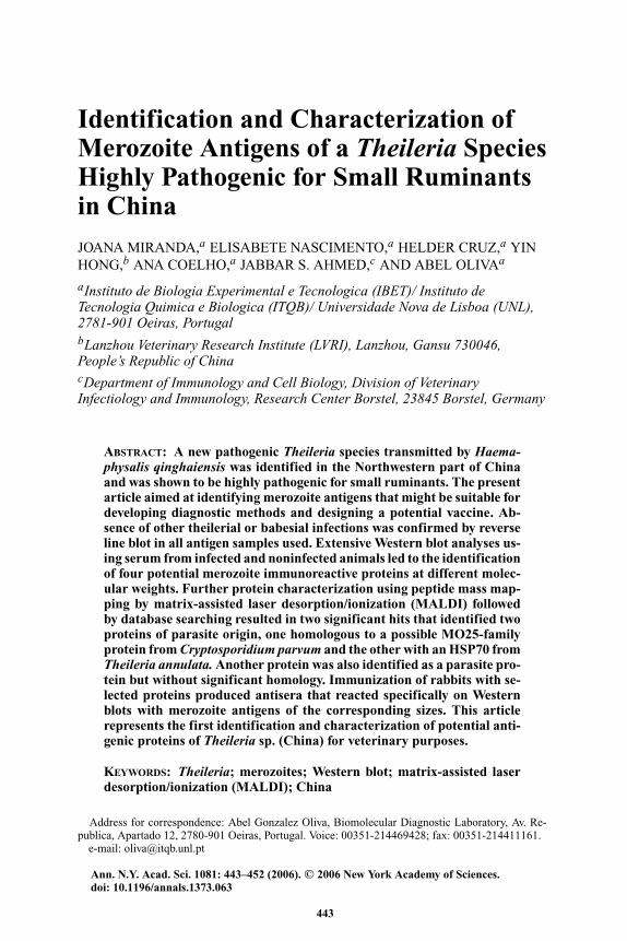
Identification and Characterization ofMerozoite Antigens of a Theileria SpeciesHighly Pathogenic for Small Ruminantsin China
JOANA MIRANDA,a ELISABETE NASCIMENTO,a HELDER CRUZ,a YINHONG,b ANA COELHO,a JABBAR S. AHMED,c AND ABEL OLIVAa
aInstituto de Biologia Experimental e Tecnologica (IBET)/ Instituto deTecnologia Quimica e Biologica (ITQB)/ Universidade Nova de Lisboa (UNL),2781-901 Oeiras, PortugalbLanzhou Veterinary Research Institute (LVRI), Lanzhou, Gansu 730046,People’s Republic of ChinacDepartment of Immunology and Cell Biology, Division of VeterinaryInfectiology and Immunology, Research Center Borstel, 23845 Borstel, Germany
ABSTRACT: A new pathogenic Theileria species transmitted by Haema-physalis qinghaiensis was identified in the Northwestern part of Chinaand was shown to be highly pathogenic for small ruminants. The presentarticle aimed at identifying merozoite antigens that might be suitable fordeveloping diagnostic methods and designing a potential vaccine. Ab-sence of other theilerial or babesial infections was confirmed by reverseline blot in all antigen samples used. Extensive Western blot analyses us-ing serum from infected and noninfected animals led to the identificationof four potential merozoite immunoreactive proteins at different molec-ular weights. Further protein characterization using peptide mass map-ping by matrix-assisted laser desorption/ionization (MALDI) followedby database searching resulted in two significant hits that identified twoproteins of parasite origin, one homologous to a possible MO25-familyprotein from Cryptosporidium parvum and the other with an HSP70 fromTheileria annulata. Another protein was also identified as a parasite pro-tein but without significant homology. Immunization of rabbits with se-lected proteins produced antisera that reacted specifically on Westernblots with merozoite antigens of the corresponding sizes. This articlerepresents the first identification and characterization of potential anti-genic proteins of Theileria sp. (China) for veterinary purposes.
KEYWORDS: Theileria; merozoites; Western blot; matrix-assisted laserdesorption/ionization (MALDI); China
Address for correspondence: Abel Gonzalez Oliva, Biomolecular Diagnostic Laboratory, Av. Re-publica, Apartado 12, 2780-901 Oeiras, Portugal. Voice: 00351-214469428; fax: 00351-214411161.
e-mail: [email protected]
Ann. N.Y. Acad. Sci. 1081: 443–452 (2006). C© 2006 New York Academy of Sciences.doi: 10.1196/annals.1373.063
443
444 ANNALS NEW YORK ACADEMY OF SCIENCES
INTRODUCTION
Theileria organisms are tick-borne protozoan parasites infecting mainly ru-minants in tropical and subtropical countries. Although capable of infectinga wide range of vertebrates, theilerial parasites require both vertebrate andnonvertebrate hosts (ticks) to maintain their life cycle. Depending on the Thei-leria species a variety of ticks are responsible for their transmission includingRhipicephalus, Haemaphysalis, and Hyalomma. Theileriosis is still a seriousdisease because it is responsible for severe economic losses in the livestockindustry.1
Due to the economic relevance of small ruminants, interest has arisen insheep-infecting Theileria parasites.2 Recently, a Theileria species causing a fa-tal disease in small ruminants has been identified in the Northwest of China.3–8
This parasite is transmitted by Haemaphysalis qinghaiensis and often occursin mixed infections with other parasites.8–10 Depending on the geographic dis-tribution, the rate of morbidity due to the Chinese Theileria parasite variesbetween 19% and 65%, whereas the mortality ranges from 17% to 75%. Thehighest mortality is observed in lambs and in imported exotic animals. Insummary, from 4% to 47% of sheep in the investigated area die due to the in-fection.4 The diagnostic techniques for small ruminant piroplasmosis detectionare mainly based on the morphological examination of blood smears and clini-cal symptoms.11–15 However, these methods require experience in microscopyand are difficult to apply on a large scale for epidemiological studies. Conse-quently, the development of specific and sensitive diagnostic tools for parasitesurveillance, as well as immunoprophylatic means to control and prevent thespread of diseases is crucial. Thus, there is an urgent need for a laboratorydiagnostic test combining specificity, sensitivity, and cost effectiveness, suchas enzyme-linked immunosorbent assay (ELISA).16,17 Furthermore, ELISAoffers an attractive alternative since reading of the results can be fully auto-mated.
Unlike the leukoproliferative Theileria species (e.g., T. annulata, T. lesto-quardi, or T. parva), the schizont stage of Theileria sp. (China) is very shortand until now it could not be cultivated in vitro.18 Additionally, the merozoitestage is the most exposed stage to the host immune system. For this reason theaim of the present article was to identify and characterize merozoite antigensusing sera from infected animals. This was achieved by screening lysates ofinfected erythrocytes as well as purified merozoites in Western blot.
MATERIALS AND METHODS
Antigen and Serum Samples
Experimental infections were conducted in the experimental animal facil-ity of the Lanzhou Veterinary Research Institute, China, using sheep from
MIRANDA et al.: THEILERIA ANTIGENS IDENTIFICATION CHARACTERIZATION 445
Theileria and other protozoa-free areas. Animals were infected by injection ofstabilated blood as previously described19 and were used for the collection ofinfected erythrocytes (L1, L2, and L3). For the collection of serum samples,sheep were experimentally infected with ticks as described.7 Negative con-trols (Ln, Ln3, Ln4) were obtained from sheep held in a Theileria-free area(Germany).
Reverse Line Blot
In order to confirm that the biological material used in this study consistedof Theileria sp. (China), the samples were analyzed by reverse line blot (RLB)as described previously.20,21
Merozoite Antigen Preparation
Merozoite antigenic material was generated by a method previously de-scribed22 with some modifications. In summary, fresh blood from infectedsheep was diluted 1:1 (v/v) in Alsever solution (Sigma-Aldrich, St. Louis,MO) and centrifuged at 900g for 10 min at 4◦C. The supernatant was dis-carded; then the pellet was washed three times in Tris–HCl buffer (10 mMTris–HCl pH 7.4, 150 mM NaCl), and was centrifuged at 500g for 10 minat 4◦C. After that, the pellet was diluted 1:5 (v/v) in Tris–HCl, erythrocytes,separated from leukocytes by using a cellulose–powder column, (Whatman cel-lulose CF11; Millipore, Schwallbach, Germany), and lyzed with �−hemolysin(Sigma) at 37◦C for 30 min. Merozoite material was generated by a Percollgradient centrifugation (40% and 60%; Amersham, Munich, Germany) wherethe merozoite-containing band was localized between the Percoll layers. Aftercollection, merozoites were washed three times at 5000g for 10 min at 4◦C,resuspended in Tris–HCl buffer, lyzed by 5 freeze/thaw cycles, and stored at–70◦C.
Sample Fractionation and Triton X-100 Extraction
Fractionation was performed based on the method of Nagamatsu et al.23
with some modifications. Briefly, 500 �L of erythrocytes obtained from cel-lulose powder column separation were homogenized in 5 mL of homogeniza-tion buffer (10 mM Tris–HCl, pH 7.4, 1 mM EDTA, 250 mM sucrose, 1 mMphenylmethylsulfonyl fluoride, 10 �g/ml leupeptin). Cell debris was removedby centrifugation at 900g for 10 min at 4◦C and the supernatants were col-lected. The antigen was then ultracentrifuged at 110,000g for 75 min at 4◦C.The supernatant fraction (sob) was removed and the membrane-containingpellet was solubilized with solubilization buffer (10 mM Tris–HCl, pH 7.4,
446 ANNALS NEW YORK ACADEMY OF SCIENCES
1 mM EDTA, 0.5% Triton X-100, 1 mM phenylmethylsulfonyl fluoride) for1 h at 4◦C. The insoluble material was removed by centrifugation at 14,000gfor 10 min at 4◦C resulting in a pellet (insoluble fraction, Fi) and a supernatant(soluble fraction, Fs). The insoluble fraction was resuspended in PBS, pH 7.4and the soluble fraction was concentrated by ultrafiltration using MilliporeCentricon ultrafiltration columns with a cutoff molecular weight of 10 kDa.All samples were stored at –70◦C.
SDS-PAGE and Western-Blot
Sodium dodecyl sulphate-polyacrylamide gel electrophoresis (SDS-PAGE)was carried out on 8–12% polyacrylamide gels under nonreducing condi-tions. Proteins were transferred onto PVDF (polyvinylfluoride, Immobilon-P; Millipore) for antigen screening or nitrocellulose membrane (Schleicher& Schuell/Whatman, Dassel, Germany) for immunoblotting with rabbit an-tisera. For the screening of antigen samples, sheep antisera were diluted to1:400 in the dilution buffer (3% skim milk powder, 0.05% Tween-20 in TBS)and immunoreactivity was detected by an indirect immunoperoxidase method(donkey anti-goat IgG HRP (Santa Cruz, Santa Cruz, CA) with chemilumines-cence detection (WestPico reagent, Pierce, Perbio Science, Bonn, Germany).In addition, fractionated samples were immunoblotted against rabbit antisera,which were raised against the individual protein band (see below). In this case,immunodetection was achieved with goat anti-rabbit alkaline phophatase con-jugated antibody (Jackson, West Grove, PA) with BCIP/NBT for chromogenicdetection.
MALDI MS Peptide Mapping
Protein SDS-gel bands were excised and polypeptides were subjected toreduction, alkylation, and digestion with modified sequencing-grade trypsin(Promega, VWR International Material de Laboratorio, Lisbon, Portugal) ac-cording to Pandey et al.24 Sample peptides were assayed for peptide massfingerprint (PMF) in a Voyager-DE STR (Applied Biosystems, Foster City,CA) matrix-assisted laser desorption ionization time-of-flight (MALDI-TOF)mass spectrometer. Peptide crystallization was achieved by using 0.5 L ofsamples on a plate and adding on top with an equal volume of recrystal-ized matrix �-cyano-4-hydroxycinnamic acid (CHCA) (10 mg/ml) preparedin acetonitrile (50%, v/v) with trifluoracetic acid (0.1%, v/v). The mixturewas allowed to air dry. Monoisotopic peptide masses were used to search forhomology and protein identification with Peptide Mass Fingerprint of Mascot(http: //www.matrixscience.com). Searches were done in MSDB database. Itwas considered a mass accuracy of 50–100 ppm for external calibrations and
MIRANDA et al.: THEILERIA ANTIGENS IDENTIFICATION CHARACTERIZATION 447
Cys carbamidomethylation and Met oxidation as fixed and variable amino acidmodifications, respectively. The criteria used to accept the identification weresignificant homology score achieved in Mascot.25 Additionally, a minimumof four peptides match, and at least 10% sequence coverage criteria were alsoconsidered.
Production of Rabbit Antisera
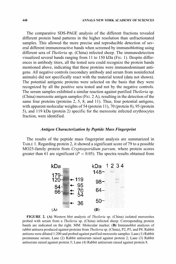
Three rabbits were immunized in the animal house facility of the InstitutoNacional de Engenharia e Tecnologia Industrial, Portugal, with selected proteinbands 2, 5, and 8 (see FIG. 1). The selected gel bands were emulsified andinjected four times at 3- to 4-week intervals. Antisera were collected 4 weeksafter the last immunization and stored at –20◦C.
RESULTS
Antigens Identification
For the identification of parasite antigens, two different types of parasite ma-terial were used: (a) isolated merozoites of the parasite Theileria sp. (China),and (b) Theileria sp. (China) infected erythrocyte lysated fractions.26 Nonin-fected erythrocytes were used as controls. The fractionated erythrocyte lysatesand the isolated merozoites were separated by SDS-PAGE under nonreducingconditions.
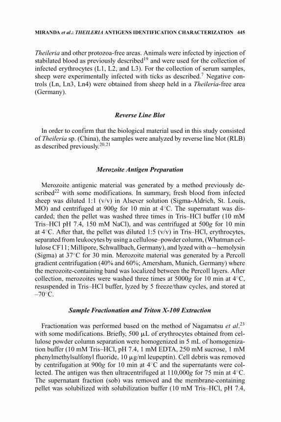
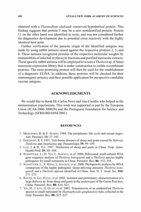
FIGURE 1. Western blots of Theileria infected (+; L1 (1), L2 (2), and L3 (3)) andnoninfected (–; Ln3 (4) and Ln4 (5)) erythrocyte fractions (Fi, Fs, and Sob) probed withantisera from Theileria sp. (China) infected sheep. Protein bands subjected to further anal-ysis are pointed out by arrows (→) and designated with the protein number as mentionedin the text. MM: molecular marker.
448 ANNALS NEW YORK ACADEMY OF SCIENCES
The comparative SDS-PAGE analysis of the different fractions revealeddifferent protein band patterns in the higher resolution than unfractionatedsamples. This allowed the more precise and reproducible detection of sev-eral different immunoreactive bands when screened by immunoblotting usingdifferent sera of Theileria sp. (China) infected sheep. The immunodetectionvisualized several bands ranging from 11 to 150 kDa (FIG. 1). Despite differ-ences in antibody titers, all the tested sera could recognize the protein bandsmentioned above, indicating that these proteins were immunodominant anti-gens. All negative controls (secondary antibody and serum from noninfectedanimals) did not specifically react with the material tested (data not shown).The potential antigenic proteins were selected on the basis that they wererecognized by all the positive sera tested and not by the negative controls.The serum samples exhibited a similar reaction against purified Theileria sp.(China) merozoite antigen samples (FIG. 2 A), resulting in the detection of thesame four proteins (proteins 2, 5, 8, and 11). Thus, four potential antigens,with apparent molecular weights of 54 (protein 11), 70 (protein 8), 95 (protein5), and 119 kDa (protein 2) specific for the merozoite infected erythrocytesfraction, were identified.
Antigen Characterization by Peptide Mass Fingerprint
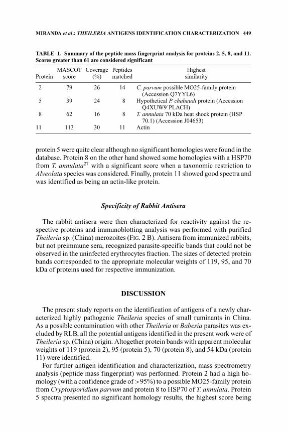
The results of the peptide mass fingerprint analysis are summarized inTABLE 1. Regarding protein 2, it showed a significant score of 79 to a possibleMO25-family protein from Cryptosporidium parvum, where protein scoresgreater than 61 are significant (P < 0.05). The spectra results obtained from
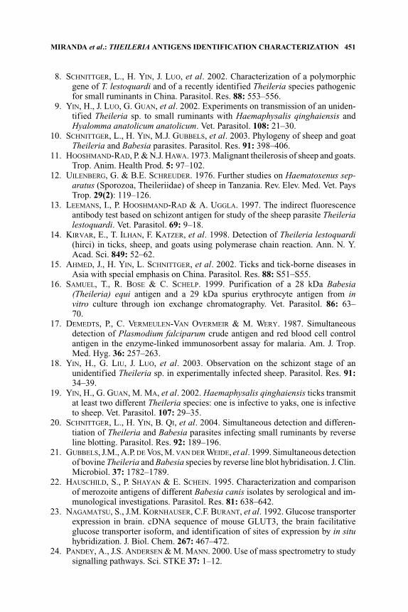
FIGURE 2. (A) Western blot analysis of Theileria sp. (China) isolated merozoitesprobed with serum from a Theileria sp. (China) infected sheep. Corresponding proteinbands are indicated on the right. MM: Molecular marker. (B) Immunoblot analyses ofrabbit antisera produced against proteins from Theileria sp. (China), P2, P5, and P8. Rabbitantisera were diluted 1:200 and probed against purified merozoite samples: Lane (1) Rabbitpreimmune serum; Lane (2) Rabbit antiserum raised against protein 2; Lane (3) Rabbitantiserum raised against protein 5; Lane (4) Rabbit antiserum raised against protein 8.
MIRANDA et al.: THEILERIA ANTIGENS IDENTIFICATION CHARACTERIZATION 449
TABLE 1. Summary of the peptide mass fingerprint analysis for proteins 2, 5, 8, and 11.Scores greater than 61 are considered significant
MASCOT Coverage Peptides HighestProtein score (%) matched similarity
2 79 26 14 C. parvum possible MO25-family protein(Accession Q7YYL6)
5 39 24 8 Hypothetical P. chabaudi protein (AccessionQ4XUW9˙PLACH)
8 62 16 8 T. annulata 70 kDa heat shock protein (HSP70.1) (Accession J04653)
11 113 30 11 Actin
protein 5 were quite clear although no significant homologies were found in thedatabase. Protein 8 on the other hand showed some homologies with a HSP70from T. annulata27 with a significant score when a taxonomic restriction toAlveolata species was considered. Finally, protein 11 showed good spectra andwas identified as being an actin-like protein.
Specificity of Rabbit Antisera
The rabbit antisera were then characterized for reactivity against the re-spective proteins and immunoblotting analysis was performed with purifiedTheileria sp. (China) merozoites (FIG. 2 B). Antisera from immunized rabbits,but not preimmune sera, recognized parasite-specific bands that could not beobserved in the uninfected erythrocytes fraction. The sizes of detected proteinbands corresponded to the appropriate molecular weights of 119, 95, and 70kDa of proteins used for respective immunization.
DISCUSSION
The present study reports on the identification of antigens of a newly char-acterized highly pathogenic Theileria species of small ruminants in China.As a possible contamination with other Theileria or Babesia parasites was ex-cluded by RLB, all the potential antigens identified in the present work were ofTheileria sp. (China) origin. Altogether protein bands with apparent molecularweights of 119 (protein 2), 95 (protein 5), 70 (protein 8), and 54 kDa (protein11) were identified.
For further antigen identification and characterization, mass spectrometryanalysis (peptide mass fingerprint) was performed. Protein 2 had a high ho-mology (with a confidence grade of >95%) to a possible MO25-family proteinfrom Cryptosporidium parvum and protein 8 to HSP70 of T. annulata. Protein5 spectra presented no significant homology results, the highest score being
450 ANNALS NEW YORK ACADEMY OF SCIENCES
obtained with a Plasmodium chabaudi conserved hypothetical protein. Thisfinding suggests that protein 5 may be a new nonidentified protein. Protein11 on the other hand was identified as actin, and was not considered furtherfor diagnostics development due to potential cross reactivity with the highlyidentical host actin.
Further verification of the parasite origin of the identified antigens wasmade by using rabbit antisera raised against the respective proteins 2, 5, and8. These antisera recognized proteins of the respective molecular weights byimmunoblots of infected erythrocyte fractions and purified merozoite extracts.These specific rabbit antisera will be employed to screen a Theileria sp. (China)merozoite expression library that is under construction to isolate recombinantproteins. The most promising protein will then be used for the establishmentof a diagnostic ELISA. In addition, these proteins will be checked for theirimmunogenic potency and their possible application for perspective candidatevaccine antigens.
ACKNOWLEDGMENTS
We would like to thank Dr. Carlos Novo and Ana Custdio who helped in theimmunization experiments. This work was supported in part by the EuropeanUnion (ICA4-2000-300028) and the Portuguese Foundation for Science andTechnology (SFRH/BD/6494/2001).
REFERENCES
1. MEHLHORN, H. & E. SCHEIN. 1984. The piroplasms: life cycle and sexual stages.Adv. Parasitol. 23: 37–103.
2. FRIEDHOFF, K.T. 1997. Tick-borne diseases of sheep and goats caused by Babesia,Theileria and Anaplasma spp. Parasitologia 39: 99–109.
3. LUO, J. & H. YIN. 1997. Theilerosis of sheep and goats in China. Trop. Anim.Health Prod. 29: 8S–10S.
4. SCHNITTGER, L., H. YIN, L. JIANXUN, et al. 2000. Ribosomal small-subunit RNAgene sequence analysis of Theileria lestoquardi and a Theileria species highlypathogenic for small ruminants in China. Parasitol. Res. 86: 352–358.
5. SCHNITTGER, L., Y. HONG, L. JIANXUN, et al. 2000. Phylogenetic analysis by rRNAcomparison of the highly pathogenic sheep-infecting parasites Theileria lesto-quardi and a Theileria species identified in China. Ann. N. Y. Acad. Sci. 916:271–275.
6. BAI, Q., G. LIU, D. LIU, et al. 2002. Isolation and preliminary characterization of alarge Babesia sp. from sheep and goats in the eastern part of the Gansu Province,China. Parasitol. Res. 88: S16–S21.
7. YIN, H., J. LUO, G. GUAN, et al. 2002. Transmission of an unidentified Theileriaspecies to small ruminants by Haemaphysalis qinghaiensis ticks collected in thefield. Parasitol. Res. 88: S25–S27.
MIRANDA et al.: THEILERIA ANTIGENS IDENTIFICATION CHARACTERIZATION 451
8. SCHNITTGER, L., H. YIN, J. LUO, et al. 2002. Characterization of a polymorphicgene of T. lestoquardi and of a recently identified Theileria species pathogenicfor small ruminants in China. Parasitol. Res. 88: 553–556.
9. YIN, H., J. LUO, G. GUAN, et al. 2002. Experiments on transmission of an uniden-tified Theileria sp. to small ruminants with Haemaphysalis qinghaiensis andHyalomma anatolicum anatolicum. Vet. Parasitol. 108: 21–30.
10. SCHNITTGER, L., H. YIN, M.J. GUBBELS, et al. 2003. Phylogeny of sheep and goatTheileria and Babesia parasites. Parasitol. Res. 91: 398–406.
11. HOOSHMAND-RAD, P. & N.J. HAWA. 1973. Malignant theilerosis of sheep and goats.Trop. Anim. Health Prod. 5: 97–102.
12. UILENBERG, G. & B.E. SCHREUDER. 1976. Further studies on Haematoxenus sep-aratus (Sporozoa, Theileriidae) of sheep in Tanzania. Rev. Elev. Med. Vet. PaysTrop. 29(2): 119–126.
13. LEEMANS, I., P. HOOSHMAND-RAD & A. UGGLA. 1997. The indirect fluorescenceantibody test based on schizont antigen for study of the sheep parasite Theilerialestoquardi. Vet. Parasitol. 69: 9–18.
14. KIRVAR, E., T. ILHAN, F. KATZER, et al. 1998. Detection of Theileria lestoquardi(hirci) in ticks, sheep, and goats using polymerase chain reaction. Ann. N. Y.Acad. Sci. 849: 52–62.
15. AHMED, J., H. YIN, L. SCHNITTGER, et al. 2002. Ticks and tick-borne diseases inAsia with special emphasis on China. Parasitol. Res. 88: S51–S55.
16. SAMUEL, T., R. BOSE & C. SCHELP. 1999. Purification of a 28 kDa Babesia(Theileria) equi antigen and a 29 kDa spurius erythrocyte antigen from invitro culture through ion exchange chromatography. Vet. Parasitol. 86: 63–70.
17. DEMEDTS, P., C. VERMEULEN-VAN OVERMEIR & M. WERY. 1987. Simultaneousdetection of Plasmodium falciparum crude antigen and red blood cell controlantigen in the enzyme-linked immunosorbent assay for malaria. Am. J. Trop.Med. Hyg. 36: 257–263.
18. YIN, H., G. LIU, J. LUO, et al. 2003. Observation on the schizont stage of anunidentified Theileria sp. in experimentally infected sheep. Parasitol. Res. 91:34–39.
19. YIN, H., G. GUAN, M. MA, et al. 2002. Haemaphysalis qinghaiensis ticks transmitat least two different Theileria species: one is infective to yaks, one is infectiveto sheep. Vet. Parasitol. 107: 29–35.
20. SCHNITTGER, L., H. YIN, B. QI, et al. 2004. Simultaneous detection and differen-tiation of Theileria and Babesia parasites infecting small ruminants by reverseline blotting. Parasitol. Res. 92: 189–196.
21. GUBBELS, J.M., A.P. DE VOS, M. VAN DER WEIDE, et al. 1999. Simultaneous detectionof bovine Theileria and Babesia species by reverse line blot hybridisation. J. Clin.Microbiol. 37: 1782–1789.
22. HAUSCHILD, S., P. SHAYAN & E. SCHEIN. 1995. Characterization and comparisonof merozoite antigens of different Babesia canis isolates by serological and im-munological investigations. Parasitol. Res. 81: 638–642.
23. NAGAMATSU, S., J.M. KORNHAUSER, C.F. BURANT, et al. 1992. Glucose transporterexpression in brain. cDNA sequence of mouse GLUT3, the brain facilitativeglucose transporter isoform, and identification of sites of expression by in situhybridization. J. Biol. Chem. 267: 467–472.
24. PANDEY, A., J.S. ANDERSEN & M. MANN. 2000. Use of mass spectrometry to studysignalling pathways. Sci. STKE 37: 1–12.
452 ANNALS NEW YORK ACADEMY OF SCIENCES
25. GONNET, F., G. LEMAITRE, G. WAKSMAN, et al. 2003. MALDI/MS peptide massfingerprinting for proteome analysis: identification of hydrophobic proteins at-tached to eukaryote keratinocyte cytoplasmic membrane using different matricesin concert. Proteome Sci. 1(1): 2.
26. MIRANDA, J., B. STUMME, D. BEYER, et al. 2004. Identification of antigenic proteinsof a Theileria species pathogenic for small ruminants in China recognized byantisera of infected animals. Ann. N. Y. Acad. Sci. 1026: 161–164.
27. SCHNITTGER, L., P. SHAYAN, R. BIERMANN R, et al. 2000. Molecular genetic char-acterization and subcellular localization of Theileria annulata mitochondrialheat-shock protein 70. Parasitol. Res. 86: 444–452.
Copyright © 2022 FDOKUMEN