Identification of novel Theileria parva candidate vaccine ...
157
-
Upload
khangminh22 -
Category
Documents
-
view
3 -
download
0
Transcript of Identification of novel Theileria parva candidate vaccine ...
Identification of novel Theileria parva candidate vaccine
antigens and discovery of new therapeutic drugs for the
control of East Coast fever
James J. Nyagwange
Thesis committee
Promotors
Prof. Dr H.F.J. Savelkoul
Professor of Cell Biology and Immunology
Wageningen University & Research
Prof. Dr V. Nene
Director and co-leader, Animal and Human Health program
International Livestock Research Institute, Nairobi, Kenya
Co-promotors
Dr E.J. Tijhaar
Assistant professor, Cell Biology and Immunology
Wageningen University & Research
Dr Roger Pelle
Principal scientist, Biosciences Eastern and Central Africa
International Livestock Research Institute, Nairobi, Kenya
Other members
Prof. Dr V.P.M.G. Rutten, Utrecht University
Prof. Dr M.M. van Oers, Wageningen University & Research
Prof. Dr G.F. Wiegertjes, Wageningen University & Research
Dr H.K. Parmentier, Wageningen University & Research
This research was conducted under the auspices of the Graduate School Wageningen Institute
of Animal Sciences
Identification of novel Theileria parva candidate vaccine
antigens and discovery of new therapeutic drugs for the
control of East Coast fever
James J. Nyagwange
Thesis
submitted in fulfilment of the requirements for the degree of doctor
at Wageningen University
by the authority of the Rector Magnificus,
Prof. Dr A.P.J. Mol,
in the presence of the
Thesis Committee appointed by the Academic Board
to be defended in public
on Tuesday 14 May 2019
at 1:30 p.m. in the Aula.
James Jimmy Nyagwange
Identification of novel Theileria parva candidate vaccine antigens and discovery of new
therapeutic drugs for the control of East Coast fever
154 pages
PhD thesis, Wageningen University, Wageningen, the Netherlands (2019)
With references, with summary in English
ISBN 978-94-6343-578-9
DOI https://doi.org/10.18174/467903
Contents
CHAPTER 1: General Introduction ................................................................................................... 7
CHAPTER 2: Characterization of the Theileria parva sporozoite proteome ............................... 37
CHAPTER 3: Characterization of Rhipicephalus appendiculatus salivary glands proteins co-
purified with Theileria parva sporozoites .......................................................................................... 65
CHAPTER 4: Antibodies to in silico selected GPI-anchored Theileria parva proteins neutralize
sporozoite infection in vitro ................................................................................................................ 81
CHAPTER 5: Leveraging the Medicines for Malaria Venture malaria and pathogen boxes to
discover lead chemical inhibitors of East Coast fever .................................................................... 99
CHAPTER 6: Summarizing Discussion .......................................................................................... 115

7 | C h a p t e r 1
CHAPTER
1
General Introduction
8 | C h a p t e r 1
East Coast fever
East Coast fever (ECF) is lymphoproliferative disease of cattle caused by Theileria
parva, an apicomplexan parasite transmitted by ticks (Hayashida, Abe et al. 2013). Dr. Arnold
Theiler working in South Africa in the early1900s, identified T. parva as the causal organism
of ECF and distinguished the disease from Redwater that is caused by Babesia (Theiler 1912).
He also identified the three host tick Rhipicephalus appendiculatus as the main vector for ECF
transmission (Theiler 1912). In the past, T. parva was thought to comprise three distinct sub-
species; T. parva parva, T. parva lawrencei and T. parva bovis, causing ECF, Corridor disease
and January disease respectively. However, it is now known that T. parva is genetically a
single species and the sub-species nomenclature was abandoned (Perry and Young 1993).
Parasite isolates are now described as either cattle-derived or buffalo-derived depending on
their originating host species (reviewed in Nene, Kiara et al. 2016). The most common form
of the disease caused by cattle-derived T. parva is called classical ECF. It presents with high
parasitemia and high mortality in cattle. A less virulent form of the disease known as January
disease caused by cattle derived T. parva is found in Zimbabwe (reviewed in Latif and Hove
2011). The latter disease occurs seasonally, with outbreaks occurring in January simultaneously
with emergence of adult ticks from diapause (Matson 1967). It presents with high morbidity
but with low mortality and low parasitemia (reviewed in Latif and Hove 2011). However,
another form of the disease caused by buffalo derived parasite known as Corridor disease is
more acute than ECF and in the face of an outbreak, the mortality rate can exceed 90%
(reviewed in Norval, Perry et al. 1992, Mbizeni, Potgieter et al. 2013). Classical ECF was
introduced to South Africa and Zimbabwe in the 1900s following importation of cattle from
East Africa for restocking after devastation of cattle by Rinderpest in 1895 (Lawrence 1979).
Currently, ECF is present in 12 countries in eastern, central and southern Africa namely;
Kenya, Tanzania, Uganda, South Sudan, Burundi, Rwanda, Democratic Republic of Congo,
Malawi, Mozambique, Zambia, Zimbabwe and Comoro Islands (Nene, Kiara et al. 2016).
T. parva pathogenicity to cattle varies per breed and mortality appears to be higher in
Bos taurus and their crosses compared to Bos indicus (reviewed in Morrison, Connelley et al.
2015). In Bos taurus mortality can approach 100% and T. parva is considered a major factor
undermining the introduction and sustenance of highly productive taurine breeds of cattle in
sub-Saharan Africa (reviewed in Morrison, Connelley et al. 2015). Rearing taurine breeds in
T. parva endemic regions is usually accompanied by high costs associated with the use of toxic
and expensive acaricides to kill the tick vector. Epidemics of ECF in areas previously
unaffected by T. parva infection are renowned for their devastating effect on cattle populations.
For smallholder farmers, extinction of the entire herd is common when actions are not taken to
mitigate the outbreaks. This has dramatic social, economic and health effects due to loss of
food and income. On regional basis, it was estimated that ECF kills approximately 1 million
cattle/year causing annual losses of approximately USD 300 million (McLeod and Kristjanson
1999). However, these estimates were made several years ago and it is likely that the current
impact is much larger.
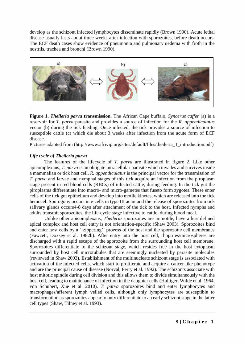
T. parva has probably made a host jump from the African Cape buffalo in which it
does not cause disease (reviewed in Norval, Perry et al. 1992) to cattle, where it causes ECF
(review ed in Norval, Perry et al. 1992). The African buffalo plays an important role as parasite
reservoir and drives the epidemiology of the disease (figure 1). In regions where there is
wildlife-cattle interface, the buffaloes provide a source of infection for the ticks which in turn
infect cattle. Indigenous cattle that spontaneously recover from the disease after mild reactions
are solidly immune to reinfection and form another reservoir for the parasite since they do not
usually clear the infection (Kariuki, Young et al. 1995). ECF is characterised by high fever,
leukocytopenia, severe damage to the lymphoid system and pronounced clinical symptoms
9 | C h a p t e r 1
develop as the schizont infected lymphocytes disseminate rapidly (Brown 1990). Acute lethal
disease usually lasts about three weeks after infection with sporozoites, before death occurs.
The ECF death cases show evidence of pneumonia and pulmonary oedema with froth in the
nostrils, trachea and bronchi (Brown 1990).
Figure 1. Theileria parva transmission. The African Cape buffalo, Syncerus caffer (a) is a
reservoir for T. parva parasite and provides a source of infection for the R. appendiculatus
vector (b) during the tick feeding. Once infected, the tick provides a source of infection to
susceptible cattle (c) which die about 3 weeks after infection from the acute form of ECF
disease.
Pictures adapted from (http://www.afrivip.org/sites/default/files/theileria_1_introduction.pdf)
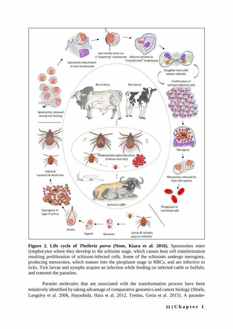
Life cycle of Theileria parva
The features of the lifecycle of T. parva are illustrated in figure 2. Like other
apicomplexans, T. parva is an obligate intracellular parasite which invades and survives inside
a mammalian or tick host cell. R. appendiculatus is the principal vector for the transmission of
T. parva and larvae and nymphal stages of this tick acquire an infection from the piroplasm
stage present in red blood cells (RBCs) of infected cattle, during feeding. In the tick gut the
piroplasms differentiate into macro- and micro-gametes that fuseto form zygotes. These enter
cells of the tick gut epithelium and develop into motile kinetes, which are released into the tick
hemocel. Sporogony occurs in e-cells in type III acini and the release of sporozoites from tick
salivary glands occurs4-8 days after attachment of the tick to the host. Infected nymphs and
adults transmit sporozoites, the life-cycle stage infective to cattle, during blood meal.
Unlike other apicomplexans, Theileria sporozoites are immotile, have a less defined
apical complex and host cell entry is not orientation-specific (Shaw 2003). Sporozoites bind
and enter host cells by a ‘‘zippering’’ process of the host and the sporozoite cell membranes
(Fawcett, Doxsey et al. 1982b). After entry into the host cell, rhoptries/microspheres are
discharged with a rapid escape of the sporozoite from the surrounding host cell membrane.
Sporozoites differentiate to the schizont stage, which resides free in the host cytoplasm
surrounded by host cell microtubules that are seemingly nucleated by parasite molecules
(reviewed in Shaw 2003). Establishment of the multinucleate schizont stage is associated with
activation of the infected cells, which start to proliferate and acquire a cancer-like phenotype
and are the principal cause of disease (Norval, Perry et al. 1992). The schizonts associate with
host mitotic spindle during cell division and this allows them to divide simultaneously with the
host cell, leading to maintenance of infection in the daughter cells (Hulliger, Wilde et al. 1964,
von Schubert, Xue et al. 2010). T. parva sporozoites bind and enter lymphocytes and
macrophages/afferent lymph veiled cells, although only lymphocytes are susceptible to
transformation as sporozoites appear to only differentiate to an early schizont stage in the latter
cell types (Shaw, Tilney et al. 1993).
10 | C h a p t e r 1
Schizonts undergo merogony, from approximately 10-14 days post infection, producing
merozoites which are released by host cell rapture (reviewed in Morrison, Connelley et al.
2015). Through a similar process like sporozoite entry into lymphocytes, merozoites invade
red blood cells where they develop into the tick infective piroplasm stage which resides freely
in the cytoplasm (Shaw and Tilney 1995). Piroplasms undergo limited multiplication and
anaemia caused by destruction of the erythrocytes does not contribute significantly to the
pathology of ECF (reviewed in Morrison, Connelley et al. 2015). However, in animals that
recover from infection, parasites persist for several months or years and are not detectable by
microscopy, this asymptomatic carrier state functions as an important source of infection for
ticks (Kariuki, Young et al. 1995).
Transformation of the host cells
Schizont-infected B and T-cells acquire a cancer-like phenotype characterized by
proliferation, immortality and dissemination throughout the lymphoid system and other tissues,
especially the lungs and gut mucosa (Irvin and Morrison 1987, Sivakumar, Hayashida et al.
2014). Transformation occurs more in the infected T-cell lineage than in the infected B-cell
lineage and high level pathology is associated with T-cell transformation (Morrison, MacHugh
et al. 1996). Proliferation of the infected cells is independent of exogenous growth factors and
is completely dependent on the presence of live parasites (Dobbelaere, Roditi et al. 1991).
Killing of the parasites by anti-theilerial drugs such as buparvaquone results in the cells
reverting to non-proliferative state (Brown, Shaw et al. 1989).
11 | C h a p t e r 1
Figure 2. Life cycle of Theileria parva (Nene, Kiara et al. 2016). Sporozoites enter
lymphocytes where they develop to the schizont stage, which causes host cell transformation
resulting proliferation of schizont-infected cells. Some of the schizonts undergo merogony,
producing merozoites, which mature into the piroplasm stage in RBCs, and are infective to
ticks. Tick larvae and nymphs acquire an infection while feeding on infected cattle or buffalo,
and transmit the parasites.
Parasite molecules that are associated with the transformation process have been
tentatively identified by taking advantage of comparative genomics and cancer biology (Shiels,
Langsley et al. 2006, Hayashida, Hara et al. 2012, Tretina, Gotia et al. 2015). A parasite-
12 | C h a p t e r 1
encoded prolyl isomerase has been recently identified as playing a key role in this complex
host cell transformation process (Marsolier, Perichon et al. 2015). In addition, several host cell-
signalling pathways that contribute to host cell transformation have been studied (reviewed in
Dobbelaere and Küenzi 2004, Dessauge, Lizundia et al. 2005). Understanding the mechanisms
of host cell transformation by these parasites presents opportunities for cancer therapy and
control of ECF as novel drugs may target these pathways for disruption.
Control of ECF by vaccination
A live parasite-based vaccine, which immunizes the animals by an infection and
treatment method (ITM), is available (Radley, Brown et al. 1975, Radley, Brown et al. 1975b).
It involves deliberate infection of the animals with live parasite followed by treatment with
antibiotic such as long acting oxytetracycline. The antibiotic controls but does not kill the
parasite, allowing the animal to develop a protective acquired immune response. For control of
ECF in southern Africa ITM is performed with a single parasite isolate and in parts of eastern
Africa a composite ITM vaccine, the Muguga cocktail, is used (Morzaria, Nene et al. 2000).
The Muguga cocktail consists of three isolates namely; Muguga, Serengeti transformed and
Kiambu-5. These isolates have been shown to confer broad-spectrum protection against ECF.
The use of combinations of isolates in ITM followed the discovery that a single parasite isolate
did not offer sufficient protection against isolates of different geographical location. The choice
of parasite isolates/strains included in the vaccine is based on the degree of cross-protection
among the isolates/strains. Usually, there is 20–30% cross-protection between the individual
isolates in different cross-immunity groups (reviewed in Nene, Kiara et al. 2016).
Although the ITM vaccine is effective in controlling ECF, it has serious disadvantages. It
requires a liquid nitrogen cold chain for delivery and oxytetracycline co-treatment. In addition,
the very laborious production of the vaccine from infected ticks, which takes 18 months, makes
it expensive and animals vaccinated by the ITM protocol remain life-long carries of the
parasite, which could pose a risk for spread of the disease (reviewed in Di Giulio, Lynen et al.
2009). Therefore, there is need for development of subunit vaccines without these
disadvantages.
Over the years, there have been efforts in identifying antigens that are targets of
neutralizing antibodies and CD8+ T-cells (reviewed in Nene, Kiara et al. 2016). Neutralizing
antibodies are directed against sporozoite surface proteins to prevent it from infecting host cells
while CD8+ cytotoxic T cells (CTLs) are directed against antigens of the schizont stage to
interrupt the infection by killing the schizont infected host cells. A number of antigens that are
targets of neutralizing antibodies have been identified before, including p67, p32, p104, PIM
and p150. The most preferred antigen, p67 was identified by murine monoclonal antibodies
which neutralized sporozoite infection in vitro (Dobbelaere, Spooner et al. 1984, Musoke,
Nantulya et al. 1984, Dobbelaere, Shapiro et al. 1985). This 709 amino acid residue protein, is
totally conserved among cattle derived T. parva strains, making it a suitable candidate to
function as cross-protective immunogen among cattle derived T. parva strains (Nene, Musoke
et al. 1996). It can also function as immunogen among buffalo derived T. parva strains because
even though it’s polymorphic among buffalo derived T. parva strains, its alleles share greater
than 90% sequence identity (Nene, Musoke et al. 1996). There have been several vaccine trial
experiments using several constructs of full length and recombinant fragments of the protein
with a number of adjuvants and gene based delivery systems. Results from these experimental
vaccine trials were recently summarized by Nene et al (2016) and protection from parasite
challenge following p67 immunization ranges from 13 to 70% depending on construct,
concentration, dosage regimen and formulation (Nene, Kiara et al. 2016). The best outcome of
these experiments involved p67C fragment, an 80 amino acid peptide from the C-terminal with
13 | C h a p t e r 1
epitopes recognized by sporozoite neutralizing monoclonal antibodies (Bishop, Nene et al.
2003). This fragment is easier to express in E. coli in a stable manner and in high yield than
the p67 full length protein and it induces comparable immunity, suggesting it represents a
critical target (Bishop, Nene et al. 2003). The consistent evidence of immunity induced by p67
constructs, although variable, cements p67 as a strong candidate vaccine antigen and research
is ongoing to increase its efficacy by testing different formulations and immunization schemes.
The limitation with p67 is that in most experiments, there has been no consistent correlate of
the various antibody assays (such as antibody titres, CD4+ T-cell proliferation, etc.) with
immunity, and immunity in field trials is relatively lower than laboratory challenge experiments
(Musoke, Morzaria et al. 1992, Nene, Musoke et al. 1996, Schetters, Arts et al. 2015). Although
the field vs laboratory based experimental differences can be attributed to some cattle naturally
recovering from infection in the control group under field conditions, Schetters et al (2015)
recently reported susceptibility to disease in immunized and control cattle in the field
following p67 immunization (Schetters, Arts et al. 2015).
Research was discontinued on the 32 kDa (p32) protein, identified
through immunoprecipitation of biotin labeled sporozoites with the murine monoclonal mAb
4C9 (Skilton, Musoke et al. 2000). A recombinant p32 protein produced bovine antisera that
did not induce neutralizing antibodies and the p32 immunized cattle were susceptible to
parasite challenge (Skilton, Musoke et al. 2000). A series of murine monoclonal antibodies
raised to the schizont stage of the parasite identified another antigen, the polymorphic immuno-
dominant molecule (PIM). Named for its polymorphic nature and identification by a number
of antibodies, PIM is localized in the rhoptries/micronemes in the sporozoites (Toye, Nyanjui
et al. 1996) but located on surface of the schizonts (Shapiro, Fujisaki et al. 1987). However,
due to its highly polymorphic sequence in both cattle- and buffalo-derived T. parva strains
(Geysen, Bazarusanga et al. 2004) and findings that rats immunized by PIM produce
neutralizing antibodies,(Toye, Metzelaar et al. 1995) but cattle immunized by PIM do not
make sporozoite neutralizing antibodies, the role of PIM as a vaccine candidate antigen was
not been evaluated (Toye, Nyanjui et al. 1996). We have investigated a role of p104 antigen in
this study but the role of p150 as a candidate vaccine antigen remains to be evaluated. Based
on the large number of apicomplexan proteins involved in host cell entry, it possible that there
are additional novel T. parva candidate sporozoite vaccine antigens to be discovered. The main
aim of this project was to identify novel sporozoite vaccine candidates that can be used by
themselves or in combination with p67 to increase the effectiveness of vaccination for ECF.
Control of ECF by chemotherapy
In addition to vaccination, ECF is controlled by direct killing of the parasites using
chemotherapeutic drugs and indirectly by using acaricides to kill the tick vectors responsible
for transmission of the parasites. Regular spraying and/or dipping of cattle in acaricide have
effectively been used in preventing ECF but have limitations. Firstly, the acaricides are toxic
chemicals, raising concerns about environmental pollution and possible contamination of the
food chain by leaving residues in milk and meat products. Secondly, many of the poor farmers
in the affected regions cannot afford the chemicals for spraying and dipping. Finally, the ticks
are increasingly developing resistance to the acaricides. Therefore, control by acaricides seems
unsustainable.
Halofuginone lactate under trade name Terit® was the first commercial drug to be
licensed against Theileria (Kaba 2003). It was followed by hydroxynaphthoquinone
parvaquone traded as Clexon® (Kaba 2003). Buparvaquone traded as Butalex®, a derative of
14 | C h a p t e r 1
parvaquone was subsequently synthesized and found to be more active and since its discovery
about 30 years ago, it has remained the frontline drug of choice for the control of both T. parva
and T. annulata infections (Nene, Kiara et al. 2016).
Buparvaquone effectiveness requires administration during the early stage of parasite
infection before widespread immune system destruction by the advanced stages. During the
latter stage several doses of the drug are required to resolve infection (Morrison and McKeever
2006). Buparvaquone is also relatively expensive to small holder farmers and is not always
available to these farmers (Morrison and McKeever 2006). As with other
hydroxynaphtoquinones, buparvaquone most likely inhibits mitochondrial electron transport in
the parasite (McHardy, Wekesa et al. 1985) (Birth, Kao et al. 2014). Resistance to the drug has
been described in T. annulata and is associated with mutations in cytochrome b (cytb) gene
encoding for ubiquinone reductase of the respiratory chain (Sharifiyazdi, Namazi et al. 2012,
Mhadhbi, Chaouch et al. 2015). Therefore, recent identification of nuclear encoded peptidyl-
prolyl isomerase in T. annulata as a potential target for burpavaquone resistance was not
expected (Marsolier, Perichon et al. 2015). Nevertheless, the findings of buparavaquone
resistance in T. annulata are of great concern for future control of theileriosis and ECF
(Mhadhbi, Naouach et al. 2010). Although resistance in T. parva has not been documented, it
might just be a matter of time before it happens. We have undertaken studies to identify new
drug compounds as starting points to anti-theilerial drug discovery and part of this thesis
describes these efforts.
Scope of thesis
This work is part of a larger theme focused on improvement of vaccines for the control
of ECF in cattle in Africa. The larger theme aimed on improving aspects of the live infection
and treatment method of vaccination against ECF, inducing antibody based immunity by
targeting the sporozoite stage of the parasite and inducing a T-cell mediated immunity by
targeting the schizont stage of the parasite.
In Chapter 1, and overview of ECF biology and the different approaches to control the
disease are discussed, with a focus on vaccine development.
In Chapter 2, to identify novel potential sporozoite vaccine candidates the proteome
of T. parva sporozoites, purified by ion-exchange chromatography (Musoke, Morzaria et al.
1992), was characterized by a Multidimensional Protein Identification Technology (MudPIT)
mass spectrometry-based approach (reviewed in Schirmer, Yates et al. 2003) To the 2,007
parasite proteins revealed by this approach bioinformatics analysis identified orthologs of
Plasmodium falciparum surface proteins and proteins involved in host cell infection, of which
some may represent novel T. parva vaccine candidates.
In Chapter 3, using bioinformatics and literature review, we analyzed the 2,773 vector
proteins co-purified with the sporozoite proteins in the infected salivary glands and identified
known R. appendiculatus antigens that are targets of transmission blocking vaccines and novel
antigens that may be involved in sporozoite infection of lymphocytes. The control of ECF may
benefit from vaccines that can block sporozoite infection of lymphocytes as well as R.
appendiculatus attachment and feeding.
Chapter 4 describes selection of potential sporozoite surface proteins, recombinant
expression of putative vaccine candidate antigens and in vitro neutralization assay with antisera
15 | C h a p t e r 1
against the recombinant proteins. The proteins, which in majority were identified in Chapter
2, were selected based on the presence of a predicted C-terminal Glycosylphosphatidylinositol
(GPI) anchor signal and /or N-terminal signal peptide, since proteins with such features are
likely to be located on the surface of the cell where they are involved in extracellular interaction
(Ferguson 1999).
In Chapter 5 the results of screening the MMV malaria box, pathogen box and
dasatanib drug compounds against T. parva infected lymphocytes is presented. Dasatanib, a
tyrosine kinase inhibitor indicated in certain leukemias was found to inhibit proliferation of T.
parva infected lymphocytes comparable to buparvaquone and non-toxic (CC50 > 8µM) on
bovine PBMCs immortalized by Concanavalin A (Con A blasts). One of the 400 malaria box
compounds and one of the 396 pathogen box compounds inhibited proliferation of T. parva
infected lymphocytes with a therapeutic index greater than five.
Finally, in Chapter 6, we discuss the implications of our research for the rational design
of intervention strategies for ECF and propose future research opportunities.
16 | C h a p t e r 1
References
Abdelhaleem, M. (2004). "Do human RNA helicases have a role in cancer?" Biochimica et
Biophysica Acta (BBA) - Reviews on Cancer 1704(1): 37-46.
Aurrecoechea, C., J. Brestelli, B. P. Brunk, S. Fischer, B. Gajria, X. Gao, A. Gingle, G. Grant,
O. S. Harb, M. Heiges, F. Innamorato, J. Iodice, J. C. Kissinger, E. T. Kraemer, W. Li, J. A.
Miller, V. Nayak, C. Pennington, D. F. Pinney, D. S. Roos, C. Ross, G. Srinivasamoorthy, J.
C. J. Stoeckert, R. Thibodeau, C. Treatman and H. Wang (2010). "EuPathDB: a portal to
eukaryotic pathogen databases." Nucleic Acids Research 38(suppl_1): D415-D419.
Babo Martins, S., G. Di Giulio, G. Lynen, A. Peters and J. Rushton (2010). "Assessing the
impact of East Coast Fever immunisation by the infection and treatment method in Tanzanian
pastoralist systems." Preventive Veterinary Medicine 97(3–4): 175-182.
Baldwin, C. L., S. J. Black, W. C. Brown, P. A. Conrad, B. M. Goddeeris, S. W. Kinuthia, P.
A. Lalor, N. D. MacHugh, W. I. Morrison and S. P. Morzaria (1988). "Bovine T cells, B cells,
and null cells are transformed by the protozoan parasite Theileria parva." Infection and
Immunity 56(2): 462-467.
Ballingall, K. T., D. M. Mwangi, N. D. MacHugh, E. L. N. Taracha, P. Totte and D. J.
McKeever (2000). "A highly sensitive, non-radioactive assay for T cell activation in cattle:
applications in screening for antigens recognised by CD4+ and CD8+ T cells." Journal of
Immunological Methods 239(1–2): 85-93.
Beaufays, J., B. Adam, C. Menten-Dedoyart, L. Fievez, A. Grosjean, Y. Decrem, P. P. Prevot,
S. Santini, R. Brasseur, M. Brossard, M. Vanhaeverbeek, F. Bureau, E. Heinen, L. Lins, L.
Vanhamme and E. Godfroid (2008). "Ir-LBP, an ixodes ricinus tick salivary LTB4-binding
lipocalin, interferes with host neutrophil function." PLoS One 3(12): e3987.
Birth, D., W. C. Kao and C. Hunte (2014). "Structural analysis of atovaquone-inhibited
cytochrome bc1 complex reveals the molecular basis of antimalarial drug action." Nat
Commun 5: 4029.
Bishop, R., B. Lambson, C. Wells, P. Pandit, J. Osaso, C. Nkonge, S. Morzaria, A. Musoke
and V. Nene (2002). "A cement protein of the tick Rhipicephalusappendiculatus, located in the
secretory e cell granules of the type III salivary gland acini, induces strong antibody responses
in cattle." International Journal for Parasitology 32(7): 833-842.
Bishop, R., V. Nene, J. Staeyert, J. Rowlands, J. Nyanjui, J. Osaso, S. Morzaria and A. Musoke
(2003). "Immunity to East Coast fever in cattle induced by a polypeptide fragment of the major
surface coat protein of Theileria parva sporozoites." Vaccine 21(11-12): 1205-1212.
17 | C h a p t e r 1
Brown, C. G. (1990). "Control of tropical theileriosis (Theileria annulata infection) of cattle."
Parassitologia 32(1): 23-31.
Brown, W. C., T. F. McElwain, G. H. Palmer, S. E. Chantler and D. M. Estes (1999). "Bovine
CD4+ T-Lymphocyte Clones Specific for Rhoptry-Associated Protein 1 of Babesia bigemina
Stimulate Enhanced Immunoglobulin G1 (IgG1) and IgG2 Synthesis." Infection and Immunity
67(1): 155-164.
Brown, W. C., M. K. Shaw, P. A. Conrad and T. T. Dolan (1989). "Theileria parva:
reappearance of schizonts in infected lymphoblastoid cells treated with parvaquone is
dependent on interleukin 2-like growth factors." Exp Parasitol 68(3): 308-325.
Brown, W. C., C. Sugimoto and D. J. Grab (1989). "Theileria parva: Bovine helper T cell
clones specific for both infected lymphocytes and schizont membrane antigens." Experimental
Parasitology 69(2): 234-248.
Brown, W. C., S. Zhao, K. S. Logan, D. J. Grab and A. C. Rice-Ficht (1995). "Identification of
candidate vaccine antigens of bovine hemoparasites Theileria parva and Babesia bovis by use
of helper T cell clones." Vet Parasitol 57(1-3): 189-203.
Budu, A. and C. R. S. Garcia (2012). "Generation of second messengers in Plasmodium."
Microbes and Infection 14(10): 787-795.
Büscher, G. and J. Tangus (1986). "Quantitative studies on Theileria parva in the salivary
glands of Rhipicephalus appendiculatus adults: Search for conditions for high infections."
International Journal for Parasitology 16(2): 121-129.
Cao, J., O. Kaneko, A. Thongkukiatkul, M. Tachibana, H. Otsuki, Q. Gao, T. Tsuboi and M.
Torii (2009). "Rhoptry neck protein RON2 forms a complex with microneme protein AMA1
in Plasmodium falciparum merozoites." Parasitology International 58(1): 29-35.
Chen, Z., F. Y. Lee, K. N. Bhalla and J. Wu (2006). "Potent inhibition of platelet-derived
growth factor-induced responses in vascular smooth muscle cells by BMS-354825 (dasatinib)."
Mol Pharmacol 69(5): 1527-1533.
Cingolani, P., A. Platts, L. L. Wang, M. Coon, T. Nguyen, L. Wang, S. J. Land, X. Lu and D.
M. Ruden (2012). "A program for annotating and predicting the effects of single nucleotide
polymorphisms, SnpEff: SNPs in the genome of Drosophila melanogaster strain w1118; iso-2;
iso-3." Fly 6(2): 80-92.
Cowman, A. F. and B. S. Crabb (2006). "Invasion of Red Blood Cells by Malaria Parasites."
Cell 124(4): 755-766.

18 | C h a p t e r 1
Crane, M. S., P. K. Murray, M. J. Gnozzio and T. T. MacDonald (1988). "Passive protection
of chickens against Eimeria tenella infection by monoclonal antibody." Infection and Immunity
56(4): 972-976.
Cruz, F. M., J. D. Colbert, E. Merino, B. A. Kriegsman and K. L. Rock (2017). "The Biology
and Underlying Mechanisms of Cross-Presentation of Exogenous Antigens on MHC-I
Molecules." Annual Review of Immunology 35(1): 149-176.
Cui, L., S. Mharakurwa, D. Ndiaye, P. K. Rathod and P. J. Rosenthal (2015). "Antimalarial
Drug Resistance: Literature Review and Activities and Findings of the ICEMR Network." The
American Journal of Tropical Medicine and Hygiene 93(3_Suppl): 57-68.
Cupp, E. W. (1991). "Biology of Ticks." Veterinary Clinics of North America: Small Animal
Practice 21(1): 1-26.
Curtidor, H., L. C. Patiño, G. Arévalo-Pinzón, M. E. Patarroyo and M. A. Patarroyo (2011).
"Identification of the Plasmodium falciparum rhoptry neck protein 5 (PfRON5)." Gene 474(1–
2): 22-28.
Daban, J.-R., S. Bartolomé and A. Bermúdez (1996). Rapid Staining of Proteins in
Polyacrylamide Gels with Nile Red. The Protein Protocols Handbook. J. M. Walker. Totowa,
NJ, Humana Press: 179-185.
Dalmo, R. A. (2018). "DNA vaccines for fish: Review and perspectives on correlates of
protection." Journal of Fish Diseases 41(1): 1-9.
De Deken, R., V. Martin, A. Saido, M. Madder, J. Brandt and D. Geysen (2007). "An outbreak
of East Coast Fever on the Comoros: A consequence of the import of immunised cattle from
Tanzania?" Veterinary Parasitology 143(3): 245-253.
Decrem, Y., J. Beaufays, V. Blasioli, K. Lahaye, M. Brossard, L. Vanhamme and E. Godfroid
(2008). "A family of putative metalloproteases in the salivary glands of the tick Ixodes ricinus."
Febs j 275(7): 1485-1499.
Delorenzi, M., A. Sexton, H. Shams-Eldin, R. T. Schwarz, T. Speed and L. Schofield (2002).
"Genes for Glycosylphosphatidylinositol Toxin Biosynthesis in Plasmodium falciparum."
Infection and Immunity 70(8): 4510-4522.
Dessauge, F., R. Lizundia, M. Baumgartner, M. Chaussepied and G. Langsley (2005). "Taking
the Myc is bad for Theileria." Trends in Parasitology 21(8): 377-385.
19 | C h a p t e r 1
Di Giulio, G., G. Lynen, S. Morzaria, C. Oura and R. Bishop (2009). "Live immunization
against East Coast fever – current status." Trends in Parasitology 25(2): 85-92.
Dobbelaere, D. and V. Heussler (1999). "Transformation of Leukocytes by Theileria parva and
T. annulata." Annual Review of Microbiology 53(1): 1-42.
Dobbelaere, D., S. Z. Shapiro and P. Webster (1985). "Identification of a surface antigen on
Theileria parva sporozoites by monoclonal antibody." Proceedings of the National Academy
of Sciences 82(6): 1771-1775.
Dobbelaere, D. A. E. and P. Küenzi (2004). "The strategies of the Theileria parasite: a new
twist in host–pathogen interactions." Current Opinion in Immunology 16(4): 524-530.
Dobbelaere, D. A. E., I. J. Roditi, T. s. M. Coquerelle, C. Kelke, M. Eichhorn and R. O.
Williams (1991). "Lymphocytes infected with Theileria parva require both cell-cell contact and
growth factor to proliferate." European Journal of Immunology 21(1): 89-95.
Dobbelaere, D. A. E., P. R. Spooner, W. C. Barry and A. D. Irvin (1984). "Monoclonal antibody
neutralizes the sporozoite stage of different Theileria parva stocks." Parasite Immunology 6(4):
361-370.
Duffy, S., M. L. Sykes, A. J. Jones, T. B. Shelper, M. Simpson, R. Lang, S.-A. Poulsen, B. E.
Sleebs and V. M. Avery (2017). "Screening the Medicines for Malaria Venture Pathogen Box
across Multiple Pathogens Reclassifies Starting Points for Open-Source Drug Discovery."
Antimicrobial Agents and Chemotherapy 61(9).
Dvorin, J. D., D. C. Martyn, S. D. Patel, J. S. Grimley, C. R. Collins, C. S. Hopp, A. T. Bright,
S. Westenberger, E. Winzeler, M. J. Blackman, D. A. Baker, T. J. Wandless and M. T.
Duraisingh (2010). "A Plant-Like Kinase in Plasmodium falciparum Regulates Parasite Egress
from Erythrocytes." Science 328(5980): 910-912.
Ebel, T., J. Gerhards, B. R. Binder and J. Lipp (1999). "Theileria parva 104 kDa microneme–
rhoptry protein is membrane-anchored by a non-cleaved amino-terminal signal sequence for
entry into the endoplasmic reticulum." Molecular and Biochemical Parasitology 100(1): 19-26.
Emanuelsson, O., H. Nielsen, S. Brunak and G. von Heijne (2000). "Predicting Subcellular
Localization of Proteins Based on their N-terminal Amino Acid Sequence." Journal of
Molecular Biology 300(4): 1005-1016.
Emery, D. L. (1981). "Adoptive transfer of immunity to infection with Theileria parva (East
Coast fever) between cattle twins." Research in Veterinary Science 30(3): 364-367.
20 | C h a p t e r 1
Fankhauser, N. and P. Mäser (2005). "Identification of GPI anchor attachment signals by a
Kohonen self-organizing map." Bioinformatics 21(9): 1846-1852.
Fawcett, D. W., G. Büscher and S. Doxsey (1982). "Salivary gland of the tick vector of east
coast fever. IV. Cell type selectivity and host cell responses to Theileria parva." Tissue and
Cell 14(2): 397-414.
Fawcett, D. W., S. Doxsey, D. A. Stagg and A. S. Young (1982b). "The entry of sporozoites
of Theileria parva into bovine lymphocytes in vitro. Electron microscopic observations."
European journal of cell biology 27(1): 10-21.
Fenner, F. (2000). "Adventures with poxviruses of vertebrates." FEMS Microbiology Reviews
24(2): 123-133.
Ferguson, M. A. (1999). "The structure, biosynthesis and functions of
glycosylphosphatidylinositol anchors, and the contributions of trypanosome research." Journal
of Cell Science 112(17): 2799-2809.
Ferguson MAJ, K. T., Hart GW (2009). Essentials of Glycobiology. Cold Spring Harbor (NY),
Cold Spring Harbor Laboratory Press.
Finn, R. S., J. Dering, C. Ginther, C. A. Wilson, P. Glaspy, N. Tchekmedyian and D. J. Slamon
(2007). "Dasatinib, an orally active small molecule inhibitor of both the src and abl kinases,
selectively inhibits growth of basal-type/“triple-negative” breast cancer cell lines growing in
vitro." Breast Cancer Research and Treatment 105(3): 319-326.
Florens, L., M. P. Washburn, J. D. Raine, R. M. Anthony, M. Grainger, J. D. Haynes, J. K.
Moch, N. Muster, J. B. Sacci, D. L. Tabb, A. A. Witney, D. Wolters, Y. Wu, M. J. Gardner, A.
A. Holder, R. E. Sinden, J. R. Yates and D. J. Carucci (2002). "A proteomic view of the
Plasmodium falciparum life cycle." Nature 419(6906): 520-526.
Francischetti, I. M. B., V. My Pham, B. J. Mans, J. F. Andersen, T. N. Mather, R. S. Lane and
J. M. C. Ribeiro (2005). "The transcriptome of the salivary glands of the female western black-
legged tick Ixodes pacificus (Acari: Ixodidae)." Insect Biochemistry and Molecular Biology
35(10): 1142-1161.
Gachet, C. (2008). "P2 receptors, platelet function and pharmacological implications." Thromb
Haemost 99(3): 466-472.
Gardner, M. J., R. Bishop, T. Shah, E. P. de Villiers, J. M. Carlton, N. Hall, Q. Ren, I. T.
Paulsen, A. Pain, M. Berriman, R. J. M. Wilson, S. Sato, S. A. Ralph, D. J. Mann, Z. Xiong, S.
J. Shallom, J. Weidman, L. Jiang, J. Lynn, B. Weaver, A. Shoaibi, A. R. Domingo, D. Wasawo,
J. Crabtree, J. R. Wortman, B. Haas, S. V. Angiuoli, T. H. Creasy, C. Lu, B. Suh, J. C. Silva,
21 | C h a p t e r 1
T. R. Utterback, T. V. Feldblyum, M. Pertea, J. Allen, W. C. Nierman, E. L. N. Taracha, S. L.
Salzberg, O. R. White, H. A. Fitzhugh, S. Morzaria, J. C. Venter, C. M. Fraser and V. Nene
(2005). "Genome Sequence of Theileria parva, a Bovine Pathogen That Transforms
Lymphocytes." Science 309(5731): 134-137.
Garrison, E. and G. Marth (2012). "Haplotype-based variant detection from short-read
sequencing." arXiv preprint arXiv:1207.3907.
Geysen, D., T. Bazarusanga, J. Brandt and T. T. Dolan (2004). "An unusual mosaic structure
of the PIM gene of Theileria parva and its relationship to allelic diversity." Molecular and
Biochemical Parasitology 133(2): 163-173.
Gilson, P. R., T. Nebl, D. Vukcevic, R. L. Moritz, T. Sargeant, T. P. Speed, L. Schofield and
B. S. Crabb (2006). "Identification and Stoichiometry of Glycosylphosphatidylinositol-
anchored Membrane Proteins of the Human Malaria Parasite Plasmodium falciparum."
Molecular & Cellular Proteomics 5(7): 1286-1299.
Graham, S. P., Y. Honda, R. Pellé, D. M. Mwangi, E. J. Glew, E. P. de Villiers, T. Shah, R.
Bishop, P. van der Bruggen, V. Nene and E. L. N. Taracha (2007). "A novel strategy for the
identification of antigens that are recognised by bovine MHC class I restricted cytotoxic T cells
in a protozoan infection using reverse vaccinology." Immunome Research 3: 2-2.
Graham, S. P., R. Pelle, Y. Honda, D. M. Mwangi, N. J. Tonukari, M. Yamage, E. J. Glew, E.
P. de Villiers, T. Shah, R. Bishop, E. Abuya, E. Awino, J. Gachanja, A. E. Luyai, F. Mbwika,
A. M. Muthiani, D. M. Ndegwa, M. Njahira, J. K. Nyanjui, F. O. Onono, J. Osaso, R. M. Saya,
C. Wildmann, C. M. Fraser, I. Maudlin, M. J. Gardner, S. P. Morzaria, S. Loosmore, S. C.
Gilbert, J. C. Audonnet, P. van der Bruggen, V. Nene and E. L. Taracha (2006). "Theileria
parva candidate vaccine antigens recognized by immune bovine cytotoxic T lymphocytes."
Proc Natl Acad Sci U S A 103(9): 3286-3291.
Graham, S. P., R. Pellé, M. Yamage, D. M. Mwangi, Y. Honda, R. S. Mwakubambanya, E. P.
de Villiers, E. Abuya, E. Awino, J. Gachanja, F. Mbwika, A. M. Muthiani, C. Muriuki, J. K.
Nyanjui, F. O. Onono, J. Osaso, V. Riitho, R. M. Saya, S. A. Ellis, D. J. McKeever, N. D.
MacHugh, S. C. Gilbert, J.-C. Audonnet, W. I. Morrison, P. van der Bruggen and E. L. N.
Taracha (2008). "Characterization of the Fine Specificity of Bovine CD8 T-Cell Responses to
Defined Antigens from the Protozoan Parasite Theileria parva." Infection and Immunity 76(2):
685-694.
Hall, R., N. R. Boulter, C. G. Brown, G. Wilkie, E. Kirvar, V. Nene, A. J. Musoke, E. J. Glass
and S. P. Morzaria (2000). "Reciprocal cross-protection induced by sporozoite antigens SPAG-
1 from Theileria annulata and p67 from Theileria parva." Parasite Immunology 22(5): 223-230.
Han, X., A. Aslanian and J. R. Yates (2008). "Mass spectrometry for proteomics." Current
Opinion in Chemical Biology 12(5): 483-490.
22 | C h a p t e r 1
Hansson, M., P.-A. k. Nygren and S. Sta˚hl (2000). "Design and production of recombinant
subunit vaccines." Biotechnology and Applied Biochemistry 32(2): 95-107.
Hashemi-Fesharki, R. (1991). "Chemotherapeutic value of parvaquone and buparvaquone
against Theileria annulata infection of cattle." Research in Veterinary Science 50(2): 204-207.
Havlíková, S., L. Roller, J. Koči, A. R. Trimnell, M. Kazimírová, B. Klempa and P. A. Nuttall
(2009). "Functional role of 64P, the candidate transmission-blocking vaccine antigen from the
tick, Rhipicephalus appendiculatus." International Journal for Parasitology 39(13): 1485-1494.
Hayashida, K., T. Abe, W. Weir, R. Nakao, K. Ito, K. Kajino, Y. Suzuki, F. Jongejan, D.
Geysen and C. Sugimoto (2013). "Whole-genome sequencing of Theileria parva strains
provides insight into parasite migration and diversification in the African continent." DNA Res
20(3): 209-220.
Hayashida, K., Y. Hara, T. Abe, C. Yamasaki, A. Toyoda, T. Kosuge, Y. Suzuki, Y. Sato, S.
Kawashima, T. Katayama, H. Wakaguri, N. Inoue, K. Homma, M. Tada-Umezaki, Y. Yagi, Y.
Fujii, T. Habara, M. Kanehisa, H. Watanabe, K. Ito, T. Gojobori, H. Sugawara, T. Imanishi,
W. Weir, M. Gardner, A. Pain, B. Shiels, M. Hattori, V. Nene and C. Sugimoto (2012).
"Comparative Genome Analysis of Three Eukaryotic Parasites with Differing Abilities To
Transform Leukocytes Reveals Key Mediators of Theileria-Induced Leukocyte
Transformation." mBio 3(5).
Hayashida, K., M. Hattori, R. Nakao, Y. Tanaka, J.-Y. Kim, N. Inoue, V. Nene and C.
Sugimoto (2010). "A schizont-derived protein, TpSCOP, is involved in the activation of NF-
κB in Theileria parva-infected lymphocytes." Molecular and Biochemical Parasitology 174(1):
8-17.
Hochhaus, A., M. Baccarani, M. Deininger, J. F. Apperley, J. H. Lipton, S. L. Goldberg, S.
Corm, N. P. Shah, F. Cervantes, R. T. Silver, D. Niederwieser, R. M. Stone, H. Dombret, R.
A. Larson, L. Roy, T. Hughes, M. C. Müller, R. Ezzeddine, A. M. Countouriotis and H. M.
Kantarjian (2008). "Dasatinib induces durable cytogenetic responses in patients with chronic
myelogenous leukemia in chronic phase with resistance or intolerance to imatinib." Leukemia
22: 1200.
Holder, A. A., M. A. Mohd Ridzuan and J. L. Green (2012). "Calcium dependent protein kinase
1 and calcium fluxes in the malaria parasite." Microbes and Infection 14(10): 825-830.
Hostettler, I., J. Müller and A. Hemphill (2016). "In vitro screening of the open source MMV
malaria box reveals novel compounds with profound activities against Theileria annulata
schizonts." Antimicrobial Agents and Chemotherapy.
23 | C h a p t e r 1
Huber, S., R. Theiler, D. de Quervain, O. Wiens, T. Karangenc, V. Heussler, D. Dobbelaere
and K. Woods (2017). "The Microtubule-Stabilizing Protein CLASP1 Associates with the
<em>Theileria annulata</em> Schizont Surface via Its Kinetochore-Binding Domain."
mSphere 2(4).
Hulliger, L., K. H. Wilde, C. G. Brown and L. Turner (1964). "MODE OF MULTIPLICATION
OF THEILERIA IN CULTURES OF BOVINE LYMPHOCYTIC CELLS." Nature 203: 728-
730.
Iams, K. P., J. R. Young, V. Nene, J. Desai, P. Webster, O. K. ole-MoiYoi and A. J. Musoke
(1990). "Characterisation of the gene encoding a 104-kilodalton micronemerhoptry protein of
Theileria parva." Molecular and Biochemical Parasitology 39(1): 47-60.
Ikezawa, H. (2002). "Glycosylphosphatidylinositol (GPI)-Anchored Proteins." Biological and
Pharmaceutical Bulletin 25(4): 409-417.
Imamura, S., S. Konnai, S. Vaz Ida, S. Yamada, C. Nakajima, Y. Ito, T. Tajima, J. Yasuda, M.
Simuunza, M. Onuma and K. Ohashi (2008). "Effects of anti-tick cocktail vaccine against
Rhipicephalus appendiculatus." Jpn J Vet Res 56(2): 85-98.
Imamura, S., B. Namangala, T. Tajima, M. E. Tembo, J. Yasuda, K. Ohashi and M. Onuma
(2006). "Two serine protease inhibitors (serpins) that induce a bovine protective immune
response against Rhipicephalus appendiculatus ticks." Vaccine 24(13): 2230-2237.
Irvin, A. D. and W. I. Morrison (1987). Immunopathology, immunology, and
immunoprophylaxis of Theileria infections. Boca Raton, Florida, CRC Press, Inc.: 223-274.
Ishihama, Y., Y. Oda, T. Tabata, T. Sato, T. Nagasu, J. Rappsilber and M. Mann (2005).
"Exponentially Modified Protein Abundance Index (emPAI) for Estimation of Absolute
Protein Amount in Proteomics by the Number of Sequenced Peptides per Protein." Molecular
& Cellular Proteomics 4(9): 1265-1272.
Jaworski, D. C., F. A. Simmen, W. Lamoreaux, L. B. Coons, M. T. Muller and G. R. Needham
(1995). "A secreted calreticulin protein in ixodid tick (Amblyomma americanum) saliva."
Journal of Insect Physiology 41(4): 369-375.
Jeon, K. I., M. S. Byun and D. M. Jue (2003). "Gold compound auranofin inhibits IkappaB
kinase (IKK) by modifying Cys-179 of IKKbeta subunit." Exp Mol Med 35(2): 61-66.
Johnson, F. M., B. N. Bekele, L. Feng, I. Wistuba, X. M. Tang, H. T. Tran, J. J. Erasmus, L.-
L. Hwang, N. Takebe, G. R. Blumenschein, S. M. Lippman and D. J. Stewart (2010). "Phase
II Study of Dasatinib in Patients With Advanced Non–Small-Cell Lung Cancer." Journal of
Clinical Oncology 28(30): 4609-4615.
24 | C h a p t e r 1
Jones, M. L., E. L. Kitson and J. C. Rayner (2006). "Plasmodium falciparum erythrocyte
invasion: A conserved myosin associated complex." Molecular and Biochemical Parasitology
147(1): 74-84.
Kaba, S. A. (2003). Development of a novel subunit vaccine against East Coast fever based on
the Theileria parva sporozoite surface protein p67.
Kariu, T., T. Ishino, K. Yano, Y. Chinzei and M. Yuda (2006). "CelTOS, a novel malarial
protein that mediates transmission to mosquito and vertebrate hosts." Molecular Microbiology
59(5): 1369-1379.
Kariuki, D. P., A. S. Young, S. P. Morzaria, A. C. Lesan, S. K. Mining, P. Omwoyo, J. L.
Wafula and D. H. Molyneux (1995). "Theileria parva carrier state in naturally infected and
artificially immunised cattle." Trop Anim Health Prod 27(1): 15-25.
Khan, S. M., B. Franke-Fayard, G. R. Mair, E. Lasonder, C. J. Janse, M. Mann and A. P. Waters
(2005). "Proteome Analysis of Separated Male and Female Gametocytes Reveals Novel Sex-
Specific Plasmodium Biology." Cell 121(5): 675-687.
Krogh, A., B. Larsson, G. von Heijne and E. L. L. Sonnhammer (2001). "Predicting
transmembrane protein topology with a hidden markov model: application to complete
genomes1." Journal of Molecular Biology 305(3): 567-580.
Kumar, K. A., G. A. Oliveira, R. Edelman, E. Nardin and V. Nussenzweig (2004).
"Quantitative Plasmodium sporozoite neutralization assay (TSNA)." Journal of Immunological
Methods 292(1): 157-164.
Lancet (2015). "Efficacy and safety of RTS,S/AS01 malaria vaccine with or without a booster
dose in infants and children in Africa: final results of a phase 3, individually randomised,
controlled trial." Lancet 386(9988): 31-45.
Lasonder, E., C. J. Janse, G.-J. van Gemert, G. R. Mair, A. M. W. Vermunt, B. G. Douradinha,
V. van Noort, M. A. Huynen, A. J. F. Luty, H. Kroeze, S. M. Khan, R. W. Sauerwein, A. P.
Waters, M. Mann and H. G. Stunnenberg (2008). "Proteomic Profiling of
<italic>Plasmodium</italic> Sporozoite Maturation Identifies New Proteins Essential for
Parasite Development and Infectivity." PLoS Pathog 4(10): e1000195.
Latif, A. A. and T. Hove (2011). "History and critical review of Theileria parva (Boleni), the
vaccine stock against Zimbabwean cattle theileriosis." Ticks and Tick-borne Diseases 2(3):
163-167.
Lauterbach, S. B. and T. L. Coetzer (2008). "The M18 aspartyl aminopeptidase of Plasmodium
falciparum binds to human erythrocyte spectrin in vitro." Malaria Journal 7: 1-10.
25 | C h a p t e r 1
Lawrence, J. A. (1979). "The differential diagnosis of the bovine theilerias of southern Africa."
Journal of the South African Veterinary Association 50(4): 311-313.
Li, L., C. J. Stoeckert and D. S. Roos (2003). "OrthoMCL: Identification of Ortholog Groups
for Eukaryotic Genomes." Genome Research 13(9): 2178-2189.
Liljeqvist, S. and S. Ståhl (1999). "Production of recombinant subunit vaccines: protein
immunogens, live delivery systems and nucleic acid vaccines." Journal of Biotechnology
73(1): 1-33.
Lindner, S. E., K. E. Swearingen, A. Harupa, A. M. Vaughan, P. Sinnis, R. L. Moritz and S.
H. Kappe (2013). "Total and putative surface proteomics of malaria parasite salivary gland
sporozoites." Mol Cell Proteomics 12(5): 1127-1143.
Marsolier, J., M. Perichon, J. D. DeBarry, B. O. Villoutreix, J. Chluba, T. Lopez, C. Garrido,
X. Z. Zhou, K. P. Lu, L. Fritsch, S. Ait-Si-Ali, M. Mhadhbi, S. Medjkane and J. B. Weitzman
(2015). "Theileria parasites secrete a prolyl isomerase to maintain host leukocyte
transformation." Nature 520(7547): 378-382.
Marsolier, J., M. Perichon, J. D. DeBarry, B. O. Villoutreix, J. Chluba, T. Lopez, C. Garrido,
X. Z. Zhou, K. P. Lu, L. Fritsch, S. Ait-Si-Ali, M. Mhadhbi, S. Medjkane and J. B. Weitzman
(2015). "Theileria parasites secrete a prolyl isomerase to maintain host leukocyte
transformation." Nature 520: 378.
Matson, B. A. (1967). "Theileriosis in Rhodesia: 1. A study of diagnostic specimens over two
seasons." Journal of the South African Veterinary Association 38(1): 93-102.
Mbizeni, S., F. T. Potgieter, C. Troskie, B. J. Mans, B. L. Penzhorn and A. A. Latif (2013).
"Field and laboratory studies on Corridor disease (Theileria parva infection) in cattle
population at the livestock/game interface of uPhongolo-Mkuze area, South Africa." Ticks and
Tick-borne Diseases 4(3): 227-234.
McHardy, N., L. S. Wekesa, A. T. Hudson and A. W. Randall (1985). "Antitheilerial activity
of BW720C (buparvaquone): a comparison with parvaquone." Research in Veterinary Science
39(1): 29-33.
McKeever, D. J., E. L. Taracha, E. L. Innes, N. D. MacHugh, E. Awino, B. M. Goddeeris and
W. I. Morrison (1994). "Adoptive transfer of immunity to Theileria parva in the CD8+ fraction
of responding efferent lymph." Proceedings of the National Academy of Sciences 91.
McLeod, A. and R. Kristjanson (1999). "Impact of ticks and associated diseases on cattle in
Asia, Australia and Africa. ILRI and eSYS report to ACIAR."
26 | C h a p t e r 1
Mhadhbi, M., M. Chaouch, K. Ajroud, M. A. Darghouth and S. BenAbderrazak (2015).
"Sequence Polymorphism of Cytochrome b Gene in Theileria annulata Tunisian Isolates and
Its Association with Buparvaquone Treatment Failure." PLoS ONE 10(6): e0129678.
Mhadhbi, M., A. Naouach, A. Boumiza, M. F. Chaabani, S. BenAbderazzak and M. A.
Darghouth (2010). "In vivo evidence for the resistance of Theileria annulata to buparvaquone."
Veterinary Parasitology 169(3–4): 241-247.
Mogire, R. M., H. M. Akala, R. W. Macharia, D. W. Juma, A. C. Cheruiyot, B. Andagalu, M.
L. Brown, H. A. El-Shemy and S. G. Nyanjom (2017). "Target-similarity search using
Plasmodium falciparum proteome identifies approved drugs with anti-malarial activity and
their possible targets." PLOS ONE 12(10): e0186364.
Morrison, W. I. (2009). "Progress towards understanding the immunobiology of Theileria
parasites." Parasitology 136.
Morrison, W. I., T. Connelley, J. D. Hemmink and N. D. MacHugh (2015). "Understanding
the basis of parasite strain-restricted immunity to Theileria parva." Annu Rev Anim Biosci 3:
397-418.
Morrison, W. I. and B. M. Goddeeris (1990). Cytotoxic T Cells in Immunity to Theileria parva
in Cattle. T-Cell Paradigms in Parasitic and Bacterial Infections. S. H. E. Kaufmann. Berlin,
Heidelberg, Springer Berlin Heidelberg: 79-93.
Morrison, W. I., N. D. MacHugh and P. A. Lalor (1996). "Pathogenicity of Theileria parva is
influenced by the host cell type infected by the parasite." Infection and Immunity 64(2): 557-
562.
Morrison, W. I. and D. J. McKeever (2006). "Current status of vaccine development against
Theileria parasites." Parasitology 133(S2): S169-S187.
Morrissette, N. S. and L. D. Sibley (2002). "Cytoskeleton of Apicomplexan Parasites."
Microbiology and Molecular Biology Reviews 66(1): 21-38.
Morzaria, S., V. Nene, R. Bishop and A. Musoke (2000). "Vaccines against Theileria parva."
Annals of the New York Academy of Sciences 916(1): 464-473.
Morzaria, S. P., J. Katende, A. Musoke, V. Nene, R. Skilton and R. Bishop (1999).
"Development of sero-diagnostic and molecular tools for the control of important tick-borne
pathogens of cattle in Africa." Parassitologia 41 Suppl 1: 73-80.
27 | C h a p t e r 1
Mulenga, A., C. Sugimoto and M. Onuma (2000). "Issues in tick vaccine development:
identification and characterization of potential candidate vaccine antigens." Microbes and
Infection 2(11): 1353-1361.
Musoke, A., S. Morzaria, C. Nkonge, E. Jones and V. Nene (1992). "A recombinant sporozoite
surface antigen of Theileria parva induces protection in cattle." Proceedings of the National
Academy of Sciences 89(2): 514-518.
Musoke, A. J., V. M. Nantulya, G. Buscher, R. A. Masake and B. Otim (1982). "Bovine
immune response to Theileria parva: neutralizing antibodies to sporozoites." Immunology
45(4): 663-668.
Musoke, A. J., V. M. Nantulya, F. R. Rurangirwa and G. Buscher (1984). "Evidence for a
common protective antigenic determinant on sporozoites of several Theileria parva strains."
Immunology 52(2): 231-238.
Nam, S., A. Williams, A. Vultur, A. List, K. Bhalla, D. Smith, F. Y. Lee and R. Jove (2007).
"Dasatinib (BMS-354825) inhibits Stat5 signaling associated with apoptosis in chronic
myelogenous leukemia cells." Mol Cancer Ther 6(4): 1400-1405.
Nene, V., H. Kiara, A. Lacasta, R. Pelle, N. Svitek and L. Steinaa (2016). "The biology of
Theileria parva and control of East Coast fever – Current status and future trends." Ticks and
Tick-borne Diseases 7(4): 549-564.
Nene, V., D. Lee, S. Kang’a, R. Skilton, T. Shah, E. de Villiers, S. Mwaura, D. Taylor, J.
Quackenbush and R. Bishop (2004). "Genes transcribed in the salivary glands of female
Rhipicephalus appendiculatus ticks infected with Theileria parva." Insect Biochemistry and
Molecular Biology 34(10): 1117-1128.
Nene, V. and W. I. Morrison (2016). "Approaches to vaccination against Theileria parva and
Theileria annulata." Parasite Immunology 38(12): 724-734.
Nene, V., A. Musoke, E. Gobright and S. Morzaria (1996). "Conservation of the sporozoite
p67 vaccine antigen in cattle-derived Theileria parva stocks with different cross-immunity
profiles." Infection and Immunity 64(6): 2056-2061.
Nixon, G. L., D. M. Moss, A. E. Shone, D. G. Lalloo, N. Fisher, P. M. O'Neill, S. A. Ward and
G. A. Biagini (2013). "Antimalarial pharmacology and therapeutics of atovaquone." Journal of
Antimicrobial Chemotherapy 68(5): 977-985.
Norval, R. A. I., B. D. Perry and A. Young (1992). The epidemiology of theileriosis in Africa,
Academic press, London.
28 | C h a p t e r 1
Nyagwange, J., E. Tijhaar, N. Ternette, F. Mobegi, K. Tretina, J. C. Silva, R. Pelle and V. Nene
(2018). "Characterization of the Theileria parva sporozoite proteome." International Journal
for Parasitology 48(3): 265-273.
O'Flaherty, J. T. and J. F. Cordes (1994). "Human neutrophil degranulation responses to
nucleotides." Lab Invest 70(6): 816-821.
O. Salas, C., M. Faundez, A. Morello, J. Diego Maya and R. A. Tapia (2011). "Natural and
Synthetic Naphthoquinones Active Against Trypanosoma Cruzi: An Initial Step Towards New
Drugs for Chagas Disease." Current Medicinal Chemistry 18(1): 144-161.
Odongo, D. O., J. D. Sunter, H. K. Kiara, R. A. Skilton and R. P. Bishop (2010). "A nested
PCR assay exhibits enhanced sensitivity for detection of Theileria parva infections in bovine
blood samples from carrier animals." Parasitol Res 106(2): 357-365.
Oxberry, M. E., T. G. Gear and R. K. Prichard (2001). "Assessment of benzimidazole binding
to individual recombinant tubulin isotypes from Haemonchus contortus." Parasitology 122(Pt
6): 683-687.
Paesen, G. C., P. L. Adams, K. Harlos, P. A. Nuttall and D. I. Stuart (1999). "Tick histamine-
binding proteins: isolation, cloning, and three-dimensional structure." Mol Cell 3(5): 661-671.
Pain, A., H. Renauld, M. Berriman, L. Murphy, C. A. Yeats, W. Weir, A. Kerhornou, M. Aslett,
R. Bishop, C. Bouchier, M. Cochet, R. M. R. Coulson, A. Cronin, E. P. de Villiers, A. Fraser,
N. Fosker, M. Gardner, A. Goble, S. Griffiths-Jones, D. E. Harris, F. Katzer, N. Larke, A. Lord,
P. Maser, S. McKellar, P. Mooney, F. Morton, V. Nene, S. Neil, C. Price, M. A. Quail, E.
Rabbinowitsch, N. D. Rawlings, S. Rutter, D. Saunders, K. Seeger, T. Shah, R. Squares, S.
Squares, A. Tivey, A. R. Walker, J. Woodward, D. A. E. Dobbelaere, G. Langsley, M.-A.
Rajandream, D. McKeever, B. Shiels, A. Tait, B. Barrell and N. Hall (2005). "Genome of the
Host-Cell Transforming Parasite Theileria annulata Compared with T. parva." Science
309(5731): 131.
Palacios, R. (1982). "Concanavalin A triggers T lymphocytes by directly interacting with their
receptors for activation." The Journal of Immunology 128(1): 337-342.
Parizi, L. F., N. W. Githaka, C. Logullo, S. Konnai, A. Masuda, K. Ohashi and I. da Silva Vaz
(2012). "The quest for a universal vaccine against ticks: Cross-immunity insights." The
Veterinary Journal 194(2): 158-165.
Patel, E., S. Mwaura, H. Kiara, S. Morzaria, A. Peters and P. Toye (2016). "Production and
dose determination of the Infection and Treatment Method (ITM) Muguga cocktail vaccine
used to control East Coast fever in cattle." Ticks and Tick-borne Diseases 7(2): 306-314.
29 | C h a p t e r 1
Pelle, R., S. P. Graham, M. N. Njahira, J. Osaso, R. M. Saya, D. O. Odongo, P. G. Toye, P. R.
Spooner, A. J. Musoke, D. M. Mwangi, E. L. N. Taracha, W. I. Morrison, W. Weir, J. C. Silva
and R. P. Bishop (2011). "Two Theileria parva CD8 T Cell Antigen Genes Are More Variable
in Buffalo than Cattle Parasites, but Differ in Pattern of Sequence Diversity." PLOS ONE 6(4):
e19015.
Perry, B. D. and A. S. Young (1993). "The naming game: the changing fortunes of East Coast
fever and Theileria parva." Vet Rec 133(25-26): 613-616.
Perryman, L. E., M. W. Riggs, P. H. Mason and R. Fayer (1990). "Kinetics of Cryptosporidium
parvum sporozoite neutralization by monoclonal antibodies, immune bovine serum, and
immune bovine colostrum." Infection and Immunity 58(1): 257-259.
Petersen, T. N., S. Brunak, G. von Heijne and H. Nielsen (2011). "SignalP 4.0: discriminating
signal peptides from transmembrane regions." Nature Methods 8(10): 785-786.
Pierleoni, A., P. L. Martelli and R. Casadio (2008). "PredGPI: a GPI-anchor predictor." BMC
Bioinformatics 9(1): 392.
Pradel, G. (2007). "Proteins of the malaria parasite sexual stages: expression, function and
potential for transmission blocking strategies." Parasitology 134(14): 1911-1929.
Quintas-Cardama, A., H. Kantarjian and J. Cortes (2006). "Targeting ABL and SRC kinases in
chronic myeloid leukemia: experience with dasatinib." Future Oncol 2(6): 655-665.
Radley, D. E. (1981). Infection and Treatment Method of Immunization Against Theileriosis.
Advances in the Control of Theileriosis: Proceedings of an International Conference held at the
International Laboratory for Research on Animal Diseases in Nairobi, 9–13th February, 1981.
A. D. Irvin, M. P. Cunningham and A. S. Young. Dordrecht, Springer Netherlands: 227-237.
Radley, D. E., C. G. D. Brown, M. J. Burridge, M. P. Cunningham, I. M. Kirimi, R. E. Purnell
and A. S. Young (1975). "East coast fever: 1. Chemoprophylactic immunization of cattle
against Theileria parva (Muguga) and five theilerial strains." Veterinary Parasitology 1(1): 35-
41.
Radley, D. E., C. G. D. Brown, M. P. Cunningham, C. D. Kimber, F. L. Musisi, R. C. Payne,
R. E. Purnell, S. M. Stagg and A. S. Young (1975b). "East coast fever: 3. Chemoprophylactic
immunization of cattle using oxytetracycline and a combination of theilerial strains."
Veterinary Parasitology 1(1): 51-60.
Remarque, E. J., B. W. Faber, C. H. M. Kocken and A. W. Thomas (2008). "Apical membrane
antigen 1: a malaria vaccine candidate in review." Trends in Parasitology 24(2): 74-84.
30 | C h a p t e r 1
Ribeiro, J. M., J. M. Anderson, N. C. Manoukis, Z. Meng and I. M. Francischetti (2011). "A
further insight into the sialome of the tropical bont tick, Amblyomma variegatum." BMC
Genomics 12(1): 136.
Ribeiro, J. M. and T. N. Mather (1998). "Ixodes scapularis: salivary kininase activity is a
metallo dipeptidyl carboxypeptidase." Exp Parasitol 89(2): 213-221.
Ribeiro, J. M. C. (1987). "Role of Saliva in Blood-Feeding by Arthropods." Annual Review of
Entomology 32(1): 463-478.
Ribeiro, J. M. C., F. Alarcon-Chaidez, I. M. B. Francischetti, B. J. Mans, T. N. Mather, J. G.
Valenzuela and S. K. Wikel (2006). "An annotated catalog of salivary gland transcripts from
Ixodes scapularis ticks." Insect Biochemistry and Molecular Biology 36(2): 111-129.
Ridley, R. G., B. Takacs, H. Etlinger and J. G. Scaife (1990). "A rhoptry antigen of Plasmodium
falciparum is protective in Saimiri monkeys." Parasitology 101(2): 187-192.
Rigobello, M. P., A. Folda, M. C. Baldoin, G. Scutari and A. Bindoli (2005). "Effect of
auranofin on the mitochondrial generation of hydrogen peroxide. Role of thioredoxin
reductase." Free Radic Res 39(7): 687-695.
Sabadin, G. A., L. F. Parizi, I. Kiio, M. A. Xavier, R. da Silva Matos, M. I. Camargo-Mathias,
N. W. o. Githaka, V. Nene and I. da Silva Vaz (2017). "Effect of recombinant glutathione S-
transferase as vaccine antigen against Rhipicephalus appendiculatus and Rhipicephalus
sanguineus infestation." Vaccine.
Sam-Yellowe, T. Y., L. Florens, T. Wang, J. D. Raine, D. J. Carucci, R. Sinden and J. R. Yates
(2004). "Proteome Analysis of Rhoptry-Enriched Fractions Isolated from Plasmodium
Merozoites." Journal of Proteome Research 3(5): 995-1001.
Sauer, J. R., R. C. Essenberg and A. S. Bowman (2000). "Salivary glands in ixodid ticks:
control and mechanism of secretion." Journal of Insect Physiology 46(7): 1069-1078.
Schetters, T., G. Arts, R. Niessen, R. Van Binsbergen, L. Sanders, T. Mols-Vorstemans, T.
Strydom, D. Geysen and D. Schaap (2015). "A recombinant P67 vaccine against Theileria
parva in cattle: conflicting results from experimental and field challenges." ICOPA XIII,
Mexico.
Schirmer, E. C., J. R. Yates, 3rd and L. Gerace (2003). "MudPIT: A powerful proteomics tool
for discovery." Discov Med 3(18): 38-39.
31 | C h a p t e r 1
Schmuckli-Maurer, J., C. Casanova, S. Schmied, S. Affentranger, I. Parvanova, S. Kang'a, V.
Nene, F. Katzer, D. McKeever, J. Müller, R. Bishop, A. Pain and D. A. E. Dobbelaere (2009).
"Expression Analysis of the Theileria parva Subtelomere-Encoded Variable Secreted Protein
Gene Family." PLoS ONE 4(3): e4839.
Shapiro, S. Z., K. Fujisaki, S. P. Morzaria, P. Webster, T. Fujinaga, P. R. Spooner and A. D.
Irvin (1987). "A life-cycle stage-specific antigen of Theileria parva recognized by anti-
macroschizont monoclonal antibodies." Parasitology 94(1): 29-37.
Sharifiyazdi, H., F. Namazi, A. Oryan, R. Shahriari and M. Razavi (2012). "Point mutations in
the Theileria annulata cytochrome b gene is associated with buparvaquone treatment failure."
Vet Parasitol 187(3-4): 431-435.
Shaw, M. K. (1995). "Mobilization of intrasporozoite Ca2+ is essential for Theileria parva
sporozoite invasion of bovine lymphocytes." Eur J Cell Biol 68(1): 78-87.
Shaw, M. K. (1996). "Theileria parvaSporozoite Entry into Bovine Lymphocytes Involves both
Parasite and Host Cell Signal Transduction Processes." Experimental Parasitology 84(3): 344-
354.
Shaw, M. K. (2003). "Cell invasion by Theileria sporozoites." Trends in Parasitology 19(1): 2-
6.
Shaw, M. K. and L. G. Tilney (1995). "The entry of Theileria parva merozoites into bovine
erythrocytes occurs by a process similar to sporozoite invasion of lymphocytes." Parasitology
111 ( Pt 4): 455-461.
Shaw, M. K., L. G. Tilney and D. J. McKeever (1993). "Tick salivary gland extract and
interleukin-2 stimulation enhance susceptibility of lymphocytes to infection by Theileria parva
sporozoites." Infection and Immunity 61(4): 1486-1495.
Shaw, M. K. and A. S. Young (2009). "Differential development and emission of Theileria
parva sporozoites from the salivary gland of Rhipicephalus appendiculatus." Parasitology
111(2): 153-160.
Shiels, B., G. Langsley, W. Weir, A. Pain, S. McKellar and D. Dobbelaere (2006). "Alteration
of host cell phenotype by Theileria annulata and Theileria parva: mining for manipulators in
the parasite genomes." International Journal for Parasitology 36(1): 9-21.
Šimo, L., M. Kazimirova, J. Richardson and S. I. Bonnet (2017). "The Essential Role of Tick
Salivary Glands and Saliva in Tick Feeding and Pathogen Transmission." Frontiers in Cellular
and Infection Microbiology 7(281).
32 | C h a p t e r 1
Sivakumar, T., K. Hayashida, C. Sugimoto and N. Yokoyama (2014). "Evolution and genetic
diversity of Theileria." Infection, Genetics and Evolution 27: 250-263.
Skilton, R. A., R. P. Bishop, C. W. Wells, P. R. Spooner, E. Gobright, C. Nkonge, A. J. Musoke,
M. Macklin and K. P. Iams (1998). "Cloning and characterization of a 150 kDa microsphere
antigen of Theileria parva that is immunologically cross-reactive with the polymorphic
immunodominant molecule (PIM)." Parasitology 117 ( Pt 4): 321-330.
Skilton, R. A., A. J. Musoke, C. W. Wells, Y. Yagi, V. Nene, P. R. Spooner, J. Gachanja, J.
Osaso, R. P. Bishop and S. P. Morzaria (2000). "A 32 kDa surface antigen of Theileria parva:
characterization and immunization studies." Parasitology 120 ( Pt 6): 553-564.
Steinberg, M. (2007). "Dasatinib: a tyrosine kinase inhibitor for the treatment of chronic
myelogenous leukemia and philadelphia chromosome-positive acute lymphoblastic leukemia."
Clin Ther 29(11): 2289-2308.
Tachado, S. D., P. Gerold, M. J. McConville, T. Baldwin, D. Quilici, R. T. Schwarz and L.
Schofield (1996). "Glycosylphosphatidylinositol toxin of Plasmodium induces nitric oxide
synthase expression in macrophages and vascular endothelial cells by a protein tyrosine kinase-
dependent and protein kinase C-dependent signaling pathway." The Journal of Immunology
156(5): 1897-1907.
Talpaz, M., N. P. Shah, H. Kantarjian, N. Donato, J. Nicoll, R. Paquette, J. Cortes, S. O'Brien,
C. Nicaise, E. Bleickardt, M. A. Blackwood-Chirchir, V. Iyer, T. T. Chen, F. Huang, A. P.
Decillis and C. L. Sawyers (2006). "Dasatinib in imatinib-resistant Philadelphia chromosome-
positive leukemias." N Engl J Med 354(24): 2531-2541.
Tan, A. W. L., I. M. B. Francischetti, M. Slovak, R. M. Kini and J. M. C. Ribeiro (2015).
"Sexual differences in the sialomes of the zebra tick, Rhipicephalus pulchellus." Journal of
Proteomics 117: 120-144.
Tebaldi, G., L. B. Williams, A. E. Verna, F. Macchi, V. Franceschi, L. M. Fry, D. P. Knowles
and G. Donofrio (2017). "Assessment and optimization of Theileria parva sporozoite full-
length p67 antigen expression in mammalian cells." PLOS Neglected Tropical Diseases 11(8):
e0005803.
Theiler, A. (1912). "The immunisation of cattle against East Coast fever." Second Rep. Dir.
Vet. Res. 1912: 216–314.
Toye, P., J. Nyanjui, B. Goddeeris and A. J. Musoke (1996). "Identification of neutralization
and diagnostic epitopes on PIM, the polymorphic immunodominant molecule of Theileria
parva." Infection and Immunity 64(5): 1832-1838.
33 | C h a p t e r 1
Toye, P., J. Nyanjui, B. Goddeeris and A. J. Musoke (1996). "Identification of neutralization
and diagnostic epitopes on PIM, the polymorphic immunodominant molecule of Theileria
parva." Infection and Immunity 64(5): 1832-1838.
Toye, P. G., B. M. Goddeeris, K. Iams, A. J. Musoke and W. I. Morrison (1991).
"Characterization of a polymorphic immunodominant molecule in sporozoites and schizonts
of Theileria parva." Parasite Immunology 13(1): 49-62.
Toye, P. G., M. J. Metzelaar, P. L. Wijngaard, V. Nene, K. Iams, J. Roose, J. K. Nyanjui, E.
Gobright, A. J. Musoke and H. C. Clevers (1995). "Characterization of the gene encoding the
polymorphic immunodominant molecule, a neutralizing antigen of Theileria parva." The
Journal of Immunology 155(3): 1370-1381.
Treeck, M., S. Zacherl, S. Herrmann, A. Cabrera, M. Kono, N. S. Struck, K. Engelberg, S.
Haase, F. Frischknecht, K. Miura, T. Spielmann and T. W. Gilberger (2009). "Functional
Analysis of the Leading Malaria Vaccine Candidate AMA-1 Reveals an Essential Role for the
Cytoplasmic Domain in the Invasion Process." PLoS Pathog 5(3): e1000322.
Tretina, K., H. T. Gotia, D. J. Mann and J. C. Silva (2015). "Theileria-transformed bovine
leukocytes have cancer hallmarks." Trends in Parasitology 31(7): 306-314.
Tretina, K., R. Pelle and J. C. Silva (2016). "Cis regulatory motifs and antisense transcriptional
control in the apicomplexan Theileria parva." BMC Genomics 17(1): 128.
Tyagi, N., L. S. Swapna, S. Mohanty, G. Agarwal, V. S. Gowri, K. Anamika, M. L. Priya, O.
Krishnadev and N. Srinivasan (2009). "Evolutionary Divergence of Plasmodium falciparum:
Sequences, Protein- Protein Interactions, Pathways and Processes." Infectious Disorders - Drug
TargetsDisorders) 9(3): 257-271.
Uilenberg, G. (1999). "Immunization against diseases caused by Theileria parva." Tropical
Medicine and International Health 4: A12-A20.
Van Voorhis, W. C., J. H. Adams, R. Adelfio, V. Ahyong, M. H. Akabas, P. Alano, A. Alday,
Y. Alemán Resto, A. Alsibaee, A. Alzualde, K. T. Andrews, S. V. Avery, V. M. Avery, L.
Ayong, M. Baker, S. Baker, C. Ben Mamoun, S. Bhatia, Q. Bickle, L. Bounaadja, T. Bowling,
J. Bosch, L. E. Boucher, F. F. Boyom, J. Brea, M. Brennan, A. Burton, C. R. Caffrey, G.
Camarda, M. Carrasquilla, D. Carter, M. Belen Cassera, K. Chih-Chien Cheng, W.
Chindaudomsate, A. Chubb, B. L. Colon, D. D. Colón-López, Y. Corbett, G. J. Crowther, N.
Cowan, S. D’Alessandro, N. Le Dang, M. Delves, J. L. DeRisi, A. Y. Du, S. Duffy, S. Abd El-
Salam El-Sayed, M. T. Ferdig, J. A. Fernández Robledo, D. A. Fidock, I. Florent, P. V. T.
Fokou, A. Galstian, F. J. Gamo, S. Gokool, B. Gold, T. Golub, G. M. Goldgof, R. Guha, W. A.
Guiguemde, N. Gural, R. K. Guy, M. A. E. Hansen, K. K. Hanson, A. Hemphill, R. Hooft van
Huijsduijnen, T. Horii, P. Horrocks, T. B. Hughes, C. Huston, I. Igarashi, K. Ingram-Sieber,
M. A. Itoe, A. Jadhav, A. Naranuntarat Jensen, L. T. Jensen, R. H. Y. Jiang, A. Kaiser, J.

34 | C h a p t e r 1
Keiser, T. Ketas, S. Kicka, S. Kim, K. Kirk, V. P. Kumar, D. E. Kyle, M. J. Lafuente, S.
Landfear, N. Lee, S. Lee, A. M. Lehane, F. Li, D. Little, L. Liu, M. Llinás, M. I. Loza, A.
Lubar, L. Lucantoni, I. Lucet, L. Maes, D. Mancama, N. R. Mansour, S. March, S. McGowan,
I. Medina Vera, S. Meister, L. Mercer, J. Mestres, A. N. Mfopa, R. N. Misra, S. Moon, J. P.
Moore, F. Morais Rodrigues da Costa, J. Müller, A. Muriana, S. Nakazawa Hewitt, B. Nare,
C. Nathan, N. Narraidoo, S. Nawaratna, K. K. Ojo, D. Ortiz, G. Panic, G. Papadatos, S.
Parapini, K. Patra, N. Pham, S. Prats, D. M. Plouffe, S.-A. Poulsen, A. Pradhan, C. Quevedo,
R. J. Quinn, C. A. Rice, M. Abdo Rizk, A. Ruecker, R. St. Onge, R. Salgado Ferreira, J. Samra,
N. G. Robinett, U. Schlecht, M. Schmitt, F. Silva Villela, F. Silvestrini, R. Sinden, D. A. Smith,
T. Soldati, A. Spitzmüller, S. M. Stamm, D. J. Sullivan, W. Sullivan, S. Suresh, B. M. Suzuki,
Y. Suzuki, S. J. Swamidass, D. Taramelli, L. R. Y. Tchokouaha, A. Theron, D. Thomas, K. F.
Tonissen, S. Townson, A. K. Tripathi, V. Trofimov, K. O. Udenze, I. Ullah, C. Vallieres, E.
Vigil, J. M. Vinetz, P. Voong Vinh, H. Vu, N.-a. Watanabe, K. Weatherby, P. M. White, A. F.
Wilks, E. A. Winzeler, E. Wojcik, M. Wree, W. Wu, N. Yokoyama, P. H. A. Zollo, N. Abla,
B. Blasco, J. Burrows, B. Laleu, D. Leroy, T. Spangenberg, T. Wells and P. A. Willis (2016).
"Open Source Drug Discovery with the Malaria Box Compound Collection for Neglected
Diseases and Beyond." PLOS Pathogens 12(7): e1005763.
von Heijne, G. (1990). "The signal peptide." The Journal of Membrane Biology 115(3): 195-
201.
von Schubert, C., G. Xue, J. Schmuckli-Maurer, K. L. Woods, E. A. Nigg and D. A. E.
Dobbelaere (2010). "The Transforming Parasite Theileria Co-opts Host Cell Mitotic and
Central Spindles to Persist in Continuously Dividing Cells." PLOS Biology 8(9): e1000499.
Wampande, E. M., J. Richard McIntosh and G. W. Lubega (2007). "Classical ligands interact
with native and recombinant tubulin from Onchocerca volvulus with similar rank order of
magnitude." Protein Expr Purif 55(2): 236-245.
Witschi, M., D. Xia, S. Sanderson, M. Baumgartner, J. M. Wastling and D. A. E. Dobbelaere
(2013). "Proteomic analysis of the Theileria annulata schizont." International Journal for
Parasitology 43(2): 173-180.
Xu, T., A. Lew-Tabor and M. Rodriguez-Valle (2016). "Effective inhibition of thrombin by
Rhipicephalus microplus serpin-15 (RmS-15) obtained in the yeast Pichia pastoris." Ticks and
Tick-borne Diseases 7(1): 180-187.
Xue, G., C. von Schubert, P. Hermann, M. Peyer, R. Maushagen, J. Schmuckli-Maurer, P.
Bütikofer, G. Langsley and D. A. E. Dobbelaere (2010). "Characterisation of gp34, a GPI-
anchored protein expressed by schizonts of Theileria parva and T. annulata." Molecular and
Biochemical Parasitology 172(2): 113-120.
Zhang, H., M. K. Compaore, E.-g. Lee, M. Liao, G. Zhang, C. Sugimoto, K. Fujisaki, Y.
Nishikawa and X. Xuan (2007). "Apical membrane antigen 1 is a cross-reactive antigen
35 | C h a p t e r 1
between Neospora caninum and Toxoplasma gondii, and the anti-NcAMA1 antibody inhibits
host cell invasion by both parasites." Molecular and biochemical parasitology 151(2): 205-212.

36 | C h a p t e r 1
37 | C h a p t e r 2
CHAPTER
2
Characterization of the Theileria parva sporozoite
proteome
James Nyagwange
Edwin Tijhaar
Nicola Ternette
Fredrick Mobegi
Kyle Tretina
Joana C Silva
Roger Pelle
Vishvanath Nene
Published in International Journal for Parasitology, 2018; 48(3): 265-273
38 | C h a p t e r 2
Abstract
East Coast fever (ECF) is a lymphoproliferative disease caused by the tick-borne protozoan
parasite Theileria parva. The sporozoite stage of this parasite, harbored and released from the
salivary glands of the tick Rhipicephalus appendiculatus during feeding, invades and
establishes infection in bovine lymphocytes. Blocking this initial stage of invasion presents a
promising vaccine strategy for control of ECF and can in part be achieved by targeting the
major sporozoite surface protein p67. To support research on the biology of T. parva and the
identification of additional candidate vaccine antigens we report on the sporozoite proteome,
as defined by LC-MS/MS analysis. In total, 4,780 proteins were identified in an enriched
preparation of sporozoites. Of these, 2,007 were identified as T. parva proteins, representing
close to 50% of the total predicted parasite proteome. The remaining 2,773 proteins were
derived from the tick vector. The identified sporozoite proteins include a set of known T. parva
antigens targeted by antibodies and cytotoxic T cells from cattle that are immune to ECF. We
also identified proteins predicted to be orthologs of Plasmodium falciparum sporozoite surface
molecules and invasion organelle proteins, and proteins that may contribute to the phenomenon
of bovine lymphocyte transformation. Overall, these data establish a protein expression profile
of T. parva sporozoites as an important starting point for further study of a parasitic species of
considerable agricultural impact.
Keywords
Theileria, sporozoites, proteomics, mudPIT, antigens, East Coast fever
39 | C h a p t e r 2
Introduction
East Coast fever (ECF), a fatal bovine disease, is caused by the apicomplexan parasite
T. parva (Norval, Perry et al. 1992). The disease kills about one million cattle every year in
Africa and causes significant economic losses to small holder farmers in eastern, central and
southern Africa in the range of $300 million annually (reviewed in Nene, Kiara et al. 2016) .
These estimates were derived several years ago and it is highly likely that the current losses
due to ECF are much larger. Vaccination against ECF by an infection and treatment method
(ITM) is available (Radley, Brown et al. 1975). For control of ECF in southern Africa, except
South Africa where vaccination is not permitted as it is free of ECF, ITM is performed with a
single parasite isolate, and in parts of eastern Africa a composite ITM vaccine, the Muguga
cocktail, is used (Morzaria, Nene et al. 2000). Although ITM confers solid and long-lived
immunity it has disadvantages in that it is an unstable and potentially lethal product, and
animals vaccinated by the ITM protocol remain life-long asymptomatic carries of the parasite
that poses risk for spread of the disease. It also requires a liquid nitrogen cold chain for storage
and oxytetracycline for co-treatment (reviewed in Di Giulio, Lynen et al. 2009). The vaccine
is expensive and laborious to produce, and it requires skilled personnel for delivery (reviewed
in Di Giulio, Lynen et al. 2009). Therefore, development of a subunit vaccine that is easier to
produce and with minimal risks is necessary.
Although classified in the phylum Apicomplexa, aspects of the biology of Theileria are
atypical of such organisms. For example, Theileria sporozoites are not motile, they have a less
defined apical complex and host cell entry is not orientation-specific (Shaw 2003). Sporozoites
bind and enter lymphocytes by a ‘‘zippering’’ process of the host and the sporozoite cell
membranes (Fawcett, Doxsey et al. 1982b). After entry into the host cell,
rhoptries/microspheres are discharged with a rapid escape of the sporozoite from the
surrounding host cell membrane. Sporozoites differentiate to the schizont stage, which resides
free in the host cytoplasm surrounded by host cell microtubules that are seemingly nucleated
by parasite molecules (reviewed in Shaw 2003). Schizont-infected cells acquire a cancer-like
phenotype and are the cause of disease (Norval, Perry et al. 1992). Sporozoites also bind and
enter macrophages/afferent lymph veiled cells by a zippering process, but these cells are not
susceptible to transformation and sporozoites appear to only differentiate to an early schizont
stage (Shaw, Tilney et al. 1993). Several host cell-signaling pathways that contribute to host
cell transformation have been studied (Dobbelaere and Küenzi 2004, Dessauge, Lizundia et al.
2005). In addition, parasite molecules that are associated with the transformation process have
been tentatively identified by taking advantage of comparative genomics and cancer biology
(Shiels, Langsley et al. 2006, Hayashida, Hara et al. 2012, Tretina, Gotia et al. 2015). A
parasite-encoded prolyl isomerase has been recently identified as playing a key role in this
complex host cell transformation process (Marsolier, Perichon et al. 2015).
There is good evidence for a role of T. parva sporozoite and schizont antigens as candidate
vaccine antigens. Several schizont antigens are targets of cytotoxic T lymphocytes (CTLs) that
lyse schizont-infected cells (Graham, Pelle et al. 2006, Graham, Honda et al. 2007, Graham,
Pellé et al. 2008) and two sporozoite antigens are targets of neutralizing antibodies
(Dobbelaere, Shapiro et al. 1985, Shapiro, Fujisaki et al. 1987). One of the latter antigens, p67,
has been shown to consistently protect a proportion of immunized cattle against challenge with
needle-injected sporozoite and by infected ticks in field trials (reviewed in Nene, Kiara et al.
2016). The p67-based vaccine might be improved by including additional antigens that can
neutralize sporozoite infectivity.
40 | C h a p t e r 2
As a first step towards improving our molecular understanding of the biology of T. parva
we have undertaken a Multidimensional Protein Identification Technology (MudPIT) mass
spectrometry-based approach (reviewed in Schirmer, Yates et al. 2003) to characterize the
proteome of T. parva sporozoites purified by ion-exchange chromatography (Musoke,
Morzaria et al. 1992). We have employed bioinformatic tools to analyze 2,007 parasite proteins
that we were able to detect and identified T. parva orthologs of PlasmodiumPlasmodium
falciparum invasion organelle proteins, calcium signaling proteins and surface proteins. Some
of these may represent novel T. parva vaccine candidates.
41 | C h a p t e r 2
Materials and methods
Animals and sporozoite production
The procedure for sporozoites production has been described in detail before (Patel,
Mwaura et al. 2016). Briefly, cattle, aged seven to nine months, previously managed under
strict acaricide regime, were maintained for at least two months and tested for exposure to T.
parva, Theileria mutans, Babesia bigemina, and Anaplasma marginale using ELISA and, in
certain instances, PCR analysis (Morzaria, Katende et al. 1999, Odongo, Sunter et al. 2010).
The naïve cattle were then inoculated with T. parva stabilates of the Muguga isolate and
infection of the cattle was assessed. A colony of R. appendiculatus nymphs, previously tested
for viruses, such as Bovine Viral Diarrhea virus (BVDV) and Bunya viruses, and demonstrated
to be free from tick-borne haemoparasites (Patel, Mwaura et al. 2016), was then applied to the
infected cattle. The fully-fed nymphs were collected and incubated for 6 weeks at 24°C to
induce molting into adults and infection rates assessed by microscopy as described before
(Büscher and Tangus 1986). A total of 300 adult ticks infected with T. parva Muguga isolate
(mean infection rate of 167 acini per tick) were allowed to feed on rabbits to induce sporozoite
maturation. After four days, the tick salivary glands were dissected from the ticks.
Sporozoite purification
Dissected tick salivary glands were collected in a tube with 2 ml cold PBS, transferred
to a chilled glass hand homogenizer (‘UNI-FORM’ 4-5 ml homogenizer) and disrupted by
gentle up, down and circular motion of the pestle until a uniform cloudy suspension was
formed. The suspension was then centrifuged at 1000 x g at 4°C for 5 min. The resulting
supernatant containing sporozoites was applied to an 8 ml diethylaminoethyl cellulose (DE-52;
Sigma-Aldrich Cat. No. D3764) column (Musoke, Morzaria et al. 1992). Flow-through
fractions of 3.5 ml were collected in siliconised 10 ml conical bottomed glass tubes on ice. The
sporozoite rich cloudy fractions (number 2 to 5) were pooled and centrifuged at 12,000 x g at
4°C for 5 min. The resulting sporozoite rich small yellowish pellet was re-suspended in SDS-
PAGE sample buffer and heated at 100°C for 5 min prior to fractionation on 10% one-
dimensional SDS-PAGE and staining with Coomassie Blue.
In-gel trypsin digestion for LC-MS/MS analysis
The gel lane containing sporozoite material was excised and divided into four fractions of
different size proteins (>100 kDa, 100-55 kDa, 55-35 kDa and <35 kDa). The gel pieces were
de-stained overnight in 50% methanol, 5% acetic acid in water. After dehydration with
acetonitrile, proteins were reduced with 10 mM DTT in 100 mM ammonium bicarbonate and
subsequently alkylated using 50 mM iodoacetamide in ammonium bicarbonate for 30 min at
room temperature. Following washing the gel pieces in 100 mM ammonium bicarbonate,
proteolytic digestion was carried out with 100 ng trypsin in 50 mM ammonium bicarbonate
over night at 37°C. Tryptic peptides were extracted from each gel with 50% acetonitrile, 5%
acetic acid in water, dried and re-suspended in 20 μl 2% acetonitrile, 0.1% formic acid in water
for LC-MS/MS analysis.
LC-MS/MS analysis
Peptides from each gel fraction were separated on an Ultimate 3000 RSLC nano System
utilizing a PepMap C18 column, 2 µm particle size, 75 µm x 50 cm (Thermo Scientific) and
42 | C h a p t e r 2
subsequently analyzed on a QExactive mass spectrometer (Thermo Scientific). Peptides were
introduced into the mass spectrometer using an EASY-Spray™ nano source at a flow rate of
250 nl/min at approximately 400-600 bar. The 15 most intense precursors were selected for
MS/MS analysis using higher-energy collisional dissociation (HCD) fragmentation and all
fragmented precursor ions were actively excluded from repeated MS/MS analysis for 27s.
Data Analysis
Protein identification and relative abundance
RNAseq data of the schizont stage of T. parva has recently been made available in Genbank
(BioSample accession SAMN03981746) and used to improve the annotation of the genome (in
preparation, as cited in Tretina, Pelle et al. 2016). The format for open reading frame (ORF)
locus tags in the re-annotated genome of the Muguga strain has been changed from
TPXX_YYYY (Gardner, Bishop et al. 2005) to TpMuguga_0Xg0YYYY, where X stands for
the chromosome number and YYYY for the gene number. This study relied on the re-annotated
genome and the new format for T. parva ORF locus tags has been adopted.
Raw MS/MS data files were analysed with the Peaks software (Bioinformatics solutions)
using a database containing all Uniprot database entries for R. appendiculatus and the re-
annotated proteome of T. parva (a total of 16,969 protein entries with 4,084 T. parva and
12,884 R. appendiculatus protein entries, July 2016). A peptide false discovery rate (FDR) of
1.2% was applied and only proteins with unique peptide identification were included in the
final sporozoite proteome list.
In order to rank the identified proteins by their abundance in the sample, an Exponentially
Modified Protein Abundance Index (emPAI) was calculated using Mascot (Matrix Science)
(Ishihama, Oda et al. 2005). EmPAI is the exponential form of protein abundance index (PAI)
minus 1 (emPAI =10PAI -1) and is proportional to protein content in a protein mixture
(Ishihama, Oda et al. 2005). PAI is calculated by dividing the number of observed peptides by
the number of observable peptides per protein.
Classification of subcellular localization of the identified T. parva sporozoite proteins
Proteins were classified according to their putative localization in the sporozoite using
TargetP (Emanuelsson, Nielsen et al. 2000). Trans-membrane domains (TMDs) were predicted
by TMHMM Server v. 2.0 (Krogh, Larsson et al. 2001), signal peptides (SP) by SignalP 4.1
(Petersen, Brunak et al. 2011) and GPI-anchor signal by GPI-SOM (Fankhauser and Mäser
2005).
Classification of the identified T. parva sporozoite proteins by orthology
We clustered all sporozoite protein coding sequences of T. parva with the complete predicted
proteome of P. falciparum into putative orthologous groups using the OrthoMCL standalone
software Version 2.0.2 (Li, Stoeckert et al. 2003). The blast step of OrthoMCL was performed
using an E-value cut-off of 1e-10, and minimum 70% sequence identity over 75% sequence
coverage. PlasmodiumPlasmodium falciparum 3D7 release of 26-06-2017 sequence data was
downloaded from PlasmoDB (Aurrecoechea, Brestelli et al. 2010)
43 | C h a p t e r 2
Results
Sporozoite proteome determination and relative quantification of expressed sporozoite
proteins
LC-MS/MS was used to identify proteins expressed in a sample of semi-purified T.
parva sporozoites. A complete list of all the proteins identified in this study is provided in the
supplemental information (Supplementary Table S1). In total 4,780 proteins were identified of
which 2,007 originated from T. parva and 2,773 from R. appendiculatus. Most proteins (3,847)
were identified with two or more peptides, while 933 protein identifications were based on a
single peptide match, which includes 296 T. parva proteins (Supplementary Table S2). Thus,
there is a higher confidence in the identification of 1,711 expressed parasite proteins. Sequence
coverage of over 85% was achieved for three proteins, glyceraldehyde 3-phosphate
dehydrogenase NAD binding domain, histone H2B.1 and ribosomal protein S21e, all encoded
by single copy genes. Protein TpMuguga_03g00168, annotated as a hypothetical protein, was
identified by as many as 94 unique peptides.
The 2,007 T. parva sporozoites proteins that were identified represent approximately 50%
of the 4084 proteins predicted to be encoded by the re-annotated T. parva genome (in
preparation, as cited in Tretina, Pelle et al. 2016). Of the 2,007 proteins, 1,287 were further
ranked by calculation of relative abundance in the sample using emPAI values (Supplementary
Table S3), a parameter that is useful for obtaining an overview of proteome profiles with a
wide dynamic range of concentrations (30 fmol to 1.8 pmol/μl) (Ishihama, Oda et al. 2005).
The 20 most abundant T. parva sporozoite proteins are shown in Table 1. Even within this data
set, there is a large range in the abundance of parasite proteins with histones being the most
abundant. Other housekeeping proteins, e.g., ribosomal proteins, heat shock protein 70,
phosphoglycerate mutase and glyceraldehyde 3-phosphate dehydrogenase, feature in this list.
Identification of known T. parva antigens within the sporozoite proteome
The major circumsporozoite surface protein of T. parva, p67, which is a target of sporozoite
neutralizing antibodies (Dobbelaere, Spooner et al. 1984), was identified among the 20 most
abundant proteins (Table 1). Additional antigens that had been previously characterized were
also identified in this study, e.g., p104 (Ebel, Gerhards et al. 1999), p150 microneme/rhoptry
proteins (Skilton, Bishop et al. 1998) and p32, a merozoite antigen (Skilton, Musoke et al.
2000). We searched the sporozoite proteome data (Supplementary Table S1 ) for characterized
schizont CD8 T cell antigens (Graham, Pelle et al. 2006, Graham, Pellé et al. 2008) and found
evidence for expression of all known CTL antigens (reviewed in Nene and Morrison 2016) at
the sporozoite stage. In contrast to CD8 T cell antigens, CD4 T cell antigens still require better
characterization. CD4 antigens are presented by intact schizont-infected cells (Brown,
Sugimoto et al. 1989) as well as cell extracts (Ballingall, Mwangi et al. 2000). Soluble cytosolic
parasite antigens stimulatory to CD4 T cell clones with fractions ranging between 10 and 24
kDa (Brown, Zhao et al. 1995) have been described, but further analysis and identification of
the specific constituent proteins is required. However, among the most abundant sporozoite
proteins we identified a protein that is homologous to a T. annulata protein annotated as rhoptry
associated protein-1 (RAP-1) (Table 1). RAP-1 of Babesia bigemina has been shown to be a
CD4 T cell antigen (Brown, McElwain et al. 1999), suggesting that the T. parva RAP-1 is a
potential CD4 T cell antigen.
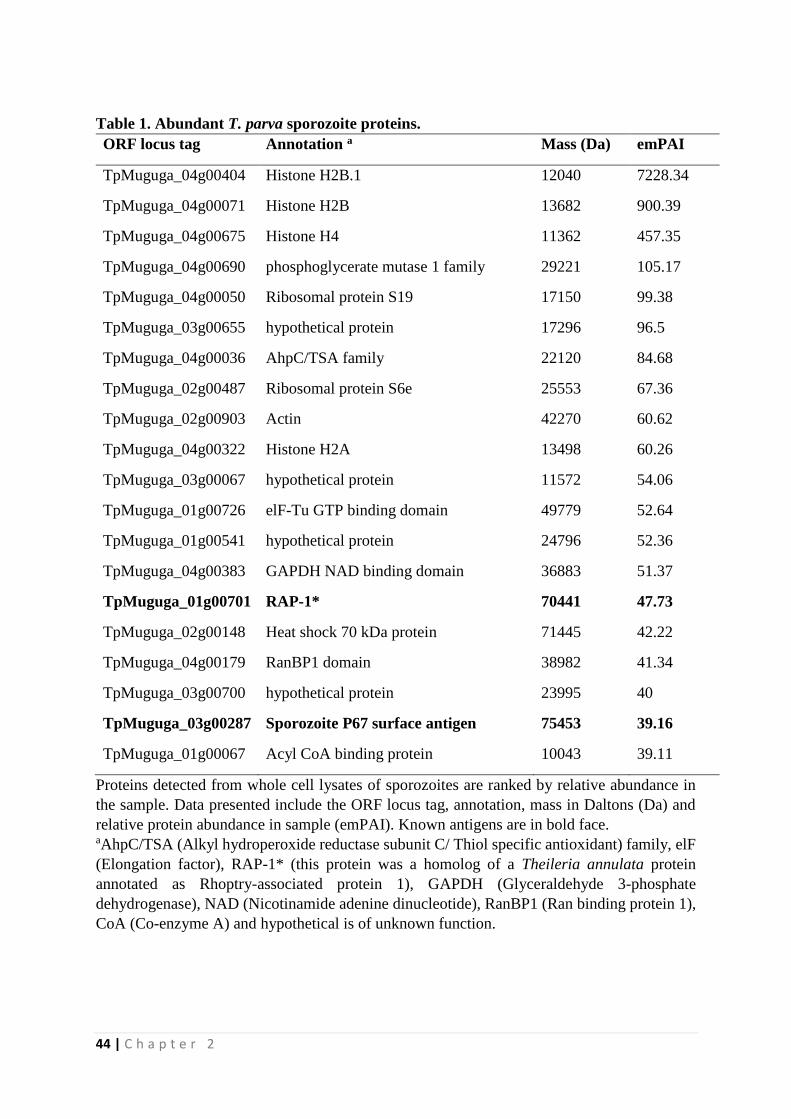
44 | C h a p t e r 2
Table 1. Abundant T. parva sporozoite proteins.
ORF locus tag Annotation a Mass (Da) emPAI
TpMuguga_04g00404 Histone H2B.1 12040 7228.34
TpMuguga_04g00071 Histone H2B 13682 900.39
TpMuguga_04g00675 Histone H4 11362 457.35
TpMuguga_04g00690 phosphoglycerate mutase 1 family 29221 105.17
TpMuguga_04g00050 Ribosomal protein S19 17150 99.38
TpMuguga_03g00655 hypothetical protein 17296 96.5
TpMuguga_04g00036 AhpC/TSA family 22120 84.68
TpMuguga_02g00487 Ribosomal protein S6e 25553 67.36
TpMuguga_02g00903 Actin 42270 60.62
TpMuguga_04g00322 Histone H2A 13498 60.26
TpMuguga_03g00067 hypothetical protein 11572 54.06
TpMuguga_01g00726 elF-Tu GTP binding domain 49779 52.64
TpMuguga_01g00541 hypothetical protein 24796 52.36
TpMuguga_04g00383 GAPDH NAD binding domain 36883 51.37
TpMuguga_01g00701 RAP-1* 70441 47.73
TpMuguga_02g00148 Heat shock 70 kDa protein 71445 42.22
TpMuguga_04g00179 RanBP1 domain 38982 41.34
TpMuguga_03g00700 hypothetical protein 23995 40
TpMuguga_03g00287 Sporozoite P67 surface antigen 75453 39.16
TpMuguga_01g00067 Acyl CoA binding protein 10043 39.11
Proteins detected from whole cell lysates of sporozoites are ranked by relative abundance in
the sample. Data presented include the ORF locus tag, annotation, mass in Daltons (Da) and
relative protein abundance in sample (emPAI). Known antigens are in bold face. aAhpC/TSA (Alkyl hydroperoxide reductase subunit C/ Thiol specific antioxidant) family, elF
(Elongation factor), RAP-1* (this protein was a homolog of a Theileria annulata protein
annotated as Rhoptry-associated protein 1), GAPDH (Glyceraldehyde 3-phosphate
dehydrogenase), NAD (Nicotinamide adenine dinucleotide), RanBP1 (Ran binding protein 1),
CoA (Co-enzyme A) and hypothetical is of unknown function.
45 | C h a p t e r 2
T. parva sporozoite proteins associated with apicomplexan invasion of host cells
Sporozoite secretory organelle proteins are typically located in the micronemes, rhoptries
and dense granules in P. falciparum (reviewed in Lindner, Swearingen et al. 2013). To
determine whether these proteins are present in T. parva sporozoites, we clustered our
identified T. parva sporozoite proteins using OrthoMCL into orthologous groups with all P.
falciparum genes. A total of 1105 proteins of the 2,007 T. parva proteins were defined as P.
falciparum orthologs (Supplementary Table S 4) and we searched this list for apical organelle
proteins. We identified T. parva orthologs of eight rhoptry proteins, three microneme proteins
and a dense granule protein (Table 2). Featured in the list are orthologs of P. falciparum AMA-
1, a microneme protein essential during host cell invasion (Treeck, Zacherl et al. 2009), and
CelTOS, a malarial antigen mediating host cell invasion (Kariu, Ishino et al. 2006). AMA-1 is
a leading malaria vaccine candidate (Remarque, Faber et al. 2008), and orthologs of it are also
found in Neospora caninum and Toxoplasma gondii (Zhang, Compaore et al. 2007).
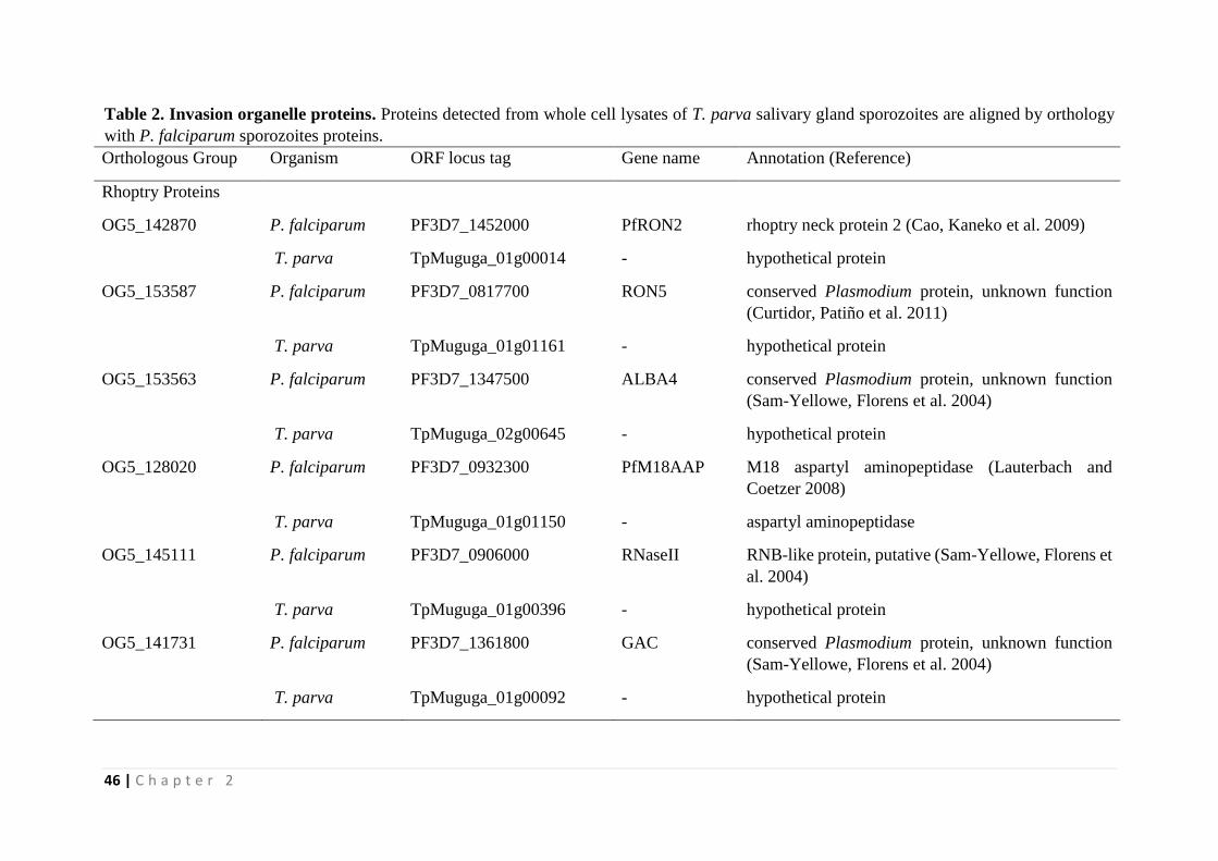
46 | C h a p t e r 2
Table 2. Invasion organelle proteins. Proteins detected from whole cell lysates of T. parva salivary gland sporozoites are aligned by orthology
with P. falciparum sporozoites proteins.
Orthologous Group Organism ORF locus tag Gene name Annotation (Reference)
Rhoptry Proteins
OG5_142870 P. falciparum PF3D7_1452000 PfRON2 rhoptry neck protein 2 (Cao, Kaneko et al. 2009)
T. parva TpMuguga_01g00014 - hypothetical protein
OG5_153587 P. falciparum PF3D7_0817700 RON5 conserved Plasmodium protein, unknown function
(Curtidor, Patiño et al. 2011)
T. parva TpMuguga_01g01161 - hypothetical protein
OG5_153563 P. falciparum PF3D7_1347500 ALBA4 conserved Plasmodium protein, unknown function
(Sam-Yellowe, Florens et al. 2004)
T. parva TpMuguga_02g00645 - hypothetical protein
OG5_128020 P. falciparum PF3D7_0932300 PfM18AAP M18 aspartyl aminopeptidase (Lauterbach and
Coetzer 2008)
T. parva TpMuguga_01g01150 - aspartyl aminopeptidase
OG5_145111 P. falciparum PF3D7_0906000 RNaseII RNB-like protein, putative (Sam-Yellowe, Florens et
al. 2004)
T. parva TpMuguga_01g00396 - hypothetical protein
OG5_141731 P. falciparum PF3D7_1361800 GAC conserved Plasmodium protein, unknown function
(Sam-Yellowe, Florens et al. 2004)
T. parva TpMuguga_01g00092 - hypothetical protein
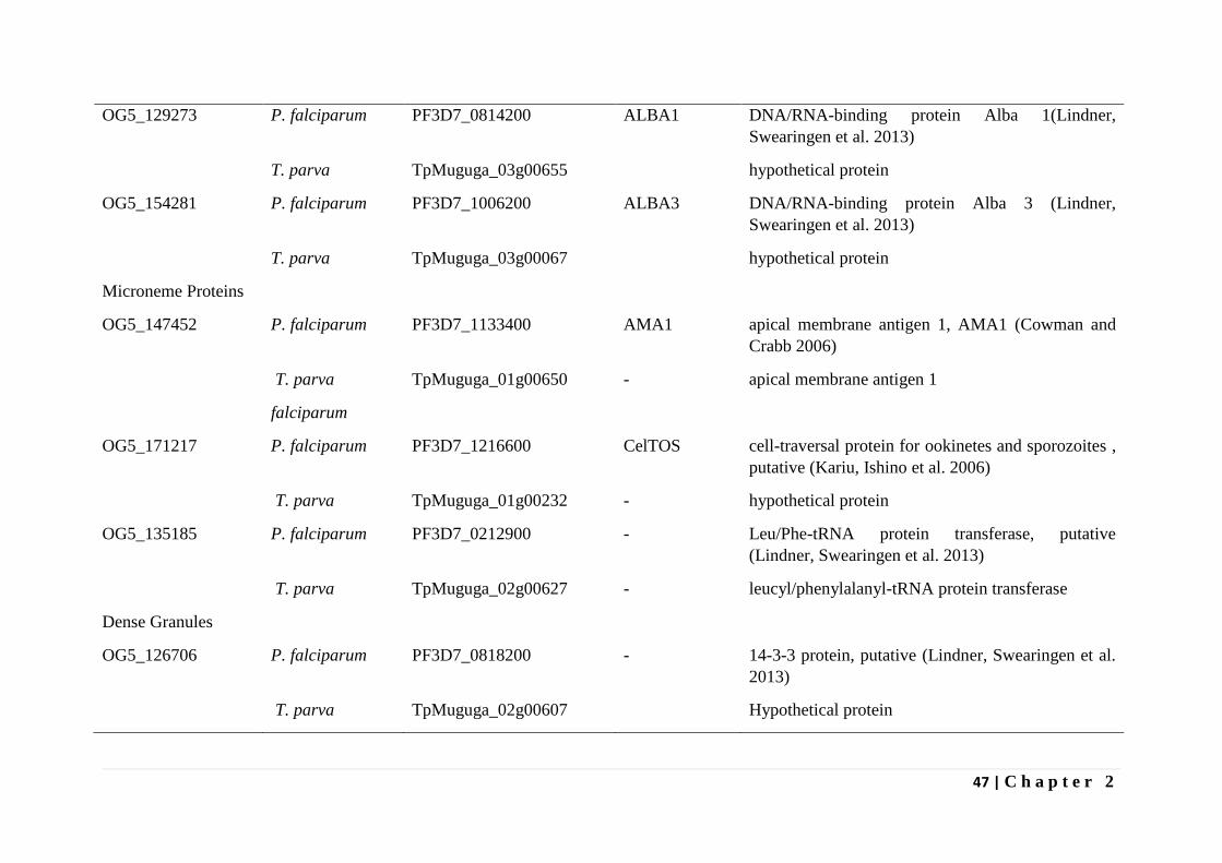
47 | C h a p t e r 2
OG5_129273 P. falciparum PF3D7_0814200 ALBA1 DNA/RNA-binding protein Alba 1(Lindner,
Swearingen et al. 2013)
T. parva TpMuguga_03g00655 hypothetical protein
OG5_154281 P. falciparum PF3D7_1006200 ALBA3 DNA/RNA-binding protein Alba 3 (Lindner,
Swearingen et al. 2013)
T. parva TpMuguga_03g00067 hypothetical protein
Microneme Proteins
OG5_147452 P. falciparum PF3D7_1133400 AMA1 apical membrane antigen 1, AMA1 (Cowman and
Crabb 2006)
T. parva TpMuguga_01g00650 - apical membrane antigen 1
falciparum
OG5_171217 P. falciparum PF3D7_1216600 CelTOS cell-traversal protein for ookinetes and sporozoites ,
putative (Kariu, Ishino et al. 2006)
T. parva TpMuguga_01g00232 - hypothetical protein
OG5_135185 P. falciparum PF3D7_0212900 - Leu/Phe-tRNA protein transferase, putative
(Lindner, Swearingen et al. 2013)
T. parva TpMuguga_02g00627 - leucyl/phenylalanyl-tRNA protein transferase
Dense Granules
OG5_126706 P. falciparum PF3D7_0818200 - 14-3-3 protein, putative (Lindner, Swearingen et al.
2013)
T. parva TpMuguga_02g00607 Hypothetical protein
48 | C h a p t e r 2
In T. parva, mobilization of Ca2+ is necessary for successful sporozoite invasion of bovine
lymphocytes and reagents that prevent Ca2+ mobilization significantly curb zippering and
internalization stages of the entry process (Shaw 1995). Table 3 shows 13 proteins identified
in T. parva clustered with the Plasmodium proteins that play a role in calcium signaling,
including calmodulin, an intracellular target for Ca2+ activation that acts on proteins such as
guanylyl cyclases, protein kinases and phosphatases to aid in signal transduction.
Putative T. parva surface proteins within the sporozoite proteome
We used two methods to search for novel putative surface proteins. The first method was
to use bioinformatics tools to predict the presence of GPI anchors and signal peptide (SP) in
proteins expressed by sporozoites. Only 12 proteins of the 2007 sporozoite proteins were found
in this category (Table 4). The list includes TpMuguga_01g00939 (gp34), a protein that
undergoes GPI modification when expressed in mammalian cells and a schizont surface protein
(Xue, von Schubert et al. 2010). Since there is no direct evidence for addition of GPI anchors
to parasite expressed proteins, we searched for enzymes of the GPI biosynthesis pathway
encoded by the T. parva genome. Eight genes are essential for GPI synthesis in P. falciparum
(Delorenzi, Sexton et al. 2002). We found homologs of all eight genes in T. parva
(TpMuguga_04g00759, TpMuguga_04g00881, TpMuguga_04g00525,
TpMuguga_01g00169, TpMuguga_02g00201, TpMuguga_03g00397, TpMuguga_03g00846
and TpMuguga_02g00741) and identified that the latter four are expressed in the sporozoite
proteome. This suggests the existence of GPI anchored proteins in T. parva, although this
remains to be formally proven.
Secondly, we compiled from the literature a list of surface-exposed P. falciparum proteins
and determined if orthologs exist within the T. parva sporozoite proteome. We identified
orthologs of AMA-1 (TpMuguga_01g00650) and a hexose transporter (TpMuguga_01g01069)
which were both identified by specific biotinylation of P. falciparum and Plasmodium yoelii
surface proteins (Lindner, Swearingen et al. 2013).
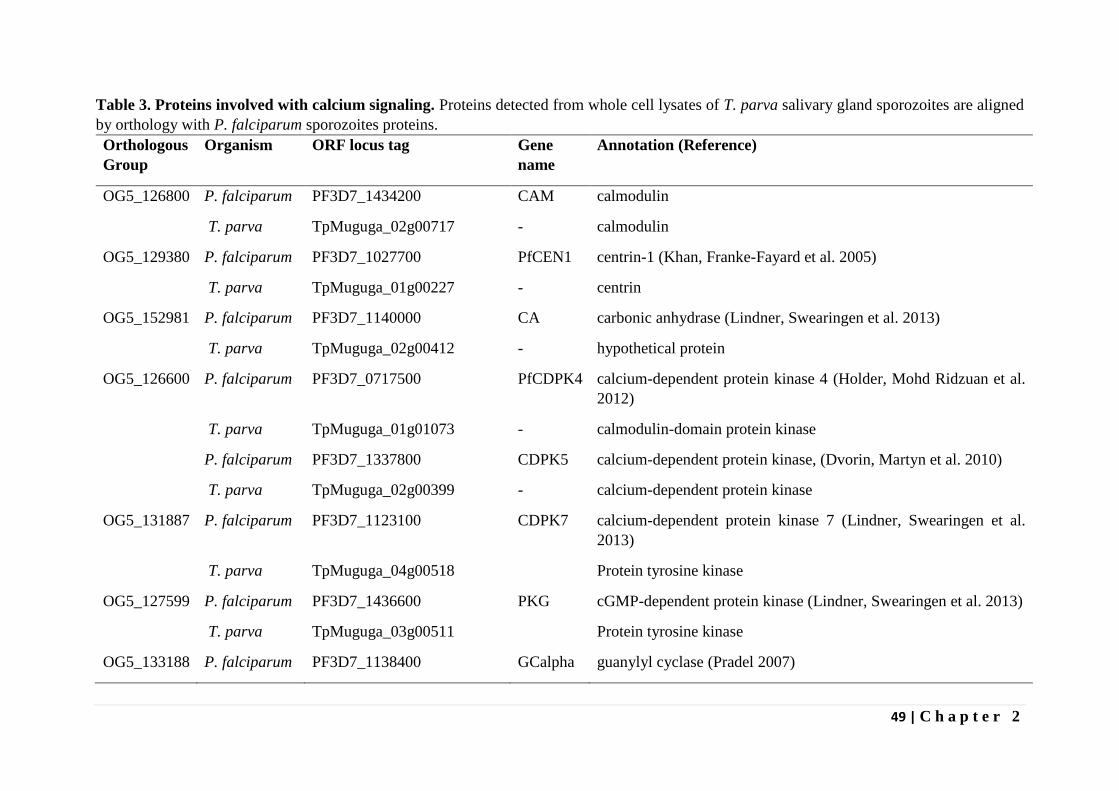
49 | C h a p t e r 2
Table 3. Proteins involved with calcium signaling. Proteins detected from whole cell lysates of T. parva salivary gland sporozoites are aligned
by orthology with P. falciparum sporozoites proteins.
Orthologous
Group
Organism ORF locus tag Gene
name
Annotation (Reference)
OG5_126800 P. falciparum PF3D7_1434200 CAM calmodulin
T. parva TpMuguga_02g00717 - calmodulin
OG5_129380 P. falciparum PF3D7_1027700 PfCEN1 centrin-1 (Khan, Franke-Fayard et al. 2005)
T. parva TpMuguga_01g00227 - centrin
OG5_152981 P. falciparum PF3D7_1140000 CA carbonic anhydrase (Lindner, Swearingen et al. 2013)
T. parva TpMuguga_02g00412 - hypothetical protein
OG5_126600 P. falciparum PF3D7_0717500 PfCDPK4 calcium-dependent protein kinase 4 (Holder, Mohd Ridzuan et al.
2012)
T. parva TpMuguga_01g01073 - calmodulin-domain protein kinase
P. falciparum PF3D7_1337800 CDPK5 calcium-dependent protein kinase, (Dvorin, Martyn et al. 2010)
T. parva TpMuguga_02g00399 - calcium-dependent protein kinase
OG5_131887
P. falciparum PF3D7_1123100 CDPK7 calcium-dependent protein kinase 7 (Lindner, Swearingen et al.
2013)
T. parva TpMuguga_04g00518 Protein tyrosine kinase
OG5_127599 P. falciparum PF3D7_1436600 PKG cGMP-dependent protein kinase (Lindner, Swearingen et al. 2013)
T. parva TpMuguga_03g00511 Protein tyrosine kinase
OG5_133188 P. falciparum PF3D7_1138400 GCalpha guanylyl cyclase (Pradel 2007)
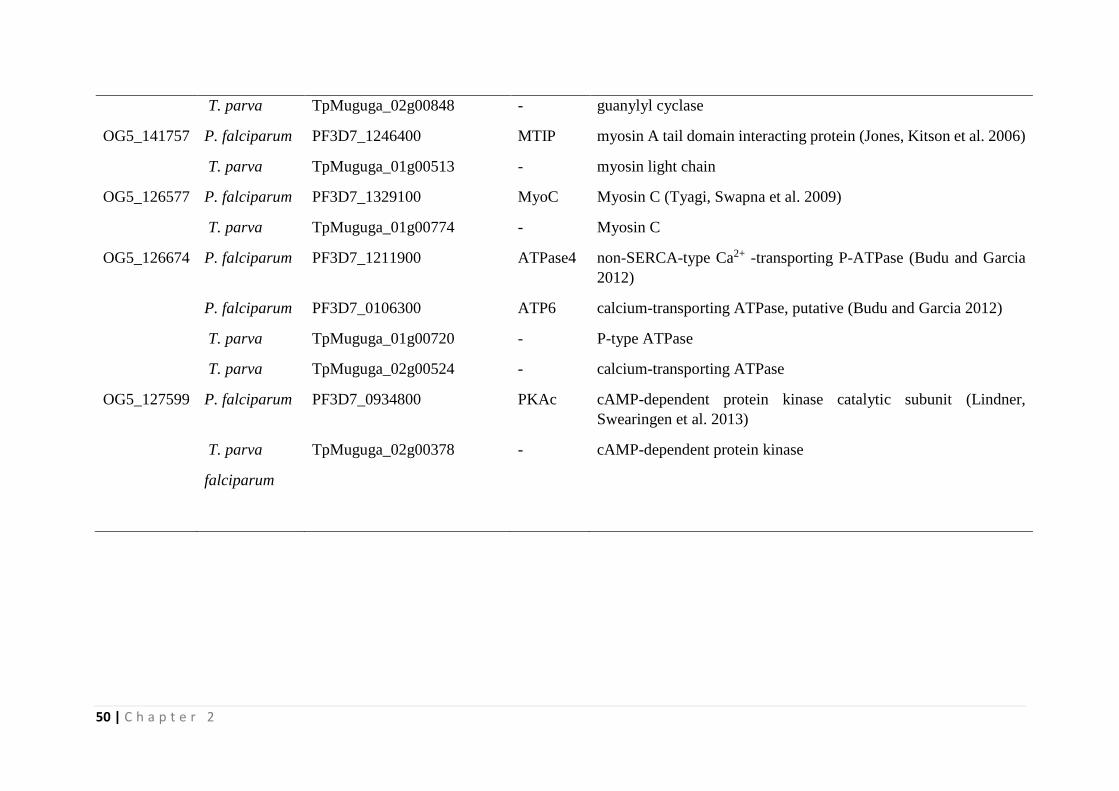
50 | C h a p t e r 2
T. parva TpMuguga_02g00848 - guanylyl cyclase
OG5_141757 P. falciparum PF3D7_1246400 MTIP myosin A tail domain interacting protein (Jones, Kitson et al. 2006)
T. parva TpMuguga_01g00513 - myosin light chain
OG5_126577 P. falciparum PF3D7_1329100 MyoC Myosin C (Tyagi, Swapna et al. 2009)
T. parva TpMuguga_01g00774 - Myosin C
OG5_126674 P. falciparum PF3D7_1211900
ATPase4 non-SERCA-type Ca2+ -transporting P-ATPase (Budu and Garcia
2012)
P. falciparum PF3D7_0106300 ATP6 calcium-transporting ATPase, putative (Budu and Garcia 2012)
T. parva TpMuguga_01g00720 - P-type ATPase
T. parva TpMuguga_02g00524 - calcium-transporting ATPase
OG5_127599 P. falciparum PF3D7_0934800 PKAc cAMP-dependent protein kinase catalytic subunit (Lindner,
Swearingen et al. 2013)
T. parva TpMuguga_02g00378 - cAMP-dependent protein kinase
falciparum
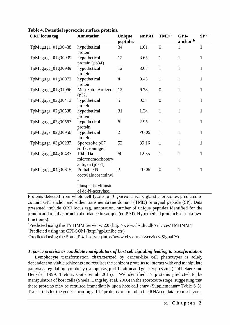
51 | C h a p t e r 2
Table 4. Potential sporozoite surface proteins.
ORF locus tag
Annotation Unique
peptides
emPAI TMD a GPI-
anchor b
SP c
TpMuguga_01g00438 hypothetical
protein
34 1.01 0 1 1
TpMuguga_01g00939 hypothetical
protein (gp34)
12 3.65 1 1 1
TpMuguga_01g00939 hypothetical
protein
12 3.65 1 1 1
TpMuguga_01g00972 hypothetical
protein
4 0.45 1 1 1
TpMuguga_01g01056 Merozoite Antigen
(p32)
12 6.78 0 1 1
TpMuguga_02g00412 hypothetical
protein
5 0.3 0 1 1
TpMuguga_02g00538 hypothetical
protein
31 1.34 1 1 1
TpMuguga_02g00553 hypothetical
protein
6 2.95 1 1 1
TpMuguga_02g00950 hypothetical
protein
2 <0.05 1 1 1
TpMuguga_03g00287 Sporozoite p67
surface antigen
53 39.16 1 1 1
TpMuguga_04g00437 104 kDa
microneme/rhoptry
antigen (p104)
60 12.35 1 1 1
TpMuguga_04g00615 Probable N-
acetylglucosaminyl
-
phosphatidylinosit
ol de-N-acetylase
2 <0.05 0 1 1
Proteins detected from whole cell lysates of T. parva salivary gland sporozoites predicted to
contain GPI anchor and either transmembrane domain (TMD) or signal peptide (SP). Data
presented include ORF locus tag, annotation, number of unique peptides identified for the
protein and relative protein abundance in sample (emPAI). Hypothetical protein is of unknown
function(s). aPredicted using the TMHMM Server v. 2.0 (http://www.cbs.dtu.dk/services/TMHMM/)
bPredicted using the GPI-SOM (http://gpi.unibe.ch/)
cPredicted using the SignalP 4.1 server (http://www.cbs.dtu.dk/services/SignalP/).
T. parva proteins as candidate manipulators of host cell signaling leading to transformation
Lymphocyte transformation characterized by cancer-like cell phenotypes is solely
dependent on viable schizonts and requires the schizont proteins to interact with and manipulate
pathways regulating lymphocyte apoptosis, proliferation and gene expression (Dobbelaere and
Heussler 1999, Tretina, Gotia et al. 2015). We identified 17 proteins predicted to be
manipulators of host cells (Shiels, Langsley et al. 2006) in the sporozoite stage, suggesting that
these proteins may be required immediately upon host cell entry (Supplementary Table S 5).
Transcripts for the genes encoding all 17 proteins are found in the RNAseq data from schizont-
52 | C h a p t e r 2
infected cells (see Materials and Methods), although to different levels (data not shown).
Among the identified proteins are members of two multigene families, the Sub-telomere-
encoded Variable Secreted Protein (SVSP) gene family and the Theileria annulata schizont
AT-hook (TashAT) gene family, or T. parva host nuclear (TpHN) gene family. TashAT
proteins display a high degree of sequence conservation in their DNA-binding domains (AT-
hook) in T. annulata but not in T. parva and contain an N-terminal signal sequence for transport
across the parasite plasma membrane (reviewed in Tretina, Gotia et al. 2015). TashAT family
proteins are implicated in regulation of host gene expression and as accessory factors for
parasite transcription (Shiels, Langsley et al. 2006). Other families featured are DEAD-box
RNA helicases with members implicated in alteration of RNA secondary structures and
promotion of proto-oncogene c-myc expression (e.g., DDX6) (Abdelhaleem 2004),
thioredoxin/glutaredoxin family that are redox regulators and influence important signaling
pathways such as NF-kB and AP1 pathways that are both activated in Theileria-transformed
cells (Shiels, Langsley et al. 2006). In addition, we also identified T. parva Schizont-derived
Cytoskeleton-binding Protein (TpSCOP), a protein that activates host NF-κB and MAPK
pathways, leading to resistance to programmed host cell death (Hayashida, Hattori et al. 2010)
and a peptidyl-prolyl isomerase, which has been demonstrated to interact with and lead to
degradation of host ubiquitin ligase FBW7, thereby stabilizing host c-JUN and promoting
transformation of host cells (Marsolier, Perichon et al. 2015). Interestingly, the bulk schizont
RNAseq data identified a high level of anti-sense transcription of the TpSCOP gene (data not
shown), suggesting that expression of this gene in the schizont stage may be under differential
control. Resolution of the significance of this observation will require analysis of cell-cycle
synchronized samples.
Sporozoite proteins involved in glycolysis and tricarboxylic acid (TCA) cycle
Proteins of the glycolytic pathway are conserved in most eukaryotic organisms and 10
genes encoding glycolytic enzymes have been found in Theileria genomes (Gardner, Bishop
et al. 2005, Pain, Renauld et al. 2005). We identified expression of all the 10 enzymes of the
glycolytic pathway in this study (Figure 1) including lactate dehydrogenase, an enzyme that
converts pyruvate, the last product of glycolysis, to lactate during low or no oxygen conditions.
We also identified all the TCA cycle enzymes by mass spectrometry (Figure 1) with the
exception that malate dehydrogenase is functionally replaced by malate-quinone
oxidoreductase (Gardner, Bishop et al. 2005), and was identified in the proteome data.
Glutamate dehydrogenase was also identified in the proteome and glutamate has been
suggested to be a supplementary intermediate for the TCA cycle (Gardner, Bishop et al. 2005).
The only functional link we have found between glycolysis and the TCA cycle is the presence
of phosphoenolpyruvate carboxykinase. Neither glycerol kinase nor glycerol 3-phosphate
dehydrogenase were identified, although T. parva encodes genes for both enzymes. These
enzymes were identified with low levels in the schizont stage of T. annulata (Witschi, Xia et
al. 2013).
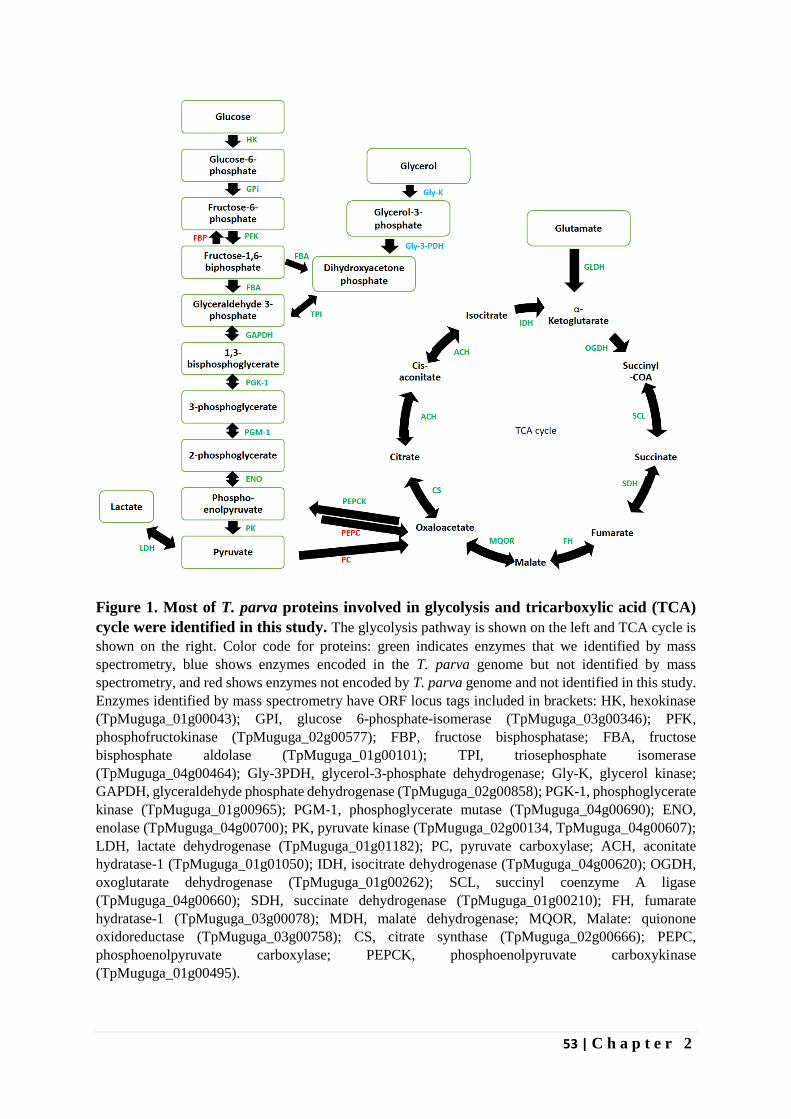
53 | C h a p t e r 2
Figure 1. Most of T. parva proteins involved in glycolysis and tricarboxylic acid (TCA)
cycle were identified in this study. The glycolysis pathway is shown on the left and TCA cycle is
shown on the right. Color code for proteins: green indicates enzymes that we identified by mass
spectrometry, blue shows enzymes encoded in the T. parva genome but not identified by mass
spectrometry, and red shows enzymes not encoded by T. parva genome and not identified in this study.
Enzymes identified by mass spectrometry have ORF locus tags included in brackets: HK, hexokinase
(TpMuguga_01g00043); GPI, glucose 6-phosphate-isomerase (TpMuguga_03g00346); PFK,
phosphofructokinase (TpMuguga_02g00577); FBP, fructose bisphosphatase; FBA, fructose
bisphosphate aldolase (TpMuguga_01g00101); TPI, triosephosphate isomerase
(TpMuguga_04g00464); Gly-3PDH, glycerol-3-phosphate dehydrogenase; Gly-K, glycerol kinase;
GAPDH, glyceraldehyde phosphate dehydrogenase (TpMuguga_02g00858); PGK-1, phosphoglycerate
kinase (TpMuguga_01g00965); PGM-1, phosphoglycerate mutase (TpMuguga_04g00690); ENO,
enolase (TpMuguga_04g00700); PK, pyruvate kinase (TpMuguga_02g00134, TpMuguga_04g00607);
LDH, lactate dehydrogenase (TpMuguga_01g01182); PC, pyruvate carboxylase; ACH, aconitate
hydratase-1 (TpMuguga_01g01050); IDH, isocitrate dehydrogenase (TpMuguga_04g00620); OGDH,
oxoglutarate dehydrogenase (TpMuguga_01g00262); SCL, succinyl coenzyme A ligase
(TpMuguga_04g00660); SDH, succinate dehydrogenase (TpMuguga_01g00210); FH, fumarate
hydratase-1 (TpMuguga_03g00078); MDH, malate dehydrogenase; MQOR, Malate: quionone
oxidoreductase (TpMuguga_03g00758); CS, citrate synthase (TpMuguga_02g00666); PEPC,
phosphoenolpyruvate carboxylase; PEPCK, phosphoenolpyruvate carboxykinase
(TpMuguga_01g00495).
54 | C h a p t e r 2
Tick proteins
Searching the Uniprot database entries for R. appendiculatus with the LC MS/MS data led
to identification of 2,773 tick proteins in addition to the T. parva proteins. The complete list of
all the identified tick proteins is presented in Supplementary Table S 1, but a detailed analysis
of vector proteins will be reported in Chapter 3.
55 | C h a p t e r 2
Discussion
In this study, we employed a DE-52 ion-exchange column purification method to semi-
purify T. parva sporozoites from infected tick salivary glands. Although tick proteins
constituted 58% (2773/4780) of the total number of proteins of all proteins identified by LC-
MS/MS and the Peaks and Mascot software packages, we were able to identify 2,007 T. parva
proteins expressed in the sporozoite life-cycle stage. This represents ~50% of the total predicted
re-annotated T. parva proteome of 4084 proteins (see Materials and methods). About 40% of
these proteins are annotated as hypothetical proteins. Hence, our data provides evidence that
real genes encode these proteins, although their function remains unknown.
Studies on the proteome of P. falciparum sporozoite stages have reported identification of
~13% (Lasonder, Janse et al. 2008), ~19% (Florens, Washburn et al. 2002) and ~36% (Lindner,
Swearingen et al. 2013) of the 5,524 annotated P. falciparum genes using materials purified by
DEAE-cellulose chromatography only (Florens, Washburn et al. 2002, Lasonder, Janse et al.
2008), and on 17% w/v Accudenz cushion followed by DEAE-cellulose chromatography
(Lindner, Swearingen et al. 2013). The range of vector contamination is an important
component of such studies and varied depending on the method of parasite purification.
Lasonder et al (2008) reported 65% to 89% mosquito protein contamination in the samples
analyzed, while Lindner et al (2013) reported 60.1% mosquito proteins in the sample, but had
to do double purification, initially on a 17% w/v Accudenz cushion then followed by DEAE-
cellulose chromatography to reduce mosquito protein contamination to 29% (Lasonder, Janse
et al. 2008, Lindner, Swearingen et al. 2013). The presence of mosquito material in the sample
was not discussed by Florens and colleagues (Florens, Washburn et al. 2002). We managed to
achieve low levels of vector protein contamination with a single DE-52 column purification
step.
Positive identification of proteins by mass spectrometry confers confidence in their
presence, especially when the proteins are identified by more than one unique peptide hit. 1,708
of the 2,007 parasite proteins were identified by more than one hit, providing a high degree of
confidence in 85% of the data. For the purposes of our current analyses we have, however,
reported on the entire data set of 2007 proteins. Failure to identify T. parva proteins does not
directly imply their absence in the sample. Peptides from proteins with low abundance may fall
below the detection limit or are simply not sequenced by the mass spectrometer due to limited
dynamic range and/or speed of instrument acquisition. Furthermore, unknown, complex, or
amino acid modifications leading to alteration of the peptide mass can only be detected in some
cases by the Peaks software. Finally, trypsin digestion of proteins with low or irregular
arginine/lysine content can lead to very short and very long tryptic peptides that fall out of the
acquired mass range and may therefore not be detected.
Identification of T. parva sporozoite proteins through mass spectrometry has allowed us to
assess the proteomic component of the mammalian-infective stage of the parasite. We have
also calculated the emPAI using Mascot to estimate the relative abundance ranking of proteins
in the sample, which revealed a very high range of abundance with histones ranked with an
emPAI of 60~7200 for the different histone subunits to an emPAI index of 39 for the major
circumsporozoite p67 protein. Subunits of two enzymes within the glycolytic pathway,
phosphoglycerate mutase and glyceraldehyde 3-phosphate dehydrogenase, are among the 20
most abundant sporozoite proteins detected (Table 1). A high abundance of histones is not
unusual. Lindner et al (2013) reported histones as the third most abundant protein in
56 | C h a p t e r 2
Plasmodium sporozoite proteome after the circumsporozoite protein and mitochondrial ATP
synthase, subunit beta (Lindner, Swearingen et al. 2013).
A number of known antigens were identified within the sporozoite proteome, including
p67, a leading vaccine antigen, and previously described components of secretory organelles
(e.g., p104 and p150) (Table 4). RAP-1, a rhoptry antigen protective against P. falciparum
infection in Saimiri monkeys (Ridley, Takacs et al. 1990) was also identified (Table 1).
Unexpectedly, the polymorphic immuno-dominant molecule (PIM), a well-known antigen
(Toye, Goddeeris et al. 1991), was not identified in this study, even though p150, an antigen
that is immunologically cross-reactive with PIM (Skilton, Bishop et al. 1998) was identified.
The PIM protein sequence contains a large repetitive domain rich in glutamine and proline
amino acid residues that is devoid of lysine and arginine residues (Toye, Goddeeris et al. 1991).
An in silico digestion of PIM with trypsin leads to prediction of few detectable tryptic peptides
(data not shown). This may explain why we did not detect it. All schizont proteins that have
been identified as CTL antigens are also expressed in sporozoites (emPAI range of 0.76 to
31.69). Some of these antigens are encoded by housekeeping proteins (e.g., hsp90) and
translation elongation initiation factor 1A, so expression by sporozoites of some CTL antigens
is not unexpected. The finding that all the known CTL antigens are expressed by sporozoites
raises the question of whether infection of dendritic cells by sporozoites results in direct
presentation of antigen for priming CTL responses in vivo, provided the antigen load is
sufficient.
A study on the proteome of T. annulata schizont stage, in which 798 proteins were
identified, failed to identify any member of SVSP and TashAT families (Witschi, Xia et al.
2013). In contrast, we identified members of both families in the sporozoite stage
(Supplementary Table S 5). There is increasing evidence pointing to a role of these proteins in
host-parasite interaction, for instance SVSP epitope-tagged TpMuguga_03g00882 protein
expressed in mammalian cells was found to translocate into the host nucleus pointing to
potential role in transforming host cells (Schmuckli-Maurer, Casanova et al. 2009). In addition,
we identified other predicted host manipulation proteins such as DEAD-box RNA helicases,
peptidases and two proteins, namely a peptidyl prolyl-isomerase and TpSCOP, demonstrated
to play role in host manipulation by stabilizing c-JUN and activation of NF-kB respectively
(Shiels, Langsley et al. 2006, Hayashida, Hattori et al. 2010, Marsolier, Perichon et al. 2015).
Host cell manipulation is known to occur at the schizont stage and the finding of these proteins
in the sporozoite stage suggests early expression of these proteins may be required, or multi-
functionality of the proteins.
Sporozoite invasion of lymphocytes begins with binding events that trigger the
mobilization of Ca2+ within the sporozoites (Dobbelaere and Heussler 1999). The free
intracellular Ca2+ can be utilized by calmodulin to remodel itself structurally so as to act on
downstream effectors like CDPKs, which have been shown to be useful for parasite infectivity.
We detected both calmodulin and CDPKs in this study (Table 3). We have also detected other
calcium signaling proteins including guanylyl cyclases, cGMP-dependent protein kinases and
cAMP-dependent protein kinases, all of which are important for downstream calcium signaling
and have been shown to be essential for sporozoite invasion (reviewed in Dobbelaere and
Heussler 1999).
In motile apicomplexans such as Plasmodium, free calcium induces microneme secretion
by sporozoites to aid in gliding motility and host invasion (Lindner, Swearingen et al. 2013).
Although, T. parva sporozoites are immotile (Shaw 2003), we identified orthologs of
57 | C h a p t e r 2
Plasmodium microneme components, including AMA-1, CelTOS and Leu/Phe-tRNA protein
transferase (Cowman and Crabb 2006, Kariu, Ishino et al. 2006, Lindner, Swearingen et al.
2013). These proteins may be involved solely in invasion processes together with the detected
rhoptry proteins (PfRON2, Conserved Plasmodium protein, PfM18AAP and RNB-like protein)
and dense granules protein (14-3-3 protein) (Sam-Yellowe, Florens et al. 2004, Lauterbach and
Coetzer 2008, Cao, Kaneko et al. 2009, Curtidor, Patiño et al. 2011, Lindner, Swearingen et al.
2013). Since most of these proteins are still classified as hypothetical in T. parva, they present
an opportunity for further analysis in studies aiming to block parasite invasion of host cells.
In conclusion, these data establish the expression profile of T. parva sporozoite proteins,
the only such profile available to date. One or more of the newly identified proteins, in
particular putative surface proteins, may prove to be effective vaccine candidates and might be
combined with the major surface protein p67 to induce broader anti-sporozoite immunity, and
protection against ECF.
Acknowledgement
We are grateful to Stephen Mwaura and Milton Owido for providing the dissected tick salivary
glands. We thank the CGIAR Research Program on Livestock and Fish, the Norman Borlaug
Commemorative Research Initiative, an initiative between the Feed the Future program of
USAID and USDA-ARS (58-5348-2-117F) and the Department for International Development
of the United Kingdom and the Bill and Melinda Gates Foundation (OPP1078791) for financial
support. Mass spectrometry analysis was performed in the Target Discovery Institute Mass
Spectrometry Laboratory led by Benedikt M. Kessler.
Conflicts of interest
The authors declare no conflict of interest.
58 | C h a p t e r 2
References
Abdelhaleem, M., 2004. Do human RNA helicases have a role in cancer? Biochim. Biophys.
Acta 1704, 37-46.
Anamika, Srinivasan, N., Krupa, A., 2005. A genomic perspective of protein kinases in
Plasmodium falciparum. Proteins: Struct., Funct., Bioinf. 58, 180-189.
Aurrecoechea, C., Brestelli, J., Brunk, B.P., Fischer, S., Gajria, B., Gao, X., Gingle, A., Grant,
G., Harb, O.S., Heiges, M., Innamorato, F., Iodice, J., Kissinger, J.C., Kraemer, E.T., Li, W.,
Miller, J.A., Nayak, V., Pennington, C., Pinney, D.F., Roos, D.S., Ross, C., Srinivasamoorthy,
G., Stoeckert, J.C.J., Thibodeau, R., Treatman, C., Wang, H., 2010. EuPathDB: a portal to
eukaryotic pathogen databases. Nucleic Acids Res. 38, D415-D419.
Ballingall, K.T., Mwangi, D.M., MacHugh, N.D., Taracha, E.L.N., Totte, P., McKeever, D.J.,
2000. A highly sensitive, non-radioactive assay for T cell activation in cattle: applications in
screening for antigens recognised by CD4+ and CD8+ T cells. J. Immunol. Methods 239, 85-
93.
Brown, W.C., McElwain, T.F., Palmer, G.H., Chantler, S.E., Estes, D.M., 1999. Bovine CD4+
T-Lymphocyte Clones Specific for Rhoptry-Associated Protein 1 of Babesia bigemina
Stimulate Enhanced Immunoglobulin G1 (IgG1) and IgG2 Synthesis. Infect. Immun. 67, 155-
164.
Brown, W.C., Sugimoto, C., Grab, D.J., 1989. Theileria parva: Bovine helper T cell clones
specific for both infected lymphocytes and schizont membrane antigens. Exp. Parasitol. 69,
234-248.
Brown, W.C., Zhao, S., Logan, K.S., Grab, D.J., Rice-Ficht, A.C., 1995. Identification of
candidate vaccine antigens of bovine hemoparasites Theileria parva and Babesia bovis by use
of helper T cell clones. Vet. Parasitol. 57, 189-203.
Budu, A., Garcia, C.R.S., 2012. Generation of second messengers in Plasmodium. Microb.
Infect. 14, 787-795.
Büscher, G., Tangus, J., 1986. Quantitative studies on Theileria parva in the salivary glands of
Rhipicephalus appendiculatus adults: Search for conditions for high infections. Int. J. Parasitol.
16, 121-129.
Cao, J., Kaneko, O., Thongkukiatkul, A., Tachibana, M., Otsuki, H., Gao, Q., Tsuboi, T., Torii,
M., 2009. Rhoptry neck protein RON2 forms a complex with microneme protein AMA1 in
Plasmodium falciparum merozoites. Parasitol. Int. 58, 29-35.
Cowman, A.F., Crabb, B.S., 2006. Invasion of Red Blood Cells by Malaria Parasites. Cell 124,
755-766.
59 | C h a p t e r 2
Curtidor, H., Patiño, L.C., Arévalo-Pinzón, G., Patarroyo, M.E., Patarroyo, M.A., 2011.
Identification of the Plasmodium falciparum rhoptry neck protein 5 (PfRON5). Gene 474, 22-
28.
Delorenzi, M., Sexton, A., Shams-Eldin, H., Schwarz, R.T., Speed, T., Schofield, L., 2002.
Genes for Glycosylphosphatidylinositol Toxin Biosynthesis in Plasmodium falciparum. Infect.
Immun. 70, 4510-4522.
Dessauge, F., Lizundia, R., Baumgartner, M., Chaussepied, M., Langsley, G., 2005. Taking the
Myc is bad for Theileria. Trends Parasitol. 21, 377-385.
Di Giulio, G., Lynen, G., Morzaria, S., Oura, C., Bishop, R., 2009. Live immunization against
East Coast fever – current status. Trends Parasitol. 25, 85-92.
Dobbelaere, D., Heussler, V., 1999. Transformation of Leukocytes by Theileria parva and T.
annulata. Annu. Rev. Microbiol. 53, 1-42.
Dobbelaere, D., Shapiro, S.Z., Webster, P., 1985. Identification of a surface antigen on
Theileria parva sporozoites by monoclonal antibody. Proc. Natl. Acad. Sci. U. S. A. 82, 1771-
1775.
Dobbelaere, D.A.E., Küenzi, P., 2004. The strategies of the Theileria parasite: a new twist in
host–pathogen interactions. Curr. Opin. Immunol. 16, 524-530.
Dobbelaere, D.A.E., Spooner, P.R., Barry, W.C., Irvin, A.D., 1984. Monoclonal antibody
neutralizes the sporozoite stage of different Theileria parva stocks. Parasite Immunol. 6, 361-
370.
Dvorin, J.D., Martyn, D.C., Patel, S.D., Grimley, J.S., Collins, C.R., Hopp, C.S., Bright, A.T.,
Westenberger, S., Winzeler, E., Blackman, M.J., Baker, D.A., Wandless, T.J., Duraisingh,
M.T., 2010. A Plant-Like Kinase in Plasmodium falciparum Regulates Parasite Egress from
Erythrocytes. Science 328, 910-912.
Ebel, T., Gerhards, J., Binder, B.R., Lipp, J., 1999. Theileria parva 104 kDa microneme–
rhoptry protein is membrane-anchored by a non-cleaved amino-terminal signal sequence for
entry into the endoplasmic reticulum. Mol. Biochem. Parasitol. 100, 19-26.
Emanuelsson, O., Nielsen, H., Brunak, S., von Heijne, G., 2000. Predicting Subcellular
Localization of Proteins Based on their N-terminal Amino Acid Sequence. J. Mol. Biol. 300,
1005-1016.
Fankhauser, N., Mäser, P., 2005. Identification of GPI anchor attachment signals by a Kohonen
self-organizing map. Bioinformatics 21, 1846-1852.
60 | C h a p t e r 2
Fawcett, D.W., Doxsey, S., Stagg, D.A., Young, A.S., 1982b. The entry of sporozoites of
Theileria parva into bovine lymphocytes in vitro. Electron microscopic observations. Eur. J.
Cell Biol. 27, 10-21.
Florens, L., Washburn, M.P., Raine, J.D., Anthony, R.M., Grainger, M., Haynes, J.D., Moch,
J.K., Muster, N., Sacci, J.B., Tabb, D.L., Witney, A.A., Wolters, D., Wu, Y., Gardner, M.J.,
Holder, A.A., Sinden, R.E., Yates, J.R., Carucci, D.J., 2002. A proteomic view of the
Plasmodium falciparum life cycle. Nature 419, 520-526.
Gardner, M.J., Bishop, R., Shah, T., de Villiers, E.P., Carlton, J.M., Hall, N., Ren, Q., Paulsen,
I.T., Pain, A., Berriman, M., Wilson, R.J.M., Sato, S., Ralph, S.A., Mann, D.J., Xiong, Z.,
Shallom, S.J., Weidman, J., Jiang, L., Lynn, J., Weaver, B., Shoaibi, A., Domingo, A.R.,
Wasawo, D., Crabtree, J., Wortman, J.R., Haas, B., Angiuoli, S.V., Creasy, T.H., Lu, C., Suh,
B., Silva, J.C., Utterback, T.R., Feldblyum, T.V., Pertea, M., Allen, J., Nierman, W.C.,
Taracha, E.L.N., Salzberg, S.L., White, O.R., Fitzhugh, H.A., Morzaria, S., Venter, J.C.,
Fraser, C.M., Nene, V., 2005. Genome Sequence of Theileria parva, a Bovine Pathogen That
Transforms Lymphocytes. Science 309, 134-137.
Graham, S.P., Honda, Y., Pellé, R., Mwangi, D.M., Glew, E.J., de Villiers, E.P., Shah, T.,
Bishop, R., van der Bruggen, P., Nene, V., Taracha, E.L.N., 2007. A novel strategy for the
identification of antigens that are recognised by bovine MHC class I restricted cytotoxic T cells
in a protozoan infection using reverse vaccinology. Immunome Res 3, 2-2.
Graham, S.P., Pelle, R., Honda, Y., Mwangi, D.M., Tonukari, N.J., Yamage, M., Glew, E.J.,
de Villiers, E.P., Shah, T., Bishop, R., Abuya, E., Awino, E., Gachanja, J., Luyai, A.E.,
Mbwika, F., Muthiani, A.M., Ndegwa, D.M., Njahira, M., Nyanjui, J.K., Onono, F.O., Osaso,
J., Saya, R.M., Wildmann, C., Fraser, C.M., Maudlin, I., Gardner, M.J., Morzaria, S.P.,
Loosmore, S., Gilbert, S.C., Audonnet, J.C., van der Bruggen, P., Nene, V., Taracha, E.L.,
2006. Theileria parva candidate vaccine antigens recognized by immune bovine cytotoxic T
lymphocytes. Proc. Natl. Acad. Sci. USA 103, 3286-3291.
Graham, S.P., Pellé, R., Yamage, M., Mwangi, D.M., Honda, Y., Mwakubambanya, R.S., de
Villiers, E.P., Abuya, E., Awino, E., Gachanja, J., Mbwika, F., Muthiani, A.M., Muriuki, C.,
Nyanjui, J.K., Onono, F.O., Osaso, J., Riitho, V., Saya, R.M., Ellis, S.A., McKeever, D.J.,
MacHugh, N.D., Gilbert, S.C., Audonnet, J.-C., Morrison, W.I., van der Bruggen, P., Taracha,
E.L.N., 2008. Characterization of the Fine Specificity of Bovine CD8 T-Cell Responses to
Defined Antigens from the Protozoan Parasite Theileria parva. Infect. Immun. 76, 685-694.
Hayashida, K., Hara, Y., Abe, T., Yamasaki, C., Toyoda, A., Kosuge, T., Suzuki, Y., Sato, Y.,
Kawashima, S., Katayama, T., Wakaguri, H., Inoue, N., Homma, K., Tada-Umezaki, M., Yagi,
Y., Fujii, Y., Habara, T., Kanehisa, M., Watanabe, H., Ito, K., Gojobori, T., Sugawara, H.,
Imanishi, T., Weir, W., Gardner, M., Pain, A., Shiels, B., Hattori, M., Nene, V., Sugimoto, C.,
2012. Comparative Genome Analysis of Three Eukaryotic Parasites with Differing Abilities
To Transform Leukocytes Reveals Key Mediators of Theileria-Induced Leukocyte
Transformation. mBio 3.
61 | C h a p t e r 2
Hayashida, K., Hattori, M., Nakao, R., Tanaka, Y., Kim, J.-Y., Inoue, N., Nene, V., Sugimoto,
C., 2010. A schizont-derived protein, TpSCOP, is involved in the activation of NF-κB in
Theileria parva-infected lymphocytes. Mol. Biochem. Parasitol. 174, 8-17.
Holder, A.A., Mohd Ridzuan, M.A., Green, J.L., 2012. Calcium dependent protein kinase 1
and calcium fluxes in the malaria parasite. Microb. Infect. 14, 825-830.
Hopp, C.S., Bowyer, P.W., Baker, D.A., 2012. The role of cGMP signalling in regulating life
cycle progression of Plasmodium. Microb. Infect. 14, 831-837.
Ishihama, Y., Oda, Y., Tabata, T., Sato, T., Nagasu, T., Rappsilber, J., Mann, M., 2005.
Exponentially Modified Protein Abundance Index (emPAI) for Estimation of Absolute Protein
Amount in Proteomics by the Number of Sequenced Peptides per Protein. Mol. Cell.
Proteomics 4, 1265-1272.
Jones, M.L., Kitson, E.L., Rayner, J.C., 2006. Plasmodium falciparum erythrocyte invasion: A
conserved myosin associated complex. Mol. Biochem. Parasitol. 147, 74-84.
Kariu, T., Ishino, T., Yano, K., Chinzei, Y., Yuda, M., 2006. CelTOS, a novel malarial protein
that mediates transmission to mosquito and vertebrate hosts. Mol. Microbiol. 59, 1369-1379.
Khan, S.M., Franke-Fayard, B., Mair, G.R., Lasonder, E., Janse, C.J., Mann, M., Waters, A.P.,
2005. Proteome Analysis of Separated Male and Female Gametocytes Reveals Novel Sex-
Specific Plasmodium Biology. Cell 121, 675-687.
Krogh, A., Larsson, B., von Heijne, G., Sonnhammer, E.L.L., 2001. Predicting transmembrane
protein topology with a hidden markov model: application to complete genomes1. J. Mol. Biol.
305, 567-580.
Lasonder, E., Janse, C.J., van Gemert, G.-J., Mair, G.R., Vermunt, A.M.W., Douradinha, B.G.,
van Noort, V., Huynen, M.A., Luty, A.J.F., Kroeze, H., Khan, S.M., Sauerwein, R.W., Waters,
A.P., Mann, M., Stunnenberg, H.G., 2008. Proteomic Profiling of Plasmodium Sporozoite
Maturation Identifies New Proteins Essential for Parasite Development and Infectivity. PLoS
Pathog 4, e1000195.
Lauterbach, S.B., Coetzer, T.L., 2008. The M18 aspartyl aminopeptidase of Plasmodium
falciparum binds to human erythrocyte spectrin in vitro. Malaria Journal 7, 1-10.
Li, L., Stoeckert, C.J., Roos, D.S., 2003. OrthoMCL: Identification of Ortholog Groups for
Eukaryotic Genomes. Genome Res. 13, 2178-2189.
Lindner, S.E., Swearingen, K.E., Harupa, A., Vaughan, A.M., Sinnis, P., Moritz, R.L., Kappe,
S.H., 2013. Total and putative surface proteomics of malaria parasite salivary gland
sporozoites. Mol. Cell. Proteomics 12, 1127-1143.
62 | C h a p t e r 2
Marsolier, J., Perichon, M., DeBarry, J.D., Villoutreix, B.O., Chluba, J., Lopez, T., Garrido,
C., Zhou, X.Z., Lu, K.P., Fritsch, L., Ait-Si-Ali, S., Mhadhbi, M., Medjkane, S., Weitzman,
J.B., 2015. Theileria parasites secrete a prolyl isomerase to maintain host leukocyte
transformation. Nature 520, 378-382.
Morzaria, S., Nene, V., Bishop, R., Musoke, A., 2000. Vaccines against Theileria parva. Ann.
N. Y. Acad. Sci. 916, 464-473.
Morzaria, S.P., Katende, J., Musoke, A., Nene, V., Skilton, R., Bishop, R., 1999. Development
of sero-diagnostic and molecular tools for the control of important tick-borne pathogens of
cattle in Africa. Parassitologia 41 Suppl 1, 73-80.
Musoke, A., Morzaria, S., Nkonge, C., Jones, E., Nene, V., 1992. A recombinant sporozoite
surface antigen of Theileria parva induces protection in cattle. Proc. Natl. Acad. Sci. U. S. A.
89, 514-518.
Nene, V., Kiara, H., Lacasta, A., Pelle, R., Svitek, N., Steinaa, L., 2016. The biology of
Theileria parva and control of East Coast fever – Current status and future trends. Ticks Tick
Borne Dis 7, 549-564.
Nene, V., Morrison, W.I., 2016. Approaches to vaccination against Theileria parva and
Theileria annulata. Parasite Immunol. 38, 724-734.
Norval, R.A.I., Perry, B.D., Young, A., 1992. The epidemiology of theileriosis in Africa. ILRI
(aka ILCA and ILRAD).
Odongo, D.O., Sunter, J.D., Kiara, H.K., Skilton, R.A., Bishop, R.P., 2010. A nested PCR
assay exhibits enhanced sensitivity for detection of Theileria parva infections in bovine blood
samples from carrier animals. Parasitol. Res. 106, 357-365.
Pain, A., Renauld, H., Berriman, M., Murphy, L., Yeats, C.A., Weir, W., Kerhornou, A., Aslett,
M., Bishop, R., Bouchier, C., Cochet, M., Coulson, R.M.R., Cronin, A., de Villiers, E.P.,
Fraser, A., Fosker, N., Gardner, M., Goble, A., Griffiths-Jones, S., Harris, D.E., Katzer, F.,
Larke, N., Lord, A., Maser, P., McKellar, S., Mooney, P., Morton, F., Nene, V., Neil, S., Price,
C., Quail, M.A., Rabbinowitsch, E., Rawlings, N.D., Rutter, S., Saunders, D., Seeger, K., Shah,
T., Squares, R., Squares, S., Tivey, A., Walker, A.R., Woodward, J., Dobbelaere, D.A.E.,
Langsley, G., Rajandream, M.-A., McKeever, D., Shiels, B., Tait, A., Barrell, B., Hall, N.,
2005. Genome of the Host-Cell Transforming Parasite Theileria annulata Compared with T.
parva. Science 309, 131.
Patarroyo, M.E., Alba, M.P., Curtidor, H., 2011. Biological and structural characteristics of the
binding peptides from the sporozoite proteins essential for cell traversal (SPECT)-1 and -2.
Peptides 32, 154-160.
Patel, E., Mwaura, S., Kiara, H., Morzaria, S., Peters, A., Toye, P., 2016. Production and dose
determination of the Infection and Treatment Method (ITM) Muguga cocktail vaccine used to
control East Coast fever in cattle. Ticks Tick Borne Dis 7, 306-314.
63 | C h a p t e r 2
Petersen, T.N., Brunak, S., von Heijne, G., Nielsen, H., 2011. SignalP 4.0: discriminating
signal peptides from transmembrane regions. Nat Meth 8, 785-786.
Pradel, G., 2007. Proteins of the malaria parasite sexual stages: expression, function and
potential for transmission blocking strategies. Parasitology 134, 1911-1929.
Radley, D.E., Brown, C.G.D., Burridge, M.J., Cunningham, M.P., Kirimi, I.M., Purnell, R.E.,
Young, A.S., 1975a. East coast fever: 1. Chemoprophylactic immunization of cattle against
Theileria parva (Muguga) and five theilerial strains. Vet. Parasitol. 1, 35-41.
Remarque, E.J., Faber, B.W., Kocken, C.H.M., Thomas, A.W., 2008. Apical membrane
antigen 1: a malaria vaccine candidate in review. Trends Parasitol. 24, 74-84.
Ridley, R.G., Takacs, B., Etlinger, H., Scaife, J.G., 1990. A rhoptry antigen of Plasmodium
falciparum is protective in Saimiri monkeys. Parasitology 101, 187-192.
Sam-Yellowe, T.Y., Florens, L., Wang, T., Raine, J.D., Carucci, D.J., Sinden, R., Yates, J.R.,
2004. Proteome Analysis of Rhoptry-Enriched Fractions Isolated from Plasmodium
Merozoites. J. Proteome Res. 3, 995-1001.
Schirmer, E.C., Yates, J.R., 3rd, Gerace, L., 2003. MudPIT: A powerful proteomics tool for
discovery. Discov Med 3, 38-39.
Schmuckli-Maurer, J., Casanova, C., Schmied, S., Affentranger, S., Parvanova, I., Kang'a, S.,
Nene, V., Katzer, F., McKeever, D., Müller, J., Bishop, R., Pain, A., Dobbelaere, D.A.E., 2009.
Expression Analysis of the Theileria parva Subtelomere-Encoded Variable Secreted Protein
Gene Family. PLoS ONE 4, e4839.
Shapiro, S.Z., Fujisaki, K., Morzaria, S.P., Webster, P., Fujinaga, T., Spooner, P.R., Irvin,
A.D., 1987. A life-cycle stage-specific antigen of Theileria parva recognized by anti-
macroschizont monoclonal antibodies. Parasitology 94, 29-37.
Shaw, M.K., 1995. Mobilization of intrasporozoite Ca2+ is essential for Theileria parva
sporozoite invasion of bovine lymphocytes. Eur. J. Cell Biol. 68, 78-87.
Shaw, M.K., 2003. Cell invasion by Theileria sporozoites. Trends Parasitol. 19, 2-6.
Shaw, M.K., Tilney, L.G., McKeever, D.J., 1993. Tick salivary gland extract and interleukin-
2 stimulation enhance susceptibility of lymphocytes to infection by Theileria parva
sporozoites. Infect. Immun. 61, 1486-1495.
Shiels, B., Langsley, G., Weir, W., Pain, A., McKellar, S., Dobbelaere, D., 2006. Alteration of
host cell phenotype by Theileria annulata and Theileria parva: mining for manipulators in the
parasite genomes. Int. J. Parasitol. 36, 9-21.
64 | C h a p t e r 2
Skilton, R.A., Bishop, R.P., Wells, C.W., Spooner, P.R., Gobright, E., Nkonge, C., Musoke,
A.J., Macklin, M., Iams, K.P., 1998. Cloning and characterization of a 150 kDa microsphere
antigen of Theileria parva that is immunologically cross-reactive with the polymorphic
immunodominant molecule (PIM). Parasitology 117 ( Pt 4), 321-330.
Skilton, R.A., Musoke, A.J., Wells, C.W., Yagi, Y., Nene, V., Spooner, P.R., Gachanja, J.,
Osaso, J., Bishop, R.P., Morzaria, S.P., 2000. A 32 kDa surface antigen of Theileria parva:
characterization and immunization studies. Parasitology 120 ( Pt 6), 553-564.
Toye, P.G., Goddeeris, B.M., Iams, K., Musoke, A.J., Morrison, W.I., 1991. Characterization
of a polymorphic immunodominant molecule in sporozoites and schizonts of Theileria parva.
Parasite Immunol. 13, 49-62.
Treeck, M., Zacherl, S., Herrmann, S., Cabrera, A., Kono, M., Struck, N.S., Engelberg, K.,
Haase, S., Frischknecht, F., Miura, K., Spielmann, T., Gilberger, T.W., 2009. Functional
Analysis of the Leading Malaria Vaccine Candidate AMA-1 Reveals an Essential Role for the
Cytoplasmic Domain in the Invasion Process. PLoS Pathog 5, e1000322.
Tretina, K., Gotia, H.T., Mann, D.J., Silva, J.C., 2015. Theileria-transformed bovine leukocytes
have cancer hallmarks. Trends Parasitol. 31, 306-314.
Tretina, K., Pelle, R., Silva, J.C., 2016. Cis regulatory motifs and antisense transcriptional
control in the apicomplexan Theileria parva. BMC Genomics 17, 128.
Tyagi, N., Swapna, L.S., Mohanty, S., Agarwal, G., Gowri, V.S., Anamika, K., Priya, M.L.,
Krishnadev, O., Srinivasan, N., 2009. Evolutionary Divergence of Plasmodium falciparum:
Sequences, Protein- Protein Interactions, Pathways and Processes. Infect Disord Drug Targets
9, 257-271.
Witschi, M., Xia, D., Sanderson, S., Baumgartner, M., Wastling, J.M., Dobbelaere, D.A.E.,
2013. Proteomic analysis of the Theileria annulata schizont. Int. J. Parasitol. 43, 173-180.
Xue, G., von Schubert, C., Hermann, P., Peyer, M., Maushagen, R., Schmuckli-Maurer, J.,
Bütikofer, P., Langsley, G., Dobbelaere, D.A.E., 2010. Characterisation of gp34, a GPI-
anchored protein expressed by schizonts of Theileria parva and T. annulata. Mol. Biochem.
Parasitol. 172, 113-120.
Zhang, H., Compaore, M.K., Lee, E.-g., Liao, M., Zhang, G., Sugimoto, C., Fujisaki, K.,
Nishikawa, Y., Xuan, X., 2007. Apical membrane antigen 1 is a cross-reactive antigen between
Neospora caninum and Toxoplasma gondii, and the anti-NcAMA1 antibody inhibits host cell
invasion by both parasites. Mol. Biochem. Parasitol. 151, 205-212.
65 | C h a p t e r 3
CHAPTER
3
Characterization of Rhipicephalus appendiculatus salivary
glands proteins co-purified with Theileria parva
sporozoites
James Nyagwange
Edwin Tijhaar
Nicola Ternette
Milcah Kigoni
Roger Pelle
Vishvanath Nene
Manuscript in preparation
66 | C h a p t e r 3
Abstract
Ticks are obligate hematophagous arthropods and are vectors of many pathogens
including parasites, viruses and bacteria. The brown ear tick Rhipicephalus appendiculatus is
the major vector of Theileria parva, an apicomplexan parasite that causes a lymphoproliferative
disease in cattle known as East Coast fever (ECF). Sporozoites, the bovine infective stage of
T. parva, are harbored in the salivary glands of R. appendiculatus and are transmitted to the
bovine host during tick feeding. Tick salivary glands and saliva contain proteins that facilitate
tick feeding by enhancing attachment and dampening of bovine host defenses. Therefore, these
proteins are targets for development of anti-tick vaccines, which block attachment or
compromise tick feeding, which subsequently results in poor reproductive fitness and limited
or no pathogen transmission. We have characterized the salivary gland proteome of R.
appendiculatus ticks infected by T. parva sporozoites using LC-MS/MS. In total 4,780 proteins
were identified, of which 2,773 were R. appendiculatus proteins and 2,007 were of parasite
origin. The R. appendiculatus proteins identified include known R. appendiculatus salivary
glands antigens such as Rhipicephalus Immuno-dominant Molecule 36 (RIM 36) and orthologs
of proteins playing roles in host defense modulation including, calreticulins, ixodegrins and
histamine binding proteins, among others . Some of the novel tick proteins identified that are
associated or attached to the sporozoites could induce antibodies which opsonize the
sporozoites leading to destruction of the parasite through phagocytosis or complement lysis.
Keywords:
Rhipicephalus appendiculatus, salivary glands, proteomics, vaccine antigens
67 | C h a p t e r 3
Introduction
Ticks are obligate blood sucking arachnids and vectors of viruses, bacteria and protozoa
which, when transmitted to vertebrate hosts, may cause disease (Cupp 1991). Ticks acquire
pathogens from infected blood and pathogens are transmitted by a trans-stadial route, i.e.,
where infection is acquired by one life-cycle stage and transmitted by the next tick instar, or a
trans-ovarial route, which gives rise to infectious larvae upon hatching of infected eggs
(Norval, Perry et al. 1992) Tick infection is usually followed by pathogen replication, although
passive transfer of pathogens can also occur (Norval, Perry et al. 1992). Tick saliva is the major
route for pathogen transmission and release of pathogens from infected salivary glands
coincides with excretion of excess water acquired during feeding (Sauer, Essenberg et al.
2000). Components of the tick saliva may enhance infectivity of the pathogens by regulating
host hemostasis, immune and inflammatory responses (Sauer, Essenberg et al. 2000).
Furthermore tick proteins bound to the surface of the sporozoite might induce protective
antibodies resulting in activation of complement and enhanced phagocytosis and destruction
by macrophages. Thus, understanding the proteins involved in these processes, may be useful,
for example, in designing anti-sporozoite vaccines, transmission blocking vaccines, and
gaining a better understanding of tick biology.
Rhipicephalus appendiculatus is a three-host tick and the principal vector for the
transmission of Theileria parva, an apicomplexan parasite causing, a lymphoproliferative cattle
disease known as East Coast fever (ECF) (Theiler 1912). Transmission of T. parva occurs
trans-stadially by the nymph and the adult ticks after acquiring infections as either larvae or
nymphs (reviewed by Norval, Perry et al. 1992). After a blood meal the ticks are infected by
T. parva piroplasms, a life cycle stage of the parasite that resides inside red blood cells
(reviewed by Norval, Perry et al. 1992) . The piroplasms differentiates into gametes in the tick
gut post-ingestion. The macro- and micro-gametes then fuse forming zygotes, which enters the
gut epithelial cells and differentiates into motile kinetes, that are subsequently released into the
tick hemocoel (reviewed in Nene, Lee et al. 2004). The kinetes invade e-cells of type III acini
of the salivary glands and goes through final differentiation, with complete sporogony
occurring only after the tick molts into the next instar, a process essentially triggered by blood
feeding (Fawcett, Büscher et al. 1982). Release of mature mammalian infective sporozoites
from the salivary glands occurs as a trickle between day 4-8, with peak production on day 4
and 5, after initiation of feeding (reviewed in Nene, Lee et al. 2004, Shaw and Young 2009).
Control of ECF includes killing of R. appendiculatus by acaricide treatment and by
chemotherapy using drugs such as buparvaquone. However, both strategies have serious
limitations ranging from resistance of ticks to acaricides to concerns of T. parva developing
resistance to buparvaquone following resistance in Theileria annulata. There are also concerns
of toxic residues in food and in the environment following acaricide use. A live vaccine is
available and involves deliberate infection with sporozoites, followed by treatment with long
acting oxytetracycline (Radley, Brown et al. 1975). There are ongoing efforts to develop next
generation subunit vaccines based on what is known about the mechanism of immunity of this
live vaccine, including characterization of the sporozoite proteome for identification of
antigens and testing of novel and known antigens in different formulations for both humoral
and cellular immune responses (Nene, Kiara et al. 2016). Another area of research attracting
attention is the development of novel transmission blocking vaccines, which can be integrated
with the other methods to control ECF. Therefore, there is still need for identification of tick
antigens with cross-protective epitopes.
As an initiative to improve the understanding of the biology of T. parva, we recently
undertook a Multidimensional Protein Identification Technology (MudPIT) mass
spectrometry-based approach (reviewed in Schirmer, Yates et al. 2003) to characterize the
68 | C h a p t e r 3
proteome of T. parva sporozoites purified from tick salivary glands by ion-exchange
chromatography and precipitation (Nyagwange, Tijhaar et al. 2018), where we identified 2,007
parasite proteins. In the process, we also identified 2,773 R. appendiculatus proteins. Here, we
have employed bioinformatics tools to analyze these R. appendiculatus proteins to identify tick
salivary glands proteins associated with or bound to sporozoites as they may be involved in
infectivity and/ or transmission of the parasites. Moreover, the tick proteins bound to
sporozoites could be targets for opsonizing antibodies that may aid in the destruction of the
sporozoite.Therefore some of these tick proteins could represent potential ECF vaccine
candidates.
69 | C h a p t e r 3
Materials and methods
DE-52 column purification and LC-MS/MS analysis
The procedure for production of T. parva infected ticks has been described before (Patel,
Mwaura et al. 2016) and we have recently described the DE-52 column purification method
used for purification of sporozoites (Nyagwange, Tijhaar et al. 2018). Briefly, sporozoite
infected tick salivary glands were dissected, homogenized and lysate centrifugated at 1000
rpm, 5 min, 4oC, in a pre-chilled centrifuge before the resulting supernatant added onto the DE-
52 column bed and the sporozoites eluted by SP buffer (0.16M NaCl, 0.16M D-glucose, 0.16M
Na2HPO4, 0.16M Trisbase, pH 8.2) . The resulting cloudy suspension was centrifuged and the
resulting pellet re-suspended in SDS-PAGE sample buffer and denatured at 100°C for 5 min
then fractionated on 10% one-dimensional SDS-PAGE and stained with Coomassie Blue.
Trypsin digestion was performed in-gel and tryptic peptides analyzed by LC-MS/MS
(Nyagwange, Tijhaar et al. 2018).
Data Analysis
Protein identification and relative abundance
Raw MS/MS data files were analysed with the Peaks software (Bioinformatics solutions)
as described in (Nyagwange, Tijhaar et al. 2018). Briefly, a database containing the re-
annotated proteome of T. parva (4,084 proteins) and all Uniprot database entries for R.
appendiculatus (12,884 proteins, July 2016) was used for analysis and a peptide false discovery
rate (FDR) of 1.2% was applied.
Exponentially Modified Protein Abundance Index (emPAI) was calculated using Mascot
(Matrix Science) (Ishihama, Oda et al. 2005) and was used to rank the identified proteins by
their abundance in the sample.
Classification of the identified R. appendiculatus proteins by orthology
We clustered all protein coding sequences of R. appendiculatus with the predicted salivary
gland proteomes of Rhipicephalus microplus, Ambylomma variegatum, Rhipicephalus
pulchellus, Ixodes scapularis and Ixodes ricinus (Ribeiro, Alarcon-Chaidez et al. 2006,
Ribeiro, Anderson et al. 2011, Tan, Francischetti et al. 2015) into putative orthologous groups
using the OrthoMCL standalone software Version 2.0.2 (Li, Stoeckert et al. 2003). The blast
step of OrthoMCL was performed using an E-value cut-off of 1e-10, and minimum 70%
sequence identity over 75% sequence coverage.
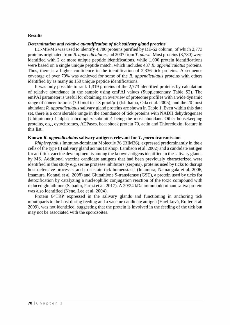
70 | C h a p t e r 3
Results
Determination and relative quantification of tick salivary gland proteins
LC-MS/MS was used to identify 4,780 proteins purified by DE-52 column, of which 2,773
proteins originated from R. appendiculatus and 2007 from T. parva. Most proteins (3,780) were
identified with 2 or more unique peptide identifications, while 1,000 protein identifications
were based on a single unique peptide match, which includes 437 R. appendiculatus proteins.
Thus, there is a higher confidence in the identification of 2,336 tick proteins. A sequence
coverage of over 70% was achieved for some of the R. appendiculatus proteins with others
identified by as many as 150 unique peptide identifications.
It was only possible to rank 1,319 proteins of the 2,773 identified proteins by calculation
of relative abundance in the sample using emPAI values (Supplementary Table S2). The
emPAI parameter is useful for obtaining an overview of proteome profiles with a wide dynamic
range of concentrations (30 fmol to 1.8 pmol/μl) (Ishihama, Oda et al. 2005), and the 20 most
abundant R. appendiculatus salivary gland proteins are shown in Table 1. Even within this data
set, there is a considerable range in the abundance of tick proteins with NADH dehydrogenase
(Ubiquinone) 1 alpha subcomplex subunit 4 being the most abundant. Other housekeeping
proteins, e.g., cytochromes, ATPases, heat shock protein 70, actin and Thioredoxin, feature in
this list.
Known R. appendiculatus salivary antigens relevant for T. parva transmission
Rhipicephalus Immuno-dominant Molecule 36 (RIM36), expressed predominantly in the e
cells of the type III salivary gland acinus (Bishop, Lambson et al. 2002) and a candidate antigen
for anti-tick vaccine development is among the known antigens identified in the salivary glands
by MS. Additional vaccine candidate antigens that had been previously characterized were
identified in this study e.g. serine protease inhibitors (serpins), proteins used by ticks to disrupt
host defensive processes and to sustain tick homeostasis (Imamura, Namangala et al. 2006,
Imamura, Konnai et al. 2008) and Glutathione S-transferase (GST), a protein used by ticks for
detoxification by catalyzing a nucleophilic conjugation reaction of the toxic compound with
reduced glutathione (Sabadin, Parizi et al. 2017). A 20/24 kDa immunodominant saliva protein
was also identified (Nene, Lee et al. 2004).
Protein 64TRP expressed in the salivary glands and functioning in anchoring tick
mouthparts to the host during feeding and a vaccine candidate antigen (Havlíková, Roller et al.
2009), was not identified, suggesting that the protein is involved in the feeding of the tick but
may not be associated with the sporozoites.
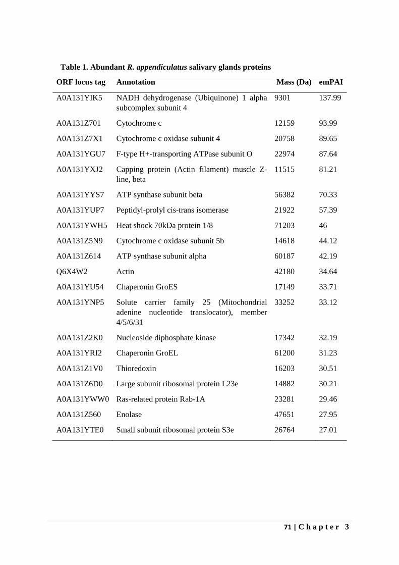
71 | C h a p t e r 3
Table 1. Abundant R. appendiculatus salivary glands proteins
ORF locus tag Annotation Mass (Da) emPAI
A0A131YIK5 NADH dehydrogenase (Ubiquinone) 1 alpha
subcomplex subunit 4
9301 137.99
A0A131Z701 Cytochrome c 12159 93.99
A0A131Z7X1 Cytochrome c oxidase subunit 4 20758 89.65
A0A131YGU7 F-type H+-transporting ATPase subunit O 22974 87.64
A0A131YXJ2 Capping protein (Actin filament) muscle Z-
line, beta
11515 81.21
A0A131YYS7 ATP synthase subunit beta 56382 70.33
A0A131YUP7 Peptidyl-prolyl cis-trans isomerase 21922 57.39
A0A131YWH5 Heat shock 70kDa protein 1/8 71203 46
A0A131Z5N9 Cytochrome c oxidase subunit 5b 14618 44.12
A0A131Z614 ATP synthase subunit alpha 60187 42.19
Q6X4W2 Actin 42180 34.64
A0A131YU54 Chaperonin GroES 17149 33.71
A0A131YNP5 Solute carrier family 25 (Mitochondrial
adenine nucleotide translocator), member
4/5/6/31
33252 33.12
A0A131Z2K0 Nucleoside diphosphate kinase 17342 32.19
A0A131YRI2 Chaperonin GroEL 61200 31.23
A0A131Z1V0 Thioredoxin 16203 30.51
A0A131Z6D0 Large subunit ribosomal protein L23e 14882 30.21
A0A131YWW0 Ras-related protein Rab-1A 23281 29.46
A0A131Z560 Enolase 47651 27.95
A0A131YTE0 Small subunit ribosomal protein S3e 26764 27.01
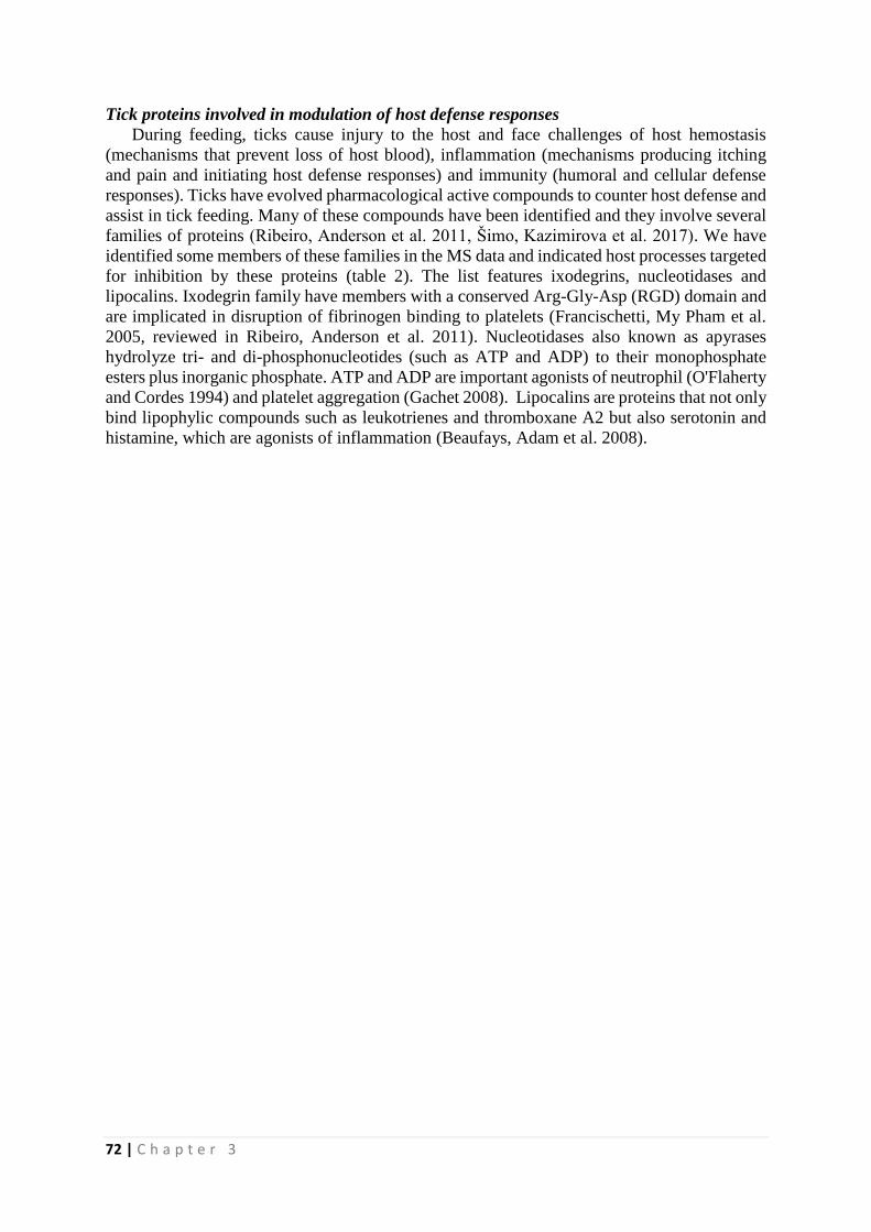
72 | C h a p t e r 3
Tick proteins involved in modulation of host defense responses
During feeding, ticks cause injury to the host and face challenges of host hemostasis
(mechanisms that prevent loss of host blood), inflammation (mechanisms producing itching
and pain and initiating host defense responses) and immunity (humoral and cellular defense
responses). Ticks have evolved pharmacological active compounds to counter host defense and
assist in tick feeding. Many of these compounds have been identified and they involve several
families of proteins (Ribeiro, Anderson et al. 2011, Šimo, Kazimirova et al. 2017). We have
identified some members of these families in the MS data and indicated host processes targeted
for inhibition by these proteins (table 2). The list features ixodegrins, nucleotidases and
lipocalins. Ixodegrin family have members with a conserved Arg-Gly-Asp (RGD) domain and
are implicated in disruption of fibrinogen binding to platelets (Francischetti, My Pham et al.
2005, reviewed in Ribeiro, Anderson et al. 2011). Nucleotidases also known as apyrases
hydrolyze tri- and di-phosphonucleotides (such as ATP and ADP) to their monophosphate
esters plus inorganic phosphate. ATP and ADP are important agonists of neutrophil (O'Flaherty
and Cordes 1994) and platelet aggregation (Gachet 2008). Lipocalins are proteins that not only
bind lipophylic compounds such as leukotrienes and thromboxane A2 but also serotonin and
histamine, which are agonists of inflammation (Beaufays, Adam et al. 2008).
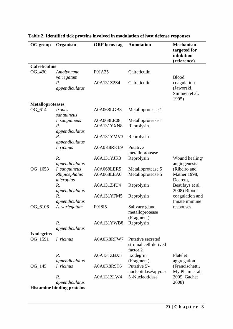
73 | C h a p t e r 3
Table 2. Identified tick proteins involved in modulation of host defense responses
OG group Organism ORF locus tag Annotation Mechanism
targeted for
inhibition
(reference)
Calreticulins
OG_430 Amblyomma
variegatum F0JA25 Calreticulin
Blood
coagulation
(Jaworski,
Simmen et al.
1995)
R.
appendiculatus
A0A131Z2S4 Calreticulin
Metalloproteases
OG_614 Ixodes
sanguineus
A0A068LGB8 Metalloprotease 1
Wound healing/
angiogenesis
(Ribeiro and
Mather 1998,
Decrem,
Beaufays et al.
2008) Blood
coagulation and
Innate immune
responses
I. sanguineus A0A068LE08 Metalloprotease 1
R.
appendiculatus
A0A131YXN8 Reprolysin
R.
appendiculatus
A0A131YMV3 Reprolysin
I. ricinus A0A0K8RKL9 Putative
metalloprotease
R.
appendiculatus
A0A131YJK3 Reprolysin
OG_1653 I. sanguineus A0A068LER5 Metalloprotease 5
Rhipicephalus
microplus
A0A068LEA0 Metalloprotease 5
R.
appendiculatus
A0A131Z4U4 Reprolysin
R.
appendiculatus
A0A131YFM5 Reprolysin
OG_6106 A. variegatum F0J8I5 Salivary gland
metalloprotease
(Fragment)
R.
appendiculatus
A0A131YWB8 Reprolysin
Ixodegrins
OG_1591 I. ricinus A0A0K8RFW7 Putative secreted
stromal cell-derived
factor 2
Platelet
aggregation
(Francischetti,
My Pham et al.
2005, Gachet
2008)
R.
appendiculatus
A0A131ZBX5 Ixodegrin
(Fragment)
OG_145 I. ricinus A0A0K8R9T6 Putative 5'-
nucleotidase/apyrase
R.
appendiculatus
A0A131Z1W4 5'-Nucleotidase
Histamine binding proteins
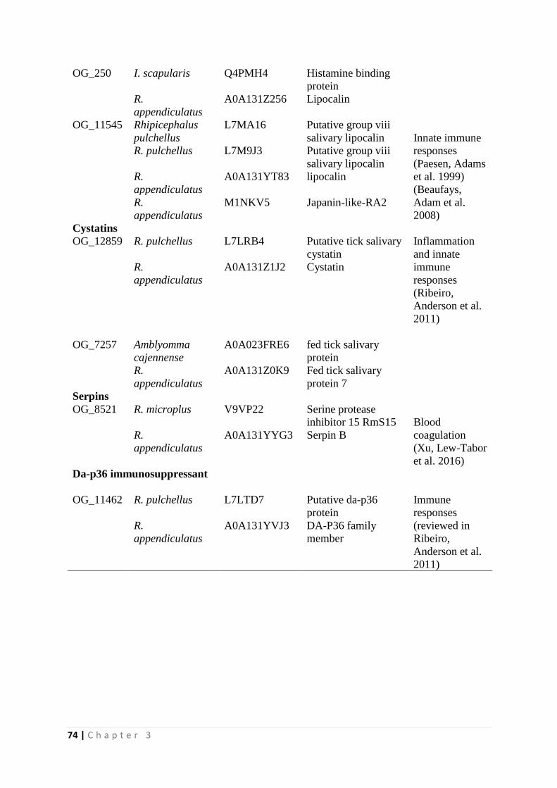
74 | C h a p t e r 3
OG_250 I. scapularis Q4PMH4 Histamine binding
protein
Innate immune
responses
(Paesen, Adams
et al. 1999)
(Beaufays,
Adam et al.
2008)
R.
appendiculatus
A0A131Z256 Lipocalin
OG_11545 Rhipicephalus
pulchellus
L7MA16 Putative group viii
salivary lipocalin
R. pulchellus L7M9J3 Putative group viii
salivary lipocalin
R.
appendiculatus
A0A131YT83 lipocalin
R.
appendiculatus
M1NKV5 Japanin-like-RA2
Cystatins
OG_12859 R. pulchellus L7LRB4 Putative tick salivary
cystatin
Inflammation
and innate
immune
responses
(Ribeiro,
Anderson et al.
2011)
R.
appendiculatus
A0A131Z1J2 Cystatin
OG_7257 Amblyomma
cajennense
A0A023FRE6 fed tick salivary
protein
R.
appendiculatus
A0A131Z0K9 Fed tick salivary
protein 7
Serpins
OG_8521 R. microplus V9VP22 Serine protease
inhibitor 15 RmS15
Blood
coagulation
(Xu, Lew-Tabor
et al. 2016)
R.
appendiculatus
A0A131YYG3 Serpin B
Da-p36 immunosuppressant
OG_11462 R. pulchellus L7LTD7 Putative da-p36
protein
Immune
responses
(reviewed in
Ribeiro,
Anderson et al.
2011)
R.
appendiculatus
A0A131YVJ3 DA-P36 family
member

75 | C h a p t e r 3
Discussion
The current study reports on R. appendiculatus proteins co-purified with T. parva
sporozoites. Although our initial aim was to identify T. parva sporozoites proteins from
infected tick salivary glands, tick proteins constituted 58% (2773/4780) of all the proteins
identified by LC-MS/MS and Peaks and Mascot software packages, in addition to the identified
2,007 parasite proteins which we have reported on elsewhere. Ideally the DE-52 is a positively
charged resin that should trap the negatively charged proteins and allow the positively charged
sporozoite cells to elute in the SP buffer used. However, abundant tick proteins, proteins that
are not negatively charged or those tightly associated with sporozoites can still end up in the
flow-through fraction. The following centrifugation of the flow through fraction should
eliminate the soluble tick proteins from the sporozoite fraction leaving tick proteins bound to
sporozoites, insoluble uncharged and positively charged tick proteins. We have searched this
list of tick proteins co-purified with the sporozoites to identify proteins that are likely to play a
role in sporozoite infection.
Positive identification of proteins by MS especially, by multiple unique peptides,
confers confidence in the presence of such proteins. A total of 2,088 of the 2,773 tick proteins
identified with more than a single unique peptide, giving confidence in 75% of the data. We
have however reported on the complete set of the 2,773 proteins. Although positive
identification confer confidence in the presence of the protein, failure to identify a protein does
not necessarily imply their absence in the sample.
We have calculated the emPAI using Mascot to estimate the relative abundance of
proteins in the sample. Although the list consisted of many housekeeping genes, such as NADH
dehydrogenase (Ubiquinone) 1 alpha subcomplex subunit 4, Cytochrome c oxidase subunit 4,
ATP synthases (alpha and beta subunits) and solute carrier family 25 (mitochondrial adenine
nucleotide translocator), member 4/5/6/31 (table 1), some proteins were identified that are
involved in immune modulation and therefore might be useful for sporozoite infection (table 2
and supplementary table S1). The salivary glands is a highly active organ requiring energy for
its functions such as maintenance of water and ion balance during blood feeding, formation of
cement for tick attachment to the host and secretion of pharmacological armament modulating
host processes (reviewed in Šimo, Kazimirova et al. 2017) and so high expression of
mitochondrial proteins is not surprising.
Targeting salivary proteins involved in tick feeding as vaccine antigens, is expected to
either lead to tick rejection, blocking/ limiting pathogen transmission or interfere with
completion of blood meal and compromising tick reproductive fitness (Šimo, Kazimirova et
al. 2017). Since tick proteins can induce host immune responses, tick proteins associated with
sporozoites can also be targeted as anti-sporozoite vaccines. Such a vaccine would function on
the basis of antibodies induced by the respective sporozoite bound tick antigen leading to
opsonization of the sporozoites, increased phagocytosis by macrophages and complement
dependent lysis. A number of known antigens were identified within the R. appendiculatus
salivary glands proteome including serpins, anti-tick vaccine candidates targeted for their role
in disrupting host haemostasis thereby facilitating tick feeding (Imamura, Namangala et al.
2006, Imamura, Konnai et al. 2008) and RIM36, a salivary gland protein and a component of
tick cement targeted for its role in tick attachment and protecting the tick hypostome during
feeding (Bishop, Lambson et al. 2002). However, a vaccine candidate antigen, 64TRP protein
expressed in the salivary glands (Havlíková, Roller et al. 2009) was not identified. Although,
an in silico 64TRP tryptic digestion leads to only few detectable peptides (data not shown), it
is also possible that this protein was efficiently bound by the column or is not bound to
sporozoites, hence not identified.
Hard ticks such as R. appendiculatus require extended periods of between 10 to 14 days to
feed to repletion (Cupp 1991). During the feeding process, the ticks inflict injuries to the host
76 | C h a p t e r 3
which triggers host defense and repair reactions including; platelet activation and aggregation,
blood clotting and vasocontraction (Ribeiro 1987). All these processes are aimed at expelling
the intruding tick. The intruding ticks respond by secreting numerous active substances in their
saliva to promote feeding by countering the host hemostatic and immune effector mechanisms
to maintain a steady blood flow to the feeding site (Ribeiro 1987). Host animals can naturally
acquire resistance to these salivary glands associated molecules following multiple tick
infestations or active immunization with crude tick extracts (reviewed in Mulenga, Sugimoto
et al. 2000). Therefore, tick salivary gland molecules are potential vaccine candidate antigens.
Many tick proteins inhibiting host coagulation cascade and modulating host immune responses
have been described but their molecular identities have not been elucidated (Mulenga,
Sugimoto et al. 2000). However, some have been characterized and have molecular identities
(Parizi, Githaka et al. 2012, Šimo, Kazimirova et al. 2017). We have identified known R.
appendiculatus antigens (section 3.2) and orthologs of other tick antigens known to modulate
host defenses (Table 2). The list features calreticulin, histamine binding proteins and
metalloproteases. Whereas calreticulin is implicated in inhibiting blood coagulation by
sequestering calcium ions which act as cofactors of the coagulation enzymes (Jaworski,
Simmen et al. 1995), histamine binding proteins and salivary metalloproteases bind histamine
and hydrolyze bradykinins respectively (Ribeiro and Mather 1998, Paesen, Adams et al. 1999).
Histamines and bradykinins produced by mast cells in the skin promote inflammation, pain and
itching at the site of tick intrusion and prevention of these processes interferes with downstream
recruitment of host immune cells against the invading tick (Šimo, Kazimirova et al. 2017).
In conclusion, these data confirm the expression of 2,773 proteins of R. appendiculatus
salivary glands infected by T. parva, the only such profile available to date. Although known
vaccine antigens targeting transmission blocking were identified, determining the specific
sporozoite bound proteins was not possible owing to the large number of tick proteins still
present. A method for further isolation of sporozoites from the mixture of tick proteins might
help in identifying the specific sporozoite bound proteins. Labelling sporozoites by monoclonal
antibodies against p67 followed by FACS sorting before LC-MS/MS can increase the
likelihood that the identified vector proteins are truly bound to the sporozoites. Nevertheless,
we have confirmed the expression and abundance of some of the previously identified
transmission blocking vaccine candidates and identified many orthologs of R. appendiculatus
antigens implicated in modulating host defenses in other ticks. Some of the latter may represent
novel antigens that could be used in transmission blocking anti-tick vaccines.
77 | C h a p t e r 3
References
Beaufays, J., B. Adam, C. Menten-Dedoyart, L. Fievez, A. Grosjean, Y. Decrem, P. P. Prevot,
S. Santini, R. Brasseur, M. Brossard, M. Vanhaeverbeek, F. Bureau, E. Heinen, L. Lins, L.
Vanhamme and E. Godfroid (2008). "Ir-LBP, an ixodes ricinus tick salivary LTB4-binding
lipocalin, interferes with host neutrophil function." PLoS One 3(12): e3987.
Bishop, R., B. Lambson, C. Wells, P. Pandit, J. Osaso, C. Nkonge, S. Morzaria, A. Musoke
and V. Nene (2002). "A cement protein of the tick Rhipicephalus appendiculatus, located in
the secretory e cell granules of the type III salivary gland acini, induces strong antibody
responses in cattle." International Journal for Parasitology 32(7): 833-842.
Cupp, E. W. (1991). "Biology of Ticks." Veterinary Clinics of North America: Small Animal
Practice 21(1): 1-26.
Decrem, Y., J. Beaufays, V. Blasioli, K. Lahaye, M. Brossard, L. Vanhamme and E. Godfroid
(2008). "A family of putative metalloproteases in the salivary glands of the tick Ixodes ricinus."
Febs j 275(7): 1485-1499.
Fawcett, D. W., G. Büscher and S. Doxsey (1982). "Salivary gland of the tick vector of east
coast fever. IV. Cell type selectivity and host cell responses to Theileria parva." Tissue and
Cell 14(2): 397-414.
Francischetti, I. M. B., V. My Pham, B. J. Mans, J. F. Andersen, T. N. Mather, R. S. Lane and
J. M. C. Ribeiro (2005). "The transcriptome of the salivary glands of the female western black-
legged tick Ixodes pacificus (Acari: Ixodidae)." Insect Biochemistry and Molecular Biology
35(10): 1142-1161.
Gachet, C. (2008). "P2 receptors, platelet function and pharmacological implications." Thromb
Haemost 99(3): 466-472.
Havlíková, S., L. Roller, J. Koči, A. R. Trimnell, M. Kazimírová, B. Klempa and P. A. Nuttall
(2009). "Functional role of 64P, the candidate transmission-blocking vaccine antigen from the
tick, Rhipicephalus appendiculatus." International Journal for Parasitology 39(13): 1485-1494.
Imamura, S., S. Konnai, S. Vaz Ida, S. Yamada, C. Nakajima, Y. Ito, T. Tajima, J. Yasuda, M.
Simuunza, M. Onuma and K. Ohashi (2008). "Effects of anti-tick cocktail vaccine against
Rhipicephalus appendiculatus." Jpn J Vet Res 56(2): 85-98.
Imamura, S., B. Namangala, T. Tajima, M. E. Tembo, J. Yasuda, K. Ohashi and M. Onuma
(2006). "Two serine protease inhibitors (serpins) that induce a bovine protective immune
response against Rhipicephalus appendiculatus ticks." Vaccine 24(13): 2230-2237.
Ishihama, Y., Y. Oda, T. Tabata, T. Sato, T. Nagasu, J. Rappsilber and M. Mann (2005).
"Exponentially Modified Protein Abundance Index (emPAI) for Estimation of Absolute
Protein Amount in Proteomics by the Number of Sequenced Peptides per Protein." Molecular
& Cellular Proteomics 4(9): 1265-1272.
78 | C h a p t e r 3
Jaworski, D. C., F. A. Simmen, W. Lamoreaux, L. B. Coons, M. T. Muller and G. R. Needham
(1995). "A secreted calreticulin protein in ixodid tick (Amblyomma americanum) saliva."
Journal of Insect Physiology 41(4): 369-375.
Li, L., C. J. Stoeckert and D. S. Roos (2003). "OrthoMCL: Identification of Ortholog Groups
for Eukaryotic Genomes." Genome Research 13(9): 2178-2189.
Mulenga, A., C. Sugimoto and M. Onuma (2000). "Issues in tick vaccine development:
identification and characterization of potential candidate vaccine antigens." Microbes and
Infection 2(11): 1353-1361.
Nene, V., H. Kiara, A. Lacasta, R. Pelle, N. Svitek and L. Steinaa (2016). "The biology of
Theileria parva and control of East Coast fever – Current status and future trends." Ticks and
Tick-borne Diseases 7(4): 549-564.
Nene, V., D. Lee, S. Kang’a, R. Skilton, T. Shah, E. de Villiers, S. Mwaura, D. Taylor, J.
Quackenbush and R. Bishop (2004). "Genes transcribed in the salivary glands of female
Rhipicephalus appendiculatus ticks infected with Theileria parva." Insect Biochemistry and
Molecular Biology 34(10): 1117-1128.
Norval, R. A. I., B. D. Perry and A. Young (1992). The epidemiology of theileriosis in Africa,
Academic press, London.
Nyagwange, J., E. Tijhaar, N. Ternette, F. Mobegi, K. Tretina, J. C. Silva, R. Pelle and V. Nene
(2018). "Characterization of the Theileria parva sporozoite proteome." International Journal
for Parasitology 48(3): 265-273.
O'Flaherty, J. T. and J. F. Cordes (1994). "Human neutrophil degranulation responses to
nucleotides." Lab Invest 70(6): 816-821.
Paesen, G. C., P. L. Adams, K. Harlos, P. A. Nuttall and D. I. Stuart (1999). "Tick histamine-
binding proteins: isolation, cloning, and three-dimensional structure." Mol Cell 3(5): 661-671.
Parizi, L. F., N. W. Githaka, C. Logullo, S. Konnai, A. Masuda, K. Ohashi and I. da Silva Vaz
(2012). "The quest for a universal vaccine against ticks: Cross-immunity insights." The
Veterinary Journal 194(2): 158-165.
Patel, E., S. Mwaura, H. Kiara, S. Morzaria, A. Peters and P. Toye (2016). "Production and
dose determination of the Infection and Treatment Method (ITM) Muguga cocktail vaccine
used to control East Coast fever in cattle." Ticks and Tick-borne Diseases 7(2): 306-314.
Radley, D. E., C. G. D. Brown, M. J. Burridge, M. P. Cunningham, I. M. Kirimi, R. E. Purnell
and A. S. Young (1975). "East coast fever: 1. Chemoprophylactic immunization of cattle
against Theileria parva (Muguga) and five theilerial strains." Veterinary Parasitology 1(1): 35-
41.
79 | C h a p t e r 3
Ribeiro, J. M., J. M. Anderson, N. C. Manoukis, Z. Meng and I. M. Francischetti (2011). "A
further insight into the sialome of the tropical bont tick, Amblyomma variegatum." BMC
Genomics 12(1): 136.
Ribeiro, J. M. and T. N. Mather (1998). "Ixodes scapularis: salivary kininase activity is a
metallo dipeptidyl carboxypeptidase." Exp Parasitol 89(2): 213-221.
Ribeiro, J. M. C. (1987). "Role of Saliva in Blood-Feeding by Arthropods." Annual Review of
Entomology 32(1): 463-478.
Sabadin, G. A., L. F. Parizi, I. Kiio, M. A. Xavier, R. da Silva Matos, M. I. Camargo-Mathias,
N. W. o. Githaka, V. Nene and I. da Silva Vaz (2017). "Effect of recombinant glutathione S-
transferase as vaccine antigen against Rhipicephalus appendiculatus and Rhipicephalus
sanguineus infestation." Vaccine.
Sauer, J. R., R. C. Essenberg and A. S. Bowman (2000). "Salivary glands in ixodid ticks:
control and mechanism of secretion." Journal of Insect Physiology 46(7): 1069-1078.
Schirmer, E. C., J. R. Yates, 3rd and L. Gerace (2003). "MudPIT: A powerful proteomics tool
for discovery." Discov Med 3(18): 38-39.
Shaw, M. K. and A. S. Young (2009). "Differential development and emission of Theileria
parva sporozoites from the salivary gland of Rhipicephalus appendiculatus." Parasitology
111(2): 153-160.
Šimo, L., M. Kazimirova, J. Richardson and S. I. Bonnet (2017). "The Essential Role of Tick
Salivary Glands and Saliva in Tick Feeding and Pathogen Transmission." Frontiers in Cellular
and Infection Microbiology 7(281).
Theiler, A. (1912). "The immunisation of cattle against East Coast fever." Second Rep. Dir.
Vet. Res. 1912: 216–314.
Xu, T., A. Lew-Tabor and M. Rodriguez-Valle (2016). "Effective inhibition of thrombin by
Rhipicephalus microplus serpin-15 (RmS-15) obtained in the yeast Pichia pastoris." Ticks and
Tick-borne Diseases 7(1): 180-187.

80 | C h a p t e r 3
81 | C h a p t e r 4
CHAPTER
4
Antibodies to in silico selected GPI-anchored Theileria
parva proteins neutralize sporozoite infection in vitro
James Nyagwange
Vishvanath Nene
Stephen Mwalimu
Sonal Henson
Lucilla Steinaa
Benjamin Nzau
Edwin Tijhaar
Roger Pelle
Published in Veterinary Immunology and Immunopathology, 2018; 199: 8-14
82 | C h a p t e r 4
Abstract
East Coast fever (ECF) caused by Theileria parva kills cattle in East, Central and Southern
Africa leading to significant economic losses. Vaccination is used as a control strategy against
ECF and is presently dependent on deliberate infection with live sporozoites and simultaneous
treatment with a long-acting oxytetracycline. Although effective, this method has serious
limitations; the immunity is parasite strain specific and immunized cattle can become life-long
asymptomatic carriers of the parasite, posing risk for the spread of the disease. In efforts to
develop a subunit vaccine, the role of antibodies in the neutralization of T. parva sporozoites
infection of host cells has been investigated and a circumsporozoite protein, p67, is able to
induce such neutralizing antibodies. However, the p67 protein only protects a proportion of
immunized cattle against T. parva challenge and such protection might be improved by
inclusion of additional parasite antigens that neutralize sporozoite infection. In an attempt to
identify such antigens, we searched the re-annotated T. parva genome for genes predicted to
contain GPI anchor signals, since they are likely to be located on the cell surface, and expressed
fragments of six of the selected genes in E. coli. The recombinant proteins were used to raise
antisera in mice. Antisera to two proteins, TpMuguga_01g00876 and TpMuguga_01g00939,
neutralized sporozoite infectivity to a high degree, while antisera to two additional proteins,
TpMuguga_01g00095 and TpMuguga_04g00437, exhibited moderate neutralizing capacity.
We conclude that these four antigens are potential vaccine candidates, which should be
evaluated further in cattle.
Key words
Theileria, sporozoites, antigens, neutralising antibodies, vaccine
83 | C h a p t e r 4
Introduction
East Coast fever (ECF) caused by Theileria parva is prevalent in East, Central and
Southern Africa where it causes significant losses by reducing cattle productivity and kills a
large number of them (Nene, Kiara et al. 2016). The disease is of major economic importance
because of the high mortality it causes, and the expensive measures used to control the tick
vector. In the 1900s, Dr. Arnold Theiler identified the three-host life cycle tick, Rhipicephalus
appendiculatus, as the chief vector for transmission of T. parva, which occurs trans-
stadially (Norval, Perry et al. 1992). The sporozoites, which are the mammalian infective stage
of the parasite develop in the tick salivary glands and are introduced into the bovine host during
tick feeding (Shaw 1996). The sporozoites enter the host lymphocytes rapidly by a zippering
process of the host and sporozoite cell membranes (Fawcett, Doxsey et al. 1982b, Shaw 1996).
Once inside the lymphocytes, the sporozoites differentiate into schizonts that undergo several
multiplication cycles (Shaw 2003). A proportion of the schizonts undergo merogony resulting
in the production of merozoites that invade erythrocytes and develop into piroplasms. These
piroplasms are the tick infective stage and after uptake during blood feeding they will restart
the life cycle of the parasite (Shaw 2003). Blocking sporozoite proteins involved in the
lymphocyte invasion process, such as p67, presents a vaccine control strategy for ECF. The
p67 protein, named for its size ∼67 kDa protein, is the major surface antigen of sporozoites
and the primary target of monoclonal antibodies that neutralize sporozoite infectivity in in vitro
assays (Dobbelaere, Spooner et al. 1984, Musoke, Nantulya et al. 1984, Dobbelaere, Shapiro
et al. 1985)
Apart from controlling the tick vectors by acaricides, infected cattle can be treated and
burpavaquone has remained the commercial drug of choice three decades after its discovery
(McHardy, Wekesa et al. 1985). However, the drug needs to be administered early in infection
in order to be effective (Babo Martins, Di Giulio et al. 2010) and resistance has been reported
in Theileria annulata (Mhadhbi, Naouach et al. 2010), which raises concerns for future ECF
control as resistance could occur in T. parva. A live vaccine, based on an infection and
treatment method (ITM) is also used to control ECF (Radley, Brown et al. 1975, Radley, Brown
et al. 1975b). It involves infection with live sporozoites and simultaneous treatment with a
long-acting oxytetracycline (Radley, Brown et al. 1975b). The drug controls but does not kill
the parasite allowing generation of protective acquired immunity (reviewed in Nene, Kiara et
al. 2016). However, the generated immunity is strain specific and animals vaccinated using
the ITM can become life-long carries of the parasite, posing risk for spread of the disease
(Uilenberg 1999). Production of the vaccine from infected ticks is also very laborious and the
vaccine requires a liquid nitrogen cold chain for delivery making it expensive (Uilenberg
1999).
The protection conferred by the ITM vaccination is mediated by major
histocompatibility complex (MHC) class I-restricted cytotoxic T lymphocytes (CTL)
(Morrison and Goddeeris 1990, Morrison 2009). The sporozoites injected in the animal
differentiate into schizonts and produces a transient parasitosis resulting in induction of specific
MHC class I-restricted CTL that are directed against the schizont infected lymphoblasts
(Morrison and Goddeeris 1990). These cellular responses were established in experiments that
passively transferred immunity from immune animals to their naïve twins by transferring
thoracic duct leukocytes from the former to the latter (Emery 1981). It was later determined
that immunity was related to CD8+ cells as demonstrated by transfer of efferent lymph CD8+
cells enriched by monoclonal antibody mediated complement lysis of CD4+ cells, γδ T-cells
and B-cells (McKeever, Taracha et al. 1994). However, there is indirect evidence for a role of
antibodies in mediating immunity to ECF derived from observations that animals that survive
repetitive challenge with infected ticks either in the field or experimentally develop sporozoite
84 | C h a p t e r 4
neutralizing antibodies (Musoke, Nantulya et al. 1982). Monoclonal antibodies against p67, a
circumsporozoite protein, also neutralizes sporozoite infection in vitro (Dobbelaere, Spooner
et al. 1984, Musoke, Nantulya et al. 1984) and experimental vaccines based on this protein has
shown partial protection (Musoke, Morzaria et al. 1992, Hall, Boulter et al. 2000, Bishop, Nene
et al. 2003). The p67 based vaccine might be improved by including additional sporozoite
antigens.
In order to identify vaccine candidate antigens that might neutralize sporozoite
infectivity, we performed a bioinformatics search of the re-annotated T. parva genome ( cited
in Tretina, Pelle et al. 2016) for proteins predicted to contain a C-terminal
Glycosylphosphatidylinositol (GPI) anchor signal and /or N-terminal signal peptide. GPI-
anchored proteins are usually expressed on the cell surface where they are involved in
extracellular interaction (Ferguson 1999). Proteins with signal peptides are usually destined to
the secretory pathway (von Heijne 1990). Therefore, proteins with these features are likely to
be located on the cell surface and are likely vaccine candidates to induce sporozoite neutralizing
antibodies. Structurally, the proteins are linked via the C-terminal to ethanolamine with a
phosphodiester bond linking the core glycan (tri-mannoside glucosamine), which in turn is
linked to inositol phospholipid (Ikezawa 2002). GPI-anchored proteins are ubiquitous among
eukaryotic species and play different roles including infection (Tachado, Gerold et al. 1996,
Delorenzi, Sexton et al. 2002) and can elicit strong immune responses, making them targets of
vaccine development (Gilson, Nebl et al. 2006). We report on the expression of six of the in
silico selected GPI anchored proteins and neutralization of sporozoite infection by antisera
raised against four of the recombinant proteins.
Materials and methods
In silico analysis and selection of genes encoding GPI-anchored protein
We performed a bioinformatics search of the re-annotated T. parva genome (cited in
Tretina, Pelle et al. 2016) for proteins predicted to contain a C-terminal GPI anchor signal
and/or an N-terminal signal peptide using PredGPI (Pierleoni, Martelli et al. 2008) and SignalP
4.1 (Petersen, Brunak et al. 2011), respectively. Following PredGPI analysis, GPI-proteins
were sorted based on their Hidden Markov Model (HMM) scores in decreasing order of
prediction accuracy, from highly probable, probable and weakly probable. The selected
proteins were further analyzed for the presence of predicted N-terminal signal peptide.
To determine if the selected genes are conserved across various isolates of T. parva,
DNA sequence reads for the genomes of 16 T. parva isolates for which data is available in the
European Nucleotide Archive (ENA) were mapped to the re-annotated T. parva Muguga
reference genome (cited in Tretina, Pelle et al. 2016) using the smalt short read aligner
(www.sanger.ac.uk/resources/software/smalt) set at default settings. Duplicates were marked
using Picard Tools (http://broadinstitute.github.io/picard) set at default parameters. FreeBayes
(Garrison and Marth 2012) was used for calling single nucleotide polymorphisms (parameters:
-K -i -X -u -q 20 --min-coverage 6). SNPs were annotated using snpEff (Cingolani, Platts et al.
2012).
Sporozoite RNA preparation and cloning of gene fragments
The procedure for sporozoites production has been described before (Patel, Mwaura et
al. 2016) and we have recently reported on DE-52 column purification of T. parva sporozoites
(Nyagwange, Tijhaar et al. 2018). RNA was extracted from the sporozoites using high pure
RNA isolation kit (cat no. 11828665001; Roche) and primers used in RT-PCR reaction (one
step RT of 3 mins at 95 °C, 30 cycles of 30s at 95 °C, 60s at 60 °C, 60s at 72 °C and final
elongation of 5 mins at 72 oC). We designed primers (Supplementary Table S1b) to amplify
85 | C h a p t e r 4
not the whole predicted protein, but fragments from the highly-conserved regions of the
selected genes. The fragments would also reduce expression and solubility problems associated
with full length recombinant proteins. The resulting PCR products were run on 2% agarose gel
and purified with Qiaquick gel extraction kit (cat no. 28704; Qiagen) according to the
manufacturers’ protocol. The gel-extracted products were cloned in pJET1.2 vector (cat no.
K1231; Thermo Fisher Scientific) using the CloneJET PCR cloning kit’s protocol.
Expression and purification of recombinant proteins
The gene fragments were digested with BamHI (site designed in the forward primer)
and NotI from pJET1.2 vector and ligated in pET28a expression vector, which was used to
transform BL21 (DE3) star and/or JM109 (DE3) E. coli strains. An overnight culture was
generated by inoculating 50 ml of 2x YT medium (tryptone 16 g/liter; yeast extract, 10 g/liter;
NaCl, 5.0 g/liter) containing 50 µg/ml kanamycin monosulphate (kanamycin A), with a loop
of E. coli cells containing pET-28a with the cloned T. parva gene fragments and incubated at
37 oC with shaking. The next morning, 5 ml of this overnight culture was added to 500 ml of
2x YT containing 50 µg/ml kanamycin and incubated at 37 °C with shaking until the cells
reached A600 between 0.5 and 0.7 then isopropyl-1-thio-b-D-galactopyranoside (IPTG)
added to a final concentration of 2mM. Samples of 2 ml were taken just before induction
(non-induced control), 4hrs post induction and overnight post induction and were used to
screen expression levels. Cells were harvested by centrifugation and then sonicated in buffer
B (100 mM NaH2PO4, 10 mM Tris·Cl, 8 M urea, pH 8.0). The resulting supernatant was
bound to Ni-sepharose (GE Healthcare Life Sciences, cat no. 17-5318-01) overnight and
column purified. The column was washed with buffer C (buffer B at pH 6.3) and protein
eluted in elution buffer (0.1M NaH2PO4, 0.3M NaCl, 10% glycerol, pH 3) and dialyzed
extensively in PBS. Two proteins (TpMuguga_01g00972 and TpMuguga_01g00095) could
not be eluted successfully from the resin. These resin bound proteins were denatured, run on
SDS-PAGE gels and stained with Nile Red (8 µg/ml final concentration in deionized water)
as described in (Daban, Bartolomé et al. 1996). The proteins were visualized by UV trans-
illuminator and cut from the gel. The gel pieces with the proteins were ground using mortar
and pestle, dissolved in PBS, vortexed briefly then centrifuged and the supernatants
containing the were proteins retained.
Generation of murine antibodies to purified recombinant protein
All animal procedures described in this article were approved by ILRI’s Institute
Animal Care and Use Committee (IACUC File Number 2015.16). The mice used for
production of polyclonal antibodies were Swiss mice, 6-8 weeks old and each recombinant
antigen was used for immunization of two mice. Blood was collected from the tails of the mice
(pre-immunization control) and each mouse inoculated intraperitoneally with 75µg
recombinant protein (in PBS) and Freund’s adjuvant, Incomplete (cat no. F5506) mixture.
Antigen and adjuvant were mixed on equal volume basis. Boosting was performed bi-weekly
until day 42. Blood was collected by cardiac puncture after cervical dislocation at the end of
the experiment.
Assessment of immune responses via ELISA
ELISA was performed by coating the Nunc Maxisorp 96 well plates (cat no.439454)
with 100 µl/well recombinant protein (500 ng/ml) in PBS and incubated overnight at 4 oC. The
coating solution was flicked out and the inverted plates slapped onto paper towels and washed
with 150 µl of PBS-T20 (0.1% tween 20 in PBS) four times. Blocking was then done by adding
150 µl of blocking buffer (0.2 % casein in PBS-T20) per well and incubated for 1 hr. at 37oC.
The blocking buffer was flicked out and washed as before, four times with 150 µl of PBS-T20.
Corresponding sera diluted from 1/33 to 1/72900 were added to the wells at 100 µl per well
86 | C h a p t e r 4
and incubated for 1.5hr at 37oC. The sera were flicked out and washed four times as before
with 150 µl of PBS-T20. Secondary antibody, anti-mouse IgG peroxidase produced in rabbit
(Sigma A9044), was added at 1/1000 dilution, 100 µl/well. The reactions were revealed using
the substrate 2, 2’-azino-di-[3-ethyl-benzothiazoline-6 sulfonic acid] diammonium salt
(ABTS). Optical density was read at 405nm on a microplate reader (Labsystems Multiskan
MCC 340, Helsinki, Finland).
Sporozoite neutralization assays
The neutralization of sporozoite infectivity was performed using a slight modification
of a previously described method (Musoke, Morzaria et al. 1992). The procedure for
sporozoites production and infection rates assessment has been described before (Patel,
Mwaura et al. 2016). In each well of a 96-well microtiter plate, 5 x 10⁵ bovine peripheral blood
mononuclear cells isolated from uninfected bovine blood by Ficoll-Paque density gradient
centrifugation were added and incubated for 2hr at 37 °C, 5% C02. A sporozoite suspension
obtained from 1050 infected acini (approximately 3.7 x 10⁷ sporozoites, mean infection rate of
28,570 sporozoites/acinus) in 100 µl RPMI 1640 medium with 7.5 % fetal bovine serum and
5% DMSO was diluted 100 times and 100 µl with approximately 3.7 x 10⁵ sporozoites/well
added to various dilutions (1/10, 1/100 and 1/1000) of pre- and day 56 post-immunization sera
(poled for each antigen, heat inactivated at 56 °C for 30 min) and positive control monoclonal
antibody (ARIV21.4) with neat concentration of 670µg/ml, previously generated against the
major sporozoite protein p67 and incubated for 10 min at 37 °C, 5% C02. Giemsa-stained
cytospin smears prepared from each well were examined for the presence of schizonts at day
14. One hundred cells from each well were counted and the percentage of cells containing
schizonts was determined. Percent reduction in infection intensity were calculated relative to
the control (PBMCs incubated with sporozoites only). The assays were performed in triplicate
and scored by a blinded operator. All analyses were performed using GraphPad PRISM®
version 7.01 with alpha = 0.05. For each antiserum, the Mann–Whitney U test was used to
assess the differences in neutralizing ability of the recombinant antisera in relation to positive
control monoclonal antibody, antip67c.
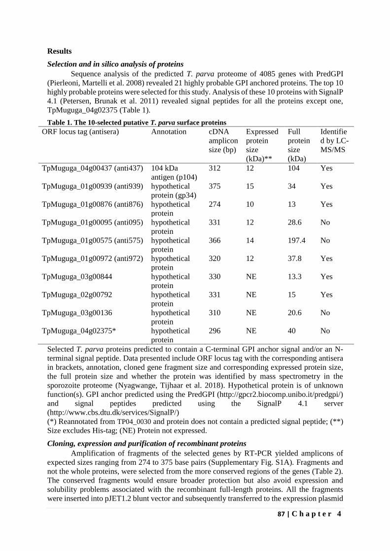
87 | C h a p t e r 4
Results
Selection and in silico analysis of proteins
Sequence analysis of the predicted T. parva proteome of 4085 genes with PredGPI
(Pierleoni, Martelli et al. 2008) revealed 21 highly probable GPI anchored proteins. The top 10
highly probable proteins were selected for this study. Analysis of these 10 proteins with SignalP
4.1 (Petersen, Brunak et al. 2011) revealed signal peptides for all the proteins except one,
TpMuguga_04g02375 (Table 1).
Table 1. The 10-selected putative T. parva surface proteins
ORF locus tag (antisera) Annotation cDNA
amplicon
size (bp)
Expressed
protein
size
(kDa)**
Full
protein
size
(kDa)
Identifie
d by LC-
MS/MS
TpMuguga_04g00437 (anti437) 104 kDa
antigen (p104)
312 12 104 Yes
TpMuguga_01g00939 (anti939) hypothetical
protein (gp34)
375 15 34 Yes
TpMuguga_01g00876 (anti876) hypothetical
protein
274 10 13 Yes
TpMuguga_01g00095 (anti095) hypothetical
protein
331 12 28.6 No
TpMuguga_01g00575 (anti575) hypothetical
protein
366 14 197.4 No
TpMuguga_01g00972 (anti972) hypothetical
protein
320 12 37.8 Yes
TpMuguga_03g00844 hypothetical
protein
330 NE 13.3 Yes
TpMuguga_02g00792 hypothetical
protein
331 NE 15 Yes
TpMuguga_03g00136 hypothetical
protein
310 NE 20.6 No
TpMuguga_04g02375* hypothetical
protein
296 NE 40 No
Selected T. parva proteins predicted to contain a C-terminal GPI anchor signal and/or an N-
terminal signal peptide. Data presented include ORF locus tag with the corresponding antisera
in brackets, annotation, cloned gene fragment size and corresponding expressed protein size,
the full protein size and whether the protein was identified by mass spectrometry in the
sporozoite proteome (Nyagwange, Tijhaar et al. 2018). Hypothetical protein is of unknown
function(s). GPI anchor predicted using the PredGPI (http://gpcr2.biocomp.unibo.it/predgpi/)
and signal peptides predicted using the SignalP 4.1 server
(http://www.cbs.dtu.dk/services/SignalP/)
(*) Reannotated from TP04_0030 and protein does not contain a predicted signal peptide; (**)
Size excludes His-tag; (NE) Protein not expressed.
Cloning, expression and purification of recombinant proteins
Amplification of fragments of the selected genes by RT-PCR yielded amplicons of
expected sizes ranging from 274 to 375 base pairs (Supplementary Fig. S1A). Fragments and
not the whole proteins, were selected from the more conserved regions of the genes (Table 2).
The conserved fragments would ensure broader protection but also avoid expression and
solubility problems associated with the recombinant full-length proteins. All the fragments
were inserted into pJET1.2 blunt vector and subsequently transferred to the expression plasmid
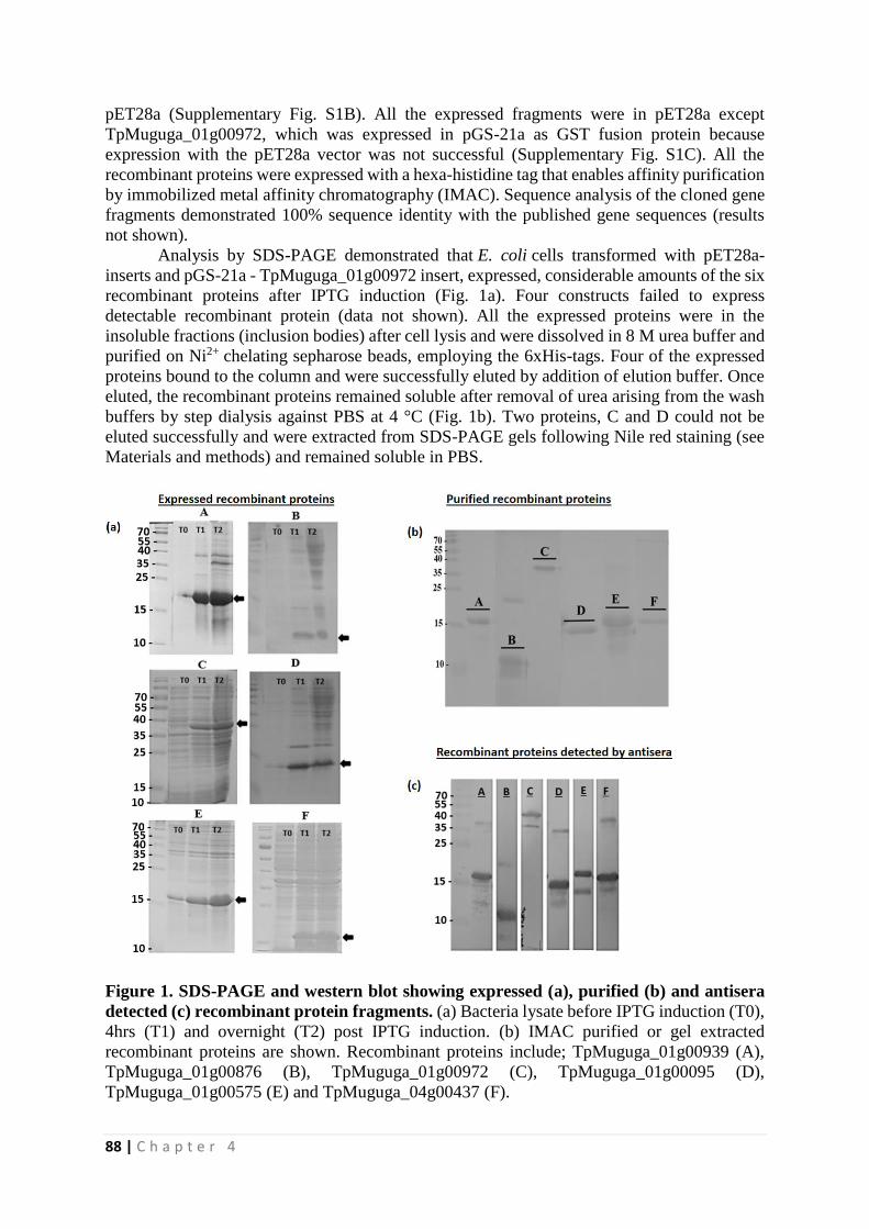
88 | C h a p t e r 4
pET28a (Supplementary Fig. S1B). All the expressed fragments were in pET28a except
TpMuguga_01g00972, which was expressed in pGS-21a as GST fusion protein because
expression with the pET28a vector was not successful (Supplementary Fig. S1C). All the
recombinant proteins were expressed with a hexa-histidine tag that enables affinity purification
by immobilized metal affinity chromatography (IMAC). Sequence analysis of the cloned gene
fragments demonstrated 100% sequence identity with the published gene sequences (results
not shown).
Analysis by SDS-PAGE demonstrated that E. coli cells transformed with pET28a-
inserts and pGS-21a - TpMuguga_01g00972 insert, expressed, considerable amounts of the six
recombinant proteins after IPTG induction (Fig. 1a). Four constructs failed to express
detectable recombinant protein (data not shown). All the expressed proteins were in the
insoluble fractions (inclusion bodies) after cell lysis and were dissolved in 8 M urea buffer and
purified on Ni2+ chelating sepharose beads, employing the 6xHis-tags. Four of the expressed
proteins bound to the column and were successfully eluted by addition of elution buffer. Once
eluted, the recombinant proteins remained soluble after removal of urea arising from the wash
buffers by step dialysis against PBS at 4 °C (Fig. 1b). Two proteins, C and D could not be
eluted successfully and were extracted from SDS-PAGE gels following Nile red staining (see
Materials and methods) and remained soluble in PBS.
Figure 1. SDS-PAGE and western blot showing expressed (a), purified (b) and antisera
detected (c) recombinant protein fragments. (a) Bacteria lysate before IPTG induction (T0),
4hrs (T1) and overnight (T2) post IPTG induction. (b) IMAC purified or gel extracted
recombinant proteins are shown. Recombinant proteins include; TpMuguga_01g00939 (A),
TpMuguga_01g00876 (B), TpMuguga_01g00972 (C), TpMuguga_01g00095 (D),
TpMuguga_01g00575 (E) and TpMuguga_04g00437 (F).
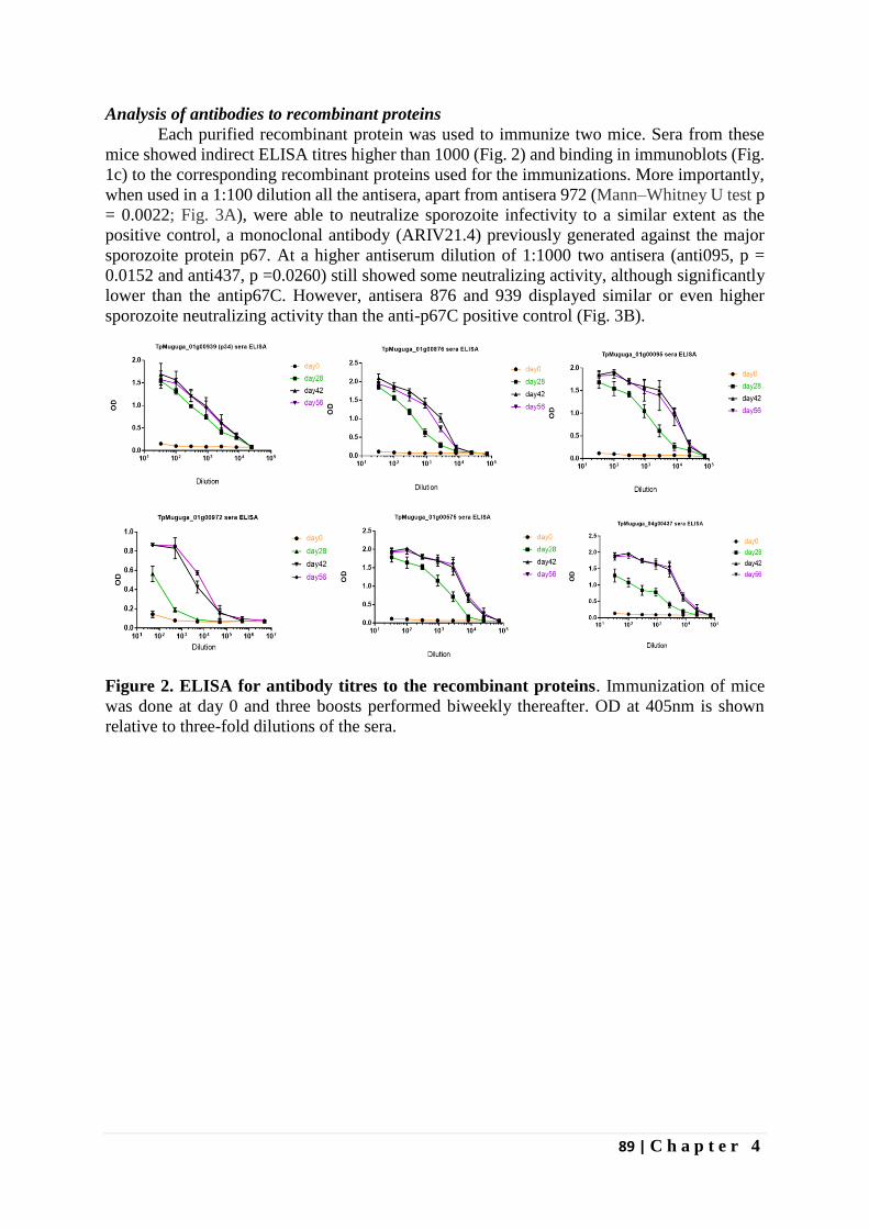
89 | C h a p t e r 4
Analysis of antibodies to recombinant proteins
Each purified recombinant protein was used to immunize two mice. Sera from these
mice showed indirect ELISA titres higher than 1000 (Fig. 2) and binding in immunoblots (Fig.
1c) to the corresponding recombinant proteins used for the immunizations. More importantly,
when used in a 1:100 dilution all the antisera, apart from antisera 972 (Mann–Whitney U test p
= 0.0022; Fig. 3A), were able to neutralize sporozoite infectivity to a similar extent as the
positive control, a monoclonal antibody (ARIV21.4) previously generated against the major
sporozoite protein p67. At a higher antiserum dilution of 1:1000 two antisera (anti095, p =
0.0152 and anti437, p =0.0260) still showed some neutralizing activity, although significantly
lower than the antip67C. However, antisera 876 and 939 displayed similar or even higher
sporozoite neutralizing activity than the anti-p67C positive control (Fig. 3B).
Figure 2. ELISA for antibody titres to the recombinant proteins. Immunization of mice
was done at day 0 and three boosts performed biweekly thereafter. OD at 405nm is shown
relative to three-fold dilutions of the sera.
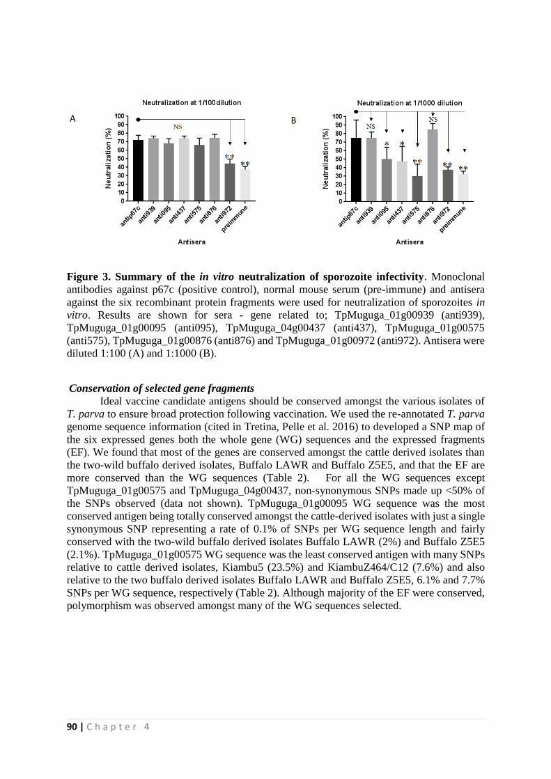
90 | C h a p t e r 4
Figure 3. Summary of the in vitro neutralization of sporozoite infectivity. Monoclonal
antibodies against p67c (positive control), normal mouse serum (pre-immune) and antisera
against the six recombinant protein fragments were used for neutralization of sporozoites in
vitro. Results are shown for sera - gene related to; TpMuguga_01g00939 (anti939),
TpMuguga_01g00095 (anti095), TpMuguga_04g00437 (anti437), TpMuguga_01g00575
(anti575), TpMuguga_01g00876 (anti876) and TpMuguga_01g00972 (anti972). Antisera were
diluted 1:100 (A) and 1:1000 (B).
Conservation of selected gene fragments
Ideal vaccine candidate antigens should be conserved amongst the various isolates of
T. parva to ensure broad protection following vaccination. We used the re-annotated T. parva
genome sequence information (cited in Tretina, Pelle et al. 2016) to developed a SNP map of
the six expressed genes both the whole gene (WG) sequences and the expressed fragments
(EF). We found that most of the genes are conserved amongst the cattle derived isolates than
the two-wild buffalo derived isolates, Buffalo LAWR and Buffalo Z5E5, and that the EF are
more conserved than the WG sequences (Table 2). For all the WG sequences except
TpMuguga_01g00575 and TpMuguga_04g00437, non-synonymous SNPs made up <50% of
the SNPs observed (data not shown). TpMuguga_01g00095 WG sequence was the most
conserved antigen being totally conserved amongst the cattle-derived isolates with just a single
synonymous SNP representing a rate of 0.1% of SNPs per WG sequence length and fairly
conserved with the two-wild buffalo derived isolates Buffalo LAWR (2%) and Buffalo Z5E5
(2.1%). TpMuguga_01g00575 WG sequence was the least conserved antigen with many SNPs
relative to cattle derived isolates, Kiambu5 (23.5%) and KiambuZ464/C12 (7.6%) and also
relative to the two buffalo derived isolates Buffalo LAWR and Buffalo Z5E5, 6.1% and 7.7%
SNPs per WG sequence, respectively (Table 2). Although majority of the EF were conserved,
polymorphism was observed amongst many of the WG sequences selected.
91 | C h a p t e r 4
Table 2. Nucleotide sequence polymorphism of open reading frames and recombinantly expressed fragments, in cattle and buffalo derived T. parva
isolates. SNPs expressed as percentage of DNA sequence length for the whole gene (WG) sequence and recombinantly expressed fragment (EF).
Isolate TP01_0095 TP01_0575 TP01_0876 TP01_0939 TP01_0972 TP04_0437
WG EF WG EF WG EF WG EF WG EF WG EF
ChitongoZ2 0 0 5.7 0.3 0 0 2.3 0.3 0.5 0 1.5 0
Entebbe 0 0 4.1 0.3 0 0 2.1 0.3 0.4 0 0.7 0
KateteB2 0 0 5.5 0.3 0.8 0 2.3 0.3 0.5 0 1.1 0
Katumba 0 0 6.0 1.9 2.6 0.4 2.6 1.6 0.5 0 0.8 0
Kiambu5 0 0 23.5 2.5 7.1 1.1 3.0 1.6 0.5 0 1.8 0
KiambuZ464/C12 0 0 7.6 2.5 2.6 1.1 2.7 1.6 0.5 0 1.5 0
MandaliZ22 0 0 4.4 0.3 0 0 2.1 0.3 0.4 0 0.6 0
Marikebuni 0 0 3.8 0.3 5.8 1.1 0.1 0.3 0.1 0 1.7 0
MugMar 0 0 3.7 0.3 6.1 1.1 0.1 0.3 0.1 0 1.7 0
MugUg 0 0 5.9 0.3 0.3 0 2.4 0.3 0.6 0 1.7 0
Muguga2* 0 0 0 0 0 0 0 0 0 0 0 0
Nyakizu 0.1 0 0.7 0.3 5.0 1.1 2.9 1.6 0.5 0 1.2 0
Serengeti 0 0 0 0 0 0 0 0 0 0 0 0
Uganda 0 0 5.9 0.3 0.3 0 2.4 0.3 0.6 0 1.7 0
Buffalo LAWR** 2.0 1.2 6.1 0.3 3.2 0.4 2.0 1.1 1.4 1.9 2.8 0.6
Buffalo Z5E5** 2.1 0.9 7.7 0.8 1.9 0.7 2.0 0.8 1.0 1.6 3.0 0.6
The isolates shown include cattle derived and two buffalo derived isolates (**) and the reference genome isolate (*).
92 | C h a p t e r 4
Discussion
GPI anchors are common attachment signals for surface proteins of parasites such as
Plasmodium, Trypanosomes, Toxoplasma, etc. and many are promising vaccine candidate
antigens (Ferguson 1999, Ferguson MAJ 2009). One such example is the circumsporozoite
(CS) protein of Plasmodium which is the antigenic target of the malaria vaccine RTS, S
(Lancet, 2015). Researchers have employed the strategy of targeting GPI anchored proteins
for evaluation as candidate vaccine antigens. In this study, we also employed this strategy to
select T. parva proteins predicted to contain GPI anchor signals for evaluation as vaccine
candidates.
We selected a list of 10 genes with high probability of containing a GPI-anchored tail
(Table 1). Using specific primers for the more conserved regions of the selected genes, we were
able to synthesize cDNA of expected sizes in a one-step RTPCR reaction (Supplementary Fig.
1A). We have expressed six out of the 10 selected genes as recombinant proteins in E. coli.
Five of the proteins were expressed with a His tag, which increases the molecular weight by
approximately 1 kDa (Fig. 1). TpMuguga_01g00972 was expressed as a GST fusion protein,
adding 26 kDa to the size resulting in a total size of 39 kDa (Fig. 1C). Although sequencing
data showed that the cloned sequences were in-frame, we were not successful in expressing
four of the 10 selected gene fragments even after transfer to various expression vectors
(pET28a, pQE30 and pGS-21a) and E. coli strains, BL21(DE3) star and JM109(DE3).
The six expressed recombinant proteins were used to raise antisera in mice. The
antisera were found to bind to the respective protein product of the cloned gene fragments (Fig.
1c) and most importantly, antisera against two of the recombinant proteins,
TpMuguga_01g00876 and TpMuguga_01g00939, highly (>60%) neutralize sporozoite
infectivity at 1000-fold dilution. Antisera against two additional proteins,
TpMuguga_01g00095 and TpMuguga_04g00437, moderately (>30%) neutralize sporozoite
infection of bovine PBMCs in vitro (Fig. 3B).
In this study, we tested gene fragments. It is tempting to speculate that antisera to full
length proteins of the four antigens would produce higher neutralizing activities because of the
longer sequence with putative additional epitopes. Therefore, it is desirable to test the full
length recombinant proteins for immunogenicity. However, expression of long proteins is
usually accompanied by solubility and expression problems of the recombinant proteins. To
overcome expression problems, other expression systems could be employed or several shorter
fragments comprising the full protein can be combined and evaluated.
Among the protein fragments that produce high sporozoite neutralizing antibodies is
TpMuguga_01g00939, a protein previously referred to as gp34, that undergoes GPI
modification when expressed in mammalian cells (Xue, von Schubert et al. 2010). Although
originally reported as a schizont stage specific antigen (Xue, von Schubert et al. 2010), we have
recently identified gp34 protein in the sporozoite proteome (Nyagwange, Tijhaar et al. 2018).
We therefore conclude that the protein is expressed in both parasite life-cycle stages. In the
schizont stage, gp34 seems to play a role in parasite-host interaction during host cell division
(Xue, von Schubert et al. 2010). Immunization with TpMuguga_04g00437, also known as p104
a sporozoite microneme/rhoptry protein (Ebel, Gerhards et al. 1999), resulted in mouse antisera
that moderately neutralized sporozoite infection of bovine PBMCs in vitro. The p104 protein
was originally identified by sporozoite neutralizing bovine antisera C16 (Iams, Young et al.
1990), but was never evaluated as a vaccine candidate antigen (Nene, Kiara et al. 2016). The
p104 protein is also expressed at the surface of the schizont and recent evidence suggests a role
for this protein in interacting with the host-cell mitotic machinery (Huber, Theiler et al. 2017).
With the results presented here, it appears that gp34 and p104 also play a role in the lymphocyte
93 | C h a p t e r 4
invasion process, perhaps through additional interactions with host cell microtubules during
invasion.
Neutralization of parasite infection of host cells is one of the most important features
of an anti-sporozoite vaccine candidate antigen, and in this study, we have identified four
vaccine candidates that are able to induce sporozoite neutralizing antibodies. Two of these
proteins (p104 and gp34) were identified before and two are completely new, including
TpMuguga_01g00876 which produced antibodies inducing the strongest sporozoite
neutralizing activity. However, following previous observations in which rats immunized with
recombinant polymorphic immuno-dominant molecule (PIM) make neutralizing antibodies
while cattle immunized with the same do not (Toye, Metzelaar et al. 1995, Toye, Nyanjui et al.
1996), it is important to raise and test bovine antibodies against these antigens to formally
confirm their role as candidate vaccine antigens.
Acknowledgement
We are grateful to Thomas Njoroge, Elias Awino and Robert Muriuki for analysis of the slides.
We thank the CGIAR Research Program on Livestock and Fish, the Norman Borlaug
Commemorative Research Initiative, an initiative between the Feed the Future program of
USAID and USDA-ARS (58-5348-2-117F) and the Department for International Development
of the United Kingdom and the Bill and Melinda Gates Foundation (OPP1078791) for financial
support.
94 | C h a p t e r 4
References
Babo Martins, S., G. Di Giulio, G. Lynen, A. Peters and J. Rushton (2010). "Assessing the
impact of East Coast Fever immunisation by the infection and treatment method in Tanzanian
pastoralist systems." Preventive Veterinary Medicine 97(3–4): 175-182.
Bishop, R., V. Nene, J. Staeyert, J. Rowlands, J. Nyanjui, J. Osaso, S. Morzaria and A. Musoke
(2003). "Immunity to East Coast fever in cattle induced by a polypeptide fragment of the major
surface coat protein of Theileria parva sporozoites." Vaccine 21(11-12): 1205-1212.
Cingolani, P., A. Platts, L. L. Wang, M. Coon, T. Nguyen, L. Wang, S. J. Land, X. Lu and D.
M. Ruden (2012). "A program for annotating and predicting the effects of single nucleotide
polymorphisms, SnpEff: SNPs in the genome of Drosophila melanogaster strain w1118; iso-2;
iso-3." Fly 6(2): 80-92.
Daban, J.-R., S. Bartolomé and A. Bermúdez (1996). Rapid Staining of Proteins in
Polyacrylamide Gels with Nile Red. The Protein Protocols Handbook. J. M. Walker. Totowa,
NJ, Humana Press: 179-185.
Delorenzi, M., A. Sexton, H. Shams-Eldin, R. T. Schwarz, T. Speed and L. Schofield (2002).
"Genes for Glycosylphosphatidylinositol Toxin Biosynthesis in Plasmodium falciparum."
Infection and Immunity 70(8): 4510-4522.
Dobbelaere, D., S. Z. Shapiro and P. Webster (1985). "Identification of a surface antigen on
Theileria parva sporozoites by monoclonal antibody." Proceedings of the National Academy
of Sciences 82(6): 1771-1775.
Dobbelaere, D. A. E., P. R. Spooner, W. C. Barry and A. D. Irvin (1984). "Monoclonal antibody
neutralizes the sporozoite stage of different Theileria parva stocks." Parasite Immunology 6(4):
361-370.
Ebel, T., J. Gerhards, B. R. Binder and J. Lipp (1999). "Theileria parva 104 kDa microneme–
rhoptry protein is membrane-anchored by a non-cleaved amino-terminal signal sequence for
entry into the endoplasmic reticulum." Molecular and Biochemical Parasitology 100(1): 19-26.
Emery, D. L. (1981). "Adoptive transfer of immunity to infection with Theileria parva (East
Coast fever) between cattle twins." Research in Veterinary Science 30(3): 364-367.
Fawcett, D. W., S. Doxsey, D. A. Stagg and A. S. Young (1982b). "The entry of sporozoites
of Theileria parva into bovine lymphocytes in vitro. Electron microscopic observations."
European journal of cell biology 27(1): 10-21.
Ferguson, M. A. (1999). "The structure, biosynthesis and functions of
glycosylphosphatidylinositol anchors, and the contributions of trypanosome research." Journal
of Cell Science 112(17): 2799-2809.
Ferguson MAJ, K. T., Hart GW (2009). Essentials of Glycobiology. Cold Spring Harbor (NY),
Cold Spring Harbor Laboratory Press.
Garrison, E. and G. Marth (2012). "Haplotype-based variant detection from short-read
sequencing." arXiv preprint arXiv:1207.3907.
95 | C h a p t e r 4
Gilson, P. R., T. Nebl, D. Vukcevic, R. L. Moritz, T. Sargeant, T. P. Speed, L. Schofield and
B. S. Crabb (2006). "Identification and Stoichiometry of Glycosylphosphatidylinositol-
anchored Membrane Proteins of the Human Malaria Parasite Plasmodium falciparum."
Molecular & Cellular Proteomics 5(7): 1286-1299.
Hall, R., N. R. Boulter, C. G. Brown, G. Wilkie, E. Kirvar, V. Nene, A. J. Musoke, E. J. Glass
and S. P. Morzaria (2000). "Reciprocal cross-protection induced by sporozoite antigens SPAG-
1 from Theileria annulata and p67 from Theileria parva." Parasite Immunology 22(5): 223-230.
Huber, S., R. Theiler, D. de Quervain, O. Wiens, T. Karangenc, V. Heussler, D. Dobbelaere
and K. Woods (2017). "The Microtubule-Stabilizing Protein CLASP1 Associates with the
<em>Theileria annulata</em> Schizont Surface via Its Kinetochore-Binding Domain."
mSphere 2(4).
Iams, K. P., J. R. Young, V. Nene, J. Desai, P. Webster, O. K. ole-MoiYoi and A. J. Musoke
(1990). "Characterisation of the gene encoding a 104-kilodalton micronemerhoptry protein of
Theileria parva." Molecular and Biochemical Parasitology 39(1): 47-60.
Ikezawa, H. (2002). "Glycosylphosphatidylinositol (GPI)-Anchored Proteins." Biological and
Pharmaceutical Bulletin 25(4): 409-417.
Lancet (2015). "Efficacy and safety of RTS,S/AS01 malaria vaccine with or without a booster
dose in infants and children in Africa: final results of a phase 3, individually randomised,
controlled trial." Lancet 386(9988): 31-45.
McHardy, N., L. S. Wekesa, A. T. Hudson and A. W. Randall (1985). "Antitheilerial activity
of BW720C (buparvaquone): a comparison with parvaquone." Research in Veterinary Science
39(1): 29-33.
McKeever, D. J., E. L. Taracha, E. L. Innes, N. D. MacHugh, E. Awino, B. M. Goddeeris and
W. I. Morrison (1994). "Adoptive transfer of immunity to Theileria parva in the CD8+ fraction
of responding efferent lymph." Proceedings of the National Academy of Sciences 91.
Mhadhbi, M., A. Naouach, A. Boumiza, M. F. Chaabani, S. BenAbderazzak and M. A.
Darghouth (2010). "In vivo evidence for the resistance of Theileria annulata to buparvaquone."
Veterinary Parasitology 169(3–4): 241-247.
Morrison, W. I. (2009). "Progress towards understanding the immunobiology of Theileria
parasites." Parasitology 136.
Morrison, W. I. and B. M. Goddeeris (1990). Cytotoxic T Cells in Immunity to Theileria parva
in Cattle. T-Cell Paradigms in Parasitic and Bacterial Infections. S. H. E. Kaufmann. Berlin,
Heidelberg, Springer Berlin Heidelberg: 79-93.
Musoke, A., S. Morzaria, C. Nkonge, E. Jones and V. Nene (1992). "A recombinant sporozoite
surface antigen of Theileria parva induces protection in cattle." Proceedings of the National
Academy of Sciences 89(2): 514-518.
Musoke, A. J., V. M. Nantulya, G. Buscher, R. A. Masake and B. Otim (1982). "Bovine
immune response to Theileria parva: neutralizing antibodies to sporozoites." Immunology
45(4): 663-668.
96 | C h a p t e r 4
Musoke, A. J., V. M. Nantulya, F. R. Rurangirwa and G. Buscher (1984). "Evidence for a
common protective antigenic determinant on sporozoites of several Theileria parva strains."
Immunology 52(2): 231-238.
Nene, V., H. Kiara, A. Lacasta, R. Pelle, N. Svitek and L. Steinaa (2016). "The biology of
Theileria parva and control of East Coast fever – Current status and future trends." Ticks and
Tick-borne Diseases 7(4): 549-564.
Norval, R. A. I., B. D. Perry and A. Young (1992). The epidemiology of theileriosis in Africa,
Academic press, London.
Patel, E., S. Mwaura, H. Kiara, S. Morzaria, A. Peters and P. Toye (2016). "Production and
dose determination of the Infection and Treatment Method (ITM) Muguga cocktail vaccine
used to control East Coast fever in cattle." Ticks and Tick-borne Diseases 7(2): 306-314.
Petersen, T. N., S. Brunak, G. von Heijne and H. Nielsen (2011). "SignalP 4.0: discriminating
signal peptides from transmembrane regions." Nature Methods 8(10): 785-786.
Pierleoni, A., P. L. Martelli and R. Casadio (2008). "PredGPI: a GPI-anchor predictor." BMC
Bioinformatics 9(1): 392.
Radley, D. E., C. G. D. Brown, M. J. Burridge, M. P. Cunningham, I. M. Kirimi, R. E. Purnell
and A. S. Young (1975a). "East coast fever: 1. Chemoprophylactic immunization of cattle
against Theileria parva (Muguga) and five theilerial strains." Veterinary Parasitology 1(1): 35-
41.
Radley, D. E., C. G. D. Brown, M. P. Cunningham, C. D. Kimber, F. L. Musisi, R. C. Payne,
R. E. Purnell, S. M. Stagg and A. S. Young (1975b). "East coast fever: 3. Chemoprophylactic
immunization of cattle using oxytetracycline and a combination of theilerial strains."
Veterinary Parasitology 1(1): 51-60.
Shaw, M. K. (1996). "Theileria parvaSporozoite Entry into Bovine Lymphocytes Involves both
Parasite and Host Cell Signal Transduction Processes." Experimental Parasitology 84(3): 344-
354.
Shaw, M. K. (2003). "Cell invasion by Theileria sporozoites." Trends in Parasitology 19(1): 2-
6.
Tachado, S. D., P. Gerold, M. J. McConville, T. Baldwin, D. Quilici, R. T. Schwarz and L.
Schofield (1996). "Glycosylphosphatidylinositol toxin of Plasmodium induces nitric oxide
synthase expression in macrophages and vascular endothelial cells by a protein tyrosine kinase-
dependent and protein kinase C-dependent signaling pathway." The Journal of Immunology
156(5): 1897-1907.
Toye, P., J. Nyanjui, B. Goddeeris and A. J. Musoke (1996). "Identification of neutralization
and diagnostic epitopes on PIM, the polymorphic immunodominant molecule of Theileria
parva." Infection and Immunity 64(5): 1832-1838.
Toye, P. G., M. J. Metzelaar, P. L. Wijngaard, V. Nene, K. Iams, J. Roose, J. K. Nyanjui, E.
Gobright, A. J. Musoke and H. C. Clevers (1995). "Characterization of the gene encoding the
polymorphic immunodominant molecule, a neutralizing antigen of Theileria parva." The
Journal of Immunology 155(3): 1370-1381.
97 | C h a p t e r 4
Tretina, K., R. Pelle and J. C. Silva (2016). "Cis regulatory motifs and antisense transcriptional
control in the apicomplexan Theileria parva." BMC Genomics 17(1): 128.
Uilenberg, G. (1999). "Immunization against diseases caused by Theileria parva." Tropical
Medicine and International Health 4: A12-A20.
von Heijne, G. (1990). "The signal peptide." The Journal of Membrane Biology 115(3): 195-
201.
Xue, G., C. von Schubert, P. Hermann, M. Peyer, R. Maushagen, J. Schmuckli-Maurer, P.
Bütikofer, G. Langsley and D. A. E. Dobbelaere (2010). "Characterisation of gp34, a GPI-
anchored protein expressed by schizonts of Theileria parva and T. annulata." Molecular and
Biochemical Parasitology 172(2): 113-120.
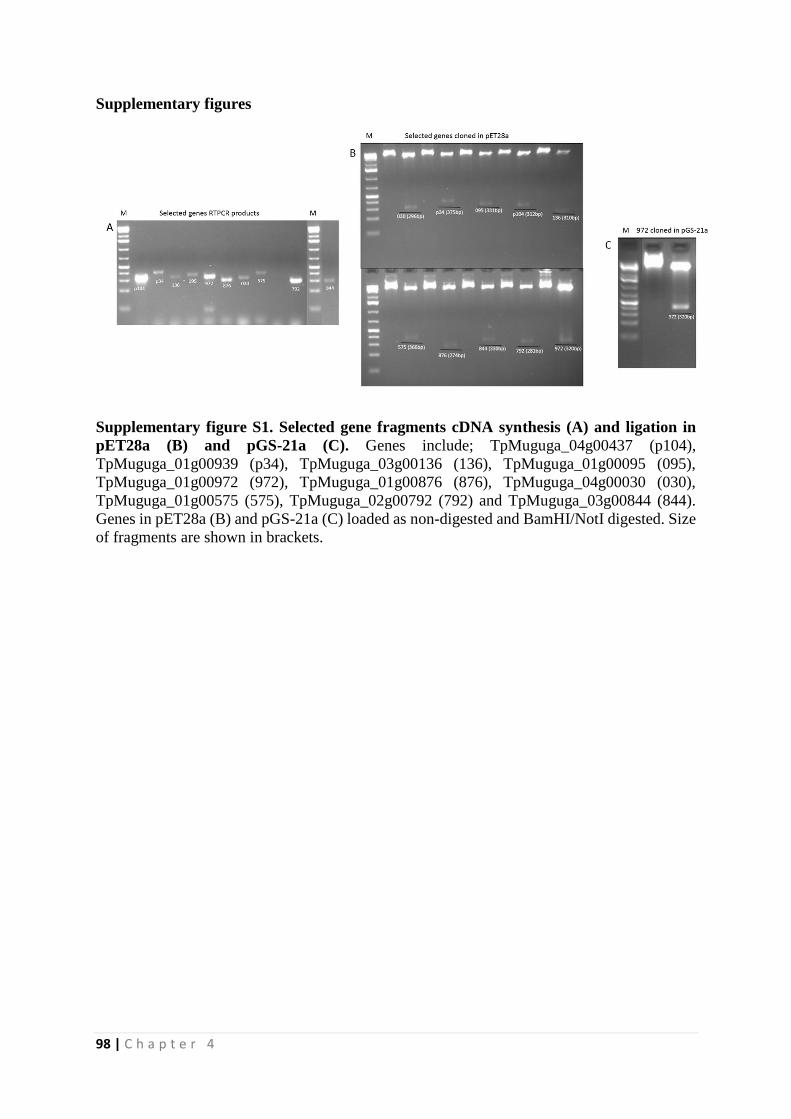
98 | C h a p t e r 4
Supplementary figures
Supplementary figure S1. Selected gene fragments cDNA synthesis (A) and ligation in
pET28a (B) and pGS-21a (C). Genes include; TpMuguga_04g00437 (p104),
TpMuguga_01g00939 (p34), TpMuguga_03g00136 (136), TpMuguga_01g00095 (095),
TpMuguga_01g00972 (972), TpMuguga_01g00876 (876), TpMuguga_04g00030 (030),
TpMuguga_01g00575 (575), TpMuguga_02g00792 (792) and TpMuguga_03g00844 (844).
Genes in pET28a (B) and pGS-21a (C) loaded as non-digested and BamHI/NotI digested. Size
of fragments are shown in brackets.
99 | C h a p t e r 5
CHAPTER
5
Leveraging the Medicines for Malaria Venture malaria
and pathogen boxes to discover lead chemical inhibitors of
East Coast fever
James Nyagwange
Elias Awino
Edwin Tijhaar
Nicholas Svitek
Roger Pelle
Vishvanath Nene
Published in IJP: Drugs and Drug Resistance, 2019; 9:80-86
100 | C h a p t e r 5
Abstract
Chemotherapy of East Coast fever, a lymphoproliferative cancer-like disease of cattle causing
significant economic losses in Africa, is largely dependent on the use of buparvaquone, a drug
that was developed in the late 1980’s. The disease is caused by the tick-borne pathogen
Theileira parva. Buparvaquone can be used prophylactically to prevent disease and it is also
active against tropical theileriosis, caused by the related parasite Theileria annulata. Recently,
drug resistance was reported in T. annulata, and could also occur in T. parva. Therefore, there
is need to identify new drugs that could be used for the control of Theileria infections. Using a 3H-thymidine incorporation assay we screened the Medicines for Malaria Venture (MMV)
open source 400 malaria box and 396 pathogen box compounds to discover novel compounds
with anti-parasitic activity. We identified nine malaria box compounds and eight pathogen box
compounds that inhibit the proliferation of a T. parva infected cell line. However, only two
compounds, MMV008212 (malaria box) and MMV688372 (pathogen box) represent
promising leads with half maximal inhibitory concentration (IC50) values of 0.78 and 0.61 µM,
respectively, and half maximal cytotoxic concentration (CC50) values > 5 µM. The remaining
selected compounds exhibited a high degree of non-specific cytotoxicity (CC50 values < 1.09
µM) on the proliferation of bovine peripheral blood mononuclear cells stimulated with
concanavalin A. We also tested the anti-cancer drug, dasatinib, used in the therapy of some
leukemias. Dasatinib was as active and safe as buparvaquone in vitro, with an IC50 of 5 and 4.2
nM, respectively, and CC50 > 10 µM. Our preliminary data suggest that it may be possible to
repurpose compounds from the cancer field as well as MMV malaria and pathogen boxes.
101 | C h a p t e r 5
Introduction
East Coast fever (ECF), a lymphoproliferative disease caused by Theileira parva is the
most important tick-borne disease in sub-Saharan Africa. It kills a million cattle a year with an
estimated economic loss of USD 300 million and impedes growth of the livestock industry.
Theileria belongs to the phylum Apicomplexa, which contains other parasites responsible for
major human and animal diseases, such as malaria, babesiosis, toxoplasmosis and
cryptosporidiosis (Morrissette and Sibley 2002). Considerable progress has been made in
developing vaccines for the control of some of these diseases. For example, a live infection and
treatment method of immunization is commercially available for the control of ECF (Radley
1981) and tissue-culture vaccines are available for the control of tropical theileriosis caused by
T. annulata (Brown 1990). Advanced clinical trials are in progress with a malaria vaccine
(Lancet 2015). However, drug treatment often remains a frontline method of disease control.
Derivatives of naphthoquinones exhibit significant pharmacological properties and have
given rise to development of anti-parasitic drugs, including commercial products to control
malaria, e.g., atavaquone, a hydroxy-napthoquinone (Nixon, Moss et al. 2013). One of the
byproducts of this effort gave rise to development of anti-theilerial drugs, e.g., parvaquone and
the improved derivative, buparvaquone, that is the current drug of choice for treatment of
theileriosis (McHardy, Wekesa et al. 1985). Members of this class of compounds display
activity against other human and animals’ parasites, e.g., trypanosomes (O. Salas, Faundez et
al. 2011). Unfortunately, resistance to atavaquone, an analog of ubiquinone, emerged quite
rapidly and drug resistance to atavaquone in Plasmodium is associated with mutations in the
mitochondrial gene encoding apo-cytochrome b (Nixon, Moss et al. 2013). Resistance to other
classes of anti-malarial drugs has also emerged, limiting the efficacy of several frontline drugs
(Cui, Mharakurwa et al. 2015). The Medicines for Malaria Venture (MMV) was inaugurated
in 1999, with a mandate of developing new anti-malarial drugs, as it was recognized that the
pipeline for developing such drugs was small and not an attractive venture for the
pharmaceutical industry.
Fortunately, T. parva resistance to buparvaquone has not been described. However, the
recent identification of drug resistance in T. annulata is a cause for concern and suggests that
this drug may be of limited veterinary use in the future. As for many tropical diseases, there is
no program to develop new anti-theilerial drugs. Although focused on developing novel anti-
malarial drugs, MMV has catalyzed neglected disease drug discovery by making available
drug-like and probe-like compounds available to the scientific community through the Malaria
and Pathogen Box (Van Voorhis, Adams et al. 2016). A unique aspect of the biology of T.
parva and T. annulata is that parasite infected leukocytes behave and proliferate like cancer
cells in vitro, a property that is dependent on parasite viability. We have taken advantage of
this phenomenon and the availability of MMV compounds to screen for molecules that inhibit
the growth the T. parva cells.
We screened compounds in both the malaria and pathogen boxes against a bovine
lymphocyte cell line infected with the schizont stage of T. parva and against bovine peripheral
blood mononuclear cells (PBMC) stimulated by concanavalin A (ConA), the latter to determine
cytotoxicity. We have identified two compounds with an in vitro therapeutic index >5, which
could act as starting points for discovery of novel anti-theilerial drugs. In addition, we screened
an anti-cancer drug, dasatinib that is used for treatment of chronic myelogenous leukemia and
acute lymphoblastic leukemia, and found that it selectively kills the parasite with an in vitro
therapeutic index > 2,000
102 | C h a p t e r 5
Methods
Experimental compounds
The MMV malaria box and pathogen box were obtained from the Medicines for Malaria
Venture (MMV, Geneva, Switzerland). Plate mapping and full data on the malaria box were
accessed at (http://www.mmv.org/research-development/malaria-box-supporting information)
and (http://www.pathogenbox.org/) for the pathogen box. All plates were received at 10mM
stock concentrations and diluted in 100% DMSO to form copies of the plates at 1mM
concentration. From the 1mM stock plates serial dilutions were made in RPMI 1640 culture
media to a 10 µM working stock plates. Buparvaquone (Butalex) was used as a positive control
drug and Dasatinib was purchased from Selleckchem, USA (cat no. S1021).
In vitro parasite cytotoxicity assay
All the compounds were initially tested for inhibitory activity on the growth of T. parva
infected lymphocytes at 1µM final concentration. F100TpM, a bovine lymphocyte cell line
infected by T. parva, was seeded in 100l RPMI 1640 media at 1 x 10⁴ cells per well in 96 well
plates and incubated overnight in 5% CO2 at 37oC before addition of drug compounds (0 to 2
µM final concentration). Compounds found to inhibit growth of T. parva infected lymphocytes
were further screened for non-specific cytotoxicity on the proliferation of uninfected bovine
cells. Briefly, 1 x 10⁴ peripheral blood mononuclear cells (PBMCs) isolated from uninfected
bovine blood by Ficoll-Paque density gradient centrifugation were seeded in 100l RPMI 1640
media per well in 96 well plates and cultured overnight in 5% CO2 at 37oC in the presence of
concanavalin A (final concentration of 5µg/ml) before addition of drug compounds (0 to 10
µM). Both cell lines were cultured in RPMI 1640 medium for 48 hours prior to pulsing with 3H-thymidine. Lower drug concentrations of buparvaquone were used to a maximum of 0.08
µM. All compounds tested in triplicate wells.
Thymidine incorporation as a marker for inhibitory activity
Radiolabeled DNA precursor, 3H-thymidine is incorporated in DNA strands during cell
replication and is often used as a measure of cell proliferation. All cells were pulsed with 20µl
of RPMI 1640 media containing 3H-thymidine (0.5µCi) per well and incubated in 5% CO2 at
37oC for a minimum of 8 hrs before harvesting. The cells were then harvested by a Filtermat
Harvester (Perkin Elmer) onto Glass fiber matt filters (part No. 6005422) that captures the
labelled DNA. The filters were then air- dried and placed in Omni Filter cassette and 30µl of
MicroScintTM (Perkin Elmer Cat No 6013611) liquid added, then sealed with TopSeal (Perkin
Elmer, Part No 6050195) and loaded on TopCount NXT reader (Perkin Elmer) for
determination of incorporation of 3H-thymidine.
Calculation of IC50 and CC50 for bioactive compounds
IC50 values were calculated using the Graph Pad Prism 7 software (GraphPad Software, Inc.,
La Jolla, CA), and average values of radio-isotope incorporation in wells with and without
compound on the growth of F100TpM cells determined. The CC50 values were calculated using 3H-thymidine incorporation values of PBMC stimulated with conA incubated with different
concentrations of the tested compound.
103 | C h a p t e r 5
Results and Discussion
In a search for new lead anti-theilerial compounds, we screened the open source MMV
malaria box and pathogen box using a two-step process. Compound-induced inhibition (final
concentration of 1 µM) of 3H-thymidine incorporation by a T. parva infected cell line
(F100TpM) was used as a marker of anti-theilerial activity. Proliferation of infected cells is
parasite-dependent, and appears to occur through manipulation of several complex host-cell
signaling pathways (Shiels, Langsley et al. 2006). To get an indication of the in vitro potency
of the different compounds on T.parva the concentration that inhibited 50% of the proliferation
of F100TpM (the IC50 value) was determined. To determine the non-specific cytotoxicity of
the compounds, the concentration that inhibited 50% (CC50 values) of the proliferation of
bovine PBMCs stimulated with conA was determined. Most infected cells lines are derived by
“transformation” of T-lymphocytes by the parasite (Baldwin, Black et al. 1988). Since conA
drives the proliferation of T-lymphocytes (Palacios 1982) we reasoned that these conA blasts
represent a suitable control to assess non-specific cytotoxicity in vitro.
In the malaria and pathogen boxes, nine (IC50 values ranging from 0.04 to 1.93 µM)
and eight (IC50 values ranging from 0.35 to 0.97 µM) compounds inhibiting TpM proliferation
were identified, respectively, representing approximately a 2% hit rate (Table 1 and 2).
However, the majority of these compounds were also highly active on inhibiting the uninfected
conA stimulated PBMCs. Only two compounds, one from each box, MMV008212 and
MMV688372, exhibited an in vitro therapeutic index (CC50/IC50) of 8 and 10, respectively. An
index > 5 has been used to determine parasite selectivity of MMV compounds (reviewed in
Duffy, Sykes et al. 2017). In many cases the therapeutic index was close to or less than one in
our assay. This might be due to the different cell lines and assays used. In our hands, the
positive control drug, buparavquone had an IC50 of 4.2 nM and a CC50 >10 µM, with a
therapeutic index of more than 2,000. Interestingly, four pathogen box compounds were more
inhibitory on the control conA stimulated PBMCs, than on F100TpM, indicating that there
could be differences in the molecular and cellular biology of the two cell lines.
Of the malaria box compounds identified here, MMV008212, with a therapeutic index
of 12 for T.parva infected lymphocytes, was previously shown to also have activities against
Trypanosoma cruzi (100% growth inhibition at 5 µM), Plasmodium berghei ookinete (76%
growth inhibition at 10 µM), P. falciparum gametocyte NF54-late stage (91% growth inhibition
at 5 µM) and P. falciparum 3D7 asexual blood stage (98% growth inhibition at 5 µM)
(reviewed in Van Voorhis, Adams et al. 2016). Cytotoxicity of this compound was observed
only in one cell line (U87, 76% cell death at 5 µM) of the nine cell lines tested previously (Van
Voorhis, Adams et al. 2016). However, the mode of action of this compound is still unknown.
The remaining eight malaria box compounds with activity against T.parva infected
lymphocytes exhibited varied inhibitory activities on other organisms including Plasmodium,
Toxoplasma, Leishmania, Cryptosporidium and Wolbachia (Table 1). Among them, compound
MMV665800 has shown P. falciparum ATP4 inhibitor activity, while MMV006455 possibly
acts through a respiratory target (reviewed in Van Voorhis, Adams et al. 2016). In general,
these compounds exhibited less cytotoxicity on mammalian cell lines and zebrafish embryo
that were tested previously (Van Voorhis, Adams et al. 2016).
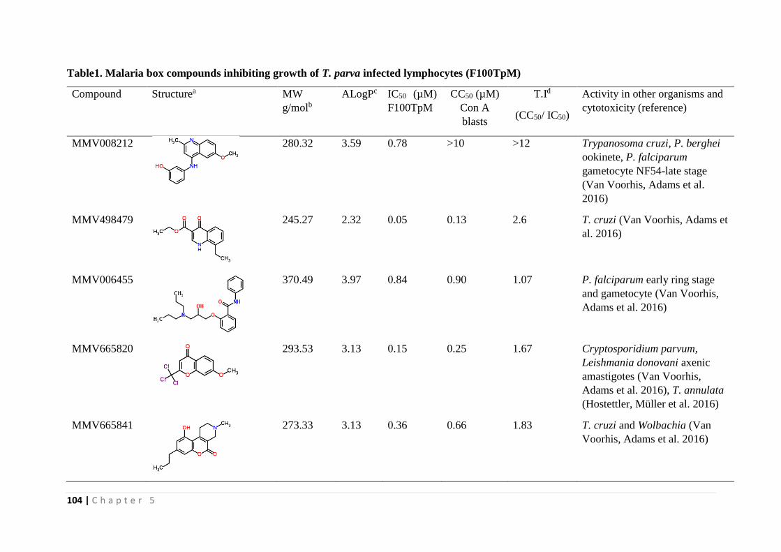
104 | C h a p t e r 5
Table1. Malaria box compounds inhibiting growth of T. parva infected lymphocytes (F100TpM)
Compound Structurea MW
g/molb
ALogPc IC50 (µM)
F100TpM
CC50 (µM)
Con A
blasts
T.Id
(CC50/ IC50)
Activity in other organisms and
cytotoxicity (reference)
MMV008212
280.32 3.59 0.78 >10 >12 Trypanosoma cruzi, P. berghei
ookinete, P. falciparum
gametocyte NF54-late stage
(Van Voorhis, Adams et al.
2016)
MMV498479
245.27
2.32 0.05 0.13 2.6 T. cruzi (Van Voorhis, Adams et
al. 2016)
MMV006455
370.49 3.97 0.84 0.90 1.07 P. falciparum early ring stage
and gametocyte (Van Voorhis,
Adams et al. 2016)
MMV665820
293.53 3.13 0.15 0.25 1.67 Cryptosporidium parvum,
Leishmania donovani axenic
amastigotes (Van Voorhis,
Adams et al. 2016), T. annulata
(Hostettler, Müller et al. 2016)
MMV665841
273.33 3.13 0.36 0.66 1.83 T. cruzi and Wolbachia (Van
Voorhis, Adams et al. 2016)
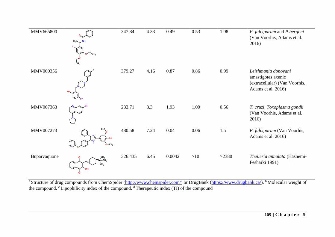
105 | C h a p t e r 5
MMV665800
347.84 4.33 0.49 0.53 1.08 P. falciparum and P.berghei
(Van Voorhis, Adams et al.
2016)
MMV000356
379.27 4.16 0.87 0.86 0.99 Leishmania donovani
amastigotes axenic
(extracellular) (Van Voorhis,
Adams et al. 2016)
MMV007363
232.71 3.3 1.93 1.09 0.56 T. cruzi, Toxoplasma gondii
(Van Voorhis, Adams et al.
2016)
MMV007273
480.58 7.24 0.04 0.06 1.5 P. falciparum (Van Voorhis,
Adams et al. 2016)
Buparvaquone
326.435 6.45 0.0042 >10 >2380 Theileria annulata (Hashemi-
Fesharki 1991)
a Structure of drug compounds from ChemSpider (http://www.chemspider.com/) or DrugBank (https://www.drugbank.ca/). b Molecular weight of
the compound. c Lipophilicity index of the compound. d Therapeutic index (TI) of the compound
106 | C h a p t e r 5
Of the 8 pathogen box compounds that were shown in this study to display activity
against T.parva infected lymphocytes, five were previously shown to also have anti-
trypanosomal activity, one has anti-malarial activity and two are reference compounds (Table
2). Mebendazole (MMV003152) is a known anti-helminthic drug that binds tubulin, inhibiting
their assembly into microtubules and consequently perturbing the motility, division and the
secretion processes of cells (Wampande, Richard McIntosh et al. 2007). In helminthes, binding
of the drug leads to loss of cytoplasmic microtubules which perturbs glucose intake leading to
exhaustion of glycogen reserves and consequent death (Oxberry, Gear et al. 2001). Auranofin
(MMV688978) is a drug indicated for rheumatoid arthritis and thought to act through inhibition
of nuclear factor kappa-B kinase subunit beta (Jeon, Byun et al. 2003) and thioredoxin
reductase (Rigobello, Folda et al. 2005) leading to reduced inflammatory responses and
reduced free radical production, respectively. The antimalarial compound, MMV023985 has
CDPK1 or PK7 proposed as the target (reviewed in Duffy, Sykes et al. 2017). Although these
compounds demonstrated activities on F100TpM with IC50s < 1 µM, their low therapeutic
index questions a role for them as anti-theilerial lead compounds. MV688372, which had a
high therapeutic index against F100TpM cells has anti-trypanosomal activity. This compound
is a substituted 2-phenylimidazopyridine with activities against Trypanosoma brucei brucei
and T. cruzi with therapeutic indexes of 230 and 24, respectively, in relation to HEK293 cells
(Duffy, Sykes et al. 2017). The mode of action of MMV688372 is still unclear, but a
structurally related azabenzoxazole has been identified as a proteasome inhibitor in
kinetoplastid parasites (Duffy, Sykes et al. 2017).
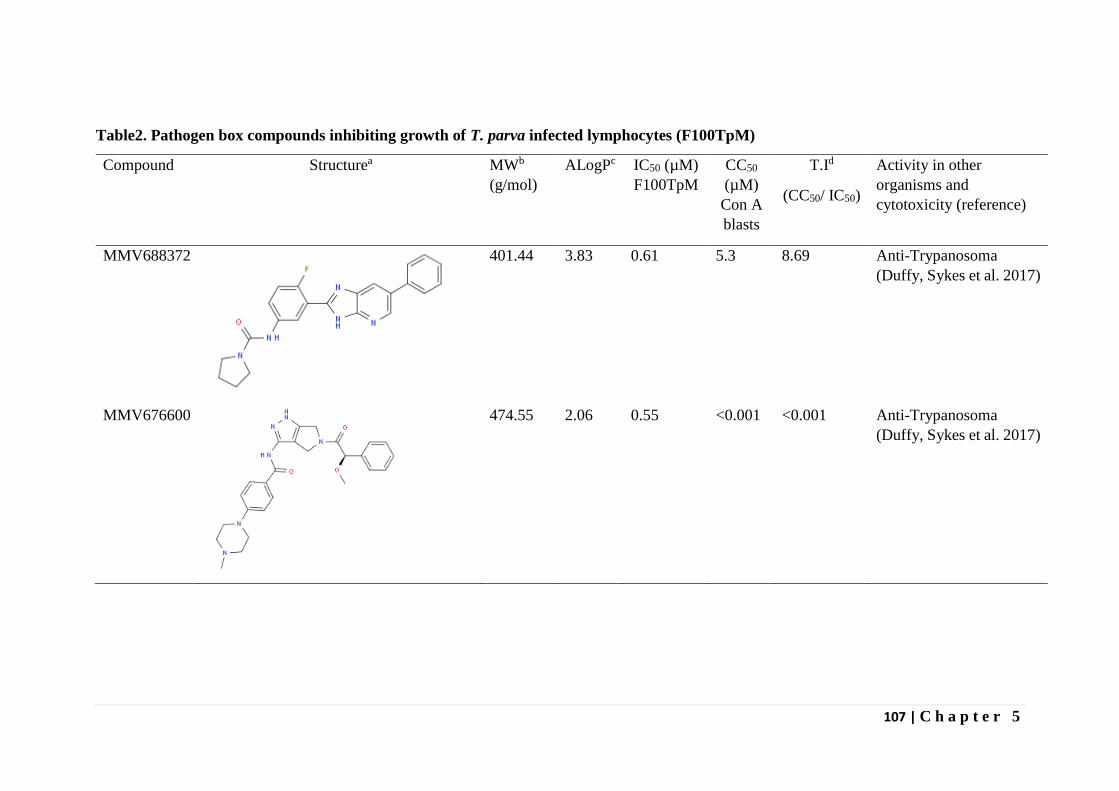
107 | C h a p t e r 5
Table2. Pathogen box compounds inhibiting growth of T. parva infected lymphocytes (F100TpM)
Compound Structurea MWb
(g/mol)
ALogPc IC50 (µM)
F100TpM
CC50
(µM)
Con A
blasts
T.Id
(CC50/ IC50)
Activity in other
organisms and
cytotoxicity (reference)
MMV688372
401.44 3.83 0.61 5.3 8.69 Anti-Trypanosoma
(Duffy, Sykes et al. 2017)
MMV676600
474.55 2.06 0.55 <0.001 <0.001 Anti-Trypanosoma
(Duffy, Sykes et al. 2017)
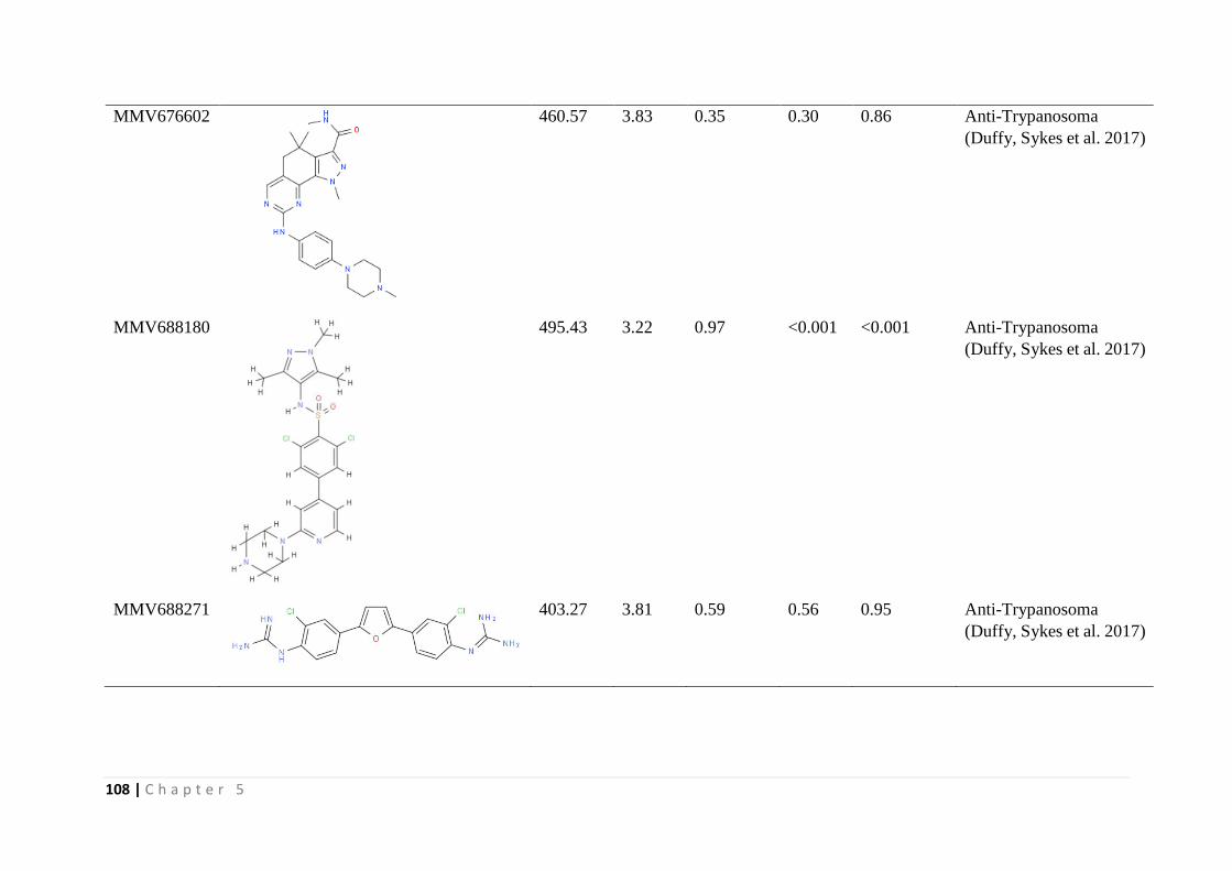
108 | C h a p t e r 5
MMV676602
460.57 3.83 0.35 0.30 0.86 Anti-Trypanosoma
(Duffy, Sykes et al. 2017)
MMV688180
495.43 3.22 0.97 <0.001 <0.001 Anti-Trypanosoma
(Duffy, Sykes et al. 2017)
MMV688271
403.27 3.81 0.59 0.56 0.95 Anti-Trypanosoma
(Duffy, Sykes et al. 2017)
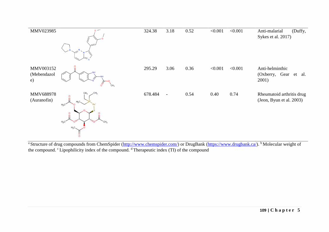
109 | C h a p t e r 5
MMV023985
324.38 3.18 0.52 <0.001 <0.001 Anti-malarial (Duffy,
Sykes et al. 2017)
MMV003152
(Mebendazol
e)
295.29 3.06 0.36 <0.001 <0.001 Anti-helminthic
(Oxberry, Gear et al.
2001)
MMV688978
(Auranofin)
678.484 - 0.54 0.40 0.74 Rheumatoid arthritis drug
(Jeon, Byun et al. 2003)
a Structure of drug compounds from ChemSpider (http://www.chemspider.com/) or DrugBank (https://www.drugbank.ca/). b Molecular weight of
the compound. c Lipophilicity index of the compound. d Therapeutic index (TI) of the compound
110 | C h a p t e r 5
The MMV malaria box was previously screened for activity against Theileria annulata
by Hostettler et al (2016) and MMV665820 was the only compound that was identified in our
anti-T.parva assay that also had activity against T. annulata (Hostettler, Müller et al. 2016).
Although this compound did not impair viability of the human foreskin fibroblast control cells
in the T. annulata assay, it was eliminated as a lead compound for not specifically targeting the
parasite since it failed to lower the relative expression of T. annulata gene TaSP by 50%
(Hostettler, Müller et al. 2016). In our assay, the therapeutic index for this compound was 1.67
leading to its elimination as a lead compound. The differences in drug activities between these
two related parasites illustrates the differences in biology between these parasites and the
tightrope walk towards development of anti-theilerial drug. The pathogen box compounds we
have identified should therefore, be tested against T. annulata, just as was done for the MMV
malaria box compounds.
Dasatinib exhibited very potent inhibitory activity on F100TpM cells with IC50 (5 nM)
and CC50 (>10 µM) values similar to buparvaquone, the current anti-ECF drug of choice. It
will be interesting to test dasatinib activity on T. annulata and other transforming Theileria
pathogen cell lines. Dasatinib is a commercial drug approved and indicated for chronic
myelogenous leukemia and acute lymphoblastic leukemia (Talpaz, Shah et al. 2006, Steinberg
2007). It is being considered for treatment of other cancers (Finn, Dering et al. 2007, Hochhaus,
Baccarani et al. 2008, Johnson, Bekele et al. 2010). In humans, the drug has multiple targets
including tyrosine-protein kinases (Bcr-Abl, Lck and Src family) (Quintas-Cardama,
Kantarjian et al. 2006), platelet-derived growth factor receptor beta (Chen, Lee et al. 2006) and
signal transducer and activator of transcription 5B (Nam, Williams et al. 2007). Recent target
similarity search using another apicomplexan parasite (P. falciparum) proteome identified
ABC transporters as possible target for dasatinib (Mogire, Akala et al. 2017). Therefore,
dasatinib’s mode of action on T. parva infected lymphocytes could be through one or a
combination of the targets, as these cells exhibit cancer-like properties with an up-regulation
of growth factor receptors, NF-kB and tyrosine protein kinase cell-signaling pathways, among
other cellular changes (Shiels, Langsley et al. 2006). This raises the possibility that the
inhibitory activity of dasatinib on parasite infected cells is via inhibiting a host cellular
pathway, rather than directly targeting the pathogen, and opens an additional avenue for drug
discovery for Theileria. Although dasatinib might have a direct effect on the parasite, this was
not tested in our current assay and we recommend testing this in future as this may reveal the
mode of action of this drug on the parasites.
In conclusion, we have identified two lead MMV compounds one from the malaria box
and the other from the pathogen box with a therapeutic index > 5 that present starting points
for development of alternative drugs to control ECF, and, possibly other forms of theileriosis.
In addition, we have demonstrated a very high therapeutic index of dasatinib on a T. parva
infected lymphocyte cell line. A large number of related tyrosine kinase inhibitors are available
and our results suggests that these molecules as well as other anti-cancer drugs could be
explored for their use as repurposed anti-theilerial drugs, provided cost is not an inhibitory
factor.
Acknowledgement
We are grateful to Robert Mugambi and Elizabeth Kibwana (International Livestock
Research Institute, Kenya) for providing cells. We thank the CGIAR Research Program on
Livestock and Fish, Kenya, the Norman Borlaug Commemorative Research Initiative, an
initiative between the Feed the Future program of United States Agency for International
Development, USA and United States Department of Agriculture-Agricultural Research
111 | C h a p t e r 5
Service, USA (58-5348-2-117F) and the Department for International Development of the
United Kingdom and the Bill and Melinda Gates Foundation, USA (OPP1078791) for financial
support. We would also like to thank MMV for provision of the Malaria and Pathogen box
compounds.
112 | C h a p t e r 5
References
Baldwin, C. L., et al. (1988). "Bovine T cells, B cells, and null cells are transformed by the
protozoan parasite Theileria parva." Infection and Immunity 56(2): 462-467.
Brown, C. G. (1990). "Control of tropical theileriosis (Theileria annulata infection) of cattle."
Parassitologia 32(1): 23-31.
Chen, Z., et al. (2006). "Potent inhibition of platelet-derived growth factor-induced responses
in vascular smooth muscle cells by BMS-354825 (dasatinib)." Molecular Pharmacology 69(5):
1527-1533.
Cui, L., et al. (2015). "Antimalarial Drug Resistance: Literature Review and Activities and
Findings of the ICEMR Network." The American Journal of Tropical Medicine and Hygiene
93(3_Suppl): 57-68.
Duffy, S., et al. (2017). "Screening the Medicines for Malaria Venture Pathogen Box across
Multiple Pathogens Reclassifies Starting Points for Open-Source Drug Discovery."
Antimicrobial Agents and Chemotherapy 61(9).
Finn, R. S., et al. (2007). "Dasatinib, an orally active small molecule inhibitor of both the src
and abl kinases, selectively inhibits growth of basal-type/“triple-negative” breast cancer cell
lines growing in vitro." Breast Cancer Research and Treatment 105(3): 319-326.
Hashemi-Fesharki, R. (1991). "Chemotherapeutic value of parvaquone and buparvaquone
against Theileria annulata infection of cattle." Research in Veterinary Science 50(2): 204-207.
Hochhaus, A., et al. (2008). "Dasatinib induces durable cytogenetic responses in patients with
chronic myelogenous leukemia in chronic phase with resistance or intolerance to imatinib."
Leukemia 22: 1200.
Hostettler, I., et al. (2016). "In vitro screening of the open source MMV malaria box reveals
novel compounds with profound activities against Theileria annulata schizonts." Antimicrobial
Agents and Chemotherapy.
Jeon, K. I., et al. (2003). "Gold compound auranofin inhibits IkappaB kinase (IKK) by
modifying Cys-179 of IKKbeta subunit." Exp Mol Med 35(2): 61-66.
Johnson, F. M., et al. (2010). "Phase II Study of Dasatinib in Patients With Advanced Non–
Small-Cell Lung Cancer." Journal of Clinical Oncology 28(30): 4609-4615.
Lancet (2015). "Efficacy and safety of RTS,S/AS01 malaria vaccine with or without a booster
dose in infants and children in Africa: final results of a phase 3, individually randomised,
controlled trial." Lancet 386(9988): 31-45.
113 | C h a p t e r 5
McHardy, N., et al. (1985). "Antitheilerial activity of BW720C (buparvaquone): a comparison
with parvaquone." Research in Veterinary Science 39(1): 29-33.
Mogire, R. M., et al. (2017). "Target-similarity search using Plasmodium falciparum proteome
identifies approved drugs with anti-malarial activity and their possible targets." PLoS One
12(10): e0186364.
Morrissette, N. S. and L. D. Sibley (2002). "Cytoskeleton of Apicomplexan Parasites."
Microbiology and Molecular Biology Reviews 66(1): 21-38.
Nam, S., et al. (2007). "Dasatinib (BMS-354825) inhibits Stat5 signaling associated with
apoptosis in chronic myelogenous leukemia cells." Molecular Cancer Therapeutics 6(4): 1400-
1405.
Nixon, G. L., et al. (2013). "Antimalarial pharmacology and therapeutics of atovaquone."
Journal of Antimicrobial Chemotherapy 68(5): 977-985.
O. Salas, C., et al. (2011). "Natural and Synthetic Naphthoquinones Active Against
Trypanosoma Cruzi: An Initial Step Towards New Drugs for Chagas Disease." Current
Medicinal Chemistry 18(1): 144-161.
Oxberry, M. E., et al. (2001). "Assessment of benzimidazole binding to individual recombinant
tubulin isotypes from Haemonchus contortus." Parasitology 122(Pt 6): 683-687.
Palacios, R. (1982). "Concanavalin A triggers T lymphocytes by directly interacting with their
receptors for activation." The Journal of Immunology 128(1): 337-342.
Quintas-Cardama, A., et al. (2006). "Targeting ABL and SRC kinases in chronic myeloid
leukemia: experience with dasatinib." Future Oncol 2(6): 655-665.
Radley, D. E. (1981). Infection and Treatment Method of Immunization Against Theileriosis.
Advances in the Control of Theileriosis: Proceedings of an International Conference held at the
International Laboratory for Research on Animal Diseases in Nairobi, 9–13th February, 1981.
A. D. Irvin, M. P. Cunningham and A. S. Young. Dordrecht, Springer Netherlands: 227-237.
Rigobello, M. P., et al. (2005). "Effect of auranofin on the mitochondrial generation of
hydrogen peroxide. Role of thioredoxin reductase." Free Radic Res 39(7): 687-695.
Shiels, B., et al. (2006). "Alteration of host cell phenotype by Theileria annulata and Theileria
parva: mining for manipulators in the parasite genomes." International Journal for Parasitology
36(1): 9-21.
114 | C h a p t e r 5
Steinberg, M. (2007). "Dasatinib: a tyrosine kinase inhibitor for the treatment of chronic
myelogenous leukemia and philadelphia chromosome-positive acute lymphoblastic leukemia."
Clin Ther 29(11): 2289-2308.
Talpaz, M., et al. (2006). "Dasatinib in imatinib-resistant Philadelphia chromosome-positive
leukemias." N Engl J Med 354(24): 2531-2541.
Van Voorhis, W. C., et al. (2016). "Open Source Drug Discovery with the Malaria Box
Compound Collection for Neglected Diseases and Beyond." PLoS Pathogens 12(7): e1005763.
Wampande, E. M., et al. (2007). "Classical ligands interact with native and recombinant tubulin
from Onchocerca volvulus with similar rank order of magnitude." Protein Expr Purif 55(2):
236-245.

115 | C h a p t e r 6
Chapter
6
Summarizing Discussion
James Nyagwange
116 | C h a p t e r 6
Introduction and aims of thesis
ECF caused by the hemoprotozoan pathogen Theileria parva is the leading tick-borne
disease of cattle in sub-Saharan Africa, causing death of cattle and significant economic losses
(Norval, Perry et al. 1992, De Deken, Martin et al. 2007). Currently control of the disease is
dependent on vaccination and drug treatment. The infection and treatment method of
vaccination involves deliberate infection with live sporozoites from three strains namely T.
parva Muguga, Kiambu5 and Serengeti (known as Muguga cocktail) and simultaneous
treatment with a long-acting oxytetracycline. Although effective, this method has serious
limitations which have been discussed in detail in Chapter 1. Briefly, the immunity generated
by the vaccine is strain specific and immunized cattle can become life-long asymptomatic
carriers of the parasite, posing risk for the spread of the disease through introduction of
‘foreign’ T. parva strains in areas where the strains in the vaccine are not present. The
production and distribution of the vaccine involves complex procedures requiring
infrastructure and specialized expertise which are not always available. These concerns
undermine the widespread uptake of this vaccine (Chapter 1). There have been efforts to
identify sporozoite antigens that are targets of neutralizing antibodies and one protein, p67 is
considered the lead candidate for development of a subunit vaccine. However, this protein only
protects only a proportion of immunized cattle (Chapter 1). Although research is ongoing on
different formulations and immunization regimen to improve the efficacy of this antigen,
protection against ECF might be improved by additional sporozoite antigens that induce
sporozoite neutralizing antibodies. Chemotherapy is another method of ECF control and is
currently dependent on the use of buparvaquone, a drug that was developed in the late 1980’s
and to which resistance has been reported in the related parasite Theileria annulata.
Fortunately, resistance has not been reported in T. parva, but this could occur, underscoring
the urgency for discovery of new cost effective drugs.
Traditionally, acaricides were used to prevent tick infestation and by extension ECF control.
However, as discussed in Chapter 1, this method is not sustainable as parasite challenge occurs
over many months of the year, thereby requiring application of acaricides over prolonged
periods making it expensive and detrimental to the environment. Additionally, this method
leaves the cattle population susceptible to infection by tick borne pathogens and vulnerable to
disease outbreaks, should the acaricides be unavailable due to local political or economic
changes.
These shortcomings of ECF control measures highlight the need for development of an
effective subunit vaccine and discovery of alternative drug compounds to provide a sustainable
means of controlling this disease.
The research described in this thesis (summarized in Figure 1) therefore aimed to
characterize the sporozoite proteome and identify novel proteins from the sporozoite stage that
can be evaluated as vaccine antigens. Furthermore, this thesis aimed to recombinantly express
and evaluate some of the identified proteins for neutralization of sporozoite infection of
lymphocytes. In addition, this study aimed at identifying new drug compounds that could be
used for the control of Theileria infections. Towards this end we took advantage of the open
source MMV malaria box, pathogen box and anti-cancer drug dasatanib to screen for molecules
that inhibit the growth of T. parva infected lymphoblastoid cells.
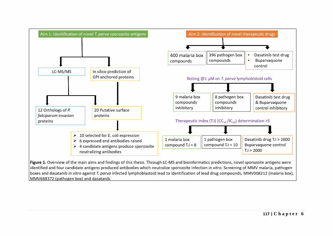
117 | C h a p t e r 6
118 | C h a p t e r 6
ECF vaccine development challenges
Vaccine development had its roots in the work of Edward Jenner (1749-1823), who
discovered how to protect people from smallpox through inoculation with a harmless but
related virus, cowpox and Louis Pasteur (1822-1895) who developed the first rabies vaccine
(Fenner 2000). Since then, vaccines have become the most cost-effective means of controlling
diseases with successful eradication of diseases such as smallpox and rinderpest, with great
progress towards poliomyelitis eradication.
Vaccine development follows approaches that either make use of whole organisms,
weakened or alive or components of the pathogens. Live vaccines can elicit both humoral and
cellular immunity and single or few doses usually give lifelong protection (Hansson, Nygren
et al. 2000). The Infection and Treatment Method (ITM) vaccination against ECF is a good
example of this type of vaccine in veterinary use. However, live vaccines have the potential to
cause disease through replication of the pathogens in the host. To circumvent this, the live
organisms are usually attenuated, for example by multiple passages in media, to prevent them
from causing disease (Hansson, Nygren et al. 2000). In Theileria, vaccination with passaged
infected leukocyte cell lines was evaluated and this approach induced immunity against T.
annulata but a similar approach for T. parva was not successful (Morrison and McKeever
2006). Transfer of infection by T. parva infected leukocyte cells occurred at a much lower
frequency, and immunity was only achieved definitively by administering large doses (≥ 108
cells) of parasitized cells (Morrison and McKeever 2006). Therefore, this method of
vaccination continued to be used in T. annulata while T. parva vaccination adopted the ITM
method (discussed in Chapter 1). The ITM method is possible because of oxtetracycline,
which is administered as a slow release formulation with activity lasting 5 to 6 days. This delays
development of the parasite leading to mild infection with recovery and induction of
immunological memory. Nevertheless, the risk of these organisms reverting to the original
virulent strain or forming new recombinant virulent strains presents a disadvantage of live
vaccines such as ITM.
Subunit vaccines on the other hand are designed to include only portions of the
pathogen required for protective immunization and are therefore safer than whole-inactivated
or live-attenuated vaccines (Hansson, Nygren et al. 2000). By combining technological
possibilities with knowledge in areas of immunology and molecular biology, candidate
antigens can be identified and delivered to the susceptible hosts inducing broad immunological
response and long-lived memory. Recombinant DNA techniques are now dominating the strive
for subunit vaccines with DNA vaccines such as West Nile-Innovator® DNA, by Fort Dodge
and APEX-IHN®, by Novartis already licenced for veterinary use in horses and farm raised
salmons respectively (Dalmo 2018). However, unlike live attenuated or killed vaccines, most
subunit antigens lack self-adjuvanting and immunomodulatory components (Hansson, Nygren
et al. 2000). This poses challenge in the development of subunit vaccines since they are often
poorly immunogenic and have short in vivo half-life (Hansson, Nygren et al. 2000). However,
these can be circumvented for example, by choice of a good delivery system and/or adjuvants
(Liljeqvist and Ståhl 1999, Hansson, Nygren et al. 2000). In T. parva, studies are in progress
with different formulations of p67 including; hepatitis B core expressed surface p67, p67
antigen in slow release vesicles and p67 expressing nanoparticles (Lacasta et al, unpublished
data). Previous formulations of p67 based vaccines have induced varied protection capacities
ranging from 13 to 70% (reveiwed in Nene, Kiara et al. 2016) as discussed in Chapter 1.
119 | C h a p t e r 6
Challenges with p67 vaccines stem from observations that cattle immunized produce high
antibody titres against p67, but in most experiments there has been no consistent correlate of
various antibody assays with immunity (Nene, Kiara et al. 2016). Furthermore levels of
immunity elicited by the vaccines in the field trials were overall lower compared to the results
obtained under well controlled experimental challenge (Nene, Kiara et al. 2016).
Identification of T. parva candidate vaccine antigens
To identify novel antigens and to improve understanding of the biology of T. parva, a
Multidimensional Protein Identification Technology (MudPIT) mass spectrometry-based
approach (reviewed in Schirmer, Yates et al. 2003) was followed in Chapter 2, to characterize
the proteome of T. parva sporozoites purified by ion-exchange chromatography (Musoke,
Morzaria et al. 1992). The MudPIT method chosen allows for analysis of fractions containing
several proteins using the bottom–up approach. The method basically involves direct
proteolysis of the proteins in the mixture and generation of a large number of peptides which
are subsequently separated by liquid chromatography before tandem mass spectrometry (Han,
Aslanian et al. 2008). Mass spectra are then generated and collected for as many peptides as
possible, and the results are searched by an algorithmic comparison (via SEQUEST or
MASCOT) against a database of proteins derived from genomic sequencing to identify the
peptides (Han, Aslanian et al. 2008). This approach was more appropriate for our sample,
which contained a mixture of proteins than the top down methods which allows for
identification of single or less complex protein mixtures and involves ionization of intact
proteins without enzymatic digestions (Han, Aslanian et al. 2008). As reported in Chapter 2,
a total of 4,780 proteins were identified; 2,773 R. appendiculatus proteins and 2,007 T. parva
proteins, representing approximately 50% of the total predicted re-annotated T. parva proteome
of 4084 proteins. About 40% of the identified proteins are annotated as hypothetical proteins
providing evidence that real genes encode for these proteins, although their function remains
unknown. This pool of hypothetical proteins presents an opportunity for discovery of novel
antigens.
The DE-52 column purification method we employed provided better coverage for T. parva
proteins than reported for P. falciparum sporozoites that also resides in the salivary glands of
the vector. P. falciparum proteomic studies based on DE-52 column purification reported
~13% (Lasonder, Janse et al. 2008), ~19% (Florens, Washburn et al. 2002) and ~36% (Lindner,
Swearingen et al. 2013) of the 5524 annotated P. falciparum genes. Although tick vector
proteins constituted 58% of the total proteins identified, this was still better compared with the
P. falciparum studies in which mosquito proteins made up 60.1% to 89% of the proteins
(Lasonder, Janse et al. 2008, Lindner, Swearingen et al. 2013). There are a number of factors
that could have resulted in better coverage for T. parva proteome compared to P. falciparum
proteome and also less contamination with vector derived proteins. Since the same DE-52
column purification method was used, differences in sample quality and processing prior to the
column purification could have contributed to the difference. For instance, after
homogenization of the sporozoite infected tick salivary glands we included a centrifugation
step to pellet the insoluble tick material and enrich the sporozoites in the supernatant. In the
three studies mentioned above, only Florens et al (2002) reported inclusion of a centrifugation
step to remove the large mosquito debris (Florens, Washburn et al. 2002). The quality of the
starting sample such as the number of the starting sporozoites, which is dependent on infection
rates can also influence the coverage for the parasite proteins, as more parasite starting material
should lead to more parasite protein identifications. For our study salivary glands were
dissected from 300 adult ticks infected with T. parva Muguga isolate with approximately 4–
5×10⁸ sporozoites (mean infection rate of ~28,570 sporozoites/acinus, ~15-20,000 infected
120 | C h a p t e r 6
acini) as starting material. Whereas Lindner et al and Florens et al did not state the number of
starting of sporozoites in the starting sample, Lasonder et al reported approximately 1–4×10⁷
sporozoites as starting material, approximately 10-fold less than our study with T. parva. The
main challenge with proteomic studies on salivary glands sporozoites is the contamination with
vector proteins since there are no in vitro methods to culture sporozoite stages and harvest
vector free parasites. To reduce vector contamination, double purification in which 17% w/v
Accudenz cushion purification followed by DEAE-cellulose chromatography was employed
Lindner et al (2013) and found to reduce vector contamination from 60.1% to 21% (Lindner,
Swearingen et al. 2013). Such a method can be adapted for purification of T. parva sporozoites
from tick salivary glands in future studies. Another method to improve sporozoite purity is by
using antibodies which bind sporozoite surface proteins such as anti-p67 monoclonal
antibodies to label and sort sporozoites from vector mixtures by Fluorescence-activated cell
sorting (FACS) provided there are enough sporozoites and its cost effective. In our hands
staining with monoclonal antibody, ARIV21.4 against p67C fragment resulted in very little
recovery of sporozites, requiring several samples to get enough sporozoites for mass
spectrometry. Optimization of antibody staining protocol and/or use of multiple antibodies
targeting different surface proteins might improve sporozoite yield after sorting.
We employed bioinformatics following mass spectrometry on sporozoite whole cell lysates
to identify surface proteins that are likey to be vaccine candidate antigens ( Chapter 2).
Although it was possible to identify many proteins including known surface antigens such as
p67, providing confidence in the data, other studies such as Lindner et al (2013) have employed
surface biotinylation followed by streptavidin capture to identify such proteins in P. falciparum
(Lindner, Swearingen et al. 2013). The latter method focuses on the surface proteome of
sporozoites and provides more confidence that the proteins are located on the surface or
secreted and could therefore be vaccine antigens. However, by targeting the whole cell
proteome, we were able to identify more proteins providing better understanding of the parasite
biology. For instance, we identified proteins involved in metabolic processes such as glycolysis
and citric acid cycle (Chapter 2, Figure 1) and proteins involved host cell transformation
(Chapter 2, section 3.5). To identify novel T. parva sporozoites proteins that play a role in
host entry, we clustered the identified proteins into orthologous groups with P. falciparum
genes, since this malaria causing parasite is well studied and its genomics, transcriptomics and
proteomics data is available. Clustering revealed orthologs of several proteins associated with
host cell invasion by P. falciparum, among them AMA-1 and CelTOS, which are microneme
proteins and leading malaria vaccine candidates (Kariu, Ishino et al. 2006, Remarque, Faber et
al. 2008). Orthologs of these proteins in T. parva can be recombinantly expressed and tested
for immunogenicity in cattle, and protection against parasite challenge.
Failure to identify some of the expected T. parva proteins e.g. the polymorphic immuno-
dominant molecule (PIM), a well-known antigen (Toye, Goddeeris et al. 1991) does not
directly imply absence of these proteins in the sample. There are several reasons why a protein
may not be identified by proteomics. Trypsin digestion of proteins with low or irregular
arginine/lysine content can lead to very short and very long tryptic peptides that fall out of the
acquired mass range and may therefore not be detected. This was possibly the case with PIM
protein whose sequence contains large repetitive domain rich in glutamine and proline amino
acid residues but devoid of lysine and arginine residues (Toye, Goddeeris et al. 1991). An in
silico digestion of PIM with trypsin led to prediction of few detectable tryptic peptides. Other
possible reasons for failure to detect proteins include; peptides from low abundance proteins
121 | C h a p t e r 6
may fall below the detection limit or are simply not sequenced by the mass spectrometer due
to limited dynamic range and/or speed of instrument acquisition. Furthermore, unknown,
complex, or amino acid modifications leading to alteration of the peptide mass can only be
detected in some cases by the Peaks software.
In Chapter 3, the 2,773 tick proteins co-purified with sporozoites were also analyzed. We
hypothesized that some of these proteins persisting after DE-52 column purification may be
bound or associated with sporozoites and therefore may be important vaccine candidates
themselves. Our analysis revealed a list of known tick antigens, some of which are being
explored for development of anti-tick vaccines such as RIM36 (Bishop, Lambson et al. 2002).
Tick proteins involved in host immunomodulation such as ixodegrins and lipocalins were also
found through this analysis. Whereas host immunomodulation proteins are reported to be useful
for successful tick feeding, association with sporozoites would make them important antigens
as they could aid sporozoite infection of host cells. These tick proteins therefore present an
opportunity for a combined vaccine approach targeting both the vector and the parasite.
However, it was not possible to identify the specific sporozoite bound tick proteins due to the
large number of tick proteins still present after elimination of housekeeping proteins and
proteins of known functions highlighting the limitation of the purification method used. As
discussed above, methods that lead to a purer sporozoite sample such as antibody staining and
FACS sorting, following DE-52 column and 17% w/v Accudenz cushion purification would
improve chances of identifying these proteins.
Neutralization of infection by antibodies from candidate antigens
Whereas different formulations and delivery systems for p67 antigen, e.g. viral vectors,
could improve immune response to the antigen, addition of novel sporozoite (associated)
antigens to a p67 vaccine might increase the level of induced protection, due to the additional
immunogenic epitopes. GPI anchors are common attachment signals for surface proteins of
parasites such as Plasmodium, Trypanosomes, Toxoplasma, etc. and many are promising
vaccine candidate antigens (Ferguson 1999, Ferguson MAJ 2009). One such example is the
circumsporozoite (CS) protein of Plasmodium which is the antigenic target of the malaria
vaccine RTS, S (Lancet, 2015). In an attempt to identify surface antigens, we searched the re-
annotated T. parva genome (cited in Tretina, Pelle et al. 2016) for genes predicted to contain
C-terminal GPI anchor signals, since they are likely to be located on the cell surface. Out of
the 21 highly probable GPI anchored proteins predicted by PredGPI (Pierleoni, Martelli et al.
2008), we selected the top 10 highly probable proteins for our study. We were able to express
conserved fragments of six of the proteins, raised murine antisera with these recombinant
proteins and performed sporozoite neutralization assays (SNA) (Chapter 4). SNA is a
common assay for screening of antigens to establish whether they are candidates for anti-
sporozoite vaccine development. This method is used in several studies involving parasites
with sporozoite stages such as Plasmodium, Cryptosporidium, Eimeria, etc. (Crane, Murray et
al. 1988, Perryman, Riggs et al. 1990, Kumar, Oliveira et al. 2004). In Chapter 3, we show
that antisera to two proteins, TpMuguga_01g00876 and TpMuguga_01g00939 (gp34), showed
similar or better sporozoites neutralization compared to the positive control p67 monoclonal
antibody. Whereas TpMuguga_01g00876 is a novel antigen with sporozoite neutralization
activity, gp34 was described before as a schizont specific antigen by Xue et al (2010), but was
never tested for sporozoite neutralization, perhaps since it was believed to be a schizont specific
antigen. However, we identified this protein by mass spectrometry (Chapter 2) with 12 unique
122 | C h a p t e r 6
peptides (peptides that match the particular protein sequence only, and do not match any other
protein in the database), providing confidence of its existence in the sporozoite stage. The other
two recombinant proteins, TpMuguga_01g00095 and TpMuguga_04g00437 (p104), produced
antisera which showed some neutralization, but to a lesser extent when compared to the p67
monoclonal antibody (Chapter 4). TpMuguga_01g00095 is another novel antigen but p104,
is a sporozoite microneme/rhoptry protein (Ebel, Gerhards et al. 1999) which was identified
before, but was never evaluated as vaccine antigen . These four are therefore promising vaccine
candidate antigens that should be formally validated in cattle for production of sporozoite
neutralizing antibodies and for protection against parasite challenge. However, priority should
be given to the two proteins, TpMuguga_01g00876 and gp34 which, produced the strongest
neutralizing antisera. The latter protein has been shown to undergo GPI modification when
expressed in mammalian cells (Xue, von Schubert et al. 2010) pointing to a possibility of this
protein existing as a GPI anchored protein in T. parva . Since there is no direct evidence for
addition of GPI anchors to T. parva expressed proteins, we searched for enzymes of the GPI
biosynthesis pathway and found eight Plasmodium homologs (Delorenzi, Sexton et al. 2002)
in T. parva genome pointing to the existence of a process for GPI anchor addition to T. parva
proteins. Four of the eight enzymes identified from the genome were confirmed to be expressed
in the sporozoite proteome (Chapter 2).
Mechanism of protective immunity by prospective subunit vaccine
Surface and secreted proteins such as p67 and the vaccine candidates identified in this thesis
can elicit antibody production leading to protective immunity by limiting the establishment of
the sporozoites and preventing infection. Antibodies to these proteins can also enhance
phagocytosis of sporozoites by opsonization and provide Fc receptors for C1q binding
triggering complement cascade and formation of membrane attack complex on the surface of
sporozoites (Musoke, Morzaria et al. 1992). Generally, identified exogenous antigens such as
p67, which is shed before parasite entry into host cells provides a CD4 dependent immunity.
The mechanism of which involves the protein being endocytosed by lymphocytes and dendritic
cells, processed into peptides and displayed on MHC class II molecules. Such a mechanism
would lead to stimulation of CD4 T cells which helps in production of antibodies, useful for
neutralization, opsonization and complement dependent lysis (Musoke, Morzaria et al. 1992).
In addition to known sporozoite surface and secreted proteins, we identified all the
characterized schizont CD8 T cell antigens in the sporozoite proteome (Graham, Pelle et al.
2006, Graham, Pellé et al. 2008). Among the schizont CD8 T cell antigens are housekeeping
genes such as, hsp90, translation elongation initiation factor 1A, etc. and therefore not
surprising to find at the sporozoites stage. However, identification of all schizont CD8 T cell
antigens in the sporozoite stage, suggests that for T. parva infection, both direct and cross
presentation mechanisms are involved in priming CTL responses in vivo by dendritic cells.
Cross presentation is the processes by which antigen presenting cells such as dendritic cells
uptake exogenous antigens, process them into peptides and display them on MHC class I
molecules for priming CTL response (Cruz, Colbert et al. 2017). The process of peptide display
on MHC class I molecules is present in all cells, but presentation of peptides is usually
restricted for self- antigens or antigens of pathogens infecting the cells. This mechanism known
as direct presentation, has been adapted by the immune system to help in identifying cells
infected by pathogens such that, cells displaying foreign peptides are destroyed while cells
displaying self-peptides are tolerated by CTL (Cruz, Colbert et al. 2017). However, since naïve
CD8 T cells do not directly patrol peripheral tissues looking for antigen presenting cells,
dendritic cells must survey the peripheral tissues where they collect exogenous antigens
123 | C h a p t e r 6
through phagocytosis or other endocytic mechanisms and then migrate to the T cell zone in
lymph nodes and display the antigens acquired as MHC class I bound peptides (Cruz, Colbert
et al. 2017). This ability of dendritic cells to present exogenous antigens as peptides on MHC
class I peptides implies that dendritic cells can present T. parva peptides to CD8 T cells via
both direct and cross presentation. The latter by uninfected dendritic cells while the former by
T. parva infected dendritic cells. The finding of CD8 antigens in the sporozoite suggests that
sporozoites phagocytosed by uninfected dendritic cells are processed and peptides cross
presented to CD8 T cells. Therefore, an ideal subunit vaccine for ECF control should be able
to induce B cells, CD4 and CD8 T-cells. A future vaccine can therefore combine humoral
inducing antigens such as p67 or the candidates identified herein with CD8 T cell antigens
especially Tp1 and Tp2 since they have been found to be dominant targets of the CD8+ T-cell
response in cattle expressing the A18 and A10 class I MHC haplotypes, respectively (Pelle,
Graham et al. 2011).
Identification of lead chemical inhibitors of T. parva infected lymphoblastoid cells
Chemotherapy of ECF and tropical theileriosis, caused by T. parva and T. annulata
respectively, is largely dependent on the use of buparvaquone, a derivative of naphthoquinones
developed in the 1980’s. Other than buparvaquone, derivatives of naphthoquinones have given
rise to development of anti-parasitic drugs, including commercial products to control malaria,
such as, atavaquone, a hydroxy-napthoquinone (Nixon, Moss et al. 2013). Unfortunately,
resistance to atavaquone emerged quite rapidly, and drug resistance to atavaquone in
Plasmodium is associated with mutations in the mitochondrial gene encoding apo-cytochrome
b (Nixon, Moss et al. 2013). Fortunately, T. parva resistance to buparvaquone has not been
described. However, the recent identification of drug resistance in T. annulata is a cause for
concern as resistance might occur in T. parva, which would make this drug of limited veterinary
use in the future.
The Medicines for Malaria Venture (MMV) was inaugurated in 1999, with a mandate to
develope new anti-malarial drugs, as it was recognized that the pipeline for developing such
drugs was small and not an attractive venture for the pharmaceutical industry. MMV has
catalyzed neglected disease drug discovery by making drug-like and probe-like compounds
available to the scientific community through the Malaria and Pathogen Box (Van Voorhis,
Adams et al. 2016). We took advantage of the availability of these compounds and screened
them against a bovine lymphocyte cell line infected with the schizont stage of T. parva (TpM).
A unique aspect of the biology of T. parva and T. annulata is that parasite infected leukocytes
behave and proliferate like cancer cells in vitro, a property that is dependent on parasite
viability. This phenomenon made it possible to screen for molecules that inhibit the growth of
the T. parva infected cells. In addition to MMV compounds, we also screened the cancer drug
dasatinib and used buparvaquone as control drug in these in vitro assays.
In the malaria and pathogen boxes, nine (IC50 values ranging from 0.04 to 1.93 µM) and
eight (IC50 values ranging from 0.35 to 0.97 µM) compounds inhibiting TpM proliferation were
identified, respectively, representing approximately 2% hit rate. Two compounds,
MMV008212 and MMV688372, from malaria box and pathogen box respectively, exhibited
an in vitro therapeutic index (CC50/IC50) of 8 and 10, respectively. Dasatanib and control drug
buparvaquone exhibited therapeutic indexes > 2000. An index > 5 has been used to determine
parasite selectivity of MMV compounds (reviewed in Duffy, Sykes et al. 2017). These
compounds with therapeutic indexes > 5 present lead starting point compounds for the
development of new anti-theilerial drugs. Although the majority of the malaria and pathogen
box compounds inhibiting TpM were also highly active on inhibiting the control cell line, they
124 | C h a p t e r 6
have been found to be less toxic in other cells such as zebrafish, HEK293 etc (Duffy, Sykes et
al. 2017). However, the low therapeutic indexes <5, raises questions on the role of these
compounds as anti-theilerial drugs. Interestingly, four pathogen box compounds were
inhibitory on the control cell line, than on F100TpM, indicating that there could be differences
in the molecular and cellular biology of the two cell lines.
The lead malaria box compound, MMV008212, has been tested against other parasites and
has activities against Trypanosoma cruzi (100% growth inhibition at 5 µM), Plasmodium
berghei ookinete (76% growth inhibition at 10 µM), P. falciparum gametocyte NF54-late stage
(91% growth inhibition at 5 µM) and P. falciparum 3D7 asexual blood stage (98% growth
inhibition at 5 µM) (reviewed in Van Voorhis, Adams et al. 2016). Cytotoxicity of this
compound was observed only in one cell line (U87, 76% cell death at 5 µM) of the nine cell
lines tested (Van Voorhis, Adams et al. 2016). However, its mode of action is still unknown.
MV688372, pathogen box compound with the high therapeutic index against F100TpM cells
has anti-trypanosomal activity. This compound is a substituted 2-phenylimidazopyridine with
activities against Trypanosoma brucei brucei and T. cruzi with therapeutic indexes of 230 and
24, respectively, in relation to HEK293 cells (Duffy, Sykes et al. 2017). The mode of action of
MMV688372 is still unclear, but a structurally related azabenzoxazole has been identified as a
proteasome inhibitor in kinetoplastid parasites (Duffy, Sykes et al. 2017). Dasatinib, which
exhibited very potent inhibitory activity on F100TpM cells is a commercial drug approved and
indicated for chronic myelogenous leukemia and acute lymphoblastic leukemia (Talpaz, Shah
et al. 2006, Steinberg 2007). It is being considered for treatment of other cancers (Finn, Dering
et al. 2007, Hochhaus, Baccarani et al. 2008, Johnson, Bekele et al. 2010)
Comparing the screening results of the malaria box in T. annulata (Hostettler, Müller et al.
2016) to T. parva revealed only one common inhibitory compound, MMV665820 in both
parasites. However, in both cases the compound was eliminated as a lead, in T. parva we
eliminated it for low therapeutic index while in T. annulata it was eliminated for not
specifically targeting the parasite (Hostettler, Müller et al. 2016). The rest of T. parva inhibitory
compounds were not leads in T. annulata screens and vice versa, illustrating the difficulty of
developing a universal anti-theilerial drug. The pathogen box compound, MMV688372 and
dasatanib, which are inhibitory to T. parva infected cells and have high therapeutic indexes,
should be tested in T. annulata.
Recommendations for future research
The major sporozoite antigen, p67 has been the focus and lead antigen for the
development of a subunit vaccine against ECF since the 1980s. Although considerable progress
has been made with this antigen, it is becoming clear that this single antigen may not be able
to provide sufficient protection. To provide a denser map of sporozoite antigens, we defined
the sporozoite proteome by mass spectrometry and identified novel parasite antigens. We
recommend screening some of these antigens for production of anti-sporozoite neutralizing
antibodies. Priority should be given to antigens that have been tested in related organisms as
vaccine antigens and found to induce protective antibodies such as CelTOs and AMA-1 (Table
2, Chapter 2) and novel putative surface proteins (Table 4, Chapter 2) which we have not tested.
Although desirable to test these antigens directly in cattle, it might be cost –effective to
eliminate antigens producing non-neutralizing antibodies by a preliminary murine study as
described in this thesis.
We have provided preliminary evidence that some of the antigens we selected and
expressed recombinantly generates sporozoite neutralizing antibodies. However, following
findings in which rats produced sporozoite neutralizing antibodies but cattle did not against
certain recombinant antigens, we recommend evaluation of these antigens in cattle, in
combination with p67 antigen to assess the nature of protection conferred by such a multivalent
125 | C h a p t e r 6
vaccine. Since we tested gene fragments but not the whole protein, it is tempting to speculate
that antisera to full length proteins of the four promising antigens would produce higher
neutralizing activities because of the longer sequence with putative additional epitopes.
Therefore, it is desirable to test the full length recombinant proteins for immunogenicity.
However, since E. coli expression of long proteins is usually accompanied by solubility and
expression problems of the recombinant proteins, we recommend other expression systems
such as the baculovirus expression system in insect cells or the yeast Pichia pastoris or
mammalian cells for full length expressions (Tebaldi, Williams et al. 2017). Alternatively,
several shorter fragments comprising the full protein can be expressed, combined and
evaluated.
In Chapter 3, we have reported on known tick vaccine antigens identification and
several tick proteins associated with host immune modulation. However, for anti-sporozoite
vaccines, it is necessary to identify the specific tick proteins bound on the sporozoite surface,
which was not possible from the purification method described in this thesis. We recommend
additional purification step, involving staining the sporozoites by monoclonal antibodies
against surface protein, such as p67, followed by FACS analysis and sorting. Such a purer
sample would reveal the specific tick proteins associated with the sporozoites.
Finally, on vaccine development, the finding of CD8 T-cell antigens in the sporozoite
proteome suggests a possibility of cross-presentation of exogenous antigens by dendritic cells
to prime CTL responses in vivo. By taking advantage of this process, future vaccines could be
a combination of sporozoite antigens such as p67 and/or the ones identified in this study and
schizont antigens, especially Tp1 and Tp2 since they have been found to be dominant targets
of the CD8+ T-cell response in cattle expressing the A18 and A10 class I MHC haplotypes,
respectively (Pelle, Graham et al. 2011). Such a combination vaccine might provide an
improved protection by inducing both humoral and cellular responses.
The drug compounds identified in this thesis (Chapter 5), via in vitro assays represent
lead compounds that could act as starting points for the development of drugs against T. parva
and possibly T. annulata. Although we have observed inhibition of growth of T. parva infected
lymphoblasts, we did not test the effect of these compounds on the parasite directly. We
recommend such tests as they could reveal the modes of action of these compounds and rule
out the possibility that the observed inhibition is based on other differences between the test
and control cells other than infection by the parasite. Such a test can be achieved for example,
by measuring the transcription levels of parasite genes in the course of drug administration in
relation to host genes by quantitative reverse transcriptase PCR.
Whereas the malaria box compounds have been screened in T. annulata, the pathogen
box compounds have not been screened for activity against this parasite. We recommend
screening of the pathogen box compounds in T. annulata especially the compounds found to
be active in T. parva as that could lead to discovery of a lead anti-theilerial compound. The
cancer drug, dasatinib was as effective as the standard anti-Theileria drug buparvaquone in in
vitro assays described in Chapter 5. This drug should be screened in T. annulata as well and
should be evaluated in cattle for control of ECF.

126 | C h a p t e r 6
References
Abdelhaleem, M. (2004). "Do human RNA helicases have a role in cancer?" Biochimica et
Biophysica Acta (BBA) - Reviews on Cancer 1704(1): 37-46.
Aurrecoechea, C., J. Brestelli, B. P. Brunk, S. Fischer, B. Gajria, X. Gao, A. Gingle, G. Grant,
O. S. Harb, M. Heiges, F. Innamorato, J. Iodice, J. C. Kissinger, E. T. Kraemer, W. Li, J. A.
Miller, V. Nayak, C. Pennington, D. F. Pinney, D. S. Roos, C. Ross, G. Srinivasamoorthy, J.
C. J. Stoeckert, R. Thibodeau, C. Treatman and H. Wang (2010). "EuPathDB: a portal to
eukaryotic pathogen databases." Nucleic Acids Research 38(suppl_1): D415-D419.
Babo Martins, S., G. Di Giulio, G. Lynen, A. Peters and J. Rushton (2010). "Assessing the
impact of East Coast Fever immunisation by the infection and treatment method in Tanzanian
pastoralist systems." Preventive Veterinary Medicine 97(3–4): 175-182.
Baldwin, C. L., S. J. Black, W. C. Brown, P. A. Conrad, B. M. Goddeeris, S. W. Kinuthia, P.
A. Lalor, N. D. MacHugh, W. I. Morrison and S. P. Morzaria (1988). "Bovine T cells, B cells,
and null cells are transformed by the protozoan parasite Theileria parva." Infection and
Immunity 56(2): 462-467.
Ballingall, K. T., D. M. Mwangi, N. D. MacHugh, E. L. N. Taracha, P. Totte and D. J.
McKeever (2000). "A highly sensitive, non-radioactive assay for T cell activation in cattle:
applications in screening for antigens recognised by CD4+ and CD8+ T cells." Journal of
Immunological Methods 239(1–2): 85-93.
Beaufays, J., B. Adam, C. Menten-Dedoyart, L. Fievez, A. Grosjean, Y. Decrem, P. P. Prevot,
S. Santini, R. Brasseur, M. Brossard, M. Vanhaeverbeek, F. Bureau, E. Heinen, L. Lins, L.
Vanhamme and E. Godfroid (2008). "Ir-LBP, an ixodes ricinus tick salivary LTB4-binding
lipocalin, interferes with host neutrophil function." PLoS One 3(12): e3987.
Birth, D., W. C. Kao and C. Hunte (2014). "Structural analysis of atovaquone-inhibited
cytochrome bc1 complex reveals the molecular basis of antimalarial drug action." Nat
Commun 5: 4029.
Bishop, R., B. Lambson, C. Wells, P. Pandit, J. Osaso, C. Nkonge, S. Morzaria, A. Musoke
and V. Nene (2002). "A cement protein of the tick Rhipicephalusappendiculatus, located in the
secretory e cell granules of the type III salivary gland acini, induces strong antibody responses
in cattle." International Journal for Parasitology 32(7): 833-842.
Bishop, R., V. Nene, J. Staeyert, J. Rowlands, J. Nyanjui, J. Osaso, S. Morzaria and A. Musoke
(2003). "Immunity to East Coast fever in cattle induced by a polypeptide fragment of the major
surface coat protein of Theileria parva sporozoites." Vaccine 21(11-12): 1205-1212.
127 | C h a p t e r 6
Brown, C. G. (1990). "Control of tropical theileriosis (Theileria annulata infection) of cattle."
Parassitologia 32(1): 23-31.
Brown, W. C., T. F. McElwain, G. H. Palmer, S. E. Chantler and D. M. Estes (1999). "Bovine
CD4+ T-Lymphocyte Clones Specific for Rhoptry-Associated Protein 1 of Babesia bigemina
Stimulate Enhanced Immunoglobulin G1 (IgG1) and IgG2 Synthesis." Infection and Immunity
67(1): 155-164.
Brown, W. C., M. K. Shaw, P. A. Conrad and T. T. Dolan (1989). "Theileria parva:
reappearance of schizonts in infected lymphoblastoid cells treated with parvaquone is
dependent on interleukin 2-like growth factors." Exp Parasitol 68(3): 308-325.
Brown, W. C., C. Sugimoto and D. J. Grab (1989). "Theileria parva: Bovine helper T cell
clones specific for both infected lymphocytes and schizont membrane antigens." Experimental
Parasitology 69(2): 234-248.
Brown, W. C., S. Zhao, K. S. Logan, D. J. Grab and A. C. Rice-Ficht (1995). "Identification of
candidate vaccine antigens of bovine hemoparasites Theileria parva and Babesia bovis by use
of helper T cell clones." Vet Parasitol 57(1-3): 189-203.
Budu, A. and C. R. S. Garcia (2012). "Generation of second messengers in Plasmodium."
Microbes and Infection 14(10): 787-795.
Büscher, G. and J. Tangus (1986). "Quantitative studies on Theileria parva in the salivary
glands of Rhipicephalus appendiculatus adults: Search for conditions for high infections."
International Journal for Parasitology 16(2): 121-129.
Cao, J., O. Kaneko, A. Thongkukiatkul, M. Tachibana, H. Otsuki, Q. Gao, T. Tsuboi and M.
Torii (2009). "Rhoptry neck protein RON2 forms a complex with microneme protein AMA1
in Plasmodium falciparum merozoites." Parasitology International 58(1): 29-35.
Chen, Z., F. Y. Lee, K. N. Bhalla and J. Wu (2006). "Potent inhibition of platelet-derived
growth factor-induced responses in vascular smooth muscle cells by BMS-354825 (dasatinib)."
Mol Pharmacol 69(5): 1527-1533.
Cingolani, P., A. Platts, L. L. Wang, M. Coon, T. Nguyen, L. Wang, S. J. Land, X. Lu and D.
M. Ruden (2012). "A program for annotating and predicting the effects of single nucleotide
polymorphisms, SnpEff: SNPs in the genome of Drosophila melanogaster strain w1118; iso-2;
iso-3." Fly 6(2): 80-92.
Cowman, A. F. and B. S. Crabb (2006). "Invasion of Red Blood Cells by Malaria Parasites."
Cell 124(4): 755-766.
128 | C h a p t e r 6
Crane, M. S., P. K. Murray, M. J. Gnozzio and T. T. MacDonald (1988). "Passive protection
of chickens against Eimeria tenella infection by monoclonal antibody." Infection and Immunity
56(4): 972-976.
Cruz, F. M., J. D. Colbert, E. Merino, B. A. Kriegsman and K. L. Rock (2017). "The Biology
and Underlying Mechanisms of Cross-Presentation of Exogenous Antigens on MHC-I
Molecules." Annual Review of Immunology 35(1): 149-176.
Cui, L., S. Mharakurwa, D. Ndiaye, P. K. Rathod and P. J. Rosenthal (2015). "Antimalarial
Drug Resistance: Literature Review and Activities and Findings of the ICEMR Network." The
American Journal of Tropical Medicine and Hygiene 93(3_Suppl): 57-68.
Cupp, E. W. (1991). "Biology of Ticks." Veterinary Clinics of North America: Small Animal
Practice 21(1): 1-26.
Curtidor, H., L. C. Patiño, G. Arévalo-Pinzón, M. E. Patarroyo and M. A. Patarroyo (2011).
"Identification of the Plasmodium falciparum rhoptry neck protein 5 (PfRON5)." Gene 474(1–
2): 22-28.
Daban, J.-R., S. Bartolomé and A. Bermúdez (1996). Rapid Staining of Proteins in
Polyacrylamide Gels with Nile Red. The Protein Protocols Handbook. J. M. Walker. Totowa,
NJ, Humana Press: 179-185.
Dalmo, R. A. (2018). "DNA vaccines for fish: Review and perspectives on correlates of
protection." Journal of Fish Diseases 41(1): 1-9.
De Deken, R., V. Martin, A. Saido, M. Madder, J. Brandt and D. Geysen (2007). "An outbreak
of East Coast Fever on the Comoros: A consequence of the import of immunised cattle from
Tanzania?" Veterinary Parasitology 143(3): 245-253.
Decrem, Y., J. Beaufays, V. Blasioli, K. Lahaye, M. Brossard, L. Vanhamme and E. Godfroid
(2008). "A family of putative metalloproteases in the salivary glands of the tick Ixodes ricinus."
Febs j 275(7): 1485-1499.
Delorenzi, M., A. Sexton, H. Shams-Eldin, R. T. Schwarz, T. Speed and L. Schofield (2002).
"Genes for Glycosylphosphatidylinositol Toxin Biosynthesis in Plasmodium falciparum."
Infection and Immunity 70(8): 4510-4522.
Dessauge, F., R. Lizundia, M. Baumgartner, M. Chaussepied and G. Langsley (2005). "Taking
the Myc is bad for Theileria." Trends in Parasitology 21(8): 377-385.
129 | C h a p t e r 6
Di Giulio, G., G. Lynen, S. Morzaria, C. Oura and R. Bishop (2009). "Live immunization
against East Coast fever – current status." Trends in Parasitology 25(2): 85-92.
Dobbelaere, D. and V. Heussler (1999). "Transformation of Leukocytes by Theileria parva and
T. annulata." Annual Review of Microbiology 53(1): 1-42.
Dobbelaere, D., S. Z. Shapiro and P. Webster (1985). "Identification of a surface antigen on
Theileria parva sporozoites by monoclonal antibody." Proceedings of the National Academy
of Sciences 82(6): 1771-1775.
Dobbelaere, D. A. E. and P. Küenzi (2004). "The strategies of the Theileria parasite: a new
twist in host–pathogen interactions." Current Opinion in Immunology 16(4): 524-530.
Dobbelaere, D. A. E., I. J. Roditi, T. s. M. Coquerelle, C. Kelke, M. Eichhorn and R. O.
Williams (1991). "Lymphocytes infected with Theileria parva require both cell-cell contact and
growth factor to proliferate." European Journal of Immunology 21(1): 89-95.
Dobbelaere, D. A. E., P. R. Spooner, W. C. Barry and A. D. Irvin (1984). "Monoclonal antibody
neutralizes the sporozoite stage of different Theileria parva stocks." Parasite Immunology 6(4):
361-370.
Duffy, S., M. L. Sykes, A. J. Jones, T. B. Shelper, M. Simpson, R. Lang, S.-A. Poulsen, B. E.
Sleebs and V. M. Avery (2017). "Screening the Medicines for Malaria Venture Pathogen Box
across Multiple Pathogens Reclassifies Starting Points for Open-Source Drug Discovery."
Antimicrobial Agents and Chemotherapy 61(9).
Dvorin, J. D., D. C. Martyn, S. D. Patel, J. S. Grimley, C. R. Collins, C. S. Hopp, A. T. Bright,
S. Westenberger, E. Winzeler, M. J. Blackman, D. A. Baker, T. J. Wandless and M. T.
Duraisingh (2010). "A Plant-Like Kinase in Plasmodium falciparum Regulates Parasite Egress
from Erythrocytes." Science 328(5980): 910-912.
Ebel, T., J. Gerhards, B. R. Binder and J. Lipp (1999). "Theileria parva 104 kDa microneme–
rhoptry protein is membrane-anchored by a non-cleaved amino-terminal signal sequence for
entry into the endoplasmic reticulum." Molecular and Biochemical Parasitology 100(1): 19-26.
Emanuelsson, O., H. Nielsen, S. Brunak and G. von Heijne (2000). "Predicting Subcellular
Localization of Proteins Based on their N-terminal Amino Acid Sequence." Journal of
Molecular Biology 300(4): 1005-1016.
Emery, D. L. (1981). "Adoptive transfer of immunity to infection with Theileria parva (East
Coast fever) between cattle twins." Research in Veterinary Science 30(3): 364-367.
130 | C h a p t e r 6
Fankhauser, N. and P. Mäser (2005). "Identification of GPI anchor attachment signals by a
Kohonen self-organizing map." Bioinformatics 21(9): 1846-1852.
Fawcett, D. W., G. Büscher and S. Doxsey (1982). "Salivary gland of the tick vector of east
coast fever. IV. Cell type selectivity and host cell responses to Theileria parva." Tissue and
Cell 14(2): 397-414.
Fawcett, D. W., S. Doxsey, D. A. Stagg and A. S. Young (1982b). "The entry of sporozoites
of Theileria parva into bovine lymphocytes in vitro. Electron microscopic observations."
European journal of cell biology 27(1): 10-21.
Fenner, F. (2000). "Adventures with poxviruses of vertebrates." FEMS Microbiology Reviews
24(2): 123-133.
Ferguson, M. A. (1999). "The structure, biosynthesis and functions of
glycosylphosphatidylinositol anchors, and the contributions of trypanosome research." Journal
of Cell Science 112(17): 2799-2809.
Ferguson MAJ, K. T., Hart GW (2009). Essentials of Glycobiology. Cold Spring Harbor (NY),
Cold Spring Harbor Laboratory Press.
Finn, R. S., J. Dering, C. Ginther, C. A. Wilson, P. Glaspy, N. Tchekmedyian and D. J. Slamon
(2007). "Dasatinib, an orally active small molecule inhibitor of both the src and abl kinases,
selectively inhibits growth of basal-type/“triple-negative” breast cancer cell lines growing in
vitro." Breast Cancer Research and Treatment 105(3): 319-326.
Florens, L., M. P. Washburn, J. D. Raine, R. M. Anthony, M. Grainger, J. D. Haynes, J. K.
Moch, N. Muster, J. B. Sacci, D. L. Tabb, A. A. Witney, D. Wolters, Y. Wu, M. J. Gardner, A.
A. Holder, R. E. Sinden, J. R. Yates and D. J. Carucci (2002). "A proteomic view of the
Plasmodium falciparum life cycle." Nature 419(6906): 520-526.
Francischetti, I. M. B., V. My Pham, B. J. Mans, J. F. Andersen, T. N. Mather, R. S. Lane and
J. M. C. Ribeiro (2005). "The transcriptome of the salivary glands of the female western black-
legged tick Ixodes pacificus (Acari: Ixodidae)." Insect Biochemistry and Molecular Biology
35(10): 1142-1161.
Gachet, C. (2008). "P2 receptors, platelet function and pharmacological implications." Thromb
Haemost 99(3): 466-472.
Gardner, M. J., R. Bishop, T. Shah, E. P. de Villiers, J. M. Carlton, N. Hall, Q. Ren, I. T.
Paulsen, A. Pain, M. Berriman, R. J. M. Wilson, S. Sato, S. A. Ralph, D. J. Mann, Z. Xiong, S.
J. Shallom, J. Weidman, L. Jiang, J. Lynn, B. Weaver, A. Shoaibi, A. R. Domingo, D. Wasawo,
J. Crabtree, J. R. Wortman, B. Haas, S. V. Angiuoli, T. H. Creasy, C. Lu, B. Suh, J. C. Silva,
131 | C h a p t e r 6
T. R. Utterback, T. V. Feldblyum, M. Pertea, J. Allen, W. C. Nierman, E. L. N. Taracha, S. L.
Salzberg, O. R. White, H. A. Fitzhugh, S. Morzaria, J. C. Venter, C. M. Fraser and V. Nene
(2005). "Genome Sequence of Theileria parva, a Bovine Pathogen That Transforms
Lymphocytes." Science 309(5731): 134-137.
Garrison, E. and G. Marth (2012). "Haplotype-based variant detection from short-read
sequencing." arXiv preprint arXiv:1207.3907.
Geysen, D., T. Bazarusanga, J. Brandt and T. T. Dolan (2004). "An unusual mosaic structure
of the PIM gene of Theileria parva and its relationship to allelic diversity." Molecular and
Biochemical Parasitology 133(2): 163-173.
Gilson, P. R., T. Nebl, D. Vukcevic, R. L. Moritz, T. Sargeant, T. P. Speed, L. Schofield and
B. S. Crabb (2006). "Identification and Stoichiometry of Glycosylphosphatidylinositol-
anchored Membrane Proteins of the Human Malaria Parasite Plasmodium falciparum."
Molecular & Cellular Proteomics 5(7): 1286-1299.
Graham, S. P., Y. Honda, R. Pellé, D. M. Mwangi, E. J. Glew, E. P. de Villiers, T. Shah, R.
Bishop, P. van der Bruggen, V. Nene and E. L. N. Taracha (2007). "A novel strategy for the
identification of antigens that are recognised by bovine MHC class I restricted cytotoxic T cells
in a protozoan infection using reverse vaccinology." Immunome Research 3: 2-2.
Graham, S. P., R. Pelle, Y. Honda, D. M. Mwangi, N. J. Tonukari, M. Yamage, E. J. Glew, E.
P. de Villiers, T. Shah, R. Bishop, E. Abuya, E. Awino, J. Gachanja, A. E. Luyai, F. Mbwika,
A. M. Muthiani, D. M. Ndegwa, M. Njahira, J. K. Nyanjui, F. O. Onono, J. Osaso, R. M. Saya,
C. Wildmann, C. M. Fraser, I. Maudlin, M. J. Gardner, S. P. Morzaria, S. Loosmore, S. C.
Gilbert, J. C. Audonnet, P. van der Bruggen, V. Nene and E. L. Taracha (2006). "Theileria
parva candidate vaccine antigens recognized by immune bovine cytotoxic T lymphocytes."
Proc Natl Acad Sci U S A 103(9): 3286-3291.
Graham, S. P., R. Pellé, M. Yamage, D. M. Mwangi, Y. Honda, R. S. Mwakubambanya, E. P.
de Villiers, E. Abuya, E. Awino, J. Gachanja, F. Mbwika, A. M. Muthiani, C. Muriuki, J. K.
Nyanjui, F. O. Onono, J. Osaso, V. Riitho, R. M. Saya, S. A. Ellis, D. J. McKeever, N. D.
MacHugh, S. C. Gilbert, J.-C. Audonnet, W. I. Morrison, P. van der Bruggen and E. L. N.
Taracha (2008). "Characterization of the Fine Specificity of Bovine CD8 T-Cell Responses to
Defined Antigens from the Protozoan Parasite Theileria parva." Infection and Immunity 76(2):
685-694.
Hall, R., N. R. Boulter, C. G. Brown, G. Wilkie, E. Kirvar, V. Nene, A. J. Musoke, E. J. Glass
and S. P. Morzaria (2000). "Reciprocal cross-protection induced by sporozoite antigens SPAG-
1 from Theileria annulata and p67 from Theileria parva." Parasite Immunology 22(5): 223-230.
Han, X., A. Aslanian and J. R. Yates (2008). "Mass spectrometry for proteomics." Current
Opinion in Chemical Biology 12(5): 483-490.
132 | C h a p t e r 6
Hansson, M., P.-A. k. Nygren and S. Sta˚hl (2000). "Design and production of recombinant
subunit vaccines." Biotechnology and Applied Biochemistry 32(2): 95-107.
Hashemi-Fesharki, R. (1991). "Chemotherapeutic value of parvaquone and buparvaquone
against Theileria annulata infection of cattle." Research in Veterinary Science 50(2): 204-207.
Havlíková, S., L. Roller, J. Koči, A. R. Trimnell, M. Kazimírová, B. Klempa and P. A. Nuttall
(2009). "Functional role of 64P, the candidate transmission-blocking vaccine antigen from the
tick, Rhipicephalus appendiculatus." International Journal for Parasitology 39(13): 1485-1494.
Hayashida, K., T. Abe, W. Weir, R. Nakao, K. Ito, K. Kajino, Y. Suzuki, F. Jongejan, D.
Geysen and C. Sugimoto (2013). "Whole-genome sequencing of Theileria parva strains
provides insight into parasite migration and diversification in the African continent." DNA Res
20(3): 209-220.
Hayashida, K., Y. Hara, T. Abe, C. Yamasaki, A. Toyoda, T. Kosuge, Y. Suzuki, Y. Sato, S.
Kawashima, T. Katayama, H. Wakaguri, N. Inoue, K. Homma, M. Tada-Umezaki, Y. Yagi, Y.
Fujii, T. Habara, M. Kanehisa, H. Watanabe, K. Ito, T. Gojobori, H. Sugawara, T. Imanishi,
W. Weir, M. Gardner, A. Pain, B. Shiels, M. Hattori, V. Nene and C. Sugimoto (2012).
"Comparative Genome Analysis of Three Eukaryotic Parasites with Differing Abilities To
Transform Leukocytes Reveals Key Mediators of Theileria-Induced Leukocyte
Transformation." mBio 3(5).
Hayashida, K., M. Hattori, R. Nakao, Y. Tanaka, J.-Y. Kim, N. Inoue, V. Nene and C.
Sugimoto (2010). "A schizont-derived protein, TpSCOP, is involved in the activation of NF-
κB in Theileria parva-infected lymphocytes." Molecular and Biochemical Parasitology 174(1):
8-17.
Hochhaus, A., M. Baccarani, M. Deininger, J. F. Apperley, J. H. Lipton, S. L. Goldberg, S.
Corm, N. P. Shah, F. Cervantes, R. T. Silver, D. Niederwieser, R. M. Stone, H. Dombret, R.
A. Larson, L. Roy, T. Hughes, M. C. Müller, R. Ezzeddine, A. M. Countouriotis and H. M.
Kantarjian (2008). "Dasatinib induces durable cytogenetic responses in patients with chronic
myelogenous leukemia in chronic phase with resistance or intolerance to imatinib." Leukemia
22: 1200.
Holder, A. A., M. A. Mohd Ridzuan and J. L. Green (2012). "Calcium dependent protein kinase
1 and calcium fluxes in the malaria parasite." Microbes and Infection 14(10): 825-830.
Hostettler, I., J. Müller and A. Hemphill (2016). "In vitro screening of the open source MMV
malaria box reveals novel compounds with profound activities against Theileria annulata
schizonts." Antimicrobial Agents and Chemotherapy.
133 | C h a p t e r 6
Huber, S., R. Theiler, D. de Quervain, O. Wiens, T. Karangenc, V. Heussler, D. Dobbelaere
and K. Woods (2017). "The Microtubule-Stabilizing Protein CLASP1 Associates with the
<em>Theileria annulata</em> Schizont Surface via Its Kinetochore-Binding Domain."
mSphere 2(4).
Hulliger, L., K. H. Wilde, C. G. Brown and L. Turner (1964). "MODE OF MULTIPLICATION
OF THEILERIA IN CULTURES OF BOVINE LYMPHOCYTIC CELLS." Nature 203: 728-
730.
Iams, K. P., J. R. Young, V. Nene, J. Desai, P. Webster, O. K. ole-MoiYoi and A. J. Musoke
(1990). "Characterisation of the gene encoding a 104-kilodalton micronemerhoptry protein of
Theileria parva." Molecular and Biochemical Parasitology 39(1): 47-60.
Ikezawa, H. (2002). "Glycosylphosphatidylinositol (GPI)-Anchored Proteins." Biological and
Pharmaceutical Bulletin 25(4): 409-417.
Imamura, S., S. Konnai, S. Vaz Ida, S. Yamada, C. Nakajima, Y. Ito, T. Tajima, J. Yasuda, M.
Simuunza, M. Onuma and K. Ohashi (2008). "Effects of anti-tick cocktail vaccine against
Rhipicephalus appendiculatus." Jpn J Vet Res 56(2): 85-98.
Imamura, S., B. Namangala, T. Tajima, M. E. Tembo, J. Yasuda, K. Ohashi and M. Onuma
(2006). "Two serine protease inhibitors (serpins) that induce a bovine protective immune
response against Rhipicephalus appendiculatus ticks." Vaccine 24(13): 2230-2237.
Irvin, A. D. and W. I. Morrison (1987). Immunopathology, immunology, and
immunoprophylaxis of Theileria infections. Boca Raton, Florida, CRC Press, Inc.: 223-274.
Ishihama, Y., Y. Oda, T. Tabata, T. Sato, T. Nagasu, J. Rappsilber and M. Mann (2005).
"Exponentially Modified Protein Abundance Index (emPAI) for Estimation of Absolute
Protein Amount in Proteomics by the Number of Sequenced Peptides per Protein." Molecular
& Cellular Proteomics 4(9): 1265-1272.
Jaworski, D. C., F. A. Simmen, W. Lamoreaux, L. B. Coons, M. T. Muller and G. R. Needham
(1995). "A secreted calreticulin protein in ixodid tick (Amblyomma americanum) saliva."
Journal of Insect Physiology 41(4): 369-375.
Jeon, K. I., M. S. Byun and D. M. Jue (2003). "Gold compound auranofin inhibits IkappaB
kinase (IKK) by modifying Cys-179 of IKKbeta subunit." Exp Mol Med 35(2): 61-66.
Johnson, F. M., B. N. Bekele, L. Feng, I. Wistuba, X. M. Tang, H. T. Tran, J. J. Erasmus, L.-
L. Hwang, N. Takebe, G. R. Blumenschein, S. M. Lippman and D. J. Stewart (2010). "Phase
II Study of Dasatinib in Patients With Advanced Non–Small-Cell Lung Cancer." Journal of
Clinical Oncology 28(30): 4609-4615.
134 | C h a p t e r 6
Jones, M. L., E. L. Kitson and J. C. Rayner (2006). "Plasmodium falciparum erythrocyte
invasion: A conserved myosin associated complex." Molecular and Biochemical Parasitology
147(1): 74-84.
Kaba, S. A. (2003). Development of a novel subunit vaccine against East Coast fever based on
the Theileria parva sporozoite surface protein p67.
Kariu, T., T. Ishino, K. Yano, Y. Chinzei and M. Yuda (2006). "CelTOS, a novel malarial
protein that mediates transmission to mosquito and vertebrate hosts." Molecular Microbiology
59(5): 1369-1379.
Kariuki, D. P., A. S. Young, S. P. Morzaria, A. C. Lesan, S. K. Mining, P. Omwoyo, J. L.
Wafula and D. H. Molyneux (1995). "Theileria parva carrier state in naturally infected and
artificially immunised cattle." Trop Anim Health Prod 27(1): 15-25.
Khan, S. M., B. Franke-Fayard, G. R. Mair, E. Lasonder, C. J. Janse, M. Mann and A. P. Waters
(2005). "Proteome Analysis of Separated Male and Female Gametocytes Reveals Novel Sex-
Specific Plasmodium Biology." Cell 121(5): 675-687.
Krogh, A., B. Larsson, G. von Heijne and E. L. L. Sonnhammer (2001). "Predicting
transmembrane protein topology with a hidden markov model: application to complete
genomes1." Journal of Molecular Biology 305(3): 567-580.
Kumar, K. A., G. A. Oliveira, R. Edelman, E. Nardin and V. Nussenzweig (2004).
"Quantitative Plasmodium sporozoite neutralization assay (TSNA)." Journal of Immunological
Methods 292(1): 157-164.
Lancet (2015). "Efficacy and safety of RTS,S/AS01 malaria vaccine with or without a booster
dose in infants and children in Africa: final results of a phase 3, individually randomised,
controlled trial." Lancet 386(9988): 31-45.
Lasonder, E., C. J. Janse, G.-J. van Gemert, G. R. Mair, A. M. W. Vermunt, B. G. Douradinha,
V. van Noort, M. A. Huynen, A. J. F. Luty, H. Kroeze, S. M. Khan, R. W. Sauerwein, A. P.
Waters, M. Mann and H. G. Stunnenberg (2008). "Proteomic Profiling of
<italic>Plasmodium</italic> Sporozoite Maturation Identifies New Proteins Essential for
Parasite Development and Infectivity." PLoS Pathog 4(10): e1000195.
Latif, A. A. and T. Hove (2011). "History and critical review of Theileria parva (Boleni), the
vaccine stock against Zimbabwean cattle theileriosis." Ticks and Tick-borne Diseases 2(3):
163-167.
Lauterbach, S. B. and T. L. Coetzer (2008). "The M18 aspartyl aminopeptidase of Plasmodium
falciparum binds to human erythrocyte spectrin in vitro." Malaria Journal 7: 1-10.
135 | C h a p t e r 6
Lawrence, J. A. (1979). "The differential diagnosis of the bovine theilerias of southern Africa."
Journal of the South African Veterinary Association 50(4): 311-313.
Li, L., C. J. Stoeckert and D. S. Roos (2003). "OrthoMCL: Identification of Ortholog Groups
for Eukaryotic Genomes." Genome Research 13(9): 2178-2189.
Liljeqvist, S. and S. Ståhl (1999). "Production of recombinant subunit vaccines: protein
immunogens, live delivery systems and nucleic acid vaccines." Journal of Biotechnology
73(1): 1-33.
Lindner, S. E., K. E. Swearingen, A. Harupa, A. M. Vaughan, P. Sinnis, R. L. Moritz and S.
H. Kappe (2013). "Total and putative surface proteomics of malaria parasite salivary gland
sporozoites." Mol Cell Proteomics 12(5): 1127-1143.
Marsolier, J., M. Perichon, J. D. DeBarry, B. O. Villoutreix, J. Chluba, T. Lopez, C. Garrido,
X. Z. Zhou, K. P. Lu, L. Fritsch, S. Ait-Si-Ali, M. Mhadhbi, S. Medjkane and J. B. Weitzman
(2015). "Theileria parasites secrete a prolyl isomerase to maintain host leukocyte
transformation." Nature 520(7547): 378-382.
Marsolier, J., M. Perichon, J. D. DeBarry, B. O. Villoutreix, J. Chluba, T. Lopez, C. Garrido,
X. Z. Zhou, K. P. Lu, L. Fritsch, S. Ait-Si-Ali, M. Mhadhbi, S. Medjkane and J. B. Weitzman
(2015). "Theileria parasites secrete a prolyl isomerase to maintain host leukocyte
transformation." Nature 520: 378.
Matson, B. A. (1967). "Theileriosis in Rhodesia: 1. A study of diagnostic specimens over two
seasons." Journal of the South African Veterinary Association 38(1): 93-102.
Mbizeni, S., F. T. Potgieter, C. Troskie, B. J. Mans, B. L. Penzhorn and A. A. Latif (2013).
"Field and laboratory studies on Corridor disease (Theileria parva infection) in cattle
population at the livestock/game interface of uPhongolo-Mkuze area, South Africa." Ticks and
Tick-borne Diseases 4(3): 227-234.
McHardy, N., L. S. Wekesa, A. T. Hudson and A. W. Randall (1985). "Antitheilerial activity
of BW720C (buparvaquone): a comparison with parvaquone." Research in Veterinary Science
39(1): 29-33.
McKeever, D. J., E. L. Taracha, E. L. Innes, N. D. MacHugh, E. Awino, B. M. Goddeeris and
W. I. Morrison (1994). "Adoptive transfer of immunity to Theileria parva in the CD8+ fraction
of responding efferent lymph." Proceedings of the National Academy of Sciences 91.
McLeod, A. and R. Kristjanson (1999). "Impact of ticks and associated diseases on cattle in
Asia, Australia and Africa. ILRI and eSYS report to ACIAR."

136 | C h a p t e r 6
Mhadhbi, M., M. Chaouch, K. Ajroud, M. A. Darghouth and S. BenAbderrazak (2015).
"Sequence Polymorphism of Cytochrome b Gene in Theileria annulata Tunisian Isolates and
Its Association with Buparvaquone Treatment Failure." PLoS ONE 10(6): e0129678.
Mhadhbi, M., A. Naouach, A. Boumiza, M. F. Chaabani, S. BenAbderazzak and M. A.
Darghouth (2010). "In vivo evidence for the resistance of Theileria annulata to buparvaquone."
Veterinary Parasitology 169(3–4): 241-247.
Mogire, R. M., H. M. Akala, R. W. Macharia, D. W. Juma, A. C. Cheruiyot, B. Andagalu, M.
L. Brown, H. A. El-Shemy and S. G. Nyanjom (2017). "Target-similarity search using
Plasmodium falciparum proteome identifies approved drugs with anti-malarial activity and
their possible targets." PLOS ONE 12(10): e0186364.
Morrison, W. I. (2009). "Progress towards understanding the immunobiology of Theileria
parasites." Parasitology 136.
Morrison, W. I., T. Connelley, J. D. Hemmink and N. D. MacHugh (2015). "Understanding
the basis of parasite strain-restricted immunity to Theileria parva." Annu Rev Anim Biosci 3:
397-418.
Morrison, W. I. and B. M. Goddeeris (1990). Cytotoxic T Cells in Immunity to Theileria parva
in Cattle. T-Cell Paradigms in Parasitic and Bacterial Infections. S. H. E. Kaufmann. Berlin,
Heidelberg, Springer Berlin Heidelberg: 79-93.
Morrison, W. I., N. D. MacHugh and P. A. Lalor (1996). "Pathogenicity of Theileria parva is
influenced by the host cell type infected by the parasite." Infection and Immunity 64(2): 557-
562.
Morrison, W. I. and D. J. McKeever (2006). "Current status of vaccine development against
Theileria parasites." Parasitology 133(S2): S169-S187.
Morrissette, N. S. and L. D. Sibley (2002). "Cytoskeleton of Apicomplexan Parasites."
Microbiology and Molecular Biology Reviews 66(1): 21-38.
Morzaria, S., V. Nene, R. Bishop and A. Musoke (2000). "Vaccines against Theileria parva."
Annals of the New York Academy of Sciences 916(1): 464-473.
Morzaria, S. P., J. Katende, A. Musoke, V. Nene, R. Skilton and R. Bishop (1999).
"Development of sero-diagnostic and molecular tools for the control of important tick-borne
pathogens of cattle in Africa." Parassitologia 41 Suppl 1: 73-80.
137 | C h a p t e r 6
Mulenga, A., C. Sugimoto and M. Onuma (2000). "Issues in tick vaccine development:
identification and characterization of potential candidate vaccine antigens." Microbes and
Infection 2(11): 1353-1361.
Musoke, A., S. Morzaria, C. Nkonge, E. Jones and V. Nene (1992). "A recombinant sporozoite
surface antigen of Theileria parva induces protection in cattle." Proceedings of the National
Academy of Sciences 89(2): 514-518.
Musoke, A. J., V. M. Nantulya, G. Buscher, R. A. Masake and B. Otim (1982). "Bovine
immune response to Theileria parva: neutralizing antibodies to sporozoites." Immunology
45(4): 663-668.
Musoke, A. J., V. M. Nantulya, F. R. Rurangirwa and G. Buscher (1984). "Evidence for a
common protective antigenic determinant on sporozoites of several Theileria parva strains."
Immunology 52(2): 231-238.
Nam, S., A. Williams, A. Vultur, A. List, K. Bhalla, D. Smith, F. Y. Lee and R. Jove (2007).
"Dasatinib (BMS-354825) inhibits Stat5 signaling associated with apoptosis in chronic
myelogenous leukemia cells." Mol Cancer Ther 6(4): 1400-1405.
Nene, V., H. Kiara, A. Lacasta, R. Pelle, N. Svitek and L. Steinaa (2016). "The biology of
Theileria parva and control of East Coast fever – Current status and future trends." Ticks and
Tick-borne Diseases 7(4): 549-564.
Nene, V., D. Lee, S. Kang’a, R. Skilton, T. Shah, E. de Villiers, S. Mwaura, D. Taylor, J.
Quackenbush and R. Bishop (2004). "Genes transcribed in the salivary glands of female
Rhipicephalus appendiculatus ticks infected with Theileria parva." Insect Biochemistry and
Molecular Biology 34(10): 1117-1128.
Nene, V. and W. I. Morrison (2016). "Approaches to vaccination against Theileria parva and
Theileria annulata." Parasite Immunology 38(12): 724-734.
Nene, V., A. Musoke, E. Gobright and S. Morzaria (1996). "Conservation of the sporozoite
p67 vaccine antigen in cattle-derived Theileria parva stocks with different cross-immunity
profiles." Infection and Immunity 64(6): 2056-2061.
Nixon, G. L., D. M. Moss, A. E. Shone, D. G. Lalloo, N. Fisher, P. M. O'Neill, S. A. Ward and
G. A. Biagini (2013). "Antimalarial pharmacology and therapeutics of atovaquone." Journal of
Antimicrobial Chemotherapy 68(5): 977-985.
Norval, R. A. I., B. D. Perry and A. Young (1992). The epidemiology of theileriosis in Africa,
Academic press, London.
138 | C h a p t e r 6
Nyagwange, J., E. Tijhaar, N. Ternette, F. Mobegi, K. Tretina, J. C. Silva, R. Pelle and V. Nene
(2018). "Characterization of the Theileria parva sporozoite proteome." International Journal
for Parasitology 48(3): 265-273.
O'Flaherty, J. T. and J. F. Cordes (1994). "Human neutrophil degranulation responses to
nucleotides." Lab Invest 70(6): 816-821.
O. Salas, C., M. Faundez, A. Morello, J. Diego Maya and R. A. Tapia (2011). "Natural and
Synthetic Naphthoquinones Active Against Trypanosoma Cruzi: An Initial Step Towards New
Drugs for Chagas Disease." Current Medicinal Chemistry 18(1): 144-161.
Odongo, D. O., J. D. Sunter, H. K. Kiara, R. A. Skilton and R. P. Bishop (2010). "A nested
PCR assay exhibits enhanced sensitivity for detection of Theileria parva infections in bovine
blood samples from carrier animals." Parasitol Res 106(2): 357-365.
Oxberry, M. E., T. G. Gear and R. K. Prichard (2001). "Assessment of benzimidazole binding
to individual recombinant tubulin isotypes from Haemonchus contortus." Parasitology 122(Pt
6): 683-687.
Paesen, G. C., P. L. Adams, K. Harlos, P. A. Nuttall and D. I. Stuart (1999). "Tick histamine-
binding proteins: isolation, cloning, and three-dimensional structure." Mol Cell 3(5): 661-671.
Pain, A., H. Renauld, M. Berriman, L. Murphy, C. A. Yeats, W. Weir, A. Kerhornou, M. Aslett,
R. Bishop, C. Bouchier, M. Cochet, R. M. R. Coulson, A. Cronin, E. P. de Villiers, A. Fraser,
N. Fosker, M. Gardner, A. Goble, S. Griffiths-Jones, D. E. Harris, F. Katzer, N. Larke, A. Lord,
P. Maser, S. McKellar, P. Mooney, F. Morton, V. Nene, S. Neil, C. Price, M. A. Quail, E.
Rabbinowitsch, N. D. Rawlings, S. Rutter, D. Saunders, K. Seeger, T. Shah, R. Squares, S.
Squares, A. Tivey, A. R. Walker, J. Woodward, D. A. E. Dobbelaere, G. Langsley, M.-A.
Rajandream, D. McKeever, B. Shiels, A. Tait, B. Barrell and N. Hall (2005). "Genome of the
Host-Cell Transforming Parasite Theileria annulata Compared with T. parva." Science
309(5731): 131.
Palacios, R. (1982). "Concanavalin A triggers T lymphocytes by directly interacting with their
receptors for activation." The Journal of Immunology 128(1): 337-342.
Parizi, L. F., N. W. Githaka, C. Logullo, S. Konnai, A. Masuda, K. Ohashi and I. da Silva Vaz
(2012). "The quest for a universal vaccine against ticks: Cross-immunity insights." The
Veterinary Journal 194(2): 158-165.
Patel, E., S. Mwaura, H. Kiara, S. Morzaria, A. Peters and P. Toye (2016). "Production and
dose determination of the Infection and Treatment Method (ITM) Muguga cocktail vaccine
used to control East Coast fever in cattle." Ticks and Tick-borne Diseases 7(2): 306-314.
139 | C h a p t e r 6
Pelle, R., S. P. Graham, M. N. Njahira, J. Osaso, R. M. Saya, D. O. Odongo, P. G. Toye, P. R.
Spooner, A. J. Musoke, D. M. Mwangi, E. L. N. Taracha, W. I. Morrison, W. Weir, J. C. Silva
and R. P. Bishop (2011). "Two Theileria parva CD8 T Cell Antigen Genes Are More Variable
in Buffalo than Cattle Parasites, but Differ in Pattern of Sequence Diversity." PLOS ONE 6(4):
e19015.
Perry, B. D. and A. S. Young (1993). "The naming game: the changing fortunes of East Coast
fever and Theileria parva." Vet Rec 133(25-26): 613-616.
Perryman, L. E., M. W. Riggs, P. H. Mason and R. Fayer (1990). "Kinetics of Cryptosporidium
parvum sporozoite neutralization by monoclonal antibodies, immune bovine serum, and
immune bovine colostrum." Infection and Immunity 58(1): 257-259.
Petersen, T. N., S. Brunak, G. von Heijne and H. Nielsen (2011). "SignalP 4.0: discriminating
signal peptides from transmembrane regions." Nature Methods 8(10): 785-786.
Pierleoni, A., P. L. Martelli and R. Casadio (2008). "PredGPI: a GPI-anchor predictor." BMC
Bioinformatics 9(1): 392.
Pradel, G. (2007). "Proteins of the malaria parasite sexual stages: expression, function and
potential for transmission blocking strategies." Parasitology 134(14): 1911-1929.
Quintas-Cardama, A., H. Kantarjian and J. Cortes (2006). "Targeting ABL and SRC kinases in
chronic myeloid leukemia: experience with dasatinib." Future Oncol 2(6): 655-665.
Radley, D. E. (1981). Infection and Treatment Method of Immunization Against Theileriosis.
Advances in the Control of Theileriosis: Proceedings of an International Conference held at the
International Laboratory for Research on Animal Diseases in Nairobi, 9–13th February, 1981.
A. D. Irvin, M. P. Cunningham and A. S. Young. Dordrecht, Springer Netherlands: 227-237.
Radley, D. E., C. G. D. Brown, M. J. Burridge, M. P. Cunningham, I. M. Kirimi, R. E. Purnell
and A. S. Young (1975). "East coast fever: 1. Chemoprophylactic immunization of cattle
against Theileria parva (Muguga) and five theilerial strains." Veterinary Parasitology 1(1): 35-
41.
Radley, D. E., C. G. D. Brown, M. P. Cunningham, C. D. Kimber, F. L. Musisi, R. C. Payne,
R. E. Purnell, S. M. Stagg and A. S. Young (1975b). "East coast fever: 3. Chemoprophylactic
immunization of cattle using oxytetracycline and a combination of theilerial strains."
Veterinary Parasitology 1(1): 51-60.
Remarque, E. J., B. W. Faber, C. H. M. Kocken and A. W. Thomas (2008). "Apical membrane
antigen 1: a malaria vaccine candidate in review." Trends in Parasitology 24(2): 74-84.

140 | C h a p t e r 6
Ribeiro, J. M., J. M. Anderson, N. C. Manoukis, Z. Meng and I. M. Francischetti (2011). "A
further insight into the sialome of the tropical bont tick, Amblyomma variegatum." BMC
Genomics 12(1): 136.
Ribeiro, J. M. and T. N. Mather (1998). "Ixodes scapularis: salivary kininase activity is a
metallo dipeptidyl carboxypeptidase." Exp Parasitol 89(2): 213-221.
Ribeiro, J. M. C. (1987). "Role of Saliva in Blood-Feeding by Arthropods." Annual Review of
Entomology 32(1): 463-478.
Ribeiro, J. M. C., F. Alarcon-Chaidez, I. M. B. Francischetti, B. J. Mans, T. N. Mather, J. G.
Valenzuela and S. K. Wikel (2006). "An annotated catalog of salivary gland transcripts from
Ixodes scapularis ticks." Insect Biochemistry and Molecular Biology 36(2): 111-129.
Ridley, R. G., B. Takacs, H. Etlinger and J. G. Scaife (1990). "A rhoptry antigen of Plasmodium
falciparum is protective in Saimiri monkeys." Parasitology 101(2): 187-192.
Rigobello, M. P., A. Folda, M. C. Baldoin, G. Scutari and A. Bindoli (2005). "Effect of
auranofin on the mitochondrial generation of hydrogen peroxide. Role of thioredoxin
reductase." Free Radic Res 39(7): 687-695.
Sabadin, G. A., L. F. Parizi, I. Kiio, M. A. Xavier, R. da Silva Matos, M. I. Camargo-Mathias,
N. W. o. Githaka, V. Nene and I. da Silva Vaz (2017). "Effect of recombinant glutathione S-
transferase as vaccine antigen against Rhipicephalus appendiculatus and Rhipicephalus
sanguineus infestation." Vaccine.
Sam-Yellowe, T. Y., L. Florens, T. Wang, J. D. Raine, D. J. Carucci, R. Sinden and J. R. Yates
(2004). "Proteome Analysis of Rhoptry-Enriched Fractions Isolated from Plasmodium
Merozoites." Journal of Proteome Research 3(5): 995-1001.
Sauer, J. R., R. C. Essenberg and A. S. Bowman (2000). "Salivary glands in ixodid ticks:
control and mechanism of secretion." Journal of Insect Physiology 46(7): 1069-1078.
Schetters, T., G. Arts, R. Niessen, R. Van Binsbergen, L. Sanders, T. Mols-Vorstemans, T.
Strydom, D. Geysen and D. Schaap (2015). "A recombinant P67 vaccine against Theileria
parva in cattle: conflicting results from experimental and field challenges." ICOPA XIII,
Mexico.
Schirmer, E. C., J. R. Yates, 3rd and L. Gerace (2003). "MudPIT: A powerful proteomics tool
for discovery." Discov Med 3(18): 38-39.
141 | C h a p t e r 6
Schmuckli-Maurer, J., C. Casanova, S. Schmied, S. Affentranger, I. Parvanova, S. Kang'a, V.
Nene, F. Katzer, D. McKeever, J. Müller, R. Bishop, A. Pain and D. A. E. Dobbelaere (2009).
"Expression Analysis of the Theileria parva Subtelomere-Encoded Variable Secreted Protein
Gene Family." PLoS ONE 4(3): e4839.
Shapiro, S. Z., K. Fujisaki, S. P. Morzaria, P. Webster, T. Fujinaga, P. R. Spooner and A. D.
Irvin (1987). "A life-cycle stage-specific antigen of Theileria parva recognized by anti-
macroschizont monoclonal antibodies." Parasitology 94(1): 29-37.
Sharifiyazdi, H., F. Namazi, A. Oryan, R. Shahriari and M. Razavi (2012). "Point mutations in
the Theileria annulata cytochrome b gene is associated with buparvaquone treatment failure."
Vet Parasitol 187(3-4): 431-435.
Shaw, M. K. (1995). "Mobilization of intrasporozoite Ca2+ is essential for Theileria parva
sporozoite invasion of bovine lymphocytes." Eur J Cell Biol 68(1): 78-87.
Shaw, M. K. (1996). "Theileria parvaSporozoite Entry into Bovine Lymphocytes Involves both
Parasite and Host Cell Signal Transduction Processes." Experimental Parasitology 84(3): 344-
354.
Shaw, M. K. (2003). "Cell invasion by Theileria sporozoites." Trends in Parasitology 19(1): 2-
6.
Shaw, M. K. and L. G. Tilney (1995). "The entry of Theileria parva merozoites into bovine
erythrocytes occurs by a process similar to sporozoite invasion of lymphocytes." Parasitology
111 ( Pt 4): 455-461.
Shaw, M. K., L. G. Tilney and D. J. McKeever (1993). "Tick salivary gland extract and
interleukin-2 stimulation enhance susceptibility of lymphocytes to infection by Theileria parva
sporozoites." Infection and Immunity 61(4): 1486-1495.
Shaw, M. K. and A. S. Young (2009). "Differential development and emission of Theileria
parva sporozoites from the salivary gland of Rhipicephalus appendiculatus." Parasitology
111(2): 153-160.
Shiels, B., G. Langsley, W. Weir, A. Pain, S. McKellar and D. Dobbelaere (2006). "Alteration
of host cell phenotype by Theileria annulata and Theileria parva: mining for manipulators in
the parasite genomes." International Journal for Parasitology 36(1): 9-21.
Šimo, L., M. Kazimirova, J. Richardson and S. I. Bonnet (2017). "The Essential Role of Tick
Salivary Glands and Saliva in Tick Feeding and Pathogen Transmission." Frontiers in Cellular
and Infection Microbiology 7(281).

142 | C h a p t e r 6
Sivakumar, T., K. Hayashida, C. Sugimoto and N. Yokoyama (2014). "Evolution and genetic
diversity of Theileria." Infection, Genetics and Evolution 27: 250-263.
Skilton, R. A., R. P. Bishop, C. W. Wells, P. R. Spooner, E. Gobright, C. Nkonge, A. J. Musoke,
M. Macklin and K. P. Iams (1998). "Cloning and characterization of a 150 kDa microsphere
antigen of Theileria parva that is immunologically cross-reactive with the polymorphic
immunodominant molecule (PIM)." Parasitology 117 ( Pt 4): 321-330.
Skilton, R. A., A. J. Musoke, C. W. Wells, Y. Yagi, V. Nene, P. R. Spooner, J. Gachanja, J.
Osaso, R. P. Bishop and S. P. Morzaria (2000). "A 32 kDa surface antigen of Theileria parva:
characterization and immunization studies." Parasitology 120 ( Pt 6): 553-564.
Steinberg, M. (2007). "Dasatinib: a tyrosine kinase inhibitor for the treatment of chronic
myelogenous leukemia and philadelphia chromosome-positive acute lymphoblastic leukemia."
Clin Ther 29(11): 2289-2308.
Tachado, S. D., P. Gerold, M. J. McConville, T. Baldwin, D. Quilici, R. T. Schwarz and L.
Schofield (1996). "Glycosylphosphatidylinositol toxin of Plasmodium induces nitric oxide
synthase expression in macrophages and vascular endothelial cells by a protein tyrosine kinase-
dependent and protein kinase C-dependent signaling pathway." The Journal of Immunology
156(5): 1897-1907.
Talpaz, M., N. P. Shah, H. Kantarjian, N. Donato, J. Nicoll, R. Paquette, J. Cortes, S. O'Brien,
C. Nicaise, E. Bleickardt, M. A. Blackwood-Chirchir, V. Iyer, T. T. Chen, F. Huang, A. P.
Decillis and C. L. Sawyers (2006). "Dasatinib in imatinib-resistant Philadelphia chromosome-
positive leukemias." N Engl J Med 354(24): 2531-2541.
Tan, A. W. L., I. M. B. Francischetti, M. Slovak, R. M. Kini and J. M. C. Ribeiro (2015).
"Sexual differences in the sialomes of the zebra tick, Rhipicephalus pulchellus." Journal of
Proteomics 117: 120-144.
Tebaldi, G., L. B. Williams, A. E. Verna, F. Macchi, V. Franceschi, L. M. Fry, D. P. Knowles
and G. Donofrio (2017). "Assessment and optimization of Theileria parva sporozoite full-
length p67 antigen expression in mammalian cells." PLOS Neglected Tropical Diseases 11(8):
e0005803.
Theiler, A. (1912). "The immunisation of cattle against East Coast fever." Second Rep. Dir.
Vet. Res. 1912: 216–314.
Toye, P., J. Nyanjui, B. Goddeeris and A. J. Musoke (1996). "Identification of neutralization
and diagnostic epitopes on PIM, the polymorphic immunodominant molecule of Theileria
parva." Infection and Immunity 64(5): 1832-1838.
143 | C h a p t e r 6
Toye, P., J. Nyanjui, B. Goddeeris and A. J. Musoke (1996). "Identification of neutralization
and diagnostic epitopes on PIM, the polymorphic immunodominant molecule of Theileria
parva." Infection and Immunity 64(5): 1832-1838.
Toye, P. G., B. M. Goddeeris, K. Iams, A. J. Musoke and W. I. Morrison (1991).
"Characterization of a polymorphic immunodominant molecule in sporozoites and schizonts
of Theileria parva." Parasite Immunology 13(1): 49-62.
Toye, P. G., M. J. Metzelaar, P. L. Wijngaard, V. Nene, K. Iams, J. Roose, J. K. Nyanjui, E.
Gobright, A. J. Musoke and H. C. Clevers (1995). "Characterization of the gene encoding the
polymorphic immunodominant molecule, a neutralizing antigen of Theileria parva." The
Journal of Immunology 155(3): 1370-1381.
Treeck, M., S. Zacherl, S. Herrmann, A. Cabrera, M. Kono, N. S. Struck, K. Engelberg, S.
Haase, F. Frischknecht, K. Miura, T. Spielmann and T. W. Gilberger (2009). "Functional
Analysis of the Leading Malaria Vaccine Candidate AMA-1 Reveals an Essential Role for the
Cytoplasmic Domain in the Invasion Process." PLoS Pathog 5(3): e1000322.
Tretina, K., H. T. Gotia, D. J. Mann and J. C. Silva (2015). "Theileria-transformed bovine
leukocytes have cancer hallmarks." Trends in Parasitology 31(7): 306-314.
Tretina, K., R. Pelle and J. C. Silva (2016). "Cis regulatory motifs and antisense transcriptional
control in the apicomplexan Theileria parva." BMC Genomics 17(1): 128.
Tyagi, N., L. S. Swapna, S. Mohanty, G. Agarwal, V. S. Gowri, K. Anamika, M. L. Priya, O.
Krishnadev and N. Srinivasan (2009). "Evolutionary Divergence of Plasmodium falciparum:
Sequences, Protein- Protein Interactions, Pathways and Processes." Infectious Disorders - Drug
TargetsDisorders) 9(3): 257-271.
Uilenberg, G. (1999). "Immunization against diseases caused by Theileria parva." Tropical
Medicine and International Health 4: A12-A20.
Van Voorhis, W. C., J. H. Adams, R. Adelfio, V. Ahyong, M. H. Akabas, P. Alano, A. Alday,
Y. Alemán Resto, A. Alsibaee, A. Alzualde, K. T. Andrews, S. V. Avery, V. M. Avery, L.
Ayong, M. Baker, S. Baker, C. Ben Mamoun, S. Bhatia, Q. Bickle, L. Bounaadja, T. Bowling,
J. Bosch, L. E. Boucher, F. F. Boyom, J. Brea, M. Brennan, A. Burton, C. R. Caffrey, G.
Camarda, M. Carrasquilla, D. Carter, M. Belen Cassera, K. Chih-Chien Cheng, W.
Chindaudomsate, A. Chubb, B. L. Colon, D. D. Colón-López, Y. Corbett, G. J. Crowther, N.
Cowan, S. D’Alessandro, N. Le Dang, M. Delves, J. L. DeRisi, A. Y. Du, S. Duffy, S. Abd El-
Salam El-Sayed, M. T. Ferdig, J. A. Fernández Robledo, D. A. Fidock, I. Florent, P. V. T.
Fokou, A. Galstian, F. J. Gamo, S. Gokool, B. Gold, T. Golub, G. M. Goldgof, R. Guha, W. A.
Guiguemde, N. Gural, R. K. Guy, M. A. E. Hansen, K. K. Hanson, A. Hemphill, R. Hooft van
Huijsduijnen, T. Horii, P. Horrocks, T. B. Hughes, C. Huston, I. Igarashi, K. Ingram-Sieber,
M. A. Itoe, A. Jadhav, A. Naranuntarat Jensen, L. T. Jensen, R. H. Y. Jiang, A. Kaiser, J.
144 | C h a p t e r 6
Keiser, T. Ketas, S. Kicka, S. Kim, K. Kirk, V. P. Kumar, D. E. Kyle, M. J. Lafuente, S.
Landfear, N. Lee, S. Lee, A. M. Lehane, F. Li, D. Little, L. Liu, M. Llinás, M. I. Loza, A.
Lubar, L. Lucantoni, I. Lucet, L. Maes, D. Mancama, N. R. Mansour, S. March, S. McGowan,
I. Medina Vera, S. Meister, L. Mercer, J. Mestres, A. N. Mfopa, R. N. Misra, S. Moon, J. P.
Moore, F. Morais Rodrigues da Costa, J. Müller, A. Muriana, S. Nakazawa Hewitt, B. Nare,
C. Nathan, N. Narraidoo, S. Nawaratna, K. K. Ojo, D. Ortiz, G. Panic, G. Papadatos, S.
Parapini, K. Patra, N. Pham, S. Prats, D. M. Plouffe, S.-A. Poulsen, A. Pradhan, C. Quevedo,
R. J. Quinn, C. A. Rice, M. Abdo Rizk, A. Ruecker, R. St. Onge, R. Salgado Ferreira, J. Samra,
N. G. Robinett, U. Schlecht, M. Schmitt, F. Silva Villela, F. Silvestrini, R. Sinden, D. A. Smith,
T. Soldati, A. Spitzmüller, S. M. Stamm, D. J. Sullivan, W. Sullivan, S. Suresh, B. M. Suzuki,
Y. Suzuki, S. J. Swamidass, D. Taramelli, L. R. Y. Tchokouaha, A. Theron, D. Thomas, K. F.
Tonissen, S. Townson, A. K. Tripathi, V. Trofimov, K. O. Udenze, I. Ullah, C. Vallieres, E.
Vigil, J. M. Vinetz, P. Voong Vinh, H. Vu, N.-a. Watanabe, K. Weatherby, P. M. White, A. F.
Wilks, E. A. Winzeler, E. Wojcik, M. Wree, W. Wu, N. Yokoyama, P. H. A. Zollo, N. Abla,
B. Blasco, J. Burrows, B. Laleu, D. Leroy, T. Spangenberg, T. Wells and P. A. Willis (2016).
"Open Source Drug Discovery with the Malaria Box Compound Collection for Neglected
Diseases and Beyond." PLOS Pathogens 12(7): e1005763.
von Heijne, G. (1990). "The signal peptide." The Journal of Membrane Biology 115(3): 195-
201.
von Schubert, C., G. Xue, J. Schmuckli-Maurer, K. L. Woods, E. A. Nigg and D. A. E.
Dobbelaere (2010). "The Transforming Parasite Theileria Co-opts Host Cell Mitotic and
Central Spindles to Persist in Continuously Dividing Cells." PLOS Biology 8(9): e1000499.
Wampande, E. M., J. Richard McIntosh and G. W. Lubega (2007). "Classical ligands interact
with native and recombinant tubulin from Onchocerca volvulus with similar rank order of
magnitude." Protein Expr Purif 55(2): 236-245.
Witschi, M., D. Xia, S. Sanderson, M. Baumgartner, J. M. Wastling and D. A. E. Dobbelaere
(2013). "Proteomic analysis of the Theileria annulata schizont." International Journal for
Parasitology 43(2): 173-180.
Xu, T., A. Lew-Tabor and M. Rodriguez-Valle (2016). "Effective inhibition of thrombin by
Rhipicephalus microplus serpin-15 (RmS-15) obtained in the yeast Pichia pastoris." Ticks and
Tick-borne Diseases 7(1): 180-187.
Xue, G., C. von Schubert, P. Hermann, M. Peyer, R. Maushagen, J. Schmuckli-Maurer, P.
Bütikofer, G. Langsley and D. A. E. Dobbelaere (2010). "Characterisation of gp34, a GPI-
anchored protein expressed by schizonts of Theileria parva and T. annulata." Molecular and
Biochemical Parasitology 172(2): 113-120.
Zhang, H., M. K. Compaore, E.-g. Lee, M. Liao, G. Zhang, C. Sugimoto, K. Fujisaki, Y.
Nishikawa and X. Xuan (2007). "Apical membrane antigen 1 is a cross-reactive antigen
145 | C h a p t e r 6
between Neospora caninum and Toxoplasma gondii, and the anti-NcAMA1 antibody inhibits
host cell invasion by both parasites." Molecular and biochemical parasitology 151(2): 205-212.

146 | Ac k n o w l e d g e m e n t

147 | Ac k n o w l e d g e m e n t
ACKNOWLEDGEMENT

148 | Ac k n o w l e d g e m e n t
ACKNOWLEDGEMENT
However far a stream flows, it never forgets its origin ~ African proverb
There are many people and organizations that have supported me over the course of this PhD.
It was a long marathon consisting of fright, happiness, frustration and victory but overall
satisfactory. To all of you who contributed towards this success, I’m extremely grateful for all
of the support you’ve given me. This thesis appears in its current form because of your
assistance and guidance. Undoubtedly, there is likely to be some omissions, for which I
sincerely regret and apologize
Firstly, I would like to thank my promoters and co-promoters; Vish Nene, Huub Salvekuol,
Roger Pelle and Edwin Tijhaar for giving me opportunity to undertake this joint PhD between
ILRI and Wageningen University. Special thanks to Roger for his guidance in the lab and kind
words of encouragement and discussions. Special thanks to Vish and Edwin for insightful
discussions, and for dedicating time to read and promptly offer critical comments and
correction to the manuscripts that form this thesis. Thank you all very much for invaluable
advice you offered that led to this accomplishment.
Thanks also to senior members of the ECF group including Lucilla Steinaa, Nicholas Svitek
and Anna Lacasta for the critical comments and corrections during lab meetings. I would also
like to acknowledge my laboratory colleagues: Benjamin Nzau for keeping me sane whilst in
the lab, making sure I don’t ‘burn the lab’. Charity Muthoni for her patience whilst I
remembered how to ’do western blots’, Stephen Mwalimu for his dedication to the
immunization and collection of blood from mice. Thanks to Liz Kibwana for her kind words
of encouragement. Robert Muriuki thank you very much for always offering to collect blood
from the farm and for playing good music. To Rosemary Saya and Elias Awino ‘Prof’ thanks
for your guidance on culturing cells and for encouragement. Thanks to Thomas Njoroge for
accepting to examine the slides and for guidance on sporozoite purification by DE-52 column.
The animal attendants at ILRI also deserve a mention, this work would not be possible without
their hard work and dedication to keeping the animals in such good condition. Simon Otieno
and Jane Ikanye. Special thanks to the members of the ILRI tick unit including Stephen
Mwaura, Milton Owido and Naftally Githaka without your steadfast supply of sporozoites this
study would not be possible.
To my fellow PhD students at ILRI, there’s always comfort in knowing that others are going
through the same experiences as you. I’d like to acknowledge Paul Sajjakambwe, Barberine
Assongo, Gaston Amzati, Triza Tonui, Milcah Wagio and Rachel Miller for the all the
discussions and suggestions during journal club. I would also like to thank friends including
Faten Almani, Malbert Rogers, Jared Kirui, Velma Kivali, Martin, Cecilia, Alice, Kenneth and
David for keeping the stress at bay with laughter and kind words.
My accomplishments wouldn’t be possible without the unconditional love and support from
my family. Gratitude to my loving mother mama Abigael Nyagwange, you sacrificed
everything to make sure I succeed in life. Thanks for all the invaluable life lessons you taught
me about independence, responsibility, hard work, and open-mindedness. Thanks to my
brothers Amos Nyagwange, Joseph Nyagwange, Elvis Nyagwange, sisters Oliver Nyagwange

149 | Ac k n o w l e d g e m e n t
and Elizabeth Nyagwange. Special thanks to Millicent and Job Nyaoke for believing in me and
giving me financial support throughout my education. Thanks to sisters in law Agnes and
Hazel, aunt Millicent Wandiga and Josiah Odero for all your sacrifices. Many thanks to my
new family, mama Maria Chidzanga, Mr. Gweru, Mr. and Mrs. Mugari, Munenyasha,
Harmony and Murandashe for the prayers and guidance. You are best in laws.
Finally, I would like to thank my wife Charity Chidzanga for her patience, love, and immense
support during this PhD. Thank you very much for coping with all of the ups and downs over
the past four years, putting up with my divided attention between studies and family time
without complaint and for listening to my sporozoite stories patiently.
‘‘From them will come songs of thanksgiving and the sound of rejoicing, I will add to their
numbers, and they will not be decreased; I will bring them honor, and they will not be
disdained’’ [Jeremiah 30:19]. Above all, thanks to God for seeing me through four years of
relentless effort to acquire knowledge providing good health and resources.

150 | Ab o u t t h e a u t h o r
ABOUT THE AUTHOR
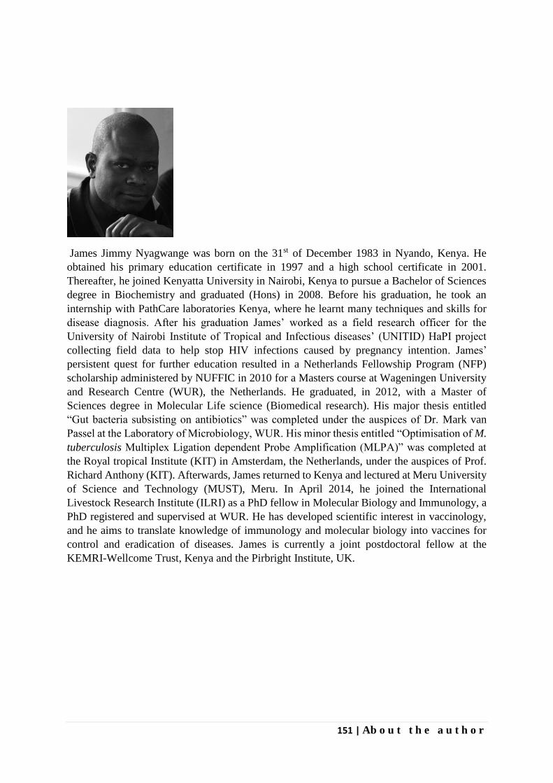
151 | Ab o u t t h e a u t h o r
James Jimmy Nyagwange was born on the 31st of December 1983 in Nyando, Kenya. He
obtained his primary education certificate in 1997 and a high school certificate in 2001.
Thereafter, he joined Kenyatta University in Nairobi, Kenya to pursue a Bachelor of Sciences
degree in Biochemistry and graduated (Hons) in 2008. Before his graduation, he took an
internship with PathCare laboratories Kenya, where he learnt many techniques and skills for
disease diagnosis. After his graduation James’ worked as a field research officer for the
University of Nairobi Institute of Tropical and Infectious diseases’ (UNITID) HaPI project
collecting field data to help stop HIV infections caused by pregnancy intention. James’
persistent quest for further education resulted in a Netherlands Fellowship Program (NFP)
scholarship administered by NUFFIC in 2010 for a Masters course at Wageningen University
and Research Centre (WUR), the Netherlands. He graduated, in 2012, with a Master of
Sciences degree in Molecular Life science (Biomedical research). His major thesis entitled
“Gut bacteria subsisting on antibiotics” was completed under the auspices of Dr. Mark van
Passel at the Laboratory of Microbiology, WUR. His minor thesis entitled “Optimisation of M.
tuberculosis Multiplex Ligation dependent Probe Amplification (MLPA)” was completed at
the Royal tropical Institute (KIT) in Amsterdam, the Netherlands, under the auspices of Prof.
Richard Anthony (KIT). Afterwards, James returned to Kenya and lectured at Meru University
of Science and Technology (MUST), Meru. In April 2014, he joined the International
Livestock Research Institute (ILRI) as a PhD fellow in Molecular Biology and Immunology, a
PhD registered and supervised at WUR. He has developed scientific interest in vaccinology,
and he aims to translate knowledge of immunology and molecular biology into vaccines for
control and eradication of diseases. James is currently a joint postdoctoral fellow at the
KEMRI-Wellcome Trust, Kenya and the Pirbright Institute, UK.
List of publications
Nyagwange, J., V. Nene, S. Mwalimu, S. Henson, L. Steinaa, B. Nzau, E. Tijhaar and R. Pelle
(2018). "Antibodies to in silico selected GPI-anchored Theileria parva proteins neutralize
sporozoite infection in vitro." Veterinary Immunology and Immunopathology 199: 8-14.
Nyagwange, J., E. Tijhaar, N. Ternette, F. Mobegi, K. Tretina, J. C. Silva, R. Pelle and V.
Nene (2018). "Characterization of the Theileria parva sporozoite proteome." International
Journal for Parasitology 48(3): 265-273.
Nyagwange, J., E. E. Awino, E. Tijhaar, N. Svitek, R. Pelle, V. Nene (2019). "Leveraging the
Medicines for Malaria Venture malaria and pathogen boxes to discover chemical inhibitors of
East Coast fever". International Journal for Parasitology: Drugs and Drug Resistance 9: 80-86.
Nyagwange,J., E. Tijhaar, N. Ternette, M. Kigoni, R. Pelle, V. Nene (2018). "Characterization
of Rhipicephalus appendiculatus salivary glands proteins co-purified with Theileria parva
sporozoites". In preparation.
Abstract and presentation
Nyagwange, J., et al: Proteomics of the Theileria parva sporozoites. New Approaches to
Vaccines for Human and Veterinary Tropical Diseases, Keystone symposium, May 22—26,
2016; Cape Town, South Africa (Poster presentation).
153 | Funding information
The research described in this thesis was financially supported by the CGIAR Research
Program on Livestock and Fish, Kenya, the Norman Borlaug Commemorative Research
Initiative, an initiative between the Feed the Future program of United States Agency for
International Development, USA and United States Department of Agriculture-Agricultural
Research Service, USA (58-5348-2-117F) and the Department for International Development
of the United Kingdom and the Bill and Melinda Gates Foundation, USA (OPP1078791).
Cover design by James Nyagwange


Propositions
1. Reverse vaccinology presents a tool for identification of novel Theileria parva
vaccine candidates.
(this thesis)
2. Repurposing of drugs or drug like compounds can accelerate the process of drug
discovery for Theileria.
(this thesis)
3. Cross presentation by dendritic cells is a mechanism for presenting exogenous
antigens on MHC class I pathway, common for viruses but still poorly acknowledged
for parasites.
4. Intermittent funding is a major factor that slows down scientific discoveries.
5. The irony of many social media networks is that they greatly contribute to anti-social
behaviour.
6. In scientific research, imagination is a more important driver of discovery than
knowledge.
Propositions belonging to the thesis, entitled
Identification of novel Theileria parva candidate vaccine antigens and discovery of new
therapeutic drugs for the control of East Coast fever
James J. Nyagwange
Wageningen, 14 May 2019