Use of a Monoclonal Antibody against Babesia bovis Merozoite Surface Antigen-2c for the Development...
6
165 Ann. N.Y. Acad. Sci. 1026: 165–170 (2004). © 2004 New York Academy of Sciences. doi: 10.1196/annals.1307.025 Use of a Monoclonal Antibody against Babesia bovis Merozoite Surface Antigen-2c for the Development of a Competitive ELISA Test MARIANA DOMINGUEZ, a OSVALDO ZABAL, a SILVINA WILKOWSKY, a IGNACIO ECHAIDE, b SUSANA TORIONI DE ECHAIDE, b GUSTAVO ASENZO, a ANABEL RODRÍGUEZ, a PATRICIA ZAMORANO, a MARISA FARBER, a CARLOS SUAREZ, c AND MONICA FLORIN-CHRISTENSEN a a Instituto Nacional de Tecnología Agropecuaria , Centro de Investigación en Ciencias Veterinarias y Agronómicas, Castelar, Argentina b Instituto Nacional de Tecnología Agropecuaria Rafaela, Estación Experimental Agropecuaria, Rafaela, Argentina c Animal Disease Research Unit, Agricultural Research Service, United States Department of Agriculture, Pullman, Washington, USA ABSTRACT: Bovine babesiosis caused by Babesia bovis is a disease that hampers the production of beef and dairy cattle in tropical and subtropical regions of the world. New diagnostic methods based on recombinant antigens constitute valuable biotechnological tools for the strategic control of this disease. We have developed a competitive enzyme-linked immunosorbent assay that includes a recombinant form of the merozoite surface antigen-2c and a novel monoclonal antibody against it. Preliminary results showed that this test is able to identify specific antibodies against B. bovis from experimentally and naturally infected cattle. KEYWORDS: Bovine babesiosis; Babesia bovis; competitive enzyme-linked im- munosorbent assay (competitive ELISA); merozoite surface antigen-2c (MSA- 2c); monoclonal antibodies INTRODUCTION Babesia bovis is an intraerythrocytic parasite transmitted to bovines by Boophilus sp. tick vectors. The disease caused by this parasite is endemic in tropical and sub- tropical regions of the world and causes great economic losses because of the limi- tations it imposes on meat and milk production. 1 In an ideal scenario, equilibrium between bovines, ticks, and hemoparasites avoids the occurrence of outbreaks in en- Address for correspondence: Monica Florin-Christensen, Instituto Nacional de Tecnología Agropecuaria Castelar, Centro de Investigación en Ciencias Veterinarias y Agronómicas, Los Reseros y Las Cabañas, 1712 Castelar, Argentina. Voice/fax: +54 114 4621-1743. [email protected]
Transcript of Use of a Monoclonal Antibody against Babesia bovis Merozoite Surface Antigen-2c for the Development...
165
Ann. N.Y. Acad. Sci. 1026: 165–170 (2004). © 2004 New York Academy of Sciences.doi: 10.1196/annals.1307.025
Use of a Monoclonal Antibody against Babesia bovis Merozoite Surface Antigen-2c forthe Development of a CompetitiveELISA Test
MARIANA DOMINGUEZ,a OSVALDO ZABAL,a SILVINA WILKOWSKY,a
IGNACIO ECHAIDE,b SUSANA TORIONI DE ECHAIDE,b GUSTAVO ASENZO,a ANABEL RODRÍGUEZ,a PATRICIA ZAMORANO,a MARISA FARBER,a
CARLOS SUAREZ,c AND MONICA FLORIN-CHRISTENSENa
aInstituto Nacional de Tecnología Agropecuaria , Centro de Investigación en Ciencias Veterinarias y Agronómicas, Castelar, ArgentinabInstituto Nacional de Tecnología Agropecuaria Rafaela, Estación Experimental Agropecuaria, Rafaela, ArgentinacAnimal Disease Research Unit, Agricultural Research Service, United States Department of Agriculture, Pullman, Washington, USA
ABSTRACT: Bovine babesiosis caused by Babesia bovis is a disease that hampersthe production of beef and dairy cattle in tropical and subtropical regions ofthe world. New diagnostic methods based on recombinant antigens constitutevaluable biotechnological tools for the strategic control of this disease. We havedeveloped a competitive enzyme-linked immunosorbent assay that includes arecombinant form of the merozoite surface antigen-2c and a novel monoclonalantibody against it. Preliminary results showed that this test is able to identifyspecific antibodies against B. bovis from experimentally and naturally infectedcattle.
KEYWORDS: Bovine babesiosis; Babesia bovis; competitive enzyme-linked im-munosorbent assay (competitive ELISA); merozoite surface antigen-2c (MSA-2c); monoclonal antibodies
INTRODUCTION
Babesia bovis is an intraerythrocytic parasite transmitted to bovines by Boophilussp. tick vectors. The disease caused by this parasite is endemic in tropical and sub-tropical regions of the world and causes great economic losses because of the limi-tations it imposes on meat and milk production.1 In an ideal scenario, equilibriumbetween bovines, ticks, and hemoparasites avoids the occurrence of outbreaks in en-
Address for correspondence: Monica Florin-Christensen, Instituto Nacional de TecnologíaAgropecuaria Castelar, Centro de Investigación en Ciencias Veterinarias y Agronómicas, LosReseros y Las Cabañas, 1712 Castelar, Argentina. Voice/fax: +54 114 4621-1743.

166 ANNALS NEW YORK ACADEMY OF SCIENCES
demic regions. Newborn calves from these zones are protected by innate mecha-nisms of defense and colostral antibodies. If these animals become naturally infectedwith babesias before 1 year of age, they remain persistently infected and are protect-ed against babesiosis. However, this ideal equilibrium is broken when the tick pop-ulation decreases in the region because of climatic changes or frequent use ofacaricides. In these cases, a significant number of calves in a herd may not receivetheir natural immunization. Moreover, if the Babesia-infected tick population re-turns to its original high numbers, babesiosis outbreaks can occur with serious eco-nomic consequences. Therefore, diagnosis is necessary to periodically assess theepidemiological situation of babesiosis and to decide on the timely implementationof strategic control measures such as vaccination.
At present, detection of antibodies against B. bovis is mostly carried out by tech-niques that use crude antigen preparations, such as the immunofluorescence anti-body test (IFAT) and the enzyme-linked immunosorbent assay (ELISA).2 The maindrawback of these techniques is the difficulty of purifying the Babesia antigens fromthe erythrocyte antigens, which are responsible for unspecific reactions and, in thecase of IFAT, the subjectivity in the interpretation of results.
Diagnostic methods based on recombinant proteins are thus highly desirable. Tothis end, it is necessary to identify widely conserved, immunodominant antigens ex-posed to the bovine immune system. B. bovis rhoptry-associated protein 1 (RAP-1)meets these requirements, and an indirect ELISA test3 and a competitive ELISAtest4 based on a recombinant form of this protein have been successfully developed.
Previous studies from our group have detected another valuable diagnostic can-didate for B. bovis infections: merozoite surface antigen-2c (MSA-2c). This antigenhas a surface localization and is highly conserved among geographically distantstrains.5,6 In addition, a variety of sera from experimentally and naturally B. bovis–infected bovines from Argentina recognized a recombinant form of MSA-2c (rMSA-2c) from the Argentine R1A-attenuated strain in Western blot analyses.6 Thus, weproduced monoclonal antibodies against rMSA-2c and explored their usefulness forthe development of competitive ELISA tests for B. bovis. We here present the dataobtained thus far, which indicate that rMSA-2c and a monoclonal antibody (MAb)against it can be useful diagnostic tools for bovine babesiosis.
MATERIALS AND METHODS
Development of Monoclonal Antibodies
Recombinant B. bovis MSA-2c was produced in the pBAD/thioTOPO (Invitro-gen, Carlsbad, CA, USA) prokaryotic expression system using DNA from the Ar-gentine R1A strain as starting material, and purified by affinity chromatography onNi-agarose, as previously described.6 Protein quantification was performed by com-parison of band sizes with known amounts of bovine serum albumin after sodiumdodecyl sulfate polyacrylamide gel electrophoresis (SDS-PAGE) and staining withCoomassie blue. Three mice were intradermally inoculated with 30 µg of rMSA-2cemulsified in a mineral oil adjuvant developed at the National Institute of Agricul-tural Technology, Argentina. This adjuvant was composed of Arlacel C (Farma In-ternational, Miami, FL, USA), Markol 52 (Univar, Richmond, BC, Canada) and
167DOMINGUEZ et al.: MONOCLONAL ANTIBODY AGAINST MSA-2c
Tween 80 (Sigma Chemical Co., St. Louis, MO, USA). After the first inoculation, asecond followed 15 days later, and a third 30 days later. Development of anti-MSA-2c antibody titers was confirmed by indirect ELISA using rMSA-2c as antigen, fol-lowing conditions described before.6 Mice were then euthanized; their spleens wereexcised and homogeneized; and the preparation of MAbs was carried out accordingto standard protocols. The screening of clones secreting anti-MSA-2c antibodieswas performed by indirect ELISA. Parallel ELISA assays were performed using re-combinant Anaplasma marginale MSP-5, produced in the same expression system,to discard those clones secreting antibodies against nonspecific B cell epitopes, in-cluding those in the co-expressed thioredoxin fusion protein. A clone identified inour research as H9P2C2 displayed the highest antibody titers against rMSA-2c andshowed no reaction against rMSP-5. This clone was amplified in mice ascitis andfurther characterized. The MAb titer was determined in serial dilutions by indirectELISA using rMSA-2c as antigen. Detection was carried out by incubation with per-oxidase-conjugated anti-mouse immunoglobulin G followed by the orthophenylenediamine (OPD)-H2O2 chromogenic substrate and absorbance measurements at 490nm. A titer of 4.4 was calculated for the ascitis preparations used in this study (20mg/mL protein concentration) and corresponded to –log10 of the maximal dilutionof MAb that duplicated the A490 values obtained with the unrelated MAb Tryp1.
Serum Samples
Serum samples from bovines experimentally infected with B. bovis R1A (attenu-ated) and S2P (pathogenic) strains were collected before infection as well as at dif-ferent time points (T21, T35, and T65) after infection. In addition, sera fromnaturally infected bovines from a babesiosis-endemic region of Salta, a province inNorthern Argentina, were also used in this study. Before the assay, serum sampleswere diluted 1:50 in phosphate-buffered saline/0.5% Tween 20 and incubated with0.2 mg/mL of an Esherichia coli lysate for 1 hr at 37°C, to adsorb antibodies againstE. coli antigens, as described previously.5,6 After this period, samples were centri-fuged (10 min, 14,000 × g, 4°C), and the supernatants were used in the assays.
Competitive ELISA
Recombinant MSA-2c in carbonate/bicarbonate buffer, pH 9.6, was bound to Im-mulon II plates (Dynatech, Chantilly, VA) in a concentration of 8.5 ng/well. Afterblocking the plates with phosphate-buffered saline/0.5% Tween 20/0.5% gelatin,they were sequentially incubated with serial dilutions of test sera, an adequate dilu-tion of MAb H9P2C2, peroxidase-conjugated anti-mouse immunoglobulin G(Kirkegaard and Perry Laboratories, Gaithersburg, MD), and the chromogenic sub-strate OPD-H2O2. Each incubation was followed by thorough washes with phos-phate-buffered saline/0.5% Tween 20. Reactions were stopped with H2SO4/H2O(1:8, v/v), and absorbance was read at 490 nm. All determinations were carried outin triplicate.
Isotype Determination
The immunoglobulin G isotype of MAb H9P2C2 was determined by indirectELISA. rMSA-2c was bound to Immulon 2 HB plates, as above, and sequentially in-
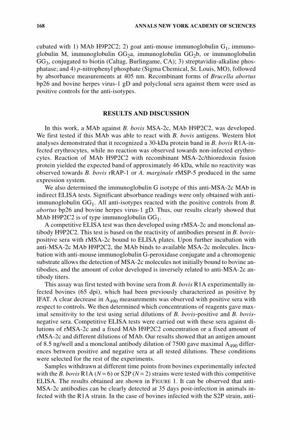
168 ANNALS NEW YORK ACADEMY OF SCIENCES
cubated with 1) MAb H9P2C2; 2) goat anti-mouse immunoglobulin G1, immuno-globulin M, immunoglobulin GG2a, immunoglobulin GG2b, or immunoglobulinGG3, conjugated to biotin (Caltag, Burlingame, CA); 3) streptavidin-alkaline phos-phatase; and 4) p-nitrophenyl phosphate (Sigma Chemical, St. Louis, MO), followedby absorbance measurements at 405 nm. Recombinant forms of Brucella abortusbp26 and bovine herpes virus-1 gD and polyclonal sera against them were used aspositive controls for the anti-isotypes.
RESULTS AND DISCUSSION
In this work, a MAb against B. bovis MSA-2c, MAb H9P2C2, was developed.We first tested if this MAb was able to react with B. bovis antigens. Western blotanalyses demonstrated that it recognized a 30-kDa protein band in B. bovis R1A-in-fected erythrocytes, while no reaction was observed towards non-infected erythro-cytes. Reaction of MAb H9P2C2 with recombinant MSA-2c/thioredoxin fusionprotein yielded the expected band of approximately 46 kDa, while no reactivity wasobserved towards B. bovis rRAP-1 or A. marginale rMSP-5 produced in the sameexpression system.
We also determined the immunoglobulin G isotype of this anti-MSA-2c MAb inindirect ELISA tests. Significant absorbance readings were only obtained with anti-immunoglobulin GG1. All anti-isotypes reacted with the positive controls from B.abortus bp26 and bovine herpes virus-1 gD. Thus, our results clearly showed thatMAb H9P2C2 is of type immunoglobulin GG1.
A competitive ELISA test was then developed using rMSA-2c and monclonal an-tibody H9P2C2. This test is based on the reactivity of antibodies present in B. bovis-positive sera with rMSA-2c bound to ELISA plates. Upon further incubation withanti-MSA-2c MAb H9P2C2, the MAb binds to available MSA-2c molecules. Incu-bation with anti-mouse immunoglobulin G-peroxidase conjugate and a chromogenicsubstrate allows the detection of MSA-2c molecules not initially bound to bovine an-tibodies, and the amount of color developed is inversely related to anti-MSA-2c an-tibody titers.
This assay was first tested with bovine sera from B. bovis R1A experimentally in-fected bovines (65 dpi), which had been previously characterized as positive byIFAT. A clear decrease in A490 measurements was observed with positive sera withrespect to controls. We then determined which concentrations of reagents gave max-imal sensitivity to the test using serial dilutions of B. bovis-positive and B. bovis-negative sera. Competitive ELISA tests were carried out with these sera against di-lutions of rMSA-2c and a fixed MAb H9P2C2 concentration or a fixed amount ofrMSA-2c and different dilutions of MAb. Our results showed that an antigen amountof 8.5 ng/well and a monclonal antibody dilution of 7500 gave maximal A490 differ-ences between positive and negative sera at all tested dilutions. These conditionswere selected for the rest of the experiments.
Samples withdrawn at different time points from bovines experimentally infectedwith the B. bovis R1A (N = 6) or S2P (N = 2) strains were tested with this competitiveELISA. The results obtained are shown in FIGURE 1. It can be observed that anti-MSA-2c antibodies can be clearly detected at 35 days post-infection in animals in-fected with the R1A strain. In the case of bovines infected with the S2P strain, anti-

169DOMINGUEZ et al.: MONOCLONAL ANTIBODY AGAINST MSA-2c
MSA-2c antibodies could be detected at 65 dpi in both cases In one of these animals(II), a high percent inhibition (%I) was also obtained at 21 dpi. Interestingly, this bo-vine was inoculated with 107 parasites, while the inoculum used with bovine I wasof 105. Samples that showed significant %I according to this test were also positivelydiagnosed for B. bovis by IFAT.
In addition, we tested sera from bovines naturally infected with B. bovis fieldstrains from a babesiosis endemic region in Salta province, Argentina. These sam-ples were characterized as positive by IFAT. All tested sera (N = 8) gave a significantdecrease in A490 readings and therefore high %I values (FIG. 2).
Our results clearly show that a recombinant form of the B. bovis surface antigenMSA-2c and a MAb against it can be employed in the development of a competitiveELISA test for this pathogen. Trials using a larger amount of serum samples fromdifferent endemic regions of Argentina and neighboring countries are now underwayin our laboratory. In addition, we will explore other competitive ELISA designs us-ing the reagents we have developed to achieve maximal sensitivity and minimalbackground.
FIGURE 1. Detection by competitive ELISA of infections in bovines experimentallyinoculated with the Argentine B. bovis S2P (pathogen) or R1A (attenuated) strains. Serumsamples were withdrawn just before (T0) and at different time points after inoculation, anddiagnosed by competitive ELISA using rMSA-2c and MAb H9P2C2 (serum dilution: 1:50).Roman numerals indicate different bovines. Results are expressed as percent inhibition(%I), calculated as 100 – [(A490 test serum × 100)/mean A490 of negative sera], and repre-sent the mean of triplicate determinations. Standard deviations between wells did not exceed20%.

170 ANNALS NEW YORK ACADEMY OF SCIENCES
ACKNOWLEDGMENTS
This work was supported by Grant PICT 98/08-3838 from Agencia Nacional Pro-mocion Cientifica y Technologica and by Fundacion Antorchas, Argentina.
REFERENCES
1. UILENBERG, G. 1995. International collaborative research: significance of tick-bornehemoparasitic diseases to world animal health. Vet. Parasitol. 57: 19–41.
2. BOSE, R., W.K. JORGENSEN, R.J. DALGLIESH, et al. 1995. Current state and future trendsin the diagnosis of babesiosis. Vet. Parasitol. 57: 61–74.
3. BOONCHIT, S., X. XUAN, N. YOKOYAMA, et al. 2002. Evaluation of an enzyme-linkedimmunosorbent assay with recombinant rhoptry-associated protein 1 antigen againstBabesia bovis for the detection of specific antibodies in cattle. J. Clin. Microbiol. 40:3771–3775.
4. GOFF, W., T. MCELWAIN, C. SUAREZ, et al. 2003. Competitive enzyme-linked immun-osorbent assay based on a rhoptry-associated protein 1 epitope specifically identifiesBabesia bovis-infected cattle. Clin. Diagn. Lab. Immunol. 10: 38–43.
5. FLORIN-CHRISTENSEN, M., C.E. SUAREZ, S.A. HINES, et al. 2002. The Babesia bovismerozoite surface antigen-2 locus contains four tandemly arranged and expressedgenes encoding immunologically distinct proteins. Infect. Immun. 70: 3566–3575.
6. WILKOWSKY, S., M. FARBER, I. ECHAIDE, et al. 2003. Babesia bovis merozoite surfaceantigen-2c contains highly immunogenic, conserved B-cell epitopes that elicit neu-tralization-sensitive antibodies in cattle. Mol. Biochem. Parasitol. 127(2): 133–141.
FIGURE 2. Detection of natural B. bovis infections from a babesiosis-endemic regionof Argentina. B. bovis-infected sera, characterized as positive by IFAT, were diagnosed bycompetitive ELISA (serum dilution: 1:50). Roman numbers indicate different bovines. Thenegative and positive serum samples correspond to an R1A experimentally infected bovine(T0 and 65 dpi, respectively).