A guide to the online BovIS carcass benchmarking application ...
Wildlife Disease and Zoonotics
Michigan Bovine Tuberculosis Bibliography
and Database
University of Nebraska - Lincoln Year
Animal-side serologic assay for rapid
detection of Mycobacterium Bovis
infection in multiple species of
free-ranging wildlifeK.P. Lyashchenko, Chembio Diagnostic Systems, Inc., 3661 Horse-block Road, Medford, NY 11763, USAR. Greenwald, Chembio Diagnostic Systems, Inc., 3661 HorseblockRoad, Medford, NY 11763, USAJ. Esfandiari, Chembio Diagnostic Systems, Inc., 3661 HorseblockRoad, Medford, NY 11763, USAM.A. Chambers, TB Research Group, Department of Statutory andExotic Bacterial Diseases, Veterinary Laboratories Agency Wey-bridge, New Haw, Surrey KT15 3NB, UKJ. Vicente, Instituto de Investigacion en Recursos Cinegeticos IREC(CSIC–UCLM–JCCM), Ciudad Real, SpainC. Gortazar, Instituto de Investigacion en Recursos Cinegeticos IREC(CSIC–UCLM–JCCM), Ciudad Real, SpainN. Santos, Faculty of Veterinary Medicine, Lisbon, PortugalM. Correia-Neves, Life and Health Sciences Research Institute (ICVS),School of Health Sciences, University of Minho, Braga, PortugalB.M. Buddle, AgResearch, Hopkirk Research Institute, PalmerstonNorth, New ZealandR. Jackson, Institute of Veterinary, Animal & Biomedical Sciences,Massey University, Palmerston North, New ZealandD.J. O’Brien, Wildlife Disease Laboratory, Department of Natural

Resources, Lansing, MI, USAS. Schmitt, Wildlife Disease Laboratory, Department of Natural Re-sources, Lansing, MI, USAM.V. Palmer, United States Department of Agriculture (USDA),Agricultural Research Service, National Animal Disease Center, Bac-terial Diseases of Livestock Research Unit, National Animal DiseaseCenter, Ames, IA, USAR.J. Delahay, Wildlife Disease Ecology Team, Central Science Lab-oratory, Sand Hutton, York YO41 1LZ, UKW.R. Waters, United States Department of Agriculture (USDA),Agricultural Research Service, National Animal Disease Center, Bac-terial Diseases of Livestock Research Unit, National Animal DiseaseCenter, Ames, IA, USA
This paper is posted at DigitalCommons@University of Nebraska - Lincoln.
http://digitalcommons.unl.edu/michbovinetb/48

Animal-side serologic assay for rapid detection of Mycobacterium
bovis infection in multiple species of free-ranging wildlife
K.P. Lyashchenko a,*, R. Greenwald a, J. Esfandiari a, M.A. Chambers b, J. Vicente c,C. Gortazar c, N. Santos d,e, M. Correia-Neves e, B.M. Buddle f, R. Jackson g,
D.J. O’Brien h, S. Schmitt h, M.V. Palmer i, R.J. Delahay j, W.R. Waters i
a Chembio Diagnostic Systems, Inc., 3661 Horseblock Road, Medford, NY 11763, USAb TB Research Group, Department of Statutory and Exotic Bacterial Diseases, Veterinary Laboratories Agency Weybridge,
New Haw, Surrey KT15 3NB, UKc Instituto de Investigacion en Recursos Cinegeticos IREC (CSIC–UCLM–JCCM), Ciudad Real, Spain
d Faculty of Veterinary Medicine, Lisbon, Portugale Life and Health Sciences Research Institute (ICVS), School of Health Sciences, University of Minho, Braga, Portugal
f AgResearch, Hopkirk Research Institute, Palmerston North, New Zealandg Institute of Veterinary, Animal & Biomedical Sciences, Massey University, Palmerston North, New Zealand
h Wildlife Disease Laboratory, Department of Natural Resources, Lansing, MI, USAi United States Department of Agriculture (USDA), Agricultural Research Service, National Animal Disease Center,
Bacterial Diseases of Livestock Research Unit, National Animal Disease Center, Ames, IA, USAj Wildlife Disease Ecology Team, Central Science Laboratory, Sand Hutton, York YO41 1LZ, UK
Received 7 April 2008; received in revised form 20 May 2008; accepted 26 May 2008
Abstract
Numerous species of mammals are susceptible to Mycobacterium bovis, the causative agent of bovine tuberculosis (TB).
Several wildlife hosts have emerged as reservoirs of M. bovis infection for domestic livestock in different countries. In the
present study, blood samples were collected from Eurasian badgers (n = 1532), white-tailed deer (n = 463), brushtail possums
(n = 129), and wild boar (n = 177) for evaluation of antibody responses to M. bovis infection by a lateral-flow rapid test (RT) and
multiantigen print immunoassay (MAPIA). Magnitude of the antibody responses and antigen recognition patterns varied among
the animals as determined by MAPIA; however, MPB83 was the most commonly recognized antigen for each host studied.
Other seroreactive antigens included ESAT-6, CFP10, and MPB70. The agreement of the RT with culture results varied from
74% for possums to 81% for badgers to 90% for wild boar to 97% for white-tailed deer. Small numbers of wild boar and deer
exposed to M. avium infection or paratuberculosis, respectively, did not cross-react in the RT, supporting the high specificity of
the assay. In deer, whole blood samples reacted similarly to corresponding serum specimens (97% concordance), demonstrating
the potential for field application. As previously demonstrated for badgers and deer, antibody responses to M. bovis infection in
wild boar were positively associated with advanced disease. Together, these findings suggest that a rapid TB assay such as the RT
www.elsevier.com/locate/vetmic
Available online at www.sciencedirect.com
Veterinary Microbiology 132 (2008) 283–292
* Corresponding author. Tel.: +1 631 924 1135; fax: +1 631 924 6033.
E-mail address: [email protected] (K.P. Lyashchenko).
0378-1135/$ – see front matter # 2008 Elsevier B.V. All rights reserved.
doi:10.1016/j.vetmic.2008.05.029

may provide a useful screening tool for certain wildlife species that may be implicated in the maintenance and transmission of M.
bovis infection to domestic livestock.
# 2008 Elsevier B.V. All rights reserved.
Keywords: Tuberculosis; Mycobacterium bovis; Wildlife; Serology
1. Introduction
Bovine tuberculosis (TB) remains an important
zoonotic disease with significant impacts on the
economy in many countries (Corner, 2006; Cosivi
et al., 1998; Michel et al., 2006). Several wild mammal
species are implicated in the maintenance and
transmission of Mycobacterium bovis infection and
thereby impede national bovine TB control programs
and international trade (Cousins, 2001; Palmer, 2007).
Well-known examples of wildlife maintenance hosts
include Eurasian badgers (Meles meles) in Great
Britain and Ireland (Clifton-Hadley et al., 1993;
Griffin et al., 2005), white-tailed deer (Odocoileus
virginianus) in the United States (Schmitt et al., 1997;
O’Brien et al., 2002, 2006), brushtail possums
(Trichosurus vulpecula) in New Zealand (Coleman
et al., 2006; Porphyre et al., 2007), wild boar (Sus
scrofa) in Spain (Gortazar et al., 2003; Naranjo et al.,
2008), and African buffalo (Syncerus caffer) in South
Africa (Michel et al., 2006).
The successful eradication of bovine TB from
livestock in countries with a wildlife reservoir is likely
to require focusing on the wildlife reservoir(s) of M.
bovis infection (Palmer, 2007). Current methods of
diagnosis in live animals, such as the intradermal
tuberculin test or interferon-gamma (IFN-g) release
assays, are not fully validated for species other than
cattle (Monaghan et al., 1994). Rapid and accurate
assays that could be used in a variety of M. bovis-
susceptible mammals under field conditions would
significantly improve wildlife TB surveillance efforts
worldwide.
Prior studies have demonstrated the utility of
membrane-based assays to detect specific antibodies
during TB in cattle (Waters et al., 2006), cervids (Waters
et al., 2004, 2005), elephants (Lyashchenko et al., 2006),
camelids (Wernery et al., 2007; Lyashchenko et al.,
2007a,b), and tapirs (Moser et al., 2008). The objectives
of the present study were to (1) characterize the antibody
responses of wild mammals infected with M. bovis,
including badgers, deer, possums, and wild boar, (2)
evaluate the usefulness of a lateral-flow test under field
conditions, and (3) determine if a single serologic assay
can be used for TB surveillance in a variety of different
wildlife host species.
2. Methods
2.1. Animals and samples
Sera were collected from Eurasian badgers in Great
Britain, white-tailed deer in the United States,
brushtail possums in New Zealand, and wild boar in
Spain and Portugal (Table 1).
Badger sera were obtained from two sources: (1)
1464 animals killed as part of the Randomised Badger
Culling Trial (RBCT) (Donnelly et al., 2007), and (2) 68
animals captured, bled, and released as part of an on-
going ecological study by the Central Science
Laboratory (CSL) in Woodchester Park, south-west
K.P. Lyashchenko et al. / Veterinary Microbiology 132 (2008) 283–292284
Table 1
Study populations
Species Institute, country Number of animals
TB Control
Badger VLA, UK 386 1078
CSL, UK 68 0
Deer MDNR, USA 9 425
NADC, USA 19a 10b
Possum AgR HRI, New Zealand 38 91
Wild boar IREC, Spain 50 50
ICVS, Portugal 14 63c
a Inoculated with M. bovis intratonsilarly.b Includes three animals experimentally infected with M. avium
subsp. paratuberculosis.c Includes three animals with M. avium and five animals with
other non-TB mycobacteria identified by culture and PCR at
necropsy.

England (Chambers et al., 2008). All work with animals
was conducted under licenses issued by the Home
Office, UK, following ethical clearance by the VLA and
CSL. All sera were stored frozen at �20 8C until used
for testing. Each badger from the RBCT was subjected
to routine post-mortem examination and culture for the
presence of M. bovis as described in Sawyer et al.
(2007). The infection status of Woodchester Park
badgers was determined by bacterial culture of clinical
specimens (feces, urine, sputum, purulent exudate from
abscesses, and bite wound swabs) collected from
anaesthetized animals (Clifton-Hadley et al., 1993;
Delahay et al., 2000). Since Woodchester Park badgers
were live-sampled, only those animals excreting M.
bovis were included in the present study.
Deer samples were obtained from experimentally
infected animals at different stages of disease. White-
tailed deer (1–3 years of age) were either raised within
a TB-free herd at the National Animal Disease Center
(NADC), Ames, Iowa, USA, or obtained from farmed
herds with no history of TB. Groups consisted of seven
non-infected animals, three deer inoculated with M.
avium subsp. paratuberculosis, and 19 deer inoculated
via the intratonsilar route with various doses of M.
bovis (3 � 102–2 � 108 colony-forming units), as
described previously (Waters et al., 2004; Palmer
et al., 2007). All animals were euthanized at 4–11
months post-infection. Blood, diaphragm fluid and
aqueous humor were collected during necropsy of
experimentally infected deer. Specimens were
extracted from diaphragms by freeze/thaw and
mechanical disruption (using a garlic press). Aqueous
humor was obtained by fine needle aspiration. Various
tissues were collected for bacteriologic culture and
microscopic examination. Disease was confirmed at
necropsy in each infected deer by the presence of gross
lesions, histopathological examination, and mycobac-
terial culture. The Institutional Animal Care and Use
Committee approved protocols detailing procedures
and animal care prior to initiation of the experiments.
In addition, fresh whole blood samples were
collected from 434 free-ranging white-tailed deer in
Michigan State where M. bovis infection is persistent
in wild deer (Schmitt et al., 1997). The deer enrolled in
the study inhabited the ‘‘core’’ area of the Michigan
TB outbreak area (O’Brien et al., 2002). Blood
samples were obtained from four sources: (1) culls of
free-ranging deer conducted by hunters during 2005/
2006 and 2006/2007 winter seasons as part of herd-
health checks; (2) depopulation of a fenced captive
deer shooting preserve in late 2006 following culture
confirmation of grossly lesioned M. bovis-positive
deer; (3) nuisance deer shot on cattle farms under
disease control permits; and (4) deer tested as part of
an ongoing live trap/test/cull project. Tissue samples
used for culture included medial retropharyngeal
lymph nodes, parietal pleura and lungs.
A group of brushtail possums naturally infected
with M. bovis consisted of 29 animals captured at
Castlepoint, Wairarapa and nine possums from the
Orongorongo Valley near Wellington. All these had
macroscopic TB lesions, from which M. bovis was
cultured. Blood was collected from all of the animals
immediately prior to euthanasia. A negative control
group included 91 possums captured in Manawatu, an
area of New Zealand that is free of bovine TB in
domestic livestock and wild animals.
Hunter-harvested wild boar were sampled in Spain
between November 1999 and February 2005 (n = 100)
and in Portugal between December 2005 and January
2007 (n = 77). Various lymph nodes were examined
for gross lesions and cultured for M. bovis as described
previously (Gortazar et al., 2003; Martin-Hernando
et al., 2007). Portuguese mycobacterial isolates were
identified by PCR for a panel of selected genes: 16S
RNA, IS1081, Rv3120, Rv1510, and IS1245, and
confirmed by spoligotyping. According to Huard et al.
(2003) and Bartos et al. (2006), this set of genes allows
for differentiation between M. bovis or other members
of the M. tuberculosis complex and M. avium or other
non-TB mycobacteria.
2.2. Multiantigen print immunoassay (MAPIA)
We used a panel of 12 mycobacterial antigens
including 8 purified recombinant proteins (ESAT-6,
CFP10, MPB64, MPB59, MPB70, MPB83, Acr1, and
the 38 kDa protein), two protein fusions (CFP10/
ESAT-6 and Acr1/MPB83), and two native antigens,
bovine protein purified derivative (B-PPD) and M.
bovis culture filtrate (MBCF). MAPIA was performed
as described previously (Lyashchenko et al., 2000).
Briefly, antigens were immobilized on a nitrocellulose
membrane (Schleicher & Schuell, Keene, N.H.) at a
protein concentration of 0.05 mg/ml using a semi-
automatic micro-aerosolization device (Linomat IV,
K.P. Lyashchenko et al. / Veterinary Microbiology 132 (2008) 283–292 285

Camag Scientific Inc., Wilmington, Delaware) to
generate invisible parallel bands. After antigen
printing, the membrane was cut into 3 mm wide
strips, perpendicular to the antigen bands, so that each
strip carried all antigens. Strips were blocked for 1 h
with 1% non-fat skimmed bovine milk in phosphate-
buffered saline containing 0.05% Tween 20 and then
incubated with individual serum samples diluted 1:50
in blocking solution for 1 h at room temperature. After
washing, the strips were incubated for 1 h with
peroxidase-conjugated protein G (deer, wild boars) or
protein A (badgers, possums) diluted 1:1000 (Kirke-
gaard & Perry Laboratories), and subsequently
washed again. IgG antibodies bound to immobilized
antigens were visualized with 3,30,5,50-tetramethyl
benzidine (Kirkegaard & Perry Laboratories). MAPIA
results were read visually, with a band of any intensity
being considered as a positive reaction.
2.3. Rapid test (RT)
A simple and rapid antibody detection assay was
developed by Chembio Diagnostic Systems, Inc. using
colored latex-based lateral-flow technology and a
cocktail of selected M. bovis antigens including ESAT-
6, CFP10, and MPB83 (Greenwald et al., 2003; Waters
et al., 2006). Serum specimens were tested for the
presence of specific antibody as previously described
(Lyashchenko et al., 2006). Results were read at
20 min after adding sample buffer. Any visible band in
the test area of the RT, in addition to the control line,
was considered an antibody positive result, whereas no
band in the test area in addition to the visible control
line was considered a negative result.
2.4. Data analysis
Diagnostic performance of the RT was evaluated
against the gold standard of M. bovis culture by
calculating test sensitivity, specificity, and accuracy
(% of RT results in agreement with TB status
determined by culture) using available software
(Lowry, 2007) and reported with the 95% confidence
interval (CI). Test of significance between proportions
(Fisher’s exact test) and the calculation of odds ratio
were performed using GraphPad InStat version 3.00
for Windows 95, GraphPad Software, San Diego
California USA, www.graphpad.com. The statistical
significance of the proportion of antibody detection
rate in lesion positive wild boar in relation to those
without visible lesions was performed by Chi-square
tests (SPSS 11.0 Statistical Program).
3. Results
3.1. Antibody responses and antigen recognition
To compare antibody responses to M. bovis infection
in different hosts, serum samples of all animals
(possums) or of randomly selected culture-positive
and culture-negative animals from naturally infected
populations (badgers, deer, wild boar) were tested by
MAPIA. Fig. 1 provides representative examples of the
individual antigen reactivity patterns in each of the four
mammal species. Table 2 shows the frequencies of IgG
reactivity to M. bovis antigens found in infected and
control animals. The MAPIA results confirmed our
previous observations that MPB83 protein is serodo-
minant in badgers with M. bovis infection and in
experimentally infected white-tailed deer (Greenwald
et al., 2003; Waters et al., 2004). The present study
further revealed that this molecule is the most reactive
antigen in brushtail possums and wild boar. In the
naturally infected animals, the MPB83 seroreactivity
rates ranged from 34% in possums to 89% in deer,
whereas in culture-negative controls it varied from 1%
in deer to 10% in badgers. ESAT-6 and CFP10 antigens
were the second and third most frequently recognized
antigen in the MAPIA in all the species, except for wild
boar. MPB70 protein elicited serological responses in
greater numbers of infected wild boar (68%) than did
ESAT-6 (58%) or CFP10 (52%), although levels of
MPB70 antibodies (evaluated visually by band inten-
sity) were commonly lower than those of ESAT-6,
CFP10, or MPB83 antibodies (Fig. 1). The other single
proteins of M. bovis used in MAPIA reacted with
variable numbers of sera from infected animals, ranging
from 0% to 44%. Significant seroreactivity was found
for MBCF, with a few M. bovis-infected animals
displaying antibody only to this antigen (Fig. 1, B4).
However, the crude native preparation of M. bovis
showed high rates of false-positive results (from 10% in
badgers to 26% in deer).
Heterogeneous antigen recognition patterns were
observed in each host species, so that there was no
K.P. Lyashchenko et al. / Veterinary Microbiology 132 (2008) 283–292286

single antigen target common to all seropositive
animals. It was demonstrated that the serological
performance of the two antigen fusion proteins,
CFP10/ESAT-6 and Acr1/MPB83, reflected the
reactivity of the corresponding single antigens
(Fig. 1). Subjective evaluations of the MAPIA band
intensities and the numbers of antigens recognized by
each animal in this study suggested that the strongest
antibody responses were found in wild boars, whereas
possums produced relatively weak antibody responses
to M. bovis infection.
3.2. Antibody detection by rapid test
Serological evaluation of the RT revealed variable
diagnostic performance when used in different host
species (Table 3). Higher test sensitivity and specificity
were found for deer and wild boar, compared to those
observed in badgers and possums. The overall accuracy
of the test ranged from 74% in possums to 97% in deer.
When the diagnostic sensitivity was analyzed sepa-
rately for deer with experimental and natural M. bovis
infections, a lower rate of serological detection was
found in the latter group (67%) than in animals
inoculated intratonsilarly with high dose of M bovis
(79%). To further gauge the RT specificity, serum
samples from three deer developing experimental
paratuberculosis (all three had strong antibody
responses, as previously shown by Palmer et al.,
2007) and from eight wild boar naturally infected with
M. avium or other non-TB mycobacteria were tested.
None of these 11 animals produced a positive RT result.
K.P. Lyashchenko et al. / Veterinary Microbiology 132 (2008) 283–292 287
Table 2
Seroreactivity rates (%) of M. bovis antigens obtained in MAPIA with sera from naturally infected and uninfected animals
Antigen Badger Deer Possum Wild boar
TB
(n = 15)
Control
(n = 29)
TB
(n = 9)
Control
(n = 98)
TB
(n = 38)
Control
(n = 91)
TB
(n = 50)
Control
(n = 50)
MPB83 67 10 89 1 34 3 78 2
ESAT-6 27 0 67 2 21 1 58 8
CFP10 13 0 56 1 3 4 52 2
MPB70 7 0 44 0 2 0 68 0
38 kDa 7 0 11 1 2 3 30 0
Acr1 7 0 22 0 0 1 22 0
MPB64 13 0 11 1 0 0 2 0
MPB59 27 7 44 3 0 0 0 0
MBCF 53 10 89 26 34 20 44 12
Fig. 1. Antibody responses detected by MAPIA in Eurasian badgers (A), white-tailed deer (B), brushtail possums (C), and European wild boar
(D) naturally infected with M. bovis. Antigens printed onto nitrocellulose membrane, as described in Section 2, are indicated on the right. One
control serum from culture-negative animal (strip 1) and three sera from different culture-positive animals (strips 2–4) were selected to show
variable antigen recognition patterns in each host species. Visible bands of any intensity observed with certain antigen are IgG antibody positive
reactions with respective antigens.
3.3. Antibody in various types of biological
samples
Serum, plasma, fresh whole blood, diaphragm
fluid, and aqueous humor were collected from three
white-tailed deer experimentally infected with M.
bovis and one non-infected deer. All the samples from
the infected deer, except for the aqueous humor,
yielded positive responses (Table 4). The intensity of
RT reactions obtained with plasma, whole blood, and
diaphragm fluid were comparable to that found for
serum samples. No antibody reactivity was detected in
any of the biological samples collected from the
control deer. The MAPIA demonstrated that the
antigen recognition patterns of antibodies found in
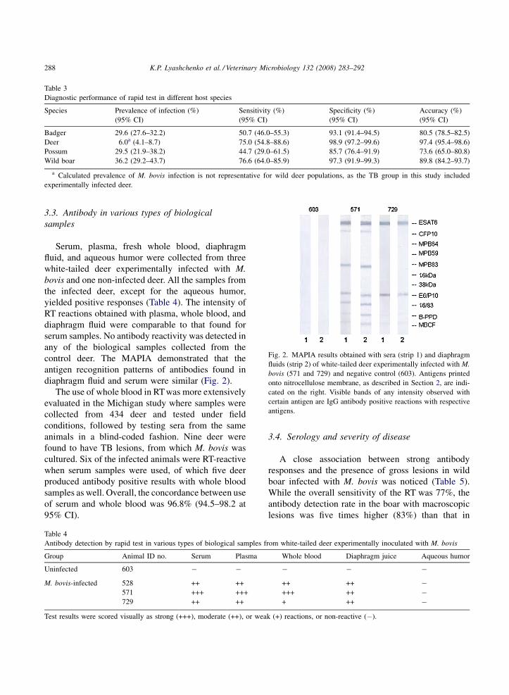
diaphragm fluid and serum were similar (Fig. 2).
The use of whole blood in RT was more extensively
evaluated in the Michigan study where samples were
collected from 434 deer and tested under field
conditions, followed by testing sera from the same
animals in a blind-coded fashion. Nine deer were
found to have TB lesions, from which M. bovis was
cultured. Six of the infected animals were RT-reactive
when serum samples were used, of which five deer
produced antibody positive results with whole blood
samples as well. Overall, the concordance between use
of serum and whole blood was 96.8% (94.5–98.2 at
95% CI).
3.4. Serology and severity of disease
A close association between strong antibody
responses and the presence of gross lesions in wild
boar infected with M. bovis was noticed (Table 5).
While the overall sensitivity of the RT was 77%, the
antibody detection rate in the boar with macroscopic
lesions was five times higher (83%) than that in
K.P. Lyashchenko et al. / Veterinary Microbiology 132 (2008) 283–292288
Table 3
Diagnostic performance of rapid test in different host species
Species Prevalence of infection (%)
(95% CI)
Sensitivity (%)
(95% CI)
Specificity (%)
(95% CI)
Accuracy (%)
(95% CI)
Badger 29.6 (27.6–32.2) 50.7 (46.0–55.3) 93.1 (91.4–94.5) 80.5 (78.5–82.5)
Deer 6.0a (4.1–8.7) 75.0 (54.8–88.6) 98.9 (97.2–99.6) 97.4 (95.4–98.6)
Possum 29.5 (21.9–38.2) 44.7 (29.0–61.5) 85.7 (76.4–91.9) 73.6 (65.0–80.8)
Wild boar 36.2 (29.2–43.7) 76.6 (64.0–85.9) 97.3 (91.9–99.3) 89.8 (84.2–93.7)
a Calculated prevalence of M. bovis infection is not representative for wild deer populations, as the TB group in this study included
experimentally infected deer.
Fig. 2. MAPIA results obtained with sera (strip 1) and diaphragm
fluids (strip 2) of white-tailed deer experimentally infected with M.
bovis (571 and 729) and negative control (603). Antigens printed
onto nitrocellulose membrane, as described in Section 2, are indi-
cated on the right. Visible bands of any intensity observed with
certain antigen are IgG antibody positive reactions with respective
antigens.
Table 4
Antibody detection by rapid test in various types of biological samples from white-tailed deer experimentally inoculated with M. bovis
Group Animal ID no. Serum Plasma Whole blood Diaphragm juice Aqueous humor
Uninfected 603 � � � � �
M. bovis-infected 528 ++ ++ ++ ++ �571 +++ +++ +++ ++ �729 ++ ++ + ++ �
Test results were scored visually as strong (+++), moderate (++), or weak (+) reactions, or non-reactive (�).

animals without visible lesions (17%). The difference
was highly significant (x2 13.24, d.f. 1, p = 0.0003;
p = 0.002, Fisher’s exact test), with an odds ratio of 24
(2.5–228.4, 95% CI). In other words, the boar with
gross lesions were on average 24 times more likely to
be positive in the RT than those without visible
lesions. With samples from Portugal, where most of
the M. bovis-infected wild boar without lesions
originated, all 10 animals with gross lesions were
RT-reactive, whereas none of the four culture-positive
boar without lesions had detectable antibodies.
4. Discussion
The present study characterized serological
responses in infected animals of four species of wild
mammals that are implicated in the persistence of M.
bovis infection in cattle in different countries. Our
results demonstrated that tuberculous badgers, white-
tailed deer, brushtail possums, and wild boar produced
variable levels of IgG antibodies against several M.
bovis antigens. Importantly, the responses could be
detected in each host by a recently developed ‘point-
of-care’-type lateral-flow test for bovine TB (Lyash-
chenko et al., 2006; Waters et al., 2006). MAPIA
revealed antibodies to multiple proteins of M. bovis
with heterogeneous antigen recognition patterns
observed in the infected animals. These findings are
in agreement with our previous reports on human TB
serology (Lyashchenko et al., 2000) as well as other
mammal species susceptible to organisms of the M.
tuberculosis complex (Lyashchenko et al., 2004, 2006,
2007a,b; Waters et al., 2005, 2006). The serodomi-
nance of the MPB83 protein known to elicit the
strongest antibody responses to experimental TB
infection in cattle, badger, and deer (Lesellier et al.,
2008; Lyashchenko et al., 2004; Waters et al., 2004)
was extended in the present study to naturally infected
animals as well as other, previously untested species
(possums and wild boar).
Antibody detection methods are generally simple,
rapid, and inexpensive. The lateral-flow assay format
offers important features that make the RT evaluated in
the present study an attractive screening tool for field
applications. This is an easy-to-perform animal-side
disposable kit which can use serum, plasma, whole
blood, or other samples to provide ‘‘yes-or-no’’ visual
read-outs within 15–20 min. The test speed is
particularly useful for wildlife surveillance where a
‘‘euthanize or release’’ decision may be needed
quickly on a physically restrained animal. The RT
kits are stable at room temperature for up to 18–24
months, and they do not require refrigeration for
storage, a power source, equipment, laboratory
environment, or skilled personnel to perform the
assay and interpret results. The test is suitable for use
under field conditions, as it can be minimally affected
by extremes of ambient temperature. Test results are
unambiguous; high levels of reproducibility (lot-to-
lot, operator-to-operator, laboratory-to-laboratory, and
day-to-day) have been demonstrated in our unpub-
lished studies, with the overall precision of 98.5%
(Lci = 96.3%). Although the immunoassay itself is
straightforward, obtaining the actual blood sample for
testing may be less practical for some species (e.g.
badgers) than others (e.g. deer). The requirement to
anaesthetise an animal in order to obtain a blood
sample may complicate surveillance or control
programmes but not necessarily render them unfea-
sible.
The RT described here has been designed to detect
specific antibodies of three major classes, IgM, IgG,
and IgA. Moreover, the immunoassay format makes it
independent of antibody origin, as long as the
molecule has at least two functional antigen-binding
sites. This multi-host diagnostic potential was
demonstrated in the present study as an additional
important feature of the testing technology. In fact, the
majority of animals in naturally infected populations
of the four species studied were correctly identified by
the same device. The data described here suggest that
the use of such a quick and easy field test may
accommodate wildlife surveillance and bovine TB
control strategies in many countries where persisting
M. bovis infection in free-ranging wild mammals
poses a constant threat to livestock.
K.P. Lyashchenko et al. / Veterinary Microbiology 132 (2008) 283–292 289
Table 5
Association between the presence of gross lesions and positive rapid
test results in infected wild boar
Test Visible lesions No visible lesions Total
RT-positive 48 1 49
RT-negative 10 5 15
Total 58 6 64

Suboptimal RT sensitivities found in this study for
badgers and possums (51% and 45%, respectively) are
in line with previous attempts to detect antibodies to
M. bovis in these animals (Buddle et al., 1995;
Chambers et al., 2002, 2008; Greenwald et al., 2003).
The strong IgG responses in wild boar appear in
agreement with the reported observation that Ig heavy
and light chains were up-regulated during M. bovis
infection (Naranjo et al., 2006). This finding offers the
possibility of using serology for large-scale testing of
wildlife in epidemiological surveys. Additionally, the
availability of the RT makes test-and-cull schemes an
attractive approach in situations where mere reduction
of wild populations is not satisfactory.
In wild boar, for which pathology data were
available for analysis, strong antibody responses were
commonly associated with the presence of gross
lesions. Similar observations have been previously
made for other host species (Chambers et al., 2002;
Lesellier et al., 2008; Lyashchenko et al., 2004; Waters
et al., 2004). This supports the view that serological
assays may predominantly target animals in the
advanced stages of disease progression typically
characterized by higher rates of shedding, which
may reflect enhanced potential for transmission
(Chambers et al., 2008).
White-tailed deer experimentally infected with M.
bovis produced antibodies that could be found by
MAPIA and RT in serum, whole blood and other
biological fluids equally well. The demonstrated
feasibility of antibody detection from non-serological
samples, such as tissue exudates from animal
carcasses, provides a useful option for RT application
under field conditions when a fresh blood specimen
cannot be collected, although a recent badger study
suggested that sample quality could affect the
diagnostic performance of the RT by reducing its
sensitivity if haemolized or lipaemic blood specimens
were used (Chambers et al., 2008).
Although deer MAPIA analyses showed no
significant difference between experimental and
natural M. bovis infection in terms of the magnitude
of antibody responses and variable antigen recognition
profiles, the RT sensitivity appeared higher in animals
inoculated intratonsilarly, when compared to that
found in Michigan free-ranging deer. This difference
may be due to (1) the use of high dose of M. bovis for
most of the experimentally infected deer, and (2)
collection of blood samples for serological testing at
certain time-points of well-synchronized infection
when most of the infected animals could have
detectable antibody responses already developed.
Recent studies on nonhuman primates found no
difference in the diagnostic performance of similar
serologic assays between natural and low-dose
experimental infections with M. tuberculosis or M.
bovis (Lyashchenko et al., 2007a).
As was shown for wild boar, our unpublished field
studies on Michigan white-tailed deer also suggest that
the RT is most effective at detecting animals with severe
disseminated TB (based on post-mortem examination
and culture) and that it typically yields a strong positive
result within 3–5 min. Because deer with more
advanced disease are most likely to be excreting M.
bovis in significant quantities (Schmitt et al., 1997;
Palmer, 2007), they pose the greatest risk of exposure to
uninfected wildlife and livestock. Consequently, they
are the most important animals to cull from the
population. A similar strategy may also be appropriate
for other countries with established wildlife reservoirs
of M. bovis infection. Thus, serologic assays such as the
RT may provide useful screening tools for controlling
bovine TB in populations of different wildlife species,
especially where the sensitivity is at its highest and the
practical limitations can be overcome.
Acknowledgments
The authors are grateful to Peter Andersen and Jim
McNair for kindly providing certain antigens used in
this study. Badger samples were taken under projects
funded by the Department for Environment, Food, and
Rural Affairs (Defra), UK. The authors acknowledge
the support of staff from CSL, VLA Starcross, Defra
Wildlife Unit, and permission from the Independent
Scientific Group for use of sera from the RBCT.
Spanish wild boar samples were obtained with support
from MEC Plan Nacional AGL2005-07401 and
Santander – Fundacion M. Botın.
References
Bartos, M., Hlozek, P., Svastova, P., Dvorska, L., Bull, T., Matlova,
L., Parmova, I., Kuhn, I., Stubbs, J., Morakova, M., Kintr, J.,
K.P. Lyashchenko et al. / Veterinary Microbiology 132 (2008) 283–292290
Beran, V., Melicharek, I., Ocepek, M., Pavlik, I., 2006. Identi-
fication of members of Mycobacterium avium species by Accu-
Probes, serotyping, and single IS900, IS901, IS1245 and IS901-
flanking region PCR with internal standards. J. Microbiol.
Methods 64, 333–345.
Buddle, B.M., Nolan, A., McCarthy, A.R., Heslop, J., Aldwell, F.E.,
Jackson, R., Pfeiffer, D.U., 1995. Evaluation of three serological
assays for the diagnosis of Mycobacterium bovis infection in
brushtail possums. N.Z. Vet. J. 43, 91–95.
Chambers, M.A., Pressling, W.A., Cheeseman, C.L., Clifton-Had-
ley, R.S., Hewinson, R.G., 2002. Value of existing serological
tests for identifying badgers that shed Mycobacterium bovis. Vet.
Microbiol. 86, 183–189.
Chambers, M.A., Crawshaw, T., Waterhouse, S., Delahay, R.,
Hewinson, R.G., Lyashchenko, K.P., 2008. Validation of the
BrockTB STAT-PAK assay for the detection of tuberculosis in
Eurasian badgers (Meles meles) and influence of disease severity
on diagnostic accuracy. J. Clin. Microbiol. 46, 1498–1500.
Clifton-Hadley, R.S., Wilesmith, J.W., Stuart, F.A., 1993. Myco-
bacterium bovis in the European badger (Meles meles): epide-
miological findings in tuberculous badgers from a naturally
infected population. Epidemiol. Infect. 111, 9–19.
Coleman, J.D., Coleman, M.C., Warburton, B., 2006. Trends in the
incidence of tuberculosis in possums and livestock, associated
with differing control intensities applied to possum populations.
N.Z. Vet. J. 54, 52–60.
Corner, L.A., 2006. The role of wild animal populations in the
epidemiology of tuberculosis in domestic animals: how to assess
the risk. Vet. Microbiol. 112, 303–312.
Cosivi, O., Grange, J.M., Daborn, C.J., Raviglione, M.C., Fujikura, T.,
Cousins, D., Robinson, R.A., Huchzermeyer, H.F., de Kantor, I.,
Meslin, F.X., 1998. Zoonotic tuberculosis due to Mycobacterium
bovis in developing countries. Emerg. Infect. Dis. 4, 59–70.
Cousins, D.V., 2001. Mycobacterium bovis infection and control in
domestic livestock. Dev. Sci. tech. Off. Int. Epiz. 20, 71–85.
Delahay, R.J., Langton, S., Smith, G.C., Clifton-hadley, R.S., Chee-
seman, C.L., 2000. The spatio-temporal distribution of Myco-
bacterium bovis (bovine tuberculosis) infection in a high-density
badger population. J. Anim. Ecol. 69, 428–441.
Donnelly, C.A., Wei, G., Johnston, W.T., Cox, D.R., Woodroffe, R.,
Bourne, F.J., Cheeseman, C.L., Clifton-Hadley, R.S., Gettinby,
G., Gilks, P., Jenkins, H.E., Le Fevre, A.M., McInerney, J.P.,
Morrison, W.I., 2007. Impacts of widespread badger culling on
cattle tuberculosis: concluding analyses from a large-scale field
trial. Int. J. Infect. Dis. 11, 300–308.
Gortazar, C., Vicente, J., Gavier-Widen, D., 2003. Pathology of
bovine tuberculosis in the European wild boar. Vet. Rec. 152,
779–780.
Greenwald, R., Esfandiari, J., Lesellier, S., Houghton, R., Pollock,
J., Aagaard, C., Andersen, P., Hewinson, R.G., Chambers, M.,
Lyashchenko, K., 2003. Improved serodetection of Mycobacter-
ium bovis infection in badgers (Meles meles) using multiantigen
formats. Diagn. Microbiol. Infect. Dis. 46, 197–203.
Griffin, J.M., Williams, D.H., Kelly, G.E., Clegg, T.A., O’Boyle, I.,
Collins, J.D., More, S.J., 2005. The impact of badger removal on
the control of tuberculosis in cattle herds in Ireland. Prev. Vet.
Med. 67, 237–266.
Huard, R., Lazzarini, L., Butler, W., van Soolingen, D., Ho, J., 2003.
PCR-based method to differentiate the subspecies of the Myco-
bacterium tuberculosis complex on the basis of genomic dele-
tions. J. Clin. Microbiol. 41, 1637–1650.
Lesellier, S., Corner, L., Costello, E., Sleeman, P., Lyashchenko, K.,
Greenwald, R., Esfandiari, J., Singh, M., Hewinson, R.G.,
Chambers, M., Gormley, E., 2008. Antigen specific immunolo-
gical responses of badgers (Meles meles) experimentally
infected with Mycobacterium bovis. Vet. Immunol. Immuno-
pathol. 122, 35–45.
Lowry, R., 2007. VassarStats: web site for statistical computation.
Clinical calculator 1. From an observed sample: estimates of
population prevalence, sensitivity, specificity, predictive values,
and likelihood ratios. Vassar College, Poughkeepsie, New York,
(http://faculty.vassar.edu/lowry/VassarStats.html), last accessed
5 July 2007.
Lyashchenko, K.P., Singh, M., Colangeli, R., Gennaro, M.L., 2000.
A multiantigen print immunoassay for the serological diagnosis
of infectious diseases. J. Immunol. Methods 242, 91–100.
Lyashchenko, K., Whelan, A.O., Greenwald, R., Pollock, J.M.,
Andersen, P., Hewinson, R.G., Vordermeier, H.M., 2004. Asso-
ciation of tuberculin-boosted antibody responses with pathology
and cell-mediated immunity in cattle vaccinated with Mycobac-
terium bovis BCG and infected with M. bovis. Infect. Immun. 72,
2462–2467.
Lyashchenko, K.P., Greenwald, R., Esfandiari, J., Olsen, J.H., Ball,
R., Dumonceaux, G., Dunker, F., Buckley, C., Richard, M.,
Murray, S., Payeur, J.B., Andersen, P., Pollock, J.M., Mikota,
S., Miller, M., Sofranko, D., Waters, W.R., 2006. Tuberculosis in
elephants: antibody responses to defined antigens of Mycobac-
terium tuberculosis, potential for early diagnosis, and monitor-
ing of treatment. Clin. Vaccine Immunol. 13, 722–732.
Lyashchenko, K.P., Greenwald, R., Esfandiari, J., Greenwald, D.,
Nacy, C.A., Gibson, S., Didier, P.J., Washington, M., Szczerba,
P., Motzel, S., Handt, L., Pollock, J.M., McNair, J., Andersen, P.,
Langermans, J.A.M., Verreck, F., Ervin, S., Ervin, F., McCombs,
C., 2007a. PrimaTB STAT-PAK assay, a novel rapid lateral-flow
test for tuberculosis in nonhuman primates. Clin. Vaccine
Immunol. 14, 1158–1164.
Lyashchenko, K.P., Greenwald, R., Esfandiari, Meylan, M., Hen-
grave Burri, I., Zanolari, P., 2007b. Antibody responses in New
World camelids with tuberculosis caused by Mycobacterium
microti. Vet. Microbiol. 125, 265–273.
Martin-Hernando, M.P., Hofle, U., Vicente, J., Ruiz-Fons, F., Vidal,
D., Barral, M., Garrido, J.M., de la Fuente, J., Gortazar, C., 2007.
Lesions associated with Mycobacterium tuberculosis complex
infection in the European wild boar. Tuberculosis 87, 360–367.
Michel, A.L., Bengis, R.G., Keet, D.F., Hofmeyr, M., Klerk, L.M.,
Cross, P.C., Jolles, A.E., Cooper, D., Whyte, I.J., Buss, P.,
Godfroid, J., 2006. Wildlife tuberculosis in South African con-
servation areas: implications and challenges. Vet. Microbiol.
112, 91–100.
Monaghan, M.L., Doherty, M.L., Collins, J.D., Kazda, J.E., Quinn,
P.J., 1994. The tuberculin test. Vet. Microbiol. 40, 111–124.
Moser, I., Prodinger, W.M., Hotzel, H., Greenwald, R., Lyash-
chenko, K.P., Bakker, D., Gomis, D., Seidler, T., Ellenberger,
C., Hetzel, U., Wuennemann, K., Moisson, P., 2008. Mycobac-
K.P. Lyashchenko et al. / Veterinary Microbiology 132 (2008) 283–292 291

terium pinnipedii: transmission from South American sea lion
(Otaria byronia) to Bactrian camel (Camelus bactrianus bac-
trianus) and Malayan tapirs (Tapirus indicus). Vet. Microbiol.
127, 399–406.
Naranjo, V., Hofle, U., Vicente, J., Martin, M.P., Ruiz-Fons, F.,
Gortazar, C., Kocan, K.M., de la Fuente, J., 2006. Genes
differentially expressed in oropharyngeal tonsils and mandibular
lymph nodes of tuberculous and nontuberculous European wild
boar naturally exposed to Mycobacterium bovis. FEMS Immu-
nol. Med. Microbiol. 46, 298–312.
Naranjo, V., Gortazar, C., Vicente, J., de la Fuente, J., 2008.
Evidence for the role of European wild boar as a reservoir of
Mycobacterium tuberculosis complex. Vet. Microbiol. 127, 1–9.
O’Brien, D.J., Schmitt, S.M., Fierke, J.S., Hogle, S.A., Winterstein,
S.R., Cooley, T.M., Moritz, W.E., Diegel, K.L., Fitzgerald, S.D.,
Berry, D.E., Kaneene, J.B., 2002. Epidemiology of Mycobac-
terium bovis in free-ranging white-tailed deer, Michigan, USA,
1995–2000. Prevent. Vet. Med. 54, 47–63.
O’Brien, D.J., Schmitt, S.M., Fitzgerald, S.D., Berry, D.E., Hickling,
G.J., 2006. Managing the wildlife reservoir of Mycobacterium
bovis: the Michigan, USA, experience. Vet. Microbiol. 112,
313–323.
Palmer, M.V., 2007. Tuberculosis: a reemerging disease at the
interface of domestic animals and wildlife. Curr. Top. Microbiol.
Immunol. 315, 195–215.
Palmer, M.V., Stabel, J.R., Waters, W.R., Bannantine, J.P., Miller,
J.M., 2007. Experimental infection of white-tailed deer (Odo-
coileus virginianus) with Mycobacterium avium subsp. para-
tuberculosis. J. Wildl. Dis. 43, 597–608.
Porphyre, T., McKenzie, J., Stevenson, M., 2007. A descriptive
spatial analysis of bovine tuberculosis in intensively controlled
cattle farms in New Zealand. Vet. Res. 38, 465–479.
Schmitt, S.M., Fitzerald, S.D., Cooley, T.M., Bruning-Fann, C.S.,
Sullivan, L., Berry, D., Carlson, T., Minnis, R.B., Payeur, J.B.,
Sikarskie, J., 1997. Bovine tuberculosis in free-ranging white-
tailed deer from Michigan. J. Wildl. Dis. 33, 749–758.
Sawyer, J., Mealing, D., Dalley, D., Dave, D., Lesellier, S., Palmer,
S., Bowen-Davies, J., Crawshaw, T.R., Chambers, M.A., 2007.
Development and evaluation of a test for tuberculosis in live
European badgers (Meles meles) based on measurement of
gamma interferon mRNA by real-time PCR. J. Clin. Microbiol.
45, 2398–2403.
Waters, W.R., Palmer, M.V., Bannantine, J.P., Whipple, D.L., Green-
wald, R., Esfandiari, J., Andersen, P., McNair, J., Pollock, J.M.,
Lyashchenko, K.P., 2004. Antigen recognition by serum anti-
bodies in white-tailed deer (Odocoileus virginianus) experimen-
tally infected with Mycobacterium bovis. Clin. Diagn. Lab.
Immunol. 11, 849–855.
Waters, W.R., Pamer, M.V., Bannantine, J.P., Greenwald, R., Esfan-
diari, J., Andersen, P., McNair, J., Pollock, J.M., Lyashchenko,
K.P., 2005. Antibody responses in reindeer (Rangifer tarandus)
infected with Mycobacterium bovis. Clin. Diagn. Lab. Immunol.
12, 727–735.
Waters, W.R., Palmer, M.V., Thacker, T.C., Bannantine, J.P.,
Vordermeier, H.M., Hewinson, R.G., Greenwald, R., Esfan-
diari, J., McNair, J., Pollock, J.M., Andersen, P., Lyashchenko,
K.P., 2006. Early antibody responses to experimental Myco-
bacterium bovis infection of cattle. Clin. Vaccine Immunol. 13,
648–654.
Wernery, U., Kinne, J., Jahans, K.L., Vordermeier, H.M., Esfandiari,
J., Greenwald, R., Johnson, B., Ul-Hag, A., Lyashchenko, K.P.,
2007. Tuberculosis outbreak in a dromedary racing herd and
rapid serological detection of infected camels. Vet. Microbiol.
122, 108–115.
K.P. Lyashchenko et al. / Veterinary Microbiology 132 (2008) 283–292292
Copyright © 2022 FDOKUMEN