High Throughput Screen Identifies Small Molecule Inhibitors Specific for Mycobacterium tuberculosis...
32
Sharma, Sudipto Saha and Ramandeep Singh Shubhra Mandal, Arpit Gupta, Deepak Garima Arora, Prabhakar Tiwari, Rahul Phosphatase Mycobacterium tuberculosis Phosphoserine Molecule Inhibitors Specific for High Throughput Screen Identifies Small Enzymology: published online July 18, 2014 J. Biol. Chem. 10.1074/jbc.M114.597682 Access the most updated version of this article at doi: . JBC Affinity Sites Find articles, minireviews, Reflections and Classics on similar topics on the Alerts: When a correction for this article is posted • When this article is cited • to choose from all of JBC's e-mail alerts Click here http://www.jbc.org/content/early/2014/07/18/jbc.M114.597682.full.html#ref-list-1 This article cites 0 references, 0 of which can be accessed free at at BIOTECH SCIENCE CLUSTER CENTRE on July 19, 2014 http://www.jbc.org/ Downloaded from at BIOTECH SCIENCE CLUSTER CENTRE on July 19, 2014 http://www.jbc.org/ Downloaded from
Transcript of High Throughput Screen Identifies Small Molecule Inhibitors Specific for Mycobacterium tuberculosis...

Sharma, Sudipto Saha and Ramandeep SinghShubhra Mandal, Arpit Gupta, Deepak Garima Arora, Prabhakar Tiwari, Rahul PhosphataseMycobacterium tuberculosis PhosphoserineMolecule Inhibitors Specific for High Throughput Screen Identifies SmallEnzymology:
published online July 18, 2014J. Biol. Chem.
10.1074/jbc.M114.597682Access the most updated version of this article at doi:
.JBC Affinity SitesFind articles, minireviews, Reflections and Classics on similar topics on the
Alerts:
When a correction for this article is posted•
When this article is cited•
to choose from all of JBC's e-mail alertsClick here
http://www.jbc.org/content/early/2014/07/18/jbc.M114.597682.full.html#ref-list-1
This article cites 0 references, 0 of which can be accessed free at
at BIO
TE
CH
SCIE
NC
E C
LU
STE
R C
EN
TR
E on July 19, 2014
http://ww
w.jbc.org/
Dow
nloaded from
at BIO
TE
CH
SCIE
NC
E C
LU
STE
R C
EN
TR
E on July 19, 2014
http://ww
w.jbc.org/
Dow
nloaded from
1
High Throughput Screen Identifies Small Molecule Inhibitors Specific for Mycobacterium tuberculosis Phosphoserine Phosphatase
Garima Arora1,2, Prabhakar Tiwari1, Rahul Shubhra Mandal3, Arpit Gupta4, Deepak Sharma4 Sudipto Saha5 and Ramandeep Singh1*
1From the Vaccine and Infectious Disease Research Centre, Translational Health Science and Technology Institute, Gurgaon - 122016, Haryana, India
2 Symbiosis School of Biomedical Sciences, Symbiosis International University, Lavale, Pune 3Biomedical Informatics Center, National Institute of Cholera and Enteric Diseases, Kolkata, West Bengal, India 4 CSIR- Institute of Microbial Technology, Sector 39A, Chandigarh 5Bioinformatics Centre, Bose Institute, Kolkata, West Bengal, India *Running title: High throughput screen identifies M. tuberculosis PSP specific inhibitor
To whom correspondence should be addresses: Ramandeep Singh, Vaccine and Infectious Disease Research Centre, Translational Health Science and Technology Institute, 496 UdyogVihar, Phase III, Gurgaon - 122016, Haryana, India. Tel:+91-124-2876501 Fax: +91-124-2876502 E-mail: [email protected]
Keywords: Mycobacterium tuberculosis, phosphoserine phosphatase, enzyme based screening, molecular docking Background: Phosphoserine phosphatase (PSP) is an essential enzyme involved in L-serine biosynthesis. Results: High throughput screen was performed to identify specific PSP inhibitors with activity against intracellular bacteria. Conclusion: Validation of PSP as a drug target would lead to identification of scaffolds with a novel mechanism. Significance: This is the first report demonstrating selective inhibition of M. tuberculosis PSP enzyme. ABSTRACT The emergence of drug resistant strains of Mycobacterium tuberculosis (M. tuberculosis) makes identification and validation of newer drug targets a global priority. Phosphoserine phosphatase (PSP), a key essential metabolic enzyme involved in conversion of O-phospho-L-serine to L-serine was characterized in this study. The M. tuberculosis genome harbors all enzymes involved in L-serine biosynthesis including 2 PSP homologs, Rv0505c (SerB1) and Rv3042c (SerB2). In the present study we have biochemically characterized SerB2 enzyme and
developed malachite green based high throughput assay system (HTS) to identify SerB2 inhibitors. We have identified 10 compounds that were structurally different from known PSP inhibitors and few of these scaffolds were highly specific in their ability to inhibit SerB2 enzyme, non-cytotoxic against mammalian cell lines and inhibited M. tuberculosis growth in vitro. Surface plasma resonance experiments demonstrated the relative binding for these inhibitors. The two best hits identified in our screen, clorobiocin and rosaniline were bactericidal in activity and killed intracellular bacteria in a dose dependent manner. We have also identified amino acid residues critical for these SerB2-small molecule interactions. This is the first study where we validate that M. tuberculosis SerB2 is a druggable and suitable target to pursue for further HTS screening. INTRODUCTION Tuberculosis (TB) caused by M. tuberculosis is an enormous health burden in developing countries. WHO currently estimates that approximately 1.8 billion people are latently infected with M. tuberculosis, there are 9 million new incident rates and approximately 2 million annual deaths arising
http://www.jbc.org/cgi/doi/10.1074/jbc.M114.597682The latest version is at JBC Papers in Press. Published on July 18, 2014 as Manuscript M114.597682
Copyright 2014 by The American Society for Biochemistry and Molecular Biology, Inc.
at BIO
TE
CH
SCIE
NC
E C
LU
STE
R C
EN
TR
E on July 19, 2014
http://ww
w.jbc.org/
Dow
nloaded from

2
from TB infections (1). The current DOTS regimen involves drugs that inhibit either DNA replication or transcription or cell wall biosynthesis but is very complex and lengthy. Numerous factors such as poor patient compliance or treatment failure have led to emergence of various drug resistant (multi-, extensively- and total-) TB strains (2). The major challenge in the field of TB drug development is to identify drug targets that are vulnerable in vitro and to identify scaffolds (i) with a novel mechanism of action (ii) that have the potential to shorten chemotherapy (iii) that target drug resistant and latent bacteria and (iv) are compatible with current TB and anti-retroviral therapy (3). In the past decade substantial progress has been made in development of genetic tools to identify and biochemically characterize metabolic pathways that are essential for M. tuberculosis growth in vitro. These pathways are potential drug targets to identify scaffolds with a novel mechanism of action (4-7).
L-serine biosynthetic pathway represents one such pathway and enzymes involved in L-serine biosynthesis have been predicted to be essential for M. tuberculosis growth in vitro (4-7). In bacteria there are two distinct pathways involved in L-serine biosynthesis (8,9). The first pathway involves serine hydroxy methyl transferase (SHMT) that catalyzes simultaneous reversible conversion of glycine and 5,10-methylenetetrahydrofolate to serine and 5,6,7,8 tetrahydrofolate, respectively (10). In an alternative pathway, 3-phosphoglycerate dehydrogenase (PGDH) oxidizes 3-phosphoglycerate to 3-phosphohydroxy pyruvate in a NAD+/NADH dependent manner. Phosphoserine aminotransferase (PSAT), a PLP dependent enzyme converts 3-phosphohydroxy pyruvate to O-phospho-L-serine that is subsequently dephosphorylated by phosphoserine phosphatase (PSP) into L-serine (11). Both PGDH and PSAT homologs in M. tuberculosis have been extensively biochemically characterized and their crystal structures have also been determined (12-14). In a recent study it has been shown that intracellular cyclic AMP regulates levels of PSAT enzyme and extracellular addition of L-serine restores the growth defect of M. tuberculosis crp mutant in vitro (15).
PSP enzymes belong to haloacid dehalogenase (HAD) superfamily of enzymes which are known to regulate diverse cellular functions such as membrane transport, metabolism, signal transduction and nucleic acid repair (16). HAD family of enzymes are characterized by the presence of three specific motifs, motif I DXDX (T/V), motif II S/TXX and motif III K-(X)18-30-(G/S)(D/S)XXX(D/N) (17,18). These enzymes have an absolute requirement for divalent metal ion and the conserved Asp residue in motif I forms a phosphoenzyme intermediate with the substrate via nucleophilic attack (19,20). HAD family of enzymes possess the Rossmannoid α/β fold and an inserted “cap” which regulates substrate access to its active site (21-23). PSP enzymes have been extensively biochemically characterized in various microorganisms and have been demonstrated to facilitate entry of Porphyromonas gingivalis into host cells by modulating host cytoskeletal architecture, innate immune responses and dephosphorylating colicin and NF-κΒ (24-26).
Despite the importance of PSP enzymes in L-serine biosynthesis, biochemical characterization of mycobacterial PSP homologs has not been reported so far. In the present study we have biochemically characterized SerB2 enzyme in vitro and developed a HTS assay system to identify novel SerB2 specific inhibitors. These identified new scaffolds (i) were structurally different from known PSP inhibitors (ii) selective in their ability to inhibit SerB2 enzyme in comparison to human phosphoserine phosphatase (HPSP) enzyme and (iii) inhibited M. tuberculosis growth in vitro in a dose dependent manner.
EXPERIMENTAL PROCEDURES Chemicals, strains and growth conditions: Most of the chemicals used in the present study unless mentioned were purchased from Sigma Aldrich (USA). Various strains and plasmids used in the study are shown in table 1. E. coli strains XL-1 Blue and BL-21 (λDE3, plysS) were used for cloning and expression studies, respectively. M. tuberculosis H37Rv and M. bovis BCG strains were used for growth inhibition and macrophage infection studies. Various E. coli and mycobacterial strains were cultured in Luria Bertani (LB) and Middlebrook (MB) medium,
at BIO
TE
CH
SCIE
NC
E C
LU
STE
R C
EN
TR
E on July 19, 2014
http://ww
w.jbc.org/
Dow
nloaded from
3
respectively as per manufacturer’s standard protocols. The antibiotics were used in the following concentration; ampicillin (50 µg/ml), kanamycin (25 µg/ml), tetracycline (10 µg/ml) and chloramphenicol (34 µg/ml). Multiple sequence alignment and phylogenetic analysis of PSP homologs: Protein sequences of PSP homologs from various organisms were retrieved from the National Center for Biotechnology Information (NCBI) protein database (www.ncbi.nlm.nih.gov/). Multiple sequence alignment analysis was performed using Clustal Omega (version 1.2.0) alignment tool and edited using GeneDoc (27). The evolutionary history was inferred using the Neighbor-Joining method (28,29). Construction and validation of SerB1 and SerB2 homology models: The best templates for homology modelling of SerB1 and SerB2 proteins were identified using Position-Specific Iterative Basic Local Alignment Search Tool (PSI-BLAST) analysis against protein data bank. The homology models for SerB1 and SerB2 were built using Discovery Studios 2.5 (Accelrys). The built models were further refined with repetitive loop modelling and energy minimization studies. The refined homology models were finally validated with PROCHECK, Verify_3D and Errat programs (30-32). Expression and Purification of PSP proteins: For expression studies, both serB1 and serB2 were PCR amplified and cloned into either pET28b (Novagen, USA) or pMALc2x (New England Biolabs, USA) or pGEX4T-1 (GE healthcare, USA). Various active site point mutants of SerB2 enzyme were generated by two-step PCR using gene specific primers having the desired mutations. E. coli BL-21 (λDE3, plysS) transformed with either wild type or mutant constructs were grown in LB medium at 37oC. Protein expression was induced at OD600nm ~ 0.8 with the addition of isopropyl β-D- galactopyranoside (IPTG) at a final concentration of 1 mM. The cultures were grown overnight at 18oC, pelleted by centrifugation and frozen at -20oC for later use. Purification of (His)6-tagged and MBP-tagged proteins was performed by affinity chromatography using Nickel nitrilotriacetic acid (Ni-NTA) or amylose resin, respectively as per manufacturer’s
recommendations. Purified proteins were visualised on 10% SDS-PAGE by coomassie brilliant blue staining. The purified fractions were dialysed, concentrated using Amicon Ultra-15 centrifugal units (Millipore, USA) and stored as aliquots in enzyme storage buffer (50 mM Tris pH-7.4, 100 mM NaCl and 10% glycerol). Protein concentration was estimated using coomassie plus protein assay reagent (Thermo Fisher, USA). Steady state kinetics of SerB2 enzyme: In order to determine kinetic parameters for SerB2 enzyme, assays were performed in 25 µl reaction volume (100 mM Tris pH-7.4, 5 mM MgCl2, 5 mM DΤΤ) using varying concentration of O-phospho-L-serine (20 – 320 µM) and 1 µM (His)6-SerB2 enzyme at 37oC for 10 minutes. The formation of inorganic phosphate (Pi) in enzyme reaction was monitored by measuring absorbance at 630 nm using Quantichrome phosphate assay kit (Bioassay Systems, USA) as per manufacturer’s recommendations. The initial velocities in enzyme reactions (rate of Pi release in µM/min) were plotted against concentration of O-phospho-L-serine using non-linear regression to the Michaelis Menton’s equation using Prism 5 software (Graphpad software, Inc., version 5.01, USA). The apparent Km, Vmax and kcat/Km for SerB2 enzyme was determined from the plotted area. The substrate specificity for SerB2 enzyme was determined by performing assays in the presence of varying concentration of either O-phospho-L-serine or O-phospho-L-threonine. In order to determine optimum conditions for SerB2 activity, assays were performed using buffers in the pH range 6.0 – 9.0, in the presence of various cations and non-ionic detergent (0.01% Triton X-100) using 100 µM O-phospho-L-serine. For identification of catalytically important residues, enzyme assays were performed using 100 µM of O-phospho-L-serine in the presence of either 1 µM wild type or mutant SerB2 protein. Far Ultraviolet Circular dichroism (Far UV-CD) studies: CD spectra of various proteins were recorded on a JASCO-J-815 spectropolarimeter using a 1-mm pathlength cuvette with scan rate of 10 nm/min and averaged over five scans. The raw CD data was converted into molar ellipticity (ΦM) as follows:
ΦM = (100*Φobs)/[d*C]
at BIO
TE
CH
SCIE
NC
E C
LU
STE
R C
EN
TR
E on July 19, 2014
http://ww
w.jbc.org/
Dow
nloaded from
4
Where Φobs is the observed ellipticity (in degrees), d is path length (in centimeters), C is protein concentration (molar). The ΦM of MBP was calculated and subtracted from the ΦM of MBP-SerB2 fusion protein to obtain molar ellipticity of free SerB2. ΦM was converted to Mean Residue Ellipticity (ΦMRE) as follows: ΦMRE = ΦM /(n-‐1) where n is the total number of amino acids in the protein. High throughput screen to identify PSP inhibitors: Inorganic phosphate release was adapted for a high throughput screen to identify novel PSP inhibitors. This endpoint assay was performed in a final volume of 50 µl and validated using varying concentrations (10 µM – 100 mM) of known PSP inhibitors such as DL-AP3 (DL-2-amino-3-phosphonopropionic acid) and sodium orthovandate in the presence of 1 µM (His)6-SerB2 (33,34). Small molecule library from National Cancer Institute – Developmental Therapeutic Program (NCI-DTP; http://dtp.nci.nih.gov/repositories.html) comprising of 2300 structurally diverse compounds were transferred to 96-well plate (Nunc, USA) at a final concentration of 100 µM in assay buffer (containing 1 µM His6-SerB2) for preliminary screening. All reaction plates included proper controls such as buffer only, no substrate and DL-AP3 control. The enzyme-scaffold mix was incubated at room temperature for 10 mins and the reaction was initiated by addition of 200 µM O-phospho-L-serine. After incubation for further 10 mins enzyme activity was measured as described above and percentage inhibition was calculated. Half-maximal inhibitory (IC50) concentration determination experiment was performed in the presence of increasing concentration of compounds in triplicates. IC50 values were determined by non-linear regression analysis using Prism 5 software. Determination of specificity of primary hits for SerB2: The hits identified in our HTS screen were also evaluated for their ability to inhibit mycobacterial Ser/Thr phosphatase (PstP, Rv0018c), alkaline phosphatase (Bangalore Genei, India) and HPSP enzyme (Calbiochem, USA) in vitro. PstP and alkaline phosphatase inhibition studies were performed in the presence of 100 µM of phoshopeptide (K–R–pT–I–R–R, Millipore,
USA) and 200 µM p-nitrophenyl phosphate, respectively as per standard protocols. HPSP inhibition studies were performed in conditions similar to those standardised for SerB2 protein. Surface plasma resonance studies: Surface plasma resonance (SPR) experiments were performed at 25oC using BIACORE T200 apparatus (GE Healthcare, USA). SerB2 was diluted to a concentration of 400 µg/ml in 10 mM sodium acetate buffer, pH-4.0. The protein was immobilized on CM5 sensor chip by the use of amine coupling chemistry as per standard protocols. The free surface was blocked with 1M ethanolamine-HCl (pH – 8.5) and washed with 50 mM NaOH to remove free SerB2. The immobilization levels ranged from 8,000 to 10,000 resonance units (RU). For binding studies drugs at concentration of 1 mM in running buffer (20 mM Tris, pH-7.4, 200 mM NaCl, 0.005% Tween – 20, 2% DMSO) were injected at a flow rate of 30 µl/min for 2 mins over the immobilized protein or a reference surface without protein. The surfaces were than washed with the running buffer and regenerated twice using 10 mM glycine, pH-2.5. Cytotoxicity of primary hits: THP-1 (human acute monocytic leukemia) or Vero (green monkey kidney epithelial cell line) were cultured in Rosewell Park Memorial Institute (RPMI) medium containing 10% fetal bovine serum (FBS). These cell lines were procured from the National cell repository, National Centre for Cell Science, Pune, India. THP-1 monocytes were differentiated into macrophages by addition of 30 nM phorbol-12-myristate-13-acetate (PMA). For cytotoxicity assays, cells were diluted to a density of 5 x 10⁵ per ml in fresh medium and 100 µl was aliquoted into 96-well flat bottom plates (Nunc, USA). After 24 hrs of incubation at 37oC, cells were overlaid with RPMI medium containing drugs (0.625 µM to 50 µM) along with DMSO control in triplicate wells. After 96 hrs post-incubation, cellular viability was assayed by measuring lactate dehydrogenase (LDH) activity in culture supernatants using Cytotox96 non-radioactive cytotoxicity kit as per manufacturer’s recommendations (Promega Corporation, Madison, USA). These assays were performed three independent times and concentration causing 50% cytotoxicity (TC50) was calculated.
at BIO
TE
CH
SCIE
NC
E C
LU
STE
R C
EN
TR
E on July 19, 2014
http://ww
w.jbc.org/
Dow
nloaded from
5
Mycobacterial growth inhibition assays: Minimum inhibitory concentration (MIC99) against M. tuberculosis H37Rv and M. bovis BCG for these primary hits was determined using standard procedures. Bacterial cells were incubated at 37oC in the presence of varying concentration of drugs for 14 days. MIC99 was determined as the lowest concentration of drug inhibiting visible growth. To determine whether the observed growth inhibition by SerB2 specific inhibitors was bactericidal or bacteriostatic, mycobacterial cultures were grown till an OD600nm of 0.1 and subsequently exposed to drugs at 10x MIC99 concentration. For bacterial enumeration, samples were withdrawn at designated time points and 100 µl of 10-fold serial dilutions were plated on MB-7H11 plates and incubated at 37oC for 3 weeks. For intracellular killing experiments, THP-1 cells were seeded at a density of 5x10⁵ per well in a 24 well plate (Nunc, USA) in RPMI medium supplemented with 10% FBS. Macrophages were infected with bacteria at a MOI of 1:10 in triplicate wells for 4 hrs, washed with antibiotic free RPMI medium and overlaid with RPMI medium containing 200 µg/ml amikacin for 2 hrs. Subsequently, macrophages were washed twice with antibiotic free RPMI medium and overlaid with RPMI medium supplemented with 10% FBS. After 24 hrs post-infection, cells were overlaid with RPMI medium containing drugs at either 4x or 16x MIC99 concentration for 4 days. For bacterial enumeration, macrophages were lysed by addition of 1x PBS-0.1% Triton X-100 and number of intracellular bacteria were determined by plating 100 µl of 10-fold serial dilutions on MB-7H11 plates. Molecular docking studies: All programs used for molecular docking studies were from Suite 2012 from Schrodinger LLC (http:// schrodinger.com). SerB1, SerB2 models and HPSP crystal structure (PDB ID 1L8L) were optimized for docking studies with protein preparation wizard. These structures were finally minimized by converging it to a Root-Mean-Square Deviation (RMSD) tolerance of 0.3 Å using OPLS 2005 force field. Three dimensional ligand structures were generated using LigPrep module. Ligands were fitted to SerB1, SerB2 models and HPSP crystal structure using GLIDE-extra precision (XP) method (35). Images were
obtained and visualized through the Maestro interface. RESULTS Bioinformatic and homology modelling studies. The enzymes involved in L-serine biosynthesis are highly conserved in various mycobacterial species (Fig. 1A and Table 2). M. tuberculosis PGDH and PSAT enzymes catalyzing the first two steps of L- serine biosynthesis have been biochemically well characterized (13-15). Phylogenetic analysis among PSP proteins from various organisms revealed that PSP proteins from actinobacter and helicobacter species were more similar to each other in comparison to enterobacterial and human PSP enzymes (Fig. 1B). As shown in Fig. 1B, both SerB1 and SerB2 proteins were distantly related to each other and HPSP enzyme with which they shared an overall sequence similarity of 18% and 27%, respectively. Multiple sequence alignment among PSP enzymes from various microorganisms revealed that these proteins share an overall homology of 15% among themselves and both SerB1 and SerB2 possessed HAD specific motifs (motif I – III, Fig. 2A). In these enzymes, motif I DXDX(T/V) is responsible for phosphoprotein intermediate formation with the substrate along with Mg2+ ion binding whereas motif (II) S/TXX and motif III K-(X)18-30-(G/S)(D/S)XXX(D/N) are involved in coordination of the Mg2+ ion and phosphoprotein intermediate (17-20).
The best templates for homology modelling for SerB1 and SerB2 proteins were identified by PSI-BLAST analysis using PDB database. The closest homolog for SerB1 protein was PSP enzyme from Bordetella pertussis (PDB ID: 3FVV) with 34% sequence identity, 41% query coverage (consisting only the HAD superfamily domain region) and an E-value of 7e-
16. We were unable to build full length structure of SerB1 as homologous template with high sequence coverage was not available. The closest homolog for SerB2 protein was SerB protein from M. avium (PDB ID: 3P96) with 84% sequence identity, 99% query coverage and an E-value of 0.0. The superimpositions of SerB1 and SerB2 modelled proteins over 3FVV and 3P96, respectively resulted in backbone RMSD of 0.449 Å and 0.451 Å, respectively. The superimpositions of SerB1 and SerB2 models over HPSP crystal structure (1L8L) resulted in RMSD of 6.216 Å
at BIO
TE
CH
SCIE
NC
E C
LU
STE
R C
EN
TR
E on July 19, 2014
http://ww
w.jbc.org/
Dow
nloaded from
6
and 2.565 Å, respectively (Fig 2B). These computational studies predicted 11 α−helices, 7 β-sheets for SerB1 domain region and 16 α-helices, 15 β-sheets for SerB2 protein (Fig. 2B). The Ramachandran plot, a measure of the stereochemical parameters of modelled structures revealed that 87% and 92% of the amino acid residues were in the favoured region for SerB1 and SerB2 built models, respectively (data not shown). In addition, Verify_3D and Errat scores for SerB1 model were 81.02% and 85.85%, respectively whereas these values were 96.21% and 94.06% respectively, for SerB2 model. Overall, these results suggested that both models were of acceptable quality and suitable for molecular docking studies. Molecular docking of O-phospho-L-serine using Discovery Studios 2.5 revealed that amino acid residues D127 (motif I), T234 (motif II), K279 and D302 (motif III) in the case of SerB1 protein; residues D185 (motif I), S273 (motif II), K318 and D341 (motif III) in the case of SerB2 protein and residues D20 (motif I), S109 (motif II), K158 and D179 (motif III) in the case of HPSP are part of their respective substrate binding pockets (Fig. 2C). In addition to these above mentioned critical interacting residues, docking studies also revealed that V186 and S188 of SerB2 enzyme might also interact with O-phospho-L-serine. Expression and Purification of M. tuberculosis phosphoserine phosphatases: In order to biochemically characterize PSP enzymes, pET28b-serB1 and pET28b-serB2 were transformed into BL-21 (λDE3, plysS) and expression of recombinant protein in transformants was induced by addition of 1 mM IPTG. As shown in Fig. 3A, (His)6-SerB2 was expressed at high levels in soluble fraction and purified at levels >95% using Ni-NTA chromatography with a total yield of 8 mg/litre. However, we observed that expression of (His)6-SerB1 in E. coli transformants was very minimal and only detectable by immunoblot analysis using α-(His)6 antibody. As expected, both (His)6-SerB1 and (His)6-SerB2 migrated at their predicted molecular weight of 42.0 kDa and 45.0 kDa, respectively on 10% SDS-PAGE (data not shown). In order to achieve better expression, serB1 was also cloned in other prokaryotic expression vectors such as pMALc2x or pGEX4T-
1. However, as observed in the case of (His)6-SerB1, expression of both MBP-SerB1 and GST-SerB1 in IPTG induced transformants was very minimal and non-detectable in coomassie brilliant blue stained 10% SDS-PAGE (data not shown). Biochemical characterization of SerB2 enzyme: To determine kinetic parameters for SerB2 enzyme, steady state kinetics was performed by varying O-phospho-L-serine concentration in the presence of 1 µM (His)6-SerB2 enzyme. Conversion of O-phospho-L-serine to L-serine by SerB2 followed Michaelis Menten kinetics with a Km of 92.68 µM and kcat of 8.83 min-1 (Fig. 3B). The catalytic efficient constant (kcat/Km) for SerB2 enzyme was 0.0952 min-1µM-1. These kinetic constants were observed to be lower in comparison to those obtained for PSP enzymes characterized from either Hydrogenobacter thermophiles (Km of 1.6 mM) or Porphyromonas gingivalis (Km of 2.0 mM and 2.6 mM for phosphoserine peptides) or Pseudomonas aeruginosa (Km of 207 µM) (26,36,37). We also observed that maximal SerB2 activity was observed in initial 5 minutes and inclusion of 0.01% Triton X-100 enhanced SerB2 activity by 15 – 20% (Fig. 3C).
As expected, SerB2 displayed substrate preference for O-phospho-L-serine over O- phospho-L-threonine (Fig. 3D). We did not observe any Pi release even at 80 µM of O-phospho-L-threonine (Fig. 3D). The amino acid residues of SerB2 enzyme predicted to interact with O-phospho-L-serine were mutated, cloned into either pET28b or pMALc2x and purified as (His)6-tagged or MBP-tagged proteins. We observed that mutation of aspartic acid at position 185 and lysine at position 318 completely abolished SerB2 activity (Fig. 3E). As shown in Fig. 3E, mutation of aspartic acid at position 341, serine at position 273 and valine at position 186 abolished SerB2 activity by 80%, 60% and 50%, respectively as compared to wild type protein activity.
In order to examine the changes in secondary structural content (such as α-helix, β sheet or random coil) of SerB2 upon amino acid changes, both wild type and mutant proteins were analysed using Far UV–CD spectroscopy between wavelength range of 195 nm and 250 nm. As shown in Fig. 3F, SerB2 showed characteristic
at BIO
TE
CH
SCIE
NC
E C
LU
STE
R C
EN
TR
E on July 19, 2014
http://ww
w.jbc.org/
Dow
nloaded from
7
spectra of a protein consisting of a mixture of secondary structure elements such as α helix and β sheet. We observed that mutant proteins S273A and D341G showed similar spectra as that of the wild type protein. However, D185G and K318E proteins showed decreased and increased secondary structural content, respectively in comparison to that of the wild type protein (Fig. 3F). This data suggests that reduced enzymatic activity of S273A and D341G proteins was not due to changes in their secondary structures but could be due to their altered interaction with O-phospho-L-serine as predicted by molecular docking. However, decrease in activity of both D185G and K318E proteins could be due to combined effect of both altered structure and interaction with O-phospho-L-serine. Optimization of assay conditions for HTS. In order to optimize the assay conditions for HTS to identify novel SerB2 inhibitors, influence of various parameters such as cations and buffer pH on its activity was evaluated in saturating O-phospho-L-serine concentration. We observed that inclusion of divalent ions such as Mg2+, Mn2+
significantly enhanced SerB2 activity in comparison to inclusion of Ca2+, Zn2+ and Fe3+ in the assay buffer. The optimum SerB2 activity was observed upon inclusion of 5 mM Mg2+ or Mn2+
ions in the assay buffer (Fig. 4A). In our assay conditions increasing Fe3+ ion concentration in the buffer did not affect SerB2 activity where as significant reduction in activity was observed upon inclusion of Zn2+ ion in assay buffer in a dose dependent manner (Fig. 4A). We also observed a slight decrease in SerB2 dephosphorylating activity upon increasing concentration of Ca2+ ion in the assay buffer (Fig. 4A). To determine pH optima for SerB2 enzyme, assays were performed in buffers of pH ranging from 6.0 - 9.0 and optimum activity was achieved at pH 7.5 (Fig. 4B). Therefore, the optimum conditions for HTS assays were 100 mM Tris pH-7.5, 5 mM MgCl2, 5 mM DTT and 0.01% Triton X-100. For further optimization and validation of HTS assays, SerB2 activity was determined in the presence of increasing concentration of known PSP inhibitors such as DL-AP3 and sodium orthovandate (26,33,34). We observed that both DL-AP3 and sodium orthovandate inhibited SerB2 activity by 50% at the concentration of 458 µM
and 670 µM, respectively (data not shown). The IC50 values obtained for these two inhibitors for SerB2 enzyme were comparable to those obtained for other PSP enzymes (26,33,34). HTS assays and identification of inhibitors for SerB2 enzyme: In our preliminary screening, we determined percentage inhibition for each compound (belonging to either NCI diversity set or mechanistic set or natural product set) at 100 µM concentration and the compounds which inhibited SerB2 activity by at least 50% were considered as primary hits. In our initial assays majority of the compounds were inactive whereas 21 compounds inhibited SerB2 activity by >50% in vitro (Fig. 4C and 4D). We observed that SerB2 enzymatic activity was inhibited by >80%, 60 - 80%, 40 - 60% in the presence of 5 compounds, 4 compounds and 12 compounds, respectively (Fig. 4C and 4D). However, in our repeat in vitro assays only 10 out of these 21 compounds inhibited SerB2 enzyme by at least 50% at 100 µM concentration. These 10 active scaffolds were structurally different from known PSP inhibitors (Fig. 5). As expected, these primary hits inhibited SerB2 activity in a dose dependent manner with atleast 50% inhibition observed at the highest concentration. As shown in Table 3, CID 4 and CID 8 were most potent in our in vitro SerB2 inhibition assays with an IC50 value of 4.12 + 1.2 µM and 3.89 + 1.2 µM, respectively. IC50 values for remaining primary hits varied between 10 µM and 40 µM with CID 2 showing least inhibitory activity in our in vitro enzymatic assays (Table 3). In order to determine specificity of these primary hits, we next evaluated their ability to inhibit either M. tuberculosis PstP (Rv 0018c) or alkaline phosphatase enzyme. Both PstP and alkaline phosphatase were suitable control for these assays as they belong to different phosphatase families. Alkaline phosphatase is a hydrolase responsible for removing phosphate group from various molecules including nucleotides, proteins and alkaloids. PstP belongs to PP2C phosphatase (PPM) family that strictly requires Mn2+ ion for binding. We observed that CID 1, CID 5, CID 6, CID 7, CID 8, CID 9 and CID 10 failed to significantly inhibit PstP enzyme even at 100 µM concentration, whereas CID 2, CID 3 and CID 4 displayed equal inhibitory activity (~80 – 90%) for both SerB2 and PstP enzymes, in vitro (Fig.
at BIO
TE
CH
SCIE
NC
E C
LU
STE
R C
EN
TR
E on July 19, 2014
http://ww
w.jbc.org/
Dow
nloaded from
8
6A). We observed that CID 1, CID 5, CID 6, CID 7, CID 8, CID 9 and CID 10 inhibited enzymatic activity of SerB2 and PstP enzymes by approximately 70% and 0-20%, respectively. We also observed that none of these inhibitors were able to inhibit alkaline phosphatase activity even at 100 µM concentration (Fig. 6A). We also performed SPR experiments to confirm binding of CID 1, CID 7, CID 8, CID 9 and CID 10 with SerB2 enzyme. The protein immobilization over the flow cell varied between 8,000 to 10,000 response units (RU) and binding responses were recorded at 1 mM drug concentration. At this tested concentration, all the compounds showed increase in RU indicative of interaction with SerB2, however the increase in RU varied for different compounds (Fig. 6B). We observed that CID 1, CID 7 and CID 8 displayed higher binding to SerB2 enzyme in comparison to CID 9 and CID 10 (Fig. 6B). Cell cytotoxicity, antimycobacterial activity and HPSP inhibition studies: In our LDH based cell viability assays, we observed that primary hits CID 1, CID 5, CID 6, CID 7, CID 8 and CID 10 that specifically inhibited SerB2 enzyme in vitro were non-cytotoxic to THP-1 macrophages even at 50 µM concentration (Table 3). Scaffolds such as CID 2, CID 3 and CID 4 that inhibited both PstP and SerB2 activity by >90% were highly cytotoxic to THP-1 macrophages with TC50 values of 5 µM, 5 µM and 2.5 µM, respectively (Table 3). Similar pattern of cell cytotoxicity for these scaffolds was also observed in Vero cell lines (Table 3). Next these inhibitors were also evaluated for their ability to inhibit mycobacterial growth in vitro. In our MIC99 determination assays most of these compounds possessed modest activity (ranging from 2 µM to 25 µM) against both M. tuberculosis H37Rv and M. bovis BCG Danish strains (Table 3). As shown in Table 3, CID 5, CID 6, CID 8 and CID 10 were less active against mycobacteria in vitro in our whole cell based assays which might be attributed to lower intracellular concentration of drugs either due to (i) their poor penetration or (ii) their effluxing out by various pumps or (iii) their modification by intracellular enzymes. The most potent inhibitors in our whole cell based assays were CID 1 and CID 7 both of which displayed MIC99 values of 2
µM against both M. tuberculosis and M. bovis BCG in vitro.
Next we performed kill-kinetics assays in vitro by exposing actively growing mycobacteria to 10x MIC99 concentration of either CID 1 or CID 7 or isoniazid. As shown in Fig. 6C, both CID 1 and CID 7 were bactericidal in their mode of killing with approximately 10-fold killing observed after exposure of mycobacteria for 3 days. In our time-kill experiments, 7-day exposure to CID 1 and CID 7 led to approximately ~80 and 20-fold killing, respectively (Fig. 6C). As expected, no significant growth inhibition was observed in the presence of DMSO during the course of experiment (Fig. 6C). Since M. tuberculosis is an intracellular pathogen, we next evaluated the ability of both CID 1 and CID 7 to kill bacteria in THP-1 macrophages at either 4x or 16x MIC99 concentration. In our macrophage experiments both CID 1 and CID 7 were able to arrest M. bovis BCG replication in a dose dependent manner. As shown in Fig. 6D, at 4 days post drug-exposure, approximately 100-fold and 50-fold intracellular killing was observed in the presence of 16x MIC99 concentration of CID 7 and CID 1, respectively.
Cross reactivity of these inhibitors with the human homologue would be a major concern in further validation of SerB2 as a drug target. Therefore, we compared the ability of these active and non-cytotoxic primary hits to inhibit HPSP enzyme in vitro at 100 µM concentration. As shown in Fig. 7A, both CID 7 and CID 10 were non-selective PSP inhibitors, inhibiting both HPSP and SerB2 enzymes to a similar extent of 70% at 100 µM concentration. However, rest of the compounds CID 1, CID 5, CID 8 and CID 9 were highly specific in their ability to inhibit SerB2 enzyme in comparison to HPSP enzyme even at 100 µM concentration (Fig. 7A). These results suggest that despite presence of a human homolog, subtle differences exist between secondary structures of these two enzymes which could be explored further for identification of inhibitors with more potency and specificity towards SerB2 enzyme. Molecular docking studies of scaffolds on M. tuberculosis SerB2 model protein and HPSP enzyme. Molecular docking studies were performed for CID 1 (clorobiocin), novobiocin,
at BIO
TE
CH
SCIE
NC
E C
LU
STE
R C
EN
TR
E on July 19, 2014
http://ww
w.jbc.org/
Dow
nloaded from
9
CID 5, CID 7 (rosaniline), CID 8, CID 9 and CID 10 using SerB2 built model and HPSP protein as described in experimental procedures. The binding free energies for interaction of these inhibitors with SerB2 protein ranged from -4.44 to -5.14 kcal/mol which is comparable to binding energy for interaction of O-phospho-L-serine with SerB2 (-6.89 kcal/mol, Table 3). Since amino coumarins are clinically utilized antibiotics with tolerable toxicity, we were particularly intrigued by the ability of clorobiocin to inhibit SerB2 enzyme in vitro. This class of compounds which include novobiocin, clorobiocin and coumermycin are known to inhibit DNA topoisomerase and heat shock proteins by binding to their nucleotide binding pockets (38,39). Interestingly, novobiocin, a compound structurally similar to clorobiocin did not inhibit SerB2 activity even at 10-fold higher concentration in our in vitro assays (Fig. 7B). Molecular docking of SerB2 built model with clorobiocin and novobiocin predicted that this difference in ability of these structural analogs to inhibit SerB2 enzyme could be attributed to (i) interaction of D341 residue of SerB2 enzyme with clorobiocin via hydrogen bond formation and (ii) better fit of the pyrrole ring of clorobiocin in comparison to the substituted amino group of novobiocin in the substrate binding pocket. In addition, K361 and R365 residues of SerB2 enzyme might interact with clorobiocin via hydrogen bond formation and D187 and E197 residues were observed to be closely associated (2.2Å and 2.8Å respectively) with clorobiocin (Fig. 7C and 7D). In concordance with our in vitro activity results, molecular docking of clorobiocin in HPSP revealed that D179 (identical to D341 in SerB2) is interacting with O-phospho-L-serine but not with clorobiocin (Fig. 2C and 7E). In addition, further analysis revealed that conserved FDVDST motif forms different secondary structure (right handed helix in case of HPSP) as compared to SerB2 protein which might block the accessibility of clorobiocin to the substrate binding pocket of HPSP (Fig. 7E).
Molecular docking studies revealed that CID 5, CID 7, CID8, CID9 and CID10 also interact with critical D341 and D187 residues of SerB2 protein (Fig. 8A, 8B, 8C, 8D and 8E). In addition to these interactions CID 5 is also interacting with V186 and E197 via hydrogen bond and salt bridge interactions. Molecular
docking studies revealed that CID 7 might also interact with E197 and V186 residues of SerB2 protein and there might be some electrostatic interactions with E214, as it resides in close proximity of 3.8 Å (Fig. 8B). As shown in Fig. 8C, we observed that CID 8 might also interact with K361 residue of SerB2 enzyme through π-cation interaction, whereas, CID 9 might interact with E194, S188 and V186 residues of SerB2 protein through hydrogen bond formation (Fig. 8D). As shown in Fig. 8E, CID 10 might also be possibly interacting with E197 and E214 residues through electrostatic interaction as these two residues seem to be in close contact (2.23 Å and 1.99 Å respectively). Despite lack of structural similarities among these primary hits, these scaffolds possess a sub-structure that fits well in the SerB2 modelled protein. Molecular docking of CID 7 with HPSP crystal structure revealed that D20, D22, D179 and A71 might interact with rosaniline via hydrogen bond formation and binding free energy for this interaction was -5.7 kcal/mol (Fig. 9B). In concordance with our in vitro activity assays, we observed that CID 5, CID 8 and CID 9 are not interacting with known HPSP critical residues, thereby explaining their inability to inhibit HPSP enzyme (Fig. 9A, 9C and 9D). Molecular docking of CID 10 with HPSP revealed that it might interact with V56 and T182 residues via hydrogen bond formation and with K158 through cation- 𝜋 interaction (Fig. 9E). Even though V56 and T182 of HPSP are not conserved with SerB2 enzyme, K158 is a conserved active site residue between SerB2 and HPSP (as per pair wise sequence alignment studies). Validation of SerB2-small molecule interactions predicted by molecular docking studies.
To validate SerB2-small molecule interactions predicted by molecular docking, lysine at position 361, arginine at position 365, glutamic acid at position 197 and position 214 and aspartic acid at position 187 were mutated to alanine residue as described in experimental procedures. As shown in Fig. 10A, mutation of aspartic acid at position 187 and glutamic acid at position 214 reduced SerB2 activity by 50% as compared to wild type protein whereas the enzymatic activity of K361A-SerB2, R365A-SerB2 and E197A-SerB2 were almost similar to that of wild type protein. Far UV-CD studies
at BIO
TE
CH
SCIE
NC
E C
LU
STE
R C
EN
TR
E on July 19, 2014
http://ww
w.jbc.org/
Dow
nloaded from
10
revealed that mutation of these amino acids did not significantly alter the folded state of these mutant proteins except for E197A-SerB2 where we observed increase in secondary structure content (Fig. 10B). These results suggest that reduction in activity of D187A-SerB2 and E214A-SerB2 could be due to their altered interaction with O-phospho-L-serine (Fig. 10A and 10B). In our enzymatic assays, we observed that mutation of aspartic acid at position 187 and position 341 prevented efficient binding of CID 1, CID 5, CID 7, CID 8, CID 9 and CID 10 to the binding pocket of these mutant proteins (Fig. 10C, 10D, 10E, 10F, 10G and 10H). We also observed that mutation of glutamic acid residue at position 197 to alanine residue reduced the ability of CID 1, CID 5 and CID 7 to inhibit dephosphorylation activity of SerB2 enzyme. As shown in Fig. 10E and 10F, valine at position 191 and lysine at position 361 were important for interaction of SerB2 with CID 7 and CID 8, respectively. In concordance with our docking studies, we report that valine at position 186 is important for interaction of SerB2 with CID 9 whereas glutamic acid residue at position 214 is critical for interaction of CID 7 and CID 10 with SerB2 enzyme (Fig. 10E, 10G and 10H). DISCUSSION
The development of novel inhibitors for essential and conserved M. tuberculosis pathways is one plausible solution to shorten duration of TB chemotherapy and eradicate drug resistant TB. The advent of new computational methods, combinatorial synthetic chemistry approach, whole cell and target based HTS assays have led to identification of several anti-tubercular scaffolds that are currently being evaluated in different stages of clinical trial. Numerous studies have shown that M. tuberculosis strains deficient in enzymes involved in various amino acid biosynthetic pathways are compromised in their ability to infect mice in comparison to the ability of parental strain (40-42). In addition, several of these enzymes involved in amino acid biosynthesis are being currently explored for development of more potent anti-tubercular scaffolds (43-48). L-serine biosynthesis is an attractive and unexplored anti-microbial drug target as L-serine is not only involved in protein synthesis but also acts as precursor for various cellular metabolites (15,49-54). The enzymes
involved in L-serine biosynthetic pathway are widely conserved across various mycobacterial species including M. leprae, an organism that has undergone massive gene decay, thereby suggesting that these enzymes are indispensable and are essential for survival of bacteria in the host.
This is the first study where a detailed biochemical characterization of PSP homolog from M. tuberculosis has been performed. Phylogenetic and sequence alignment analysis revealed that both SerB1 and SerB2 proteins are distantly related to each other and share an identity of 18% and 27%, respectively, with HPSP enzyme. Multiple sequence alignment analysis revealed that HAD specific motifs responsible for Mg2+ ion binding, phosphoprotein formation and stabilisation are present in both SerB1 and SerB2. Despite several attempts, we were unable to express SerB1 in detectable amounts as either (His)6-tagged, GST-tagged or MBP-tagged proteins. We observed that SerB2 had a substrate preference for O-phospho-L-serine over O-phospho-L-threonine and displayed lower kinetic constants in comparison to PSP enzymes previously characterized from Hydrogenobacter thermophiles, P. gingivalis and P. aeruginosa (26,36,37). The optimum SerB2 activity was observed in the pH range of 7.0 – 8.5 and in the presence of 5 mM Mg2+ or Mn2+ ions. The activity of SerB2 enzyme was unaltered by the inclusion of Fe3+ ion in the assay conditions. However, inclusion of Zn2+ ion in the assay buffer abolished SerB2 enzymatic activity in a dose dependent manner which might be attributed to disruption of the SerB2 secondary structure, thereby preventing binding of O-phospho-L-serine to its active site. We also observed reduction in SerB2 activity upon inclusion of Ca2+ ion in the assay buffer which might be attributed to disruption of nucleophilic attack by the conserved Asp residue (motif-I) on the phosphate group of O-phospho-L-serine as reported earlier in the case of HPSP (22). The activity of SerB2 enzyme was enhanced by 15-20% by inclusion of non-ionic detergent in assay conditions which might be attributed to stabilization of SerB2 secondary structure. Molecular docking and in vitro enzymatic assays using purified mutant proteins revealed that amino acid residues D185, D187, V186, S273, K318,
at BIO
TE
CH
SCIE
NC
E C
LU
STE
R C
EN
TR
E on July 19, 2014
http://ww
w.jbc.org/
Dow
nloaded from
11
E214 and D341 are critical for SerB2 dephosphorylation activity.
Despite the importance for PSP enzymes in biosynthesis of L-serine, these enzymes have not been extensively studied as an antimicrobial drug target except for a report where dihydroquinolinone derivatives were shown to inhibit SerB enzymes from P. gingivalis (55). In the present study HTS was performed using 2300 compounds belonging to library received from NCI-DTP and we identified 10 scaffolds that inhibited dephosphorylation activity of SerB2 enzyme. The interaction of some of these chemical entities with SerB2 protein was further confirmed using SPR studies. A subset of these primary hits inhibited M. tuberculosis growth in vitro and displayed low cytotoxicity towards both THP-1 and Vero cell lines. The best two chemical entities in our primary hits, clorobiocin and rosaniline with a therapeutic index (Ti, ratio of TC50/MIC99 values) of >21 were evaluated for their ability to kill mycobacteria in liquid cultures and in infected macrophages. Both clorobiocin and rosaniline were bactericidal in their mode of killing and inhibited bacterial growth in infected macrophages in a dose dependent manner. NSC 76027 the most potent compound in our in vitro enzymatic assays with an IC50 value of 3.8 µM was highly specific for SerB2 enzyme but displayed MIC99 value of 150 µM against both M. tuberculosis H37Rv and M. bovis BCG which might be attributed to its poor penetration or intracellular stability. NSC 693172 displayed an IC50 value of 4 µM in our enzymatic assays but was highly cytotoxic in THP-1 cells which might be due to its ability to inhibit other phosphatases in a non-specific manner. Therefore, future experiments would involve designing of their structural analogs with more potent in vitro SerB2 specific and intracellular activity.
To the best of our knowledge, this is the first study where we show that clorobiocin inhibits SerB2 enzyme of M. tuberculosis, a non-ATP binding protein in addition to its other known bacterial targets such as DNA gyrase, heat shock protein and UDP-galactose-4’-epimerase (38,39,56). Interestingly, novobiocin, a close structural analogue of clorobiocin failed to inhibit SerB2 enzymatic activity even at 10-fold higher concentration which might be attributed to hydrogen bond formation between D341 residue
of SerB2 protein with clorobiocin and better fit of its pyrrole ring in the SerB2 substrate binding pocket. Similar subtle differences were also observed in inhibition of UDP-galactose 4’-epimerase by clorobiocin and novobiocin, respectively (56). Further experiments to solve crystal and co-crystal structure of SerB2 enzyme either alone or with clorobiocin would be useful to understand such subtle differences in the ability of these aminocoumarins to inhibit SerB2 enzyme. We propose that use of scaffolds like clorobiocin rather than novobiocin might be more effective for eradication of drug resistant TB. Molecular docking of CID 5, CID 7, CID 9 and CID 10 in SerB2 built model revealed that these scaffolds interact with common critical residues D187 and D341 of SerB2 protein via hydrogen bond formation. Since we did not observe much difference in the Far-UV CD spectra for K361A-SerB2, E214A-SerB2, D187A-SerB2, D341G-SerB2 and wild type protein, we speculate that the observed reduced inhibition for these mutant proteins could be due to loss of interaction of these mutant proteins with their respective scaffolds. However, the altered folded state of E197A-SerB2 could have contributed to the loss of inhibition observed in the case of CID 1, CID 5 and CID 7.
Validation of enzymes with human homologs is hampered by lack of safe and highly specific inhibitors. Interestingly except CID 7 and CID 10 all other scaffolds were highly specific in their ability to inhibit M. tuberculosis SerB2 enzyme. As expected, we did not observe any significant interactions between CID 5, CID 8 and CID 9 with HPSP enzyme, therefore, explaining their ability to specifically inhibit SerB2 enzyme. Since RNAi mediated inhibition of PSAT and PSP enzymes reduces tumor formation in breast cancer, we propose that non-specific inhibitors (NSC 93739 and NSC 165701) could be exploited further for treating such disorders or infections caused by P. gingivalis (57). Based on our observations, future experiments would involve (i) screening of more libraries to identify novel PSP inhibitors and (ii) Structure Activity Relationship studies involving these SerB2 specific scaffolds in an attempt to design analogs that display enhanced potency, specificity towards SerB2 enzyme and better intracellular activity. These findings suggest that despite being widely conserved, enzymes
at BIO
TE
CH
SCIE
NC
E C
LU
STE
R C
EN
TR
E on July 19, 2014
http://ww
w.jbc.org/
Dow
nloaded from

12
involved in energy metabolism can be targeted to combat the problem of drug resistance in intracellular pathogens. Collectively these results
demonstrate feasibility of HTS to obtain novel PSP inhibitors that would be useful for development of anti-mycobacterial agents.
REFERENCES 1. Dye, C., and Williams, B. G. (2010) The population dynamics and control of tuberculosis.
Science 328, 856-861 2. Green, K. D., and Garneau-Tsodikova, S. (2013) Resistance in tuberculosis: what do we know
and where can we go? Frontiers in microbiology 4, 208 3. Koul, A., Arnoult, E., Lounis, N., Guillemont, J., and Andries, K. (2011) The challenge of new
drug discovery for tuberculosis. Nature 469, 483-490 4. Cole, S. T., Brosch, R., Parkhill, J., Garnier, T., Churcher, C., Harris, D., Gordon, S. V.,
Eiglmeier, K., Gas, S., Barry, C. E., 3rd, Tekaia, F., Badcock, K., Basham, D., Brown, D., Chillingworth, T., Connor, R., Davies, R., Devlin, K., Feltwell, T., Gentles, S., Hamlin, N., Holroyd, S., Hornsby, T., Jagels, K., Krogh, A., McLean, J., Moule, S., Murphy, L., Oliver, K., Osborne, J., Quail, M. A., Rajandream, M. A., Rogers, J., Rutter, S., Seeger, K., Skelton, J., Squares, R., Squares, S., Sulston, J. E., Taylor, K., Whitehead, S., and Barrell, B. G. (1998) Deciphering the biology of Mycobacterium tuberculosis from the complete genome sequence. Nature 393, 537-544
5. Griffin, J. E., Pandey, A. K., Gilmore, S. A., Mizrahi, V., McKinney, J. D., Bertozzi, C. R., and Sassetti, C. M. (2012) Cholesterol catabolism by Mycobacterium tuberculosis requires transcriptional and metabolic adaptations. Chem Biol 19, 218-227
6. Sassetti, C. M., Boyd, D. H., and Rubin, E. J. (2003) Genes required for mycobacterial growth defined by high density mutagenesis. Mol Microbiol 48, 77-84
7. Sassetti, C. M., and Rubin, E. J. (2003) Genetic requirements for mycobacterial survival during infection. Proc Natl Acad Sci U S A 100, 12989-12994
8. Helgadottir, S., Rosas-Sandoval, G., Soll, D., and Graham, D. E. (2007) Biosynthesis of phosphoserine in the Methanococcales. J Bacteriol 189, 575-582
9. Snell, K. (1984) Enzymes of serine metabolism in normal, developing and neoplastic rat tissues. Adv Enzyme Regul 22, 325-400
10. Schirch, V., Hopkins, S., Villar, E., and Angelaccio, S. (1985) Serine hydroxymethyltransferase from Escherichia coli: purification and properties. J Bacteriol 163, 1-7
11. Ho, C. L., Noji, M., and Saito, K. (1999) Plastidic pathway of serine biosynthesis. Molecular cloning and expression of 3-phosphoserine phosphatase from Arabidopsis thaliana. J Biol Chem 274, 11007-11012
12. Coulibaly, F., Lassalle, E., Baker, H. M., and Baker, E. N. (2012) Structure of phosphoserine aminotransferase from Mycobacterium tuberculosis. Acta Crystallogr D Biol Crystallogr 68, 553-563
13. Dey, S., Grant, G. A., and Sacchettini, J. C. (2005) Crystal structure of Mycobacterium tuberculosis D-3-phosphoglycerate dehydrogenase: extreme asymmetry in a tetramer of identical subunits. J Biol Chem 280, 14892-14899
14. Dey, S., Hu, Z., Xu, X. L., Sacchettini, J. C., and Grant, G. A. (2005) D-3-Phosphoglycerate dehydrogenase from Mycobacterium tuberculosis is a link between the Escherichia coli and mammalian enzymes. J Biol Chem 280, 14884-14891
15. Bai, G., Schaak, D. D., Smith, E. A., and McDonough, K. A. (2011) Dysregulation of serine biosynthesis contributes to the growth defect of a Mycobacterium tuberculosis crp mutant. Mol Microbiol 82, 180-198
16. Seifried, A., Schultz, J., and Gohla, A. (2013) Human HAD phosphatases: structure, mechanism, and roles in health and disease. The FEBS journal 280, 549-571
at BIO
TE
CH
SCIE
NC
E C
LU
STE
R C
EN
TR
E on July 19, 2014
http://ww
w.jbc.org/
Dow
nloaded from

13
17. Koonin, E. V., and Tatusov, R. L. (1994) Computer analysis of bacterial haloacid dehalogenases defines a large superfamily of hydrolases with diverse specificity. Application of an iterative approach to database search. J Mol Biol 244, 125-132
18. Wang, W., Kim, R., Jancarik, J., Yokota, H., and Kim, S. H. (2001) Crystal structure of phosphoserine phosphatase from Methanococcus jannaschii, a hyperthermophile, at 1.8 A resolution. Structure 9, 65-71
19. Burroughs, A. M., Allen, K. N., Dunaway-Mariano, D., and Aravind, L. (2006) Evolutionary genomics of the HAD superfamily: understanding the structural adaptations and catalytic diversity in a superfamily of phosphoesterases and allied enzymes. J Mol Biol 361, 1003-1034
20. Cho, H., Wang, W., Kim, R., Yokota, H., Damo, S., Kim, S. H., Wemmer, D., Kustu, S., and Yan, D. (2001) BeF(3)(-) acts as a phosphate analog in proteins phosphorylated on aspartate: structure of a BeF(3)(-) complex with phosphoserine phosphatase. Proc Natl Acad Sci U S A 98, 8525-8530
21. Kim, H. Y., Heo, Y. S., Kim, J. H., Park, M. H., Moon, J., Kim, E., Kwon, D., Yoon, J., Shin, D., Jeong, E. J., Park, S. Y., Lee, T. G., Jeon, Y. H., Ro, S., Cho, J. M., and Hwang, K. Y. (2002) Molecular basis for the local conformational rearrangement of human phosphoserine phosphatase. J Biol Chem 277, 46651-46658
22. Peeraer, Y., Rabijns, A., Verboven, C., Collet, J. F., Van Schaftingen, E., and De Ranter, C. (2003) High-resolution structure of human phosphoserine phosphatase in open conformation. Acta Crystallogr D Biol Crystallogr 59, 971-977
23. Rossmann, M. G., Moras, D., and Olsen, K. W. (1974) Chemical and biological evolution of nucleotide-binding protein. Nature 250, 194-199
24. Moffatt, C. E., Inaba, H., Hirano, T., and Lamont, R. J. (2012) Porphyromonas gingivalis SerB-mediated dephosphorylation of host cell cofilin modulates invasion efficiency. Cellular microbiology 14, 577-588
25. Takeuchi, H., Hirano, T., Whitmore, S. E., Morisaki, I., Amano, A., and Lamont, R. J. (2013) The serine phosphatase SerB of Porphyromonas gingivalis suppresses IL-8 production by dephosphorylation of NF-kappaB RelA/p65. PLoS Pathog 9, e1003326
26. Tribble, G. D., Mao, S., James, C. E., and Lamont, R. J. (2006) A Porphyromonas gingivalis haloacid dehalogenase family phosphatase interacts with human phosphoproteins and is important for invasion. Proc Natl Acad Sci U S A 103, 11027-11032
27. Sievers, F., Wilm, A., Dineen, D., Gibson, T. J., Karplus, K., Li, W., Lopez, R., McWilliam, H., Remmert, M., Soding, J., Thompson, J. D., and Higgins, D. G. (2011) Fast, scalable generation of high-quality protein multiple sequence alignments using Clustal Omega. Mol Syst Biol 7, 539
28. Saitou, N., and Nei, M. (1987) The neighbor-joining method: a new method for reconstructing phylogenetic trees. Mol Biol Evol 4, 406-425
29. Tamura, K., Peterson, D., Peterson, N., Stecher, G., Nei, M., and Kumar, S. (2011) MEGA5: molecular evolutionary genetics analysis using maximum likelihood, evolutionary distance, and maximum parsimony methods. Mol Biol Evol 28, 2731-2739
30. Colovos, C., and Yeates, T. O. (1993) Verification of protein structures: patterns of nonbonded atomic interactions. Protein Sci 2, 1511-1519
31. Laskowski, R. A. (2001) PDBsum: summaries and analyses of PDB structures. Nucleic Acids Res 29, 221-222
32. Luthy, R., Bowie, J. U., and Eisenberg, D. (1992) Assessment of protein models with three-dimensional profiles. Nature 356, 83-85
33. Hawkinson, J. E., Acosta-Burruel, M., Ta, N. D., and Wood, P. L. (1997) Novel phosphoserine phosphatase inhibitors. Eur J Pharmacol 337, 315-324
34. Hawkinson, J. E., Acosta-Burruel, M., and Wood, P. L. (1996) The metabotropic glutamate receptor antagonist L-2-amino-3-phosphonopropionic acid inhibits phosphoserine phosphatase. Eur J Pharmacol 307, 219-225
at BIO
TE
CH
SCIE
NC
E C
LU
STE
R C
EN
TR
E on July 19, 2014
http://ww
w.jbc.org/
Dow
nloaded from
14
35. Friesner, R. A., Murphy, R. B., Repasky, M. P., Frye, L. L., Greenwood, J. R., Halgren, T. A., Sanschagrin, P. C., and Mainz, D. T. (2006) Extra precision glide: docking and scoring incorporating a model of hydrophobic enclosure for protein-ligand complexes. J Med Chem 49, 6177-6196
36. Chiba, Y., Oshima, K., Arai, H., Ishii, M., and Igarashi, Y. (2012) Discovery and analysis of cofactor-dependent phosphoglycerate mutase homologs as novel phosphoserine phosphatases in Hydrogenobacter thermophilus. J Biol Chem 287, 11934-11941
37. Singh, S. K., Yang, K., Karthikeyan, S., Huynh, T., Zhang, X., Phillips, M. A., and Zhang, H. (2004) The thrH gene product of Pseudomonas aeruginosa is a dual activity enzyme with a novel phosphoserine:homoserine phosphotransferase activity. J Biol Chem 279, 13166-13173
38. Anderle, C., Stieger, M., Burrell, M., Reinelt, S., Maxwell, A., Page, M., and Heide, L. (2008) Biological activities of novel gyrase inhibitors of the aminocoumarin class. Antimicrob Agents Chemother 52, 1982-1990
39. Marcu, M. G., Chadli, A., Bouhouche, I., Catelli, M., and Neckers, L. M. (2000) The heat shock protein 90 antagonist novobiocin interacts with a previously unrecognized ATP-binding domain in the carboxyl terminus of the chaperone. J Biol Chem 275, 37181-37186
40. Awasthy, D., Bharath, S., Subbulakshmi, V., and Sharma, U. (2012) Alanine racemase mutants of Mycobacterium tuberculosis require D-alanine for growth and are defective for survival in macrophages and mice. Microbiology 158, 319-327
41. Hondalus, M. K., Bardarov, S., Russell, R., Chan, J., Jacobs, W. R., Jr., and Bloom, B. R. (2000) Attenuation of and protection induced by a leucine auxotroph of Mycobacterium tuberculosis. Infect Immun 68, 2888-2898
42. Smith, D. A., Parish, T., Stoker, N. G., and Bancroft, G. J. (2001) Characterization of auxotrophic mutants of Mycobacterium tuberculosis and their potential as vaccine candidates. Infect Immun 69, 1142-1150
43. Anthony, K. G., Strych, U., Yeung, K. R., Shoen, C. S., Perez, O., Krause, K. L., Cynamon, M. H., Aristoff, P. A., and Koski, R. A. (2011) New classes of alanine racemase inhibitors identified by high-throughput screening show antimicrobial activity against Mycobacterium tuberculosis. PLoS One 6, e20374
44. Kishor, C., Arya, T., Reddi, R., Chen, X., Saddanapu, V., Marapaka, A. K., Gumpena, R., Ma, D., Liu, J. O., and Addlagatta, A. (2013) Identification, Biochemical and Structural Evaluation of Species-Specific Inhibitors against Type I Methionine Aminopeptidases. J Med Chem
45. Lee, Y., Mootien, S., Shoen, C., Destefano, M., Cirillo, P., Asojo, O. A., Yeung, K. R., Ledizet, M., Cynamon, M. H., Aristoff, P. A., Koski, R. A., Kaplan, P. A., and Anthony, K. G. (2013) Inhibition of mycobacterial alanine racemase activity and growth by thiadiazolidinones. Biochem Pharmacol 86, 222-230
46. Poyraz, O., Jeankumar, V. U., Saxena, S., Schnell, R., Haraldsson, M., Yogeeswari, P., Sriram, D., and Schneider, G. (2013) Structure-Guided Design of Novel Thiazolidine Inhibitors of O-Acetyl Serine Sulfhydrylase from Mycobacterium tuberculosis. J Med Chem 56, 6457-6466
47. Shen, H., Wang, F., Zhang, Y., Huang, Q., Xu, S., Hu, H., Yue, J., and Wang, H. (2009) A novel inhibitor of indole-3-glycerol phosphate synthase with activity against multidrug-resistant Mycobacterium tuberculosis. The FEBS journal 276, 144-154
48. Wang, D., Zhu, X., Cui, C., Dong, M., Jiang, H., Li, Z., Liu, Z., Zhu, W., and Wang, J. G. (2013) Discovery of novel acetohydroxyacid synthase inhibitors as active agents against Mycobacterium tuberculosis by virtual screening and bioassay. J Chem Inf Model 53, 343-353
49. Hirabayashi, Y., and Furuya, S. (2008) Roles of l-serine and sphingolipid synthesis in brain development and neuronal survival. Progress in lipid research 47, 188-203
50. Kitabatake, M., So, M. W., Tumbula, D. L., and Soll, D. (2000) Cysteine biosynthesis pathway in the archaeon Methanosarcina barkeri encoded by acquired bacterial genes? J Bacteriol 182, 143-145
at BIO
TE
CH
SCIE
NC
E C
LU
STE
R C
EN
TR
E on July 19, 2014
http://ww
w.jbc.org/
Dow
nloaded from
15
51. Largen, M., and Belser, W. L. (1975) Tryptophan biosynthetic pathway in the Enterobacteriaceae: some physical properties of the enzymes. J Bacteriol 121, 239-249
52. Pizer, L. I. (1965) Glycine Synthesis and Metabolism in Escherichia Coli. J Bacteriol 89, 1145-1150
53. Snyder, S. H., and Kim, P. M. (2000) D-amino acids as putative neurotransmitters: focus on D-serine. Neurochemical research 25, 553-560
54. Ulane, R., and Ogur, M. (1972) Genetic and physiological control of serine and glycine biosynthesis in Saccharomyces. J Bacteriol 109, 34-43
55. Jung, S. K., Ko, Y., Yu, K. R., Kim, J. H., Lee, J. Y., Chae, C. H., Ji, S., Kim, C. H., Lee, H. K., Choi, E. B., Kim, B. Y., Erikson, R. L., Chung, S. J., and Kim, S. J. (2012) Identification of 3-acyl-2-phenylamino-1,4-dihydroquinolin-4-one derivatives as inhibitors of the phosphatase SerB653 in Porphyromonas gingivalis, implicated in periodontitis. Bioorg Med Chem Lett 22, 2084-2088
56. Durrant, J. D., Urbaniak, M. D., Ferguson, M. A., and McCammon, J. A. (2010) Computer-aided identification of Trypanosoma brucei uridine diphosphate galactose 4'-epimerase inhibitors: toward the development of novel therapies for African sleeping sickness. J Med Chem 53, 5025-5032
57. Possemato, R., Marks, K. M., Shaul, Y. D., Pacold, M. E., Kim, D., Birsoy, K., Sethumadhavan, S., Woo, H. K., Jang, H. G., Jha, A. K., Chen, W. W., Barrett, F. G., Stransky, N., Tsun, Z. Y., Cowley, G. S., Barretina, J., Kalaany, N. Y., Hsu, P. P., Ottina, K., Chan, A. M., Yuan, B., Garraway, L. A., Root, D. E., Mino-Kenudson, M., Brachtel, E. F., Driggers, E. M., and Sabatini, D. M. (2011) Functional genomics reveal that the serine synthesis pathway is essential in breast cancer. Nature 476, 346-350
ACKNOWLEDGEMENTS This work was supported by intramural funding to THSTI received from Department of Biotechnology, Govt. of India. RS (BT/HRD/35/02/18/2009) and SS (BT/RLF/Re-entry/11/2011) thank Department of Biotechnology for Ramalingaswamy fellowship. GA and PT thank ICMR and DBT (BT/HRD/35/02/18/2009, BT/PR15074/GBD/27/297/2011) for their research fellowships. RSM thank ICMR (IRIS ID: 2013-1551G) for funding support. We acknowledge Dr. Rohan Dhiman and Dr. Shubhra Ghosh Dastidar for critical reading of the manuscript. Dr. Rohan Dhiman and Sakshi Agarwal are acknowledged for their help with macrophage experiments. Dr.Ujjini Manjunatha is acknowledged for scientific discussions during the course of study. The authors acknowledge National Cancer Institute – Developmental Therapeutic Program (NCI-DTP; http://dtp.nci.nih.gov/repositories.html) for providing small molecule libraries. The authors thank Saqib Kidwai for excellent technical assistance. Lab attendant Mr. Kumar Amarender Bharti is acknowledged for his assistance. The authors acknowledge Prof. Anil K Tyagi (Department of Biochemistry, University of Delhi) for access to BSL-3 facility. The authors thank Dr. Sailesh Bajpai (GE healthcare) and Dr. Sanjay Kapoor (Department of Plant Molecular Biology, University of Delhi) for help with Biacore experiments. AUTHOR CONTRIBUTIONS RS conceived and supervised the study. GA and PT performed biochemical characterization, HTS assays and mycobacterial experiments. SS and RSM performed modelling and molecular docking studies. RS, GA, SS, DS and RSM have written the manuscript. AG performed Far UV-CD studies. All authors have given approval to the final version of the manuscript. FIGURE LEGENDS Figure 1: (A) Schematic representation of L-Serine biosynthetic pathway in M. tuberculosis. The gene ID and genetic essentiality of enzymes involved in L-serine biosynthesis in M. tuberculosis has been
at BIO
TE
CH
SCIE
NC
E C
LU
STE
R C
EN
TR
E on July 19, 2014
http://ww
w.jbc.org/
Dow
nloaded from
16
mentioned. PGM: phosphoglyceratemutase, PGDH: phosphoglycerate dehydrogenase, PSAT: phosphoserine aminotransferase, PSP: phosphoserine phosphatase. (B) Phylogenetic analysis of PSP proteins. The evolutionary history was inferred using Neighbor-Joining method using MEGA5 software and distance were computed using the POISSON correction method and are in the units of number of amino acid substitutions per site. The branches are labelled with the protein accession number along with organism name. The bootstrap consensus tree inferred from 100 replicates is taken to represent the evolutionary history of the taxa. Figure 2: (A) Multiple sequence alignment among PSP proteins. Multiple sequence alignment among PSP proteins from various microorganisms was performed using Clustal Omega software. Highly conserved residues among PSP enzymes from various bacterial species are shaded in black whereas light shaded areas denote level of conservation among these proteins. The accession numbers for these proteins are EFC04663, Staphylococcus aureus; YP_177732, M. tuberculosis SerB1; EDN61796, Saccharomyces cerevisiae; AAA97284, Escherichia coli; ZP_18026828, Vibrio cholerae; YP_005780390, Helicobacter pylori; NP_217558, M. tuberculosis SerB2; YP_002342576, Neisseria meningitidis; YP_004510496, Porphyromonas gingivalis; NP_253647, Pseudomonas aeruginosa. (B and C) Superimposition of modelled structures of SerB1, SerB2 and HPSP (PDB ID 1L8L). B) The built SerB1 model (green), SerB2 model (pink) and HPSP (yellow) were superimposed and visualised using PYMOL. (C) The binding site for O-phospho-L-serine in superimposed models is zoomed. The critical amino acid residues of SerB1 (green), SerB2 (pink) and HPSP (yellow) predicted to interact with O-phospho-L-serine have been highlighted. Figure 3: Protein expression, purification and biochemical characterization. (A) The expression of recombinant proteins was analysed in IPTG induced BL-21 (λDE3, plysS) cells transformed with either pET28b (lane 1) or pET28b-serB1 (lane 2) or pET28b-serB2 (lane 3). Purified fractions: Purified elution fractions of (His)6-SerB2 using Ni-NTA chromatography. (B) Michaelis Menten plot for SerB2 enzyme activity. The conversion of O-phospho-L-serine to L-serine was monitored at 630 nm using Quantichrome phosphate assay kit. The value represents mean + S.E. of initial velocities of µM Pi release/minute obtained from three independent experiments. (C) Time course analysis of SerB2 enzymatic activity in the presence and absence of 0.01% Triton X-100. The values depicted are mean + S.E. of µM Pi release obtained from three independent experiments. (D) Substrate specificity of SerB2 enzyme. To determine substrate specificity, enzyme assays were performed using varying concentration of either O-phospho-L-serine or O-phospho-L-threonine or O-phospho-DL-serine. Pi release was measured in enzyme reactions and data depicted is mean + S.E. obtained from three independent experiments. (E) Phosphoserine phosphatase activity of wild type and mutant SerB2 proteins. Pi release assays were performed using either 1 µM of wild type or mutant SerB2 enzyme in the presence of 100 µM of O-phospho-L-serine. Pi release was measured in enzyme assays and data depicted is mean + S.E obtained from three independent experiments. (F) Secondary structural analysis of wild type and mutant SerB2 proteins using Far-UV CD spectroscopy. The spectra was recorded using 10 µM protein in the wavelength range between 195 nm-250 nm. Both wild type and mutant proteins show CD-spectra characteristic of a mixture of α-helix and β-sheet containing protein. Figure 4: (A) Effect of ions on SerB2 enzymatic activity. SerB2 enzymatic assays were performed in the presence of varying concentration of either CaCl2 or MgCl2 or ZnCl2 or FeCl3 or MnCl2 using 100 µM of O-phospho-L-serine. Data depicted in each bar is mean + S.E. values obtained from two independent experiments.
at BIO
TE
CH
SCIE
NC
E C
LU
STE
R C
EN
TR
E on July 19, 2014
http://ww
w.jbc.org/
Dow
nloaded from
17
(B) Effect of buffer pH on SerB2 enzyme activity. SerB2 activity assays were performed in buffers of pH ranging from 6.0 – 9.0 in the presence of 100 µM of O-phospho-L-serine. Pi release in enzymatic reactions was determined and data depicted is mean + S.E. values obtained from three independent experiments. (C and D) Preliminary inhibition studies of SerB2 activity using NCI-DTP library. The entire compounds in NCI-DTP library belonging to either NCI diversity set (C) or mechanistic and natural product set (D) were evaluated for their ability to inhibit SerB2 enzyme at 100 µM concentration. Data depicted is average of percentage inhibition obtained from two independent reactions. Figure 5: Chemical structures of novel phosphoserine phosphatase inhibitors and novobiocin used in the study. Figure 6: (A) SerB2, PstP and alkaline phosphatase inhibition by primary hits. SerB2, PstP and alkaline phosphatase activity assays in the presence of primary hits were performed as described in experimental procedures. The values depicted in this panel are mean + S.E. from three independent experiments. (B) SPR experiments to confirm binding of inhibitors to SerB2 enzyme. The binding of CID 1, CID 7, CID 8, CID 9 and CID 10 was evaluated by SPR. The experiment was done in duplicates and data shown is representative of two separate experiments. Inset shows the sensorgram obtained upon passing various inhibitors over the SerB2 immobilized surface. (C) Time kill curves of CID 1, CID 7 and INH against mycobacteria in liquid cultures. Early logarithmic cultures of M. bovis BCG were exposed to either CID 1 or CID 7 or INH at 10x MIC99 concentrations and bacterial enumeration was performed by plating 100 µl of 10-fold serial dilutions on MB-7H11 plates at day 3 and day 7 post-exposure. (D) Intracellular activity of CID 1 and CID 7 against bacteria in infected macrophages. Intracellular bacterial numbers in THP-1 macrophages after 4 days post-exposure to either CID 1 or CID 7 at 4x or 16x MIC99 concentration were determined by lysing macrophages in 1x PBS-0.1% Triton X-100 and plating 100 µl of 10-fold serial dilutions on MB-7H11 plates. Figure 7: (A) HPSP and SerB2 inhibition studies in the presence of CID 1, CID 5, CID 7, CID 8, CID 9 and CID 10. HPSP and SerB2 inhibition assays were performed as described in experimental procedures. Data depicted in this panel is mean + S.E. for percentage inhibition obtained from three independent experiments. (B) Inhibition studies of SerB2 enzyme in the presence of novobiocin and clorobiocin. SerB2 enzymatic assays were performed in the absence and presence of either novobiocin or clorobiocin. Data depicted in each bar represents mean + S.E. for percentage inhibition obtained from three independent experiments. (C and D) Molecular docking of clorobiocin and novobiocin in the SerB2 modelled structure. Docking of clorobiocin (C) and novobiocin (D) in the modeled structure of SerB2 protein was performed as described in experimental procedures. The H-bond interactions between amino acid residues of SerB2 enzyme and aminocoumarins have been highlighted in yellow dotted line. The pyrrole ring of clorobiocin has been highlighted by a red circle. Identical residues in both panels have been highlighted. (E) Molecular docking of HPSP with clorobiocin. Molecular docking of HPSP with clorobiocin was performed as described in experimental procedures. The conserved motif FDVDST in HPSP blocking access of clorobiocin to substrate binding pocket is highlighted in black. Figure 8: Molecular docking of primary hits CID 5, CID 7, CID 8, CID 9 and CID 10 in SerB2 model. Molecular docking of SerB2 built model with CID 5 (A), CID 7 (B), CID 8 (C), CID 9 (D) and CID 10 (E) was performed as described in experimental procedures. The H-bond interactions between amino acid residues of SerB2 enzyme and various scaffolds have been highlighted in yellow dotted line.
at BIO
TE
CH
SCIE
NC
E C
LU
STE
R C
EN
TR
E on July 19, 2014
http://ww
w.jbc.org/
Dow
nloaded from
18
Figure 9: Molecular docking of primary hits CID 5, CID 7, CID 8, CID 9 and CID 10 in HPSP protein. Molecular docking of HPSP with CID 5 (A), CID 7 (B), CID 8 (C), CID 9 (D) and CID 10 (E) was performed as described in experimental procedures. We did not observe any significant interactions between CID 5 or CID 8 or CID 9 and HPSP enzyme. The H-bond interactions between CID 7, CID 10 and amino acid residues of HPSP enzyme have been highlighted in yellow dotted line. Figure 10 (A) Phosphoserine phosphatase activity of wild type and mutant SerB2 proteins. Pi release assays were performed using either 1 µM of wild type or mutant SerB2 proteins in the presence of 100 µM of O-phospho-L-serine. Pi release was measured in enzymatic reactions and data depicted is mean + S.E obtained from three independent experiments. (B) Secondary structural analysis of wild type and mutant SerB2 protein using Far-UV CD spectroscopy. The CD studies were performed as described above in legend for Fig. 3F. We observed that except E197A, other mutant proteins did not show much significant alterations in their secondary structure as compared to the wild type protein. (C-H) Inhibition assays in the presence of SerB2 specific inhibitors. Activity assays for wild type and mutant proteins were performed in the absence and presence of CID 1 (C), CID 5 (D), CID 7 (E), CID 8 (F), CID 9 (G) or CID 10 (H) at 200 µM concentration. Data presented in each panel is mean + S.E. of percentage inhibition obtained from three independent experiments.
at BIO
TE
CH
SCIE
NC
E C
LU
STE
R C
EN
TR
E on July 19, 2014
http://ww
w.jbc.org/
Dow
nloaded from

19
Table 1: List of bacterial strains and plasmids used in the present study. Strains Description Reference H37Rv Virulent strain of M. tuberculosis ATCC 27294 M. bovis BCG Danish
Vaccine strain against tuberculosis A kind gift from Prof. Anil K Tyagi
Plasmids pET28b E. coli T7 based expression system used to generate
NH2-terminus His6 tagged proteins Novagen
pET-28b-serB1 pET28b carrying serB1 This work pET-28b-serB2 pET28b carrying serB2 This work pMAL-c2x E. coli T7 based expression system used to generate
NH2-terminus MBP tagged proteins New England Biolabs
pMAL-c2x-serB1 pMAL-c2X carrying serB1 This work pMAL-c2x-serB2 pMAL-c2X carrying serB2 This work pMAL-c2x-D341G serB2
pMAL-c2X carrying D341G serB2 This work
pMAL-c2x-D185G serB2
pMAL-c2X carrying D185G serB2 This work
pET28b-S273A serB2
pET28b carrying S273A serB2 This work
pET28b-S188A serB2
pET28b carrying S188A serB2 This work
pET28b-V186Q serB2
pET28b carrying V186Q serB2 This work
pMAL-c2x-K318E serB2
pMAL-c2X carrying K318E serB2 This work
pMAL-c2x-K361A serB2
pMAL-c2x carrying K361A serB2 This work
pMAL-c2x-R365A serB2
pMAL-c2x carrying R365A serB2 This work
pMAL-c2x-E197A serB2
pMAL-c2x carrying E197A serB2 This work
pMAL-c2x-D187A serB2
pMAL-c2x carrying D187A serB2 This work
pMAL-c2x-E214A serB2
pMAL-c2x carrying E214A serB2 This work
pMAL-c2x-V191A-serB2
pMAL-c2x-carrying V191A serB2 This work
pGEX-4T1 E. coli T7 based expression system used to generate GST fusion proteins
GE healthcare
pGEX-4T1-serB1 pGEX-4T1 carrying serB1 This work pET-DUET-pstPcat pETDuet1 vector carrying His6-tagged PstP cytosolic
domain (PstPcat) A kind gift from Dr. Y. Singh
at BIO
TE
CH
SCIE
NC
E C
LU
STE
R C
EN
TR
E on July 19, 2014
http://ww
w.jbc.org/
Dow
nloaded from
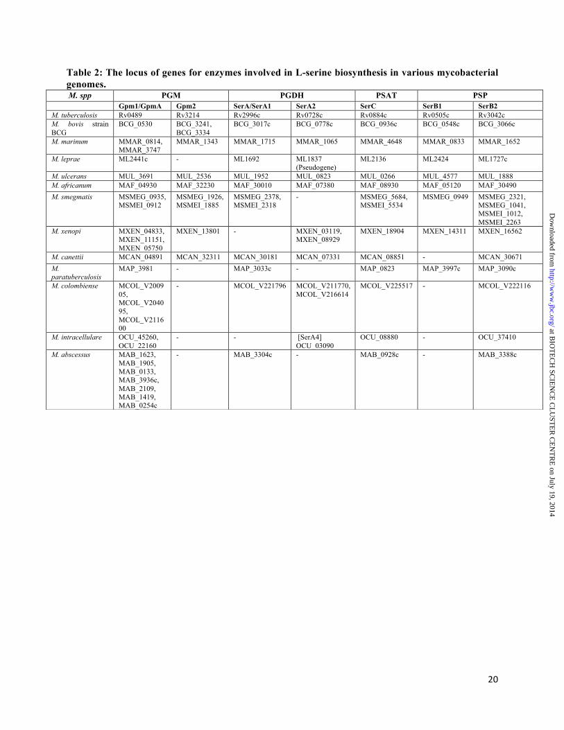
20
Table 2: The locus of genes for enzymes involved in L-serine biosynthesis in various mycobacterial genomes.
M. spp PGM PGDH PSAT PSP Gpm1/GpmA Gpm2 SerA/SerA1 SerA2 SerC SerB1 SerB2 M. tuberculosis Rv0489 Rv3214 Rv2996c Rv0728c Rv0884c Rv0505c Rv3042c M. bovis strain BCG
BCG_0530 BCG_3241, BCG_3334
BCG_3017c BCG_0778c BCG_0936c BCG_0548c BCG_3066c
M. marinum MMAR_0814, MMAR_3747
MMAR_1343 MMAR_1715 MMAR_1065 MMAR_4648 MMAR_0833 MMAR_1652
M. leprae ML2441c - ML1692 ML1837 (Pseudogene)
ML2136 ML2424 ML1727c
M. ulcerans MUL_3691 MUL_2536 MUL_1952 MUL_0823 MUL_0266 MUL_4577 MUL_1888 M. africanum MAF_04930 MAF_32230 MAF_30010 MAF_07380 MAF_08930 MAF_05120 MAF_30490 M. smegmatis MSMEG_0935,
MSMEI_0912 MSMEG_1926, MSMEI_1885
MSMEG_2378, MSMEI_2318
- MSMEG_5684, MSMEI_5534
MSMEG_0949 MSMEG_2321, MSMEG_1041, MSMEI_1012, MSMEI_2263
M. xenopi MXEN_04833, MXEN_11151, MXEN_05750
MXEN_13801 - MXEN_03119, MXEN_08929
MXEN_18904 MXEN_14311 MXEN_16562
M. canettii MCAN_04891 MCAN_32311 MCAN_30181 MCAN_07331 MCAN_08851 - MCAN_30671 M. paratuberculosis
MAP_3981 - MAP_3033c - MAP_0823 MAP_3997c MAP_3090c
M. colombiense MCOL_V200905, MCOL_V204095, MCOL_V211600
- MCOL_V221796 MCOL_V211770, MCOL_V216614
MCOL_V225517 - MCOL_V222116
M. intracellulare OCU_45260, OCU_22160
- - [SerA4] OCU_03090
OCU_08880 - OCU_37410
M. abscessus MAB_1623, MAB_1905, MAB_0133, MAB_3936c, MAB_2109, MAB_1419, MAB_0254c
- MAB_3304c - MAB_0928c - MAB_3388c
at BIO
TE
CH
SCIE
NC
E C
LU
STE
R C
EN
TR
E on July 19, 2014
http://ww
w.jbc.org/
Dow
nloaded from
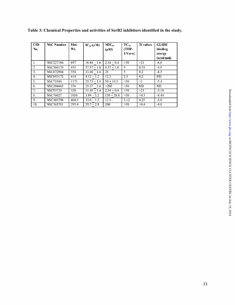
21
Table 3: Chemical Properties and activities of SerB2 inhibitors identified in the study.
at BIO
TE
CH
SCIE
NC
E C
LU
STE
R C
EN
TR
E on July 19, 2014
http://ww
w.jbc.org/
Dow
nloaded from

22
at BIO
TE
CH
SCIE
NC
E C
LU
STE
R C
EN
TR
E on July 19, 2014
http://ww
w.jbc.org/
Dow
nloaded from
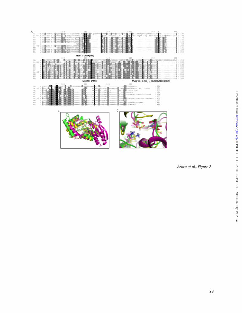
23
at BIO
TE
CH
SCIE
NC
E C
LU
STE
R C
EN
TR
E on July 19, 2014
http://ww
w.jbc.org/
Dow
nloaded from

24
at BIO
TE
CH
SCIE
NC
E C
LU
STE
R C
EN
TR
E on July 19, 2014
http://ww
w.jbc.org/
Dow
nloaded from

25
at BIO
TE
CH
SCIE
NC
E C
LU
STE
R C
EN
TR
E on July 19, 2014
http://ww
w.jbc.org/
Dow
nloaded from
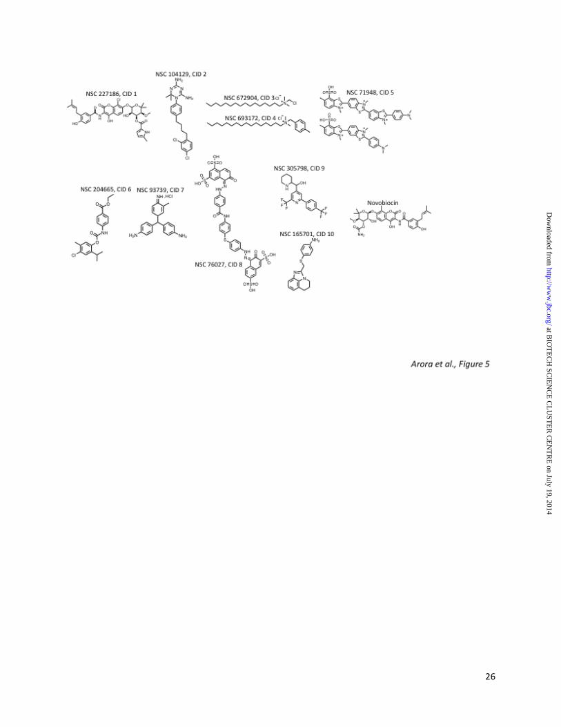
26
at BIO
TE
CH
SCIE
NC
E C
LU
STE
R C
EN
TR
E on July 19, 2014
http://ww
w.jbc.org/
Dow
nloaded from

27
at BIO
TE
CH
SCIE
NC
E C
LU
STE
R C
EN
TR
E on July 19, 2014
http://ww
w.jbc.org/
Dow
nloaded from
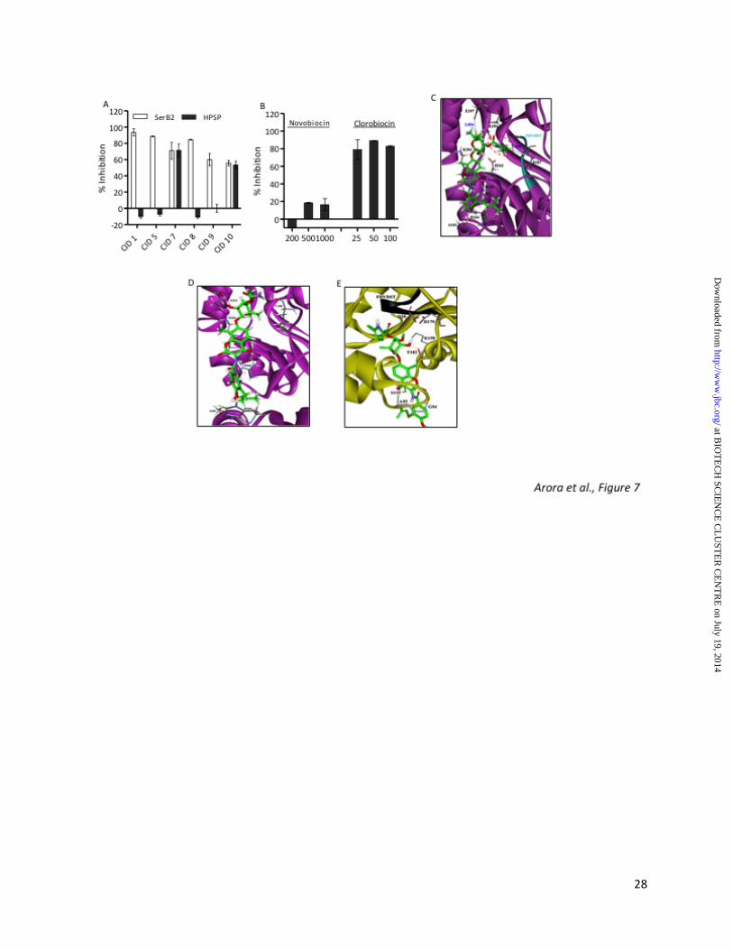
28
at BIO
TE
CH
SCIE
NC
E C
LU
STE
R C
EN
TR
E on July 19, 2014
http://ww
w.jbc.org/
Dow
nloaded from
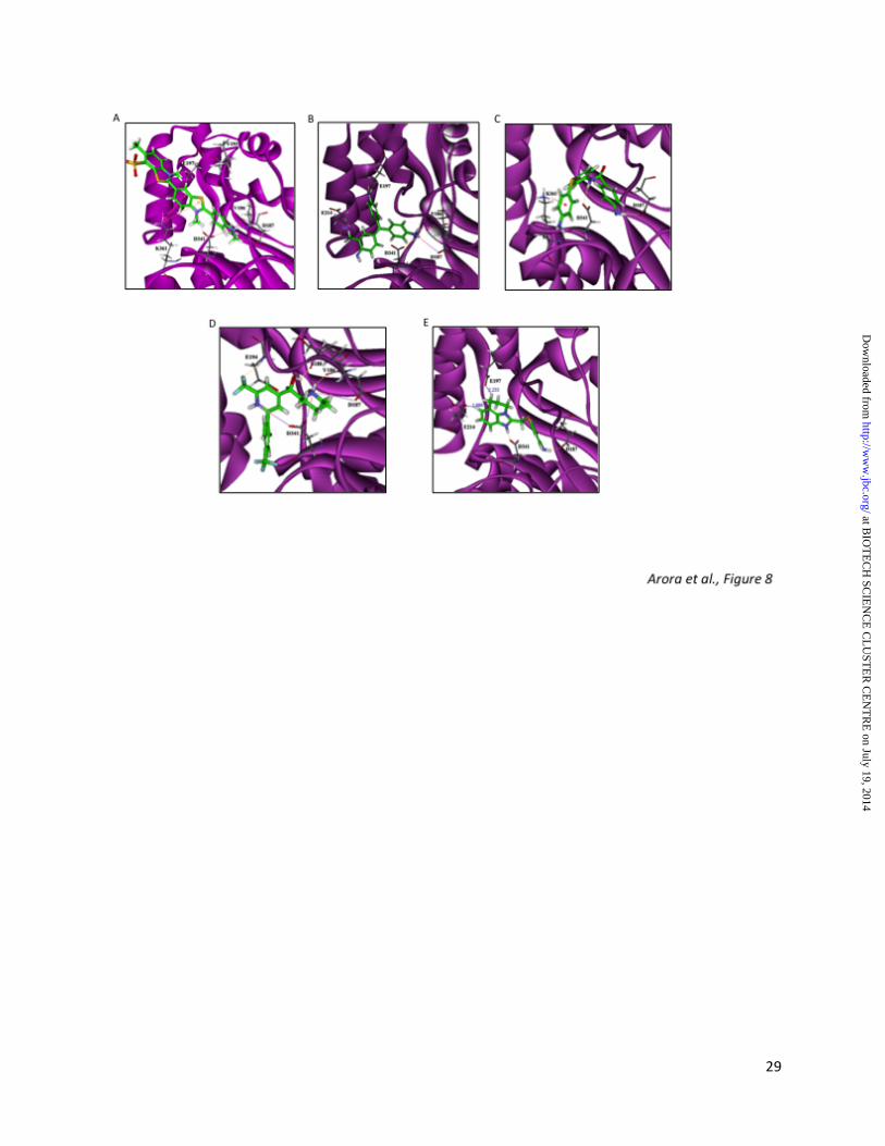
29
at BIO
TE
CH
SCIE
NC
E C
LU
STE
R C
EN
TR
E on July 19, 2014
http://ww
w.jbc.org/
Dow
nloaded from
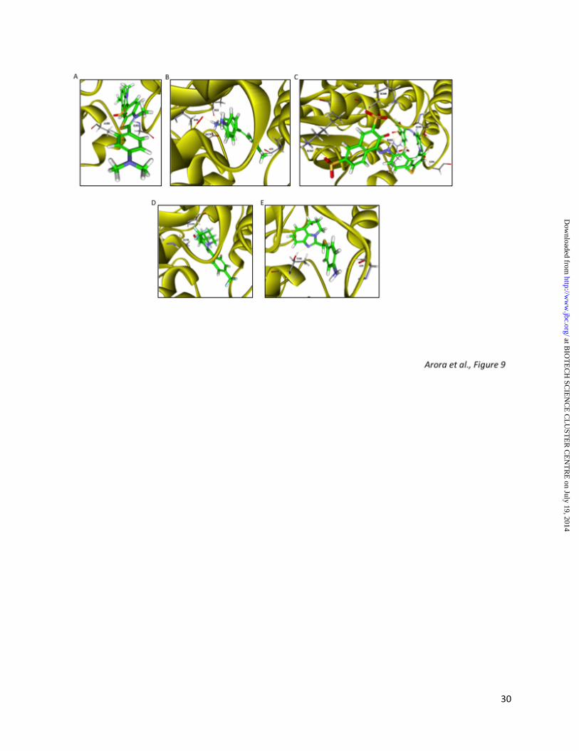
30
at BIO
TE
CH
SCIE
NC
E C
LU
STE
R C
EN
TR
E on July 19, 2014
http://ww
w.jbc.org/
Dow
nloaded from
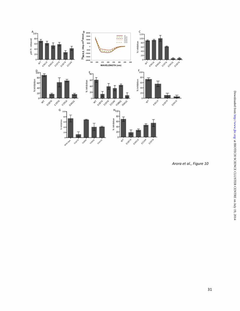
31
at BIO
TE
CH
SCIE
NC
E C
LU
STE
R C
EN
TR
E on July 19, 2014
http://ww
w.jbc.org/
Dow
nloaded from