Independently coupled HMM switching classifier for a bimodel brain-machine interface

BioMed Central
ss
Molecular Cancer
Open AcceResearchPTEN inhibits BMI1 function independently of its phosphatase activityCatherine Fan†1,2,3, Lizhi He†1,2,3, Anil Kapoor4, Adrian P Rybak1,2,3, Jason De Melo1,2,3, Jean-Claude Cutz5 and Damu Tang*1,2,3
Address: 1Division of Nephrology, Department of Medicine, McMaster University, McMaster University, Hamilton, ON, Canada, 2Father Sean O'Sullivan Research Institute, St Joseph's Hospital, Hamilton, ON, Canada, 3The Hamilton Centre for Kidney Research (HCKR), St Joseph's Hospital, Hamilton, ON, Canada, 4Department of Surgery, McMaster University, Hamilton, ON, Canada and 5Department of Pathology and Molecular Medicine, McMaster University, Hamilton, ON, Canada
Email: Catherine Fan - [email protected]; Lizhi He - [email protected]; Anil Kapoor - [email protected]; Adrian P Rybak - [email protected]; Jason De Melo - [email protected]; Jean-Claude Cutz - [email protected]; Damu Tang* - [email protected]
* Corresponding author †Equal contributors
AbstractBackground: PTEN is the second most mutated tumor suppressor gene other than p53. It suppresses tumorigenesis bydephosphorylating phosphatidylinositol (3,4,5)-triphosphate (PIP3) to phosphatidylinositol (4,5)-biphosphate (PIP2),thereby directly inhibiting phosphatidylinositol 3 kinase (PI3K)-mediated tumorigenic activities. Consistent with thismodel of action, cytosolic PTEN is recruited to the plasma membrane to dephosphorylate PIP3. While nuclear PTEN hasbeen shown to suppress tumorigenesis by governing genome integrity, additional mechanisms may also contribute tonuclear PTEN-mediated tumor suppression. The nuclear protein BMI1 promotes stem cell self-renewal andtumorigenesis and PTEN inhibits these events, suggesting that PTEN may suppress BMI1 function.
Results: We investigated whether PTEN inhibits BMI1 function during prostate tumorigenesis. PTEN binds to BMI1exclusively in the nucleus. This interaction does not require PTEN's phosphatase activity, as phosphatase-deficient PTENmutants, PTEN/C124S (CS), PTEN/G129E (GE), and a C-terminal PTEN fragment (C-PTEN) excluding the catalyticdomain, all associate with BMI1. Furthermore, the residues 186-286 of C-PTEN are sufficient for binding to BMI1. Thisinteraction reduces BMI1's function. BMI1 enhances hTERT activity and reduces p16INK4A and p14ARF expression. Theseeffects were attenuated by PTEN, PTEN(CS), PTEN(GE), and C-PTEN. Furthermore, knockdown of PTEN in DU145cells increased hTERT promoter activity, which was reversed when BMI1 was concomitantly knocked-down, indicatingthat PTEN reduces hTERT promoter activity via inhibiting BMI1 function. Conversely, BMI1 reduces PTEN's ability toinhibit AKT activation, which can be attributed to its interaction with PTEN in the nucleus, making PTEN unavailable todephosphorylate membrane-bound PIP3. Furthermore, BMI1 appears to co-localize with PTEN more frequently inclinical prostate tissue samples from patients diagnosed with PIN (prostatic intraepithelial neoplasia) and carcinomacompared to normal prostate epithelium. While PTEN co-localized with BMI1 in 2.4% of normal prostate epithelial cells,co-localization was observed in 37.6% and 18.5% of cells in PIN and carcinoma, respectively. Collectively, wedemonstrate that PTEN inhibits BMI1 function via binding to BMI1 in a phosphatase independent manner.
Conclusion: We demonstrate that nuclear PTEN reduces BMI1 function independently of its phosphatase activity. Itwas recently observed that nuclear PTEN also suppresses tumorigenesis. Our results, therefore, provide a plausiblemechanism by which nuclear PTEN prevents tumorigenesis.
Published: 10 November 2009
Molecular Cancer 2009, 8:98 doi:10.1186/1476-4598-8-98
Received: 6 September 2009Accepted: 10 November 2009
This article is available from: http://www.molecular-cancer.com/content/8/1/98
© 2009 Fan et al; licensee BioMed Central Ltd. This is an Open Access article distributed under the terms of the Creative Commons Attribution License (http://creativecommons.org/licenses/by/2.0), which permits unrestricted use, distribution, and reproduction in any medium, provided the original work is properly cited.
Page 1 of 14(page number not for citation purposes)
Molecular Cancer 2009, 8:98 http://www.molecular-cancer.com/content/8/1/98
IntroductionThe polycomb group (PcG) BMI1 gene maintains the pro-liferation potential and self-renewal of hematopoietic andneural stem cells [1,2]. This is in part attributable to BMI1-mediated suppression of p16INK4A, p19ARF/p14ARF, andE4F1 [3-6]. This developmental function of BMI1 is inline with its oncogenic role in leukemia. The BMI1 genewas initially isolated as an oncogene which cooperatedwith c-Myc in retrovirus-induced B and T cell leukemia[7,8]. Overexpression of BMI1 transformed lymphocytes[9,10] and was detected in 25% of mantle cell lymphomas[11]. BMI1 is positively associated with unfavorable prog-nosis in patients with diffuse large B cell lymphomas andmyelodysplastic syndrome [12,13]. Increases in BMI1were also reported in epithelial malignancies, includingnon-small cell lung cancer (NSCLC) [14], colon cancer[15], breast cancer [16], and nasopharyngeal carcinoma[17].
BMI1 may also promote prostate tumorigenesis. Increasesin BMI1 mRNA were detected in prostate cancer cell lines,xenografts and human primary prostate carcinomas, aswell as primary prostate tumors derived from the TRAMPtransgenic mouse model [18]. Prostate cancer patientswith an 11-gene signature, which is associated with BMI1expression, are more likely to have an unfavorable prog-nosis when compared to those without this signature[18]. Additionally, metastatic prostate carcinoma precur-sor cells that are double-positive for BMI1 and anotherpolycomb-group protein EZH2 are more tumorigenicthan those which are negative for both proteins [19].
Mechanistically, BMI1 promotes tumorigenesis, at least inpart, via inhibiting p16INK4A and p19ARF expression, andenhancing human telomerase reverse transcriptase(hTERT) activity [17,20], leading to a bypass of senes-cence. BMI1-/- hematopoietic progenitors expressincreased levels of p16INK4A and p19ARF, and accumulatehigh levels of the senescence marker SA-β-Gal [5]. BMI1-/-
mouse embryonic fibroblasts (MEFs) undergo prematuresenescence [21] and overexpression of BMI1 in MEFs andhuman fibroblasts extends their replicative life spans[21,22]. Consistent with these observations, BMI1immortalizes human nasopharyngeal and mammary epi-thelial cells [17,20]. However, how BMI1 is regulated dur-ing tumorigenesis remains to be determined.
PTEN is a tumor suppressor gene that is frequentlymutated in human cancers. This is at least in part attribut-able to PTEN's action in inhibiting PI3K. PTEN dephos-phorylates the 3-position phosphate from the inositolring of phosphatidylinositol (3,4,5)-triphosphate (PIP3)[23], thereby directly inhibiting phosphatidylinositol 3kinase (PI3K)-mediated tumorigenic activities. WhilePTEN-mediated suppression of the PI3K/AKT pathway is
well established, accumulating evidence suggests thatnuclear PTEN also plays a critical role in tumor suppres-sion [23]. Although several mechanisms responsible fornuclear PTEN-mediated tumor suppression have beenobserved [23] (see Discussion for details), additionalmechanisms remain likely.
In this investigation, we provide evidence showing thatnuclear PTEN suppresses BMI1 function. PTEN binds toBMI1 in the nucleus of prostate cancer cells and reducesBMI1-mediated suppression of p16INK4A and p14ARF aswell as BMI1-mediated enhancement of hTERT. Addition-ally, PTEN co-localizes with BMI1 more frequently in pri-mary prostate carcinomas compared to normal prostateglands. Our observations are consistent with previousfindings showing that while BMI1 maintains the prolifer-ation potential of neural stem cells (NSCs) [2], PTENinhibits this process [24].
Materials and methodsCell lines and plasmidsDU145, MCF7, and 293T cells were purchased fromATCC, and cultured in MEM (DU145) and DMEM(MCF7and 293T) containing 10% FBS and 1% Penicillin-Streptomycin (Invitrogen). Among the most widely usedthree human prostate cancer cell lines (LNCaP, PC3, andDU145), only DU145 cells express wild type PTEN andBMI1, and therefore were chosen for this research. HumanBMI1 cDNA was amplified by RT-PCR from HeLa cells,and subsequently subcloned in pcDNA3 and pBabe retro-virus vectors. pGL3-hTERTmin-Luc reporter plasmid, con-taining a 59bp region of the hTERT promoter (-208 to -150) which has been shown to display maximal promoteractivity [25] was constructed from HeLa cell genomicDNA using routine molecular biology techniques.
Retroviral InfectionRetroviral infection was performed following our previ-ously published procedure [26,27]. Briefly, a gag-polexpressing vector and an envelope-expressing vector (VSV-G) (Stratagene) were transiently co-transfected with adesigned retroviral plasmid into 293T cells. After 48hours, the virus-containing medium was harvested, fil-tered through a 0.45 μM filter, and centrifuged at 50,000g for 90 minutes to concentrate the retrovirus. Followingthe addition of 10 μg/ml of polybrene (Sigma), themedium was used to infect cells. Infection for pBabe-based constructs was selected in puromycin, while infec-tion for pLHCX-based constructs was selected in hygro-mycin.
Collecting primary prostate cancerProstate tissue was collected at St. Joseph's Hospital inHamilton, Ontario, Canada with the approval from thelocal Ethics Board and with consent from the patients.
Page 2 of 14(page number not for citation purposes)

Molecular Cancer 2009, 8:98 http://www.molecular-cancer.com/content/8/1/98
Tumors were examined and graded by pathologists at theHospital. 42 primary prostate cancer specimens were col-lected.
TRAP (Telomeric Repeat Amplification Protocol) assayA TRAP kit (TRAPEZE® Telomerase Detection kit) was pur-chased from Chemicon International. TRAP assay wasperformed according to the manufacturer's instruction.
Western blot and immunoprecipitationCell lysates were prepared and western blot was per-formed according to our published procedure [26]. 50 μgprotein of total lysate was separated on SDS-PAGE gel andtransferred onto Immobilon-P membranes (Millipore).Membranes were blocked with 5% skim milk and thenincubated with the indicated antibodies at room temper-ature for 1 hour. Signals were detected using an ECL West-ern Blotting Kit (Amersham). Primary antibodies usedwere: polyclonal anti-BMI1 (1:100, Santa Cruz Biotech-nology), polyclonal anti-PTEN (1:100, Upstate Technolo-gies), polyclonal anti-p16INK4A (1:500, Santa CruzBiotechnology), and polyclonal anti-p14ARF (1:5000,Sigma). Immunoprecipitation of ectopic PTEN and BMI1was performed by incubation of 200 μg cell lysate proteinwith specific antibodies plus Protein G agarose (Invitro-gen) at 4°C overnight, followed by wash for 6 times in abuffer containing 50 mM Tris (PH 7.5), 100 mM NaCl,1.5 mM EGTA, 0.1% Triton X-100. The antibodies usedfor immunoprecipitation were monoclonal anti-PTEN(Santa Cruz Biotechnology), monoclonal anti-FLAG (M2,Sigma) for BMI1 and its mutants, and mouse IgG (Sigma)as a negative control. The immunoprecipitation was ana-lyzed by western blot using polyclonal anti-PTEN (SantaCruz) and anti-FLAG (Sigma). Immunoprecipitation ofthe endogenous PTEN-BMI1 complex was carried out bylysing DU145 cell in a HEPES lysate buffer, pH7.0 (20mM HEPES, 150 mM NaCl, 1 mM EDTA, 1 mM EGTA, 1%Triton X-100, 25 mM sodium pyrophosphate, 1 mM NaF,1 mM β-glycerophosphate, 0.1 mM sodium orthovanad-ate, 1 mM PMSF, 2 μg/ml leupeptin and 10 μg/ml apro-tinin) containing DSP 2 μM (PIRECE) on ice for 2 hoursand quenching free DSP by incubating in 50 mM Tris pH7.5 on ice for 15 minutes. Immunoprecipitation was thencarried out as described above.
ImmunofluorescenceDouble immunofluorescence staining was carried outusing the following antibodies: monoclonal anti-PTEN(Santa Cruz, 1 μg/ml) or a polyclonal anti-PTEN (1:100;Upstate Technologies), polyclonal anti-FLAG or a mono-clonal anti-FLAG (M2, 1:500; Sigma), FITC-Donkey anti-mouse IgG (1:200; Jackson Immuno Research) and Rhod-amine-Donkey anti-rabbit IgG (1:200; Jackson ImmunoResearch) were used as secondary antibodies. Images werecaptured using Axiovert 200 M confocal microscope andAxioVision 3 software.
For double immunofluorescence staining prostate tissues,tissues were deparaffinized, rehydrated, and subjected toantigen-retrieval and endogenous peroxidase-quenching.Tissue sections were blocked for 1 hour at room tempera-ture in 3% donkey serum and 3% BSA in TBST. Dual-IFstaining was carried out using a TSA Plus kit (Perk-inElmer) according to the manufacturer's protocol. Sec-tions were counterstained with DAPI and digital imageswere processed as described above.
Statistical analysisAnalysis was performed using Northern Eclipse 4.0 soft-ware (manual cell counter) for Windows. Approximately1000 cells from randomly selected fields were counted foreach normal, PIN, and cancer foci per patient. Mean per-centages of positively-stained cells were then analyzedusing GraphPad 4.0 for Windows.
ResultsBMI1 interacts with PTENBMI1 determines the proliferation potential of neuralstem cells (NSCs) [2], a process that is inhibited by PTEN[24]. Additionally, while BMI1 promotes tumorigenesisin a variety of human cancers [28], PTEN potently sup-presses tumorigenesis [29,30]. These observations suggestthat PTEN may negatively regulate BMI1 function. Sinceboth BMI1 [7,8] and PTEN reside in the nucleus [31], wehypothesized that BMI1 may associate with PTEN. Whentransiently co-expressed in 293T cells, a complex contain-ing both BMI1 and PTEN could be immunoprecipitatedvia either BMI1 or PTEN (Fig 1A), while control IgG didnot precipitate either protein (data not shown). This asso-ciation was also detected between endogenous BMI1 andendogenous PTEN (Fig 1B).
To further examine this interaction, we determinedwhether PTEN co-localizes with BMI1 inside the cell.When ectopically expressed, PTEN and BMI1 were co-localized in the nucleus (Fig 1C, the 293T cell panel). Fur-thermore, endogenous BMI1, which was stained in a"punctuate" manner in the nuclei of DU145 and MCF7cells, co-localized with endogenous nuclear PTEN (Fig1C). Both anti-BMI1 and anti-PTEN antibodies specifi-cally recognized their respective proteins. The anti-BMI1antibody did not produce any detectable signals in BMI1-negative LNCaP cells in western blot and in IF procedures(see our recent publication) [32]. The anti-PTEN antibodyproduced no-detectable signals in PTEN-null LNCaP andU87 cells as well as the signal level was significantlyreduced in PTEN siRNA treated DU145 cells in IF proce-dures (data not shown). We observed that approximately20% of cells expressed BMI1 at a given time point, a typi-cal expression pattern observed with other polycomb pro-teins [33]. However, in all BMI1-expressing cells, PTENco-localized with BMI1 in the nucleus. Taken together, the
Page 3 of 14(page number not for citation purposes)

Molecular Cancer 2009, 8:98 http://www.molecular-cancer.com/content/8/1/98
Page 4 of 14(page number not for citation purposes)
PTEN binds to BMI1Figure 1PTEN binds to BMI1. (A) 293T cells were transiently transfected with FLAG-tagged BMI1 and HA-tagged PTEN. Cell lysates were prepared and immunoprecipitated with anti-PTEN (top panel) and anti-FLAG (M2) (middle panel) antibodies. The precip-itates and lysates (bottom panel) were analyzed by western blot using the indicated antibodies. The * symbol indicates endog-enous PTEN. (Note: the reason why endogenous PTEN was not detected in the lysate panel was attributable to a low level of endogenous PTEN in 293T cells). (B) DU145 cell lysates were cross-linked with DSP, immunoprecipitated with anti-BMI1 anti-body or control IgG, and analyzed by western blot for PTEN and BMI1. Twenty percent of cell lysate used for immunoprecipi-tation was also analyzed by western blot. (C) Co-localization between PTEN and BMI1. Ectopic PTEN and ectopic BMI1 in 293T cells and their respective endogenous proteins in MCF-7 and DU145 cells were examined by double immunofluorescent (IF) staining. Nuclei were counter-stained with DAPI (blue). Scale bar represents 10 μM.

Molecular Cancer 2009, 8:98 http://www.molecular-cancer.com/content/8/1/98
above observations demonstrate that PTEN associateswith BMI1 in the nucleus.
PTEN interacts with BMI1 independently of its phosphatase activityTo characterize the interaction between PTEN and BMI1,we examined whether PTEN's phosphatase activity isrequired for the association. When ectopically expressedin 293T cells, wild type PTEN, phosphatase-deficientPTEN(C124S) [PTEN(CS)], and PIP3 specific phos-phatase-deficient PTEN(G129E) [PTEN(GE)] [34,35]formed a complex with BMI1 as detected by co-immuno-precipitation (Fig 2A). Additionally, a C-terminal frag-ment of PTEN (C-PTEN) (encompassing residues 186-403, and thus excluding the catalytic domain that liesbetween residues 1-185) [36] bound to BMI1 (Fig 2A, B).In comparison to PTEN, PTEN(CS) interacted with BMI1with reduced affinity [Fig 2A, comparing BMI1 co-immu-noprecipitated via PTEN with BMI1 co-immunoprecipi-tated via PTEN(CS) as well as comparing PTEN withPTEN(CS) that were co-immunoprecipitated via BMI1].This may be attributed to potential conformationalchanges that might be caused by this mutation rather thandue to the lack of phosphatase activity, as C-PTEN bindsto BMI1 with increased affinity (Fig 2A). We also exam-ined whether N-PTEN (residues 1-185) interacts withBMI1 and found that N-PTEN was expressed at undetect-able levels when co-expressed with BMI1 in 293T cells(data not shown). It was thus difficult to determine if N-PTEN binds to BMI1. Nonetheless, our experiments dem-onstrate that C-PTEN is sufficient to interact with BMI1and that PTEN binds to BMI1 independently of its phos-phatase activity.
PTEN binds to BMI1 via residues 186-286C-PTEN contains two functional domains, the C-2domain and the C-terminal tail (Fig 2B). To further mapthe BMI1-binding region, we generated HA-tagged C2 andC-tail PTEN fragments (Fig 2B) and examined their asso-ciation with BMI1. When co-expressed in 293T cells,immunoprecipitation of FLAG-tagged BMI1 efficiently co-precipitated the C-2 fragment (Fig 2C). We further trun-cated the C-2 fragment into HA-tagged C2N and C2C (Fig2B) and co-expressed BMI1 with either C2N or C2C in293T cells (Fig 2D, bottom-left panel). Immunoprecipita-tion of BMI1 co-precipitated C2N (Fig 2D, left panel).However, we could not detect C2C and C-tail fragments(Fig 2D, bottom-left panel). This may have been causedby the potential instability of these fragments due to theirinability to properly fold. To address this potential issueof instability, we fused the fragments to green fluorescentprotein (GFP) and demonstrated their expression in 293Tcells (Fig 2D, bottom-right panel). However, these frag-ments were either immunoprecipitated with control IgG
(GFP-C2C) or anti-FLAG (M2) antibody (GFP-C-tail)without the co-expression of FLAG-tagged BMI1 (Fig 2D,top-right panel), demonstrating that these fragments werenon-specifically immunoprecipitated. This indicates thatthe fusion proteins did not fold properly, which is consist-ent with our inability to detect the expression of C2C andC-trail fragments by western blot (Fig 2D, bottom-leftpanel).
To further examine C2N-medaited BMI1 binding, wefused C2N to GFP. When co-expressed in 293T cells, GFP-C2N was co-immunoprecipitated via FLAG-tagged BMI1(Fig 2D, right panel). Taken together, the above experi-ments reveal that the C2N (186-286) PTEN fragment issufficient to interact with BMI1.
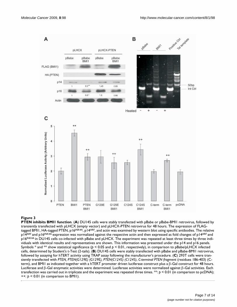
PTEN inhibits BMI1 function independently of its phosphatase activitySince PTEN and BMI1 function in opposite directions inboth stem cell biology and tumorigenesis, the observationthat PTEN binds to BMI1 indicates that PTEN may inhibitBMI1 function. BMI1 has been shown to suppress theexpression of the INK4A/ARF locus, p16INK4A and p14ARF
[3,4], which we have also demonstrated recently in pros-tate cancer cells [32]. To examine whether PTEN affectsBMI1-mediated inhibition of p16INK4A and p14ARF expres-sion, we stably expressed BMI1 and PTEN individuallyand in combination into DU145 cells (Fig 3A). Consistentwith previous publications [3,4], ectopic BMI1 reducedendogenous p16INK4A and, to a greater degree, p14ARF
expression (Fig 3A). While ectopic PTEN did not enhancep16INK4A or p14ARF expression, it prevented the BMI1-mediated reduction of p16INK4A and p14ARF (Fig 3A).
BMI1 has been shown to increase hTERT activity in mam-mary epithelial cells [20]. To examine whether BMI1 alsoup-regulates hTERT activity in prostate cancer, we ectopi-cally expressed BMI1 in DU145 prostate cancer cells usinga retrovirus. A TRAP (Telomeric Repeat Amplification Pro-tocol) assay revealed that BMI1 enhanced hTERT activity(Fig 3B). To address the impact of PTEN on BMI1-inducedhTERT activation, we co-transfected a BMI1-expressingplasmid along with a hTERT promoter-reporter construct(pGL3-hTERTmin-Luc) [25] with the addition of either anempty vector, PTEN, PTEN(CS), PTEN(GE) or C-PTENconstruct into 293T cells (Fig 3C). Consistent with theTRAP assay, BMI1 increased hTERT promoter activity (Fig3C). Interestingly, PTEN, PTEN(CS), PTEN(GE), and C-PTEN all inhibited the BMI1-mediated activation of thehTERT promoter (Fig 3C). This is consistent with theobservation that all these PTEN proteins interact withBMI1 (Fig 2A). Collectively, the above results demonstratethat PTEN inhibits BMI1 function independently of itsPIP3 phosphatase activity.
Page 5 of 14(page number not for citation purposes)

Molecular Cancer 2009, 8:98 http://www.molecular-cancer.com/content/8/1/98
Page 6 of 14(page number not for citation purposes)
Characterization of the interaction between PTEN and BMI1 proteinsFigure 2Characterization of the interaction between PTEN and BMI1 proteins. (A) PTEN binds to BMI1 independently of its phosphatase activity. 293T cells were transiently transfected with BMI1, PTEN, PTEN(C124S) (C124S), PTEN(G129E) (G129E), a C-terminal PTEN fragment (residues 186-403) (C-PTEN) for 48 hours. Cell lysates were prepared and immunoprecipitated with anti-PTEN and anti-FLAG (M2) (for ectopic BMI1) antibodies. The precipitates and lysates were analyzed by western blot using the indicated antibodies. The # and * symbols indicate endogenous PTEN and a possible oligomer of C-PTEN, respec-tively. (B) Mapping the BMI1 binding motif of the PTEN protein. A set of PTEN truncation mutants were constructed. Their interaction with BMI1 was examined. C2: C2 domain. The + and - symbols indicate binding or not-binding of individual PTEN proteins to BMI1. (C) C2 binds to BMI1. FLAG-tagged BMI1 and HA-tagged C2 were transfected into 293T cells as indicated. BMI1 was immunoprecipitated with an anti-FLAG antibody (M2) or a control IgG (IgG), followed by western blot examination for BMI1 and C2. 20% of the cell lysates that were used for immunoprecipitations were also analyzed. The * symbols indicate background bands. (D) C2N binds to BMI1. C2N, C2C, and C-tail (left panel) and their GFP fusion counterparts (right panel) were co-transfected with either an empty vector (-) or FLAG-tagged BMI1 as indicated, followed by immunoprecipitation with M2 or control IgG (IgG) and then western blot (WB) with the indicated antibodies. The respective cell lysates were shown at the bottom panels.
Molecular Cancer 2009, 8:98 http://www.molecular-cancer.com/content/8/1/98
Page 7 of 14(page number not for citation purposes)
PTEN inhibits BMI1 functionFigure 3PTEN inhibits BMI1 function. (A) DU145 cells were stably transfected with pBabe or pBabe-BMI1 retrovirus, followed by transiently transfected with pLHCX (empty vector) and pLHCX-PTEN retrovirus for 48 hours. The expression of FLAG-tagged BMI1, HA-tagged PTEN, p16INK4A, p14ARF, and actin was examined by western blot using specific antibodies. The relative p14ARF and p16INK4A expression was normalized against the respective actin and then expressed as fold changes of p14ARF and p16INK4A in DU145 cells co-infected with pBabe and pLHCX. The experiment was repeated at least three times by three indi-viduals with identical results and representatives are shown. This information was presented under the p14 and p16 panels. Symbols * and ** show statistical significance (p < 0.05 and p < 0.01, respectively), in comparison to pBabe/pLHCX infected cells, determined by Student's t-Test (2-tails). (B) DU145 cells were stably transfected with pBabe and pBabe-BMI1 retrovirus, followed by assaying for hTERT activity using TRAP assay following the manufacturer's procedure. (C) 293T cells were tran-siently transfected with PTEN, PTEN(G129E) (G129E), PTEN(C124S) (C124S), C-terminal PTEN fragment (residues 186-403) (C-term), and BMI1 as indicated together with a hTERT promoter driven luciferase construct plus a β-Gal construct for 48 hours. Luciferase and β-Gal enzymatic activities were determined. Luciferase activities were normalized against β-Gal activities. Each transfection was carried out in triplicate and the experiment was repeated three times. **: p < 0.01 (in comparison to pcDNA); ++: p < 0.01 (in comparison to BMI1).

Molecular Cancer 2009, 8:98 http://www.molecular-cancer.com/content/8/1/98
Since PTEN interacts with BMI1 exclusively in the nucleus(Fig 1), we examined the impact of nuclear and cytosolicPTEN mutants on BMI1's function. It has been shown thatthe Chimpanzee PTEN fragments 1-375 and 1-375/K13Aresided largely in the nucleus and cytosol, respectively[37]. Dr. Pulido (Spain) kindly provided us these PTENmutants together with Chimpanzee PTEN. When co-expressed in 293T cells, PTEN, 1-375, and 1-375/K13A
were all co-precipitated via BMI1 (Fig 4A). While 1-375/K13A largely localizes in the cytosol, a small proportion ofit remains in the nucleus [37] (Fig 4B). This remainingPTEN 1-375/K13A may contribute to the co-immunopre-cipitation of 1-375/K13A via BMI1 (Fig 4A) (see Discus-sion for details). However, in comparison to 1-375/K13A,1-375 displayed enhanced activity in up-regulation ofendogenous p14ARF when transiently transfected into
Nuclear PTEN reduces BMI1 functionFigure 4Nuclear PTEN reduces BMI1 function. (A) Interaction of nuclear PTEN with BMI1. Chimpanzee PTEN and the indicated mutants PTEN/1-375 and PTEN/1-375(K13A) were transfected without and with FLAG-tagged BMI1 in 293T cells, followed by immunoprecipitation of BMI1 using an anti-FLAG (α-FLAG) and then immunobloted (IB) with the indicated antibodies. Control IgG did not precipitate either BMI1 or PTEN (data not shown). (B) Nuclear PTEN inhibits BMI1 function. DU145 cells were transiently expressed with PTEN/1-375 (top panel) or PTEN/1-375(K13A) (bottom panel). Cells were then double IF stained for ectopic PTEN mutants using an anti-HA antibody (red) or endogenous p14ARF (green). Nuclei were counter-stained with DAPI (blue). More than 200 transfected cells were randomly counted. Typical images of 1-375 and 1-375(K13A) were shown and the related quantification was discussed (see Discussion for details).
Page 8 of 14(page number not for citation purposes)

Molecular Cancer 2009, 8:98 http://www.molecular-cancer.com/content/8/1/98
DU145 cells (Fig 4B) (see Discussion for details). The up-regulated p14ARF shows the typical pattern of nucleolardistribution (Fig 4B) [38], suggesting that the enhancedp14ARF was functional. Taken together, these observationssupport the concept that nuclear PTEN reduces BMI1function.
To further consolidate the concept that PTEN reducesBMI1 function, we knocked-down PTEN and BMI1 inDU145 cells using specific siRNAs (Fig 5, top panel). Asexpected, knockdown of PTEN activated AKT (Fig 5, toppanel) and knockdown of BMI1 increased p14ARF andp16INK4A expression (data not shown). Consistent withBMI1 increasing hTERT promoter activity (Fig 3C), weobserved that knockdown of BMI1 reduced hTERT pro-moter activity to approximately 77% of that observed incontrol siRNA treated DU145 cells (Fig 5, bottom panel).Furthermore, knockdown of PTEN significantly enhancedhTERT promoter activity in DU145 cells (Fig 5, bottompanel) and this occurs only in cells expressing endog-enous BMI1 and not in cells whose BMI1 was concomi-tantly knocked-down (Fig 5, bottom panel), whichdemonstrates that endogenous PTEN reduces hTERT pro-moter activity via inhibiting endogenous BMI1 function.
BMI1 reduces PTEN functionIt is well established that PTEN antagonizes the activity ofPI3K via dephosphorylation of membrane-bound PIP3.This inhibition is dependent on recruiting cytosolic PTENto the plasma membrane. Since BMI1 binds PTEN in thenucleus, high levels of BMI1 may attenuate PTEN's abilityto inhibit the PI3K-AKT pathway by sequestering PTEN inthe nucleus. To test this possibility, we infected DU145cells with a retrovirus expressing BMI1, PTEN, or bothBMI1 and PTEN (Fig 6). BMI1 overexpression indeedenhanced AKT activation (Fig 6). While overexpression ofPTEN alone did not affect AKT activation in DU145 cells,which is consistent with other publications [39,40],ectopic PTEN reversed the increase in AKT activationobserved in DU145 cells overexpressing BMI1 (Fig 6). Asectopic PTEN alone does not directly affect the PI3K-AKTpathway (Fig 6), ectopic PTEN may indirectly affect thePI3K-AKT pathway by interacting with ectopic BMI1. Thispossibility is further supported by the observation thatknockdown of BMI1 alone or in combination with knock-down of PTEN slightly reduced AKT activation (Fig 5, toppanel, comparing AKT phosphorylation in BMI1 siRNAand PTEN siRNA/BMI1 siRNA lanes with that in CtrlsiRNA and PTEN siRNA lanes, respectively).
PTEN appears to co-localize with BMI1 in primary prostate cancerThe fact that PTEN binds BMI1 and reduces BMI1 func-tion in cultured prostate cancer cells prompted us toexamine whether PTEN co-localizes with BMI1 in primary
prostate cancer. While PTEN marginally co-localizes withBMI1 in normal prostate epithelial cells, PTEN extensivelyco-localizes with BMI1 in PIN and in PTEN positive (butnot negative) prostate carcinoma (Fig 7), which furtherdemonstrates the specificity of the anti-PTEN antibodyused. We performed double immunofluorescent (IF)staining for PTEN and BMI1 in 42 primary prostate cancerspecimens. By taking advantage of the heterogeneousnature of primary prostate cancer specimens [41], we wereable to locate normal prostate glands, PIN, and carcinomawithin each specimen. One thousand cells were randomlycounted within individual tissues (normal prostateglands, PINs, and carcinomas) for each specimen in oursample set. Approximately 56% of prostate carcinomasexpress PTEN (data not shown), a number that is consist-ent with previous publications [29]. While only 2.4% ofepithelial cells from normal prostate glands show low lev-els of co-localization between PTEN and BMI1 (Fig 7,Table 1), 37.6% and 18.5% of PIN and carcinoma cellsdisplay extensive co-localization between PTEN and BMI1(Fig 7, Table 1). Interestingly, while PTEN stays largelyoutside of the nucleus in the epithelial cells of normalprostate glands, a significant increase in nuclear PTEN isobserved in PINs and PTEN-positive carcinoma (Fig 7).This may be attributable to the observed increases in theco-localization between PTEN and BMI1 in PINs andPTEN-positive prostate carcinoma in comparison to nor-mal prostate epithelium (Fig 7, Table 1). Taken together,the above observations suggest that nuclear PTEN plays animportant role in inhibiting BMI1 function during pros-tate tumorigenesis.
DiscussionWhile it is known that BMI1 promotes tumorigenesis, atleast in part, by suppressing p16INK4A and p14ARF expres-sion, it remains to be determined what mechanisms regu-late BMI1-mediated oncogenic activities. We demonstratehere that one of these mechanisms is PTEN-mediatedattenuation of BMI1 function.
PTEN physically associates with BMI1 in cultured prostatecancer cells and appears to co-localize with BMI1 in pri-mary prostate cancer. It was observed that PTEN co-local-izes with BMI1 more extensively in PINs compared toboth carcinomas and normal prostate glands (Fig 7; Table1). As high grade PINs are pre-cancerous lesions, theabove observation supports the concept that PTEN inhib-its (or plays a surveillant role to) BMI1's oncogenic activ-ities during prostate tumorigenesis and that escape fromPTEN's suppression (or surveillance) enables BMI1 topromote prostate cancer progression. This is consistentwith the observed increases in BMI1 mRNA [18] andBMI1 protein [32] levels during prostate tumorigenesis.Through its interaction with BMI1, PTEN inhibits BMI1'sfunction.
Page 9 of 14(page number not for citation purposes)

Molecular Cancer 2009, 8:98 http://www.molecular-cancer.com/content/8/1/98
Page 10 of 14(page number not for citation purposes)
PTEN reduces hTERT promoter activity via inhibiting BMI1 functionFigure 5PTEN reduces hTERT promoter activity via inhibiting BMI1 function. DU145 cells were transfected with 100 nM PTEN siRNA, BMI1 siRNA, PTEN siRNA plus BMI1 siRNA, and the respective control (Ctrl) siRNA (Dharmcon) using LipofectAMINE2000 (Invitrogene) for 48 hours following our published procedure [52]. The expression of individual proteins was examined by western blot using specific antibodies (top panel). These cells were transfected with a pGL3-hTERTmin-Luc reporter, a lacZ vector, and plus the indicated siRNAs for 48 hours. Luciferase activities were determined and normalized against the respective lacZ activity. Experiments were carried out in triplicate and were repeated three times. Average data of these independent experiments is shown. Luciferase activities in PTEN siRNA (p < 0.001) and BMI1 siRNA (p < 0.05) cells are significantly different from that in Ctrl siRNA cells (bottom panel).

Molecular Cancer 2009, 8:98 http://www.molecular-cancer.com/content/8/1/98
The above conclusion is further supported by the observa-tions that PTEN binds to BMI1 in the nucleus (Fig 1) andthat a nuclear PTEN mutant (1-375) was more potent ininhibiting BMI1-mediated reduction of p14ARF than itscytosolic counterpart (PTEN mutant 1-375/K13A) (Fig4B). However, 1-375/K13A was efficiently immunopre-cipitated through BMI1 (Fig 4A). This could be attributa-ble to two factors, 1) 1-375/K13A is not an exclusivecytosolic protein [37] (Fig 4B) and 2) substitution of K13with alanine may alter the protein conformation, whichcould increase its affinity to BMI1. Therefore, althoughthere is far less 1-375/K13A in the nucleus, an increase inits binding affinity to BMI1 may allow 1-375/K13A to beefficiently co-immunoprecipitated via BMI1 (Fig 4A).Alternatively, the effective co-immunoprecipitation of 1-375/K13A via BMI1 might be an artifact caused by the celllysate preparation, which allowed cytosolic 1-375/K13Ato interact with nuclear BMI1. The association of nuclear1-375/K13A (although a minor population) with BMI1 isconsistent with our observation that approximately 11%of 1-375/K13A transfected cells displayed upregulation ofendogenous p14ARF, while approximately 30% of 1-375transfected DU145 cells showed p14ARF upregulation.
Taken together, these results support the notion thatnuclear PTEN reduces BMI1 function.
While it is well documented that PTEN suppresses tumor-igenesis via its PIP3 phosphatase activity at the plasmamembrane, the potential function of nuclear PTEN is lessclear. As PTEN appears to co-localize with BMI1 in thenucleus, our work suggests that nuclear PTEN inhibitsBMI1 function, which is independent of PTEN's phos-phatase activity. This is consistent with recent reportsshowing that nuclear PTEN maintains chromosome sta-bility independently of its phosphatase activity [42] aswell as induces G1 and G2 arrest in breast cancer andmelanoma cells [31,43]. PTEN may regulate cell cycle pro-gression via modulating p21CIP1 [44]. Importantly, loss ofnuclear PTEN was observed to associate with the tumorprogression of melanoma and colorectal cancer [45,46].Interestingly, the nuclear PTEN maintains chromosomestability via a C2 domain-mediated interaction with cen-tromeres [42]. This agrees well with our finding thatnuclear PTEN binds to BMI1 through the N-terminal 101residues of its C2 domain (Fig 2B-D). Germline mutationsof PTEN have been well documented to cause PTEN-defi-cient syndromes, such as the PTEN hamartoma tumorsyndrome (PHTS) [47]. Two PHTS-associated hotspotmutations, R233X and R235X [47], lie in our definedBMI1 binding region (residues 186-286) (Fig 2B-D).Based on the above observations, it is tempting to proposethat the C2 domain may play an important role in thetumor suppression function of nuclear PTEN and thatR233 and R235 residues are functionally important toPTEN's interaction with BMI1.
The interaction with BMI1 modestly reduces PTEN's abil-ity to inhibit the PI3K/AKT pathway (Fig 6). This can beattributed to BMI1 overexpression and the potentialBMI1-mediated sequestration of PTEN in the nucleus.Whether changes in BMI1 expression in vivo can activatethe PI3K-AKT pathway requires further investigation, asknockdown of BMI1 only slightly reduced AKT activation(Fig 5, top panel). This cautious interpretation is sup-ported by our observations that 1) ectopic expression ofBMI1 in DU145 cells did not affect cell cycle distribution[G1 (58.18%)-S (34.44%)-G2/M (7.37%) for empty vec-tor DU145 cells versus G1 (59.32%)-S (31.25%)-G2/M(9.52%) for BMI1-overexpressing DU145 cells] and 2)ectopic BMI1 did not reduce ectopic PTEN-mediatedgrowth inhibition of LNCaP and U87 cells (data notshown). Both of these events require PTEN's PIP3 phos-phatase activity. BMI1 may also affect PTEN's nuclearfunctions. An intriguing possibility is that BMI1 mayinterfere with PTEN's ability to maintain chromosomestability, which adds an additional mechanism by whichBMI1 can promote tumorigenesis. The increased co-local-ization of BMI1 and PTEN observed in PIN lesions and
BMI1 reduces PTEN's ability to inactivate the PI3K-AKT pathwayFigure 6BMI1 reduces PTEN's ability to inactivate the PI3K-AKT pathway. DU145 cells were infected by PTEN, BMI1, or the respective empty retrovirus (-) as indicated. Cells were selected in the respective antibiotics for a few days to achieve 100% infection. The expression of AKT phosphoryla-tion (AKT-P), total AKT (AKT), ectopic BMI1, ectopic PTEN, and actin was determined by western blot using the respec-tive antibodies. The relative levels of AKT-P were quantified.
Page 11 of 14(page number not for citation purposes)

Molecular Cancer 2009, 8:98 http://www.molecular-cancer.com/content/8/1/98
PTEN-positive prostate carcinomas compared to normalprostate epithelium supports this hypothesis (Table 1).
As PTEN is inactivated in human cancers [48], loss ofPTEN function may release its inhibition on BMI1 duringtumorigenesis. This would facilitate a role of BMI1 in pro-moting cancer stem cells, which is in line with thereported up-regulation of genes involved in stem cell self-renewal in hTERT-immortalized human cells [49]. Con-versely, BMI1 may also inhibit PTEN function. It hasrecently been reported by van Lohuizen and his col-leagues that BMI1 transgenic mice produce PINs in theprostate (see the meeting report of the CNIO Cancer Con-
ference on Stem Cell and Cancer held between Feb 23-25,2009 in Madrid, Spain) [50], which closely resembles thepathology observed in the prostate of PTEN+/- mice [51].Although the underlying mechanism regulating the inter-action between PTEN and BMI1 is currently unknown, thephysiological and functional association between thesetwo proteins will certainly be an exciting avenue and war-rants further investigation.
ConclusionThis research demonstrates for the first time that nuclearPTEN reduces BMI1 function via an interaction withBMI1. PTEN inhibits BMI1 function independently of its
Co-localization between PTEN and BMI1 in primary prostate cancer tissuesFigure 7Co-localization between PTEN and BMI1 in primary prostate cancer tissues. Double IF staining for BMI1 (red) and PTEN (green) in normal prostatic gland (normal), PIN, PTEN-negative carcinoma, and PTEN-positive carcinoma. Nuclei were counter-stained with DAPI (blue). Images were captured with a confocal microscope. Scale bar represents 20 μM.
Table 1: Co-localization between PTEN and BMI1 in primary prostate cancer
PTEN and BMI1 co-localization (%) p-value
Normal 2.4 ± 9.5 Normal versus PIN <0.001PIN 37.6 ± 7.5 Normal versus Carcinoma 0.024Carcinoma 18.5 ± 5.5 PIN versus Carcinoma 0.009
Statistical analysis (2-tails) was performed using GraphPad 4.0 for Windows
Page 12 of 14(page number not for citation purposes)

Molecular Cancer 2009, 8:98 http://www.molecular-cancer.com/content/8/1/98
phosphatase activity. This is different from PTEN-medi-ated inhibition of the PI3K/AKT pathway, which requiresPTEN's PIP3 phosphatase activity. These results togetherwith research on nuclear PTEN reported by other groups[23,31,43,45,46,49] indicate that nuclear PTEN repressestumorigenesis via multiple mechanisms. As PTEN plays arole in maintaining genome stability, our results suggestthat by binding to PTEN, BMI1 may induce genome insta-bility, which in turn promotes tumorigenesis.
Competing interestsThe authors declare that they have no competing interests.
Authors' contributionsCF determined the interaction between PTEN and BMI1,PTEN-mediated inhibition of BMI1 function, and the rela-tionship between PTEN and BMI1 in primary prostatecancer. LH characterized the interaction between PTENand BMI1, including siRNA-mediated knockdown ofPTEN and BMI1. AK collected primary prostate cancer andorganized patient's information. APR examined thepotential connection between PI3K and BMI1. JDM con-firmed the interaction between PTEN and BMI1. JCCexamined the pathologies associated with primary pros-tate cancer tissues. DT designed and supervised the exper-iments as well as finalized the manuscript.
AcknowledgementsThe authors are very grateful to Dr. Rafael Pulido, Centro de Investigación Príncipe Felipe, Valencia 46013, Spain for providing PTEN and its mutants. We thank Drs. Xinchang Feng, Lieqi Liu, and Jing Zhang as well as Mr. Aubrey Gillis for their excellent technical support and assistance. This work was supported by grants from the Prostate Cancer Canada to DT (2005-2007, 2007-2008). We also like to acknowledge the financial support from St. Joseph's HealthCare at Hamilton, Ontario, Canada to the Hamilton Centre for Kidney Research (HCKR).
References1. Park IK, Qian D, Kiel M, Becker MW, Pihalja M, Weissman IL, Morri-
son SJ, Clarke MF: Bmi-1 is required for maintenance of adultself-renewing haematopoietic stem cells. Nature 2003,423:302-305.
2. Molofsky AV, Pardal R, Iwashita T, Park IK, Clarke MF, Morrison SJ:Bmi-1 dependence distinguishes neural stem cell self-renewal from progenitor proliferation. Nature 2003,425:962-967.
3. Bruggeman SW, Valk-Lingbeek ME, Stoop PP van der, Jacobs JJ, Kie-boom K, Tanger E, Hulsman D, Leung C, Arsenijevic Y, Marino S, vanLohuizen M: Ink4a and Arf differentially affect cell prolifera-tion and neural stem cell self-renewal in Bmi1-deficientmice. Genes Dev 2005, 19:1438-1443.
4. Molofsky AV, He S, Bydon M, Morrison SJ, Pardal R: Bmi-1 pro-motes neural stem cell self-renewal and neural developmentbut not mouse growth and survival by repressing thep16Ink4a and p19Arf senescence pathways. Genes Dev 2005,19:1432-1437.
5. Chagraoui J, Niessen SL, Lessard J, Girard S, Coulombe P, SauvageauM, Meloche S, Sauvageau G: E4F1: a novel candidate factor formediating BMI1 function in primitive hematopoietic cells.Genes Dev 2006, 20:2110-2120.
6. Akala OO, Park IK, Qian D, Pihalja M, Becker MW, Clarke MF: Long-term haematopoietic reconstitution by Trp53-/-p16Ink4a-/-p19Arf-/- multipotent progenitors. Nature 2008, 453:228-232.
7. Haupt Y, Alexander WS, Barri G, Klinken SP, Adams JM: Novel zincfinger gene implicated as myc collaborator by retrovirallyaccelerated lymphomagenesis in E mu-myc transgenic mice.Cell 1991, 65:753-763.
8. van Lohuizen M, Verbeek S, Scheijen B, Wientjens E, Gulden H vander, Berns A: Identification of cooperating oncogenes in E mu-myc transgenic mice by provirus tagging. Cell 1991,65:737-752.
9. Haupt Y, Bath ML, Harris AW, Adams JM: bmi-1 transgeneinduces lymphomas and collaborates with myc in tumori-genesis. Oncogene 1993, 8:3161-3164.
10. Alkema MJ, Jacobs H, van Lohuizen M, Berns A: Pertubation of Band T cell development and predisposition to lymphom-agenesis in Emu Bmi1 transgenic mice require the Bmi1RING finger. Oncogene 1997, 15:899-8910.
11. Bea S, Tort F, Pinyol M, Puig X, Hernández L, Hernández S, FernandezPL, van Lohuizen M, Colomer D, Campo E: BMI-1 gene amplifica-tion and overexpression in hematological malignanciesoccur mainly in mantle cell lymphomas. Cancer Res 2001,61:2409-2412.
12. van Galen JC, Muris JJ, Oudejans JJ, Vos W, Giroth CP, OssenkoppeleGJ, Otte AP, Raaphorst FM, Meijer CJ: Expression of the poly-comb-group gene BMI1 is related to an unfavourable prog-nosis in primary nodal DLBCL. J Clin Pathol 2007, 60:167-172.
13. Mihara K, Chowdhury M, Nakaju N, Hidani S, Ihara A, Hyodo H,Yasunaga S, Takihara Y, Kimura A: Bmi-1 is useful as a novelmolecular marker for predicting progression of myelodys-plastic syndrome and patient prognosis. Blood 2006,107:305-308.
14. Vonlanthen S, Heighway J, Altermatt HJ, Gugger M, Kappeler A,Borner MM, van Lohuizen M, Betticher DC: The bmi-1 oncopro-tein is differentially expressed in non-small cell lung cancerand correlates with INK4A-ARF locus expression. Br J Cancer2001, 84:1372-1376.
15. Kim JH, Yoon SY, Kim CN, Joo JH, Moon SK, Choe IS, Choe YK, KimJW: The Bmi-1 oncoprotein is overexpressed in human color-ectal cancer and correlates with the reduced p16INK4a/p14ARF proteins. Cancer Lett 2004, 203:217-224.
16. Kim JH, Yoon SY, Jeong SH, Kim SY, Moon SK, Joo JH, Lee Y, ChoeIS, Kim JM: Overexpression of Bmi-1 oncoprotein correlateswith axillary lymph node metastases in invasive ductal breastcancer. Breast 2004, 13:383-388.
17. Song LB, Zeng MS, Liao WT, Zhang L, Mo HY, Liu WL, Shao JY, WuQL, Li MZ, Xia YF, Fu LW, Huang WL, Dimri GP, Band V, Zeng YX:Bmi-1 is a novel molecular marker of nasopharyngeal carci-noma progression and immortalizes primary humannasopharyngeal epithelial cells. Cancer Res 2006, 66:6225-6232.
18. Glinsky GV, Berezovska O, Glinskii AB: Microarray analysis iden-tifies a death-from- cancer signature predicting therapy fail-ure in patients with multiple types of cancer. J Clin Invest 2005,115:1503-1521.
19. Berezovska OP, Glinskii AB, Yang Z, Li XM, Hoffman RM, Glinsky GV:Essential role for activation of the Polycomb group (PcG)protein chromatin silencing pathway in metastatic prostatecancer. Cell Cycle 2006, 5:1886-1901.
20. Dimri GP, Martinez JL, Jacobs JJ, Keblusek P, Itahana K, Van LohuizenM, Campisi J, Wazer DE, Band V: The Bmi-1 oncogene inducestelomerase activity and immortalizes human mammary epi-thelial cells. Cancer Res 2002, 62:4736-4745.
21. Jacobs JJ, Kieboom K, Marino S, DePinho RA, van Lohuizen M: Theoncogene and Polycomb-group gene bmi-1 regulates cellproliferation and senescence through the ink4a locus. Nature1999, 397:164-168.
22. Itahana K, Zou Y, Itahana Y, Martinez JL, Beausejour C, Jacobs JJ, vanLohuizen M, Band V, Campisi J, Dimri GP: Control of the replica-tive life span of human fibroblasts by p16 and the polycombprotein Bmi-1. Mol Cell Biol 2003, 23:389-401.
23. Planchon SM, Waite KA, Eng C: The nuclear affairs of PTEN. JCell Sci 2008, 121:249-53.
24. Groszer M, Erickson R, Scripture-Adams DD, Lesche R, Trumpp A,Zack JA, Kornblum HI, Liu X, Wu H: Control of the replicativelife span of human fibroblasts by p16 and the polycomb pro-tein Bmi-1. Science 2001, 294:2186-2189.
25. Horikawa I, Cable PL, Afshari C, Barrett JC: Cloning and charac-terization of the promoter region of human telomerasereverse transcriptase gene. Cancer Res 1999, 59:826-830.
Page 13 of 14(page number not for citation purposes)

Molecular Cancer 2009, 8:98 http://www.molecular-cancer.com/content/8/1/98
Publish with BioMed Central and every scientist can read your work free of charge
"BioMed Central will be the most significant development for disseminating the results of biomedical research in our lifetime."
Sir Paul Nurse, Cancer Research UK
Your research papers will be:
available free of charge to the entire biomedical community
peer reviewed and published immediately upon acceptance
cited in PubMed and archived on PubMed Central
yours — you keep the copyright
Submit your manuscript here:http://www.biomedcentral.com/info/publishing_adv.asp
BioMedcentral
26. Tang D, Okada H, Ruland J, Liu L, Stambolic V, Mak TW, Ingram AJ:Akt is activated in response to an apoptotic signal. J Biol Chem2001, 276:30461-30466.
27. Li Y, Wu D, Chen B, Ingram A, He L, Liu L, Zhu D, Kapoor A, TangD: ATM activity contributes to the tumor suppressing func-tions of p14ARF. Oncogene 2004, 23:7355-7365.
28. Valk-Lingbeek ME, Bruggeman SW, van Lohuizen M: Stem cells andcancer; the polycomb connection. Cell 2004, 118:409-418.
29. Whang YE, Wu X, Suzuki H, Reiter RE, Tran C, Vessella RL, Said JW,Isaacs WB, Sawyers CL: Inactivation of the tumor suppressorPTEN/MMAC1 in advanced human prostate cancer throughloss of expression. Proc Natl Acad Sci USA 1998, 95:5246-5250.
30. Cantley LC, Neel BG: New insights into tumor suppression:PTEN suppresses tumor formation by restraining the phos-phoinositide 3-kinase/AKT pathway. Proc Natl Aca Sci USA 1999,96:4240-4245.
31. Chung JH, Ginn-Pease ME, Eng C: Phosphatase and tensin homo-logue deleted on chromosome 10 (PTEN) has nuclear local-ization signal-like sequences for nuclear import mediated bymajor vault protein. Cancer Res 2005, 65:4108-4116.
32. Fan C, He L, Kapoor A, Gillis A, Rybak AP, Cutz JC, Tang D: Bmi1promotes prostate tumorigenesis via inhibiting p16(INK4A)and p14(ARF) expression. Biochim Biophys Acta 2008,1782:642-648.
33. Bernard D, Martinez-Leal JF, Rizzo S, Martinez D, Hudson D, Visako-rpi T, Peters G, Carnero A, Beach D, Gil J: CBX7 controls thegrowth of normal and tumor-derived prostate cells byrepressing the Ink4a/Arf locus. Oncogene 2005, 24:5543-5551.
34. Li J, Yen C, Liaw D, Podsypanina K, Bose S, Wang SI, Puc J, MiliaresisC, Rodgers L, McCombie R, Bigner SH, Giovanella BC, Ittmann M,Tycko B, Hibshoosh H, Wigler MH, Parsons R: PTEN, a putativeprotein tyrosine phosphatase gene mutated in human brain,breast, and prostate cancer. Science 1997, 275:1943-1947.
35. Myers MP, Pass I, Batty IH, Kaay J Van der, Stolarov JP, Hemmings BA,Wigler MH, Downes CP, Tonks NK: The lipid phosphatase activ-ity of PTEN is critical for its tumor supressor function. ProcNatl Acad Sci USA 1998, 95:13513-13518.
36. Simpson L, Parsons R: PTEN: life as a tumor suppressor. Exp CellRes 2001, 264:29-41.
37. Gil A, Andrés-Pons A, Fernández E, Valiente M, Torres J, Cervera J,Pulido R: Nuclear localization of PTEN by a Ran-dependentmechanism enhances apoptosis: Involvement of an N-termi-nal nuclear localization domain and multiple nuclear exclu-sion motifs. Mol Biol Cell 2006, 17:4002-4013.
38. Li Y, He L, Bruce A, Parihar K, Ingram A, Liu L, Tang D: p14ARF inhib-its the growth of p53 deficient cells independently of Mdm2and E2F1. Biochimica et Biophysica Acta 2006, 1763:787-796.
39. Davies MA, Koul D, Dhesi H, Berman R, McDonnell TJ, McConkey D,Yung WK, Steck PA: Regulation of Akt/PKB activity, cellulargrowth, and apoptosis in prostate carcinoma cells by MMAC/PTEN. Cancer Res 1999, 59:2551-2556.
40. Davies MA, Kim SJ, Parikh NU, Dong Z, Bucana CD, Gallick GE: Ade-noviral-mediated Expression of MMAC/PTEN Inhibits Prolif-eration and Metastasis of Human Prostate Cancer Cells. ClinCancer Res 2002, 8:1904-1914.
41. Abate-Shen C, Shen MM: Molecular genertics of prostate can-cer. Genes Dev 2000, 14:2410-2434.
42. Shen WH, Balajee AS, Wang J, Wu H, Eng C, Pandolfi PP, Yin Y:Essential role for nuclear PTEN in maintaining chromo-somal integrity. Cell 2007, 128:157-170.
43. Jacob AI, Romigh T, Waite KA, Eng C: Nuclear PTEN levels andG2 progression in melanoma cells. Melanoma Res 2009,19:203-10.
44. Lin PY, Fosmire SP, Park SH, Park JY, Baksh S, Modiano JF, Weiss RH:Attenuation of PTEN increases p21 stability and cytosoliclocalization in kidney cancer cells: a potential mechanism ofapoptosis resistance. Mol Cancer 2007, 6:16.
45. Whiteman DC, Zhou XP, Cummings MC, Pavey S, Hayward NK, EngC: Nuclear PTEN expression and clinicopathologic featuresin a population-based series of primary cutaneousmelanoma. Int J Cancer 2002, 99:63-7.
46. Zhou XP, Loukola A, Salovaara R, Nystrom-Lahti M, Peltomäki P, dela Chapelle A, Aaltonen LA, Eng C: PTEN mutational spectra,expression levels, and subcellular localization in microsatel-lite stable and unstable colorectal cancers. Am J Pathol 2002,161:439-47.
47. Waite KA, Eng C: Protean PTEN: Form and Function. Am JHum Genet 2002, 70:829-844.
48. Abubaker J, Bavi P, Al-Haqawi W, Jehan Z, Munkarah A, Uddin S, Al-Kuraya KS: PIK3CA alterations in Middle Eastern ovarian can-cers. Mol Cancer 2009, 8:51.
49. Chapman EJ, Kelly G, Knowles MA: Genes involved in differenti-ation, stem cell renewal, and tumorigenesis are modulatedin telomerase-immortalized human urothelial cells. Mol Can-cer Res 2008, 6:1154-68.
50. Ruiz i Altaba A, Brand AH: Entity versus property: tracking thenature, genesis and role of stem cells in cancer. Conferenceon Stem cells and cancer. EMBO Rep 2009, 10:832-6.
51. Trotman LC, Niki M, Dotan ZA, Koutcher JA, Di Cristofano A, XiaoA, Khoo AS, Roy-Burman P, Greenberg NM, Van Dyke T, Cordon-Cardo C, Pandolfi PP: Pten dose dictates cancer progression inthe prostate. PLoS Biol 2003, 1:E59.
52. Wu D, Chen B, Parihar K, He L, Fan C, Zhang J, Liu L, Gillis A, BruceA, Kappor A, Tang D: ERK activity facilitates activation of theS-phase DNA damage checkpoint by modulating ATR func-tion. Oncogene 25:1153-1164.
Page 14 of 14(page number not for citation purposes)
Copyright © 2022 FDOKUMEN