Erythema multiforme associated with Trichophyton mentagrophytes infection

PTEN Mutation, EGFR Amplification, and Outcomein Patients With Anaplastic Astrocytoma andGlioblastoma Multiforme
Justin S. Smith, Issei Tachibana, Sandra M. Passe, Brenda K. Huntley, Thomas J.Borell, Nancy Iturria, Judith R. O’Fallon, Paul L. Schaefer, Bernd W. Scheithauer,C. David James, Jan C. Buckner, Robert B. Jenkins
Background: Survival of patients with anaplastic astrocy-toma is highly variable. Prognostic markers would thus beuseful to identify clinical subsets of such patients. Becausespecific genetic alterations have been associated with glio-blastoma, we investigated whether similar genetic alterationscould be detected in patients with anaplastic astrocytomaand used to identify those with particularly aggressive dis-ease. Methods: Tissue specimens were collected from 174patients enrolled in Mayo Clinic Cancer Center and NorthCentral Cancer Treatment Group clinical trials for newlydiagnosed gliomas, including 63 with anaplastic astrocytomaand 111 with glioblastoma multiforme. Alterations of theEGFR, PTEN, and p53 genes and of chromosomes 7 and 10were examined by fluorescence in situ hybridization, semi-quantitative polymerase chain reaction, and DNA sequenc-ing. All statistical tests were two-sided. Results: Mutationof PTEN, amplification of EGFR, and loss of the q arm ofchromosome 10 were statistically significantly less commonin anaplastic astrocytoma than in glioblastoma multiforme(P = .033, P = .001, and P<.001, respectively), and mutationof p53 was statistically significantly more common (P<.001).Univariate survival analyses of patients with anaplastic as-trocytoma identified PTEN (P = .002) and p53 (P = .012)mutations as statistically significantly associated with re-duced and prolonged survival, respectively. MultivariateCox analysis of patients with anaplastic astrocytoma showedthat PTEN mutation remained a powerful prognostic factorafter adjusting for patient age, on-study performance score,and extent of tumor resection (hazard ratio = 4.34; 95%confidence interval = 1.82 to 10.34). Multivariate classifica-tion and regression-tree analysis of all 174 patients identifiedEGFR amplification as an independent predictor of pro-longed survival in patients with glioblastoma multiformewho were older than 60 years of age. Conclusion: PTENmutation and EGFR amplification are important prognosticfactors in patients with anaplastic astrocytoma and in olderpatients with glioblastoma multiforme, respectively. [J NatlCancer Inst 2001;93:1246–56]
Diffuse astrocytomas are a heterogeneous collection of glialcell neoplasms that exhibit a remarkable range of morphologicfeatures and clinical behavior (1). The current World HealthOrganization (WHO) guidelines distinguish three malignancygrades (grades 2–4) on the basis of histologic features that pre-dict patient survival (2). Whereas glioblastoma multiforme(WHO grade 4 astrocytoma) is associated with a uniformly pooroutcome, survival varies considerably among patients with low-grade astrocytoma (WHO grade 2) and anaplastic astrocytoma(WHO grade 3) (3). Our ability to stratify these patients contin-
ues to be limited primarily to histologic grading and clinicalparameters, such as age and performance score (1,4–7). How-ever, these parameters do not fully account for the observedvariation in survival (3,8), and additional indicators are neededto more accurately determine prognosis and to identify noveltherapeutic approaches that minimize patient morbidity andmortality. In this regard, the further stratification of anaplasticastrocytomas is extremely important, because survival for pa-tients with these tumors ranges from several years (often ob-served for patients with low-grade astrocytomas) to just a fewmonths (observed with most patients with glioblastoma multi-forme) (3).
Multiple genetic alterations have been identified in astrocyticgliomas and linked together as a sequence of events whose oc-currence parallels the malignant progression of these tumors(1,9). The association of genetic alterations with astrocyticglioma behavior has stimulated multiple investigations into theprognostic relevance of genetic markers (1). These studies haveprimarily focused on the more common glioblastoma multi-forme and have included examination of the PTEN and p53tumor suppressor genes, amplification of the EGFR oncogene,loss of the q arm of chromosome 10, and gain of chromosome 7.
In this study, we have examined tumors from a large cohortof uniformly treated patients with anaplastic astrocytoma forspecific alterations in the p53, EGFR, and PTEN genes and ofchromosomes 7 and 10 to determine whether a subset of thesepatients with particularly aggressive disease can be identified.For cross-grade comparison, we also analyzed a large collectionof patients with glioblastoma multiforme treated on similar pro-spective clinical trials for the same alterations.
SUBJECTS AND METHODS
Tumors and PatientsTissues from 174 patients were studied. The anaplastic astrocytoma subgroup
included all 63 patients with biopsy-proven WHO grade 3 astrocytoma andsufficient tissue for the marker studies to be performed. These patients wereenrolled in one of three consecutive North Central Cancer Treatment Group(NCCTG) phase III trials for newly diagnosed high-grade gliomas, protocols79-72-51, 85-72-51, and 88-72-52 (10–12). The glioblastoma multiforme con-
Affiliations of authors: J. S. Smith, I. Tachibana, S. M. Passe, B. K. Huntley,T. J. Borell, C. D. James, R. B. Jenkins (Department of Laboratory Medicineand Pathology), N. Iturria, J. R. O’Fallon (Department of Biostatistics),B. W. Scheithauer (Division of Anatomic Pathology), J. C. Buckner (Depart-ment of Medical Oncology), Mayo Clinic and Foundation, Rochester, MN;P. L. Schaefer, Toledo Community Hospital Oncology Program, OH.
Correspondence to: Robert B. Jenkins, M.D., Ph.D., Department of Labora-tory Medicine and Pathology, Mayo Clinic and Foundation, 200 First St., S.W.,Rochester, MN 55905 (e-mail: [email protected]).
See “Notes” following “References.”
© Oxford University Press
1246 ARTICLES Journal of the National Cancer Institute, Vol. 93, No. 16, August 15, 2001
by guest on August 8, 2014
http://jnci.oxfordjournals.org/D
ownloaded from
trol group included all 111 Mayo patients with newly diagnosed, biopsy-proven,WHO grade 4 astrocytoma and sufficient tissue for marker ascertainment. Thesepatients were enrolled in the same three NCCTG trials, six concurrent MayoClinic Cancer Center (MCCC) high-grade glioma trials, or a four-arm NCCTG-ledintergroup trial for newly diagnosed glioblastoma multiforme. Clinical informa-tion and survival information were available for both groups.
All tissue specimens were obtained at initial diagnosis by resection or bybiopsy before initial resection and were classified morphologically and gradedaccording to the current WHO system (2). All marker studies were performedafter receiving approval from the institutional review boards from each of theparticipating institutions. All patients gave written informed consent to partici-pate in the parent NCCTG clinical trials.
Patient entry in the parent NCCTG clinical trials required pathologic evalu-ation of all relevant surgical specimens by a single neuropathologist (B. W.Scheithauer). Because the biologic studies consumed a large portion of theparaffin blocks, the anaplastic astrocytomas were rereviewed by Dr. Scheithauerand another neuropathologist (Dr. Arie Perry, Washington University, St. Louis,MO). This rereview found evidence of tumor heterogeneity and sampling biasbut did not change the conclusion of the study (see the “Discussion” section).
For each NCCTG trial, patients were stratified before randomization by age,extent of surgery, tumor grade, and performance score. All patients receivedcranial radiation therapy and chemotherapy. No differences in survival wereidentified between the randomized treatment arms in any of the trials, althougha modest overall increase in survival from the first to the third NCCTG trial wasnoted (10–12). The six concurrent MCCC trials were one-arm pilot studiesdesigned to gain preliminary information about the efficacy and toxicity of newtherapies. Typical treatment included surgical resection followed by radiationtherapy and adjuvant chemotherapy with either a single chemotherapeutic agent,such as carmustine, or a combination of agents, such as cisplatin, etoposide, andcarmustine. Several of these regimens were incorporated into subsequentNCCTG phase III trials.
The on-study performance score was based on a scale of 0–4, as describedpreviously (13,14). In general, 0 � fully active, 1 � mildly restricted physicalactivity, 2 � ambulatory more than 50% of the day, 3 � confined to bed or chairmore than 50% of the day, and 4 � totally confined to bed or chair.
Genetic Analysis
Dual-probe fluorescence in situ hybridization (FISH) analyses were performedon paraffin-embedded sections as described previously (15), with locus-specificprobes for EGFR and PTEN paired with centromere probes for chromosomes7 (CEP7) and 10 (CEP10), respectively (Vysis, Downers Grove, IL). Criteria forFISH anomalies were defined by use of histologically normal brain specimens asdescribed previously (16,17). Simple gain required 10% or more of nuclei withthree or more locus-specific probe signals. Loss of the q arm of chromosome 10required the overall mean PTEN/CEP10 ratio to be less than 0.90. Amplificationwas applied only for EGFR hybridizations and required that the overall meanEGFR/CEP7 ratio must be 1.2 or more and that 10% or more of nuclei had morethan three EGFR signals. Representative dual-probe (PTEN/CEP10) FISH im-ages from two different tumors are shown in Fig. 1.
DNA for polymerase chain reaction (PCR)-based analysis was isolated fromparaffin-embedded tumor sections by use of a microwave-based extractionmethod (18). Mutation analysis of all coding exons of PTEN and exons 5–8 ofp53 was performed by directly sequencing PCR-amplified products (primersequence available on request from the authors).
Tumors were screened for EGFR gene amplification by differential PCR byuse of genomic primers for EGFR and genomic primers for the cystic fibrosis(CF) gene as described previously (19). EGFR/CF ratios of more than 2.0 wereregarded as evidence for amplification (19). Tumors were screened for PTENhomozygous deletion by differential PCR by use of genomic primers for exon 5of PTEN and genomic primers for the glyceraldehyde-3-phosphate dehydroge-nase (GAPDH) gene as described previously (20). PCRs and quantification ofPCR products were performed under the conditions used for EGFR analysis,except that 2 and 0.5 pM of each PTEN and GAPDH primer pair, respectively,were used, and PTEN/GAPDH ratios of less than 0.3 were regarded as evidencefor homozygous deletion (20).
Statistical Analysis
Frequency distributions and summary statistics were calculated for all clinicaland tumor marker variables. For categoric variables, cross-tabulations were gen-erated, and Wilcoxon tests (for ordered variables) and �2 tests (for nonordered
variables) were used to compare their distributions. For continuous variables,Wilcoxon tests were used to investigate differences in the distributions betweensubsets of patients classified by categoric data (e.g., sex or p53 status). Inaddition, for several continuous variables, categoric variables were defined tocreate scientifically appropriate groups (e.g., age <40 years, 40–60 years, or >60years). Tumors were considered to have EGFR amplification if either the FISHor PCR assays demonstrated this alteration. Likewise, tumors were considered tohave PTEN alteration if sequencing or homozygous deletion assays exhibited ananomaly.
The Kaplan–Meier method (21) was used to estimate survival, defined as thetime between study registration and a patient’s death. In univariate survivalanalyses, two-sided log-rank tests (22) were used to assess the prognostic sig-nificance of age, sex, on-study performance score, extent of tumor resection, andabnormalities in EGFR, p53, and PTEN genes and chromosomes 7 and 10.Multivariate survival analyses to assess the prognostic significance of these samefactors and to identify interactions among them were performed by use of clas-sification and regression-tree (CART) models (23) in all 174 patients in thecombined anaplastic astrocytoma/glioblastoma multiforme database. Subse-quently, multivariate survival analyses by use of Cox proportional hazards mod-els (24) were performed separately in the anaplastic astrocytoma and glioblas-toma multiforme groups for the most promising factors on the basis of the CARTmodel and univariate survival results. A bootstrap model selection procedurecombining bootstrap methodology (25) with Cox model backward selectionprocedures (26) was used to validate the Cox model variable selection. Variablesidentified as statistically significant (P<.05) in 70% or more of the 500 bootstrapsamples generated were considered to be valid prognostic indicators. All statis-tical tests were two-sided.
RESULTS
Patient Characteristics and Clinical Parameters
The study group consisted of 63 patients with anaplastic as-trocytoma and 111 patients with glioblastoma multiforme. Table1 compares the clinical characteristics of the anaplastic astrocy-
Fig. 1. Representative fluores-cence in situ hybridization (FISH)images for two glioblastomasfrom the study set, one withoutEGFR gene amplification (A)and one with EGFR amplifica-tion (B). Representative FISHimages of tumor nuclei dual hy-bridized with EGFR probe(red) and centromere 7 (CEN7) probe (green) are shown.
Journal of the National Cancer Institute, Vol. 93, No. 16, August 15, 2001 ARTICLES 1247
by guest on August 8, 2014
http://jnci.oxfordjournals.org/D
ownloaded from
toma study group with those of the eligible patients with ana-plastic astrocytoma who were not studied because of the lack ofsufficient tissue blocks from their tumors. The majority of pa-tients with anaplastic astrocytoma enrolled in the oldest trial(79-72-51) were not studied, reflecting the difficulty in obtain-ing tissue for these patients. The patients with anaplastic astro-cytoma in the study set did not differ statistically significantlyfrom the remaining patients with anaplastic astrocytoma withrespect to distributions of age, sex, on-study performance score,extent of tumor resection, survival, follow-up time of living
patients, or the fraction deceased. Likewise, the patients withglioblastoma multiforme in the study set and the patients withglioblastoma multiforme who were not analyzed (data notshown) did not differ statistically significantly in any of thesemeasurements.
Genetic Analysis
The incidence of genetic alterations in specimens of anaplas-tic astrocytoma and glioblastoma multiforme is shown in Ta-ble 2. EGFR gene amplification was statistically significantly(P � .001) less common in the anaplastic astrocytomas (17%)than in the glioblastomas (41%). There was no evidence of anassociation between the incidence of EGFR gene amplificationand patient age (data not shown). In the tumors with EGFR geneamplification detected by FISH, the ratio of EGFR to centromerecopy number ranged from 1.2 to 10.0 (mean � 4.0) in theanaplastic astrocytomas and from 1.5 to 11.1 (mean � 6.5) inthe glioblastomas. Differential PCR and FISH analyses detected39 and 49 specimens with EGFR amplification, respectively,and both methods detected EGFR amplification in 35 specimens.The concordance and correlation coefficient between FISH and
Table 2. Incidence of genetic alterations in anaplastic astrocytomasversus glioblastomas*
Genetic alterationAnaplastic
astrocytomas Glioblastomas P†
EGFR amplification 11/63 (17) 46/111 (41) .001p53 point mutation 22/61 (36) 11/106 (10) <.001PTEN alteration 11/62 (18) 37/110 (34)‡ .033
Point mutation 11/62 (18) 32/110 (29) .10Homozygous deletion 0/62 (0) 11/110 (10) .008
Gain of chromosome 7 25/62 (40) 43/111 (39) .84Loss of 10q§ 4/55 (7) 48/107 (45) <.001
*Number with alteration/number successfully analyzed (%).†�2 or Fisher’s exact test (anaplastic astrocytomas versus glioblastomas).
Statistically significant values are in boldface type. All statistical tests weretwo-sided.
‡Six specimens had deletion by a differential polymerase chain reaction-basedapproach and point mutation.
§Of the specimens with the loss of the q arm of chromosome 10, apparentmonosomy 10 was demonstrated in two (50%) anaplastic astrocytomas and in 42(88%) glioblastomas.
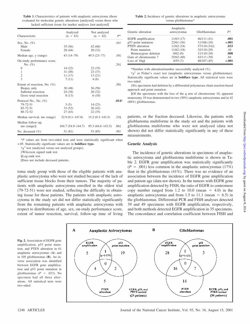
Fig. 2. Association of EGFR geneamplification, p53 point muta-tion, and PTEN alteration in 61anaplastic astrocytomas (A) andin 105 glioblastomas (B). An in-verse association was identifiedbetween EGFR gene amplifica-tion and p53 point mutation inglioblastomas (P � .023). Nospecimen had all three alter-ations. All statistical tests weretwo-sided.
Table 1. Characteristics of patients with anaplastic astrocytoma (thoseevaluated for molecular genetic alterations [analyzed] versus those who
lacked sufficient tissue for marker analyses [not analyzed]
CharacteristicAnalyzed(n � 63)
Not analyzed(n � 62) P*
Sex, No. (%) .16†Male 35 (56) 42 (68)Female 28 (44) 20 (32)
Median age, y (range) 43 (14–79) 49.5 (23–77) .10‡
On-study performance score,No. (%) .24‡
0 14 (22) 22 (35)1 31 (49) 23 (37)2 11 (17) 13 (21)3 7 (11) 4 (6)
Extent of resection, No. (%) .30‡Biopsy only 30 (48) 36 (58)Subtotal resection 24 (38) 20 (32)Gross total resection 8 (13) 6 (10)
Protocol No., No. (%) .014†79-72-51 3 (5) 14 (23)85-72-51 33 (52) 26 (42)88-72-52 27 (43) 22 (35)
Median survival, mo (range) 22.9 (0.1–147.8) 15.2 (0.3–143.5) .23§
Median follow-up,mo (range)� 104.7 (54.9–144.7) 95.3 (64.6–143.5) .86‡
No. deceased (%) 51 (81) 53 (85) .50†
*P values are from two-sided tests and were statistically significant when<.05. Statistically significant values are in boldface type.
†�2 test (analyzed versus not analyzed groups).‡Wilcoxon signed rank test.§Log-rank test.�Does not include deceased patients.
1248 ARTICLES Journal of the National Cancer Institute, Vol. 93, No. 16, August 15, 2001
by guest on August 8, 2014
http://jnci.oxfordjournals.org/D
ownloaded from
differential PCR for the detection of EGFR amplification were89% and .73, respectively.
p53 point mutations were statistically significantly (P<.001)more common in the anaplastic astrocytomas (36%) than in theglioblastomas (10%). Within the glioblastoma multiforme studygroup, younger patients were more likely to have p53 pointmutation (P � .007); a similar trend was observed within theanaplastic astrocytoma study group but was not statistically sig-nificant (P � .14; data not shown). All mutations were missensemutations except for one nonsense mutation in an anaplasticastrocytoma and two in-frame deletions or insertions in twoglioblastomas. The distribution of missense mutations was simi-lar between the anaplastic astrocytoma and glioblastoma multi-forme study groups (data not shown) and was consistent withprevious reports (27).
PTEN alteration was statistically significantly (P � .033)less frequent in anaplastic astrocytomas (18%) than in the glio-blastomas (34%). PTEN alteration was statistically significantlymore common among older patients with anaplastic astrocytoma(P � .014) and statistically significantly more common amongyounger patients with glioblastoma multiforme (P � .008; datanot shown). No apparent clustering of missense mutations wasidentified, and nonsense and frameshift mutations were primar-ily confined to exons 7 and 8 (data not shown). All homozygous
deletions identified were in specimens of glioblastoma multi-forme (10%; P � .008).
Gain of chromosome 7 was identified in approximately 40%of both anaplastic astrocytoma and glioblastoma multiformestudy groups (P � .84). There was no evidence of an associationbetween the incidence of chromosome 7 gain and patient age(data not shown).
Hemizygous deletion of the q arm of chromosome 10 wasstatistically significantly (P<.001) less common in anaplasticastrocytomas (7%) than in glioblastomas (45%). In the tumorswith loss of 10q, apparent loss of an entire copy of chromosome10 was identified in 50% of anaplastic astrocytomas and in 88%of glioblastomas. There was no evidence of an association be-tween the incidence of hemizygous deletion of 10q and patientage (data not shown).
Fig. 2 presents Venn diagrams of EGFR gene amplification,p53 mutation, and PTEN alteration for the 61 anaplastic astro-cytoma specimens and 105 glioblastoma multiforme specimensfor which all three parameters were assessed successfully. Nospecimens had all three alterations. EGFR gene amplificationand p53 point mutation rarely occurred together; both alterationswere found in only one anaplastic astrocytoma (2%) and in oneglioblastoma multiforme (1%). The two alterations that occurredtogether most frequently were EGFR gene amplification and
Table 3. Univariate associations of clinical and genetic factors with survival
Anaplastic astrocytomas GlioblastomasAnaplastic astrocytoma
and glioblastoma multiforme
No. ofpatients
No. ofdeaths (%)
Mediansurvival, mos P*
No. ofpatients
No. ofdeaths (%)
Mediansurvival, mos P*
Mediansurvival, mos P*
Age, y <.001 <.001 <.001<40 25 16 (64) 65.5 9 9 (100) 12.3 36.640–60 23 20 (87) 24.2 58 56 (97) 14.3 15.8>60 15 15 (100) 4.4 44 44 (100) 8.4 8.0
Sex, No. .21 .70 .13Male 35 30 (86) 19.2 75 73 (97) 11.7 12.2Female 28 21 (75) 26.4 36 36 (100) 11.7 14.3
Performance score† .030 .017 .0030 14 10 (71) 32.3 29 28 (97) 15.0 16.21 31 24 (77) 36.4 54 53 (98) 10.9 12.62–3 18 17 (94) 6.3 28 28 (100) 8.6 8.6
Extent of resection .81 .31 .94Biopsy 30 24 (80) 19.2 32 32 (100) 10.2 12.8Subtotal 24 20 (83) 38.7 55 54 (98) 11.5 12.3Gross total 8 6 (75) 18.5 24 23 (96) 11.7 13.0
EGFR .24 .58 .089Amplification 11 10 (91) 22.1 46 46 (100) 12.6 12.7No amplification 52 41 (79) 22.9 65 63 (97) 10.2 12.5
p53 .012 .33 <.001Point mutation 22 16 (73) 59.0 11 9 (82) 11.7 38.7Wild-type 39 33 (85) 16.0 95 95 (100) 11.5 11.6
PTEN .002 .74 .001Alteration 11 11 (100) 4.4 37 35 (95) 11.7 10.4No alternative 51 39 (76) 34.4 73 73 (100) 11.2 14.7
Chromosome 7 .60 .76 .98Gain 25 19 (76) 16.0 43 43 (100) 11.5 12.1No gain 37 31 (84) 26.4 68 66 (97) 11.7 13.6
Chromosome 10 q-arm .97 .65 .030Hemizygous loss 4 3 (75) 22.1 48 48 (100) 12.2 12.4No hemizygous loss 51 43 (84) 26.4 59 57 (97) 10.5 14.6
*P values are from two-sided tests and were statistically significant when <.05. Statistically significant values are in boldface type.†Baseline performance score.
Journal of the National Cancer Institute, Vol. 93, No. 16, August 15, 2001 ARTICLES 1249
by guest on August 8, 2014
http://jnci.oxfordjournals.org/D
ownloaded from

PTEN alteration, which were found together in four anaplasticastrocytomas (6%) and in 16 glioblastomas (15%). Among the62 anaplastic astrocytomas and 111 glioblastomas whose speci-mens could be assessed for both EGFR gene amplification andchromosome 7 gain (data not shown), no association was de-tected between these markers in the anaplastic astrocytomas(P � .74), but the markers were positively associated in theglioblastomas (P = .018)
Univariate Analysis of Survival
Among the patients with anaplastic astrocytoma, survival wasfound to be statistically significantly associated with age (log-rank P<.001), on-study performance score (P � .030), p53 sta-tus (P � .012), and PTEN status (P = .002) by univariateanalysis (Table 3). p53 mutation was associated with longersurvival, and PTEN alteration, old age, and poor performancescore were associated with shorter survival. Patients with EGFRgene amplification exhibited a trend toward a shorter survival,although the difference was not statistically significant (P � .24).
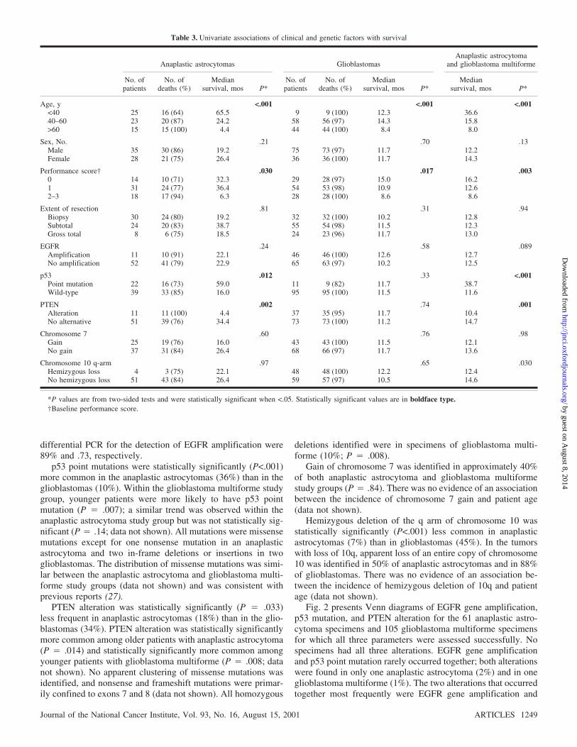
Fig. 3 shows the Kaplan–Meier survival curves for patients withanaplastic astrocytoma versus patients with glioblastoma multi-forme and for the anaplastic astrocytoma subsets defined by age,EGFR gene amplification, PTEN alteration, and p53 mutation.
In the patients with glioblastoma multiforme, univariateanalyses of the nine variables in Table 3 found that survival wasstatistically significantly associated with age (P<.001) and on-study performance score (P � .017). There was no evidence ofan association between survival and any of the tumor markers.
Multivariate Analysis of Survival
We performed a series of multivariate analyses that demon-strated that PTEN and EGFR alterations are independent pre-dictors of survival in this series of glioma patients. CART mod-eling was performed in the total group of 174 anaplasticastrocytoma and glioblastoma multiforme specimens to identifysubsets of patients with distinctly different survival distributionson the basis of histologic grade and the nine independent vari-ables listed in Table 3. CART procedures generate a tree con-
Fig. 3. Survival curves according to histologic tumor grade (A)and for the anaplastic astrocytoma patient subsets defined by thefollowing: patient age (B), EGFR gene amplification (C), andstatus of PTEN and p53 (D). All statistical tests were two-sided.N(t) and S(t) indicate, respectively, the number of patients at riskand the Kaplan–Meier estimate of survival at time t. CI � 95%confidence interval for the Kaplan–Meier survival estimate.
1250 ARTICLES Journal of the National Cancer Institute, Vol. 93, No. 16, August 15, 2001
by guest on August 8, 2014
http://jnci.oxfordjournals.org/D
ownloaded from
sisting of lymph nodes (subsets of patients) by successivelysplitting each lymph node into two lymph nodes correspondingto the various ways that the values of each independent baselinevariable can be split into two groups. The variable that splits agiven lymph node into two subsets with the greatest differencein survival is chosen as the optimum split for that lymph node.This technique can identify interactions between independentbaseline variables.
For the total group of 174 patients, CART procedures iden-tified six groups of patients with distinctly different survivaldistributions (Fig. 4, A). Fig. 4, B, shows the Kaplan–Meiersurvival curves for these six groups. Those with the best survivalwere the 25 patients with anaplastic astrocytoma who wereyounger than 40 years old, and those with the worst survivalwere the 15 patients with anaplastic astrocytoma who were olderthan 60 years. Notably, the CART model shows that EGFRamplification is associated with a better survival in the 44 pa-tients with glioblastoma multiforme who were older than 60years. However, the CART modeling process also revealed that
EGFR amplification is associated with worse survival in patientswho were 60 years old or younger (data not shown), althoughthis association was not strong enough for inclusion in the finalCART model. PTEN and p53 alterations were also consistentlyshown to be moderately associated with poorer survival and withbetter survival, respectively.
Backwards Cox proportional hazards modeling procedureswere then applied with independent variables that were chosenfrom the results of the univariate analyses and CART modelsdescribed above: Given the relatively small number of anaplasticastrocytomas, three variables (sex, chromosome 7 gain, andchromosome 10q loss) were omitted because of the lack of evi-dence of association with survival and (for 10q loss) a higherpercentage of missing data. Therefore, five clinical variables(age <40 years, age >60 years, on-study performance score,biopsy, and gross total resection) and three tumor markers (sta-tus of EGFR, p53, and PTEN) were used. The best model for agiven patient group was obtained by starting with the Cox modelcontaining all eight variables and successively eliminating the
Fig. 3. (Continued).
Journal of the National Cancer Institute, Vol. 93, No. 16, August 15, 2001 ARTICLES 1251
by guest on August 8, 2014
http://jnci.oxfordjournals.org/D
ownloaded from

least statistically significant variables until only statistically sig-nificant variables (P<.05) were left. Table 4 summarizes the Coxmodeling process and results in each patient group.
Table 4 shows that, in the total group of patients with ana-plastic astrocytoma and glioblastoma multiforme, p53 and
PTEN mutations were statistically significant prognostic factorsafter adjustment for the effects of on-study performance scoreand age. The fact that EGFR was not statistically significantin the total-group Cox model is not surprising, given the inter-action between EGFR and age found by CART. However, only
Fig. 4. A) Classification and regression-tree (CART) survivalanalysis to identify which of 10 variables (age, sex, on-studyperformance score, extent of tumor resection, histologic tumorgrade, EGFR gene amplification, PTEN alteration, p53 mutation,chromosome 7 gain, and loss of the q arm chromosome 10) weremost strongly associated with survival in the combined populationof 63 anaplastic astrocytomas (AAs) and 111 glioblastomas(GBMs). Ovals and square boxes indicate, respectively, interme-diate and terminal subsets of patients defined by the sequentialsplitting process. The lower number within each oval or squareindicates the number of deaths and patients in that subset. Theupper number is the normalized hazard rate for the subset ob-tained by dividing the constant hazard rate for the subset by theconstant hazard rate for the total group (n � 174), calculated byuse of rescaled survival times. (CART rescales the total set ofsurvival times to fit an exponential model, i.e., so that the Kaplan–Meier survival curve of all 174 patients is a straight line whenplotted on the logarithmic scale.) For each square box, the groupnumber refers to the corresponding survival curve in panel B.The variable used for each split is noted. Amp � amplification.B) Survival curves for the six groups identified by CART survivalanalysis of the combined population of AA and GBM patients.The groups are patients with 1) AA who were younger than40 years, 2) AA who were 40–60 years old, 3) GBM who were60 years old or younger, 4) GBM who were older than 60 yearswith EGFR amplification, 5) GBM who were older than 60 yearswithout EGFR amplification, and 6) AA who were older than60 years. All statistical tests were two-sided. N(t) and S(t) indi-cate, respectively, the number of patients at risk and the Kaplan–Meier estimate of survival at time t. CI � 95% confidence inter-val for the Kaplan–Meier estimate.
1252 ARTICLES Journal of the National Cancer Institute, Vol. 93, No. 16, August 15, 2001
by guest on August 8, 2014
http://jnci.oxfordjournals.org/D
ownloaded from
PTEN was statistically significantly prognostic after adjustmentfor age in the patients with anaplastic astrocytoma (hazard ratio� 4.34; 95% confidence interval [CI] � 1.82 to 10.34) butnot in the patients with glioblastoma multiforme, and p53 wasnot statistically significantly prognostic in either group. Boot-strapping procedures applied to the Cox modeling process con-firmed the prognostic significance of age 60 years or older andPTEN status in the patients with anaplastic astrocytoma (datanot shown).
In the patients with glioblastoma multiforme, the bestmodel incorporated two parameters (i.e., age >60 years andperformance score). Both were validated by bootstrappingprocedures.
DISCUSSION
The 174 specimens of high-grade astrocytomas studied werefrom all 63 patients with biopsy-proven anaplastic astrocytomaand sufficient tissue for these marker studies who were enrolledin one of the three consecutive NCCTG phase-III trials for newlydiagnosed high-grade gliomas (10–12) and all 111 Mayo pa-tients with biopsy-proven glioblastoma multiforme and suffi-cient tissue for marker ascertainment who were enrolled in thesame three NCCTG trials or six concurrent MCCC trials innewly diagnosed high-grade glioma or an NCCTG-led inter-group trial in newly diagnosed glioblastoma multiforme. Withregard to clinical characteristics, the patients with available tu-mor tissue did not differ substantially from those without ad-equate tissue for analysis. In addition, this series of patients isbroadly typical of other series of patients with high-grade astro-cytoma with regard to clinical features and incidences of geneticalterations (1,28–31).
Of the molecular markers analyzed in this study, alteration ofthe PTEN tumor suppressor gene showed the strongest associa-tion with survival in patients with anaplastic astrocytoma. Pa-tients with anaplastic astrocytoma who had a PTEN alterationhad statistically significantly worse survival than patients with-
out PTEN alteration, both in univariate analyses and in multi-variate model analyses after adjustment for key clinical vari-ables, i.e., age, on-study performance score, and extent ofresection. Although previous studies have suggested an associa-tion between PTEN and survival in patients with astrocyticglioma, to our knowledge, this is the first report of an associationbetween PTEN gene alteration and survival among adult patientswith anaplastic astrocytoma. Lin et al. (32) noted an associationbetween hemizygous deletion of the region surrounding thePTEN locus and shorter survival among patients with high-gradeglioma. Sano et al. (33) noted a statistically significantly betterprognosis for patients with glioblastoma multiforme whose tu-mors expressed high levels of PTEN messenger RNA. Further-more, Raffel et al. (34) reported an association between PTENmutation and survival among pediatric patients with high-gradeastrocytoma, after adjustment for the effects of patient age andtumor grade. Although two studies (35,36) identified no asso-ciation between survival and point mutation of PTEN amongpatients with high-grade astrocytic glioma, neither of these stud-ies examined a large number of patients with anaplastic astro-cytoma.
Mutation of PTEN has been implicated in the malignant pro-gression of astrocytic gliomas, because these alterations are ob-served most frequently in glioblastoma multiforme, less com-monly in anaplastic astrocytoma, and only rarely in low-gradeastrocytoma (36). PTEN mutation among the patients with ana-plastic astrocytoma in this series conferred a median survivalthat was similar to that of the patient population with glioblas-toma multiforme. Consequently, PTEN analysis of anaplasticastrocytomas may distinguish those tumors that have acquiredthe genetic features of glioblastoma multiforme but have yet toshow their characteristic histologic aberrations.
Assessment of PTEN may assist in appropriately classifyingpatients with anaplastic astrocytoma for therapeutic trials. Spe-cifically, analysis of therapies that may benefit patients withanaplastic astrocytoma could be adversely affected by inclusionof patients whose disease was classified histologically as ana-
Table 4. Cox backward elimination survival models, by patient group
Independent variable
Best Cox models* for indicated cohorts of patients†
All tumors studied(n � 165)
Anaplastic astrocytomas(n � 60)
Glioblastomas(n � 105)
P
Hazard ratio
P
Hazard ratio
P
Hazard ratio
Point estimate 95% CI Point estimate 95% CI Point estimate 95% CI
Young, age <40 y .003 0.48 0.29 to 0.78 .042 0.48 0.24 to 0.98 XOld, age >60 y .001 2.54 1.74 to 3.69 <.001 7.52 3.02 to 18.73 .008 1.75 1.16 to 2.65Biopsy, yes/no X X XGross total resection, yes/no X X XFirst performance score, 0–4 .008 1.30 1.07 to 1.57 X .005 1.42 1.11 to 1.82Anaplastic astrocytoma, yes/no X ND† NDEGFR amplification, yes/no X X Xp53 mutation, yes/no .011 0.55 0.35 to 0.87 X XPTEN mutation, yes/no .001 2.11 1.46 to 3.06 <.001 4.34 1.82 to 10.34 X
*The best model for a given patient cohort was obtained by successively eliminating the least statistically significant variables from the full Cox model containingall eight (or nine) independent variables until only statistically significant (P<.05) variables were left.
†For variables statistically significantly associated with survival in the best Cox model for a given patient cohort, the corresponding P value, hazard ratio pointestimate, and 95% confidence interval (CI) are shown. Factors with hazard ratios of less than 1.0 (or >1.0) are associated with better (or worse) survival. The P valueof variables not statistically significantly associated with survival in the best Cox model are indicated by an X.
‡ND � variable was not included in the full Cox model for the patient cohort.
Journal of the National Cancer Institute, Vol. 93, No. 16, August 15, 2001 ARTICLES 1253
by guest on August 8, 2014
http://jnci.oxfordjournals.org/D
ownloaded from
plastic astrocytoma but exhibiting PTEN mutation. Thus, wesuggest that assessment of PTEN status should be incorporatedinto future clinical trials in patients with anaplastic astrocytomato verify its prognostic significance and to identify any differ-ence in therapeutic benefit in patients with and without PTENalteration.
In addition to patient age and PTEN alteration, univariateanalyses of the anaplastic astrocytoma study group identifiedmutation of p53 as a statistically significant predictor of pro-longed survival. This association is consistent with previous ob-servations that mutation of p53 is more frequent among youngerpatients with astrocytic glioma (37). However, after adjustmentfor the effect of patient age, p53 mutation was not associatedwith survival in the anaplastic astrocytoma study group, in ac-cordance with the majority of previous reports (1).
Gain of chromosome 7 is a frequent event in both low- andhigh-grade astrocytic glioma (28,29), suggesting that this alter-ation is an early event in astrocytoma development. Huhn et al.(29) reported an association between gain of chromosome 7 andradiation resistance in glioblastoma multiforme, suggesting thatthis aberration may predict poor prognosis. However, gain ofchromosome 7 was not associated with survival in either theanaplastic astrocytoma or glioblastoma multiforme patientgroups in this series.
Deletion of the q arm of chromosome 10 has been reported inapproximately 80% of high-grade astrocytomas (9), and severalcandidate tumor suppressor genes have been identified from thischromosome arm, including PTEN (38–40). However, deletionof 10q was not predictive of survival in patients with anaplasticastrocytoma or glioblastoma multiforme.
In the glioblastoma multiforme study group, the statisticallysignificant predictors of overall survival identified by both uni-variate and multivariate analyses were patient age and on-studyperformance score. None of the genetic markers analyzed in thisstudy was identified as a statistically significant predictor ofsurvival among patients with glioblastoma multiforme. Indeed,other investigators (5,7) have obtained similar results. Inabilityto distinguish differences in survival among patients with glio-blastoma multiforme based on genetic markers associated withmalignant progression suggests that there may be multiple ge-netic pathways of glioblastoma multiforme development thatconverge on a similar phenotype with an equally poor prognosis.
As an alternative means of assessing prognostic indicators inour study group, clinical and molecular data from all 174 speci-mens (63 anaplastic astrocytomas and 111 glioblastomas) weresubjected to CART-survival modeling procedures. This analysisgenerates a regression tree consisting of lymph nodes by suc-cessively splitting each lymph node based on the variable thatdistinguishes the greatest difference in survival. Initial splittingwas based on patient age. Subsequent splitting in the older pa-tients (age >60 years) indicated particularly poor survival forolder patients with anaplastic astrocytoma and longer survival inolder patients with glioblastoma multiforme who also had EGFRgene amplification. However, in the younger patients (age �60years), the splitting process consistently indicated shorter sur-vival in younger patients with EGFR amplification, although thisassociation was not strong enough for inclusion in the finalCART model. These observations suggest that EGFR gene am-plification may have prognostic utility and that the biologic sig-nificance of EGFR gene amplification in astrocytic gliomas maybe related to patient age, as reported by Simmons et al. (41). For
example, when EGFR is amplified in gliomas, it is often alsomutated (42). It is possible that different mutations (e.g., 3�deletion, 5� deletion, or various point mutations) occur withdifferent incidence in various glioma malignancy grades or intumors from patients of various ages.
We observed a paucity of specimens with both EGFR ampli-fication and p53 mutation, which lends support to the hypothesisthat p53 mutation and EGFR gene amplification are markers fortwo pathways of astrocytic glioma development or two alterna-tive alterations in the same pathway (9,37). It is interesting tonote that the incidence of p53 mutation was statistically signifi-cantly less in the glioblastoma multiforme specimens than in theanaplastic astrocytoma specimens in this study. Similar resultshave been reported previously (37) and suggest that the NCCTGcohort of patients with glioblastoma multiforme includes a largeproportion of “primary” glioblastomas.
Perry et al. (43) recently analyzed multiple clinicopathologicvariables in the same anaplastic astrocytoma patient group ex-amined in this study and observed that foci of necrosis and/orendothelial hyperplasia were identified in five of the patientswith anaplastic astrocytoma when tissue blocks were recut toobtain deeper sections. When we reanalyzed the survival of theremaining 58 patients with pure anaplastic astrocytoma, the me-dian survival time increased from 23 to 29 months, but none ofthe conclusions regarding the clinical and molecular geneticvariables changed (data not shown). This finding emphasizes thehistologic grading difficulties introduced by the heterogeneity ofthese tumors and exemplifies the need for more objective tumormarkers. Furthermore, because 48% of our patients with ana-plastic astrocytoma were diagnosed on biopsy only, the risks ofundersampling are substantial. However, because this reflectsthe practices of both community and academic neurosurgicalpractices, it should be regarded as a “realistic flaw” associatedwith the evaluation of astrocytic neoplasms. Notably, just one ofthe five anaplastic astrocytoma specimens with focal necrosis orendothelial proliferation had a PTEN alteration.
In summary, we have assessed the prognostic value of severalgenetic alterations in patients with high-grade astrocytic glioma.Key attributes of our patient population include mature follow-up, protocolized prospective follow-up, and relatively largesample sizes. The most clinically significant findings of thisstudy are that patients with anaplastic astrocytoma who had analteration of the PTEN tumor suppressor gene had a statisticallysignificantly shorter survival than patients with anaplastic astro-cytoma without PTEN mutation and that this association wasmaintained after adjustment for the effects of key clinical vari-ables, including patient age, on-study performance score, andextent of tumor resection. In addition, the presence of EGFRamplification independently predicts a longer survival in olderpatients with glioblastoma multiforme. The substantial variationin survival among the patients with anaplastic astrocytoma sug-gests that combining histologic and genotypic assessment couldpotentially improve existing strategies for patient stratificationand management.
REFERENCES
(1) Smith JS, Jenkins RB. Genetic alterations in adult diffuse glioma: occur-rence, significance, and prognostic implications. Front Biosci 2000;5:213–31. Available on line at: http://www.bioscience.org/2000/v5/d/smith/fulltext.htm.
(2) Kleihues P, Cavenee WK, editors. Pathology and genetics of tumours of the
1254 ARTICLES Journal of the National Cancer Institute, Vol. 93, No. 16, August 15, 2001
by guest on August 8, 2014
http://jnci.oxfordjournals.org/D
ownloaded from
nervous system. 2nd ed. International Agency for Research on Cancer. Lyon(France): IARC Press; 2000. p. 9–54.
(3) Daumas-Duport C, Scheithauer BW, O’Fallon JR, Kelly P. Grading ofastrocytomas. A simple and reproducible method. Cancer 1988;62:2152–65.
(4) Prados MD, Gutin PH, Phillips TL, Wara WM, Larson DA, Sneed PK, etal. Highly anaplastic astrocytoma: a review of 357 patients treated between1977 and 1989. Int J Radiat Oncol Biol Phys 1992;23:3–8.
(5) Cunningham JM, Kimmel DW, Scheithauer BW, O’Fallon JR, Novotny PJ,Jenkins RB. Analysis of proliferation markers and p53 expression in glio-mas of astrocytic origin: relationships and prognostic value. J Neurosurg1997;86:121–30.
(6) Leighton C, Fisher B, Bauman G, Depiero S, Stitt L, Macdonald D, et al.Supratentorial low-grade gliomas in adults: an analysis of prognostic fac-tors and timing of radiation. J Clin Oncol 1997;15:1294–301.
(7) Olson JJ, Barnett D, Yang J, Assietti R, Cotsonis G, James CD. Geneamplification as a prognostic factor in primary brain tumors. Clin CancerRes 1998;4:215–22.
(8) Lantos PL, VandenBerg SR, Kleihues P. Tumours of the nervous system.In: Graham DI, Lantos PL, editors. Greenfield’s neuropathology. 6th ed.London (U.K.): Arnold; 1997. p. 600–47.
(9) Louis DN. A molecular genetic model of astrocytoma histopathology.Brain Pathol 1997;7:755–64.
(10) Elliott TE, Dinapoli RP, O’Fallon JR, Krook JE, Earle JD, Morton RF, etal. Randomized trial of radiation therapy (RT) plus dibromodulcitol (DBD)versus RT plus BCNU in high grade astrocytoma. J Neurooncol 1997;33:239–50.
(11) Dinapoli RP, Brown LD, Arusell RM, Earle JD, O’Fallon JR, Buckner JC,et al. Phase III comparative evaluation of PCNU and carmustine combinedwith radiation therapy for high-grade glioma. J Clin Oncol 1993;11:1316–21.
(12) Buckner JC, Schomberg PJ, Bashir R, McGinnis WL, Cascino TL, Schei-thauer BW, et al. Phase III study of radiation therapy (RT) plus BCNU withor without recombinant interferon alpha (IFN) in high grade glioma: anNCCTG study [abstract]. Proc ASCO 1997;16:385a.
(13) Zubrod CG, Schneiderman M, Frei E III, Brindley C, Gold GL, Shnider B,et al. Appraisal of methods for study of chemotherapy of cancer in man:comparative therapeutic trial of nitrogen mustard and triethylene thiophos-phoramide. J Chron Dis 1960;11:7–33.
(14) Oken MM, Creech RH, Tormey DC, Horton J, Davis TE, McFadden ET,et al. Toxicity and response criteria of the Eastern Cooperative OncologyGroup. Am J Clin Oncol 1982;5:649–55.
(15) Smith JS, Perry A, Borell TJ, Lee HK, O’Fallon J, Hosek SM, et al.Alterations of chromosome arms 1p and 19q as predictors of survival inoligodendrogliomas, astrocytomas, and mixed oligoastrocytomas. J ClinOncol 2000;18:636–45.
(16) Jenkins RB, Qian J, Lieber MM, Bostwick DG. Detection of c-myc onco-gene amplification and chromosomal anomalies in metastatic prostaticcarcinoma by fluorescence in situ hybridization. Cancer Res 1997;57:524–31.
(17) Smith JS, Tachibana I, Lee HK, Qian J, Pohl U, Mohrenweiser HW, et al.Mapping of the chromosome 19 q-arm glioma tumor suppressor gene usingfluorescence in situ hybridization and novel microsatellite markers. GenesChromosomes Cancer 2000;29:16–25.
(18) Banerjee SK, Makdisi WF, Weston AP, Mitchell SM, Campbell DR.Microwave-based DNA extraction from paraffin-embedded tissue for PCRamplification. Biotechniques 1995;18:768–73, 772–3.
(19) Hunter SB, Abbott K, Varma VA, Olson JJ, Barnett DW, James CD.Reliability of differential PCR for the detection of EGFR and MDM2 geneamplification in DNA extracted from FFPE glioma tissue. J NeuropatholExp Neurol 1995;54:57–64.
(20) Watanabe K, Peraud A, Gratas C, Wakai S, Kleihues P, Ohgaki H. p53 andPTEN gene mutations in gemistocytic astrocytomas. Acta Neuropathol1998;95:559–64.
(21) Kaplan EL, Meier P. Nonparametric estimation from incomplete observa-tions. J Am Stat Assoc 1958;53:457–81.
(22) Mantel N. Evaluation of survival data and two new rank order statisticsarising in its consideration. Cancer Chemother Rep 1966;50:163–70.
(23) LeBlanc M, Crowley J. Relative risk trees for censored survival data.Biometrics 1992;48:411–25.
(24) Cox DR. Regression models and life tables. J R Stat Soc 1972;34:187–220.(25) Efron B. Bootstrap methods: another look at the jackknife. Ann Stat 1979;
7:1–26.(26) Sauerbrei W, Schumacher M. A bootstrap resampling procedure for model
building: application to the Cox regression model. Stat Med 1992;11:2093–109.
(27) Greenblatt MS, Bennett WP, Hollstein M, Harris CC. Mutations in the p53tumor suppressor gene: clues to cancer etiology and molecular pathogen-esis. Cancer Res 1994;54:4855–78.
(28) Bigner SH, Mark J, Burger PC, Mahaley MS Jr, Bullard DE, MuhlbaierLH, et al. Specific chromosomal abnormalities in malignant human glio-mas. Cancer Res 1988;48:405–11.
(29) Huhn SL, Mohapatra G, Bollen A, Lamborn K, Prados MD, Feuerstein BG.Chromosomal abnormalities in glioblastoma multiforme by comparativegenomic hybridization: correlation with radiation treatment outcome. ClinCancer Res 1999;5:1435–43.
(30) Winger MJ, Macdonald DR, Cairncross JG. Supratentorial anaplastic glio-mas in adults. The prognostic importance of extent of resection and priorlow-grade glioma. J Neurosurg 1989;71:487–93.
(31) Salminen E, Nuutinen JM, Huhtala S. Multivariate analysis of prognosticfactors in 106 patients with malignant glioma. Eur J Cancer 1996;32A:1918–23.
(32) Lin H, Bondy ML, Langford LA, Hess KR, Delclos GL, Wu X, et al.Allelic deletion analysis of MMAC/PTEN and DMBT1 loci in gliomas:relationship to prognostic significance. Clin Cancer Res 1998;4:2447–54.
(33) Sano T, Lin H, Chen X, Langford LA, Koul D, Bondy ML, et al. Differ-ential expression of MMAC/PTEN in glioblastoma multiforme: relation-ship to localization and prognosis. Cancer Res 1999;59:1820–4.
(34) Raffel C, Frederick L, O’Fallon JR, Atherton-Skaff P, Perry A, Jenkins RB,et al. Analysis of oncogene and tumor suppressor gene alterations in pe-diatric malignant astrocytomas reveals reduced survival for patients withPTEN mutations. Clin Cancer Res 1999;5:4085–90.
(35) Zhou XP, Li YJ, Hoang-Xuan K, Laurent-Puig P, Mokhtari K, Longy M,et al. Mutational analysis of the PTEN gene in gliomas: molecular andpathological correlations. Int J Cancer 1999;84:150–4.
(36) Rasheed BK, Stenzel TT, McLendon RE, Parsons R, Friedman AH, Fried-man HS, et al. PTEN gene mutations are seen in high-grade but not inlow-grade gliomas. Cancer Res 1997;57:4187–90.
(37) Watanabe K, Tachibana O, Sata K, Yonekawa Y, Kleihues P, Ohgaki H.Overexpression of the EGF receptor and p53 mutations are mutually ex-clusive in the evolution of primary and secondary glioblastomas. BrainPathol 1996;6:217–24.
(38) Ali IU, Schriml LM, Dean M. Mutational spectra of PTEN/MMAC1 gene:a tumor suppressor with lipid phosphatase activity. J Natl Cancer Inst1999;91:1922–32.
(39) Steck PA, Pershouse MA, Jasser SA, Yung WK, Lin H, Ligon AH, et al.Identification of a candidate tumour suppressor gene, MMAC1, at chro-mosome 10q23.3 that is mutated in multiple advanced cancers. Nat Genet1997;15:356–62.
(40) Li J, Yen C, Liaw D, Podsypanina K, Bose S, Wang SI, et al. PTEN, aputative protein tyrosine phosphatase gene mutated in human brain, breast,and prostate cancer. Science 1997;275:1943–7.
(41) Simmons ML, Lamborn KR, Takahashi M, Chen P, Israel MA, Berger MS,et al. Analysis of complex relationships between age, p53, epidermalgrowth factor receptor, and survival in glioblastoma patients. Cancer Res2001;61:1122–8.
(42) Frederick L, Wang XY, Eley G, James CD. Diversity and frequency ofepidermal growth factor receptor mutations in human glioblastomas. Can-cer Res 2000;60:1383–7.
(43) Perry A, Jenkins RB, O’Fallon JR, Schaefer PL, Kimmel DW, MahoneyMR, et al. Clinicopathologic study of 85 similarly treated patients withanaplastic astrocytoma. An analysis of DNA content (ploidy), cellular pro-liferation, and p53 expression. Cancer 1999;86:672–83.
NOTES
J. S. Smith and I. Tachibana contributed equally to this study.Present address: J. S. Smith, Department of Neurological Surgery, University
of California, San Francisco.Supported by Public Health Service grants CA50905 (to R. B. Jenkins,
Journal of the National Cancer Institute, Vol. 93, No. 16, August 15, 2001 ARTICLES 1255
by guest on August 8, 2014
http://jnci.oxfordjournals.org/D
ownloaded from

J. R. O’Fallon, B. W. Scheithauer, and J. C. Buckner), CA25224 (to J. C. Buck-ner, N. Iturria, J.tR. O’Fallon, and B. W. Scheithauer), and CA35415 (toP. L. Schaefer) from the National Cancer Institute, National Institutes of Health,Department of Health and Human Services; by the Linse-Bock Foundation (toI. Tachibana and J. C. Buckner); and by the Sydney Luckman Physician ScientistScholarship (to J. S. Smith).
We thank Jill Burton, Debra Kvittem, and the North Central Cancer TreatmentGroup (NCCTG) clinical research associates for excellence in clinical database
collection and submission of tumor blocks for analysis. Drs. Peter Burger andArie Perry from The Johns Hopkins University (Baltimore, MD) and Washing-ton University (St. Louis, MO), respectively, performed NCCTG neuropathol-ogy data audits. We also thank Gladdie Hebl, Mayo Clinic, for assistance inpreparing the manuscript and Michelle Mahoney, Mayo Clinic, for assistancein organizing data for statistical analysis.
Manuscript received January 17, 2001; revised June 18, 2001; accepted June22, 2001.
1256 ARTICLES Journal of the National Cancer Institute, Vol. 93, No. 16, August 15, 2001
by guest on August 8, 2014
http://jnci.oxfordjournals.org/D
ownloaded from
Copyright © 2022 FDOKUMEN


![Eritema multiforme reaccional como manifestación atípica de lepra. Reporte de caso [Reactive erythema multiforme as atypical manifestation of leprosy. Case report]](https://static.fdokumen.com/doc/165x107/632459174d8439cb620d572d/eritema-multiforme-reaccional-como-manifestacion-atipica-de-lepra-reporte-de.jpg)