High-throughput screening of mouse gene knockouts identifies ...
30
ARTICLE High-throughput screening of mouse gene knockouts identifies established and novel skeletal phenotypes Robert Brommage, Jeff Liu, Gwenn M Hansen, Laura L Kirkpatrick, David G Potter, Arthur T Sands, Brian Zambrowicz, David R Powell and Peter Vogel Screening gene function in vivo is a powerful approach to discover novel drug targets. We present high-throughput screening (HTS) data for 3 762 distinct global gene knockout (KO) mouse lines with viable adult homozygous mice generated using either gene-trap or homologous recombination technologies. Bone mass was determined from DEXA scans of male and female mice at 14 weeks of age and by microCT analyses of bones from male mice at 16 weeks of age. Wild-type (WT) cagemates/littermates were examined for each gene KO. Lethality was observed in an additional 850 KO lines. Since primary HTS are susceptible to false positive findings, additional cohorts of mice from KO lines with intriguing HTS bone data were examined. Aging, ovariectomy, histomorphometry and bone strength studies were performed and possible non-skeletal phenotypes were explored. Together, these screens identified multiple genes affecting bone mass: 23 previously reported genes (Calcr, Cebpb, Crtap, Dcstamp, Dkk1, Duoxa2, Enpp1, Fgf23, Kiss1/Kiss1r, Kl (Klotho), Lrp5, Mstn, Neo1, Npr2, Ostm1, Postn, Sfrp4, Slc30a5, Slc39a13, Sost, Sumf1, Src, Wnt10b), five novel genes extensively characterized (Cldn18, Fam20c, Lrrk1, Sgpl1, Wnt16), five novel genes with preliminary characterization (Agpat2, Rassf5, Slc10a7, Slc26a7, Slc30a10) and three novel undisclosed genes coding for potential osteoporosis drug targets. Bone Research (2014) 2, 14034; doi:10.1038/boneres.2014.34; Published online: 28 October 2014 INTRODUCTION Following successful sequencing of the human and mouse genomes, a major goal of the genomics community has been to determine the functions of the ,20 000 mammalian protein-coding genes by generating and examining phe- notypes of knockout (KO) mice for each gene. Initial efforts towards this goal were pursued by Lexicon Pharmaceuticals (summarized in this report) and Deltagen. 1 More recently, academic centers joined forces to form the International Knockout Mouse Consortium (IKMC) encompassing efforts by the Knockout Mouse Project, European Conditional Mouse Mutagenesis Program, North American Conditional Mouse Mutagenesis Program and the Texas A&M Institute of Genomic Medicine 2–6 . From 2000 through 2008, Lexicon Pharmaceuticals per- formed high-throughput mouse knockout and compre- hensive phenotypic analyses (Genome5000 TM ) for .4 650 genes. KO strategies (described below) involved both gene trapping, 7 using the OmniBank H I embryonic stem (ES) cell library, and homologous recombination technologies. Lexicon generated a second ES cell library (OmniBank H II) for the TIGM gene trap repository 8 and since 2009, over 100 studies have used this resource to examine gene disruptions in mice (http://www.tigm.org/ publications/). Phenotyping involved a battery of tests in the areas of behavior, cardiology, immunology, metabo- lism, oncology and ophthalmology, and included serum chemistry, histopathology and a high fat diet obesity challenge. Mice and ES cells from many KOs generated through this Genome 5000 TM program are available through the USA NIH Mutant Mouse Regional Resource Center (https://www.mmrrc.org/catalog/overview_Major_ Collection.php), Wellcome Trust (http://www.tigm.org/ wellcome-trust/) and Taconic Farms (http://www.taconic. com/KO). The USA NIH Mutant Mouse Regional Resource Center collection includes 472 genes analyzed as part of Genentech’s Secreted Protein Discovery Initiative 9 and having published phenotypes. 10 Lexicon published body composition data for gene KOs (through 2005) that resulted in lean and obese phenotypes among the first 2 322 KO lines Lexicon Pharmaceuticals, The Woodlands, TX, USA Correspondence: R Brommage ([email protected]) Received: 16 July 2014; Revised: 29 July 2014; Accepted: 31 July 2014 OPEN Citation: Bone Research (2014) 2, 14034; doi:10.1038/boneres.2014.34 ß 2014 Sichuan University All rights reserved 2095-4700/14 www.nature.com/boneres
-
Upload
khangminh22 -
Category
Documents
-
view
5 -
download
0
Transcript of High-throughput screening of mouse gene knockouts identifies ...

ARTICLE
High-throughput screening of mouse gene knockouts
identifies established and novel skeletal phenotypes
Robert Brommage, Jeff Liu, Gwenn M Hansen, Laura L Kirkpatrick, David G Potter, Arthur T Sands, Brian Zambrowicz,David R Powell and Peter Vogel
Screening gene function in vivo is a powerful approach to discover novel drug targets. We presenthigh-throughput screening (HTS) data for 3 762 distinct global gene knockout (KO) mouse lines with viableadult homozygous mice generated using either gene-trap or homologous recombination technologies. Bonemasswas determined fromDEXAscans ofmale and femalemice at 14 weeks of age andbymicroCTanalyses ofbones frommale mice at 16 weeks of age. Wild-type (WT) cagemates/littermates were examined for each geneKO. Lethality was observed in an additional 850 KO lines. Since primary HTS are susceptible to false positivefindings, additional cohorts of mice from KO lines with intriguing HTS bone data were examined. Aging,ovariectomy, histomorphometry and bone strength studies were performed and possible non-skeletalphenotypes were explored. Together, these screens identified multiple genes affecting bone mass: 23previously reported genes (Calcr,Cebpb,Crtap,Dcstamp,Dkk1,Duoxa2,Enpp1, Fgf23,Kiss1/Kiss1r,Kl (Klotho),Lrp5, Mstn, Neo1, Npr2, Ostm1, Postn, Sfrp4, Slc30a5, Slc39a13, Sost, Sumf1, Src, Wnt10b), five novel genesextensively characterized (Cldn18, Fam20c, Lrrk1, Sgpl1, Wnt16), five novel genes with preliminarycharacterization (Agpat2, Rassf5, Slc10a7, Slc26a7, Slc30a10) and three novel undisclosed genes coding forpotential osteoporosis drug targets.
Bone Research (2014) 2, 14034; doi:10.1038/boneres.2014.34; Published online: 28 October 2014
INTRODUCTIONFollowing successful sequencing of the human and mouse
genomes, a major goal of the genomics community has
been to determine the functions of the,20000mammalian
protein-coding genes by generating and examining phe-
notypes of knockout (KO) mice for each gene. Initial efforts
towards this goal were pursuedby Lexicon Pharmaceuticals
(summarized in this report) and Deltagen.1 More recently,
academic centers joined forces to form the International
Knockout Mouse Consortium (IKMC) encompassing efforts
by the Knockout Mouse Project, European Conditional
Mouse Mutagenesis Program, North American Conditional
MouseMutagenesis Programand the Texas A&M Institute of
Genomic Medicine2–6.
From 2000 through 2008, Lexicon Pharmaceuticals per-
formed high-throughput mouse knockout and compre-
hensive phenotypic analyses (Genome5000TM) for .4 650
genes. KO strategies (described below) involved both
gene trapping,7 using the OmniBankH I embryonic
stem (ES) cell library, and homologous recombination
technologies. Lexicon generated a second ES cell library
(OmniBankH II) for the TIGM gene trap repository8 and
since 2009, over 100 studies have used this resource to
examine gene disruptions in mice (http://www.tigm.org/
publications/). Phenotyping involved a battery of tests in
the areas of behavior, cardiology, immunology, metabo-
lism, oncology and ophthalmology, and included serum
chemistry, histopathology and a high fat diet obesity
challenge. Mice and ES cells from many KOs generated
through this Genome 5000TM program are available
through the USA NIH Mutant Mouse Regional Resource
Center (https://www.mmrrc.org/catalog/overview_Major_
Collection.php), Wellcome Trust (http://www.tigm.org/
wellcome-trust/) and Taconic Farms (http://www.taconic.
com/KO). The USA NIH Mutant Mouse Regional Resource
Center collection includes 472 genes analyzed as part of
Genentech’s Secreted Protein Discovery Initiative9 and
having published phenotypes.10 Lexicon published body
composition data for gene KOs (through 2005) that resulted
in leanandobesephenotypesamong the first 2322 KO lines
Lexicon Pharmaceuticals, The Woodlands, TX, USA
Correspondence: R Brommage ([email protected])
Received: 16 July 2014; Revised: 29 July 2014; Accepted: 31 July 2014
OPEN Citation: Bone Research (2014) 2, 14034; doi:10.1038/boneres.2014.34� 2014 Sichuan University All rights reserved 2095-4700/14
www.nature.com/boneres
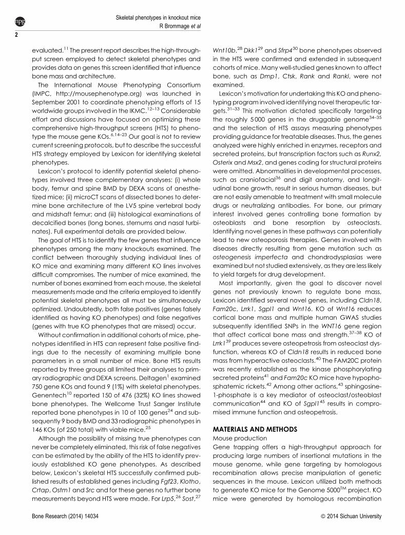
evaluated.11 Thepresent report describes the high-through-
put screen employed to detect skeletal phenotypes and
provides data on genes this screen identified that influence
bone mass and architecture.
The International Mouse Phenotyping Consortium
(IMPC, http://mousephenotype.org) was launched in
September 2001 to coordinate phenotyping efforts of 15
worldwide groups involved in the IKMC.12–13 Considerable
effort and discussions have focused on optimizing these
comprehensive high-throughput screens (HTS) to pheno-
type the mouse gene KOs.6,14–23 Our goal is not to review
current screening protocols, but to describe the successful
HTS strategy employed by Lexicon for identifying skeletal
phenotypes.
Lexicon’s protocol to identify potential skeletal pheno-
types involved three complementary analyses: (i) whole
body, femur and spine BMD by DEXA scans of anesthe-
tized mice; (ii) microCT scans of dissected bones to deter-
mine bone architecture of the LV5 spine vertebral body
and midshaft femur; and (iii) histological examinations of
decalcified bones (long bones, sternums and nasal turbi-
nates). Full experimental details are provided below.
The goal of HTS is to identify the few genes that influence
phenotypes among the many knockouts examined. The
conflict between thoroughly studying individual lines of
KO mice and examining many different KO lines involves
difficult compromises. The number of mice examined, the
number of bones examined from eachmouse, the skeletal
measurementsmadeand the criteria employed to identify
potential skeletal phenotypes all must be simultaneously
optimized. Undoubtedly, both false positives (genes falsely
identified as having KO phenotypes) and false negatives
(genes with true KO phenotypes that are missed) occur.
Without confirmation in additional cohorts ofmice, phe-
notypes identified in HTS can represent false positive find-
ings due to the necessity of examining multiple bone
parameters in a small number of mice. Bone HTS results
reported by three groups all limited their analyses to prim-
ary radiographic and DEXA screens. Deltagen1 examined
750 gene KOs and found 9 (1%) with skeletal phenotypes.
Genentech10 reported 150 of 476 (32%) KO lines showed
bone phenotypes. The Wellcome Trust Sanger Institute
reported bone phenotypes in 10 of 100 genes24 and sub-
sequently 9 body BMDand 33 radiographic phenotypes in
146 KOs (of 250 total) with viable mice.25
Although the possibility of missing true phenotypes can
never be completely eliminated, this risk of false negatives
can be estimated by the ability of the HTS to identify prev-
iously established KO gene phenotypes. As described
below, Lexicon’s skeletal HTS successfully confirmed pub-
lished results of established genes including Fgf23, Klotho,
Crtap,Ostm1 and Src and for these genes no further bone
measurements beyond HTS were made. For Lrp5,26 Sost,27
Wnt10b,28 Dkk129 and Sfrp430 bone phenotypes observed
in the HTS were confirmed and extended in subsequent
cohorts ofmice. Manywell-studied genes known to affect
bone, such as Dmp1, Ctsk, Rank and Rankl, were not
examined.
Lexicon’smotivation for undertaking this KOandpheno-
typing program involved identifying novel therapeutic tar-
gets.31–33 This motivation dictated specifically targeting
the roughly 5 000 genes in the druggable genome34–35
and the selection of HTS assays measuring phenotypes
providingguidance for treatable diseases. Thus, thegenes
analyzed were highly enriched in enzymes, receptors and
secreted proteins, but transcription factors such as Runx2,
Osterix andMsx2, and genes coding for structural proteins
were omitted. Abnormalities in developmental processes,
such as craniofacial36 and digit anatomy, and longit-
udinal bone growth, result in serious human diseases, but
are not easily amenable to treatment with small molecule
drugs or neutralizing antibodies. For bone, our primary
interest involved genes controlling bone formation by
osteoblasts and bone resorption by osteoclasts.
Identifying novel genes in these pathways can potentially
lead to new osteoporosis therapies. Genes involved with
diseases directly resulting from gene mutation such as
osteogenesis imperfecta and chondrodysplasias were
examinedbut not studied extensively, as theyare less likely
to yield targets for drug development.
Most importantly, given the goal to discover novel
genes not previously known to regulate bone mass,
Lexicon identified several novel genes, including Cldn18,
Fam20c, Lrrk1, Sgpl1 and Wnt16. KO of Wnt16 reduces
cortical bone mass and multiple human GWAS studies
subsequently identified SNPs in the WNT16 gene region
that affect cortical bone mass and strength.37–38 KO of
Lrrk139 produces severe osteopetrosis from osteoclast dys-
function, whereas KO of Cldn18 results in reduced bone
mass from hyperactive osteoclasts.40 The FAM20C protein
was recently established as the kinase phosphorylating
secreted proteins41 and Fam20c KOmice have hypopho-
sphatemic rickets.42 Among other actions,43 sphingosine-
1-phosphate is a key mediator of osteoclast/osteoblast
communication44 and KO of Sgpl145 results in compro-
mised immune function and osteopetrosis.
MATERIALS AND METHODSMouse production
Gene trapping offers a high-throughput approach for
producing large numbers of insertional mutations in the
mouse genome, while gene targeting by homologous
recombination allows precise manipulation of genetic
sequences in the mouse. Lexicon utilized both methods
to generate KO mice for the Genome 5000TM project. KO
mice were generated by homologous recombination
Skeletal phenotypes in knockout mice
R Brommage et al
2
Bone Research (2014) 14034 � 2014 Sichuan University
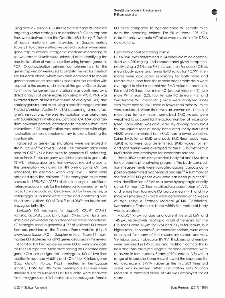
usingboth a lphage KOS shuttle system46 and PCR-based
targeting vector strategies as described.47 Gene trapped
lines were derived from the OmniBankH I library.48 Details
of each mutation are provided in Supplementary
Table S1. To achieve effective gene disruption when using
gene-trap mutations, intragenic insertions intersecting all
known transcript units were selected after identifying the
precise location of vector insertion using inverse genomic
PCR. Oligonucleotide primers complementary to the
gene-trap vectorwere used toamplify the vector insertion
site for each clone, which was then compared to mouse
genomesequenceassemblies to localize the insertionwith
respect to the exons and introns of the gene. Gene disrup-
tion in vivo for gene-trap mutations was confirmed by a
direct analysis of gene expression using RT-PCR. RNA was
extracted from at least two tissues of wild-type (WT) and
homozygousmutantmice usingabeadhomogenizer and
RNAzol (Ambion, Austin, TX, USA) according to manufac-
turer’s instructions. Reverse transcription was performed
with SuperScript II (Invitrogen, Carlsbad, CA, USA) and ran-
dom hexamer primers, according to the manufacturer’s
instructions. PCR amplification was performed with oligo-
nucleotide primers complementary to exons flanking the
insertion site.
Targeted or gene-trap mutations were generated in
strain 129SvEvBrd-derived ES cells. The chimeric mice were
bred to C57BL/6J albino mice to generate F1 heterozyg-
ous animals. These progenywere intercrossed togenerate
F2 WT, heterozygous and homozygous mutant progeny.
This generation was used for HTS phenotyping. On rare
occasions, for example when very few F1 mice were
obtained from the chimera, F1 heterozygous mice were
crossed to 129SvEvBrd/C57 hybrid mice to yield additional
heterozygous animals for the intercross to generate the F2
mice. KOmicecould not begenerated for threegenes, as
heterozygousPstkmicewere infertile and, confirmingpub-
lished observations, KOofCask49 andDll450 resulted in het-
erozygous lethality.
Lexicon’s KO strategies for Agpat2, Clcn7, Cldn18,
Fam20c, Gnptab, Lrp5, Lrrk1, Sgpl1, Stk36, Tph1, Tph2 and
Wnt16are provided in the publications of these phenotypes.
KO strategies used to generate 4077 of Lexicon’s KOmouse
lines are provided at the Taconic Farms website (http://
www.taconic.com/KO). Supplementary Table S1 sum-
marizes KO strategies for all 93 genes discussed in this review.
A total of 139 X-linked genes were KO’d, with bone data
for 133 KOs reported.Malemice havingan Xchromosome
gene KO’d are designated hemizygous. KO of two lines
resulted in reduced viability and KO of four X-linked genes
(Ebp, Mmgt1, Porcn, Prps1) resulted in hemizygous
lethality. Data for 105 male hemizygous KO lines were
analyzed. For 28 X-linked KOs DEXA data were analyzed
for hemizygous and WT males plus homozygous female
KO mice compared to age-matched WT female mice
from the breeding colony. For 90 of these 105 KOs,
data for only two male WT mice were available for DEXA
calculations.
High-throughput screening assays
DEXA BMD was determined in 14-week-old mice anesthe-
tized with 250 mg?kg21 tribromoethanol given intraperito-
neally using aGE/Lunar PIXImus scanner. For each KO line,
mean body spine and femur BMD ratios for KO/WT litter-
mates were calculated separately for both male and
femalemice, and then thesemale and female datawere
averaged to yield a normalized BMD value for each site.
For most KO lines, four male KO (actual mean54.2), two
male WT (mean52.2), four female KO (mean54.2) and
two female WT (mean52.1) mice were analyzed. Lines
with fewer than four KOmice or fewer than three WTmice
were excluded. When there was an uneven distribution of
male and female mice, normalized BMD values were
weighted to account for the actual number of mice ana-
lyzed. Body vBMD was calculated by dividing body BMD
by the square root of body bone area. Body BMD and
vBMD were correlated but vBMD had a lower variation.
Spine BMD, femur BMD and body BMC/lean body mass
(LBM) ratio were also determined. BMD values for left
and right femurs were averaged for the HTS, but left femur
BMD alone was employed for secondary screens.
These DEXA scans also provided body fat and LBM data
for our obesity phenotyping program. The body composi-
tion measurements were validated against carcass com-
position determined by chemical analysis.51 A summary of
the first 2 322 KO genes evaluated has been published11,
with identification of Ksr2 as a novel hyperphagic obesity
gene. For most KO lines, architectural parameters of LV5s
and femurs from fourmale KO (actualmean54.1) and two
male WT (mean52.1) mice were determined at 16 weeks
of age using a Scanco Medical mCT40 (Bruttisellen,
Switzerland). Trabecular bone within the vertebral body
was evaluated.
MicroCT X-ray voltage and current were 55 keV and
145 mA, respectively. Isotropic voxel dimensions for the
HTS scans were 16 mm for LV5s and 20 mm for femurs, but
higher resolution scans (8 mmvoxel dimensions)wereoften
employed for many of the secondary screen analyses.
Vertebral body trabecular BV/TV, thickness and number
were analyzed in LV5 scans and midshaft cortical thick-
ness and total area (a surrogate for bone diameter) were
analyzed in femur scans. Scans of 10 random LV5s with a
range of trabecular bone mass showed the expected lin-
ear decrease in BV/TV values as the microCT threshold
value was increased. After consultation with Scanco
Medical, a threshold value of 240 was employed for all
scans.
Skeletal phenotypes in knockout miceR Brommage et al
3
� 2014 Sichuan University Bone Research (2014) 14034

To generate high-throughput microCT scans, we
engaged a machinist to construct Plexiglas inserts to hold
multiple bones and fit snuggly inside the mCT40 sample
holders. These inserts held 48 LV5s (12 rows of 4 bones per
row) for overnight scanning, allowing four LV5s to be
scanned simultaneously. Separate inserts held 18 femurs
(three rows of six bones per row) allowing six femurs to be
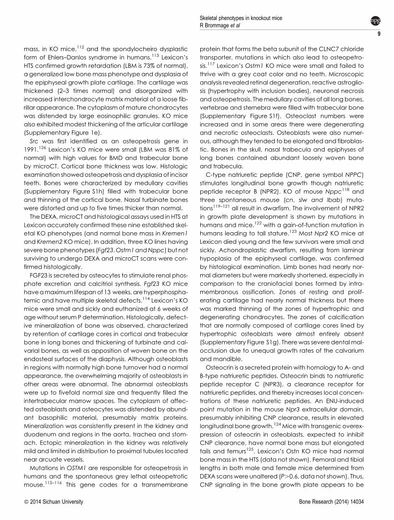
scanned simultaneously in 10 min. Photographs of these
inserts are provided in Figure 1.
Histological examinations of bonewere includedaspart
of a comprehensive analysis of multiple tissues.22 The HTS
analyses examined femur, tibia, sternum and nasal turbi-
nates. Additional bones (vertebrae, calvarium and fore-
limbs) were examined in follow-up studies for selected KO
mice having skeletal phenotypes in the HTS.
Advanced bone phenotyping assays
Deciding which KOs showing potential skeletal pheno-
types in the primary HTSmerited advancement to second-
ary screening involved both statistical and judgmental
considerations. Given that multiple parameters were
measured from a small numbers of mice, strict statistical
tests were not employed. Knowing the means and stand-
ard deviations of each parameter, we sought consistency
among the various DEXA and microCT values. Values for
KO mice were compared to both littermate WT controls
and historical WT data.
Additional methods, such as ovariectomy, daily sub-
cutaneous teriparatide treatment, measurement of
serum levels of PINP as an index of bone formation52 and
bone breaking strength, all involved standard protocols.
Biomechanical parameters were measured at Numira
Biosciences (previously SkeleTech, MDS Pharma Services
and Ricerca Biosciences, Salt Lake City, Utah, USA) using
standard procedures for LV5 compression and femur shaft
four-point bending. Body CT scans were performed using
an ImTek scanner (Siemens, Munich, Germany). Three-
dimensional images were reconstructed using the
Feldkamp algorithm with ImTek 3D RECON software.
Lexicon developed two neutralizing mouse antibodies
to Dickkopf 1 (DKK1). Mice were immunized with purified
mouse DKK1 protein produced in HEK293 cells. Total RNA
was obtained from the spleens of immunized mice and a
phage library displaying FAb fragments was constructed.
Phage displaying DKK1-specific FAbs were selected on
immobilized DKK1 protein and monoclonal FAbs were
generated in Escherichia coli. The specificity of FAbs for
DKK1 was confirmed by ELISA. Chimeric proteins com-
posed of combinations of the N-terminal leader/CYS1
domain and the C-terminal CYS2 domain/tail of DKK1,
DKK2 and DKK4 were produced by transient transfection
in HEK293 cells and used to map FAb epitopes. The ability
of FAbs to inhibit theactivity of DKK1wasdeterminedusing
the CellSensorH LEF/TCF-bla FreeStyleTM 293F reporter cell
line (Invitrogen, Grand Island, New York, USA) in the pres-
ence of exogenous Wnt3a (R&D Systems, Minneapolis,
Minnesota, USA) and exogenous DKK1 protein. The ability
of FAbs to inhibit binding between DKK1 and LRP6 was
determined in an ELISA-based binding assay utilizing puri-
fied DKK1 protein and purified LRP6 ectodomain-Fc fusion
protein (R&D Systems). Based on their ability to inhibit DKK1
function in vitro and their binding mapping to distinct
domains of DKK1, two FAbs were selected for conversion
to full length mouse IgG1 antibody, production from CHO
cells, and testing in vivo. Full-length antibody affinities for
mouse and human DKK1 were measured using a Biacore
3000 (GE Healthcare, Pittsburgh, Pennsylvania, USA) with
DKK1 in the solution phase.
Mouse husbandry
Mice were housed in micro-isolator cages within a barrier
facility at 24 6C on a fixed 12-h light and 12-h dark cycle
and were provided ad libitum acidified water and Purina
rodentchow#5001 (Purina, St Louis,MO,USA). Procedures
involving animals were conducted in conformance with
Lexicon Pharmaceuticals’ Institutional Animal Care and
Use Committee guidelines, that are in compliance with
state and federal laws and the standards outlined in the
Guide for the Care and Use of Laboratory Animals
(National Research Council, 2011). Quarterly sentinel sur-
veillance showed no evidence of pathogenic rodent
viruses, Mycoplasma or Helicobacter species in the
Lexicon Pharmaceuticals source colonies.
Figure 1.Photograph showingPlexiglasplastic inserts employed to alignLV5 vertebral bodies (left) and femurs (right) inside sample holders forthe Scanco mCT40 microCT scans. The red dots indicate placement ofLV5s, with 12 rows of 4 columns. The lack femurs indicate placement offemurs, with three rows of six columns. Bones are fixed in place byadhering soft tissue and removable tape around the outside of the insert.
Skeletal phenotypes in knockout mice
R Brommage et al
4
Bone Research (2014) 14034 � 2014 Sichuan University
RESULTSHigh-throughput screen data
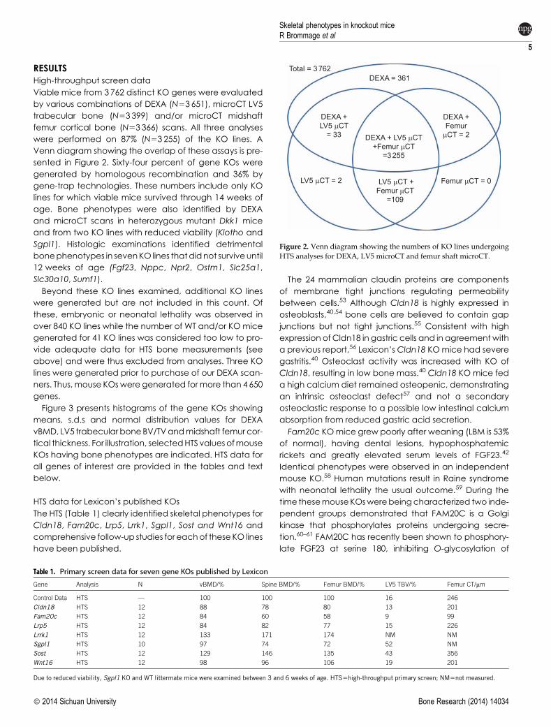
Viable mice from 3762 distinct KO genes were evaluated
by various combinations of DEXA (N53651), microCT LV5
trabecular bone (N53 399) and/or microCT midshaft
femur cortical bone (N53 366) scans. All three analyses
were performed on 87% (N53 255) of the KO lines. A
Venn diagram showing the overlap of these assays is pre-
sented in Figure 2. Sixty-four percent of gene KOs were
generated by homologous recombination and 36% by
gene-trap technologies. These numbers include only KO
lines for which viable mice survived through 14 weeks of
age. Bone phenotypes were also identified by DEXA
and microCT scans in heterozygous mutant Dkk1 mice
and from two KO lines with reduced viability (Klotho and
Sgpl1). Histologic examinations identified detrimental
bonephenotypes in sevenKO lines thatdidnot surviveuntil
12 weeks of age (Fgf23, Nppc, Npr2, Ostm1, Slc25a1,
Slc30a10, Sumf1).
Beyond these KO lines examined, additional KO lines
were generated but are not included in this count. Of
these, embryonic or neonatal lethality was observed in
over 840 KO lines while the number of WT and/or KO mice
generated for 41 KO lines was considered too low to pro-
vide adequate data for HTS bone measurements (see
above) and were thus excluded from analyses. Three KO
lines were generated prior to purchase of our DEXA scan-
ners. Thus, mouse KOswere generated formore than 4650
genes.
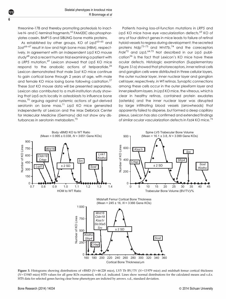
Figure 3 presents histograms of the gene KOs showing
means, s.d.s and normal distribution values for DEXA
vBMD, LV5 trabecular boneBV/TVandmidshaft femur cor-
tical thickness. For illustration, selectedHTS values ofmouse
KOs having bone phenotypes are indicated. HTS data for
all genes of interest are provided in the tables and text
below.
HTS data for Lexicon’s published KOs
The HTS (Table 1) clearly identified skeletal phenotypes for
Cldn18, Fam20c, Lrp5, Lrrk1, Sgpl1, Sost and Wnt16 and
comprehensive follow-up studies for eachof these KO lines
have been published.
The 24 mammalian claudin proteins are components
of membrane tight junctions regulating permeability
between cells.53 Although Cldn18 is highly expressed in
osteoblasts,40,54 bone cells are believed to contain gap
junctions but not tight junctions.55 Consistent with high
expression of Cldn18 in gastric cells and in agreement with
a previous report,56 Lexicon’s Cldn18 KOmice had severe
gastritis.40 Osteoclast activity was increased with KO of
Cldn18, resulting in low bone mass.40 Cldn18 KO mice fed
a high calcium diet remained osteopenic, demonstrating
an intrinsic osteoclast defect57 and not a secondary
osteoclastic response to a possible low intestinal calcium
absorption from reduced gastric acid secretion.
Fam20c KOmice grew poorly after weaning (LBM is 53%
of normal), having dental lesions, hypophosphatemic
rickets and greatly elevated serum levels of FGF23.42
Identical phenotypes were observed in an independent
mouse KO.58 Human mutations result in Raine syndrome
with neonatal lethality the usual outcome.59 During the
time thesemouseKOswerebeingcharacterized two inde-
pendent groups demonstrated that FAM20C is a Golgi
kinase that phosphorylates proteins undergoing secre-
tion.60–61 FAM20C has recently been shown to phosphory-
late FGF23 at serine 180, inhibiting O-glycosylation of
Total = 3 762
DEXA + LV5 mCT+Femur mCT
=3 255
LV5 mCT +Femur mCT
=109
DEXA +Femur
mCT = 2
DEXA = 361
Femur mCT = 0
DEXA +LV5 mCT
= 33
LV5 mCT = 2
Figure 2. Venn diagram showing the numbers of KO lines undergoingHTS analyses for DEXA, LV5 microCT and femur shaft microCT.
Table 1. Primary screen data for seven gene KOs published by Lexicon
Gene Analysis N vBMD/% Spine BMD/% Femur BMD/% LV5 TBV/% Femur CT/mm
Control Data HTS — 100 100 100 16 246
Cldn18 HTS 12 88 78 80 13 201
Fam20c HTS 12 84 60 58 9 99
Lrp5 HTS 12 84 82 77 15 226
Lrrk1 HTS 12 133 171 174 NM NM
Sgpl1 HTS 10 97 74 72 52 NM
Sost HTS 12 129 146 135 43 356
Wnt16 HTS 12 98 96 106 19 201
Due to reduced viability, Sgpl1 KO and WT littermate mice were examined between 3 and 6 weeks of age. HTS5high-throughput primary screen; NM5not measured.
Skeletal phenotypes in knockout miceR Brommage et al
5
� 2014 Sichuan University Bone Research (2014) 14034
threonine-178 and thereby promoting proteolysis to inact-
ive N- andC-terminal fragments.62 FAM20C also phosphor-
ylates casein, BMP15 and SIBLING bone matrix proteins.
As established by other groups, KO of Lrp563–65 and
Sost66–67 result in low and high bone mass (HBM), respect-
ively. In agreement with an independent Lrp5 KO mouse
study68 and a recent human trial examining a patient with
a LRP5 mutation,69 Lexicon showed that Lrp5 KO mice
respond to the anabolic actions of teriparatide.26
Lexicon demonstrated that male Sost KO mice continue
to gain cortical bone through 2 years of age, with male
and female KO mice losing bone following castration.27
These Sost KO mouse data will be presented separately.
Lexicon also contributed to a multi-institution study show-
ing that Lrp5 acts locally in osteoblasts to influence bone
mass,70 arguing against systemic actions of gut-derived
serotonin on bone mass.71 Lrp5 KO mice generated
independently at Lexicon and the Max Delbruck Center
for Molecular Medicine (Germany) did not show any dis-
turbances in serotonin metabolism.70
Patients having loss-of-function mutations in LRP5 and
Lrp5 KO mice have eye vascularization defects.63 KO of
any of four distinct genes in mice leads to failure of retinal
hyloid vessels to regress duringdevelopment: the secreted
proteins Ndp72–73 and Wnt7b,74 and the coreceptors
Frd472 and Lrp5.64,75 Not described in our Lrp5 publi-
cation26 is the fact that Lexicon’s KO mice have these
ocular defects. Histologic examination (Supplementary
Figure S1a) showed that photoreceptors, inner retinal cells
and ganglion cells were distributed in three cellular layers,
the outer nuclear layer, inner nuclear layer and ganglion
cell layer, respectively, inWT retinas. Synaptic connections
among these cells occur in the outer plexiform layer and
inner plexiform layers. In Lrp5KOmice, the vitreous,which is
clear in healthy retinas, contained protein exudates
(asterisks) and the inner nuclear layer was disrupted
by large infiltrating blood vessels (arrowheads) that
apparently failed to disperse, but formedadeep capillary
plexus. Lexicon has also confirmed and extended findings
of similar ocular vascularization defects in Fzd4 KOmice.76
00.7 1.31.21.11.0
HOM to WT Ratio0.90.8
Lrp5
Wnt16
Cldn18Crtap
Sost
Src
Lrrk1Klotho
1.4
150
750
Num
ber o
f KO
Lin
es
600
450
300
900
Body vBMD KO to WT Ratio(Mean = 0.995 ± 0.038, N = 3 651 Gene KOs)
± 2 SD
0160
Crtap
Wnt16Cldn18Kiss1rSlc30a5
Src
SostKlothoSfrp4
340320300280260240220200180 360
750
Num
ber o
f KO
Lin
es
500
250
1 000
Midshaft Femur Cortical Bone Thickness(Mean = 245 ± 16, N = 3 366 Gene KOs)
± 2 SD
00 403530252015
Trabecular Bone Volume (BV/TV)/%
Cortical Bone Thickness/mm
105
Crtap
Kiss1r
SostKlotho
Src
Sfrp4
45
Num
ber o
f KO
Lin
es 600
200
400
900Spine LV5 Trabecular Bone Volume
(Mean = 16.1 ± 3.8, N = 3 399 Gene KOs)
± 2 SD
Figure 3. Histograms showing distributions of vBMD (N546 228 mice), LV5 Tb BV/TV (N513 979 mice) and midshaft femur cortical thickness(N513 845 mice) HTS values for all gene KOs examined, with s.d. indicated. Lines show normal distributions for the calculated means and s.d.s.HTS data for selected genes having clear bone phenotypes are indicted by arrows. s.d., standard deviation.
Skeletal phenotypes in knockout mice
R Brommage et al
6
Bone Research (2014) 14034 � 2014 Sichuan University
LRRK1 is a serine/threonine kinasecontaining threeankyrin
repeat domains, seven leucine-rich repeat domains, a Roc
GTPase domain, a COR domain and a kinase domain.77
Mutations in the related LRRK2geneare themost common
genetic cause of Parkinson’s disease and since these
mutations often result in enhanced kinase activity, drug
development efforts are underway todiscover smallmole-
cule inhibitors of this target. Lexicon’s Lrrk1 KOmice (which
lack theGTPase domain as a result of targeted deletion of
exons 16–19) have severe osteopetrosis resulting from dys-
functional osteoclasts.39 A second Lrrk1 KO line (C57BL/6-
Lrrk1tm[1.]1Mjff/J, lacking exons 24–29 which encode the
kinase domain) and Lrrk1 KO rats (LEH-Lrrk1em1sage[2/2 ])
are available from the Michael J. Fox Foundation78
through Jackson Laboratories (Bar Harbor, Maine, USA)
and SAGE Labs (Boyertown, Pennsylvania, USA), respect-
ively. C57BL/6-Lrrk1tm1.1Mjff/J KO mice suffer from neonatal
lethality and reasons for phenotype differences resulting
from the two different KO strategies are unclear.
Sphingosine-1-phosphate (S1P), a signaling phospholi-
pid produced from ceramides by the actions of cerami-
dase and sphingosine kinase, is a ligand for five GPCR
receptors (S1P1 through S1P5) and is inactivated by S1P
lyase (SGPL1). Among other actions, S1P inhibits migration
of T lymphocytes from thymus and lymph nodes into the
circulation and therefore pharmacological analogues of
S1P (fingolimod) or treatments that increase endogenous
S1P levels are immunosuppressive. Plasma S1P levels are
elevated in postmenopausal women with vertebral frac-
tures.79 Several laboratories, including Lexicon,45 have
shown that global KO of Sgpl1 results in sickly mice having
lymphopenia and markedly reduced lifespan. Lexicon’s
comprehensive histological examination showed lung,
heart and urinary tract lesions, along with osteopetrosis.
Osteoclasts are plentiful but have increased cytoplasmic
volume and show increased incidence of degeneration
and apoptosis. Thickened trabecular bones suggest that
osteoclasts are not fully functional. Complementing the
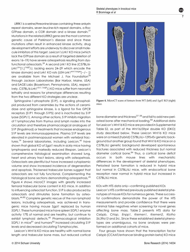
histological bone sections demonstrating osteopetrosis,45
Figure 4 shows microCT images with greatly increased
femoral trabecular bone content in KO mice. In addition
to influencing osteoclast function, S1P is also produced by
osteoclasts and stimulates bone formation by osteo-
blasts.44,80 Complete genetic rescue of the non-lymphoid
lesions, including osteopetrosis, was achieved in trans-
genic mice having mouse Sgpl1 replaced by human
SGPL1. These humanizedmice have spleen SGPL1 enzyme
activity 17% of normal and are healthy, but continue to
exhibit lymphoid defects.45 Pharmacological inhibition
SGPL1 in mice81 and humans82 increased circulating S1P
levels and decreased circulating T-lymphocytes.
Lexicon’sWnt16 KOmice are healthy with normal bone
length and trabecular bone mass, but reduced cortical
bonediameterand thickness37–38and fail toaddnewperi-
osteal bone after mechanical loading.83 Additional data
for Lexicon’sWnt16KOmiceareprovided in Supplemental
Table S2, as part of the Wnt16/Sfrp4 double KO (DKO)
study described below. These Lexicon Wnt16 KO mice
wereonamixed (hybrid)C57BL/6J–129SvEvgeneticback-
groundbut another group found thatWnt16 KOmice ona
C57BL/6J genetic background developed spontaneous
fractures associated with reduced thickness but normal
diameter cortical bone.84 Thus, a cortical bone deficit
occurs in both mouse lines with mechanistic
differences in the development of skeletal phenotypes.
Periosteal bone formation is reduced in hybrid mice
but normal in C57BL/6J mice, with endocortical bone
resorption near normal in hybrid mice but increased in
C57BL/6J mice.
KOs with HTS data only—confirming published KOs
Lexicon’s HTS confirmed previously published skeletal phe-
notypes of mouse KOs for numerous genes. These success-
ful confirmations demonstrate the power of the HTS
measurements and provide confidence that there were
minimal false negatives when screening for novel genes
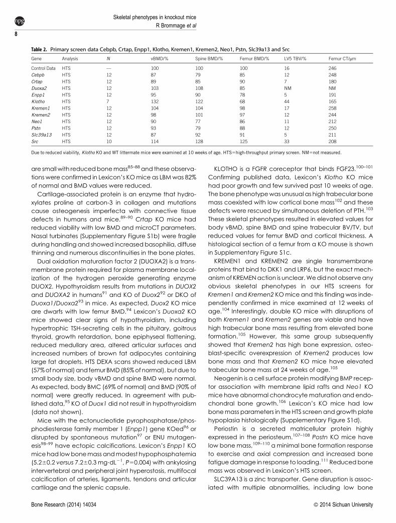
affecting bone mass. Table 2 presents HTS data for
Cebpb, Crtap, Enpp1, Kremen1, Kremen2, Klotho
Slc39a13 and Src. Since these established skeletal pheno-
types are unambiguous, no bone analyses were per-
formed on additional cohorts of mice.
Four groups have shown that the transcription factor
Cebpb (CCAAT/enhancer binding protein beta) KOmice
Figure 4. MicroCT scans of femurs from WT (left) and Sgpl1 KO (right)mice.
Skeletal phenotypes in knockout miceR Brommage et al
7
� 2014 Sichuan University Bone Research (2014) 14034
are small with reducedbonemass85–88 and theseobserva-
tions were confirmed in Lexicon’s KOmice as LBMwas 82%
of normal and BMD values were reduced.
Cartilage-associated protein is an enzyme that hydro-
xylates proline at carbon-3 in collagen and mutations
cause osteogenesis imperfecta with connective tissue
defects in humans and mice.89–90 Crtap KO mice had
reduced viability with low BMD and microCT parameters.
Nasal turbinates (Supplementary Figure S1b) were fragile
during handling and showed increasedbasophilia, diffuse
thinning and numerous discontinuities in the bone plates.
Dual oxidation maturation factor 2 (DUOXA2) is a trans-
membrane protein required for plasma membrane local-
ization of the hydrogen peroxide generating enzyme
DUOX2. Hypothyroidism results from mutations in DUOX2
and DUOXA2 in humans91 and KO of Duox292 or DKO of
Duoxa1/Duoxa293 in mice. As expected, Duox2 KO mice
are dwarfs with low femur BMD.94 Lexicon’s Duoxa2 KO
mice showed clear signs of hypothyroidism, including
hypertrophic TSH-secreting cells in the pituitary, goitrous
thyroid, growth retardation, bone epiphyseal flattening,
reduced medullary area, altered articular surfaces and
increased numbers of brown fat adipocytes containing
large fat droplets. HTS DEXA scans showed reduced LBM
(57%ofnormal)and femurBMD(85%ofnormal), butdue to
small body size, body vBMD and spine BMD were normal.
As expected, body BMC (69% of normal) and BMD (90% of
normal) were greatly reduced. In agreement with pub-
lished data,95 KO of Duox1 did not result in hypothyroidism
(data not shown).
Mice with the ectonucleotide pyrophosphatase/phos-
phodiesterase family member 1 (Enpp1) gene KOed96 or
disrupted by spontaneous mutation97 or ENU mutagen-
esis98–99 have ectopic calcifications. Lexicon’s Enpp1 KO
micehad lowbonemass andmodest hypophosphatemia
(5.260.2 versus 7.260.3 mg?dL21, P50.004) with ankylosing
intervertebral and peripheral joint hyperostosis, multifocal
calcification of arteries, ligaments, tendons and articular
cartilage and the splenic capsule.
KLOTHO is a FGFR coreceptor that binds FGF23.100–101
Confirming published data, Lexicon’s Klotho KO mice
had poor growth and few survived past 10 weeks of age.
Thebonephenotypewas unusual as high trabecular bone
mass coexisted with low cortical bone mass102 and these
defects were rescued by simultaneous deletion of PTH.103
These skeletal phenotypes resulted in elevated values for
body vBMD, spine BMD and spine trabecular BV/TV, but
reduced values for femur BMD and cortical thickness. A
histological section of a femur from a KO mouse is shown
in Supplementary Figure S1c.
KREMEN1 and KREMEN2 are single transmembrane
proteins that bind to DKK1 and LRP6, but the exact mech-
anismof KREMENaction is unclear.Wedidnotobserveany
obvious skeletal phenotypes in our HTS screens for
Kremen1 and Kremen2 KOmice and this findingwas inde-
pendently confirmed in mice examined at 12 weeks of
age.104 Interestingly, double KO mice with disruptions of
both Kremen1 and Kremen2 genes are viable and have
high trabecular bone mass resulting from elevated bone
formation.105 However, this same group subsequently
showed that Kremen2 has high bone expression, osteo-
blast-specific overexpression of Kremen2 produces low
bone mass and that Kremen2 KO mice have elevated
trabecular bone mass at 24 weeks of age.105
Neogenin is a cell surface proteinmodifying BMP recep-
tor association with membrane lipid rafts and Neo1 KO
mice have abnormal chondrocytematuration and endo-
chondral bone growth.106 Lexicon’s KO mice had low
bonemass parameters in the HTS screenandgrowth plate
hypoplasia histologically (Supplementary Figure S1d).
Periostin is a secreted matricellular protein highly
expressed in the periosteum.107–108 Postn KO mice have
low bone mass,109–110 a minimal bone formation response
to exercise and axial compression and increased bone
fatiguedamage in response to loading.111 Reducedbone
mass was observed in Lexicon’s HTS screen.
SLC39A13 is a zinc transporter. Gene disruption is assoc-
iated with multiple abnormalities, including low bone
Table 2. Primary screen data Cebpb, Crtap, Enpp1, Klotho, Kremen1, Kremen2, Neo1, Pstn, Slc39a13 and Src
Gene Analysis N vBMD/% Spine BMD/% Femur BMD/% LV5 TBV/% Femur CT/mm
Control Data HTS — 100 100 100 16 246
Cebpb HTS 12 87 79 85 12 248
Crtap HTS 12 89 85 90 7 180
Duoxa2 HTS 12 103 108 85 NM NM
Enpp1 HTS 12 95 90 78 5 191
Klotho HTS 7 132 122 68 44 165
Kremen1 HTS 12 104 104 98 17 258
Kremen2 HTS 12 98 101 97 12 244
Neo1 HTS 12 90 77 86 11 212
Pstn HTS 12 93 79 88 12 250
Slc39a13 HTS 12 87 92 91 5 211
Src HTS 10 114 128 125 33 208
Due to reduced viability, Klotho KO and WT littermate mice were examined at 10 weeks of age. HTS5high-throughput primary screen. NM5not measured.
Skeletal phenotypes in knockout mice
R Brommage et al
8
Bone Research (2014) 14034 � 2014 Sichuan University
mass, in KO mice,112 and the spondylocheiro dysplastic
form of Ehlers–Danlos syndrome in humans.113 Lexicon’s
HTS confirmed growth retardation (LBM is 73% of normal),
a generalized low bonemass phenotype and dysplasia of
the epiphyseal growth plate cartilage. The cartilage was
thickened (2–3 times normal) and disorganized with
increased interchondrocyte matrix material of a loose fib-
rillar appearance. The cytoplasmofmature chondrocytes
was distended by large eosinophilic granules. KO mice
also exhibitedmodest thickening of the articular cartilage
(Supplementary Figure 1e).
Src was first identified as an osteopetrosis gene in
1991.126 Lexicon’s KO mice were small (LBM was 81% of
normal) with high values for BMD and trabecular bone
by microCT. Cortical bone thickness was low. Histologic
examination showedosteopetrosis anddysplasiaof incisor
teeth. Bones were characterized by medullary cavities
(Supplementary Figure S1h) filled with trabecular bone
and thinning of the cortical bone. Nasal turbinate bones
were distorted and up to five times thicker than normal.
TheDEXA,microCT andhistological assays used in HTS at
Lexicon accurately confirmed these nine established skel-
etal KO phenotypes (and normal bone mass in Kremen1
and Kremen2 KOmice). In addition, three KO lines having
severebonephenotypes (Fgf23,Ostm1andNppc) but not
surviving to undergo DEXA and microCT scans were con-
firmed histologically.
FGF23 is secreted by osteocytes to stimulate renal phos-
phate excretion and calcitriol synthesis. Fgf23 KO mice
haveamaximum lifespanof 13 weeks, arehyperphospha-
temic and have multiple skeletal defects.114 Lexicon’s KO
mice were small and sickly and euthanized at 6 weeks of
agewithout serumPdetermination. Histologically, defect-
ive mineralization of bone was observed, characterized
by retention of cartilage cores in cortical and trabecular
bone in long bones and thickening of turbinate and cal-
varial bones, as well as apposition of woven bone on the
endosteal surfaces of the diaphysis. Although osteoblasts
in regions with normally high bone turnover had a normal
appearance, the overwhelming majority of osteoblasts in
other areas were abnormal. The abnormal osteoblasts
were up to fivefold normal size and frequently filled the
intertrabecular marrow spaces. The cytoplasm of affec-
ted osteoblasts and osteocytes was distended by abund-
ant basophilic material, presumably matrix proteins.
Mineralization was consistently present in the kidney and
duodenum and regions in the aorta, trachea and stom-
ach. Ectopic mineralization in the kidney was relatively
mild and limited in distribution to proximal tubules located
near arcuate vessels.
Mutations in OSTM1 are responsible for osteopetrosis in
humans and the spontaneous grey lethal osteopetrotic
mouse.115–116 This gene codes for a transmembrane
protein that forms the beta subunit of the CLNC7 chloride
transporter, mutations in which also lead to osteopetro-
sis.117 Lexicon’s Ostm1 KO mice were small and failed to
thrive with a grey coat color and no teeth. Microscopic
analysis revealed retinal degeneration, reactive astroglio-
sis (hypertrophy with inclusion bodies), neuronal necrosis
andosteopetrosis. Themedullary cavities of all longbones,
vertebrae and sternebra were filled with trabecular bone
(Supplementary Figure S1f). Osteoclast numbers were
increased and in some areas there were degenerating
and necrotic osteoclasts. Osteoblasts were also numer-
ous, although they tended to be elongated and fibroblas-
tic. Bones in the skull, nasal trabecula and epiphyses of
long bones contained abundant loosely woven bone
and trabecula.
C-type natriuretic peptide (CNP, gene symbol NPPC)
stimulates longitudinal bone growth though natriuretic
peptide receptor B (NPR2). KO of mouse Nppc118 and
three spontaneous mouse (cn, slw and lbab) muta-
tions119–121 all result in dwarfism. The involvement of NPR2
in growth plate development is shown by mutations in
humans and mice,122 with a gain-of-function mutation in
humans leading to tall stature.123 Most Npr2 KO mice at
Lexicon died young and the few survivors were small and
sickly. Achondroplastic dwarfism, resulting from laminar
hypoplasia of the epiphyseal cartilage, was confirmed
by histological examination. Limb bones had nearly nor-
mal diameters but weremarkedly shortened, especially in
comparison to the craniofacial bones formed by intra-
membranous ossification. Zones of resting and prolif-
erating cartilage had nearly normal thickness but there
was marked thinning of the zones of hypertrophic and
degenerating chondrocytes. The zones of calcification
that are normally composed of cartilage cores lined by
hypertrophic osteoblasts were almost entirely absent
(Supplementary Figure S1g). Therewas severe dentalmal-
occlusion due to unequal growth rates of the calvarium
and mandible.
Osteocrin is a secreted protein with homology to A- and
B-type natriuretic peptides. Osteocrin binds to natriuretic
peptide receptor C (NPR3), a clearance receptor for
natriuretic peptides, and thereby increases local concen-
trations of these natriuretic peptides. An ENU-induced
point mutation in the mouse Npr3 extracellular domain,
presumably inhibiting CNP clearance, results in elevated
longitudinal bone growth.124 Micewith transgenic overex-
pression of osteocrin in osteoblasts, expected to inhibit
CNP clearance, have normal bone mass but elongated
tails and femurs125. Lexicon’s Ostn KO mice had normal
bonemass in the HTS (data not shown). Femoral and tibial
lengths in both male and female mice determined from
DEXA scanswere unaltered (P.0.6, data not shown). Thus,
CNP signaling in the bone growth plate appears to be
Skeletal phenotypes in knockout miceR Brommage et al
9
� 2014 Sichuan University Bone Research (2014) 14034
unaffected by disruption of osteocrin, but stimulated by
both elevated osteocrin levels and disruption of the
NPR3 clearance receptor.
Genes involved in WNT signaling
Great interest in the roles of Wnt signaling on bone mass
followed the 2001/2002 discoveries that various LRP5 and
SOST gene mutations in humans are responsible for dra-
matic alterations in patients with osteoporosis pseudo-
glioma, the HBM phenotype, sclerosteosis and van
Buchem’s disease. Along with Frizzled receptors, LRP5
and LRP6arecoreceptors for the 19WNT ligandswith scler-
ostin (coded by the SOST gene) blocking WNT binding to
LRP5/6. Since inactivating LRP5 (osteoporosis pseudo-
glioma syndrome) and LRP6127–128 mutations decrease
bone mass, whereas activating LRP5 mutations (HBM
phenotype) and mutations disrupting SOST expression
(sclerosteosis and van Buchem’s disease) increase bone
mass, WNT signaling must activate bone formation. As
described above, KO of Lrp5 and Sost in mice produce
the identical skeletal phenotypes as the human inactiv-
ating mutations. Furthermore, transgenic mice with Lrp5
activating mutations have extremely HBM.70,129
WNT signaling can also be inhibited by DKKs that bind
LRP5/6 and Secreted frizzed-related proteins (SFRPs) that
bind WNTs. As is the case with sclerostin, reducing levels of
DKKs and SFRPs in bone should remove inhibitors of WNT
signaling, thereby presumably increasing bone mass.
Support for this idea was available in the original HBM
publication describing the LRP5 activating mutation,
which in cells eliminated the inhibitory actions of DKK1.130
The key role of WNT signaling in bone prompted us to
examine KO mice for all four Dkks, five Sfrps and Wif1
(Table 3). KO of Dkk1 leads to embryonic lethality,
but mice with disruptions of the other eight genes were
viable.
HBM is observed in doubleridege mice having a spon-
taneous mutation leading to reduced DKK1 expression,131
Dkk1 heterozygousmutantmice132 andDkk1 homozygous
KO mice generated on a Wnt3 heterozygous mutant
background.133–134 Osteoblast-specific overexpression of
Dkk1 in mice leads to severe osteopenia.135 Lexicon con-
firmedobservations of HBM in heterozygousDkk1 KOmice.
These and other observations prompted several groups,
including Novartis,136 Merck,137–138 Amgen,139 Eli Lilly140
and Lexicon, to develop anti-DKK1 neutralizing antibodies
for potential therapeutic treatment of osteoporosis and
other bone diseases, particularly multiple myeloma.141
Lexicon’s DKK1 Antibody #1 recognizes an epitope in
the C-terminal half of DKK1 that prevents DKK1 binding
to LRP6. Antibody #2 recognizes an epitope in the N-ter-
minal half of Dkk-1 that does not affect Dkk1 binding to
LRP6. Identical anti-DKK1 epitope specificities for LRP6
binding have been reported by Amgen.142 Both Lexicon
antibodies have potencies below 500 pM as determined
by binding affinities to humanDKK1 (416 pM for Ab #1 and
Table 3. Primary and secondary screen data for WNT antagonists
Gene Analysis N vBMD/% Spine BMD/% Femur BMD/% LV5 TBV/% Femur CT/mm
Control Data HTS … 100 100 100 16 246
Dkk1 (HETs) HTS 12 100 103 105 29 254
SS 23 112 136 108 29 267
Dkk2 HTS 12 97 106 97 19 242
SS 28 109 112 108 20 252
Dkk3 HTS 12 100 104 102 18 255
SS 42 96 93 98 16 255
Dkk4 HTS 13 102 94 104 16 246
SS 19 99 104 100 20 268
Sfrp1 HTS 12 97 90 97 18 249
SS 30 100 107 104 22 277
Sfrp2 HTS 12 102 101 102 21 253
SS 23 99 100 101 NM NM
Sfrp3 HTS 12 98 96 106 16 245
SS 17 100 105 99 16 264
Sfrp4 HTS 12 93 95 88 26 167
SS 39 93 98 93 30 159
Sfrp5 HTS 12 103 96 95 13 242
SS 43 102 103 101 22 258
Wif1 HTS 12 100 100 94 19 252
SS 28 101 91 100 NM NM
Wnt10a HTS 12 98 100 98 17 243
Wnt10b HTS 12 92 97 92 14 236
Mouse ages in the secondary screens were 23 weeks for Dkk1, 14 and 28 weeks for Dkk2, 42 to 64 weeks for Dkk3, 19 to 29 weeks for Dkk4, 52 weeks for Sfrp1, 7 weeks for
Sfrp2, 24 weeks forSfrp3, 15weeks forSfrp4, 16 to 21weeks forSfrp5, and 20 to 30weeks forWif1. HTS5high-throughput primary screen; SS5secondary screen; NM5not
measured.
Skeletal phenotypes in knockout mice
R Brommage et al
10
Bone Research (2014) 14034 � 2014 Sichuan University
347 pM for Ab #2) andmouse Dkk1 (128 pM for Ab #1 and
233 pM for Ab #2) and IC50 values (270 pM for Ab #1
and 370 pM for A b#2) in a mouse cell-based assay
employing Wnt3a as a stimulator of Wnt signaling.
Three preliminary studies examining these two antibod-
ies in adult male mice (.12 weeks of age) at weekly
doses of 3–30 mg?kg21 for 6 or 12 weeks all provided
evidence of bone efficacy (data not shown). Since
DKK1 expression in rat bone declines with age,143 we
performed a fourth study of 4 weeks duration in male
mice starting at 4 weeks of age. Antibody #1 and anti-
body #2 were each given subcutaneously weekly at
doses of 30 mg?kg21. A negative control group of mice
was treated similarly with a control antibody. A positive
control group of mice was treated at the start of the
study with a single subcutaneous dose of 50 mg?kg21
zoledronate to inhibit bone resorption.
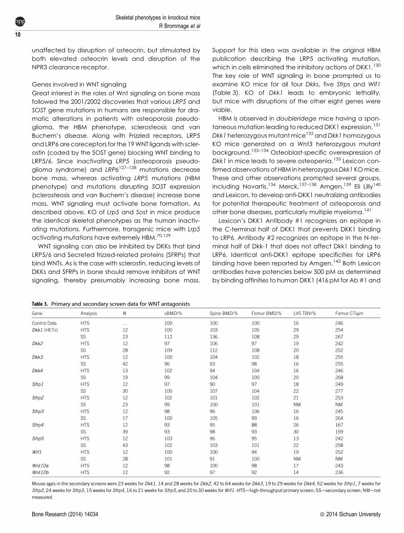
Treatment with antibody #2 and zoledronate increased
spine and femur BMD, and cancellous BV/TV in the LV5
vertebral body and distal femur metaphysis (Figure 5).
Focusing on the LV5 vertebral body, antibody #2 treat-
ment increased trabecular thickness but not trabecular
number, whereas zoledronate treatment increased tra-
becular number but not trabecular thickness. This differ-
ential mechanism of cancellous bone gain is consistent
with proposals that DKK1 inhibition acts to increase bone
formation but zoledronate treatment acts to block
bone resorption. Treatment with antibody #1 consistently
showed trends to increase bone mass but this antibody
was clearly inferior to antibody #2.
0Control Ab Ab-1 Ab-2 Zoledronate
80
60
40
20
100D = 19%P<0.01D = 0.9%
P=0.18
ANOVA P<0.001
D = 21%P<0.01
0Control Ab Ab-1 Ab-2 Zoledronate
80
60
40
20
100
D = 11%P<0.01
D = 6%P=0.22
ANOVA P=0.006
D = 11%P<0.01
0%Control Ab Ab-1 Ab-2 Zoledronate
40%
30%
20%
10%
D = 18%P=0.006
D = 15%P=0.02
ANOVA P<0.001
D = 29%P<0.001
0%Control Ab Ab-1 Ab-2 Zoledronate
20%
15%
10%
5%
25%
D = 92%P<0.001D = 36%
P=0.24
ANOVA P<0.001
D = 3.5-foldP<0.001
0Control Ab Ab-1 Ab-2 Zoledronate
8
6
4
2
D = 2%P=0.77
D = 3%P=0.34
ANOVA P<0.001
D = 19%P<0.001
0.00Control Ab Ab-1 Ab-2 Zoledronate
0.06
0.04
0.02
0.08
D = 10%P=0.04
D = 7%P=0.22
ANOVA P=0.04
D = 1%P=0.99
Spi
ne B
MD
/(mg.
cm–2
)
Fem
ur B
MD
/(mg.
cm–2
)
LV5
Trab
ecul
ar B
V/T
V
DFM
Tra
becu
lar B
V/T
VLV
5 Tr
abec
ular
Thi
ckne
ss/m
m
LV5
Trab
ecul
ar N
umbe
r
Figure 5.DKK1 antibody treatment in youngmice. Data are means6s.e.m. for 10 mice per group treated between 4 and 8 weeks of age with antibodydoses of 30 mg?kg21 weekly or a single 10 mg?kg21 zoledronate dose. Statistical evaluation involved ANOVA followed by Dunnett’s test for multiplecomparisons.
Skeletal phenotypes in knockout miceR Brommage et al
11
� 2014 Sichuan University Bone Research (2014) 14034
In agreementwithprevious reports,144–145 Lexicon’sDkk2
KO mice had numerous ocular abnormalities, including
a lack of Harderian glands and malformed eyelids. The
cornea and sclera contained sebaceous glands and
hair follicles with multifocal keratitis and ulceration and
fibrosis of the collageneous stroma. Although Dkk2 KO
mice have a normal skeletal architecture, their bone
contains an excess of unmineralized osteoid.146 Lexicon’s
KO mice had normal, or even slightly elevated, BMD.
Undecalcified bone sections were not examined and
therefore the excess osteoid phenotype was not evalu-
ated. We rescanned eight WT and eight KO midshaft
femurs at the highest microCT resolution possible (6 mm
voxel size) and did not observe any hypomineralization
of cortical bone, as material BMD was unaffected (0.7%
higher with P50.27) in bones from Dkk2 KO mice. No gross
skeletal phenotypes were previously detected in Dkk3 KO
mice147 and Lexicon did not observe any bone abnormal-
ities in the HTS.
Several groups generated viable Sfrp2, Sfrp5148–149 KOor
ENU-mutant Sfrp5150 mice without describing any bone
abnormalities. Interestingly, DKO of Sfrp1/Sfrp2 results in
embryonic lethality,151 whereas Sfrp2/Sfrp5 DKO mice are
viable.150 Lexicon did not detect any skeletal phenotypes
in Sfrp2 or Sfrp5 KO mice. Sfrp2 KO mice have subtle limb
deformities including brachydactyly and syndactyly152
and these digit abnormalities were likely missed in
Lexicon’s HTS.
Published reports describe skeletal phenotypes in Sfrp1
and Sfrp3 KOmice. Global overexpression of Sfrp1 in trans-
genicmice results in reducedbonemass.153 Sfrp1 KOmice
have elevated trabecular bone mass after 13 weeks of
age,154–155 but Lexicon’s primary and secondary screens
did not confirm this observation. Interestingly, the pub-
lished Sfrp1 KO (targeting exon 1) mice examined were
generated at Lexicon prior to 2000. Thesemice are distinct
from the KO mouse subsequently studied at Lexicon. The
reason for this discrepancy is unclear and additional stud-
ies are required. DEXA HTS analyses performed at the
Harwell MRC site as part of the Europhenome mouse KO
project observed reduced BMD in Sfrp1 KO mice. At least
three additional Sfrp1 KO mice lines have been gener-
ated151,156–157 without skeletal analyses performed.
Frzb (Sfrp3) KOmice have normal trabecular bonemass
but a 7% increase in cortical bone thickness and increased
articular cartilage loss during arthritis triggered by instab-
ility, enzymatic injury or inflammation.158 KO mice also
showed an exaggerated cortical bone gain in response
to repetitive loading of the ulna, with the added bone
being predominantly periosteal.157 Loss of bone following
ovariectomywas not affected by Sfrp3 KO.157 GWAS ana-
lysis identified SFRP3 SNPs that influence hip osteoarthritis
and geometry.159 Lexicon’s HTS and secondary screens
did not showan obvious bone phenotype. Further analysis
of the secondary screenmicroCT data (excluding one KO
outlier) found a 6% increase in midshaft femur cortical
thickness (P50.07, with N58 for both WT and KO mice).
No challenge studies, such as mechanical loading, were
performed.
Including Lexicon’s data, three independent laborat-
ories have presented data in abstracts showing bones
from Sfrp4 KOmicehave increased trabecular bonemass,
elevated cortical bone diameter and reduced cortical
bone thickness.30,160–161 High trabecular bonemass results
in increased compressive breaking strength of the LV5 ver-
tebral body, whereas reduced cortical bone thickness
leads to reduced four-point breaking strength of the femur
shaft.30 Differential effects of disrupting Sfrp4 function in
cortical and trabecular bone likely reflect distinct signaling
pathways as canonical WNT signaling is involved in tra-
becular bone formation but non-canonical Wnt signaling
is involved in cortical bone formation.162 The Yale/Harvard
and Lexicon data are being prepared for a joint publica-
tion. Here we show our HTS and secondary screen data
characterizing these skeletal phenotypes and data from
a Sfrp4/Wnt16 DKO study.
As described above, Wnt16 KO mice have normal tra-
becular bone mass but reduced cortical bone diameter
and thickness. We examined the effects of disruption of
WNT16 signaling on the skeletal phenotypes of male and
female Sfrp4 KO mice by microCT at 16 weeks of age.
In the absence of an effect of Wnt16 KO on trabecular
bone, theelevated trabecular bonemass in Sfrp4KOmice
should not be influenced by disruption ofWnt16. However,
the enlarged cortical bone diameter in Sfrp4 KO mice
might involve WNT16 signaling. Since cortical bone thick-
ness is reduced in both Sfrp4andWnt16 KOmice, simultan-
eous disruption of both genes might result in greatly
reduced thickness and even spontaneous fractures.
As shown in Supplementary Table S2, skeletal pheno-
types in both male and female Sfrp4 and Wnt16 single
KO mice are exactly as determined previously. LV5 tra-
becular bone is elevated in Sfrp4 KO mice but normal in
Wnt16 KOmice. Cortical bone total area (diameter) is ele-
vated in Sfrp4 KO mice and reduced in Wnt16 KO mice.
Cortical thickness is reduced in both KOs, but to a greater
extent with Sfrp4 than Wnt16 disruption. KO of Wnt16 did
not influence the elevated LV5 trabecular bone mass in
Sfrp4 KO mice. Bones from DKO mice had intermediate
cortical total area between elevated Sfrp4 KO and
reduced Wnt16 KO values (interaction P values .0.50),
indicating these two genes have independent actions
on bone diameter. Bone areas were reduced similarly in
both KOs and not further influenced by DKO. Marrow
areas were greatly elevated with KO of Sfrp4, slightly
reduced with KO of Wnt16 and elevated in DKOs.
Skeletal phenotypes in knockout mice
R Brommage et al
12
Bone Research (2014) 14034 � 2014 Sichuan University
Therefore, inactivation of Wnt16 does not prevent the
stimulation of endocortical bone resorption responsible
for the enlarged marrow cavity in Sfrp4 KO mice. Bones
from DKO mice had lower cortical thickness than the
already reduced values in single KOs but no spontaneous
fractures were observed.
Like SFRPs, WNT inhibitory factor 1 antagonizes Wnt sig-
naling by binding to WNTs. Wif1 KO mice have a normal
skeleton but are sensitive to radiation-induced osteosar-
comas163 and mice with osteoblast-specific Wif1 overex-
pression display no overt bone phenotype.164 Lexicon’s
Wif1 KO mice had no clear bone phenotypes in HTS or
secondary screens.
WNT10A suppresses adipogenesis and stimulates osteo-
blastogenesis in ST2 and 3T3-L1 cells.165 Lexicon’s HTS did
not identify a bone phenotype in Wnt10a KO mice, but
follow-up studies showed a 5% decrease in body length
in both male and female KO mice at 33 weeks of age
(data not shown). WNT10B is expressed in primary cultures
of mature mouse calvarial osteoblasts166 and activates
canonicalWNT signaling in the immune system,mammary
gland, adipose tissue, bone and skin.167 Wnt10b KO mice
develop low bonemass with age.168 Transgenicmicewith
high Wnt10b expression driven by the FABP4 promoter in
adipocytes and the osteocalcin promoter in osteoblasts
have HBM.169–170 The anabolic effect of teriparatide treat-
ment involves WNT10B stimulation of T lymphocytes.171
Lexicon’s Wnt10b KO mice showed modestly low bone
mass in the HTS and follow-up studies. At 32 weeks of age
(Supplementary Table S3), vBMD was reduced 8% in both
male and female mice. MicroCT analyses indicated
reduced LV5 trabecular number but normal cortical bone
total area (diameter) with minimal decreases in cortical
thickness.28
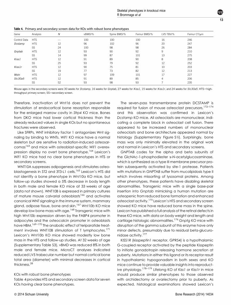
KOs with robust bone phenotypes
Table 4provides HTS and secondary screendata for seven
KOs having clear bone phenotypes.
The seven-pass transmembrane protein DCSTAMP is
required for fusion of mouse osteoclast precursors,172–174
and this observation was confirmed in Lexicon’s
Dcstamp KO mice. All osteoclasts are mononuclear, indi-
cating a complete block in osteoclast cell fusion. There
appeared to be increased numbers of mononuclear
osteoclasts and bone architecture appeared normal by
histology (Supplementary Figure S1i). Surprisingly, bone
mass was only minimally elevated in the original work
and normal in Lexicon’s HTS and secondary screens.
GNPTAB codes for the alpha and beta subunits of
the GlcNAc-1-phosphodiester a-N-acetylglucosaminase,
which is synthesized as a type III membrane precursor pro-
tein subsequently activated by site-1 protease. Patients
with mutations in GNPTAB suffer from mucolipidosis type II
which involves missorting of lysosomal proteins. Among
other phenotypes, these patients have disabling skeletal
abnormalities. Transgenic mice with a single base-pair
insertion into Gnptab mimicking a human mutation are
osteopenic from reduced bone formation and enhanced
osteoclast activity.175 Lexicon’s HTS and secondary screen
showed KO mice have reduced bone mass in the spine.
Lexicon haspublisheda full analysis of the retinal defects in
these KOmice, with data on body weight and length and
cartilage histologic abnormalities.176 Gnptg KO mice with
disruption of the gamma subunit of this enzyme have only
minor defects, presumably due to residual beta-glucuro-
nidase activity.177
KISS1R (kisspeptin1 receptor, GPR54) is a hypothalamic
G-coupled receptor activated by the peptide Kisspeptin
to initiate gonadotropin releasing hormone secretion at
puberty. Mutations in either this ligand or its receptor result
in hypothalamic hypogonadism in both sexes and KO
mice continue to provide valuable insights into reproduct-
ive physiology.178–179 Lifelong KO of Kiss1 or Kiss1r in mice
should produce similar phenotypes to those observed
with orchidectomy or ovariectomy prior to puberty. As
expected, histological examinations showed Lexicon’s
Table 4. Primary and secondary screen data for KOs with robust bone phenotypes
Gene Analysis N vBMD/% Spine BMD/% Femur BMD/% LV5 TBV/% Femur CT/mm
Control Data HTS — 100 100 100 16 246
Dcstamp HTS 12 96 102 96 20 247
SS 24 100 98 98 26 284
Gnptab HTS 12 93 90 92 9 210
SS 14 99 87 101 13 275
Kiss1 HTS 12 91 89 90 8 208
SS 25 93 79 92 12 252
Kiss1r HTS 12 90 75 81 10 203
SS 19 77 73 82 12 213
Mstn HTS 12 97 109 101 17 227
Slc30a5 HTS 12 91 89 85 4 206
SS 52 93 98 93 7 235
Mouse ages in the secondary screens were 30 weeks for Dcstamp, 16 weeks for Gnptab, 27 weeks for Kiss1, 15 weeks for Kiss1r, and 24 weeks for Slc30a5. HTS5high-
throughput primary screen; SS5secondary screen.
Skeletal phenotypes in knockout miceR Brommage et al
13
� 2014 Sichuan University Bone Research (2014) 14034
Kiss1 and Kiss1r KO mice both had typical lesions of hypo-
gonadotropic hypogonadism including immature
gonads and absence of sexual dimorphism (kidney and
salivary glands in the male, mammary gland in the
female). All bone parameters were low in the HTS and
secondary screens. For Kiss1r KOs, DEXAandmicroCT data
from the secondary screenareprovided in Supplementary
Figure S2. Bothmale and female KOmicewere obese, but
through different mechanisms. Male mice had normal
body fat content but low LBM, whereas female mice
had normal LBM but excess body fat.180
Myostatin is a secreted protein that inhibits myogenesis
with spontaneous mutations in humans, dogs and cattle
resulting in dramatic muscle hypertrophy. In addition to
muscle hyperplasia, Mstn KO mice have elevated bone
mass.181–183 In Lexicon’s HTS, Mstn KO mice had high mus-
cle mass as determined by DEXA LBM (129% of normal),
QMR LBM (126% of normal), gastrocnemius muscle weight
(198% of normal) and increased muscle fiber number
but not size by histology. An additional seven KO and
five WT male mice were examined at 116 weeks of age
(Supplementary Table S4). As expected, QMR LBM (120%)
and gastrocnemius weight (188%) were increased. Spine
trabecular bone, femurand tibia lengths (datanot shown)
and cortical bone diameters at the femoral midshaft
or tibia–fibula junction were all unaltered in KO mice.
Cortical bone thickness was elevated at the femoral mid-
shaft and tibia–fibula junction. Cortical bone in the distal
tibia (halfway between the tibia–fibula junction and the
distal bone end) had an enlarged diameter but normal
thickness. These site-specific bone changes demonstrate
that distinct skeletal locations are influenced by lifelong
muscle hypertrophy.
SLC30A5 is a zinc transporter with Slc30a5 KO mice
reported to have extremely low bone mass184 and this
observation was confirmed for Lexicon’s KO mice in both
primary and secondary screens. Mechanisms responsible
for this severe osteopenia are unclear.
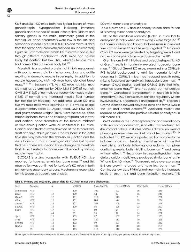
KOs with minor bone phenotypes
Table 5 provides HTS and secondary screen data for ten
KOs having minor bone phenotypes.
KO of the calcitonin receptor (Calcr) in mice led to
embryonic lethality when exons 6 and 7 were targeted185
but normal viability and trabecular bonemass in spineand
femur when exons 13 and 14 were targeted.186 Lexicon’s
Calcr KO mice were generated by targeting exon 1 and
no clear bone phenotype was observed in the HTS.
Gremlins are BMP inhibitors and osteoblast-specific KO
of Grem1 results in transiently elevated trabecular bone
mass.187 Global Grem1 KO mice, examined in a C57BL/6-
FVB hybrid background to minimize neonatal lethality
occurring in C57BL/6 mice, had reduced growth rates,
missing fibula and generally low trabecular bone mass.188
Human GWAS studies identified GREM2 SNPs that influ-
ence hip bone mass189 and trabecular but not cortical
bone.190 Craniofacial development in zebrafish is influ-
encedbyGREM2expression,aspart ofa regulatory system
involving BMP4, endothelin-1 and jagged 1b.191 Lexicon’s
Grem2 KOmice showed elevated spine and femur BMD in
the HTS and dental defects.192 Additional studies are
required to characterize possible skeletal phenotypes in
this mouse KO.
IL6RAcodes for the IL-6 receptor-alphaandanantibody
to this receptor (tocilizumab) is an effective treatment for
rheumatoid arthritis. In studies of Il6ra KOmice, no skeletal
phenotypes were observed but one of two studies193–194
indicated that KOmice are protected from ovariectomy-
induced bone loss. Treating normal mice with an IL-6
neutralizing antibody following ovariectomy has given
conflicting results, both inhibiting bone loss195 and being
without effect.196 Secondary hyperparathyroidism from
dietary calcium deficiency produced similar bone loss in
WT and IL-6 KO mice.197 Transgenic mice overexpressing
IL-6 are growth retarded and have low bone mass.198
Continuous low-dose PTH infusion in normalmice increases
levels of serum IL-6 and bone resorption markers. This
Table 5. Primary and secondary screen data for KOs with minor bone phenotypes
Gene Analysis N vBMD/% Spine BMD/% Femur BMD/% LV5 TBV/% Femur CT/mm
Control Data HTS — 100 100 100 16 246
Calcr HTS 12 100 93 98 16 245
Grem2 HTS 12 101 113 110 9 221
Il6ra HTS 12 101 NM NM 15 264
Slc26a7 HTS 12 101 107 103 30 253
Slc39a1 HTS 12 100 96 105 22 290
Sparc HTS 12 98 89 95 9 243
SS 44 98 96 97 14 261
Spp1 HTS 12 103 97 102 17 260
Ssh1 HTS 12 110 120 117 10 221
Sostdc1 HTS 12 102 99 100 16 252
Wnt5b HTS 13 107 109 110 20 258
SS 24 113 110 112 20 269
Mouse ages in the secondary screens were 26 weeks for Sparc and 19 weeks for Wnt5b. HTS5high-throughput primary screen; SS5secondary screen.
Skeletal phenotypes in knockout mice
R Brommage et al
14
Bone Research (2014) 14034 � 2014 Sichuan University
PTH-stimulated bone resorption, but not bone formation,
was suppressed with cotreatment with an IL-6 neutralizing
antibody.199
FACS analysis confirmed that isolated spleen cells from
Lexicon’s Il6ra KOmice (N510 for both KOandWT) did not
bind a commercial IL-6 receptor antibody. KO mice had
normal bonemass in Lexicon’s HTSandnormal body, spine
and femur BMD in two additional cohorts (21 and
25 weeks of age (Supplementary Table S5A). Mice in both
cohorts underwent sham or ovariectomy surgery with
bone analyses on the combined cohorts after 6 weeks.
There was no effect of Il6ra KO on bone loss following ova-
riectomy, determined by both DEXA BMD scans and
microCT analyses of spine BV/TV and midshaft femur dia-
meter and cortical thickness (Supplementary Table S5B).
Treating a third mouse cohort (97 weeks of age) with teri-
paratide produced similar elevations in serum PINP levels
in control and KO mice (Supplementary Figure S3a),
suggesting IL-6 is not involved in the anabolic actions of
teriparatide.
SLC26A7 is an anion transporter with Slc26a7 KO mice
having reduced gastric acid secretion and distal renal
tubular acidosis.200 Lexicon’s Slc26a7 KO mice were
hypothyroid with males more strongly affected than
females. Serum thyroxine levels (N56 or 7) were reduced
by 87% in males (P,0.001) and 47% in females (P50.003).
Histologicobservations showedhyperplastic thyrotrophs in
male but not female mice. Bone mass was slightly ele-
vated in the HTS screen. Follow-up studies examinedmale
and femalemice separately between 18 and 21 weeks of
age (Supplementary Table S6). Consistent with modest
hypothyroidism, there were minimal effects of Slc26a7
KOonbones from femalemice.Malemice, being severely
hypothyroid, had modest reductions in LBM (20%) and
femur length (6%), but elevated vertebral trabecular bone
as indicated by high spine BMD (20%), LV5 trabecular
BV/TV (42%) and LV5 trabecular thickness (28%).
The sodium/iodide symporter SLC5A5 plays a major role
in thyroid iodide uptakeandmutations in this gene result in
hypothyroidism. Analyses of 12 different SLC5A5mutations
shows marked clinical heterogeneity in patients with the
same and different mutations without a clear genotype-
phenotype correlation. This observation ‘… suggests
that additional unknown factors are involved in the
clinicalmanifestation’ of this defective iodide transport.201
There do not appear to be any data for Slc5a5 KO mice.
SLC26A4 (PENDRIN) is an anion exchanger with broad
specificity, including iodide, and Slc26a4 KO mice are
deaf without a thyroid phenotype.202 Since Slc26a7 KO
mice are hypothyroid, this anion transporter might
also contribute to iodide uptake by thyrotrophs as
mouse SLC26A7 is capable of both chloride and iodide
transport.203
SLC39A1 is a zinc transporter expressed in osteoclasts.204
Triple KO of mouse Slc39a1, Slc39a2 and Slc39a3 did not
produce an obvious phenotype unless the mice were fed
a zinc-deficient diet.205 Lexicon’s Slc39a1 KO mice had
normal BMD and LV5 trabecular bone in the HTS but ele-
vatedcortical thickness.MicroCTanalyses of bones froma
second cohort of female mice examined at 42 weeks of
age (N518 for WTs and N521 for KOs) confirmed the lack
of a LV5 trabecular bone phenotype and femur length
was normal. Midshaft cortical bone scans at 6micrometer
voxel size showed elevated total area (23%, P,0.001) and
cortical thickness (6%, P50.09) but reduced material BMD
(3.2%, P,0.001) consistent with cortical porosity visually
observed in the scans.
SPARC (secreted protein, acid, cysteine-rich, also
known as osteonectin) is a widely expressed matricellular
glycoprotein that has been extensively studied. Two of
three Lexicon Sparc KO mice examined had cataracts,
aspreviously reported.206–207 KOmicehave increased sub-
cutaneous fat with normal body weight.208 Lexicon’s KO
micedid not have anobesity phenotypewhen fedmouse
chow or high fat diet (data not shown) but subcutaneous
fat was not examined. KO mice develop intervertebral
disk degeneration with aging,209 but this defect was
not noticed in Lexicon’s mature KO mice. Several studies
have reported modest osteopenia in KO mice210–212
and Lexicon’s Sparc KO mice showed trends for low
bone mass.
Secreted phosphoprotein 1 (osteopontin) is a widely-
expressed, highly acidic SIBLING phosphoprotein
degraded into small fragments by PHEX213 and having
many non-skeletal actions. Numerous abnormalities,
including fibrosis, abnormalwoundhealing, defectivecar-
diac remodeling, tumor metastases, aberrant angiogen-
esis and ectopic calcification, have been observed in
Spp1 KO mice. Trabecular bone mass has been reported
to be both normal214–216 and high.217–220 Lexicon’s Spp1
KO mice had normal bone mass in the HTS.
Actin plays a critical role in osteoclast activity and is a
component of the podosome and ruffled borders. Actin
dynamics are regulatedbymany factors221 including cofi-
lin, one of the most highly expressed genes in osteo-
clasts.222 There are three cofilin genes: cofilin1, cofilin2
and destrin (actin depolymerizing factor). Lexicon’s KOs
ofCfl1 andCfl2were both embryonic lethal, with no bone
phenotypes in HTS or secondary screens for heterozygous
mutant mice. KO of destrin increases bone loss following
unloading.223Cofilin activity is inhibitedby LIMkinasephos-
phorylation and reactivated after dephosphorylation by
slingshot phosphatases.224–226 Osteoclasts isolated from
Limk1 KO mice are hyperactive and published Limk1 KO
mice have spine abnormalities227 and reduced bone
mass.228 However, Lexicon’s Limk1 and Limk2 KO mice
Skeletal phenotypes in knockout miceR Brommage et al
15
� 2014 Sichuan University Bone Research (2014) 14034
had no HTS skeletal phenotypes. There are threemembers
in the Slingshot (SSH) family and disruption of Slingshot
activity might suppress osteoclast activity. KOs of Ssh2
and Ssh3 did not have HTS skeletal phenotypes (data not
shown), but Ssh1 KO mice had HBM in the HTS and three
additional cohorts of KO mice were examined (data not
shown). Elevated bone mass was confirmed in Cohort #1
female, but not male mice; was not confirmed in Cohort
#2 female mice (males were not examined); and was
confirmed in Cohort #3 male and female mice. Ssh1 KO
did not protect Cohort #3 mice from ovariectomy-
induced bone loss between 16 and 24 weeks of age.
Given its smallmagnitude, the inconsistency of this skeletal
phenotype is not surprising. Normal bone loss following
ovariectomy made this gene uninteresting as an osteo-
porosis drug target. Ssh2 and/or Ssh3might also be active
in osteoclasts and a double or triple KO study is required to
examine this topic.
Sclerostin domain containing 1 (SOSTDC1) codes for
Wise (Ectodin) protein, which has a close homology to
sclerostin and also inhibits canonical Wnt signaling by
binding to Lrp5 and Lrp6. Lexicon’s Sostdc1 KO mice did
not show any obvious skeletal phenotypes in the HTS, con-
sistent with observations of no phenotype229 and mildly
elevated BMD through 3 months of age that disappears
at 4 months of age.230
Robinow syndrome, with major skeletal abnormities,
results from mutations in WNT5A.231 KO of Wnt5a results in
perinatal lethality232 with mouse fetuses having numerous
dental233 and skeletal234 defects. Wnt5a heterozygous
mutant mice exhibit impaired osteoclastogenesis.235
Skeletal actions of Wnt5b are less clear, as KO mice have
been examined only for neuronal development.236
Chondrocyte hypertrophy and bone mineralization are
delayed in transgenic mice overexpressing Wnt5b in car-
tilage.234 Lexicon’s Wnt5b KO mice had elevated bone
mass in the HTS and secondary screens. Studies on addi-
tional cohorts ofmaleand femalemice showedWnt5aKO
mice often had slightly elevated bone mass, but this
phenotype was not consistently observed (data not
shown). The low magnitude of this potential elevated
bone mass phenotype does not permit definitive conclu-
sions about Wnt5b.
Novel genes with detrimental bone phenotypes
SLC10A7 is aNacotransporterwithunknown substrate spe-
cificity. Lexicon’s Slc10a7 KO mice exhibited moderate
skeletal dysplasia (osteochondrodyplasia), characterized
by markedly shortened and mildly bowed limbs. In con-
trast to the shorted diaphyseal regions of the bone, the
epiphyses and joints were of normal size and the proximal
metaphyses weremildly flared, suggesting that limb short-
ening was due to reduced endochondral bone growth
(data not shown). At necropsy, the knees and other joints
were loose due to ligamentous laxity, which would con-
tribute tobowingof the legsduringdevelopment. KOmice
were small, as LBM was 77% of normal. The HTS indicated
reduced bonemass (88% for spine BMDand 90% for femur
BMD). MicroCT scans gave normal values for LV5 BV/TV
(16%) and midshaft femur (269 mm for CT and 1.8 mm2 for
total area).
SLC25A1 is amitochondrial citrate transporter. Lexicon’s
Slc25a1 KO mice were small and sickly and surviving mice
were examined at 2 weeks of age. Microscopic analysis
revealed generalized hypoplasia. At the growth plate,
the zones of hypertrophy were narrowed and there were
notable reductions in the number of osteoblasts and
the amount of osteoid. These lesions are suggestive of a
metabolic deficit, resulting in a generalized hypoplasia
that was most severe in liver, bone and bone marrow
(data not shown).
SLC30A10 is a manganese transporter and patients
with gene mutations have hypermanganesemia and
Parkinsonian-like gate disturbances.237 Lexicon’s Slc30a10
KO mice were sickly and euthanized at 8 weeks of age.
There were multiple abnormalities, including osteopenia,
characterized by the loss of most trabecular bone at the
metaphyses (data not shown).
Miscellaneous genes/topics
For most genes, the KO strategies employed completely
inactivate gene function and genetically null mice are
examined. Occasionally translation of some exons results
in expressed proteins having residual activities, providing
information on functions of various protein domains and
their tissue specificity. This topic was mentioned above for
Lrrk1, as Lexicon’s KO mouse is osteopetrotic whereas a
different KO targeting strategy results in neonatal lethality.
Two additional examples (Clnc7, Hdac4) from Lexicon’s
database should be mentioned.
Mutations in theCLCN7chloride channel geneproduce
osteopetrosis in human patients and several groups have
observed osteopetrosis inClcn7 KOmice. Transgenicmice
having their Clcn7 gene engineered to contain the
human CLCN7 G213R missense mutation develop lethal
osteopetrosis.238 Although Clcn7 KO mice generated at
Lexicon showed retinal and neuronal degeneration, their
skeletons and coat color were normal. AllClcn7 KOs show
growth retardation with poor viability and Lexicon’s mice
were examined at 6 weeks of age. Lexicon’s KO mice
were generated by disruption of exon 1, allowing express-
ion of an active bone-specific Clcn7 isoform that did not
involve translation of the first exon.239 Previous studies
involved disruption of exons 3–7240–241 or 8–10.241
The histone deacetylase 4 gene contains 31 exons, with
the N-terminal portion of the protein interacting with
Skeletal phenotypes in knockout mice
R Brommage et al
16
Bone Research (2014) 14034 � 2014 Sichuan University
MEF2C and RUNX2 and the C-terminal portion containing
the deacetylase domain. Hdac4 KO mice generated by
targeting exon 6 do not survive to weaning and have
chondrocyte hypertrophy similar to that observed with
KO of Runx2.242 This lethality and chondrocyte defect
can be rescued in the presence of Mef2c heterozygos-
ity.243 Lexicon examined Hdac4 KO mice generated by
gene trapping with an insertion between exons 15 and
16, allowing (reduced) expression of the MEF2C and
RUNX2 interacting domains but disrupting the C-terminal
deacetylase domain.244 These Hdac4 KO mice survived
for at least 12 months without any obvious skeletal abnor-
malities andminimal neurological defects. HTSDEXA scans
gave101% for vBMD, 101% for spine BMDand96% for femur
BMD. MicroCT scans gave normal values for LV5 BV/TV
(17%) and midshaft femur (222 mm for CT and 2.68 mm2
for total area). LBM was slightly low, at 89% in the HTS and
86% in high fat diet challenged mice.
Mutations in PLEKHM1 in both human and the spontan-
eous incisors absent rat cause severe osteopetrosis.245–246
In contrast, Lexicon’s Plekhm1 mouse mutation had nor-
mal bonemass in the HTS with no evidence of osteopetro-
sis. This allele was generated through gene-trapping and
carriesan insertionwithin the first intronof thegene. RT-PCR
of RNA collected from heart and kidney tissue clearly
demonstrated the lack of endogenous transcription in
the homozygous mutant animals (Supplementary
Figure S3b). This lack of bone phenotype was indepen-
dently confirmed in 8-week-old male mice, with lack of
PLEKHM1 protein expression verified by western blotting
of osteoclasts generated from KO mice (Fraser Coxon,
University of Aberdeen; personal communication). The
reason for this phenotype difference between mice and
humans/rats with gene disruption is unclear.
Since Lexicon is primarily interested in identifying genes
affecting bone mass for evaluation as osteoporosis drug
targets, KOmicewere not specifically examined for cranio-
facial abnormalities. However, histological examinations
detected several KOs with abnormal craniofacial struc-
tures. We previously described hydrocephalus with cranio-
facial abnormalities in Stk36 (serine threonine kinase 36) KO
miceaspart of ananalysis of 12 KO lines having hydroceph-
alus.247 Lexicon’s KO of Usp34 (ubiquitin-specific peptidase
34) resulted in half of themice dying prior to 4weeks of age.
Surviving mice grew poorly and exhibited numerous neuro-
logic abnormalities with lymphoid and hepatocyte hypo-
plasia. Flattened skulls were noted during behavior testing,
but no additional observations weremade. There does not
appear to be any published mouse KO for this gene.
Disruption of the Pgap1 (post-GPI attachment to pro-
teins 1) gene by KO248 and ENU-generated mutation249
results in severe developmental facial abnormalities with
incomplete penetrance in KOs and strain dependence
with ENU mutations. Two Lexicon Pgap1 KO mice were
born with no facial features (Supplementary Figure S1j),
lacking nostrils, mouth, tongue, mandible and ear canals.
Eyesandother structuresof the facewerehypoplasticand
deformed. However, most KOs survived into adulthood
with reduced BW and length. For survivors, DEXA scans
gave 85% for LBM, 97% for vBMD, 82% for spine BMD and
87% for femur BMD.
RASSF5 is a tumor suppressor gene and the published
mouse KO study focused on oncologic parameters.250
Lexicon’s Rassf5 KO mice had craniofacial malformations
noted by gross observations and CAT scans (skulls had
reduced length, a short nasal bone, a short zygomatic
bone and a short mandible with a stunted coronoid pro-
cess). Therewas histological evidence of sternal malforma-
tion consistent with chondrodysplasia (Supplementary
Figure S1k). Post-cranial skeletal architecture appeared
generally normal at 102% for vBMD and 99% for femur
BMD. Trabecular bone mass might be elevated as spine
BMD was 110% of normal and LV5 trabecular BV/TV (23%)
was slightly elevated.
The three steps in ascorbic acid synthesis starting from
D-glucuronate are catalyzed by aldehyde reductase/
aldose reductase, gulonolactase, and gulonolactone
oxidase. The genes coding for these four enzymes are
AKR1A1, AKR1B3, RGN and GULO, respectively. Rgn251
and Gulo252 KO mice develop lethal scurvy and spontan-
eous fractures occur in Rgn KO mice. Haplorhini primates,
guinea pigs, Shionogi (ODS) rats253 and spontaneous frac-
ture (Sfx) mice254–255 all have spontaneous GULO gene
mutations. KO of Akr1a1 was performed at Lexicon, but
these KO mice were not examined in Lexicon’s HTS.
Akr1a1 KO mice develop normally but have spontaneous
bone fractures during pregnancy and following ovariec-
tomy. Further analyses showed ascorbate levels in these
KO mice were 15% of normal, which are sufficient for nor-
mal development but result in scurvy during states invol-
ving oxidative stress. DKO of Akr1a1 and Akr1b3 results in
complete ascorbic acid deficiency and scurvy.256
Surprisingly, the contributions of aldehyde and aldose
reductases toascorbicacid synthesiswerenot knownprior
to this study.
Many genes affecting bone are also involved in tooth
development.257–258 As summarized in Supplementary
Table S7, KO of Cebpb, Fam20a,42 Fam20c,42 Grem2,192
Ostm1,259 Postn,260 Sostdc1261–262 and Src263 resulted in
dental abnormalities.
KOs not confirming published bone phenotypes
Tryptophan hydroxylase (TPH) is the rate-limiting enzyme in
serotonin synthesis anddifferent genescode forperipheral
(TPH1) and neuronal (TPH2, with two splice variants)
enzymes. Greater than 90% of peripheral serotonin is
Skeletal phenotypes in knockout miceR Brommage et al
17
� 2014 Sichuan University Bone Research (2014) 14034
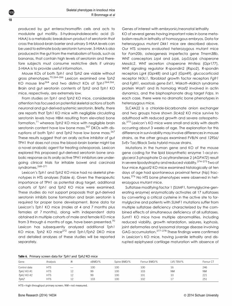
produced by gut enterochromaffin cells and acts to
modulate gut motility. 5-hydroxyindoleacetic acid (5-
HIAA) is ametabolic breakdown product of serotonin that
cross the blood-brain barrier and urinary 5-HIAA levels can
beused to estimate body serotonin turnover. 5-HIAA is also
produced in thegut from themetabolismof foods, suchas
bananas, that contain high levels of serotonin and there-
fore subjects must consume restrictive diets if urinary
5-HIAA is to provide useful information.
Mouse KOs of both Tph1 and Tph2 are viable without
gross phenotypes.70,264–266 Lexicon examined one Tph2
KO mouse line264 and two distinct KOs of Tph1264,266.
Brain and gut serotonin contents of Tph2 and Tph1 KO
mice, respectively, are extremely low.
From studies on Tph1 and Tph2 KO mice, considerable
attention has focusedonpotential skeletal actions of both
neuronal and gut-derived systemic serotonin. Briefly, there
are reports that Tph1 KO mice with negligible circulating
serotonin levels have HBM resulting from elevated bone
formation,71 whereas Tph2 KO mice with negligible brain
serotonin content have low bone mass.267 DKOs with dis-
ruptions of both Tph1 and Tph2 have low bone mass.267
These results suggest that an orally active inhibitor of gut
TPH1 that does not cross the blood–brain barrier might be
a novel anabolic agent for treating osteoporosis. Lexicon
explored this proposed gut-derived serotonin bone ana-
bolic response as its orally active TPH1 inhibitors are under-
going clinical trials for irritable bowel and carcinoid
syndromes.268–271
Lexicon’s Tph1 and Tph2 KO mice had no skeletal phe-
notypes in HTS analyses (Table 6). Given the therapeutic
importance of TPH1 as potential drug target, additional
cohorts of Tph1 and Tph2 KO mice were examined.
These studies do not support proposals that gut-derived
serotonin inhibits bone formation and brain serotonin is
required for proper bone development. Bone data for
Lexicon’s Tph1 KO mice (males at 4 and 7 months plus
females at 7 months), along with independent data
obtained in multiple cohorts of male and female KOmice
from 3 through 6 months of age, have been presented.70
Lexicon has subsequently analyzed additional Tph1
KO mice, Tph2 KO mice272 and Tph1/Tph2 DKO mice
and detailed analyses of these studies will be reported
separately.
Genes of interest with embryonic/neonatal lethality
KO of several genes having important roles in bonemeta-
bolism results in lethality of homozygous embryos. Data for
heterozygous mutant Dkk1 mice are described above.
Our HTS screens evaluated heterozygous mutant mice
for Fam20b, osteogenesis imperfecta gene Tmem38b,
WNT coreceptors Lrp4 and Lrp6, Lrp5/Lrp6 chaperone
Mesdc2, WNT secretion chaperone Wntless (Gpr177),
WNT signaling regulator R-spondin2 (Rspo2), R-spondin
receptors Lgr4 (Gpr48) and Lgr5 (Gpr49), glucocorticoid
receptor Nr3c1, fibroblast growth factor receptors Fgfr1
and Fgfrl1, exostosis gene Ext1, Wiskott–Aldrich syndrome
protein Wasf1 and its homolog Wasf2 involved in actin
dynamics, and the bisphosphonate drug target Fdps. In
each case, there were no dramatic bone phenotypes in
heterozygous mice.
SLC4A22 is a chloride-bicarbonate anion exchanger
and two groups have shown Slc4a2 KO mice survive to
adulthood with reduced growth and severe osteopetro-
sis.273 Lexicon’s KO mice were small and sickly with death
occurring about 3 weeks of age. The explanation for this
difference in survivabilitymay involvedifferences inmouse
strains, as the other groups examined FVB/N and 129S6/
SvEv Tac/Black Swiss hybrid mouse strains.
Mutations in the human gene and KO of the mouse
gene coding for the lipid biosynthetic enzyme 1-acyl-sn-
glycerol 3-phosphate O-acyltransferase 2 (AGPAT2) result
in severe lipodystrophy and reduced viability.274–275 Twoof
four mice Agpat2 KO mice examined histologically at ten
days of age had spontaneous proximal femur (hip) frac-
tures.276 No HTS bone phenotypes were observed in het-
erozygous mutant mice.
Sulfatasemodifying factor 1 (SUMF1, formylglycine-gen-
erating enzyme) enzymatically activates all 17 sulfatases
by converting a critical cysteine in the active site to for-
mylglycine and patients with SUMF1 mutations suffer from
multiple sulfatase deficiency characterized by the com-
bined effects of simultaneous deficiency of all sulfatases.
Sumf1 KO mice have multiple abnormalities, including
reduced viability, growth retardation, seizures, kyphosis,
joint deformaties and lysosomal storage disease involving
GAG accumulation.277–278 These findings were confirmed
in Lexicion’s KO mice, having juvenile lethality and dis-
rupted epiphyseal cartilage maturation with absence of
Table 6. Primary screen data for Tph1 and Tph2 KO mice
Gene Analysis N vBMD/% Spine BMD/% Femur BMD/% LV5 TBV/% Femur CT
Control data HTS — 100 100 100 16 246
Tph1 KO #1 HTS 12 99 100 103 NM NM
Tph1 KO #2 HTS 12 99 100 101 10 221
Tph2 HTS 12 103 100 102 17 251
HTS5high-throughput primary screen; NM5not measured.
Skeletal phenotypes in knockout mice
R Brommage et al
18
Bone Research (2014) 14034 � 2014 Sichuan University
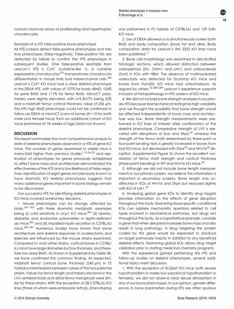
normal columnar arrays of proliferating and hypertrophic
chondrocytes.
Example of a HTS false-positive bone phenotype
All HTS screens detect false-positive phenotypes and miss
true phenotypes (false-negatives). False-positive hits are
detected by failure to confirm the HTS phenotype in
subsequent studies. One false-positive example from
Lexicon’s HTS is Cytl1 (cytokine-like 1), a cytokine
expressed in chondrocytes279 that promotes chondrocyte
differentiation in mouse limb bud mesenchymal cells.280
Lexicon’s Cytl1 KO mice had a clear skeletal phenotype
in the DEXA HTS, with values of 107% for body vBMD, 106%
for spine BMD and 111% for femur BMD. MicroCT para-
meters were slightly elevated, with LV5 BV/TV being 22%
and a midshaft femur cortical thickness value of 256 mm.
This HTS high BMD phenotype could not be confirmed in
follow-up DEXA or microCT scans of bones (N510 for both
male and female mice) from an additional cohort of KO
mice examined at 18 weeks of age (data not shown).
DISCUSSIONThis report summarizes themost comprehensive analysis to
date of skeletal phenotypes observed in a HTS of gene KO
mice. The number of genes examined in viable mice is
many-fold higher than previous studies1,10,24–25 and rep-
lication of phenotypes for genes previously established
to affect bone mass and architecture demonstrated the
effectiveness of the HTS anda low likelihood of false nega-
tives. Identification of eight genes not previously known to
have dramatic KO skeletal phenotypes suggests that
many additional genes important in bone biology remain
to be discovered.
Our successful HTS for identifying skeletal phenotypes in
KO mice involved several key decisions.
1. Mouse phenotypes can be strongly affected by
strain,281–283 with three dramatic metabolic examples
being (i) cold sensitivity in Ucp1 KO mice;284 (ii) obesity,
diabetes and endocrine parameters in leptin-deficient
ob mice;285 and (iii) impaired insulin secretion in C57BL/6J
mice.286–287 Numerous studies have shown that bone
architecture and skeletal responses to ovariectomy and
exercise are influenced by the mouse strains examined.
Compared to most other strains, cortical bones in C57BL/
6Jmicehave largediameters but low thickness, and there-
fore low areal BMD. As shown in Supplementary Table S8,
we have confirmed this common finding. As expected,
midshaft femur cortical bone thickness (245 mm) in F2
hybrids is intermediatebetweenvaluesof the twoparental
strains. Values for femur length and trabecular bone in the
LV5 vertebral body and distal femur metaphysis were sim-
ilar for these strains. With the exception of 28 C57BL/6J KO
lines (three of which were embryonic lethal), phenotyping
was performed in F2 hybrids of C57BL/6J and 129 SvEv
KO mice.
2. Use of DEXA allowed us to simultaneously screen both
BMD and body composition (body fat and LBM). Body
composition data for Lexicon’s first 2322 KO lines have
been published.11
3. Bone cell morphology was examined in decalcified
histologic sections, which allowed distinction between
osteopetrosis (Src, Ostm1 and Lrrk1) and osteosclerosis
(Sost) in KOs with HBM. The absence of multinucleated
osteoclasts was detected for Dcstamp KO mice and
bones from Fam20c KO mice had osteomalacia. As
argued by others,22,288–289 Lexicon’s experience supports
inclusion of histopathology in HTS screens of KO mice.
4.Wedid not includebone strength analyses in our prim-
aryHTSbecausebiomechanical testinghashighvariability
and we thought the possibility that bone strength would
be affected independently of bone mass and architec-
ture was low. Bone strength measurements were per-
formed in KO lines of interest after confirmation of the
skeletal phenotype. Compressive strength of LV5 is ele-
vated with disruptions of Sost and Sfrp4,30 whereas the
strength of the femur shaft determined by three-point or
four-point binding tests is greatly increased in bones from
Sost KOmice. but decreasedwith Sfrp430 andWnt1638 dis-
ruption. Supplemental Figure 3c shows the excellent cor-
relation of femur shaft strength and cortical thickness
(three-point bending) in WT and Wnt16 KO mice.38
5. Although we did not include bone length measure-
ments in our primary screen, we believe this information is
important in secondary screens. Bone length was un-
affected in KOs of Wnt16 and Sfrp4 but reduced slightly
with KO of Lrrk1.39
6. Analyzing global gene KOs to identify drug targets
provides information on the effects of gene disruption
throughout the body. Examining tissue-specific conditional
KOs can address mechanistic questions related to cell
types involved in biochemical pathways, but drugs act
throughout the body. As a hypothetical example, consider
a gene that when disrupted increases bonemass and also
results in lung pathology. A drug targeting the protein
coded by this gene would be expected to produce
on-target pulmonary toxicity in addition to any beneficial
skeletal effects. Examining global KOs allows drug target
validation prior to starting medicinal chemistry programs.
With the experience gained performing this HTS and
follow-up studies on skeletal phenotypes, several addi-
tional topics merit discussion.
1. With the exception of Slc26a7 KO mice (with severe
hypothyroidism in males but subclinical hypothyroidism in
females), we did not observe clear sexual dimorphism in
any of our bone phenotypes. In our opinion, gender differ-
ences in bone parameters during HTS are often spurious
Skeletal phenotypes in knockout miceR Brommage et al
19
� 2014 Sichuan University Bone Research (2014) 14034
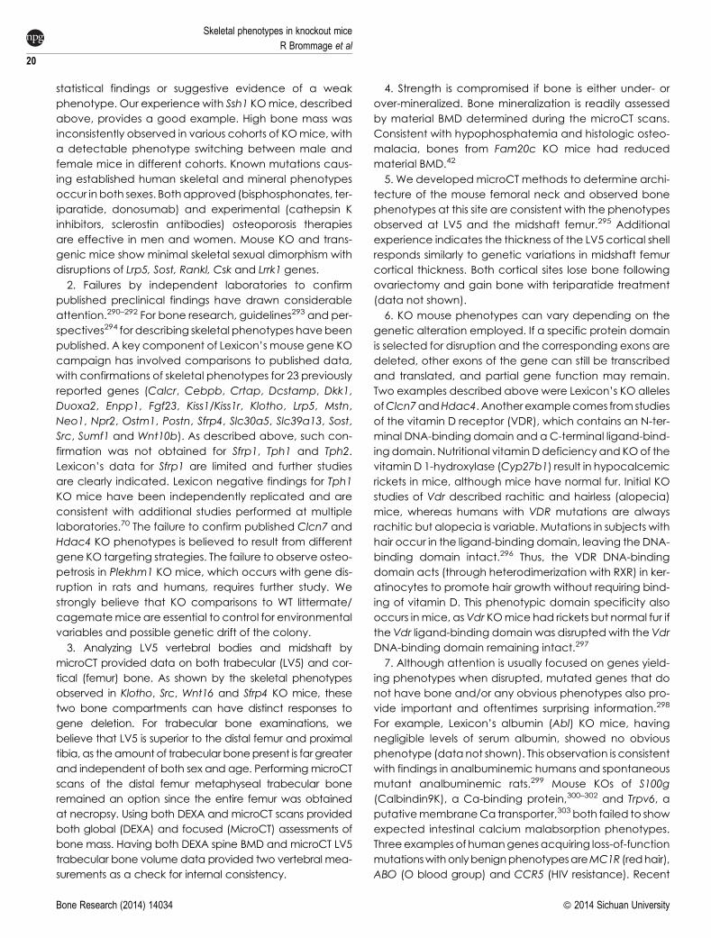
statistical findings or suggestive evidence of a weak
phenotype. Our experience with Ssh1 KOmice, described
above, provides a good example. High bone mass was
inconsistently observed in various cohorts of KOmice, with
a detectable phenotype switching between male and
female mice in different cohorts. Known mutations caus-
ing established human skeletal and mineral phenotypes
occur in both sexes. Bothapproved (bisphosphonates, ter-
iparatide, donosumab) and experimental (cathepsin K
inhibitors, sclerostin antibodies) osteoporosis therapies
are effective in men and women. Mouse KO and trans-
genic mice show minimal skeletal sexual dimorphism with
disruptions of Lrp5, Sost, Rankl, Csk and Lrrk1 genes.
2. Failures by independent laboratories to confirm
published preclinical findings have drawn considerable
attention.290–292 For bone research, guidelines293 and per-
spectives294 for describing skeletal phenotypes havebeen
published. A key component of Lexicon’s mouse gene KO
campaign has involved comparisons to published data,
with confirmations of skeletal phenotypes for 23 previously
reported genes (Calcr, Cebpb, Crtap, Dcstamp, Dkk1,
Duoxa2, Enpp1, Fgf23, Kiss1/Kiss1r, Klotho, Lrp5, Mstn,
Neo1, Npr2, Ostm1, Postn, Sfrp4, Slc30a5, Slc39a13, Sost,
Src, Sumf1 and Wnt10b). As described above, such con-
firmation was not obtained for Sfrp1, Tph1 and Tph2.
Lexicon’s data for Sfrp1 are limited and further studies
are clearly indicated. Lexicon negative findings for Tph1
KO mice have been independently replicated and are
consistent with additional studies performed at multiple
laboratories.70 The failure to confirm published Clcn7 and
Hdac4 KO phenotypes is believed to result from different
gene KO targeting strategies. The failure to observe osteo-
petrosis in Plekhm1 KO mice, which occurs with gene dis-
ruption in rats and humans, requires further study. We
strongly believe that KO comparisons to WT littermate/
cagematemice are essential to control for environmental
variables and possible genetic drift of the colony.
3. Analyzing LV5 vertebral bodies and midshaft by
microCT provided data on both trabecular (LV5) and cor-
tical (femur) bone. As shown by the skeletal phenotypes
observed in Klotho, Src, Wnt16 and Sfrp4 KO mice, these
two bone compartments can have distinct responses to
gene deletion. For trabecular bone examinations, we
believe that LV5 is superior to the distal femur and proximal
tibia, as theamount of trabecular bonepresent is far greater
and independent of both sex and age. PerformingmicroCT
scans of the distal femur metaphyseal trabecular bone
remained an option since the entire femur was obtained
at necropsy. Using both DEXA and microCT scans provided
both global (DEXA) and focused (MicroCT) assessments of
bone mass. Having both DEXA spine BMD and microCT LV5
trabecular bone volume data provided two vertebral mea-
surements as a check for internal consistency.
4. Strength is compromised if bone is either under- or
over-mineralized. Bone mineralization is readily assessed
by material BMD determined during the microCT scans.
Consistent with hypophosphatemia and histologic osteo-
malacia, bones from Fam20c KO mice had reduced
material BMD.42
5. We developed microCT methods to determine archi-
tecture of the mouse femoral neck and observed bone
phenotypes at this site are consistent with the phenotypes
observed at LV5 and the midshaft femur.295 Additional
experience indicates the thickness of the LV5 cortical shell
responds similarly to genetic variations in midshaft femur
cortical thickness. Both cortical sites lose bone following
ovariectomy and gain bone with teriparatide treatment
(data not shown).
6. KO mouse phenotypes can vary depending on the
genetic alteration employed. If a specific protein domain
is selected for disruption and the corresponding exons are
deleted, other exons of the gene can still be transcribed
and translated, and partial gene function may remain.
Two examples described above were Lexicon’s KO alleles
ofClcn7andHdac4. Another examplecomes from studies
of the vitamin D receptor (VDR), which contains an N-ter-
minal DNA-binding domain and aC-terminal ligand-bind-
ing domain. Nutritional vitaminDdeficiency and KOof the
vitamin D 1-hydroxylase (Cyp27b1) result in hypocalcemic
rickets in mice, although mice have normal fur. Initial KO
studies of Vdr described rachitic and hairless (alopecia)
mice, whereas humans with VDR mutations are always
rachitic but alopecia is variable. Mutations in subjects with
hair occur in the ligand-binding domain, leaving the DNA-
binding domain intact.296 Thus, the VDR DNA-binding
domain acts (through heterodimerization with RXR) in ker-
atinocytes to promote hair growth without requiring bind-
ing of vitamin D. This phenotypic domain specificity also
occurs inmice, asVdr KOmice had rickets but normal fur if
the Vdr ligand-binding domain was disruptedwith the Vdr
DNA-binding domain remaining intact.297
7. Although attention is usually focused on genes yield-
ing phenotypes when disrupted, mutated genes that do
not have bone and/or any obvious phenotypes also pro-
vide important and oftentimes surprising information.298
For example, Lexicon’s albumin (Abl) KO mice, having
negligible levels of serum albumin, showed no obvious
phenotype (datanot shown). This observation is consistent
with findings in analbuminemic humans and spontaneous
mutant analbuminemic rats.299 Mouse KOs of S100g
(Calbindin9K), a Ca-binding protein,300–302 and Trpv6, a
putativemembraneCa transporter,303 both failed to show
expected intestinal calcium malabsorption phenotypes.
Threeexamples of humangenesacquiring loss-of-function
mutationswithonlybenignphenotypesareMC1R (redhair),
ABO (O blood group) and CCR5 (HIV resistance). Recent
Skeletal phenotypes in knockout mice
R Brommage et al
20
Bone Research (2014) 14034 � 2014 Sichuan University
explorations of the human genome have identified healthy
people havinggeneswith predicted loss-of-function homo-
zygousmutations.304–306 Theoccurrenceofgenes in healthy
individuals having loss-of-functioncompoundheterozygous
alleles has been less thoroughly explored.
Genes can have critical functions only under environ-
mental conditions not occurring during standard mouse
phenotyping. Examples include (i) the GCN2 kinase
regulating feeding behavior in response to meals with
imbalances of essential amino acids;307 (ii) KO of themito-
chondrial uncoupling protein UCP1 influences body fat
depending on the age of the mice and the ambient tem-
perature at which they are housed;308 (iii) normal iron sta-
tus with KO of the iron-binding protein lactoferrin,309 but
elevated colorectal dysplasia following inflammation;310
and (iv) Gc KO mice without the serum vitamin D-binding
protein have normal serum bioactive vitamin D levels but
respond more quickly to dietary deficiency and show
hypercalcemic toxicity at lower vitamin D doses.311–312
Compared to sedentary KO mice, physical exercise
(treadmills and runningwheels) can uncover pathological
phenotypes inotherwisehealthyKOmice (Mybpc3, Lipe) or
prevent obesity in Mc4r KO mice.313 Thus, KO mouse lines
without skeletal phenotypes in HTS might show abnormal
disturbances in bone metabolism with increasing age or
following challenges such as ovariectomy, mechanical
loading or unloading, fracture healing, arthritis induction,
dietary deficiencies and reproduction.
8. Comparisons of skeletal phenotypes showed excel-
lent agreement for genes having mutations or SNPs in
humans and corresponding mouse KOs. Phenotype con-
cordance was observed for CRTAP, FAM20A, FAM20C,
GNPTAB, LRP5, NPR2, OSTM1, SLC39A13, SOST and
WNT16, with PLEKHM1 being the single exception.
9. During 2012, the IMPC estimated that,30% of mouse
KO lines are embryonic or perinatal lethal314 and increas-
ing efforts are being focused on examining embryos to
determine mechanisms involved in the developmental
defects.314–315 With 556 KO lines examined, the February
2014 IMPC update lists lethal (21%) and subviable (9%)
lines. Analyses of the MGI database found lethality in
23% of 6812 mouse KO genes curated316 and 2472 lethal
gene KOs (39%) among 6283 genes annotated to have
direct human orthologs.317 A comprehensive review
described birth trauma, respiratory failure, homeostasis
deficiencies and inability to suckle as common causes of
lethality during the first 24 h after birth.318 KO lethality is
influenced by mouse strain and can sometimes be res-
cued by simultaneous disruptions in different genes, as is
the case of Dkk1 KO survivability in the presence of Wnt3
heterozygosity described above.133–134 The lower 18%
lethality value observed by Lexicon might be related to
examining few genes coding for transcription factors
and structural proteins and hybrid vigor from studying
C57BL6/J–129SvEv hybrid mice. As described above,
Dkk1 heterozygous mutant mice and KO mice examined
prior to 12weeks of agedue to reduced viability can show
skeletal phenotypes (Fgf23, Klotho, Nppc, Npr2, Ostm1,
Sgpl1, Slc25a1, SLC30a10, Sumf1).
Mouse resources
There are many resources providing both KO mice and
phenotype data. During 2006, Francis Collins, Director of
the US NIH, stated ‘A graduate student shouldn’t spend a
year making a knockout that’s already been made. It’s
not a good use of resources’.319 There are many reviews
describing the evolution of the IKMC since the 2003
Banbury OrganizingConference.320 KOmouse genes with
available ES cells and/or cryopreserved sperm are com-
piled in the International Mouse Strain Resource, with a link
from the Mouse Genomic Informatics (MGI) website and
published links to centralized mouse repositories.321 The
Gene Expression Database322 is included within the MGI
resource. To facilitate tissue-specific gene KOs, most IKMC
lines include the cre driver system during the gene KO tar-
geting design and the MGI website maintains a CrePortal
providing up-to-date information. Underappreciated
complications of conditional cre-loxP excision include
unexpected activity in off-target tissues, mosaicism and
parent-of-origin effects.323–324 Problems with the osterix-
cre mouse line have been described.325–326
The first compendium of 263 published mouse KO pheno-
types appeared in 1995.327 The IMPC13 coodinates pheno-
type data from the IKMC, with 441 KO mouse lines fully
analyzed and 643 KO mouse lines under examination
(September 2014). Recent results include KOs of Lrrk1
andWnt16, both having skeletal phenotypes similar to those
describedby Lexicon. TheMGI includes phenotypedata for
251 NIH-sponsored Deltagen and Lexicon KO mouse lines.
Commentaries on genome-wide KO efforts involving
zebrafish,328 nematodes329 and yeast330 provide interest-
ing perspectives for mouse KO efforts. Information on rat
gene KOs is compiled in the Rat Genome Database.331
SUMMARYLexicon Pharmaceuticals, during the decade preceding
the initiation of the IKMC program, successfully employed
industrialized biology to generate and phenotype over
4 650 mouse gene knockout lines. The genes examined
were highly represented in the druggable genome, fore-
shadowing the Illuminating the Druggable Genome pro-
gram within the NIH Common Fund (http://commonfund.
nih.gov/idg/overview). Global KO strategies involved
both gene trapping and homologous recombination
technologies. Phenotyping screens included a battery of
tests in the areas of behavior, bone, cardiology, immuno-
Skeletal phenotypes in knockout miceR Brommage et al
21
� 2014 Sichuan University Bone Research (2014) 14034
logy, metabolism, oncology and ophthalmology, and
included serum chemistry, histopathology and a high fat
diet obesity challenge. Softwarewaswritten for an internal
database that tracked mice undergoing breeding and
moving through the phenotyping screens and allowed
investigators to view all phenotyping data for all mice.
Lexicon’s KO mouse campaign successfully identified
previously published and novel genes influencing bone
mass, architecture, strength and mineralization (the
Skeletome). Three of these novel genes (undisclosed)
code for enzymes or secreted proteins that are potential
osteoporosis drug targets.
Further review of the Lexicon dataset showed elevated
spine bonemass phenotypes in both primary and second-
ary screens for Slc37a2 (glucose-6-phosphate/phosphate
antiporter) and Slc41a2 (Mg transporter). Further studies
are required to fully characterize skeletal phenotypes in
these two gene KOs.
Competing interestsThe authors declare no conflicts of interest.
AcknowledgementsRamiro Ramirez-Solis, Dianne Markesich, Elizabeth Richter, Jeff Schrick andJames Piggott for HTS organization; Zheng-Zheng Shi, Laurie Minze, LuisFreay and Tim Wilkins for HTS DEXA and microCT scans; Ken Platt andKatherine Holt for molecular biology supervision; Robert Read, JuneWingert, Mary Thiel, Ryan Vance and others for histopathology; DawnBright for DKK1 antibody generation; Deon Doree for thyroxine assays;David Harris for FACS analysis; Wade Walke and William Sonnenberg forbioinformatics; Jay Mitchell and Larry Rodriquez for HTS databasemanagement.
References1 Moore MW. High-throughput gene knockouts and phenotyping in
mice. Ernst Schering Res Found Workshop 2005; 50: 27–44.2 InternationalMouseKnockout Consortium, Collins FS, Rossant J,Wurst
W. A mouse for all reasons. Cell 2007; 128: 9–13.3 Collins FS, Finnell RH, Rossant J. Wurst W. A new partner for the
International Knockout Mouse Consortium. Cell 2007; 129: 235.4 BradleyA,AnastassiadisK,AyadiA et al.Themammalian gene function
resource: the International Knockout Mouse Consortium. Mamm
Genome 2012; 23: 580–586.5 Schofield PN, Hoehndorf R, Gkoutos GV. Mouse genetic and
phenotypic resources for human genetics. Hum Mutat 2012; 33: 826–836.
6 Ramirez-Solis R, Ryder E, Houghton R, White, JK, Bottomley J. Large-scale mouse knockouts and phenotypes. Wiley Interdiscip Rev Syst Biol
Med 2012; 4: 547–563.7 Friddle CJ, Abuin A, Ramirez-Solis R et al. High-throughput mouse
knockouts provide a functional analysis of the genome. Cold Spring
Harb Symp Quant Biol 2003; 68: 311–315.8 Hansen GM,Markesich DC, Burnett MB et al. Large-scale gene trapping in
C57BL/6N mouse embryonic stem cells. Genome Res 2008; 18: 1670–1679.9 Clark HF, Gurney AL, Abaya E et al. The secreted protein discovery
initiative (SPDI), a large-scale effort to identify novel human secreted
and transmembrane proteins: a bioinformatics assessment. Genome Res
2003; 13: 2265–2270.10 Tang T, Li L, Tang J et al. A mouse knockout library for secreted and
transmembrane proteins. Nat Biotechnol 2010; 28: 749–775.11 Brommage R, Desai U, Revelli JP et al. High-throughput screening of
mouse knockout lines identifies true lean and obese phenotypes.Obesity
(Silver Spring) 2008; 16: 2362–2367.12 Brown SD, Moore MW. The International Mouse Phenotyping
Consortium: past and future perspectives on mouse phenotyping.Mammalian Genome 2012; 23: 632–640.
13 Koscielny G, Yaikhom G, Iyer V et al. The International MousePhenotyping Consortium Web Portal, a unified point of access forknockout mice and related phenotyping data. Nucleic Acids Res 2014;42: D802–D809.
14 Ayadi A, Birling MC, Bottomley J et al. Mouse large-scale phenotypinginitiatives: overview of the EuropeanMouseDisease Clinic (EUMODIC)and of the Wellcome Trust Sanger Institute Mouse Genetics Project.Mamm Genome 2012; 23: 600–610.
15 Beckers J, Wurst W, de Angelis MH. Towards better mouse models:enhanced genotypes, systemic phenotyping and envirotypemodelling. Nat Rev Genet 2009; 10: 371–380.
16 Brown MJ, Murray KA. Phenotyping of genetically engineered mice:humane, ethical, environmental, and husbandry issues. ILAR J 2006;47: 118–123.
17 Fuchs H, Lisse T, Hans W et al. Phenotypic characterization of mousemodels for bone-related diseases in the German Mouse Clinic. J
Musculoskelet Neuronal Interact 2008; 8: 13–14.18 Fuchs H, Gailus-Durner V, Neschen S et al. Innovations in phenotyping
of mouse models in the German Mouse Clinic. Mamm Genome 2012; 23:611–622.
19 Justice MJ. Removing the cloak of invisibility: phenotyping the mouse.Dis Models Mech 2008; 1: 109–112.
20 Murray KA. Issues to consider when phenotyping mutant mousemodels. Lab Anim (NY) 2002; 31: 25–29.
21 Schofield PN, Dubus P, Klein L et al. Pathology of the laboratory mouse:an International Workshop on Challenges for High ThroughputPhenotyping. Toxicol Pathol 2011; 39: 559–562.
22 Schofield PN, Vogel P, Gkoutos GV, Sundberg JP. Exploring theelephant: histopathology in high-throughput phenotyping of mutantmice. Dis Models Mech 2012; 5: 19–25.
23 Zeiss CJ, Ward JM, Allore HG. Designing phenotyping studies forgenetically engineered mice. Vet Pathol 2012; 49: 24–31.
24 Bassett JH, Gogakos A, White JK et al. Rapid-throughput skeletalphenotyping of 100 knockout mice identifies 9 new genes thatdetermine bone strength. PLoS Genet 2012; 8: e1002858.
25 White JK, Gerdin AK, Karp NA et al. Genome-wide generation andsystematic phenotyping of knockout mice reveals new roles for manygenes. Cell 2013; 154: 452–464.
26 Iwaniec UT, Wronski TJ, Liu J et al. PTH stimulates bone formation inmice deficient in Lrp5. J Bone Miner Res 2007; 22: 394–402.
27 Brommage R, Liu J, Suwanichkul A, Powell DR. High bone mass insclerostin-deficient knockout mice. J Musculoskelet Neuronal Interact
2006; 6: 392.28 Brommage R, Liu J, Revelli JP, Kirkpatrick LL, Powell DR. Gene
knockouts of Wnt10b and Wnt16 in mice result in low bone mass.Bone 2007; 40(Suppl 2): S187.
29 Brommage R, Liu J, Bright D et al. Elevated bone mass in mice treatedwith anti-Dickkopf-1 neutralizing antibodies. J Bone Miner Res 2010;25(Suppl 1): S81.
Skeletal phenotypes in knockout mice
R Brommage et al
22
Bone Research (2014) 14034 � 2014 Sichuan University
30 Brommage R, Liu J, Lee E. Elevated trabecular bone mass with reducedcortical bone thickness in sFRP4 knockout mice. J Bone Miner Res 2009;24(Suppl 1): S82.
31 Walke DW, Han C, Shaw J, Wann E, Zambrowicz B, Sands A. In vivodrug target discovery: identifying the best targets from the genome.CurrOpin Biotechnol 2001; 12: 626–631.
32 Zambrowicz BP, Sands AT. Knockouts model the 100 best-sellingdrugs—will theymodel the next 100?Nat RevDrugDiscov 2003; 2: 38–51.
33 Zambrowicz BP, Turner CA, Sands AT. Predicting drug efficacy:knockouts model pipeline drugs of the pharmaceutical industry. CurrOpin Pharmacol 2003; 3: 563–570.
34 Russ AP, Lampel S. The druggable genome: an update. Drug DiscovToday 2005; 10: 1607–1610.
35 Plewczynski D, Rychlewski L. Meta-basic estimates the size ofdruggable human genome. J Mol Model 2009; 15: 695–699.
36 NiemanBJ, BlankMC,RomanBB,HenkelmanRM,MillenKJ. If the skullfits: magnetic resonance imaging and microcomputed tomography forcombined analysis of brain and skull phenotypes in the mouse. PhysiolGenomics 2012; 44: 992–1002.
37 Medina-Gomez C, Kemp JP, Estrada K et al. Meta-analysis of genome-wide scans for total body BMD in children and adults reveals allelicheterogeneity and age-specific effects at the WNT16 locus. PLoS Genet2012; 8: e1002718.
38 Zheng HF, Tobias JH, Duncan E et al. WNT16 influences bone mineraldensity, cortical bone thickness, bone strength, and osteoporotic fracturerisk. PLoS Genet 2012; 8: e1002745.
39 Xing W, Liu J, Cheng S, Vogel P, Mohan S, Brommage R. Targeteddisruption of leucine-rich repeat kinase 1 but not leucine-rich repeatkinase 2 in mice causes severe osteopetrosis. J Bone Miner Res 2013; 28:1962–1974.
40 Linares GR, Brommage R, Powell DR et al.Claudin 18 is a novel negativeregulator of bone resorption and osteoclast differentiation. J Bone MinerRes 2012; 27: 1553–1565.
41 Tagliabracci VS, Pinna LA, Dixon JE. Secreted protein kinases. TrendsBiochem Sci 2013; 38: 121–130.
42 Vogel P,HansenGM,ReadRW et al.Amelogenesis imperfecta and otherbiomineralization defects in Fam20a and Fam20c null mice. Vet Pathol2012; 49: 998–1017.
43 Hla T, Dannenberg AJ. Sphingolipid signaling in metabolic disorders.Cell Metab 2012; 16: 420–434.
44 Lotinun S, Kiviranta R,Matsubara T et al.Osteoclast-specific cathepsinKdeletion stimulates S1P-dependent bone formation. J Clin Invest 2013;123: 666–681.
45 Vogel P, DonovielMS, ReadR et al. Incomplete inhibition of sphingosine1-phosphate lyase modulates immune system function yet preventsearly lethality and non-lymphoid lesions. PloS One 2009; 4: e4112.
46 Wattler S, KellyM,NehlsM.Construction of gene targeting vectors fromlambda KOS genomic libraries. Biotechniques 1999; 26: 1150–1156, 1158,1160.
47 Zambrowicz BP, Holt KH, Walke W, Kirkpatrick LL, Eberhart DE.Generation of transgenic animals. In: Metcalf B, Dillon S (ed.), Targetvalidation in drug discovery. New York: Elsevier Academic Press, 2006:3–25.
48 Abuin A, Hansen GM, Zambrowicz B. Gene trap mutagenesis. HandbExp Pharmacol 2007; 178: 129–147.
49 Atasoy D, Schoch S, Ho A et al. Deletion of CASK in mice is lethal andimpairs synaptic function. Proc Natl Acad Sci USA 2007; 104: 2525–2530.
50 Gale NW, Dominguez MG, Noguera I et al.Haploinsufficiency of delta-like 4 ligand results in embryonic lethality due tomajor defects in arterial
and vascular development. Proc Natl Acad Sci USA 2004; 101: 15949–15954.
51 Brommage R. Validation and calibration of DEXA body composition inmice. Am J Physiol Endocrinol Metab 2003; 285: E454–E459.
52 Rissanen JP, Suominen MI, Peng Z et al. Short-term changes in serumPINPpredict long-term changes in trabecular bone in the rat ovariectomymodel. Calcified Tissue Int 2008; 82: 155–161.
53 Anderson JM, van Itallie CM. Physiology and function of the tightjunction. Cold Spring Harb Perspect Biol 2009; 1: a002584.
54 Wongdee K, Pandaranandaka J, Teerapornpuntakit J et al. Osteoblastsexpress claudins and tight junction-associated proteins. Histochem CellBiol 2008; 130: 79–90.
55 Doty SB. Morphological evidence of gap junctions between bone cells.Calcif Tissue Int 1981; 33: 509–512.
56 Hayashi D, Tamura A, Tanaka H et al. Deficiency of claudin-18 causesparacellular H1 leakage, up-regulation of interleukin-1beta, andatrophic gastritis in mice. Gastroenterology 2012; 142: 292–304.
57 Alshbool FZ, Alarcon C, Wergedal J, Mohan S. A high-calcium dietfailed to rescue an osteopenia phenotype in claudin-18 knockout mice.Physiol Rep 2014; 2: e00200.
58 Wang X, Wang S, Li C et al. Inactivation of a novel FGF23 regulator,FAM20C, leads to hypophosphatemic rickets in mice. PLoS Genet 2012;8: e1002708.
59 Simpson MA, Hsu R, Keir LS et al.Mutations in FAM20C are associatedwith lethal osteosclerotic bonedysplasia (Raine syndrome), highlightinga crucial molecule in bone development. Am J Hum Genet 2007; 81:906–912.
60 Tagliabracci VS, Engel JL, Wen J et al. Secreted kinase phosphorylatesextracellular proteins that regulate biomineralization. Science 2012; 336:1150–1153.
61 Ishikawa HO, Xu A, Ogura E, Manning G, Irvine KD. The Rainesyndrome protein FAM20C is a Golgi kinase that phosphorylates bio-mineralization proteins. PLoS One 2012; 7: e42988.
62 Tagliabracci VS, Engel JL, Wiley SE et al. Dynamic regulation of FGF23by Fam20C phosphorylation, GalNAc-T3 glycosylation, and furinproteolysis. Proc Natl Acad Sci USA 2014; 111: 5520–5525.
63 Gong Y, Slee RB, Fukai N et al. LDL receptor-related protein 5 (LRP5)affects bone accrual and eye development. Cell 2001; 107: 513–523.
64 Kato M, Patel MS, Levasseur R et al. Cbfa1-independent decrease inosteoblast proliferation, osteopenia, and persistent embryonic eyevascularization in mice deficient in Lrp5, a Wnt coreceptor. J Cell Biol2002; 157: 303–314.
65 Holmen SL, Giambernardi TA, Zylstra CR et al. Decreased BMD andlimb deformities in mice carrying mutations in both Lrp5 and Lrp6. JBone Miner Res 2004; 19: 2033–2040.
66 Li X, OminskyMS, Niu QT et al. Targeted deletion of the sclerostin genein mice results in increased bone formation and bone strength. J BoneMiner Res 2008; 23: 860–869.
67 Kramer I, Loots GG, Studer A, Keller H, Kneissel M. Parathyroidhormone (PTH)-induced bone gain is blunted in SOST overexpressingand deficient mice. J Bone Miner Res 2010; 25: 178–189.
68 Sawakami K, Robling AG, Ai M et al. The Wnt co-receptor LRP5 isessential for skeletal mechanotransduction but not for the anabolicbone response to parathyroid hormone treatment. J Biol Chem 2006;281: 23698–23711.
69 Arantes HP, Barros, ER, Kunii I, Bilezikian JP, Lazaretti-Castro M.Teriparatide increases bone mineral density in a man withosteoporosis pseudoglioma. J Bone Miner Res 2011; 26: 2823–2826.
70 Cui Y, Niziolek PJ, MacDonald BT et al. Lrp5 functions in bone toregulate bone mass. Nat Med 2011; 17: 684–691.
Skeletal phenotypes in knockout miceR Brommage et al
23
� 2014 Sichuan University Bone Research (2014) 14034
71 Yadav VK, Ryu JH, Suda N et al. Lrp5 controls bone formationby inhibiting serotonin synthesis in theduodenum.Cell2008; 135: 825–837.
72 XuQ,Wang Y, DabdoubA et al.Vascular development in the retina andinner ear: control by Norrin and Frizzled-4, a high-affinity ligand-receptor pair. Cell 2004; 116: 883–895.
73 Ke J, Harikumar KG, Erice C et al. Structure and function of Norrin inassembly andactivation of a Frizzled 4-Lrp5/6 complex.GenesDev 2013;27: 2305–2319.
74 Lobov IB, Rao S, Carroll TJ et al.WNT7bmediates macrophage-inducedprogrammed cell death in patterning of the vasculature. Nature 2005;437: 417–421.
75 Xia CH, Yablonka-Reuveni Z, Gong X. LRP5 is required for vasculardevelopment in deeper layers of the retina. PloS One 2010; 5: e11676.
76 Paes KT, Wang E, Henze K et al. Frizzled 4 is required for retinalangiogenesis and maintenance of the blood–retina barrier. InvestOphthalmol Vis Sci 2011; 52: 6452–6461.
77 Marin I. TheParkinsondisease geneLRRK2: evolutionary and structuralinsights. Mol Biol Evol 2006; 23: 2423–2433.
78 Baptista MA, Dave KD, Sheth NP et al. A strategy for the generation,characterization and distribution of animal models by The Michael J.Fox Foundation for Parkinson’s Research. Dis Model Mech 2013; 6:1316–1324.
79 KimBJ, Koh JM, Lee SY et al. Plasma sphingosine 1-phosphate levels andthe risk of vertebral fracture in postmenopausal women. J ClinEndocrinol Metab 2012; 97: 3807–3814.
80 Pederson L, RuanM,Westendorf JJ, Khosla S, Oursler MJ. Regulation ofbone formation by osteoclasts involves Wnt/BMP signaling and thechemokine sphingosine-1-phosphate. Proc Natl Acad Sci USA 2008;105: 20764–20769.
81 Bagdanoff JT, Donoviel MS, Nouraldeen A et al. Inhibition ofsphingosine-1-phosphate lyase for the treatment of autoimmunedisorders. J Med Chem 2009; 52: 3941–3953.
82 Bagdanoff JT, Donoviel MS, Nouraldeen A et al. Inhibition ofsphingosine 1-phosphate lyase for the treatment of rheumatoidarthritis: discovery of (E)-1-(4-((1R,2S,3R)-1,2,3,4-tetrahydroxybutyl)-1H-imidazol-2-yl)ethanone oxime (LX2931) and (1R,2S,3R)-1-(2-(isoxazol-3-yl)-1H-imidazol-4-yl)butane-1,2,3,4-tetraol (LX2932). J MedChem 2010; 53: 8650–8662.
83 Kesavan C, Mohan S, Brommage R, Wergedal J. Wnt16 is an importantregulator of bone size and the periosteal bone formation response tomechanical loading. J Bone Miner Res 2013; 28(Suppl 1): S100.
84 Skrtic SM et al.WNT16 is a novel osteoblast-derived paracrine regulatorof osteoclastogenesis, cortical bone mass and fracture susceptibility.J Bone Miner Res 2013; 28(Suppl 1): S39.
85 Motyl KJ, Raetz M, Tekalur SA, Schwartz RC, McCabe LR. CCAAT/enhancer binding protein beta-deficiency enhances type 1 diabetic bonephenotype by increasing marrow adiposity and bone resorption. Am JPhysiol Regul Integr Comp Physiol 2011; 300: R1250–R1260.
86 Staiger J, Lueben MJ, Berrigan D et al. C/EBPbeta regulates bodycomposition, energy balance-related hormones and tumor growth.Carcinogenesis 2009; 30: 832–840.
87 Tominaga H, Maeda S, Hayashi M et al. CCAAT/enhancer-bindingprotein beta promotes osteoblast differentiation by enhancing Runx2activity with ATF4. Mol Biol Cell 2008; 19: 5373–5386.
88 Zanotti S, Stadmeyer L, Smerdel-Ramoya A, Durant, D, Canalis E.Misexpression of CCAAT/enhancer binding protein beta causesosteopenia. J Endocrinol 2009; 201: 263–274.
89 Morello R, Bertin TK, Chen Y et al. CRTAP is required for prolyl 3-hydroxylation and mutations cause recessive osteogenesis imperfecta.Cell 2006; 127: 291–304.
90 Baldridge D, Lennington J, Weis M et al. Generalized connective tissuedisease in Crtap–/– mouse. PLoS One 2010; 5: e10560.
91 Weber G, Rabbiosi S, Zamproni I, Fugazzola L. Genetic defects ofhydrogen peroxide generation in the thyroid gland. J Endocrinol Invest2013; 36: 261–266.
92 Johnson KR, Marden CC, Ward-Bailey P, Gagnon LH, Bronson RT,Donahue LR. Congenital hypothyroidism, dwarfism, and hearingimpairment caused by a missense mutation in the mouse dual oxidase2 gene, Duox2. Mol Endocrinol 2007; 21: 1593–1602.
93 Grasberger H, de Deken X,MayoOB et al.Mice deficient in dual oxidasematuration factors are severely hypothyroid. Mol Endocrinol 2012; 26:481–492.
94 Xing W, Govoni KE, Donahue LR et al. Genetic evidence that thyroidhormone is indispensable for prepubertal insulin-like growth factor-Iexpression and bone acquisition in mice. J Bone Miner Res 2012; 27:1067–1079.
95 Donko A, Ruisanchez E, Orient A et al. Urothelial cells producehydrogen peroxide through the activation of Duox1. Free Radic BiolMed 2010; 49: 2040–2048.
96 Mackenzie NC, Zhu D, Milne EM et al. Altered bone development andan increase in FGF-23 expression in Enpp1(–/–) mice. PloS One 2012; 7:e32177.
97 Koshizuka Y, Ikegawa S, Sano M, Nakamura K, Nakamura Y. Isolationof novel mouse genes associated with ectopic ossification by differentialdisplay method using ttw, a mouse model for ectopic ossification.Cytogenet Cell Genet 2001; 94: 163–168.
98 Babij P, Roudier M, Graves T et al.New variants in the Enpp1 and Ptpn6genes cause low BMD, crystal-related arthropathy, and vascularcalcification. J Bone Miner Res 2009; 24: 1552–1564.
99 LiQ,GuoH,ChouDW,BerndtA, Sundberg JP,Uitto J.Mutant Enpp1asjmice as a model for generalized arterial calcification of infancy. DisModel Mech 2013; 6: 1227–1235.
100 Martin A, David V, Quarles LD. Regulation and function of the FGF23/klotho endocrine pathways. Physiol Rev 2012; 92: 131–155.
101 Bhattacharyya N, Chong WH, Gafni RI, Collins MT. Fibroblast growthfactor 23: state of the field and future directions. Trends Endocrinol Metab2012; 23: 610–618.
102 Yamashita T, Yoshitake H, Tsuji K, Kawaguchi N, Nabeshima Y, NodaM. Retardation in bone resorption after bone marrow ablation in klothomutant mice. Endocrinology 2000; 141: 438–445.
103 Yuan Q, Sato T, Densmore M et al. Deletion of PTH rescues skeletalabnormalities and high osteopontin levels in Klotho–/– mice. PLoSGenet 2012; 8: e1002726.
104 Ellwanger K, SaitoH, Clement-Lacroix P et al.Targeted disruption of theWnt regulator Kremen induces limb defects and high bone density.MolCell Biol 2008; 28: 4875–4882.
105 Schulze J, Seitz S, Saito H et al.Negative regulation of bone formation bythe transmembraneWnt antagonist Kremen-2.PLoSOne 2010; 5: e10309.
106 Zhou Z, Xie J, Lee D et al. Neogenin regulation of BMP-inducedcanonical Smad signaling and endochondral bone formation. Dev Cell2010; 19: 90–102.
107 Contie S, Voorzanger-Rousselot N, Litvin J et al. Development of a newELISA for serum periostin: evaluation of growth-related changes andbisphosphonate treatment in mice. Calcif Tissue Int 2010; 87: 341–350.
108 Horiuchi K, Amizuka N, Takeshita S et al. Identification andcharacterization of a novel protein, periostin, with restricted expressionto periosteum and periodontal ligament and increased expression bytransforming growth factor beta. J Bone Miner Res 1999; 14: 1239–1249.
109 Bonnet N, Standley KN, Bianchi EN et al. The matricellular proteinperiostin is required for sost inhibition and the anabolic response to
Skeletal phenotypes in knockout mice
R Brommage et al
24
Bone Research (2014) 14034 � 2014 Sichuan University

mechanical loading and physical activity. J Biol Chem 2009; 284: 35939–35950.
110 Rios H, Koushik SV,Wang H et al. Periostin null mice exhibit dwarfism,incisor enamel defects, and an early-onset periodontal disease-likephenotype. Mol Cell Biol 2005; 25: 11131–11144.
111 Bonnet N, Gineyts E, Ammann P, Conway SJ, Garnero P, Ferrari S.Periostin deficiency increases bone damage and impairs injuryresponse to fatigue loading in adult mice. PLoS One 2013; 8: e78347.
112 Fukada T, Civic N, Furuichi T et al. The zinc transporter SLC39A13/ZIP13 is required for connective tissue development; its involvement inBMP/TGF-beta signaling pathways. PLoS One 2008; 3: e3642.
113 Giunta C, Elcioglu NH, Albrecht B et al. Spondylocheiro dysplastic formof the Ehlers–Danlos syndrome—an autosomal-recessive entity causedby mutations in the zinc transporter gene SLC39A13. Am J Hum Genet2008; 82: 1290–1305.
114 Shimada T, Kakitani M, Yamazaki Y et al. Targeted ablation of Fgf23demonstrates an essential physiological role of FGF23 in phosphate andvitamin D metabolism. J Clin Invest 2004; 113: 561–568.
115 Chalhoub N, Benachenhou N, Rajapurohitam V et al. Grey-lethalmutation induces severe malignant autosomal recessive osteopetrosisin mouse and human. Nat Med 2003; 9: 399–406.
116 Pata M, Heraud C, Vacher J. OSTM1 bone defect reveals an intercellularhematopoietic crosstalk. J Biol Chem 2008; 283: 30522–30530.
117 Lange PF, Wartosch L, Jentsch TJ, Fuhrmann JC. ClC-7 requires Ostm1as a beta-subunit to support bone resorption and lysosomal function.Nature 2006; 440: 220–223.
118 Chusho H, Tamura N, Ogawa Y et al.Dwarfism and early death in micelacking C-type natriuretic peptide. Proc Natl Acad Sci USA 2001; 98:4016–4021.
119 Kondo E, Yasoda A, Tsuji T et al. Skeletal analysis of the long boneabnormality (lbab/lbab) mouse, a novel chondrodysplastic C-typenatriuretic peptide mutant. Calcif Tissue Int 2012; 90: 307–318.
120 Tsuji T, Kunieda T. A loss-of-function mutation in natriuretic peptidereceptor 2 (Npr2) gene is responsible for disproportionate dwarfism incn/cn mouse. J Biol Chem 2005; 280: 14288–14292.
121 Sogawa C, Tsuji T, Shinkai Y, Katayama K, Kunieda T. Short-limbeddwarfism: slw is a new allele of Npr2 causing chondrodysplasia. J Hered2007; 98: 575–580.
122 Bartels CF, Bukulmez H, Padayatti P et al. Mutations in thetransmembrane natriuretic peptide receptor NPR-B impair skeletalgrowth and cause acromesomelic dysplasia, type Maroteaux. Am JHum Genet 2004; 75: 27–34.
123 MiuraK,NambaN, FujiwaraM et al.Anovergrowthdisorder associatedwith excessive production of cGMP due to a gain-of-function mutationof the natriuretic peptide receptor 2 gene. PLoS One 2012; 7: e42180.
124 Dauphinee SM, Eva MM, Yuki KE et al. Characterization of two ENU-induced mutations affecting mouse skeletal morphology. G3 (Bethesda)2013; 3: 1753–1758.
125 Moffatt P, Thomas G, Sellin K et al. Osteocrin is a specific ligand of thenatriuretic peptide clearance receptor thatmodulates bone growth. J BiolChem 2007; 282: 36454–36462.
126 Soriano P, Montgomery C, Geske R, Bradley A. Targeted disruption ofthe c-src proto-oncogene leads to osteopetrosis in mice. Cell 1991; 64:693–702.
127 Kubota T, Michigami T, Sakaguchi N et al. Lrp6 hypomorphic mutationaffects bone mass through bone resorption in mice and impairsinteraction with Mesd. J Bone Miner Res 2008; 23: 1661–1671.
128 Mani A, Radhakrishnan J, WangH et al. LRP6mutation in a family withearly coronary disease and metabolic risk factors. Science 2007; 315:1278–1282.
129 Babij P, Zhao W, Small C et al. High bone mass in mice expressing amutant LRP5 gene. J Bone Miner Res 2003; 18: 960–974.
130 Boyden LM,Mao J, Belsky J et al.High bone density due to amutation inLDL-receptor-related protein 5. N Engl J Med 2002; 346: 1513–1521.
131 MacDonald BT, Joiner DM, Oyserman SM et al. Bone mass is inverselyproportional to Dkk1 levels in mice. Bone 2007; 41: 331–339.
132 Morvan F, Boulukos K, Clement-Lacroix P et al.Deletion of a single alleleof the Dkk1 gene leads to an increase in bone formation and bonemass. JBone Miner Res 2006; 21: 934–945.
133 Morse A, Frank C, Kelly NH et al. Sclerostin null and dickkopf-1 nullmice show an increased response tomechanical loading. Proc Orthop ResSoc Annu Meet 2014; Poster Number 0567.
134 McDonald M, Morse M, Baldock P et al. Homozygous deletion ofDickkopf1 results in a high bone mass phenotype. J Bone Miner Res2010; 25(Suppl 1): S23–S24.
135 Li J, Sarosi I, Cattley RC et al.Dkk1-mediated inhibition ofWnt signalingin bone results in osteopenia. Bone 2006; 39: 754–766.
136 Fulciniti M, Tassone P, Hideshima T et al. Anti-DKK1 mAb (BHQ880)as a potential therapeutic agent for multiple myeloma. Blood 2009; 114:371–379.
137 Glantschnig H, Hampton RA, Lu P et al. Generation and selection ofnovel fully human monoclonal antibodies that neutralize Dickkopf-1(DKK1) inhibitory function in vitro and increase bone mass in vivo. JBiol Chem 2010; 285: 40135–40147.
138 Glantschnig H, Scott K, Hampton R et al. A rate-limiting role forDickkopf-1 in bone formation and the remediation of bone loss inmouse and primate models of postmenopausal osteoporosis by an experi-mental therapeutic antibody. J Pharmacol Exp Ther 2011; 338: 568–578.
139 Ke HZ, Richards WG, Li X, Ominsky MS. Sclerostin and Dickkopf-1 astherapeutic targets in bone diseases. Endocr Rev 2012; 33: 747–783.
140 Pozzi S, Fulciniti M, YanH et al. In vivo and in vitro effects of a novel anti-Dkk1 neutralizing antibody in multiple myeloma. Bone 2013; 53: 487–496.
141 Zhou F, Meng S, Song H, Claret FX. Dickkopf-1 is a key regulator ofmyeloma bone disease: opportunities and challenges for therapeuticintervention. Blood Rev 2013; 27: 261–267.
142 FanW, et al.Differential impact of Dkk1 neutralizing antibodies onWntsignaling and bone mineral density. J Bone Miner Res 2011; 26(Suppl 1):S117.
143 Li X, Grisanti M, Fan W et al. Dickkopf-1 regulates bone formation inyoung growing rodents and upon traumatic injury. J Bone Miner Res2011; 26: 2610–2621.
144 Mukhopadhyay M, Gorivodsky M, Shtrom S et al. Dkk2 plays anessential role in the corneal fate of the ocular surface epithelium.Development 2006; 133: 2149–2154.
145 Gage PJ, Qian M, Wu D, Rosenberg KI. The canonical Wnt signalingantagonist DKK2 is an essential effector of PITX2 function duringnormal eye development. Dev Biol 2008; 317: 310–324.
146 Li X, Liu P, Liu W et al. Dkk2 has a role in terminal osteoblastdifferentiation and mineralized matrix formation. Nat Genet 2005; 37:945–952.
147 Barrantes Idel B, Montero-Pedrazuela A, Guadano-Ferraz A, et al.Generation and characterization of dickkopf3 mutant mice. Mol CellBiol 2006; 26: 2317–2326.
148 Ouchi N, Higuchi A, Ohashi K et al. Sfrp5 is an anti-inflammatoryadipokine that modulates metabolic dysfunction in obesity. Science2010; 329: 454–457.
149 Mori H, Prestwich TC, Reid MA et al. Secreted frizzled-related protein 5suppresses adipocyte mitochondrial metabolism through WNTinhibition. J Clin Invest 2012; 122: 2405–2416.
Skeletal phenotypes in knockout miceR Brommage et al
25
� 2014 Sichuan University Bone Research (2014) 14034
150 Cox S, Smith L, Bogani D, Cheeseman M, Siggers P, Greenfield A.Sexually dimorphic expression of secreted frizzled-related (SFRP)genes in the developing mouse Mullerian duct. Mol Reprod Dev 2006;73: 1008–1016.
151 SatohW,GotohT, TsunematsuY,Aizawa S, ShimonoA. Sfrp1 and Sfrp2regulate anteroposterior axis elongation and somite segmentationduring mouse embryogenesis. Development 2006; 133: 989–999.
152 Morello R, Bertin TK, Schlaubitz S et al. Brachy-syndactyly caused byloss of Sfrp2 function. J Cell Physiol 2008; 217: 127–137.
153 Yao W, Cheng Z, Shahnazari M, Dai W, Johnson ML, Lane NE.Overexpression of secreted frizzled-related protein 1 inhibits boneformation and attenuates parathyroid hormone bone anabolic effects.J Bone Miner Res 2010; 25: 190–199.
154 Bodine PV, Zhao W, Kharode YP et al. The Wnt antagonist secretedfrizzled-related protein-1 is a negative regulator of trabecular boneformation in adult mice. Mol Endocrinol 2004; 18: 1222–1237.
155 Bodine PV, Seestaller-Wehr L, Kharode YP, Bex FJ, Komm BS. Boneanabolic effects of parathyroid hormone are blunted by deletion of theWnt antagonist secreted frizzled-related protein-1. J Cell Physiol 2007;210: 352–357.
156 Joesting MS, Cheever TR, Volzing KG et al. Secreted frizzled relatedprotein 1 is a paracrine modulator of epithelial branchingmorphogenesis, proliferation, and secretory gene expression in theprostate. Dev Biol 2008; 317: 161–173.
157 Gauger KJ, Bassa LM, Henchey EM et al. Mice deficient in sfrp1 exhibitincreased adiposity, dysregulated glucose metabolism, and enhancedmacrophage infiltration. PloS One 2013; 8: e78320.
158 Lories RJ, Peeters J, Bakker A et al.Articular cartilage and biomechanicalproperties of the long bones in Frzb-knockout mice. Arthritis Rheum2007; 56: 4095–4103.
159 Baker-Lepain JC, Lynch JA, Parimi N et al. Variant alleles of the Wntantagonist FRZB are determinants of hip shape and modify therelationship between hip shape and osteoarthritis. Arthritis Rheum2012; 64: 1457–1465.
160 Saito H, Wissmann C, Suzuki H et al. Deletion of the Wnt signalingantagonist Secreted frizzled related protein 4 (sFRP4) in mice inducesopposite bone formation phenotypes in trabecular and cortical bone. JBone Miner Res 2008; 23(Suppl 1) : S3.
161 Kao R, Louie A, Lu WD, Nissenson R. Differential regulation of corticaland trabecular bone by Secreted frizzled-related protein 4 (Sfrp4). J BoneMiner Res 2010; 25(Suppl 1): S425.
162 Saito H, et al. sFrp4 differentially regulates trabecular and cortical bonemass via canonical Wnt and Ror2-BMP-sclerostin signaling,respectively. J Bone Miner Res 2011; 26(Suppl 1): S50.
163 Kansara M, Tsang M, Kodjabachian L et al. Wnt inhibitory factor 1 isepigenetically silenced in human osteosarcoma, and targeted disruptionaccelerates osteosarcomagenesis inmice. J Clin Invest 2009; 119: 837–851.
164 Schaniel C, Sirabella D, Qiu J, Niu X, Lemischka IR, Moore KA. Wnt-inhibitory factor 1 dysregulation of the bone marrow niche exhaustshematopoietic stem cells. Blood 2011; 118: 2420–2429.
165 Cawthorn WP, Bree AJ, Yao Y et al. Wnt6, Wnt10a and Wnt10b inhibitadipogenesis and stimulate osteoblastogenesis through a beta-catenin-dependent mechanism. Bone 2012; 50: 477–489.
166 Mak W, Shao X, Dunstan CR, Seibel MJ, Zhou H. Biphasicglucocorticoid-dependent regulation of Wnt expression and itsinhibitors in mature osteoblastic cells. Calcif Tissue Int 2009; 85: 538–545.
167 Wend P,WendK, Krum SA,Miranda-Carboni GA. The role ofWNT10Bin physiology and disease. Acta Physiol (Oxf) 2012; 204: 34–51.
168 Stevens JR, Miranda-Carboni GA, Singer MA, Brugger SM, Lyons KM,Lane TF. Wnt10b deficiency results in age-dependent loss of bone mass
andprogressive reduction ofmesenchymal progenitor cells. J BoneMinerRes 2010; 25: 2138–2147.
169 Bennett CN, Longo KA, Wright WS et al. Regulation ofosteoblastogenesis and bone mass by Wnt10b. Proc Natl Acad Sci USA2005; 102: 3324–3329.
170 Bennett CN, Ouyang H, Ma YL et al. Wnt10b increases postnatal boneformation by enhancing osteoblast differentiation. J BoneMiner Res 2007;22: 1924–1932.
171 Terauchi M, Li JY, Bedi B et al. T lymphocytes amplify the anabolicactivity of parathyroid hormone through Wnt10b signaling. Cell Metab2009; 10: 229–240.
172 Kukita T, Wada N, Kukita A et al. RANKL-induced DC-STAMP isessential for osteoclastogenesis. J Exp Med 2004; 200: 941–946.
173 Yagi M, Miyamoto T, Sawatani Y et al. DC-STAMP is essential for cell–cell fusion in osteoclasts and foreign body giant cells. J Exp Med 2005;202: 345–351.
174 Miyamoto T. Regulators of osteoclast differentiation and cell–cell fusion.Keio J Med 2011; 60: 101–105.
175 Kollmann K, Pestka JM, Kuhn SC et al. Decreased bone formation andincreased osteoclastogenesis cause bone loss in mucolipidosis II. EMBOMol Med 2013; 5: 1871–1886.
176 GelfmanCM,Vogel P, Issa TM et al.Mice lacking alpha/beta subunits ofGlcNAc-1-phosphotransferase exhibit growth retardation, retinaldegeneration, and secretory cell lesions. Invest Ophthalmol Vis Sci 2007;48: 5221–5228.
177 Vogel P, Payne BJ, Read R et al. Comparative pathology of murinemucolipidosis types II and IIIC. Vet Pathol 2009; 46: 313–324.
178 Colledge WH. Transgenic mouse models to study Gpr54/kisspeptinphysiology. Peptides 2009; 30: 34–41.
179 Dungan Lemko HM, Elias CF. Kiss of the mutant mouse: howgenetically altered mice advanced our understanding of kisspeptin’srole in reproductive physiology. Endocrinology 2012; 153: 5119–5129.
180 Brommage R, QianN, Vogel P et al.Mice failing to undergo puberty dueto targeted inactivation of the GPR54 gene exhibit alterations in bodycomposition and reduced bonemass. J BoneMiner Res 2004; 19(Suppl 1):S93.
181 Hamrick MW, Samaddar T, Pennington C, McCormick J. Increasedmuscle mass with myostatin deficiency improves gains in bonestrength with exercise. J Bone Miner Res 2006; 21: 477–483.
182 ElkasrawyMN,HamrickMW.Myostatin (GDF-8) as a key factor linkingmuscle mass and bone structure. J Musculoskelet Neuronal Interact 2010;10: 56–63.
183 Mitchell AD,Wall RJ. In vivo evaluation of changes in body compositionof transgenic mice expressing the myostatin pro domain using dualenergy X-ray absorptiometry. Growth Dev Aging 2007; 70: 25–37.
184 Inoue K, Matsuda K, Itoh M et al. Osteopenia and male-specific suddencardiac death in mice lacking a zinc transporter gene, Znt5. Hum MolGenet 2002; 11: 1775–1784.
185 Dacquin R, Davey RA, Laplace C et al. Amylin inhibits bone resorptionwhile the calcitonin receptor controls bone formation in vivo. J Cell Biol2004; 164: 509–514.
186 Davey RA, Turner AG, McManus JF et al. Calcitonin receptor plays aphysiological role to protect against hypercalcemia inmice. J Bone MinerRes 2008; 23: 1182–1193.
187 Gazzerro E, Smerdel-Ramoya A, Zanotti S et al. Conditional deletion ofgremlin causes a transient increase in bone formation and bone mass. JBiol Chem 2007; 282: 31549–31557.
188 Canalis E, Parker K, Zanotti S. Gremlin1 is required for skeletaldevelopment and postnatal skeletal homeostasis. J Cell Physiol 2012;227: 269–277.
Skeletal phenotypes in knockout mice
R Brommage et al
26
Bone Research (2014) 14034 � 2014 Sichuan University
189 Cheung CL, Lau KS, Sham PC, Tan KC, Kung AW. Genetic variants inGREM2 are associated with bone mineral density in a southern Chinesepopulation. J Clin Endocrinol Metab 2013; 98: E1557–E1561.
190 Paternoster L, Lorentzon M, Lehtimaki T et al. Genetic determinants oftrabecular and cortical volumetric bone mineral densities and bonemicrostructure. PLoS Genet 2013; 9: e1003247.
191 Zuniga E, Rippen M, Alexander C, Schilling TF, Crump JG. Gremlin 2regulates distinct roles of BMP and Endothelin 1 signaling indorsoventral patterning of the facial skeleton. Development 2011; 138:5147–5156.
192 Vogel P, Liu J, Platt KA et al.Malformation of incisor teeth in Grem2–/–
mice. Vet Pathol 2014; [Epub ahead of print].193 Poli V, Balena R, Fattori E et al. Interleukin-6 deficient mice are protected
from bone loss caused by estrogen depletion. EMBO J 1994; 13: 1189–1196.
194 Dai J, Lin D, Zhang J et al. Chronic alcohol ingestion inducesosteoclastogenesis and bone loss through IL-6 in mice. J Clin Invest
2000; 106: 887–895.195 Jilka RL, Hangoc G, Girasole G et al. Increased osteoclast development
after estrogen loss: mediation by interleukin-6. Science 1992; 257: 88–91.196 Kimble RB, Bain S, Pacifici R. The functional block of TNF but not of IL-6
prevents bone loss in ovariectomized mice. J Bone Miner Res 1997; 12:935–941.
197 O’Brien CA, Jilka RL, FuQ, Stewart S,Weinstein RS,Manolagas SC. IL-6is not required for parathyroid hormone stimulation of RANKLexpression, osteoclast formation, and bone loss in mice. Am J Physiol
Endocrinol Metab 2005; 289: E784–E793.198 del FattoreA, Cappariello A, CapulliM et al.An experimental therapy to
improve skeletal growth and prevent bone loss in a mouse modeloverexpressing IL-6. Osteoporos Int 2014; 25: 681–692.
199 Grey A, Mitnick MA, Masiukiewicz U et al. A role for interleukin-6 inparathyroid hormone-induced bone resorption in vivo. Endocrinology1999; 140: 4683–4690.
200 Xu J, Song P, Nakamura S et al. Deletion of the chloride transporterslc26a7 causes distal renal tubular acidosis and impairs gastric acidsecretion. J Biol Chem 2009; 284: 29470–29479.
201 Spitzweg C,Morris JC. Genetics and phenomics of hypothyroidism andgoiter due to NIS mutations. Mol Cell Endocrinol 2010; 322: 56–63.
202 Everett LA, Belyantseva IA, Noben-Trauth K et al. Targeted disruptionof mouse Pds provides insight about the inner-ear defects encounteredin Pendred syndrome. Hum Mol Genet 2001; 10: 153–161.
203 Kim KX, Sanneman JD, Kim HM et al. Slc26a7 chloride channel activityand localization in mouse Reissner’s membrane epithelium. PLoS One
2014; 9: e97191.204 KhadeerMA, Sahu SN, BaiG,Abdulla S,GuptaA. Expression of the zinc
transporter ZIP1 in osteoclasts. Bone 2005; 37: 296–304.205 Kambe T, Geiser J, Lahner B, Salt DE, Andrews GK. Slc39a1 to 3
(subfamily II) Zip genes in mice have unique cell-specific functionsduring adaptation to zinc deficiency. Am J Physiol Regul Integr Comp
Physiol 2008; 294: R1474–R1481.206 Gilmour DT, Lyon GJ, Carlton MB et al. Mice deficient for the secreted
glycoprotein SPARC/osteonectin/BM40 develop normally but showsevere age-onset cataract formation and disruption of the lens. EMBO
J 1998; 17: 1860–1870.207 Norose K, Clark JI, SyedNA et al. SPARC deficiency leads to early-onset
cataractogenesis. Invest Ophthalmol Vis Sci 1998; 39: 2674–2680.208 Bradshaw AD, Graves DC, Motamed K, Sage EH. SPARC-null mice
exhibit increased adiposity without significant differences in overallbody weight. Proc Natl Acad Sci USA 2003; 100: 6045–6050.
209 Gruber HE, Sage EH, Norton HJ et al. Targeted deletion of the SPARCgene accelerates disc degeneration in the aging mouse. J Histochem
Cytochem 2005; 53: 1131–1138.210 Delany AM, AmlingM, Priemel M et al.Osteopenia and decreased bone
formation in osteonectin-deficient mice. J Clin Invest 2000; 105: 1325.211 Boskey AL, Moore DJ, Amling M, Canalis E, Delany AM. Infrared
analysis of the mineral and matrix in bones of osteonectin-null miceand their wildtype controls. J Bone Miner Res 2003; 18: 1005–1011.
212 Mansergh FC, Wells T, Elford C et al.Osteopenia in Sparc (osteonectin)-deficient mice: characterization of phenotypic determinants of femoralstrength and changes in gene expression. Physiol Genomics 2007; 32: 64–73.
213 BarrosNM,Hoac B,Neves RL et al.Proteolytic processing of osteopontinby PHEX and accumulation of osteopontin fragments in Hyp mousebone, the murine model of X-linked hypophosphatemia. J Bone Miner
Res 2013; 28: 688–699.214 Ishijima M, Rittling SR, Yamashita T et al. Enhancement of osteoclastic
bone resorption and suppression of osteoblastic bone formation inresponse to reduced mechanical stress do not occur in the absence ofosteopontin. J Exp Med 2001; 193: 399–404.
215 Boskey AL, Spevak L, Paschalis E, Doty SB, McKee MD. Osteopontindeficiency increases mineral content and mineral crystallinity in mousebone. Calcif Tissue Int 2002; 71: 145–154.
216 OnoN,Nakashima K, Rittling SR et al.Osteopontin negatively regulatesparathyroid hormone receptor signaling in osteoblasts. J Biol Chem 2008;283: 19400–19409.
217 Yoshitake H, Rittling SR, Denhardt DT, Noda M. Osteopontin-deficientmice are resistant to ovariectomy-induced bone resorption. Proc Natl
Acad Sci USA 1999; 96: 8156–8160.218 Harmey D, Johnson KA, Zelken J et al. Elevated skeletal osteopontin
levels contribute to the hypophosphatasia phenotype in Akp2(–/–)mice. J Bone Miner Res 2006; 21: 1377–1386.
219 Malaval L,Wade-Gueye NM, BoudiffaM et al. Bone sialoprotein plays afunctional role in bone formation and osteoclastogenesis. J Exp Med
2008; 205: 1145–1153.220 Thurner PJ, Chen CG, Ionova-Martin S et al. Osteopontin deficiency
increases bone fragility but preserves bonemass.Bone 2010; 46: 1564–1573.221 Novack DV, Faccio R. Osteoclast motility: putting the brakes on bone
resorption. Ageing Res Rev 2011; 10: 54–61.222 Sakai D, Tong HS, Minkin C. Osteoclast molecular phenotyping by
random cDNA sequencing. Bone 1995; 17: 111–119.223 Shuang F, Sun Y, Yang HH et al.Destrin deletion enhances the bone loss
in hindlimb suspended mice. Eur J Appl Physiol 2013; 113: 403–410.224 Niwa R,Nagata-Ohashi K, TakeichiM,MizunoK, Uemura T. Control of
actin reorganization by Slingshot, a family of phosphatases thatdephosphorylate ADF/cofilin. Cell 2002; 108: 233–246.
225 Blangy A, Touaitahuata H, Cres G, Pawlak G. Cofilin activation duringpodosome belt formation in osteoclasts. PLoS One 2012; 7: e45909.
226 Mizuno K. Signaling mechanisms and functional roles of cofilinphosphorylation and dephosphorylation. Cell Signal 2013; 25: 457–469.
227 Meng Y, Zhang Y, Tregoubov V et al. Abnormal spine morphology andenhanced LTP in LIMK-1 knockout mice. Neuron 2002; 35: 121–133.
228 Kawano T, Zhu M, Troiano N et al. LIM kinase 1 deficient mice havereduced bone mass. Bone 2013; 52: 70–82.
229 Collette NM, Yee CS, Murugesh D et al. Sost and its paralog Sostdc1coordinate digit number in aGli3-dependentmanner.Dev Biol 2013; 383:90–105.
230 Ellies DL, Economou A, Viviano B et al.Wise regulates bone depositionthrough genetic interactions with Lrp5. PLoS One 2014; 9: e96257.
Skeletal phenotypes in knockout miceR Brommage et al
27
� 2014 Sichuan University Bone Research (2014) 14034
231 Person AD, Beiraghi S, Sieben CM et al. WNT5A mutations in patientswith autosomal dominant Robinow syndrome.Dev Dyn 2010; 239: 327–337.
232 Yamaguchi TP, Bradley A, McMahon AP, Jones S. A Wnt5a pathwayunderlies outgrowth of multiple structures in the vertebrate embryo.Development 1999; 126: 1211–1223.
233 Lin M, Li L, Liu C et al. Wnt5a regulates growth, patterning, andodontoblast differentiation of developing mouse tooth. Dev Dyn 2011;240: 432–440.
234 Yang Y, Topol L, Lee H, Wu J. Wnt5a and Wnt5b exhibit distinctactivities in coordinating chondrocyte proliferation and differentiation.Development 2003; 130: 1003–1015.
235 MaedaK,KobayashiY,UdagawaN et al.Wnt5a-Ror2 signaling betweenosteoblast-lineage cells and osteoclast precursors enhancesosteoclastogenesis. Nat Med 2012; 18: 405–412.
236 AgalliuD, Takada S, Agalliu I,McMahonAP, Jessell TM.Motor neuronswith axial muscle projections specified by Wnt4/5 signaling. Neuron2009; 61: 708–720.
237 DeWitt MR, Chen P, Aschner M. Manganese efflux in Parkinsonism:insights from newly characterized SLC30A10 mutations. BiochemBiophys Res Commun 2013; 432: 1–4.
238 Alam I, Gray AK, Chu K et al. Generation of the first autosomaldominant osteopetrosis type II (ADO2) disease models. Bone 2014; 59:66–75.
239 Rajan I, Read R, Small, DL, Perrard J, Vogel P. An alternative splicingvariant in Clcn7–/– mice prevents osteopetrosis but not neural andretinal degeneration. Vet Pathol 2011; 48: 663–675.
240 Kornak U, Kasper D, Bosl MR et al. Loss of the ClC-7 chloride channelleads to osteopetrosis in mice and man. Cell 2001; 104: 205–215.
241 Neutzsky-Wulff AV, Karsdal MA, Henriksen K. Characterization of thebone phenotype in ClC-7-deficient mice. Calcif Tissue Int 2008; 83: 425–437.
242 Vega RB, Matsuda K, Oh J et al. Histone deacetylase 4 controlschondrocyte hypertrophyduring skeletogenesis.Cell 2004; 119: 555–566.
243 Arnold MA, Kim Y, Czubryt MP et al. MEF2C transcription factorcontrols chondrocyte hypertrophy and bone development. Dev Cell2007; 12: 377–389.
244 Rajan I, Savelieva KV, Ye GL et al. Loss of the putative catalytic domainof HDAC4 leads to reduced thermal nociception and seizures whileallowing normal bone development. PloS One 2009; 4: e6612.
245 van Wesenbeeck L, Odgren PR, Coxon FP et al. Involvement ofPLEKHM1 in osteoclastic vesicular transport and osteopetrosis inincisors absent rats and humans. J Clin Invest 2007; 117: 919–930.
246 del Fattore A, Fornari R, Van Wesenbeeck L et al. A new heterozygousmutation (R714C) of the osteopetrosis gene, pleckstrin homolog domaincontaining familyM (with run domain) member 1 (PLEKHM1), impairsvesicular acidification and increases TRACP secretion in osteoclasts. JBone Miner Res 2008; 23: 380–391.
247 Vogel P, Read RW, Hansen GM et al. Congenital hydrocephalus ingenetically engineered mice. Vet Pathol 2012; 49: 166–181.
248 Ueda Y, Yamaguchi R, Ikawa M et al. PGAP1 knock-out mice showotocephaly and male infertility. J Biol Chem 2007; 282: 30373–30380.
249 McKean DM, Niswander L. Defects in GPI biosynthesis perturb Criptosignaling during forebrain development in two new mouse models ofholoprosencephaly. Biol Open 2012; 1: 874–883.
250 Park J, Kang SI, Lee SY et al. Tumor suppressor ras association domainfamily 5 (RASSF5/NORE1) mediates death receptor ligand-inducedapoptosis. J Biol Chem 2010; 285: 35029–35038.
251 Kondo Y, Inai Y, Sato Y et al. Senescence marker protein 30 functionsas gluconolactonase in L-ascorbic acid biosynthesis, and its
knockout mice are prone to scurvy. Proc Natl Acad Sci USA 2006; 103:5723–5728.
252 MaedaN,HagiharaH,NakataY et al.Aorticwall damage inmice unableto synthesize ascorbic acid. Proc Natl Acad Sci USA 2000; 97: 841–846.
253 Kawai T, Nishikimi M, Ozawa T, Yagi K. A missense mutation ofL-gulono-gamma-lactone oxidase causes the inability of scurvy-proneosteogenic disorder rats to synthesize L-ascorbic acid. J Biol Chem 1992;267: 21973–21976.
254 Beamer WG, Rosen CJ, Bronson RT et al. Spontaneous fracture (sfx): amouse genetic model of defective peripubertal bone formation. Bone2000; 27: 619–626.
255 Mohan S, Kapoor A, Singgih A et al. Spontaneous fractures in themousemutant sfx are caused by deletion of the gulonolactone oxidase gene,causing vitamin C deficiency. J Bone Miner Res 2005; 20: 1597–1610.
256 Gabbay KH, Bohren KM, Morello R, Bertin T, Liu J, Vogel P. Ascorbatesynthesis pathway: dual role of ascorbate in bone homeostasis. J BiolChem 2010; 285: 19510–19520.
257 Opsahl Vital S, Gaucher C, Bardet C et al. Tooth dentin defects reflectgenetic disorders affecting bone mineralization. Bone 2012; 50: 989–997.
258 Foster BL, Ramnitz MS, Gafni RI et al. Rare bone diseases and theirdental, oral, and craniofacial manifestations. J Dent Res 2014; 93: 7S–19S.
259 Al-Douri SM, Johnson DR. The development of the teeth of themicrophthalmic (mi/mi) mouse. J Anat 1987; 153: 139–149.
260 Ma D, Zhang R, Sun Y et al. A novel role of periostin in postnatal toothformation and mineralization. J Biol Chem 2011; 286: 4302–4309.
261 Kassai Y, Munne P, Hotta Y et al. Regulation of mammalian tooth cusppatterning by ectodin. Science 2005; 309: 2067–2070.
262 Ahn Y, Sanderson BW, Klein OD, Krumlauf R. Inhibition of Wntsignaling by Wise (Sostdc1) and negative feedback from Shh controlstooth number and patterning. Development 2010; 137: 3221–3231.
263 Miyata A, Baba O, Oda T, Ishikawa I, Takano Y. Diverse effects of c-srcdeficiency onmolar tooth development and eruption inmice.ArchHistolCytol 2007; 70: 63–78.
264 Savelieva KV, Zhao S, Pogorelov VM et al. Genetic disruption of bothtryptophan hydroxylase genes dramatically reduces serotonin andaffects behavior in models sensitive to antidepressants. PloS One 2008;3: e3301.
265 Walther DJ, Peter JU, Bashammakh S et al. Synthesis of serotonin by asecond tryptophan hydroxylase isoform. Science 2003; 299: 76.
266 Liu Q, Yang Q, Sun W et al. Discovery and characterization of noveltryptophan hydroxylase inhibitors that selectively inhibit serotoninsynthesis in the gastrointestinal tract. J Pharmacol Exp Ther 2008; 325:47–55.
267 Yadav VK, Oury F, Suda N et al. A serotonin-dependent mechanismexplains the leptin regulation of bone mass, appetite, and energyexpenditure. Cell 2009; 138: 976–989.
268 Shi ZC, Devasagayaraj A, GuK et al.Modulation of peripheral serotoninlevels by novel tryptophan hydroxylase inhibitors for the potentialtreatment of functional gastrointestinal disorders. J Med Chem 2008;51: 3684–3687.
269 Jin H, Cianchetta G, Devasagayaraj A et al. Substituted 3-(4-(1,3,5-triazin-2-yl)-phenyl)-2-aminopropanoic acids as novel tryptophanhydroxylase inhibitors. Bioorg Med Chem Lett 2009; 19: 5229–5232.
270 Cianchetta G, Stouch T, Yu W et al. Mechanism of inhibition of noveltryptophan hydroxylase inhibitors revealed by co-crystal structures andkinetic analysis. Curr Chem Genomics 2010; 4: 19–26.
271 Brown PM, Drossman DA, Wood AJ et al. The tryptophan hydroxylaseinhibitor LX1031 shows clinical benefit in patients with nonconstipatingirritable bowel syndrome. Gastroenterology 2011; 141: 507–516.
Skeletal phenotypes in knockout mice
R Brommage et al
28
Bone Research (2014) 14034 � 2014 Sichuan University
272 Brommage R, Smith DD, Liu J, Yu W, Yang QM, Powell DR. Geneticdisruption of brain serotonin content in mice produces leanness withhyperphagia and elevated physical activity but minimal skeletalchanges. F1000Posters 2014; 5: 1029.
273 Wu J, Glimcher LH, Aliprantis AO. HCO3-/Cl- anion exchangerSLC4A2 is required for proper osteoclast differentiation and function.Proc Natl Acad Sci USA 2008; 105: 16934–16939.
274 Jansen ID, Mardones P, Lecanda F et al. Ae2(a,b)-deficient mice exhibitosteopetrosis of long bones but not of calvaria. FASEB J 2009; 23: 3470–3481.
275 Cortes VA, Curtis DE, Sukumaran S et al. Molecular mechanisms ofhepatic steatosis and insulin resistance in the AGPAT2-deficientmouse model of congenital generalized lipodystrophy. Cell Metab2009; 9: 165–176.
276 Vogel P, Read R, Hansen G et al. Pathology of congenital generalizedlipodystrophy in Agpat2–/– mice. Vet Pathol 2011; 48: 642–654.
277 Settembre C, Annunziata I, Spampanato C et al. Systemic inflammationand neurodegeneration in a mouse model of multiple sulfatasedeficiency. Proc Natl Acad Sci USA 2007; 104: 4506–4511.
278 Settembre C, Arteaga-Solis E, McKee M et al. Proteoglycan desulfationdetermines the efficiency of chondrocyte autophagy and the extent ofFGF signaling during endochondral ossification. Genes Dev 2008; 22:2645–2650.
279 Kim JS, Ryoo ZY, Chun JS. Cytokine-like 1 (Cytl1) regulates the chon-drogenesis of mesenchymal cells. J Biol Chem 2007; 282: 29359–29367.
280 Jeon J, OhH, Lee G et al.Cytokine-like 1 knock-out mice (Cytl1–/–) shownormal cartilage and bone development but exhibit augmentedosteoarthritic cartilage destruction. J Biol Chem 2011; 286: 27206–27213.
281 Doetschman T. Influence of genetic background on geneticallyengineered mouse phenotypes. Methods Mol Biol 2009; 530: 423–433.
282 Matthaei KI. Identification of therapeutic drug targets throughgenetically manipulated mice: are we getting it right? Pharmacol Ther2009; 123: 32–36.
283 Keane TM, Goodstadt L, Danecek P et al.Mouse genomic variation andits effect on phenotypes and gene regulation. Nature 2011; 477: 289–294.
284 Hofmann WE, Liu X, Bearden CM, Harper ME, Kozak LP. Effects ofgenetic background on thermoregulation and fatty acid-induceduncoupling of mitochondria in UCP1-deficient mice. J Biol Chem 2001;276: 12460–12465.
285 Qiu J, Ogus S,Mounzih K, Ewart-TolandA, Chehab FF. Leptin-deficientmice backcrossed to the BALB/cJ genetic background have reducedadiposity, enhanced fertility, normal body temperature, and severediabetes. Endocrinology 2001; 142: 3421–3425.
286 Toye AA, Lippiat JD, Proks P et al. A genetic and physiological study ofimpaired glucose homeostasis control in C57BL/6J mice. Diabetologia2005; 48: 675–686.
287 Freeman HC, Hugill A, Dear NT, Ashcroft FM, Cox RD. Deletion ofnicotinamide nucleotide transhydrogenase: a new quantitive traitlocus accounting for glucose intolerance in C57BL/6J mice. Diabetes2006; 55: 2153–2156.
288 Scudamore CL. Integrating pathology into human disease modelling--how to eat the elephant. Dis Model Mech 2014; 7: 495–497.
289 Adissu HA, Estabel J, Sunter D et al.Histopathology reveals correlativeand unique phenotypes in a high-throughput mouse phenotypingscreen. Dis Model Mech 2014; 7: 515–524.
290 Prinz F, Schlange T,AsadullahK. Believe it or not: howmuch canwe relyon published data on potential drug targets? Nat Rev Drug Discov 2011;10: 712.
291 Begley CG, Ellis LM. Drug development: Raise standards for preclinicalcancer research. Nature 2012; 483: 531–533.
292 Collins FS, Tabak LA. Policy: NIH plans to enhance reproducibility.Nature 2014; 505: 612–613.
293 Boskey AL, van der Meulen MC, Wright TM. Guidelines for describingmouse skeletal phenotype. J Orthop Res 2003; 21: 1–5.
294 Manolagas SC, Kronenberg HM. Reproducibility of results in pre-clinical studies: a perspective from the bone field. J Bone Miner Res2014; 29: 2131–2140.
295 Brommage R, Jeter-Jones S, Xiong WC, Champ R, Liu J. Mouse femoralneck architecture determined by microCT reflects skeletal architectureobserved at other bone sites. J Bone Miner Res 2013; 28(Suppl 1): S347–S348.
296 Malloy PJ, Feldman D. The role of vitamin D receptor mutations in thedevelopment of alopecia. Mol Cell Endocrinol 2011; 347: 90–96.
297 DemayMB,MacDonald PN, Skorija K, DowdDR, Cianferotti L, CoxM.Role of the vitamin D receptor in hair follicle biology. J Steroid BiochemMol Biol 2007; 103: 344–346.
298 Barbaric I, Miller G, Dear TN. Appearances can be deceiving:phenotypes of knockout mice. Brief Funct Genomic Proteomic 2007; 6:91–103.
299 Nagase S, Shimamune K, Shumiya S. Albumin-deficient rat mutant.Science 1979; 205: 590–591.
300 Kutuzova GD, Akhter S, Christakos S, Vanhooke J, Kimmel-Jehan C,Deluca HF. Calbindin D(9k) knockout mice are indistinguishable fromwild-typemice in phenotype and serum calcium level. ProcNatl Acad SciUSA 2006; 103: 12377–12381.
301 Lee GS, Lee KY, Choi KC et al. Phenotype of a calbindin-D9k geneknockout is compensated for by the induction of other calciumtransporter genes in a mousemodel. J BoneMiner Res 2007; 22: 1968–1978.
302 Akhter S, Kutuzova GD, Christakos S, DeLuca HF. Calbindin D9k is notrequired for 1,25-dihydroxyvitamin D3-mediated Ca21 absorption insmall intestine. Arch Biochem Biophys 2007; 460: 227–232.
303 KutuzovaGD, Sundersingh F, Vaughan J et al. TRPV6 is not required for1alpha,25-dihydroxyvitamin D3-induced intestinal calcium absorptionin vivo. Proc Natl Acad Sci USA 2008; 105: 19655–19659.
304 Yngvadottir B, Xue Y, Searle S et al. A genome-wide survey of theprevalence and evolutionary forces acting on human nonsense SNPs.Am J Hum Genet 2009; 84: 224–234.
305 MacArthur DG, Balasubramanian S, Frankish A et al. A systematicsurvey of loss-of-function variants in human protein-coding genes.Science 2012; 335: 823–828.
306 LimET,Wurtz P, HavulinnaAS et al.Distribution andmedical impact ofloss-of-function variants in the finnish founder population. PLoS Genet2014; 10: e1004494.
307 Maurin AC, Jousse C, Averous J et al. The GCN2 kinase biases feedingbehavior to maintain amino acid homeostasis in omnivores. Cell Metab2005; 1: 273–277.
308 Feldmann HM, Golozoubova V, Cannon B, Nedergaard J. UCP1ablation induces obesity and abolishes diet-induced thermogenesis inmice exempt from thermal stress by living at thermoneutrality. CellMetab 2009; 9: 203–209.
309 Ward PP, Mendoza-Meneses M, Cunningham GA, Conneely OM. Ironstatus in mice carrying a targeted disruption of lactoferrin.Mol Cell Biol2003; 23: 178–185.
310 Ye Q, Zheng Y, Fan S et al. Lactoferrin deficiency promotes colitis-associated colorectal dysplasia in mice. PloS One 2014; 9: e103298.
311 Safadi FF, Thornton P, Magiera H et al. Osteopathy and resistance tovitamin D toxicity in mice null for vitamin D binding protein. J ClinInvest 1999; 103: 239–251.
312 Zella LA, Shevde NK, Hollis BW, Cooke NE, Pike JW. Vitamin D-binding protein influences total circulating levels of 1,25-
Skeletal phenotypes in knockout miceR Brommage et al
29
� 2014 Sichuan University Bone Research (2014) 14034
dihydroxyvitamin D3 but does not directly modulate the bioactivelevels of the hormone in vivo. Endocrinology 2008; 149: 3656–3667.
313 Booth FW, LayeMJ. Lack of adequate appreciation of physical exercise’scomplexities can pre-empt appropriate design and interpretation inscientific discovery. J Physiol 2009; 587: 5527–5539.
314 Adams D, Baldock R, Bhattacharya S et al. Bloomsbury report onmouse embryo phenotyping: recommendations from the IMPCworkshop on embryonic lethal screening.DisModel Mech 2013; 6: 571–579.
315 Mohun T, Adams DJ, Baldock R et al. Deciphering the Mechanisms ofDevelopmental Disorders (DMDD): a new programme for phenotypingembryonic lethal mice. Dis Model Mech 2013; 6: 562–566.
316 Kitsios GD, TangriN, Castaldi PJ, Ioannidis JP. Laboratorymousemodelsfor the human genome-wide associations. PLoS One 2010; 5: e13782.
317 Georgi B, Voight BF, Bucan M. From mouse to human: evolutionarygenomics analysis of human orthologs of essential genes. PLoS Genet
2013; 9: e1003484.318 Turgeon B,Meloche S. Interpreting neonatal lethal phenotypes inmouse
mutants: insights into gene function and human diseases. Physiol Rev2009; 89: 1–26.
319 Grimm D. Mouse genetics. A mouse for every gene. Science 2006; 312:1862–1866.
320 Austin CP, Battey JF, Bradley A et al. The knockout mouse project. Nat
Genet 2004; 36: 921–924.321 Donahue LR, Hrabe de Angelis M, Hagn M et al. Centralized mouse
repositories. Mamm Genome 2012; 23: 559–571.322 Smith CM, Finger JH, Kadin JA, Richardson JE, Ringwald M. The gene
expression database for mouse development (GXD): Puttingdevelopmental expression information at your fingertips. Dev Dyn
2014; 243: 1176–1186.323 Matthaei KI. Genetically manipulated mice: a powerful tool with
unsuspected caveats. J Physiol 2007; 582: 481–488.
324 Heffner CS, Herbert Pratt C, Babiuk RP et al. Supporting conditionalmouse mutagenesis with a comprehensive cre characterizationresource. Nat Commun 2012; 3: 1218.
325 Liu Y, Strecker S, Wang L et al. Osterix-cre labeled progenitor cellscontribute to the formation and maintenance of the bone marrowstroma. PLoS One 2013; 8: e71318.
326 Chen J, Shi Y, Regan J, Karuppaiah K, Ornitz DM, Long F et al.Osx-Cretargets multiple cell types besides osteoblast lineage in postnatal mice.PLoS One 2014; 9: e85161.
327 Brandon EP, Idzerda RL, McKnight GS. Knockouts. Targeting themouse genome: a compendium of knockouts (Part I). Curr Biol 1995; 5:625–634.
328 Kettleborough RN, Busch-Nentwich EM, Harvey SA et al. A systematicgenome-wide analysis of zebrafish protein-coding gene function.Nature
2013; 496: 494–497.329 The C. elegans Deletion Mutant Consortium. Large-scale screening for
targeted knockouts in the Caenorhabditis elegans genome.G3 (Bethesda)2012; 2: 1415–1425.
330 Giaever G, Nislow C. The yeast deletion collection: a decade offunctional genomics. Genetics 2014; 197: 451–465.
331 Laulederkind SJ, Hayman GT, Wang SJ et al. The Rat Genome Database2013—data, tools and users. Brief Bioinform 2013; 14: 520–526.
This work is licensed under a Creative Commons Attribution-NonCommercial-NoDerivs 3.0 Unported License. The images or
other third party material in this article are included in the article’s CreativeCommons license, unless indicated otherwise in the credit line; if thematerial is notincluded under the Creative Commons license, users will need to obtainpermission from the license holder to reproduce thematerial. To view a copy of thislicense, visit http://creativecommons.org/licenses/by-nc-nd/3.0/
Supplemental Information for this article can be found on the Bone Research website (http://www.nature.com/boneres).
Skeletal phenotypes in knockout mice
R Brommage et al
30
Bone Research (2014) 14034 � 2014 Sichuan University