Nanomaterials in the Environment: From Materials to High-Throughput Screening to Organisms
17
1 © 2012 Wiley-VCH Verlag GmbH & Co. KGaA, Weinheim wileyonlinelibrary.com Photoactive Nanomaterials Emerging In Vitro Models for Safety Screening of High-Volume Production Nanomaterials under Environmentally Relevant Exposure Conditions Mustafa Hussain Kathawala, Sijing Xiong, Mark Richards, Kee Woei Ng, Saji George,* and Say Chye Joachim Loo* The rising production of nanomaterial-based consumer products has raised safety concerns. Testing these with animal and other direct models is neither ethically nor economically viable, nor quick enough. This review aims to discuss the strength of in vitro testing, including the use of 2D and 3D cultures, stem cells, and tissue constructs, etc., which would give fast and repeatable answers of a highly specific nature, while remaining relevant to in vivo outcomes. These results can then be combined and the overall toxicity predicted with relative accuracy. Such in vitro models can screen potentially toxic nanomaterials which, if required, can undergo further stringent studies in animals. The cyto- and phototoxicity of some high-volume production nanomaterials, using in vitro models, is also reviewed. Introduction .............................................. 1. 2 Paradigm Shift in Toxicity Testing ............... 2. 3 Emerging In vitro Models and Tools 3. to Enhance Predictability of Nanotoxicity .......................................... 4 In Vitro Safety Screening Under 4. Simulated Environmental Exposure Conditions .................................. 9 A Class of High-Volume Production 5. Nanomaterials: Photoactive Nanomaterials .......................................... 10 Conclusion and Perspectives 6. ................... 13 From the Contents small 2012, DOI: 10.1002/smll.201201452
Transcript of Nanomaterials in the Environment: From Materials to High-Throughput Screening to Organisms

Photoactive Nanomaterials
Emerging In Vitro Models for Safety Screening of High-Volume Production Nanomaterials under Environmentally Relevant Exposure Conditions Mustafa Hussain Kathawala , Sijing Xiong , Mark Richards , Kee Woei Ng , Saji George , * and Say Chye Joachim Loo *
1© 2012 Wiley-VCH Verlag GmbH & Co. KGaA, Weinheim wileyonlinelibrary.com
The rising production of nanomaterial-based consumer products has raised safety concerns. Testing these with animal and other direct models is neither ethically nor economically viable, nor quick enough. This review aims to discuss the strength of in vitro testing, including the use of 2D and 3D cultures, stem cells, and tissue constructs, etc., which would give fast and repeatable answers of a highly specifi c nature, while remaining relevant to in vivo outcomes. These results can then be combined and the overall toxicity predicted with relative accuracy. Such in vitro models can screen potentially toxic nanomaterials which, if required, can undergo further stringent studies in animals. The cyto- and phototoxicity of some high-volume production nanomaterials, using in vitro models, is also reviewed.
Introduction .............................................. 1. 2
Paradigm Shift in Toxicity Testing ............... 2. 3
Emerging In vitro Models and Tools 3. to Enhance Predictability of Nanotoxicity .......................................... 4
In Vitro Safety Screening Under 4. Simulated Environmental Exposure Conditions ..................................9
A Class of High-Volume Production 5. Nanomaterials: Photoactive Nanomaterials .......................................... 10
Conclusion and Perspectives 6. ................... 13
From the Contents
small 2012, DOI: 10.1002/smll.201201452

M. H. Kathawala et al.reviews
DOI: 10.1002/smll.201201452
M. H. Kathawala, S. Xiong, Prof. K. W. Ng, Prof. S. C. J. LooNanyang Technological UniversitySchool of Materials Science and Engineering50 Nanyang Avenue, Singapore 639798, SingaporePhone: + 65 6790-4603; Fax: + 65 6790-9081 E-mail: [email protected]
Dr. M. Richards, Dr. S. GeorgeCentre for Sustainable NanotechnologySchool of Chemical & Life SciencesNanyang Polytechnic180 Ang Mo Kio Avenue 8, Singapore 569830, SingaporePhone: + 65 6550-1517; Fax: + 65 6552-0844E-mail: [email protected]
Figure 1 . (A) Total Products Listed by Nanotechnology consumer product inventory until 2010. (B) Product categories as of March 2011. Reproduced with permission. [ 2 ] Copyright The Project on Emerging Nanotechnologies (PEN).
1. Introduction
The quest to reduce size beyond reasonable proportions
where materials exhibit ‘novel’ properties has perhaps led to
the conquest of nanomaterials. Rapid advances in the fi eld
of nanotechnology have created a whole array of nano-sized
particles with very different chemical and physical properties
as compared to particles in their bulk matter form. [ 1 ] Gener-
ally, nanomaterials can be classifi ed as the category of mate-
rials with at least one of their dimension(s) in the size range
of 1–100 nm. In detail, materials with one out of three dimen-
sions less than 100 nm fall into the categories of nano-fi lms,
coatings, etc., materials with two out of three dimensions
below 100 nm are identifi ed as nanorods, -prisms, -wires, etc.,
while materials with all three dimensions below 100 nm are
classifi ed into the categories of nanoparticles, -fl akes, etc.
When the size of material is reduced to microscopic levels,
there is little effect on the material properties. However,
when sizes reach down to nanoscopic levels, quantum effects
kick in possibly due the exponential increase in surface area
to volume ratio. Surface properties start dominating, and
quantum mechanical and electronic effects are exhibited.
Materials that are generally regarded as ‘inert’ can become
‘active’ at nanometric size and may display unique properties.
These properties offer many attractive possibilities in terms
of cultivable high performance and value-added applications.
Understandably so, nanomaterials are fast impregnating their
footmarks into various fi elds ranging from aerospace, bio-
medical, cosmetics, electronics, food and beverages, to sports
goods, structural components, water purifi cation industries,
and many more.
Because of the vast exploitation of these novel materials,
many of these nanomaterials are commercially available and
are being produced in high volumes, whereby these synthetic
nanomaterials are often referred to as engineered nano-
materials (ENMs). The nanotechnology consumer products
inventory maintained by the Project on Emerging Nanotech-
nologies (PEN) contained 1317 nanomaterial based consumer
products as of March 2011, representing a massive increase of
521% since March 2006. [ 2 ] Figure 1 shows the rising trend of
consumer products in the market and the product categories.
As refl ected from Figure 1 , it is evident that nanomate-
rials have penetrated many fi elds. The various possibilities
2 www.small-journal.com © 2012 Wiley-VCH Verlag GmbH & Co. KGaA,
encompass medicine, drug delivery, regen-
erative sciences, ceramics, electronics,
micro-packaging, sports goods, cosmetics,
antibiotics, etc. [ 3–12 ] Most of the high
volume production nanomaterials belong
to the class of photoactive nanomaterials
which is a group of nanomaterials that are
activated under visible or invisible elec-
tromagnetic waves, and are beginning to
gain importance in a variety of applica-
tions. [ 13–17 ] TiO 2 and ZnO nanoparticles
fi nd themselves in the thick of this class of
materials, and are the third and fi fth most
abundant constituent in nanotechnology
consumer products according to PEN. [ 2 ]
We will discuss more on their potency and
drawbacks in the latter part of this review.
In the recent years of industrial development in exploiting
nanotechnology, ENMs may have inevitably leached and dis-
charged into the environment as pollutants and will continue
to accumulate over the years. However, many of these ENMs
have not been adequately scrutinized for potential harm. It
is therefore imperative to have not only a comprehensive
knowledge about these ENMs, but also an understanding of
their potential health effects. An important lesson that can
therefore be drawn from asbestos, for which the discovery
of its toxicity came too late and its impact on the health of
humans was detrimental. However, unlike drugs intended
for the treatment of human diseases, the majority of these
ENMs have also not been subjected to the stringent criteria
in determining toxicity by way of pre-clinical safety assess-
ment in animal models and ensuing clinical trials. Further-
more, exposure to these substances is usually unintentional,
thus parameters that typically contribute to toxicity, such as
dose, duration of exposure are diffi cult to assess.
These considerations coupled with increasing public
awareness have led to a renewed interest in predictive toxi-
cology assessment for chemical hazards in the environment
and potentially harmful effects on human health. Exposure of
high volume production (HVP) ENMs to workers and con-
sumers is growing as nanotechnology becomes increasingly
more pervasive in society. [ 18–21 ] Human health concerns for
Weinheim small 2012, DOI: 10.1002/smll.201201452

In Vitro Models for Safety Screening Nanomaterials in Relevant Exposure Conditions
Joachim Loo is an Associate Professor in the
School of Materials Science and Engineering,
Nanyang Technological University (NTU). He
received his PhD from NTU as a recipient of
the A ∗ STAR Graduate Scholarship, and his
post-doctoral training in Mayo Clinic (MN,
USA). His research interests include con-
trolled drug-delivery systems and nanotoxicol-
ogy. On the nano-safety front, he is a member
in both the ISO/TC 229 Nanotechnology
National Working Group under SPRING
Singapore, and the National Environment
Agency (NEA) Expert Resource Panel, deal-
ing with nano-safety issues in Singapore.
Saji George is a lecturer at the School of
Chemical and Life Sciences at Nanyang
Polytechnic (NYP) Singapore. He obtained
his PhD from the National University of
Singapore and joined the Centre for Envi-
ronmental Implications of Nanotechnology
at University of California, Los Angeles, for
his postdoctoral studies. At the Centre for
Sustainable Nanotechnology, NYP Singapore,
Dr. George is overseeing research activities
related to nanomaterial properties that lead
to benefi cial and hazardous outcomes in dif-
ferent biological systems, and in developing
high-throughput screening platforms for the
safety screening of nanomaterials. His research interests include environmental fac-
tors that shape nano-bio interactions, and risks.
ENMs are established historically by epidemiologic and clin-
ical studies on naturally occurring fi bres and particles such
as asbestos and silica. [ 18 , 19 , 21 ] Furthermore, although nano-
toxicology research is well-established, the reported effects
of bulk-production ENMs such as nano-Ag and nano-ZnO in
vitro appears to be quite varied and dependant on a variety
of factors such as cell type, particle size, species, coating and
manufacturing process. [ 22–35 ] For these reasons, new screening
assays are urgently required to assess a plethora of chemi-
cally and physically diverse ENMs present in consumer prod-
ucts. The prohibitive cost of in vivo experiments and negative
public sentiment towards animal testing also favours the use
of novel in vitro models for preliminary testing of ENMs
to assess potential toxicity and their ability to elicit human
disease.
The aim of this review is therefore to emphasize the
applicability of various screening models that have been
developed to address the potential hazardous effects of
ENMs. The next section would discuss the paradigm shifts
in toxicity testing and the efforts that scientifi c boards have
directed towards this cause. We then delve straight into the
thick of emerging in vitro toxicity screening models, which
are gaining popularity due to the speed of output or the spe-
cifi city of the outcome. Subsequently, some attempts to simu-
late actual exposure conditions and mimic real life conditions
through in vitro models are also discussed. Finally, a group of
high volume production nanomaterials, i.e., the photoactive
nanomaterials (e.g., TiO 2 and ZnO), and their mechanisms of
action in terms of photocatalysis, and cyto- and photo toxicity
through these in vitro approaches are reviewed. In conclu-
sion, this review aims to summarize some of the key fi nd-
ings and suggests effective approaches which may expedite
solving the labyrinth, i.e., nanotoxicology, by employing novel
toxicity models, as detailed in this review.
2. Paradigm Shift in Toxicity Testing
Although modern toxicology has a history of over 80 years
as a scientifi c discipline, it is probably the only scientifi c fi eld
where core experimental protocols and methods of testing
have remained unchanged for more than 40 years. [ 36 ] Inter-
estingly enough, it is in the last 40 years that it has witnessed
an exponential growth in the science and applications of
newer materials–including nanomaterials. Furthermore, over
these recent years, the awareness and expectation about the
safety of chemicals and materials has increased among the
consumers. These developments have been the impetus for
regulatory and government agencies all around the world to
advance the safety screening of chemicals and materials, the
European Union’s regulation by legislation in 2007, known
as Registration, Evaluation, Authorisation and Restriction of
Chemicals (REACH) being one such example. One should
bear in mind that the key aspects that determine the effec-
tiveness of environmental health and safety regulations are,
(1) the capability to assess the safety of the materials and,
(2) the ability to detect chemicals and materials in complex
media. With regards to safety screening, the overwhelming
concern is that the traditional toxicology methods dominated
© 2012 Wiley-VCH Verlag Gmsmall 2012, DOI: 10.1002/smll.201201452
by animal models are too tedious and resource intensive to
cope with the infl ow of novel synthetic chemicals and mate-
rials. The ineffi ciency of traditional toxicology methods is evi-
dent when we realize that safety data for 86% of all the major
chemicals in use are lacking despite the fact that 97% of these
chemicals were produced three decades ago. [ 36 ] These exam-
ples signify the pressing need of alternate safety screening
strategies that are faster, cheaper and more reliable. [ 36 ]
Similar to the dilemma with synthetic chemicals, the
advances in nanomaterial fabrication over the past few dec-
ades have made it possible to generate thousands of nano-
materials with unique physicochemical characteristics, even
from a single element, by changing either of the nanoscale
design parameters such as size, shape, phase, aspect ratio,
surface chemistry, and surface functional groups. Here too,
the traditional toxicology approach involving animal experi-
ments may not be appropriate for the timely completions of
toxicological profi ling of the huge number of new nanomate-
rials being introduced. [ 36 , 37 ] In spite of the huge cost involved
in animal experiments, this approach suffers from the limited
mechanistic information on the toxicity of ENMs. Therefore,
the National Research Council (2007) advocated the use of
mechanistically informative in vitro assays based on human
cells or human cell constituents that measure effects on
“toxicity pathways” leading to human disease, necessitating
assaying for multiple biological endpoints. Thus, contradic-
tory to the traditional approach of using animal models in
3www.small-journal.combH & Co. KGaA, Weinheim

M. H. Kathawala et al.reviews
the front lines of toxicity screening, the new approach advo-cates for the use of in vitro models in the initial stages of tox-
icity screening. The advantage of such a paradigm shift being
the increased throughput of toxicity screening with minimal
resource expenditure. As in the case of synthetic chemicals,
similar approach could be adopted for nanotoxicology where
the in vitro models could facilitate faster identifi cation of
potentially ‘hazardous’ nanomaterials which, at a later stage,
should undergo more stringent studies in animal models.
3. Emerging In vitro Models and Tools to Enhance Predictability of Nanotoxicity
3.1. In vitro Model 1: 2D Culture, Assays, and Towards High-Throughput Screening
As is in the case of synthetic chemicals, there is a surge in the
production and application of ENMs which hence poses the
same conundrum as synthetic chemicals on our capacity to
assess their safety. An article published in 2009 estimated that
the time taken to complete the safety testing of ENMs gener-
ated then in US alone would be 34–53 years at an estimated
cost of $1.18 billion. [ 38 ] Added with the severe market pres-
sure for newly generated nanomaterials, it is understandable
that many of them will fi nd their way to consumers without
undergoing adequate safety evaluation. Therefore, it is the
requirement of time to revitalize the safety screening methods
that are effi cient in terms of time and resource involvement.
The commission communiqué issued in Rome in the month
of August 2009 at the World Congress on Alternatives and
Animal observed that a “Faster, cheaper and more reliable
alternative methods will contribute to increased safety” while
reducing the use of animals. [ 39 ] In fact, the National Research
Council (2007) of the US National Academy of Sciences pro-
posed the use of more robust models for in vitro toxicological
testing that can be carried out for larger batches of toxicants
but still maintain relevance to in vivo outcomes. [ 40 ]
The advancements in medical science have helped in
gaining suffi cient insights into the molecular and cellular
mechanisms underpinning disease conditions in human
arising from workplace exposure to toxicants. One such
example is the ‘metal fume fever’, an acute infl ammatory
response caused by the inhalation of metal oxide (e.g., nano-
ZnO) particles generated during the welding process. Metal
fume fever is characterized by the presence of pro-infl amma-
tory cytokines such as IL-8 and TNF- α in the bronchoalve-
olar lavage fl uid of patients. [ 41 ] Noteworthy is the possibility
of demonstrating the induction of these cytokines by cellular
models of inhalation toxicology [ 42 ] that signifi es the possibility
of using in vitro models in predicting the possible infl amma-
tory response in vivo. Similarly, recent reports suggest the
potential of carbon nanotubes in causing mesothelioma, a
cancer of the lining of the lungs often caused by exposure to
asbestos. [ 43–45 ] Detailed investigation into the molecular and
cellular mechanism suggested the role of ‘frustrated phagocy-
tosis’, and subsequent induction of infl ammatory responses in
causing mesothelioma like pathological conditions in animal
4 www.small-journal.com © 2012 Wiley-VCH V
model. [ 46 ] Interestingly, many in vitro investigations that fol-
lowed could delineate the cellular markers responses of injury
mechanisms highlighting the possibility of predicting in vivo
outcomes from in vitro studies. [ 47–50 ] Another example is the
pathological condition named ‘Ardystil syndrome’ in textile
paint sprayers which is caused by the exposure to polycati-
onic components of paint, where it is possible to demonstrate
the membrainolytic action of cationic polymers using cel-
lular models with relatively high degree of in vitro to in vivo
predictability. [ 51 , 52 ]
The above-mentioned examples suggest that the knowl-
edge about the molecular and cellular injury mechanisms
underpinning pathological conditions could be fruitfully
utilized for identifying potentially harmful chemicals and
nanomaterials without relying primarily on animal models.
However, unlike animal model, molecular and cellular assays
provide a much more focused view on the effect of a poten-
tial toxicant. Accidental or therapeutic exposure of nanoma-
terials may not necessarily be restricted to a single tissue type
in human body and often it becomes necessary to test an array
of cell types for cytotoxicity evaluation. As an example, it has
been demonstrated that TiO 2 that is either injected intraperi-
toneally or ingested orally could transcytosis across epithelial
lining or across endothelial cells into the blood circulation,
respectively, could be entrapped by the reticular-endothelial
system, all the way engaging many different cell types. [ 53 , 54 ]
The knowledge on the route of exposure of the nanomaterial
and their accumulation in specifi c tissues in the body would
enable one to choose the appropriate cell lines to be selected
for the study. Often it becomes pertinent to include more
than one cell line and multiple cytotoxicity parameters assays
to improve the predictability of in vitro observations. A study
conducted by Weissleder’s group has shown that the predic-
tive power of in vitro toxicity assessment of nanomaterials
could be substantially improved by including multiple cell
lines and testing for multiple cytotoxicity parameters. [ 55 ] Dif-
ferent cell lines, representing almost entire cell types in human
body, have been successfully cultivated under defi ned growth
conditions and the versatility in the selection of cell types
and culture conditions has been of great advantage in evalu-
ating the potential health hazards due to the intentional and
unintentional exposure to nanomaterials. However, testing
for multiple parameters in multiple cell line added with the
necessity to conduct toxicity studies in physicochemical vari-
ants of a single nanomaterial is a costly and time consuming
process which again will impede the toxicity evaluation of
nanomaterials. High throughput screening approach, where
the key operations involved in the cytotoxicity screening are
automated, is therefore a viable solution to this bottleneck.
High throughput screening (HTS), has been employed
by drug industry for decades to advance the drug screening
process. Generally, a HTS platform is constituted by inter-
linked modular units that automate one or more operations
in the workfl ow of compound testing. Some of the key opera-
tions involved in the screening of molecule library are, repli-
cation of mother plate, plate stamping, preparation of working
concentrations of compounds, cell seeding to multiwell plates,
plate washing, addition of compounds and reagents to multi-
well plates, reading of the plate, data archiving and data
erlag GmbH & Co. KGaA, Weinheim small 2012, DOI: 10.1002/smll.201201452

In Vitro Models for Safety Screening Nanomaterials in Relevant Exposure Conditions
analysis. Since all these key operations are automated in a
HTS, the time taken for entire process of lead compound
identifi cation is drastically lowered. The reagent consump-
tion in a HTS is substantially lowered since the assays are run
with microliter quantities of assay reagents. Furthermore, the
possibility of multiplexing—combining more than a single
parameter in one assay—reduces the resource requirements.
Although, HTS was conceived for drug screening, the utility
of this powerful tool for toxicity screening should not be
underestimated. In fact, HTS has also been adopted in safety
screening as an alternate tool in place of the more labour-
intensive and descriptive toxicological approaches. [ 55–58 ]
The use of HTS and libraries of well characterized nano-
materials for identifying material properties in relation
to their potential hazard has been exemplifi ed by several
reports from Andre Nel’s group at the University of Cali-
fornia Los Angeles. A multi-parametric HTS platform and
several toxicity testing methods were optimized for in vitro
toxicity screening of nanomaterials. The choice of cell lines
and cell response pathways selected has been in line with the
relevance to route of exposure of specifi c nanomaterial in
question, as discussed previously. This multi-parametric HTS
measures cellular responses encapsulated in the fi nal stage of
hierarchical oxidative stress paradigm. According to hierar-
chical oxidative stress paradigm, when a cell is subjected to
low levels of oxidative stress (Tier 1), genes for antioxidant
enzymes are activated in an attempt to restore cellular redox
homeostasis. This includes genes that encode the phase II
enzymes having Nrf-2 as the transcription factor. At higher
level of oxidative stress (Tier 2), activation of MAPK and
NF-kB cascades induces pro-infl ammatory responses, e.g.,
induced production of cytokines and chemokines. When the
oxidative stress is beyond the tolerable limit of cell (Tier 3),
cytotoxic events such as perturbation of the mitochondrial
permeability transition pore, infl ux of calcium, membrane
damage etc. takes place which ultimately lead to apoptosis or
necrosis. [ 59 , 60 ] The cellular events in cytotoxicity pathway are
common to many toxic assaults to be aptly named as ‘fi nal
common pathway of cytotoxicity’. Therefore, although, origi-
nally developed to accommodate toxicity due to high levels
of oxidative stress, the cellular events in cytotoxicity pathway
such as mitochondrial superoxide generation, mitochondrial
depolarization, and intracellular calcium fl ux are shown to be
appropriate for screening of nanomaterials. Thus their HTS
platform measures intracellular reactive oxidative species
(ROS) generation, mitochondrial depolarization, increased
intracellular calcium [Ca 2 + ] and plasma membrane damage by
using an automated epifl uoresence microscope after staining
the cells with fl uorescent dyes MitoSox Red, JC1, Fluo-4 and
Propidium iodide, respectively. [ 61 ] This platform was indeed
instrumental in delineating several nanotoxicology paradigms.
Examples include the, screening of compositional library
of nanomaterials for toxicity ranking, [ 62 ] identifi cation and
demonstration of particle dissolution and shedding of toxic
metal ions in mediating toxicities of ZnO and Qdot nanopar-
ticles, [ 61 , 62 ] demonstration of the role of band-gap energy in
determining the potential phototoxicity of TiO 2 nanoparti-
cles under visible light activation, [ 63 ] and demonstrating the
role of cationic density at the material surface of mesoporous
© 2012 Wiley-VCH Verlag Gmsmall 2012, DOI: 10.1002/smll.201201452
nanoparticles in infl uencing its toxic potential. [ 64 ] Similarly,
a HTS assay was optimized by the same group to speed up
the screening of high aspect ratio nanomaterials such as
carbon nanotubes (CNTs) for their potential profi brogenic
effects by using a combination of cell nuclear fl uorescent dye
Hoechst and a DNA synthesis fl uorescence labelled EdU
(Click-iT EdU cell proliferation assay). [ 50 ] Fibroblast prolif-
eration can be regarded as an early and important marker of
lung fi brosis, a pathological condition often associated with
chronic exposure to long aspect ratio materials. [ 65 , 66 ] This
HTS assay measurement substantiated by mice model studies
was instrumental in demonstrating the higher fi brogenesis
potential of well-dispersed multiwalled carbon nanotubes
(MWCNTs). [ 50 ] Similar strategy was also devised to demon-
strate that at lengths ≥ 200 nm and aspect ratios ≥ 22, CeO 2 nano-
rods induced progressive cytotoxicity and pro-infl ammatory
effects. [ 67 ] Recently, the same group also reported HTS plat-
form and automated image scoring algorithm for toxicity
testing in zebrafi sh embryo–an emerging in vivo model for
toxicity testing. [ 68 , 69 ]
ToxCast program initiated by Environmental Protection
Agency (EPA) in the USA practices and advocates for the
use of HTS assay platforms in the toxicity screening and iden-
tifying injury pathways induced by large number of chemi-
cals in their inventory. [ 70 ] Recently, ToxCast initiated many
cell-based HTS assays on nanomaterials, with the ultimate
goal of identifying toxicity/biological pathways affected by
nanomaterials and fi nding correlations among nanomaterial
physicochemical characteristics, testing conditions, and nano-
material toxicities/bioactivities. [ 71 ] The use of HTS in identi-
fying potentially toxic nanomaterials with potential medical
applications was shown by Shaw et al. [ 55 ] They tested fi fty
different nanomaterials (mainly of iron and quantum dots)
with potential medical application in multiple cells lines for
multiple cytotoxicity parameters. By the use of computational
tools, they identifi ed nanomaterials that behaved similarly
with regard to biologic response. [ 55 ] Their study signifi es the
utility of HTS for quick assessment of risk to benefi t ratio
of nanomaterials for medical application- exemplifying the
possibility of implementing a decision directed approach in
developing novel nanotherapeutic applications.
3.2. In vitro Model 2: Predictive Toxicity through the Use of Stem Cells
Human embryonic stem cell (hESC) cell lines fi rst derived
in 1998 from the isolation and serial sub-culture of inner
cell masses from 5-day old blastocysts are capable of dif-
ferentiating into all somatic cell types in the adult human.
hESCs have enormous potential in providing a source of tis-
sues for replacement in diseases in which native cell types
are inactivated or destroyed. [ 72–74 ] The ability of hESCs to
undergo multi-lineage spontaneous differentiation in vitro
and in vivo into all 220 cell types in the body is very well
documented. [ 75–77 ] Induced pluripotent stem cells (iPSCs)
represent another type of pluripotent stem cells artifi cially
derived in vitro from any somatic cell by inducing expression
of specifi c stem cell genes. [ 78–83 ] iPSCs are similar to hESCs
5www.small-journal.combH & Co. KGaA, Weinheim

M. H. Kathawala et al.reviews
in many respects such as gene and protein expression pro-fi les, chromatin methylation patterns, population doubling
times, embryoid body, teratoma and chimera formation, and
plasticity. [ 79–83 ] iPSCs were fi rst derived in 2006 from mouse
cells [ 78 ] and subsequently in 2007 from human cells in a series
of experiments by Yamanaka and co-workers from Kyoto
University. [ 79 ] iPSC technology is considered an important
advance in stem cell research because it allows scientists to
derive genetically matched pluripotent stem cell lines for
patients without recourse to destroying human embryos
thereby circumventing many of the ethical concerns associ-
ated with hESC research.
hESCs, iPSCs and their differentiated somatic progenies
represent a promising tool for novel toxicology paradigms
particularly if these cell types can be shown consistently to
be a reliable, renewable and unlimited source of differenti-
ated specialized human cells (e.g., neurons, hepatocytes, and
cardiac muscle cells). The potential of applying hESCs and
iPSCs in the development of new nanotoxicology paradigms
are outlined below.
First, the ability of pluripotent stem cells to consist-
ently differentiate into a variety of bona fi de specialized cell
types of wide pharmacological interest such as liver hepato-
cytes, [ 84–86 ] lung epithelia [ 87 ] and cardiomyocytes [ 77 ] and their
ability to develop even into organ systems [ 88 ] could allow
them to replace transformed cell lines and primary cells
which are currently favoured for in vitro nanotoxicity studies.
Using a well-characterized defi ned population of normal
human specialized cells in toxicity tests will greatly improve
the relevance and reproducibility of predictive assays.
Second, hESCs and iPSCs are superior to primary cells
or immortalized cell lines because they are an inexhaustible
and renewable resource. Pluripotent stem cell lines also have
normal karyotype and physiology but still retain the ability to
self-renew indefi nitely. [ 73 , 74 ] This eliminates irreproducibility
and supply limitations associated with primary cell lines as
well as issues with a tumorigenic phenotype when trans-
formed cell lines are used in toxicity screens. [ 18 , 89 ]
Third, pluripotent stem cell lines are routinely geneti-
cally modifi ed with reporter constructs, knockouts or siRNA
knockdowns. [ 90 , 91 ] The ease at which pluripotent stem cells
can be genetically manipulated suggests that stem cell lines
can be engineered with fl uorescence reporter constructs to
detect the presence of heavy metals, teratogenic compounds
or other toxic ENMs. Transgenic reporter lines can add a new
dimension and will be advantageous in developing new nano-
toxicity assays.
Fourth, the human origin of these cells would refl ect
human physiology more closely compared to live animal
models or animal cells. Testing ENMs in a human system
should increase the relevance and accuracy of predicting
toxicological outcomes and at the same time reduce reliance
on controversial and ethically contentious animal testing
procedures.
Fifth, an ability to derive iPSCs from individual human
subjects would offer unprecedented opportunities to analyze
the contribution of individual genotype and epigenetic fac-
tors that affect susceptibility to toxicity at the level of the
individual.
6 www.small-journal.com © 2012 Wiley-VCH
Sixth, pluripotent stem cells immediately offer new
models for embryotoxicity and teratogenecity testing because
of their unique ability to form embryoid bodies. [ 73 , 74 , 79 ]
Embryoid bodies are three-dimensional aggregates of differ-
entiating pluripotent stem cells that form when these cells are
grown in suspension cultures. [ 73 ] Cellular differentiation in
embryoid bodies is believed to recapitulate some of the proc-
esses during embryonic development; this special feature of
pluripotent stem cells can be easily harnessed to develop new
embryotoxicity tests for ENMs. The differentiation of mouse
embryonic stem cell embryoid bodies into cardiomyocytes
has been successfully used as a model to assess embryotox-
icity in vitro. [ 92 ] This embryonic stem cell test has been suc-
cessfully validated by the European Center for the Validation
of Alternative Methods [ 92 ] and models fundamental mecha-
nisms in embryotoxicity such as cytotoxicity and differen-
tiation. The inhibition of embryoid body differentiation into
beating cardiomyocytes, the cytotoxic effects on stem cells
and the cytotoxic effects on control fi broblasts are used col-
lectively as end-points to determine the embryotoxic poten-
tial of a test compound. This in vitro test also eliminates the
need to use embryonic cells and tissues from pregnant ani-
mals for embryotoxicity assessment and is one of the few cur-
rently adopted and accepted toxicology test protocols which
utilize stem cells.
Seventh, hESCs and iPSCs are among the best char-
acterized and studied cell types today. The scientifi c
literature is replete with reports on the characterization, deri-
vation, genomics and differentiation of pluripotent stem cell
lines. [ 73–87 , 91 , 93 , 94 ] This comprehensive background of scientifi c
literature is not only a wealth of information but also serves
as a very valuable resource on which future studies can build
on. Additionally, several research consortiums and initiatives
are already in place to oversee the development of harmo-
nized standards for pluripotent stem cell culture, differentia-
tion and characterization protocols. [ 95 , 96 ] Taken together, the
robustness of stem cell science is already clearly evident and
improvements to technical handling protocols and SOPs will
likely increase steadily over time. These considerations make
pluripotent stem cell lines an ideal foundation cell type from
which enhanced and more sophisticated nanotoxicology pro-
tocols can be developed.
Eighth, appropriate panels of several hundred hESC
or iPSC lines and their specialized derivatives can also be
used to measure ENM-induced toxicity on diverse cell types
across a milieu of different genetic backgrounds. This would
enable discovery of physiologically relevant markers, genes
and pathways specifi cally associated with nanotoxicity early
in safety studies.
The option to use a large number of cell lines with dif-
ferent genetic backgrounds in parallel for nanotoxicology
assessment also allows for the development of a “tissue-
directed in vitro pre-clinical trial”, prior to any safety trial
involving in vivo animal models. The use of a series of human
pluripotent stem cell lines with different genotypes will facili-
tate large-scale in vitro analyses, predicting nanotoxicity at
the population level against genetic backgrounds, as well
as identifying biomarkers and signalling pathways for the
development of complementary diagnostic assays to predict
Verlag GmbH & Co. KGaA, Weinheim small 2012, DOI: 10.1002/smll.201201452
In Vitro Models for Safety Screening Nanomaterials in Relevant Exposure Conditions
nanotoxicity across different genotypes. Therefore, the idea
and proposal of developing “tissue-directed in vitro pre-
clinical trials” using stem cells represents a unique step
towards making broad-based, large-scale toxicity studies
in the domain of stratifi ed medicine a reality. Data gener-
ated from such a study would be necessarily complex but
extremely valuable in accurately predicting and pinpointing
the toxic effects of ENMs on human health.
Taken together, the use of human stem cell systems in
toxicology has the potential to dramatically increase our
ability to predict toxic responses in humans. Stem cells could
increase the predictive power of high throughput cell-based
assays by providing large quantities of reliable, high-quality
human cells for assay development and mechanistic studies.
However, it is noteworthy that the literature is also pep-
pered with studies which question the genetic stability of
pluripotent cell lines. [ 97 , 98 ] Pluripotent stem cell lines gener-
ally remain karyotypically normal but progressively tend to
acquire genetic changes in prolonged culture. These muta-
tions commonly occur in chromosomes 1, 12, 17, and 20 and
have been reported by several groups. [ 99–101 ] DNA methyla-
tion patterns have also been observed to change somewhat
haphazardly with no link to time in culture. [ 102 , 103 ] Genetic
stability issues are therefore of key concern if pluripotent
stem cell-based toxicity paradigms are to enter mainstream
toxicology. These issues must be addressed and resolved with
urgency. Proof-of-concept studies must also be performed
on new stem cell-based toxicology tests in comparison with
accepted toxicology protocols to validate their suitability for
widespread adoption.
Stem cells and their ability to differentiate into diverse cell
types have created opportunities to rethink the way in which
ENMs are evaluated for risk to human health. If certain chal-
lenges can be overcome, it might be possible to improve or
replace many conventional models of nanotoxicity screening
with more relevant human systems, resulting in improved
nanotoxicology prediction and perhaps an even better under-
standing of how genetics and environmental exposure con-
tribute to sensitivity and susceptibility to disease.
3.3. In Vitro Model 3: 3D Culture and Tissue Engineering Approaches
Conventional biomedical research has relied heavily on the
use of in vitro, 2D cell culture systems for probing cellular
behavior. [ 104 , 105 ] These offer quick, convenient and relatively
reproducible means of understanding biological processes
in relation to any form of disturbances to the cellular envi-
ronment. However, it has become clear that 2D cell culture
systems are often inadequate in recapitulating the multitude
of physiological stimuli and cellular responses that are repre-
sentative of the in vivo, 3D scenario. [ 106 ] On a 2D substrate,
cells attach to a synthetic surface via cell surface receptors
that recognize cell attachment motifs (supplemented or
secreted) that are adsorbed on the surface. In the native 3D
environment of the extracellular matrix (ECM), cells regu-
late various functions through discrete cell-matrix adhesions
that exist in confi gurations that are specifi c to each matrix
© 2012 Wiley-VCH Verlag Gmbsmall 2012, DOI: 10.1002/smll.201201452
environment. [ 107 ] Not surprisingly, the resulting phenotype
and behavior of the same cell type become highly varied and
are highly dependent on the complex interactions with the
ECM and neighbouring cells. Driven by the evolution of tissue
engineering, [ 108 ] explorations into the relevance of 3D culture
systems has permeated into the fi elds of tissue regeneration
and regenerative medicine, [ 109 ] cancer research, [ 104 ] develop-
mental biology [ 105 ] and more recently, toxicology. [ 110 , 111 ]
In 1993 Robert Langer and Joseph Vacanti from Massa-
chusetts Institute of Technology and Massachusetts General
Hospital published the landmark paper on tissue engineering
which demonstrated the realistic possibility that 3D cultures
can bring–growing transplantable tissues and organs in the
laboratory. [ 108 ] The success of tissue engineering will clearly
help to solve the acute problem of organ shortage. In addi-
tion, it has provided a platform for recreating more real-
istic tissue cultures in the laboratory for scientists to answer
a whole range of biological questions. In cancer research,
the landmark study which convincingly proved the poten-
tial biological discrepancies that 2D and 3D models can
give was reported in 1997. Mina Bissell’s group at the Law-
rence Berkeley National Laboratory showed that antibodies
against β 1-integrin resulted in breast cancer cells losing their
abnormal shapes and patterns of growth when cultured in
3D. [ 112 ] The cells appear to become non-cancerous, which was
never observed in 2D. Not surprisingly, the use of 3D culture
models has also been explored in nanotoxicology research.
Evaluating nanomaterial uptake and transport through
cells and tissues is vital in establishing fundamental under-
standing of the mechanism of nanomaterial induced toxicity.
In this respect, the availability of realistic models that imi-
tate the cellular architecture of the tissue of interest will be
important. [ 113 ] Furthermore, 3D culture systems also ensure
the relevance of the biochemical landscape within the tested
environment. For example, mammary epithelial cells only
enhance their protein secretion functions when organized in
a 3D acinus structure. [ 114 ]
3.3.1. 3D Spheroids and Cell Sheets
Cellular spheroids are perhaps the most straightforward and
easy strategy to move from 2D to 3D. [ 115 , 116 ] Early efforts in
nanotoxicology research using 3D spheroid cultures focused
on hepatocyte spheroids for testing, where efforts in liver
tissue engineering demonstrated these spheroids to remain
highly viable while preserving the relevant metabolic func-
tions. [ 117 , 118 ] Hepatocyte spheroids are often chosen because
the liver is where nanomaterials tend to accumulate. [ 119 ] Lee
et al. recently adopted a polyacrylamide based hydrogel scaf-
fold to create a 3D liver tissue spheroid model for the in vitro
testing of cadmium telluride (CdTe) and gold (Au) nano-
particles. Using a human hepatocellular carcinoma cell line
(HepG2), the group showed that the toxic effects induced by
the nanoparticles were signifi cantly reduced in the spheroid
cultures when compared to conventional 2D cultures. [ 111 ] No
mechanism for this observed difference was proposed but the
authors postulated that reduced transport of the nanoparti-
cles through the dense tissue-like spheroids and phenotypic
changes due to intensifi ed cell–cell interactions were the
reasons for the diminished toxicity. This fi nding is somewhat
7www.small-journal.comH & Co. KGaA, Weinheim

M. H. Kathawala et al.
8
reviews
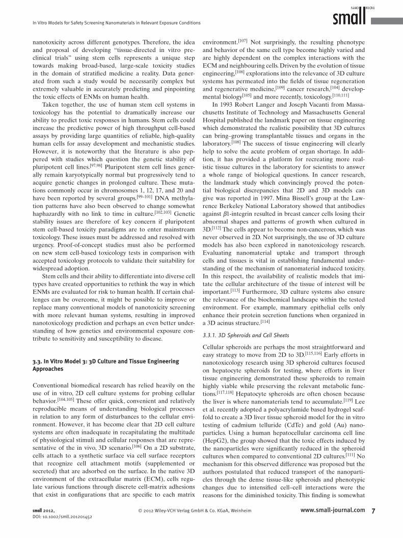
Figure 2 . Organotypic culture of human skin, recapitulating the bilayered anatomy of skin but void of all appendages.
comforting since it implies that the signifi cant toxicological
effects of nanomaterials found in 2D studies may not be
as severe as they seem in the context of real life exposure.
However, it has been rightly cautioned that the absence of
cell death does not necessarily mean that the cells are not
undergoing any undesirable changes in function or signal-
ling, which could lead to cynical outcomes. [ 120 ] Although 3D
spheroids represent an important step towards enhancing the
relevance of nanotoxicological studies, their size limitation,
typically up to a few hundreds of microns in diameter due to
diffusion limits, could restrict their widespread acceptance as
the model of choice in the fi eld.
Cell sheets are an alternative way to build up 3D constructs
that mimic native tissue architecture. These has been shown
to be feasible in producing blood vessels, [ 121 ] skin, [ 122 , 123 ] cor-
neas, [ 124 ] urothelium [ 125 ] and cardiac patches. [ 126 ] Essentially,
this strategy allows cells to be cultured to high densities to
form sheets which are then detached from culture surfaces
simply by mechanical means [ 123 ] or by changing temperature
if the cells are cultured on thermo-responsive surfaces. [ 127 ]
Using cell sheets has the advantage that an entirely natural
neo-tissue assembled by the cells can be produced. However,
there has not been any study employing the use of cell sheets
specifi cally to nanotoxicology research. This could be an
interesting approach, especially in evaluating the mass trans-
port of nanomaterials through tissues.
3.3.2. 3D Scaffold-Based Cultures
Borrowing the concept of scaffold-based tissue engineering,
toxicologists have also adopted toxicity testing in 3D cultures
achieved by growing cells in different scaffold confi gurations.
Cui et al. did this by culturing human bone marrow cells in
3D scaffolds made from combinations of collagen, Matrigel
and poly(lactic acid) fi bers. [ 110 ] These were maintained in
a multiple parallel microbioreactor platform and used for
testing of two model drugs, trimethoprim and pyrimethamine,
over a 7 day culture period. The group’s results demonstrated
that data obtained from the 3D culture system is signifi cantly
different from that of 2D cultures. However, comparisons
between the 2D and 3D systems are diffi cult given the com-
plexity of such systems where infl uence from the biomaterials
used will also need to be taken into consideration. Nonethe-
less, such systems enable the creation of a controlled and per-
fused environment to enhance reproducibility of 3D cultures.
In fact, more advanced versions of microbioreactors have
been developed to enable the co-culture of different cell
types in 3D scaffolds, for drug testing in a more complex but
realistic scenario. [ 128 ] It will be interesting to follow what 3D
scaffold systems in combination with microbioreactors will
reveal in nanotoxicology research.
3.3.3. Organotypic and Ex Vivo Cultures
An extension of 3D spheroids and scaffold-based cultures is
the strategy of using organotypic cultures to achieve physi-
ologically more realistic models for toxicology testing. These
are culture systems that take into consideration the spatial
distribution of cell type(s) to impart structural organiza-
tion that is unique to the tissue of interest. The earliest, and
www.small-journal.com © 2012 Wiley-VCH Ve
perhaps still most successful, model of that is the skin organ-
otypic culture ( Figure 2 ), which was developed with the moti-
vation of producing transplantable skin grafts. Early work by
Bell et al. showed that bovine collagen lattices seeded with
dermal fi broblasts contracted to form dermal-like tissues. [ 129 ]
Culturing keratinocytes on top of these dermal lattices in
an air-exposed condition led to their differentiation to form
the stratifi ed layers of a matured epidermis. [ 130 , 131 ] These cul-
tures retain most of the anatomical features of the epidermis
but are void of all skin appendages. Although not real skin,
they represent the next closest and most convenient model
for testing the effects of topical agents before moving onto
animal or human trials–easy to apply histological and immu-
nohistochemical techniques for analysis of cell/tissue integrity
and marker expression without having to harvest biopsies.
As a result, skin organotypic cultures have been successfully
used for the testing of carcinogens [ 132 ] and various topical
agents [ 133 ] to good effect. The enzymatic profi le in skin orga-
notypic cultures has recently been reported to be suitable for
dermatotoxicological studies. [ 134 ] Ex vivo skin cultures have
mostly been used for studying nanomaterial penetration
through the epidermal barrier. Ravichandran et al. managed
to establish the feasibility of using ex vivo human skin cul-
tures to assess the effects of tape stripping versus a depilatory
agent on quantum dot penetration through the epidermis. [ 135 ]
By optimizing fl uorescent microscopy analysis of microtomed
skin sections, the group showed that depilatory treatment
increased penetration of quantum dots but to a lesser extent
than tape stripping. In studying penetration of nanomate-
rials through skin, it was highlighted that careful considera-
tions need to be made in various factors such as time course
of treatments that may compromise skin barrier functions,
the skin model to use and the characterization methods. [ 136 ]
Ex vivo porcine skin cultures have also been used recently
to demonstrate the importance of nanomaterial biodistribu-
tion in relation to vascular physiological effects. [ 137 ] In this
study, six different nanoparticles of different size and com-
position (silica- or dextran-coated Fe 2 O 3 , silica- or citrate-
coated silver, PEG, and carboxylated quantum dots) were
rlag GmbH & Co. KGaA, Weinheim small 2012, DOI: 10.1002/smll.201201452
In Vitro Models for Safety Screening Nanomaterials in Relevant Exposure Conditions
infused through porcine skin fl aps and were found to result
in statistically signifi cant post-infusion fl ap weight gain and
an increase in arterial perfusion pressure. Interestingly, infu-
sion with nC(60) nanoparticles did not produce these effects.
Ex vivo skin cultures are certainly useful in studying some
aspects of nanomaterial exposure and biological interaction
through dermal exposure. They are realistic models since
they are derived from real skin, can be kept viable in vitro
for a period of time and can be easily processed for different
assessments.
Besides the fi rst organ or tissue of contact, it is equally
important to understand nanotoxicology in terms of nano-
material biodistribution in the body and consequently their
excretion from the body, if at all. Different studies have
showed the potential of various nanomaterials to accumulate
in organs including the liver, lung, kidney and spleen. [ 138–141 ]
Thus, the use of organotypic cultures of these organs is an
exciting proposition that will certainly accelerate our under-
standing of nanotoxicology. As part of the group’s efforts in
developing an artifi cial kidney, Zhang et al. induced tubu-
logenesis of primary human renal proximal tubule cells on
2D surfaces by using transforming growth factor- β 1, without
any 3D matrices. The resulting renal proximal tubules were
of several millimetres in length and were easily accessible
for manipulations and imaging. [ 142 ] The group explored
using these tubules for nanotoxicology studies by investi-
gating the interactions of human and porcine renal proximal
tubules with core-shell CdSe@ZnS quantum dots. [ 143 ] Results
revealed that porcine and human proximal tubules showed
markedly different uptake behaviors, and that human prox-
imal tubule cells were most sensitive to the cytotoxic effects
of the quantum dots.
Some interesting variations of 3D organotypic cultures
for interaction studies with nanomaterials have also been
reported recently. For the purpose of testing the transfection
effi ciency of poly( β -amino ester) based nanoparticles as gene
delivery vehicles, Bhise et al. successfully suspended frag-
ments of mammary gland epithelium in Matrigel to create
“organoid” breast cancer cultures, and found the effi cacy of
transfection using the polymeric nanoparticles to be twice that
of a commercial transfection agent. [ 144 ] In another study to
develop organotypic brain cultures for nanomaterial uptake
evaluation, Meng et al. cultured medulloblastoma-derived
cell spheroids on rat cerebellum slices and maintained these
for 6 days in vitro. [ 145 ] Interestingly, poly(glycerol-adipate)
nanoparticles introduced into this system were preferentially
taken up into the medulloblastoma-derived cells rather than
the healthy brain cells in the cerebellum slices.
4. In Vitro Safety Screening Under Simulated Environmental Exposure Conditions
Nanotoxicology as a distinct discipline has its roots from
inhalation toxicology. Consequently, for historic reasons early
nanotoxicology studies were conducted with in vitro and in
vivo models of inhalation toxicology. In the fi rst generation
nanotoxicology studies, little or no attention was given to
understand the infl uence of environmental factors in shaping
© 2012 Wiley-VCH Verlag Gmbsmall 2012, DOI: 10.1002/smll.201201452
the potential toxicity of ENMs. Another reason is that, over
the past decades nanotechnology was in the early phase of
research and development. The main concern over these
years of small or medium scale nanomaterial production
was the work-place-safety due to concerns over the potential
hazard arising from direct human contact during the nano-
materials synthesize. However, this decade will witness many
prototypes of nanotechnology applications being transformed
to mass production, implying large scale production and uti-
lization of nanomaterials. Although, it is diffi cult to fi gure out
the exact production volume of nanomaterials worldwide,
a recent study by Hendren et al. estimated the production
volume in the range of metric tonnes/year for fi ve of the
most popular nanomaterials. [ 146 ] Therefore, it is becoming
imperative that more contextualized environmental health
and safety studies needs to be carried out to account for the
nano-bio interactions of direct ecological relevance. More
recently, there have been some studies devoted in under-
standing possible hazardous nano-bio interactions of rele-
vance to ecotoxicology. Patricia Holden’s group, at University
of California Santa Barbara, demonstrated that the bare
CdSe quantum dots that have accumulated in Pseudomonas aeruginosa bacteria can be transferred to and biomagnifi ed in
the Tetrahymena thermophila protozoa that prey on the bac-
teria, signifying the possibility of biomagnifi cation of nano-
materials in a food chain. [ 147 ] The possibility of organ damage
in higher aquatic organism such as fi sh has been the subject
of limited studies. One of the early studies was that by Smith
et al., who reported the gill and brain injury caused in adult
rain bow trout exposed to single walled carbon nanotubes
(SWCNTs). [ 148 ] Their study suggested the possibility of oxi-
dative stress depended respiratory damage due to exposure
to SWCNTs. Recently, George et al. reported the use of cell
line derived from the gill of rain bow trout (RT-G-W1). [ 149 ]
They showed oxidative stress dependent cellular damage
caused by silver nanoplate whose toxicity was higher than
nanospheres and nanowires. Thorough examination of silver
nanoplate using high resolution transmission electron micro-
scopy (HRTEM) revealed the presence of crystal defects
the prevalence of which was more in plate morphology. [ 149 ]
While, in vivo model (zebrafi sh embryo) could also pick up
the higher toxic potential of silver nanoplates in comparison
to other shapes, the in vitro model was indispensable in
delineating the mechanism of toxicity. The excellent corre-
lation between in vitro and in vivo outcomes suggested the
utility of cellular studies in screening for toxicity and identi-
fying mechanisms of nanotoxicity without having to resort to
more expensive animal model studies. The above mentioned
studies could help in identifying the primary nanomaterial
property/ies in relation to its potential toxicity. However,
when addressing the ecotoxicity of nanomaterials, one should
realize the important role played by environmental factors in
shaping the biological outcome.
One of the environmental factors that may have more
direct and immediate infl uence on shaping the nano-bio
interactions is the sunlight. The infl uence of solar light in
determining the outcome of nanomaterial interactions with
biological systems cannot be overlooked as most, if not all,
high-volume-production nanomaterials are photoactive. TiO 2
9www.small-journal.comH & Co. KGaA, Weinheim

M. H. Kathawala et al.
1
reviews
nanoparticle, for example, is generally regarded safe and isoften used as a negative control nanomaterials in biological
assays. However, many studies have shown the potential
of this material to generate reactive oxygen species (ROS)
and induce deleterious biological outcomes when exposed
to near-visible [ 62 ] or simulated solar light. [ 150 ] Recently, the
potential photoxicity of TiO 2 to marine phytoplankton under
relatively low levels of ultraviolet light, consistent with those
found in nature, was reported by Miller et al. [ 151 ] While, these
studies highlight potential toxicity of TiO 2 under the infl uence
of solar light, it is worth noticing the scarcity of data on many
other high-volume-production nanomaterials. Although, ROS
generation is a mechanism for phototoxicity of nanomate-
rials, very little attention has been devoted in understanding
the infl uence of other factors such as bioavailability, light-
activated dissolution of nanomaterials, light-assisted agglom-
eration and deagglomeration etc.
5. A Class of High-Volume Production Nanomaterials: Photoactive Nanomaterials
5.1. High-Volume Production Nanomaterials
Recent years of development has seen an exponential
infl ux of nanomaterials into the market in various applica-
tions. Nanomaterials are actively making their way and even
replacing traditional materials in services like electronics,
photovoltaic, construction, biomaterials applications, drug
delivery, etc. All these applications make use of some prop-
erty of nanomaterials, which their bulk and even their micro-
sized counterparts fail to provide. The enhanced surface area
of nanomaterials not only enhances their surface properties
and makes them more reactive, it also kicks into action some
unique properties which are otherwise suppressed, unex-
pressed or non-dominating.
This section will delve exclusively into the fi rst kind of
photoactive materials, i.e., the photocatalysts, namely TiO 2
and ZnO, both of which have cemented their places in many
commercially available applications already and are making
their way into many other applications.
5.2. Cytotoxicity of Photoactive Nanomaterials
Photoactive materials are, as the name suggests, active
under exposure to light. They can be excited and/or give
rise to charged species (e.g., ROS, etc.) under photon expo-
sure. Recent research has heavily focused on these kind of
materials to extract applications which can use these excited
states, or reactive charged species for cultivating effects like
electricity in the case of solar cells, or killing bacteria in the
case of sea water purifi cation, etc. [ 14–17 , 152 , 153 ]
Generally photoactive nanomaterials can be classifi ed
in two distinct classes: photosensitizers and semiconductors.
The mechanisms for both these types of photoactive nano-
materials differ distinctly and so does the end result of their
photo activity. Photosensitizers on the other hands are organic
0 www.small-journal.com © 2012 Wiley-VCH
compounds which have electronic confi gurations that allow
for photoactivity. Fullerenes are one of the main categories
of photosensitizers. Semiconductors or photocatalysts are
mostly oxides or combinations of oxides and become active
once their band gap energy is overcome. This review will
focus on the latter class of photoactive nanomaterials.
5.2.1. Titanium Dioxide (TiO 2 )
Nano-TiO 2 is fast becoming the material of choice for appli-
cations ranging from photocatalyst in antibacterial, antifungal,
antiviral and antitumor systems to UV-fi lters and absorbers
in sunscreens and solar cells due its superior photo activity.
TiO 2 occurs as three allotropes; rutile (tetragonal), anatase
(tetragonal), and brookite (orthorhombic). [ 154 ] Rutile and
anatase are of the highest commercial importance repre-
senting 90% and 10% of the market, respectively. [ 155 ] For this
particular reason most of the research has focused on these
two crystalline forms and this review paper will be relating
extensively only to these.
The band gap energies for anatase and rutile are 3.23 eV
and 3.06 eV [ 156 ] which corresponds to light of about 385 nm
and 400 nm. Most studies conclude that the anatase phase
has greater photoactivity than the rutile phase [ 157–159 ] while
the reverse or comparable photoactivity has also been sug-
gested under certain conditions. [ 160 , 161 ] Anatase and rutile are
thermodynamically more stable for particle sizes less than
11 nm and more than 35 nm respectively. [ 154 ] Considering the
possibility that stability accounts for the lack of photoactivity,
the contradictions might be due to different crystal sizes and/
or types. However, a combination of the two (approximate
ratio = 3:1 of anatase:rutile) was found to possess superior
photoactivity than any single pure form. [ 13 , 158 ]
Tuning TiO 2 to be responsive to visible light is a poten-
tial area of research. [ 162–165 ] Doping with other elements has
been exploited e.g. using iron (1–10 wt%) to decrease Nano-
TiO 2 band gap to near-visible light. [ 166 ] When developing a
formulation with high SPF D. Nesseem showed the highest
SPF from a formulation which used ZnO as an additive to
the active TiO 2 . [ 167 ] R. Dunford et al. showed that TiO 2 (no
mention of size in study) in sunscreens can cause phenol
photodegradation which was greatest in sample containing
ZnO. [ 168 ]
Photocatalysis has been documented with regards to
these reactive oxygen species (ROS), and a detailed review
on its properties, and prospects as an effective bactericide,
anti-viral agent, anti-tumor agent and anti-fungal agent has
been published by A. Markowska. [ 13 ] Nano-TiO 2 induced
reduction in cell viability, compromised antioxidant system,
morphological alterations, intracellular ROS production and
signifi cant DNA damage in human amnion epithelial (WISH)
cells. [ 169 ] HaCaT cells also displayed drop in cell viability
when exposed to moderate concentrations (50 ppm) of two
different types of nano-TiO 2 made up of a mixture of ana-
tase and rutile of spherical shape, but not when exposed to
the same concentration of polygonal pure anatase nano-TiO 2 .
The nanoparticles were commercially available and differed
in size as well (pure anatase: 5.8 nm, mixture: 25.8 nm and
14.6 nm). [ 155 ] However, there are so many differences between
the sample in terms of shape, size and crystal structure that it
Verlag GmbH & Co. KGaA, Weinheim small 2012, DOI: 10.1002/smll.201201452
In Vitro Models for Safety Screening Nanomaterials in Relevant Exposure Conditions
would be hard to be certain which physiochemical parameter
was the real contributing factor. Nonetheless, most studies
have shown TiO 2 nanoparticles to have low cytotoxicity until
very high doses. [ 170–173 ] Fujita et al. [ 174 ] concluded their study
on the notion that “Gene expression profi le suggests that the
ultrafi ne TiO 2 particles without illumination have no signifi -
cant impact on ROS-associated oxidative damage, but affect
the cell–matrix adhesion in keratinocytes for extracellular
matrix remodelling.” Jang et al. [ 175 ] observed an inverse rela-
tionship between nano-TiO 2 particle size and antimicrobial
effect after photoactivation suggesting directly that smaller
size increases photocatalytic abilities. Micro-particles of
TiO 2 have been generally accepted as inert [ 176 , 177 ] but their
nano-scale counterparts have cell penetrative [ 154 ] as well as
enhanced ROS production capabilities. [ 175 ]
ROS generation is widely considered as a molecular para-
digm for the toxicity of nanoparticles. [ 178 ] Excessive ROS
generation would cause oxidative stress in cells. [ 179 , 180 ] If the
oxidative stress exceeds the threshold of the cellular antioxi-
dant defenses, additional mitochondrial perturbation such as
mitochondrial depolarization would occur. [ 59 , 181 , 182 ] These dis-
turbances might further cause apoptosis or necrosis of cells
followed by an increase in cell membrane permeability.
Phototoxicity has also been widely studied, especially
in context of sunscreens and the effect is thought to be sig-
nifi cant. [ 155 , 168 , 183 ] George et al. [ 166 ] reported that iron-doped
(0–10 wt%) Nano-TiO 2 which showed a dose dependent
decrease in band gap energy increased oxidant injury and cell
death in parallel with a decrease in band gap energy. Near-
visible light exposure was used in the study. Similarly and pro-
gressively, Reeves et al. [ 184 ] observed increased damage in TiO 2
treated fi sh cells after UVA activations, whereby hydroxyl
radicals generated (extra- and intra-cellular) were the likely
suspects for damage. Nakagawa et al. [ 185 ] also pointed out the
potential photogenotoxicity of TiO 2 nanoparticles to Chinese
hamster cell line. Hee-Ok Park et al. showed using commer-
cially available TiO 2 nanoparticles that TiO 2 samples with
higher photocatalytic activity caused higher toxicity with or
without simultaneous irradiation of UV. [ 155 ] The effect of UV
was not seen on low photoactive sample which again rein-
states the potential of TiO 2 to be a phototoxic material. ROS
measured by DCF fl uorescence also followed the same trend,
but no DNA damage was seen using the comet assay.
One interesting study by Gopalan et al. [ 186 ] reported that
TiO 2 tend to introduce dose-dependent photogenotoxic effect
to human lymphocytes but not to human sperms. It has also
been reported that photoactivated TiO 2 nanoparticles could
selectively induce toxicity against cancer cells [ 187–189 ] and in
fact numerous studies on TiO 2 as an effective photocatalyst
talk about TiO 2 ’s selective anti-tumor cell action as a pros-
pect worth investing. It is thus clear that cell-line is one very
important factor when determining the cellular responses of
cell lines. Possibly, the action of TiO 2 would remain the same
i.e. its photocatalytic action and generation of ROS, but dif-
ferent cells would respond differently to the change in home-
ostasis. Oxidative stress is a residual phenomenon in cells and
antioxidant mechanisms of defense are in place. Possibly the
threshold of these defenses differ in different cells and so
does the onset of toxicity.
© 2012 Wiley-VCH Verlag Gmsmall 2012, DOI: 10.1002/smll.201201452
5.2.2. Zinc Oxide (ZnO)
Zinc oxide is another prime material candidate in the fi eld
of photoactive materials and is often discussed hand-in-hand
with TiO 2 . Like TiO 2 , it falls into the category of semicon-
ductor and has a broad spectrum adsorption with a bandgap
of 3.2 eV, [ 190 ] high refractive index of 1.95–2.10 [ 191 ] and the
ability to absorb UVA and UVB. [ 192 ] It has been studied
heavily and even employed in numerous photoactive appli-
cations ranging from solar cells, photovoltaics to the most
common UV-fi lters in sunscreens.
However, there are certain limitations to using ZnO which
restrict the practical applications of ZnO. Amongst them is
the anodic photocorrosion of ZnO which, unlike in the case
of TiO 2 , is unhindered by oxidation of water, and solubility
of ZnO especially in acidic and alkaline conditions as well as
in the presence of Zn 2 + chelators. These properties severely
limit the pH range in which ZnO can be used as a photo-
catalyst. Nonetheless ZnO displays similar photoactivity as
TiO 2 in certain conditions, [ 193 ] and has shown faster initial
degradation rate (r0) of herbicide triclopyr (3,5,6-trichloro-2-
pyridiyloxyacetic acid) compared to P25 TiO 2 . [ 194 ] There have
been various attempts to overcome the drawbacks of ZnO
and modify it to fi t photocatalytic applications. Details can be
found in these reviews. [ 190 , 195 , 196 ]
ZnO has three known crystal structures: hexagonal wur-
tzite, cubic zinc-blende structure and cubic rock-salt (NaCl-
type). The most common of them and also the most relevant
in the scope of this paper is the wurtzite form. ZnO has been
in use since a long period and was considered a safe mate-
rial in its bulk form. [ 197 ] However, the same material when
reduced to the nano-scale has raised alerts owing to its
cyto- and geno-toxic attributes. To add to that its photoac-
tivity raises further concerns over its behavior under irradia-
tive conditions especially given the superior surface area to
volume ratio of nano-ZnO. For perceptive purpose, the rate
of H 2 O 2 production is 100–1000 times faster with ZnO nano-
particles of 4 nm to 5 nm, than with larger ZnO particles of
100 nm. [ 198 ] Several theories about the toxicity of nano-ZnO
have been going around in the scientifi c community. They
mostly revolve around ROS production and Zn 2 + ions. ZnO
has even been shown to be genotoxic. [ 199 ] Here we attempt
to sum up the most relevant fi ndings, discuss the most logical
conclusions and suggest an approach for future studies.
Xia et al. published a comprehensive and detailed study
to link the dissolution of nano-ZnO to its cellular response,
albeit in the dark. Amongst other wonderful fi ndings, two of
them have notable relevance when considering the context of
phototoxicity. Firstly, it was shown that nano-ZnO is readily
soluble to upto 190 μ M and 225 μ M in BEGM and DMEM,
both of which are common cell culture mediums, and that
nano-ZnO can achieve more than 80% solubility within 3 h.
Given the rigorous sonication regimes followed by toxicity
studies to achieve homogenous suspensions that might well
be an understatement. Furthermore, it was shown in two sep-
arate experiments that shredded Fluorescein isothiocyanate
(FITC) tags from nano-ZnO particles, as well as the Zn 2 + spe-
cifi c dye, NewPort Green DCF, overlapped with the LAMP-
1 + fl uorescence (LAMP-1 + stains lysosomal membranes). [ 200 ]
11www.small-journal.combH & Co. KGaA, Weinheim

M. H. Kathawala et al.reviews
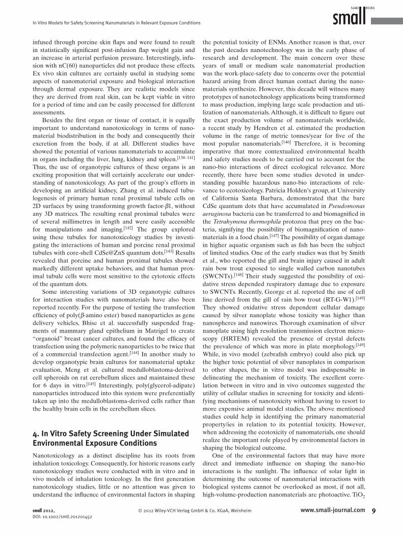
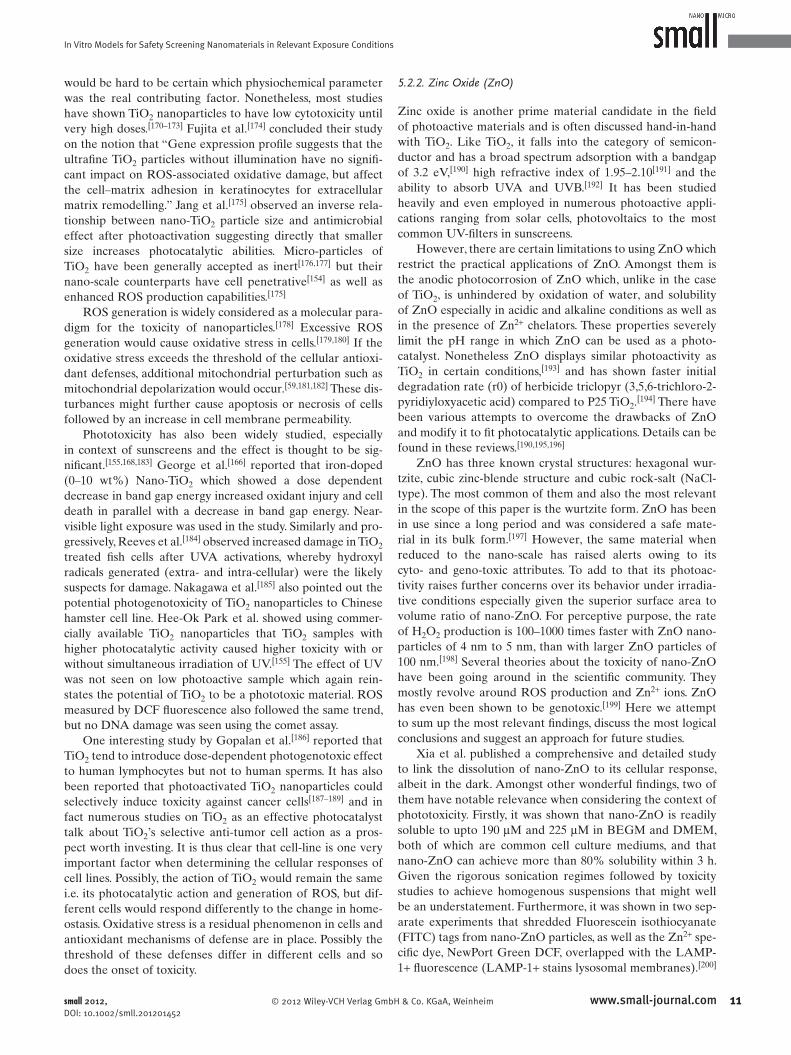
Figure 3 . The toxicological pathway of ZnO-Nanoparticles. Reproduced with permission. [ 201 ] Copyright 2011, Elsevier.
Although no actual nano-ZnO particles were seen under con-
focal microscope, the group inferred that perhaps the FITC
tags were shredded off from remnants of undissolved nano-
ZnO which were taken up by the cells through endocytosis
after which they dissolved under relatively acidic conditions
of the endosomes/lysosomes giving Zn 2 + ions. This hypoth-
esis has been shared by other studies as well. [ 201 ] The second
fi nding relates to ROS production. It was shown nano-ZnO
and nano-CeO 2 , but not nano-TiO 2 , produced H 2 O 2 in abi-
otic conditions, while only nano-ZnO, but not nano-CeO 2 or
nano-TiO 2 , produced H 2 O 2 or superoxide in biotic conditions
under dark. [ 200 ]
Both of these fi ndings have serious implications in the
context of phototoxicity and also photoactivity in biotic envi-
ronments. Firstly, it is evident that solubility of nano-ZnO
is pronounced at lower concentrations probably due to the
greater surface area, and even more so inside the cellular
peripheral where lower pH further facilitates the dissolution.
If so, then the phototoxicity can be ruled out since once dis-
solved, nano-ZnO’s integrity as a semiconductor, with photo-
activation prospects, is lost. Solubility has been mentioned
as a drawback of cultivating ZnO’s photoactive properties.
However, its solubility certainly has strong implications on
the toxicity of ZnO.
The above fi nding also opens up the possibility of dif-
ferent oxidative responses of the particle in biotic and abi-
otic environments. CeO 2 , although seemingly an oxidative
agent, in cellular environments actually has anti-oxidative
effects. [ 200 ] It is thus clear, that ROS production mechanisms
are different in biotic and abiotic conditions. Studies should
be wary of such effects before prematurely extrapolating
results to and from different environments. In the case of
ZnO, this is even more imperative as the effect of Zn 2 + on
mitochondrial function is well known. Zn 2 + can cause mito-
chondria membrane potential and respiration to be disturbed
causing excess ROS to accumulate inside the mitochondria
12 www.small-journal.com © 2012 Wiley-VCH Verlag GmbH & Co. KGaA
and ultimately inducing apoptotic
death. [ 200 , 201 ] Figure 3 [ 201 ] reveals this
mechanism in detail. ROS may accentuate
ZnO’s toxicity as shown by Chin et al. [ 202 ]
Although ZnO-induced ROS spikes
have been well reported, [ 192 , 200 , 201 , 203–205 ]
well planned dissolution studies must run
parallel to cytotoxicity and ROS-measure-
ment studies in order to correctly attribute
the source of ROS production to the nan-
oparticles or the ions.
A comparison of toxicity and ROS
production due to ZnO treatment under
natural sunlight (NSL), dark, and artifi cial
ambient laboratory light (AALL) was also
studied, and revealed that for nano-ZnO
the toxicity was in the order of NSL(2h) +
AALL(22h) > NSL(2h) + dark(22h) >
AALL(24h) > AALL(2h) + dark (22h),
with [AALL(2h) + dark(22h)] being the
least toxic. [ 205 ] ROS production was also in
agreement of the trend. Considering that
NSL might be much more potent to cause
photoactivation compared to AALL based on good reason,
the study concluded that ZnO was indeed phototoxic.
However, a similar yet much older study by Dufour
et al. [ 206 ] handled the data in a much different way. They
studied the effect of two different treatments: pre-irradiation
(PI) and simultaneous irradiation (SI). They chose two sepa-
rate regimes of irradiation as well: low dose (350 mJ cm − 2 and
3 h prior to particle addition for PI) and high dose (700 mJ cm − 2
and only 1–2 h prior to particle addition for PI). Generally
SI was more cytotoxic and clastogenic than PI, however PI
should signifi cantly higher cytotoxicity and clastogenicity
when compared to dark. Even in the dark ZnO showed con-
siderable cytotoxicity and clastogenicity. Instead of concluding
that ZnO is a phototoxic and photo-clastogenic agent, several
competing factors were analyzed and it was suggested that
UV-induced DNA damage could make the cells even more
susceptible to ZnO-induced cytoxicity and clastogenicity.
This fi ts in logically as PI even should an increased in adverse
effects which were probably less enhanced than SI due to
recovery of cells during the waiting period. In the high-dose
treatment where recovery time is low, indeed there was no
observable difference between PI and SI as far as chromo-
some aberrations (clastogenicity) was concerned.
Lisa Martorano et al. [ 192 ] proved UV irradiation-induced
zinc dissolution in commercial zinc oxide sunscreens. Photo-
degradation of zinc was mentioned earlier as a drawback of
ZnO in the photocatalytic fi eld, and this may well be a con-
nected phenomenon whereby UV induces a greater avail-
ability of Zn 2 + ions which therefore cause higher toxicity.
Dadong et al. [ 207 ] when studying the effect ZnO on the uptake
of anti-cancer drug, daunorubicin, in Multi Drug Resistant
cells, noted more effi cient uptake of drug in the presence of
ZnO although no reasons were discussed. Also, in the pres-
ence of UV-irradiation, ZnO displayed greater toxicity. Com-
paring three size particles, 20, 60, and 100 nm, they were able
to show no effect of size on the action. Similarly, J. H. Yuan
, Weinheim small 2012, DOI: 10.1002/smll.201201452
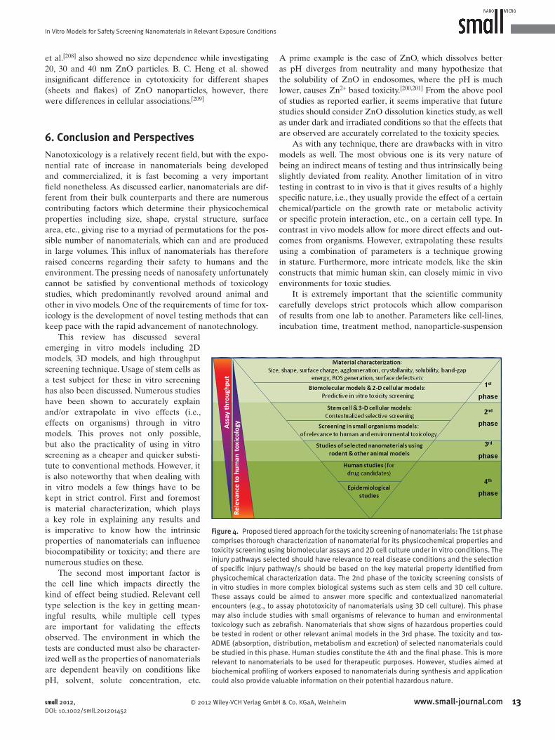
In Vitro Models for Safety Screening Nanomaterials in Relevant Exposure Conditions
et al. [ 208 ] also showed no size dependence while investigating
20, 30 and 40 nm ZnO particles. B. C. Heng et al. showed
insignifi cant difference in cytotoxicity for different shapes
(sheets and fl akes) of ZnO nanoparticles, however, there
were differences in cellular associations. [ 209 ]
6. Conclusion and Perspectives
Nanotoxicology is a relatively recent fi eld, but with the expo-
nential rate of increase in nanomaterials being developed
and commercialized, it is fast becoming a very important
fi eld nonetheless. As discussed earlier, nanomaterials are dif-
ferent from their bulk counterparts and there are numerous
contributing factors which determine their physicochemical
properties including size, shape, crystal structure, surface
area, etc., giving rise to a myriad of permutations for the pos-
sible number of nanomaterials, which can and are produced
in large volumes. This infl ux of nanomaterials has therefore
raised concerns regarding their safety to humans and the
environment. The pressing needs of nanosafety unfortunately
cannot be satisfi ed by conventional methods of toxicology
studies, which predominantly revolved around animal and
other in vivo models. One of the requirements of time for tox-
icology is the development of novel testing methods that can
keep pace with the rapid advancement of nanotechnology.
Figure 4 . Proposed tiered approach for the toxicity screening of nanomaterials: The 1st phase comprises thorough characterization of nanomaterial for its physicochemical properties and toxicity screening using biomolecular assays and 2D cell culture under in vitro conditions. The injury pathways selected should have relevance to real disease conditions and the selection of specifi c injury pathway/s should be based on the key material property identifi ed from physicochemical characterization data. The 2nd phase of the toxicity screening consists of in vitro studies in more complex biological systems such as stem cells and 3D cell culture. These assays could be aimed to answer more specifi c and contextualized nanomaterial encounters (e.g., to assay phototoxicity of nanomaterials using 3D cell culture). This phase may also include studies with small organisms of relevance to human and environmental toxicology such as zebrafi sh. Nanomaterials that show signs of hazardous properties could be tested in rodent or other relevant animal models in the 3rd phase. The toxicity and tox-ADME (absorption, distribution, metabolism and excretion) of selected nanomaterials could be studied in this phase. Human studies constitute the 4th and the fi nal phase. This is more relevant to nanomaterials to be used for therapeutic purposes. However, studies aimed at biochemical profi ling of workers exposed to nanomaterials during synthesis and application could also provide valuable information on their potential hazardous nature.
This review has discussed several
emerging in vitro models including 2D
models, 3D models, and high throughput
screening technique. Usage of stem cells as
a test subject for these in vitro screening
has also been discussed. Numerous studies
have been shown to accurately explain
and/or extrapolate in vivo effects (i.e.,
effects on organisms) through in vitro
models. This proves not only possible,
but also the practicality of using in vitro
screening as a cheaper and quicker substi-
tute to conventional methods. However, it
is also noteworthy that when dealing with
in vitro models a few things have to be
kept in strict control. First and foremost
is material characterization, which plays
a key role in explaining any results and
is imperative to know how the intrinsic
properties of nanomaterials can infl uence
biocompatibility or toxicity; and there are
numerous studies on these.
The second most important factor is
the cell line which impacts directly the
kind of effect being studied. Relevant cell
type selection is the key in getting mean-
ingful results, while multiple cell types
are important for validating the effects
observed. The environment in which the
tests are conducted must also be character-
ized well as the properties of nano materials
are dependent heavily on conditions like
pH, solvent, solute concentration, etc.
© 2012 Wiley-VCH Verlag Gmsmall 2012, DOI: 10.1002/smll.201201452
A prime example is the case of ZnO, which dissolves better
as pH diverges from neutrality and many hypothesize that
the solubility of ZnO in endosomes, where the pH is much
lower, causes Zn 2 + based toxicity. [ 200 , 201 ] From the above pool
of studies as reported earlier, it seems imperative that future
studies should consider ZnO dissolution kinetics study, as well
as under dark and irradiated conditions so that the effects that
are observed are accurately correlated to the toxicity species.
As with any technique, there are drawbacks with in vitro
models as well. The most obvious one is its very nature of
being an indirect means of testing and thus intrinsically being
slightly deviated from reality. Another limitation of in vitro
testing in contrast to in vivo is that it gives results of a highly
specifi c nature, i.e., they usually provide the effect of a certain
chemical/particle on the growth rate or metabolic activity
or specifi c protein interaction, etc., on a certain cell type. In
contrast in vivo models allow for more direct effects and out-
comes from organisms. However, extrapolating these results
using a combination of parameters is a technique growing
in stature. Furthermore, more intricate models, like the skin
constructs that mimic human skin, can closely mimic in vivo
environments for toxic studies.
It is extremely important that the scientifi c community
carefully develops strict protocols which allow comparison
of results from one lab to another. Parameters like cell-lines,
incubation time, treatment method, nanoparticle-suspension
13www.small-journal.combH & Co. KGaA, Weinheim

M. H. Kathawala et al.
1
reviews
[ 1 ] A. S. Edelstein , R. C. Cammaratra , Nanomaterials: Synthesis, Properties and Applications , 2nd Ed., Taylor and Francis , Bristol, Philadelphia 1998 .
[ 2 ] The Project on Emerging Nanotechnologies (PEN); 2012 ; Avail-able from: http://www.nanotechproject.org/consumerproducts, accessed: May, 2012.
[ 3 ] N. Staggers , T. McCasky , N. Brazelton , R. Kennedy , Nursing Out-look 2008 , 56 , 268 .
[ 4 ] W. L. Lee , C. Loei , E. Widjaja , S. C. J. Loo , J. Control. Release 2011 , 151 , 229 .
[ 5 ] S. C. J. Loo , T. Moore , B. Banik , F. Alexis , Curr. Pharm. Bio-technol. 2010 , 11 , 333 .
[ 6 ] Y. F. Zhao , S. C. J. Loo , J. Ma , J. Nanosci. Nanotechnol. 2009 , 9 , 3720 .
preparation for toxicity studies, etc., must all be standardized.
When physicochemical aspects are considered, in-depth char-
acterization is essential. Size and shape are of prime impor-
tance, especially shape because of its high impact on surface
area as well as surface properties, both of which are the core
competencies of nanoparticles in general. Thus, while, in
vitro models are becoming an indispensable part of toxicity
screening, what is becoming apparent is the need to incorpo-
rate a multitude of test variable which may overwhelm the in
vitro screening if one were to test nanoparticle toxicity case-
by-case. This bottleneck could be overcome by strategizing
tiered-testing approaches and by the use of high throughput
screening platforms wherever applicable. An example of a
tiered-approach is highlighted in Figure 4 .
Incidentally, many of the high volume production nano-
materials such as TiO 2 , ZnO, Q-dots, fullerenes, graphene,
carbon nanotubes, etc., are photoactive. The benefi ts of these
interactions between ENMs and light are being exploited for
a variety of applications such as sunscreen formulations, self-
cleaning windows, solid lighting, solar panels, etc. While we
continue to enjoy these benefi ts of photoactive nanomaterials,
there is an overriding concern that the same effects could lead
to hazardous outcomes. However, so far the toxicity testing
methods are more developed for ‘ambient laboratory condi-
tions’, giving little attention to ‘real exposure conditions’. As
many of the nanomaterials are already in mass-scale produc-
tion with many more to follow, what is immediately required
is to develop testing methods that can imitate real exposure
conditions. This has to take place not only for understanding
the potential toxicity of nanomaterials to humans under
environmentally relevant conditions but also to other organ-
isms of relevance to ecosystem. As in vitro platforms become
indispensable tools of nanotoxicology, there is an urgent need
to identify and standardize test methods in appropriate in
vitro models of relevance to ecotoxicology.
Finally, the in vitro models discussed in this review may
hold the key to future nanotoxicology studies, as they are
capable of revealing information which may not be economi-
cally and practically possible to get through conventional
means, especially given the rate at which high-production
nanomaterials are being manufactured. We believe that these
emerging approaches may hold the key to expedite many
safety or toxicity studies, and answer the pressing need for
these data to be comprehensively revealed to the scientifi c
community, the consumers, and the public in general.
4 www.small-journal.com © 2012 Wiley-VCH V
[ 7 ] W. L. Lee , E. Widjaja , S. C. J. Loo , J. Mater. Sci.: Mater. Med. 2012 , 23 , 81 .
[ 8 ] S. C. J. Loo , Z. Y. S. Tan , Y. J. Chow , S. L. I. Lin , J. Pharm. Sci. 2010 , 99 , 3060 .
[ 9 ] T. W. J. Steele , C. L. Huang , E. Widjaja , F. Y. C. Boey , J. S. C. Loo , S. S. Venkatraman , Acta Biomater. 2011 , 7 , 1973 .
[ 10 ] H. T. Ong , J. S. C. Loo , F. Y. C. Boey , S. J. Russell , J. Ma , K. W. Peng , J. Nanoparticle Res. 2008 , 10 , 141 .
[ 11 ] T. W. J. Steele , C. L. Huang , S. Kumar , E. Widjaja , F. Y. C. Boey , J. S. C. Loo , S. S. Venkatraman , J. Pharm. Sci. 2011 , 100 , 4317 .
[ 12 ] W. L. Lee , E. Widjaja , S. C. J. Loo , Small 2010 , 6 , 1003 . [ 13 ] A. Markowska-Szczupak , K. Ulfi g , A. W. Morawski , Catal. Today
2011 , 169 , 249 . [ 14 ] Q. C. Xu , D. V. Wellia , M. A. Sk , K. H. Lim , J. S. C. Loo , D. W. Liao ,
R. Amal , T. T. Y. Tan , J. Photochem. Photobiol. A 2010 , 210 , 181 . [ 15 ] Q. C. Xu , D. V. Wellia , R. Amal , D. W. Liao , S. C. J. Loo , T. T. Y. Tan ,
Nanoscale 2010 , 2 , 1122 . [ 16 ] Z. M. He , G. H. Guai , J. Liu , C. X. Guo , J. S. C. Loo , C. M. Li ,
T. T. Y. Tan , Nanoscale 2011 , 3 , 4613 . [ 17 ] Q. C. Xu , Y. H. Ng , Y. Zhang , J. S. C. Loo , R. Amal , T. Y. T. Timothy ,
Chem. Commun. 2011 , 47 , 8641 . [ 18 ] G. Oberdorster , A. Maynard , K. Donaldson , V. Castranova ,
J. Fitzpatrick , K. Ausman , J. Carter , B. Karn , W. Kreyling , D. Lai , S. Olin , N. Monteiro-Riviere , D. Warheit , H. Yang , Particle Fibre Toxicol. 2005 , 2 , 8 .
[ 19 ] G. Oberdorster , E. Oberdorster , J. Oberdorster , Environ. Health Perspectives 2005 , 113 , 823 .
[ 20 ] S. Friedrichs , Sci. Technol. Adv. Mater. 2007 , 8 , 12 . [ 21 ] J. M. Hillegass , A. Shukla , S. A. Lathrop , M. B. MacPherson ,
N. K. Fukagawa , B. T. Mossman , Wiley interdisciplinary reviews. Nanomedicine and Nanobiotechnology 2010 , 2 , 219 .
[ 22 ] L. Braydich-Stolle , S. Hussain , J. J. Schlager , M. C. Hofmann , Toxicol. Sci: J. Soc. Toxicol. 2005 , 88 , 412 .
[ 23 ] E. Fabian , R. Landsiedel , L. Ma-Hock , K. Wiench , W. Wohlleben , B. van Ravenzwaay , Arch. Toxicol. 2008 , 82 , 151 .
[ 24 ] S. George , S. Pokhrel , T. Xia , B. Gilbert , Z. Ji , M. Schowalter , A. Rosenauer , R. Damoiseaux , K. A. Bradley , L. Madler , A. E. Nel , ACS Nano 2010 , 4 , 15 .
[ 25 ] C. Hanley , A. Thurber , C. Hanna , A. Punnoose , J. Zhang , D. G. Wingett , Nanoscale Res. Lett. 2009 , 4 , 1409 .
[ 26 ] J. Zhao , L. Xu , T. Zhang , G. Ren , Z. Yang , Neurotoxicology 2009 , 30 , 220 .
[ 27 ] X. Deng , Q. Luan , W. Chen , Y. Wang , M. Wu , H. Zhang , Z. Jiao , Nanotechnology 2009 , 20 , 115101 .
[ 28 ] P. V. AshaRani , G. Low Kah Mun , M. P. Hande , S. Valiyaveettil , ACS Nano 2009 , 3 , 279 .
[ 29 ] R. Foldbjerg , D. A. Dang , H. Autrup , Arch. Toxicol. 2011 , 85 , 743. [ 30 ] M. V. Park , A. M. Neigh , J. P. Vermeulen , L. J. de la Fonteyne ,
H. W. Verharen , J. J. Briede , H. van Loveren , W. H. de Jong , Bio-materials 2011 , 32 , 9810 .
[ 31 ] M. E. Samberg , E. G. Loboa , S. J. Oldenburg , N. A. Monteiro-Riviere , Nanomedicine 2012 , DOI: 10.2217/nnm.12.18.
[ 32 ] S. Hackenberg , A. Scherzed , M. Kessler , S. Hummel , A. Technau , K. Froelich , C. Ginzkey , C. Koehler , R. Hagen , N. Kleinsasser , Toxicol. Lett. 2011 , 201 , 27 .
[ 33 ] L. K. Braydich-Stolle , B. Lucas , A. Schrand , R. C. Murdock , T. Lee , J. J. Schlager , S. M. Hussain , M. C. Hofmann , Toxicol. Sci: J. Soc. Toxicol. 2010 , 116 , 577 .
[ 34 ] M. Ahamed , M. Karns , M. Goodson , J. Rowe , S. M. Hussain , J. J. Schlager , Y. Hong , Toxicol. Appl. Pharmacol. 2008 , 233 , 404.
[ 35 ] M. Ahamed , R. Posgai , T. J. Gorey , M. Nielsen , S. M. Hussain , J. J. Rowe , Toxicol. Appl. Pharmacol. 2010 , 242 , 263 .
[ 36 ] T. Hartung , Nature 2009 , 460 , 208 . [ 37 ] R. F. Service , Science 2008 , 321 , 1036 . [ 38 ] J. Y. Choi , G. Ramachandran , M. Kandlikar , Environ. Sci. Technol.
2009 , 43 , 3030 . [ 39 ] A. Abbott , Nature 2009 , 461 , 158 .
erlag GmbH & Co. KGaA, Weinheim small 2012, DOI: 10.1002/smll.201201452
In Vitro Models for Safety Screening Nanomaterials in Relevant Exposure Conditions
[ 40 ] National Research Council, Toxicity Testing in the 21st Century: A Vision and a Strategy , National Academy Press , Washington, D.C. 2007 .
[ 41 ] L. C. Rohrs , Archives of Internal Medicine 1957 , 100 , 44 . [ 42 ] T. Xia , M. Kovochich , M. Liong , L. Madler , B. Gilbert , H. Shi ,
J. I. Yeh , J. I. Zink , A. E. Nel , ACS Nano 2008 , 2 , 2121 . [ 43 ] C. A. Poland , R. Duffi n , I. Kinloch , A. Maynard , W. A. H. Wallace ,
A. Seaton , V. Stone , S. Brown , W. MacNee , K. Donaldson , Nat. Nanotechnol. 2008 , 3 , 423 .
[ 44 ] A. Takagi , A. Hirose , T. Nishimura , N. Fukumori , A. Ogata , N. Ohashi , S. Kitajima , J. Kanno , J. Toxicol. Sci. 2008 , 33 , 105 .
[ 45 ] Y. Sakamoto , D. Nakae , N. Fukumori , K. Tayama , A. Maekawa , K. Imai , A. Hirose , T. Nishimura , N. Ohashi , A. Ogata , J. Toxicol. Sci. 2009 , 34 , 65 .
[ 46 ] K. Donaldson , F. Murphy , R. Duffi n , C. Poland , Particle Fibre Tox-icol. 2010 , 7 , 5 .
[ 47 ] D. M. Brown , I. A. Kinloch , U. Bangert , A. H. Windle , D. M. Walter , G. S. Walker , C. A. Scotchford , K. Donaldson , V. Stone , Carbon 2007 , 45 , 1743 .
[ 48 ] M. Pacurari , X. J. Yin , J. S. Zhao , M. Ding , S. S. Leonard , D. Schwegier-Berry , B. S. Ducatman , D. Sbarra , M. D. Hoover , V. Castranova , V. Vallyathan , Environ. Health Perspect. 2008 , 116 , 1211 .
[ 49 ] X. Wang , T. Xia , S. A. Ntim , Z. X. Ji , S. George , H. Meng , H. Y. Zhang , V. Castranova , S. Mitra , A. E. Nel , ACS Nano 2010 , 4 , 7241 .
[ 50 ] X. Wang , T. Xia , S. A. Ntim , Z. Ji , S. Lin , H. Meng , C. Chung , S. George , H. Zhang , M. Wang , N. Li , Y. Yang , V. Castranova , S. Mitra , J. Bonner , A. E. Nel, ACS Nano 2011 , 5 , 9772 .
[ 51 ] P. H. Hoet , L. P. Gilissen , M. Leyva , B. Nemery , Toxicol. Sci. 1999 , 52 , 209 .
[ 52 ] P. H. M. Hoet , L. Gilissen , B. Nemery , Toxicol. Appl. Pharmacol. 2001 , 175 , 184 .
[ 53 ] J. Wang , Y. Liu , F. Jiao , F. Lao , W. Li , Y. Gu , Y. Li , C. Ge , G. Zhou , B. Li , Y. Zhao , Z. Chai , C. Chen , Toxicology 2008 , 254 , 82 .
[ 54 ] D. Olmedo , D. Tasat , M. Guglielmotti , R. Cabrini , J. Mater. Sci.: Mater. Med. 2008 , 19 , 3049 .
[ 55 ] S. Y. Shaw , E. C. Westly , M. J. Pittet , A. Subramanian , S. L. Schreiber , R. Weissleder , Proc. Natl. Acad. Sci. USA 2008 , 105 , 7387 .
[ 56 ] V. C. Abraham , D. L. Towne , J. F. Waring , U. Warrior , D. J. Burns , J. Biomol. Screen. 2008 , 13 , 527 .
[ 57 ] W. I. P. J. O’Brien , D. Diaz , E. Howard-Cofi eld , C. M. Krejsa , M. R. Slaughter , B. Gao , N. Kaludercic , A. Angeline , P. Bernardi , P. Brain , C. Hougham , Arch. Toxicol. 2006 , 80 , 580 .
[ 58 ] C. F. Jones , D. W. Grainger , Adv. Drug Deliv. Rev. 2009 , 61 , 438. [ 59 ] A. Nel , T. Xia , L. Madler , N. Li , Science 2006 , 311 , 622 . [ 60 ] H. Meng , T. Xia , S. George , A. E. Nel , ACS Nano 2009 , 3 , 1620 . [ 61 ] S. George , S. Pokhrel , T. Xia , B. Gilbert , Z. Ji , M. Schowalter ,
A. Rosenauer , R. Damoiseaux , K. A. Bradley , L. Madler , A. E. Nel , ACS Nano 2010 , 4 , 15 .
[ 62 ] S. George , T. Xia , R. Rallo , Y. Zhao , Z. Ji , S. Lin , X. Wang , H. Zhang , B. France , D. Schoenfeld , R. Damoiseaux , R. Liu , S. Lin , K. A. Bradley , Y. Cohen , A. E. Nel , ACS Nano 2011 , 5 , 1805 .
[ 63 ] S. George , S. Pokhrel , Z. Ji , B. L. Henderson , T. Xia , L. Li , J. I. Zink , A. E. Nel , L. Madler , J. Am. Chem. Soc. 2011 , 133 , 11270 .
[ 64 ] H. Zhang , T. Xia , H. Meng , M. Xue , S. George , Z. Ji , X. Wang , R. Liu , M. Wang , B. France , R. Rallo , R. Damoiseaux , Y. Cohen , K. A. Bradley , J. I. Zink , A. E. Nel , ACS Nano 2011 , 5 , 2756 .
[ 65 ] J. C. Bonner , Fibrogenesis Tissue Repair 2010 , 3 , 15 . [ 66 ] J. C. Bonner , Cytokine Growth Factor Rev. 2004 , 15 , 255 . [ 67 ] Z. Ji , X. Wang , H. Zhang , S. Lin , H. Meng , B. Sun , S. George ,
T. Xia , A. E. Nel , J. I. Zink , ACS Nano 2012 , 6 , 5366 . [ 68 ] S. Lin , Y. Zhao , T. Xia , H. Meng , Z. Ji , R. Liu , S. George , S. Xiong ,
X. Wang , H. Zhang , S. Pokhrel , L. Madler , R. Damoiseaux , S. Lin , A. E. Nel , ACS Nano 5 , 7284 .
[ 69 ] R. Liu , S. Lin , R. Rallo , Y. Zhao , R. Damoiseaux , T. Xia , S. Lin , A. Nel , Y. Cohen , PLoS One 2012 , 7 , e35014 .
© 2012 Wiley-VCH Verlag Gmbsmall 2012, DOI: 10.1002/smll.201201452
[ 70 ] N. S. Sipes , M. T. Martin , D. M. Reif , N. C. Kleinstreuer , R. S. Judson , A. V. Singh , K. J. Chandler , D. J. Dix , R. J. Kavlock , T. B. Knudsen , Toxicol. Sci. 124 , 109 .
[ 71 ] G. Sumit , A. H. Keith , W. Amy , J. S. Richard , A. C. Elaine , in Nanoinformatics 2010 , November 3-5, 2010 , Arlington.
[ 72 ] A. Bongso , C. Y. Fong , S. C. Ng , S. Ratnam , Hum. Reprod. 1994 , 9 , 2110 .
[ 73 ] J. A. Thomson , J. Itskovitz-Eldor , S. S. Shapiro , M. A. Waknitz , J. J. Swiergiel , V. S. Marshall , J. M. Jones , Science 1998 , 282 , 1145 .
[ 74 ] B. E. Reubinoff , M. F. Pera , C. Y. Fong , A. Trounson , A. Bongso , Nat. Biotechnol. 2000 , 18 , 399 .
[ 75 ] S. Assady , G. Maor , M. Amit , J. Itskovitz-Eldor , K. L. Skorecki , M. Tzukerman , Diabetes 2001 , 50 , 1691 .
[ 76 ] D. S. Kaufman , E. T. Hanson , R. L. Lewis , R. Auerbach , J. A. Thomson , Proc. Natl. Acad. Sci. USA 2001 , 98 , 10716 .
[ 77 ] C. Mummery , D. Ward-van Oostwaard , P. Doevendans , R. Spijker , S. van den Brink , R. Hassink , M. van der Heyden , T. Opthof , M. Pera , A. B. de la Riviere , R. Passier , L. Tertoolen , Circulation 2003 , 107 , 2733 .
[ 78 ] K. Takahashi , S. Yamanaka , Cell 2006 , 126 , 663 . [ 79 ] K. Takahashi , K. Tanabe , M. Ohnuki , M. Narita , T. Ichisaka ,
K. Tomoda , S. Yamanaka , Cell 2007 , 131 , 861 . [ 80 ] I. H. Park , N. Arora , H. Huo , N. Maherali , T. Ahfeldt ,
A. Shimamura , M. W. Lensch , C. Cowan , K. Hochedlinger , G. Q. Daley , Cell 2008 , 134 , 877 .
[ 81 ] I. H. Park , P. H. Lerou , R. Zhao , H. Huo , G. Q. Daley , Nat. Proto-cols 2008 , 3 , 1180 .
[ 82 ] I. H. Park , R. Zhao , J. A. West , A. Yabuuchi , H. Huo , T. A. Ince , P. H. Lerou , M. W. Lensch , G. Q. Daley , Nature 2008 , 451 , 141 .
[ 83 ] M. Stadtfeld , N. Maherali , D. T. Breault , K. Hochedlinger , Cell Stem Cell 2008 , 2 , 230 .
[ 84 ] S. Bandi , K. Cheng , B. Joseph , S. Gupta , J. Cell Sci. 2012 , 125 , 1274 .
[ 85 ] T. N. Bukong , T. Lo , G. Szabo , A. Dolganiuc , Liver Int. 2012 , 32 , 732 .
[ 86 ] S. Asgari , M. Moslem , K. Bagheri-Lankarani , B. Pournasr , M. Miryounesi , H. Baharvand , Stem Cell Rev. 2011 DOI:10.1007/s12015-011-9330-y.
[ 87 ] L. Van Haute , G. De Block , I. Liebaers , K. Sermon , M. De Rycke , Respiratory Res. 2009 , 10 , 105 .
[ 88 ] C. Y. Fong , L. L. Chak , A. Subramanian , J. H. Tan , A. Biswas , K. Gauthaman , M. Choolani , W. K. Chan , A. Bongso , Stem Cell Rev. 2009 , 5 , 410 .
[ 89 ] California Institute of Regenerative Medicine (CIRM) report (2008), Stem cells in predictive toxicology, 2008 , Available from: http://www.cirm.ca.gov/pub/pdf/CIRM_Predictive_Tox.pdf, accessed: May, 2012 .
[ 90 ] R. Ardehali , M. A. Inlay , S. R. Ali , C. Tang , M. Drukker , I. L. Weissman , Proc. Natl. Acad. Sci. USA 2011 , 108 , 3282 .
[ 91 ] N. Y. Chia , Y. S. Chan , B. Feng , X. Lu , Y. L. Orlov , D. Moreau , P. Kumar , L. Yang , J. Jiang , M. S. Lau , M. Huss , B. S. Soh , P. Kraus , P. Li , T. Lufkin , B. Lim , N. D. Clarke , F. Bard , H. H. Ng , Nature 2010 , 468 , 316 .
[ 92 ] A. E. Seiler , H. Spielmann , Nat. Protocols 2011 , 6 , 961 . [ 93 ] M. Richards , S. P. Tan , W. K. Chan , A. Bongso , Stem Cells 2006 ,
24 , 1162 . [ 94 ] B. Bhattacharya , S. Puri , R. K. Puri , Curr. Stem Cell Res. Ther.
2009 , 4 , 98 . [ 95 ] P. W. Andrews , N. Benvenisty , R. McKay , M. F. Pera , J. Rossant ,
H. Semb , G. N. Stacey , Nat. Biotechnol. 2005 , 23 , 795 . [ 96 ] O. Adewumi , B. Afl atoonian , L. Ahrlund-Richter , M. Amit ,
P. W. Andrews , G. Beighton , P. A. Bello , N. Benvenisty , L. S. Berry , S. Bevan , B. Blum , J. Brooking , K. G. Chen , A. B. Choo , G. A. Churchill , M. Corbel , I. Damjanov , J. S. Draper , P. Dvorak , K. Emanuelsson , R. A. Fleck , A. Ford , K. Gertow , M. Gertsenstein , P. J. Gokhale , R. S. Hamilton , A. Hampl , L. E. Healy , O. Hovatta , J. Hyllner , M. P. Imreh ,
15www.small-journal.comH & Co. KGaA, Weinheim

M. H. Kathawala et al.reviews
J. Itskovitz-Eldor , J. Jackson , J. L. Johnson , M. Jones , K. Kee , B. L. King , B. B. Knowles , M. Lako , F. Lebrin , B. S. Mallon , D. Manning , Y. Mayshar , R. D. McKay , A. E. Michalska , M. Mikkola , M. Mileikovsky , S. L. Minger , H. D. Moore , C. L. Mummery , A. Nagy , N. Nakatsuji , C. M. O’Brien , S. K. Oh , C. Olsson , T. Otonkoski , K. Y. Park , R. Passier , H. Patel , M. Patel , R. Pedersen , M. F. Pera , M. S. Piekarczyk , R. A. Pera , B. E. Reubinoff , A. J. Robins , J. Rossant , P. Rugg-Gunn , T. C. Schulz , H. Semb , E. S. Sherrer , H. Siemen , G. N. Stacey , M. Stojkovic , H. Suemori , J. Szatkiewicz , T. Turetsky , T. Tuuri , S. van den Brink , K. Vintersten , S. Vuoristo , D. Ward , T. A. Weaver , L. A. Young , W. Zhang , Nat. Biotechnol. 2007 , 25 , 803 .[ 97 ] J. S. Draper , K. Smith , P. Gokhale , H. D. Moore , E. Maltby , J. Johnson , L. Meisner , T. P. Zwaka , J. A. Thomson , P. W. Andrews , Nat. Biotechnol. 2004 , 22 , 53 .
[ 98 ] M. M. Mitalipova , R. R. Rao , D. M. Hoyer , J. A. Johnson , L. F. Meisner , K. L. Jones , S. Dalton , S. L. Stice , Nat. Biotechnol. 2005 , 23 , 19 .
[ 99 ] A. Maitra , D. E. Arking , N. Shivapurkar , M. Ikeda , V. Stastny , K. Kassauei , G. Sui , D. J. Cutler , Y. Liu , S. N. Brimble , K. Noaksson , J. Hyllner , T. C. Schulz , X. Zeng , W. J. Freed , J. Crook , S. Abraham , A. Colman , P. Sartipy , S. Matsui , M. Carpenter , A. F. Gazdar , M. Rao , A. Chakravarti , Nature genetics 2005 , 37 , 1099 .
[ 100 ] D. E. Baker , N. J. Harrison , E. Maltby , K. Smith , H. D. Moore , P. J. Shaw , P. R. Heath , H. Holden , P. W. Andrews , Nat. Bio-technol. 2007 , 25 , 207 .
[ 101 ] J. J. Buzzard , N. M. Gough , J. M. Crook , A. Colman , Nat. Bio-technol. 2004 , 22 , 381 .
[ 102 ] K. Amps , P. W. Andrews , G. Anyfantis , L. Armstrong , S. Avery , H. Baharvand , J. Baker , D. Baker , M. B. Munoz , S. Beil , N. Benvenisty , D. Ben-Yosef , J. C. Biancotti , A. Bosman , R. M. Brena , D. Brison , G. Caisander , M. V. Camarasa , J. Chen , E. Chiao , Y. M. Choi , A. B. Choo , D. Collins , A. Colman , J. M. Crook , G. Q. Daley , A. Dalton , P. A. De Sousa , C. Denning , J. Downie , P. Dvorak , K. D. Montgomery , A. Feki , A. Ford , V. Fox , A. M. Fraga , T. Frumkin , L. Ge , P. J. Gokhale , T. Golan-Lev , H. Gourabi , M. Gropp , G. Lu , A. Hampl , K. Harron , L. Healy , W. Herath , F. Holm , O. Hovatta , J. Hyllner , M. S. Inamdar , A. K. Irwanto , T. Ishii , M. Jaconi , Y. Jin , S. Kimber , S. Kiselev , B. B. Knowles , O. Kopper , V. Kukharenko , A. Kuliev , M. A. Lagarkova , P. W. Laird , M. Lako , A. L. Laslett , N. Lavon , D. R. Lee , J. E. Lee , C. Li , L. S. Lim , T. E. Ludwig , Y. Ma , E. Maltby , I. Mateizel , Y. Mayshar , M. Mileikovsky , S. L. Minger , T. Miyazaki , S. Y. Moon , H. Moore , C. Mummery , A. Nagy , N. Nakatsuji , K. Narwani , S. K. Oh , C. Olson , T. Otonkoski , F. Pan , I. H. Park , S. Pells , M. F. Pera , L. V. Pereira , O. Qi , G. S. Raj , B. Reubinoff , A. Robins , P. Robson , J. Rossant , G. H. Salekdeh , T. C. Schulz , K. Sermon , J. Sheik Mohamed , H. Shen , E. Sherrer , K. Sidhu , S. Sivarajah , H. Skottman , C. Spits , G. N. Stacey , R. Strehl , N. Strelchenko , H. Suemori , B. Sun , R. Suuronen , K. Takahashi , T. Tuuri , P. Venu , Y. Verlinsky , D. Ward-van Oostwaard , D. J. Weisenberger , Y. Wu , S. Yamanaka , L. Young , Q. Zhou , Nat. Biotechnol. 2011 , 29 , 1132 .
[ 103 ] P. Y. Chen , S. Feng , J. W. Joo , S. E. Jacobsen , M. Pellegrini , Genome Biol. 2011 , 12 , R62 .
[ 104 ] D. W. Hutmacher , Nat. Mater. 2010 , 9 , 90 . [ 105 ] E. Cukierman , R. Pankov , D. R. Stevens , K. M. Yamada , Science
2001 , 294 , 1708 . [ 106 ] A. Abbott , Nature 2003 , 424 , 870 . [ 107 ] J. S. Harunaga , K. M. Yamada , Matrix Biol. 2011 , 30 , 363 . [ 108 ] R. Langer , J. P. Vacanti , Science 1993 , 260 , 920 . [ 109 ] S. Kale , S. Biermann , C. Edwards , C. Tarnowski , M. Morris ,
M. W. Long , Nat. Biotechnol. 2000 , 18 , 954 . [ 110 ] Z. F. Cui , X. Xu , N. Trainor , J. T. Triffi tt , J. P. Urban , U. K. Tirlapur ,
Toxicol. in Vitro 2007 , 21 , 1318 . [ 111 ] J. Lee , G. D. Lilly , R. C. Doty , P. Podsiadlo , N. A. Kotov , Small
2009 , 5 , 1213 .
16 www.small-journal.com © 2012 Wiley-VCH
[ 112 ] V. M. Weaver , O. W. Petersen , F. Wang , C. A. Larabell , P. Briand , C. Damsky , M. J. Bissell , J. Cell Biol. 1997 , 137 , 231 .
[ 113 ] L. G. Griffi th , M. A. Swartz , Nat. Rev. Mol. Cell Biol. 2006 , 7 , 211 . [ 114 ] M. J. Bissell , A. Rizki , I. S. Mian , Curr. Opin. Cell Biol. 2003 , 15 , 753 . [ 115 ] R. M. Sutherland , Science 1988 , 240 , 177 . [ 116 ] D. W. Hutmacher , D. Loessner , S. Rizzi , D. L. Kaplan ,
D. J. Mooney , J. A. Clements , Trends in Biotechnol. 2010 , 28 , 125 . [ 117 ] M. Dvir-Ginzberg , I. Gamlieli-Bonshtein , R. Agbaria , S. Cohen ,
Tissue Eng. 2003 , 9 , 757 . [ 118 ] A. Dash , W. Inman , K. Hoffmaster , S. Sevidal , J. Kelly ,
R. S. Obach , L. G. Griffi th , S. R. Tannenbaum , Expert Opin. Drug Metab. Toxicol. 2009 , 5 , 1159 .
[ 119 ] J. P. Almeida , A. L. Chen , A. Foster , R. Drezek , Nanomedicine 2011 , 6 , 815 .
[ 120 ] M. M. Stevens , Nat. Nanotechnol. 2009 , 4 , 342 . [ 121 ] N. L’Heureux , S. Paquet , R. Labbe , L. Germain , F. A. Auger ,
FASEB J. 1998 , 12 , 47 . [ 122 ] K. W. Ng , D. W. Hutmacher , Biomaterials 2006 , 27 , 4591 . [ 123 ] K. W. Ng , W. Tham , T. C. Lim , D. Werner Hutmacher , J. Biomed.
Mater. Res. Part A 2005 , 75 , 425 . [ 124 ] K. Nishida , M. Yamato , Y. Hayashida , K. Watanabe , K. Yamamoto ,
E. Adachi , S. Nagai , A. Kikuchi , N. Maeda , H. Watanabe , T. Okano , Y. Tano , N. Engl. J. Med. 2004 , 351 , 1187 .
[ 125 ] Y. Shiroyanagi , M. Yamato , Y. Yamazaki , H. Toma , T. Okano , Tissue Eng. 2003 , 9 , 1005 .
[ 126 ] T. Shimizu , M. Yamato , A. Kikuchi , T. Okano , Biomaterials 2003 , 24 , 2309 .
[ 127 ] Y. Haraguchi , T. Shimizu , T. Sasagawa , H. Sekine , K. Sakaguchi , T. Kikuchi , W. Sekine , S. Sekiya , M. Yamato , M. Umezu , T. Okano , Nat. Protoc. 2012 , 7 , 850 .
[ 128 ] L. Ma , J. Barker , C. Zhou , W. Li , J. Zhang , B. Lin , G. Foltz , J. Kublbeck , P. Honkakoski , Biomaterials 2012 , 33 , 4353 .
[ 129 ] E. Bell , B. Ivarsson , C. Merrill , Proc. Natl. Acad. Sci. USA 1979 , 76 , 1274 .
[ 130 ] E. Bell , H. P. Ehrlich , D. J. Buttle , T. Nakatsuji , Science 1981 , 211 , 1052 .
[ 131 ] E. Bell , S. Sher , B. Hull , C. Merrill , S. Rosen , A. Chamson , D. Asselineau , L. Dubertret , B. Coulomb , C. Lapiere , B. Nusgens , Y. Neveux , J. Investigative Dermatol. 1983 , 81 , 2s .
[ 132 ] W. T. Klimecki , A. H. Borchers , R. E. Egbert , R. B. Nagle , D. E. Carter , G. T. Bowden , Toxicol. in Vitro 1997 , 11 , 89 .
[ 133 ] A. O. Abu-Yousif , K. A. Smith , S. Getsios , K. J. Green , R. T. Van Dross , J. C. Pelling , Cancer Res. 2008 , 68 , 3057 .
[ 134 ] C. Gotz , R. Pfeiffer , J. Tigges , V. Blatz , C. Jackh , E. M. Freytag , E. Fabian , R. Landsiedel , H. F. Merk , J. Krutmann , R. J. Edwards , C. Pease , C. Goebel , N. Hewitt , E. Fritsche , Exp. Dermatol. 2012 , 21 , 358 .
[ 135 ] S. Ravichandran , L. J. Mortensen , L. A. Delouise , Nanotoxicology 2011 , 5 , 675 .
[ 136 ] L. J. Mortensen , S. Ravichandran , H. Zheng , L. A. DeLouise , J. Biomed. Nanotechnol. 2010 , 6 , 596 .
[ 137 ] J. E. Riviere , T. L. Leavens , J. D. Brooks , N. A. Monteiro-Riviere , Nanomedicine 2012 , 8 , 428 .
[ 138 ] S. Abe , S. Itoh , D. Hayashi , T. Kobayashi , T. Kiba , T. Akasaka , M. Uo , Y. Yawaka , S. Sato , F. Watari , T. Takada , J. Nanosci. Nano-technol. 2012 , 12 , 700 .
[ 139 ] H. Liu , T. Liu , L. Li , N. Hao , L. Tan , X. Meng , J. Ren , D. Chen , F. Tang , Nanoscale 2012 , 4 , 3523 .
[ 140 ] R. R. Arvizo , S. Bhattacharyya , R. A. Kudgus , K. Giri , R. Bhattacharya , P. Mukherjee , Chem. Soc. Rev. 2012 , 41 , 2943 .
[ 141 ] C. H. Li , C. C. Shen , Y. W. Cheng , S. H. Huang , C. C. Wu , C. C. Kao , J. W. Liao , J. J. Kang , Nanotoxicology 2011 .
[ 142 ] H. Zhang , S. F. Lau , B. F. Heng , P. Y. Teo , P. K. Alahakoon , M. Ni , F. Tasnim , J. Y. Ying , D. Zink , J. Cell Mol. Med. 2011 , 15 , 1287 .
[ 143 ] Y. Li , Y. Zheng , K. Zhang , J. Y. Ying , D. Zink , Nanotoxicology 2012 , 6 , 121 .
[ 144 ] N. S. Bhise , R. S. Gray , J. C. Sunshine , S. Htet , A. J. Ewald , J. J. Green , Biomaterials 2010 , 31 , 8088 .
Verlag GmbH & Co. KGaA, Weinheim small 2012, DOI: 10.1002/smll.201201452

In Vitro Models for Safety Screening Nanomaterials in Relevant Exposure Conditions
[ 145 ] W. Meng , P. Kallinteri , D. A. Walker , T. L. Parker , M. C. Garnett , Exp. Biol. Med. 2007 , 232 , 1100 .
[ 146 ] C. O. Hendren , X. Mesnard , J. Dröge , M. R. Wiesner , Environ. Sci. Technol. 2011 , 45 , 2562 .
[ 147 ] R. Werlin , J. H. Priester , R. E. Mielke , S. Kramer , S. Jackson , P. K. Stoimenov , G. D. Stucky , G. N. Cherr , E. Orias , P. A. Holden , Nat. Nanotechnol. 2010 , 6 , 65 .
[ 148 ] C. J. Smith , B. J. Shaw , R. D. Handy , Aquat. Toxicol. 2007 , 82 , 94 . [ 149 ] S. George , S. Lin , Z. Ji , C. R. Thomas , M. Mecklenburg , H. Meng ,
X. Wang , H. Zhang , T. Xia , J. N. Hohman , S. Lin , J. I. Zink , P. Weiss , A. E. Nel , ACS Nano 2012 , 22 , 3745 .
[ 150 ] O. Bar-Ilan , K. M. Louis , S. P. Yang , J. A. Pedersen , R. J. Hamers , R. E. Peterson , W. Heideman , Nanotoxicology 2012 , 6 , 670 .
[ 151 ] R. J. Miller , S. Bennett , A. A. Keller , S. Pease , H. S. Lenihan , PLoS One 2012 , 7 , e30321 .
[ 152 ] Y.-H. Tsuang , J.-S. Sun , Y.-C. Huang , C.-H. Lu , W. H.-S. Chang , C.-C. Wang , Artifi cial Organs 2008 , 32 , 167 .
[ 153 ] K. Sunada , T. Watanabe , K. Hashimoto , J. Photochem. Photobiol. A 2003 , 156 , 227 .
[ 154 ] O. Carp , C. L. Huisman , A. Reller , Prog. Solid State Chem. 2004 , 32 , 33 .
[ 155 ] H.-O. Park , M. Yu , S. K. Kang , S. I. Yang , Y.-J. Kim , Molecular Cel-lular Toxicol. 2011 , 7 , 67 .
[ 156 ] V. P. S. Judin , Chem. Br. 1993 , 29 , 947 . [ 157 ] A. L. Linsebigler , G. Q. Lu , J. T. Yates , Chem. Rev. 1995 , 95 , 735 . [ 158 ] D. S. Muggli , L. F. Ding , Appl. Catal. B 2001 , 32 , 181 . [ 159 ] H. M. Coleman , C. P. Marquis , J. A. Scott , S. S. Chin , R. Amal ,
Chem. Eng. J. 2005 , 113 , 55 . [ 160 ] A. H. Lu , Y. J. Guo , J. Liu , F. Liu , C. Q. Wang , N. Li , Q. R. Li , Chin.
Sci. Bull. 2004 , 49 , 2284 . [ 161 ] A. Mills , G. Hill , S. Bhopal , I. P. Parkin , S. A. O’Neill , J. Photo-
chem. Photobiol. A 2003 , 160 , 185 . [ 162 ] A. W. Morawski , M. Janus , B. Tryba , M. Inagaki , K. Kalucki ,
Comptes Rendus Chimie 2006 , 9 , 800 . [ 163 ] M. Anpo , Pure Appl. Chem. 2000 , 72 , 1787 . [ 164 ] S. Gelover , L. A. Gomez , K. Reyes , M. T. Leal , Water Res. 2006 ,
40 , 3274 . [ 165 ] R. van Grieken , J. Marugan , C. Sordo , C. Pablos , Catal. Today
2009 , 144 , 48 . [ 166 ] S. George , S. Pokhrel , Z. Ji , B. L. Henderson , T. Xia , L. Li , J. I. Zink ,
A. E. Nel , L. Madler , J. Am. Chem. Soc. 2011 , 133 , 11270 . [ 167 ] D. Nesseem , Int. J. Cosmetic Science 2011 , 33 , 70 . [ 168 ] R. Dunford , A. Salinaro , L. Z. Cai , N. Serpone , S. Horikoshi ,
H. Hidaka , J. Knowland , FEBS Lett. 1997 , 418 , 87 . [ 169 ] Q. Saquib , A. A. Al-Khedhairy , M. A. Siddiqui ,
F. M. Abou-Tarboush , A. Azam , J. Musarrat , Toxicol. in Vitro 2012 , 26 , 351 .
[ 170 ] K. Peters , R. E. Unger , C. J. Kirkpatrick , A. M. Gatti , E. Monari , J. Mater. Sci.-Mater. Med. 2004 , 15 , 321 .
[ 171 ] A. Yamamoto , R. Honma , M. Sumita , T. Hanawa , J. Biomed. Mater. Res. Part A 2004 , 68A , 244 .
[ 172 ] Q. W. Zhang , Y. Kusaka , K. Sato , K. Nakakuki , N. Kohyama , K. Donaldson , J. Toxicol. Environ. Health A 1998 , 53 , 423 .
[ 173 ] A. Fujishima , K. Honda , Nature 1972 , 238 , 37 . [ 174 ] K. Fujita , M. Horie , H. Kato , S. Endoh , M. Suzuki , A. Nakamura ,
A. Miyauchi , K. Yamamoto , S. Kinugasa , K. Nishio , Y. Yoshida , H. Iwahashi , J. Nakanishi , Toxicol. Lett. 2009 , 191 , 109 .
[ 175 ] H. D. Jang , S.-K. Kim , S.-J. Kim , J. Nanopart. Res. 2001 , 3 , 141 . [ 176 ] R. Baan , K. Straif , Y. Grosse , W. Secretan , F. El Ghissassi ,
V. Cogliano , WHO Int. Agency, Lancet Oncology 2006 , 7 , 295 . [ 177 ] J. P. Fryzek , B. Chadda , D. Marano , K. White , S. Schweitzer ,
J. K. McLaughlin , W. J. Blot , J. Occupational Environ. Med. 2003 , 45 , 400 .
[ 178 ] T. Xia , M. Kovochich , J. Brant , M. Hotze , J. Sempf , T. Oberley , C. Sioutas , J. I. Yeh , M. R. Wiesner , A. E. Nel , Nano Lett. 2006 , 6 , 1794 .
[ 179 ] A. L. Perraud , H. M. Knowles , C. Schmitz , Mol. Immunol. 2004 , 41 , 657 .
© 2012 Wiley-VCH Verlag Gmbsmall 2012, DOI: 10.1002/smll.201201452
[ 180 ] S. Tan , Y. Sagara , Y. Liu , P. Maher , D. Schubert , J. Cell Biol. 1998 , 141 , 1423 .
[ 181 ] P. S. Brookes , Y. S. Yoon , J. L. Robotham , M. W. Anders , S. S. Sheu , Am. J. Phys. Cell Phys. 2004 , 287 , C817 .
[ 182 ] M. T. Lin , M. F. Beal , Nature 2006 , 443 , 787 . [ 183 ] N. J. F. Dodd , A. N. Jha , Mutation Res. Fundamental Mol. Mech.
Mutagenesis 2009 , 660 , 79 . [ 184 ] J. F. Reeves , S. J. Davies , N. J. F. Dodd , A. N. Jha , Mutation Res.
Fundamental Mol. Mech. Mutagenesis 2008 , 640 , 113 . [ 185 ] Y. Nakagawa , S. Wakuri , K. Sakamoto , N. Tanaka , Mutation Res.
Genetic Toxicol. Environ. Mutagenesis 1997 , 394 , 125 . [ 186 ] R. C. Gopalan , I. F. Osman , A. Amani , M. De Matas , D. Anderson ,
Nanotoxicology 2009 , 3 , 33 . [ 187 ] R. Cai , Y. Kubota , T. Shuin , H. Sakai , K. Hashimoto , A. Fujishima ,
Cancer Res. 1992 , 52 , 2346 . [ 188 ] N. Lagopati , P. V. Kitsiou , A. I. Kontos , P. Venieratos ,
E. Kotsopoulou , A. G. Kontos , D. D. Dionysiou , S. Pispas , E. C. Tsilibary , P. Falaras , J. Photochem. Photobiol. A 2010 , 214 , 215.
[ 189 ] E. Stefanou , A. Evangelou , P. Falaras , Catal. Today 2010 , 151 , 58 . [ 190 ] M. D. Hernández-Alonso , F. Fresno , S. Suárez , J. M. Coronado ,
Energy & Environmental Science 2009 , 2 , 1231 . [ 191 ] A. Moezzi , A. M. McDonagh , M. B. Cortie , Chem. Eng. J. 2012 ,
185 , 1 . [ 192 ] L. M. Martorano , C. J. Stork , Y. V. Li , J. Cosmetic Dermatol. 2010 ,
9 , 276 . [ 193 ] M. R. Hoffmann , S. T. Martin , W. Y. Choi , D. W. Bahnemann ,
Chem. Rev. 1995 , 95 , 69 . [ 194 ] I. Poulios , M. Kositzi , A. Kouras , J. Photochem. Photobiol. A
1998 , 115 , 175 . [ 195 ] V. V. Kislyuk , O. P. Dimitriev , J. Nanosci. Nanotechnol. 2008 , 8 ,
131 . [ 196 ] B. R. Saunders , M. L. Turner , Adv. Colloid Interface Sci. 2008 ,
138 , 1 . [ 197 ] K. Schilling , B. Bradford , D. Castelli , E. Dufour , J. F. Nash ,
W. Pape , S. Schulte , I. Tooley , J. van den Bosch , F. Schellauf , Photochem. Photobiol. Sci. 2010 , 9 , 495 .
[ 198 ] A. J. Hoffman , E. R. Carraway , M. R. Hoffmann , Environ. Sci. Technol. 1994 , 28 , 776 .
[ 199 ] K. W. Ng , S. P. K. Khoo , B. C. Heng , M. I. Setyawati , E. C. Tan , X. X. Zhao , S. J. Xiong , W. R. Fang , D. T. Leong , J. S. C. Loo , Bio-materials 2011 , 32 , 8218 .
[ 200 ] T. Xia , M. Kovochich , M. Liong , L. Mädler , B. Gilbert , H. Shi , J. I. Yeh , J. I. Zink , A. E. Nel , ACS Nano 2008 , 2 , 2121 .
[ 201 ] Y. Y. Kao , Y. C. Chen , T. J. Cheng , Y. M. Chiung , P. S. Liu , Toxicol. Sci. 2011 , 125 , 462 .
[ 202 ] B. C. Heng , X. X. Zhao , S. J. Xiong , K. W. Ng , F. Y. C. Boey , J. S. C. Loo , Food Chem. Toxicol. 2010 , 48 , 1762 .
[ 203 ] S. J. Park , Y. C. Park , S. W. Lee , M. S. Jeong , K. N. Yu , H. Jung , J. K. Lee , J. S. Kim , M. H. Cho , Toxicol. Lett. 2011 , 207 , 197 .
[ 204 ] H. C. Bae , H. J. Ryu , S. H. Jeong , E. Y. Lee , Y.-H. Park , K. G. Lee , B. H. Choi , E. H. Maeng , M.-K. Kim , S. W. Son , Molecular Cellular Toxicol. 2012 , 7 , 333 .
[ 205 ] H. Ma , N. J. Kabengi , P. M. Bertsch , J. M. Unrine , T. C. Glenn , P. L. Williams , Environ. Pollut. 2011 , 159 , 1473 .
[ 206 ] E. K. Dufour , T. Kumaravel , G. J. Nohynek , D. Kirkland , H. Toutain , Mutation Res. Genetic Toxicol. Environ. Mutagenesis 2006 , 607 , 215 .
[ 207 ] D. D. Guo , C. H. Wu , H. Jiang , Q. N. Li , X. M. Wang , B. A. Chen , J. Photochem. Photobiol. B 2008 , 93 , 119 .
[ 208 ] J.-H. Yuan , Y. Chen , H.-X. Zha , L.-J. Song , C.-Y. Li , J.-Q. Li , X.-H. Xia , Colloids Surf. B 2010 , 76 , 145 .
[ 209 ] B. C. Heng , X. X. Zhao , E. C. Tan , N. Khamis , A. Assodani , S. J. Xiong , C. Ruedl , K. W. Ng , J. S. C. Loo , Arch. Toxicol. 2011 , 85 , 1517 .
Received: June 26, 2012Published online:
17www.small-journal.comH & Co. KGaA, Weinheim