Platelet Glycoprotein Ib Is a Counterreceptor for the Leukocyte Integrin Mac1 (Cd11b/Cd18
12
J. Exp. Med. The Rockefeller University Press • 0022-1007/2000/07/193/12 $5.00 Volume 192, Number 2, July 17, 2000 193–204 http://www.jem.org/cgi/current/full/192/2/193 193 Platelet Glycoprotein Iba Is a Counterreceptor for the Leukocyte Integrin Mac-1 (CD11b/CD18) By Daniel I. Simon,* Zhiping Chen,* Hui Xu,* Chester Q. Li, ‡§ Jing-fei Dong, ‡§ Larry V. McIntire, ¶ Christie M. Ballantyne, § Li Zhang,** Mark I. Furman, ‡‡ Michael C. Berndt, §§ and José A. López ‡§i From the *Cardiovascular Division, Brigham and Women’s Hospital, Boston, Massachusetts 02115; the ‡ Thrombosis Research Section, the § Department of Medicine, and the i Department of Molecular and Human Genetics, Baylor College of Medicine, Houston,Texas 77030; the ¶ Cox Laboratory for Bioengineering, Rice University, Houston,Texas 77251; the **American Red Cross, Holland Laboratory, Rockville, Maryland 20855; the ‡‡ Center for Platelet Function Studies, Division of Cardiovascular Medicine, University of Massachusetts,Worcester, Massachusetts 01655; and the §§ Baker Medical Research Institute, Prahran,Victoria 3181, Australia Abstract The firm adhesion and transplatelet migration of leukocytes on vascular thrombus are both de- pendent on the interaction of the leukocyte integrin, Mac-1, and a heretofore unknown platelet counterreceptor. Here, we identify the platelet counterreceptor as glycoprotein (GP) Ib a, a component of the GP Ib-IX-V complex, the platelet von Willebrand factor (vWf) receptor. THP-1 monocytic cells and transfected cells that express Mac-1 adhered to GP Ib a–coated wells. Inhibition studies with monoclonal antibodies or receptor ligands showed that the interaction in- volves the Mac-1 I domain (homologous to the vWf A1 domain), and the GP Ib a leucine-rich repeat and COOH-terminal flanking regions. The specificity of the interaction was confirmed by the finding that neutrophils from wild-type mice, but not from Mac-1–deficient mice, bound to purified GP Iba and to adherent platelets, the latter adhesion being inhibited by pretreatment of the platelets with mocarhagin, a protease that specifically cleaves GP Ib a. Finally, immobilized GP Iba supported the rolling and firm adhesion of THP-1 cells under conditions of flow. These observations provide a molecular target for disrupting leukocyte–platelet complexes that promote vascular inflammation in thrombosis, atherosclerosis, and angioplasty-related restenosis. Key words: inflammation • leukocytes • platelets • adhesion • receptors Introduction Adhesive interactions between vascular cells play impor- tant roles in orchestrating the inflammatory response. Re- cruitment of circulating leukocytes to vascular endothe- lium requires multistep adhesive and signaling events including selectin-mediated attachment and rolling, leuko- cyte activation, and integrin-mediated firm adhesion and diapedesis that result in the infiltration of inflammatory cells into the blood vessel wall (1). Firm attachment is me- diated by members of the b2 integrin family, LFA-1 (aLb2, CD11a/CD18), Mac-1 (aMb2, CD11b/CD18), and p150,95 (aXb2, CD11c/CD18), which bind to endo- thelial counterligands (e.g., intercellular adhesion molecule 1 [ICAM-1] 1 ) (2), to endothelial-associated extracellular matrix proteins (e.g., fibrinogen) (3), or to glycosamino- glycans (4). Leukocyte recruitment and infiltration also occur at sites of vascular injury where the lining endothelial cells have been denuded and platelets and fibrin have been deposited. In vivo studies show that leukocytes and platelets colocalize at sites of hemorrhage, within atherosclerotic and postan- gioplasty restenotic lesions, and in areas of ischemia-reper- fusion injury (5–8). This heterotypic interaction between platelets and leukocytes links the hemostatic/thrombotic Address correspondence to Daniel I. Simon, Brigham and Women’s Hospital, Cardiovascular Division, 75 Francis St. Tower 3, Boston, MA 02115. Phone: 617-732-7820; Fax: 617-732-5132; E-mail: [email protected] 1 Abbreviations used in this paper: BSS, Bernard-Soulier syndrome; GP, gly- coprotein; IC 50 , 50% inhibitory concentration; ICAM-1, intercellular ad- hesion molecule 1; RGDS, arginine-glycine-aspartate-serine; TRAP, thrombin receptor activating peptide; vWf, von Willebrand factor.
Transcript of Platelet Glycoprotein Ib Is a Counterreceptor for the Leukocyte Integrin Mac1 (Cd11b/Cd18
J. Exp. Med.
The Rockefeller University Press • 0022-1007/2000/07/193/12 $5.00Volume 192, Number 2, July 17, 2000 193–204http://www.jem.org/cgi/current/full/192/2/193
193
Platelet Glycoprotein Ib
a
Is a Counterreceptorfor the Leukocyte Integrin Mac-1 (CD11b/CD18)
By Daniel I. Simon,
*
Zhiping Chen,
*
Hui Xu,
*
Chester Q. Li,
‡§
Jing-fei Dong,
‡§
Larry V. McIntire,
¶
Christie M. Ballantyne,
§
Li Zhang,
**
Mark I. Furman,
‡‡
Michael C. Berndt,
§§
and José A. López
‡§
i
From the
*
Cardiovascular Division, Brigham and Women’s Hospital, Boston, Massachusetts 02115; the
‡
Thrombosis Research Section, the
§
Department of Medicine, and the
i
Department of Molecular and Human Genetics, Baylor College of Medicine, Houston, Texas 77030; the
¶
Cox Laboratory for Bioengineering, Rice University, Houston, Texas 77251; the
**
American Red Cross, Holland Laboratory, Rockville, Maryland 20855; the
‡‡
Center for Platelet Function Studies, Division of Cardiovascular Medicine, University of Massachusetts, Worcester, Massachusetts 01655; and the
§§
Baker Medical Research Institute, Prahran, Victoria 3181, Australia
Abstract
The firm adhesion and transplatelet migration of leukocytes on vascular thrombus are both de-pendent on the interaction of the leukocyte integrin, Mac-1, and a heretofore unknown platelet
counterreceptor. Here, we identify the platelet counterreceptor as glycoprotein (GP) Ib
a
, acomponent of the GP Ib-IX-V complex, the platelet von Willebrand factor (vWf) receptor.THP-1 monocytic cells and transfected cells that express Mac-1 adhered to GP Ib
a
–coated wells.Inhibition studies with monoclonal antibodies or receptor ligands showed that the interaction in-volves the Mac-1 I domain (homologous to the vWf A1 domain), and the GP Ib
a
leucine-richrepeat and COOH-terminal flanking regions. The specificity of the interaction was confirmedby the finding that neutrophils from wild-type mice, but not from Mac-1–deficient mice, boundto purified GP Ib
a
and to adherent platelets, the latter adhesion being inhibited by pretreatmentof the platelets with mocarhagin, a protease that specifically cleaves GP Ib
a
. Finally, immobilizedGP Ib
a
supported the rolling and firm adhesion of THP-1 cells under conditions of flow. Theseobservations provide a molecular target for disrupting leukocyte–platelet complexes that promotevascular inflammation in thrombosis, atherosclerosis, and angioplasty-related restenosis.
Key words: inflammation • leukocytes • platelets • adhesion • receptors
Introduction
Adhesive interactions between vascular cells play impor-tant roles in orchestrating the inflammatory response. Re-cruitment of circulating leukocytes to vascular endothe-lium requires multistep adhesive and signaling eventsincluding selectin-mediated attachment and rolling, leuko-cyte activation, and integrin-mediated firm adhesion anddiapedesis that result in the infiltration of inflammatorycells into the blood vessel wall (1). Firm attachment is me-diated by members of the
b
2 integrin family, LFA-1(
a
L
b
2, CD11a/CD18), Mac-1 (
a
M
b
2, CD11b/CD18),and p150,95 (
a
X
b
2, CD11c/CD18), which bind to endo-thelial counterligands (e.g., intercellular adhesion molecule 1
[ICAM-1]
1
) (2), to endothelial-associated extracellularmatrix proteins (e.g., fibrinogen) (3), or to glycosamino-glycans (4).
Leukocyte recruitment and infiltration also occur at sitesof vascular injury where the lining endothelial cells havebeen denuded and platelets and fibrin have been deposited.In vivo studies show that leukocytes and platelets colocalizeat sites of hemorrhage, within atherosclerotic and postan-gioplasty restenotic lesions, and in areas of ischemia-reper-fusion injury (5–8). This heterotypic interaction betweenplatelets and leukocytes links the hemostatic/thrombotic
Address correspondence to Daniel I. Simon, Brigham and Women’s Hospital,Cardiovascular Division, 75 Francis St. Tower 3, Boston, MA 02115. Phone:617-732-7820; Fax: 617-732-5132; E-mail: [email protected]
1
Abbreviations used in this paper:
BSS, Bernard-Soulier syndrome; GP, gly-coprotein; IC
50
, 50% inhibitory concentration; ICAM-1, intercellular ad-hesion molecule 1; RGDS, arginine-glycine-aspartate-serine; TRAP,thrombin receptor activating peptide; vWf, von Willebrand factor.
194
GP Ib
a
Is a Counterreceptor for Mac-1
and inflammatory responses (5). Although less well charac-terized, a similar sequential adhesion model of leukocyteattachment to and transmigration across surface-adherentplatelets has been proposed (9–14). The initial tetheringand rolling of leukocytes on platelet P-selectin (9, 10, 15)are followed by their firm adhesion and transplatelet migra-tion, processes that are dependent on the leukocyte inte-grin, Mac-1 (12–14). In addition to promoting the accu-mulation of leukocytes at sites of platelet coverage withinthe vasculature, the binding of platelets to neutrophils in-fluences key cellular effector responses by inducing neutro-phil activation, upregulating expression of cell adhesionmolecules (16), and generating signals that promote inte-grin activation (13), chemokine synthesis (17, 18), and therespiratory burst (16). Interestingly, both neutrophil–plate-let and monocyte–platelet aggregates have been identifiedin the peripheral blood of patients with coronary artery dis-ease (16, 19) and may be markers of disease activity (16).
Leukocyte Mac-1 binds endothelial cell adhesion mole-cules of the immunoglobulin superfamily, most promi-nently ICAM-1. This receptor is not found on platelets, al-though platelets express a related receptor, ICAM-2 (20).Nevertheless, Diacovo et al. (12) have shown that ICAM-2blockade has no effect on the firm adhesion of neutrophilson monolayers of activated platelets under flow. Becauseactivated Mac-1 binds fibrinogen, one possibility is thatfirm adhesion is mediated by Mac-1 binding to fibrinogenthat has been immobilized on platelet glycoprotein (GP)IIb-IIIa (
a
IIb
b
3
). Indeed, Weber and Springer (21) foundthat neutrophil adhesion to activated platelets under flowwas partially blocked by an antibody against GP IIb-IIIa andthat platelets from a patient with Glanzmann thrombasthe-nia (which lack GP IIb-IIIa) did not support neutrophil ac-cumulation nearly as well as did wild-type platelets. Never-theless, neither of these manipulations was successful incompletely blocking neutrophil accumulation, even in con-junction with ICAM-2–blocking antibodies. Against a rolefor the Mac-1–fibrinogen–GP IIb-IIIa axis in neutrophil ar-rest, Ostrovsky et al. (22) found that neither arginine-gly-cine-aspartate-serine (RGDS) peptides nor the replacementof normal platelets with thrombasthenic platelets affectedthe accumulation of the leukocytes on platelets. Despite thedifferences in the two studies, both groups proposed the ex-istence of another Mac-1 receptor on the platelet surface.
Evaluation of the structural features of integrins has pro-vided us with insights into a candidate platelet counter-receptor for Mac-1. Integrins are heterodimeric proteinscomposed of one
a
and one
b
subunit (23). A subset of in-tegrin
a
subunits, including CD11b of Mac-1, contains aninserted domain (I domain) of
z
200 amino acids that isimplicated in ligand binding (24–28) and is strikingly simi-lar to the A domains of von Willebrand factor (vWf) (29–31), one of which, A1, mediates the interaction of vWfwith its platelet receptor, the GP Ib-IX-V complex.Through this interaction, platelets are able to adhere to re-gions of vascular injury in a process entirely analogous tothe interaction between leukocytes and activated endothe-lium. Platelets recognize vWf in the subendothelium, and
roll along the region until they become activated and ad-here firmly through GP IIb-IIIa (32).
One interesting aspect of the GP Ib-IX-V–vWf interac-tion is that both platelets and vWf circulate freely in blood,but do not interact unless vWf is immobilized on the sub-endothelium or in the presence of very high shear stresses(such as might be found at sites of arterial stenosis). Understatic conditions in vitro, the interaction requires the pres-ence of modulators: botrocetin, a snake venom protein, orristocetin, a peptide antibiotic from the soil bacterium
No-cardia lurida
(33). The modulators induce conformationalchanges in vWf (and possibly also in GP Ib
a
, in the case ofristocetin) that enable the interaction.
The GP Ib-IX-V complex comprises four phylogeneti-cally related polypeptides, GP Ib
a
, GP Ib
b
, GP IX, andGP V, of which only GP Ib
a
has been shown to bindligands (34). The ligand-binding domain of this polypep-tide resides within the NH
2
-terminal 300 amino acids, a re-gion containing seven 24–amino acid leucine-rich repeats,which assign this polypeptide to a large protein superfamilywith similar motifs. This region is held above the plateletmembrane by a heavily
O
-glycosylated mucin-like regioncalled the macroglycopeptide. Following a single trans-membrane domain, a cytoplasmic domain of
z
100 aminoacids mediates association of the entire complex with thecytoskeleton and with signaling proteins (33).
Because of the similarity of the vWf A1 domain and the
a
M I domain, we hypothesized that GP Ib
a
might also beable to bind Mac-1. Here, we report that GP Ib
a
is a con-stitutively expressed counterreceptor for Mac-1.
Materials and Methods
Materials.
Human fibrinogen depleted of plasminogen, vWf,and fibronectin were purchased from Enzyme Research Labora-tories. Porcine heparin (10,000 U/ml) was obtained from Elkins-Sinn, Inc. TGF-
b
1 was from Collaborative Research, Inc., and1,25-(OH)
2
vitamin D
3
was from Calbiochem. The snake venommetalloprotease, mocarhagin, was purified as described previously(35). Glycocalicin was purified by a minor modification of themethod of Canfield et al. (36) by successive chromatography onwheat germ lectin Sepharose 6MB (Amersham Pharmacia Bio-tech) and jacalin agarose (Pierce Chemical Co.) (Fig. 1). A 39/34-kD dispase fragment of vWf encompassing the A1 domain(Leu480–Gly718) was purified as described previously (37). Pep-tide P2, corresponding to amino acid residues 377–395 in the
g
chain of fibrinogen, which binds to the I domain of Mac-1 andblocks fibrinogen binding (38), was obtained from the W.M.Keck Biotechnology Resource Center (Yale University, NewHaven, CT). 2
9
,7
9
-bis-(2-carboxyethyl)-5-(and-6)-carboxyfluo-rescein, acetoxymethyl ester (BCECF AM) was purchased fromMolecular Probes.
CD11/CD18 mAbs used included the following: LPM19c, di-rected to the
a
M subunit of human Mac-1 (CD11b) and capableof blocking fibrinogen, ICAM-1, and C3bi binding (a gift of Dr.Karen Pulford, John Radcliffe Hospital, Oxford, UK) (24); M1/70, directed to the
a
M subunit of mouse Mac-1 (CD11b), withbroad ligand blocking properties (39; American Type CultureCollection); TS1/22, directed to the
a
L subunit of LFA-1(CD11a) and capable of blocking ICAM binding (provided by Dr.
195
Simon et al.
Lloyd Klickstein, Brigham and Women’s Hospital); and IB4, di-rected to the common
b
2 subunit (CD18; provided by Dr. LloydKlickstein). The stimulating CD18 mAb KIM 127 was a gift ofDr. Martyn Robinson (Celltech Ltd., Slough, England) (40).
The GP Ib
a
mAbs used were: AK2, AP1, VM16d, SZ2, andWM23. All but WM23 bind within the GP Ib
a
ligand-bindingregion, within the first 282 amino acids at the GP Ib
a
NH
2
ter-minus. AK2 blocks vWf binding induced by both ristocetin andbotrocetin and binds within the first leucine-rich repeat (aminoacid residues 36–59). AP1 (provided by Dr. Dermot Kenny,Beaumont Hospital, The Royal College of Surgeons, Dublin,Ireland) also blocks both ristocetin- and botrocetin-induced vWfbinding (epitope 201–268); VM16d (gift of Dr. Alexey Mazurov,Russian Ministry of Health, Moscow, Russia) only blocks botro-cetin-induced vWf binding (epitope 201–268), as does SZ2(epitope 269–282) (41). WM23 binds within the GP Ib
a
macro-glycopeptide and does not interfere with the binding of any GPIb
a
ligands (42). The polyclonal GP Ib
a
antibody was preparedin rabbits immunized with glycocalicin (the soluble extracellularregion of GP Ib
a
) and affinity-purified over a glycocalicin Affigel10/15 column (43).
7E3 and 10E5 (44), murine mAbs to GP IIb and/or IIIa thatblock platelet fibrinogen binding, were provided by Dr. Barry S.Coller (Mt. Sinai Medical Center, New York, NY). Y2/51(Dako) is directed against CD61 (GP IIIa) and was purchasedconjugated to FITC. TUK4 (Dako) is directed against CD14 onthe monocyte surface and was purchased conjugated to PE. Y2/51 and TUK4 are IgG murine antibodies. Murine IgG-FITC(Dako) was used as a nonspecific isotype control. All mAbs werepurified unless otherwise indicated.
Cell Lines and Culture Conditions.
THP-1 monocytic cells(American Type Culture Collection) were maintained in RPMI1640 supplemented with 10% fetal bovine serum, 2 mMglutamine, 25 mM Hepes, penicillin, and streptomycin. Differen-tiation of THP-1 cells (10
6
/ml), which is accompanied by in-creased expression of Mac-1, was induced by treatment with 1ng/ml TGF-
b
1 and 50 nM 1,25-(OH)
2
vitamin D
3
(TGF-
b
/vitD
3
) for 24 h (45). Nontransfected 293 cells (American Type Cul-ture Collection) were maintained in DMEM/F12 supplementedwith 10% fetal bovine serum, 25 mM Hepes, penicillin, and strep-tomycin; 293 cells expressing human LFA-1, Mac-1, or a chi-meric LFA-1 receptor that contained the I domain of Mac-1 (i.e.,LFA-1 [I
a
M]-transfected 293 cells), established as described previ-ously (46–48), were maintained in similar media containing G418.
Preparation of Neutrophils and Platelets.
Mac-1–deficient (Mac-1
2
/
2
) mice, generated as described previously (49), were back-crossed for
.
12 generations onto the C57BL/J6 strain. Murine
neutrophils were harvested from the peritoneal cavity of wild-type C57BL/J6 (Mac-1
1
/
1
) or Mac-1
2
/
2
mice by lavage with 10ml RPMI 4 h after the intraperitoneal injection of 1 ml sterile 3%thioglycollate broth. Isolated neutrophils were resuspended inRPMI containing low-endotoxin BSA (0.5%). Cytospin prepara-tions stained with Giemsa revealed that
.
90% of the cells wereneutrophils. Cell viability (
.
95%) was assessed in all cases usingtrypan blue exclusion.
Venous blood was obtained from volunteers who had notconsumed aspirin or other nonsteroidal antiinflammatory drugsfor at least 10 d, and was anticoagulated with 13 mM trisodiumcitrate. Platelet-rich plasma was prepared by centrifugation at 150
g
for 10 min. Gel-filtered platelets were obtained by passage ofplatelet-rich plasma over a Sepharose 2B column in calcium-freeTyrode’s Hepes buffer, as described previously (50). Plateletcounts were measured using a Coulter counter (model ZM) andadjusted to 150,000/
m
l by the addition of buffer.
Adhesion Assays.
Adherent cells were assayed by colorimetry(45, 51) or by loading THP-1 or 293 cells and thioglycollate-elic-ited murine neutrophils with BCECF AM (1
m
M) according tothe manufacturer’s protocol. Cells (10
5
/well) were placed in 96-well microtiter plates coated with purified GP Ib
a
(10
m
g/ml) orfibrinogen (10
m
g/ml) and blocked with gelatin (0.2%). Adhesionwas stimulated with PMA (17 ng/ml) or the
b
2-stimulating mAbKIM 127 (5
m
g/ml). Plates were washed with 0.9% NaCl (threeto five times), adherent cells were fixed in methanol for 15 minand stained with Giemsa, and adhesion was quantified by measur-ing absorbance at 540 nm. Alternatively, adhesion was quantifiedby measuring the fluorescence of BCECF AM–loaded cells usinga Cytofluor II fluorescence multiwell microplate reader (PerSep-tive Biosystems). The effect of anti-CD11/CD18 mAbs or solu-ble Mac-1 ligands (i.e, fibrinogen, heparin) on adhesion was as-sessed by preincubating cells with the indicated mAb (10
m
g/ml)or ligand for 15 min at 37
8
C; the effect of anti-GP Ib
a
mAbs onadhesion was investigated by incubating the indicated mAb (10
m
g/ml) with GP Ib
a
–coated wells for 30 min at 37
8
C before theaddition of cells. Data are expressed as percent inhibition of max-imum adherent responses of respective sets of treatment.
In the case of 293 cell adhesion experiments, low passage (1 to3) human saphenous vein endothelial cells (provided by Dr. PeterLibby, Brigham and Women’s Hospital) were grown to conflu-ence in 96-well microtiter wells and stimulated with TNF-
a
(10ng/ml) for 4 h to upregulate ICAM-1 expression (52). 293 cellswere loaded with BCECF AM for 45 min at 37
8
C, washed, andstimulated with KIM 127 (5
m
g/ml) before adding to endothelialcell monolayers.
Purified I Domain Binding Experiments.
High-binding microti-ter plates (MaxiSorp; Nunc) were coated with purified I domain(10
m
g/ml), obtained as described previously (38), in Tris-buf-fered saline (TBS), pH 7.4, and then blocked with buffer contain-ing 0.5% gelatin. Biotinylated glycocalicin (0–50 mg/ml) wasadded to each well in TBS containing 1 mM CaCl2 and MgCl2and 0.5% gelatin, and plates were incubated for 60 min at 258C.After washing, bound glycocalicin was quantified with avidinperoxidase. Specific binding was determined by subtracting bind-ing to wells coated with gelatin alone and accounted for up to40% of the total binding.
Neutrophil Adhesion to Surface-adherent Platelets. Neutrophiladhesion to surface-adherent platelets was investigated as de-scribed previously (12). Gel-filtered human platelets (z1.5 3107) were added to 96-well microtiter plates coated overnightwith 0.2% gelatin. After 45 min at 378C, unbound platelets wereremoved by washing. Neutrophils (1.5 3 105) were loaded with
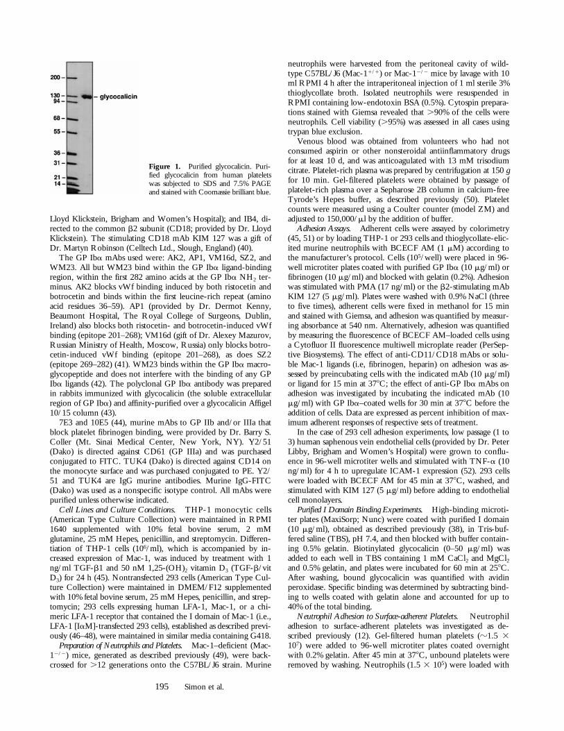
Figure 1. Purified glycocalicin. Puri-fied glycocalicin from human plateletswas subjected to SDS and 7.5% PAGEand stained with Coomassie brilliant blue.
196 GP Iba Is a Counterreceptor for Mac-1
1 mM BCECF AM, washed twice, and then added to each wellfor 60 min at 378C in 5% CO2. After washing, neutrophil adhe-sion was quantified as the percentage of total cells adherent bymeasuring the fluorescence of BCECF AM–loaded cells using aCytofluor II fluorescence multiwell microplate reader (PerSep-tive Biosystems). Fluorescence of input neutrophils before wash-ing served as a measure of total cell number. The effect of mAbson neutrophil adhesion to platelets was assessed as describedabove for purified GP Iba; the effect of the snake venom metal-loprotease, mocarhagin, which cleaves GP Iba at peptide bond282–283 (35), on leukocyte adhesion to platelets was examinedby preincubating surface-adherent platelets with mocarhagin for30 min at 378C. Data are expressed as percent inhibition of max-imum adherent responses of respective sets of treatment.
Whole Blood Detection of Platelet–Leukocyte Aggregates. Leuko-cyte–platelet aggregates were measured by two-color flow cy-tometry in a FACSCalibur™ flow cytometer (Becton Dickinson)by slight modifications of methods described previously (19). Pe-ripheral blood was drawn from a healthy volunteer or, as indi-cated, from a patient with Bernard-Soulier syndrome (BSS) (53)who had not ingested aspirin or other antiplatelet drugs duringthe previous 10 d. The first 2 ml of drawn blood was discarded.Blood was then drawn into a 3.2% sodium citrate Vacutainer(Becton Dickinson). The sample was diluted 1:1 with modifiedTyrode’s Hepes buffer, pH 7.4, and then immediately incubatedat 228C for 10 min with either (a) buffer alone; (b) 0.5 mM ADP(Biodata); or (c) 5 mM thrombin receptor activating peptide(TRAP; 14-mer; Calbiochem) and saturating concentrations ofY2/51-FITC and TUK4-PE. The samples were then fixed at228C for 10 min to a final concentration of 1% formalin (Poly-sciences, Inc.) and 1.53 HBSS concentrate (GIBCO BRL). Thesamples were then diluted with 500 ml of distilled H2O, vor-texed, and incubated at 228C for 10 min to lyse the red bloodcells. Monocytes and neutrophils were identified by CD14-PEpositivity and their characteristic light scatter.
THP-1 Adhesion to Glycocalicin under Flow. The interaction ofTGF-b1/vit D3–stimulated THP-1 cells with immobilized gly-cocalicin was examined using a parallel-plate flow chamber sys-tem, which has been described previously (54). Glass coverslips,which form the bottom of the chamber, were coated with puri-fied glycocalicin (100 mg/ml) by immersing them in the glyco-calicin solution for 3 h at 378C. Residual nonspecific bindingsites were then blocked with 3% BSA for 30 min. The chamberwas then assembled and mounted onto an inverted-stage micro-scope (DIAPHOT-TMD; Nikon) equipped with a silicon-inten-sified target video camera (model C2400; Hammatus) connectedto a video cassette recorder. Cells were suspended in culture me-dium and the suspension was brought to room temperature. 1 mlof the cell suspension (106 cells/ml) was then injected into thechamber and incubated for 1 min. The cells were allowed to set-tle onto the glycocalicin substrate for 1 min; the chamber wasthen perfused with the PBS at a flow rate calculated to generatefluid shear stress of 2 dyn/cm2. The attachment and rolling ofcells in a single-view field were recorded in real time for 3 min,and the video data were then analyzed using imaging software(IC-300 Modular Image Processing Workstation; InovisionCorp.) to calculate the rolling velocities. The rolling eventsscored were ligand specific as confirmed in parallel determina-tions on control substrates coated with heat-stable antigen (HSA).In the study to inhibit glycocalicin, the coverslip was incubatedwith the GP Iba mAb VM16d (25 mg/ml) for 30 min at roomtemperature and washed before assembly of the chamber. To testthe effect of Mac-1 inhibition, the THP-1 cells were incubated
with LMP19c (25 mg/ml) for 30 min at room temperature beforeinjection into the chamber. Rolling cells were those observed tobe moving in the direction of flow while maintaining constantcontact with the glycocalicin substrate.
Statistics. All data are presented as the mean 6 SD. Groupswere compared using the nonpaired t test. P values , 0.05 wereconsidered significant.
ResultsMac-1–expressing Cells Bind to GP Iba. Given the ho-
mology between the receptor-binding vWf A1 domain andthe Mac-1 I domain, we hypothesized that Mac-1 wouldbind the platelet GP Ib-IX-V complex. To assess this po-tential interaction, we assayed the adhesion of Mac-1–bear-ing cells to purified glycocalicin, the soluble extracellulardomain of GP Iba. We have previously shown that stimu-lation of THP-1 monocytic cells (which constitutively ex-
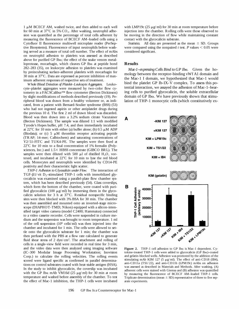
Figure 2. THP-1 cell adhesion to GP Iba is Mac-1 dependent. Cy-tokine-treated THP-1 cells were added to glycocalicin (GP Iba)–coatedand gelatin-blocked wells. Adhesion was promoted by the addition of thestimulating mAb KIM 127 (5 mg/ml). The effect of anti-CD18 (IB4),anti-CD11a (TS1/22), and anti-CD11b (LPM19c) mAbs on adhesionwas assessed as described in Materials and Methods. After washing, (A)adherent cells were stained with Giemsa and (B) adhesion was quantifiedby measuring the fluorescence of BCECF AM–loaded THP-1 cells.Triplicate determination (mean 6 SD) representative of three to five sep-arate experiments.

197 Simon et al.
press Mac-1) with TGF-b1 and 1,25-(OH)2 vitamin D3 in-creases Mac-1 surface expression z2.0-fold (45). Whencytokine-treated THP-1 cells were stimulated with eitherphorbol ester (PMA) or KIM 127, an mAb to CD18 thatinduces a change in the conformation of CD18 and pro-motes both LFA-1– and Mac-1–dependent adhesion (40),they adhered robustly to wells coated with GP Iba andblocked with gelatin, but not to wells coated with gelatinalone (Fig. 2). Adhesion of the cells to GP Iba was inhib-ited by IB4, an anti-CD18 mAb that blocks both LFA-1–and Mac-1–dependent functions, indicating that adhesionis CD18 dependent (Fig. 2, and Table I). THP-1 cell adhe-sion to GP Iba was also completely blocked by LPM19c,an mAb that binds to the I domain of the aM subunit ofMac-1 (CD11b) and blocks the binding of fibrinogen,
C3bi, and ICAM (24). In contrast, the anti–LFA-1 mAbTS1/22 had no effect on adhesion. Similar results were ob-tained with integrin mAbs whether THP-1 cell adhesionwas stimulated with KIM 127 or PMA, ruling out the pos-sibility that the mAbs inhibited adhesion by interferingwith the binding of KIM 127 to CD18.
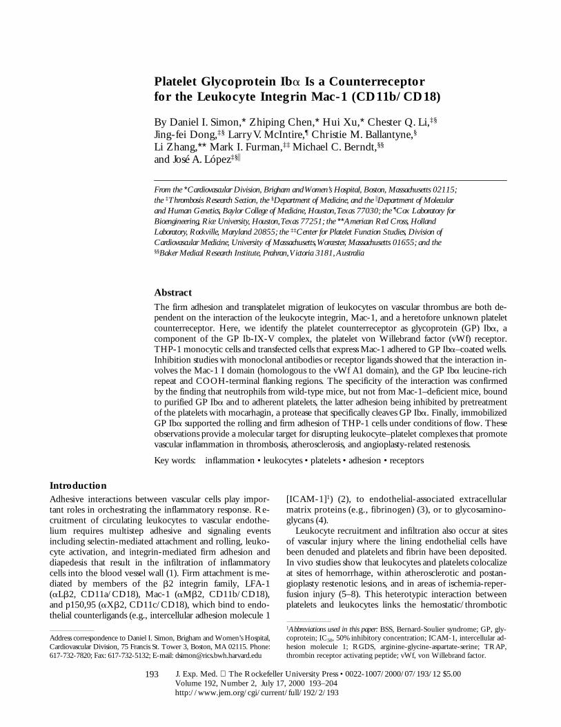
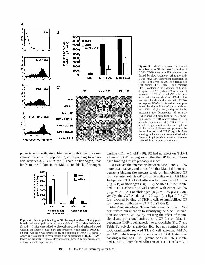
To confirm that Mac-1 mediates the adhesion of THP-1cells to GP Iba and to eliminate the possibility that mAbsto Mac-1 inhibit THP-1 cell adhesion indirectly, we as-sessed the adhesion of 293 cells transfected with eitherLFA-1 or Mac-1 to GP Iba. Flow cytometry confirmedsimilar expression levels of LFA-1 and Mac-1 (Fig. 3 A).Both LFA-1–transfected and Mac-1–transfected 293 cellsadhered robustly to human endothelial cells expressingICAM-1 (Fig. 3 B), indicating that these transfected 293cells are functional. Mac-1–expressing 293 cells adhered toGP Iba, and this adhesion was enhanced by KIM 127 (Fig.3 C). KIM 127–stimulated adhesion was blocked by mAbsdirected to both CD11b and CD18, and to GP Iba(VM16d). Neither untransfected 293 cells nor LFA-1–transfected 293 cells bound to GP Iba.
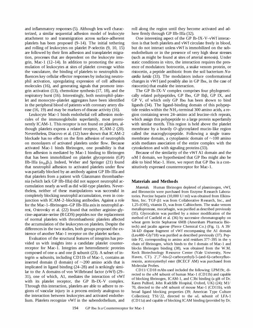
Neutrophil Binding to GP Iba Requires Mac-1. To inves-tigate further the role of Mac-1 in leukocyte adhesion toGP Iba, we examined the binding of neutrophils fromwild-type (Mac-11/1) and Mac-1–deficient (Mac-12/2)mice to GP Iba. Mac-11/1 neutrophils bound to GP Iba,adhesion that was enhanced by PMA pretreatment (Fig. 4).In contrast, Mac-12/2 neutrophils showed significantly re-duced adhesion to GP Iba, whether in the absence or pres-ence of PMA. Taken together, these observations indicatethat Mac-1 mediates adhesion to purified GP Iba.
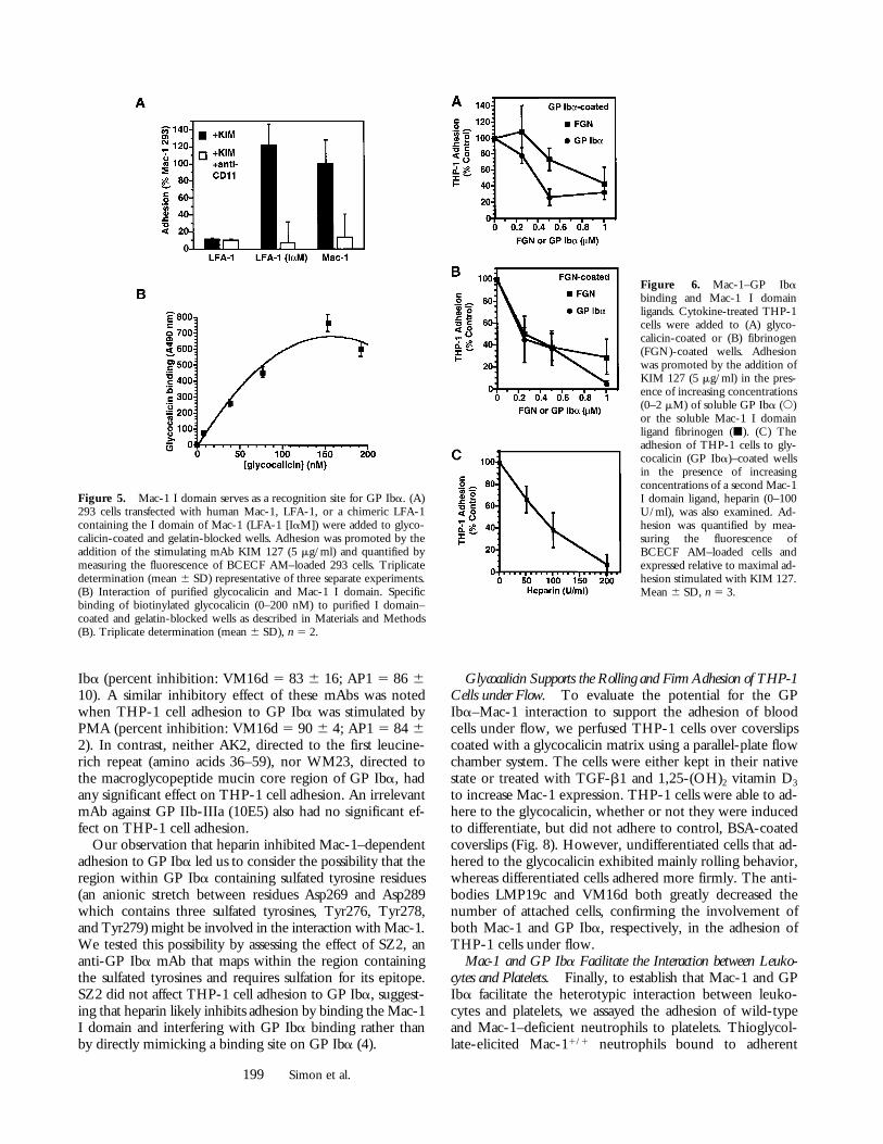
The Mac-1 I Domain Serves as a Recognition Site for GP Iba.The adhesion of Mac-1–bearing THP-1 cells to GP Ibawas inhibited by LPM19c (Fig. 2), an mAb that binds tothe I domain of the aM subunit of Mac-1 (CD11b). Toestablish definitively that the Mac-1 I domain serves as arecognition site for GP Iba, we transfected 293 cells with achimeric LFA-1 receptor that contained the I domain ofMac-1 (i.e., LFA-1 [IaM]–transfected 293 cells). Flow cy-tometry showed comparable expression of LFA-1 (IaM),LFA-1, and Mac-1 in transfected 293 cells (Fig. 3 A). LFA-1–transfected 293 cells did not adhere to GP Iba (Fig. 3 C);in contrast, LFA-1 (IaM)–transfected 293 cells adhered ro-bustly to GP Iba (Fig. 5 A), indicating that the I domain ofMac-1 is required for adhesion to GP Iba. We next inves-tigated whether GP Iba would directly bind to purified Idomain of Mac-1. Biotinylated glycocalicin bound to im-mobilized I domain in a concentration-dependent mannerover the range 0–200 nM (Fig. 5 B).
To evaluate further the I domain as a recognition site onMac-1 for GP Iba, we examined the ability of Mac-1 I do-main ligands (e.g., fibrinogen [24] and heparin [4]) to in-hibit THP-1 adhesion to GP Iba. Soluble fibrinogen (50%inhibitory concentration [IC50] z 1 mM) and heparin (IC50
z 75 U/ml) both inhibited adhesion in a dose-dependentmanner (Fig. 6, A–C). Inhibition by fibrinogen suggestedto us the possibility that the GP Iba and fibrinogen bindingsites in Mac-1 may be partially overlapping. To exclude
Table I. Summary of Inhibition of KIM 127–stimulated THP-1 Adhesion to GP Iba by Antibodies or Soluble Ligands
Antibody or ligand Receptor (epitope)Percent
inhibition
IB4 Anti-CD18 99 6 1*TS1/22 Anti-CD11a (I domain) 10 6 13LPM19c Anti-CD11b (I domain) 92 6 12*Polyclonal Anti-GP Iba 80 6 17*Polyclonal Rabbit IgG control 16 6 20VM16d Anti-GP Iba
(aa residues 201–268) 83 6 16*AP1 Anti-GP Iba
(aa residues 201–268) 86 6 10*AK2 Anti-GP Iba
(aa residues 36–59) 16 6 6SZ2 Anti-GP Iba (sulfated
tyrosine residues 269–282) 8 6 6WM23 Anti-GP Iba
(macroglycopeptide) 11 6 57E3 Anti-GP IIb-IIIa,
-avb3, –Mac-1 16 6 810E5 Anti-GP IIb-IIIa 18 6 6Fibrinogen (2 mM) Mac-1 99 6 1*P2g377–395 (10 mM) Mac-1 0Heparin (200 U/ml) Mac-1 93 6 9*vWF A1 (20 mg/ml) GP Iba 83 6 13*
The adhesion of cytokine-treated THP-1 cells to glycocalicin (GP Iba)–coated microtiter wells was stimulated by the addition of KIM 127 (5mg/ml) in the presence and absence of antibodies directed to or solubleligands of Mac-1 and GP Iba. Polyclonal antibodies were added at 20mg/ml and purified mAbs at 10 mg/ml as described in Materials andMethods. Soluble ligand concentrations are indicated. CD11/CD18mAbs included IB4, TS1/22, and LPM19c; GP Iba mAbs used wereAK2, AP1, VM16d, SZ2, and WM23. Data are expressed as percentinhibition of maximal KIM-stimulated adhesion by the antibodies orsoluble ligands (mean 6 SD, n 5 3–5). aa, amino acids.*P , 0.01.
198 GP Iba Is a Counterreceptor for Mac-1
binding (IC50 z 1 mM) (38). P2 had no effect on THP-1adhesion to GP Iba, suggesting that the GP Iba and fibrin-ogen binding sites are probably distinct.
To evaluate the interaction between Mac-1 and GP Ibamore quantitatively and to confirm that Mac-1 did not rec-ognize a binding site present solely on immobilized GPIba, we tested soluble GP Iba for its ability to inhibit Mac-1–dependent THP-1 cell adhesion to immobilized GP Iba(Fig. 6 B) or fibrinogen (Fig. 6 C). Soluble GP Iba inhib-ited THP-1 adhesion to wells coated with either GP Iba(IC50 5 0.5 mM) or fibrinogen (IC50 5 0.25 mM). Con-versely, the vWf A1 domain (20 mg/ml), a ligand for GPIba, blocked binding of THP-1 cells to immobilized GPIba (percent inhibition 5 83 6 13) (Table I).
Identifying the Mac-1 Binding Site within GP Iba. Wenext turned our attention to identifying the Mac-1 interac-tion site within GP Iba by assessing the effect of mono-clonal and polyclonal antibodies to GP Iba on Mac-1–dependent THP-1 cell adhesion to glycocalicin (Fig. 7, andTable I). Polyclonal anti–GP Iba, but not control rabbitIgG, significantly reduced THP-1 cell adhesion. VM16dand AP1, which map to the leucine-rich COOH-terminalflanking region of GP Iba (amino acids 201–268), inhib-ited KIM 127–stimulated adhesion of THP-1 cells to GP
Figure 3. Mac-1 expression is requiredfor adhesion to GP Iba. (A) Expression ofCD11/CD18 integrin in 293 cells was con-firmed by flow cytometry using the anti-CD18 mAb IB4. Equivalent expression ofCD18 is observed in 293 cells transfectedwith human LFA-1, Mac-1, or a chimericLFA-1 containing the I domain of Mac-1,designated LFA-1 (IaM). (B) Adhesion ofuntransfected 293 cells and 293 cells trans-fected with human Mac-1 or LFA-1 to hu-man endothelial cells stimulated with TNF-ato express ICAM-1. Adhesion was pro-moted by the addition of the stimulatingmAb KIM 127 (5 mg/ml) and quantified bymeasuring the fluorescence of BCECFAM–loaded 293 cells; triplicate determina-tion (mean 6 SD) representative of twoseparate experiments. (C) 293 cells wereadded to glycocalicin-coated and gelatin-blocked wells. Adhesion was promoted bythe addition of KIM 127 (5 mg/ml). Afterwashing, adherent cells were stained withGiemsa. Triplicate determination represen-tative of three separate experiments.
Figure 4. Neutrophil binding to GP Iba requires Mac-1. Thioglycol-late-elicited neutrophils from wild-type (Mac-11/1) or Mac-1–deficient(Mac-12/2) mice were added to glycocalicin-coated and gelatin-blockedwells in the absence (black bars) and presence (white bars) of PMA (17ng/ml). Adhesion was promoted by the addition of PMA (17 ng/ml).Adhesion was quantified by measuring the fluorescence of BCECF AM–loaded neutrophils. Triplicate determination (mean 6 SD) representativeof three separate experiments.
potential nonspecific steric hindrance of fibrinogen, we ex-amined the effect of peptide P2, corresponding to aminoacid residues 377–395 in the g chain of fibrinogen, thatbinds to the I domain of Mac-1 and blocks fibrinogen
199 Simon et al.
Iba (percent inhibition: VM16d 5 83 6 16; AP1 5 86 610). A similar inhibitory effect of these mAbs was notedwhen THP-1 cell adhesion to GP Iba was stimulated byPMA (percent inhibition: VM16d 5 90 6 4; AP1 5 84 62). In contrast, neither AK2, directed to the first leucine-rich repeat (amino acids 36–59), nor WM23, directed tothe macroglycopeptide mucin core region of GP Iba, hadany significant effect on THP-1 cell adhesion. An irrelevantmAb against GP IIb-IIIa (10E5) also had no significant ef-fect on THP-1 cell adhesion.
Our observation that heparin inhibited Mac-1–dependentadhesion to GP Iba led us to consider the possibility that theregion within GP Iba containing sulfated tyrosine residues(an anionic stretch between residues Asp269 and Asp289which contains three sulfated tyrosines, Tyr276, Tyr278,and Tyr279) might be involved in the interaction with Mac-1.We tested this possibility by assessing the effect of SZ2, ananti-GP Iba mAb that maps within the region containingthe sulfated tyrosines and requires sulfation for its epitope.SZ2 did not affect THP-1 cell adhesion to GP Iba, suggest-ing that heparin likely inhibits adhesion by binding the Mac-1I domain and interfering with GP Iba binding rather thanby directly mimicking a binding site on GP Iba (4).
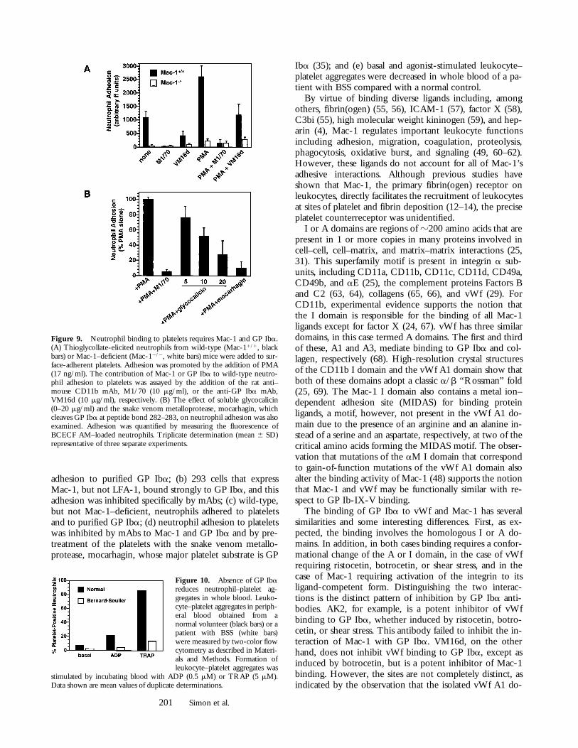
Glycocalicin Supports the Rolling and Firm Adhesion of THP-1Cells under Flow. To evaluate the potential for the GPIba–Mac-1 interaction to support the adhesion of bloodcells under flow, we perfused THP-1 cells over coverslipscoated with a glycocalicin matrix using a parallel-plate flowchamber system. The cells were either kept in their nativestate or treated with TGF-b1 and 1,25-(OH)2 vitamin D3
to increase Mac-1 expression. THP-1 cells were able to ad-here to the glycocalicin, whether or not they were inducedto differentiate, but did not adhere to control, BSA-coatedcoverslips (Fig. 8). However, undifferentiated cells that ad-hered to the glycocalicin exhibited mainly rolling behavior,whereas differentiated cells adhered more firmly. The anti-bodies LMP19c and VM16d both greatly decreased thenumber of attached cells, confirming the involvement ofboth Mac-1 and GP Iba, respectively, in the adhesion ofTHP-1 cells under flow.
Mac-1 and GP Iba Facilitate the Interaction between Leuko-cytes and Platelets. Finally, to establish that Mac-1 and GPIba facilitate the heterotypic interaction between leuko-cytes and platelets, we assayed the adhesion of wild-typeand Mac-1–deficient neutrophils to platelets. Thioglycol-late-elicited Mac-11/1 neutrophils bound to adherent
Figure 5. Mac-1 I domain serves as a recognition site for GP Iba. (A)293 cells transfected with human Mac-1, LFA-1, or a chimeric LFA-1containing the I domain of Mac-1 (LFA-1 [IaM]) were added to glyco-calicin-coated and gelatin-blocked wells. Adhesion was promoted by theaddition of the stimulating mAb KIM 127 (5 mg/ml) and quantified bymeasuring the fluorescence of BCECF AM–loaded 293 cells. Triplicatedetermination (mean 6 SD) representative of three separate experiments.(B) Interaction of purified glycocalicin and Mac-1 I domain. Specificbinding of biotinylated glycocalicin (0–200 nM) to purified I domain–coated and gelatin-blocked wells as described in Materials and Methods(B). Triplicate determination (mean 6 SD), n 5 2.
Figure 6. Mac-1–GP Ibabinding and Mac-1 I domainligands. Cytokine-treated THP-1cells were added to (A) glyco-calicin-coated or (B) fibrinogen(FGN)-coated wells. Adhesionwas promoted by the addition ofKIM 127 (5 mg/ml) in the pres-ence of increasing concentrations(0–2 mM) of soluble GP Iba (s)or the soluble Mac-1 I domainligand fibrinogen (j). (C) Theadhesion of THP-1 cells to gly-cocalicin (GP Iba)–coated wellsin the presence of increasingconcentrations of a second Mac-1I domain ligand, heparin (0–100U/ml), was also examined. Ad-hesion was quantified by mea-suring the fluorescence ofBCECF AM–loaded cells andexpressed relative to maximal ad-hesion stimulated with KIM 127.Mean 6 SD, n 5 3.
200 GP Iba Is a Counterreceptor for Mac-1
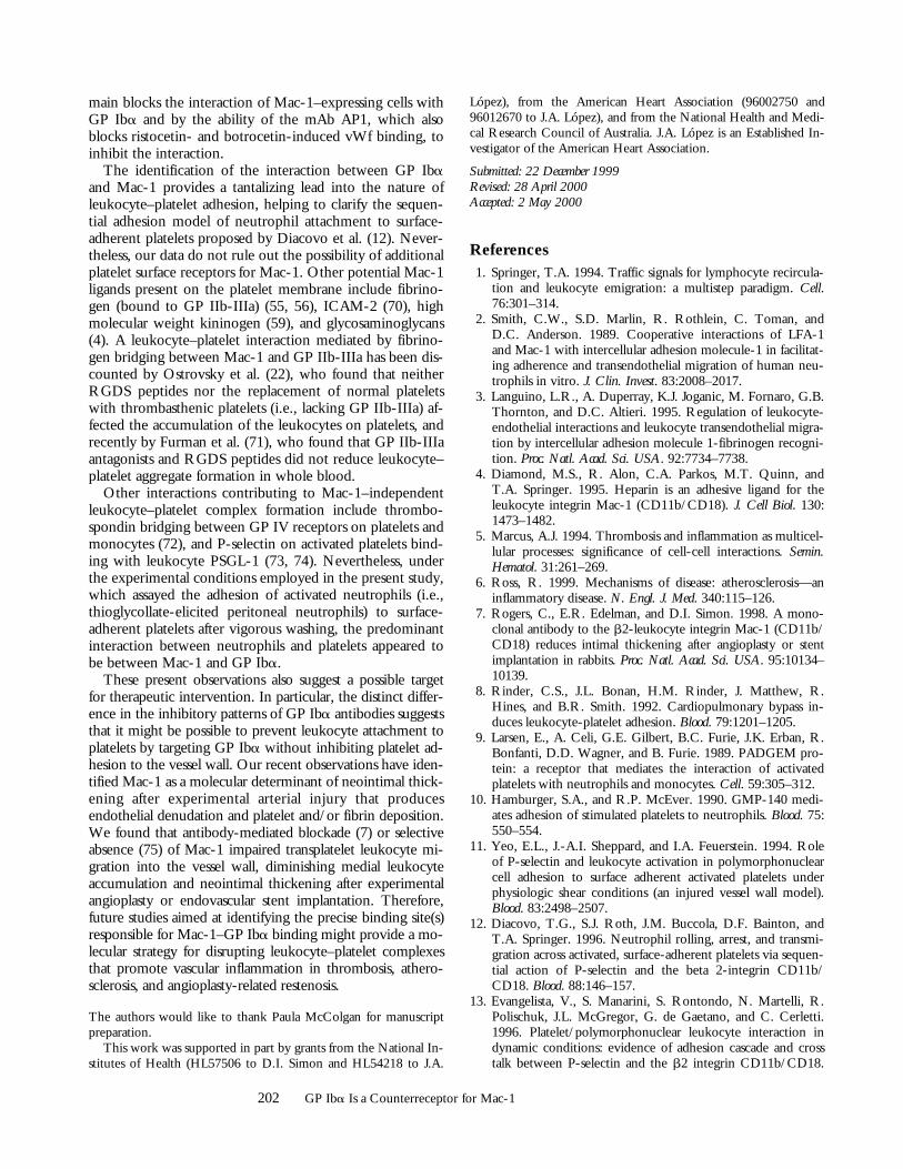
platelets, and this adhesion was promoted by PMA (Fig. 9A). In contrast, Mac-12/2 neutrophils demonstrated mark-edly reduced adhesion to platelets. Adhesion of Mac-11/1
neutrophils was also blocked by the rat anti–mouse Mac-1mAb M1/70 (percent inhibition 5 95 6 5) and the anti-GP Iba mAb VM16d (percent inhibition 5 55 6 10). Fur-thermore, Mac-11/1 leukocyte adhesion to platelets was
inhibited dose-dependently by soluble glycocalicin (per-cent inhibition 5 72 6 18) and by pretreatment of adher-ent platelets with the snake venom metalloprotease, mocar-hagin (percent inhibition 5 89 6 8), which cleaves GP Ibaat peptide bond 282–283 as the only detectable proteolyticevent on the platelet surface (35) (Fig. 9 B). Taken to-gether, these observations indicate that neutrophil adhesionto platelets is primarily mediated by Mac-1 and GP Iba.
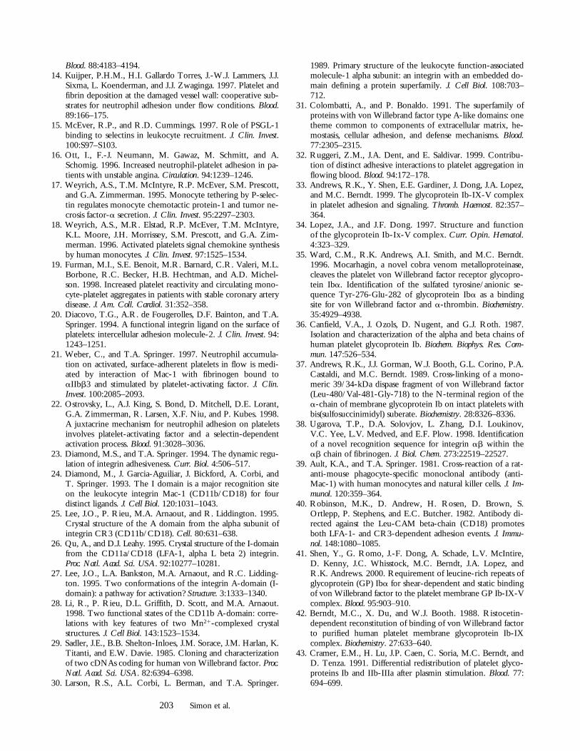
Finally, to provide additional evidence supporting a rolefor GP Iba in platelet binding to leukocytes, we assessedthe presence of leukocyte–platelet aggregates in wholeblood obtained from a normal volunteer or a patient withBSS. We have previously shown that spontaneous or ago-nist-induced leukocyte–platelet aggregate formation re-quire an interaction between P-selectin glycoprotein ligand1 (PSGL-1) and P-selectin that is strengthened by integrins(19). Leukocyte–platelet aggregates were decreased in thecirculation of a patient with BSS compared with a normalcontrol (2 vs. 7% platelet-positive neutrophils; Fig. 10).Moreover, these leukocyte–platelet aggregates were lesslikely to form in the BSS patient after agonist stimulationwith either 0.5 mM ADP (4 vs. 21% platelet-positive neu-trophils) or 5 mM TRAP (13 vs. 86% platelet-positive neu-trophils). Mac-1 dependence in leukocyte–platelet aggre-gate formation in this assay was confirmed by the fact thatthe anti–Mac-1 mAb LPM19c completely inhibited ago-nist-induced platelet–neutrophil complex formation.
DiscussionIn this study, we have identified a direct interaction be-
tween the leukocyte integrin Mac-1 and platelet GP Iba.The following evidence was obtained for this interaction:(a) mAbs to both Mac-1 and GP Iba inhibited THP-1 cell
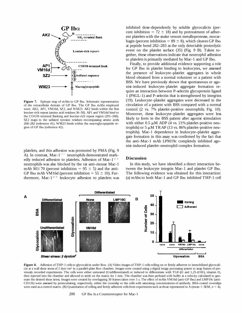
Figure 7. Epitope map of mAbs to GP Iba. Schematic representationof the extracellular domain of GP Iba. The GP Iba mAbs employedwere: AK2, AP1, VM16d, SZ2, and WM23. AK2 binds within the firstleucine-rich repeat (amino acid residues 36–58). AP1 and VM16d bind tothe COOH-terminal flanking and leucine-rich repeat region (201–268).SZ2 maps to the sulfated tyrosine residues encompassing amino acids268–282 (reference 41). WM23 binds within the macroglycopeptide re-gion of GP Iba (reference 42).
Figure 8. Adhesion of THP-1 cells to glycocalicin under flow. (A) Video images of THP-1 cells rolling on or firmly adherent to immobilized glycocali-cin at a wall shear stress of 2 dyn/cm2 in a parallel-plate flow chamber. Images were created using a digital image processing system to snap frames of pre-viously recorded experiments. The cells were either untreated (Undifferentiated) or induced to differentiate with TGF-b1 and 1,25-(OH)2 vitamin D3
then injected into the chamber and allowed to settle on the matrix for 1 min. The chamber was then perfused with buffer at a velocity calculated to gen-erate the desired shear stress. Images were created by overlapping 30 frames taken over 1 s. The effect of mAbs VM16d (anti-GP Iba) and LMP19c (anti-CD11b) were assessed by preincubating, respectively, either the coverslip or the cells with saturating concentrations of antibody. BSA-coated coverslipswere used as a control matrix. (B) Quantitation of rolling and firmly adherent cells from experiments such as those represented in A (mean 6 SEM, n 5 4).
201 Simon et al.
adhesion to purified GP Iba; (b) 293 cells that expressMac-1, but not LFA-1, bound strongly to GP Iba, and thisadhesion was inhibited specifically by mAbs; (c) wild-type,but not Mac-1–deficient, neutrophils adhered to plateletsand to purified GP Iba; (d) neutrophil adhesion to plateletswas inhibited by mAbs to Mac-1 and GP Iba and by pre-treatment of the platelets with the snake venom metallo-protease, mocarhagin, whose major platelet substrate is GP
Iba (35); and (e) basal and agonist-stimulated leukocyte–platelet aggregates were decreased in whole blood of a pa-tient with BSS compared with a normal control.
By virtue of binding diverse ligands including, amongothers, fibrin(ogen) (55, 56), ICAM-1 (57), factor X (58),C3bi (55), high molecular weight kininogen (59), and hep-arin (4), Mac-1 regulates important leukocyte functionsincluding adhesion, migration, coagulation, proteolysis,phagocytosis, oxidative burst, and signaling (49, 60–62).However, these ligands do not account for all of Mac-1’sadhesive interactions. Although previous studies haveshown that Mac-1, the primary fibrin(ogen) receptor onleukocytes, directly facilitates the recruitment of leukocytesat sites of platelet and fibrin deposition (12–14), the preciseplatelet counterreceptor was unidentified.
I or A domains are regions of z200 amino acids that arepresent in 1 or more copies in many proteins involved incell–cell, cell–matrix, and matrix–matrix interactions (25,31). This superfamily motif is present in integrin a sub-units, including CD11a, CD11b, CD11c, CD11d, CD49a,CD49b, and aE (25), the complement proteins Factors Band C2 (63, 64), collagens (65, 66), and vWf (29). ForCD11b, experimental evidence supports the notion thatthe I domain is responsible for the binding of all Mac-1ligands except for factor X (24, 67). vWf has three similardomains, in this case termed A domains. The first and thirdof these, A1 and A3, mediate binding to GP Iba and col-lagen, respectively (68). High-resolution crystal structuresof the CD11b I domain and the vWf A1 domain show thatboth of these domains adopt a classic a/b “Rossman” fold(25, 69). The Mac-1 I domain also contains a metal ion–dependent adhesion site (MIDAS) for binding proteinligands, a motif, however, not present in the vWf A1 do-main due to the presence of an arginine and an alanine in-stead of a serine and an aspartate, respectively, at two of thecritical amino acids forming the MIDAS motif. The obser-vation that mutations of the aM I domain that correspondto gain-of-function mutations of the vWf A1 domain alsoalter the binding activity of Mac-1 (48) supports the notionthat Mac-1 and vWf may be functionally similar with re-spect to GP Ib-IX-V binding.
The binding of GP Iba to vWf and Mac-1 has severalsimilarities and some interesting differences. First, as ex-pected, the binding involves the homologous I or A do-mains. In addition, in both cases binding requires a confor-mational change of the A or I domain, in the case of vWfrequiring ristocetin, botrocetin, or shear stress, and in thecase of Mac-1 requiring activation of the integrin to itsligand-competent form. Distinguishing the two interac-tions is the distinct pattern of inhibition by GP Iba anti-bodies. AK2, for example, is a potent inhibitor of vWfbinding to GP Iba, whether induced by ristocetin, botro-cetin, or shear stress. This antibody failed to inhibit the in-teraction of Mac-1 with GP Iba. VM16d, on the otherhand, does not inhibit vWf binding to GP Iba, except asinduced by botrocetin, but is a potent inhibitor of Mac-1binding. However, the sites are not completely distinct, asindicated by the observation that the isolated vWf A1 do-
Figure 9. Neutrophil binding to platelets requires Mac-1 and GP Iba.(A) Thioglycollate-elicited neutrophils from wild-type (Mac-11/1, blackbars) or Mac-1–deficient (Mac-12/2, white bars) mice were added to sur-face-adherent platelets. Adhesion was promoted by the addition of PMA(17 ng/ml). The contribution of Mac-1 or GP Iba to wild-type neutro-phil adhesion to platelets was assayed by the addition of the rat anti–mouse CD11b mAb, M1/70 (10 mg/ml), or the anti-GP Iba mAb,VM16d (10 mg/ml), respectively. (B) The effect of soluble glycocalicin(0–20 mg/ml) and the snake venom metalloprotease, mocarhagin, whichcleaves GP Iba at peptide bond 282–283, on neutrophil adhesion was alsoexamined. Adhesion was quantified by measuring the fluorescence ofBCECF AM–loaded neutrophils. Triplicate determination (mean 6 SD)representative of three separate experiments.
Figure 10. Absence of GP Ibareduces neutrophil–platelet ag-gregates in whole blood. Leuko-cyte–platelet aggregates in periph-eral blood obtained from anormal volunteer (black bars) or apatient with BSS (white bars)were measured by two-color flowcytometry as described in Materi-als and Methods. Formation ofleukocyte–platelet aggregates was
stimulated by incubating blood with ADP (0.5 mM) or TRAP (5 mM).Data shown are mean values of duplicate determinations.
202 GP Iba Is a Counterreceptor for Mac-1
main blocks the interaction of Mac-1–expressing cells withGP Iba and by the ability of the mAb AP1, which alsoblocks ristocetin- and botrocetin-induced vWf binding, toinhibit the interaction.
The identification of the interaction between GP Ibaand Mac-1 provides a tantalizing lead into the nature ofleukocyte–platelet adhesion, helping to clarify the sequen-tial adhesion model of neutrophil attachment to surface-adherent platelets proposed by Diacovo et al. (12). Never-theless, our data do not rule out the possibility of additionalplatelet surface receptors for Mac-1. Other potential Mac-1ligands present on the platelet membrane include fibrino-gen (bound to GP IIb-IIIa) (55, 56), ICAM-2 (70), highmolecular weight kininogen (59), and glycosaminoglycans(4). A leukocyte–platelet interaction mediated by fibrino-gen bridging between Mac-1 and GP IIb-IIIa has been dis-counted by Ostrovsky et al. (22), who found that neitherRGDS peptides nor the replacement of normal plateletswith thrombasthenic platelets (i.e., lacking GP IIb-IIIa) af-fected the accumulation of the leukocytes on platelets, andrecently by Furman et al. (71), who found that GP IIb-IIIaantagonists and RGDS peptides did not reduce leukocyte–platelet aggregate formation in whole blood.
Other interactions contributing to Mac-1–independentleukocyte–platelet complex formation include thrombo-spondin bridging between GP IV receptors on platelets andmonocytes (72), and P-selectin on activated platelets bind-ing with leukocyte PSGL-1 (73, 74). Nevertheless, underthe experimental conditions employed in the present study,which assayed the adhesion of activated neutrophils (i.e.,thioglycollate-elicited peritoneal neutrophils) to surface-adherent platelets after vigorous washing, the predominantinteraction between neutrophils and platelets appeared tobe between Mac-1 and GP Iba.
These present observations also suggest a possible targetfor therapeutic intervention. In particular, the distinct differ-ence in the inhibitory patterns of GP Iba antibodies suggeststhat it might be possible to prevent leukocyte attachment toplatelets by targeting GP Iba without inhibiting platelet ad-hesion to the vessel wall. Our recent observations have iden-tified Mac-1 as a molecular determinant of neointimal thick-ening after experimental arterial injury that producesendothelial denudation and platelet and/or fibrin deposition.We found that antibody-mediated blockade (7) or selectiveabsence (75) of Mac-1 impaired transplatelet leukocyte mi-gration into the vessel wall, diminishing medial leukocyteaccumulation and neointimal thickening after experimentalangioplasty or endovascular stent implantation. Therefore,future studies aimed at identifying the precise binding site(s)responsible for Mac-1–GP Iba binding might provide a mo-lecular strategy for disrupting leukocyte–platelet complexesthat promote vascular inflammation in thrombosis, athero-sclerosis, and angioplasty-related restenosis.
The authors would like to thank Paula McColgan for manuscriptpreparation.
This work was supported in part by grants from the National In-stitutes of Health (HL57506 to D.I. Simon and HL54218 to J.A.
López), from the American Heart Association (96002750 and96012670 to J.A. López), and from the National Health and Medi-cal Research Council of Australia. J.A. López is an Established In-vestigator of the American Heart Association.
Submitted: 22 December 1999Revised: 28 April 2000Accepted: 2 May 2000
References1. Springer, T.A. 1994. Traffic signals for lymphocyte recircula-
tion and leukocyte emigration: a multistep paradigm. Cell.76:301–314.
2. Smith, C.W., S.D. Marlin, R. Rothlein, C. Toman, andD.C. Anderson. 1989. Cooperative interactions of LFA-1and Mac-1 with intercellular adhesion molecule-1 in facilitat-ing adherence and transendothelial migration of human neu-trophils in vitro. J. Clin. Invest. 83:2008–2017.
3. Languino, L.R., A. Duperray, K.J. Joganic, M. Fornaro, G.B.Thornton, and D.C. Altieri. 1995. Regulation of leukocyte-endothelial interactions and leukocyte transendothelial migra-tion by intercellular adhesion molecule 1-fibrinogen recogni-tion. Proc. Natl. Acad. Sci. USA. 92:7734–7738.
4. Diamond, M.S., R. Alon, C.A. Parkos, M.T. Quinn, andT.A. Springer. 1995. Heparin is an adhesive ligand for theleukocyte integrin Mac-1 (CD11b/CD18). J. Cell Biol. 130:1473–1482.
5. Marcus, A.J. 1994. Thrombosis and inflammation as multicel-lular processes: significance of cell-cell interactions. Semin.Hematol. 31:261–269.
6. Ross, R. 1999. Mechanisms of disease: atherosclerosis—aninflammatory disease. N. Engl. J. Med. 340:115–126.
7. Rogers, C., E.R. Edelman, and D.I. Simon. 1998. A mono-clonal antibody to the b2-leukocyte integrin Mac-1 (CD11b/CD18) reduces intimal thickening after angioplasty or stentimplantation in rabbits. Proc. Natl. Acad. Sci. USA. 95:10134–10139.
8. Rinder, C.S., J.L. Bonan, H.M. Rinder, J. Matthew, R.Hines, and B.R. Smith. 1992. Cardiopulmonary bypass in-duces leukocyte-platelet adhesion. Blood. 79:1201–1205.
9. Larsen, E., A. Celi, G.E. Gilbert, B.C. Furie, J.K. Erban, R.Bonfanti, D.D. Wagner, and B. Furie. 1989. PADGEM pro-tein: a receptor that mediates the interaction of activatedplatelets with neutrophils and monocytes. Cell. 59:305–312.
10. Hamburger, S.A., and R.P. McEver. 1990. GMP-140 medi-ates adhesion of stimulated platelets to neutrophils. Blood. 75:550–554.
11. Yeo, E.L., J.-A.I. Sheppard, and I.A. Feuerstein. 1994. Roleof P-selectin and leukocyte activation in polymorphonuclearcell adhesion to surface adherent activated platelets underphysiologic shear conditions (an injured vessel wall model).Blood. 83:2498–2507.
12. Diacovo, T.G., S.J. Roth, J.M. Buccola, D.F. Bainton, andT.A. Springer. 1996. Neutrophil rolling, arrest, and transmi-gration across activated, surface-adherent platelets via sequen-tial action of P-selectin and the beta 2-integrin CD11b/CD18. Blood. 88:146–157.
13. Evangelista, V., S. Manarini, S. Rontondo, N. Martelli, R.Polischuk, J.L. McGregor, G. de Gaetano, and C. Cerletti.1996. Platelet/polymorphonuclear leukocyte interaction indynamic conditions: evidence of adhesion cascade and crosstalk between P-selectin and the b2 integrin CD11b/CD18.
203 Simon et al.
Blood. 88:4183–4194.14. Kuijper, P.H.M., H.I. Gallardo Torres, J.-W.J. Lammers, J.J.
Sixma, L. Koenderman, and J.J. Zwaginga. 1997. Platelet andfibrin deposition at the damaged vessel wall: cooperative sub-strates for neutrophil adhesion under flow conditions. Blood.89:166–175.
15. McEver, R.P., and R.D. Cummings. 1997. Role of PSGL-1binding to selectins in leukocyte recruitment. J. Clin. Invest.100:S97–S103.
16. Ott, I., F.-J. Neumann, M. Gawaz, M. Schmitt, and A.Schomig. 1996. Increased neutrophil-platelet adhesion in pa-tients with unstable angina. Circulation. 94:1239–1246.
17. Weyrich, A.S., T.M. McIntyre, R.P. McEver, S.M. Prescott,and G.A. Zimmerman. 1995. Monocyte tethering by P-selec-tin regulates monocyte chemotactic protein-1 and tumor ne-crosis factor-a secretion. J. Clin. Invest. 95:2297–2303.
18. Weyrich, A.S., M.R. Elstad, R.P. McEver, T.M. McIntyre,K.L. Moore, J.H. Morrissey, S.M. Prescott, and G.A. Zim-merman. 1996. Activated platelets signal chemokine synthesisby human monocytes. J. Clin. Invest. 97:1525–1534.
19. Furman, M.I., S.E. Benoit, M.R. Barnard, C.R. Valeri, M.L.Borbone, R.C. Becker, H.B. Hechtman, and A.D. Michel-son. 1998. Increased platelet reactivity and circulating mono-cyte-platelet aggregates in patients with stable coronary arterydisease. J. Am. Coll. Cardiol. 31:352–358.
20. Diacovo, T.G., A.R. de Fougerolles, D.F. Bainton, and T.A.Springer. 1994. A functional integrin ligand on the surface ofplatelets: intercellular adhesion molecule-2. J. Clin. Invest. 94:1243–1251.
21. Weber, C., and T.A. Springer. 1997. Neutrophil accumula-tion on activated, surface-adherent platelets in flow is medi-ated by interaction of Mac-1 with fibrinogen bound toaIIbb3 and stimulated by platelet-activating factor. J. Clin.Invest. 100:2085–2093.
22. Ostrovsky, L., A.J. King, S. Bond, D. Mitchell, D.E. Lorant,G.A. Zimmerman, R. Larsen, X.F. Niu, and P. Kubes. 1998.A juxtacrine mechanism for neutrophil adhesion on plateletsinvolves platelet-activating factor and a selectin-dependentactivation process. Blood. 91:3028–3036.
23. Diamond, M.S., and T.A. Springer. 1994. The dynamic regu-lation of integrin adhesiveness. Curr. Biol. 4:506–517.
24. Diamond, M., J. Garcia-Aguiliar, J. Bickford, A. Corbi, andT. Springer. 1993. The I domain is a major recognition siteon the leukocyte integrin Mac-1 (CD11b/CD18) for fourdistinct ligands. J. Cell Biol. 120:1031–1043.
25. Lee, J.O., P. Rieu, M.A. Arnaout, and R. Liddington. 1995.Crystal structure of the A domain from the alpha subunit ofintegrin CR3 (CD11b/CD18). Cell. 80:631–638.
26. Qu, A., and D.J. Leahy. 1995. Crystal structure of the I-domainfrom the CD11a/CD18 (LFA-1, alpha L beta 2) integrin.Proc. Natl. Acad. Sci. USA. 92:10277–10281.
27. Lee, J.O., L.A. Bankston, M.A. Arnaout, and R.C. Lidding-ton. 1995. Two conformations of the integrin A-domain (I-domain): a pathway for activation? Structure. 3:1333–1340.
28. Li, R., P. Rieu, D.L. Griffith, D. Scott, and M.A. Arnaout.1998. Two functional states of the CD11b A-domain: corre-lations with key features of two Mn21-complexed crystalstructures. J. Cell Biol. 143:1523–1534.
29. Sadler, J.E., B.B. Shelton-Inloes, J.M. Sorace, J.M. Harlan, K.Titanti, and E.W. Davie. 1985. Cloning and characterizationof two cDNAs coding for human von Willebrand factor. Proc.Natl. Acad. Sci. USA. 82:6394–6398.
30. Larson, R.S., A.L. Corbi, L. Berman, and T.A. Springer.
1989. Primary structure of the leukocyte function-associatedmolecule-1 alpha subunit: an integrin with an embedded do-main defining a protein superfamily. J. Cell Biol. 108:703–712.
31. Colombatti, A., and P. Bonaldo. 1991. The superfamily ofproteins with von Willebrand factor type A-like domains: onetheme common to components of extracellular matrix, he-mostasis, cellular adhesion, and defense mechanisms. Blood.77:2305–2315.
32. Ruggeri, Z.M., J.A. Dent, and E. Saldivar. 1999. Contribu-tion of distinct adhesive interactions to platelet aggregation inflowing blood. Blood. 94:172–178.
33. Andrews, R.K., Y. Shen, E.E. Gardiner, J. Dong, J.A. Lopez,and M.C. Berndt. 1999. The glycoprotein Ib-IX-V complexin platelet adhesion and signaling. Thromb. Haemost. 82:357–364.
34. Lopez, J.A., and J.F. Dong. 1997. Structure and functionof the glycoprotein Ib-Ix-V complex. Curr. Opin. Hematol.4:323–329.
35. Ward, C.M., R.K. Andrews, A.I. Smith, and M.C. Berndt.1996. Mocarhagin, a novel cobra venom metalloproteinase,cleaves the platelet von Willebrand factor receptor glycopro-tein Iba. Identification of the sulfated tyrosine/anionic se-quence Tyr-276-Glu-282 of glycoprotein Iba as a bindingsite for von Willebrand factor and a-thrombin. Biochemistry.35:4929–4938.
36. Canfield, V.A., J. Ozols, D. Nugent, and G.J. Roth. 1987.Isolation and characterization of the alpha and beta chains ofhuman platelet glycoprotein Ib. Biochem. Biophys. Res. Com-mun. 147:526–534.
37. Andrews, R.K., J.J. Gorman, W.J. Booth, G.L. Corino, P.A.Castaldi, and M.C. Berndt. 1989. Cross-linking of a mono-meric 39/34-kDa dispase fragment of von Willebrand factor(Leu-480/Val-481-Gly-718) to the N-terminal region of thea-chain of membrane glycoprotein Ib on intact platelets withbis(sulfosuccinimidyl) suberate. Biochemistry. 28:8326–8336.
38. Ugarova, T.P., D.A. Solovjov, L. Zhang, D.I. Loukinov,V.C. Yee, L.V. Medved, and E.F. Plow. 1998. Identificationof a novel recognition sequence for integrin ab within theab chain of fibrinogen. J. Biol. Chem. 273:22519–22527.
39. Ault, K.A., and T.A. Springer. 1981. Cross-reaction of a rat-anti-mouse phagocyte-specific monoclonal antibody (anti-Mac-1) with human monocytes and natural killer cells. J. Im-munol. 120:359–364.
40. Robinson, M.K., D. Andrew, H. Rosen, D. Brown, S.Ortlepp, P. Stephens, and E.C. Butcher. 1982. Antibody di-rected against the Leu-CAM beta-chain (CD18) promotesboth LFA-1- and CR3-dependent adhesion events. J. Immu-nol. 148:1080–1085.
41. Shen, Y., G. Romo, J.-F. Dong, A. Schade, L.V. McIntire,D. Kenny, J.C. Whisstock, M.C. Berndt, J.A. Lopez, andR.K. Andrews. 2000. Requirement of leucine-rich repeats ofglycoprotein (GP) Iba for shear-dependent and static bindingof von Willebrand factor to the platelet membrane GP Ib-IX-Vcomplex. Blood. 95:903–910.
42. Berndt, M.C., X. Du, and W.J. Booth. 1988. Ristocetin-dependent reconstitution of binding of von Willebrand factorto purified human platelet membrane glycoprotein Ib-IXcomplex. Biochemistry. 27:633–640.
43. Cramer, E.M., H. Lu, J.P. Caen, C. Soria, M.C. Berndt, andD. Tenza. 1991. Differential redistribution of platelet glyco-proteins Ib and IIb-IIIa after plasmin stimulation. Blood. 77:694–699.
204 GP Iba Is a Counterreceptor for Mac-1
44. Coller, B.S., E.I. Peerschke, L.E. Scudder, and C.A. Sullivan.1983. A murine monoclonal antibody that completely blocksthe binding of fibrinogen to platelets produces a thrombas-thenic-like state in normal platelets and binds to glycoproteinsIIb and/or IIIa. J. Clin. Invest. 72:325–338.
45. Simon, D.I., N.K. Rao, H. Xu, Y. Wei, O. Majdic, E.Ronne, L. Kobzik, and H.A. Chapman. 1996. Mac-1(CD11b/CD18) and the urokinase receptor (CD87) form afunctional unit on monocytic cells. Blood. 88:3185–3194.
46. Zhang, L., and E.F. Plow. 1997. Identification and recon-struction of the binding site within aMb2 for a specific andhigh affinity ligand, NIF. J. Biol. Chem. 272:17558–17564.
47. Zhang, L., and E.F. Plow. 1996. Overlapping, but not identi-cal, sites are involved in the recognition of C3bi, neutrophilinhibitory factor, and adhesive ligands by the aMb2 integrin.J. Biol. Chem. 271:18211–18216.
48. Zhang, L., and E.F. Plow. 1996. A discrete site modulates ac-tivation of I domains. J. Biol. Chem. 271:29953–29957.
49. Lu, H., C.W. Smith, J. Perrard, D. Bullard, L. Tang, S.B.Shappell, M.L. Entman, A.L. Beaudet, and C.M. Ballantyne.1997. LFA-1 is sufficient in mediating neutrophil emigrationin Mac-1-deficient mice. J. Clin. Invest. 99:1340–1350.
50. Hawiger, J., S. Parkinson, and S. Timmons. 1980. Prostacyclininhibits mobilization of fibrinogen-binding sites on humanADP- and thrombin-treated platelets. Nature. 283:195–198.
51. Wei, Y., D.A. Waltz, N. Rai, R.J. Drummond, S. Rosen-berg, M.V. Doyle, and H.A. Chapman. 1994. Identificationof the urokinase receptor as an adhesion receptor for vit-ronectin. J. Biol. Chem. 269:32380–32388.
52. Bevilacqua, M.P., J.S. Pober, M.E. Wheeler, R.S. Cotran,and M.A.J. Gimbrone. 1985. Interleukin 1 acts on culturedhuman vascular endothelium to increase the adhesion ofpolymorphonuclear leukocytes, monocytes, and related leu-kocyte cell lines. J. Clin. Invest. 76:2003–2011.
53. LaRosa, C.A., M.J. Rohrer, S.E. Benoit, M.R. Barnard, andA.D. Michelson. 1994. Neutrophil cathepsin G modulatesthe platelet surface expression of the glycoprotein (GP) Ib-IXcomplex by proteolysis of the von Willebrand factor bindingsite on GPIba and by a cytoskeletal-mediated redistributionof the remainder of the complex. Blood. 84:158–168.
54. Romo, G.M., J.F. Dong, A.J. Schade, E.E. Gardiner, G.S.Kansas, C.Q. Li, L.V. McIntire, M.C. Berndt, and J.A. Lo-pez. 1999. The glycoprotein Ib-IX-V complex is a plateletcounterreceptor for P-selectin. J. Exp. Med. 190:803–814.
55. Wright, S.D., J.S. Weitz, A.J. Huang, S.M. Levin, S.C. Sil-verstein, and J.D. Loike. 1988. Complement receptor typethree (CD11b/CD18) of human polymorphonuclear leuko-cytes recognizes fibrinogen. Proc. Natl. Acad. Sci. USA. 85:7734–7738.
56. Altieri, D.C., R. Bader, P.M. Mannucci, and T.S. Edgington.1988. Oligospecificity of the cellular adhesion receptor Mac-1encompasses an inducible recognition specificity for fibrino-gen. J. Cell Biol. 107:1893–1900.
57. Diamond, M.S., D.E. Staunton, S.D. Marlin, and T.A.Springer. 1991. Binding of the integrin Mac-1 (CD11b/CD18) to the third immunoglobulin-like domain of ICAM-1(CD54) and its regulation by glycosylation. Cell. 65:961–971.
58. Altieri, D.C., J.H. Morrissey, and T.S. Edgington. 1988. Ad-hesive receptor Mac-1 coordinates the activation of factor Xon stimulated cells of monocytic and myeloid differentiation:an alternative initiation of the coagulation cascade. Proc. Natl.Acad. Sci. USA. 85:7462–7466.
59. Wachtfogel, Y.T., R.A. DeLa Cadena, S.P. Kunapuli, L. Rick,
M. Miller, R.L. Schultz, D.C. Altieri, T.S. Edgington, andR.W. Colman. 1994. High molecular weight kininogen bindsto Mac-1 on neutrophils by its heavy chain (domain 3) and itslight chain (domain 5). J. Biol. Chem. 269:19307–19312.
60. Arnaout, M.A. 1990. Structure and function of the leukocyteadhesion molecules CD11/CD18. Blood. 75:1037–1050.
61. Plow, E.F., and L. Zhang. 1997. A MAC-1 attack: integrinfunctions directly challenged in knockout mice. J. Clin. In-vest. 99:1145–1146.
62. Coxon, A., P. Rieu, F.J. Barkalow, S. Askari, A.H. Sharpe,U.H. von Andrian, M.A. Arnaout, and T.N. Mayadas. 1996.A novel role for the beta 2 integrin CD11b/CD18 in neutro-phil apoptosis: a homeostatic mechanism in inflammation. Im-munity. 5:653–666.
63. Campbell, R.D., and R.R. Porter. 1983. Molecular cloningand characterization of the gene coding for human complementprotein factor B. Proc. Natl. Acad. Sci. USA. 80:4464–4468.
64. Bentley, D.R. 1986. Primary structure of human complementcomponent C2. Biochem. J. 239:339–345.
65. Chu, M.-L., R.-Z. Zhang, T.-C. Pan, D. Stokes, D. Con-way, H.-J. Kuo, R. Glanville, U. Mayer, K. Mann, R.Deutzmann, and R. Timple. 1990. Mosaic structure of glob-ular domains in the human type VI collagen a3 chain: simi-larity to von Willebrand factor, fibronectin, actin, salivaryproteins and aprotinin type protease inhibitors. EMBO (Eur.Mol. Biol. Organ.) J. 9:385–393.
66. Colombati, A., P. Bonaldo, and R. Doliana. 1993. Type Amodules: interacting domains found in several non-fibrillarcollagens and in other matrix proteins. Matrix. 13:297–306.
67. Zhou, L., D.H. Lee, J. Plescia, C.Y. Lau, and D.C. Altieri.1994. Differential ligand binding specificities of recombinantCD11b/CD18 integrin I-domain. J. Biol. Chem. 269:17075–17079.
68. Ruggeri, Z.M. 1999. Structure and function of von Wille-brand factor. Thromb. Haemost. 82:576–584.
69. Emsley, J., M. Cruz, R. Handin, and R. Liddington. 1998.Crystal structure of the von Willebrand Factor A1 domainand implications for the binding of platelet glycoprotein Ib. J.Biol. Chem. 273:10396–10401.
70. Xie, J., R. Li, P. Kotovuori, C. Vermont-Desroches, J.Wijdenes, M.A. Arnaout, P. Nortamo, and C.G. Gahmberg.1995. Intercellular adhesion molecule-2 (CD102) binds to theleukocyte integrin CD11b/CD18 through the A domain. J.Immunol. 155:3619–3628.
71. Furman, M.I., L.A. Krueger, A.L. Frelinger III, M.R. Barnard,M.A. Mascelli, M.T. Nakada, and A.D. Michelson. 1999.Tirofiban and eptifibatide, but not abciximab, induce leuko-cyte-platelet aggregation. Circulation. 100:1-681. (Abstr.)
72. Silverstein, R., A.S. Asch, and R.L. Nachman. 1989. Glyco-protein IV mediates thrombospondin-dependent platelet-monocyte and platelet-U937 cell adhesion. J. Clin. Invest. 84:546–552.
73. Skinner, M.P., C.M. Lucas, G.F. Burns, C.N. Chesterman,and M.C. Berndt. 1991. GMP-140 binding to neutrophils isinhibited by sulfated glycans. J. Biol. Chem. 266:5371–5374.
74. Moore, K.L., A. Varki, and R.P. McEver. 1991. GMP-140binds to a glycoprotein receptor on human neutrophils: evi-dence for a lectin-like interaction. J. Cell Biol. 112:491–499.
75. Simon, D.I., Z. Chen, P. Seifert, E.R. Edelman, C.M. Bal-lantyne, and C. Rogers. 2000. Decreased neointimal forma-tion in Mac-12/2 mice reveals a role for inflammation invascular repair after angioplasty. J. Clin. Invest. 105:293–300.