Leukocyte adhesion deficiency-III is caused by mutations in KINDLIN3 affecting integrin activation
Upload
independentCategory
view
2download
0
© 2001 Hindawi Publishing Corporation
Journal of Biomedicine and Biotechnology • 1:3 (2001) 114–121 • PII. S1110724301000250 • http://jbb.hindawi.com
RESEARCH ARTICLE
Two novel frame shift, recurrent and de novo mutationsin the ITGB2 (CD18) gene causing leukocyte
adhesion deficiency in a highly inbredNorth African population
D. M. Fathallah,1∗ T. Jamal,1 M. R. Barbouche,1 M. Bejaoui,2 M. Ben Hariz,3 and K. Dellagi1
1Laboratory of Immunology (The Molecular Genetics Group) AUPELF-UREF LAF 301,Institut Pasteur de Tunis, BP74 1002 le Belvédère, Tunis, Tunisia
2Center for Bone marrow Transplantation, Tunis, Tunisia3Department of Pediatrics, Mongi Slim Hospital, La Marsa, Tunisia
We have identified four different mutations causing leukocyte adhesion deficiency (LAD) in the ITGB2 gene of patients from ahighly inbred population. Two were novel single-bp deletions (1497delG and 1920delG) causing frame shift and the two otherswere the missense mutations G284S and R593C. In our study, the G284S was a recurrent mutation while the R593C occurred denovo. We have also characterized a novel Xba1 polymorphic site located at the 5′ end of the ITGB2 locus. Family studies showedthat the 1497delG mutation segregated with this marker and the intragenic AvaII polymorphic marker, suggesting the presence of afounder effect. The observation of a heterogeneous spectrum including de novo and recurrent mutations causing LAD in a highlyinbred population is rather unexpected. In view of the literature published on the molecular genetics of LAD and considering theethnic origin of the patients studied, our findings confirm the heterogeneity of the mutations causing LAD and point out potentialmutational hot spots in the ITGB2 gene.
INTRODUCTION
Leukocyte adhesion deficiency (LAD) is a primary im-munedeficiency inherited as a rare autosomal, recessive ge-netic disorder. Clinically LAD is characterized by a delayedumbilical cord separation, recurrent life-threatening infec-tions, impaired pus formation, poor wound healing, andpersistant leukocytosis. These clinical features are due todefective leukocyte adhesion to endothelial cells, absenceof transmigration into inflammed tissues as well as defi-cient phagocytosis and chemotaxis [1]. Patients with LADhave impaired cell surface expression of the leukocytesspecific adhesion molecules or β2 integrin heterodimers[2]: LFA-1 (CD11a/CD18), Mac-1 or CR3 (CD11b/CD18),p150/95 (CD11c/CD18) and probably the newly describedCD11d/CD18 molecule [3], due to heterogeneous defect inthe common β2(CD18) subunit. LAD clinical expression ishowever heterogeneous and the disease course can be severeor moderate depending on the level of β2 integrins the patientleukocytes express [4]. The assessment of the ITGB2 (CD18)gene mRNA size and expression level along with the study ofthe CD18 precursor protein define five different types of LAD[5]. Mutational studies of β2 integrins deficient patients ofdifferent ethnic origins have disclosed molecular genetic het-erogeneity underlying the disease and helped getting new in-sights into β2 integrin structure-function. Several mutationsscattered throughout the ITGB2 gene have been described,
some of which resulted in single amino acid changes whileothers led to the generation of frame shifts, premature stopcodons or splicing defects [6–8].
In this study, we identified the spectrum of mutationscausing LAD in five patients from Tunisia (North Africa)where the population is highly inbred [9] and described twonovel mutations in the ITGB2 gene leading to the severeform of the disease. Both mutations are single-bp deletionsresulting in frame shifts and premature termination of themolecule. The other mutations encountered (G284S andR593C) were already reported in patients of different eth-nic origin except that the R593C substitution seems to haveoccurred de novo in one of our patients. These two muta-tions are probably truly recurrent mutations that with otheralready reported mutations might define hot spots mutationsin the ITGB2 gene.
MATERIALS AND METHODS
Patients
The five patients studied (K, I, S, R, M) were children (oneboy and four girls). They belong to four unrelated families andwere all born from related healthy parents. Patients S, R, M,and I had the severe form and patient K the moderate formof the disease. They all had delayed umbilical cord separationand suffered recurrent infections.
1:3 (2001) Molecular basis of leukocyte adhesion deficiency in a north african population 115
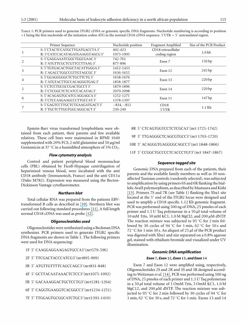
Table 1: PCR primers used to generate ITGB2 cDNA or genomic specific DNA fragments. Nucleotide numbering is according to position+1 being the first nucleotide of the initiation codon ATG in the normal CD18 cDNA sequence. 5′UTR = 5′ untranslated region.
Primer Sequence Nucleotide position Fragment Amplified Size of the PCR Product
1S: 5′CTACTCCATGCTTGATGACCTA 3′ 402-423 CD18 extracellular
1.6 kbR: 5′CATCCACATAGATGAAGGTAGCG 3′ 1973-1995 coding region
2S: 5′GAGGAAATCGGCTGGCGAAC 3′ 742-761
Exon 7 156 bpR: 5′ATGTTCGCTCGTTCCTTAAG 3′ 877-896
3S: 5′TGTGACACTGGCTACATTGGGA 3′ 1412-1433
Exon 12 245 bpR: 5′AGACCTGGCCGTTGTAGCGC 3′ 1636-1655
4S: 5′GGAGGGGGCTCTGCTTCTG 3′ 1658-1676
Exon 13 220 bpR: 5′ATGTACTTGCCACAGGGTGAG 3′ 1858-1877
5S: 5′CTCCTGCGCCGACTGCCT 3′ 1879-1896
Exon 14 220 bpR: 5′CTCGGCTCTCATCCACATAG 3′ 2079-2098
6S: 5′ACAGAGTGCATCCAGGAGCA 3′ 1252-1271
Exon 11 147 bpR: 5′CTCCAAGAAGCCCTTGCCAT 3′ 1378-1397
7S: 5′GAGTCCTTGCTCTGAAGATGACT 3′ −834,−812 CD18
5′UTR1.1 KbR: 5′TGCTCTTGGTGGCAGGCACT 3′ 230-249
Epstein-Barr virus transformed lymphoblasts were ob-tained from each patient, their parents and few availablerelatives. These cell lines were maintained in RPMI 1640supplemented with 20% FCS, 2 mM glutamine and 50µg/mlGentamicin at 37 C in a humidified atmosphere of 5% CO2.
Flow cytometry analysis
Control and patient peripheral blood mononuclearcells (PBL) obtained by Ficoll-Hypaque centrifugation ofheparinized venous blood, were incubated with the antiCD18 antibody (Immunotech, France) and the anti CD11a(Dako M782). Expression was measured using the Becton-Dickinson Vantage cytofluorometer.
Northern blot
Total cellular RNA was prepared from the patients EBV-transformed B cells as described in [10]. Northern blot wascarried out following standard procedures [11]. A full lengthnormal CD18 cDNA was used as probe [12].
Oligonucleotides used
Oligonucleotides were synthesized using a Beckman DNAsynthesizer. PCR primers used to generate ITGB2 specificDNA fragments are shown in Table 1. The following primerswere used for DNA sequencing:
1F 5′ CAAGGAGGAAGAGTGCCA3′(nct579-596)
2F 5′ TTCGACTACCCATCG3′(nct895-909)
3R 5′ ATGTTGTTTTCAGCCAGC3′(nct831-848)
4F 5′ GCTTACAATAAACTCTCC3′(nct1075-1092)
5R 5′ GACAAAGGACTGCTCCTG3′(nct1281-1264)
6F 5′ CAGGTGAAGGTCACGGCC3′(nct1234-1251)
7F 5′ TTGGAGTGCGGCATCTGC3′(nct1393-1410)
8R 5′ CTCAGTGGTCCTCTCGCA3′(nct 1725-1742)
9F 5′ TTGAGGGCTCAGCGTGCC3′(nct 1703-1720)
10R 5′ ACAGGGTGAGGGGCAGCC3′(nct 1848-1866)
11F 5′ CCGGCTGCCCCTCACCCTGT3′(nct 1847-1867)
Sequence-tagged site
Genomic DNA prepared from each of the patients, theirparents and the available family members as well as 30 non-affected Tunisian controls (randomly selected), was subjectedto amplification by using primers 6S and 6R flanking the bial-lelic AvaII polymorphism, as described by Mastuura and Kishi[13]. Primers 7S and 7R (see Table 1) flanking the Xba1 sitelocated at the 5′ end of the ITGB2 locus were designed andused to amplify a CD18 specific 1.12 Kb genomic fragment.PCR was performed using 500 ng of DNA, 25 pmoles of eachprimer and 1.5 U Taq polymerase in a 50µl total volume of10 mM Tris, 50 mM KC1, 1.0 M MgCl2, and 200µM dNTP.The reaction mixture was subjected to 95 C for 2 min fol-lowed by 30 cycles of 94 C for 1 min, 62 C for 50 s and72 C for 1 min 30 s. An aliquot of 25µl of the PCR productwas digested with Xba1 and size separated on a 0.8% agarosegel, stained with ethidium bromide and visualized under UVillumination.
Genomic DNA amplification
Exon 7 , Exon 12 , Exon 13 , and Exon 14
Exon 7 and Exon 12 were amplified using, respectively,Oligonucleotides 2S and 2R and 3S and 3R designed accord-ing to Weitzman et al. [14]. PCR was performed using 500 ngof DNA, 25 pmoles of each primer and 1.5 U Taq polymerasein a 50µl total volume of 1 OmM Tris, 5 OmM KC1, 1.0 MMgC12, and 200µM dNTP. The reaction mixture was sub-jected to 95 C for 2 min followed by 30 cycles of 94 C for1 min, 62 C for 30 s, and 72 C for 1 min. Exons 13 and 14

116 D. M. Fathallah et al. 1:3 (2001)
were amplified in a single fragment using oligos n6 and n7at the same conditions described above. Thermocycling wasas follows: 30 cycles of 94 C for 1 min, 60 C for 1 min, and72 C for 1 min 30 s.
RT/PCR and DNA SequencingReverse transcription (RT) was performed on 1µg of
total RNA or 0.5µg mRNA prepared by affinity oligodTcolumn (Qiagen mRNA extraction kit) by first, a 5 minincubation at 65 C then transfer to ice followed by onehour incubation at 62 C in a 20µl reaction containing0.5 pM of OligodT, 0.2 mM of each dNTP, 50 mM Tris-HClpH 8, 50 mM KC1, 5 mM MgCl2, RNase inhibitor (Rnasin,Boehringer Mannheim) and 1 unit of AMV reverse tran-scriptase. The reaction was stopped by a 5 min heat pulseat 90 C.
CD18 specific DNA fragments were amplified by PCRusing 10µl aliquot of the RT reaction, 1µM of each primerin a 50µl reaction containing 2 mM MgCl2, 50 mM KCl,10 mM Tris-HCl, and 2.5 units of Taq Polymerase (Amer-sham, France). Cycling was as follows: 35 cycles of 1 minat 94 C, 1 min at 62 C, and 2 min at 72 C to amplify the1.6 Kb fragment using primers 1S and 1R. The Venta DNApolymerase (New England Biolabs) was alternatively used togenerate blunt end PCR fragments that were ligated into thepCDNA3 vector. To rule out PCR artifacts and mutationsintroduced by the Taq polymerase. A minimum of four inde-pendent PCR reactions were performed using three differ-ent batches of enzyme and the fragments were controlled byhybridization to an ITGB2 specific cDNA probe and/or byrestriction mapping.
Direct sequencing of PCR products was performed usingthe sequenase Kit (Amersham) and the cloned PCR productswere sequenced as double stranded DNA using the same Kitas recommended by the manufacturer (Amersham).
Parentage assessmentPaternity was established in family KH by DNA Finger-
printing usingVNTR Probes 33.6 and 33.15 (kindly providedby Prof A. Jeffreys, Leicester, UK) as described elsewhere [15].
RESULTS
β2 integrins expression level in LAD patients
We used immunofluorescence flow cytometry to analyzethe level of the CD18 subunit expression on the surface ofPBMCs from five Tunisian LAD patients and their parents.As shown in Figure 1, low levels of CD18 expression (1% to5%) were observed in the patients with the severe form ofthe disease. Patient K had a 16.5% CD18 expression level ascompared to the normal control, which is consistent with themoderate form of LAD. All patients exhibited a normal cellsurface expression of the leukocyte markers CD3, CD4, CD8,CD19, and HLA DR antigens (data not shown).
Identification of a novel polymorphic biallelic markerin the ITGB2 locus
An STS assay was designed to amplify a 1.1 Kb genomicDNA a fragment that contains an Xba1 site. Digestion of this
PCR product with Xba I permitted the identification of twoalleles: 1.1 kb (allele A1 or Xba−) and 829 bp+283 bp (alleleA2 or Xba+) (see Figure 2). The Xba I alleles frequency was0.5 for each allele within the same sample of non-affectedTunisian individuals.
Analysis of the LAD patients CD18 cDNA fragments
Northern blot showed that all the patients exhibited a nor-mal sized CD18 specific mRNA (data not shown). We used theRT/PCR strategy to generate CD18 specific cDNA fragments.We have first made a 1.6 kb fragment corresponding to mostof the region coding for the extracellular domain of the CD18subunit. The majority of the mutations reported to datemapped to this region of the molecule. Sequencing of severalindependently generated PCR products from each patientrevealed (see Figure 3) that all the patients have a deletion ofthe G at position 1497 (1497delG) causing a frame shift andyielding a premature stop codon, 28 residues downstream ofthe deletion. Patient R was homozygous for this mutation.PCR products from both parents subcloned into the pCDNA3vector exhibited clones missing the G at position 1497 as wellas normal clones. This finding is in agreement with patient R’sparents being first cousins. The second mutation, identifiedin patient M, was another single-bp deletion: 1920delG thatresults in a frame shift and the occurrence of a stop codon16 residues down stream of the deletion. The third mutationwas a G to A transition affecting codon 284 (GGC/AGC)leading to the substitution of a conserved glycine by a serine(G284S). Patients S and I were compound heterozygous1497delG/G284S. The G284S mutation was reported inde-pendently in at least one unrelated patient [16]. The fourthmutation was a single nucleotide transition in codon 593(CGT/TGT) that leads to the replacement of the argininenormally encoded at this position by a cysteine (R593C).This mutation found in patient K, was reported as aninherited mutation in two other patients, one of whom beingwith certainty of different ethnic origin [16, 17]. Sequencingof multiple RT/PCR products generated from the patient’sparents showed that no one of them carried this mutation.Patient K was a compound heterozygous 1497delG/R593C.The latter occurred on a different allele than the one carryingthe 1497delG. It has previously been shown using CD18transfection studies that the G284S and R593C mutationsprevent β2 integrin heterodimer formation and thereforeaccount for LAD phenotype [16, 17]. The mutations wereconfirmed in all the individuals studied by amplification andsequencing from the genomic DNA of the correspondingcoding exon: G284S (Exon 7) 1497delG (Exon 12), 1920delG,and R593C within Exon 13 and Exon 14, respectively.
For further confirmation of the de novo character of theR593C mutation, an eventual mismatching between patientK and his biological parents was ruled out by paternity testingusing DNA fingerprinting (data not shown).
Allelic association analysis
Segregation of two CD18 intragenic polymorphic mark-ers Ava II and Xba1 with the mutations underlying LAD in

1:3 (2001) Molecular basis of leukocyte adhesion deficiency in a north african population 117
100 101 102 103 104
CD18:98.5%
(a)
CD18:5%
101 102 103 10 4100
(c)
CD18:16.5%
10 0 101 102 103 104
(d)
100 101 102 103 10 4
CD18:1%
(b)
Cel
ls n
um
ber
Log fluorescence
Figure 1: FACS analysis of the β2 integrin CD18 expression at the surface of PBMCs from Tunisian patients with leukocyte adhesiondeficiency (LAD). (a) control from a non-affected individual. (b), (c) and (d) represent respectively, the CD18 expression pattern obtainedin patients (I, S, R), M, and K.
1.1 Kb829 bp
283 bp
1 2 3 4 5 6 7
Figure 2: Polymorphism of the Xba1 site in a 1.1 Kb fragment generated from the 5′ region of the human ITGB2 (CD18) gene. 1.2% agarosegel electrophoresis of amplified DNA from Tunisian individuals following Xba1 restriction enzyme digestion. Lane 1: individual homozygousfor allele A1 (absence of the polymorphic Xba1 site). Lanes 2, 3, 4, and 5: individuals homozygous for allele A2 (829 and 283 bp). Lanes 6 and7: individuals heterozygous A1/A2.
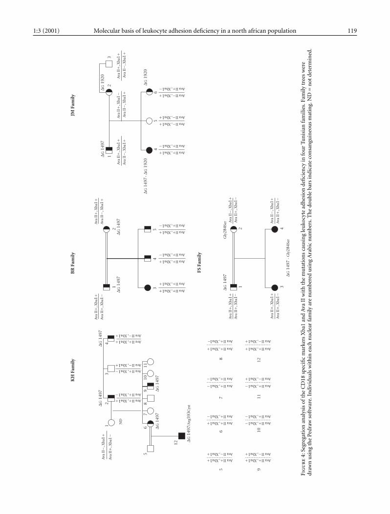
our patients was studied using site tagged sequence analysis(STS). The Ava II polymorphism was studied by an STS assaydescribed elsewhere [13]. Familial studies have shown (seeFigure 4) that the 1497delG mutation, present in all the pa-
tients, segregates with the (Ava II+, Xba I+) markers. Thisstrong linkage desequilibrium was confirmed by the studyof the patients close relatives; Chi square = 41.5, p = 0.001as calculated using the Four Fold Table method described
118 D. M. Fathallah et al. 1:3 (2001)
CCGTGGCCGGTGTTGGTGATGTGAG
A C G T
(c)
CTTGCTGTCGGCGACAGTGGACC
A C G T
(d)
CAACAACAGAAGGCCGTCGAAGG
A C G T
(b)
G
A
CGTCA
A
GAAGGTTTC
C
CC
GGG
A C G T
(a)
∗
∗
∗
∗
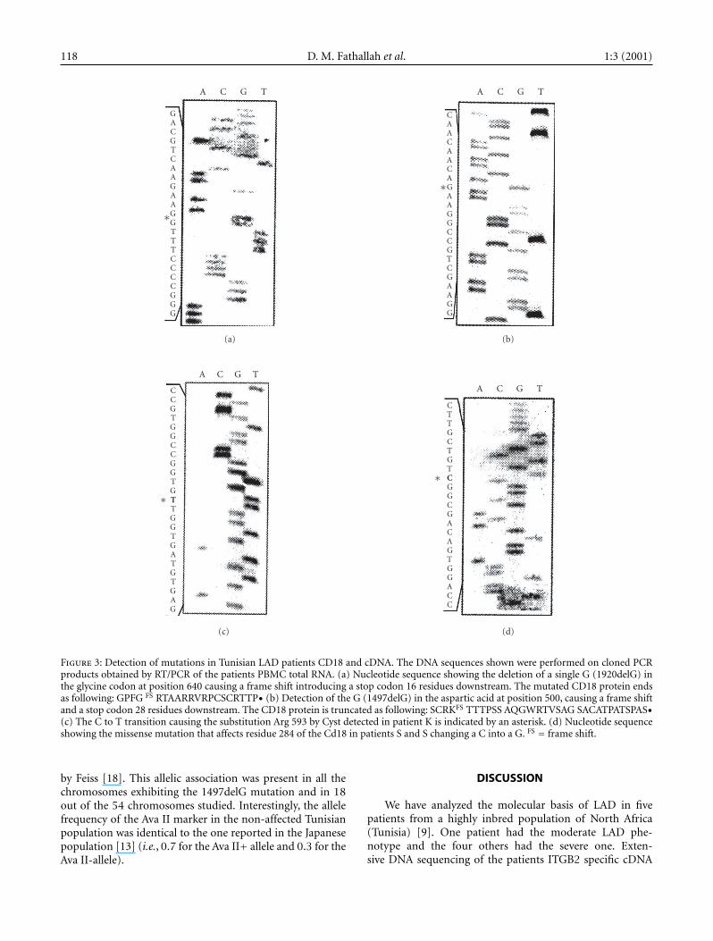
Figure 3: Detection of mutations in Tunisian LAD patients CD18 and cDNA. The DNA sequences shown were performed on cloned PCRproducts obtained by RT/PCR of the patients PBMC total RNA. (a) Nucleotide sequence showing the deletion of a single G (1920delG) inthe glycine codon at position 640 causing a frame shift introducing a stop codon 16 residues downstream. The mutated CD18 protein endsas following: GPFG FS RTAARRVRPCSCRTTP• (b) Detection of the G (1497delG) in the aspartic acid at position 500, causing a frame shiftand a stop codon 28 residues downstream. The CD18 protein is truncated as following: SCRKFS TTTPSS AQGWRTVSAG SACATPATSPAS•(c) The C to T transition causing the substitution Arg 593 by Cyst detected in patient K is indicated by an asterisk. (d) Nucleotide sequenceshowing the missense mutation that affects residue 284 of the Cd18 in patients S and S changing a C into a G. FS = frame shift.
by Feiss [18]. This allelic association was present in all thechromosomes exhibiting the 1497delG mutation and in 18out of the 54 chromosomes studied. Interestingly, the allelefrequency of the Ava II marker in the non-affected Tunisianpopulation was identical to the one reported in the Japanesepopulation [13] (i.e., 0.7 for the Ava II+ allele and 0.3 for theAva II-allele).
DISCUSSION
We have analyzed the molecular basis of LAD in fivepatients from a highly inbred population of North Africa(Tunisia) [9]. One patient had the moderate LAD phe-notype and the four others had the severe one. Exten-sive DNA sequencing of the patients ITGB2 specific cDNA
1:3 (2001) Molecular basis of leukocyte adhesion deficiency in a north african population 119
KH
Fam
ily
∆G
14
97
∆G
14
97
Ava
II−
, Xba
1+
Ava
II+
, Xba
1−
Ava II+, Xba1+Ava II+, Xba1−
Ava II+, Xba1−Ava II−, Xba1+
Ava II+, Xba1+Ava II−, Xba1+
12
34
56
78
91
01
1
12
ND
∆G
14
97
∆G
14
97
∆G
14
97
/Arg
593
Cys
t
Ava II+, Xba1+Ava II−, Xba1+
Ava II+, Xba1+Ava II+, Xba1−
Ava II+, Xba1−Ava II+, Xba1−
Ava II+, Xba1
Ava II+, Xba1+
Ava II+, Xba1+Ava II−, Xba1+
Ava II+, Xba1−Ava II+, Xba1−
Ava II+, Xba1−Ava II−, Xba1+
Ava II+, Xba1+Ava II−, Xba1+
56
78
91
01
11
2
BR
Fam
ily
Ava
II+
, Xba
1+
Ava
II+
, Xba
1−
Ava
II+
, Xba
1+
Ava
II−
, Xba
1+
12
34
5
∆G
14
97
∆G
14
97
Ava II+, Xba1+Ava II+, Xba1+
Ava II+, Xba1+Ava II+, Xba1−
Ava II+, Xba1+Ava II+, Xba1−
FS F
amil
y
Ava
II+
, Xba
1+
Ava
II+
, Xba
1A
va I
I+, X
ba1−
Ava
II−
, Xba
1+
12
34
Ava
II+
, Xba
1+
Ava
II+
, Xba
1−
Ava
II+
, Xba
1−
Ava
II−
, Xba
1+
∆G
14
97 ∆
G 1
49
7 -
Gly
28
4Se
rGly
284
Ser
JM F
amil
y
12
3
45
6
∆G
14
97
∆G
19
20
∆G
14
97∆
G 1
92
0
-
Ava
II+
, Xba
1+
Ava
II−
, Xba
1+
Ava
II−
, Xba
1+
Ava
II+
, Xba
1−
Ava
II+
, Xba
1+
Ava
II−
, Xba
1+
∆G
19
20
Ava II+, Xba1+Ava II+, Xba1−
Ava II−, Xba1+Ava II−, Xba1+
Ava II−, Xba1+Ava II+, Xba1−
−
−
Fig
ure
4:Se
greg
atio
nan
alys
isof
the
CD
18sp
ecifi
cm
arke
rsX
ba1
and
Ava
IIw
ith
the
mu
tati
ons
cau
sin
gle
uko
cyte
adh
esio
nde
fici
ency
info
ur
Tun
isia
nfa
mili
es.F
amily
tree
sw
ere
draw
nu
sin
gth
ePe
draw
soft
war
e.In
divi
dual
sw
ith
inea
chn
ucl
ear
fam
ilyar
en
um
bere
du
sin
gA
rabi
cn
um
bers
.Th
edo
ubl
eba
rsin
dica
teco
nsa
ngu
ineo
us
mat
ing.
ND
=n
otde
term
ined
.
120 D. M. Fathallah et al. 1:3 (2001)
L 1 2 3 4 CD
CONSERVED REGION TM
Ψω εO ΦΩ θ Σ
Σ
θ
Ω
Φ
O
ε
Ψ
ω
R593C
R586C
N351S
G284S
K196T
L178P
G169R
L149/P
D128N
ITGB2CD18
∗
∗
∗∗∗
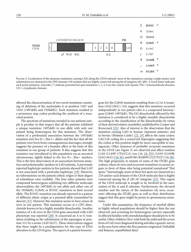
Figure 5: Localization of the missense mutations causing LAD, along the CD18 subunit: most of the mutations causing a single amino-acidsubstitution are clustered in the NH2 domain 250 residues that are highly conserved among the β integrins (β1–β8). A Greek letter indicateseach point mutation. Asterisks (*) indicate potential hot spot mutations 1, 2, 3, 4 are the cystein rich repeats, TM = transmembrane domain,CD = cytoplasmic domain.
allowed the characterization of two novel mutations consist-ing of deletions of the nucleotides G at position 1497 and1920 (1497delG and 1920delG). Each mutation resulted ina premature stop codon predicting the synthesis of a trun-cated protein.
The spectrum of mutations revealed in our patients sam-ple is peculiar in that respect that all the patients exhibiteda unique mutation: 1497delG in one allele with only onepatient being homozygous for this mutation. The obser-vation of a preferential association between the 1497delGmutation and Ava II+, Xba I+ alleles and the fact that all thepatients were born from consanguineous marriages, stronglysuggests the presence of a founder effect at the basis of thismutation in our group of patients. It also suggests that thismutation was introduced in this population on an ancestralchromosome, tightly linked to the Ava II+, Xba+ markers.This is the first observation of an association between muta-tion and polymorphic markers in LAD patients. A previouslyreported haplotype analysis in five patients showed that LADis not associated with a particular haplotype [19]. However,no informations on the patients ethnic origin or their degreeof relatedness were available. The four other patients werecompound heterozygous exhibiting two different molecularabnormalities: the 1497delG in one allele and either one ofthe 1920delG, G284S, or R593C mutations in their secondallele. The R593C mutation was already reported in a patientof different ethnic origin as a parental allele transmitted bydescent [17]. However this mutation seems to have arisen denovo in our patient. This mutation occurs at a CPG dinu-cleotide known to be a highly mutable dinucleotide. Anotherinstance of a de novo in the CD18 gene associated with LADphenotype was reported [20]. It concerned an A to G tran-sition resulting in the substitution of the asparagine at posi-tion 351 by a serine (AAT/AGT). These observations suggestthat there might be a predisposition for this type of DNAalteration in the CD18 gene. The report of a patient homozy-
gous for the G284S mutation resulting from a G to A transi-tion (GGC/AGC) [16] suggests that this mutation occurredindependently in our patient who is a compound heterozy-gous G284S-1497delG. The GG dinucleotide affected by thismutation is considered to be a highly mutable dinucleotideaccording to the classification of the dinucleotides by virtueof their derived relative mutability established by Cooper andKrowczack [21]. Also of interest, is the observation that themutation causing LAD in human (Japanese patients) andin bovine (Holstein Cattle) [22, 23] affects the same codon:GAC128 coding for a conserved Asparagine suggesting thatthe codon at this position might be more susceptible to mu-tagenesis. Other instances of probably recurrent mutationsin the CD18 (see Figure 5) are observed and affect residuesL149 (L149P: CTA/CCA) (see [16, 24, 25]), G169 (G169R:GGG/AGG) [24, 26], and R586 (R586W: CGT/TGT) [16, 20].The high propensity to mutate of some of the ITGB2 genecodons, where de novo and recurrent mutations occurred, ar-gues in favor of these sites being potential mutational “Hotspots.” Interestingly, most of these hot spots are clustered in a250 amino-acid domain of the CD18 molecule that is highlyconserved among the β (β1–8) integrins [12]. This regionof the CD18 molecule is sought to be involved in the asso-ciation of the α and β subunits. Furthermore, the elevatednumber and the nature of the mutations (de novo, recur-rent) affecting the ITGB2 gene as well as their distributionsuggest that this gene might be prone to spontaneous muta-tions.
Under this assumption, the frequency of morbid allelesin highly inbred populations should be higher than the oneobserved, and screening for heterozygous carriers especiallyin affected families with extended pedigree should prove to beuseful. Other children (five) with both the mild and the severeform of LAD were diagnosed during and after a genetic surveyin the area from where the first patients originated (Fathallahand Bejaoui, unpublished data).

1:3 (2001) Molecular basis of leukocyte adhesion deficiency in a north african population 121
REFERENCES
[1] Arnaout MA. Leukocyte adhesion molecules deficiency;its structural basis, pathophysiology, and implicationsfor modulating the inflammatory response. ImmunolRev. 1990;114:145–180.
[2] Arnaout MA. Structure and function of the leukocyteadhesion molecules CD11/CD18. Blood. 1990;75:1037–1050.
[3] Wong EM, Davis M, Le Beau N, Springer TA.Cloning and chromosomal localization of a novel gene-encoding a human β2-integrin alpha subunit. Gene.1996;171:291–294.
[4] Anderson DC, Schmalsteig FC, Finegold MJ. The severeand moderate phenotypes of heritable Mac-1, LFA-1deficiency: their quantitative definition and relation toleukocyte dysfunction and clinical features. J Infect Dis.1985;152:668–688.
[5] Kishimoto TK, O’Connor K, Springer TA. Leukocyte ad-hesion deficiency: aberrant splicing of a conserved inte-grin sequence causes a moderate deficiency phenotype.Biol Chem. 1989;264(6):3588–3595.
[6] Sligh JE, Hurwitz MY, Zhu C, Anderson DC, BeaudetAL. An initiation codon mutation in CD18 in associa-tion with the moderate phenotype of leukocyte adhe-sion deficiency. Biol Chem. 1992;267(2):714–718.
[7] Lopez-Rodriguez C, Nueda A, Grospierre B, et al.Characterization of two new CD18 alleles causing se-vere leukocyte adhesion deficiency. Eur J Immunol.1993;23:2792–2798.
[8] Arnaout MA, Michishta M. Genetic abnormalities inleukocyte adhesion molecules deficiency. In: New Con-cepts in immunodeficiency diseases. CRC press.YoshikazuTakada; 1993:191–202.
[9] Riou S, El Younsi C, Chaabouni H. Consanguinité dansla population du nord de la Tunisie. Tunisie Médicale.1989; 52:73–76.
[10] Chomczynski P, Sacchi N. Single-step method ofRNA isolation by acid guanidium yhiocyanate-phenol-chloroform extraction. Anal Biochem. 1987;162(1):156–159.
[11] Sambrook J, Fritsh EF, Maniatis T. Molecular cloning: Alaboratory manual. 2nd ed. New York, NY: Cold SpringHarbor Laboratory Press; 1989.
[12] Kishimoto TK, O’Connor K, Lee A, Roberts TM,Springer TA. Cloning of the β subunit of the leuko-cyte adhesion proteins: homology to an extracellularmatrix receptor defines a novel supergene family. Cell.1987;48:681–690.
[13] Mastuura S, Kishi F. Investigation of the polymorphicAva II site by a PCR-based assay at the human CD18gene locus. Hum Genet. 1994;93:721.
[14] Weitzman JB, Wells CE, Wright AH, Clark PA, Law SKA.The gene organization of the humanβ2 integrin subunit(CD18). FEBS Lett. 1991;294(1):97–103.
[15] Helminen P, Ehnholm C, Lokki ML, Jeffreys A, Pelto-nen L. Application of DNA “Fingerprints” to paternitydeterminations. Lancet. 1988;12:574–576.
[16] Wright AH, Douglas WA, Taylor GM, et al. Molecularcharacterization of leukocyte adhesion deficiency in sixpatients. Eur J Immunol. 1995;25:717–722.
[17] Arnaout MA, Dana N, Gupta SK, Tenen DG, FathallahDM. Point mutations impairing cell Surface expressionof the common β subunit (CD18) in a patient withleukocyte adhesion molecule (Leu-CAM) deficiency. JClin Invest. 1990;85:977–981.
[18] Fleiss J. Statistical Methods for Rates and Proportions. 2nded. New York, NY: John Wiley & Sons; 1980.
[19] Law SKA, Taylor GM. Restriction fragment length poly-morphism of the gene of the human leukocyte integrinβ-subunit (CD18). Immunogenetics. 1991;34:341–345.
[20] Nelson C, Rabb H, Arnaout MA. Genetic cause of leuko-cyte adhesion molecule deficiency: abnormal splicingand a missense mutation in a conserved region of CD18impair cell surface expression of β2 integrins. J BiolChem. 1992;267(5):3351–3357.
[21] Cooper DN, Krawczak M. The mutational spectrumof single base-pair substitutions causing human ge-netic disease: patterns and predictions. Hum Genet.1990;85:55–74.
[22] Matsuura S, Kishi F, Tsukahara M, Nunoi H, KobayashiK, Kajii T. Leukocyte adhesion deficiency: identificationof novel mutations in two Japanese patients with a severeform. Biochem Biophys Res Commun. 1992;184:1460–1467.
[23] Shuster DE, Marcus EK Jr, Ackermann MR, Gilbert RO.Identification and prevalence of a genetic defect thatcauses leukocyte adhesion deficiency in Holstein cattle.Proc Natl Acad Sci USA. 1992;89:9225–9229.
[24] Wardlaw AJ, Hibbs ML, Stacker AS, Springer TA. Dis-tinct mutations in two patients with leukocyte adhesiondeficiency and their functional correlates. J Exp Med.1990;172:335–345.
[25] Back AL, Kwok WW, Hickstein DD. Identification of twomolecular defects in a child with leukocyte adherencedeficiency. J Biol Chem. 1992;267:5482–5487.
[26] Corbi AL, Vara A, Ursa A, Garcia Rodriguez MC, FontanG, and Sanchez-Madrid F. Molecular basis for a severecase of leukocyte adhesion deficiency. Eur J Immun.1992;22:1877–1881.
* Corresponding author.E-mail: [email protected]: +216 1 791833; Tel: +216 1 789608
Copyright © 2022 FDOKUMEN