Humanization of the anti-CD18 antibody 6.7: an unexpected effect of a framework residue in binding...
12
Molecular Immunology 39 (2003) 941–952 Humanization of the anti-CD18 antibody 6.7: an unexpected effect of a framework residue in binding to antigen Cristina Caldas a,b , Verˆ onica Coelho b , Jorge Kalil b , Ana Maria Moro c , Andrea Q. Maranhão a , Marcelo M. Br´ ıgido a,∗ a Departamento de Biologia Celular, Universidade de Bras´ ılia, 70910-900 Bras´ ılia, DF, Brazil b Immunology Laboratory, Heart Institute (InCor), University of São Paulo Medical School, 05403-000 São Paulo, SP, Brazil c Instituto Butantan, 05503-900 São Paulo, SP, Brazil Received 7 November 2002; accepted 6 February 2003 Abstract Humanization of monoclonal antibodies by complementary determinant region (CDR)-grafting has become a standard procedure to improve the clinical usage of animal antibodies. However, antibody humanization may result in loss of activity that has been attributed to structural constraints in the framework structure. In this paper, we report the complete humanization of the 6.7 anti-human CD18 monoclonal antibody in a scFv form. We used a germline-based approach to design a humanized VL gene fragment and expressed it together with a previously described humanized VH. The designed humanized VL has only 14 mutations compared to the closest human germline sequence. The resulting humanized scFv maintained the binding capacity and specificity to human CD18 expressed on the cell surface of peripheral blood mononuclear cells (PBMC), and showed the same pattern of staining T-lymphocytes sub-populations, in comparison to the original monoclonal antibody. We observed an unexpected effect of a conserved mouse–human framework position (L37) that hinders the binding of the humanized scFv to antigen. This paper reveals a new framework residue that interferes with paratope and antigen binding and also reinforces the germline approach as a successful strategy to humanize antibodies. © 2003 Elsevier Science Ltd. All rights reserved. Keywords: Humanization; Framework; CDR-grafting; CD18 1. Introduction Antibody engineering has become a popular technol- ogy for development of a new generation of drugs (Presta, 2002; Maynard and Georgiou, 2000). The amount knowl- edge available with regards to antibody structure at atomic resolution, allied to its conserved conformation, permits the elaboration of general rules for antigen binding site struc- tural and functional features. To date, the commonest an- tibody manipulation is the process of humanization. In this process, a murine antibody sequence is replaced by a ho- mologous human sequence yielding a human-like antibody, reducing its immunogenicity, and preserving the binding to the original antigen (Jones et al., 1986; Riechmann et al., 1988;Verhoeyen et al., 1988). This methodology has revo- lutionized the clinical use of monoclonal antibodies (mAb), Abbreviations: bp, base-pair; CDR, complementary determinant re- gion; Fab, antigen binding fragment; FR, framework; PCR, polymerase chain reaction; scFv, single chain variable fragment; mAb, monoclonal antibody; VL, variable light chain; VH, variable heavy chain ∗ Corresponding author. Tel.: +55-61-3072423; fax: +55-61-3498411. E-mail address: [email protected] (M.M. Br´ ıgido). since the patients‘ response to heterologous rodent se- quences prevents the full biological effect of the monoclonal antibody (Winter and Harris, 1993). Moreover, heterologous mAbs present a reduced half-life and a diminished capac- ity to induce effector functions (Isaacs et al., 1992). The limit of this process is in the preservation of the antibody’s affinity and specificity. After a decade of research in this process, many rules have been established that help main- tain the overall antibody structure while the mouse primary sequence is being saturated with human residues. The use of human germline sequences (Rosok et al., 1996; Caldas et al., 2000) and the maintenance of key framework residues are examples of these rules (Tempest et al., 1991). Many successfully humanized antibodies have been reported, in- cluding in this group those that are already being routinely used in therapeutics (Glennie and Johnson, 2000). Antibody humanization is becoming a trivial methodol- ogy, where many different experimental procedures result in an active antibody. In spite of this, many of the designed human antibodies do not fully mimic the behavior of the original murine protein. Loss of affinity, specificity, or sta- bility are the main parameters affected by this experimental 0161-5890/03/$ – see front matter © 2003 Elsevier Science Ltd. All rights reserved. doi:10.1016/S0161-5890(03)00022-1
-
Upload
iberopuebla -
Category
Documents
-
view
0 -
download
0
Transcript of Humanization of the anti-CD18 antibody 6.7: an unexpected effect of a framework residue in binding...
Molecular Immunology 39 (2003) 941–952
Humanization of the anti-CD18 antibody 6.7: an unexpected effectof a framework residue in binding to antigen
Cristina Caldasa,b, Veronica Coelhob, Jorge Kalilb, Ana Maria Moroc,Andrea Q. Maranhãoa, Marcelo M. Brıgidoa,∗
a Departamento de Biologia Celular, Universidade de Brasılia, 70910-900 Brasılia, DF, Brazilb Immunology Laboratory, Heart Institute (InCor), University of São Paulo Medical School, 05403-000 São Paulo, SP, Brazil
c Instituto Butantan, 05503-900 São Paulo, SP, Brazil
Received 7 November 2002; accepted 6 February 2003
Abstract
Humanization of monoclonal antibodies by complementary determinant region (CDR)-grafting has become a standard procedure toimprove the clinical usage of animal antibodies. However, antibody humanization may result in loss of activity that has been attributedto structural constraints in the framework structure. In this paper, we report the complete humanization of the 6.7 anti-human CD18monoclonal antibody in a scFv form. We used a germline-based approach to design a humanized VL gene fragment and expressed ittogether with a previously described humanized VH. The designed humanized VL has only 14 mutations compared to the closest humangermline sequence. The resulting humanized scFv maintained the binding capacity and specificity to human CD18 expressed on thecell surface of peripheral blood mononuclear cells (PBMC), and showed the same pattern of staining T-lymphocytes sub-populations, incomparison to the original monoclonal antibody. We observed an unexpected effect of a conserved mouse–human framework position(L37) that hinders the binding of the humanized scFv to antigen. This paper reveals a new framework residue that interferes with paratopeand antigen binding and also reinforces the germline approach as a successful strategy to humanize antibodies.© 2003 Elsevier Science Ltd. All rights reserved.
Keywords:Humanization; Framework; CDR-grafting; CD18
1. Introduction
Antibody engineering has become a popular technol-ogy for development of a new generation of drugs (Presta,2002; Maynard and Georgiou, 2000). The amount knowl-edge available with regards to antibody structure at atomicresolution, allied to its conserved conformation, permits theelaboration of general rules for antigen binding site struc-tural and functional features. To date, the commonest an-tibody manipulation is the process of humanization. In thisprocess, a murine antibody sequence is replaced by a ho-mologous human sequence yielding a human-like antibody,reducing its immunogenicity, and preserving the binding tothe original antigen (Jones et al., 1986; Riechmann et al.,1988;Verhoeyen et al., 1988). This methodology has revo-lutionized the clinical use of monoclonal antibodies (mAb),
Abbreviations:bp, base-pair; CDR, complementary determinant re-gion; Fab, antigen binding fragment; FR, framework; PCR, polymerasechain reaction; scFv, single chain variable fragment; mAb, monoclonalantibody; VL, variable light chain; VH, variable heavy chain∗ Corresponding author. Tel.:+55-61-3072423; fax:+55-61-3498411.E-mail address:[email protected] (M.M. Brıgido).
since the patients‘ response to heterologous rodent se-quences prevents the full biological effect of the monoclonalantibody (Winter and Harris, 1993). Moreover, heterologousmAbs present a reduced half-life and a diminished capac-ity to induce effector functions (Isaacs et al., 1992). Thelimit of this process is in the preservation of the antibody’saffinity and specificity. After a decade of research in thisprocess, many rules have been established that help main-tain the overall antibody structure while the mouse primarysequence is being saturated with human residues. The useof human germline sequences (Rosok et al., 1996; Caldaset al., 2000) and the maintenance of key framework residuesare examples of these rules (Tempest et al., 1991). Manysuccessfully humanized antibodies have been reported, in-cluding in this group those that are already being routinelyused in therapeutics (Glennie and Johnson, 2000).
Antibody humanization is becoming a trivial methodol-ogy, where many different experimental procedures resultin an active antibody. In spite of this, many of the designedhuman antibodies do not fully mimic the behavior of theoriginal murine protein. Loss of affinity, specificity, or sta-bility are the main parameters affected by this experimental
0161-5890/03/$ – see front matter © 2003 Elsevier Science Ltd. All rights reserved.doi:10.1016/S0161-5890(03)00022-1

942 C. Caldas et al. / Molecular Immunology 39 (2003) 941–952
procedure. Mild losses are normally associated with thisprocedure (Carter et al., 1992; Hsiao et al., 1994). A loweraffinity is normally tolerated for humanized antibodies.
These effects have been partially remedied by reengineer-ing the framework (Kettleborough et al., 1991; Singer et al.,1993; Nakatani et al., 1994; Zhu and Carter, 1995; Saldanhaet al., 1999). Therefore, it is crucial to describe more sub-tle structural features that allow the preservation of antibodyparatope integrity during this process. The rules are far frombeing completely understood. Long-range effects promotedby certain residues are among those forces responsible forsubtle changes in the global structure. Unfortunately theseare the most difficult issues to probe, since even small devia-tions could be responsible for removing residue–residue con-tact, or reducing surface complementarity. Therefore, evendistant residues could interfere with paratope conformationand antigen binding (Chien et al., 1989).
We used an anti-human CD18 as a model antibody forhumanization. CD18 is an integrin family membrane proteininvolved in cell adhesion. It is found in many different celltypes and it is always associated with one of the differentisoforms of CD11. Antibodies to CD18 may inhibit cell–cellattachment and especially leukocyte-tissue adhesion. There-fore, anti-CD18 antibodies have been proposed as adjuvantin many therapies that involves leukocytes infiltration andinflammation. They have been tested successfully as protec-tive agents in ischemic myocardial injury in animal models(Gao et al., 2002), but much of this enthusiasm was lostafter failure of human trials (Dove, 2000). Its potential ap-plication is much wider, and it has also been cited as a po-tential treatment for preventing meningitis sequels or graftrejection (Tuomanen et al., 1989; Isobe et al., 1997). Theantibody used in this work, mAb 6.7, binds to CD18 (Davidet al., 1991) in a unique inhibitory epitope recently mappedto residues 350–432 (Lu et al., 2001).
In this work we describe the complete humanization ofmurine mAb 6.7 anti-human CD18. In a previous paper wedescribed the successful humanization of the VH domainby means of a germline human VH gene fragment sequenceusing an expanded CDR1 (CDR1+H1) graft (Caldas et al.,2000). We apply this same concept to the design of two ver-sions of the humanized VL domain. While testing them, wehave shown the effect of a mutation far from the antibody’sbinding site that resulted in a loss of affinity for the intactCD18 molecule on the cell surface. We propose it could bethe result of a new, unexpected effect of a residue that inter-feres with binding of the completely humanized scFv eventhough it is located at a distance from the binding site.
2. Materials and methods
2.1. Computational analysis
Similarity analysis was initially performed using FASTA(Pearson, 2000) and the Swiss-prot database (Bairoch and
Apweiler, 2000). Blastp (Altschul et al., 1997) was also usedeither through the IgBlast page (http://www.ncbi.nlm.nih.gov/igblast/) or to analyze the PDB database (Berman et al.,2000). Amino acid residues usage in mouse and human VLwas calculated from all-mouse and all-human VL files fromKabat Database (Johnson and Wu, 2000), using perl scripts.Germline VL sequences were obtained from the IgBlastpage. Clustal W (Higgins et al., 1996) was used for multi-sequence alignment that was visualized using BioEdit ver-sion 5.0.9 (http://www.mbio.ncsu.edu/bioedit/bioedit.html).Tri-dimensional structure was visualized using RASMOLversion 2.6 (Bernstein, 2000). This version of RASMOLpermits direct distance calculations, but we also used perlscripts to perform such calculations. Accessibility of eachatom in PDB file was calculated using the program Surfraceversion 1.1 (Tsodikov et al., 2002). Variable region number-ing follows Kabat’s convention (Kabat et al., 1991).
2.2. Synthetic oligonucleotides
The overlapping oligonucleotides used for the synthe-sis of the humanized versions were supplied by DNA-gency (Malvern, PA). The oligonucleotides used were: L1( 5′ AGAAGATCTGACGTGGTTATGACCCAAAGCCCC-TTGTCCCTGCCAGTCACTCTGGGC3′); L2 (5′GTGCA-CCAAGCGTTGGCTA GACCTGCAGCTTATAGAGGCA-GGCTGGCCCAGAGTGACTGG3′); L3L (5′CAACGCTT-GGTGCACACCAACGGTAACACCTACTTCCACTGGT-TTCTTCAAAGACCAGGACAG3′); L3Q (5′CAACGCTT-GGTGCACACCAACGGTAACACCTACTTCCACTGGT-TTCAACAAAGACCAGGACAG3′); L4 (5′AAAGAATCT-ATTGGAAACCTTGTAAATCAACAGACGGGGGCTCT-GTCCTGGTCTTTG3′); L5 (5′ TCCAATAGATTCTTTGG-AGTCCCAGACAGGTTTTCTGGCTCTGGTAGCGGGA-CTGATTTC3′); L6 (5′ATACACCCCGACATCCTCAGCT-TCTACCCTGGAAATTTTGAGTGTGAAATCAGTCCC-GCT3′); L7 (5′GATGTCGGGGTGTATTATTGTTCACAG-TCAACACATGTTCCCCGGACTTTCGGTGGTGGC3′ ) ;L8 (5′ACCATGGGCTCTCTTGATCTCGAGCTTTGTGC-CACCACCGAAAGT3′); EXTL1 (5′GC TAGTAGAAGAT-CT3′) and EXTL2 (5′CACACCATGGGCTCT3′).
The 5′ L1 oligonucleotide contains aBglII restriction siteand the 3′ L8 carries aNcoI and aXhoI site (these sites areunderlined). The L3L and L3Q contain the codon change be-tween the two VL humanized versions in bold (see results).
2.3. Assembly of the VL humanized versions
The 6.7 VL sequence was previously determined (Caldaset al., 2000) and its amino acid sequence was used for asearch for the closest human germline sequence. The closesthuman germline sequence chosen was used as a frameworkto graft the murine CDRs. The DNA fragments for twohumanized VL versions were generated using eight overlap-ping oligonucleotides ranging from 45 to 63 bp, with 15 bpof complementarity, in a PCR-based-protocol. Aliquots of

C. Caldas et al. / Molecular Immunology 39 (2003) 941–952 943
10 pmol of each pair of complementary oligonucleotideswere annealed separately in a 50�l reaction containing9 mM Tris–HCl (pH 7.6), 13 mM MgCl2, 21 mM DTT and200�M dNTPs. The samples were incubated in 400 ml ofboiling water for 5 min and left standing until the waterreached room temperature. Each pair of primers were elon-gated by the addition of 24 U DNA polymerase I (Klenowfragment, Biolabs) for 30 min at room temperature. The twopairs of primers coding for the N-terminus were mixed andamplified by PCR, and the same procedure was followedfor the two pairs of primers coding for the C-terminus. TheDNA fragments for the two N-terminus and C-terminus re-gions were amplified by 20 thermal cycles of 94◦C for 30 s,60◦C for 40 s and 72◦C for 2 min and analyzed in agarosegel. Finally, the full-length DNA fragment was amplified us-ing as DNA templates the amplified fragments from the firstPCR, extracted directly from the agarose gel with a pipettip. The 5′and 3′ external primers (EXTL1 and EXTL2)were used in this second PCR; the DNA fragments werePCR-amplified after 25 thermal cycles of 94◦C for 30 s,60◦C for 40 s and 72◦C for 2 min, purified with Quiaquickgel purification system (QIAGEN), using the manufacturer’srecommendation, cloned in the pGEM-T vector (Promega)and sequenced using the T7 Sequencing Kit (Pharmacia).
2.4. Construction of the expression vectors
The constructs that code for the humanized scFvs were as-sembled based on the pIg17hVH/mVL (Caldas et al., 2000)derived from pIg17Z22 (Brigido et al., 1993). In this sys-tem, the proteins can be expressed as a fusion product witha staphylococcal protein A domain, in order to allow the de-tection of the recombinant protein and also to facilitate thepurification in an IgG sepharose chromatography column.The DNA fragments of the humanized VL were cloned in thevector pIg17hVH/mVL, which had its murine VL replacedby the humanized VLs, through digestion withBglII andNcoI. After the construction and verification of the expres-sion cassettes, these were transferred to thePichia pastorisexpression vector pPIg16 (Andrade et al., 2000) by replac-ing the existing scFv, through digestion with the restrictionenzymesXmaI andEcoRI.
2.5. Expression of the humanized scFvs in Pichia pastoris
P. pastorisGS115 cells (Invitrogen, San Diego, CA) weregrown in liquid medium and made competent by resuspen-sion in 1 M sorbitol. The cells were eletroporated by pulsedischarge (1500 V, 25�F, 400�; Bio-Rad Gene Pulser) for5 ms in the presence of 5–10�g of plasmid DNA linearizedwith SalI. This enzyme cuts within the plasmid-encodedHIS4 gene and favors homologous recombination withthe endogenous, non-functionalhis4 gene of GS115 cells.Therefore, transformants (His+) were screened by theircapacity to grow in the absence of histidine as describedby the manufacturer (Invitrogen). Protein expression kinet-
ics were determined by growing clones expressing the twohumanized scFvs in 25 ml of BMGY medium (1% yeastextract, 2% peptone, 10 mM potassium phosphate, pH 6.0,1,34% yeast nitrogen base, 4× 10⁻⁵% biotin, 1% glycerol)at 30◦C in a shaking incubator (250 rpm) until the culturereachedA600 = 2.0–6.0. Cells were then centrifuged andresuspended in 100–200 ml of BMMY medium (whichhas 0.5% methanol instead of 1% glycerol of the BMGYmedium, while the other components are the same) to in-duce protein expression. Cells were incubated for 4 days at30◦C in a shaking incubator (250 rpm). Aliquots of culturesupernatants were taken daily, and examined by SDS–PAGEand Western blotting. For large scale expression, the cloneswere grown exactly the same way as above, for 80 h at30◦C under agitation. The supernatants were harvestedfollowing centrifugation and filtration through a 0.45�mcellulose acetate filter. After the addition of 80�g of Pep-statin A and 14�g of PMSF to the supernatants, these wereconcentrated to about 5 ml using an ultrafiltrating stirredcell (Corning) with a membrane filter with a cut-off of10,000 Da according to manufacturer’s instructions.
2.6. Purification of recombinant scFvs
The concentrated supernatants were run through an IgGSepharose 6B Fast Flow column (Pharmacia) previously ac-tivated by three alternating washes with 0.5 M acetic acid, pH3.4, and PBST (PBS and Tween 20, 0.1%) and finally equi-librated with PBS. ScFv fragments were eluted with 0.5 Macetic acid, immediately neutralized with 1.5 M Tris–HCl,pH 8.8. The purified proteins were dialyzed against PBS andquantified using the BCA Protein Assay Kit (Pierce).
2.7. Flow cytometric analysis
Peripheral blood mononuclear cells (PBMC) obtainedfrom a normal individual by gradient centrifugation wereused for immunofluorescence assays. Antibodies utilizedwere: recombinant Z22 scFv (Andrade et al., 2000) as anegative control; rabbit anti-human IgG-FITC (Dakopatts,Denmark; used in the second step of the indirect immunoflu-orescence reaction to bind to the protein A domain presentin the recombinant anti-CD18 scFvs, through the Fc frag-ment); rabbit anti-mouse IgG (Sigma); 6.7 anti-CD18 FITC(Instituto Butantan-InCor, Brazil); anti-CD19PE (Dakopatts,Denmark); anti-CD3 FITC (Dakopatts, Denmark); anti-CD4FITC (Dakopatts, Denmark); anti-CD8 FITC (Dakopatts,Denmark) and anti-CD45RO PE (Pharmingen). The sampleincubated with both anti-CD19PE and rabbit anti-humanIgG-FITC was used to evaluate binding of the rabbit an-tibody to IgG expressed on B cells and exclude any otherunspecific binding from the tests with the scFvs. Anti-CD3was used as a positive control of the assays. 2× 10⁵ cellswere incubated with the different antibodies for 30 minat 4◦C and washed three times. For the samples with therecombinant humanized scFvs, a second incubation was
944 C. Caldas et al. / Molecular Immunology 39 (2003) 941–952
performed with rabbit anti-human IgG-FITC. All sampleswere resuspended in 400�l of FACS buffer (PBS, 2%FCS and 0.01% sodium azide) and analyzed using a FAC-Scan flow cytometer (Becton Dickinson, CA, USA). Tenthousand events were analyzed for each sample, inside thegate of lymphocytes. Recombinant proteins were added inequimolar quantities. Results are expressed as the percent-age of stained cells. The antibodies anti-CD4, anti-CD8and anti-CD45RO were used in order to characterize theT-lymphocyte sub-populations that the humanized scFvswere able to bind.
2.8. Blocking capacity of the humanized scFvs
In order to analyze the binding specificity of the two hu-manized scFvs, a blocking experiment was performed. Thecapacity of the scFvs to block the binding of the original6.7 anti-CD18 FITC to surface CD18 molecules was tested.Cells were initially incubated with the two humanized scFvs,washed, incubated with rabbit anti-mouse IgG (to block theprotein A domain present in the recombinant scFvs) and thenincubated with 6.7 FITC. The percentage of positive cellsand the intensity of immunofluorescence (IF) were com-pared in samples with 6.7 FITC alone and samples with thedifferent humanized scFvs plus 6.7 FITC. The percentageof inhibition was calculated considering these differences.
3. Results
3.1. Selection of the framework Vκ and Jκ forCDR-grafting
The human framework used to accept the murine CDRswas selected based on the closest germline sequence (Caldas
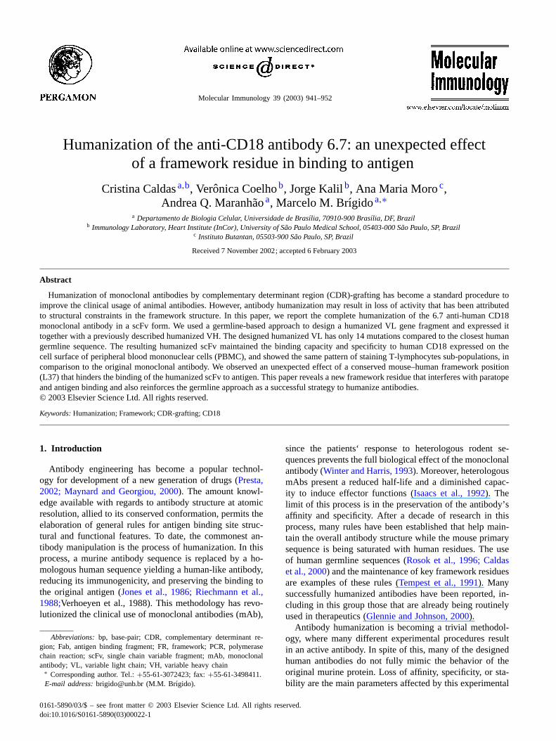
Fig. 1. Design of anti-CD18 VL humanized versions. Amino acid sequence alignments of original murine VL (VL 6.7), closest human germline VL (VkA17 and Vk A18) and humanized VL versions (VLCD18 (Q) and VLCD18 (L)). CDR residues, according toKabat et al. (1991)are indicated. Asterisksindicate the residue 37 and 46, maintained in the humanized VL as discussed in the text. Jk residues are shown in italic.
et al., 2000). We used the FASTA program to search forhuman V� sequences deposited in the Swiss-prot database.The closest human sequence found was the germline V�fragment KV2F (Klobeck et al., 1985) with 76% identityand 89% similarity to the mouse 6.7 V� gene fragment(AF135165). A similar result was obtained using the NCBIIgBlast tool. In this case, two human germline V� sequencesexhibit good hits with the mouse V�. The closest was theA17 (X63403) with 76% identity followed by the A18(X63396) with 74% identity (Lautner-Rieske et al., 1992).The alignment of the original anti-CD18 V� gene segmentto the human related sequences is shown inFig. 1. TheA17 coding sequence is identical to the KV2F Swiss-protrecord, while A18 is 81% identical (91% similar) to eitherA17 or KV2F. Either V� could be used for grafting the6.7 V� complementary determinant region (CDR), but theKV2F/A17 was chosen due to its closer proximity. The6.7 J� (AF135165) closest human J� gene segment is theJ�4 with only one difference. Thus the human germline J�4sequence was chosen for completing the FR4 of the human-ized VL. The conservative V→ L codon change observedin the recombinant FR4 (Fig. 1) is due to anXhoI site atthe end of VL used for the expression vector manipulation.
3.2. Identification of putative constraints
The visual inspection of two of the closest murine (1MRC)and human (1AD9) Fab crystals suggests an overall con-served structure in the VL domain. The systematic survey ofthe tri-dimensional structure at the replacing residues revealstwo positions that could be important for maintaining theframework structure. The murine residue Leu46 was foundto be buried in the VH–VL interface. By using a cut-offdistance of 4.0 Å, VL residue 46 makes many contacts

C. Caldas et al. / Molecular Immunology 39 (2003) 941–952 945
including one to the LCDR 2 residue Phe55, two “Vernier”zone residues (Tyr36 and Trp35), framework residues Ile48,Val58, and the HCDR3 residues Asp101 e Gly97. There-fore, we decided to preserve the original murine Leu46 dueto its array of contacts that includes contacts to VH andVL CDRs.
Murine residue Leu37 was found to be superficially buried,making many contacts within the VL domain core. Its con-tact residues in 1MRC structure at 4.0 Å cut-off are: Pro44,Lys45, Leu47 and Tyr86. The Lys45 contact is one of theconserved hydrogen bonds involved in the maintenance ofthe variable domain structure (Chothia et al., 1985). In the1AD9 structure, Gln37 contacts the same subset of residuesas above. The only discrepancy is a hypothetical hydrogenbond between the hydroxyl O of the Tyr86 and the amideN� of Gln37 (d = 282 Å). In the 1MRC VL, the residue37 is partially exposed to the solvent making a contact to awater molecule in the crystal. No water was found close toresidue Gln37 in the VL of 1AD9. A survey of the Kabatdatabase showed that position 37 is filled with either glu-tamine or leucine. For the mouse light chain, leucine appearsin 20% of the antibodies while glutamine responds for 78%of all-mouse antibodies; other amino acid residues make upless than 3%. Similar numbers occur for human light chain.Due to its location in the VL-solvent interface and to itsclose proximity to the CDR1, we chose this position to con-struct two humanized versions for the VL domain of the6.7. The first construction carries the original L37 residue(named version L) while the other has the human germlineGln37 residue (version Q). Both constructions retained themurine Leu46 residue.
3.3. Design of the recombinant humanized VL
The complete humanized VL sequence was obtained bygrafting the CDR1, 2 and 3 from the 6.7 VL in the proposedhuman germline V� (KV2F/A17) fused to J�4 gene frag-ment. CDR1 was defined as residues 24 to 34 (24–39 in se-quential numbering), CDR2, as residues 50–56 (55–61), andCDR3, residues 89–97 (94–102), following the Kabat defi-nition (Kabat et al., 1991). The grafting process introducedsix changes in CDR1, 2 in CDR2 and 5 in CDR3 of the hu-man VL, resulting in 13 differences out of the 32 residuesin the CDRs. The complete humanized sequences are shownin Fig. 1. The final proposed humanized sequence had 12differences compared to the original 6.7 VL (89% identity)and 14 differences to the original V�KV2F/A17/J�4 (87.6%identity) in the Leu37 version (Fig. 1). The J� fragment wasused as is, except for the Leu104 introduced by anXhoI site.The proposed recombinant VL was chemically synthesizedfor cloning in an scFv expression vector.
3.4. Construction and expression of the humanized scFv
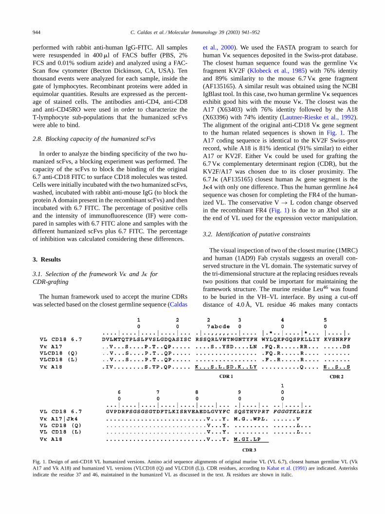
As shown inFig. 2, a set of ten oligonucleotides wasused to generate the humanized VL using recombinant PCR
as previously described for the VH domain (Caldas et al.,2000). Briefly, the primers were initially annealed, filled inby Klenow and then amplified as pairs until they reachedthe full sized VL (Fig. 2). The synthetic DNA fragmentwas cloned in pGEM-T easy vector. The recombinant cloneswere initially checked for size of insert prior to being repeat-edly sequenced. Several clones with the correct sized insertwere tested and one clone (out of six) of the L version andone (out of 8) for Q version had a correct sequence. Correctlysynthesized DNA inserts were digested withBglII andXhoI,and isolated from gel for cloning in the pIg17hVH/mVLplasmid cleaved with the same restriction endonucleases.This plasmid already harbored a hemi-humanized versionof the 6.7 anti-CD18 scFv composed of a humanized VHfused to the original murine VL (Caldas et al., 2000). Thecloning procedure eliminates the original murine VL, re-placing it with the synthetically humanized gene fragment.Two new constructions were obtained: pIg17hVH/hVL(L)and pIg17hVH/hVL(Q). For simplicity, these constructionswere named pIg17LL and pIg17LQ, respectively. The wholescFv cassette, digested withXmaI/EcoRI, was used to re-place the scFv cassette of pPIg16, aP. pastorisexpressionvector (Andrade et al., 2000). According to the notation usedabove, the resulting plasmids were named pPIg LL and pPIgLQ.
A protease defective strain ofP. pastoriswas used to re-ceive the expression vectors by electroporation. Both plas-mids were used to transform electrocompetent yeast cells.Many clones were obtained and screened for scFv produc-tion in the colony filter assay, where scFv producing cellswere detected by immunostaining (Andrade et al., 2000).Two clones of each construction, found to be positive in thefilter assay, were selected for growth on an analytical scale.In both cases, the detection of recombinant scFv was low.Filter assay positive clones were barely visible and the yieldof purified scFv was also limiting. From 200 ml of yeastcultures, we normally obtained about 0.5–1 mg/l. Even so,we were able to purify around 1 mg of recombinant scFvof both L and Q version of the humanized anti-CD18 forfurther characterization.
3.5. Immunological characterization of recombinanthumanized antibodies
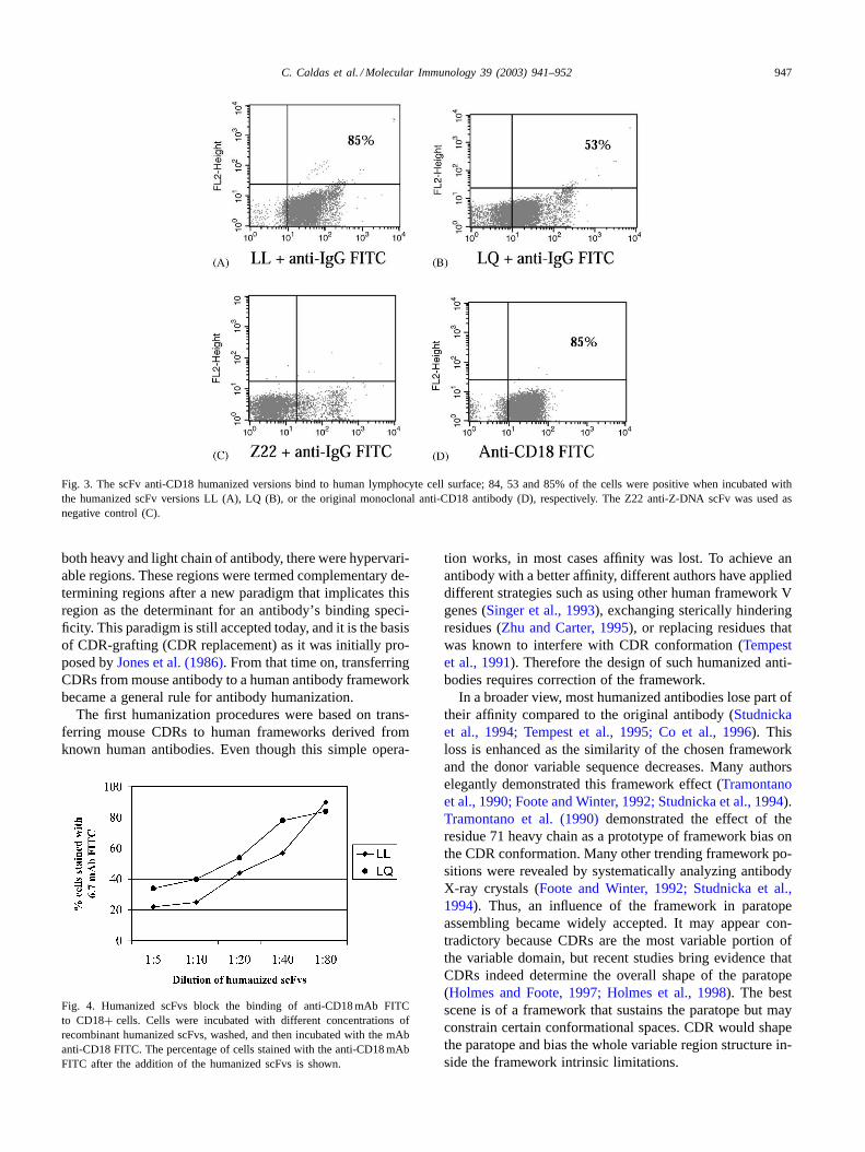
The CD18 antigen is expressed at the cell surface of allleucocytes. Therefore, we tested the two variants of recom-binant humanized antibody (LL and LQ) directly for bind-ing to peripheral blood mononuclear cells (PBMC) by FlowCytometry analysis. Our results show that the humanizedLL version presents essentially the same binding capacityto lymphocytes as the original anti-CD18 monoclonal anti-body, staining 85% of gated cells. In contrast, the LQ ver-sion only stained 53% of the cells in the gate of lymphocytes(Fig. 3). The original 6.7 anti-CD18 mAb and an anti-DNAscFv were used as positive and negative controls, respec-tively. The small population stained with high intensity in
946 C. Caldas et al. / Molecular Immunology 39 (2003) 941–952
Fig. 2. The VL humanized versions were assembled using a PCR-based protocol. Arrows indicated the sense (→) and anti-sense (←) oligonucleotidesused for VL synthesis. Two variations of the L3 oligonucleotides were synthesized (L3L and L3Q), that create the difference among the versions L and Q.
the humanized versions of anti-CD18 was interpreted as un-specific staining, since it was also detected with anti-humanIgG-FITC alone. The negative control anti-DNA scFv didnot show any significant staining.
The humanized scFvs were also able to compete effi-ciently with the 6.7 mAb for binding CD18 on lymphocytes,as shown inFig. 4. In this experiment, decreasing dilutionsof scFv were incubated with PBMC prior to incubation withthe original anti-CD18 mAb. Both humanized versions wereable to efficiently block the binding of the mAb to CD18molecules at the cell surface. Again, the LL version wasalso more efficient than LQ, blocking up to 80% of mAb’sbinding activity in a 1:5 dilution.
In order to compare the cell subsets recognized by thehumanized LL version and the original murine antibody,we performed FACS analysis, using different markers. Wefound that both the original anti-CD18 and the human-ized LL version displayed the same pattern of stainingto CD4+, CD8+ and to memory CD45RO+ lymphocytes(Fig. 5).
4. Discussion
The rules for transferring ligand specificity between donorand acceptor antibody are partially known. Today it is con-ceivable to transfer the CDR regions to a closely relatedframework acceptor, retaining the antibody’s specificity. Thefact that the three-dimensional structure of the variable do-main is highly conserved makes this strategy feasible. Inaddition, the traditional model of antibody ontogeny relieson a flexible variable domain structure for evolving B cellresponse for an infinite antigen repertoire. Somatic hyper-mutation molds the final high affinity antibody. Such flexi-bility is so impressive that even Ig transgenic mice are ableto make antibodies successfully even with a very limited Igrepertoire (Cascalho et al., 1996). It is likely that antibodiesare made for binding, and transposing specificity is an opera-tion controlled by simple rules. WhenWu and Kabat (1970)systematically analyzed the primary structure of the variabledomain of antibodies, they observed that interspaced in theoverall variable structure of the amino terminus domain of
C. Caldas et al. / Molecular Immunology 39 (2003) 941–952 947
Fig. 3. The scFv anti-CD18 humanized versions bind to human lymphocyte cell surface; 84, 53 and 85% of the cells were positive when incubated withthe humanized scFv versions LL (A), LQ (B), or the original monoclonal anti-CD18 antibody (D), respectively. The Z22 anti-Z-DNA scFv was used asnegative control (C).
both heavy and light chain of antibody, there were hypervari-able regions. These regions were termed complementary de-termining regions after a new paradigm that implicates thisregion as the determinant for an antibody’s binding speci-ficity. This paradigm is still accepted today, and it is the basisof CDR-grafting (CDR replacement) as it was initially pro-posed byJones et al. (1986). From that time on, transferringCDRs from mouse antibody to a human antibody frameworkbecame a general rule for antibody humanization.
The first humanization procedures were based on trans-ferring mouse CDRs to human frameworks derived fromknown human antibodies. Even though this simple opera-
Fig. 4. Humanized scFvs block the binding of anti-CD18 mAb FITCto CD18+ cells. Cells were incubated with different concentrations ofrecombinant humanized scFvs, washed, and then incubated with the mAbanti-CD18 FITC. The percentage of cells stained with the anti-CD18 mAbFITC after the addition of the humanized scFvs is shown.
tion works, in most cases affinity was lost. To achieve anantibody with a better affinity, different authors have applieddifferent strategies such as using other human framework Vgenes (Singer et al., 1993), exchanging sterically hinderingresidues (Zhu and Carter, 1995), or replacing residues thatwas known to interfere with CDR conformation (Tempestet al., 1991). Therefore the design of such humanized anti-bodies requires correction of the framework.
In a broader view, most humanized antibodies lose part oftheir affinity compared to the original antibody (Studnickaet al., 1994; Tempest et al., 1995; Co et al., 1996). Thisloss is enhanced as the similarity of the chosen frameworkand the donor variable sequence decreases. Many authorselegantly demonstrated this framework effect (Tramontanoet al., 1990; Foote and Winter, 1992; Studnicka et al., 1994).Tramontano et al. (1990)demonstrated the effect of theresidue 71 heavy chain as a prototype of framework bias onthe CDR conformation. Many other trending framework po-sitions were revealed by systematically analyzing antibodyX-ray crystals (Foote and Winter, 1992; Studnicka et al.,1994). Thus, an influence of the framework in paratopeassembling became widely accepted. It may appear con-tradictory because CDRs are the most variable portion ofthe variable domain, but recent studies bring evidence thatCDRs indeed determine the overall shape of the paratope(Holmes and Foote, 1997; Holmes et al., 1998). The bestscene is of a framework that sustains the paratope but mayconstrain certain conformational spaces. CDR would shapethe paratope and bias the whole variable region structure in-side the framework intrinsic limitations.

948 C. Caldas et al. / Molecular Immunology 39 (2003) 941–952
Fig. 5. T cell subpopulations stained by the humanized LL version compared to the original mAb.
In this report we synthesized the light chain variable re-gion as part of a humanized anti-human CD18 antibody. Thestrategy to design the humanized light chain variable regionwas based on the closest germline sequence (Caldas et al.,2000). Such a strategy allows us to reduce the frameworkderived constraints due to the use of the closest germlineframework sequence. Many human germline sequences areavailable to date, facilitating the search for homologous Vgene candidates (Tomlinson et al., 1995). After design weproceeded to the visual inspection of the three-dimensionalstructure of similar antibodies. In this step, we chose to useX-ray derived information instead of the molecular modelfor the humanized structure, because there is a large amountof data on crystal structures of antibodies in the PDB.(This is probably the most studied protein family at thethree-dimensional level.) We were able to find, in the PDB,two sequences which were very similar to our VL sequence;one is a mouse Fab and the other is a humanized Fab.There are 12 differences between the murine and the hu-manized VL. Most of them correspond to exposed residues
or residues far from the paratope. There was one importantresidue, Leu46, that was shown to make contacts to LCDR2and 3, besides playing an important role in the interface ofVL/VH, making close contact to HCDR3 (Padlan, 1994).The exchange of a leucine, an aliphatic amino acid, for thearginine that appears in the human closest germline, wouldprobably disrupt many atomic contacts and destabilize theoverall structure. Therefore this residue was kept as in theoriginal murine antibody.
Another observed difference was at position 37. Thisresidue sits in the bottom of the molecule between the struc-turally conserved Gln38, involved in light chain-heavy chaininterface (Chothia et al., 1985), and the “Vernier” zoneresidues after CDR1 (Foote and Winter, 1992). In the mouseantibody structure 1MRC, the Leu37 is partially accessibleto solvent where it is close to a water molecule (3.37 Å).It is probably not involved in a direct H-bond since thedistance found is over the expected distance of a regularwater-carbonyl hydrogen bond (Creigthon, 1984). In the hu-man V� germline sequence used in this work, a glutamine

C. Caldas et al. / Molecular Immunology 39 (2003) 941–952 949
Fig. 6. Structural visualization of the Gln37–Tyr86 interaction. The structure of variable domain of 1AD9 was visualized using RASMOL. VL backboneis shown in gray and its three CDRs (black) are labeled. Gln37 and Tyr86 residues are shown in a space filling representation. The atoms labeled with Nand O denotes the glutamine amide group and the tyrosine hydroxyl group, respectively. Heavy chain backbone (light gray) is shown in the background.
residue fills the position 37. This residue is found in manyhumanized antibodies, including our closest PDB crystalstructure, 1AD9. In this crystal, Gln37 is mostly buried andit contacts the same set of residues as Leu37 in the murinestructure. The only difference is a hypothetical H-bond be-tween the amide group of Gln37 and the hydroxyl group ofTyr86 (Fig. 6). The short distance between them (2.82 Å) sup-ports this assumption. Besides that the environment aroundposition 37 is conserved in both structures, as is the con-served H-bond to residue 45.
The leucine residue is less common than glutamine ineither human and mouse VL at position 37. This positionseems to be exclusively filled by one of these large residuesand therefore there should be a restriction for using eitherone of them, but polar residues seem more appropriate foroccupying this position. Analysis of the germline genes re-veals a similar proportion of Gln/Leu residues at position37 in mouse and human antibodies (Lautner-Rieske et al.,1992). Interestingly,Chothia et al. (1998)classified this po-sition as a buried polar region, barely variable, that is in-volved in the stabilization of FR2. It is possible that the
formation of the H-bond between the Gln37 and Tyr86 sta-bilizes the antibody hydrophobic core.
We tested both versions of humanized antibodies using asthe VH domain a previously characterized humanized heavychain (Caldas et al., 2000). The synthesis of both human-ized VL was performed using recombinant PCR to achievea full-length gene segment. We found that only a small pro-portion of clones carry the correct construction and we at-tributed to the purity of the synthetic oligonucleotides. Wehad to screen a large number of clones to find a correct one.A single extensively checked clone for each version (Q andL) was chosen. The VL gene segment was isolated and trans-ferred to theP. pastorisexpression vector along with theheavy chain gene fragment version L (Caldas et al., 2000).The recombinant scFv produced by this expression systemhad a staphylococcal protein A tag in its carboxi-terminus,which allowed for purification and detection of the recombi-nant protein. The yield of secreted scFv for this expressionsystem was lower than expected when compared to previousresults (Andrade et al., 2000), but it was sufficient to test forbinding in fluorescence activated cell cytometry.

950 C. Caldas et al. / Molecular Immunology 39 (2003) 941–952
The�2-integrin antigen CD18 is a cell membrane associ-ated high molecular weight glycoprotein. Although it wouldbe appropriate to test the specificity of the humanized an-tibodies directly on CD18 antigen, the binding of the 6.7antibody to soluble CD18 in simple immunoassays such asenzyme linked immunosorbent assay (ELISA) has not beenreported. Therefore, we tested the humanized antibodies di-rectly on cells from a healthy donor expressing the CD18antigen, by FACS analysis. Similarly to the 6.7 mAb, bothhumanized antibodies were able to bind to PBMC. No bind-ing was observed with the negative control, suggesting thatthe observed binding of the humanized scFv was due to itsaffinity for PBMC antigens, excluding the possibility of afortuitous activity due to misfolded proteins or due its pro-tein A affinity tag. In this experiment, the LL version showedhigher cell labeling when compared to the LQ version. Thespecificity of constructions was analyzed in a blocking ex-periment in which we showed that binding of 6.7 mAb wasspecifically blocked by the humanized scFv, but not by asimilar but non-specific scFv (anti-DNA, data not shown).This experiment indicates that the humanized scFv bindsto PBMC specifically through the CD18 antigen and thereis no unspecific binding to some other cellular structure.Moreover, both versions of the humanized antibody frag-ments bind to the same epitope as the original 6.7 mAb.The blocking experiment also reveals that the LL version ismore effective than the LQ version in competing for the 6.7binding site. The experimental design in this work does notpermit a direct comparison of the affinity of mAb with thatof the humanized versions. However, it does allow us to in-fer that the LL version affinity is similar to that of 6.7 mAbdue to the blocking efficiency of this scFv compared to thewhole 6.7 mAb. The original murine mAb is divalent andthis attribute contributes to an increased avidity comparedto a monomeric scFv, hence quantitating true affinity wasnot possible for this system.
To be useful in therapeutics, a humanized antibody has tomaximally mimetize the affinity and specificity of the orig-inal heterologous antibody. The CD18 antigen is found inleukocytes, including lymphocytes and monocytes. Here, weused the binding profile in different lymphocyte populationsof cells as evidence that the humanized antibody retained itsspecificity. The comparison of the 6.7 antibody with its L hu-manized version reveals a very similar profile for binding toCD18 in three lymphocyte sub-populations: CD4+, CD8+and CD45RO+. FACS analysis revealed that both murineand the L humanized version stain approximately the samenumber of cells. The similarity of binding profile for theLL humanized antibody, compared to the 6.7 mAb, suggeststhat it can be used for the replacement of the murine anti-body. However, further in vivo studies will be necessary forconfirming its efficacy and clinical applicability.
All the experiments used to verify and to compare the hu-manized scFv to de 6.7 mAb result in a consistently betterperformance of the LL over the LQ version. For some rea-son the presence of a leucine residue at position 37 enhances
binding activity, although the specificity was maintained inboth humanized versions compared to 6.7 mAb. The net ef-fect of this Leu→ Gln change in the binding activity ofthe humanized scFv was expected. Its volume and buriedcharacter associated with a discrepancy in the hydrophilicitysuggests an impact on the overall structure. The differenceobserved in this work may be explained by the formation ofan H-bond between residue 37 and 86 only in the case of anelectron acceptor side-chain, such as a glutamine, which isindeed present. This could reduce the flexibility required forthe FR2 and FR3 reconfiguration of the framework, whichis required for the successful graft of the CDR. The resultmay be a more rigid paratope that hampers the associationwith the antigen. Moreover, this framework rigidity may re-sult in reduced paratope flexibility that seems fundamentalfor binding of some antigens, as suggested by others (Herronet al., 1991; Stanfield et al., 1993).
We have shown the complete humanization of the6.7 mAb. The procedure used here was very simple butis reiteratively successful. It suggests that the principle ofCDR-grafting using the closest germline is indeed a stepforward. Recent publications on humanization rely on morecomplex grafting procedures that do not necessarily leadto more efficient antibodies. The SDR (specificity deter-mining residues) approach was based in the humanizationof specific contact residues (Iwahashi et al., 1999; Tamuraet al., 2000; De Pascalis et al., 2002). Even though theprocedure leads to active antibodies, it does demand eitherthe “a priori” knowledge of the antibody’s tri-dimensionalstructure, or a tantalizing screening for binding specificresidues. Moreover, since many human CDR residues weremaintained in the final structure, the determinant role ofthe CDRs for the overall antibody structure was partiallylost. Tan et al. (2002)have recently proposed a humaniza-tion procedure based on a canonical region based germlinesequence search. Their results were indeed disappointing.The authors argue that their process allows a very “human”antibody based on their comparison of the number of in-troduced mutations in their germline sequence and FDAapproved antibodies. It should be pointed out, however,that the number of mutations introduced in their chosenVL germline sequence was 21. In contrast, in the human-ized 6.7 VL we introduced 14 mutations including one dueto an XhoI restriction site introduced in FR4 far from thebinding site. Hence, our approach is robust, as it leads to apreservation of binding activity while minimizing the num-ber of murine residues. Moreover the systematic analysisof related structures leads to the detection of frameworkresidues that potentially interfere with paratope formation,as we have shown for the VL position 37.
Acknowledgements
We thank Sandra Maria Monteiro for help in the FACSanalyses, Dr. Linda Caldas and Adilton Guedes Oliveira for
C. Caldas et al. / Molecular Immunology 39 (2003) 941–952 951
English corrections to the manuscript, and Dr. Armand Ben-sussan and Dr. Laurence Boumsell for providing the originalhybridoma.
References
Altschul, S.F., Madden, T.L., Schäffer, A.A., Zhang, J., Zhang, Z., Miller,W., Lipman, D.J., 1997. Gapped BLAST and PSI-BLAST: a newgeneration of protein database search programs. Nucleic Acids Res.25, 3389–3402.
Andrade, E.V., Albuquerque, F.C., Moraes, L.M., Brigido, M.M.,Santos-Silva, M.A., 2000. Single-chain Fv with Fc fragment of thehuman IgG1 tag: construction,Pichia pastorisexpression and antigenbinding characterization. J. Biochem. (Tokyo) 128, 891–895.
Bairoch, A., Apweiler, R., 2000. The SWISS-PROT protein sequencedatabase and its supplement TrEMBL in 2000. Nucleic Acids Res. 28,45–48.
Berman, H.M., Westbrook, J., Feng, Z., Gilliland, G., Bhat, T.N., Weissig,H., Shindyalov, I.N., Bourn, P.E., 2000. The Protein Data Bank. NucleicAcids Res. 28, 235–242.
Bernstein, H.J., 2000. Recent changes to RasMol, recombining thevariants. Trends Biochem. Sci. 25, 435–453.
Brigido, M.M., Polymenis, M., Stollar, B.D., 1993. Role of mouse VH10and VL gene segments in the specific binding of antibody to Z-DNA,analyzed with recombinant single chain Fv molecules. J. Immunol.150, 469–479.
Caldas, C., Coelho, V.P., Rigden, D.J., Neschich, G., Moro, A.M., Brigido,M.M., 2000. Design and synthesis of germline-based hemi-humanizedsingle-chain Fv against the CD18 surface antigen. Protein Eng. 13,353–360.
Carter, P., Presta, L., Gorman, C.M., Ridgway, J.B., Henner, D., Wong,W.L., Rowland, A.M., Kotts, C., Carver, M.E., Shepard, H.M., 1992.Humanization of an anti-p185HER2 antibody for human cancer therapy.Proc. Natl. Acad. Sci. U.S.A. 89, 4285–4289.
Cascalho, M., Ma, A., Lee, S., Masat, L., Wabl, M., 1996. Aquasi-monoclonal mouse. Science 272, 1585–1588.
Chien, N.C., Roberts, V.A., Giusti, A.M., Scharff, M.D., Getzoff, E.D.,1989. Significant structural and functional change of an antigen-bindingsite by a distant amino acid substitution: proposal of a structuralmechanism. Proc. Natl. Acad. Sci. U.S.A. 86, 5532–5536.
Chothia, C., Novotny, J., Bruccoleri, R., Karplus, M., 1985. Domainassociation in immunoglobulin molecules. The packing of variabledomains. J. Mol. Biol. 186, 651–663.
Chothia, C., Gelfand, I., Kister, A., 1998. Structural determinants in thesequences of immunoglobulin variable domain. J. Mol. Biol. 278, 457–479.
Co, M.S., Baker, J., Bednarik, K., Janzek, E., Neruda, W., Mayer, P., Plot,R., Stumper, B., Vasquez, M., Queen, C., Loibner, H., 1996. Humanizedanti-Lewis Y antibodies: in vitro properties and pharmacokinetics inrhesus monkeys. Cancer Res. 56, 1118–1125.
Creigthon, T.E., 1984. Physical forces that determine the proprieties ofproteins. Proteins. Freeman Press, New York, Chapter 4, pp. 133–157.
David, V., Leca, G., Corvaia, N., Deist, F., Boumsell, L., Bensussan, A.,1991. Proliferation of resting lymphocytes is induced by triggering Tcells through an epitope common to the three CD18/CD11 leukocyteadhesion molecules. Cell Immunol. 136, 519–524.
De Pascalis, R., Iwahashi, M., Tamura, M., Padlan, E.A., Gonzales, N.R.,Santos, A.D., Giuliano, M., Schuck, P., Schlom, J., Kashmiri, S.V.,2002. Grafting of abbreviated complementarity-determining regionscontaining specificity-determining residues essential for ligand contactto engineer a less immunogenic humanized monoclonal antibody. J.Immunol. 169, 3076–3084.
Dove, A., 2000. CD18 trials disappoint again. Nat. Biotechnol. 18, 817–818.
Foote, J., Winter, G., 1992. Antibody framework residues affecting theconformation of the hypervariable loops. J. Mol. Biol. 224, 487–499.
Gao, F., Yue, T.L., Shi, D.W., Christopher, T.A., Lopez, B.L., Ohlstein,E.H., Barone, F.C., Ma, X.L., 2002. p38 MAPK inhibition reducesmyocardial reperfusion injury via inhibition of endothelial adhesionmolecule expression and blockade of PMN accumulation. Cardiovasc.Res. 53, 414–422.
Glennie, M.J., Johnson, P.W.M., 2000. Clinical trials of antibody therapy.Immunol. Today 21, 403–410.
Herron, J.N., He, X.M., Ballard, D.W., Blier, P.R., Pace, P.E., Bothwell,A.L., Voss Jr., E.W., Edmundson, A.B., 1991. An autoantibody tosingle-stranded DNA: comparison of the three-dimensional structuresof the unliganded Fab and a deoxynucleotide–Fab complex. Proteins11, 159–175.
Higgins, D.G., Thompson, J.D., Gibson, T.J., 1996. Using CLUSTAL formultiple sequence alignments. Methods Enzymol. 266, 383–402.
Holmes, M.A., Foote, J., 1997. Structural consequences of humanizingan antibody. J. Immunol. 158, 2192–2201.
Holmes, M.A., Buss, T.N., Foote, J., 1998. Conformational correctionmechanisms aiding antigen recognition by a humanized antibody. J.Exp. Med. 187, 479–485.
Hsiao, K.C., Bajorath, J., Harris, L.J., 1994. Humanization of 60.3, ananti-CD18 antibody: importance of the L2 loop. Protein Eng. 7, 815–822.
Isaacs, J.D., Clark, M.R., Greenwood, J., Waldmann, H., 1992. Therapywith monoclonal antibodies. An in vivo model for the assessment oftherapeutic potential. J. Immunol. 148, 3062–3071.
Isobe, M., Suzuki, J., Yamazaki, S., Yazaki, Y., Horie, S., Okubo,Y., Maemura, K., Yazaki, Y., Sekiguchi, M., 1997. Regulation bydifferential development of Th1 and Th2 cells in peripheral toleranceto cardiac allograft induced by blocking ICAM-1/LFA-1 adhesion.Circulation 96, 2247–2253.
Iwahashi, M., Milenic, D.E., Padlan, E.A., Bei, R., Schlom, J., Kashmiri,S.V., 1999. CDR substitutions of a humanized monoclonal antibody(CC49): contributions of individual CDRs to antigen binding andimmunogenicity. Mol. Immunol. 36, 1079–1091.
Johnson, G., Wu, T.T., 2000. Kabat database and its applications: 30 yearsafter the first variability plot. Nucleic Acids Res. 28, 214–218.
Jones, P.T., Dear, P.H., Foote, J., Neuberger, M.S., Winter, G., 1986.Replacing the complementarity-determining regions in a humanantibody with those from a mouse. Nature 321, 522–525.
Kabat, E.A., Wu, T.T., Perry, H.M., Gottesmann, K.S., Foeller, C., 1991.Sequences of Proteins of Imunological Interest, fifth ed. Public HealthService, National Institutes of Health, Bethesda, MD.
Kettleborough, C.A., Saldanha, J., Heath, V.J., Morrison, C.J., Bendig,M.M., 1991. Humanization of a mouse monoclonal antibody byCDR-grafting: the importance of framework residues on loopconformation. Protein Eng. 4, 773–783.
Klobeck, H.G., Meindl, A., Combriato, G., Solomon, A., Zachau, H.G.,1985. Human immunoglobulin kappa light chain genes of subgroupsII and III. Nucleic Acids Res. 13, 6499–6513.
Lautner-Rieske, A., Huber, C., Meindl, A., Pargent, W., Schable, K.F.,Thiebe, R., Zocher, I., Zachau, H.G., 1992. The human immunoglobulinkappa locus. Characterization of the duplicated A regions. Eur. J.Immunol. 22, 1023–1029.
Lu, C., Ferzly, M., Takagi, J., Springer, T.A., 2001. Epitope mapping ofantibodies to the C-terminal region of the integrin beta 2 subunit revealsregions that become exposed upon receptor activation. J. Immunol.166, 5629–5637.
Maynard, J., Georgiou, G., 2000. Antibody engineering. Annu. Rev.Biomed. Eng. 2, 339–376.
Nakatani, T., Lone, Y.C., Yamakawa, J., Kanaoka, M., Gomi, H., Wijdenes,J., Noguchi, H., 1994. Humanization of mouse anti-human IL-2receptor antibody B-B10. Protein Eng. 7, 435–443.
Padlan, E.A., 1994. Anatomy of the antibody molecule. Mol. Immunol.31, 169–217.
Pearson, W.R., 2000. Flexible sequence similarity searching with theFASTA3 program package. Methods Mol. Biol. 132, 185–219.
Presta, L.G., 2002. Engineering antibodies for therapy. Curr. Pharm.Biotechnol. 3, 237–256.

952 C. Caldas et al. / Molecular Immunology 39 (2003) 941–952
Riechmann, L., Foote, J., Winter, G., 1988. Expression of an antibodyFv fragment in myeloma cells. J. Mol. Biol. 203, 825–828.
Rosok, M.J., Yelton, D.E., Harris, L.J., Bajorath, J., Hellstrom, K.E.,Hellstrom, I., Cruz, G.A., Kristensson, K., Lin, H., Huse, W.D., Glaser,S.M., 1996. A combinatorial library strategy for the rapid humanizationof anticarcinoma BR96 Fab. J. Biol. Chem. 271, 22611–22618.
Saldanha, J.W., Martin, A.C., Leger, O.J., 1999. A single backmutation inthe human kIV framework of a previously unsuccessfully humanizedantibody restores the binding activity and increases the secretion incos cells. Mol. Immunol. 36, 709–719.
Singer, I.I., Kawka, D.W., DeMartino, J.A., Daugherty, B.L., Elliston,K.O., Alves, K., Bush, B.L., Cameron, P.M., Cuca, G.C., Davies, P.,1993. Optimal humanization of 1B4, an anti-CD18 murine monoclonalantibody, is achieved by correct choice of human V-region frameworksequences. J. Immunol. 150, 2844–2857.
Stanfield, R.L., Takimoto-Kamimura, M., Rini, J.M., Profy, A.T., Wilson,I.A., 1993. Major antigen-induced domain rearrangements in anantibody. Structure 1, 83–93.
Studnicka, G.M., Soares, S., Better, M., Williams, R.E., Nadell, R.,Horwitz, A.H., 1994. Human-engineered monoclonal antibodies retainfull specific binding activity by preserving non-CDR complementary-modulating residues. Protein Eng. 7, 805–814.
Tamura, M., Milenic, D.E., Iwahashi, M., Padlan, E., Schlom, J., Kashmiri,S.V., 2000. Structural correlates of an anticarcinoma antibody:identification of specificity-determining residues (SDRs) and deve-lopment of a minimally immunogenic antibody variant by retention ofSDRs only. J. Immunol. 164, 1432–1441.
Tan, P., Mitchell, D.A., Buss, T.N., Holmes, M.A., Anasetti, C., Foote, J.,2002. Superhumanized antibodies: reduction of immunogenic potentialby complementarity-determining region grafting with human germlinesequences: application to an anti-CD28. J. Immunol. 169, 1119–1125.
Tempest, P.R., Bremner, P., Lambert, M., Taylor, G., Furze, J.M., Carr, F.J.,Harris, W.J., 1991. Reshaping a human monoclonal antibody to inhibithuman respiratory syncytial virus infection in vivo. Biotechnology 9,266–271.
Tempest, P.R., White, P., Buttle, M., Carr, F.J., Harris, W.J., 1995.Identification of framework residues required to restore antigen bindingduring reshaping of a monoclonal antibody against the glycoproteingB of human cytomegalovirus. Int. J. Biol. Macromol. 17, 37–42.
Tomlinson, I.M., Cox, J.P., Gherardi, E., Lesk, A.M., Chothia, C., 1995.The structural repertoire of the human V kappa domain. EMBO J. 14,4628–4638.
Tramontano, A., Chothia, C., Lesk, A.M., 1990. Framework residue 71is a major determinant of the position and conformation of the secondhypervariable region in the VH domains of immunoglobulins. J. Mol.Biol. 215, 175–182.
Tsodikov, O.V., Record Jr., M.T., Sergeev, Y.V., 2002. Novel computerprogram for fast exact calculation of accessible and molecular surfaceareas and average surface curvature. J. Comput. Chem. 23, 600–609.
Tuomanen, E.I., Saukkonen, K., Sande, S., Cioffe, C., Wright, S.D.,1989. Reduction of inflammation, tissue damage, and mortality inbacterial meningitis in rabbits treated with monoclonal antibodiesagainst adhesion-promoting receptors of leukocytes. J. Exp. Med. 170,959–969.
Winter, G., Harris, W.J., 1993. Humanized antibodies. Immunol. Today14, 243–252.
Wu, T.T., Kabat, E.A., 1970. An analysis of the sequences of the variableregions of Bence Jones proteins and myeloma light chains and theirimplications for antibody complementarity. J. Exp. Med. 132, 211–250.
Zhu, Z., Carter, P., 1995. Identification of heavy chain residues in ahumanized anti-CD3 antibody important for efficient antigen bindingand T cell activation. J. Immunol. 155, 1903–1910.