Brief report: reduced expression of CD18 leads to the in vivo expansion of hematopoietic stem cells...
8
STEM CELLS 2014;00:00‐00 www.StemCells.com ©AlphaMed Press 2014 TISSUE‐SPECIFIC STEM CELLS a Division of Hematopoietic Innovative Ther‐ apies. Centro de Investigaciones Energéti‐ cas, Medioambientales y Tecnológicas (CIEMAT) and Centro de Investigación Bio‐ médica en Red de Enfermedades Raras (CIBER‐ER). b Instituto de Investigación Sani‐ taria Fundación Jiménez Díaz. (IIS‐ FJD, UAM) Madrid. Spain. c Department of Epi‐ demiology, Atherothrombosis and Imaging. Fundación Centro Nacional de Investigaciones Cardiovasculares (CNIC). Madrid, Spain. *Corresponding authors: Elena Almarza and Juan A Bueren, Division of Hematopoietic Innovative Therapies. CIEMAT/CIBERER/FJD. Avda. Complutense 40. 28040 Madrid. Phone: +34 91 346 65 18 / +34 91 496 25 24. Fax:+34 91 346 6484. Email: [email protected]; [email protected]; Disclaimer: The authors declare no conflict of interest.; ACKNOWLEDGEMENTS: The authors would like to thank Aurora de la Cal and Sergio Losada for their collaboration in the admin‐ istrative work and Miguel Angel Martín, Jesús Martínez and Edilia de Almeida for the careful maintenance of the animals. This work was supported by the following grants: Dirección General de Investigación de la Comunidad de Madrid (CellCAM; Ref S2010/BMD‐2420), Fondo de Investigaciones Sanitarias, Instituto de Salud Carlos III (RETICS‐RD12/0019/0023), and SAF2012‐31142 from MINECO, S2010/BMD‐ 2314 from Comunidad de Madrid, and FP7‐ People‐IRG Program (246655). DLR was supported by an FPU grant by the Spanish Ministry of Education (AP‐2009‐4129). The Centro Nacional de Investigaciones Cardiovasculares is supported by the MINECO and the Pro‐CNIC Foundation. Received January 29, 2014; accepted for publication May 15, 2014 ©AlphaMed Press 1066‐5099/2014/$30.00/0 This article has been accepted for publica‐ tion and undergone full peer review but has not been through the copyediting, typeset‐ ting, pagination and proofreading process which may lead to differences between this version and the Version of Record. Please cite this article as doi: 10.1002/stem.1762 Reduced Expression of CD18 Leads to the In Vivo Expansion of Hematopoietic Stem Cells in Mouse Bone Marrow DIEGO LEON‐RICO a,b ,MONTSERRAT ALDEA a,b ,REBECA SANCHEZ a,b ,JOSÉ C. SEGOVIA a,b, LINNEA A. WEISS c ,ANDRÉS HIDALGO c ,JUAN A. BUEREN *a,b AND ELENA ALMARZA *a,b Key words. Hematopoietic Stem Cells • cell adhesion molecules • stem cell‐ microenvironment • neutrophil ABSTRACT Leukocyte adhesion deficiency type‐I (LAD‐I) is a primary immunodeficien‐ cy caused by mutations in the ITGB2 gene (CD18 leukocyte integrin) which lead to defects in leukocyte extravasation. To investigate the role of CD18 in hematopoietic stem cell (HSC) biology, we have thoroughly character‐ ized the HSCs of CD18 Itgb2 tm1bay hypomorphic mice (CD18 HYP ) both by flow cytometry and using in vitro and in vivo transplantation assays. Flow cytometry analyses and cultures in methyl‐cellulose revealed that bone marrow (BM) from CD18 HYP mice was enriched in hematopoietic precur‐ sors, mainly early quiescent short‐term and long‐term HPCs. Strikingly, bone marrow competition assays showed a progressive expansion of CD18 HYP – derived hematopoiesis in recipient mice. Additionally, we provide evidence that this HSC expansion was not caused by an increased homing capacity of CD18 HYP HSCs, or by alterations in the hematopoietic environ‐ ment of CD18 HYP mice due to defects in neutrophils clearance. On the con‐ trary, our data demonstrated that the reduced expression of CD18 causes a cell‐autonomous expansion in the HSC compartment, thus revealing unex‐ pected regulatory functions for CD18 in mouse HSCs. STEM CELLS 2014; 00:000–000 INTRODUCTION Leukocyte adhesion deficiency type I (LAD‐I) is a primary immunodeficiency associated with mutations in the ITGB2 gene (β 2 common leukocyte subunit CD18). Most ITGB2 mutations lead to reduced/null expression of β 2 ‐ integrins on the leukocytes’ surface [1,2]. Hence CD18‐ deficient leukocytes fail to extravasate from blood to sites of infection [3,4,5]. Two transgenic models have been generated: a CD18 knock‐out model (Itgb2 tm2Bay [6]), resembling the severe phenotype of LAD‐I patients,
Transcript of Brief report: reduced expression of CD18 leads to the in vivo expansion of hematopoietic stem cells...
STEM CELLS 2014;00:00‐00 www.StemCells.com ©AlphaMed Press 2014
TISSUE‐SPECIFIC STEM CELLS aDivision of Hematopoietic Innovative Ther‐apies. Centro de Investigaciones Energéti‐cas, Medioambientales y Tecnológicas (CIEMAT) and Centro de Investigación Bio‐médica en Red de Enfermedades Raras (CIBER‐ER). bInstituto de Investigación Sani‐taria Fundación Jiménez Díaz. (IIS‐ FJD, UAM) Madrid. Spain. cDepartment of Epi‐demiology, Atherothrombosis and Imaging. Fundación Centro Nacional de Investigaciones Cardiovasculares (CNIC). Madrid, Spain. *Corresponding authors: Elena Almarza and Juan A Bueren, Division of Hematopoietic Innovative Therapies. CIEMAT/CIBERER/FJD. Avda. Complutense 40. 28040 Madrid. Phone: +34 91 346 65 18 / +34 91 496 25 24. Fax:+34 91 346 6484. Email: [email protected]; [email protected]; Disclaimer: The authors declare no conflict of interest.; ACKNOWLEDGEMENTS: The authors would like to thank Aurora de la Cal and Sergio Losada for their collaboration in the admin‐istrative work and Miguel Angel Martín, Jesús Martínez and Edilia de Almeida for the careful maintenance of the animals. This work was supported by the following grants: Dirección General de Investigación de la Comunidad de Madrid (CellCAM; Ref S2010/BMD‐2420), Fondo de Investigaciones Sanitarias, Instituto de Salud Carlos III (RETICS‐RD12/0019/0023), and SAF2012‐31142 from MINECO, S2010/BMD‐2314 from Comunidad de Madrid, and FP7‐People‐IRG Program (246655). DLR was supported by an FPU grant by the Spanish Ministry of Education (AP‐2009‐4129). The Centro Nacional de Investigaciones Cardiovasculares is supported by the MINECO and the Pro‐CNIC Foundation. Received January 29, 2014; accepted for publication May 15, 2014 ©AlphaMed Press 1066‐5099/2014/$30.00/0 This article has been accepted for publica‐tion and undergone full peer review but has not been through the copyediting, typeset‐ting, pagination and proofreading process which may lead to differences between this version and the Version of Record. Please cite this article as doi: 10.1002/stem.1762
Reduced Expression of CD18 Leads to the In Vivo Expansion of Hematopoietic Stem Cells in Mouse Bone Marrow DIEGO LEON‐RICO a,b, MONTSERRAT ALDEA a,b, REBECA SANCHEZ a,b, JOSÉ
C. SEGOVIA a,b, LINNEA A. WEISS c, ANDRÉS HIDALGO c, JUAN A. BUEREN *a,b
AND ELENA ALMARZA *a,b
Key words. Hematopoietic Stem Cells • cell adhesion molecules • stem cell‐
microenvironment • neutrophil ABSTRACT Leukocyte adhesion deficiency type‐I (LAD‐I) is a primary immunodeficien‐cy caused by mutations in the ITGB2 gene (CD18 leukocyte integrin) which lead to defects in leukocyte extravasation. To investigate the role of CD18 in hematopoietic stem cell (HSC) biology, we have thoroughly character‐ized the HSCs of CD18 Itgb2tm1bay hypomorphic mice (CD18HYP) both by flow cytometry and using in vitro and in vivo transplantation assays. Flow cytometry analyses and cultures in methyl‐cellulose revealed that bone marrow (BM) from CD18HYP mice was enriched in hematopoietic precur‐sors, mainly early quiescent short‐term and long‐term HPCs. Strikingly, bone marrow competition assays showed a progressive expansion of CD18HYP– derived hematopoiesis in recipient mice. Additionally, we provide evidence that this HSC expansion was not caused by an increased homing capacity of CD18HYP HSCs, or by alterations in the hematopoietic environ‐ment of CD18HYP mice due to defects in neutrophils clearance. On the con‐trary, our data demonstrated that the reduced expression of CD18 causes a cell‐autonomous expansion in the HSC compartment, thus revealing unex‐pected regulatory functions for CD18 in mouse HSCs. STEM CELLS 2014; 00:000–000
INTRODUCTION
Leukocyte adhesion deficiency type I (LAD‐I) is a primary immunodeficiency associated with mutations in the ITGB2 gene (β2 common leukocyte subunit CD18). Most
ITGB2 mutations lead to reduced/null expression of β2‐integrins on the leukocytes’ surface [1,2]. Hence CD18‐deficient leukocytes fail to extravasate from blood to sites of infection [3,4,5]. Two transgenic models have been generated: a CD18 knock‐out model (Itgb2tm2Bay
[6]), resembling the severe phenotype of LAD‐I patients,
In vivo expansion of CD18‐deficient mHSCs
www.StemCells.com ©AlphaMed Press 2014
2
and a CD18 hypomorphic mouse model (Itgb2tm1Bay; CD18HYP [7]) resembling the symptoms of moderate LAD‐I patients (mild neutrophilia, mild hyperplasia in spleen and BM, and impaired inflammatory response).
Previous studies have shown a higher proportion of Lin‐Sca1+cKit+ (LSK) cells in CD18‐/‐ mouse BM [8,9]. Simi‐larly, when CD18‐/‐ and WT foetal liver cells were used together in competitive transplant, the proportion of CD18‐/‐ hematopoietic cells in recipient mice was always higher compared to CD18WT cells [9,10]. Although these studies suggested a contribution of CD18 in HSC regula‐tion, the precise role of CD18 in HSCs remains poorly characterized.
In this study we have performed a thorough hema‐topoietic characterization of CD18HYP mice. Our results demonstrate for the first time that the down‐regulated expression of CD18 confers in vivo expansion potential of HSCs in mouse BM. Moreover, our data suggest that indirect effects due to accumulation of aged CD18HYP neutrophils in peripheral blood (PB) of CD18HYP mice, or defects in the CD18HYP BM microenvironment do not account for the expansion of the HSC pool, indicating that the CD18 deficiency itself accounts for the HSC expansion.
RESULTS AND DISCUSSION
Consistent with previously reported data [6,7,11], CD18HYP mice developed a marked leukocytosis (Figure S1) and a severe reduction in the expression levels of CD18 and the different CD11a, CD11b and CD11c mem‐brane co‐expressed subunits in PB leukocytes (Figure S2, panels A‐C).
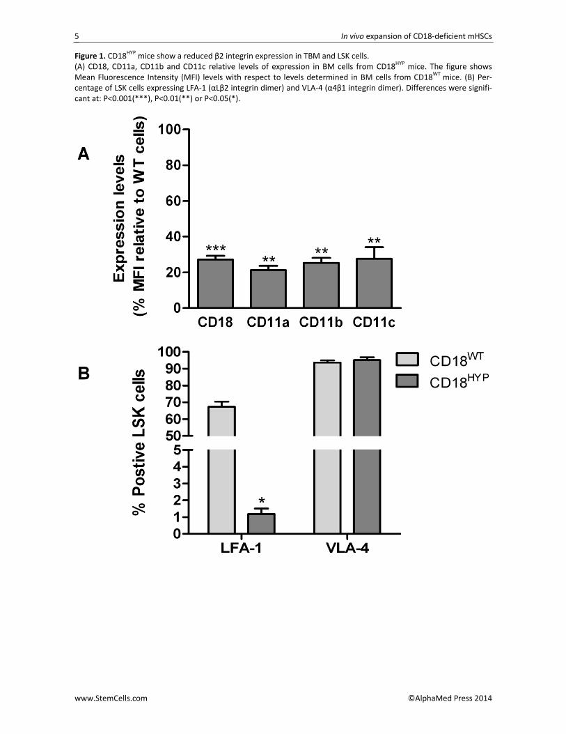
When BM cells were analyzed, the levels of CD18 expression and of their respective CD11 membrane co‐expressed subunits were also significantly decreased with respect to values observed in CD18WT mice (Figure 1A). Moreover, analyses performed in BM cells of the HSC compartment showed that, in contrast to data ob‐tained in CD18WT mice where 67% of the LSK cells were LFA‐1+ (CD18+/CD11a+), only 1.2% of LSK cells from CD18HYP mice were LFA‐1+ (Figure 1B). In addition, CD18 and CD11a expression levels determined in LFA‐1+ LSKs from CD18HYP mice were only around 20% and 30%, respectively, of the levels found in CD18WT mice (Figure S2D). In contrast to this data, no differences in VLA‐4 expression were detected in LSKs from CD18HYP (Figure 1B and S2D), indicating that β2‐integrin defects in these cells are not compensated by an overexpression of β1‐integrins.
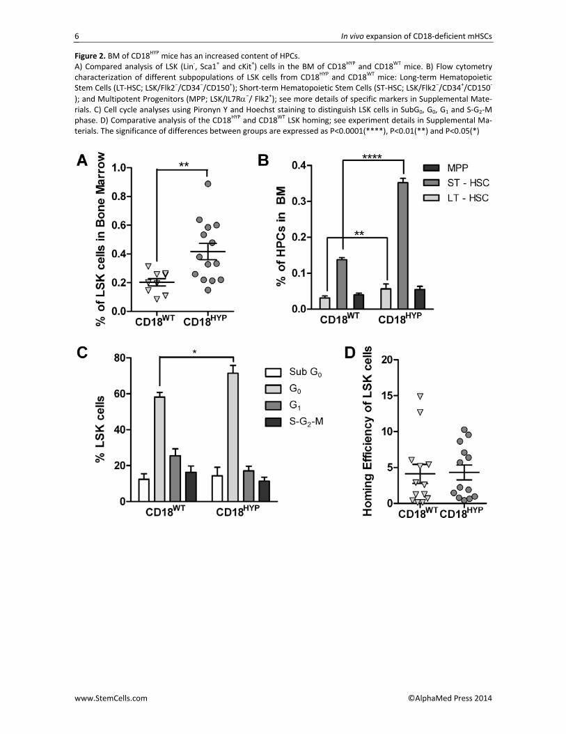
In a next set of experiments we evaluated the con‐tent of hematopoietic progenitor cells (HPCs) in the BM of CD18HYP mice. While no differences in the BM cellu‐larity were found between CD18WT and CD18HYP mice (data not shown), the proportion of LSK cells was signif‐icantly increased in CD18HYP mice, as compared to age‐matched CD18WT mice (Figure 2A). A more detailed analysis of the HPCs in the BM of CD18HYP mice showed that CD18 deficiency resulted in a significant increase in
the proportion of long‐term and short‐term HPCs, as well as a non‐significant increase in multipotent progen‐itors (Figure 2B). This increase in HPCs was confirmed in clonogenic assays, which showed a significant increase in the number of colonies generated by either total BM or Lin‐ BM cells from CD18WT as compared to CD18HYP mice (Figure S3). LSKs were then evaluated for their cell cycle and apoptosis status. A significant increase in the proportion of LSK cells in G0 was found in CD18
HYP mice (Figure 2C). This indicates that CD18 deficiency leads to enrichment in quiescent HPCs, and therefore, that the characteristic leukocytosis of CD18HYP mice does not occur at the expense of an increased population of pro‐liferating BM HPCs.
The role of CD18 in the regulation of apoptosis in neutrophils has been extensively studied. The engage‐ment of β2‐integrin Mac‐1 on neutrophils can either inhibit or enhance apoptosis depending on the activa‐tion state of the integrin and the presence of proapoptotic stimuli [12,13]. While we observed that Gr1+ BMCs from CD18HYP mice had reduced levels of apoptosis compared to WT cells (data not shown), we did not detect any difference in the proportion of apop‐totic (SubG0) LSK
HYP (Figure 2C), suggesting that CD18 is not implicated in apoptosis regulation in mouse HPCs.
Although current data indicate that β2‐integrins do not play a direct role in HSC homing, it has been de‐scribed that if the function of β1‐integrins is compro‐mised, defects in β2‐integrin expression lead to a syner‐gistic impairment in HSC homing [14,15]. To determine whether the HSC homing efficiency was affected in CD18HYP mice, FACS‐sorted and DiD‐stained LSKs from CD18HYP or CD18WT mice were intravenously injected into lethally irradiated CD18WT recipients. Sixteen hours later the proportion of DiD+ LSK cells that had homed into recipient BM was determined by flow cytometry. As shown in Figure 2D, no differences in the homing efficiency of CD18HYP and CD18WT LSK cells were ob‐served in these experiments.
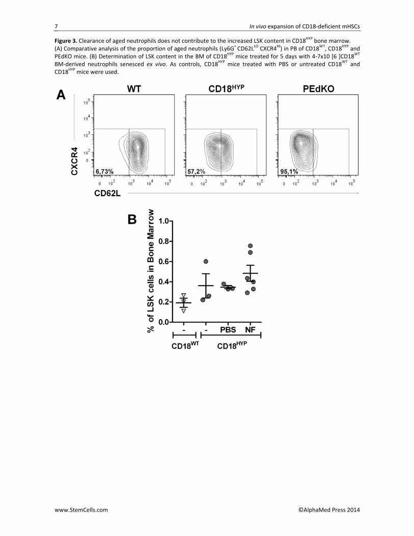
Recent observations in P‐ and E‐selectin knockout (PEdKO) mice have shown an increased population of aged neutrophils with a CD62LLO CXCR4HI phenotype in PB due to an impaired extravasation of these cells to the BM, ultimately resulting in enhanced retention of HSCs in the BM [16]. Thus, we compared the proportion of aged neutrophils in PB of CD18HYP mice with respect to CD18WT and PEdKO mice. As shown in Figure 3A, CD18HYP mice mimicked the increased proportion of aged neutrophils characteristic of PEdKO mice, suggest‐ing similar extravasation defects in CD18HYP neutrophils. Because this effect might account for the increased numbers of HPCs observed in the BM of CD18HYP mice, we investigated whether a 5‐day infusion of ex vivo se‐nesced CD18WT BM‐derived neutrophils into CD18HYP mice could decreased the number of LSKs in CD18HYP BM. No significant changes in the proportion of LSKs were, observed between CD18HYP mice treated with aged neutrophils and saline (Figure 3B), strongly sug‐gesting that increased numbers of HPCs in CD18HYP
In vivo expansion of CD18‐deficient mHSCs
www.StemCells.com ©AlphaMed Press 2014
3
mouse BM are not produced by defects in neutrophil extravasation.
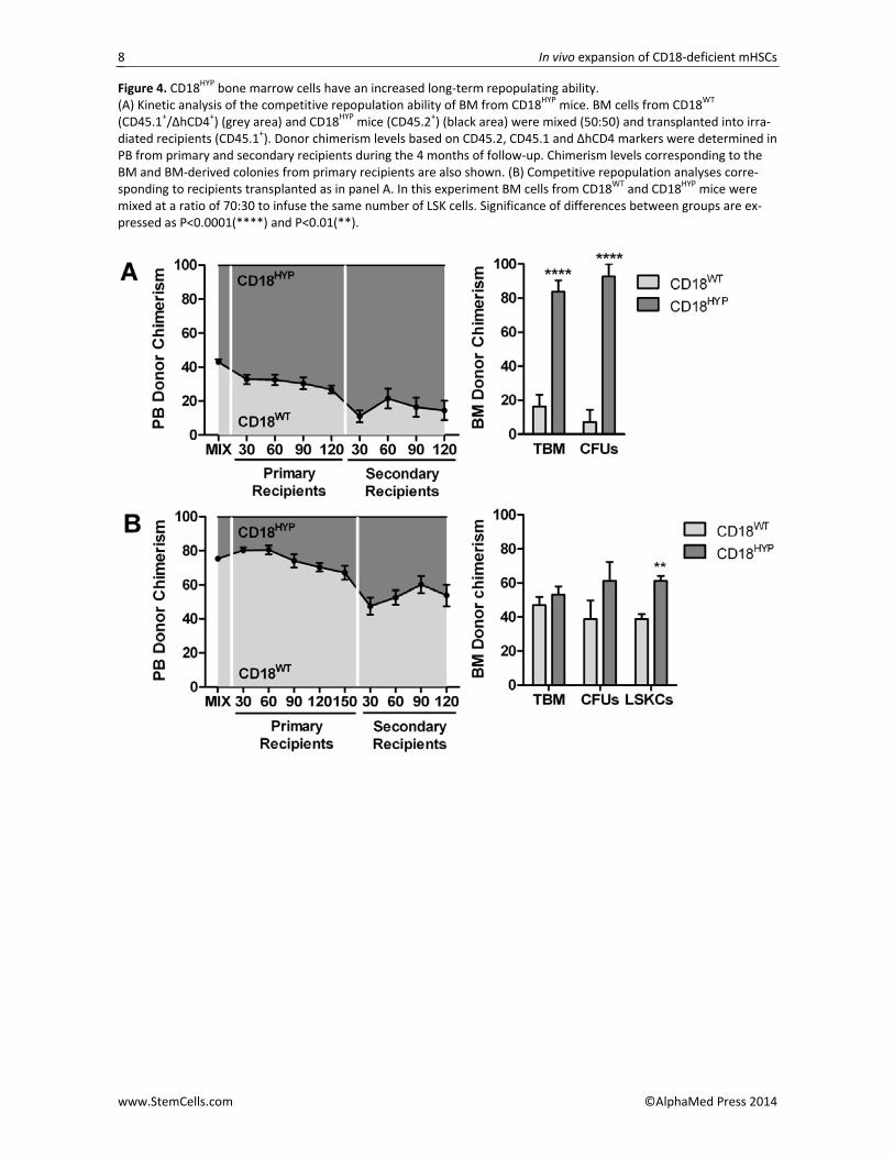
We thus considered the possibility that CD18 defi‐ciency may induce an increased self‐renewal of CD18HYP repopulating HSCs due to differences in their interac‐tion with the hematopoietic environment. To investi‐gate this hypothesis, we performed competitive repop‐ulation assays in which BMCs from CD18HYP mice (CD45.2+) and from CD18WT mice (CD45.1+/ΔhCD4+ [17]) were mixed together in the same proportion and trans‐planted into lethally irradiated CD18HYP recipients (CD45.1+). Under these conditions we could track the repopulating properties of CD18HYP and CD18WT HSCs in the same WT microenvironment. Control experiments showed that the repopulating ability of WT BMCs from these three different haplotypes was the same (Figure S4). As shown in Figure 4A, a progressive increase of CD18HYP–derived hematopoiesis was observed in trans‐planted recipients. Moreover, when BM from primary recipients was further transplanted into secondary re‐cipients we found an increment of CD18HYP–derived hematopoietic in PB. An observation that was also ob‐served, even more marked, in analyses of total BM and in BM‐derived colonies (Figure 4A).
A similar BM competitive repopulation experiment was then performed mixing together a different propor‐tion of CD18WT and CD18HYP BM cells (70:30), which con‐tained the same number of CD18HYP and CD18WT LSK cells (Figure 4B). In spite of the low proportion of total CD18HYP BMCs transplanted in these animals, a progres‐sive increase of CD18HYP‐derived hematopoietic cells could be observed in PB analyses from primary recipi‐ents. In secondary recipients, a 50% of CD18HYP chimerism was observed up to 120 days post‐transplantation, consistent with the initial proportion of CD18HYP transplanted LSK cells. Remarkably, when BM‐derived progenitor cells from secondary recipients were analysed, the proportion of CD18HYP –derived progeni‐tors was further increased, reaching statistical signifi‐cance in LSK cell analyses (Figure 4B).
Taken together, our data demonstrate that down‐regulation of CD18 results in increased numbers of hematopoietic stem and progenitor cells in the BM of CD18HYP mice. As no homing differences were found between CD18HYP and CD18WT LSK cells, our observa‐tions indicate that CD18 deficiency causes a progressive
cell‐autonomous in vivo expansion of the HSCs, thus uncovering a new role for CD18 in HSC homeostasis.
ACKNOWLEDGMENTS
The authors would like to thank Aurora de la Cal and Sergio Losada for their collaboration in the administra‐tive work and Miguel Angel Martín, Jesús Martínez and Edilia de Almeida for the careful maintenance of the animals. This work was supported by the following grants: Dirección General de Investigación de la Comunidad de Madrid (CellCAM; Ref S2010/BMD‐2420), Fondo de Investigaciones Sanitarias, Instituto de Salud Carlos III (RETICS‐RD12/0019/0023), and SAF2012‐31142 from MINECO, S2010/BMD‐2314 from Comunidad de Madrid, and FP7‐People‐IRG Program (246655). DLR was supported by an FPU grant by the Spanish Ministry of Education (AP‐2009‐4129). The Cen‐tro Nacional de Investigaciones Cardiovasculares is sup‐ported by the MINECO and the Pro‐CNIC Foundation. DISCLAIMER
The authors declare no conflict of interest.
Materials and Methods section is enclosed as supple‐mentary information
AUTHOR CONTRIBUTIONS
D.L.R. conception and design, collection and/or assem‐bly of data, data analysis and interpretation, and manu‐script writing; M.A.: collection and/or assembly of data and data analysis and interpretation; R.S.: Provision of analytical tools, collection and/or assembly of data, data analysis and interpretation; J.C.S.: Provision of ana‐lytical tools, data analysis and interpretation; L.W.: col‐lection and/or assembly of data and data analysis and interpretation and manuscript writing; A.H.: conception and design, data analysis and interpretation, and manu‐script writing; J.A.B.: conception and design, data analy‐sis and interpretation, manuscript writing and final ap‐proval of manuscript; E.A.N.: conception and design, collection and/or assembly of data, data analysis and interpretation, manuscript writing and final approval of manuscript.
REFERENCES 1 Piirila H, Valiaho J, Vihinen M. Immunode‐
ficiency mutation databases (IDbases). Hu‐man mutation. 2006;27:1200‐1208. 2 van de Vijver E, Maddalena A, Sanal O, et
al. Hematologically important mutations: leukocyte adhesion deficiency (first update). Blood cells, molecules & diseases. 2012;48:53‐61. 3 Anderson DC, Schmalsteig FC, Finegold
MJ, et al. The severe and moderate pheno‐types of heritable Mac‐1, LFA‐1 deficiency:
their quantitative definition and relation to leukocyte dysfunction and clinical features. The Journal of infectious diseases. 1985;152:668‐689. 4 Anderson DC, Springer TA. Leukocyte ad‐
hesion deficiency: an inherited defect in the Mac‐1, LFA‐1, and p150,95 glycoproteins. Annual review of medicine. 1987;38:175‐194. 5 Prevalence of rare diseases: Bibliographic
data. Orphanet Report Series, Rare Disease Collection. 2012. 6 Scharffetter‐Kochanek K, Lu H, Norman K,
et al. Spontaneous skin ulceration and defec‐
tive T cell function in CD18 null mice. J Exp Med. 1998;188:119‐131. 7 Wilson RW, Ballantyne CM, Smith CW, et
al. Gene targeting yields a CD18‐mutant mouse for study of inflammation. J Immunol. 1993;151:1571‐1578. 8 Papayannopoulou T, Priestley GV, Naka‐
moto B, et al. Synergistic mobilization of hemopoietic progenitor cells using concurrent beta1 and beta2 integrin blockade or beta2‐deficient mice. Blood. 2001;97:1282‐1288. 9 Gomez JC, Doerschuk CM. The role of
CD18 in the production and release of neu‐
In vivo expansion of CD18‐deficient mHSCs
www.StemCells.com ©AlphaMed Press 2014
4
trophils from the bone marrow. Lab Invest. 2010;90:599‐610. 10 Horwitz BH, Mizgerd JP, Scott ML, et al.
Mechanisms of granulocytosis in the absence of CD18. Blood. 2001;97:1578‐1583. 11 Gu YC, Bauer TR, Jr., Ackermann MR, et
al. The genetic immunodeficiency disease, leukocyte adhesion deficiency, in humans, dogs, cattle, and mice. Comp Med. 2004;54:363‐372. 12 Whitlock BB, Gardai S, Fadok V, et al.
Differential roles for alpha(M)beta(2) integrin clustering or activation in the control of apop‐
tosis via regulation of akt and ERK survival mechanisms. The Journal of cell biology. 2000;151:1305‐1320. 13 Mayadas TN, Cullere X. Neutrophil beta2
integrins: moderators of life or death deci‐sions. Trends in immunology. 2005;26:388‐395. 14 Sahin AO, Buitenhuis M. Molecular
mechanisms underlying adhesion and migra‐tion of hematopoietic stem cells. Cell adhe‐sion & migration. 2012;6:39‐48. 15 Papayannopoulou T, Priestley GV, Na‐
kamoto B, et al. Molecular pathways in bone
marrow homing: dominant role of al‐pha(4)beta(1) over beta(2)‐integrins and selectins. Blood. 2001;98:2403‐2411. 16 Casanova‐Acebes M, Pitaval C, Weiss LA,
et al. Rhythmic modulation of the hemato‐poietic niche through neutrophil clearance. Cell. 2013;153:1025‐1035. 17 Almarza E, Segovia JC, Guenechea G, et
al. Regulatory elements of the vav gene drive transgene expression in hematopoietic stem cells from adult mice. Exp Hematol. 2004;32:360‐364.
See www.StemCells.com for supporting information available online. STEM
CELLS; 00:000–000
In vivo expansion of CD18‐deficient mHSCs
www.StemCells.com ©AlphaMed Press 2014
5
Figure 1. CD18HYP mice show a reduced β2 integrin expression in TBM and LSK cells. (A) CD18, CD11a, CD11b and CD11c relative levels of expression in BM cells from CD18HYP mice. The figure shows Mean Fluorescence Intensity (MFI) levels with respect to levels determined in BM cells from CD18WT mice. (B) Per‐centage of LSK cells expressing LFA‐1 (αLβ2 integrin dimer) and VLA‐4 (α4β1 integrin dimer). Differences were signifi‐cant at: P<0.001(***), P<0.01(**) or P<0.05(*).
In vivo expansion of CD18‐deficient mHSCs
www.StemCells.com ©AlphaMed Press 2014
6
Figure 2. BM of CD18HYP mice has an increased content of HPCs. A) Compared analysis of LSK (Lin‐, Sca1+ and cKit+) cells in the BM of CD18HYP and CD18WT mice. B) Flow cytometry characterization of different subpopulations of LSK cells from CD18HYP and CD18WT mice: Long‐term Hematopoietic Stem Cells (LT‐HSC; LSK/Flk2−/CD34−/CD150+); Short‐term Hematopoietic Stem Cells (ST‐HSC; LSK/Flk2−/CD34+/CD150‐
); and Multipotent Progenitors (MPP; LSK/IL7Rα−/ Flk2+); see more details of specific markers in Supplemental Mate‐rials. C) Cell cycle analyses using Pironyn Y and Hoechst staining to distinguish LSK cells in SubG0, G0, G1 and S‐G2‐M phase. D) Comparative analysis of the CD18HYP and CD18WT LSK homing; see experiment details in Supplemental Ma‐terials. The significance of differences between groups are expressed as P<0.0001(****), P<0.01(**) and P<0.05(*)
In vivo expansion of CD18‐deficient mHSCs
www.StemCells.com ©AlphaMed Press 2014
7
Figure 3. Clearance of aged neutrophils does not contribute to the increased LSK content in CD18HYP bone marrow. (A) Comparative analysis of the proportion of aged neutrophils (Ly6G+ CD62LLO CXCR4HI) in PB of CD18WT, CD18HYP and PEdKO mice. (B) Determination of LSK content in the BM of CD18HYP mice treated for 5 days with 4‐7x10 [6 ]CD18WT BM‐derived neutrophils senesced ex vivo. As controls, CD18HYP mice treated with PBS or untreated CD18WT and CD18HYP mice were used.
In vivo expansion of CD18‐deficient mHSCs
www.StemCells.com ©AlphaMed Press 2014
8
Figure 4. CD18HYP bone marrow cells have an increased long‐term repopulating ability. (A) Kinetic analysis of the competitive repopulation ability of BM from CD18HYP mice. BM cells from CD18WT (CD45.1+/ΔhCD4+) (grey area) and CD18HYP mice (CD45.2+) (black area) were mixed (50:50) and transplanted into irra‐diated recipients (CD45.1+). Donor chimerism levels based on CD45.2, CD45.1 and ΔhCD4 markers were determined in PB from primary and secondary recipients during the 4 months of follow‐up. Chimerism levels corresponding to the BM and BM‐derived colonies from primary recipients are also shown. (B) Competitive repopulation analyses corre‐sponding to recipients transplanted as in panel A. In this experiment BM cells from CD18WT and CD18HYP mice were mixed at a ratio of 70:30 to infuse the same number of LSK cells. Significance of differences between groups are ex‐pressed as P<0.0001(****) and P<0.01(**).