Expression of integrin β6 enhances invasive behavior in oral squamous cell carcinoma

(189), ra57. [DOI: 10.1126/scisignal.2001811] 4Science SignalingSeptember 2011) Roberto I. Vazquez-Padron, Klaus Ley, Jochen Reiser and Vineet Gupta (6 Donnelly, Ali Nayer, Luis F. Moita, Stephan Schürer, David Traver, Phillip Ruiz,Balla, Dayami Hernandez, Constantinos J. Barth, Geanncarlo Lugo, Mary Dony Maiguel, Mohd Hafeez Faridi, Changli Wei, Yoshihiro Kuwano, Keir M.Inflammatory Disease
Small Molecule-Mediated Activation of the Integrin CD11b/CD18 Reduces`
This information is current as of 7 September 2011. The following resources related to this article are available online at http://stke.sciencemag.org.
Article Tools http://stke.sciencemag.org/cgi/content/full/sigtrans;4/189/ra57
Visit the online version of this article to access the personalization and article tools:
MaterialsSupplemental
http://stke.sciencemag.org/cgi/content/full/sigtrans;4/189/ra57/DC1 "Supplementary Materials"
Related Content http://stke.sciencemag.org/cgi/content/abstract/sigtrans;4/189/pc17
's sites:ScienceThe editors suggest related resources on
References http://stke.sciencemag.org/cgi/content/full/sigtrans;4/189/ra57#BIBL
1 article(s) hosted by HighWire Press; see: cited byThis article has been
http://stke.sciencemag.org/cgi/content/full/sigtrans;4/189/ra57#otherarticlesThis article cites 79 articles, 39 of which can be accessed for free:
Glossary http://stke.sciencemag.org/glossary/
Look up definitions for abbreviations and terms found in this article:
Permissions http://www.sciencemag.org/about/permissions.dtl
Obtain information about reproducing this article:
the American Association for the Advancement of Science; all rights reserved. byAssociation for the Advancement of Science, 1200 New York Avenue, NW, Washington, DC 20005. Copyright 2008
(ISSN 1937-9145) is published weekly, except the last week in December, by the AmericanScience Signaling
on Septem
ber 7, 2011 stke.sciencem
ag.orgD
ownloaded from
R E S E A R C H A R T I C L E
P H A R M A C O L O G Y
Small Molecule–Mediated Activation of the IntegrinCD11b/CD18 Reduces Inflammatory DiseaseDony Maiguel,1* Mohd Hafeez Faridi,1* Changli Wei,1 Yoshihiro Kuwano,2 Keir M. Balla,3
Dayami Hernandez,4 Constantinos J. Barth,1 Geanncarlo Lugo,3 Mary Donnelly,1 Ali Nayer,1
Luis F. Moita,5 Stephan Schürer,6 David Traver,3 Phillip Ruiz,4 Roberto I. Vazquez-Padron,7
Klaus Ley,2 Jochen Reiser,1 Vineet Gupta1,8†
sD
ownloaded from
The integrin CD11b/CD18 (also known as Mac-1), which is a heterodimer of the aM (CD11b) and b2 (CD18)subunits, is critical for leukocyte adhesion and migration and for immune functions. Blocking integrin-mediated leukocyte adhesion, although beneficial in experimental models, has had limited success intreating inflammatory diseases in humans. Here, we used an alternative strategy of inhibiting leukocyterecruitment by activating CD11b/CD18 with small-molecule agonists, which we term leukadherins. Thesecompounds increased the extent of CD11b/CD18-dependent cell adhesion of transfected cells and ofprimary human and mouse neutrophils, which resulted in decreased chemotaxis and transendothelialmigration. Leukadherins also decreased leukocyte recruitment and reduced arterial narrowing after injuryin rats. Moreover, compared to a known integrin antagonist, leukadherins better preserved kidney functionin a mouse model of experimental nephritis. Leukadherins inhibited leukocyte recruitment by increasingleukocyte adhesion to the inflamed endothelium, which was reversed with a blocking antibody. Thus, wepropose that pharmacological activation of CD11b/CD18 offers an alternative therapeutic approach forinflammatory diseases.
tke.s
on September 7, 2011
ciencemag.org
INTRODUCTION
The migration and recruitment of leukocytes is essential for their normalimmune response to injury and infection and for various inflammatoryand autoimmune disorders. Leukocyte functions are modulated by b2integrins, including the highly abundant integrin CD11b/CD18 (also knownas Mac-1 and CR3), which is a heterodimer of the aM (CD11b) and b2(CD18) subunits (1–3). CD11b/CD18 is normally present in an inactiveconformation in circulating leukocytes, but it is rapidly activated (4–6) tomediate leukocyte adhesion, migration, and accumulation at the sites ofinflammation. Indeed, blocking CD11b/CD18 and its ligands (7–9) andablation of the genes encoding CD11b (3) or CD18 (10) decrease the se-verity of inflammatory responses in many animal models; however, suchblocking agents have had limited success in treating inflammatory or auto-immune diseases in humans (11, 12). This may be because complete block-ade of CD11b/CD18 with antibodies is difficult owing to the availabilityof a large intracellular pool of CD11b/CD18 that can be mobilized to thecell surface (13, 14), or because the suppression of leukocyte recruitmentwith blocking agents requires >90% occupancy of active integrin receptors(15). Antibodies against b2 integrins also have unexpected side effects (16).
1Division of Nephrology and Hypertension, Department of Medicine, Universityof Miami, Miami, FL 33136, USA. 2Division of Inflammation Biology, La JollaInstitute for Allergy & Immunology, La Jolla, CA 92037, USA. 3Department ofCellular and Molecular Medicine, University of California, San Diego, CA92093, USA. 4Division of Transplantation, Department of Surgery, Universityof Miami, Miami, FL 33136, USA. 5Cell Biology of the Immune System Unit,Instituto de Medicina Molecular, Universidade de Lisboa, 1649-028 Lisboa,Portugal. 6Center for Computational Studies, Department of Medicine, Univer-sity of Miami, Miami, FL 33136, USA. 7Division of Vascular Biology, Departmentof Surgery, University of Miami, Miami, FL 33136, USA. 8Department of Bio-chemistry and Molecular Biology, University of Miami, Miami, FL 33136, USA.*These authors contributed equally to this work.†To whom correspondence should be addressed. E-mail: [email protected]
www.S
Here, we took an alternative approach to the treatment of inflamma-tory diseases that involves the activation, rather than the blockade, ofCD11b/CD18. Our premise was based on the finding by Harlan and co-workers more than 15 years ago that the trapping of integrin a4b1 in a high-avidity state with an activating antibody increases cell adhesion and decreaseseosinophil migration (17). Experiments with knock-in animals that expressactivating mutants of the integrins aLb2 (18, 19) and a4b7 (20) provide invivo support for this hypothesis. We asked whether small molecules, whichare easily delivered in vivo and can be readily optimized for use in differentmammals, could be an effective approach for activating integrins. Agonistshave an additional advantage in that they need to activate only a fraction ofcellular receptors to elicit a functional response in vivo (21). Whether tran-sient activation of a fraction of native receptors in vivo, as is expected fromsmall-molecule treatment, can have a biological effect also remains an openquestion. Here, in multiple physiologically relevant experimental models indifferent species, we showed that the severity of inflammatory diseases wasreduced by the activation of CD11b/CD18 with small molecules. These find-ings suggest that integrin activation might be a useful pharmacological ap-proach to the treatment of inflammatory and autoimmune diseases in humans.
RESULTS
Leukadherins are newly characterized, small-moleculeagonists of CD11b/CD18We previously used a cell-based, high-throughput screening (HTS) assayto screen a chemical library of >100,000 molecules for compounds thataffected the adhesion of K562 cells that expressed CD11b/CD18 at thecell surface (K562 CD11b/CD18 cells) to fibrinogen, the physiologicalligand of CD11b/CD18 (22–24). Focusing our search on compounds thatincreased cell adhesion (agonists), we identified a series of compounds,which we termed leukadherins, that contained a core furanyl thiazolidinone
CIENCESIGNALING.org 6 September 2011 Vol 4 Issue 189 ra57 1

R E S E A R C H A R T I C L E
chemical structural motif that was common in all the identified compounds(22). We explored the structure-activity relationship of various substitutionsof chemical residues in the central core common to leukadherins (23) andidentified three compounds, leukadherin-1 (LA1), leukadherin-2 (LA2),and leukadherin-3 (LA3) (Fig. 1, A to D), which showed enhanced activityin vitro. LA1, LA2, and LA3 separately increased CD11b/CD18-dependentcell adhesion to fibrinogen with 50% effective concentration (EC50, the ef-fective concentration for a 50% increase in adhesion) values of 4, 12, and14 mM, respectively. Cells that did not express CD11b/CD18 did not show
www.S
any substantial binding to fibrinogen. The binding of CD11b/CD18 to itsligands is mediated by divalent cations, with calcium ions (Ca2+) inhibitingthe interaction (thus acting as antagonists), whereas magnesium (Mg2+) andmanganese (Mn2+) ions facilitate the interaction (thus acting as agonists)(25, 26). A study described an inverse agonist of the integrin lymphocytefunction-associated antigen–1 (LFA-1, also known as the aLb2 integrin) thatincreased LFA-1–mediated adhesion under basal conditions but inhibitedit under activating conditions (27). To evaluate whether leukadherins couldsimilarly inhibit preactivated CD11b/CD18, we measured the binding of
on Septem
ber 7, 2011 stke.sciencem
ag.orgD
ownloaded from
Fig. 1. Leukadherins increaseCD11b/CD18-dependent cell adhe-sion. (A to D) Dose-response curvesshowing the percentages of inputK562 CD11b/CD18 cells (filled cir-cles) and K562 cells (open circles)that adhered to immobilized fibrin-ogen in the presence of increasingamounts of LA1, LA2, LA3, or LA-C.The chemical structures of LA1, LA2,LA3, and LA-C are also shown. Dataare means ± SEM (n = 3 to 6 repli-cates per data point) from one of atleast three independent experiments.(E) Histograms showing the adhe-sion of K562 CD11b/CD18 cells tofibrinogen in response to LA1, LA2,or LA3 in the absence or presenceof the blocking antibodies IB4 and44a. Also shown is the extent of ad-hesion in the presence of physiolog-ic concentrations of Ca2+ and Mg2+
(Con) and that in the presence of theknownagonistMn2+.Dataaremeans±SEM (n = 4 to 9 replicates per condi-tion) from one of at least three inde-pendent experiments. (F) Histogramsshowing the percentage adhesion ofwild-type (WT,CD11b+/+)andCD11b−/−
neutrophils to immobilized fibrinogen
CIENCESIGNALING.org 6 Septemb
in theabsence (DMSO)orpresenceof LA1,LA2,or LA3ascompared to that of control cells (Con).Dataaremeans±SEM(n=5 replicatesper condition) fromone of at least three independent experiments. (G) Histograms showing the percentage of binding of K562 E320A cells to immobilized fibrinogen inducedbyLA1, LA2, or LA3. Also shown is the extent of K562 E320A adhesion with Ca2+ and Mg2+ ions (Con) and Mn2+. Data are means ± SEM (n = 6 replicates percondition) from one of at least three independent experiments. (H) Histograms showing the percentage binding of recombinant GST-fused aA domainconstructs to immobilized fibrinogen (normalized to the amounts of input aA) in the absence of leukadherin (DMSO) or in the presence of LA1 or LA2. Alsoshown is the background binding obtained in the absence of any protein (−) or with the GST construct alone (GST). Data are means ± SEM (n= 3 replicatesper condition) from one of at least three independent experiments.
er 2011 Vol 4 Issue 189 ra57 2
R E S E A R C H A R T I C L E
on Septem
ber 7, 2011 stke.sciencem
ag.orgD
ownloaded from
K562 CD11b/CD18 cells to immobilized fibrinogen in the presence ofMn2+. We found that LA1, LA2, and LA3 did not inhibit cell adhesionin the presence of the agonist Mn2+ (fig. S1), suggesting that they are trueagonists. We also identified a structurally related compound, leukadherin-control (LA-C), which showed no effect on CD11b/CD18-dependent celladhesion (Fig. 1D). Increased adhesion of CD11b/CD18-expressing cells,induced by Mn2+ (28) and LA1, LA2, or LA3, was blocked by the mono-clonal antibodies (mAbs) IB4 (29) and 44a (30), which are specific forCD18 and CD11b, respectively (Fig. 1E), further confirming that these com-pounds mediate CD11b/CD18-dependent cell adhesion.
Neutrophils contain a large, intracellular pool of CD11b/CD18 thatcan be mobilized to the cell surface (13, 14), which, in addition to a con-formational switch in CD11b/CD18 from an inactive to an active form,helps to increase the extent of adhesion of neutrophils to the extracellularmatrix. To rule out an enhancement in the abundance of CD11b/CD18 atthe cell surface as a means by which LA1, LA2, and LA3 might increasecell adhesion, we measured the relative amounts of surface CD11b/CD18on K562 cells (fig. S2) and neutrophils (fig. S3) and found that they werenot increased by any of these compounds. The increase in CD11b/CD18-dependent cell adhesion stimulated by LA1, LA2, and LA3 was indepen-dent of the type of integrin ligand, because all three also increased the extentof adhesion of cells to the CD11b/CD18 ligands iC3b (fig. S4) and inter-cellular adhesion molecule–1 (ICAM-1) (fig. S5). Human monocytic THP-1cells also showed a similar leukadherin-induced increase in cell adhesion,suggesting that the effects of leukadherins are independent of cell type(fig. S6). LA1, LA2, and LA3 also increased the binding of wild-type, butnot CD11b−/−, neutrophils to immobilized fibrinogen (3) (Fig. 1F), furtherdemonstrating that these compounds target CD11b/CD18. To determinewhether leukadherins also affected CD11b/CD18-mediated phagocytosis,we incubated K562 CD11b/CD18 cells with iC3b-coated sheep red bloodcells (EiC3bs) (31). We found that LA1, LA2, and LA3 all substantially in-creased the capture of EiC3bs and rosetting (the binding of multiple EiC3bs,which are much smaller than a typical human cell, to a phagocytic cell formsa rosette), suggesting that these agonists can also stimulate CD11b/CD18-mediated phagocytosis (fig. S7).
Leukadherins bind to the ligand-binding aA domainand allosterically activate CD11b/CD18To identify the binding site of LA1, LA2, and LA3, which are pre-dicted to bind to the ligand-binding aA (or aI) domain in CD11b/CD18(22, 23, 32), we generated K562 cells stably expressing the mutant integrinCD11bE320A/CD18 (K562 E320A cells). The conserved residue Glu320
(E320) in the linker region that follows the activation-sensitive a7 helixin the CD11bA domain (aA domain) acts as an endogenous ligand ofthe von Willebrand factor type A (vWFA) domain of CD18 (bA or I do-main) (33). The E320A mutation abolishes the Mn2+-mediated increase inligand binding by CD11b/CD18; however, the stabilization of aA in a high-affinity conformation by additional activating mutations overcomes thisdeficit and induces ligand binding by the E320A mutant protein (34). LA1,LA2, and LA3 (but not Mn2+) selectively increased the binding of K562E320A cells to fibrinogen (Fig. 1G), suggesting that these compounds alsobind to and stabilize theaAdomain in a high-affinity conformation. To con-firm this,we performed experimentswith purified recombinantaA(35) andfound that LA1 and LA2 increased the binding of thewild-type aA domainto immobilized ligand (Fig. 1H) to an extent similar to that observed with amutant aA domain that contains a constitutionally activating mutation(I316G) (36). The lower solubility of LA3 compared to those of LA1 andLA2 precluded its use in this experiment.
In silico docking studies with high-resolution, three-dimensional (3D)structures of the aA domain in its low- and high-affinity conformations
www.S
(36–38) suggested that LA1, LA2, and LA3 preferentially bound to theopen, high-affinity conformation of the aA domain, near the activation-sensitive a7 helix region, thereby allosterically stabilizing the aA domainin its high-affinity conformation (fig. S8) (23). Flow cytometric analysisshowed an increase in the binding of the activation-sensitive mAb 24 toK562 CD11b/CD18 cells in the presence of LA1, confirming that LA1 ac-tivated full-length integrin on live cells (fig. S9). Björklund et al. describedan agonist of CD11b/CD18, termed IMB-10, that targets the aA domain ofCD11b/CD18 (32). We compared the relative affinities of LA1 and IMB-10for CD11b/CD18 with our cell-based adhesion assay and found that LA1showed the higher affinity (fig. S10), perhaps because of its more rotation-ally constrained furanyl thiazolidinone central scaffold.
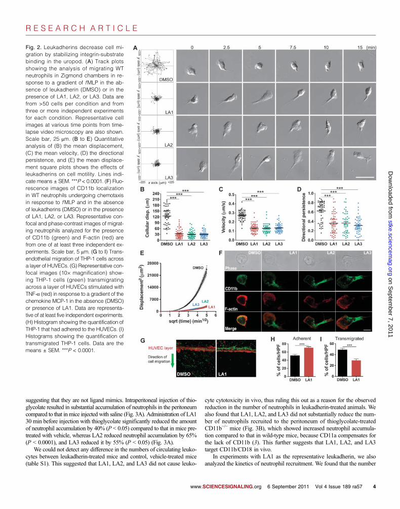
Leukadherins reduce leukocyte migration by increasingthe extent of CD11b/CD18-mediated cell adhesion andreducing de-adhesion of the uropodLeukocyte chemotaxis on 2D surfaces involves sequential integrin-mediatedadhesion and de-adhesion steps (39). Cells that have constitutively activemutant integrins show increased adhesion and reduced cell migration inchemotactic gradients, compared to those of their wild-type counterparts,through the trapping of the mutant integrins in a ligand-bound conformation(40, 41). To test whether the increased cell adhesion induced by leukadherinsaffected cell migration, we performed experiments with murine neutrophils,which undergo chemotaxis in response to a gradient of the chemoattractantpeptide N-formyl-Met-Leu-Phe ( fMLP) (42). Live-cell imaging showedthe smooth migration of neutrophils in a physiologic buffer (Fig. 2A); how-ever, treatment of the cells with LA1, LA2, or LA3 resulted in a substantialdecrease in lateral migration and migration velocity (Fig. 2, A to C). Al-though the cells treated with LA1, LA2, or LA3 showed some movementtoward the chemoattractant, they displayed reduced directional persistence(Fig. 2D) and reduced mean square displacement (MSD) (Fig. 2E), whichwas suggestive of their constrained motility, compared to the more directedmotility of control cells treated with dimethyl sulfoxide (DMSO).
In addition, unlike neutrophils undergoing chemotaxis in the absenceof leukadherins, which displayed a typical flattened leading edge and ashort, narrow tail, cells that migrated in the presence of LA1, LA2, orLA3 showed elongated uropods (Fig. 2A and movies S1 to S4), suggest-ing that a defect in cell de-adhesion was the key mechanism responsiblefor defective cell migration, as has been observed in cells with activatingmutations of their integrins (18, 20). To investigate this, we used confocalmicroscopy to show that CD11b/CD18 clustered in the extended uropodsof cells treated with LA1, LA2, or LA3 (Fig. 2F), suggesting that the fail-ure to release integrin-substrate interactions in the uropod was responsiblefor the defective migration of these cells. Leukadherins had no effect onneutrophil migration in 3D collagen gels (fig. S11 and movies S5 and S6),supporting findings that leukocyte migration in 3D substrates is integrin-independent (43). However, leukadherins reduced the efficiency of trans-endothelialmigration (TEM)byTHP-1 cells across a layer of humanumbilicalvein endothelial cells (HUVECs) activatedby tumor necrosis factor–a (TNF-a)in vitro by increasing cell adhesion to the HUVEC layer (Fig. 2, G to I). To-gether, these data suggest that leukadherins increase cell adhesion and re-duce their lateral motility, thereby inhibiting TEM.
Leukadherins reduce recruitment of leukocytes duringacute peritonitis in miceTo determine the effects of LA1, LA2, and LA3 on inflammatory responsesin vivo, we used the acute, thioglycolate-induced peritonitis model in mice(3). LA1, LA2, and LA3 showed no in vitro cytotoxicity (figs. S12 andS13) at concentrations as high as 50 mM. In addition, LA1, LA2, and LA3did not induce integrin clustering (fig. S14) or outside-in signaling (fig. S15),
CIENCESIGNALING.org 6 September 2011 Vol 4 Issue 189 ra57 3
R E S E A R C H A R T I C L E
on Septem
ber 7, 2011 stke.sciencem
ag.orgD
ownloaded from
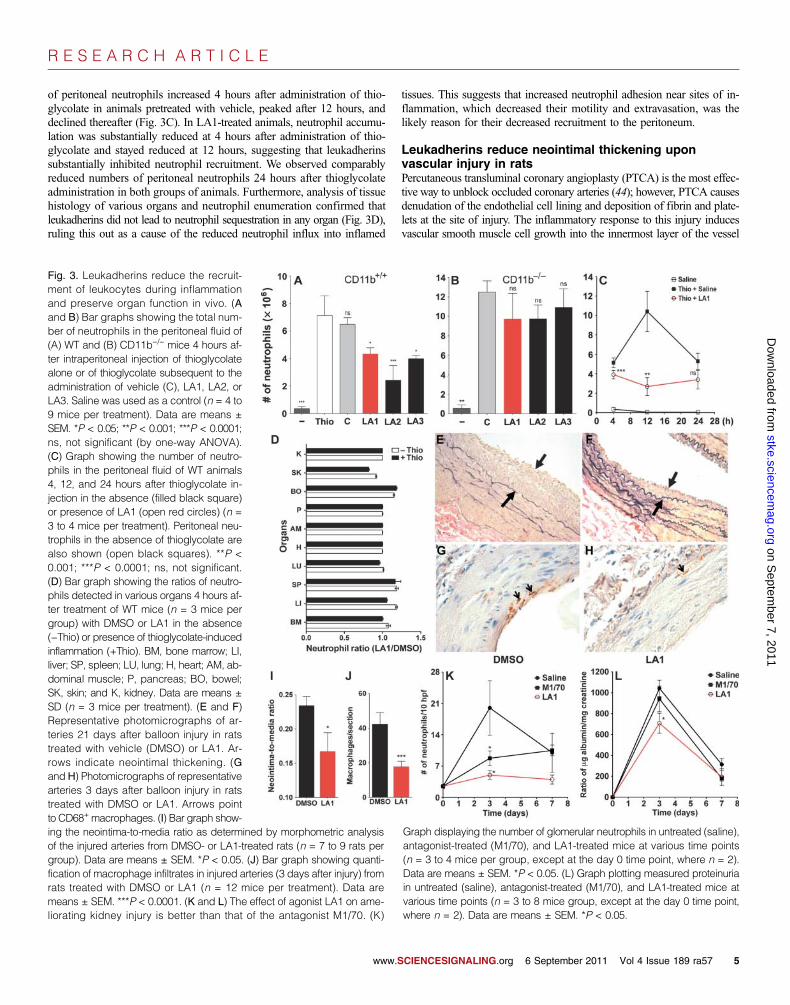
suggesting that they are not ligand mimics. Intraperitoneal injection of thio-glycolate resulted in substantial accumulation of neutrophils in the peritoneumcompared to that in mice injected with saline (Fig. 3A). Administration of LA130 min before injection with thioglycolate significantly reduced the amountof neutrophil accumulation by 40% (P < 0.05) compared to that in mice pre-treated with vehicle, whereas LA2 reduced neutrophil accumulation by 65%(P < 0.0001), and LA3 reduced it by 55% (P < 0.05) (Fig. 3A).
We could not detect any difference in the numbers of circulating leuko-cytes between leukadherin-treated mice and control, vehicle-treated mice(table S1). This suggested that LA1, LA2, and LA3 did not cause leuko-
www.S
cyte cytotoxicity in vivo, thus ruling this out as a reason for the observedreduction in the number of neutrophils in leukadherin-treated animals. Wealso found that LA1, LA2, and LA3 did not substantially reduce the num-ber of neutrophils recruited to the peritoneum of thioglycolate-treatedCD11b−/− mice (Fig. 3B), which showed increased neutrophil accumula-tion compared to that in wild-type mice, because CD11a compensates forthe lack of CD11b (3). This further suggests that LA1, LA2, and LA3target CD11b/CD18 in vivo.
In experiments with LA1 as the representative leukadherin, we alsoanalyzed the kinetics of neutrophil recruitment. We found that the number
Fig. 2. Leukadherins decrease cell mi-gration by stabilizing integrin-substratebinding in the uropod. (A) Track plotsshowing the analysis of migrating WTneutrophils in Zigmond chambers in re-sponse to a gradient of fMLP in the ab-sence of leukadherin (DMSO) or in thepresence of LA1, LA2, or LA3. Data arefrom >50 cells per condition and fromthree or more independent experimentsfor each condition. Representative cellimages at various time points from time-lapse video microscopy are also shown.Scale bar, 25 mm. (B to E) Quantitativeanalysis of (B) the mean displacement,(C) the mean velocity, (D) the directionalpersistence, and (E) the mean displace-ment square plots shows the effects ofleukadherins on cell motility. Lines indi-cate means ± SEM. ***P < 0.0001. (F) Fluo-rescence images of CD11b localizationin WT neutrophils undergoing chemotaxisin response to fMLP and in the absenceof leukadherins (DMSO) or in the presenceof LA1, LA2, or LA3. Representative con-focal and phase-contrast images of migrat-ing neutrophils analyzed for the presenceof CD11b (green) and F-actin (red) arefrom one of at least three independent ex-periments. Scale bar, 5 mm. (G to I) Trans-endothelial migration of THP-1 cells acrossa layer of HUVECs. (G) Representative con-focal images (10× magnification) show-ing THP-1 cells (green) transmigratingacross a layer of HUVECs stimulated withTNF-a (red) in response to a gradient of thechemokine MCP-1 in the absence (DMSO)or presence of LA1. Data are representa-tive of at least five independent experiments.(H) Histogram showing the quantification ofTHP-1 that had adhered to the HUVECs. (I)Histograms showing the quantification oftransmigrated THP-1 cells. Data are themeans ± SEM. ***P < 0.0001.
CIENCESIGNALING.org 6 September 2011 Vol 4 Issue 189 ra57 4
R E S E A R C H A R T I C L E
of peritoneal neutrophils increased 4 hours after administration of thio-glycolate in animals pretreated with vehicle, peaked after 12 hours, anddeclined thereafter (Fig. 3C). In LA1-treated animals, neutrophil accumu-lation was substantially reduced at 4 hours after administration of thio-glycolate and stayed reduced at 12 hours, suggesting that leukadherinssubstantially inhibited neutrophil recruitment. We observed comparablyreduced numbers of peritoneal neutrophils 24 hours after thioglycolateadministration in both groups of animals. Furthermore, analysis of tissuehistology of various organs and neutrophil enumeration confirmed thatleukadherins did not lead to neutrophil sequestration in any organ (Fig. 3D),ruling this out as a cause of the reduced neutrophil influx into inflamed
www.S
tissues. This suggests that increased neutrophil adhesion near sites of in-flammation, which decreased their motility and extravasation, was thelikely reason for their decreased recruitment to the peritoneum.
Leukadherins reduce neointimal thickening uponvascular injury in ratsPercutaneous transluminal coronary angioplasty (PTCA) is the most effec-tive way to unblock occluded coronary arteries (44); however, PTCA causesdenudation of the endothelial cell lining and deposition of fibrin and plate-lets at the site of injury. The inflammatory response to this injury inducesvascular smooth muscle cell growth into the innermost layer of the vessel
on Septem
ber 7, 2011 stke.sciencem
ag.orgD
ownloaded from
Fig. 3. Leukadherins reduce the recruit-ment of leukocytes during inflammationand preserve organ function in vivo. (Aand B) Bar graphs showing the total num-ber of neutrophils in the peritoneal fluid of(A) WT and (B) CD11b−/− mice 4 hours af-ter intraperitoneal injection of thioglycolatealone or of thioglycolate subsequent to theadministration of vehicle (C), LA1, LA2, orLA3. Saline was used as a control (n = 4 to9 mice per treatment). Data are means ±SEM. *P < 0.05; **P < 0.001; ***P < 0.0001;ns, not significant (by one-way ANOVA).(C) Graph showing the number of neutro-phils in the peritoneal fluid of WT animals4, 12, and 24 hours after thioglycolate in-jection in the absence (filled black square)or presence of LA1 (open red circles) (n =3 to 4 mice per treatment). Peritoneal neu-trophils in the absence of thioglycolate arealso shown (open black squares). **P <0.001; ***P < 0.0001; ns, not significant.(D) Bar graph showing the ratios of neutro-phils detected in various organs 4 hours af-ter treatment of WT mice (n = 3 mice pergroup) with DMSO or LA1 in the absence(−Thio) or presence of thioglycolate-inducedinflammation (+Thio). BM, bone marrow; LI,liver; SP, spleen; LU, lung; H, heart; AM, ab-dominal muscle; P, pancreas; BO, bowel;SK, skin; and K, kidney. Data are means ±SD (n = 3 mice per treatment). (E and F)Representative photomicrographs of ar-teries 21 days after balloon injury in ratstreated with vehicle (DMSO) or LA1. Ar-rows indicate neointimal thickening. (Gand H) Photomicrographs of representativearteries 3 days after balloon injury in ratstreated with DMSO or LA1. Arrows pointto CD68+macrophages. (I) Bar graph show-
ing the neointima-to-media ratio as determined by morphometric analysisof the injured arteries from DMSO- or LA1-treated rats (n = 7 to 9 rats pergroup). Data are means ± SEM. *P < 0.05. (J) Bar graph showing quanti-fication of macrophage infiltrates in injured arteries (3 days after injury) fromrats treated with DMSO or LA1 (n = 12 mice per treatment). Data aremeans ± SEM. ***P < 0.0001. (K and L) The effect of agonist LA1 on ame-liorating kidney injury is better than that of the antagonist M1/70. (K)Graph displaying the number of glomerular neutrophils in untreated (saline),antagonist-treated (M1/70), and LA1-treated mice at various time points(n = 3 to 4 mice per group, except at the day 0 time point, where n = 2).Data are means ± SEM. *P < 0.05. (L) Graph plotting measured proteinuriain untreated (saline), antagonist-treated (M1/70), and LA1-treated mice atvarious time points (n = 3 to 8 mice group, except at the day 0 time point,where n = 2). Data are means ± SEM. *P < 0.05.
CIENCESIGNALING.org 6 September 2011 Vol 4 Issue 189 ra57 5

R E S E A R C H A R T I C L E
on Septem
ber 7, 2011 stke.sciencem
ag.orgD
ownloaded from
(neointima) and, ultimately, arterial renar-rowing (restenosis). Leukocyte recruitmentand infiltration, through selective bindingbetween CD11b/CD18 expressed on the sur-face of leukocytes and the platelet cell sur-face receptor glycoprotein (GP) Iba (45),precedes neointima formation and restenosis(46). Indeed, antibody-mediated block-ade of CD11b/CD18 or loss of CD11b(for example, in CD11b−/− mice) decreasesintimal thickening after angioplasty or stentimplantation in experimental models (8).To further test the anti-inflammatory prop-erties of LA1 in vivo, we used it in an ar-terial balloon injury model in rats (47). Theextent of disease (remodeling or hyperpla-sia) was determined by calculating the ratiobetween areas of the neointima (the newlyformed innermost layer) and the media (thevessel wall) at the site of injury. We admin-istered LA1 or vehicle (DMSO) to Fischermale rats 30 min before injury and contin-ued injections every other day for 3 weeks.Injured arteries of the LA1-treated rats de-veloped significantly reduced neointimalthickening (Fig. 3, E and F) (neointima-to-media ratio of 0.16 ± 0.02 versus 0.23 ±0.01, P < 0.05) (Fig. 3I), whereas the con-trol compound LA-C showed no effect (fig.S16). To determine whether leukadherintreatment leads to reduced leukocyte accu-mulation, which precedes vascular remodel-ing, we performed immunohistochemicalanalyses of arteries 3 days after injury. Weobserved a significant reduction in the num-ber of macrophages in the arteries of LA1-treated animals (17.7 ± 3.1 versus 42.2 ±6.7,P<0.0001) (Fig. 3,G to J); LA3 showedsimilar protective effects (fig. S17). Togeth-er, these results suggest that leukadherinslead to reduced leukocyte accumulation atthe site of vascular injury and a subsequentdecrease in neointimal thickening.
Leukadherins are more efficientthan integrin antagonists intreating inflammatory injuryTo determine whether CD11b/CD18 ago-nists have any therapeutic advantages overantagonists, we performed a head-to-headcomparison between a well-characterizedCD11b/CD18 antagonist, the M1/70 anti-body against CD11b (48), and LA1 in ex-periments with an established mouse modelof kidney disease, anti–glomerular base-ment membrane (anti-GBM) nephritis (49).This model is characterized by neutrophilinfiltration that mediates urinary proteinloss, including that of albumin. Consist-ent with a role for CD11b in this disease,
Fig. 4. Leukadherins decrease leukocyte recruitment in vivo by increasing the extent of slow rolling andthe number of adherent cells, thereby decreasing leukocyte TEM. (A) Schematic representing the zebra-fish tailfin injury model. (B to D) Photomicrographs (left) and fluorescence images (right) of (B) thelarvae tail without injury, (C) the tail with injury, and (D) 4 hours after the removal of LA1 from treated,injured larvae. Images show accumulation of neutrophils (green) in the injured tail. (E) Graph showingquantification of the number of neutrophils near the site of tailfin injury in zebrafish larvae treated withDMSO (Control) and LA1 (n = 12 to 16 larvae per group). Data are means ± SEM. ***P < 0.0001; ns, notsignificant (by one-way ANOVA). (F to M) Intravital microscopy–based determination of the effects ofLA1 on leukocyte migration in vivo. (F) Graph showing the rolling velocities of individual neutrophilsin the venules of TNF-a–treated mouse cremaster muscle without (DMSO) or with LA1. Lines indicatemedians and 25 to 75% interquartile ranges. *P < 0.05. (G) Cumulative histograms of the rolling velocitiesof 100 leukocytes from DMSO-treated (black dots) and LA1-treated (red dots) animals. (H) Numbers ofadherent neutrophils in venules without (DMSO) or with LA1. Data are means ± SEM. *P < 0.05. (I)Graph showing relative efficiency of neutrophil TEM (as determined by the number of transmigratedneutrophils/the number of adherent neutrophils) in the absence (DMSO) or presence of LA1. Data aremeans ± SEM. ***P < 0.05. (J) Rolling velocities of neutrophils in TNF-a– and M1/70-treated venuleswithout (DMSO) or with LA1. Lines indicate median and interquartile ranges. ns, not significant. (K)Cumulative histograms of rolling velocity of 100 leukocytes from M1/70-treated mice in the absence(black dots) or presence of LA1 (red dots). Representative video micrograph images of cremastermuscle venules treated with (L) LA1 or (M) M1/70 and LA1. The lengths of the red arrows indicateneutrophil movement during a 25-s period.
www.SCIENCESIGNALING.org 6 September 2011 Vol 4 Issue 189 ra57 6

R E S E A R C H A R T I C L E
stke.scD
ownloaded from
CD11b−/− mice (49) and rats treated with mAb against CD11b (50) showdecreased leukocyte infiltration and protection from proteinuria relativeto their wild-type counterparts. We found that induction of disease in miceled to a peak influx of neutrophils into the kidney and maximal proteinuriaat day 3 (Fig. 3, K and L), whereas M1/70 substantially decreased neutro-phil influx and reduced proteinuria. However, LA1 produced a maximaldecrease in both the number of infiltrating neutrophils and the extent ofproteinuria in treated mice, suggesting a therapeutic advantage of agonistsover antagonists in this disease model.
The effects of leukadherins can be reversed byremoving them from the circulationTo visualize the effects of leukadherins on leukocyte accumulation in vivo,we used transgenic Tg(mpx::eGFP) zebrafish that express the gene encod-ing enhanced green fluorescent protein (eGFP) under a myeloid-specificperoxidase gene (mpx) promoter to specifically fluorescently label neutro-phils (51). Tailfin transection in zebrafish larvae at 3 days post fertilization(dpf ) (Fig. 4A) revealed a rapid and robust recruitment of neutrophils tothe site of tissue injury (Fig. 4, B and C to E) (51). LA1 had no observableeffects in uninjured larvae (Fig. 4B); however, LA1 significantly reducedthe number of neutrophils at the injured zebrafish tailfin 4 hours after in-jury compared to that in control larvae (15.6 ± 1.7 versus 34.6 ± 4.5, P <0.0001) (Fig. 4, C and E).
Furthermore, to determine whether the effects of leukadherins in vivowere reversible, we administered LA1 to uninjured zebrafish for 4 to 8hours, rinsed the zebrafish, induced tailfin injury, and quantified neutro-phil accumulation 4 hours later. We found that the removal of LA1 led to
on Septem
ber 7, 2011 iencem
ag.org
neutrophil accumulation at the injured tail-fins to an extent similar to that observed inuntreated zebrafish larvae (Fig. 4, D andE). LA2 showed protective effects similarto those of LA1 (fig. S18). Fluorescenceimaging of the injured whole zebrafish lar-vae showed no difference in total neutro-phil numbers between leukadherin-treatedand untreated larvae (fig. S19), which sug-gested that the leukadherin-mediated re-duction in neutrophil accumulation was notdue to a reduction in the overall number ofneutrophils. This result is consistent withour experiments in mice that showed thatleukadherins did not cause cytotoxicity invivo (table S1). Together, these data dem-onstrate that leukadherins inhibit the accu-mulation of neutrophils at the sites of tissueinjury and that their effects in vivo are re-versible by their removal.
Leukadherins reducethe extravasation ofleukocytes in vivoFinally, to determine the mechanism ofaction of leukadherins in vivo, we performedintravital microscopic analysis of mousecremaster muscle. CD11b/CD18 binds toICAM-1 on the surface of TNF-a–activatedendothelium to mediate slow rolling and ar-rest of leukocytes (52). We found that LA1substantially decreased the rolling velocityof leukocytes in TNF-a–stimulated venules
www.S
(Fig. 4, F and G), which led to a substantial increase in the number ofadherent cells (Fig. 4H); however, the number of transmigrated cells ad-jacent to the vessel in LA1-treated mice was similar to that in the DMSO-treated animals (fig. S20), suggesting that LA1 substantially reduced theoverall efficiency of leukocyte TEM (Fig. 4I). Injection of M1/70, whichblocks CD11b, reversed the effects of LA1 on leukocyte rolling (Fig. 4, Jto M), confirming that the effects of LA1 were CD11b/CD18-specific.Thus, these in vivo measurements mirror the in vitro data on the effectsof leukadherins on leukocyte motility.
DISCUSSION
Leukocyte infiltration is a common finding in most inflammatory dis-eases, and the leukocytic integrin CD11b/CD18 plays an important rolein this process (1, 2). CD11b/CD18-mediated leukocyte rolling and firmadhesion to the vascular wall lead to leukocyte transmigration across thevasculature and accumulation in the tissue as part of a multistep infiltrationprocess (Fig. 5) (52). Current approaches that treat leukocyte infiltration arefocused on blocking the adhesion of leukocytic integrins to their respectiveligands (“anti-adhesion” therapy) (53, 54). Antibodies against integrins,such as the M1/70 mAb, block the binding of integrins to ligands foundon the vascular wall, thus reducing the infiltration of leukocytes into thetissue (Fig. 5). Although such strategies have proven beneficial in certainanimal models, many agents that block the binding of integrins to theirligands have failed in clinical trials (11, 53), had substantial side effects,or have had to be withdrawn from the market (55).
Fig. 5. Schematic showing how integrin antagonists and agonists differ in their ability to reduce inflam-matory disease. Integrin antagonists (central panel), such as blocking mAbs, prevent leukocyte adhesionto the inflamed endothelium, thereby reducing leukocyte migration and tissue recruitment as compared tothe untreated situation (left panel). On the other hand, integrin agonists (such as leukadherins), promote theadhesion of leukocytes, which reduces their lateral and transendothelial migration and leads to an evengreater decrease in the tissue recruitment of leukocytes. Thus, small-molecule integrin agonists (such asour prototype compounds, leukadherins) represent an alternative strategy for modulating leukocyte recruit-ment and inflammatory diseases.
CIENCESIGNALING.org 6 September 2011 Vol 4 Issue 189 ra57 7

R E S E A R C H A R T I C L E
on Septem
ber 7, 2011 stke.sciencem
ag.orgD
ownloaded from
Our data demonstrate that an alternative approach to inhibiting leuko-cyte migration by enhancing the activation of integrins with small mole-cules is highly effective in reducing leukocyte infiltration and subsequentinflammation in vivo (Fig. 5). We used a cell-based HTS assay (22, 24) toidentify and optimize small-molecule agonists of CD11b/CD18, which wehave termed leukadherins. The leukadherin compounds LA1 to LA3 havesimilar chemical structures, and they bind to the ligand-binding aA do-main and convert CD11b/CD18 into its active conformation. We showedthat leukadherins, but not the structurally similar compound LA-C, pro-moted CD11b/CD18-dependent cell adhesion and decreased leukocytemotility, which led to a substantial reduction in leukocyte TEM and re-cruitment into tissues.
In addition, we found that leukadherins had a higher affinity forCD11b/CD18 than did a CD11b/CD18 agonist (32), perhaps because oftheir more rotationally constrained furanyl thiazolidinone central ringstructure. Furthermore, through experiments with various disease modelsand multiple animal species, we demonstrated that these CD11b/CD18agonists reduced inflammation in physiologically relevant settings, sug-gesting that they are viable therapeutic leads for future optimization.Our data also reveal that when directly compared with a known antagonist,CD11b/CD18 agonists better preserve organ function upon inflammatoryinjury (Fig. 5). Finally, the in vivo effects of leukadherins were transientand were reversed by removing them from the circulation. Our results sug-gest that integrin-specific, small-molecule agonists represent an effectivepharmacological approach for the treatment of inflammatory and auto-immune diseases.
Our results are consistent with previous studies that showed thatintegrin activation leads to increased cell adhesion and decreased migra-tion (17–20, 27, 32). Additionally, knock-in mice expressing constitutivelyactive mutants of the integrins aLb2 (18, 19) and a4b7 (20) in the germlineshow reduced recruitment of inflammatory cells because of an increase incellular adhesion and a reduction in cell migration through constitutivelyactive integrins. However, unlike the knock-in animals, in which all of themutant integrin receptors are expressed in a constitutively active form,whether activation of a fraction of wild-type receptors (as is expected upontreatment with a small-molecule agonist) could have a similar phenotypein vivo and could reduce inflammation in physiologically relevant diseasemodels was not known. Not all of the studies of knock-in animals withactive integrin mutants have presented the same response in vivo. Notably,homozygous knock-ins carrying an activating D759A mutation in the cyto-plasmic tail of the integrin b1 subunit show no obvious phenotype andno functional changes under physiological conditions (56). Additionally,keratinocytes from these knock-in mice show comparable adhesion,spreading, and migration in vitro to those of cells expressing wild-typeintegrins. On the other hand, introduction of active mutants of the integrina2 subunit in mice results in a marked reduction (in the case of leukocytes)or a complete loss (in the case of platelets) of activated a2 on the cellsurface during thrombopoiesis, and the expression of active integrin a2on progenitor cells has no effect on their migration in vivo (57). It is pos-sible in this case that some compensatory mechanisms during animal de-velopment overcame the adhesion and migration deficiencies of theintroduced mutations. In that respect, we believe that the chemical-biologicalapproach highlighted in this study, in which the function of endogenous,wild-type protein is perturbed by a specific agonist, represents away forwardto analyze the effects of integrin activation on cellular functions in vivo.
A potential concern with the use of integrin agonists as therapeuticsin vivo is that increased adhesion of inflammatory cells to the vascularendothelium may harm the vascular cells, leading to vascular damageand leakage. However, we have not observed any signs of vascular injuryin the various experimental models that we have studied, perhaps because
www.S
such an increase in leukocyte adhesion is only transient. Previous studieswith knock-in animals that express constitutively active mutants of theintegrins aLb2 (18, 19) and a4b7 (20) have also not reported any signs ofvascular injury. We analyzed animals that received compounds for morethan 3 months, and we found no systemic signs of vascular injury orleakage—no signs of edema or systemic vascular compromise. Where-as these observations mitigate the concern of systemic vascular toxicityassociated with integrin agonists, detailed future studies will be neededto fully address some of these concerns. In conclusion, we showed thatCD11b/CD18 agonists increased the adhesion and decreased the motilityof leukocytes, which resulted in reduced leukocyte TEM, recruitmentinto the tissues, and inflammation. We suggest that integrin-specific,small-molecule agonists represent an effective pharmacological approachfor the treatment of inflammatory and autoimmune diseases.
MATERIALS AND METHODS
Reagents and antibodiesThe 44a mAb against CD11b [an immunoglobulin G (IgG) 2a (IgG2a) iso-type] (30) and the heterodimer-specific IB4 mAb against CD18 (IgG2a)(29, 58) were from the American Type Culture Collection (ATCC). M1/70,a rat mAb against mouse CD11b (IgG2b) (48), was from the mAb core atUniversity of California, San Francisco. We obtained mAb 24 (IgG1) (59)from Abcam, and the isotype control antibodies clone X40 (IgG1) andclone X39 (IgG2a), fluorescein isothiocyanate (FITC)–conjugated mAbA85-1 (rat anti-mouse IgG1), FITC-conjugated R19-15 (rat anti-mouseIgG2a), FITC-conjugated goat antibody against mouse immunoglobulin,rat antibody against mouse GR-1, GR-1–FITC, and phycoerythrin (PE)–conjugated rat antibody against mouse Mac-1 were from BD Pharmingen.Human fibrinogen (depleted of plasminogen, von Willebrand factor, andfibronectin) was from Enzyme Research Laboratories, bovine serum albu-min (BSA) was from Sigma, recombinant human ICAM-1–Fc was fromR&D Systems, and iC3b was from Calbiochem. MaxiSorp and High Bind384-well plates were obtained from Nalgene and Corning, respectively.Nonfat milk was obtained from Bio-Rad. The cell quantitation reagent MTS[3-(4,5-dimethylthiazol-2-yl)-5-(3-carboxymethoxyphenyl)-2-(4-sulfophenyl)-2H-tetrazolium salt] was from Promega. Polymerase chain reaction (PCR)assay reagents, aswell as restriction andmodification enzymes,were obtainedfrom New England Biolabs Inc. Glutathione beads were purchased fromSigma. All cell culture reagents were from Invitrogen Corp. and Mediatech.Fetal bovine serum (FBS) was purchased from Atlanta Biologicals Inc. Theantibiotic G418was purchased from Invivogen. The sheep antibody againstrabbit GBM was a gift from S. Shankland and J. Pippin.
MiceThe C57BL/6J (B6) wild-type and the B6 CD11b−/− (Jax 3991) (3) GFPmice were purchased from The Jackson Laboratory. Lys-eGFP mice havebeen described previously (60). The wild-type Fischer 344 rats were pur-chased from Harlan Laboratories. Animal care and procedures were ap-proved by the Institutional Animal Care and Use Committee and wereperformed in accordance with institutional guidelines.
Cell linesK562 cells (ATCC) stably transfected with plasmid encoding wild-typeintegrin CD11b/CD18 (K562 CD11b/CD18 cells) have been describedpreviously (22, 31). The mutant integrin subunit CD11bE320A has beendescribed previously (33). K562 cells stably transfected with mutant integrinCD11bE320A/CD18 (K562 E320A cells) were generated according to pre-viously published protocols (22, 31). All cell lines were maintained in
CIENCESIGNALING.org 6 September 2011 Vol 4 Issue 189 ra57 8

R E S E A R C H A R T I C L E
on Septem
ber 7, 2011 stke.sciencem
ag.orgD
ownloaded from
Iscove’s modified Dulbecco’s medium (IMDM) supplemented with 10%heat-inactivated FBS, penicillin and streptomycin (50 U/ml each), andG418 (0.5 mg/ml). THP-1 cells (ATCC) were maintained in RPMI 1640supplemented with 10% heat-inactivated FBS and b-mercaptoethanol(50 mM) according to the manufacturer’s instructions.
Cell adhesion assaysCell adhesion assays with immobilized ligands were performed as previ-ously described (22). Assays with all cell lines were performed in an iden-tical fashion. A stock solution of the leukadherin family of small-moleculeagonists was prepared by dissolving the compounds in DMSO at a con-centration of 2 to 10 mM. The final concentration of DMSO in the assaywas ~1%. Assays were performed in three to six replicate wells. Data arefrom one of at least three independent experiments. The HTS assay toidentify previously uncharacterized agonists with a library of >100,000small molecules was performed as previously described (22, 61).
Neutrophil adhesion assayNeutrophils from 8- to 10-week-old wild-type and CD11b−/− B6 micewere isolated from thioglycolate-stimulated peritonea as previously de-scribed (62). Cells were suspended in serum-free IMDM and incubatedwith leukadherins in ligand-coated wells in a 384-well plate for 10 minat 37°C. The assay plates were then gently inverted and kept in the in-verted position for 30 min at room temperature to dislodge the nonadherentcells. The remaining adherent cells were quantified by imaging microscopyas previously described (22, 63). Assays were performed in triplicate wells.Data reported are from one of at least three independent experiments.
Chemotaxis assay and time-lapse video microscopyNeutrophil chemotaxis on 2D surfaces was performed with Zigmond cham-bers (Neuro Probe) as described previously (42, 64) on acid-cleaned glassor fibrinogen-coated glass coverslips. Cell migration was studied in a gra-dient of f MLP (Sigma), which was generated by placing buffer alone inone well of the chamber and f MLP (10 mM) in the other well, in the ab-sence or presence of leukadherins (15 mM). Cell migration was recordedat 5- to 30-s intervals for a period of 25 min with a Nikon Eclipse 90i ora Leica DMI16000 deconvolution microscope. Images from the Nikonmicroscope were acquired with a Nikon DS camera with a PLAN APO20× differential interference contrast (DIC) microscope objective andcaptured with Nikon Imaging software. Leica DIC images were acquiredwith an HCX PL APO 40×/0.75 numerical aperture (NA) objective with aDCF360FX camera driven by LAS-AF software. Analysis of neutrophilmigration was performed with the motile population that had moved morethan 10 mm (64) with ImageJ software [National Institutes of Health (NIH)]using manual cell tracking by the Ibidi chemotaxis and migration toolplug-in for ImageJ. Motility parameters, such as migration velocity, thetotal cellular displacement (distance from the origin), and directional per-sistence, were analyzed for individual cell tracks with the Ibidi chemotaxisplug-in for ImageJ. At least 50 cells under each condition from at leastthree independent experiments were quantified.
Neutrophil migration in 3D collagen gelsWild-type B6 neutrophils were suspended in a collagen gel solution (BDBiosciences) with a final collagen concentration of 1.6 mg/ml in the pres-ence of vehicle (1% DMSO) or LA1 (15 mM) and cast in custom-built mi-gration chambers with a thickness of 0.5 to 1 mm as described previously(65). Final neutrophil concentrations in the assay were 1 × 10⁶ cells/ml ofgel. After polymerization of the collagen fibers at 37°C for 30 min, gelswere overlaid with 50 ml of culture medium containing fMLP (10 mM) andsubsequently imaged with a Leica DMI16000 deconvolution microscope
www.S
with DIC. Cell migration was recorded at 60-s intervals for a period of45 min and was captured with an HC PL FLUOTAR 20×/0.5 NA objec-tive with a DCF360FX camera and Leica LAS-AF software. Cell motilitydata were derived by constructing individual cell tracks with the manualtracking plug-in tool for ImageJ. Analysis of individual cell tracks was per-formed to determine migration velocity, the total cellular displacement (dis-tance from the origin), and directional persistence with the chemotaxisplug-in tool for ImageJ. We quantified at least 40 independent cells undereach condition from three independent experiments.
Imaging TEM of cells across a layer of activated HUVECsTo measure the effects of leukadherins on the TEM capacity of THP-1cells, we performed vertical collagen gel invasion assays as described pre-viously (66). Briefly, we prepared collagen gel chambers containing thechemokine monocyte chemotactic protein–1 (MCP-1, also known as CCL2)(200 ng/ml, Sigma) as described earlier. HUVECs (1 × 10⁵, Millipore)were incubated with a cell-permeable red fluorescent dye (CellTrackerRed CMTPX, Invitrogen) and then seeded on top of the collagen gels. Af-ter 16 hours in culture, THP-1 cells (2 × 10⁴ cells) incubated with a cell-permeable green fluorescent dye (CellTracker Green CMFDA, Invitrogen)were placed on top of a layer of HUVECs that were activated with TNF-a(10 mg/ml) for 4 hours, and their adhesion to HUVECs and TEM across theHUVEC layer were determined with a Leica TCS SP5 confocal micro-scope or a Leica DMI16000 deconvolution microscope with an HCX PLAPO 10×/1.3 NA objective driven by Leica LAS-AF software. At least 25images were taken for each condition, and we determined the numbers ofcells that adhered to the HUVEC layer and those that completely migratedthrough the HUVEC layer by manual counting.
Immunofluorescence microscopyTo examine the localization of CD11b/CD18 and polymerized actin (F-actin)in migrating neutrophils, we stimulated 1 × 10⁴ cells with f MLP (10 mM) inserum-free RPMI 1640 on glass coverslips for 15 min at 37°C in the absenceor presence of leukadherins (15 mM). The cells were fixed, permeabilizedwith 0.1% Triton X-100, and incubated with the M1/70 mAb againstCD11b followed by Alexa Fluor 488–conjugated goat antibody against mouseIg (Invitrogen) and rhodamine-labeled phalloidin (Invitrogen). A z seriesof fluorescence images was recorded with a Leica TCS SP5 confocal micro-scope and an HCX PL APO 63×/1.4 NA objective and with Leica LAS-AFsoftware. The images presented are from a z stack projection of 15 confocalsections from the basal to the apical side of the cell (stack z spacing, 0.29 mm).Images presented are representative of at least 20 cells analyzed for eachcondition from at least two independent experiments. To examine clusteringofCD11b/CD18on the cell surface,we suspended1×104K562CD11b/CD18cells in serum-free IMDM and incubated the cells without or with fibrino-gen (100 mg) for 3 hours at 37°C, as described previously (67), in the absenceor presence of leukadherins (15 mM). The cells were fixed in suspension andincubated with the IB4 mAb, which is specific for CD11b/CD18, followedby Alexa Fluor 488–conjugated goat antibody against mouse Ig (Sigma).Fluorescence images were recorded with a Leica DMI16000 deconvolutionmicroscope and an HCX PLAPO 63×/1.3 NA objective with a DCF360FXcamera and with Leica LAS-AF software. The CD11b/CD18 clusters wereanalyzed in ImageJ, and a 3D representation of fluorescence intensity wasalso generated in ImageJ. The images presented are representative of atleast 20 cells analyzed for each condition from at least three independentexperiments.
Purification of recombinant CD11b A domain (aA domain)Recombinant human aA domains were constructed and purified accord-ing to published protocols (68). Briefly, the aA domain in its inactive
CIENCESIGNALING.org 6 September 2011 Vol 4 Issue 189 ra57 9
R E S E A R C H A R T I C L E
on Septem
ber 7, 2011 stke.sciencem
ag.orgD
ownloaded from
conformation was generated by cloning and expressing protein fragmentsspanning residues Gly111 to Gly321 (321WT) (forward primer, 5′-ggttccgc-gtggatccgagaacctgtactttcaaggaggatccaacctacggcag-3′; reverse primer, 5′-gaattcccggggatccaccctcgatcgcaaagat-3′) with the Infusion Cloning Kit(Clontech) into the Bam HI site in the vector pGEX-2T according to themanufacturer’s protocol. The aA domain in its active conformation wasgenerated by replacing Ile316 with glycine [I316G (36)] using the forwardprimer 5′-ggttccgcgtggatccgagaacctgtactttcaaggaggttttcaggaatgt-3′ and thereverse primer 5′-atatccccgggattaaccctcgatcgcaaagcccttctc-3′. The insertwas digested with Bam HI and Sma I and inserted into the pGEX-2T vec-tor that was digested with Bam HI and Sma I. All constructs were confirmedby direct DNA sequencing. All recombinant proteins were expressed asglutathione S-transferase (GST) fusion proteins in Escherichia coli and pu-rified by affinity chromatography (glutathione beads, Sigma) according tothe manufacturer’s instructions. Purified protein preparations were dialyzedagainst tris-buffered saline (TBS) [20 mm tris-HCl (pH 7.5), 150 mmNaCl],subsequently concentrated with Amicon-10 columns (Millipore), and storedat −80°C. Purity was confirmed by SDS–polyacrylamide gel electrophoresis(SDS-PAGE) analysis.
aA domain ligand-binding assayMaxiSorp 96-well plates were coated overnight with fibrinogen (1 mg perwell) in 10 mM phosphate-buffered saline (PBS, pH 7.4) and blocked with1% BSA in PBS. Binding of purified, GST-tagged aA domain (50 ml perwell of a 5 mg/ml solution) to immobilized fibrinogen was performed inTBS-based assay buffer (TBS containing 0.1% BSA, 1 mM MgCl2,1 mM CaCl2, and 0.05% Tween 20) (TBS-Ca/Mg buffer) for 1 hour at roomtemperature. The aA domain was also added to uncoated wells on theplate to estimate the maximum amount of protein that could be capturedand detected in each well for data normalization. Unbound aA domainwas removed by washing the wells twice with TBS-Ca/Mg buffer. Subse-quently, the amount of bound protein was determined by incubation withhorseradish peroxidase–conjugated antibody against GST (GE, 1:2000 di-lution) for 1 hour. Unbound antibody was removed by washing the wellstwice with TBS-Ca/Mg buffer. Detection of bound protein was performedwith 3,3′,5,5′-tetramethylbenzidine (TMB) substrate kit (Vector Labs)according to the manufacturer’s protocol. Absorbance was read with aSpectraMax M5 spectrophotometer (Molecular Devices). Absorbancevalues were normalized such that the mean absorbance from the inputaA domain wells was set at 100%, and the results are presented as thepercentage of the total input amounts of the wild-type aA domain. Assayswere performed in triplicate wells, and the data shown are from one of atleast three independent experiments.
Flow cytometryFlow cytometric analysis of K562 cells and human neutrophils for thesurface expression of CD11b/CD18 was performed as previously de-scribed (69, 70). Briefly, cells were suspended in the assay buffer [TBScontaining 1 mM each of Ca2+ and Mg2+ ions (TBS++) and 0.1% BSA].Cells (5 × 10⁵) were incubated with primary mAb [a 1:100 dilution of IB4or 44a ascites, or of mAb 24 (15 mg/ml)] in the absence or presence ofleukadherin (25 to 50 mM) in 100 ml of TBS++ on ice (except for mAb 24and the isotype control antibody, for which incubations were performed at37°C in TBS++) for 30 min. Subsequently, the cells were washed threetimes with the assay buffer and incubated with allophycocyanin (APC)–conjugated goat antibody against mouse Ig (Invitrogen) for 20 min at 4°C.Cells were washed twice with the assay buffer and analyzed with a FACS-Calibur flow cytometer (BD Biosciences), counting at least 10,000 events(cells). Data were analyzed with CellQuest software (BD Biosciences).Assays were performed in triplicate and the data shown are from one of
www.S
at least three independent experiments. Human neutrophils contain anintracellular pool of CD11b/CD18 that is rapidly brought to the cell sur-face upon activation (13, 14, 71). To determine whether leukadherins hadany effect on the mobilization of the intracellular pools of CD11b/CD18,we purified human neutrophils in a quiescent state from whole blood andkept the cells on ice, as previously described (72). For analysis, cells weresuspended in the assay buffer [Hanks’ balanced salt solution (HBSS)containing 1 mM each of CaCl2 and MgCl2 (HBSS-Ca/Mg) and 0.1%human serum albumin (HSA)]. Cells (5 × 10⁵) were incubated with themAbs described earlier in the absence or presence of LA1, LA2, or LA3(25 mM) for 30 min. The cells were then washed three times with assaybuffer, labeled with secondary antibody (APC-conjugated goat antibodyagainst mouse Ig), and analyzed by flow cytometry. As a positive control,neutrophils were activated by incubating the cells with lipopolysaccharide(100 ng/ml) or phorbol myristate acetate (20 nM) (72, 73) at 37°C in assaybuffer, after which they were labeled and analyzed as described earlier.
Phagocytosis assay with complement iC3b-coatedsheep erythrocytes (EiC3bs)Sheep erythrocytes coated with complement iC3b were prepared andused in the phagocytosis assay as described previously (31). Coatederythrocytes (EiC3bs) were diluted to a concentration of 1.5 × 10⁷ to6 × 107 cells/ml. K562 CD11b/CD18 cells were washed twice in TBS andresuspended to 1 × 10⁶/ml, of which 40 ml (4 × 10⁴ cells) was incubated insuspension with EiC3bs (1.2 × 10⁶) in a total volume of 100 ml at 37°C for25 min in the presence of 1 mM each of CaCl2 and MgCl2 (in the absenceor presence of 50 to 100 mM LA1, LA2, or LA3), in 1 mM MnCl2, or in10 mM EDTA. Binding was detected by visually analyzing the formationof rosettes [the binding of multiple erythrocytes (EiC3bs) to individualK562 cells] by phase-contrast microscopy, as has been described previous-ly (31). For scoring, only those K562 cells that were bound to ≥3 EiC3bswere scored as positive, and >200 cells were examined in multiple fieldsunder each condition. Binding results, showing the percentages of all cellsshowing rosettes in a field, are reported as histograms representing themean ± SEM of triplicate experiments; the data shown are from one ofat least three independent experiments.
Cell viability assaysCell viability assays were performed with commercially available reagentsand kits. Briefly, 1 × 10⁴ K562 CD11b/CD18 cells or wild-type B6 neutro-phils were incubated in each well of a 96-well plate (Corning) with increas-ing amounts of the indicated compounds, and the number of viable cellswasdetermined with the MTS reagent (Promega), according to the manufac-turers’ instructions, after 4 hours (neutrophils) or 24 hours (K562 cells) ofincubation. A SpectraMaxM5 spectrophotometer was used to read the assayplates. Data are representative of at least two independent experiments.
Western blotting analysisK562 CD11b/CD18 cells were incubated with LA1, LA2, or LA3 (15 mM)or fibrinogen (200 mg) in serum-free medium for 1 hour at 37°C. Celllysates were resolved on a 10% SDS-PAGE gel and transferred to a poly-vinylidene difluoride membrane (Thermo Scientific) by means of estab-lished protocols. Membranes were incubated with a 1:1000 dilution of anantibody against phosphorylated extracellular signal–regulated kinase 1/2(ERK1/2) (Thr202/Tyr204, Cell Signaling), stripped with Reblot mildstripping solution (Millipore), and then incubated, first with an antibodyagainst total ERK1/2 (Cell Signaling) and then with an antibody againstglyceraldehyde-3-phosphate dehydrogenase (GAPDH; Cell Signaling), anddeveloped according to the manufacturer’s instructions (Thermo Scientific).Data presented are representative of at least three independent experiments.
CIENCESIGNALING.org 6 September 2011 Vol 4 Issue 189 ra57 10

R E S E A R C H A R T I C L E
on Septem
ber 7, 2011 stke.sciencem
ag.orgD
ownloaded from
Blood cell countComplete peripheral blood leukocyte counts from the different mice werequantified by the University of Miami mouse pathology core by flowcytometry.
In vivo peritonitis modelThioglycolate-induced peritonitis in 8- to 10-week-old wild-type B6and CD11b−/− B6 mice was performed as previously described (62).Leukadherins were administered 30 min before intraperitoneal injectionwith 3% thioglycolate. LA1 and LA2 (200 ml of a 50 mM solution insaline) were administered intravenously, whereas LA3 was administeredintraperitoneally (500 ml of a 50 mM solution in saline). To evaluate peri-toneal neutrophil recruitment, we euthanized mice at 4, 12, or 24 hoursafter thioglycolate injection; the peritoneal lavage was collected, and thenumber of emigrated neutrophils was quantified by flow cytometricanalysis for cells expressing both GR-1 and Mac-1 on the surface, as de-scribed previously (18).
Balloon-induced arterial injury in ratsAll surgeries were performed under isoflurane anesthesia (Baxter).Compounds were administered intramuscularly (1 ml of a 50 mM solutionin saline). Balloon injury in the right iliac artery was inflicted with a 2FFogarty catheter (Baxter) adapted to a custom angiographic kit (BostonScientific, Scimed) (47). An aortotomy in the abdominal aorta was madeto insert a catheter to the level of the right iliac artery. The balloon wasinflated to 1.5 to 1.6 atm and retracted to the arteriotomy site three times.The aortic excision was repaired with eight sutures. The abdominal cavitywas closed by planes with an interrupted suture pattern. Arterial specimenswere collected 3 to 30 days after injury, fixed in 4% formalin–PBS (Sigma-Aldrich) for 5 min, and analyzed by histology and immunostaining.
Anti-GBM nephritisExperimental anti-GBM nephritis was induced in wild-type B6 mice (n =4 mice per group) by intraperitoneal injection of sheep antibody againstrabbit GBM (0.5 ml per 20 g body weight per day for 2 consecutive days)as previously described (74, 75). One group of animals was treated withleukadherins by daily intraperitoneal injection of LA1 (500 ml of a 50 mMsolution in saline) starting at 2 hours before induction of nephritis andcontinuing until the end of the experiment. A group of animals was treatedwith a known antagonist, the blocking antibody M1/70, as described pre-viously (76). Briefly, M1/70 (100 mg per injection) in a saline solution wasinjected intraperitoneally every other day starting at 2 hours before induc-tion of nephritis and continuing until the end of the experiment. Urinecollections were performed every 24 hours and analyzed for evidence ofproteinuria on SDS-PAGE gels with BSA standards, as described previ-ously (77). The amounts of creatinine were determined with a creatinineassay kit (Cayman Chemical) according to the manufacturer’s instructions.Mice were killed on days 0, 3, and 7, and renal biopsies were obtainedfrom each animal. Tissue sections were fixed with 4% paraformaldehydeand were used for histochemical analyses and leukocyte enumeration.
Evaluation of neutrophil sequestrationTo evaluate whether leukadherins induced tissue sequestration of neutro-phils, we fixed various organs from wild-type B6 mice (n = 3 mice pergroup) with formalin and stained them with hematoxylin and eosin (H&E).The numbers of neutrophils in untreated and LA1-treated animals werequantified from four random fields for each specimen at either 40× or1000× magnification in a blinded fashion. Bone marrow was isolated asdescribed previously (78). Briefly, mice were euthanized, and the femursand tibia from both hind legs were removed and freed of soft tissue. The
www.S
extreme ends of the bones were cut off, and a solution of RPMI 1640 con-taining 10% FBS was forced through the bone with a 27-gauge syringeneedle. Cell clumps were dispersed, passed through an 80-mm filter, andcollected by centrifugation. Red blood cells (RBCs) were removed by hy-potonic lysis buffer followed by two washes with HBSS buffer containing0.1% BSA. Neutrophil numbers were determined by flow cytometry asdescribed earlier.
Histology and immunostainingElastica van Gieson staining was used for histochemical analysis to eval-uate the formation of neointima. Morphometric analysis was performed ina blinded fashion with NIH ImageJ. Immunostaining with antibody againstrat CD68 (1:50 dilution, AbD Serotec) was used to detect macrophages inthe tissue. To examine renal histology in mice from experiments involvinganti-GBM nephritis, we stained kidney sections with H&E stain or periodicacid and Schiff’s reagent. The number of infiltrating neutrophils was enu-merated by immunostaining with antibody against GR-1 according to themanufacturer’s instructions. The cells were counted by the same operatorin a blinded fashion.
Intravital microscopyMice were given an intrascrotal injection of TNF-a (500 ng, PeproTech) in0.25 ml of saline 3 hours before cremaster muscle exteriorization. Someanimals also received intravenous injections of the blocking mAb M1/70(30 mg per mouse) in 0.1 ml of saline and intraperitoneal injections of LA1(100 mM) or DMSO in 0.5 ml of saline 30 min before injection withTNF-a. Mice were anesthetized with an intraperitoneal injection of keta-mine (125 mg/kg), xylazine (12.5 mg/kg), and atropine sulfate (0.025 mg/kg)and placed on a 38°C heating pad. After tracheal intubation and can-nulation of one carotid artery, the cremaster was exteriorized, pinned tothe stage, and superfused with thermocontrolled bicarbonate-bufferedsaline (131.9 mM NaCl, 18 mM NaHCO3, 4.7 mM KCl, 2.0 mM CaCl2,and 1.2 mM MgCl2) equilibrated with 5% CO2 in N2. Cremaster muscleswere illuminated with stroboscopic flash epi-illumination (DPS-1, RappOptoElectronic) and halogen transillumination. Microscopic observa-tions were made on postcapillary venules with a diameter of between 20and 40 mm by means of an intravital microscope (Axioskop; Carl ZeissMicroImaging Inc.) with a saline immersion objective (SW 40/0.75). Acharge-coupled device (CCD) camera (model SIT66, DAGE-MTI) wasused for recording. In a limited analysis, cells adjacent to the venules werecounted to determine the number of transmigrated neutrophils. The surfacearea (S) was calculated for each vessel as S = pdIn, where d is the diameterof the vessel and In is the length of the vessel. Adherent leukocytes weredefined as those cells that were stationary for more than 30 s.
Zebrafish tailfin injury assaysTransgenic Tg(mpx::eGFP) zebrafish (51) were maintained according tostandard protocols (79). Tailfin injury in larvae 3 days after fertilizationwas performed as described previously (51). Larvae were anesthetizedby immersion in E3 with 4.2% tricane, and tails were completely transectedwith a sterile microdissection scalpel, in accordance with the approvedprotocols, and were recovered at the indicated time points. Zebrafish larvae(3 dpf ) were treated with the appropriate compounds as described (80).Briefly, small-molecule compounds were administered by immersing thelarvae in a solution of the compounds in E3. The final concentration ofDMSO was kept at <1%. For the assessment of the inflammatory response,injured larvae were analyzed 4 hours after injury. For the post-wash assay,uninjured larvae incubated with the compounds in E3 for 4 to 8 hours werewashed in E3 and injured. Larvae were analyzed with a Leica DMI6000Bmicroscope and a Hamamatsu Orca-3CCD camera with Volocity. Excitation
CIENCESIGNALING.org 6 September 2011 Vol 4 Issue 189 ra57 11

R E S E A R C H A R T I C L E
stke.scieD
ownloaded from
was performed with the laser set at 488 nm, and the images were analyzedwith Volocity. The numbers of fluorescent neutrophils at sites of inflamma-tion were counted by eye in a blinded fashion.
Statistical analysisData were analyzed with GraphPad Prism and compared with the Mann-Whitney test and the Student’s t test, where appropriate, or by one-wayanalysis of variance (ANOVA) with post hoc analysis, when comparingtwo or more groups. P < 0.05 was considered statistically significant.
Computational modelingTo model the binding of leukadherins to the active, open conformation ofthe aA domain, we performed a series of computational studies as previ-ously described (81). To identify possible ligand-binding modes, we appliedan induced-fit docking (IFD) procedure implemented in the Schrodingersoftware suite as previously described (23). The optimized receptor struc-tures after IFD were then used to dock the agonists LA1 and LA2 withSchrodinger Glide and the SP scoring function. Next, the best-scoringstructures for LA1 and LA2 in their Z configuration were further optimizedin molecular dynamics (MD) simulations, which were performed as multi-step protocols with several minimization and simulation steps preceding theproduction MD run. Simulations were performed with the MD packageDesmond by DEShaw Research (82) at 300 and 325 K (NPT ensemble)with the Simple Point Charge (SPC) water model (cubic box of 10 Åaround the receptor) on an IBM E-server 1350 cluster (36 nodes of eightXeon 2.3-GHz cores and 12 gigabytes of memory). The final simulation timeswere 12 ns, in which the reported poses remained stable. The data presentedshow the poses of LA1 and LA2 after 1.2 ns of production simulation.
on Septem
ber 7, 2011 ncem
ag.org
SUPPLEMENTARY MATERIALSwww.sciencesignaling.org/cgi/content/full/4/189/ra57/DC1Fig. S1. Leukadherins are true agonists and do not inhibit cell adhesion in the presence ofthe agonist Mn2+.Fig. S2. Leukadherins do not affect the surface abundance of CD11b/CD18 on K562 cells.Fig. S3. Leukadherins do not mobilize CD11b/CD18 from internal pools or affect theamount of CD11b/CD18 on the surface of human neutrophils.Fig. S4. Leukadherins increase the adhesion of CD11b/CD18-expressing cells to iC3b.Fig. S5. Leukadherin-dependent CD11b/CD18 activation is independent of ligand type.Fig. S6. Leukadherin-dependent activation of CD11b/CD18 occurs in THP-1 cells.Fig. S7. Leukadherins increase the extent of binding of iC3b-coated RBCs by K562 cells.Fig. S8. Ribbon diagrams showing computational models for the binding of LA1 and LA2in an activation-sensitive region of the CD11b A domain.Fig. S9. Leukadherins activate full-length CD11b/CD18 on live K562 cells.Fig. S10. Leukadherins have a higher affinity than does IMB-10 for CD11b/CD18.Fig. S11. Leukadherins do not affect neutrophil migration in 3D gels in vitro.Fig. S12. Leukadherins do not cause cytotoxicity in vitro.Fig. S13. Leukadherins do not cause neutrophil cytotoxicity in vitro.Fig. S14. Leukadherins do not induce integrin clustering or outside-in signaling.Fig. S15. Leukadherins do not induce CD11b/CD18-mediated outside-in signaling.Fig. S16. The control compound LA-C has no effect on neointimal thickening upon ballooninjury in wild-type rats.Fig. S17. LA3 substantially reduces neointimal thickening after balloon injury in rats.Fig. S18. LA2 prevents neutrophil recruitment to injured tissue in a reversible manner.Fig. S19. Leukadherins do not lead to loss of neutrophil numbers in zebrafish larvae.Fig. S20. Leukadherins reduce the number of transmigrated cells in vivo.Table S1. White blood cell counts in mouse whole-blood samples.Descriptions for Movies S1 to S8ReferencesMovie S1. Neutrophil chemotaxis in 2D in the presence of DMSO (control).Movie S2. Neutrophil chemotaxis in 2D in the presence of LA1.Movie S3. Neutrophil chemotaxis in 2D in the presence of LA2.Movie S4. Neutrophil chemotaxis in 2D in the presence of LA3.Movie S5. Neutrophil migration in 3D networks in vitro in the presence of DMSO (control).Movie S6. Neutrophil migration in 3D networks in vitro in the presence of LA1.Movie S7. Intravital microscopy in TNF-a–treated cremaster muscle with LA1.Movie S8. Intravitalmicroscopy inTNF-a–treatedcremastermusclewithLA1and themAbM1/70.
www.S
REFERENCES AND NOTES1. M. A. Arnaout, Structure and function of the leukocyte adhesion molecules CD11/CD18.
Blood 75, 1037–1050 (1990).2. K. Ley, C. Laudanna, M. I. Cybulsky, S. Nourshargh, Getting to the site of inflammation:
The leukocyte adhesion cascade updated. Nat. Rev. Immunol. 7, 678–689 (2007).3. A. Coxon, P. Rieu, F. J. Barkalow, S. Askari, A. H. Sharpe, U. H. von Andrian,
M. A. Arnaout, T. N. Mayadas, A novel role for the b2 integrin CD11b/CD18 in neutrophilapoptosis: A homeostatic mechanism in inflammation. Immunity 5, 653–666 (1996).
4. D. C. Altieri, T. S. Edgington, The saturable high affinity association of factor X toADP-stimulated monocytes defines a novel function of the Mac-1 receptor. J. Biol. Chem.263, 7007–7015 (1988).
5. M. H. Ginsberg, X. Du, E. F. Plow, Inside-out integrin signalling. Curr. Opin. Cell Biol.4, 766–771 (1992).
6. R. O. Hynes, Integrins: Bidirectional, allosteric signaling machines. Cell 110, 673–687(2002).
7. H. Jaeschke, A. Farhood, A. P. Bautista, Z. Spolarics, J. J. Spitzer, C. W. Smith, Func-tional inactivation of neutrophils with a Mac-1 (CD11b/CD18) monoclonal antibodyprotects against ischemia-reperfusion injury in rat liver. Hepatology 17, 915–923 (1993).
8. C. Rogers, E. R. Edelman, D. I. Simon, A mAb to the b2-leukocyte integrin Mac-1(CD11b/CD18) reduces intimal thickening after angioplasty or stent implantation inrabbits. Proc. Natl. Acad. Sci. U.S.A. 95, 10134–10139 (1998).
9. I. Wilson, A. M. Gillinov, W. E. Curtis, J. DiNatale, R. M. Burch, T. J. Gardner,D. E. Cameron, Inhibition of neutrophil adherence improves postischemic ventricularperformance of the neonatal heart. Circulation 88, II372–II379 (1993).
10. R. W. Wilson, C. M. Ballantyne, C. W. Smith, C. Montgomery, A. Bradley, W. E. O’Brien,A. L. Beaudet, Gene targeting yields a CD18-mutant mouse for study of inflammation.J. Immunol. 151, 1571–1578 (1993).
11. A. Dove, CD18 trials disappoint again. Nat. Biotechnol. 18, 817–818 (2000).12. J. M. Harlan, R. K. Winn, Leukocyte-endothelial interactions: Clinical trials of anti-
adhesion therapy. Crit. Care Med. 30, S214–S219 (2002).13. M. Berger, J. O’Shea, A. S. Cross, T. M. Folks, T. M. Chused, E. J. Brown, M. M. Frank,
Human neutrophils increase expression of C3bi as well as C3b receptors upon activation.J. Clin. Invest. 74, 1566–1571 (1984).
14. B. J. Hughes, J. C. Hollers, E. Crockett-Torabi, C. W. Smith, Recruitment of CD11b/CD18to the neutrophil surface and adherence-dependent cell locomotion. J. Clin. Invest. 90,1687–1696 (1992).
15. A. F. Lum, C. E. Green, G. R. Lee, D. E. Staunton, S. I. Simon, Dynamic regulation ofLFA-1 activation and neutrophil arrest on intercellular adhesion molecule 1 (ICAM-1)in shear flow. J. Biol. Chem. 277, 20660–20670 (2002).
16. E. S. Molloy, L. H. Calabrese, Therapy: Targeted but not trouble-free: Efalizumab andPML. Nat. Rev. Rheumatol. 5, 418–419 (2009).
17. T. W. Kuijpers, E. P. Mul, M. Blom, N. L. Kovach, F. C. Gaeta, V. Tollefson, M. J. Elices,J. M. Harlan, Freezing adhesion molecules in a state of high-avidity binding blockseosinophil migration. J. Exp. Med. 178, 279–284 (1993).
18. M. Semmrich, A. Smith, C. Feterowski, S. Beer, B. Engelhardt, D. H. Busch, B. Bartsch,M. Laschinger, N. Hogg, K. Pfeffer, B. Holzmann, Importance of integrin LFA-1deactivation for the generation of immune responses. J. Exp. Med. 201, 1987–1998(2005).
19. E. J. Park, A. Peixoto, Y. Imai, A. Goodarzi, G. Cheng, C. V. Carman, U. H. von Andrian,M. Shimaoka, Distinct roles for LFA-1 affinity regulation during T-cell adhesion, diapedesis,and interstitial migration in lymph nodes. Blood 115, 1572–1581 (2010).
20. E. J. Park, J. R. Mora, C. V. Carman, J. Chen, Y. Sasaki, G. Cheng, U. H. von Andrian,M. Shimaoka, Aberrant activation of integrin a4b7 suppresses lymphocyte migration to thegut. J. Clin. Invest. 117, 2526–2538 (2007).
21. M. S. Diamond, T. A. Springer, A subpopulation of Mac-1 (CD11b/CD18) moleculesmediates neutrophil adhesion to ICAM-1 and fibrinogen. J. Cell Biol. 120, 545–556(1993).
22. J. Y. Park, M. A. Arnaout, V. Gupta, A simple, no-wash cell adhesion–based high-throughput assay for the discovery of small-molecule regulators of the integrinCD11b/CD18. J. Biomol. Screen. 12, 406–417 (2007).
23. M. H. Faridi, D. Maiguel, C. J. Barth, D. Stoub, R. Day, S. Schürer, V. Gupta, Identificationof novel agonists of the integrin CD11b/CD18. Bioorg. Med. Chem. Lett. 19, 6902–6906(2009).
24. M. H. Faridi, D. Maiguel, B. T. Brown, E. Suyama, C. J. Barth, M. Hedrick, S. Vasile,E. Sergienko, S. Schürer, V. Gupta, High-throughput screening based identification ofsmall molecule antagonists of integrin CD11b/CD18 ligand binding. Biochem. Biophys.Res. Commun. 394, 194–199 (2010).
25. K. Ajroud, T. Sugimori, W. H. Goldmann, D. M. Fathallah, J. P. Xiong, M. A. Arnaout,Binding affinity of metal ions to the CD11b A-domain is regulated by integrin activationand ligands. J. Biol. Chem. 279, 25483–25488 (2004).
26. M. Michishita, V. Videm, M. A. Arnaout, A novel divalent cation-binding site in the Adomain of the b2 integrin CR3 (CD11b/CD18) is essential for ligand binding. Cell 72,857–867 (1993).
CIENCESIGNALING.org 6 September 2011 Vol 4 Issue 189 ra57 12

R E S E A R C H A R T I C L E
on Septem
ber 7, 2011 stke.sciencem
ag.orgD
ownloaded from
27. W. Yang, C. V. Carman, M. Kim, A. Salas, M. Shimaoka, T. A. Springer, A smallmolecule agonist of an integrin, aLb2. J. Biol. Chem. 281, 37904–37912 (2006).
28. D. C. Altieri, Occupancy of CD11b/CD18 (Mac-1) divalent ion binding site(s) inducesleukocyte adhesion. J. Immunol. 147, 1891–1898 (1991).
29. N. Hogg, M. P. Stewart, S. L. Scarth, R. Newton, J. M. Shaw, S. K. Law, N. Klein, A novelleukocyte adhesion deficiency caused by expressed but nonfunctional b2 integrinsMac-1 and LFA-1. J. Clin. Invest. 103, 97–106 (1999).
30. M. A. Arnaout, R. F. Todd III, N. Dana, J. Melamed, S. F. Schlossman, H. R. Colten,Inhibition of phagocytosis of complement C3- or immunoglobulin G-coated particlesand of C3bi binding by monoclonal antibodies to a monocyte-granulocyte membraneglycoprotein (Mol). J. Clin. Invest. 72, 171–179 (1983).
31. V. Gupta, A. Gylling, J. L. Alonso, T. Sugimori, P. Ianakiev, J. P. Xiong, M. A. Arnaout,The b-tail domain (bTD) regulates physiologic ligand binding to integrin CD11b/CD18.Blood 109, 3513–3520 (2007).
32. M. Björklund, O. Aitio, M. Stefanidakis, J. Suojanen, T. Salo, T. Sorsa, E. Koivunen,Stabilization of the activated aMb2 integrin by a small molecule inhibits leukocytemigration and recruitment. Biochemistry 45, 2862–2871 (2006).
33. J. L. Alonso, M. Essafi, J. P. Xiong, T. Stehle, M. A. Arnaout, Does the integrin aAdomain act as a ligand for its bA domain? Curr. Biol. 12, R340–R342 (2002).
34. M. Shimaoka, C. Lu, A. Salas, T. Xiao, J. Takagi, T. A. Springer, Stabilizing theintegrin aM inserted domain in alternative conformations with a range of engineereddisulfide bonds. Proc. Natl. Acad. Sci. U.S.A. 99, 16737–16741 (2002).
35. R. Li, P. Rieu, D. L. Griffith, D. Scott, M. A. Arnaout, Two functional states of theCD11b A-domain: Correlations with key features of two Mn2+-complexed crystalstructures. J. Cell Biol. 143, 1523–1534 (1998).
36. J. P. Xiong, R. Li, M. Essafi, T. Stehle, M. A. Arnaout, An isoleucine-based allostericswitch controls affinity and shape shifting in integrin CD11b A-domain. J. Biol. Chem.275, 38762–38767 (2000).
37. J. O. Lee, L. A. Bankston, M. A. Arnaout, R. C. Liddington, Two conformations of theintegrin A-domain (I-domain): A pathway for activation? Structure 3, 1333–1340 (1995).
38. J. O. Lee, P. Rieu, M. A. Arnaout, R. Liddington, Crystal structure of the A domainfrom the a subunit of integrin CR3 (CD11b/CD18). Cell 80, 631–638 (1995).
39. L. Liu, B. R. Schwartz, N. Lin, R. K. Winn, J. M. Harlan, Requirement for RhoA kinaseactivation in leukocyte de-adhesion. J. Immunol. 169, 2330–2336 (2002).
40. A. Huttenlocher, M. H. Ginsberg, A. F. Horwitz, Modulation of cell migration by integrin-mediated cytoskeletal linkages and ligand-binding affinity. J. Cell Biol. 134, 1551–1562(1996).
41. S. P. Palecek, J. C. Loftus, M. H. Ginsberg, D. A. Lauffenburger, A. F. Horwitz, Integrin-ligand binding properties govern cell migration speed through cell-substratum adhesiveness.Nature 385, 537–540 (1997).
42. S. H. Zigmond, Orientation chamber in chemotaxis.Methods Enzymol. 162, 65–72 (1988).43. T. Lämmermann, B. L. Bader, S. J. Monkley, T. Worbs, R. Wedlich-Söldner, K. Hirsch,
M. Keller, R. Förster, D. R. Critchley, R. Fässler, M. Sixt, Rapid leukocyte migration byintegrin-independent flowing and squeezing. Nature 453, 51–55 (2008).
44. K. T. Stroupe, D. A. Morrison, M. A. Hlatky, P. G. Barnett, L. Cao, C. Lyttle, D. M. Hynes,W. G. Henderson; Investigators of Veterans Affairs Cooperative Studies Program#385 (AWESOME: Angina With Extremely Serious Operative Mortality Evaluation),Cost-effectiveness of coronary artery bypass grafts versus percutaneous coronaryintervention for revascularization of high-risk patients. Circulation 114, 1251–1257(2006).
45. Y. Wang, M. Sakuma, Z. Chen, V. Ustinov, C. Shi, K. Croce, A. C. Zago, J. Lopez,P. Andre, E. Plow, D. I. Simon, Leukocyte engagement of platelet glycoprotein Ibavia the integrin Mac-1 is critical for the biological response to vascular injury. Circulation112, 2993–3000 (2005).
46. D. I. Simon, Z. Dhen, P. Seifert, E. R. Edelman, C. M. Ballantyne, C. Rogers, Decreasedneointimal formation in Mac-1−/− mice reveals a role for inflammation in vascular repairafter angioplasty. J. Clin. Invest. 105, 293–300 (2000).
47. E. E. Gabeler, R. van Hillegersberg, R. G. Statius van Eps, W. Sluiter, E. J. Gussenhoven,P. Mulder, H. van Urk, A comparison of balloon injury models of endovascular lesions inrat arteries. BMC Cardiovasc. Disord. 2, 16 (2002).
48. T. Springer, G. Galfré, D. S. Secher, C. Milstein, Mac-1: A macrophage differentiationantigen identified by monoclonal antibody. Eur. J. Immunol. 9, 301–306 (1979).
49. T. Tang, A. Rosenkranz, K. J. Assmann, M. J. Goodman, J. C. Gutierrez-Ramos,M. C. Carroll, R. S. Cotran, T. N. Mayadas, A role for Mac-1 (CDIIb/CD18) in immunecomplex–stimulated neutrophil function in vivo: Mac-1 deficiency abrogates sustainedFcg receptor–dependent neutrophil adhesion and complement-dependent proteinuria inacute glomerulonephritis. J. Exp. Med. 186, 1853–1863 (1997).
50. A. S. De Vriese, K. Endlich, M. Elger, N. H. Lameire, R. C. Atkins, H. Y. Lan, A. Rupin,W. Kriz, M. W. Steinhausen, The role of selectins in glomerular leukocyte recruitmentin rat anti-glomerular basement membrane glomerulonephritis. J. Am. Soc. Nephrol.10, 2510–2517 (1999).
51. S. A. Renshaw, C. A. Loynes, D. M. Trushell, S. Elworthy, P. W. Ingham, M. K. Whyte,A transgenic zebrafish model of neutrophilic inflammation. Blood 108, 3976–3978(2006).
www.S
52. J. L. Dunne, R. G. Collins, A. L. Beaudet, C. M. Ballantyne, K. Ley, Mac-1, but notLFA-1, uses intercellular adhesion molecule-1 to mediate slow leukocyte rolling inTNF-a-induced inflammation. J. Immunol. 171, 6105–6111 (2003).
53. K. Yonekawa, J. M. Harlan, Targeting leukocyte integrins in human diseases.J. Leukoc. Biol. 77, 129–140 (2005).
54. D. Cox, M. Brennan, N. Moran, Integrins as therapeutic targets: Lessons andopportunities. Nat. Rev. Drug Discov. 9, 804–820 (2010).
55. M. Allison, PML problems loom for Rituxan. Nat. Biotechnol. 28, 105–106 (2010).56. A. Czuchra, H. Meyer, K. R. Legate, C. Brakebusch, R. Fässler, Genetic analysis of
b1 integrin “activation motifs” in mice. J. Cell Biol. 174, 889–899 (2006).57. Z. Zou, A. A. Schmaier, L. Cheng, P. Mericko, S. K. Dickeson, T. P. Stricker, S. A. Santoro,
M. L. Kahn, Negative regulation of activated a2 integrins during thrombopoiesis.Blood 113, 6428–6439 (2009).
58. S. D. Wright, P. E. Rao, W. C. Van Voorhis, L. S. Craigmyle, K. Iida, M. A. Talle,E. F. Westberg, G. Goldstein, S. C. Silverstein, Identification of the C3bi receptor ofhuman monocytes and macrophages by using monoclonal antibodies. Proc. Natl.Acad. Sci. U.S.A. 80, 5699–5703 (1983).
59. I. Dransfield, N. Hogg, Regulated expression of Mg2+ binding epitope on leukocyteintegrin a subunits. EMBO J. 8, 3759–3765 (1989).
60. N. Faust, F. Varas, L. M. Kelly, S. Heck, T. Graf, Insertion of enhanced green fluorescentprotein into the lysozyme gene creates mice with green fluorescent granulocytes andmacrophages. Blood 96, 719–726 (2000).
61. V. Gupta, HTS identification of compounds that enhance the binding of CD11b/CD18to fibrinogen via a luminescence assay. PubChem BioAssay AID 1499 (2009).
62. L. Y. Chen, J. J. Shieh, B. Lin, C. J. Pan, J. L. Gao, P. M. Murphy, T. F. Roe, S. Moses,J. M. Ward, E. J. Lee, H. Westphal, B. C. Mansfield, J. Y. Chou, Impaired glucosehomeostasis, neutrophil trafficking and function in mice lacking the glucose-6-phosphate transporter. Hum. Mol. Genet. 12, 2547–2558 (2003).
63. W. Bergmeier, T. Goerge, H. W. Wang, J. R. Crittenden, A. C. Baldwin, S. M. Cifuni,D. E. Housman, A. M. Graybiel, D. D. Wagner, Mice lacking the signaling moleculeCalDAG-GEFI represent a model for leukocyte adhesion deficiency type III. J. Clin. Invest.117, 1699–1707 (2007).
64. K. Szczur, H. Xu, S. Atkinson, Y. Zheng, M. D. Filippi, Rho GTPase CDC42 regulatesdirectionality and random movement via distinct MAPK pathways in neutrophils.Blood 108, 4205–4213 (2006).
65. P. Friedl, E. B. Bröcker, Reconstructing leukocyte migration in 3D extracellular matrixby time-lapse videomicroscopy and computer-assisted tracking. Methods Mol. Biol.239, 77–90 (2004).
66. C. H. Yoon, J. Hur, K. W. Park, J. H. Kim, C. S. Lee, I. Y. Oh, T. Y. Kim, H. J. Cho,H. J. Kang, I. H. Chae, H. K. Yang, B. H. Oh, Y. B. Park, H. S. Kim, Synergisticneovascularization by mixed transplantation of early endothelial progenitor cellsand late outgrowth endothelial cells: The role of angiogenic cytokines and matrixmetalloproteinases. Circulation 112, 1618–1627 (2005).
67. E. Pluskota, D. A. Soloviev, D. Szpak, C. Weber, E. F. Plow, Neutrophil apoptosis:Selective regulation by different ligands of integrin aMb2. J. Immunol. 181, 3609–3619(2008).
68. R. Li, M. A. Arnaout, Functional analysis of the b2 integrins. Methods Mol. Biol. 129,105–124 (1999).
69. C. Lu, M. Ferzly, J. Takagi, T. A. Springer, Epitope mapping of antibodies to theC-terminal region of the integrin b2 subunit reveals regions that become exposedupon receptor activation. J. Immunol. 166, 5629–5637 (2001).
70. J. P. Xiong, T. Stehle, S. L. Goodman, M. A. Arnaout, New insights into the structuralbasis of integrin activation. Blood 102, 1155–1159 (2003).
71. R. F. Todd III, M. A. Arnaout, R. E. Rosin, C. A. Crowley, W. A. Peters, B. M. Babior,Subcellular localization of the large subunit of Mo1 (Mo1a; formerly gp 110), a surfaceglycoprotein associated with neutrophil adhesion. J. Clin. Invest. 74, 1280–1290(1984).
72. C. W. Smith, S. D. Marlin, R. Rothlein, C. Toman, D. C. Anderson, Cooperative inter-actions of LFA-1 and Mac-1 with intercellular adhesion molecule-1 in facilitating ad-herence and transendothelial migration of human neutrophils in vitro. J. Clin. Invest.83, 2008–2017 (1989).
73. J. D. Griffin, O. Spertini, T. J. Ernst, M. P. Belvin, H. B. Levine, Y. Kanakura, T. F. Tedder,Granulocyte-macrophage colony-stimulating factor and other cytokines regulate surfaceexpression of the leukocyte adhesion molecule-1 on human neutrophils, monocytes, andtheir precursors. J. Immunol. 145, 576–584 (1990).
74. V. Ophascharoensuk, M. L. Fero, J. Hughes, J. M. Roberts, S. J. Shankland, Thecyclin-dependent kinase inhibitor p27Kip1 safeguards against inflammatory injury.Nat. Med. 4, 575–580 (1998).
75. V. Ophascharoensuk, J. W. Pippin, K. L. Gordon, S. J. Shankland, W. G. Couser,R. J. Johnson, Role of intrinsic renal cells versus infiltrating cells in glomerularcrescent formation. Kidney Int. 54, 416–425 (1998).
76. G. O. Ahn, D. Tseng, C. H. Liao, M. J. Dorie, A. Czechowicz, J. M. Brown, Inhibition ofMac-1 (CD11b/CD18) enhances tumor response to radiation by reducing myeloid cellrecruitment. Proc. Natl. Acad. Sci. U.S.A. 107, 8363–8368 (2010).
CIENCESIGNALING.org 6 September 2011 Vol 4 Issue 189 ra57 13

R E S E A R C H A R T I C L E
Dow
nlo
77. C. Faul, M. Donnelly, S. Merscher-Gomez, Y. H. Chang, S. Franz, J. Delfgaauw,J. M. Chang, H. Y. Choi, K. N. Campbell, K. Kim, J. Reiser, P. Mundel, The actincytoskeleton of kidney podocytes is a direct target of the antiproteinuric effect ofcyclosporine A. Nat. Med. 14, 931–938 (2008).
78. R. Boxio, C. Bossenmeyer-Pourié, N. Steinckwich, C. Dournon, O. Nüsse, Mouse bonemarrow contains large numbers of functionally competent neutrophils. J. Leukoc. Biol.75, 604–611 (2004).
79. C. Nüsslein-Volhard, Zebrafish: A Practical Approach (Oxford Univ. Press, Oxford,UK, 2002).
80. L. I. Zon, R. T. Peterson, In vivo drug discovery in the zebrafish. Nat. Rev. Drug Discov.4, 35–44 (2005).
81. M. H. Faridi, D. Maiguel, C. J. Barth, D. Stoub, R. Day, S. Schürer, V. Gupta, Iden-tification of novel agonists of the integrin CD11b/CD18. Bioorg. Med. Chem. Lett. 19,6902–6906 (2009).
82. J. B. Kevin, E. Chow, H. Xu, R. O. Dror, M. P. Eastwood, B. A. Gregersen, J. L. Klepeis,I. Kolossváry, M. A. Moraes, F. D. Sacerdoti, J. K. Salmon, Y. Shan, D. E. Shaw, Scalablealgorithms for MD simulations on commodity clusters, in Proceedings of the ACM/IEEEConference on Supercomputing (SC06), Tampa, Florida, 11 to 17 November 2006.
83. Acknowledgments: We thank H. Tannoury, J. Y. Park, and J. Rosa for their generoushelp and discussions with the cell-based assays; T. D. Y. Chung, S. Vasile, E. Sergienko,and the staff of Conrad Prebys Center for Chemical Genomics at the Burnham Institutefor Medical Research; and C. Shamu and the staff of the Institute for Chemistry andChemical Biology at Harvard Medical School for their generous support in the implemen-tation of the HTS assay. We thank S. Shankland and J. Pippin for the gift of sheep anti-body against rabbit GBM. We thank M. A. Arnaout, P. Mundel, and D. Stoub for helpful
www.S
discussions. We also thank Y. Wei and D. Mateu for help with the rat balloon injury modeland immunostaining procedures. Funding: This work was supported in part by K01-HL096413 (to R.I.V.-P.) and with resources from the Miller School of Medicine, the KatzFamily Fund, and the Center for Computational Science at the University of Miami.Author contributions: D.M. and M.H.F. contributed equally to this work and performedmost of the experiments in this study, with technical assistance from C.J.B. and M.D. C.W.performed the anti-GBM nephritis experiment; Y.K. performed the intravital microscopyexperiment; K.M.B. and G.L. performed the zebrafish experiments; D.H., A.N., and P.R.conducted the histological analyses; R.I.V.-P. conducted the balloon injury experi-ment; L.F.M., S.S., D.T., R.I.V.-P., K.L., J.R., and V.G. designed the experiments;L.F.M., D.T., K.L., and J.R. provided input in the preparation of the manuscript; andD.M., M.H.F., J.R., and V.G. co-wrote the paper. Competing interests: V.G. is aninventor of pending patents related to the study, and V.G. and the University of Miamihave the potential for financial benefit from their future commercialization. Thepending patents have been licensed to Adhaere Pharmaceuticals Inc., a companycofounded by V.G. The authors have no additional financial interests.
Submitted 4 January 2011Accepted 18 August 2011Final Publication 6 September 201110.1126/scisignal.2001811Citation: D. Maiguel, M. H. Faridi, C. Wei, Y. Kuwano, K. M. Balla, D. Hernandez,C. J. Barth, G. Lugo, M. Donnelly, A. Nayer, L. F. Moita, S. Schürer, D. Traver, P. Ruiz,R. I. Vazquez-Padron, K. Ley, J. Reiser, V. Gupta, Small molecule–mediated activation ofthe integrin CD11b/CD18 reduces inflammatory disease. Sci. Signal. 4, ra57 (2011).
ad
CIENCESIGNALING.org 6 September 2011 Vol 4 Issue 189 ra57 14
on Septem
ber 7, 2011 stke.sciencem
ag.orged from
Copyright © 2022 FDOKUMEN