Wound-induced polyploidization is dependent on Integrin-Yki ...
Upload
independentCategory
view
0download
0
Leukocyte adhesion deficiency-III is caused by mutations inKINDLIN3 affecting integrin activation
Lena Svensson1,6, Kimberley Howarth2,6, Alison McDowall1, Irene Patzak1, Rachel Evans1,Siegfried Ussar3, Markus Moser3, Ayse Metin4, Mike Fried5, Ian Tomlinson2, and NancyHogg1
1Leukocyte Adhesion Laboratory, Cancer Research UK London Research Institute, 44 Lincoln’sInn Fields, London WC2A 3PX, UK2Molecular and Population Genetics Laboratory, Cancer Research UK London Research Institute,44 Lincoln’s Inn Fields, London WC2A 3PX, UK3Department of Molecular Medicine, Max Planck Institute of Biochemistry, Am Klopferspitz 18,82152 Martinsried, Germany4Division of Immunology, SB Ankara Diskapi Children’s Hospital, 06110 Ankara, Turkey5University of California-San Francisco Cancer Research Institute, 2340 Sutter Street, SanFrancisco, California 94115, USA
AbstractIntegrins are the major adhesion receptors of leukocytes and platelets. β1 and β2 integrin functionon leukocytes is crucial for a successful immune response and the platelet integrin αIIbβ3 initiatesthe process of blood clotting through binding fibrinogen1-3. Integrins on circulating cells bindpoorly to their ligands but become active after ‘inside-out’ signaling through other membranereceptors4,5. Subjects with leukocyte adhesion deficiency-1 (LAD-I) do not express β2 integrinsbecause of mutations in the gene specifying the β2 subunit, and they suffer recurrent bacterialinfections6,7. Mutations affecting αIIbβ3 integrin cause the bleeding disorder termed Glanzmann’sthrombasthenia3. Subjects with LAD-III show symptoms of both LAD-I and Glanzmann’sthrombasthenia. Their hematopoietically-derived cells express β1, β2 and β3 integrins, butdefective inside-out signaling causes immune deficiency and bleeding problems8. The LAD-IIIlesion has been attributed to a C→A mutation in the gene encoding calcium and diacylglycerolguanine nucleotide exchange factor (CALDAGGEF1; official symbol RASGRP2) specifying theCALDAG-GEF1 protein9, but we show that this change is not responsible for the LAD-IIIdisorder. Instead, we identify mutations in the KINDLIN3 (official symbol FERMT3) genespecifying the KINDLIN-3 protein as the cause of LAD-III in Maltese and Turkish subjects. Twoindependent mutations result in decreased KINDLIN3 messenger RNA levels and loss of proteinexpression. Notably, transfection of the subjects’ lymphocytes with KINDLIN3 complementaryDNA but not CALDAGGEF1 cDNA reverses the LAD-III defect, restoring integrin-mediatedadhesion and migration.
© 2009 Nature America, Inc. All rights reserved.
Correspondence should be addressed to N.H. ([email protected])..6These authors contributed equally to this work.AUTHOR CONTRIBUTIONSL.S., K.H., I.T., M.F. and N.H. designed the experiments; L.S., K.H., A.M., I.P. and R.E. performed the experiments; and A.M.provided subject blood samples; S.U. provided the antibody to Kindlin-3; M.M. provided the EGFP-Kindlin-3 construct;. L.S., K.H.,A.M., I.P., M.F., I.T. and N.H. were involved in data analyses. All authors contributed to the writing or editing of the manuscript.N.H. supervised the project and wrote the initial manuscript.
Europe PMC Funders GroupAuthor ManuscriptNat Med. Author manuscript; available in PMC 2009 September 01.
Published in final edited form as:Nat Med. 2009 March ; 15(3): 306–312. doi:10.1038/nm.1931.
Europe PM
C Funders A
uthor Manuscripts
Europe PM
C Funders A
uthor Manuscripts

Individuals with LAD-III (also called LAD-I variant)10 have Glanzmann’s thrombasthenia-like bleeding problems and LAD-I-like life-threatening infections. Their leukocytes fail toundergo β2 and β1 integrin-mediated adhesion and migration despite normal integrinexpression, which is characteristic of the LAD-III disorder. We have investigated the natureof LAD-III lesions in a Maltese subject (family one)11 and two Turkish subjects, onepreviously described (family two; family five in ref. 12) and the other characterized here forthe first time (family three; details in the Methods and Supplementary Fig. 1 online). Giventhe likelihood that LAD-III in the Turkish and Maltese families is a recessive conditioncaused by mutations inherited from a common ancestor, we used homozygosity mapping toshow the most likely location of the gene responsible for the LAD-III disorder to be a regionbetween 60.6 megabases (Mb) and 65.3 Mb on chromosome 11 (Supplementary Fig. 2online).
The CALDAGGEF1 gene (chromosome 11q13.1: 64.25-64.27 Mb) lies on the distalboundary of this region. CALDAG-GEF1 is activated through binding of diacylglycerol andCa2+ and is a guanine exchange factor for Rap1, a GTPase that has an essential role in theactivation of integrins (refs. 13,14). The gene encodes two proteins as a result of alternativesplicing, a 68-kDa cytosolic form and a 72-kDa form that is membrane localized owing toan additional amino-terminal myristoylated and palmitoylated domain15. Caldag-gef1-deficient mice mimic the LAD-III phenotype, showing Glanzmann’s thrombasthenia-likebleeding problems16 and defective function of neutrophil β1 and β2 integrins17. A C→Abase change in the CALDAGGEF1 gene has been described recently in two Turkish subjectswith LAD-III9. This mutation, in a putative splice acceptor site for exon 16, is reported tocause a splicing problem leading to a loss of CALDAGGEF1 mRNA and protein expressionand is suggested to be responsible for LAD-III.
Sequencing of all 18 exons and intron-exon boundaries of the CALDAGGEF1 gene for thethree subjects with LAD-III and their relatives revealed that the Turkish subjects with LAD-III were homozygous for the C→A base change (Fig. 1a and Supplementary Fig. 3 online).The parents of the Turkish subjects were heterozygous, whereas the sister in family two washomozygous for the normal C allele. In contrast, the C→A change was not present in theMaltese family, and no further nucleotide changes were detected in the CALDAGGEF1gene (data not shown). The observation that a C→A change was present in the two Turkishsubjects with LAD-III in this study and also in the previously described two Turkishsubjects9, but not in the Maltese subject, suggests that this mutation is exclusive to LAD-IIIsubjects of Turkish origin and implies a Turkish founder effect maintained throughconsanguineous marriage.
The C→A mutation has been reported to prevent correct splicing of the CALDAGGEF1transcript, resulting in unstable CALDAGGEF1 mRNA9. Quantitative RT-PCR with probesthat recognize either total CALDAGGEF1 mRNA (Supplementary Fig. 4 online), or thelarger alternatively spliced form of CALDAGGEF1 mRNA (data not shown), revealed thatthe subjects with LAD-III all expressed CALDAGGEF1 mRNA, although at somewhatvariable levels. Furthermore, all subjects with LAD-III expressed the 72-kDa and 68-kDaCALDAG-GEF1 proteins (Fig. 1b). In summary, we find that the C→A change present inthe Turkish subjects had no impact on either CALDAG-GEF1 mRNA or protein levels, incontrast to results previously described9. Finally, the C→A base change was not present inthe Maltese family.
To test the possibility that the C→A base change might affect the function of CALDAG-GEF1, we generated Epstein-Barr virus (EBV)-transformed B cells from the subjects withLAD-III and their parents. The parents’ B cells, but not the subjects’ B cells, were able toattach and migrate without added stimulant on the intercellular adhesion molecule-1
Svensson et al. Page 2
Nat Med. Author manuscript; available in PMC 2009 September 01.
Europe PM
C Funders A
uthor Manuscripts
Europe PM
C Funders A
uthor Manuscripts

(ICAM-1) that is the ligand of the β2 integrin lymphocyte function associated antigen-1(LFA-1; Fig. 1c). Migration on fibronectin, which is mediated by α4β1 and α5β1, showedthe same difference (data not shown). Expression of a transfected human CALDAGGEF1cDNA construct that includes the membrane localization domain15 had no effect on themigration of the B cells from subjects with LAD-III (Fig. 1c) or on their individual celltracking patterns (data not shown). A similar negative result was obtained with cDNAsencoding either human or mouse cytosolic CALDAG-GEF1 (data not shown).
We next used interference reflection microscopy (IRM) to investigate more directly theeffect of CALDAG-GEF1 on cell adhesion to ICAM-1. Cells from family 3 subject’smother showed a pattern of close contact with ICAM-1, whereas cells from the family 3subject were only lightly attached, whether or not they expressed wild-type CALDAG-GEF1 (Fig. 1d). We reached the same conclusion after quantification of the areas ofattachment (Fig. 1d). These experiments show that expression of several forms of wild-typeCALDAG-GEF1 fails to repair the adhesion and migration defect caused by the LAD-IIIdisorder.
Taken together, these data show that mutation in the CALDAGGEF1 gene is not the causeof the LAD-III disorder in the subjects in our study. Moreover, the properties of CALDAG-GEF1 do not totally conform to the characteristics of LAD-III disorder, in which thefunctions of β1, β2 and β3 integrins are all substantially compromised. For example,knockdown of CALDAG-GEF1 expression in human T cells affects LFA-1-mediatedadhesion to ICAM-1, but not α4β1 adhesion to vascular cell adhesion molecule-1, which isprotein kinase C dependent18; in some circumstances the CALDAG-GEF1 and PKCpathways operate separately to activate mouse platelet αIIbβ3 (ref. 19).
KINDLIN3 (the encoded protein is also known as FERMT3, URP2 or MIG-2B) has recentlybecome a prime candidate as a causative gene for LAD-III because Kindlin-3-deficient miceshow a Glanzmann’s thrombasthenia-like phenotype20 and have leukocytes with integrinfunction defects (see Moser et al.21 in this issue of Nature Medicine). The KINDLIN3 gene(chromosome 11: 63.73-63.75 Mb) is located 0.5 Mb from CALDAGGEF1 at chromosome11q13.1 in the region of shared haplotypes in the LAD-III-affected families in this study(Supplementary Fig. 2). Furthermore, Kindlin-3 is exclusively expressed byhematopoietically-derived cells22. It is homologous to the adhesion plaque proteinKindlin-1, which is expressed in epithelial cells, and also to Kindlin-2, which is widelyexpressed22,23. Loss-of-function mutations in KINDLIN1 lead to Kindler syndrome, ahuman disease characterized by skin blistering due to integrin dysfunction24,25. Thekindlins have a FERM (protein4.1-ezrin-radixin-moesin) domain highly homologous to theFERM domain of talin26-28. Kindlin-2 and Kindlin-3 both bind to the integrin β subunitthrough their FERM domains; of note, the integrin activation-inducing activity of the talinFERM domain is enhanced by integrin-bound kindlins20,29,30.
We screened the human KINDLIN3 gene for germline mutations in the three subjects withLAD-III and their immediate families (Fig. 2). The two Turkish subjects with LAD-III(families two and three) were homozygous for a C→T nonsense mutation at nucleotide1525 in exon 12 that changes a CGA codon to a TGA termination codon (R509X; Fig. 2a).The parents were heterozygous for this change, and the healthy sister in family two washomozygous for the wild-type allele (Fig. 2a). This mutation lies within the amino acidsequence coding for the carboxy-terminal half of the FERM F2 subdomain and could havean impact on the integrin binding F3 subdomain (Fig. 2c).
In contrast to the Turkish families, all members of the Maltese family were homozygous forthe wild-type C allele at nucleotide 1525. However, further screening of this family revealed
Svensson et al. Page 3
Nat Med. Author manuscript; available in PMC 2009 September 01.
Europe PM
C Funders A
uthor Manuscripts
Europe PM
C Funders A
uthor Manuscripts

an A→G mutation at the splice acceptor site of exon 14 in the KINDLIN3 gene (Fig. 2b).The Maltese subject with LAD-III was homozygous for the G allele and the three otherfamily members were heterozygous (Fig. 2b). This mutation, predicted to affect the correctsplicing of exon 13 to exon 14, would also prevent the generation of an intact FERM F3subdomain, thus having implications for the binding of KINDLIN-3 to integrin (Fig. 2c).The Turkish subjects’ families were all homozygous for the wild-type A allele.
We used quantitative RT-PCR to investigate the effect of these two mutations onKINDLIN3 mRNA expression in the transformed B cell lines. We used a probe specific forexons 6-7 (distant from the mutation sites) and one for exons 13-14 (designed to measure theintegrity of the mRNA distal to the termination codon mutation in the Turkish subjects or inthe neighborhood of the exon 14 splice acceptor site mutation in the Maltese subject). Theparents of all three LAD-III-affected families had similar levels of KINDLIN3 mRNA asdetected with both sets of probes compared to normal controls (Fig. 3a). The two Turkishsubjects with LAD-III expressed substantially lower levels of KINDLIN3 mRNA, asdetected by both probe sets (Fig. 3a), indicating that the C→T mutation that specifies atermination codon at nucleotide 1525 in exon 12 affects the stability of the KINDLIN3mRNA.
For the Maltese subject, KINDLIN3 mRNA was undetectable by the exon 13-14 probes thatcovered the region where the A→G mutation was located (Fig. 3a). However, an RT-PCRassay covering exons 12-15 did generate a low level of product (Fig. 3b). Analysis of thisproduct revealed a mixture of cDNAs, all of which contained a normal sequence up to theexon 13-14 boundary, followed by a variety of aberrant splice products (Fig. 3b). Theseresults are consistent with the A→G mutation in the exon 14 splice acceptor site causingabnormal splicing of the exon 13 to exon 14 junction in the Maltese subject. However thecells of the Maltese subject also expressed lower levels of mRNA as detected using the exon6 and 7 probes (Fig. 3a), suggesting that the A→G mutation, like the Turkish C→Tmutation, destabilizes the KINDLIN3 mRNA upstream of the mutation.
By western blot analysis, KINDLIN-3 was detectable in the parents’ cells for all threefamilies, but, as predicted from the low levels of mRNA, was absent from the cells ofsubjects with LAD-III (Fig. 3c). Thus, two different mutations in the KINDLIN3 gene hadthe effect of preventing expression of the KINDLIN-3 protein.
Kindlin-3 is crucial for activating integrins on mouse hematopoietic cells and for theiradhesion-related functions20,21. However there has been no comparable study of humanKINDLIN-3, and it was essential to test whether the defects in adhesion and migrationshown by the cells from subjects with LAD-III could be repaired by the expression of thewild-type protein. Therefore, we transfected B cells from the three subjects with LAD-IIIwith EGFP-KINDLIN3 cDNA that is able to reverse the integrin activation defect inKindlin-3-deficient mouse cells21,22. As assessed by IRM, control EGFP-transfected LAD-III cells made poor contacts compared with EGFP-transfected parents’ cells (Fig. 4a). Incontrast, expression of the EGFP-KINDLIN3 cDNA in LAD-III B cells increased theiradhesion to ICAM-1. Both IRM images and quantification of the areas of attachmentprovided evidence that Kindlin-3-transfected cells from the Maltese and Turkish subjectswith LAD-III were able to make adhesions equivalent in area to the parents’ cells (Fig. 4a).
To show further that expression of wild-type Kindlin-3 was able to overcome the LAD-IIIphenotype, we tested the same cells for their ability to undergo LFA-1-mediated migrationon ICAM-1-coated surfaces. LAD-III cells from all three families expressing EGFP-Kindlin-3 migrated on ICAM-1 similarly to control EGFP-transfected parents’ cells,whereas this was not the case for control EGFP-transfected LAD-III cells (Fig. 4b and
Svensson et al. Page 4
Nat Med. Author manuscript; available in PMC 2009 September 01.
Europe PM
C Funders A
uthor Manuscripts
Europe PM
C Funders A
uthor Manuscripts

Supplementary Videos 1-4 online). B cells from the Maltese and Turkish subjects withLAD-III expressing wild-type Kindlin-3 also had an identical pattern of random motilitycompared to the parents’ cells (Fig. 4c and data not shown).
In summary, we describe two independent disabling mutations in the KINDLIN3 gene insubjects with LAD-III. The Maltese subject with LAD-III has a homozygous inactivatingmutation within the splice acceptor site of exon 14, whereas both Turkish subjects containan inactivating mutation in exon 12 resulting in a translational stop codon. This C→Tnonsense mutation was recently described in three other Turkish subjects in acorrespondence published after submission of this manuscript31. Both mutations lead to anoverall decrease in KINDLIN3 mRNA levels and loss of protein expression. Furthermore,the failure of leukocytes from subjects with LAD-III to adhere and migrate, as is typical ofthe LAD-III phenotype, is restored by expression of wild-type Kindlin-3.
Kindlin-3 binds the cytoplasmic tails of β1, β2 and β3 integrins at the membrane distalNPXY motif 20,21,29. The evidence suggests that the binding of Kindlin-3 enhances talinbinding at the membrane-proximal NPXY site, which causes an increase in integrinaffinity32,33 Further details of the relationship between Kindlin-3 and integrin activationremain to be investigated. However, the implication of our results is that KINDLIN-3 isessential for the generation, maintenance or both of integrin activity on human leukocytesand platelets and that this crucial step is faulty in individuals with LAD-III.
A distinguishing feature between individuals with LAD-III and individuals with otherintegrin deficiency syndromes is the normal expression, but lack of function, of the β1, β2and β3 integrins expressed by their leukocytes and platelets. This failure of integrin functionleads to immune deficiency and bleeding problems8. To date, 14 individuals with LAD-IIIhave been reported, of which the majority are of Turkish origin10-12,34,35. The nature ofthe disabling mutation(s) has been a focus of interest for more than a decade since the firstreports of these individuals. In this study we identify mutations in the KINDLIN3 gene inthe subjects with LAD-III and show that expression of wild-type Kindlin-3 can overcomethe LAD-III defects by generating integrin adhesive contacts and integrin-mediatedmigration of LAD-III lymphocytes. This provides strong biological evidence for thepresence of inactivating mutations in the KINDLIN3 gene as a cause of the LAD-IIIphenotype.
METHODSSubjects with LAD-III
All affected children in the three families under study showed characteristics typical of theLAD-III disorder; cerebral hemorrhages at birth (presumed to result from a failure of plateletintegrin function), an increased leukocyte blood count (presumed to result from thedifficulty of leukocytes exiting the circulation as a result of a lack of leukocyte β1 and β2integrin function) and recurrent tissue infections. In family one, the female Maltese subjectwas previously characterized as having LAD-III11 (see Supplementary Fig. 1). This childwas successfully bone marrow transplanted in November 2001 (ref. 11). In family two, thesubject, a 4-year-old Turkish boy, was also previously identified as having LAD-III (familyfive in ref. 12). A female Turkish subject with LAD-III (family three) who shows clinicalfeatures in keeping with a LAD-III diagnosis has not been previously reported on. Thissubject (<1 year old) first presented with anemia, thrombocytopenia and leukocytosis(35-100 × 109 cells per liter; neutrophilia and lymphocytosis in equal proportions). Thissubject had a history of delayed cord separation, hematuria, melena, petechias and severeinfection. These symptoms all persisted, necessitating repeated erythrocyte transfusions. Thesubject has a clinical phenotype suggestive of the severe form of LAD-I: septicemia, axillary
Svensson et al. Page 5
Nat Med. Author manuscript; available in PMC 2009 September 01.
Europe PM
C Funders A
uthor Manuscripts
Europe PM
C Funders A
uthor Manuscripts

ulceration without pus collection, diffuse cellulitis on the right arm and delayed cordseparation (surgical separation at day 20). Additionally, platelet aggregation tests showedabnormalities as seen in Glanzmann’s thrombasthenia.
We obtained approval for studies with the newly reported human primary cells from theLocal Ethical Committee of the SB Ankara Diskapi Children’s Hospital, Ankara, Turkey.Additionally, informed consent was obtained from the donor families and controlindividuals.
Antibodies and other reagentsMonoclonal antibodies 38 (specific for αL integrin, CD11a), TS1/18 (specific for β2integrin, CD18), HP2/1 (specific for α4 integrin, CD49d) and P5D2 (specific for β1 integrin,CD29) have been previously reported11,36. We also used the following antibodies: rabbitpolyclonal antibody to CALDAG-GEF1 (serum 3752) and DM 1A, an α-tubulin-specificmonoclonal antibody (Sigma-Aldrich). We coupled a human-specific KINDLIN-3 peptide(EPEEELYDLSKVVLA; amino acids 156-170) to Imject maleimide activated keyholelimpet hemocyanin (Pierce) and used it to immunize rabbits. We prepared the affinity-purified antibodies as previously described22.
We used cDNAs encoding human cytosolic Myc-CALDAG-GEF1 and membrane-localizedCALDAG-GEF1 (RASGRP2-Flag)15 and mouse cytosolic CALDAG-GEF1-Flag in thisstudy. The mouse EGFP-KINDLIN3 DNA construct has been described previously22. Weprepared full-length ICAM-1-Fc protein as previously described11.
Cells and cell transfectionsWe prepared T lymphocytes and expanded them in culture as previously described11. EBV-transformed B lymphoblastoid cells were derived from peripheral blood mononuclear cellsof subjects and their relatives by Research Cell Services, Cancer Research UK, who derivedthese cells by standard procedures. We obtained standard control EBV-transformed celllines from the same source. We maintained all cells in RPMI-1640 medium with 10% FCS.
We washed EBV-transformed B cells in OptiMEM + GlutaMAX (Invitrogen) andelectroporated 2 × 10⁷ cells with 10 μg per reaction of CALDAGGEF1 or KINDLIN3cDNA constructs or 5 μg of EGFP-N1 (BD Biosciences) with a Gene Pulser withCapacitance Extender (Bio-Rad UK) set at 960 μF and 260 mV. We maintained the DNA-transfected cells in RPMI 1640 with 10% FCS for up to 24 h. We evaluated the efficiency ofthe transfection by flow cytometry, and we sorted the EGFP-positive cells with a MoFloCell Sorter (Beckman Coulter) before use in migration assays.
Sequence analysisWe analyzed genomic DNA from all subjects, relatives and nonrelated controls for sequencealterations in all exons and intron-exon boundaries of the CALDAGGEF1 (referencegenomic data from http://genome.ucsc.edu/ (chr11: 64,250,959-64,269,504)) andKINDLIN3 (chr11: 63,730,782-63,747,930) genes by direct DNA sequencing in bothorientations. Details and PCR conditions are available from the authors.
Quantitative reverse transcription-PCRWe quantified human CALDAGGEF1 and KINDLIN3 mRNA levels with TaqMantechnology. Briefly, we extracted RNA from leukocytes with the GenElute MammalianTotal RNA Miniprep Kit (Sigma-Aldrich). We reverse-transcribed the RNA with theAffinityScript QPCR cDNA Synthesis Kit (Agilent Technologies) and purified it with theQIAquick PCR Purification Kit (Qiagen). We amplified a total of 40 ng of cDNA per
Svensson et al. Page 6
Nat Med. Author manuscript; available in PMC 2009 September 01.
Europe PM
C Funders A
uthor Manuscripts
Europe PM
C Funders A
uthor Manuscripts
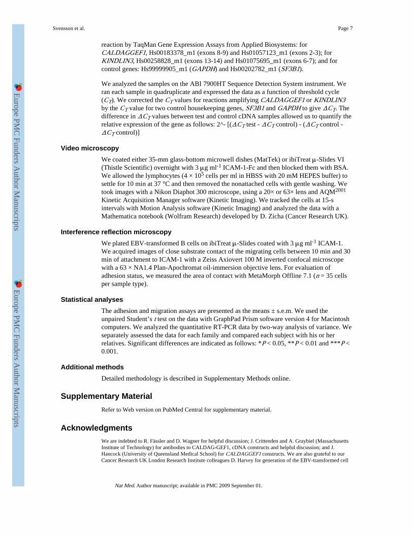
reaction by TaqMan Gene Expression Assays from Applied Biosystems: forCALDAGGEF1, Hs00183378_m1 (exons 8-9) and Hs01057123_m1 (exons 2-3); forKINDLIN3, Hs00258828_m1 (exons 13-14) and Hs01075695_m1 (exons 6-7); and forcontrol genes: Hs99999905_m1 (GAPDH) and Hs00202782_m1 (SF3B1).
We analyzed the samples on the ABI 7900HT Sequence Detection System instrument. Weran each sample in quadruplicate and expressed the data as a function of threshold cycle(CT). We corrected the CT values for reactions amplifying CALDAGGEF1 or KINDLIN3by the CT value for two control housekeeping genes, SF3B1 and GAPDH to give ΔCT. Thedifference in ΔCT values between test and control cDNA samples allowed us to quantify therelative expression of the gene as follows: 2^- [(ΔCT test - ΔCT control) - (ΔCT control -ΔCT control)]
Video microscopyWe coated either 35-mm glass-bottom microwell dishes (MatTek) or ibiTreat μ-Slides VI(Thistle Scientific) overnight with 3 μg ml-1 ICAM-1-Fc and then blocked them with BSA.We allowed the lymphocytes (4 × 10⁵ cells per ml in HBSS with 20 mM HEPES buffer) tosettle for 10 min at 37 °C and then removed the nonattached cells with gentle washing. Wetook images with a Nikon Diaphot 300 microscope, using a 20× or 63× lens and AQM2001
Kinetic Acquisition Manager software (Kinetic Imaging). We tracked the cells at 15-sintervals with Motion Analysis software (Kinetic Imaging) and analyzed the data with aMathematica notebook (Wolfram Research) developed by D. Zicha (Cancer Research UK).
Interference reflection microscopyWe plated EBV-transformed B cells on ibiTreat μ-Slides coated with 3 μg ml-1 ICAM-1.We acquired images of close substrate contact of the migrating cells between 10 min and 30min of attachment to ICAM-1 with a Zeiss Axiovert 100 M inverted confocal microscopewith a 63 × NA1.4 Plan-Apochromat oil-immersion objective lens. For evaluation ofadhesion status, we measured the area of contact with MetaMorph Offline 7.1 (n = 35 cellsper sample type).
Statistical analysesThe adhesion and migration assays are presented as the means ± s.e.m. We used theunpaired Student’s t test on the data with GraphPad Prism software version 4 for Macintoshcomputers. We analyzed the quantitative RT-PCR data by two-way analysis of variance. Weseparately assessed the data for each family and compared each subject with his or herrelatives. Significant differences are indicated as follows: *P < 0.05, **P < 0.01 and ***P <0.001.
Additional methodsDetailed methodology is described in Supplementary Methods online.
Supplementary MaterialRefer to Web version on PubMed Central for supplementary material.
AcknowledgmentsWe are indebted to R. Fässler and D. Wagner for helpful discussion; J. Crittenden and A. Graybiel (MassachusettsInstitute of Technology) for antibodies to CALDAG-GEF1, cDNA constructs and helpful discussion; and J.Hancock (University of Queensland Medical School) for CALDAGGEF1 constructs. We are also grateful to ourCancer Research UK London Research Institute colleagues D. Harvey for generation of the EBV-transformed cell
Svensson et al. Page 7
Nat Med. Author manuscript; available in PMC 2009 September 01.
Europe PM
C Funders A
uthor Manuscripts
Europe PM
C Funders A
uthor Manuscripts

lines and G. Kelly for help with the statistical analyses. L. S. was supported by a Marie Curie IndividualFellowship.
References1. von Andrian UH, Mempel TR. Homing and cellular traffic in lymph nodes. Nat. Rev. Immunol.
2003; 3:867–878. [PubMed: 14668803]
2. Hogg N, Laschinger M, Giles K, McDowall A. T-cell integrins: more than just sticking points. J.Cell Sci. 2003; 116:4695–4705. [PubMed: 14600256]
3. Coller BS, Shattil SJ. The GPIIb/IIIa (integrin αIIbβ3) odyssey: a technology-driven saga of areceptor with twists, turns, and even a bend. Blood. 2008; 112:3011–3025. [PubMed: 18840725]
4. Kellermann SA, Dell CL, Hunt SW III, Shimizu Y. Genetic analysis of integrin activation in Tlymphocytes. Immunol. Rev. 2002; 186:172–188. [PubMed: 12234371]
5. Kinashi T. Intracellular signalling controlling integrin activation in lymphocytes. Nat. Rev.Immunol. 2005; 5:546–559. [PubMed: 15965491]
6. Anderson DC, Springer TA. Leukocyte adhesion deficiency: an inherited defect in the Mac-1,LFA-1, and p150,95 glycoproteins. Annu. Rev. Med. 1987; 38:175–194. [PubMed: 3555290]
7. Hogg N, Bates PA. Genetic analysis of integrin function in man: LAD-I and other syndromes.Matrix Biol. 2000; 19:211–222. [PubMed: 10936446]
8. Etzioni A, Alon R. Leukocyte adhesion deficiency III: a group of integrin activation defects inhematopoietic lineage cells. Curr. Opin. Allergy Clin. Immunol. 2004; 4:485–490. [PubMed:15640688]
9. Pasvolsky R, et al. A LAD-III syndrome is associated with defective expression of the Rap-1activator CalDAG-GEFI in lymphocytes, neutrophils, and platelets. J. Exp. Med. 2007; 204:1571–1582. [PubMed: 17576779]
10. Kuijpers TW, et al. Leukocyte adhesion deficiency type 1 (LAD-I)/variant. A novelimmunodeficiency syndrome characterized by dysfunctional β2 integrins. J. Clin. Invest. 1997;100:1725–1733. [PubMed: 9312170]
11. McDowall A, et al. A novel form of integrin dysfunction involving β1, β2, and β3 integrins. J.Clin. Invest. 2003; 111:51–60. [PubMed: 12511588]
12. Kuijpers TW, et al. Natural history and early diagnosis of LAD-I/variant syndrome. Blood. 2007;109:3529–3537. [PubMed: 17185466]
13. Springett GM, Kawasaki H, Spriggs DR. Non-kinase second-messenger signaling: new pathwayswith new promise. Bioessays. 2004; 26:730–738. [PubMed: 15221855]
14. Stone JC. Regulation of Ras in lymphocytes: get a GRP. Biochem. Soc. Trans. 2006; 34:858–861.[PubMed: 17052215]
15. Clyde-Smith J, et al. Characterization of RasGRP2, a plasma membrane-targeted, dual specificityRas/Rap exchange factor. J. Biol. Chem. 2000; 275:32260–32267. [PubMed: 10918068]
16. Crittenden JR, et al. CalDAG-GEFI integrates signaling for platelet aggregation and thrombusformation. Nat. Med. 2004; 10:982–986. [PubMed: 15334074]
17. Bergmeier W, et al. Mice lacking the signaling molecule CalDAG-GEFI represent a model forleukocyte adhesion deficiency type III. J. Clin. Invest. 2007; 117:1699–1707. [PubMed:17492052]
18. Ghandour H, Cullere X, Alvarez A, Luscinskas FW, Mayadas TN. Essential role for Rap1 GTPaseand its guanine exchange factor CalDAG-GEFI in LFA-1 but not VLA-4 integrin mediated humanT-cell adhesion. Blood. 2007; 110:3682–3690. [PubMed: 17702895]
19. Cifuni SM, Wagner DD, Bergmeier W. CalDAG-GEFI and protein kinase C represent alternativepathways leading to activation of integrin alphaIIbß3 in platelets. Blood. 2008; 112:1696–1703.[PubMed: 18544684]
20. Moser M, Nieswandt B, Ussar S, Pozgajova M, Fassler R. Kindlin-3 is essential for integrinactivation and platelet aggregation. Nat. Med. 2008; 14:325–330. [PubMed: 18278053]
21. Moser M, et al. Kindlin-3 is required for β2 integrin-mediated leukocyte adhesion to endothelialcells. Nat. Med. Feb 22.2009 Advance online publication doi:10.1038/nm.1921.
Svensson et al. Page 8
Nat Med. Author manuscript; available in PMC 2009 September 01.
Europe PM
C Funders A
uthor Manuscripts
Europe PM
C Funders A
uthor Manuscripts
22. Ussar S, Wang HV, Linder S, Fassler R, Moser M. The Kindlins: subcellular localization andexpression during murine development. Exp. Cell Res. 2006; 312:3142–3151. [PubMed:16876785]
23. Larjava H, Plow EF, Wu C. Kindlins: essential regulators of integrin signalling and cell-matrixadhesion. EMBO Rep. 2008; 9:1203–1208. [PubMed: 18997731]
24. Siegel DH, et al. Loss of kindlin-1, a human homolog of the Caenorhabditis elegans actin-extracellular-matrix linker protein UNC-112, causes Kindler syndrome. Am. J. Hum. Genet. 2003;73:174–187. [PubMed: 12789646]
25. Jobard F, et al. Identification of mutations in a new gene encoding a FERM family protein with apleckstrin homology domain in Kindler syndrome. Hum. Mol. Genet. 2003; 12:925–935.[PubMed: 12668616]
26. Garcia-Alvarez B, et al. Structural determinants of integrin recognition by talin. Mol. Cell. 2003;11:49–58. [PubMed: 12535520]
27. Weinstein EJ, et al. URP1: a member of a novel family of PH and FERM domain containingmembrane-associated proteins is significantly over-expressed in lung and colon carcinomas.Biochim. Biophys. Acta. 2003; 1637:207–216. [PubMed: 12697302]
28. Kloeker S, et al. The Kindler syndrome protein is regulated by transforming growth factor-β andinvolved in integrin-mediated adhesion. J. Biol. Chem. 2004; 279:6824–6833. [PubMed:14634021]
29. Montanez E, et al. Kindlin-2 controls bidirectional signaling of integrins. Genes Dev. 2008;22:1325–1330. [PubMed: 18483218]
30. Ma YQ, Qin J, Wu C, Plow EF. Kindlin-2 (Mig-2): a co-activator of β3 integrins. J. Cell Biol.2008; 181:439–446. [PubMed: 18458155]
31. Mory A, et al. Kindlin-3: a new gene involved in the pathogenesis of LAD-III. Blood. 2008;112:2591. [PubMed: 18779414]
32. Campbell ID, Ginsberg MH. The talin-tail interaction places integrin activation on FERM ground.Trends Biochem. Sci. 2004; 29:429–435. [PubMed: 15362227]
33. Calderwood DA, et al. The phosphotyrosine binding-like domain of talin activates integrins. J.Biol. Chem. 2002; 277:21749–21758. [PubMed: 11932255]
34. Harris ES, et al. A novel syndrome of variant leukocyte adhesion deficiency involving defects inadhesion mediated by β1 and β2 integrins. Blood. 2001; 97:767–776. [PubMed: 11157496]
35. Alon R, et al. A novel genetic leukocyte adhesion deficiency in subsecond triggering of integrinavidity by endothelial chemokines results in impaired leukocyte arrest on vascular endotheliumunder shear flow. Blood. 2003; 101:4437–4445. [PubMed: 12595312]
36. Smith A, et al. A talin-dependent LFA-1 focal zone is formed by rapidly migrating T lymphocytes.J. Cell Biol. 2005; 170:141–151. [PubMed: 15983060]
Svensson et al. Page 9
Nat Med. Author manuscript; available in PMC 2009 September 01.
Europe PM
C Funders A
uthor Manuscripts
Europe PM
C Funders A
uthor Manuscripts

Figure 1.The CALDAGGEF1 gene C→A base change has no effect on mRNA and protein levels oron deficient LAD-III B cell adhesion and migration. (a) The DNA sequence surrounding theC→A base change in exon 16 of the CALDAGGEF1 gene in the Turkish (C→A basechange) and Maltese (no base change) families. C or A indicates homozygosity; C/Aindicates heterozygosity. The original data are shown in Supplementary Figure 1. (b)CALDAG-GEF1 protein abundance in subjects (S), relatives (M, mother; F, father) andcontrol individuals (C), as assessed by western blotting (n = 2 independent samples perfamily). α-TUBULIN was used as a loading control. (c) Migration characteristics of LAD-III B cells adhered to ICAM-1. LAD-III cells from each family were transfected with a wild-type CALDAGGEF1 or EGFP cDNA construct; EGFP-transfected parents’ (families 1 and3, mother; family 2, father) cells are shown for comparison. n = 4 independent experimentsfor each family. Data are shown as means ± s.e.m.; ***P < 0.001. (d) Left, differentialinterference contrast (DIC) and IRM images of B cells from the family 3 subject with LAD-III and from the mother (Parent) transfected as in c (n = 2). Scale bar, 10 μm. Right,quantification of the area of close contact for n = 35 cells per cell type. Data are shown asmeans ± s.e.m.; ***P < 0.001.
Svensson et al. Page 10
Nat Med. Author manuscript; available in PMC 2009 September 01.
Europe PM
C Funders A
uthor Manuscripts
Europe PM
C Funders A
uthor Manuscripts

Figure 2.Mutations in the KINDLIN3 gene. (a) Top, the DNA sequence around the location of theC→T base change in exon 12 of the KINDLIN3 gene in the Turkish (mutated) and Maltese(not mutated) families; C or T indicates homozygosity; C/T denotes heterozygosity. Bottom,traces of the DNA sequence analysis around the mutation site in exon 12 for wild-typegenome (WT), a heterozygote Turkish parent (HET) and a homozygous mutant Turkishsubject with LAD-III (LAD-III). Y indicates the presence of a pyrimidine (both C and T).(b) Top, the DNA sequence around the location of the A→G base change in the KINDLIN3exon 14 splice acceptor site in the Maltese (mutated) and Turkish (not mutated) families; Aor G at this position indicates homozygosity; A/G denotes heterozygosity. Bottom, traces ofthe DNA sequence analysis around the mutation site in splice acceptor site of exon 14 forthe WT, the heterozygote Maltese parents (HET) and the homozygous mutant Maltesesubject with LAD-III; R indicates the presence of a purine (both A and G). More extensiveoriginal data for a and b are shown in Supplementary Figure 5 online. (c) Diagrammaticalrepresentation of human KINDLIN-3 protein (residues 1-663), showing the three FERMsubdomains (F1-3) characteristically intersected with a pleckstrin homology (PH) domain.The location of the exons encoding the different regions of the KINDLIN-3 protein isshown. The position of the two LAD-III mutations, one in exon 12 (E12) and the otheraffecting the join of E13 and E14 of the KINDLIN3 gene, detailed above in a and b,respectively, are highlighted with red asterisks.
Svensson et al. Page 11
Nat Med. Author manuscript; available in PMC 2009 September 01.
Europe PM
C Funders A
uthor Manuscripts
Europe PM
C Funders A
uthor Manuscripts
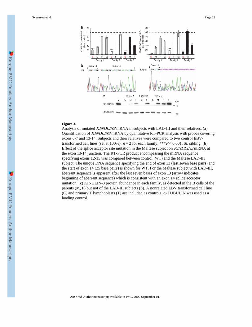
Figure 3.Analysis of mutated KINDLIN3 mRNA in subjects with LAD-III and their relatives. (a)Quantification of KINDLIN3 mRNA by quantitative RT-PCR analysis with probes coveringexons 6-7 and 13-14. Subjects and their relatives were compared to two control EBV-transformed cell lines (set at 100%). n = 2 for each family; ***P < 0.001. Si, sibling. (b)Effect of the splice acceptor site mutation in the Maltese subject on KINDLIN3 mRNA atthe exon 13-14 junction. The RT-PCR product encompassing the mRNA sequencespecifying exons 12-15 was compared between control (WT) and the Maltese LAD-IIIsubject. The unique DNA sequence specifying the end of exon 13 (last seven base pairs) andthe start of exon 14 (25 base pairs) is shown for WT. For the Maltese subject with LAD-III,aberrant sequence is apparent after the last seven bases of exon 13 (arrow indicatesbeginning of aberrant sequence) which is consistent with an exon 14 splice acceptormutation. (c) KINDLIN-3 protein abundance in each family, as detected in the B cells of theparents (M, F) but not of the LAD-III subjects (S). A nonrelated EBV transformed cell line(C) and primary T lymphoblasts (T) are included as controls. α-TUBULIN was used as aloading control.
Svensson et al. Page 12
Nat Med. Author manuscript; available in PMC 2009 September 01.
Europe PM
C Funders A
uthor Manuscripts
Europe PM
C Funders A
uthor Manuscripts

Figure 4.Adhesion and migration characteristics of LAD-III B cells expressing wild-type Kindlin-3.(a) Left, DIC and IRM images of adhesion on ICAM-1 of LAD-III B cells transfected witheither EGFP or a KINDLIN3 cDNA construct and compared with a parent’s B cells (family1 and 3, mother; family 2, father) transfected with EGFP (n = 2 for each cell type). Right,quantification of the area of close contact for n = 35 cells. Scale bar, 10 μm. Data are shownas means ± s.e.m.; *, P < 0.05, ***P < 0.001 and NS, not significant. (b) Migrationcharacteristics of LAD-III B cells adhered to ICAM-1. B cells from subjects with LAD-IIIwere transfected with either EGFP or wild-type EGFP-KINDLIN3 cDNA constructs andcompared with EGFP-transfected parents’ B cells (family 1 and 3, mother; family 2, father)(representative experiment of n = 3 for each family). Data are shown as means ± s.e.m.; **P< 0.01 and ***P < 0.001. (c) The cell-tracking results from an individual experiment withcells from the family two subject with LAD-III and from his father, transfected as in b.
Svensson et al. Page 13
Nat Med. Author manuscript; available in PMC 2009 September 01.
Europe PM
C Funders A
uthor Manuscripts
Europe PM
C Funders A
uthor Manuscripts
Copyright © 2022 FDOKUMEN