infections caused by bacteria, viruses, fungi, and parasites
Upload
khangminh22Category
view
1download
0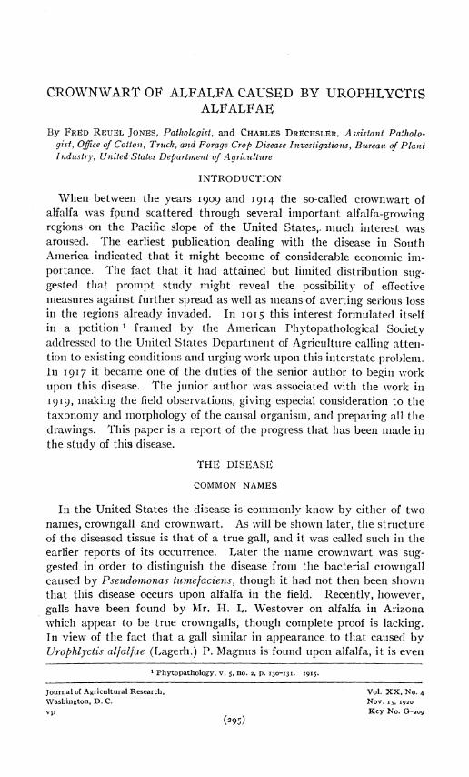
CROWNWART OF ALFALFA CAUSED BY UROPHLYCTIS ALFALFAE
By FRED RëUEL JONES, Pathologist, and CHARLES DRECHSLER, Assistant Patholo- gist, Office of Cotton, Truck, and Forage Crop Disease Investigations, Bureau of Plant Industry, United States Department of Agriculture
INTRODUCTION
When between the years 1909 and 1914 the so-called crownwart of alfalfa was fpund scattered through several important alfalfa-growing regions on the Pacific slope of the United States,, much interest was aroused. The earliest publication dealing with the disease in South America indicated that it might become of considerable economic im- portance. The fact that it had attained but limited distribution sug- gested that prompt study might reveal the possibility of effective measures against further spread as well as means of averting serious loss in the legions already invaded. In 1915 this interest formulated itself in a petition 1 framed by the American Phytopathological Society addressed to the United States Department of Agriculture calling atten- tion to existing conditions and urging work upon this interstate problem. In 1917 it became one of the duties of the senior author to begin work upon this disease. The junior author was associated with the work in 1919, making the field observations, giving especial consideration to the taxonomy and morphology of the causal organism, and preparing all the drawings. This paper is a report of the progress that has been made in the study of this disease.
THE DISEASE
COMMON NAMES
In the United States the disease is commonly know by either of two names, crowngall and crownwart. As will be shown later, the structure of the diseased tissue is that of a true gall, and it was called such in the earlier reports of its occurrence. Later the name crownwart was sug- gested in order to distinguish the disease from the bacterial crowngall caused by Pseudomonas turnefaciens, though it had not then been shown that this disease occurs upon alfalfa in the field. Recently, however, galls have been found by Mr. H. L. Westover on alfalfa in Arizona which appear to be true crowngalls, though complete proof is lacking. In view of the fact that a gall similar in appearance to that caused by Urophlyctis alfaljae (Lagerh.) P. Magnus is found upon alfalfa, it is even
1 Phytopathology, v. 5, no. 2, p. 130-131. 1915.
Journal of Agricultural Research, Vol. XX, No. 4 Washington, D. C. Nov. 15, 1920 vp Key No. G-209
(295)

296 Journal of Agricultural Research vol. xx, NO. 4
more desirable than formerly that the disease caused by Urophlyctis should have a distinctive name. The fact that the name crownwart is well established in usage is much in its favor. It will be seen, however, from facts presented later in this paper that this name is somewhat misleading, inasmuch as the galls are not typical warty growths, nor are they formed from the tissue of the so-called crown of the plant in a manner comparable with that in which crowngalls are formed. A name more truly distinctive is suggested by the French name used by Arnaud (i),1
"La Maladie des tumeurs marbrées de la Luzerne." An English equiv- alent, marbled gall of alfalfa, the word marbled referring to the mot- tled effect produced by the brown spore masses seen when any of these galls are cut, would call attention to the one distinctive character of these galls observable at any time and would be accurately descriptive.
HOST PLANTS
Of the many species of the genus Medicago introduced into the United States, Medicago sativa is the only one on which the disease has been found commonly. McKee (17) found it also on M. faicata. The two species, grown near together at the Plant Introduction Field Station at Chico, Calif., seemed to be about equally infected.
Spegazzini (32) records the fungus as occurring on Medicago denticulata and species of Adesmia in Argentina. Hauman-Merck (JJ) also records the fungus on M. denticulata from the same locality and further states that it does not occur upon alfalfa. In view of the fact that search has not revealed the fungus upon M. denticulata in the United States even when the plant is growing abundantly close in association with diseased alfalfa, it seems advisable to hold it an open question whether the fungus found in Argentina upon M. denticulata and Adesmia spp. is identical with that which causes the disease of alfalfa.2 Thus, the evidence at hand, while it is inadequate for the formation of final conclusions, appears to indicate that the species of Urophlyctis occurring on M. sativa is prob- ably limited to that species and to M. faicata.
DISTRIBUTION AND ECONOMIC IMPORTANCE
The only available information regarding the economic importance of the disease consists of expressions of opinion based on a larger or smaller amount of field observation. The trend of the opinion that has devel- oped from this observation is that the disease is, or becomes locally, very destructive to alfalfa plants.
The first report of the disease by von Lagerheim (14) from Ecuador gave inception to this trend. He states that diseased plants can easily
1 Reference is made by number (italic) to "Literature cited," pp. 321-323. 2 A portion of a collection of Urophlyctis alfalfae var. adesmiae on Adesmia bicolor, sent by Spegazzini to
the Office of Pathological Collections, Bureau of Plant Industry, has been examined and been found to contain a Synchitrium rather than a Urophlyctis.

Nov. is, 1920 Crownwart of Alfalfa Caused by Urophlyctis alfalfae 297
be distinguished in the field, and his illustrations of diseased plants with crowns encrusted with large galls contributed effective support to his state- ments. However, von Lagerheim states that he did not see the disease in the field himself, though he sought for it in fields near Quito. He received his specimens from the owner of an estate in the Andes, and his description of the effects of the disease in the field was gathered from several observers.
In Europe, Magnus (20) reports a destructive outbreak of the disease in Alsace, basing his report on the observations of two farmers. Later, from an adjoining Province of Germany, Grimm and Korff (10) report the disease as present in an alfalfa field without causing much apparent harm. In fact, the diseased plants seemed somewhat more vigorous than the others. Nevertheless, they think measures should be taken to elimi- nate it.
Peglion (25) finds the disease in Italy, and raises the question whether or not it may be a factor in producing alfalfa sickness in some fields. He suggests that experimental work should be undertaken to determine the matter. In France Arnaud (1) reports the disease as apparently doing considerable damage in a single field in the Department of Seinc-et-Oise. In 1916 Salmon (27) found a single field infested with wart in England and urged further search for the disease. No reports of serious infesta- tions have followed, though the writers have been told that occasional specimens are found. The disease has been found in Holland (8) and Sweden, but no apparent damage has been reported.
A critical reading of these reports of the destructive action of the dis- ease calls attention to the fact that the two most important reports, those of von Lagerheim and of Magnus, are not based on first-hand obser- vation. In all cases damage is noted only in small areas. Therefore we must still hold it an open question whether this disease has been primarily responsible for any serious or widespread injury to alfalfa in either South America or Europe.
In the United States the disease has been found abundant only west of the Sierra Nevada and Cascade Mountains, though it occurs in a few regions east of these mountains. It has not been found east of the Rockies. However, in view of the fact that the disease when not abun- dant is often completely concealed unless a plant is uprooted, it is pos- sible that its distribution is more widespread than records show.
The first report of the disease by Smith (31) gives no clue to its im- portance. O'Gara (22) finds the disease very common and occasionally destructive in fields in the Rogue River Valley in Oregon. Jackson (12) later reports the disease from the same region, making no comment regarding its importance. Again O'Gara (23) is first to report the disease present in the Salt Lake Valley in Utah, though he has not in this case determined to what extent it causes injury. McCallum (16) reported the disease present in Arizona. McKee (17), who has had an opportunity

298 Journal of Agricultural Research VO1.XX,NO.4
to observe the disease extensively, concludes that crownwart decreases the yield and shortens the life period of plants. He says:
Alfalfa fields that had the crown wart in abundance in 1914 produced good crops of hay in that year and in 1915. In one field sown in 1910 that has been under obser- vation the past two years, practically every plant has galls. This field has produced apparently normal crops of hay, but more critical observation shows decreased vigor in the plants and a corresponding decrease of yield.
McKee believes the disease much more widespread than is commonly supposed and urges work to determine its importance in alfalfa culture.
Thus it appears that although the disease is scattered through large alfalfa-growing areas in the United States, yet it does not appear at any place to have become regarded as a serious limiting factor in the growth of the crop, except during years of severe attack and even then in small areas.
The writers have not attempted to determine the present limits of spread of the disease in the United States. A limited amount of time has been spent in the spring of three years observing the disease, chiefly in the river valleys where it is known to be most abundant, the Sacramento River Valley in California and the Rogue River Valley in Oregon. The second of these years, 1918, appears to have been distinctly unfavorable for the development of the disease, especially in California. The winter rainfall was below normal, in consequence of which the Sacramento River did not overflow its flood plain where McKee observed the disease to be most abundant. The disease was commonly present on a larger or smaller percentage of plants, but nowhere did observation bring con- viction that considerable damage was being done. In the San Joaquín Valley that year only occasional diseased plants could be found, though in some localities there was excellent testimony from fanners of the abundance of the disease in previous years.
In 1919 there was much more winter rain, especially in the Sacra- mento River Valley, and a greater amount of disease was found. Even then it was only rarely that the disease was sufficiently abundant to appear to be of serious economic importance. Plants could be found whose early buds had become so completely infected that few were left to form the second and later cuttings, but such plants were usually widely scat- tered among others less severely infected. Rarely indeed does the dis- ease appear to be solely responsible for the killing of entire plants, though it must often weaken them. A significant estimate of the actual damage done can be made only after careful observation has extended over a period of years when the varying intensity of the annual attacks can be studied and the behavior of the diseased plants followed throughout the year.
DESCRIPTION OF THE DISEASE (PL. 47)
The disease is more easily described by stating briefly the origin and method of development of the galls. So far as the writers can discover

Nov. 15,1920 Crownwart of Alfalfa Caused by Urophlyctis alfalfae 299
all galls as they occur naturally in the field result from the infection of buds in early stages of development as they emerge from the crown of the plant. It is well known that there is an almost continuous succes- sion in the development of buds from the so-called alfalfa crown during the entire year. A portion of those buds which will produce the shoots furnishing the first crop in the spring have begun development as early as the preceding autumn. Generally speaking, the first buds to be formed in the seasonal succession have a point of origin deeper in the soil than those which are formed later, so that many of the buds from which the shoots of the third cutting arise develop from positions quite above the surface of the soil. Buds produced below the soil level in cool weather appear to have a meager protection of scaly covering, and it is for the most part such buds that become infected and give rise to galls. Thus, galls are swollen and distorted bud elements, scales, leaves, and stipules.
Unless overwintered galls which are described later are discoverable, the disease is first evidenced in the spring by a slight thickening and round- ing of the young buds. During two years this has been observed near Chico, Calif., in the latter part of March or early in April. The diseased buds become more and more rounded as growth progresses and are glis- tening white in color (PI. 54, A). Then, as the infected structures begin to push apart, some of them grow much more rapidly than others until the structure as a whole assumes a conspicuously irregular form. In most cases, however, an examination of the gall will show that it is made up of thick, scalelike layers about a central growing axis (PI. 36, B). Sometimes this axis continues growth in spite of the demands of the mass of developing gall tissue and produces a weak shoot. The earlier and more vigorous buds produce the larger galls. Smaller galls often appear to be developed from smaller buds along the stems below ground that would ordinarily remain dormant. The origin of galls that appear on stems several inches above the surface of the ground in wet weather appears to be due in part to the infection of axillary buds that would never develop in the ordinary course of events and in part to the elon- gation of the stems and petioles which force infected tissue upward.
Since a large part of the infected buds are developed at a depth of 2 or 3 inches below the surface of the soil, the majority of the galls are so far below ground that they escape observation unless the soil is removed from around the plant. If they are of large size some of them come to the surface, where they take on a green color and in extreme cases form a crust of diseased tissue around the base of the healthy stems.
Another type of gall that is not common results from local infections on young leaves. Such infections give rise to small blister-like galls much like those produced on Sanícula spp. by another species of this fungus which will be mentioned later.

300 Journal of Agricultural Research vol. xx, N0.4
The galls reach full development (PL 54, B) early in the summer, in early June in northern California. From this time on the majority of them begin to decay if moisture is abundant, or to shrivel and dry with the coming of drouth. However, in almost all fields a few galls more deeply situated become covered with a corky layer and survive the winter.
When plants are subjected to dry conditions in late summer, as they usually are in the Rogue River Valley in Oregon, many of the galls do not decay but remain living throughout the autumn and winter. It does not appear that such galls make appreciable growth during the following year. Nevertheless, gall tissue may accumulate around old plants in considerable mass. The exterior becomes covered with a brown, corky layer that has a much warted appearance. This accumu- lation of gall tissue has not been found on plants that have grown in well-irrigated fields.
At whatever age or state of development these galls are found, they possess one distinctive character that is discovered when they are cut open. The interior of the galls contains many small, irregularly shaped brown masses of fungus spores which are easily visible .(PI. 56, B). In old dried galls the host tissue has shrunken so much that the spore mass often occupies a large portion of the mass of the gall. Even in decayed galls that have not yet been broken to fragments the spore masses can be recognized by their golden brown color.
CAUSAL ORGANISM
NOMENCLATURE)
Some difference of opinion concerning the identity of the parasite causing crownwart of alfalfa has prevailed. Von Lagerheim (24) seems first to have regarded it as a new and distinct species, which he cited as Cladochytrium alfalfae. Later, however, he (14) identified it with Urophlyctis (Physoderma) le proidea, a parasite causing conspicuous mal- formations on the beet, originally described from Algeria by Trabut (54) and assigned by him as well as by Saccardo and Mattirolo (26) to a new Ustilaginous genus, Oedomyces. In making this disposition, von Lagerheim opposed the views of both Vuillemin (55), who had identi- fied Trabut's beet organism with Urophlyctis (Cladochytrium) pidposa (Wallroth), long known to be parasitic on species of Chenopodium and Atriplex, and of Magnus (18-20), who later came to regard the parasites on Chenopodium spp., on the beet, and on alfalfa as three dis- tinct species. None of these views appear to be based on evidence alto- gether conclusive; nor can we adduce such evidence here, because the lack of fresh diseased material of beet and of Chenopodium spp. have made it impossible to attempt cross-inoculation experiments.

Nov. 15,1920 Crownwart of Alfalfa Caused by Urophlyctis alfalfae 301
Provisionally, it appears advisable to follow Magnus in recognizing the alfalfa parasite as a distinct species, not, perhaps, so much on account of some differences in morbid host anatomy as because of the general improbability that two unrelated plants serve as hosts to a parasite which shows in general no omnivorous tendencies. The beet disease has not been reported in the regions where crownwart is preva- lent; and Chenopodium spp. with every chance for infection have not been observed to be attacked. Reference has been made in another connection to Spegazzini's (32) report of crownwart on Medicago denti- cidata and its absence from alfalfa in the same range. This condition could most readily be attributed to the existence of another species producing similar galls.
DEVELOPMENT AND MORPHOLOGY OF THE FUNGUS
The morphology of the crownwart organism has not hitherto received much attention. Magnus (20) made some observations regarding en- larged hyphae frequently found in old material and referred to the pres- ence of a hyaline cell attached to the concave side of the resting spores; but in the main his specific details concern the pathological anatomy of the host. In more recent years, Wilson (57) published a cytological account of Urophlyctis alfalfaey arriving at conclusions considerably at variance with those of Magnus. The utilization of old material by both these writers may largely account for their failure to observe important details of development and morphology, as well as explain interpre- tations that it appears impossible to reconcile with conditions as found in young material much more favorable for study.
GERMINATION OF THE RESTING SPORES
As has long been recognized, the fungus passes through the prolonged periods of summer drouth by means of the resting spores contained within cavities in the galls of the host. In the course of the rainy season the galls disintegrate completely, thus setting free the spores; and it is not improbable that the exposure incident to this method of liberation may be necessary for germination. However, the conditions that may favor germination remain more or less obscure; for although many at- tempts were made by the writers with spores from freshly gathered material both old and young, as well as with limited supplies of material that may, in addition, have suffered deterioration in transit, the results obtained have been so meager and dubious that this phase of the life history of the fungus must be reserved for a later paper. In a number of preparations an appearance was noted as of resting spores producing a number of subspherical bodies varying from 1 to 9, by the passage of protoplasm through pores in the spore wall. The vesicles that usually attained half the linear dimensions of the spore in some cases were seen

302 Journal of Agricultural Research VO1.XX,NO.4
to produce endogenous motile bodies resembling zoospores that later escaped through a number of openings on the distil side of the vesicular wall. As the Van Tiegham cultures in which this process was noticed were usually several days old, the development of bacteria and various protozoa brought into the observations a considerable measure of un- trustworthiness. Indications that similar contaminations may have affected the observations of Wilson (j7) on Urophlyctis aljaljac and of Bally {2) on U. rübsaameni are not entirely wanting. Both of these writers describe the resting spore as functioning directly as zoosporan- gium.1
PENETRATION OF THE HOST
Because of difficulties encountered in efforts to bring about infection under artificial conditions, it has not been possible to observe directly the penetration of the host by the germinating zoospore. However, as an abundance of conditions immediately following the entrance of the parasite were found in stained sections of buds, the course of events during the time of invasion can be followed in incipient stages in the same manner as during advanced stages.
Bodies measuring 3 to 4 jit in diameter were frequently found attached or adhering to the scales or developing axis of the bud. They appear to have made their way under the bud scales very close to the most rapidly growing meristem. Unfortunately, no clear figures showing the immedi- ate development of these bodies were observed—a failure attributable apparently to the fact that by the time the galls became noticeable many weeks had seemingly elapsed since the period during which infection took place abundantly. As a result, the earliest demonstrable stage of invasion was represented by the presence of small turbinate bodies (the "Sammelzellen," "corps centrals," "vésicules collectrices," or "vési- cules collectives'' of other writers) within the epidermal cells of the outer foliar or scale elements of buds exposed to attack, and attached to and perforating the cuticular wall by an elongated beak (PI. 49, A, ta-tg). More than one body may be present in the same epidermal cell, two or three being not unusual; and occasionally a considerable number of contiguous cells may show such evidence of multiple and concentrated attack. The beak manifestly represents the tube proliferated by the zoospore through which the contents of the latter were conveyed into the host cell after the manner prevailing very generally throughout the Chytridiales.
1 In an article that has appeared since this paper was prepared, Wilson (38) gives a more detailed ac- count of his findings. So far as his account concerns the germination of the resting spores, it appears to differ very considerably from that more recently published by C. Emlen Scott (30), according to whom each resting spore proliferates from 1 to 15 sporangia, the zoospores escaping through a number of tubes in the hyaline wall. With the latter account the observations recorded above are not at variance.

Nov. 15,1920 Crownwart of Alfalfa Caused by Urophlydis alfalfae 303
GROWTH OF THE PARASITE
The fungus cell thus produced is first uninucleated and bears at its apex a short, cylindrical projection. As it becomes older it increases in size, the single nucleus divides, giving rise to a multinucleated condition, and the short apical projection proliferates more or less successively three or four terminal branches which are directed nearly at right angles to the primary axis. These branches subsequently proliferate usually three to five secondary branches directed in the same plane or forward. As a result of this continued ramification, the larger cells may be seen to bear at their apices an apparatus consisting of a short axial stalk branching to form a score of ultimate terminations. There can be little doubt that these processes function as absorbing organs and may thus be regarded as haustoria. In stained sections they are often too badly obscured by host protoplasm to be readily distinguishable; but in prep- arations of material dissected from fresh, living host plants, they may be studied with ease and certainty.
In the meantime the turbinate cell has increased considerably in size and in number of nuclei, the latter usually ranging from 10 to 20 or even more. As no septa have appeared, the parasite is represented at this stage by a simple coenocyte. With the cessation of growth by enlarge- ment, this condition is altered by the appearance of a number of delicate septa, the ultimate number usually ranging from 3 to 5 but occasionally even reaching 7, each of which delimits a peripheral uninucleated mass of protoplasm. As the septa do not appear altogether simultaneously, the first to be inserted represent convex membranes united to the periph- eral wall of the turbinate cell along an elliptical line of juncture, the long axis being parallel with the axis of the turbinate cell. The septa in- serted later, when the surface of the turbinate cell has been appropriated in considerable measure, are more likely to be in relation to septa pre- viously laid down as well as to the peripheral wall itself. While the protoplasts first delimited thus tend to approach a double-convex, ellip- tical lenticular shape, the later ones may be more irregular and have several concave facets (Pi. 49, B).
The further development of each of the peripheral protoplasts thus delimited takes place independently of the other protoplasts similarly derived from the same turbinate cell and follows in the main the course described by Maire and Tison {21) for Urophlydis hemisphaer.ica (Speg.) Syd. (U. kriegeriana Magnus) and by Vuillemin (jó) for U. leproidea. Material embedded in paraffin, sectioned, and stained shows the proto- plasm very slightly contracted away from the septum along the inner sur- face, and indications of such contraction are present also in freshly dissected material mounted in water (Pi. 48, B, tb). This slightly con- tracted protoplast now pushes out a protuberance from the outer periph- eral wall bounding it (PI. 48, C, D, tbx). In those peripheral segments
95o80-20 5

304 Journal of Agricultural Research VO1.XX,NO.4
occupying a position on the side or toward the base of the turbinate cell, the protuberance will invariably take place at some point along the edge closest to the apical end of the turbinate structure ; while in the segments on the apical end the protuberance usually occupies a middle position. By the movement of the nucleus and part of the cytoplasm into the pro- tuberance, the tip of the latter becomes somewhat distended. The con- stricted position now rapidly elongates, resulting in the formation of an attenuated hypha, uniform in thickness and approximately 0.5 M in diam- eter (Pi. 48, A-D). The transfer of protoplasm from the peripheral segment to the distended termination continues for some time, until the former has been completely evacuated (PI. 48, B, D, ta).
The elongation of the hypha involves a translatory movement of the termination in a forward direction, from which, however, it may be de- flected by a host cell wall, or even reflected back toward the cuticular wall (Pi. 49, B, ¿&a). • Ultimately elongation ceases, and the terminal distension develops into a turbinate cell entirely similar to the original product of infection, the single nucleus dividing repeatedly to reproduce the coenocytic condition and the branching haustorial process developing from the apical projection, which becomes observable at an early stage during the period of hyphal elongation.
The proliferation of secondary turbinate cells, which tends to be more abundant from the expanded apical end than from regions more nearly basal, thus involves a certain number of lenticular uninucleated masses of protoplasm, always peripheral in position. The larger remaining por- tion of the contents of the original turbinate cell is consequently not concerned in this process. It may conveniently be designated as the sporogenous cell and always embraces the contents along the longitudi- nal axis of the spore and as much peripheral protoplasm as is not involved in the peripheral segments. The contents of the sporogenous cell func- tions in giving rise to a resting spore in the manner described in the fol- lowing paragraph.
vSooner or later after the segmentation of the turbinate cell has been initiated, the axial haustorial prolongation buds terminally to produce a small globose swelling, which, when it first becomes noticeable, has no demonstrable irregularities on its surface. Later when the swelling or young resting spore has attained a diameter of perhaps 5 /x (PI. 48, D, rb), there are proliferated along a zone midway between the equatorial region and the distil pole from 9 to 15 slender, unbranched, minute proc- esses. The swelling continues to increase in size until it attains the dimensions of the resting spore (about 25 to 35 by 40 to 50 n), growth in the earlier stages being due mainly to the transfer of protoplasmic con- tents from the sporogenous cell through the axial haustorial element but later quite largely by the assimilation of food material from the host. Although the surface of the resting spore is rendered impervious by the deposition of a thick wall during the later stages of enlargement, such

Nov. is, 1920 Crownwart of Alfalfa Caused by Urophlyctis alfalfae 305
assimilation is made possible by the zone of haustorial processes, each of which has in a manner similar to the apical process become branched to form a ramifying apparatus (PI. 48, A-D, m, rb).
DETAILS OF MORPHOLOGY AND CYTOLOGY
The branched haustorial processes with their unusually definite local- ization, either as a solitary apparatus at the apical end of the vegetative cell or arranged in a well-defined zone between the equator and the dis- til pole of the resting spore, constitute perhaps the most striking mor- phological feature of the parasite. Although the literature regarding these structures, especially with reference to their development and orientation on the resting spore, is unsatisfactory, there seems to be good reason to believe that all the other species usually referred to Uro- phlyctis, as well as many species commonly assigned to related genera, will show complete similarity to U. alfalfae in this respect. Thus De- Bary (3) in his account of Physoderma (Protomyces) menyanthis states that—
Auf demselben (distil) Ende der Blasen findet man sehr häufig ein Btischelcheii sehr feiner und kurzer in ein Köpfchen endigender Fäden, welche bald verschwinden und über deren Bau und Zweck ich nichts Näheres angeben kann ;
and in the figure referred to the appendages are clearly represented at the apices of the obovoid vesicles ("verkerteiformige Blasen"). Lüdi (15), who later studied the same fungus, figured a number of unbranched processes arising independently but in close proximity to each other from the apex of the "Sammelzelle"; and in a few cases he represented a hypha arising also independently from the midst of this cluster. Büs- gen (4) observed the same structure in Physoderma (Cladochylrium) bntomi at the apex of the swellings less rich in contents. Like DeBary this author remained uncertain as to their function but considered it probable that the apparatus consists of budding hyphae together with granular host protoplasm. He reported, too, the presence in this spec- ies of— irregular cylindrical projections which appear early on the spore, and later are not greatly inferior in length to the diameter of the spore. Stained with iodin, a mem- brane and hyaline contents with a few granules may be recognized. When the spore matures, these break down.
These structures he designated as haustoria and related their function, in our judgement altogether correctly, to the assimilation of food mater- ial. His figures, however, with the exception of figure 19, a, which shows a detached branching rhizoid, lack clearness and lead one to believe that probably groups of newly proliferated young turbinate cells were con- fused with the rhizoids. On the other hand, the haustoria he shows associated with the resting spores of Physoderma (Cladochytrium) flam- mulae suggest a good possibility of a zonate arrangement similar to that

3o6 Journal of Agricultural Research VO1.XX,NO.4
found in the alfalfa parasite corresponding, for example, to Plate 48, A-D,ra, rb; although, to be sure, the attachment of the "Sammelzellen" to the convex haustoria-bearing side would be at variance with any close homology. It appears not unreasonable, however, to suspect that Büs- gen was in error in regard to this point and that the resting spore may be attached by its concave side, the concavity, as in Urophlyctis spp. generally, very probably being opposite the side bearing the haustoria.
Clinton (5) noted the presence of a rhizoid-like process on the side of the "Sammelzellen" toward the young sporangium in Physoderma (Cladochytrium) mactdare and figured it both as a terminal apical struc- ture before the development of the resting sporangium has been initiated and as a median whorl after the latter has been formed. Regarding its function he states that—
The exact nature of these processes is not clearly shown, though they seem to bind the sporangium cell to the Sammelzellen.
In his figure 32 he shows a similar process attached to an clement that appears to be a young resting spore, although he makes no reference to this condition in the text.
Schroeter (28) observed the apical apparatus on the vegetative cells of Urophlyctis pulposa, designating it as—
ein Krönchen, ein Schopf feiner und kurzer, oft verzweigter Protoplasma Anhängsel.
Vuillemin (j5), who later studied the same species as well as the beet parasite, appears to have recognized the apparatus as consisting of a "tronc" bearing terminally a "houppe" of short ramifying processes— the "panache terminale.,, To these processes and to the haustoria on the resting spores, as well as to the "appareil nourricier" generally, he (36) assigned a structure identical to that of the striated muscle fiber of animals. We have not been able to distinguish anything suggesting striation in any portion of the thallus of U. alfalfae. The haus- toria here, moreover, appear to have a membrane that seems to persist after the contents have been withdrawn by plasmolysis or have degenerat- ed. The history of the development of the haustoria on the resting spore as given for the beet parasite again is at variance with their develop- ment as observed in U. alfalfae. For the resting spore, according to Vuillemin, ccmes about by the swelling of the "sommet du tronc du pan- ache" in such a way that—
Les branches se trouvent dissociées en plusiers buissons et entraînées à diverses hauteurs sur la boule terminale, tandis que d'autres fragments sont restés à la base.
Whereas in U. alfalfae the resting spore is initiated as a bud from the tip of the axial haustorial element, never involving translocation of any haustorial ramification. And as has been pointed out, the haustoria on the resting spores are subsequently developed as new structures in a well- defined zone and are not portions of the apical haustorium distributed

Nov. is, 1920 Crownwart of Alfalfa Caused by Urophlyctis alfalfae 307
in a miscellaneous manner over the surface of the resting spore by the enlargement of the latter.
The time of proliferation of the resting spore seems to be rather vari- able. It may follow immediately after the septum delimiting the last peripheral segment has been laid down, before the proliferation of the new order of turbinate cells has begun (Pi. 48, B, rb), or more usually somewhat later when one or more of the peripheral segments have pro- liferated secondary turbinate cells (PI. 48, C, rb). Or, as is not infre- quently the case with the unusually large primary turbinate cells, the immediate product of infection, the resting spores may not be formed until three or four successions have intervened and the original lesion has be- come a well-developed cavity (PI. 50, tba). The protoplasm in the spo- rogenous cells of such primary turbinate structures as well as the host protoplasm of cells or cavities that have long harbored the fungus fre- quently take a dense uniform stain with safranin—a result that might readily be attributed to the diffusion of a deep-staining substance. Where this abnormal condition becomes very pronounced, it is not im- probable that no resting spore is produced at all, the deep-staining protoplasm finally disintegrating in place. With perhaps this occasional exception, every turbinate cell produces always one resting spore. Ac- cording to Maire and Tison (21), Urophlyctis hemisphaerica (Speg.) Syd. produces first a succession of "vésicules collectives/' each of these in turn giving rise to several others of the next order, until ultimately each "vésicule collective,, produces only a single resting spore. Such separation of vegetative and reproductive stages is not discernible in U. alfalfae, the production of resting spores being common to each order of turbinate cells; and, although toward the end of the season, when conditions for growth become poor, the proliferation of turbinate cells may be considerably reduced, as may be inferred from the relatively small number of young conditions in old galls containing an abundance of mature resting spores, it is questionable whether their production is ever entirely stopped so long as the host tissue is alive and growing.
In this connection it may be mentioned that the presence of unfavorable conditions for development is indicated usually by a very pronounced enlargement of the hyphae. When the parasite is growing vigorously the hyphae, by which the youngest turbinate cells are attached, do not ordi- narily exceed 0.5 ¡JL in diameter. Later their diameter ordinarily increases to 0.8 to 1 /i, the increase being, as Vuillemin (j5) has pointed out, in the wall, the lumen remaining the same and, indeed, soon appearing devoid of protoplasmic contents. In old, overwintering galls, however, there may be found usually an abundance of hyphae measuring 3 to 5 /x, the surface of which may be marked with irregularities which give the structure a granular appearance, especially in stained paraffin sec- tions. Within these hyphae the turbinate cells occur as loculi in dis- tensions occupying junctional or terminal positions and are connected

3o8 Journal of Agricultural Research vol. xx, NO. 4
with each other by the persisting, very narrow, central lumina (Pi. 52, B). Magnus {20) designated this as encysted mycelium and regarded it as being probably viable; although the degenerated condition of the pro- toplasm where this is present, and more particularly the very frequent absence of any contents whatsoever, would not argue for a high degree of vitality. However this may be, the appearance of such swollen mycelium suggests a pathological condition of the parasite rather than a normal one.
Beyond a statement by Wilson (57), quite impossible of interpreta- tion in the light of the life history here presented, that the—
content, cytoplasm, and the nuclei of the resting spores in the dormant condition cor- responds to that of the plasmodium in the stage immediately preceding spore forma- tion,
there appear no cytological allusions in the literature on the alfalfa parasite. However, certain details regarding the nuclear behavior in Urophlyctis riibsaameni have been given by Bally (2), and the valuable paper on U. hemisphaerica by Maire and Tison (21) contains a brief account of nuclear changes in the congeneric parasite on Carum incrassatum and Kundmannia sicula.
The variability in size of the nucleus pointed out by these authors is well exemplified also in Urophlyctis alfalfae, the larger and smaller dimen- sions being here generally characteristic of certain stages in the develop- ment of the organism. Thus, in the young primary turbinate cell, the nucleus, which is subspherical in shape, commonly measures about 2 n'm diameter and is composed largely of réfringent, nonstainable material and a single, very conspicuous, deep-staining body (Pi. 49, A, ta-tg). Later, the nuclei may increase appreciably in size, even before their migration into the secondary turbinate cells or into the young spore (PI. 49, B, ta). Considerable increase, however, appears to take place quite invariably in the single nucleus of the young secondary turbinate cell, a maximum diameter of 5 to 6 ¿i being here attained before division occurs (Pi. 50, tb-bx). Division is initiated by the deep-staining body becoming elongated and being drawn out into a spindle-shaped figure, which may be either straight or distinctly cresentic, depending on the curvature of the portion of the nuclear membrane to which it is laterally applied (Pi. 50, tbd). This spindle-shaped structure appears to divide in the middle, yielding two bodies similar to the original, which assume positions separated from each other. A membrane is now formed between the two granules, dividing the nonstainable material about equally; and when the two hemispherical division products have rounded up, the structure of the parent nucleus is reestablished, although pairs of sister nuclei can usually be distinguished for. some time by their nucléoles facing each other—a figure that is by no means uncommon (PI. 50, tab).
We have never been able to make out in the nucleus at any stage in the development of turbinate cells anything that would need to be inter-
Nov. 15,1920 Crownwart of Alfalfa Caused by Urophlyctis alfalfae 309
preted as a chromatin network. Occasionally in nearly evacuated sporo- genous cells, where the attenuated condition of the cytoplasm permits of more accurate study, strands were observed close to the periphery of the réfringent nonstainable portion; however, from their general appear- ance and staining reaction, it is much more probable that these represent overlying strands of cytoplasm. The chromatin material here seems to be very largely if not completely concentrated in the conspicuous, densely staining body, which may thus be regarded as a karyosome or chromatin- nucleole. This mode of division presumably constitutes a type of ami- tosis; and, indeed, with a nucleus of the structure described, mitosis of the regular type is manifestly out of question. And yet the enlongated spindle shape assumed by the nucléole suggests that perhaps division here may involve some mechanism resembling in a rudimentary way the apparatus associated with mitosis. The whole process bears consider- able resemblance to that described by Kusano (ij) as occurring in the zoosporangia of Olpidium viciae.
By repeated divisions the nuclei in the turbinate cells reach a number of 10 to 20 before the latter has attained its final dimensions; and this increase in number seems to involve usually a decrease in size, which may sometimes be quite insignificant, or again quite considerable, but is nearly always perceptible. Nutrition seems to have some influence on the size of the nuclei at this stage, the turbinate cells found in recently invaded tissues rich in protoplasm generally remaining relatively large throughout, while those farther toward the origin of the cavity appear to suffer the greatest reduction.
The cytoplasm of the growing turbinate cells stains moderately deeply and seems to have a uniform, finely granular or reticulate structure. During the earlier stages of growth, a relatively large vacuole may usually be distinguished near the proximal end. Perhaps this is later associated with the insertion of a septum near the base of the cell that is probably not always concerned in delimiting a uninucleated protoplast but appears to serve more frequently in shutting off the protoplasm from the evacuated hypha. Although the number of vacuoles of a size readily to be observed may be increased during the later stages of growth to several, the difference between the basal and distil ends never becomes consider- able, the structure of the cytoplasm at the time of the insertion of the peripheral septa being generally rather uniformly granular or finely reticulate. The progressive evacuation of contents of both the peripheral segments and the sporogenous cell brings about an attenuation of the cytoplasm which, especially in the sporogenous cell, is associated with the appearance of large vacuoles that ultimately, with the exception of a few strands of cytoplasm, coalesce to fill the entire cell.
As the isthmuses between the peripheral segments and,the anlagen of the young turbinate cells, as well as that between sporogenous cell and resting spore, are considerably narrower than the nuclei, the latter

31 o Journal of A gricultural Research vol. xx, No. 4
undergo some distortion in their passage through these communications. The achromatin passes into the lumen of the connecting element as a beaked extension followed by the chromatin-nucleole, which, too, is drawn out in a conspicuous manner (PI. 49, C). The normal nuclear structure is recovered when the material has reached, for example, the flaring portion of the isthmus at the proximal end of the resting spore. The result of the total protoplasmic movement is that in Urophlyctis alfalfae the penultimate cells are either evacuated or in the process of evacuation and that all elements more basal in position, hyphae as well as peripheral segments and sporogenous cells, are always quite empty of living material.
Within the young, growing resting spore, the nuclei increase somewhat in size; but much more marked is the immediate increase in size of the chromatin-nucleoles, which at this stage measure 2 M in diameter, or approximately half the linear dimensions of the nucleus. It is not improbable that some nuclear divisions may take place. In living material the resting spores show a beautifully vacuolate structure, the vacuoles being numerous and relatively large (Pi. 48, A-D, ra, rb). This structure is apparently poorly preserved in the processes of killing, embedding, and staining. Microtome sections stained with Flemming's triple combination show the cytoplasm as having a dense reticulate structure readily distinguishable, however, even in the earliest stages from the cytoplasm of the turbinate cells by its greater affinity for gentian violet.
Later, during the maturation period, the cytoplasm of the resting spores appears more loosely reticulate, and the nuclei assume still greater dimensions, finally measuring 6 to 8 ju in diameter (Pi. 49, D-F). This increase in size is associated with the appearance of very minute granules of chromatin more or less irregularly disposed near the periphery of the achromatin mass and easily distinguished from the surrounding cytoplasm by a marked difference in staining properties. In many cases the arrangement in a definite reticulum is particularly pronounced (PI. 49, F). Maire and Tison {21) report that in the resting spore of Urophlyctis hemisphaerica certain nuclei become enlarged, their nucléoles becoming vacuolated and giving rise to large masses of a substance staining red with safranin which accumulate in the center of the spore. Something similar seems to occur in the maturing resting spores of U alfalfae, Plate 49, F, represents an early stage in the process, the three nuclei shown in the center having become conspicuously enlarged, the achromatin having partly lost its refringency, and the nuclear contours having become less distinct. Later, as in Plate 49, E, the chromatin masses are no longer distinguishable but appear to have been transformed or replaced by vacuolate cytoplasm somewhat more attenu- ated than at the periphery and inclosing in its meshes the numerous granules of red-staining material that have presumably been derived

Nov. 15,1920 Crownwart of Alfalfa Caused by Urophlyctis alfalfae 311
from the chromatin. Plate 49, D, shows a condition that frequently appears in spores that probably have been poorly nourished. The degen- eration of the central nuclei leads to the origin of a large vacuole that ultimately develops into à cavity near the periphery of which a variable number of red-staining granules are always to be found.
Maturation involves, too, a conspicuous transformation and thicken- ing of the wall of the resting spore. Even while growth* is still pro- ceeding, the spore wall becomes increasingly thick ; and during the later stages of enlargement, although still capable of further distension, in all probability it no longer permits of an easy passage of food materials. After final size is attained, thickening proceeds rapidly. The mature spore wall is a structure about 1.5 ¡x in thickness, of a yellow, vitreous appearance, inelastic and brittle; when the wall is fractured by pressure applied in manipulation, fragments may break out like pieces of shell from a nut, often leaving the contents quite intact.
When the spore has attained maturity, the haustorial processes dis- appear, whether by retraction, degeneration, abscission, or accidental fracture could not be definitely determined. However this may be, a circle of pits or scars, corresponding in number and position to the haustoria (PI. 48, F, G), is always left, because the thickening of the spore wall never involves the places of attachment of the haustoria. In examinations of herbarium material, in which turbinate cells and hyphae are only too frequently quite unrecognizable, these pits serve as a morphological feature of no mean taxonomic value.
GENERAI, TAXONOMIC CONSIDERATIONS
The taxonomic relations of the plants included under the genera Urophlyctis, Physoderma, and Cladochytrium remain in need of study. Schroeter (29) saw in the association of the "Oosporangium " of U. pulposa with the "leere Blase" a sexual apparatus consisting of two conjugating <tFruchtkörper,,, one of which has yielded its contents to the other. On the basis of this interpretation he erected the genus Urophlyctis, including it with Diplophysa and Polyphagus in the Oochytriaceae, which family he distinguished from all the other families in the Chytri- dineae not excluding the Cladochtriaceae, under which were brought Physoderma and Cladochytrium by the presence of sexuality in the origin of the resting spores. Fischer (9), on the other hand, denied the existence of sexuality in Schroeter^ genus and placed it with Physoderma as a subgenus under Cladochytrium. Schroeter's views received support from Magnus, who described a number of forms—U. kriegeriana (18), U. leproidea (18), U. rubsaameni (19), and U. alfalfae (20)—as con- generic with U. ptUposa and exhibiting the same type of oogamy. The later investigations on U. leproidea by Vuillemin (35), on U. rubsaameni by Bally (2), and on U. hetnisphaerica by Maire and Tison (21) have

312 Journal of Agricultural Research vol. xx, No. 4
not confirmed Magnus' assumption of sexuality in these forms; and from the present account it is obvious that in the formation of the resting spores of U. alfalfae there is no indication of any process of conjugation.
In order to determine more nearly in what measure the development and morphology of the alfalfa parasite might be common to related forms, the writers examined herbarium material of various species of Urophlyctis, Physoderma, and Cladochytrium. Kresh living material of a species other than U. alfalfae was obtained only from U. pluriannulatus (B. and C.) Farlow (7), occurring in the Pacific States on Sanícula men- zíesíí, on which host it was collected in excellent condition near Philo- math, Oreg., on April 7 and May 16, 1919. As its range extends over the region in which crownwart is known, suspicion has arisen now and then that the two parasites might be identical. This suspicion may now be definitely dismissed.
Urophlyctis pluriannulatus may very easily be dissected from the cavities in the wartlike protuberances on the stems and leaves of dis- eased plants of Sanícula menziesii (PL 53). Mounts of thalli consisting of hundreds of turbinate cells and resting spores in a good state of preserva- tion were obtained in this way. Plate 52, A, C, shows two small portions of such a thallus. The general method of development corresponds exactly to that described for U. alfalfae, yet morphological differences sufficient to separate the two as distinct species are readily recognizable. Greater dimensions are characteristic of U. pluriannulatus, both of turbinate cells (which measure approximately 22 /x in length and 18 ¿1 in major diameter, against 19 ju length and 15 ¡i major diameter for U. alfalfae), and of resting spores, the equatorial diameter here ranging from 45 to 60 ju, as contrasted with 40 to 50 /¿ for U. alfalfae. The turbinate cells of U. alfalfae produce usually a maximum of four to five secondary turbinate cells, a greater number being occasionally produced, however, by the very large primary turbinate structures; whereas in U. pluriannulatus y turbinate cells not infrequently produce seven or eight turbinate cells of the next order, five or six being the rule. An interesting but rather inconspicuous difference in the structure of the rhizoids on the resting spores may be noted. Since the primary branches are inserted at nearly right angles in U. alfalfae while the corresponding angles tend to be much smaller in U. pluriannulatus, there is brought about a differ- ence that might crudely be compared, for example, to the difference in habit between a palm and an elm. In U. pluriannulatus, too, the haus- toria are inserted slightly nearer the equator than in the alfalfa parasite. But the most unmistakable specific difference is to be found in the number of haustoria on each resting spore, which in U. alfalfae varies from 9 to 15 and in U. pluriannulatus ranges from 14 to 24. (Compare PL 48, E, with PL 52, D.)
In this connection it may be mentioned that resting spores from herba- rium material of all the other species of Urophlyctis examined, after being

Nov. is, 1920 Crownwart of Alfalfa Caused by Urophlyctis alfalfae 313
boiled with caustic potash and cleared with chloral hydrate, reveal a ring of pits altogether similar to those observed on spores of U. alfalfae and U. pluriannulatus. That this implies the presence of haustoria in the fol- lowing species can hardly be doubted:
Urophlyctis bohémica Bubak on Trifolium montanum, Rabenhorst- Pazsche, Fungi Europaei et extraeuropaei, No. 4378.
Urophlyctis kriegeriana Mag. on Carum carvi, Jaap. Fungi sel. exs. No. 126.
Urophlyctis kriegeriana Mag. on Pimpinella nigra, Bubak, F. Fungi Bohemici June 9, 1901.
Urophlyctis magnusiana Neger on Odoniites rubra, Vestergreen, Mic. rar. sel. No. 1614.
Urophlyctis major Schroeter on Rumex britannica, Davis, J. J., Wiscon- sin fungi. Aug. 27, 1913.
Urophlyctis pulposa (Wallr.) Schroeter on Chenopodium glaucum, Sydow Myc. ger. No. 1086.
Urophlyctis rübsaameni Magnus on Rumex scutaliis, Jaap, O Fungi sel. exs. No. 402.
Seventeen species of Physoderma and Cladochytrium were also exam- ined by the same method, and of these at least 2 species—namely, Physo- derma menthae Schroeter on Mentha aquatica, Vestergreen, Mic. rar. sel. No. 1609, and P. zeae-maydis on Zea mays, material furnished by W. H. Tisdale—revealed a zone of pits, although no direct evidence could be obtained that these had served as places of attachment for haustoria. It is interesting to note that a certain range in number of pits was found to be characteristic of species and that even numbers seemed to predomi- nate. Thus Urophlyctis rübsaameni showed either 6 or 8. Pronounced and constant disparity in number of pits may, indeed, be interpreted as indicating rather clearly that forms assigned to the same species because of close relationship of their hosts may belong to quite different species. It appears hardly admissible, for example, to designate the parasite on Pim- pinella nigra with 10 to 14 pits as U. kriegeriana, when this species of Carum carvi shows only from 6 to 10; and the identity of U. kriegeriana and U. pluriannulatus, suggested by Farlow (7) as a fair possibility, would seem to be equally improbable.
In a number of species as, for example, Physoderma maculare (5), P. butmio (4), and P. zeae-maydis (55), the germination of the resting sporangium involves the lifting off of a circumscribed portion of the spore wall by the expanding endosporangium. Although this "lid" is usually not apparent in the spore wall, its presence on the resting spores of P. comari, P. eleochardis, P. gerhardti, P. iridis, P. menthae, P. schroeteri, P. vagans, and P. graminis could be determined from an examination of herbarium material with moderate certainty. It remains a question whether the resting spores of those species in which nothing resembling a lid could be made out, including for example, P. agrostidis,

314 Journal of A gricultural Research vol. xx, No. 4
P. calami, P. hipuridisy P. spargani, and P. speciosum, genninatc, per- haps, in a manner similar to P. menyanihis, in which, according to Clinton (5), the outer wall is ruptured by the elongating protoplast, dehiscence of the zoospores taking place at the tip of the protrusion. The absence of any indication of lids from the spores of all species of Urophlyctis exam- ined may be of taxonomic significance, although this can not be deter- mined until more reliable results have been obtained in the germination of the vSpores. It would be interesting, too, to determine from living material the positional relation between the zone of haustoria and the lid in those species where both appear to be present, as seems to be the case, for example, in P. menthae and P. zeae-maydis.
The more striking recorded departures of a number of species of Physo- derma from the general thallus structure of the two species of Urophlyctis investigated by us remain in need of explanation. One of the departures is found in the septation of turbinate cells and in the fate of the different segments. As has been pointed out, in Urophlyctis aljaljae and U. pluri- annulatus the production of secondary turbinate cells always starts with the delimitation of peripheral segments that involve portions of the parent cell wall, most frequently subapical or lateral and occasionally subbasal. The distinction between a smaller basal cell and a larger distil cell, made by Büsgen for Physoderma butomi (3) and by Clinton (5) for P. maculare, is thus without significance here; while their accounts of the origin of the resting spore from the proximal cell are directly at variance with developments in U. alfalfae and U. pluriannu- latus, in which the resting spore is invariably developed from the large multinucleate residue not involved in peripheral segments. Lüdi {15) figured the <<Sammelzellen,, of P. menyanihis with 1 or 2 transverse septa and represented the resting spore as being attached to the distil segment thus delimited by a filament of considerable length. According to this writer's account, the resting spore here is not always terminal, but by itself proliferating a "Sammelzelle" it often appears as an intercalary structure associated with two <<Sammelzellen.,, Tis- dale's (JJ) account of P. zeae-maydis presents even more points of difference, showing structures consisting of two to four lobulate seg- ments set off by transverse septa, these segments, with the exception of one, capable of forming a resting spore either directly or at the end of a fiber. In this form, organization and development would appear to be of a rather miscellaneous type, contrasting sharply with the definite sequence of growth found in the two plants figured in this paper.
Reference has been made elsewhere to Büsgen's figures of Physoderma (Cladochytrium) flammulae, in which the resting spore is represented as being attached to the <<Sammelzelle,, by the side bearing the haustoria. Another detail worthy of note in the same figure of Büsgen's is the length of the hypha connecting <<Sammelzelle,, and resting spore, ap- proximating as it does half the length of the resting spore. In Cornu's

Nov. 15,1920 Crownwart of Alfalfa Caused by Urophlychs alfalfal 315
(6) figures of P. maculare (Melanotaenium alismatis), the hypha con- necting "corps central" and spore is even longer, exceeding here the length of the "corps central"; and, as has been indicated above, an entirely comparable figure is given by Lüdi to illustrate conditions in P. menyanthis. If these writers have not mistaken turbinate cells (or their homologues) for resting spores and have not erred in relating the latter to the wrong turbinate cells, it would appear that conspicuous variability in length is characteristic of the connecting isthmus which in Urophlyctis alfalfae and U. phiriannidatus is extremely short.
Magnus emphasized the difference in anatomical effects produced by species he referred to the genus Urophlyctis and by those he assigned to Physoderma. The former cause hypertrophy and thickening of host cell wall, while the latter leave the host tissue in an approximately normal condition. Perhaps a distinction on such grounds would make the classification of parasitic forms contingent in too large a measure on reactions of the host plant to be admissible in a taxonomic sense. It seems not improbable that further study of the plants now referred to Urophlyctis, Physoderma, Claclochytrium, and perhaps a few other related genera will reveal possibilities in generic regrouping based on the more significant similarities and differences in morphology and develop- ment.
PATHOLOGICAL MORPHOLOGY
It has already been stated that the fungus attacks primarily leaf scales and leaves at a very early stage of development in the growing bud. Only rarely has it been found to have penetrated to the axis in the dividing undifferentiated tissue of the bud. The stimulative effect of the fungus is limited strictly to the structure which has been invaded, while other structures in the vicinity of the main axis and the axis itself show retardation and often cessation of development.
The first morphological change in the host consequent upon invasion consists in a slight enlargement of the first cell entered so that it comes to project both outwardly and inwardly against the underlying cells. These underlying cells may also show a slight enlargement before they are actually entered by the advancing fungus. The nuclei of the affected cells enlarge notably, and the large deep-staining nucléoles persist for a long time in the fungus cavities, their number serving as an index to the number of host cells that have been destroyed.
The fungus evidently gains access to new cells by the solution of thin cell walls in advance of the growing turbinate cells. In early develop- ment when a number of these fungus cells are advancing close together in the same direction, the walls of the host cells are found dissolved before the fungus comes in contact with them (PI. 55), thus precluding the possibility of mechanical pressure as a factor in effecting the advance. In later stages, however, when turbinate cells are fewer and more scattered,

316 Journal of A gricultural Research vol. xx, No. 4
the host wall does not always yield until the advancing cell is in contact with it, suggesting that mechanical pressure may here be a factor.
The enlargement of cells under the stimulus of the fungus is the smaller factor in the production of galls. As soon as the fungus has begun its advance into the tissue, cell division is stimulated in the vicinity, and even at a considerable distance if the fungus is making rapid growth. The first notable divisions take place in the cells just beneath the epi- dermis in the region of the point of invasion (PI. 55). Walls are inserted tangentially to the outer surface of the structure, and the increase in tissue at this point surrounds and may even bury deeply the base of the fungus cavity so that it no longer leads to the exterior of the gall. The thin-walled parenchyma in which the fungus forms its cavities may show little morphological change near the invader in the early stages of its progress, especially if these cells have matured and are not readily capable of division. However, the older part of the surrounding wall of the fungus cavity is soon greatly thickened with a layer which is very brittle when cut and which is therefore poorly preserved in stained preparations. The peculiar structure and markings sometimes found in these walls has been noted by Magnus (20), though his assumption that the window-like openings between fungus cavities are due to the local absorption of these walls seems less probable than that they are the partly filled openings through which the fungus advanced at an earlier stage. As soon as this thickening is well under way, the host cells adjoining the cavity begin to divide with walls tending to be oriented tangentially to the wall of the cavity. Such divisions proceed further in the vicinity of vascular bundles than elsewhere, giving rise to a con- siderable mass of cells in parallel rows, almost cubical in shape, with walls a little thicker than those of the normal parenchyma (PI. 56, A). But these processes are rarely rapid enough to surround the newer portions of the cavity where the fungus is slowly breaking into cells and extending its ramifying maze. Perhaps the larger bulk of the cells that make up the gall are developed from the vascular bundles where division, especially in later stages in development, becomes very active. Sometimes a bundle becomes much broadened, and from the active cambial region a large mass of parenchyma on one side and a few leaf tracheids on the other are set off. Tissue from this source is likely to be richer in proto- plasmic contents than that from the other sources mentioned and is more extensively penetrated by the advancing fungus. Thus, it may be said that the response of the cells to the stimulation of the fungus is in pro- portion to their capability for meristematic activity and to their nearness to the source of stimulation. Cells near the exterior of the gall divide with walls tangential to the surface of the gall; those in close proximity to the older portions of the fungus cavity divide with walls tangential to the wall of the cavity; while vascular bundles function in division

Nov. is, 1920 Crownwart of Alfalfa Caused by Urophlyctis alfalfae 317
like stem bundles in giving rise to secondary thickening, producing "ir- regular masses of leaf elements. Thus, the normal limitation in the direction of cell division and growth which produces thin, laminated structures is removed, and thick, fleshy amorphous masses of tissue inclosing ramifying cavities filled with the fungus in all stages of de- velopment are produced. On irrigated land these structures are not usually well protected by epidermis or cortex and readily dry out or decay, but in dry regions many become covered with a corky layer that pro- tects them from destruction.
In partial contrast to the galls upon alfalfa is the gall upon Sanícula menziesii (Pi. 53) caused by Urophlyctis pluriannulatus previously men- tioned, a contrast indicated by Magnus (19) in his classification of Uro- phlyctis galls into two types, those upon underground parts of plants and those upon aerial parts. Although the earliest stages in the forma- tion of these galls have not been traced, evidence from more mature stages indicates that the general development is similar to that of galls formed on alfalfa and in fact is exactly like that of the blister-like galls sometimes found on alfalfa leaves. In the attack of the fungus on Sani- cula, infection of the leaf, petiole, and stem structures takes place at a later stage of host development than is common on alfalfa, and the re- sponse of the host tissue to the stimulus of the fungus is not nearly so great, extending only to a distance of a few cells. Apparently a small number of cells are rapidly invaded soon after the fungus enters the host. Thickening of the host cell walls around the cavity formed, especially its basal portion, soon occurs; and thereafter it appears that a part at least of the enlargement of the fungus cavity is accomplished by the pressure of the growing fungus mass against the surrounding cells, which become flattened and distorted. Thus, each infection produces one partly cham- bered cavity in the parenchymatous tissue which has become hypertro- phied to form a small blister-like gall.
INOCULATION EXPERIMENTS
In order to avert any possible danger of spread of the disease from experimental plots, inoculation experiments were limited to a few potted plants in a greenhouse at Washington and to plants in the greenhouse and on the trial grounds of the United States Plant Introduction Garden at Chico, Calif. At the latter place, perhaps because of the limited time during which work was done there, no success was attained in producing infection. Since one of these failures may be significant, it will be men- tioned. On April 15, 1918, nine days after wart was first found devel- oping on plants in the field, an inoculum was prepared by shaking soil and the fragments of decomposed warts from the crowns of a large num- ber of plants which had been badly diseased the previous year and adding a small amount of crushed warts which had been found not yet decayed.

318 Journal of Agricultural Research vol. xx, NO. 4
A- square yard of vigorously growing alfalfa plants in the corner of a 2- year-old plot was selected for inoculation. These plants were already producing shoots 1 foot or more in height. The soil and débris were carefully scraped away from around the crowns of these plants, exposing a large number of developing buds and shoots. The inoculum was care- fully packed around these crowns, the growing tops of which were finally sprinkled and dusted with crushed galls. Sphagnum was packed over and around the plants to a depth of 2 or 3 inches, water was sprayed over the plot, and the sphagnum and soil beneath were kept thoroughly wet for 10 days. On June 1 the material was removed from around the plants, but no trace of any infection was discovered. Whether the rapid growth which the plants were already making at the time when inocu- lation was made prevented infection or whether some other circumstance was responsible for the failure can not be told until further work is done. From observations which were made in the field, it appears probable that most of the warts which developed that spring resulted from infec- tions which had taken place previous to the date at which the inocula- tion was made. Thus it is possible that at the late date at which the experiment was begun the spores of the fungus had in large part ceased to germinate, or the plant itself might have passed its period of greatest susceptibility,
Inoculations of plants in the greenhouse at Washington gave two instances of successful infection. In one case a pot of seedling plants about 6 inches tall were inoculated by replacing the dirt around the crowns with crushed diseased tissue and débris from plants recently received from California. Inoculation was made October 1, and on January 3 three plants with very young infections were found.
Attempts to obtain infected plants by sowing seed in soil to which crushed warts had been added usually resulted in the destruction of the young plants by Rhizoctonia and possibly other fungi introduced with the inoculum. In one case, however, among nine plants from seed mixed with Urophlyctis spores and sown in April there were found in the following January three infected plants, two of which were dwarfed and much injured by the disease. If it were possible to obtain a large percentage of plants in the .field as badly infected as those in this experi- ment, this disease would be capable of much harm. As a matter of fact, however, only a relatively small percentage of young plants have been found infected in the field even under what would appear to be the most favorable conditions.
When germination of spores can be obtained with some degree of cer- tainty or,when field experiments under suitably controlled conditions can be freely undertaken, opportunity will be open for further infection studies that should add to our meager knowledge of the conditions necessary for infection in the field.

Nov. 15,1920 Crownwart of Alfalfa Caused by Urophlyctis alfalfae 319
CLIMATE IN RELATION TO THE DISEASE
The fact that the disease has apparently remained so long limited in its distribution to certain regions in the western portion of the country without invading the larger alfalfa-growing areas in the central portion of the country raises the question whether this limitation is due to cer- tain climatic conditions which favor the development of the fungus in these localities or to some factors which have prevented the spread of the causal organism. That the spread of the organism has been inhibited by lack of facilities for distribution is hard to imagine. Even if it should be found that the spores are incapable of withstanding the drying incident to being transported with seed or hay, still a considerable number of plants have been and still are transported by individuals for trial or experimental purposes, and it is hard to believe that no warted plants have been sent at some time into the central and eastern States. On the other hand, it is not easy to discover any common factors of climate in the regions where the disease now occurs which do not exist in the larger eastern regions. For the most part, the disease exists in valleys where the winter is very mild and where there is at least a slight growth of the plant during every month of the year. Such conditions would seem to furnish a long period favorable for infection. However, the disease also occurs in the Salt Lake Valley in Utah and in certain high mountain valleys where the winter is severe. The mere fact of severe winter does not seem to be the sole limiting factor. Thus, it is not possible to answer with an opinion based upon suitable evidence the most important ques- tion from an economic point of view that is being asked regarding the disease. Of course it might be determined decisively whether the dis- ease can develop in the central and eastern portions of the country by bringing diseased plants into these regions and observing their behavior. Fear that such experiments might result in a destructive spread of the disease has prevented the initiation of such experiments thus far.
CONTROL MEASURES
Thus far no experimental work bearing directly upon control meas- ures has been undertaken. The direction which such experimental work should take appears to be clearly indicated by the observation of the field conditions under which the disease now becomes most abundant. The one condition which more than any other appears to favor the development of the disease is an excess of moisture in the soil in the early spring when it appears that infection must take place if at all. Any measure which will avert this excess, as by drainage or a dimin- ished supply of irrigation water, should bring about a reduction in the amount of disease.
Under some conditions deep cultivation may reduce the disease. In the spring of 1918 some fields which had received a thorough and deep
9508o—20 6
320 Journal of Agricultural Research VOI.XX.NO.
cultivation in February were observed to have less of the disease than neighboring fields which had not been so treated. There was ample evidence that the disease had been severe in these fields in the previous season. However, in the following spring the difference between culti- vated and uncultivated fields had disappeared.
There is a limited amount of field evidence that the amount of dis- ease is increased when alfalfa is planted directly after alfalfa. Fortu- nately, such succession is rarely practiced. Thus, on the whole, it can be said that when conditions are made most favorable for the develop- ment of the alfalfa plant the disease is diminished, perhaps not so much because the plant is better able to withstand its attacks as because abundant infection is dependent upon conditions which are not of them- selves most favorable for plant development.
Search has been made in vain for any evidence of conspicuous cases of apparent resistance to the disease. In one instance in 1919 a plot of alfalfa was found conspicuously freer from the disease than the adjoin- ing plots which appeared to be under exactly the same conditions. It was found that the seed used in this plot was from a different source than that used in the other plots, and in fact the type of plant was different. An effort to obtain seed from this field for experimental work was frustrated by the ravages of grasshoppers. During the fol- lowing year observation failed to discover any material difference in the amount of disease in this field as compared with its neighbors, and therefore efforts to obtain seed from it were abandoned.
It hardly need be said that until it is known for a certainty whether the disease can be troublesome in the eastern alfalfa-growing regions, care should be taken to prevent its introduction. At least living plants from fields where the disease is known to occur should not be trans- ported to other localities.
SUMMARY
The disease of alfalfa caused by the fungus Urophlyctis alfalfae, com- monly known as crownwart, has been found to have its origin in the infection of very young buds, the foliar elements of which develop into abnormalities not involving the mature structures of root or stem.
Infection appears to take place only early in the spring, becoming easily discoverable in the latter part of March or in early April in northern California.
In irrigated regions, or in regions where there is abundant moisture during the entire season, most of the galls reach full development early in the summer and thereafter decay rapidly, only a few surviving until the next spring.
The thallus of the fungus consists of two types of structures, turbinate cells and resting spores. In the first turbinate cell that is the imme- diate development of the infecting fungus are inserted a number of septa

Nov. is, 1920 Crownwart of Alfalfa Caused by Urophlyctis alfalfae 321
which delimit uninucleated peripheral segments from a polynucleated central sporogenous mass. A hyphal structure of limited growth devel- ops from each of these segments and carries the nucleus in its expanded termination, the latter constituting a young turbinate cell of the next succession. At its mature stage the turbinate cell bears a branched apical haustorium, the short axial element of which proliferates at its tip a globose terminal expansion into which the polynucleate sporogen ous mass of protoplasm migrates to produce the resting spore. This is characterized by 9 to 15 branched haustoria in zonate arrangement between the equator and the distal pole.
A solution of the thinner cell walls in proximity to the young, advancing turbinate cells leads to the development of cavities in the hypertrophied tissue in which the resting spores are finally found inclosed.
The abundant development of the disease in the regions where it now occurs is apparently associated with excessive moisture during the period when infection is taking place. Any measures which can be taken to reduce the moisture near the surface of the soil at this time should reduce the disease.
LITERATURE CITED (1) ARNAUD, G.
I916. LA MALADIE DES TUMEURS MARBRÉES DELLA LUZERNE. In Jour. AgT. Prat., ann. 80, n. s. t. 29, no. 17, p. 291-292.
(2) BALLY, Walter.
1911. CYTOLOGISCHE STUDIEN AN CHYTRiDiNEEN. /«Jahrb. Wiss. Bot. [Priiig- sheim], Bd. 50, Heft 2, p. 95-156, 6 fig., pi. 1-5.
(3) BARY, A. de.
1864. BEITRÄGE ZUR MORPHOLOGIE UND PHYSIOLOGIE DER PILZE. PROTOMY- CES UND PHYSODERMA. . . . In Abhandl. Senekenb. Naturf. Gesell.,
Bd. 5, Heft 2, p. 137-169, pi. 16-17.
(4) BÜSGEN, M. 1887. BEITRAG ZUR KENNTNiss DER CLADOCHYTRIEN. In Beitx. Biol. Pflanzen,
Bd. 4, p. 269-283, pi. 15.
(5) CLINTON, G. P. 1902. CLADOCHYTRiUM ALiSMATis. In Bot. Gaz., v. 33, no. 1, p. 49-61, pi. 2-4.
Literature cited, p. 60.
(6) CORNU, Maxime.
1883. SUR QUELQUES USTILAGINÉES NOUVELLES OU PEU CONNUES. In Ann. Sei. Nat. Bot., s. 6, t. 15, p. 269-296, pi. 14-16.
(7) FARLOW, W. G. 1908. NOTES ON FUNGí, i. In Rliodora, v. 10, no. 109, p. 9-17.
(8) FERDINANDSEN, C, ROSTRUP, Sofie, and RAVN, F. K0lpin.
I918. OVERSIGT OVER LANDBRUGSPLANTERNES SYGDOMME I I917. In Tidsskr. Planteavl, Bd. 25, Haefte 2, p. 314-340.
(9) FISCHER, Alfred.
1892. PHYCOMYCETES. 505 p., illus. Leipzig. (Rabenhorst, L. Kryptoga-
men-Flora von Deutschland, Oesterreich und der Schweiz. Aufl. 2, Bd. 1, Abt. 4.)

322 Journal of Agricultural Research vol. xx, NO. 4
(10) GRIMM, and KORFF.
1909. ÜBER DIE AUFTRETEN DES DURCH UROPHLYCTIS ALFALFAE P. MAGN.
HERVORGERUFENEN WURZELKREBSES DER LUZERNE IN BAYERN. In
Prakt. Bl. Pflanzenbau u. Schutz, Jahrg. 12 (11. R. Jahrg. 7) Heft 12,
p. 157-161, 2 fig. (11) HAUMANN-MERCK, Lucien.
1915. LES PARASITES VÉGÉTAUX DES PLANTES CULTIVÉES EN ARGENTINE. In Centbl. Bakt. [etc.], Abt. 2, Bd. 43, No. 14/16, p. 420-454. Biblio-
graphie, p. 453-454- (12) JACKSON, H. S.
1912. CROWN GALL. In Oreg. Agr. Exp. Sta. Bien. Crop Pest and Hort.
Rpt. 1911-12, p. 300-302, fig. 72.
(13) KUSANO, S.
I912. ON THE LIFE-HISTORY AND CYTOLOGY OF A NEW OLPIDIUM WITH SPECIAL
REFERENCE TO THE COPULATION OF MOTILE ISOGAMETES. In Jour. Col.
Agr. Imp. Univ. Tokyo, v. 4, no. 3, p. 141-199, 1 fig., pi. 15-17.
(14) lyAGERHElM, G.
1898. MYKOLOGISCHE STUDIEN. I. BEITRÄGE ZUR KENNTNIS DER PARASITI-
SCHEN PILZE, 1-3. Bihang K. vSvenska Vet. Akad. Handl., bd. 24,
afd. 3, 110. 4, 22 p., 3 pl.
(15) LüDI, Rudolf.
1901. BEITRÄGE ZUR KENNTNISS DER CHYTRIDIACEEN. In Hedwigia, Bd. 40,
Heft 1, p. 1-44, pl. 1-2. (16) MCCALLUM, W. B.
1909. PLANT PATHOLOGY AND PHYSIOLOGY. In Ariz. Agr. Exp. vSta. 20th Ann.
Rpt. [i9o8]/o9, p. 583-586. (17) McKEE, Roland.
1916. ALFALFA CROWN WART IN TUE WESTERN UNITED STATES. In Jour. Allier.
Soc. Agron., v. 8, no. 4, p. 244-246.
(18) MAGNUS, P. 1897. ON SOME SPECIES OF THE GENUS UROPHLYCTIS. In Aim. Bot., V. II,
no. 41, p. 87-96, pl. 7-8.
(19) 1902. UEBER EINE NEUE UNTERIRDISCH LEBENDE ART DER GATTUNG URO-
PHLYCTIS. In Ber. Deut. Bot. Gesell., Jahrg. 19, General versamm-
lungs-Heft, p. 145-153- (20)
1902. UEBER DIE IN DEN KNOLLIGEN WURZELAUSWÜCHSEN DER LUZERNE LEBENDE UROPHLYCTIS. In Ber. Deut. Bot. Gesell., Bd. 20, Heft 5,
p. 291-296, pl. 15.
(21) MAIRE, René, and TISON, Adrien. 1911. RECHERCHES SUR QUELQUES CLADOCIIYTRIACéES. In Conipt. Rend.
Acad. Sei [Paris], t. 152, no. 2, p. 106-107. (22) O'GARA, P. J.
1912. UROPHLYCTIS ALFALFA, A FUNGUS DISEASE OF ALFALFA OCCURRING IN
OREGON. In Science, n. s. v. 36, no. 928, p. 487-488.
(23) 1914. EXISTENCE OF CROWN GALL OF ALFALFA, CAUSED BY UROPHLYCTIS
ALFALFA, IN THE SALT LAKE VALLEY, UTAH. In Science, n. s. v. 40,
no. 1018, p. 27.
(24) PATOUILLARD, N., and LAGERHEIM, G. de.
1895. CHAMPIGNONS DE L'EQUATEUR. In Bul. Herb. Boissier, t. 3, no. 2, p.
53-74.
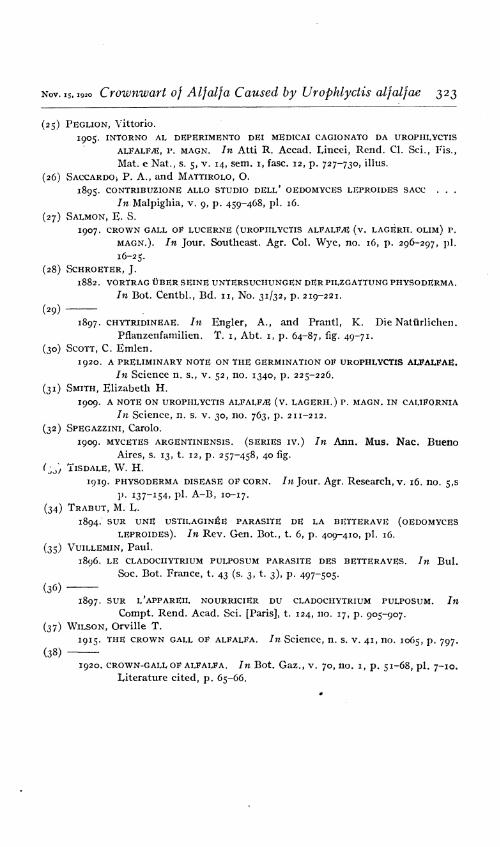
Nov. 15,1920 Crownwart of Alfalfa Caused by Urophlyctis alfalfae 323
(25) PEGUON, Vittorio. 1905. INTORNO AL DKPERIMENTO DEI MEDICA! CAGIONATO DA UROPHLYCTIS
AlVAhVM, P. MAGN. In Atti R. Accad. Lincei, Rend. Cl. Sei., Fis., Mat. e Nat., s. 5, v. 14, sem. 1, fase. 12, p. 727-730, illus.
(26) SACCARDO) P. A., and MATTIROLO, O. 1895. CONTRIBUZIONE ALLO STUDIO DELL* OEDOMYCES LEPROIDES SACC . . .
Jn-Malpighia, v. 9, p. 459-468, pi. 16.
(27) SALMON, K. S. 1907. CROWN GALL OF LUCERNE (UROPHLYCTIS ALFALM (V. LAGÉRH. OLIM) P.
MAGN.). In Jour. Southeast. Agr. Col. Wye, no. 16, p. 296-297, pi.
16-25.
(28) SCHROETER, J. 1882. VORTRAG ÜBER SEINE UNTERSUCHUNGEN DER PILZGATTUNG PHYSODERMA.
In Bot. Centbl., Bd. 11, No. 31/32, p. 219-221.
(29)
1897. CHYTRIDINEAE. In Engler, A., and Prantl, K. Die Natürlichen.
Pflanzenfamilien. T. 1, Abt. 1, p. 64-87, fig. 49-71. (30) SCOTT, C. Emlen.
1920. A PRELIMINARY NOTE ON THE GERMINATION OF UROPHLYCTIS ALFALFAE. In Science n. s., v. 52, no. 1340, p. 225-226.
(31) SMITH, Elizabeth H.
I909. A NOTE ON UROPHLYCTIS ALFALFA (v. LAGERH.) P. MAGN. IN CALIFORNIA
In Science, n. s. v. 30, no. 763, p. 211-212.
(32) SPEGAZZINI, Carolo.
1909. MYCETES ARGENTiNENSis. (SERIES iv.) In Ann. Mus. Nac Bueno
Aires, s. 13, t. 12, p. 257-458, 40 fig.
ry, TISDALE, W. H.
1919. PHYSODERMA DISEASE OF CORN. In Jour. Agr. Research, v. 16. no. 5,5
p. 137-154, pi- A-B, 10-17. (34) TRABUT, M. L.
1894. SUR UNE USTILAGINÉE PARASITE DE LA BETTERAVE (OEDOMYCES LEPROIDES). In Rev. Gen. Bot., t. 6, p. 409-410, pi. 16.
(35) VUILLEMIN, Paul. 1896. LE CLADOCHYTRIUM PULPOSUM PARASITE DES BETTERAVES. In Bul.
Soc. Bot. France, t. 43 (s. 3, t. 3), p. 497-505-
(36) 1897. SUR L'APPAREIL NOURRICIER DU CLADOCHYTRIUM PULPOSUM. /n
Compt. Rend. Acad. Sei. [Paris], t. 124, no. 17, p. 905-907. (37) WILSON, Orville T.
1915. THE CROWN GALL OF ALFALFA. In Science, n. s. v. 41, no. 1065, p. 797.
(38) 1920. CROWN-GALL OF ALFALFA. In Bot. Gaz., v. 70, no. 1, p. 51-68, pi. 7-10.
Literature cited, p. 65-66.

PLATE 47
Urophlyctis alfalfae:
Drawing of alfalfa plant, showing abundance of crownwart, as found early in May, 1919, in northern California. The dotted line gl represents the ground level. Varying degrees of abnormality in the development of the buds are shown. X H-
(324)

Crownwart of Alfalfa Caused by Urophlyct s alfalfae PLATE 47
9f
Journal of Agricultural Research Vol. XX, No. 4

Crownwart of Alfalfa Caused by Urophlyctis alfalfae PLATE 48
Journal of Agricultural Research Vol. XX, No. 4

PLATE 48
Urophlyctis alfaljae:
A-D.—Peripheral portions of actively growing thallus of parasite dissected from living host: ta, tb, tc, turbinate cells of successive orders; ra, rb, resting spores pro- duced by successive orders of turbinate cells; tbx, peripheral segments beginning to proliferate turbinate cells by budding. Note the single apical haustorium on the developing turbinate cells tc; its median position on the isthmus connecting turbinate cell tb and developing resting spore rb; its absence from isthmus between evacuated turbinate structure B, D, la, and maturing resting spore B, D, ra. Note also zonate arrangement of haustoria between equator and distil pole of resting spore, A-D, ra, rbs.
B.—Nearly mature resting spore viewed from distil side, showing 11 haustoria in zonate arrangement.
F.—Mature resting spore viewed from distil pole, showing 13 pits that mark former location of haustoria.
G.—Mature resting spore viewed in profile, showing pits in zonate arrangement and light concavity on proximal side of spore.
Drawn with the aid of the camera lucida. Approxiately X 847.

PLATE 49
Urophlyctis alfalfae:
A.—Section of epidermal region of young foliar structures, showing young primary turbinate cells ta-tg, the first products of infection, within epidermal cells. Note attachment to cuticular wall by attenuated beak, increase in number of fungus nuclei during growth of turbinate cells, and pathological enlargement of host nuclei hn, in invaded cell, hnx being normal host nucleus.
B.—Section of young foliar element, showing wall of invaded epidermal cell dis- rupted and advance of secondary turbinate cells tbc-tbe into underlying tissue. One of the other secondary turbinate cells, tbb, is forcing its way down along the host cell wall, while another, tba, has been reflected toward the cuticular wall; ta, sporogenous cell of primary turbinate structure.
C.—Section of turbinate cell, showing 3 evacuated peripheral segments pa-pc. A nucleus is passing through the narrow isthmus connecting the nearly evacuated sporo- genous cell with the resting spore, the elongated nucléole following the achromatin.
D.—Section of maturing resting spore, showing 8 nuclei and a central vacuole con- taining 4 granules staining red.
B.—Section of mature resting spore, showing numerous red-staining granules in center and 5 nuclei.
F.—Section of maturing resting spore, showing 11 normal nuclei and 4 enlarged nuclei in center, the latter apparently degenerating.
Drawn with the aid of the camera lucida. X 860.
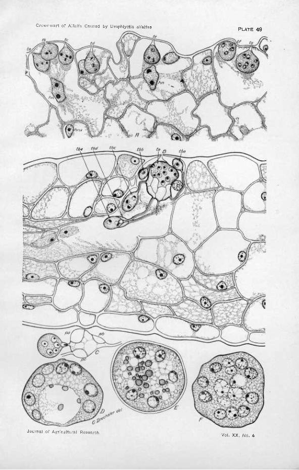
Crownwart of Alfalfa Caused by Urophlyctis alfalfas PLATE 49
Journal of Agricultural Research Vol. XX, No. 4

Crownwart of Alfalfa Caused by Urophlyctis alfalfae PLATE 50
Journal of Agricultural Research Vol. XX. No. 4

PLATE 50
Urophlyctis alfalfae:
Section of diseased bud scale of alfalfa, showing four coalescing cavities, in three of which the large primary turbinate cells taa, tba, and tc may be distinguished; taa has not started to proliferate any resting spore, while the resting spore produced by tba is moderately young, although turbinate cells of later orders tab, tbc, and others have produced resting spores further along in development. The thickening of the host cell walls bounding the cavity and the enlargement of the host nuclei hn lying free within the cavity are conspicuous. Note also the large dimensions of the nucleus in the uninuclcated turbinate cell tbbx and the relatively larger proportions of the nucléoles in the nuclei of the resting spore rs. Drawn with the aid of the camera lucida. X 860.

PLATE 51
Urophlyctis alfalfac:
Section of diseased bud scale attacked by U. alfalfae, showing a group of eight well- developed cavities a-h and their relation to the host tissue. Many of the cells adjacent to the cavities have divided at unusual angles, giving the tissue a characteristic appear- ance. In b the host cytoplasm and fungous material stain unusually deeply, as the result perhaps of general infdtration with some diiTusing substance. Drawn with the aid of the camera lucida. Approximately X 417

Crownwart of Alfalta Caused by Urophlyctis alfalfae PLATE 51
Journal of Agricultural Research Vol. XX, No. 4

Crownwart of AlfaKa Caused by Urophlyctis alfalfa« PLATE 52
Journal of Agricultural Research Vol. XX, No. 4

PLATE 52
A, C, D.—Urophlyctis pliiriannulatus. B.—Urophlyctis alfalfae.
A.—Portion of actively growing thallus of U. pluriannulatus dissected from gall on leaf of Sanícula menziesii, including a turbinate cell ta with a nearly mature resting spore ra; ta is completely evacuated, having produced 7 turbinate cells of the next order, in one of which tba peripheral segments have been delimited, another, tbb, has produced two turbinate cells of the tertiary order tea and tcb, as well as a developing resting spore rb. Approximately X 847.
B.—^Abnormally enlarged hyphae and turbinate cells of U. alfalfae, showing con- spicuous thickening of the walls. X 860.
C.—Peripheral portion of actively growing thallus of U. pluriannulatus, similar to A, showing 8 turbinate cells of the second order, of which 7 have produced turbinate cells of the last order as well as resting spores. Approximately X 847.
D.—Nearly mature resting spore of U. pluriannulatus, viewed from polar end, showing 22 haustoria in zonate arrangement. Approximately X 847.
Drawn with the aid of the camera lucida.

PLATE 53
Urophlyctis pluriannulatus:
Section of leaf of Sanícula menziesn, showing development of parasite within gall. Some of the fungus thallus appears to have dropped out of the section in the course of manipulations, as is indicated by the large unoccupied gaps; ta, primary turbinate cell ; tb, tc, turbinate structures or cells of successive orders, the former completely evacu- ated, the latter in early first or second nucleated stage; ra, rb, resting spores produced by turbinate cells of successive orders; hn, host nuclei considerably enlarged as result of influence of parasite. Note the similarity in development of parasite to U. alfalfae and the relatively slight influence of parasitism on host anatomy. Drawn with the aid of the camera lucida. X 860.

Crownwart of Alfalfa Caused by Urophlyctis alfalfas PLATE 53
Journal of Agricultural Research Vo!. XX, No. 4

Crownwart of Alfalfa Caused by Urophlyctis alfalfae PLATE 54
V?K ViW '" f ' '4TI
^1 i/i,
Journal of Agricultural Research Vol. XX, No. 4

PLATE) 54
Crowns of alfalfa plants bearing galls caused by Urophlyctis alfalfae photographed at different stages of development.
A.—A comparatively early stage of development at which the origin of the gall structures from the elements of developing buds can be traced.
B.—A later stage of development at which the origin of the tissue has become obscured. The tap root of this crown was cut off and the photograph taken from below. Galls usually become considerably larger than this before they begin to disintegrate if the plant continues vigorous growth.

PLATE ss
A comparatively early stage of host reaction to invasion by Urophlyctis alfalfae. The cavities produced by the invading fungus can still be traced from the exterior into the parenchymatous tissue. A few of the cells which are about to be entered by the advancing fungus show some hypertrophy. The division of the cells beneath the epidermis has begun.

Crownwart of Alfalfa Caused by Urophlyctis alfalfae PLATE 55
Journal of Agricultural Research Vol. XX, No. 4
Crownwart of Alfalfa Caused by Urophlyctis alfalfae PLATE 56
*■„ '• :.¡- ■". •'• * i fl
Journal of Agricultural Research Vol. XX, No. 4

PLATE 56
A.—Late stage of development of host reaction to the invasion of Uro ph lye its alfalfae. Infections have taken place near the tip of a growing point. The division of exterior cells has gone on extensively. The cells around the older portions of the cavity formed by the fungus have also been stimulated to division. The vascular bundle at the center of the mass of tissue has begun to produce parenchyma toward which the fungus is inclined to direct its course.
B.—^Vertical section through a well-developed gall near its central axis, showing its laminated structure arising from the thickening of bud elements. The cavities containing the dark-colored spore masses are seen distributed through the tissue, a condition that causes the mottled appearance of the interior of the living gall.
Copyright © 2022 FDOKUMEN