Synovial cytokine expression in ankle osteoarthritis depends on age and stage
Upload
independentCategory
view
1download
0
The
Journ
al o
f Exp
erim
enta
l M
edic
ine
ARTICLE
© 2008 GeurtsvanKessel et al.
The Rockefeller University Press $30.00
J. Exp. Med. Vol. 205 No. 7 1621-1634 www.jem.org/cgi/doi/10.1084/jem.20071365
1621
Infl uenza type A is a cytolytic virus that causes acute respiratory infection, of which the clinical outcome can vary greatly. The way in which the innate and adaptive immune systems ini-tially recognize and deal with replicating virus could be decisive in determining the outcome of infection, as this might heavily influence the kinetics of viral clearance ( 1 – 3 ). Little is known about the initial recognition event of infl uenza virus by the lung immune system in vivo .
In immediate response to viral infection, innate defense mechanisms consist of high level
production of type I IFNs by infected epithelial cells, alveolar macrophages, and natural IFN-producing cells (also known as preplasmacytoid DCs [pre-pDCs]), as well as the recruitment of conventional DCs (cDCs), neutrophils, and NK cells ( 4, 5 ). By expressing a wide array of microbial pattern recognition receptors shared with cells of the innate immune response, and at the same time displaying the potential to pro-cess and present antigen to naive T cells, DCs bridge innate and adaptive immunity. After rec-ognizing foreign antigen in the periphery of the body, DCs migrate via aff erent lymphatics into the draining LNs, where they can induce anti-gen-specifi c protective CD8 CTL responses, as well as CD4 T helper cells that enforce cellular and humoral immunity ( 6 ).
CORRESPONDENCE
Bart N. Lambrecht:
Abbreviations used: BAL, bron-
choalveolar lavage; cDC, con-
ventional DC; dpi, days
postinfection; DTR, diphtheria
toxin receptor; HA, hemagglu-
tinin; HI, HA inhibition; i.n.,
intranasal; i.t., intratracheal;
MFI, mean fl uorescence inten-
sity; MLN, mediastinal lymph
node; mPDCA-1, mouse pDC
antigen 1; NP, nucleoprotein;
pDC, plasmacytoid DC; Tg,
transgenic.
G.F. Rimmelzwaan and B.N. Lambrecht contributed equally
to this paper.
The online version of this article contains supplemental material.
Clearance of infl uenza virus from the lung depends on migratory langerin + CD11b � but not plasmacytoid dendritic cells
Corine H. GeurtsvanKessel , 1,2 Monique A.M. Willart , 3 Leonie S. van Rijt , 1 Femke Muskens , 1 Mirjam Kool , 1 Chantal Baas , 2 Kris Thielemans , 4 Clare Bennett , 5 Bj ö rn E. Clausen , 5 Henk C. Hoogsteden , 1 Albert D.M.E. Osterhaus , 2 Guus F. Rimmelzwaan , 2 and Bart N. Lambrecht 1,3
1 Department of Pulmonary Medicine and 2 Department of Virology, Erasmus University Medical Centre Rotterdam,
3015 GE Rotterdam, Netherlands
3 Department of Respiratory Diseases, University Hospital Ghent, 9000 Ghent, Belgium
4 Department of Physiology, Free University Brussels, 1090 Brussels, Belgium
5 Department of Cell Biology and Histology, University of Amsterdam, 1105 AZ Amsterdam, Netherlands
Although dendritic cells (DCs) play an important role in mediating protection against
infl uenza virus, the precise role of lung DC subsets, such as CD11b � and CD11b + conven-
tional DCs or plasmacytoid DCs (pDCs), in different lung compartments is currently un-
known. Early after intranasal infection, tracheal CD11b � CD11c hi DCs migrated to the
mediastinal lymph nodes (MLNs), acquiring co-stimulatory molecules in the process. This
emigration from the lung was followed by an accumulation of CD11b + CD11c hi DCs in the
trachea and lung interstitium. In the MLNs, the CD11b + DCs contained abundant viral
nucleoprotein (NP), but these cells failed to present antigen to CD4 or CD8 T cells, whereas
resident CD11b � CD8 � + DCs presented to CD8 cells, and migratory CD11b � CD8 � � DCs
presented to CD4 and CD8 T cells. When lung CD11c hi DCs and macrophages or
langerin + CD11b � CD11c hi DCs were depleted using either CD11c – diphtheria toxin receptor
(DTR) or langerin-DTR mice, the development of virus-specifi c CD8 + T cells was severely
delayed, which correlated with increased clinical severity and a delayed viral clearance.
120G8 + CD11c int pDCs also accumulated in the lung and LNs carrying viral NP, but in their
absence, there was no effect on viral clearance or clinical severity. Rather, in pDC-depleted
mice, there was a reduction in antiviral antibody production after lung clearance of the
virus. This suggests that multiple DCs are endowed with different tasks in mediating pro-
tection against infl uenza virus.
1622 DC SUBSETS IN INFLUENZA VIRUS INFECTION | GeurtsvanKessel et al.
present antigen to specifi c CD8 and CD4 T cells. Using genetic cell-specifi c targeting techniques, CD11c hi DCs and macro-phages or langerin + CD11c hi DCs could be depleted in CD11c – diphtheria toxin receptor (DTR) or langerin-DTR mice ( 36, 37 ), and pDCs could be depleted using mAbs. Collectively, our results demonstrate that diff erent DC subsets perform special-ized tasks during the primary encounter with infl uenza virus in the lung.
RESULTS
Lung DC subtypes and alveolar macrophages respond
to infl uenza virus infection differentially
As expected after i.n. inoculation with 5 × 10 5 tissue culture ID 50 infl uenza A virus X-31 (H3N2), there was induction of vigorous innate and adaptive immune responses in the lung. The innate cellular immune response was already present 1 day postinfection (dpi) and peaked at 4 dpi, exemplifi ed by a strong increase in neutrophils. B lymphocytes (CD19 + ) were present in the lung by day 4 and peaked around day 10. Total CD8 + cells in the lung peaked around day 10 (Fig. S1, available at http://www.jem.org/cgi/content/full/jem.20071365/DC1). To investigate the number of DC subsets and macrophages after infl uenza virus infection, enzymatically digested lungs (from which the large conducting airways were dissected) were analyzed at diff erent time points after infection by eight-color fl ow cytometry. Digested lung cells were fi rst gated for live leukocytes and expression of the panleukocyte marker CD45. cDCs were characterized as low fl uorescent cells, with a CD45 hi , MHCII hi CD11c hi phenotype (Fig. S2 A), and further subdivided into CD11b + or CD11b � cells ( 11, 15, 30, 13 ). As previously described, pDCs were small, low autofl uores-cent cells expressing a CD45 hi , CD11b lo , MHC int CD11c int mPDCA-1 + phenotype (Fig. S2 B) ( 24 ). Alveolar macrophages were identifi ed in the bronchoalveolar lavage (BAL) fl uid as highly fl uorescent cells with a CD11c hi , CD11b lo , MHCII lo , F4/80 hi phenotype (Fig. S2 C). The use of CD11c and CD11b expression in combination with the autofl uorescence typically seen in alveolar macrophages adequately discriminated alveolar DCs from macrophages, as previously described ( 38, 39, 11 ).
In the mock situation, 65% of CD11c hi DCs were CD11b � and 35% were CD11b + . After infection with infl uenza virus X-31, a signifi cant increase in both CD11b + ( Fig. 1 A ) and CD11b � ( Fig. 1 B ) lung DCs was seen from 2 dpi, and these DC subsets remained increased up to at least 10 dpi. The DCs expressed high levels of MHCII and up-regulated maturation markers such as CD86 ( Fig. 1, A and B ), CD40, CD80, and intercellular adhesion molecule 1 (not depicted). The in-crease in the expression level of these markers peaked shortly after inoculation (2 d) and gradually returned back to the lev-els seen in mock-infected mice ( Fig. 1, A and B , bar graphs). In contrast, the number of pDCs peaked shortly after infec-tion (2 dpi) and then returned to baseline conditions ( Fig. 1 C ). This increased recruitment of pDCs was accompanied by a distinct but very transient up-regulation of maturation mark-ers (CD86 is shown). Alveolar macrophages were studied in the BAL fl uid accessible by BAL. This compartment contains
In the lung, DCs are situated in immediate proximity to the respiratory epithelial cells, where they form an elaborate network that rapidly reacts to all kinds of foreign antigens and infl ammatory stimuli, including respiratory viruses ( 7 – 10 ). Diff erent DC subsets can be found in the lung, each with a functional specialization ( 11 – 15 ). In the mouse, all DC sub-sets express the integrin CD11c, and subsets are further de-fi ned based on the expression of the marker CD11b, as well as anatomical location ( 11, 13, 16 ). The trachea and large conducting airways have a well-developed network of in-traepithelial DCs, even in steady-state conditions ( 17, 18 ). These cells have been shown to express langerin and CD103 while lacking expression of CD11b ( 13, 10, 15 ). In the sub-mucosa of the conducting airways, CD103 � CD11b + CD11c + cDCs can be found, particularly under conditions of infl am-mation, and these cells might be particularly suited for priming and restimulating eff ector CD4 cells in the lung in response to protein antigens ( 19, 20, 15 ). The lung interstitium that is accessible by enzymatic digestion also contains CD11b + and CD11b � DCs that access the alveolar lumen and migrate to the mediastinal LNs (MLNs) ( 14, 11 ). In the nearby alveolar lumen, CD11c hi alveolar macrophages control the function of these interstitial DCs ( 21 ). pDCs are CD11b � CD11c int cells expressing SiglecH and bone marrow stromal antigen 2 (rec-ognized by the mAbs mouse pDC antigen 1 [mPDCA-1] or 120G8) ( 22, 23 ). In the lungs, pDCs are predominantly found in the lung interstitium and produce large amounts of IFN- � in response to triggering by CpG motifs or viral infection ex vivo ( 24, 5 ). Under particular conditions, lung pDCs can pre-vent immunopathology in response to inhalation of harmless antigens or in response to respiratory syncitial virus ( 24 – 26 ).
Several investigators have studied the involvement of APCs such as DCs, macrophages, and B cells in mediating protective immunity to infl uenza virus ( 27 ). It was previously shown that a mouse-adapted strain of infl uenza virus induced the in vivo maturation of CD11c + DCs in the lung ( 28 ) and their migration to the MLNs ( 29, 8, 2 ). DCs isolated ex vivo from the MLNs of infl uenza-infected mice presented viral nucleo-protein (NP)-derived peptides to virus-specifi c CD8 T cells in vitro for at least 10 d, a peak occurring � 72 h after infec-tion ( 27, 29 – 32 ). In humans, both monocyte-derived DCs and pDCs were shown to be capable of activating already primed infl uenza-specifi c T cells in vitro ( 33, 34 ) and upon adoptive transfer in vivo ( 35 ).
Although these studies certainly suggest the strong in-volvement of DCs in mediating protective immunity to in-fl uenza A virus, direct description of the functional in vivo involvement of the now well-described DC subsets in the diff erent compartments of the lung during early infection is lacking. In this paper, using eight-color fl ow cytometry, we have carefully studied the kinetics of the reaction of diff erent DC subsets and alveolar macrophages in various lung com-partments to intranasal (i.n.) infection with infl uenza virus X-31 (H3N2), focusing particularly on CD11b + and CD11b � subsets, as well as on pDCs, describing which subsets carry viral NP to the nodes, and carefully dissecting which subsets

JEM VOL. 205, July 7, 2008
ARTICLE
1623
strong decrease in the density of cells staining for MHCII, followed by a restoration to baseline density and morphol-ogy by 10 dpi. To better quantify these changes, eight-color fl ow cytometry was performed on tracheal digestions, dem-onstrating considerable heterogeneity in DCs at various time points after infection ( Fig. 2 B ). In the mock situation, all CD45 + MHCII + cells expressed CD11c, identifying them as DCs. The majority of these cells ( � 75%) were CD11b � and expressed the mucosal � E integrin marker CD103 ( Fig. 2 C ). At 2 dpi, this subset was depleted from the tracheal digest and a majority of cells now expressed CD11b but lacked CD103, which was most consistent with the phenotype of a freshly recruited monocyte-derived DC ( Fig. 2, B and C ). A tempo-rary increase in the expression level of CD86 was found on these CD11b + DCs at day 2 ( Fig. 2 B ). At 10 dpi, the subset balance between CD11b + and CD11b � DCs was almost re-stored to baseline, and cells again showed a highly dendritic morphology and CD86 expression returned to baseline levels.
a large amount of resident alveolar macrophages in mock situation. After infl uenza virus infection, there was a nonsig-nifi cant trend of increase in the alveolar macrophage number at 4 dpi ( Fig. 1 D ), accompanied by a temporary increase in CD86 and a more continual increase in MHCII expression (mean fl uorescence intensity [MFI] of 820 in mock and 2,769 at 10 dpi).
Response of DC subsets in the large conducting airways
to infl uenza virus infection
To examine the DC network in the large conducting airways, we stained in vivo fi xed and permeabilized tracheal whole mounts with an mAb to MHCII, revealing the presence of a highly developed network of DCs and demonstrating deli-cate dendritic processes in between bronchial epithelial cells ( Fig. 2 A ). After infection, there was an increase in MHCII + cells at 2 dpi, and cells had a more rounded appearance, mak-ing it diffi cult to quantify DC density. At 4 dpi, there was a
Figure 1. Number and surface phenotype of mouse lung CD11b + and CD11b � DCs, pDCs, and alveolar macrophages after infl uenza
infection. Populations were gated as shown in Fig. S2 (available at http://www.jem.org/cgi/content/full/jem.20071365/DC1) and as indicated by the gates
in the left column of each panel. (A) CD11b + DCs signifi cantly increased after infection and remained increased up to 10 dpi (left). CD86 expression was
plotted in histograms (right), with MFI shown in the graph (bottom). (B) The increase in CD11b � DCs is demonstrated in fl ow cytometric plots (left),
with absolute numbers shown in the graph (bottom). CD86 expression was up-regulated and plotted as MFI (C, bottom). pDCs increased signifi cantly
at 2 dpi and then returned to baseline. Recruitment of pDCs was accompanied by up-regulation of CD86 (right). (D) Alveolar macrophages slightly
increased in number, but CD86 expression was not increased. Gray histograms represent isotype controls and were measured at 4 dpi. The values
are representative of fi ve mice per group and are expressed as mean ± SEM. Similar results were obtained from at least three separate experiments.
*, P < 0.05; **, P < 0.01.

1624 DC SUBSETS IN INFLUENZA VIRUS INFECTION | GeurtsvanKessel et al.
that were further discriminated based on expression of the my-eloid marker CD11b ( Fig. 3 B ). After infection, there was an increase in the absolute amount of CD11c + MHCII + CD11b � and CD11c + MHCII + CD11b + DCs starting at 2 dpi, and the increase was more pronounced in the CD11b + subset. Within the CD11b � CD11c hi cells, there was a clear increase in cells coexpressing langerin and CD103 (30 × 10 4 cells at 4 dpi vs. 2 × 10 4 cells in mock-infected mice). Belz et al. previously suggested that a resident subset of CD11b � CD8 � + DCs was responsible for generating virus-specifi c CD8 CTLs, whereas the population of CD11b � CD11c + cells transported antigen from the periphery ( 30 ). Therefore, cells were further discriminated
As previously reported ( 11 ), we detected a minor percentage (0.35% of live tracheal CD45 + leukocytes) of mPDCA-1 + pDCs in tracheal digests of mock-infected mice ( Fig. 2 D ). Again, pDCs increased after infl uenza virus infection and tem-porarily up-regulated their expression of co-stimulatory mole-cules ( Fig. 2 E ).
Infl uenza virus infection increases DC subsets in MLNs
Upon recognition of antigen, lung DCs are known to migrate to the MLNs. We followed the kinetics of increase and ex-pression of maturation markers on various DC subsets within this node. cDC subsets were CD11c + MHCII + cells ( Fig. 3 A )
Figure 2. DC subtypes in tracheal tissue after infl uenza infection. (A) Tracheal whole mount sections stained for MHCII expression were per-
formed at various days after infection. Bar, 35 μ m. (B) Flow cytometric analysis of DCs in tracheal cell suspensions stained for CD45, CD11c, CD11b,
F4/80, and CD103. CD11c + cells contained two subsets, one CD11b � and the other CD11b + . Histograms represent CD86 expression on both subsets.
The dotted lines indicate MFI in a mock situation. (C) Absolute numbers of the two subsets after infl uenza infection expressed as mean values ± SEM.
*, P < 0.05; **, P < 0.01. Histograms represent CD103 expression on both subsets. (D) pDCs were identifi ed as CD11c int PDCA-1 + cells representing a
minor percentage of CD45 + leukocytes in tracheal cell suspensions and only temporarily expressing CD86 (histogram). (E) Absolute number of pDCs
at various days after infection expressed as mean values ± SEM. **, P < 0.01.

JEM VOL. 205, July 7, 2008
ARTICLE
1625
Figure 3. DC subsets in MLNs after infl uenza virus infection. (A) Kinetics of CD11c + MHCII + DCs demonstrating an almost fourfold increase after
infection. Percentages of living cells are shown. (B) Expression of CD11b by CD11c + DCs gated in A. The values are representative of fi ve mice per group
and are expressed as mean ± SEM. Similar results were obtained from separate experiments. (C) CD8 � expression on different DC subsets was demon-
strated in combination with CD11b, with percentages of the different populations plotted in the graph. The values are representative of fi ve mice per
group and are expressed as mean ± SEM. Similar results were obtained from separate experiments. *, P < 0.05. (D) CD86 expression on different MLN
subsets. The gray histogram represents isotype control, the dotted histogram represents mock controls, and the black histogram represents infected
animals. (E) Sorted DC subsets were stained for intracellular NP. Uninfected cells stain dull red because of Evan ’ s blue in the solution, and the green
fl uorescence indicates NP. Bar, 20 μ m. These images are representative of one experiment out of at least fi ve performed. (F) Flow cytometric analysis of
the total detectable amount of intracellular NP in DC subsets as a percentage of total live LN cells per DC subset. The values are representative of fi ve
mice per group and are expressed as mean ± SEM. Similar results were obtained from two separate experiments. (G) Plots represent CFSE-labeled T cell
proliferation 4 d after co-culture with the various sorted DC populations obtained from pooled LNs of 20 infl uenza-infected mice. (top) CD8 + T cells and
(bottom) CD4 + T cell proliferation are shown. Numbers (top left corners) represent the percentage of cells recruited into cell division. Cell sortings with
co-culture were performed three times each, and the results shown are representative plots of these experiments.

1626 DC SUBSETS IN INFLUENZA VIRUS INFECTION | GeurtsvanKessel et al.
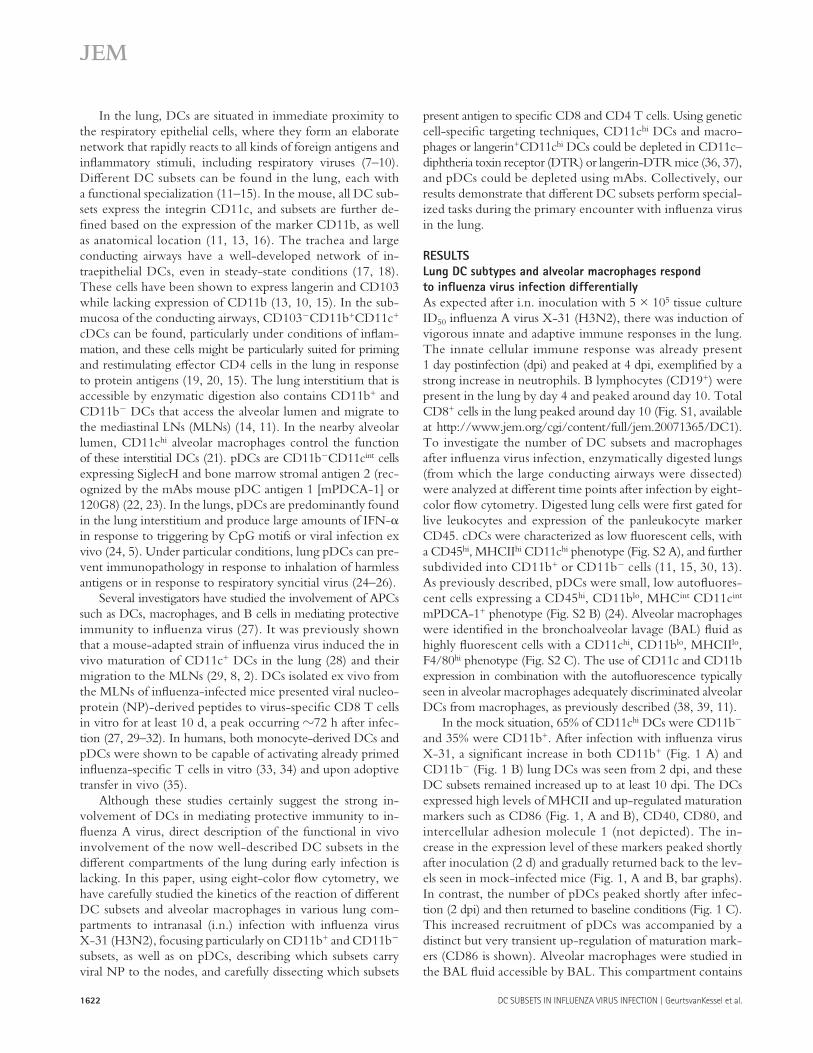
of infection was determined by measuring body weight. In general, mice show a maximal 10% weight loss after a mild X-31 infection and rapidly regain weight once the virus has been cleared from the lungs. When CD11c-DTR Tg mice were treated with DT before infection, the clinical sever-ity of infection dramatically increased and mice lost up to 20% of their weight, representing a sublethal infection. This weight loss could not be attributed to DT treatment, as the control group, which was treated with DT before mock in-fection, did not lose weight ( Fig. 4 B ). As weight loss might be related to the initial severity of infection or failure of the immune system to clear infectious particles from the lung, we next studied the generation of an effi cient immune re-sponse in CD11c hi – depleted mice. For effi cient lung clear-ance of a primary infl uenza virus infection, CD8 + CTLs play an important role. We therefore measured the number of NP 366-374 peptide/H-2D b – specifi c CTL cells using tetramer reagents and correlated their numbers to viral clearance. In DT-treated and -infected animals, the number of virus-specifi c CTLs was signifi cantly reduced in the lung, spleen ( Fig. 4 C ), and MLN (not depicted). In addition to the measurement of CTL, CD3-stimulated IFN- � production by CD4 and CD8 T cells from MLNs was determined and dramatically sup-pressed in the absence of DCs ( Fig. 4 D ).
As induction of virus-specifi c CTL responses is necessary for viral clearance from the lung, we measured viral titers in the lungs. In this mild infection model, virus is normally cleared completely at 8 dpi ( Fig. 4 E ) ( 42 ). However, in the absence of lung CD11c hi cells and mediastinal CD8 � + CD11c hi cells, the virus had not been cleared at this time.
A delay in viral clearance could be caused by a direct de-fect in CTL priming but could also result from a less effi cient innate response consisting mainly of type I IFN production. IFN- � has been described as an important antiviral cytokine that is mainly produced by epithelial cells and alveolar macro-phages, but also by pDCs and even myeloid DCs after in-fl uenza infection in vitro ( 3, 5 ). We therefore determined IFN- � production in the BAL fl uid at diff erent time points after infection in CD11c hi – depleted mice. In vivo , IFN- � was produced in the lungs after X-31 infection, but levels were not aff ected by CD11c hi cell depletion ( Fig. 4 F ).
The data in this paragraph show that lung CD11c hi cells are required for an effi cient immune response against infl uenza virus infection. However, as CD11c is also highly expressed by alveolar macrophages and these cells are also depleted by local i.t. administration of DT, we performed adoptive transfer reconstitution experiments ( 19, 37 ). Tg mice depleted of CD11c hi cells therefore received i.t. unpulsed wild-type CD11c hi DCs or alveolar macrophages at the moment of infection. DCs were capable of restoring the CTL response ( Fig. 4 G ), and as a consequence, viral clearance from the lung was com-plete by day 8 ( Fig. 4 G , right). Alveolar macrophages were not capable of restoring antiviral immunity or viral clearance. These experiments demonstrated that CD11c hi DCs are suf-fi cient for inducing an adequate immune response against in-fl uenza virus infection.
based on CD8 � ( Fig. 3 C ). Compared with mock-infected mice, at 4 dpi there was a generalized increase in all CD8 � � subsets (CD11b � and CD11b + ), whereas the resident CD8 � + CD11b � DCs were not increased. After infection, CD11c + MHCII + cells had higher levels of the CD86 maturation marker compared with mock-infected mice, which was con-sistent with their potential to prime CD8 T cell responses ( Fig. 3 D ), and this was found in all DC subsets.
To test the antigen-presenting potential of the various DC subsets in the MLNs, mice were infected with infl uenza virus encoding either the immunodominant OVA 257-264 K b – restricted MHCI epitope recognized by the OT-I TCR-transgenic (Tg) strain ( 40 ) or carrying the OVA 323-339 MHCII epitope recog-nized by the OT-II TCR-Tg strain ( 41 ), allowing us to probe presentation of sorted lung DC subsets to naive OVA-specifi c CD8 and CD4 T cells directly ex vivo. To have an indication of the uptake of viral antigen or virally infected apoptotic cells, preparations of sorted DC subsets from the MLNs were also stained for the presence of viral NP using a specifi c antibody ( Fig. 3 E ), and confi rmed using eight-color fl ow cytometric staining on permeabilized cells ( Fig. 3 F ). Viral NP was found, particularly in the CD11b + CD8 � � subset as well as abun-dantly in the pDC subset, and was practically absent from the CD11b � subsets ( Fig. 3 F ). When DC subsets were sorted and co-cultured with OVA-specifi c OTI cells ( Fig. 3 G , top) or OTII cells ( Fig. 3 G , bottom), the CD11b � CD8 � � subset presented antigen to both CD8 and CD4 T cells, whereas the CD11b � CD8 � + resident DCs presented exclusively to CD8 cells. Despite the fact that these cells had seen viral antigens ( Fig. 3, E and F , NP staining), the CD11b + DCs and pDCs did not present antigen to naive CD4 or CD8 T cells. As a control, OT-I or OT-II T cells incubated with total MLN DCs obtained from mice infected with the virus containing the OVA-MHCII or – MHCI epitope, respectively, failed to proliferate.
Conditional depletion of lung CD11c hi cells aggravates
features of infection
DCs are extremely potent APCs that are uniquely suited to prime naive T cells. To study the immune response against infl uenza virus infection with and without CD11c hi DCs, CD11c-DTR mice were treated with DT intratracheally (i.t.) 1 d before infection with infl uenza virus X-31. By this localized treatment, lung CD11c hi DCs (both CD11b + and CD11b � ) and alveolar macrophages were effi ciently depleted from the lungs ( Fig. 4 A ). In the trachea, the localized treatment with DT led to a reduction of CD11b + DCs, but not to depletion of CD11b � DCs (Fig. S3 A, available at http://www.jem.org/cgi/content/full/jem.20071365/DC1) or pDCs (not de-picted). In the MLNs, all DC subsets were partially depleted after DT treatment, with the biggest depletion occurring in the resident CD11b � CD8 � + DCs (Fig. S3 B) ( 19, 37 ). On the contrary, CD11c hi cells in the spleen or nondraining LNs were not aff ected (unpublished data) ( 19, 37 ). At diff er-ent time points after infection, the clinical immune response and viral replication were analyzed. First, clinical severity
JEM VOL. 205, July 7, 2008
ARTICLE
1627
Depletion of lung langerin + DCs aggravates
infection parameters
The conditional depletion of CD11c hi cells in CD11c-DTR
mice did not allow us to address the contribution of tracheal
CD11b � CD11c hi DCs, as neither tracheal CD11b-CD11c hi DCs nor their CD11b � progeny in the MLNs were depleted in CD11c-DTR mice. Therefore, we performed experiments in langerin-DTR mice. When DT was administered i.t. to
Figure 4. Infection parameters after conditional depletion of CD11c hi cells in a CD11c-DTR Tg mouse model. CD11c-DTR Tg mice received an i.t.
injection of DT on day � 1, followed by X-31 i.n. infection. (A) Effi cient depletion of lung CD11c hi cells by DT treatment compared with PBS treatment. (right)
Plots demonstrate fl ow cytometry data, and numbers indicate the percentage of live cells within each gate. (B) Body weight after infl uenza infection. A weight
loss of 20% represents a sublethal infection. (C) Virus-specifi c CTL response in spleen and lung as measured by Flu peptide /H-2D b tetramer (TM) staining. (D) IFN- �
levels in supernatants of MLN cell cultures restimulated with anti-CD3 antibody. (E) Viral titers measured in lung tissue after infl uenza X-31 infection. Virus is
normally completely cleared at 8 dpi. nd, nondetectable. (F) IFN- � levels in BAL fl uid. (G) Adoptive transfer of wild-type DCs and alveolar macrophages into
DT-treated CD11c-DTR Tg mice before viral infection. CD11c-DTR Tg mice were either treated with PBS or DT, and received either DCs or macrophages before
infl uenza infection. Numbers of Flu peptide /H-2D b – specifi c CTLs in spleen suspensions (left) and viral titer in lung tissue (right) are shown. The values are represen-
tative of fi ve mice per group and are expressed as mean ± SEM. Similar results were obtained from at least two separate experiments. *, P < 0.05; **, P < 0.01.

1628 DC SUBSETS IN INFLUENZA VIRUS INFECTION | GeurtsvanKessel et al.
reduction in the protein level of IFN- � in the BAL fl uid or in the mRNA level for IFN- � in lung tissue after the deple-tion of pDCs ( Fig. 6 F ).
The production of hemagglutinin (HA)-specifi c antibodies
depends on pDCs and not on CD11c hi DCs
The induction of antiviral CD8 T cell responses is only one aspect of adaptive immunity to infl uenza. We also measured the production of serum HA-specifi c antibodies at 8 and 28 dpi by measuring HA inhibition (HI) titers. Although there was no eff ect on the induction of CD8 responses after 120G8-mediated pDC depletion, virus-specifi c antibody titers mea-sured at both time points were signifi cantly reduced ( Fig. 7 A ). We found no evidence for 120G8 staining on lung B cells, making it unlikely that this would be a depleting eff ect of 120G8 on B cells directly (unpublished data). Antibody re-sponses were maintained in mice depleted of CD11c hi DCs by DT treatment of CD11c-DTR mice 1 d before infection in CD11c-DTR mice ( Fig. 7 B ).
DISCUSSION
Belz et al. have elegantly described that after infl uenza in-fection, both CD11b � CD8 � + resident MLN DCs and the CD11b � CD8 � � lung-derived migratory DC subset pre-sented antigen to naive CD8 + T cells ex vivo ( 30, 32 ). How-ever, these authors have not described in detail where exactly in the lung these CD11b � migratory DCs originated from
these mice, there was a strong reduction in CD11b � CD11c hi DCs in the trachea, whereas tracheal CD11b + DCs were un-aff ected. In the lungs, CD11c hi DCs were depleted, whereas alveolar macrophages were unaff ected. The CD8 � + CD11b � resident MLN cDC subset does not express langerin, and consequently, lung administration of DT to these mice only led to a reduction in lung-derived CD11b � CD8 � � migra-tory DCs (Fig. S3). Depletion of langerin + DCs resulted in a severe weight loss in the mice until 8 dpi ( Fig. 5 B ), and this was correlated with a signifi cant decrease in CTL response ( Fig. 5 C ) and a defi cient viral clearance at 8 dpi ( Fig. 5 D ).
Depletion of pDCs did not alter the course of infection
The experiments using CD11c-DTR and langerin-DTR mice mainly depleted CD11c hi cells, whereas CD11c int pDCs are globally not aff ected by this targeting strategy ( 43 ). To additionally address the role of pDCs, we performed ex-periments in which pDCs were depleted by injection of the 120G8 mAb ( 22 ). Using this antibody, an eff ective deple-tion of pDCs to < 10% of baseline numbers was achieved in the lung ( Fig. 6 A ), as measured using antibodies directed against both bone marrow stromal antigen and SiglecH ( 24 ). Surprisingly, this effi cient depletion of pDCs did not aff ect any of the infection parameters and virus was cleared effi ciently by 8 dpi ( Fig. 6, B – E ). As pDCs have been previously de-scribed as the main IFN- � – producing cells in response to a viral infection ( 44 ), it was similarly striking that there was no
Figure 5. Effect of conditional depletion of langerin + DCs cells during infl uenza infection. Langerin-DTR Tg mice received an i.t. injection of DT
on day � 1, followed by X-31 i.n. infection. (A) Effi cient depletion of MHCII + CD11c DCs in lung after DT treatment. Numbers indicate percentages of live
cells within the gate. (B) Body weight after infl uenza infection. A weight loss of > 20% represents a sublethal infection. (C) Virus-specifi c CTL response in
spleen and lung measured by Flu peptide /H-2D b tetramer (TM) staining. (D) Viral titers measured in lung tissue after infl uenza X-31 infection. Virus is nor-
mally cleared completely at 8 dpi. The values are representative of fi ve mice per group and are expressed as mean ± SEM. Similar results were obtained
from at least two separate experiments. *, P < 0.05; **, P < 0.01. nd, nondetectable.

JEM VOL. 205, July 7, 2008
ARTICLE
1629
and into the lung interstitium. These cells most likely diff er-entiate from recruited Ly6C + monocytes that give rise to infl ammatory-type DCs ( 16, 45 ), or that arose from local proliferation and diff erentiation of a myeloid precursor popula-tion that also generates alveolar macrophages ( 46 ). An identi-cal subset of CD11b + CD11c + DCs was found to be increased in the MLNs from days 2 to 4 onwards, again suggesting migration from the lungs to the MLN.
It was striking that there were several subsets of DCs found to be recruited and/or activated in the MLNs after in-fl uenza infection, strongly suggesting the division of labor between various APCs. Recently, several papers have shown that the cross presentation of exogenous harmless or viral antigen to CD8 + lymphocytes or presentation of exogenous antigen to CD4 T cells is a mutually exclusive function of CD8 � + or
and whether any of the DCs would also present to CD4 + T cells, and they have also not studied in detail the precise con-tribution of infl ammatory CD11b + DCs or pDCs that are re-cruited to infl amed lungs.
Our studies on the various anatomical lung compartments suggest that a predominant source of CD11b � DCs arriving in the MLNs are from the network of highly dendritic-shaped CD103 + intraepithelial DCs that lines the large conducting airways ( Fig. 1 ) ( 18, 13 ). There was a marked decrease in this subset of highly dendriform tracheal CD11b � CD103 + DCs at 2 dpi, at a time that the langerin + CD103 + CD11b � CD8 � � DCs started to accumulate in the MLNs. The disappearance of CD11b � CD11c + tracheal DCs was accompanied by a new infl ux of CD11b + CD11c + DCs into the trachea (present as more rounded cells at day 2 after infection on whole mounts)
Figure 6. Infection parameters after infl uenza in mice depleted of pDCs. Mice received three i.p. injections of the depleting mAb 120G8 before
infection with infl uenza on day 0. (A) Effi cient depletion of lung pDCs after 3 d of 120G8 i.p. treatment compared with PBS treatment. (B) Body weight
after infection. (C) Virus-specifi c CTL responses in spleen and lung cell suspensions, as measured by Flu peptide /H-2D b tetramer staining. (D) IFN- � levels in
supernatants of MLN cell cultures restimulated with anti-CD3 antibody. (E) Viral titers in lung after infl uenza infection. (F) IFN- � levels in BAL fl uid. The
values are representative of fi ve mice per group and are expressed as mean ± SEM. Similar results were obtained from at least two separate experiments.
*, P < 0.05; **, P < 0.01.

1630 DC SUBSETS IN INFLUENZA VIRUS INFECTION | GeurtsvanKessel et al.
Our data on viral NP staining suggest that the CD11b + DCs as well as the pDCs were abundantly positive for viral antigen in the MLNs and, therefore, could be the most important source for providing viral antigens to resident CD8 � + DCs.
Another striking observation was the fact that the CD11b + CD11c + subset found to be increased in the trachea, lung, and MLNs after infl uenza infection hardly presented any viral antigen to CD4 or CD8 T cells. Clearly, the CD11b + DCs had seen viral antigens (either directly or through phagocytosis of virally infected apoptotic epithelial cells) as they carried an abundant amount of viral NP in their cytoplasm. The absence of APC function by this subset is in striking contrast to the situation when harmless noninfl ammatory antigen is inhaled ( 15 ). What could be the purpose of recruitment of a diff erent DC subset, in addition to CD11b � DCs, if these cells do not present antigen to naive T cells? First, as these cells have been shown to massively produce infl ammatory chemokines, CD11b + DCs might be crucial in attracting eff ector CD4 and CD8 cells that have been generated in the LNs back to the lung and trachea, where they would mediate eff ector function ( 51 ). Second, CD11b + DCs might have direct innate antiviral activity by producing TNF- � and inducible NO synthase – dependent NO, analogous to the situation seen with Listera monocytogenes infection ( 52, 53 ). Third, recruited CD11b + DCs might also stimulate the innate antiviral activity of NK cells. The important function of infl ammatory DCs in initiat-ing the innate response is supported by the fact that infl uenza produces the nonstructural protein 1, with a specifi c aim to subvert the innate immune function of the CD11b + DC subset ( 54, 3 ). Future studies in our laboratory will have to address the direct or indirect innate and adaptive functions of infl am-matory CD11b + DCs in infl uenza infection.
Another way to study the function of lung DC subsets is to deplete them using various cell-specifi c genetic targeting strategies using expression of the DTR under control of a specifi c promoter and administration of the DT via the air-ways ( 19 ). In CD11c-DTR mice, there is a predominant de-pletion of lung CD11c hi DCs, of tracheal CD11b + CD11c hi DCs, and of resident MLN CD8 � + CD11c hi DCs. Because CD11b � CD11c hi migratory tracheal DCs are not depleted in these mice, we consequently observed no decrease in MLN CD11b � CD8 � � DCs. In CD11c-DTR mice given DT, we noticed that generation of virus-specifi c CD8 + CTLs and production of eff ector cytokines (IFN- � ) by MLN cells was severely diminished. Based on our antigen presentation stud-ies and the previous work of Belz et al. ( 30 ), we propose that this is caused by depletion of the resident CD8 � + CD11c hi DCs or to the depletion of chemokine-producing lung CD11b + DCs. CD11c hi alveolar macrophages are also depleted in CD11c-DTR mice, although it is unlikely that this contrib-uted to a decrease in antiviral immunity, as adoptive transfer of wild-type DCs but not macrophages restored immunity.
To address the specifi c role of migratory tracheal CD11b � DCs, we performed experiments in langerin-DTR mice ( 36 ). Langerin was found to be particularly present on mucosal CD11b � CD103 + DCs in the lung ( 13 ), a population of cells
CD8 � � DC subsets, respectively ( 47 – 49 ). We therefore per-formed a head-to-head comparison of the potential to present viral antigen to naive CD4 or CD8 T cells, taking advantage of infl uenza virus encoding either the MHCI or MHCII immuno-dominant OVA epitope.
As previously described by Belz et al., CD8 � + CD11b � resident DCs presented viral antigen to naive CD8 cells, sup-ported by the fact that these cells had up-regulated co-stimu-latory molecules. Strikingly, the population of airway-derived CD11b � CD8 � � DCs also up-regulated co-stimulatory mol-ecules and presented not only to CD8 T cells but also to CD4 T cells. This suggests that processing for and presentation on both MHCI and MHCII molecules can occur in a single cell population in vivo in the lung, contrary to what was shown for the spleen or lymph nodes ( 30 ). Vermaelen et al. also pre-viously demonstrated that antigen presentation of harmless antigen to naive CD4 T cells in the MLNs was an exclusive function of a migratory DC population ( 50 ). Despite the fact that both CD8 � + and CD8 � � CD11b � DCs presented anti-gen to naive T cells, the strength of viral NP staining was not abundant. This could be explained by the fact that viral NP was digested in these subsets as part of an antigen-processing step, leading to a loss of immunoreactivity toward the NP-specifi c antibody. Alternatively, Belz et al. previously sug-gested that resident CD8 � + cells acquired the antigen from other migratory APCs, proposed as the CD11b � CD8 � � lung-derived DCs, but this was never directly demonstrated ( 30 ).
Figure 7. Virus-specifi c serum antibodies of infl uenza virus – infected
mice after DC subset depletion. (A) Virus-specifi c antibodies in the
serum of C57BL/6 mice after i.p. treatment with 120G8 depleting anti-
body or PBS, or (B) in the serum of CD11c-DTR mice after i.t. treatment
with DT or PBS, measured at 8 dpi and 1 mo after infection. The values
are representative of at least fi ve mice per group and are expressed as
geometric mean titer (GMT) of HI ± SEM. The differences in HI titer can
be explained by the background of the mice. CD11c-DTR mice were
F1 (BALB/c × C57BL/6) animals and developed less high viral and HI titers
than the pure C57BL/6 mice that were used in 120G8 depletion experiments.
Similar results were obtained from at least two separate experiments.
*, P < 0.05; **, P < 0.01.

JEM VOL. 205, July 7, 2008
ARTICLE
1631
MATERIALS AND METHODS Mice. 6 – 8-wk-old C57BL/6 mice were purchased from Harlan. The
generation and screening of CD11c-DTR Tg mice has been previously
described ( 37 ). Male BALB/c background CD11c-DTR Tg (H2-D d )
mice were crossed to C57BL/6 (H2-D b ), to obtain F1 progeny, to allow
detection of the H2-D b tetramer. CD11c hi cells were depleted in CD11c-
DTR × C57BL/6 Tg mice by i.t. injection of 50 ng DT, a dose previ-
ously determined by titration ( 15 ). pDCs were depleted by i.p. injection
of pDC-selective depleting 120G8 antibody ( 22 ). All experiments were ap-
proved by an independent animal ethics committee of Erasmus Medical
Centre Rotterdam.
Infl uenza virus infection. Infl uenza virus X-31 (Medical Research
Council) was inoculated in the allantoic cavity of 11-d-old embryonated
chicken eggs. The allantoic fl uid was harvested after 2 d. Infectious vi-
rus titers were determined in Madin-Darby canine kidney (MDCK) cells
(NBL-2; American Type Culture Collection), as described previously ( 60 ).
Virus titers were obtained at days 4 and 8 after infection. Lungs were stored
at � 70 ° C. Lungs were homogenized with a homogenizer (Polytron; Kine-
matica AG) in infection medium (Eagle ’ s minimal essential medium), 0.3%
BSA (fraction V), 4 μ g/ml trypsin, 2 mM l -glutamine, 100 U/ml penicillin,
100 μ g/ml streptomycin, 0.15% NaHCO 3 , 20 mM Hepes, and 0.1 mM
of nonessential amino acids. 10-fold serial dilutions of these samples were
used 8-fold to determine the virus titers in MDCK cells, as described
previously ( 60 ). For antigen presentation assays of LN DCs, mice were in-
fected with WSN infl uenza virus encoding the OVA 257-264 K b – restricted
MHCI epitope in neuraminidase ( 40 ), and with X-31 infl uenza virus en-
coding the OVA 323-339 MHCII epitope in HA of the virus ( 41 ). The OVA
viruses were provided by R. Webby (St. Jude Children ’ s Research Hospital,
Memphis, TN).
Flow cytometry. For detection and phenotyping of DCs at days 2, 4, and
10 after infection, single-cell suspensions of MLNs and lung samples were
prepared as described previously ( 24 ). Trachea was digested in collagenase
solution for 1 h at 37 ° C to promote the release of DCs. Cells were subse-
quently stained with mAbs directed against MHCII FITC, intracellular NP
FITC, CD86 PE, CD11c PE Texas red, CD45 PECy5, CD103 PECy7, in-
tracellular langerin APC, mPDCA-1 APC, F4/80 APC Cy7, CD11b Pacifi c
blue, and a fi xable live/dead marker in Aqua (Invitrogen). Acquisition of eight
to nine color samples was performed on a cytometer (FACSAria) equipped
with FACSDiva software (both from BD Biosciences). The fi nal analysis and
graphical output were performed using FlowJo software (Tree Star, Inc.).
Cell sorting of DC subsets from MLNs was performed on a FACSAria.
The purity of sorted populations was > 95%.
Immunofl uorescence on sorted DC subsets. Sorted cell populations
were spotted on microscope slides, dried, and fi xed in acetone. Subsequently,
the slides were incubated with FITC-labeled infl uenza A NP-specifi c anti-
body (IMAGEN infl uenza virus; Dako) at 37 ° C for 15 min. The slides were
washed twice with PBS and once with distilled water, dried, and embedded
into a glycerol – PBS solution (Citifl uor; UKC Chemlab). Uninfected cells
stain dull red because of the Evan ’ s blue in the solution. Green and yellow
signals indicate the presence of NP. Fluorescence was scored using a fl uores-
cence microscope (LSM 510; Carl Zeiss, Inc.).
Analysis of T cell proliferation. OT-1 and OT-2 Tg T cells were iso-
lated from spleens and LNs of respective mice, enriched by MACS cell sort-
ing with anti-CD8 or -CD4 antibodies according to manufacturer ’ s protocol
(Miltenyi Biotec), and labeled with CFSE ( 61 ). Sorted DC subsets were co-
cultured with T cells in a v-bottom plate at a 1:10 ratio for 4 d. T cell divi-
sions were measured by fl ow cytometry. The percentage of cells recruited
into each cell division was calculated by dividing the number of individual
cells by CFSE content, as previously described ( 61 ), using the formula 100 ×
{ 1 � [ n 0 /( n 0 + n 1 /2 + n 2 /4 + n 3 /8 + n 4 /16 + n 5 /32 + n 6 /64 + n 7 /128)] } to
correct for the multiplying eff ect of division.
that strictly relies on CCR7 to migrate to the MLNs ( 15 ). In accordance, we also found increased numbers of langerin + CD103 + CD11b � CD11c + DCs in the MLNs after infl uenza infection. After i.t. administration of DT, there was a selec-tive depletion of CD11b � CD8 � � migratory DCs in the tra-chea and MLNs, while the CD8 � + resident LN population or alveolar macrophages were unaff ected. In the absence of the migratory langerin + CD11b � DC population, infl uenza ran a particularly severe course. Strikingly, these data also suggest that the langerin + DCs of the lung are much more immuno-genic than their langerin + Langerhans cell counterparts in the skin ( 55, 36, 56 ).
Finally, we also addressed the in vivo function of lung pDCs, previously known as natural IFN- � – producing cells ( 44 ). In humans, these cells produce copious amounts of IFN- � when exposed to infl uenza virus in vitro ( 57 ) and have been shown to stimulate already-primed infl uenza-specifi c CD4 and CD8 T cells ( 34 ). It has therefore been suggested that upon proper stimulation, pDCs develop into bona fi de APCs that primar-ily stimulate antiviral immune response ( 58 ). To address the antigen-presenting function of pDCs, we sorted pDCs from the MLNs of infected mice and found no evidence for presen-tation to either CD4 or CD8 T cells ex vivo, despite the fact that the cells contained copious amounts of intracellular viral NP. Previous studies with other respiratory viruses have sug-gested that pDCs have a more immunoregulatory role, which is crucial for preventing excessive immune activation and immuno-pathology ( 12, 24, 25 ). Although pDCs were attracted to the lungs and tracheal wall during infl uenza infection, their con-fi rmed depletion using 120G8 antibody did not aff ect viral titers, generation of virus-specifi c T cells, or the severity of in-fection, arguing against a predominant role for pDCs as APCs, immunoregulatory cells, or innate immune cells in this mild infection model. It was similarly striking to see that there was no eff ect on IFN- � production when pDCs were eff ectively depleted. Infections in epithelial surfaces that bathe in IFN- � (produced by epithelial cells) might be less dependent on IFN- � production by pDCs. Kumagai et al. recently demon-strated that during lung infection with RNA viruses, alveolar macro phages are a predominant source of type I IFN, and that pDCs only start producing IFN- � when macrophages are depleted from the lungs ( 5 ). The only eff ect of treatment with the 120G8 antibody was a signifi cant reduction of the titer of virus-specifi c HI antibodies at 8 and 28 dpi, signifying a possible role for pDCs in stimulating humoral antiviral im-munity, as previously suggested ( 59 ). Although we found no evidence for 120G8 staining on lung B cells after infl uenza infection, treatment with 120G8 antibody could also deplete plasma cells directly and in this way reduce HI antibody titers. Therefore, we are awaiting more specifi c (e.g., genetic) pDC targeting strategies to address this point further. In conclusion, our paper demonstrates a division of labor between diff erent DC subsets during pulmonary infl uenza infection, knowledge that might be used for the design of better infl uenza vaccines and could increase our understanding of why particular strains of infl uenza are more pathogenic than others.

1632 DC SUBSETS IN INFLUENZA VIRUS INFECTION | GeurtsvanKessel et al.
Erasmus University Rotterdam fellowship, and B.N. Lambrecht is supported by VIDI
and VIRGO grants from the Netherlands Organisation for Scientifi c Research, as
well as by an Odysseus Grant from the Flemish government. M. Kool is supported by
a grant from the Dutch Asthma foundation. B.E. Clausen is the recipient of a VIDI
grant and a career development grant from the Landsteiner Foundation for Blood
Transfusion Research.
The authors have no confl icting fi nancial interests.
Submitted: 3 July 2007
Accepted: 21 May 2008
REFERENCES 1 . Eichelberger , M. , W. Allan , M. Zijlstra , R. Jaenisch , and P.C. Doherty .
1991 . Clearance of infl uenza virus respiratory infection in mice lacking class I major histocompatibility complex-restricted CD8 + T cells. J. Exp. Med. 174 : 875 – 880 .
2 . Legge , K.L. , and T.J. Braciale . 2005 . Lymph node dendritic cells control CD8+ T cell responses through regulated FasL expression. Immunity . 23 : 649 – 659 .
3 . Pichlmair , A. , O. Schulz , C.P. Tan , T.I. Naslund , P. Liljestrom , F. Weber , and C. Reis e Sousa . 2006 . RIG-I-mediated antiviral responses to single-stranded RNA bearing 5 � -phosphates. Science . 314 : 997 – 1001 .
4 . Tecle , T. , M.R. White , and K.L. Hartshorn . 2005 . Innate immunity to infl uenza A virus infection. Current Respir. Med. Rev. 1 : 127 – 145 .
5 . Kumagai , Y. , O. Takeuchi , H. Kato , H. Kumar , K. Matsui , E. Morii , K. Aozasa , T. Kawai , and S. Akira . 2007 . Alveolar macrophages are the primary interferon-alpha producer in pulmonary infection with RNA viruses. Immunity . 27 : 240 – 252 .
6 . Banchereau , J. , and R.M. Steinman . 1998 . Dendritic cells and the con-trol of immunity. Nature . 392 : 245 – 252 .
7 . McWilliam , A.S. , D.J. Nelson , J.A. Thomas , and P.G. Holt . 1994 . Rapid dendritic cell recruitment is a hallmark of the acute infl ammatory response at mucosal surfaces. J. Exp. Med. 179 : 1331 – 1336 .
8 . Legge , K.L. , and T.J. Braciale . 2003 . Accelerated migration of respira-tory dendritic cells to the regional lymph nodes is limited to the early phase of pulmonary infection. Immunity . 18 : 265 – 277 .
9 . Lambrecht , B.N. , J.B. Prins , and H.C. Hoogsteden . 2001 . Lung den-dritic cells and host immunity to infection. Eur. Respir. J. 18 : 692 – 704 .
10 . Vermaelen , K. , and R. Pauwels . 2005 . Pulmonary dendritic cells. Am. J. Respir. Crit. Care Med. 172 : 530 – 551 .
11 . von Garnier , C. , L. Filgueira , M. Wikstrom , M. Smith , J.A. Thomas , D.H. Strickland , P.G. Holt , and P.A. Stumbles . 2005 . Anatomical loca-tion determines the distribution and function of dendritic cells and other APCs in the respiratory tract. J. Immunol. 175 : 1609 – 1618 .
12 . de Heer , H.J. , H. Hammad , M. Kool , and B.N. Lambrecht . 2005 . Dendritic cell subsets and immune regulation in the lung. Semin. Immunol. 17 : 295 – 303 .
13 . Sung , S.S. , S.M. Fu , C.E. Rose Jr ., F. Gaskin , S.T. Ju , and S.R. Beaty . 2006 . A major lung CD103 (alphaE)-beta7 integrin-positive epithelial dendritic cell population expressing Langerin and tight junction pro-teins. J. Immunol. 176 : 2161 – 2172 .
14 . Wikstrom , M.E. , and P.A. Stumbles . 2007 . Mouse respiratory tract dendritic cell subsets and the immunological fate of inhaled antigens. Immunol. Cell Biol. 85 : 182 – 188 .
15 . del Rio , M.-L. , J.-I. Rodriguez-Barbosa , E. Kremmer , and R. Forster . 2007 . CD103 � and CD103+ bronchial lymph node dendritic cells are specialized in presenting and cross-presenting innocuous antigen to CD4+ and CD8+ T cells. J. Immunol. 178 : 6861 – 6866 .
16 . Shortman , K. , and S.H. Naik . 2007 . Steady-state and infl ammatory dendritic-cell development. Nat. Rev. Immunol. 7 : 19 – 30 .
17 . Sertl , K. , T. Takemura , E. Tschachler , V.J. Ferrans , M.A. Kaliner , and E.M. Shevach . 1986 . Dendritic cells with antigen-presenting capability reside in airway epithelium, lung parenchyma, and visceral pleura. J. Exp. Med. 163 : 436 – 451 .
18 . Lambrecht , B.N. , B. Salomon , D. Klatzmann , and R.A. Pauwels . 1998 . Dendritic cells are required for the development of chronic eosinophilic airway infl ammation in response to inhaled antigen in sensitized mice. J. Immunol. 160 : 4090 – 4097 .
Tracheal whole mount staining. Animals were anesthetized with a lethal
dose of Nembutal (Sanofi ) and perfused in vivo with 1% paraformaldehyde
fi xative in PBS, pH 7.4, for 2 min through the ascending aorta ( 18 ). Tra-
cheas were removed, opened by a midline incision, and pinned fl at in sili-
cone-coated Petri dishes. After permeabilization in PBS containing 0.3%
Triton X-100 (Sigma-Aldrich), tissues were preblocked with rabbit serum,
incubated with M5/114 mAb (rat IgG2, anti – I-A, and I-E b,d,k ; Boehringer
Mannheim) for 36 h at room temperature, washed, and incubated with per-
oxidase-conjugated rabbit anti – rat IgG, followed by incubation for 20 min
in 0.05% diaminobenzidine in Tris-buff ered saline, pH 7.6. Tissues were
dehydrated in serial alcohol steps and cleared in toluene. The entire trachea
was mounted in DPX mounting medium.
Detection of virus-specifi c CTL by tetramer-staining. Single-cell
suspensions of lung and spleen samples were prepared as described previ-
ously ( 24 ). Red blood cells were removed using erythrocyte lysis buff er
(Roche). The cells were washed with 0.5% BSA in PBS and stained for
20 min at room temperature with the following antibodies: CD3e-PerCP
and CD8b.2-FITC (BD Biosciences), TO-PRO 3 – APC (Invitrogen), and
PE-labeled H-2D b tetramer with the NP 366 – 374 epitope ASNENMETM
(Sanquin Research) ( 62 ).
Eff ector cytokine production. On day 4 after infection, single-cell sus-
pensions of lung draining LNs were prepared. Cells were cultured in
RPMI 1640 medium with 5% FCS at a concentration of 2 × 10 6 cells/ml
in the presence of either 1 μ g/ml of plate-bound anti-CD3 (BD Biosci-
ences) or medium alone and incubated at 37 ° C. After 4 d, supernatants
were collected and stored at – 20 ° C until ELISA for IFN- � (BD Biosci-
ences) was performed. IFN- � ELISA (PBL Biomedical Laboratories) was
performed on BAL fl uid on several days after infection. Quantitative RT-
PCR for IFN- � was performed on homogenized lungs on day 4 after
infection (Assay-On-Demand; Applied Biosystems).
Generation of bone marrow – derived DCs. Bone marrow cells were
cultured for 9 d in DC culture medium (RPMI 1640 containing glutamax-I;
Invitrogen) supplemented with 5% (vol/vol) FCS (Sigma-Aldrich), 50 μ M
2-ME (Sigma-Aldrich), 50 μ g/ml gentamicin (Invitrogen), and 20 ng/ml
recombinant mouse GM-CSF, as previously described ( 15 ).
Adoptive transfer. Adoptive transfer reconstitution experiments were per-
formed in DT-treated CD11c-DTR × C57BL/6 mice. At the moment of
infection, they were treated i.t. with 2 × 10 6 bone marrow – derived DCs and
2 × 10 5 alveolar macrophages, as previously described ( 19 ).
Detection of virus-specifi c antibodies in serum. After treatment with
cholera fi ltrate and heat inactivation at 56 ° C, the serum samples were tested
for the presence of anti-HA antibodies. For this purpose, an HI assay was
used according to the standard protocol of 1% turkey erythrocytes and 4 HA
U of H3N2 infl uenza virus ( 63 ).
Statistical analysis. All experiments were performed using 5 – 10 animals
per group. The diff erence between groups was calculated using the Mann-
Whitney U test for unpaired data (Prism software version 4.0; GraphPad
Software, Inc.). Diff erences were considered signifi cant when P < 0.05.
Online supplemental material. Fig. S1 shows time kinetics of neutro-
phils, B cells, and CD8 + T cells in the lung after infl uenza infection. Fig. S2
shows the gating strategy for myeloid DCs (A), pDCs (B), and alveolar macro-
phages (C). Fig. S3 shows the depletion of DC subsets in the tracheas (A)
and MLNs (B) in CD11c-DTR (left) and langerin-DTR (right) mice. Online
supplemental material is available at http://www.jem.org/cgi/content/
full/jem.20071365/DC1.
C.H. GeurtsvanKessel is supported by a Viral Genomics (VIRGO) grant from the
Netherlands Organisation for Scientifi c Research. L..S. van Rijt is supported by an

JEM VOL. 205, July 7, 2008
ARTICLE
1633
37 . Jung , S. , D. Unutmaz , P. Wong , G. Sano , K. De los Santos , T. Sparwasser , S. Wu , S. Vuthoori , K. Ko , F. Zavala , et al . 2002 . In vivo depletion of CD11c(+) dendritic cells abrogates priming of CD8(+) T cells by exogenous cell-associated antigens. Immunity . 17 : 211 – 220 .
38 . Vermaelen , K. , and R. Pauwels . 2004 . Accurate and simple discrimi-nation of mouse pulmonary dendritic cell and macrophage popula-tions by fl ow cytometry: methodology and new insights. Cytometry A . 61 : 170 – 177 .
39 . van Rijt , L.S. , H. Kuipers , N. Vos , D. Hijdra , H.C. Hoogsteden , and B.N. Lambrecht . 2004 . A rapid fl ow cytometric method for determin-ing the cellular composition of bronchoalveolar lavage fl uid cells in mouse models of asthma. J. Immunol. Methods . 288 : 111 – 121 .
40 . Topham , D.J. , M.R. Castrucci , F.S. Wingo , G.T. Belz , and P.C. Doherty . 2001 . The role of antigen in the localization of naive, acutely activated, and memory CD8(+) T cells to the lung during infl uenza pneumonia. J. Immunol. 167 : 6983 – 6990 .
41 . Thomas , P.G. , S.A. Brown , W. Yue , J. So , R.J. Webby , and P.C. Doherty . 2006 . An unexpected antibody response to an engineered infl uenza virus modifi es CD8+ T cell responses. Proc. Natl. Acad. Sci. USA . 103 : 2764 – 2769 .
42 . Kreijtz , J.H. , R. Bodewes , G. van Amerongen , T. Kuiken , R.A. Fouchier , A.D. Osterhaus , and G.F. Rimmelzwaan . 2007 . Primary in-fl uenza A virus infection induces cross-protective immunity against a lethal infection with a heterosubtypic virus strain in mice. Vaccine . 25 : 612 – 620 .
43 . Sapoznikov , A. , J.A. Fischer , T. Zaft , R. Krauthgamer , A. Dzionek , and S. Jung . 2007 . Organ-dependent in vivo priming of naive CD4 + , but not CD8 + , T cells by plasmacytoid dendritic cells. J. Exp. Med. 204 : 1923 – 1933 .
44 . Asselin-Paturel , C. , A. Boonstra , M. Dalod , I. Durand , N. Yessaad , C. Dezutter-Dambuyant , A. Vicari , A. O ’ Garra , C. Biron , F. Briere , and G. Trinchieri . 2001 . Mouse type I IFN-producing cells are immature APCs with plasmacytoid morphology. Nat. Immunol. 2 : 1144 – 1150 .
45 . Naik , S.H. , D. Metcalf , A. van Nieuwenhuijze , I. Wicks , L. Wu , M. O ’ Keeff e , and K. Shortman . 2006 . Intrasplenic steady-state dendritic cell precursors that are distinct from monocytes. Nat. Immunol. 7 : 663 – 671 .
46 . Blusse van Oud Alblas , A. , B. van der Linden-Schrever , and R. van Furth . 1981 . Origin and kinetics of pulmonary macrophages during an infl ammatory reaction induced by intravenous administration of heat-killed bacillus Calmette-Guerin. J. Exp. Med. 154 : 235 – 252 .
47 . Dudziak , D. , A.O. Kamphorst , G.F. Heidkamp , V.R. Buchholz , C. Trumpfheller , S. Yamazaki , C. Cheong , K. Liu , H.W. Lee , C.G. Park , et al . 2007 . Diff erential antigen processing by dendritic cell subsets in vivo. Science . 315 : 107 – 111 .
48 . Allan , R.S. , J. Waithman , S. Bedoui , C.M. Jones , J.A. Villadangos , Y. Zhan , A.M. Lew , K. Shortman , W.R. Heath , and F.R. Carbone . 2006 . Migratory dendritic cells transfer antigen to a lymph node-resi-dent dendritic cell population for effi cient CTL priming. Immunity . 25 : 153 – 162 .
49 . Schnorrer , P. , G.M. Behrens , N.S. Wilson , J.L. Pooley , C.M. Smith , D. El-Sukkari , G. Davey , F. Kupresanin , M. Li , E. Maraskovsky , et al . 2006 . The dominant role of CD8+ dendritic cells in cross-presentation is not dic-tated by antigen capture. Proc. Natl. Acad. Sci. USA . 103 : 10729 – 10734 .
50 . Vermaelen , K.Y. , I. Carro-Muino , B.N. Lambrecht , and R.A. Pauwels . 2001 . Specifi c migratory dendritic cells rapidly transport antigen from the airways to the thoracic lymph nodes. J. Exp. Med. 193 : 51 – 60 .
51 . Beaty , S.R. , C.E. Rose Jr ., and S.S. Sung . 2007 . Diverse and potent chemokine production by lung CD11bhi dendritic cells in homeostasis and in allergic lung infl ammation. J. Immunol. 178 : 1882 – 1895 .
52 . Serbina , N.V. , T.P. Salazar-Mather , C.A. Biron , W.A. Kuziel , and E.G. Pamer . 2003 . TNF/iNOS-producing dendritic cells mediate innate immune defense against bacterial infection. Immunity . 19 : 59 – 70 .
53 . Rimmelzwaan , G.F. , M.M. Baars , P. de Lijster , R.A. Fouchier , and A.D. Osterhaus . 1999 . Inhibition of infl uenza virus replication by nitric oxide. J. Virol. 73 : 8880 – 8883 .
54 . Diebold , S.S. , M. Montoya , H. Unger , L. Alexopoulou , P. Roy , L.E. Haswell , A. Al-Shamkhani , R. Flavell , P. Borrow , and C.R. Sousa . 2003 . Viral infection switches non-plasmacytoid dendritic cells into high interferon producers. Nature . 424 : 324 – 328 .
19 . van Rijt , L.S. , S. Jung , A. Kleinjan , N. Vos , M. Willart , C. Duez , H.C. Hoogsteden , and B.N. Lambrecht . 2005 . In vivo depletion of lung CD11c + dendritic cells during allergen challenge abrogates the charac-teristic features of asthma. J. Exp. Med. 201 : 981 – 991 .
20 . van Rijt , L.S. , J.B. Prins , V.C. deVries , P.J. Leenen , K. Thielemans , H.C. Hoogsteden , and B.N. Lambrecht . 2002 . Allergen-induced accu-mulation of airway dendritic cells is supported by an increase in CD31 hi Ly-6C neg hematopoietic precursors. Blood . 100 : 3663 – 3671 .
21 . Lambrecht , B.N. 2006 . Alveolar macrophage in the driver ’ s seat. Immunity . 24 : 366 – 368 .
22 . Asselin-Paturel , C. , G. Brizard , J.J. Pin , F. Briere , and G. Trinchieri . 2003 . Mouse strain diff erences in plasmacytoid dendritic cell frequency and function revealed by a novel monoclonal antibody. J. Immunol. 171 : 6466 – 6477 .
23 . Blasius , A.L. , E. Giurisato , M. Cella , R.D. Schreiber , A.S. Shaw , and M. Colonna . 2006 . Bone marrow stromal cell antigen 2 is a specifi c marker of type I IFN-producing cells in the naive mouse, but a promiscuous cell sur-face antigen following IFN stimulation. J. Immunol. 177 : 3260 – 3265 .
24 . de Heer , H.J. , H. Hammad , T. Soullie , D. Hijdra , N. Vos , M.A. Willart , H.C. Hoogsteden , and B.N. Lambrecht . 2004 . Essential role of lung plasmacytoid dendritic cells in preventing asthmatic reactions to harmless inhaled antigen. J. Exp. Med. 200 : 89 – 98 .
25 . Smit , J.J. , B.D. Rudd , and N.W. Lukacs . 2006 . Plasmacytoid dendritic cells inhibit pulmonary immunopathology and promote clearance of respiratory syncytial virus. J. Exp. Med. 203 : 1153 – 1159 .
26 . Wang , H. , N. Peters , and J. Schwarze . 2006 . Plasmacytoid dendritic cells limit viral replication, pulmonary infl ammation, and airway hyper-responsiveness in respiratory syncytial virus infection. J. Immunol. 177 : 6263 – 6270 .
27 . Hamilton-Easton , A. , and M. Eichelberger . 1995 . Virus-specifi c antigen presentation by diff erent subsets of cells from lung and mediastinal lymph node tissues of infl uenza virus-infected mice. J. Virol. 69 : 6359 – 6366 .
28 . Yamamoto , N. , S. Suzuki , A. Shirai , M. Suzuki , M. Nakazawa , Y. Nagashima , and T. Okubo . 2000 . Dendritic cells are associated with augmentation of antigen sensitization by infl uenza A virus infection in mice. Eur. J. Immunol. 30 : 316 – 326 .
29 . Brimnes , M.K. , L. Bonifaz , R.M. Steinman , and T.M. Moran . 2003 . Infl uenza virus – induced dendritic cell maturation is associated with the induction of strong T cell immunity to a coadministered, normally non-immunogenic protein. J. Exp. Med. 198 : 133 – 144 .
30 . Belz , G.T. , C.M. Smith , L. Kleinert , P. Reading , A. Brooks , K. Shortman , F.R. Carbone , and W.R. Heath . 2004 . Distinct migrating and nonmigrating dendritic cell populations are involved in MHC class I-restricted antigen presentation after lung infection with virus. Proc. Natl. Acad. Sci. USA . 101 : 8670 – 8675 .
31 . Belz , G.T. , L. Zhang , M.D. Lay , F. Kupresanin , and M.P. Davenport . 2007 . Killer T cells regulate antigen presentation for early expansion of memory, but not naive, CD8+ T cell. Proc. Natl. Acad. Sci. USA . 104 : 6341 – 6346 .
32 . Belz , G.T. , S. Bedoui , F. Kupresanin , F.R. Carbone , and W.R. Heath . 2007 . Minimal activation of memory CD8+ T cell by tissue-derived dendritic cells favors the stimulation of naive CD8+ T cells. Nat. Immunol. 8 : 1060 – 1066 .
33 . Bender , A. , L.K. Bui , M.A.V. Feldman , M. Larsson , and N. Bhardwaj . 1995 . Inactivated infl uenza virus, when presented on dendritic cells, elicits human CD8 + cytolytic T cell responses. J. Exp. Med. 182 : 1663 – 1671 .
34 . Fonteneau , J.F. , M. Gilliet , M. Larsson , I. Dasilva , C. Munz , Y.J. Liu , and N. Bhardwaj . 2003 . Activation of infl uenza virus-specifi c CD4+ and CD8+ T cells: a new role for plasmacytoid dendritic cells in adap-tive immunity. Blood . 101 : 3520 – 3526 .
35 . Dhodapkar , M.V. , R.M. Steinman , M. Sapp , H. Desai , C. Fossella , J. Krasovsky , S.M. Donahoe , P.R. Dunbar , V. Cerundolo , D.F. Nixon , and N. Bhardwaj . 1999 . Rapid generation of broad T-cell immunity in humans after a single injection of mature dendritic cells. J. Clin. Invest. 104 : 173 – 180 .
36 . Bennett , C.L. , E. van Rijn , S. Jung , K. Inaba , R.M. Steinman , M.L. Kapsenberg , and B.E. Clausen . 2005 . Inducible ablation of mouse Langerhans cells diminishes but fails to abrogate contact hypersensitivity. J. Cell Biol. 169 : 569 – 576 .

1634 DC SUBSETS IN INFLUENZA VIRUS INFECTION | GeurtsvanKessel et al.
55 . Stoecklinger , A. , I. Grieshuber , S. Scheiblhofer , R. Weiss , U. Ritter , A. Kissenpfennig , B. Malissen , N. Romani , F. Koch , F. Ferreira , et al . 2007 . Epidermal langerhans cells are dispensable for humoral and cell-mediated immunity elicited by gene gun immunization. J. Immunol. 179 : 886 – 893 .
56 . Kissenpfennig , A. , S. Henri , B. Dubois , C. Laplace-Builhe , P. Perrin , N. Romani , C.H. Tripp , P. Douillard , L. Leserman , D. Kaiserlian , et al . 2005 . Dynamics and function of Langerhans cells in vivo: dermal dendritic cells colonize lymph node areas distinct from slower migrating Langerhans cells. Immunity . 22 : 643 – 654 .
57 . Cella , M. , F. Facchetti , A. Lanzavecchia , and M. Colonna . 2000 . Plasmacytoid dendritic cells activated by infl uenza virus and CD40L drive a potent TH1 polarization. Nat. Immunol. 1 : 305 – 310 .
58 . Soumelis , V. , and Y.J. Liu . 2006 . From plasmacytoid to dendritic cell: morphological and functional switches during plasmacytoid pre-dendritic cell diff erentiation. Eur. J. Immunol. 36 : 2286 – 2292 .
59 . Jego , G. , A.K. Palucka , J.P. Blanck , C. Chalouni , V. Pascual , and J. Banchereau . 2003 . Plasmacytoid dendritic cells induce plasma cell dif-
ferentiation through type I interferon and interleukin 6. Immunity . 19 : 225 – 234 .
60 . Rimmelzwaan , G.F. , M. Baars , E.C. Claas , and A.D. Osterhaus . 1998 . Comparison of RNA hybridization, hemagglutination assay, titration of infectious virus and immunofl uorescence as methods for monitoring infl uenza virus replication in vitro. J. Virol. Methods . 74 : 57 – 66 .
61 . Lambrecht , B.N. , R.A. Pauwels , and B. Fazekas De St Groth . 2000 . Induction of rapid T cell activation, division, and recirculation by intra-tracheal injection of dendritic cells in a TCR transgenic model. J. Immunol. 164 : 2937 – 2946 .
62 . Haanen , J.B. , M.C. Wolkers , A.M. Kruisbeek , and T.N. Schumacher . 1999 . Selective expansion of cross-reactive CD8 positive memory T cells by viral variants. J. Exp. Med. 190 : 1319 – 1328 .
63 . Masurel , N. , P. Ophof , and P. de Jong . 1981 . Antibody response to immunization with infl uenza A/USSR/77 (H1N1) virus in young indi-viduals primed or unprimed for A/New Jersey/76 (H1N1) virus. J. Hyg. (Lond.) . 87 : 201 – 209 .
Copyright © 2022 FDOKUMEN