Lymphocytoid Choriomeningitis Virus Activates Plasmacytoid Dendritic Cells and Induces a Cytotoxic...
12
Published Ahead of Print 17 October 2007. 2008, 82(1):196. DOI: 10.1128/JVI.01640-07. J. Virol. Kumar, Taro Kawai, Osamu Takeuchi and Shizuo Akira Andreas Jung, Hiroki Kato, Yutaro Kumagai, Himanshu MyD88 Induces a Cytotoxic T-Cell Response via Activates Plasmacytoid Dendritic Cells and Lymphocytoid Choriomeningitis Virus http://jvi.asm.org/content/82/1/196 Updated information and services can be found at: These include: REFERENCES http://jvi.asm.org/content/82/1/196#ref-list-1 at: This article cites 43 articles, 17 of which can be accessed free CONTENT ALERTS more» articles cite this article), Receive: RSS Feeds, eTOCs, free email alerts (when new http://journals.asm.org/site/misc/reprints.xhtml Information about commercial reprint orders: http://journals.asm.org/site/subscriptions/ To subscribe to to another ASM Journal go to: on July 10, 2014 by guest http://jvi.asm.org/ Downloaded from on July 10, 2014 by guest http://jvi.asm.org/ Downloaded from
Transcript of Lymphocytoid Choriomeningitis Virus Activates Plasmacytoid Dendritic Cells and Induces a Cytotoxic...
Published Ahead of Print 17 October 2007. 2008, 82(1):196. DOI: 10.1128/JVI.01640-07. J. Virol.
Kumar, Taro Kawai, Osamu Takeuchi and Shizuo AkiraAndreas Jung, Hiroki Kato, Yutaro Kumagai, Himanshu MyD88Induces a Cytotoxic T-Cell Response viaActivates Plasmacytoid Dendritic Cells and Lymphocytoid Choriomeningitis Virus
http://jvi.asm.org/content/82/1/196Updated information and services can be found at:
These include:
REFERENCEShttp://jvi.asm.org/content/82/1/196#ref-list-1at:
This article cites 43 articles, 17 of which can be accessed free
CONTENT ALERTS more»articles cite this article),
Receive: RSS Feeds, eTOCs, free email alerts (when new
http://journals.asm.org/site/misc/reprints.xhtmlInformation about commercial reprint orders: http://journals.asm.org/site/subscriptions/To subscribe to to another ASM Journal go to:
on July 10, 2014 by guesthttp://jvi.asm
.org/D
ownloaded from
on July 10, 2014 by guest
http://jvi.asm.org/
Dow
nloaded from
JOURNAL OF VIROLOGY, Jan. 2008, p. 196–206 Vol. 82, No. 10022-538X/08/$08.00�0 doi:10.1128/JVI.01640-07Copyright © 2008, American Society for Microbiology. All Rights Reserved.
Lymphocytoid Choriomeningitis Virus Activates PlasmacytoidDendritic Cells and Induces a Cytotoxic T-Cell
Response via MyD88�
Andreas Jung,1,2 Hiroki Kato,1 Yutaro Kumagai,1 Himanshu Kumar,1 Taro Kawai,1,3
Osamu Takeuchi,1,3 and Shizuo Akira1,3*Research Institute for Microbial Diseases, Department of Host Defense, Osaka University, Osaka, Japan1; Department of
Neonatology, Campus Mitte, Charite-Universitaetsmedizin Berlin, Berlin, Germany2; and ERATO,Japan Science and Technology Agency, Osaka, Japan3
Received 26 July 2007/Accepted 4 October 2007
Toll-like receptors (TLRs) and retinoic acid-inducible gene I-like helicases (RLHs) are two major machin-eries recognizing RNA virus infection of innate immune cells. Intracellular signaling for TLRs and RLHs ismediated by their cytoplasmic adaptors, i.e., MyD88 or TRIF and IPS-1, respectively. In the present study, weinvestigated the contributions of TLRs and RLHs to the cytotoxic T-lymphocyte (CTL) response by usinglymphocytoid choriomeningitis virus (LCMV) as a model virus. The generation of virus-specific cytotoxic Tlymphocytes was critically dependent on MyD88 but not on IPS-1. Type I interferons (IFNs) are known to beimportant for the development of the CTL response to LCMV infection. Serum levels of type I IFNs andproinflammatory cytokines were mainly dependent on the presence of MyD88, although IPS-1�/� mice showeda decrease in IFN-� levels but not in IFN-� and proinflammatory cytokine levels. Analysis of Ifna6�/GFP
reporter mice revealed that plasmacytoid dendritic cells (DCs) are the major source of IFN-� in LCMVinfection. MyD88�/� mice were highly susceptible to LCMV infection in vivo. These results suggest thatrecognition of LCMV by plasmacytoid DCs via TLRs is responsible for the production of type I IFNs in vivo.Furthermore, the activation of a MyD88-dependent innate mechanism induces a CTL response, which even-tually leads to virus elimination.
Viral infections are initially recognized by the innate im-mune system, which eliminates invading viruses by itself andactivates an antigen-specific acquired immune response (2, 4,17). Type I interferons (IFNs) are produced by innate immunecells after virus infection and play a pivotal role in antiviralresponses, including apoptosis of virus-infected cells, cellularresistance to viral infection, and activation of natural killer andT cells (15, 25, 34). The expression of type I IFN genes iscontrolled by intracellular signaling pathways triggered by rec-ognition of viral components with innate pattern recognitionreceptors. Toll-like receptors (TLRs) and retinoic acid-induc-ible gene I (RIG-I)-like helicases (RLHs), also called RIG-I-like receptors, are two major receptor families responsible forinitial viral recognition (20, 28).
TLRs are type I transmembrane receptors responsible forthe recognition of microbial components. Among TLRs, TLR7and TLR9 recognize single-stranded RNA and CpG motif-containing DNA, respectively, and play an important role invirus-induced type I IFN production by plasmacytoid dendriticcells (pDCs) (2, 20). MyD88, a cytoplasmic adaptor proteincontaining a Toll/interleukin-1 receptor (IL-1R) homology(TIR) domain, is essential for TLR/IL-1R signaling. Mice lack-ing MyD88 do not respond to various TLR ligands, except forTLR3, a receptor which utilizes TIR domain-containing adap-
tor-inducing IFN-� (TRIF) in its downstream pathway (2, 40).MyD88 forms a complex with IL-1R-associated kinase 1(IRAK-1), IRAK-4, and IFN regulatory factor 7 (IRF-7) uponligand stimulation. Phosphorylated IRF-7 finally translocatesinto the nucleus to upregulate the expression of a set of IFN-inducible genes (38).
RLHs, including RIG-I and melanoma differentiation-asso-ciated gene 5 (MDA5), are cytoplasmic proteins responsiblefor the recognition of viral double-stranded RNA (20, 41, 42).RIG-I and MDA5 are comprised of caspase recruitment do-mains (CARDs) and an RNA helicase domain. Studies usingknockout mice revealed that RIG-I is responsible for the rec-ognition of various RNA viruses, including vesicular stoma-titis virus, influenza virus, Japanese encephalitis virus, andparamyxoviruses such as Newcastle disease virus and Sendaivirus, whereas MDA5 detects viruses belonging to the picor-navirus family, such as encephalomyocarditis virus (11, 18, 19).RIG-I and MDA5 detect double-stranded RNAs via the heli-case domain and initiate downstream signaling cascades via theCARDs by associating with a CARD-containing signaling pro-tein named IFN-� promoter stimulator 1 (IPS-1) (also knownas MAVS, VISA, or CARDIF) (21, 27, 33, 39). IPS-1 signalsthrough I�B kinase-related kinases, called IKK-i and TBK1,that phosphorylate IRF-3/7 to induce the expression of IFN-inducible genes (2). Cells deficient in IPS-1 fail to produce typeI IFNs and NF-�B-dependent proinflammatory cytokines inresponse to infection with different families of RNA virusesrecognized by RIG-I and MDA5 (24, 35). The RLH signalingpathway is critical for type I IFN production in various celltypes, including conventional DCs (cDCs), fibroblasts, and
* Corresponding author. Mailing address: WPI Immunology Fron-tier Research Center, Laboratory of Host Defense, Osaka University,3-1 Yamada-oka, Suita 565-0871, Osaka, Japan. Phone: 81-6-68798303. Fax: 81-6-6879 8305. E-mail: [email protected].
� Published ahead of print on 17 October 2007.
196
on July 10, 2014 by guesthttp://jvi.asm
.org/D
ownloaded from
macrophages, but not in pDCs (23, 24). Thus, both the TLRsystem and the RLH system participate in virus recognition aswell as signal transduction leading to IFN induction. However,the exact mechanisms by which TLRs and RLHs are involvedin the development of acquired immune responses have yet tobe clarified.
Lymphocytoid choriomeningitis virus (LCMV) is an am-bisense single-stranded RNA virus belonging to the familyArenaviridae. Numerous strains of this noncytolytic pathogen,such as WE and Armstrong 53b (ARM), cause acute infectionsin mice, whereas rapidly replicating immunosuppressive vari-ants lead to virus persistence and a general immunosuppres-sion (30). Nonimmunosuppressive LCMV infections result in a
profound adaptive immune response that is highlighted by thegeneration of virus-specific CD4� and CD8� T lymphocytes.Activated T cells acquire effector functions, such as IFN-�production and cytolytic activity, that are responsible for clear-ing the virus, usually within 7 to 15 days after infection, andeventually lead to a functional T-cell memory (32). Initiation ofthis specific T-lymphocyte induction is considered to rely onantigen-presenting cells, mainly CD8�� DCs (3). After cap-turing the viral antigen, the DCs migrate to lymphoid organs,such as the spleen, where they activate naıve T cells (12). Inthis process, type I IFNs are believed to play a central role incontrolling viral infections such as LCMV infection. They havebeen shown to act directly on T lymphocytes and to induce
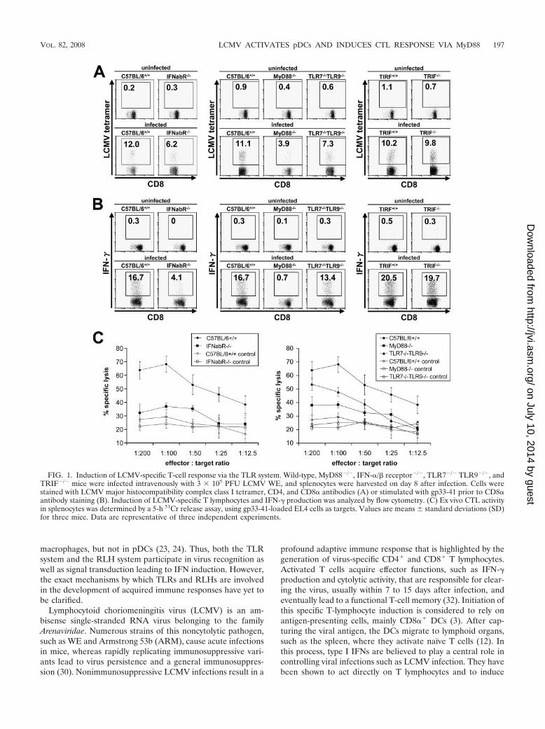
FIG. 1. Induction of LCMV-specific T-cell response via the TLR system. Wild-type, MyD88�/�, IFN-�/� receptor�/�, TLR7�/� TLR9�/�, andTRIF�/� mice were infected intravenously with 3 � 105 PFU LCMV WE, and splenocytes were harvested on day 8 after infection. Cells werestained with LCMV major histocompatibility complex class I tetramer, CD4, and CD8� antibodies (A) or stimulated with gp33-41 prior to CD8�antibody staining (B). Induction of LCMV-specific T lymphocytes and IFN-� production was analyzed by flow cytometry. (C) Ex vivo CTL activityin splenocytes was determined by a 5-h 51Cr release assay, using gp33-41-loaded EL4 cells as targets. Values are means � standard deviations (SD)for three mice. Data are representative of three independent experiments.
VOL. 82, 2008 LCMV ACTIVATES pDCs AND INDUCES CTL RESPONSE VIA MyD88 197
on July 10, 2014 by guesthttp://jvi.asm
.org/D
ownloaded from

massive expansion of antigen-specific CD8� T cells (1, 7, 22,32).
Nevertheless, the source of type I IFN in response to LCMVremains controversial, and type I IFN-producing cells are notwell characterized. pDCs have been shown to be a majorsource of type I IFNs in various murine virus infections (6, 8,9). pDCs were also implicated as the source of IFN-� in theLCMV-infected spleen (29). Conversely, it was reported thatproduction of type I IFNs in response to LCMV infection wasnot impaired in pDC-depleted mice, questioning the impor-tance of pDCs in T-cell induction in response to LCMV (8).Furthermore, cDCs extracted from mice infected with LCMVwere reported to produce high IFN-� levels (10).
In the present study, we investigated the involvement ofTLRs and RLHs in the development of antigen-specific CD8�
T cells as well as in the production of type I IFNs in responseto LCMV infection by using MyD88-deficient (MyD88�/�)and IPS-1�/� mice. The development of cytotoxic T lympho-cytes (CTLs) was critically dependent on MyD88 but not onIPS-1. MyD88-deficient mice were revealed to be highly sus-ceptible to LCMV infection. In contrast to previous reports,levels of IFN-�, IFN-�, and proinflammatory cytokines in serawere dependent on the presence of MyD88, whereas IPS-1deficiency resulted in impaired IFN-� production but other-wise normal cytokine levels. pDCs were the major source ofIFN-� in LCMV infection. These results suggest that LCMVactivates pDCs via TLRs to produce type I IFNs in vivo andthat the activation of an innate mechanism leads to the induc-tion of the CTL response and, eventually, virus elimination.
MATERIALS AND METHODS
Mice. IFN-�/� receptor�/�, MyD88�/�, TRIF�/�, TLR2�/�, TLR4�/�,TLR8�/�, and IPS-1�/� mice have been described previously (14, 16, 24, 36, 40).TLR7�/� and TLR9�/� mice were crossed to yield double TLR7- and TLR9-deficient mice (13, 14). Ifna6�/GFP mice were generated as recently described(23).
LCMV infection and virus titration. LCMV WE and ARM were obtainedfrom T. Otheki (31). For virus propagation, L cells were infected with LCMVand cultured at 37°C for 48 h. Supernatants were diluted in phosphate-bufferedsaline for infection. Mice were infected intravenously with 3 � 105 PFU LCMVstrain WE or ARM after a brief anesthesia with diethyl ether. For analysis ofsurvival rates, mice were infected with 2 � 106 PFU. Virus loads in the spleenand liver were investigated on days 4, 8, and 30 after infection. For virus titration,MC57G cells were inoculated with 10-fold serial dilutions of LCMV-containingsupernatants in a 24-well plate, covered with 2% methylcellulose, and incubatedat 37°C for 48 h. Cells were then fixed with 4% formalin, permeabilized with0.5% Triton X, and incubated with 10% fetal bovine serum. After subsequentincubation with murine LCMV immunoglobulin G1 (IgG1; Progen Biotechnik)and anti-mouse IgG–horseradish peroxidase (Amersham Biosciences), plateswere stained using an AEC peroxidase substrate kit (Vector Laboratories), andvirus plaques were counted for each well.
Induction of specific T-cell response. Splenocytes were harvested 8 days afterinfection. To investigate the activation of LCMV-specific T lymphocytes, cellswere incubated with T-select H-2Db LCMV tetramer-KAVYNFATC-phyco-erythrin (PE) (MBL), CD4-fluorescein isothiocyanate (CD4-FITC), and CD8�-allophycocyanin antibodies (BD Pharmingen). Samples were acquired on a FACS-Calibur flow cytometer (BD Biosciences) and analyzed with FlowJo software(TreeStar). IFN-� induction was analyzed by stimulating splenocytes with anLCMV H-2Db-binding peptide (glycoprotein 33-41 [gp33-41] [KAVYNFATM];Peptide Institute) and 10 ng/ml murine IL-2 prior to incubation with CD8�antibodies. Cells were then fixed, stained with IFN-�–FITC antibodies in Perm-Wash solution (BD Pharmingen) according to the manufacturer’s recommenda-tion, and assessed by flow cytometry. For determination of cytotoxicity ofLCMV-specific T cells, splenocytes were incubated for 5 h with EL-4 target cellsthat had been loaded with gp33-41 and labeled with 51Cr. The percentage ofspecific lysis for each sample was calculated as follows: [(sample release �
spontaneous release)/(maximal release � spontaneous release)] � 100. To an-alyze the generation of LCMV-specific memory T lymphocytes, mice were in-fected with 1 � 105 PFU LCMV WE, and splenocytes were analyzed foractivation of specific T memory cells and IFN-� induction as described below.
Cytokine and type I IFN production. IL-1�, IL-6, IL-10, IL-12(p40), IL-12(p70), RANTES, and tumor necrosis factor alpha were measured in sera 24and 48 h after LCMV infection by a multiplex bead-based flow cytometry assay(Bio-Plex cytokine assay; Bio-Rad Laboratories). IFN-� and IFN-� were deter-mined repeatedly between 12 and 96 h by enzyme-linked immunosorbent assay(PBL Biomedical Laboratories).
Expression of green fluorescent protein (GFP) in splenic DCs and macro-phages. Twenty-four and 48 h after LCMV infection, spleens of Ifna6�/GFP micewere injected with 150 U/ml collagenase buffer (Wako Chemicals), 10 g/mlDNase I (Sigma), and 10% fetal calf serum and incubated for 40 min at 37°C.After the addition of 10 mM EDTA and incubation for another 5 min, spleenswere shredded and passed through a nylon mesh. Erythrocytes were lysed, andthe single-cell suspension was stained with CD11b-PE, B220-PerCP, and CD11c-allophycocyanin antibodies before fluorescence-activated cell sorter analysis.
Activation of splenic DCs. Splenocytes were harvested 48 h after infection,incubated with B220-PerCP, CD11c-FITC, and CD40-, CD80-, or CD86-PEantibodies (BD Pharmingen), and analyzed by flow cytometry.
RESULTS
Impaired CTL response to LCMV infection in MyD88�/�
mice but not IPS-1�/� mice. LCMV has been shown to mounta robust CTL response in vivo. Although the involvement oftype I IFNs and innate immune cells in activating CTLresponses has clearly been demonstrated before, the mech-anism of innate recognition of LCMV in vivo is not yet fullyunderstood. Therefore, we first examined the contributionsof two major innate viral recognition systems, TLRs andRLHs, to mounting CTL responses against LCMV infection.Eight days after intravenous administration of LCMV,splenocytes of mice were harvested and stained with anH-2Db LCMV-specific tetramer (Fig. 1A) or stimulated withgp33-41 for analysis of IFN-� production (Fig. 1B), and totaland virus-specific CD8� T lymphocytes per spleen werecounted (Table 1). To assess LCMV-specific CTL activity,cells were incubated with gp33-41-loaded, 51Cr-labeled EL-4target cells and specific lysis was calculated (Fig. 1C). Wild-type mice mounted a vigorous CTL response, demonstratedby massive expansion of CD8� T cells, significant inductionof LCMV-specific CD8� T cells, strong IFN-� production,
TABLE 1. Total and virus-specific numbers of CD8� T lymphocytesper spleen for uninfected mice and mice that were intravenously
infected with 3 � 105 PFU LCMV WEa
Mouse group andgenotype
TotalCD8� cells
SpecificCD8� cells % of total
Uninfected miceC57BL/6�/� 6.6 � 2.6 0.03 � 0.04 0.5IFNabR�/� 5.4 � 0.3 0.03 � 0.01 0.6MyD88�/� 7.1 � 1.1 0.03 � 0.01 0.4TLR7�/� TLR9�/� 6.7 � 2.2 0.02 � 0.02 0.3
Infected miceC57BL/6�/� 19.7 � 9.2 2.19 � 1.04 11.1IFNabR�/� 15.7 � 4.1 0.99 � 0.25 6.3MyD88�/� 3.6 � 0.7 0.16 � 0.06 4.4TLR7�/� TLR9�/� 13.0 � 2.7 0.98 � 0.23 7.5
a Splenocytes were harvested on day 8 after infection and stained with LCMVmajor histocompatibility complex class I tetramer and CD8� antibodies. Cellnumbers were counted by flow cytometry. Depicted values are multipliers of 106
and indicate means � standard deviations for nine mice per indicated genotype.
198 JUNG ET AL. J. VIROL.
on July 10, 2014 by guesthttp://jvi.asm
.org/D
ownloaded from
and specific, T-cell-induced lysis of 60% of labeled targetcells. IFN-�/� receptor�/� mice and TLR7�/� TLR9�/�
mice demonstrated diminished clonal expansion of cytotoxicT cells in response to LCMV infection, whereas DC8� Tcells even decreased in MyD88�/� mice during infectioncompared to those in uninfected controls, resulting insplenic atrophy. Consistent with previous reports, IFN-�/�receptor�/� mice also showed impaired induction ofLCMV-specific T cells as well as decreased IFN-� produc-tion by these cells. Furthermore, cytotoxic T cells derivedfrom IFN-�/� receptor�/� mice failed to effectively lysetarget cells that presented an LCMV-specific epitope.MyD88 proved to play a crucial role in this process, asspecific T-cell induction, IFN-� production, and cytotoxicity
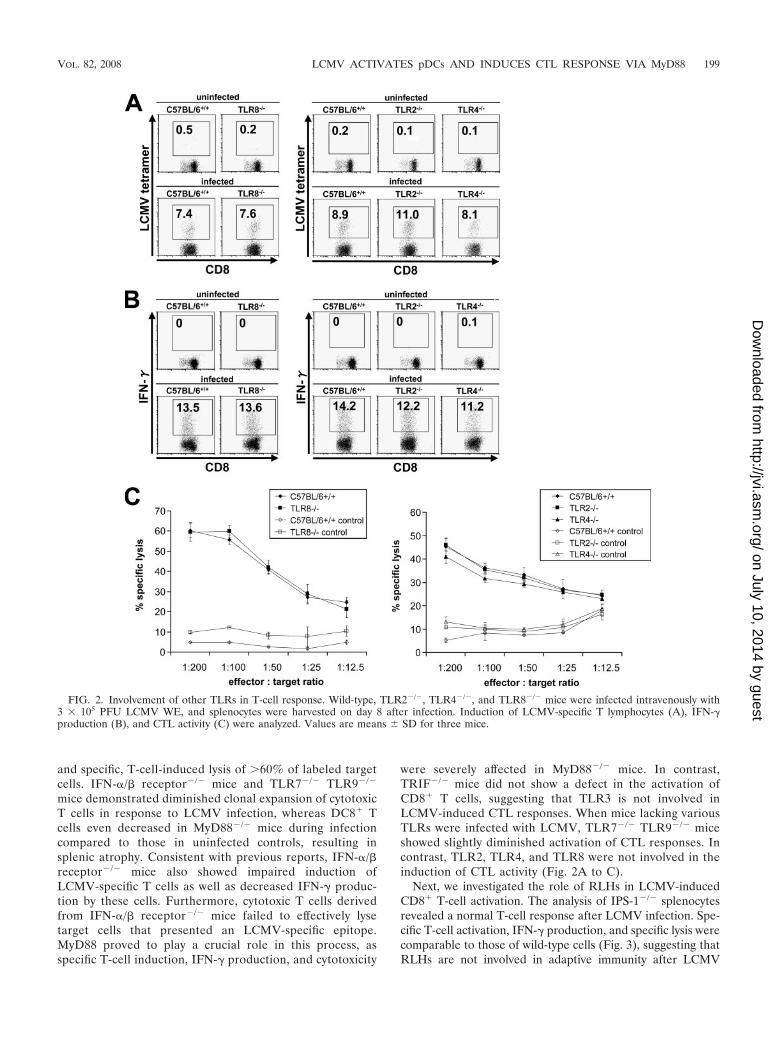
were severely affected in MyD88�/� mice. In contrast,TRIF�/� mice did not show a defect in the activation ofCD8� T cells, suggesting that TLR3 is not involved inLCMV-induced CTL responses. When mice lacking variousTLRs were infected with LCMV, TLR7�/� TLR9�/� miceshowed slightly diminished activation of CTL responses. Incontrast, TLR2, TLR4, and TLR8 were not involved in theinduction of CTL activity (Fig. 2A to C).
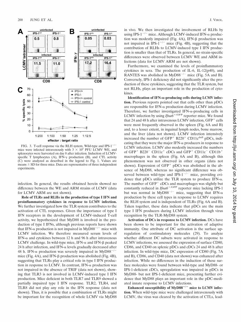
Next, we investigated the role of RLHs in LCMV-inducedCD8� T-cell activation. The analysis of IPS-1�/� splenocytesrevealed a normal T-cell response after LCMV infection. Spe-cific T-cell activation, IFN-� production, and specific lysis werecomparable to those of wild-type cells (Fig. 3), suggesting thatRLHs are not involved in adaptive immunity after LCMV
FIG. 2. Involvement of other TLRs in T-cell response. Wild-type, TLR2�/�, TLR4�/�, and TLR8�/� mice were infected intravenously with3 � 105 PFU LCMV WE, and splenocytes were harvested on day 8 after infection. Induction of LCMV-specific T lymphocytes (A), IFN-�production (B), and CTL activity (C) were analyzed. Values are means � SD for three mice.
VOL. 82, 2008 LCMV ACTIVATES pDCs AND INDUCES CTL RESPONSE VIA MyD88 199
on July 10, 2014 by guesthttp://jvi.asm
.org/D
ownloaded from
infection. In general, the results obtained herein showed nodifference between the WE and ARM strains of LCMV (datafor LCMV ARM are not shown).
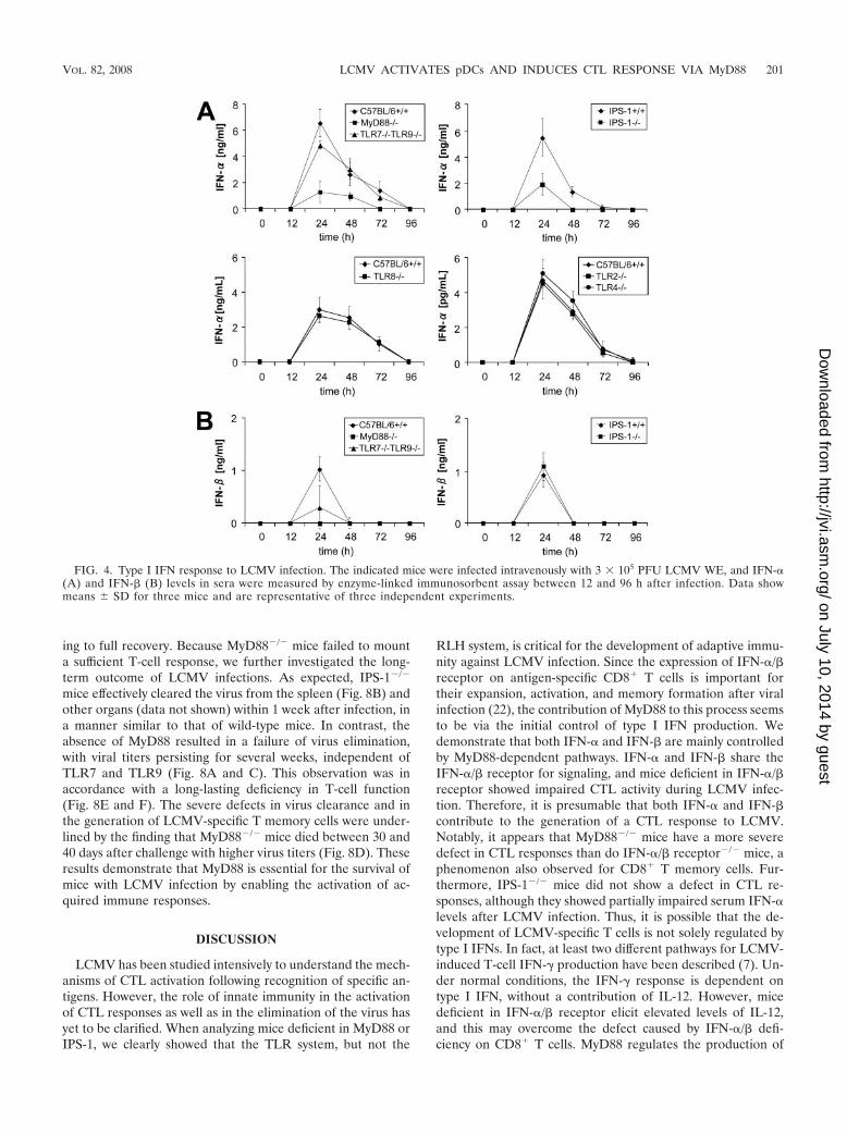
Role of TLRs and RLHs in the production of type I IFN andproinflammatory cytokines in response to LCMV infection.We further investigated how the TLR system contributes to theactivation of CTL responses. Given the importance of type IIFN receptors in the development of LCMV-induced T-cellactivity, we hypothesized that MyD88 is involved in the pro-duction of type I IFNs, although previous reports have shownthat IFN-� production is not impaired in MyD88�/� mice withLCMV infection. We therefore measured serum levels ofIFN-� and cytokines between 12 h and 96 h after intravenousLCMV challenge. In wild-type mice, IFN-� and IFN-� peaked24 h after infection, and IFN-� levels gradually decreased after48 h. IFN-� production was severely impaired in MyD88�/�
mice (Fig. 4A), and IFN-� production was abolished (Fig. 4B),suggesting that TLRs play a critical role in type I IFN produc-tion in response to LCMV. In contrast, IFN-� production wasnot impaired in the absence of TRIF (data not shown), show-ing that TLR3 is not involved in LCMV-induced type I IFNproduction. Mice deficient in both TLR7 and TLR9 showed apartially impaired type I IFN response. TLR2, TLR4, andTLR8 did not play any role in the IFN response (data notshown). Thus, it is possible that a combination of TLRs mightbe important for the recognition of whole LCMV via MyD88
in vivo. We then investigated the involvement of RLHs byusing IPS-1�/� mice. Although LCMV-induced IFN-� produc-tion was modestly impaired (Fig. 4A), IFN-� production wasnot impaired in IPS-1�/� mice (Fig. 4B), suggesting that thecontribution of RLHs to LCMV-induced type I IFN produc-tion is smaller than that of TLRs. In general, no strain-specificdifferences were observed between LCMV WE and ARM in-fections (data for LCMV ARM are not shown).
Furthermore, we examined the levels of proinflammatorycytokines in sera. The production of IL-6, IL-12(p40), andRANTES was abolished in MyD88�/� mice (Fig. 5A and B).Conversely, IPS-1 deficiency did not significantly alter the pro-duction of these cytokines, suggesting that the TLR system, butnot RLHs, plays an important role in the production of cyto-kines.
Identification of IFN-�-producing cells during LCMV infec-tion. Previous reports pointed out that cells other than pDCsare responsible for IFN-� production during LCMV infection.Therefore, we further investigated IFN-�-producing cells inLCMV infection by using Ifna6�/GFP reporter mice. We foundthat 24 and 48 h after intravenous LCMV infection, GFP� cellswere most frequently observed in the spleen (Fig. 6A and B)and, to a lesser extent, in inguinal lymph nodes, bone marrow,and the liver (data not shown). LCMV infection intensivelyincreased the number of GFP� B220� CD11cdull pDCs, indi-cating that they were the major IFN-� producers in response toLCMV infection. LCMV also modestly increased the numbersof GFP� B220� CD11c� cDCs and GFP� CD11c� CD11b�
macrophages in the spleen (Fig. 6A and B), although thisphenomenon was not observed in other organs (data notshown). Expression of GFP� pDCs was abolished in the ab-sence of MyD88, whereas no significant difference was ob-served between wild-type and IPS-1�/� mice, providing evi-dence that pDCs utilize the TLR system to produce IFN-�.The number of GFP� cDCs and macrophages was slightly butconstantly reduced in Ifna6�/GFP reporter mice lacking IPS-1but was normal in MyD88�/� mice, suggesting that IFN-�production by these cell types in response to LCMV requiresthe RLH system and is independent of TLRs (Fig. 6A and B).Taken together, these data indicate that pDCs are the maintype I IFN producers during LCMV infection through virusrecognition by the TLR-MyD88 system.
Activation of DCs in response to LCMV infection. DCs havebeen shown to be important for the activation of adaptiveimmunity. One attribute of DC activation is the surface up-regulation of costimulatory molecules (29). To analyzewhether different DC subsets were activated in response toLCMV infections, we assessed the expression of surface CD80,CD86, and CD40 on splenic pDCs and cDCs 24 and 48 h afterinfection. In wild-type mice, DC expression of CD80 (Fig. 7Aand B), CD86, and CD40 (data not shown) was enhanced afterinfection. While no differences in the induction of these sur-face molecules were found between wild-type and MyD88- orIPS-1-deficient cDCs, upregulation was impaired in pDCs inMyD88- but not IPS-1-deficient mice, presenting further evi-dence that MyD88 plays an important role in the pDC-medi-ated innate response to LCMV infections.
Enhanced susceptibility of MyD88�/� mice to LCMV infec-tion. When wild-type mice were challenged intravenously withLCMV, the virus was cleared by the activation of CTLs, lead-
FIG. 3. T-cell response via the RLH system. Wild-type and IPS-1�/�
mice were infected intravenously with 3 � 105 PFU LCMV WE, andsplenocytes were harvested on day 8 after infection. Induction of LCMV-specific T lymphocytes (A), IFN-� production (B), and CTL activity(C) were analyzed as described in the legend to Fig. 1. Values aremeans � SD for three mice. Data are representative of three independentexperiments.
200 JUNG ET AL. J. VIROL.
on July 10, 2014 by guesthttp://jvi.asm
.org/D
ownloaded from
ing to full recovery. Because MyD88�/� mice failed to mounta sufficient T-cell response, we further investigated the long-term outcome of LCMV infections. As expected, IPS-1�/�
mice effectively cleared the virus from the spleen (Fig. 8B) andother organs (data not shown) within 1 week after infection, ina manner similar to that of wild-type mice. In contrast, theabsence of MyD88 resulted in a failure of virus elimination,with viral titers persisting for several weeks, independent ofTLR7 and TLR9 (Fig. 8A and C). This observation was inaccordance with a long-lasting deficiency in T-cell function(Fig. 8E and F). The severe defects in virus clearance and inthe generation of LCMV-specific T memory cells were under-lined by the finding that MyD88�/� mice died between 30 and40 days after challenge with higher virus titers (Fig. 8D). Theseresults demonstrate that MyD88 is essential for the survival ofmice with LCMV infection by enabling the activation of ac-quired immune responses.
DISCUSSION
LCMV has been studied intensively to understand the mech-anisms of CTL activation following recognition of specific an-tigens. However, the role of innate immunity in the activationof CTL responses as well as in the elimination of the virus hasyet to be clarified. When analyzing mice deficient in MyD88 orIPS-1, we clearly showed that the TLR system, but not the
RLH system, is critical for the development of adaptive immu-nity against LCMV infection. Since the expression of IFN-�/�receptor on antigen-specific CD8� T cells is important fortheir expansion, activation, and memory formation after viralinfection (22), the contribution of MyD88 to this process seemsto be via the initial control of type I IFN production. Wedemonstrate that both IFN-� and IFN-� are mainly controlledby MyD88-dependent pathways. IFN-� and IFN-� share theIFN-�/� receptor for signaling, and mice deficient in IFN-�/�receptor showed impaired CTL activity during LCMV infec-tion. Therefore, it is presumable that both IFN-� and IFN-�contribute to the generation of a CTL response to LCMV.Notably, it appears that MyD88�/� mice have a more severedefect in CTL responses than do IFN-�/� receptor�/� mice, aphenomenon also observed for CD8� T memory cells. Fur-thermore, IPS-1�/� mice did not show a defect in CTL re-sponses, although they showed partially impaired serum IFN-�levels after LCMV infection. Thus, it is possible that the de-velopment of LCMV-specific T cells is not solely regulated bytype I IFNs. In fact, at least two different pathways for LCMV-induced T-cell IFN-� production have been described (7). Un-der normal conditions, the IFN-� response is dependent ontype I IFN, without a contribution of IL-12. However, micedeficient in IFN-�/� receptor elicit elevated levels of IL-12,and this may overcome the defect caused by IFN-�/� defi-ciency on CD8� T cells. MyD88 regulates the production of
FIG. 4. Type I IFN response to LCMV infection. The indicated mice were infected intravenously with 3 � 105 PFU LCMV WE, and IFN-�(A) and IFN-� (B) levels in sera were measured by enzyme-linked immunosorbent assay between 12 and 96 h after infection. Data showmeans � SD for three mice and are representative of three independent experiments.
VOL. 82, 2008 LCMV ACTIVATES pDCs AND INDUCES CTL RESPONSE VIA MyD88 201
on July 10, 2014 by guesthttp://jvi.asm
.org/D
ownloaded from

various cytokines, such as IL-1, IL-6, IL-12, IL-18, andRANTES, in addition to type I IFNs, and these cytokines likelycontribute to the MyD88-dependent development of LCMV-specific T cells in vivo.
Previous reports have shown that the production of type IIFNs in response to LCMV infection is not impaired in pDC-
FIG. 5. Cytokine production. IL-6, IL-12(p40), and RANTES levels in sera of MyD88�/� (A) and IPS-1�/� (B) mice after intravenous infectionwith 3 � 105 PFU LCMV WE were assessed after 24 h by a multiplex bead-based assay. Data show means � SD for three mice.
FIG. 6. Expression of GFP in splenic DCs and macrophages. Ifna6�/GFP
mice lacking MyD88 (A) or IPS-1 (B) as well as wild-type mice were infectedintravenously with 3 � 105 PFU LCMV WE. Splenocytes were harvested 24and 48 h after infection, and the expression of GFP in pDCs, cDCs, andmacrophages was analyzed by fluorescence-activated cell sorter analysis. Dataare representative of two experiments.
FIG. 7. Activation of splenic DCs. Mice were infected intrave-nously with 3 � 105 PFU LCMV WE. Splenocytes of MyD88- andIPS-1-deficient mice were investigated for the activation of CD80 onpDCs (A) and cDCs (B) by flow cytometry 48 h after infection. Datashown are representative of three mice.
202 JUNG ET AL. J. VIROL.
on July 10, 2014 by guesthttp://jvi.asm
.org/D
ownloaded from

depleted mice (8) and that splenic cDCs from infected miceproduce IFN-� (10). On the other hand, it was also suggestedthat splenic pDCs can produce IFN-� in response to LCMVinfection (29). In this study, we identified pDCs as the pre-dominant IFN-� producers by infecting Ifna6�/GFP mice withLCMV. We also observed a modest expression of GFP� cDCsand macrophages in LCMV-infected reporter mice; however,this expression was clearly subordinate to that of pDCs. Othercell types, such as T and B cells, did not express GFP in earlyLCMV infection (data not shown), and no type I IFNs could bedetected in sera later than 72 h postinfection, indicating thatlymphocytes are not IFN-� producers in response to LCMV.Furthermore, we found that IFN-� production in pDCs wasexclusively dependent on MyD88 but not on IPS-1. Consis-tently, IFN-� and IFN-� levels in sera were severely impairedin MyD88�/� mice. These results are in contradiction to aprevious report showing that the production of type I IFNs inresponse to LCMV was MyD88 independent (43). Although
we do not have an explanation for this discrepancy, we believethat the involvement of MyD88 as well as pDCs in the IFNresponse is well grounded based on the data from measure-ments of serum IFN and our novel reporter mice. On the otherhand, cDCs and macrophages from Ifna6�/GFP mice lackingIPS-1 showed reduced GFP expression in response to LCMVcompared to those from wild-type mice, although the fre-quency of GFP� cells was much lower than that for pDCs.However, given that the total number of cDCs and macro-phages in the body by far exceeds the number of pDCs, it canbe presumed that the impaired IFN-� levels in sera in IPS-1�/�
mice are a result of the failure to produce IFN in these celltypes.
Since type I IFN production in response to LCMV infectionwas mainly dependent on MyD88, we investigated the contri-bution of each TLR to the recognition of LCMV infection.Although the involvement of TLR2 was reported previously(43), we did not detect a defect in TLR2�/�, TLR4�/�, or
FIG. 8. Long-term sequelae after LCMV infection. Wild-type, MyD88�/�, and TLR7�/� TLR9�/� mice or IPS-1�/� mice were infectedintravenously with 3 � 105 PFU LCMV WE, and virus titers were measured in spleens on day 8 (A and B) and day 30 (C) by a methylcellulosetitration assay. Results for three mice are displayed, with means. Data are representative of three independent experiments. (D) Survival rates wereassessed after infecting six mice per indicated genotype with 2 � 106 PFU LCMV WE in two independent experiments. (E and F) Generation ofLCMV-specific memory CD8� T lymphocytes in MyD88�/�, TLR7�/� TLR9�/�, and IFN-�/� receptor�/� mice following infection with 1 � 105
PFU LCMV WE was analyzed as described above. Indicated values are means � SD for three mice and are representative of two independentexperiments.
VOL. 82, 2008 LCMV ACTIVATES pDCs AND INDUCES CTL RESPONSE VIA MyD88 203
on July 10, 2014 by guesthttp://jvi.asm
.org/D
ownloaded from

TLR8�/� mice. TLR1 and TLR6 both form heterodimers withTLR2 to recognize bacterial components, and they are likelynot involved in LCMV recognition, since TLR2�/� mice re-sponded appropriately to LCMV infection. Also, TLR5 is un-likely to contribute to MyD88-dependent LCMV recognition,as it does not activate IFN-inducible genes and is only margin-ally expressed in splenic cells (37). TLR7�/� TLR9�/� miceshowed only partial impairment in type I IFN levels and CTLactivity compared to MyD88�/� mice, and TRIF was not in-volved in innate and adaptive immune responses to LCMV.Taken together, the data show that other unknown MyD88-dependent but TLR7- and TLR9-independent receptors maycontribute to the complex orchestration of LCMV signal rec-ognition in vivo. Another possibility is that pDCs are indirectlyactivated by LCMV-infected cells and that MyD88-dependentsignaling is required in other cell types. Indeed, pDCs inducedfrom bone marrow failed to produce large amounts of IFN-� inresponse to LCMV infection in vitro, whereas infection withNewcastle disease virus, Sendai virus, and influenza virus wasreported to induce large amounts of IFN-� in bone marrow-derived pDCs (19). Thus, it is possible that IFN-� productionin response to LCMV is mediated through a mechanism dif-ferent from that in response to other RNA viruses. Furtherstudies are required to clarify the mechanism of IFN-� pro-duction in pDCs via the MyD88-dependent pathway.
DCs are reported to be critical for the development of an-tigen-specific T-cell responses against viral infection (3). Inresponse to LCMV infection, cDCs upregulated costimulatorymolecules, namely, CD40, CD80, and CD86, even in the ab-sence of MyD88 or IPS-1. In contrast, costimulatory moleculeexpression in pDCs was impaired in MyD88-deficient mice.The critical role of MyD88-dependent signaling in CD8 T-cellactivation and its role in IFN production in pDCs suggestedthat pDCs are essential for inducing the development of
LCMV-specific CTL responses. However, it is not clear ifpDCs play a direct role in the presentation of LCMV antigento CD8 T cells. A possible explanation is that pDCs act tofacilitate CTL activation by producing type I IFN rather thandirectly presenting antigens. A model depicting the modula-tion of the CTL response to LCMV by pDCs via MyD88 isshown in Fig. 9.
MyD88-deficient mice were highly susceptible to infectionwith LCMV and showed long-lasting virus persistence. Itseems that the defect in the acute-phase innate response can-not explain the cause of death observed for MyD88�/� mice,considering that all MyD88-deficient mice died after 30 to 40days of infection with higher virus titers. These results aresimilar to the effects of LCMV infection in perforin-deficient(Perf�/�) mice. Perf�/� mice have been shown to enhanceT-cell expansion and activation in response to persistentLCMV infection, leading to immunomediated tissue damageand increased mortality (5, 26). Thus, it has been suggestedthat the pathology and fatality of LCMV infection are mostlikely not direct results of the infection but, rather, are due tovirus-induced immunopathology. Therefore, although LCMV-mediated CTL responses, including the formation of CD8�� Tmemory cells, were impaired in MyD88�/� mice, the persistentT-cell response may eventually have caused enough pathologyto result in the death of the mice. However, further studies arerequired to clarify the precise cause of death observed forMyD88�/� mice.
In summary, our results demonstrate the importance ofMyD88-dependent signaling in the production of type I IFNsin pDCs. Consistently, MyD88 is critical for the activation ofCTL responses. Furthermore, MyD88 controls various proin-flammatory cytokines that are likely to contribute to the acti-vation of CTL responses independently of type I IFN. How-ever, the cell-specific contributions of TLRs and cytoplasmic
FIG. 9. Innate immune modulation of CTL response to LCMV through MyD88 signaling. MyD88 mediates recognition of LCMV byTLR7/TLR9 and possibly other, unknown receptors via pDCs. Type I IFNs produced by infected pDCs result in activation and clonal expansionof virus-specific CTLs. In an alternative pathway, CTLs are activated by proinflammatory cytokines in an MyD88-dependent manner. ActivatedCTLs mount effector functions that finally contribute to virus elimination.
204 JUNG ET AL. J. VIROL.
on July 10, 2014 by guesthttp://jvi.asm
.org/D
ownloaded from

RLHs to the activation of acquired immunity appear to bedifferent in response to various viruses. Future studies willclarify how these two innate viral recognition pathways areinvolved in T-cell activation depending on different viral patho-gens.
ACKNOWLEDGMENTS
We thank Y. Fujiwara, M. Shiokawa, and A. Shibano for skillfultechnical assistance, N. Kitagaki for an excellent methodological tuto-rial, and P. Lee for critically reading the manuscript. M. Hashimotodeserves special appreciation for distinguished organizational supportand secretarial assistance. T. Otheki kindly provided L cells andMC57G cells. EL4 cells were gratefully obtained from H. Tsutsui.
This work was supported in part by grants from the Yokochi Fundof the Kanehara Ichiro Foundation, from the Ministry of Education,Culture, Sports, Science and Technology in Japan, from the 21st Cen-tury Center of Excellence Program of Japan, and from the NIH(AI070167).
We have no competing financial interests.
REFERENCES
1. Aichele, P., H. Unsoeld, M. Koschella, O. Schweier, U. Kalinke, and S.Vucikuja. 2006. Cutting edge: CD8 T cells specific for lymphocytic chorio-meningitis virus require type I IFN receptor for clonal expansion. J. Immu-nol. 176:4525–4529.
2. Akira, S., S. Uematsu, and O. Takeuchi. 2006. Pathogen recognition andinnate immunity. Cell 124:783–801.
3. Belz, G. T., K. Shortman, M. J. Bevan, and W. R. Heath. 2005. CD8�dendritic cells selectively present MHC class I-restricted noncytological viraland intracellular bacterial antigens in vivo. J. Immunol. 175:196–200.
4. Beutler, B. 2004. Interferences, questions and possibilities in Toll-like recep-tor signaling. Nature 230:257–263.
5. Binder, D., M. F. van den Broek, D. Kaegi, H. Bluethmann, J. Fehr, H.Hnegartner, and R. M. Zinkernagel. 1989. Aplastic anemia rescued by ex-haustion of cytokine-secreting CD8� T cells in persistent infection withlymphocytic choriomeningitis virus. J. Exp. Med. 187:1902–1920.
6. Colonna, M., A. Krug, and M. Cella. 2002. Interferon-producing cells: on thefront line in immune responses against pathogens. Curr. Opin. Immunol.14:373–379.
7. Cousens, L. P., R. Peterson, S. Hsu, A. Dorner, J. D. Altman, R. Ahmed, andC. A. Biron. 1999. Two roads diverged: interferon �/�- and interleukin12-mediated pathways in promoting T cell interferon � responses duringviral infection. J. Exp. Med. 189:1315–1327.
8. Dalod, M., T. P. Salazar-Mather, L. Malmgard, C. Lewis, C. Asselin-Paturel,F. Briere, G. Trinchieri, and C. A. Brion. 2002. Interferon �/� and interleu-kin 12 responses to viral infections: pathways regulating dendritic cell cyto-kine expression in vivo. J. Exp. Med. 195:517–528.
9. Dalod, M., T. Hamilton, R. Salomon, T. P. Salazar-Mather, S. C. Henry,J. D. Hamilton, and C. A. Biron. 2003. Dendritic cell responses to earlycytomegalovirus infection: subset functional specialization and differentialregulation by interferon �/�. J. Exp. Med. 197:885–898.
10. Diebold, S. S., M. Montoya, H. Unger, L. Alexopoulou, P. Roy, L. E. Haswell,A. Al-Shamkhani, R. Flavell, P. Borrow, and C. Reis e Sousa. 2003. Viralinfection switches non-plasmacytoid dendritic cells into high interferon pro-ducers. Nature 424:324–328.
11. Gitlin, L., W. Barchet, S. Gilfillan, M. Cella, B. Beutler, R. A. Flavell, M. S.Diamond, and M. Colonna. 2006. Essential role of MDA5 in type I IFNresponses to polyriboinosinic acid and encephalomyocarditis picornavirus.Proc. Natl. Acad. Sci. USA 103:8459–8464.
12. Guermonprez, P., J. Valladeau, L. Zitvogel, C. Thery, and S. Amigorena.2002. Antigen presentation and T cell stimulation by dendritic cells. Annu.Rev. Immunol. 20:621–676.
13. Hemmi, H., O. Takeuchi, T. Kawai, T. Kaisho, S. Sato, H. Sanjo, M. Ma-tsumoto, K. Hoshino, H. Wagner, K. Takeda, and S. Akira. 2000. A Toll-likereceptor recognizes bacterial DNA. Nature 408:740–745.
14. Hemmi, H., T. Kaisho, O. Takeuchi, S. Sato, H. Sanjo, K. Hoshino, T.Horiuchi, H. Tomizawa, K. Takeda, and S. Akira. 2002. Small anti-viralcompounds activate immune cells via the TLR7 MyD88-dependent signalingpathway. Nat. Immunol. 3:196–200.
15. Honda, K., H. Yanai, A. Takaoka, and T. Taniguchi. 2005. Regulation of typeI IFN induction: a current view. Int. Immunol. 17:1367–1378.
16. Hoshino, K., T. Kaisho, T. Iwabe, O. Takeuchi, and S. Akira. 2002. Differ-ential involvement of IFN-beta in Toll-like receptor-stimulated dendritic cellactivation. Int. Immunol. 14:1225–1231.
17. Iwasaki, A., and R. Medzhitov. 2004. Toll-like receptor control of the adap-tive immune responses. Nat. Immunol. 5:987–995.
18. Kato, H., O. Takeuchi, S. Sato, M. Yoneyama, M. Yamamoto, K. Matsui, S.
Uematsu, A. Jung, T. Kawai, O. Yamaguchi, K. Ohtsu, T. Tsujimura, C. S.Koh, C. Reis e Sousa, Y. Matsuura, T. Fujita, and S. Akira. 2006. Differentialrole of MDA5 and RIG-I in the recognition of RNA viruses. Nature 141:101–105.
19. Kato, H., S. Sato, M. Yoneyama, M. Yamamoto, S. Uematsu, K. Matsui,T. Tsujimura, K. Takeda, T. Fujita, O. Takeuchi, and S. Akira. 2005.Cell-type specific involvement of RIG-I in antiviral response. Immunity23:19–28.
20. Kawai, T., and S. Akira. 2006. Innate immune recognition of viral infection.Nat. Immunol. 7:131–137.
21. Kawai, T., K. Takahashi, S. Sato, C. Coban, H. Kumar, H. Kato, K. J.Ishii, O. Takeuchi, and S. Akira. 2005. IPS-1, an adaptor triggeringRIG-I- and MDA5-mediated type I interferon induction. Nat. Immunol.6:1074–1076.
22. Kolumam, G. A., T. Sunil, L. J. Thompson, J. Sprent, and K. Murali-Krishna. 2005. Type I interferons act directly on CD8 T cells to allow clonalexpansion and memory formation in response to viral infection. J. Exp. Med.202:637–650.
23. Kumagai, Y., O. Takeuchi, H. Kato, H. Kumar, K. Matsui, E. Morii, K.Aozasa, T. Kawai, and S. Akira. 2007. Alveolar macrophages are the primaryinterferon-� producer in pulmonary infection with RNA viruses. Immunity27:240–252.
24. Kumar, H., T. Kawai, H. Kato, S. Sato, K. Takahashi, C. Coban, M.Yamamoto, S. Uematsu, K. J. Ishii, O. Takeuchi, and S. Akira. 2006. Es-sential role of IPS-1 in innate immune responses against RNA viruses. J.Exp. Med. 203:1795–1803.
25. Le Bon, A., and D. F. Tough. 2002. Links between innate and adaptiveimmunity via type-I interferon. Curr. Opin. Immunol. 14:432–436.
26. Matloubian, M., M. Suresh, A. Glass, M. Galvan, K. Chow, J. K. Whitmire,C. M. Walsh, W. R. Clark, and R. Ahmed. 1999. A role for perforin indownregulating T cell responses during chronic viral infections. J. Virol.73:2527–2536.
27. Meylan, E., J. Curran, K. Hofmann, D. Moradpour, M. Binder, R. Bar-tenschlager, and J. Tschopp. 2005. Cardif is an adaptor protein in theRIG-I antiviral pathway and is targeted by hepatitis C virus. Nature437:1167–1172.
28. Meylan, E., J. Tschopp, and M. Karin. 2006. Intracellular pattern recogni-tion receptors in the host response. Nature 442:39–44.
29. Montoya, M., M. J. Edwards, D. M. Reid, and P. Borrow. 2005. Rapidactivation of spleen dendritic cell subsets following lymphocytic choriomen-ingitis virus infection of mice: analysis of the involvement of type I IFN.J. Immunol. 174:1851–1861.
30. Oldstone, M. B. A. 2002. Biology and pathogenesis of lymphocytic chorio-meningitis virus infection. Curr. Top. Microbiol. Immunol. 263:83–117.
31. Otheki, T., M. Parsons, A. Zakarian, R. G. Jones, L. T. Nguyen, J. R.Woodgett, and P. S. Ohashi. 2000. Negative regulation of T cell proliferationand interleukin 2 production by the serine threonine kinase GSK-3. J. Exp.Med. 192:99–104.
32. Ou, R., S. Zhou, L. Huang, and D. Moskophidis. 2001. Critical role foralpha/beta and gamma interferons in persistence of lymphocytic chorio-meningitis virus by clonal exhaustion of cytotoxic T cells. J. Virol. 75:8407–8423.
33. Seth, R. B., L. Sun, C. K. Ea, and Z. J. Chen. 2005. Identification andcharacterization of MAVS, a mitochondrial antiviral signaling protein thatactivates NF-�B and IRF3. Cell 122:669–682.
34. Stetson, D. B., and R. Medzhitov. 2006. Type I interferons in host defense.Immunity 25:373–381.
35. Sun, Q., L. Sun, H. H. Liu, X. Chen, R. B. Seth, J. Forman, and Z. J. Chen.2006. The specific and essential role of MAVS in antiviral innate immuneresponses. Immunity 24:633–642.
36. Takeuchi, O., K. Hoshino, T. Kawai, H. Sanjo, H. Takada, T. Ogawa, K.Takeda, and S. Akira. 1999. Differential roles of TLR2 and TLR4 in recog-nition of gram-negative and gram-positive bacterial cell wall components.Immunity 11:443–451.
37. Uematsu, S., M. H. Jang, N. Chevrier, Z. Guo, Y. Kumagai, M. Yamamoto,H. Kato, N. Sougawa, H. Matsui, H. Kuwata, H. Hemmi, C. Coban, T.Kawai, K. Ishii, O. Takeuchi, M. Miyasaka, K. Takeda, and S. Akira. 2006.Detection of pathogenic intestinal bacteria by Toll-like receptor 5 on intes-tinal CD11c� lamina propria cells. Nat. Immunol. 7:868–874.
38. Uematsu, S., S. Sato, M. Yamamoto, T. Hirotani, H. Kato, F. Takeshita, M.Matsuda, C. Coban, K. Ishii, T. Kawai, O. Takeuchi, and S. Akira. 2005.Interleukin-1 receptor-associated kinase-1 plays an essential role for Toll-like receptor (TLR)7- and TLR9-mediated interferon-� induction. J. Exp.Med. 201:915–923.
39. Xu, L. G., Y. Y. Wang, K. J. Han, L. Y. Li, Z. Zhai, and H. B. Shu. 2005.VISA is an adaptor protein required for virus-triggered IFN-� signaling.Mol. Cell 19:727–740.
40. Yamamoto, M., S. Sato, H. Hemmi, K. Hoshino, T. Kaisho, H. Sanjo, O.Takeuchi, M. Sugiyama, M. Okabe, K. Takeda, and S. Akira. 2003. Role ofadaptor TRIF in the MyD88-independent Toll-like receptor signaling path-way. Science 301:640–643.
41. Yoneyama, M., M. Kikuchi, K. Matsumoto, T. Imaizumi, M. Miyagishi, K.
VOL. 82, 2008 LCMV ACTIVATES pDCs AND INDUCES CTL RESPONSE VIA MyD88 205
on July 10, 2014 by guesthttp://jvi.asm
.org/D
ownloaded from

Taira, E. Foy, Y. M. Loo, M. Gale, S. Akira, S. Yonehara, A. Kato, and T.Fujita. 2005. Shared and unique functions of the DExD/H-box helicasesRIG-I, MDA5, and LGP2 in antiviral innate immunity. J. Immunol. 175:2851–2858.
42. Yoneyama, M., M. Kikuchi, T. Natsukawa, N. Shinobu, T. Imaizumi, M.Miyagishi, K. Taira, S. Akira, and T. Fujita. 2004. The RNA helicase RIG-I
has an essential function in double-stranded RNA-induced innate antiviralresponses. Nat. Immunol. 5:730–737.
43. Zhou, S., E. A. Kurt-Jones, L. Mandell, A. Cerny, M. Chan, D. T. Golenbock,and R. W. Finberg. 2005. MyD88 is critical for the development of innate andadaptive immunity during acute lymphocytic choriomeningitis virus infec-tions. Eur. J. Immunol. 35:822–830.
206 JUNG ET AL. J. VIROL.
on July 10, 2014 by guesthttp://jvi.asm
.org/D
ownloaded from