Epithelial Cell-Specific MyD88 Signaling Mediates Ischemia/Reperfusion-induced Intestinal Injury...
10
ORIGINAL ARTICLE Epithelial Cell-Specific MyD88 Signaling Mediates Ischemia/Reperfusion-induced Intestinal Injury Independent of Microbial Status Marcus Mühlbauer, MD, PhD,* Ernesto Perez-Chanona, MS,* ,† and Christian Jobin, PhD* ,† Abstract: The Toll-like receptor/MyD88 signaling pathway has been shown to mediate protective functions during intestinal exposure to various noxious events. The goal of this study was to define the role of bacteria and MyD88 signaling in intestinal response to damage using an ischemia– reperfusion (I/R)-induced injury model. We showed that conventionalized mice displayed a better outcome to I/R-induced injury than germ-free mice (3.8 6 1.98 vs. 11.8 6 1.83, P , 0.05). However, mice with intestinal epithelial cell (IEC)-specific deletion of Myd88 (Myd88 IEC2/2 ) were protected from I/R-induced injury compared with Myd88 f/f control mice. Myd88 IEC2/2 mice also displayed a significantly reduced bacterial translocation (;85%) into lymph nodes compared with Myd88 f/f mice. Expression of ccl2 and cxcl1 mRNA was significantly reduced (85% and 62%, respectively) in intestinal tissue of Myd88 IEC2/2 mice compared with Myd88 f/f mice, which associated with a reduced number of myeloperoxidase-positive cells in intestinal tissues of I/R-exposed Myd88 IEC2/2 mice. Immunohistochemistry analysis showed a reduced IgA deposition and complement staining in ischemic tissue of Myd88 IEC2/2 mice compared with Myd88 f/f mice. These findings suggest that I/R-induced intestinal injury involves IEC-derived MyD88 signaling leading to increased IgA deposition/degradation, and complement activation in conjunction with an influx of neutrophils mediated by chemokine production. (Inflamm Bowel Dis 2013;0:1–10) Key Words: ischemia, reperfusion, MyD88, intestinal epithelial cells T he intestinal epithelium constitutes a crucial physical barrier separating the host from the luminal compartment containing various noxious agents such as bacteria, bacterial products, and food-derived particles. 1–3 In addition to its barrier function, the intestinal epithelium responds to numerous bacterial stimuli through the action of various conserved innate sensors such as Toll-like receptors (TLRs). 4 In the case of commensal bacteria, this dialogue between the microbiota and intestinal epithelial cells (IECs) is important for the maintenance of intestinal homeostasis through the induction of antimicrobial peptides, toning of the innate and adaptive immune responses, and the fortification of the barrier. 5 A compromised intestinal barrier could have delete- rious consequences for the host as luminal contents may gain access to the mucosal immune system and enter systemic circu- lation, triggering a damaging immune cell response. The epithe- lium is constantly facing injury caused by various luminal irritants, but these episodes rarely lead to life-threatening situa- tions because the host has developed an efficient adaptive wound- healing response to maintain homeostasis. 6 However, extreme challenges such as hypoxic, ischemic, or radiation events often overpower the wound-healing response, thereby exposing the host to damaging luminal products. For example, intestinal ische- mia can lead to sepsis through bacterial translocation. 7 Intestinal ischemia–reperfusion (I/R) injury can occur under several clinical conditions like sepsis, hemorrhage, mesenteric ischemia due to blood clots, neonatal necrotizing enterocolitis, and even small bowel transplantation. 8 Reperfusion after ischemic events causes epithelial cell death and disruption of barrier function leading to invasion of bacteria. 9 Antibiotic use is the mainstay of treatment to prevent sepsis from bacterial translocation after intestinal ische- mic events and has even been shown to be protective before the inciting event. 10 Interestingly, the microbiota seems to play a protective role in various experimental injury models including dextran sodium sulfate and radiation. 11–13 In contrast, global deletion of myeloid differentiation primary response gene (Myd88), an adaptor protein in innate immune signaling, protects mice from I/R-induced injury. 14 Moreover, in a model of necrotizing enterocolitis in Supplemental digital content is available for this article. Direct URL citations appear in the printed text and are provided in the HTML and PDF versions of this article on the journal’s Web site (www.ibdjournal.org). Received for publication August 13, 2013; Accepted September 11, 2013. From the *Departments of Medicine, Microbiology and Immunology, and Phar- macology, University of North Carolina, Chapel Hill, North Carolina; and † Depart- ment of Medicine and Department of Infectious Diseases & Pathology, University of Florida, Gainesville, Florida. Supported by National Institutes of Health grants R01DK047700 and R01DK073338 (C.J.) and Crohn’s and Colitis Foundation of America (M.M.). The funders had no role in the study design, data collection and analysis, decision to publish, or preparation of the article. The authors have no conflicts of interest to disclose. Reprints: Christian Jobin, PhD, Department of Medicine, University of Florida, Gainesville, FL 32608 (e-mail: [email protected]fl.edu). Copyright © 2013 Crohn’s & Colitis Foundation of America, Inc. DOI 10.1097/01.MIB.0000435445.96933.37 Published online. Inflamm Bowel Dis Volume 0, Number 0, Month 2013 www.ibdjournal.org | 1 Copyright © 2013 Crohn’s & Colitis Foundation of America, Inc. Unauthorized reproduction of this article is prohibited.
Transcript of Epithelial Cell-Specific MyD88 Signaling Mediates Ischemia/Reperfusion-induced Intestinal Injury...
ORIGINAL ARTICLE
Epithelial Cell-Specific MyD88 Signaling MediatesIschemia/Reperfusion-induced Intestinal Injury Independentof Microbial StatusMarcus Mühlbauer, MD, PhD,* Ernesto Perez-Chanona, MS,*,† and Christian Jobin, PhD*,†
Abstract: The Toll-like receptor/MyD88 signaling pathway has been shown to mediate protective functions during intestinal exposure to variousnoxious events. The goal of this study was to define the role of bacteria and MyD88 signaling in intestinal response to damage using an ischemia–reperfusion (I/R)-induced injury model. We showed that conventionalized mice displayed a better outcome to I/R-induced injury than germ-free mice(3.8 6 1.98 vs. 11.8 6 1.83, P , 0.05). However, mice with intestinal epithelial cell (IEC)-specific deletion of Myd88 (Myd88IEC2/2) were protectedfrom I/R-induced injury compared with Myd88f/f control mice. Myd88IEC2/2 mice also displayed a significantly reduced bacterial translocation (;85%)into lymph nodes compared withMyd88f/f mice. Expression of ccl2 and cxcl1 mRNA was significantly reduced (85% and 62%, respectively) in intestinaltissue of Myd88IEC2/2 mice compared with Myd88f/f mice, which associated with a reduced number of myeloperoxidase-positive cells in intestinaltissues of I/R-exposed Myd88IEC2/2 mice. Immunohistochemistry analysis showed a reduced IgA deposition and complement staining in ischemic tissueof Myd88IEC2/2 mice compared with Myd88f/f mice. These findings suggest that I/R-induced intestinal injury involves IEC-derived MyD88 signalingleading to increased IgA deposition/degradation, and complement activation in conjunction with an influx of neutrophils mediated by chemokineproduction.
(Inflamm Bowel Dis 2013;0:1–10)
Key Words: ischemia, reperfusion, MyD88, intestinal epithelial cells
T he intestinal epithelium constitutes a crucial physical barrierseparating the host from the luminal compartment containing
various noxious agents such as bacteria, bacterial products, andfood-derived particles.1–3 In addition to its barrier function, theintestinal epithelium responds to numerous bacterial stimulithrough the action of various conserved innate sensors such asToll-like receptors (TLRs).4 In the case of commensal bacteria,this dialogue between the microbiota and intestinal epithelial cells(IECs) is important for the maintenance of intestinal homeostasisthrough the induction of antimicrobial peptides, toning of theinnate and adaptive immune responses, and the fortification of
the barrier.5 A compromised intestinal barrier could have delete-rious consequences for the host as luminal contents may gainaccess to the mucosal immune system and enter systemic circu-lation, triggering a damaging immune cell response. The epithe-lium is constantly facing injury caused by various luminalirritants, but these episodes rarely lead to life-threatening situa-tions because the host has developed an efficient adaptive wound-healing response to maintain homeostasis.6 However, extremechallenges such as hypoxic, ischemic, or radiation events oftenoverpower the wound-healing response, thereby exposing thehost to damaging luminal products. For example, intestinal ische-mia can lead to sepsis through bacterial translocation.7 Intestinalischemia–reperfusion (I/R) injury can occur under several clinicalconditions like sepsis, hemorrhage, mesenteric ischemia due toblood clots, neonatal necrotizing enterocolitis, and even smallbowel transplantation.8 Reperfusion after ischemic events causesepithelial cell death and disruption of barrier function leading toinvasion of bacteria.9 Antibiotic use is the mainstay of treatmentto prevent sepsis from bacterial translocation after intestinal ische-mic events and has even been shown to be protective before theinciting event.10
Interestingly, the microbiota seems to play a protective rolein various experimental injury models including dextran sodiumsulfate and radiation.11–13 In contrast, global deletion of myeloiddifferentiation primary response gene (Myd88), an adaptor proteinin innate immune signaling, protects mice from I/R-inducedinjury.14 Moreover, in a model of necrotizing enterocolitis in
Supplemental digital content is available for this article. Direct URL citationsappear in the printed text and are provided in the HTML and PDF versions of thisarticle on the journal’s Web site (www.ibdjournal.org).
Received for publication August 13, 2013; Accepted September 11, 2013.From the *Departments of Medicine, Microbiology and Immunology, and Phar-
macology, University of North Carolina, Chapel Hill, North Carolina; and †Depart-ment of Medicine and Department of Infectious Diseases & Pathology, University ofFlorida, Gainesville, Florida.
Supported by National Institutes of Health grants R01DK047700 andR01DK073338 (C.J.) and Crohn’s and Colitis Foundation of America (M.M.).The funders had no role in the study design, data collection and analysis, decisionto publish, or preparation of the article.
The authors have no conflicts of interest to disclose.Reprints: Christian Jobin, PhD, Department of Medicine, University of Florida,
Gainesville, FL 32608 (e-mail: [email protected]).Copyright © 2013 Crohn’s & Colitis Foundation of America, Inc.DOI 10.1097/01.MIB.0000435445.96933.37Published online.
Inflamm Bowel Dis ! Volume 0, Number 0, Month 2013 www.ibdjournal.org | 1
Copyright © 2013 Crohn’s & Colitis Foundation of America, Inc. Unauthorized reproduction of this article is prohibited.

newborn mice, TLR/MyD88-mediated intestinal injury seems to becontrolled by nucleotide-binding oligomerization domain-2 signal-ing.15 However, the role of microbes in injury response to I/R is stillunclear because most studies linking bacteria to I/R responsehave used reduction strategies (antibiotic treatment) and/or innatesensor-deficient mice. Numerous cells, including IEC and mucosalimmune cells, express MyD88, and the cell type specific functionof this molecule in I/R-mediated injury response is unknown.
In this study, we investigated the impact of the microbiotausing germ-free and conventionalized mice and defined the role ofIEC-specific MyD88 signaling in I/R-induced intestinal injury. Wereport that IEC-derived MyD88 signaling promotes I/R-inducedinjury. This deleterious effect is likely mediated by 2 main events:(1) IEC-dependent MyD88 signaling leading to increased amount ofluminal IgA and subsequent complement activation during theischemic phase and (2) IEC-MyD88–dependent expression of neu-trophil chemoattractants in IECs, further damaging intestinal tissue.
MATERIALS AND METHODS
Generation of Myd88IEC2/2 MiceAll mice were on a C57BL/6 background. Villin-Cre trans-
genic mice were crossed to Myd88f/f mice (a generous gift from DrAnthony DeFranco, UCSF, CA), which contain loxP sites flankingexon 3 to generate mice lacking exon 3 of Myd88 gene in IECs(Myd88IEC2/2).16 Tail snips were collected from all pups generatedfrom crosses, and genomic DNA was isolated using a Qiagen Bloodand Tissue Kit (Qiagen; Valencia, CA). Primers used for genotypingwere as followed: Villin-Cre promoter (5’-GCGGTCTGGCAGTAAAAACTATC-3’, and 5’-GTGAAACAGCATTGCTGTCACTT-3’) and MyD88f/f (5’-GTTGTGTGTGTCCGACCGT-3’ and5’-GTCAGAAACAACCACCACCATGC-3’).
For phenotypic analysis, RNA was isolated from IEC andsplenocytes as previously described,17 and expression of MyD88 wasdetermined by PCR. Isolation of IECs was performed as previouslydescribed.18 Amplicons were resolved on a 2% agarose gel and visu-alized using a Kodak Gel Logic Imager 200 series (Kodak; Rochester,NY). Myd88 primers used were as followed: 5’-TTGGATGCCTGGCAGGGG-3’ and 5’-TCCTTCTTCATCGCCTTG-3’. The ampliconlength including exon 3 was 565 bp and the amplicon length withoutexon 3 was 384 bp. Deletion of exon 3 in epithelial cells was alsoconfirmed by sequencing of the PCR products. Purity of IEC andsplenocytes isolation was confirmed by amplifying the IEC-specificgene product Villin and the immune cell–specific marker (neutrophils,monocytes, and B-cell subtypes) CD11b using the respective set ofprimers 5’-CCCCCATCTTCCAACAAT-3’, 5’-GGACCTGAAATAGCCTCGTAG-3’ (amplicon length: 546 bp); and 5’-GGCGCTGTCTACATTTTTTAT-3’ and 5’-GCTCCCCAACCAGTGTATAAT-3’ (amplicon length 574 bp) (Fig, Supplemental Digital Content1, http://links.lww.com/IBD/A334). Low levels of CD11b contami-nation that occurred during IEC isolation account for the wild-typeMyD88 bands that appear in IEC-specific Myd88 knockout samplesof small intestine and colon tissue.
I/R-induced InjuryMyd88f/f, Myd88IEC2/2, and Myd882/2 mice (C57BL/6
background) were maintained in standard housing cages in specificpathogen free–conditions. To study the impact of the microbiota onI/R injury, 1 cohort of germ-free (GF) wild-type mice (C57BL/6background) was transferred to specific pathogen–free conditionsfor 4 weeks, and another cohort was kept in GF conditions. For allexperiments, mice (n ¼ 3–5) were anesthetized under 1% isofluor-ane, supplemented by 10 mg/kg ketamine, and 0.1 mg/kg bupre-norphine, both injected subcutaneously. A mid-line laparotomy wasmade, and peripheral branches of the superior mesenteric arterywere occluded with aneurysm clips with a force of 50 g (KentScientific, Torrington, CN), to create a 3- to 5-cm region of ische-mic ileum adjacent to the cecum. Collateral blood flow through theintestine was blocked using aneurysm clips across the intestine andcollateral vessels, demarking the region of ischemic intestine.Hematoxylin was administered to the edges of ischemic tissueto mark them, and then the incision was closed with surgicalstaples. Ischemia was maintained for 60 minutes, and then theincision was reopened, the clamps were removed, and the incisionwas reclosed. We have optimized this protocol to maximize injuryand maintain a low mortality rate (4%), as previously published.19
The mice were maintained in a heated room for a variable amountof time (0, 1.5, or 4 hours) without anesthesia for the reperfusionphase of injury. To account for any systemic reaction to injury,healthy tissue adjacent to injured tissue was harvested as an inter-nal healthy control, as previously described.19 This approach wasused in lieu of “sham” animals, which do not appropriately controlfor the systemic effects that originate from I/R-induced injury. Allanimal experiments were approved by the Institutional AnimalCare and Use Committee of the University of North Carolina atChapel Hill.
Murine Sample Collection andHistological Evaluation
Mice were anesthetized using isoflurane, and then killed bycervical dislocation. The colon was dissected and flushed withice-cold phosphate-buffered saline, longitudinally splayed, Swissrolled, fixed in 10% formalin for 24 hours, and then embedded inparaffin. Damage severity was evaluated using Hematoxylin andeosin–stained sections by an investigator blinded to the experi-mental conditions. The scoring system is based on an IEC apo-ptosis/necrosis system, where a score of 1 signified a loss of onlythe villus tips; a score of 2 corresponded to loss of 50% of thevillus; a score of 3 indicated a loss of the entire villus, but withmaintenance of the crypt; and a score of 4 signified complete lossof the epithelial layer.20 The ischemic tissue was divided into4 quarters, a score was given to each quarter separately and thenadded to generate a final damage score.
Immunohistochemistryand Immunofluorescence
Immunohistochemistry (IHC) staining was performedaccording to the manufacturer’s specifications, as previously
Mühlbauer et al Inflamm Bowel Dis ! Volume 0, Number 0, Month 2013
2 | www.ibdjournal.org
Copyright © 2013 Crohn’s & Colitis Foundation of America, Inc. Unauthorized reproduction of this article is prohibited.

described.17 Primary antibodies and dilutions were as followed:myeloperoxidase, 1:400 (Neomarkers, Fremont, CA), PIgR, 1:50(R&D Systems; Minneapolis, MN), IgA, 1:1000 (R&D Systems;Minneapolis, MN), and complement 3d, 1:50 (R&D Systems;Minneapolis, MN). IHC for activated caspase 3, 1:400 (R&DSystems; Minneapolis, MN) was performed as described.21 Allsections were counterstained with hematoxylin and eosin.
RNA Isolation and Real-time PCRRNA isolation from ileal tissues and subsequent cDNA
amplification and analysis were performed as previously described.17
Specificity and linearity of amplification for each primer set weredetermined by melting curve analysis and calculation of the slopefrom serial diluted samples. Relative fold changes were determinedusing the DDCT calculation method. Values were normalized to theinternal control GAPDH or b-actin. Primers: GAPDH (5’-GGTGAA
GGTCGGAGTCAACGGA-3’ and 5’-GAGGGATCTCGCTCCTGGAAGA-3’), b-actin (5’-TGGAATCCTGTGGCATCCATGAAAC-3’ and 5’- TAAAACGCAGCTCAGTAACAGTCCG-3’), cxcl1(5’-GCTGGGATTCACCTCAAGAA-3’ and 5’- TCTCCGTTACTTGGGGACAC-3’), ccl2 (5’-GCTGCTACTCATTCACTGGCAA-3’ and 5’- TGCTGCTGGTGATTCTCTTGTA-3’), and tnf(5’-ATGAGCACAGAAAGCATGATC-3’ and 5’- TACAGGCTTGTCACTCGAATT-3’).
Bacterial Translocation and CultureFor all bacterial translocation assays, mice were subjected
to 60 minutes of ischemia followed by 4 hours of reperfusion.Mice were killed, and 2 mesenteric lymph nodes adjacent to thedamaged tissue were harvested and weighed. The homogenateswere lysed in 1 mL phosphate-buffered saline using a MiniBeadBeater-8 (BioSpec, Bartlesville, OK) and plated on sheep
FIGURE 1. Colonization with commensal bacteria alleviates I/R-induced injury. Cohorts of 3–5 germ-free (GF) wild-type mice and 5 con-ventionalized (Conv) wild-type mice were subjected to either 60-minute ischemia only (I) or 60-minute ischemia followed by 180-minute re-perfusion (I/R). Scale bars ¼ 200 mm. A, Representative images of hematoxylin and eosin–stained ileal sections are shown. B, Histological intestinaldamage scores are depicted as mean 6 SEM; *P , 0.05. NS, not significant.
Inflamm Bowel Dis ! Volume 0, Number 0, Month 2013 Epithelial-specific MyD88 Signaling
www.ibdjournal.org | 3
Copyright © 2013 Crohn’s & Colitis Foundation of America, Inc. Unauthorized reproduction of this article is prohibited.
brain heart infusion plates (Remel, Lenexa, KA) and incubated inaerobic and anaerobic conditions for 24 hours, at which timecolonies were counted.
Statistical AnalysesUnless specifically noted, statistical analyses were per-
formed using GraphPad Prism version 5.0a (GraphPad, La Jolla,CA). Comparisons of mouse studies were made with nonpara-metric analysis of variance, and then a Mann–Whitney U test. Allgraphs depict mean 6 SEM. Experiments were considered statis-tically significant if P , 0.05.
RESULTS
Commensal Microbes Alleviate I/R-inducedIleal Damage
Although wide-spectrum antibiotic treatment attenuatesI/R-induced intestinal injury in mice, this experimental approachmay have confounding effects on the host.10 To stringently evaluatethe impact of bacteria on intestinal injury response, we used GFand conventionalized wild-type mice. Conventionalized mice andGF mice (3–5 mice per group) were exposed to either 60-minuteischemia (I) or 60-minute ischemia followed by 180-minute reperfusion (I/R), and tissue injury was assessed by histological analysis.Although no difference was observed between the histological dam-age scores of the conventionalized and GF group (12.7 6 1.80 vs.11.6 6 0.70, respectively) in the ischemic phase (Fig. 1A, B),a significant improvement was noted in conventionalized miceversus GF mice (3.8 6 1.98 vs. 11.8 6 1.83, P , 0.05) duringthe reperfusion phase (Fig. 1A, B). This finding suggests that bac-teria may promote beneficial responses during the reperfusion phase of I/R-induced tissue injury.
IEC-specific Myd88 Deletion AlleviatesI/R-induced Injury
To assess early stages of epithelial cell damage andrecovery during I/R, the reperfusion time was reduced from180 minutes to 90 minutes for the subsequent experiments. Wehave previously optimized I/R-induced injury to maximizeepithelial damage.19 To define the role of bacteria in I/R-mediated
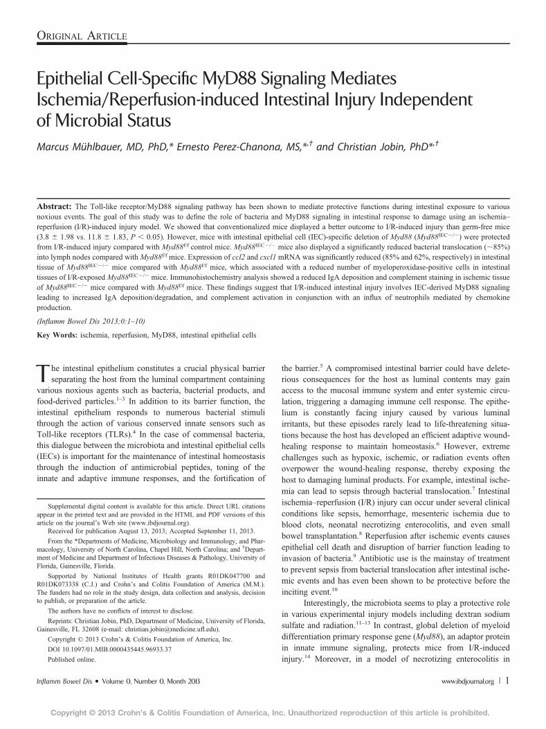
FIGURE 2. IEC-specific Myd88 deletion protects from I/R injury. Myd88f/f,Myd88IEC2/2, and Myd882/2 mice (n ¼ 5) were subjected to 60-minuteischemia (I) only (A) or 60-minute ischemia followed by 90-minutereperfusion (I/R) (B). Histological damage scores were evaluated ondamaged ileal tissues using a modified scoring system. All graphs depictmean 6 SEM; *P , 0.05. Results are representative of 2 independentexperiments. NS, not significant.
FIGURE 3. I/R-exposed Myd88IEC2/2 mice display diminished bacterialtranslocation compared to Myd88f/f mice. Myd88f/f, Myd88IEC2/2, andMyd882/2 mice (n ¼ 4) were subjected to 60-minute ischemia and240-minute reperfusion. Two mesenteric lymph nodes adjacent to theaffected ileal tissue were collected, weighed, homogenized, plated onbrain heart infusion plates, and cultured under aerobic and anaerobicconditions for 24 hours. Colony-forming units were counted, andcombined colony-forming units for aerobic and anaerobic conditionsare shown. All graphs depict mean 6 SEM. *P , 0.05. Results arerepresentative of 2 independent experiments.
Mühlbauer et al Inflamm Bowel Dis ! Volume 0, Number 0, Month 2013
4 | www.ibdjournal.org
Copyright © 2013 Crohn’s & Colitis Foundation of America, Inc. Unauthorized reproduction of this article is prohibited.

injury, we selectively deleted Myd88 in IEC by crossing Myd88f/f
mice to Villin-Cre mice to generate Myd88IEC2/2 mice. The extentof ischemia-induced intestinal injury was similar in Myd88f/f andMyd88IEC2/2 mice (8.8 6 0.86 vs. 10.8 6 1.16; not significant)during the ischemic phase (Fig. 2A). However, after 90 minutes ofreperfusion, Myd88IEC2/2 mice showed greater epithelial restitu-tion compared with Myd88f/f mice (4.6 6 1.29 vs. 10 6 1.67,respectively; P, 0.05) (Fig. 2B). The protective recovery observedinMyd88IEC2/2 mice was similar to that ofMyd882/2 mice (4.661.29 vs. 4 6 1.27, respectively) suggesting that MyD88 signalingfrom the IEC compartment was mainly responsible for thephenotype.
Since defective IEC-derived MyD88 alleviated epithelial celldamage, we predicted thatMyd88IEC2/2 mice should have a dimin-ished translocation of bacteria. As expected, a significant decreaseof colony-forming units in lymph nodes was observed in I/R-exposed Myd88IEC2/2 mice compared with Myd88f/f mice (67 627 vs. 452 6 185, respectively; P , 0.05; Myd882/2: 181 6 121)(Fig. 3).
Deletion of MyD88 Signaling ReducesI/R-induced Apoptosis in Epithelial Cells
Intestinal tissues undergo hypoxia that contributes to IECapoptosis and epithelial injury during I/R.22 Consequently, I/R-induced IEC apoptosis was compared between Myd88IEC2/2 andMyd88f/f mice by evaluating active caspase-3 using IHC staining.A significant decrease in the number of caspase-3–positive cells
was observed in Myd88IEC2/2 mice compared with Myd88f/f mice(6.4 6 2.4 vs. 26.3 6 0.53, respectively; P , 0.05) (Fig. 4A, B),a level similar to Myd882/2 mice (6.2 6 2.1). Healthy tissuerarely showed the presence of activated caspase-3 (Fig. 4A). Nodifference in epithelial cell proliferation was observed betweenthe 3 genotypes as measured by Ki-67 staining (data not shown).
IEC-derived MyD88 Signaling PromotesChemokine Gene Expression and NeutrophilRecruitment in I/R-injured Tissue
Neutrophils have been linked to enhanced tissue damage invarious injury models, including I/R in the myocardium, liver, andintestine,23–26 therefore, we next examined expression of variouschemoattractant genes involved in neutrophil recruitment. A signif-icant induction of Tnf, Cxcl1, and Ccl2 mRNA expression wasobserved in I/R-exposed tissues of Myd88f/f mice compared withcontrol healthy tissues (5.1466 1.802, 37.326 7.119, and 31.0969.391 fold, respectively). Interestingly, expression of Cxcl1 andCcl2 was not induced in the injured tissue of Myd88IEC2/2 andMyd882/2 mice compared with Myd88f/f mice (cxcl1: 14.11 63.632, 4.524 6 0.846 vs. 37.32 6 7.119, respectively; P , 0.05)(ccl2: 4.524 6 0.846, 5.561 6 1.275 vs. 31.09 6 9.391, respec-tively; P , 0.05) (Fig. 5). The expression of Tnf in injured tissue isthe same across all genotypes. IHC analysis for myeloperoxidase,a marker for neutrophils, showed a ;50% reduction of infiltratingmyeloperoxidase-positive cells in injured tissue of Myd88IEC2/2
mice compared with Myd88f/f mice (17.03 6 3.08 vs. 40.13 6
FIGURE 4. I/R-exposed Myd88IEC2/2 mice showed reduced apoptosis compared with Myd88f/f mice. Myd88f/f, Myd88IEC2/2, and Myd882/2 mice(n ¼ 4) were subjected to 60-minute ischemia and 90-minute reperfusion. A, IHC showing active caspase-3 in ileal tissues. Representative imagesare shown of 3 independent experiments. Scale bars ¼ 200 mm. B, Average caspase-3–positive cells from ileal tissues of I/R-exposed Myd88f/f,Myd88IEC2/2, and Myd882/2 mice. Data represent mean 6 SEM; P , 0.01. Results are representative of 2 independent experiments.
Inflamm Bowel Dis ! Volume 0, Number 0, Month 2013 Epithelial-specific MyD88 Signaling
www.ibdjournal.org | 5
Copyright © 2013 Crohn’s & Colitis Foundation of America, Inc. Unauthorized reproduction of this article is prohibited.

2.52, respectively; P , 0.05) (Fig. 6). Taken together, these find-ings suggest that IEC-derived MyD88 signaling promotes I/R-induced injury, and associates with elevated chemokine expressionand neutrophil infiltration.
PIgR Expression and IgA Deposition/denaturation is Reduced in I/R-injuredMyd88IEC2/2 Mice
Complement activation plays an important role in I/R-injury and inhibition of complement alleviates this pathologicalresponse.27 Hypoxia-induced denaturation of IgA has recentlybeen shown to activate complement through the mannose-bindinglectin pathway.28 Interestingly, we observed reduced expressionof IgA transporter polymeric immunoglobulin receptor in healthytissue of Myd88IEC2/2 mice compared withMyd88f/f mice (Fig. 7).We sought to determine the level of IgA staining in Myd88IEC2/2
mice after 60 minutes of ischemia, a timepoint where most “neo-antigens” are uncovered.29 As expected, IHC analysis showeddecreased IgA staining in ischemia-exposed Myd88IEC2/2 micecompared with Myd88f/f mice (Fig. 8). The IgA staining patternwas predominantly observed at the tip of the villus ofMyd88f/f mice,a region typically affected by hypoxic conditions, such as ischemia.
Moreover, the staining pattern for activated complement indamaged tissue paralleled the one observed with IgA (Fig. 9),suggesting that complement activation is partly dependent onthe presence of denaturated IgA. Taken together, these findingsindicate that MyD88 signaling contributes to I/R injury at leastpartly through a higher abundance of luminal IgA that has thepotential to bind to neoantigens in damaged tissue, leading tocomplement activation during reperfusion.
DISCUSSIONTLR/Myd88 signaling plays a critical role in various intestinal
injury responses, including chemical, radiation, and I/R-induced
FIGURE 5. I/R-exposed Myd88IEC2/2 mice display lower chemokinemRNA expression. Myd88f/f, Myd88IEC2/2, and Myd882/2 mice (n ¼ 5)were subjected to 60-minute ischemia and 90-minute reperfusion andRT-PCR was performed on ileal tissue for ccl2, cxcl1, and tnf. Expressionwas normalized using gapdh. Fold expression is shown from I/Rinjured tissues compared with healthy control tissues. Data representmean 6 SEM; *P , 0.05; **P , 0.01. Results are representative of 2independent experiments. NS, not significant.
FIGURE 6. I/R-exposed Myd88IEC2/2 and Myd882/2 mice displayimpaired neutrophil influx compared with MyD88f/f mice. Myd88f/f,Myd88IEC2/2, and Myd882/2 mice (n ¼ 4) were subjected to 60-minuteischemia and 90-minute reperfusion, and myeloperoxidase expressionwas evaluated using IHC, and positive cells were counted. Data rep-resent mean 6 SEM; *P , 0.05. Results are representative of 2 inde-pendent experiments.
Mühlbauer et al Inflamm Bowel Dis ! Volume 0, Number 0, Month 2013
6 | www.ibdjournal.org
Copyright © 2013 Crohn’s & Colitis Foundation of America, Inc. Unauthorized reproduction of this article is prohibited.
injury.11,13,30 This signaling pathway mediates host responses to bac-terial colonization/infection, and depletion of intestinal commensalbacteria through antibiotic treatment attenuates I/R-induced intesti-nal injury.10 Thus, we hypothesized that bacteria use MyD88 sig-naling to mediate injury responses in intestinal epithelial cells.However, intestinal injury was exacerbated in GF mice comparedwith conventionalized mice suggesting that bacteria protect against
injury. The central role of MyD88 in TLR2, TLR4, TLR5, andTLR9 signaling31 suggests that bacteria-mediated protective effectswould likely be dependent on this signaling protein. Surprisingly,deletion ofMyd88 from the intestinal epithelial compartment did notworsen I/R-induced injury but rather protected the epithelium. Thisfinding suggests that exacerbated I/R-induced injury in GF mice isbecause of aMyd88-independent pathway. It is still unclear how the
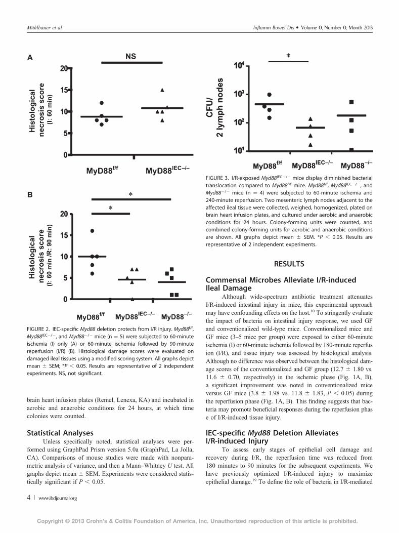
FIGURE 7. Expression of the IgA transporter polymeric immunoglobulin receptor is decreased in Myd88IEC2/2 compared with Myd88f/f mice.Myd88f/f and Myd88IEC2/2 mice (n ¼ 5) were euthanized, and polymeric immunoglobulin receptor expression was evaluated using IHC on healthysmall intestinal tissue. Representative images are shown of 2 independent experiments. Scale bars ¼ 200 mm.
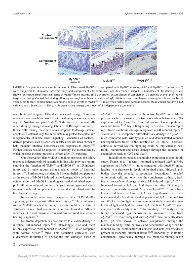
FIGURE 8. IgA bound to ischemic tissue was reduced in I/R-exposed Myd88IEC2/2 compared with Myd88f/f mice. Myd88f/f and Myd88IEC2/2 mice(n ¼ 5) were subjected to 60-minute ischemia only, and IgA expression was evaluated using IHC. IgA staining is also shown for healthy smallintestinal tissue of Myd88f/f mice (healthy SI). Black arrows: accumulation of IgA staining at the tip of the villi (region i.e., being affected first duringI/R injury). White arrows: IgA staining within B cells. White stars: staining of IgA within luminal debris. Scale bars ¼ 200 mm. Representative imagesare shown of 2 independent experiments.
Inflamm Bowel Dis ! Volume 0, Number 0, Month 2013 Epithelial-specific MyD88 Signaling
www.ibdjournal.org | 7
Copyright © 2013 Crohn’s & Colitis Foundation of America, Inc. Unauthorized reproduction of this article is prohibited.
microbiota protect against I/R-induced intestinal damage. Numerousinnate sensors have been linked to intestinal injury responses includ-ing the Nod-like receptor Nod2.15 Nod2 seems to prevent I/R-induced injury through downregulation of TLR4 expression in epi-thelial cells, making these cells less susceptible to damage-inducedapoptosis.15 Alternatively, the microbiota may protect the epitheliumindependently of innate sensor signaling. Generation of bacterial-derived products such as short-chain fatty acids has been shown tohelp maintain intestinal homeostasis and responses to injury.32,33
Further studies would be required to identify the mechanism bywhich bacteria mediate protective effects after I/R exposure.
Our observation that MyD88 signaling promotes the injuryresponse independently of bacteria is in line with previous reportsstudying the function of TLR430 and MyD8814 in I/R-inducedinjury and by other groups using a related model of intestinalinjury.15,34. Furthermore, we identified the epithelial compartmentas the source of MyD88-induced tissue damage. Mice defective inepithelial-derived MyD88 signaling showed diminished neutro-phil infiltration, reduced binding of sIgA to neoantigens and a sub-sequently reduced complement activation that correlated with thehistological damage.
Interestingly, other reports suggested that TLR2/MyD88signaling protects against I/R-induced injury.35 The contrastingrole of MyD88 in intestinal injury response could be because ofvariations in microbial communities in mice housed in differentfacilities. Different microbial compositions can modulate wound-healing responses.36
Neutrophil depletion has been shown to alleviate damage inintestinal I/R-induced injury.24,25 Interestingly, ccl2 and cxcl1mRNA expression were reduced in Myd88IEC2/2 mice comparedwith control Myd88f/f mice. This reduction correlated witha decreased infiltration of neutrophils into damaged tissue of
Myd88IEC2/2 mice compared with control Myd88f/f mice. Multi-ple studies have shown a positive association between mRNAexpression of Ccl2 and Cxcl1 and infiltration of neutrophils intoischemic tissue.25,37 MyD88 signaling is essential for neutrophilrecruitment and tissue damage in myocardial I/R-induced injury.23
Victoni et al14 also reported alleviated tissue damage inMyd882/2
mice compared with wild-type mice and demonstrated reducedneutrophil recruitment to the intestine on I/R injury. Therefore,epithelial-derived MyD88 signaling could be implicated in neu-trophil recruitment and tissue damage through the induction ofchemokines such as Ccl2 and Cxcl1.
In addition to reduced chemokine expression as seen in thisstudy, Frantz et al38 recently reported a reduced pIgR mRNAexpression in Myd88IEC2/2 mice compared with Myd88f/f mice,leading to a decrease in levels of luminal sIgA. Mucosal anti-bodies have the potential to recognize “neoantigens” revealedon ischemic cells and to activate the complement pathway, lead-ing to exacerbate damage during I/R-induced injury.10,30,39,40
Increased intestinal IgA and IgM deposition after I/R injury inmice was previously reported.10 Because Myd88IEC2/2 mice havelower basal levels of luminal IgA, we hypothesized that thesemice would display diminished deposition of IgA in injured tis-sue. We focused on IgA because a previous study reported similarlevels of IgG and IgM but lower IgA levels in Myd882/2 micecompared with wild-type mice.41 In this study, IHC analysis con-firmed decreased IgA deposition in ischemic tissue fromMyd88IEC2/2 mice compared with Myd88f/f mice. Recently dena-tured IgA was shown to activate complement through themannose-binding lectin pathway and denaturation of IgA can beinduced by the combination of acidosis and beta-galactosidasespresent in ischemic intestinal tissue.28,42 Importantly, inhibitingcomplement, specifically through the mannose-binding lectin
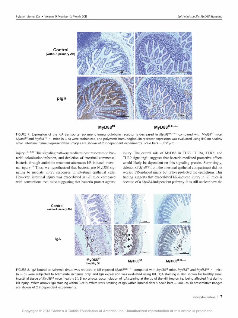
FIGURE 9. Complement activation is impaired in I/R-exposed Myd88IEC2/2 compared with Myd88f/f mice. Myd88f/f and Myd88IEC2/2 mice (n ¼ 5)were subjected to 60-minute ischemia only, and complement c3d expression was determined using IHC. Complement 3d staining is alsoshown for healthy small intestinal tissue of Myd88f/f mice (healthy SI). Black arrows: accumulation of complement 3d staining at the tip of the villi(region i.e., being affected first during I/R injury and region with accumulation of IgA). White arrows: complement staining in submucosal bloodvessels. White stars: complement staining only seen in crypts of Myd88IEC2/2 mice when histological damage exceeds stage 2 (absence of villi butvisible crypts). Scale bars ¼ 200 mm. Representative images are shown of 2 independent experiments.
Mühlbauer et al Inflamm Bowel Dis ! Volume 0, Number 0, Month 2013
8 | www.ibdjournal.org
Copyright © 2013 Crohn’s & Colitis Foundation of America, Inc. Unauthorized reproduction of this article is prohibited.
pathway, prevents I/R-induced injury.27,43 Interestingly, both IgAand complement displayed similar deposition on injured tissue ofMyd88f/f mice, a pattern reduced in Myd88IEC2/2 mice. In con-trast, GF mice have reduced IgA production44 and severe tissuedamage, suggesting that a mechanism other than IgA/complementactivation mediates intestinal injury in this system. For example,impaired production of cytoprotective molecules such as short-chain fatty acids in GF mice may explain their increased suscep-tibility to I/R injury.32
In summary, this study identified IEC-derived Myd88signaling as promoting I/R-induced injury. We showed thatIEC-derived MyD88 signaling leads to increased basal levels ofluminal IgA and activated complement in ischemic tissue. Inaddition, IEC-derived MyD88 controls the induction of neutrophilchemoattractants. This dual MyD88-dependent function is likelyimportant for I/R-induced intestinal damage. These findingsindicate that IEC-derived MyD88 signaling should be considereda central player in I/R-induced injury and that therapeuticintervention could focus on this signaling protein.
ACKNOWLEDGMENTSThe authors thank Dr Balfour Sartor and Maureen Bower of
the National Gnotobiotic Rodent Resource Center at University ofNorth Carolina for assistance with germ-free mice (NIH P40R018603). We thank Dr. Anthony DeFranco, University of CaliforniaSan Francisco, CA for the generous gift of MyD88f/f mice. Histologywas performed at the CGIBD histology core (P30 DK034987). Theauthors thank Ms Brigitte Allard for technical assistance throughoutthis project and Dr Javier Guzman for editorial assistance.
Author Contribution: MM and CJ conceived and designedthe experiments; MM and EP-C performed the experiments; MMand CJ analyzed the data; MM and CJ wrote the article.
REFERENCES1. Camilleri M, Madsen K, Spiller R, et al. Intestinal barrier function in
health and gastrointestinal disease. Neurogastroenterol Motil. 2012;24:503–512.
2. Hansen J, Gulati A, Sartor RB. The role of mucosal immunity and hostgenetics in defining intestinal commensal bacteria. Curr Opin Gastroen-terol. 2010;26:564–571.
3. McDole JR, Wheeler LW, McDonald KG, et al. Goblet cells deliverluminal antigen to CD103+ dendritic cells in the small intestine. Nature.2012;483:345–349.
4. Abreu MT. Toll-like receptor signalling in the intestinal epithelium: howbacterial recognition shapes intestinal function. Nat Rev Immunol. 2010;10:131–144.
5. Maynard CL, Elson CO, Hatton RD, et al. Reciprocal interactions of theintestinal microbiota and immune system. Nature. 2012;489:231–241.
6. Kayama H, Takeda K. Regulation of intestinal homeostasis by innate andadaptive immunity. Int Immunol. 2012;24:673–680.
7. Vollmar B, Menger MD. Intestinal ischemia/reperfusion: microcirculatorypathology and functional consequences. Langenbecks Arch Surg. 2010;396:13–29.
8. Mallick IH, Yang W, Winslet MC, et al. Ischemia-reperfusion injury ofthe intestine and protective strategies against injury. Dig Dis Sci. 2004;49:1359–1377.
9. Watanabe T, Kobata A, Tanigawa T, et al. Activation of the MyD88signaling pathway inhibits ischemia-reperfusion injury in the small intes-tine. Am J Physiol Gastrointest Liver Physiol. 2012;303:G324–G334.
10. Yoshiya K, Lapchak PH, Thai T-H, et al. Depletion of gut commensalbacteria attenuates intestinal ischemia/reperfusion injury. Am J PhysiolGastrointest Liver Physiol. 2011;301:1020–1030.
11. Fukata M, Michelsen KS, Eri R, et al. Toll-like receptor-4 is required forintestinal response to epithelial injury and limiting bacterial translocation ina murine model of acute colitis. Am J Physiol Gastrointest Liver Physiol.2005;288:G1055–G1065.
12. Burdelya LG, Krivokrysenko VI, Tallant TC, et al. An agonist of toll-likereceptor 5 has radioprotective activity in mouse and primate models.Science. 2008;320:226–230.
13. Rakoff-Nahoum S, Paglino J, Eslami-Varzaneh F, et al. Recognition ofcommensal microflora by toll-like receptors is required for intestinalhomeostasis. Cell. 2004;118:229–241.
14. Victoni T, Coelho FR, Soares AL, et al. Local and remote tissue injuryupon intestinal ischemia and reperfusion depends on the TLR/MyD88signaling pathway. Med Microbiol Immunol. 2009;199:35–42.
15. Richardson WM, Sodhi CP, Russo A, et al. Nucleotide-binding oligomer-ization domain-2 inhibits toll-like receptor-4 signaling in the intestinalepithelium. Gastroenterology. 2010;139:904–917.e6.
16. Hou B, Reizis B, DeFranco AL. Toll-like receptors activate innate andadaptive immunity by using dendritic cell-intrinsic and -extrinsic mecha-nisms. Immunity. 2008;29:272–282.
17. Uronis JM, Mühlbauer M, Herfarth HH, et al. Modulation of the intestinalmicrobiota alters colitis-associated colorectal cancer susceptibility. PLoSOne. 2009;4:e6026.
18. Jijon HB, Madsen KL, Walker JW, et al. Serum amyloid A activates NF-kB and proinflammatory gene expression in human and murine intestinalepithelial cells. Eur J Immunol. 2005;35:718–726.
19. Goldsmith JR, Perez-Chanona E, Yadav PN, et al. Intestinal epithelialcell-derived m-opioid signaling protects against ischemia reperfusioninjury through PI3K signaling. Am J Pathol. 2013;182:776–785.
20. Jilling T, Simon D, Lu J, et al. The roles of bacteria and TLR4 in rat andmurine models of necrotizing enterocolitis. J Immunol. 2006;177:3273–3282.
21. Sun X, Threadgill D, Jobin C. Campylobacter jejuni induces colitisthrough activation of mammalian target of rapamycin signaling. Gastro-enterology. 2012;142:86–95.e5.
22. Feinman R, Deitch EA, Watkins AC, et al. HIF-1 mediates pathogenicinflammatory responses to intestinal ischemia-reperfusion injury. Am JPhysiol Gastrointest Liver Physiol. 2010;299:G833–G843.
23. Feng Y, Zhao H, Xu X, et al. Innate immune adaptor MyD88 mediatesneutrophil recruitment and myocardial injury after ischemia-reperfusion inmice. Am J Physiol Heart Circ Physiol. 2008;295:H1311–H1318.
24. Hernandez LA, Grisham MB, Twohig B, et al. Role of neutrophils inischemia-reperfusion-induced microvascular injury. Am J Physiol. 1987;253:H699–H703.
25. Sisley AC, Desai T, Harig JM, et al. Neutrophil depletion attenuateshuman intestinal reperfusion injury. J Surg Res. 1994;57:192–196.
26. Martinez-Mier G, Toledo-Pereyra LH, McDuffie JE, et al. Neutrophildepletion and chemokine response after liver ischemia and reperfusion.J Invest Surg. 2001;14:99–107.
27. Huang Y, Qiao F, Atkinson C, et al. A novel targeted inhibitor of thealternative pathway of complement and its therapeutic application inischemia/reperfusion injury. J Immunol. 2008;181:8068–8076.
28. Terai I, Kobayashi K, Vaerman J-P, et al. Degalactosylated and/or dena-tured IgA, but not native IgA in any form, bind to mannose-binding lectin.J Immunol. 2006;177:1737–1745.
29. Gralinski MR, Park JL, Ozeck MA, et al. LU 51198, a highly sulfated,low-molecular-weight heparin derivative, prevents complement-mediatedmyocardial injury in the perfused rabbit heart. J Pharmacol Exp Ther.1997;282:554–560.
30. Moses T, Wagner L, Fleming SD. TLR4-mediated Cox-2 expressionincreases intestinal ischemia/reperfusion-induced damage. J Leukoc Biol.2009;86:971–980.
31. Murray PJ, Smale ST. Restraint of inflammatory signaling by interdependentstrata of negative regulatory pathways. Nat Immunol. 2012;13:916–924.
32. Baba AA, Srinivas M, Shariff A, et al. Role of short chain fatty acids inmesenteric ischemia reperfusion injury in rats. Eur J Pediatr Surg. 2010;20:98–101.
Inflamm Bowel Dis ! Volume 0, Number 0, Month 2013 Epithelial-specific MyD88 Signaling
www.ibdjournal.org | 9
Copyright © 2013 Crohn’s & Colitis Foundation of America, Inc. Unauthorized reproduction of this article is prohibited.

33. Tedelind S, Westberg F, Kjerrulf M, et al. Anti-inflammatory propertiesof the short-chain fatty acids acetate and propionate: a study with rele-vance to inflammatory bowel disease. World J Gartroenterol. 2007;13:2826–2832.
34. Neal MD, Sodhi CP, Dyer M, et al. A critical role for TLR4 induction ofautophagy in the regulation of enterocyte migration and the pathogenesisof necrotizing enterocolitis. J Immunol. 2013;190:3541–3551.
35. Aprahamian CJ, Lorenz RG, Harmon CM, et al. Toll-like receptor 2 isprotective of ischemia–reperfusion-mediated small-bowel injury ina murine model. Pediatr Crit Care Med. 2008;9:105–109.
36. Iizuka M, Konno S. Wound healing of intestinal epithelial cells. World JGartroenterol. 2011;17:2161–2171.
37. Santen S, Wang Y, Laschke MW, et al. Rho-kinase signalling regulatesCXC chemokine formation and leukocyte recruitment in colonic ischemia-reperfusion. Int J Colorectal Dis. 2010;25:1063–1070.
38. Frantz AL, Rogier EW, Weber CR, et al. Targeted deletion of MyD88 inintestinal epithelial cells results in compromised antibacterial immunity
associated with downregulation of polymeric immunoglobulin receptor,mucin-2, and antibacterial peptides. Mucosal Immunol. 2012;5:501–512.
39. Chen J, Crispín JC, Tedder TF, et al. B cells contribute to ischemia/reperfusion-mediated tissue injury. J Autoimmun. 2009;32:195–200.
40. Williams JP, Pechet TT, Weiser MR, et al. Intestinal reperfusion injury ismediated by IgM and complement. J Appl Physiol. 1999;86:938–942.
41. Woods A, Soulas-Sprauel P, Jaulhac B, et al. MyD88 negatively controlshypergammaglobulinemia with autoantibody production during bacterialinfection. Infect Immun. 2008;76:1657–1667.
42. Asp NG, Dahlqvist A. Human small intestine b-galactosidases: specificassay of three different enzymes. Anal Biochem. 1972;58:591–593.
43. Matthijsen RA, Derikx JPM, Steffensen R, et al. Mannose-binding lectinnull alleles are associated with preserved epithelial cell integrity followingintestinal ischemia reperfusion in man. Mol Immunol. 2009;46:2244–2248.
44. Hapfelmeier S, Lawson MAE, Slack E, et al. Reversible microbial coloni-zation of germ-free mice reveals the dynamics of IgA immune responses.Science. 2010;328:1705–1709.
Mühlbauer et al Inflamm Bowel Dis ! Volume 0, Number 0, Month 2013
10 | www.ibdjournal.org
Copyright © 2013 Crohn’s & Colitis Foundation of America, Inc. Unauthorized reproduction of this article is prohibited.