Peroxynitrite mediates neutrophil migration failure in severe polymicrobial sepsis
25
S1 Available online http://ccforum.com/supplements/11/S4 Critical Care Volume 11 Suppl 4, 2007 Sepsis 2007 Paris, France, 26–29 September 2007 Published online: 26 September 2007 These abstracts are available online at http://ccforum.com/supplements/11/S4 © 2007 BioMed Central Ltd P1 Cytokine-mediated regulation of renal urea transporters during sepsis Christoph Schmidt, Klaus Hoecherl, Michael Bucher Anaesthesiology Department, Hospital of the University of Regensburg, Regensburg, Germany Critical Care 2007, 11(Suppl 4):P1 (doi: 10.1186/cc5980) Background The pathogenesis of endotoxemic tubular dys- function with failure in urine concentration is poorly understood. Urea plays an important role in the urinary concentrating mechanism and expression of the urea transporters UT-A1, UT-A2, UT-A3, UT-A4 and UT-B is essential for tubular urea reabsorption. The present study attempts to assess the regulation of renal urea transporters during severe inflammation in vivo. Materials and methods By agreement of the animal protection committee C57BL/6J, mice were injected with lipopolysaccharides (LPS, 10 mg/kg) or proinflammatory cytokines. Hemodynamic, renal parameters and the expression of renal urea transporters were investigated. To clarify the role of cytokines and renal ischemia in the regulation of renal urea transporters, experiments were performed with cytokine knockout mice, mice treated with low-dose LPS (1, 5 mg/kg) as a sepsis model without induction of hypotension, glucocorticoid-treated mice, and mice with renal artery clipping serving as a model for renal ischemia. Results and discussion LPS-injected mice (10 mg/kg) presented with reduced glomerular filtration rate, fractional urea excretion and inner medulla osmolality associated with a marked decrease in expression of UT-A1, UT-A2, UT-A3, UT-A4 and UT-B (Figure 1). Similar alterations were observed after application of TNFα, IL-1β, IFNγ or IL-6. LPS-induced downregulation of urea transporters was not affected in knockout mice with deficient TNFα, IL-receptor-1, IFNγ or IL-6. Glucocorticoid treatment inhibited LPS-induced increases of tissue TNFα, IL-1β, IFNγ or IL-6 concentration, diminished LPS-induced renal dysfunction and attenuated the downregulation of renal urea transporters. Injection of low-dose LPS (1, 5 mg/kg) also led to renal dysfunction paralleled by a downregulation of renal urea transporters without alterations in blood pressure. Renal ischemia induced by renal artery clipping did not influence the expression of urea transporters. Conclusion Our findings demonstrate downregulation of renal urea transporters that probably accounts for tubular dysfunction during sepsis. Furthermore, they suggest that downregulation of Figure 1 (abstract P1) Effect of lipopolysaccharide (LPS) (10 mg/kg), dexamethasone (10 mg/kg) and the combination of both on UT-A1, UT-A2, UT-A3, UT-A4 and UT-B mRNA in the kidney 6, 12 and 24 hours after intraperitoneal injection. Values are related to signals obtained for β-actin mRNA and presented as the percentage of vehicle control. Mean ± SEM of six animals per group. *P < 0.05 versus control, # P < 0.05 versus LPS treatment.
Transcript of Peroxynitrite mediates neutrophil migration failure in severe polymicrobial sepsis

S1
Available online http://ccforum.com/supplements/11/S4
Critical Care Volume 11 Suppl 4, 2007Sepsis 2007Paris, France, 26–29 September 2007
Published online: 26 September 2007These abstracts are available online at http://ccforum.com/supplements/11/S4© 2007 BioMed Central Ltd
P1Cytokine-mediated regulation of renal urea transportersduring sepsis
Christoph Schmidt, Klaus Hoecherl, Michael BucherAnaesthesiology Department, Hospital of the University ofRegensburg, Regensburg, GermanyCritical Care 2007, 11(Suppl 4):P1 (doi: 10.1186/cc5980)
Background The pathogenesis of endotoxemic tubular dys-function with failure in urine concentration is poorly understood.Urea plays an important role in the urinary concentratingmechanism and expression of the urea transporters UT-A1, UT-A2,UT-A3, UT-A4 and UT-B is essential for tubular urea reabsorption.The present study attempts to assess the regulation of renal ureatransporters during severe inflammation in vivo.Materials and methods By agreement of the animal protectioncommittee C57BL/6J, mice were injected with lipopolysaccharides(LPS, 10 mg/kg) or proinflammatory cytokines. Hemodynamic,renal parameters and the expression of renal urea transporterswere investigated. To clarify the role of cytokines and renalischemia in the regulation of renal urea transporters, experiments
were performed with cytokine knockout mice, mice treated withlow-dose LPS (1, 5 mg/kg) as a sepsis model without induction ofhypotension, glucocorticoid-treated mice, and mice with renalartery clipping serving as a model for renal ischemia.Results and discussion LPS-injected mice (10 mg/kg) presentedwith reduced glomerular filtration rate, fractional urea excretion andinner medulla osmolality associated with a marked decrease inexpression of UT-A1, UT-A2, UT-A3, UT-A4 and UT-B (Figure 1).Similar alterations were observed after application of TNFα, IL-1β,IFNγ or IL-6. LPS-induced downregulation of urea transporters wasnot affected in knockout mice with deficient TNFα, IL-receptor-1,IFNγ or IL-6. Glucocorticoid treatment inhibited LPS-inducedincreases of tissue TNFα, IL-1β, IFNγ or IL-6 concentration,diminished LPS-induced renal dysfunction and attenuated thedownregulation of renal urea transporters. Injection of low-doseLPS (1, 5 mg/kg) also led to renal dysfunction paralleled by adownregulation of renal urea transporters without alterations inblood pressure. Renal ischemia induced by renal artery clippingdid not influence the expression of urea transporters.Conclusion Our findings demonstrate downregulation of renalurea transporters that probably accounts for tubular dysfunctionduring sepsis. Furthermore, they suggest that downregulation of
Figure 1 (abstract P1)
Effect of lipopolysaccharide (LPS) (10 mg/kg), dexamethasone (10 mg/kg) and the combination of both on UT-A1, UT-A2, UT-A3, UT-A4 and UT-BmRNA in the kidney 6, 12 and 24 hours after intraperitoneal injection. Values are related to signals obtained for β-actin mRNA and presented asthe percentage of vehicle control. Mean ± SEM of six animals per group. *P < 0.05 versus control, #P < 0.05 versus LPS treatment.

S2
Critical Care September 2007 Vol 11 Suppl 4 Sepsis 2007
renal urea transporters during LPS-induced acute renal failure ismediated by proinflammatory cytokines and is independent fromrenal ischemia due to sepsis-induced hypotension.Acknowledgement Supported by grants from the GermanResearch Foundation (SFB 699).
P2The role of regulatory T cells in the resistance of CCR4knockout mice during severe sepsis
Raphael Molinaro1, Alessandra Monteiro de França1, Josi Alves-Filho2, Fernando de Queiroz Cunha2, Marcelo Bozza3, Steven Kunkel4, Claudia Benjamim1
1Department of Pharmacology, UFRJ, Rio de Janeiro, RJ, Brazil;2Department of Pharmacology, USP, Ribeirão Preto, SP, Brazil;3Microbiology Institute, UFRJ, Rio de Janeiro, RJ, Brazil;4Department of Pathology, UMMS, Ann Arbor, MI, USACritical Care 2007, 11(Suppl 4):P2 (doi: 10.1186/cc5981)
Background Studies reveal that regulatory T (Treg) cells controlimmune responses; therefore these responses must be controlledto enable effective protection against infections and cancer. CCR4knockout (CCR4–/–) mice are more resistant to lipopolysaccharideshock. So, our aim is to study the mechanisms involved in the resis-tance of CCR4–/– mice subjected to severe sepsis by cecal ligationand puncture (CLP) and how Treg cells modulate this effect.Methods C57/BL6 mice were subjected to a CLP model, wherebythe cecum was partially ligated and punctured nine times with a21 G needle. Sham-operated mice were used as control. Micesubjected to CLP and sham surgery were treated with antibioticfrom 6 hours after surgery until 3 days.Results CCR4–/– mice subjected to CLP presented an increase inthe survival rate (78%) compared with wild-type mice (17%), andpresented a marked improvement in the innate response concern-ing neutrophil migration to the peritoneum and lung, bacterial loadand cytokine levels compared with wild-type mice. Besides, Tregcells from CCR4–/– CLP mice did not inhibit proliferation of Teffector cells as observed for Treg cells from wild CLP mice, at aproportional ratio of T effector:Treg cells. Interesting, Treg cells fromCCR4–/– CLP mice inhibit 30% of neutrophil migration to broncho-alveolar lavage when co-injected with fungal challenge assecondary infection in sham recipient mice, while the Treg cellsfrom wild CLP mice inhibit 80%, much more than expected.Conclusion These results suggest that Treg cells from CCR4–/–
mice did not present a suppressive response and this could be animportant factor in their survival. These results are strong evidencefor the role of Treg cells in immunosuppression following severesepsis.
P3Abdominal sepsis: efficacy of passive immunotherapy
Ruslan Knut, Peter Fomin, Oleg Sydorchuk, Ruslan Sydorchuk,Olexii Kolomoiets, Larysa Sydorchuk, Igor Sydorchuk,Mohhamad DaraghmehGeneral Surgery Department, Bukovina State Medical University,UkraineCritical Care 2007, 11(Suppl 4):P3 (doi: 10.1186/cc5982)
Background Owing to immune disorders playing a key role indevelopment of systemic inflammatory response syndrome, passiveimmune therapy is considered a method of choice for abdominalsepsis (AS) patients. Existing remedies (specific hyperimmuneserum, specific antibodies and immunoglobulins) are expensiveand require exact validation of pathogens. The aim of the study was
to evaluate the efficacy of using the AS reconvalescent donorplasma for passive immunotherapy of AS.Materials and methods The study was conducted experimentallyon 775 Wistar line rats and 38 inbreed dogs. A total of 296patients with AS were also involved in the study; 58 formed thecontrol group; 26 patients were selected as reconvalescentdonors of plasma. Serum concentrations (ELISA) of major anti-bodies were determined against most significant pathogens(Escherichia coli, Staphylococcus spp., Staphylococcus aureus,Bacteroides spp., Klebsiella pneumoniae, Pseudomonas aeruginosa).Results Changes of serum antibody concentrations were timedependent and fluctuating during the current of AS forming thewaveform curve. Most remarkable decreases were found during24–72 hours of AS. Serum antibody titers to the main pathogenswere slightly higher due to antibiotics and detoxification therapy.Operation by itself decreased titers from 4.42 ± 0.28 to 3.49 ± 0.25(E. coli), and from 5.41 ± 1.02 to 3.0 ± 0.58 (P. aeruginosa). Anti-staphylococcal antibody titers decreased from 7.22 ± 0.9 beforesurgery to 4.83 ± 0.47 after. Repeated operations alter antibodyconcentrations even more significantly. The highest levels ofantibodies were found in patients who underwent successfultreatment of AS 1–2 months prior to investigation. Their plasmawas used in treatment of AS patients. Intravenous administration oftwo-dose 100–200 ml hyperimmune plasma per day prevented afollowing decrease of antibody levels, and in 98% of casesincreased them (21.39 ± 1.47%). The cost of treatment was15–37% lower if compared with traditional methods (control group).Conclusion There is exact evidence of efficacy for using hyper-immune plasma in patients with abdominal sepsis; it is more cost-effective if compared with traditional methods of immunotherapy.
P4The modified light chain of inter-alpha inhibitor/antibodyfusion protein, MR1007, improves survival in the rabbit sepsismodels
Masaki Nakamura, Takashi Takeuchi, Yukiko Yatagai, Katsuki Naito, Kamon Shirakawa, Yoshitaka Hosaka, Shoji FurusakoDiscovery Research, Mochida Pharmaceutical Co., Ltd, Gotemba,JapanCritical Care 2007, 11(Suppl 4):P4 (doi: 10.1186/cc5983)
Background Inter-alpha inhibitor is an endogenous serine proteaseinhibitor and is markedly reduced in severe sepsis. Therapeuticinter-alpha inhibitor replacement showed a survival advantage inseveral animal models. The light chain is responsible for the serineprotease inhibitory activity of inter-alpha inhibitor. Since pro-coagulant and proinflammatory proteases as well as innate immunecells are activated in sepsis, we genetically engineered a novelfusion protein, MR1007, which consists of the modified light chainof inter-alpha inhibitor and the anti-CD14 antibody, and evaluatedthe potential of MR1007 as an anti-sepsis agent.Methods Inhibitory activity against serine proteases was assayedusing purified enzymes and chromogenic substrates. Anticoagulantactivity was measured using human or rabbit plasma. The inhibitoryeffect on endothelial cell injury was assessed using humanumbilical vein endothelial cells. Binding to CD14 and leukocyteswas analysed using Biacore or radiolabeling. The survival benefitwas evaluated in the endotoxin shock model and the cecal ligationand puncture (CLP) model.Results MR1007 inhibited the thromboplastin-induced thrombingeneration by inhibiting activities of coagulation factors Xa and XIaat 10–100 μg/ml. It also prevented the contact pathway generationof bradykinin at 10–30 μg/ml. Additionally, it inhibited the

S3
Available online http://ccforum.com/supplements/11/S4
leukocyte elastase-induced endothelial cell injury at 10–100 μg/ml.MR1007 had a high affinity for CD14 and bound to leukocytes, butdid not block lipopolysaccharide binding to CD14. In the rabbitendotoxin shock model, MR1007 (3 mg/kg, i.v.) even when given8 hours after the injection of endotoxin improved the survival(n = 12, P < 0.05), whereas both antithrombin and prednisoloneexhibited less efficacy. Moreover, MR1007 (10 mg/kg, i.v.) given at2 hours post-CLP improved the survival (n = 9, P < 0.05) in theCLP model.Conclusions These results suggest that the modified light chain ofinter-alpha inhibitor fusion protein, MR1007, can effectivelysuppress not only the serine protease-mediated coagulation, butalso leukocyte-induced inflammation, so that MR1007 maybecome a promising anti-sepsis agent.
P5Microbial metabolites in the blood of patients with sepsis
Anastasia Khodakova, Natalia BeloborodovaBakulev Scientific Center for Cardiovascular Surgery, Moscow,RussiaCritical Care 2007, 11(Suppl 4):P5 (doi: 10.1186/cc5984)
Background Molecular mechanisms of the pathophysiology ofsepsis remain unknown. Preliminary results allow one to supposethat investigation of biological effects of microbial metabolites,particularly aromatic acids, is one of the most promising methodsin elucidation of the problem. These compounds can be producedduring microbial fermentation of aromatic amino acids. But little isknown of which microorganisms participate in such processes.The aim was to assess the comparative level of aromatic acids inserum of cardiosurgical patients with documentary sepsis and toclarify the in vitro metabolic profile of aromatic acids in spentgrowth medium of main clinical blood isolates.Materials and methods Serum samples were collected from 83adult subjects (mean age 52 (42–58) years). The main group ofinvestigation consisted of 12 cardiosurgical patients with docu-mentary sepsis, with mortality of 42% (5/12). The comparisongroups were: 16 clinically healthy volunteers, 36 patients withacquired heart diseases before surgery, nine patients with smoothrecovery on the third day after surgery, 10 patients with pneumoniaafter surgery. The cultures (n = 32) of S. aureus, S. epidermidis,E. faecalis, K. pneumonia, S. marcesceus, E. coli, E. cloacae,A. baumanii, P. aeruginosa, C. albicans and C. parapsilosis wereisolated from the blood of cardiosurgical patients and identified.Concentrations of aromatic acids were determined by gaschromatography–mass spectrometry. Data were compared byMann–Whitney U-test, P < 0.05 considered significant.
Results Significant differences were observed among the groups(Table 1). 3-Phenylpropionic and 1-indolacetic acids were found tobe prevalent in groups of healthy volunteers and patients beforesurgery. Increased levels of phenyllactic acid (PLA), p-hydroxy-phenylacetic acid (HPAA), p-hydroxyphenyllactic acid (HPLA) and3-indolacetic acid were revealed in the group of sepsis comparedwith other groups. Moreover, the highest concentrations of PLA,HPAA and HPLA were in serum of nonsurvivors (n = 5) comparedwith survivors (n = 7): PLA, 1,651 (656–1,959) versus 233(122–360) ng/ml, P = 0.02; HPAA, 5,976 (2,689–6,667) versus1,108 (461–2,121) ng/ml, P = 0.02; HPLA, 3,313 (2,409–6,098)versus 564 (446–718) ng/ml, P = 0.005. Gas chromatography–mass spectrometry analysis of spent growth medium showed thatGram-negative enterobacteria produced increased amounts ofPLA and HPLA acids. Particularly, K. pneumonia had the highestlevel of acids PLA = 100 r.u. (r.u. – the ratio of substance contentin sample to uninoculated media) and HPLA = 60 r.u., E. coli hadPLA = 35 r.u. and HPLA = 20 r.u., and S. marcesceus andE. cloacae had PLA = 4 r.u. and HPLA = 6 r.u. The culture ofGram-positive cocci produced increased level of the same acids,for S. aureus PLA = 20 r.u. and HPLA = 17 r.u., and for S.epidermidis PLA = 6 r.u. and HPLA = 3 r.u., except for E. faecalis,which had the only PLA = 6 r.u. Gram-negative nonfermentedbacteria produced increased levels of 1-indolacetic acid and 3-indolacetic acid, but no PLA and HPLA. The level of aromatic acidsin the medium after cultivation of fungi was equal to control.Conclusions Increased levels of PLA, HPAA and HPLA in serumpatients with sepsis, especially with fatal outcome, areassociated with development of infectious complications. Thesecompounds are produced by clinically important bacteria, such asK. pneumonia > E. coli > S. aureus > S. marcesceus, E. cloacae,S. epidermidis, but not by fungi. The results can denote biologicalactivity of these microbial metabolites and their influence onpathogenesis of sepsis.
P6The impact of protocolized sepsis order set on the process ofcare in patients with severe sepsis/septic shock
Bekele Afessa, John Mullon, Andrew Badley and Ognjen GajicMayo Clinic College of Medicine, Pulmonary and Critical CareDivision, Rochester, MN, USACritical Care 2007, 11(Suppl 4):P6 (doi: 10.1186/cc5985)
Background Based on the available evidence, professionalsocieties have published practice guidelines on severe sepsis andseptic shock. We started using a paper order set based on theguidelines in our medical intensive care unit (ICU) in October
Table 1 (abstract P5)
Concentration of aromatic acids in serum sepsis patients and subjects of comparison groups
Patients Patients before Patients with Patients with with sepsis Volunteers surgery smooth recovery pneumonia
Aromatic acid (ng/ml) (n = 12) (n = 16) (n = 36) (n = 9) (n = 10)
p-Hydroxyphenylacetic acid 2,140 (631–3,516) 72 (62–93)*** 114 (53–220)*** 263 (109–313)** 456 (344–667)*
3-Phenylpropionic acid 0 35 (20–54)** 4 (0–29) 0 0 (0–3)
Phenyllactic acid 367 (217–1,098) 47 (37–64)** 58 (37–93)** 89 (68–126)* 112 (89–177)*
p-Hydroxyphenyllactic acid 1,543 (564–2,731) 195 (159–371)*** 254 (134–373)*** 288 (269–772)* 465 (314–836)
1-Indolacetic acid 0 262 (113–385)*** 47 (0–218)* 275 (228–499)*** 0 (0–27)
3-Indolacetic acid 246 (183–628) 93 (56–141)* 181 (60–542) 57(30–74)* 231 (149–375)
Data presented as median (25th–75th percentile range). *P < 0.05, **P < 0.001, ***P < 0.0001, compared with sepsis patients.

S4
2005. This prospective study aims to describe the impact of theorder set on the process of care.Materials and methods We included patients with severe sepsis/septic shock treated in our ICU between October 2005 and April2007. We collected Acute Physiology and Chronic HealthEvaluation (APACHE) III derived severity data, compliance with sixelements of early goal-directed therapy and hospital mortality.Compliance with each element was defined as the use of thefollowing within 6 hours of severe sepsis/septic shock: use ofcentral venous pressure, central venous oxygen saturationmeasurement, adequate fluid resuscitation and appropriate use ofvasopressors, inotropes and transfusion of red blood cells. TheICU admission severity of illness and sepsis stage (severe orshock) were entered in a logistic regression model to determinethe independent impact of the order set on mortality. P < 0.05 wasconsidered significant.Results Of 561 patients (168 severe sepsis and 373 septicshock), 31 were excluded for not authorizing research. The orderset was utilized in 328 (61.9%) of 530 patients. There were nosignificant differences in gender, age, race, and severity of illnessat ICU admission between the order set and nonorder set groups.The order set was more likely to be used in patients with septicshock than in those with severe sepsis (67.3% versus 51.4%;P = 0.0004). Compliance with all six elements occurred in 130(39.6%) of the order set group compared with 50 (24.8%) of thenonorder set group (P = 0.0004). Although mortality did notchange, compliance with five of the six elements improvedsignificantly with the order set. Logistic regression analysis showedthat shock (odds ratio (OR) = 2.384, 95% confidence interval (CI) =1.431–3.970; P = 0.0008) and predicted APACHE III mortality(%) (OR = 1.040, 95% CI = 1.031–1.050; P < 0.0001) wereassociated with mortality, not the order set (OR = 0.742, 95% CI =0.476–1.157; P = 0.1881).Conclusions This study showed that a protocolized order setimproves compliance with the standard of care in patients withsevere sepsis and septic shock. However, it did not resolve someof the noncompliance problems and did not improve survival.
P7Impact of autologous centrifuged shed mediastinal blood onprocalcitonin, C-reactive protein levels and postoperativecomplications during the early period following cardiacsurgery
Judita Andrejaitiene1, Audrone Veikutiene2, Edmundas Širvinskas1, Rimantas Benetis3
1Institute for Biomedical Research, Kaunas Medical University,Kaunas, Lithuania; 2Kaunas University of Medicine Hospital,Department of Cardiothoracic and Vascular Surgery, Kaunas,Lithuania; 3Kaunas University of Medicine, Institute for Cardiology,Kaunas, LithuaniaCritical Care 2007, 11(Suppl 4):P7 (doi: 10.1186/cc5986)
Background Cardiac surgery with cardiopulmonary bypass (CPB)is associated with a number of adverse effects due to systemicinflammatory response syndrome, a physiologic reaction to tissueinjury. Because of this response, conventional clinical andbiological signs may be misleading in the diagnosis of post-operative complications, particularly infections. The aim of thestudy was to evaluate the impact of autologous centrifuged shedmediastinal blood procedures in attitude of infection, estimatingthe predictive role of procalcitonin (PCT) and C-reactive protein(CRP) changes during the postoperative period.
Materials We have analysed data on 90 patients, who had beensubjected to cardiac surgical procedures on CPB: there are 41patients in Group I, who were reinfused with the centrifugedautologous mediastinal blood 4 hours after the end of surgery; and49 patients in Group II, whose shed mediastinal blood was notreinfused (control group).Methods We studied the quantity of haemoglobin, haematocritand leucocyte counts, and the value of CRP and PCT concen-trations before the surgery (baseline), 4 and 20 hours after the endof surgery and during 5 days after surgery. Preoperative patientconditions, intraoperative and postoperative periods, were recorded.Statistical significance was accepted at a level of P < 0.05.Results In Group I, patients who were reinfused with thecentrifuged autologous shed mediastinal blood, requirement for theallogenic blood transfusion procedures was significantly lower(14.6% versus 38.8%, P < 0.05). The CRP concentration wasgreater, but there were no significant differences between thegroups in all postoperative periods. At 20 hours after the end ofsurgery and the second postoperative day, the increase of the PCTconcentration was significant and often observed in group II(33.3% versus 58.3%), where there were significantly morecomplications of infection (2.4% versus 10.2% P < 0.05) and asignificantly longer length of postoperative hospital stay(9.32 ± 2.55 versus 14.38 ± 4.27 days, P < 0.05).Conclusions Our data suggest that the early reinfusion ofautologous centrifugated shed mediastinal blood procedures didnot increase bleeding and statistically significantly reduced therequirement of allogenic blood transfusion procedures, reducedthe number of infection complications, and significantly shortenedthe length of postoperative hospital stay. In evaluation ofpostoperative infection rates, PCT is highly suggestive as a markerof postoperative complications.
P8System approach to the diagnosis and treatment of septicarthritis in newborns
Gennadiy Khanes1, Svitlana Bidnenko2, Volodymyr Grygorovskyy3, Iryna Maksakova1, Valeriy Romashko4
1Neonatal Surgery Department, Ukrainian Specialized PediatricHospital OkhMatDyt, Kyiv, Ukraine; 2Bacteriologic Laboratory ofScientific Research Institute of Traumatology and Orthopedics ofthe Academy of Medical Sciences of Ukraine, Kyiv, Ukraine;3Morphologic Laboratory of Scientific Research Institute ofTraumatology and Orthopedics of the Academy of MedicalSciences of Ukraine, Kyiv, Ukraine; 4Immunologic Laboratory,Ukraine Specialized Pediatric Hospital OkhMatDyt, Kyiv, UkraineCritical Care 2007, 11(Suppl 4):P8 (doi: 10.1186/cc5987)
Background Bone and joint sepsis occurs in 20% of newbornshaving perinatal sepsis. The feature of septic arthritis against thebackground of age-specific functional immunodeficiency gives us areason to consider this decease as the manifestation of immuno-deficiency. Septic arthritis in newborns is lethal in 10% of casesand gives orthopedic complications in 20% of cases. The startingmoments of septic arthritis development are: mother’s intrauterineinfection and nosocomial infecting in maternity hospitals andintensive care departments, and purulent omphalitis.Materials and methods We present the experience of diagnosticsand treatment of 180 newborns aged 3 days and older havingseptic affection of the hip, knee, shoulder and other joints. Thecrucial role in diagnostics of septic arthritis is played by: cytology
Critical Care September 2007 Vol 11 Suppl 4 Sepsis 2007

S5
of smear, ultrasonic examination of a joint, X-ray examination of ajoint, bacteriological, serological, PCR and PCT-Q. The bacterio-logical monitoring in 30% of newborns among the pathogensshowed CoMRSA, often mixed with fungi and Pseudomonasaerogenes or Klebsiella pneumonia, and in 10% there was PCR ofToxoplasma gondi, Chlamidia trachomatis or cytomegalovirus.Immunological monitoring allowed one to determine the patientswho needed substitute therapy by immunoglobulin and a numberof immunomodulators. The degree of infection process severityand the adequacy of antibacterial therapy are determined byserologic investigations, PCR and PCT-Q. The treating complexincluded joint lavage, antibiotics, anticytokine, antifungal agents,probiotics, and magnetic-laser therapy.Results The lethality was reduced to zero, transition into thechronic form was up to 2.5%, and orthopedic complications werepresented in 10% of cases.Conclusion The after-history of treatment was researched andshowed that when the diagnosis was made in less than 3 days,complications occurred in 3.3% of cases, and after 6–7 days thecomplications occurred in 22.4% of patients.
P9Protease-activated receptor 2 blocking peptide counteractsendotoxin-induced inflammation and coagulation andameliorates glomerular fibrin deposition in a rat model ofacute renal failure
Satoshi Gando, Subrina Jesmin, Sohel Zaedi, Shamsul Haque Prodhan, Atsushi Sawamura, Takashi Miyauchi, Naoto YamaguchDivision of Acute and Critical Care Medicine, Department ofAnesthesiology and Critical Care Medicine, Hokkaido UniversityGraduate School of Medicine, Sapporo, JapanCritical Care 2007, 11(Suppl 4):P9 (doi: 10.1186/cc5988)
Background Glomerular and microvascular thrombosis due to theactivation of the inflammation and coagulation pathway contributeto the occurrence of acute renal failure in sepsis. The protease-activated receptors (PARs) have been shown to play an importantrole in the interplay between the inflammation and coagulation.Materials and methods We hypothesized that PAR2 blockingwould improve glomerular and vascular thrombosis by attenuatingthe inflammation and coagulation, leading to the prevention ofacute renal failure, and we assessed the effects of the PAR2blocking peptide (PAR2 BP) in a rat model of lipopolysaccharide(LPS)-induced acute renal failure.Results Levels of TNFα were significantly expressed 1 hour afterLPS administration, followed by: (i) an increase in levels of tissuefactor, factor VIIa, factor Xa, thrombin and plasminogen activatorinhibitor-1; (ii) unchanged levels of tissue factor pathway inhibitor;and (iii) subsequent deposition of fibrin in kidney tissues, which ledto the elevation of creatinine and blood urea nitrogen. Time-dependent PAR2 expression was observed at both the gene andprotein levels. Immunoreactivities of PAR2 and fibrin were co-localized in the glomerulus and the other kidney tissues. PAR2 BPsuppressed TNFα elevation, and attenuated activation of thecoagulation, thus leading to a decrease in fibrin formation and itsdeposition in the glomerulus. However, the levels of creatinine andblood urea nitrogen remained unchanged.Conclusions These results show that PAR2 plays a crucial role inthe inflammatory and coagulation process of LPS-induced renalfailure and may in part participate in the pathogenesis of thedisease.
P10Resistin in severe bacterial infections
Linda Johansson, Anna Linnér, Jonas Sundén-Cullberg, Carl-Johan Treutiger, Anna Norrby-TeglundCenter for Infectious Medicine, Institutionen för Medicin Huddinge,F59 Karolinska Institutet, Stockholm, SwedenCritical Care 2007, 11(Suppl 4):P10 (doi: 10.1186/cc5989)
Background Resistin has recently been recognized to act as aproinflammatory cytokine in humans. Patients with severe sepsis orseptic shock had significantly elevated systemic levels of resistin,which correlated with severity of disease. Here we have furthercharacterized the release of resistin during severe bacterialinfections.Materials and methods Acute phase sera collected from patientswith septic shock caused by Gram-negative (n = 19) or Gram-positive (n = 19) bacteria were analyzed for resistin by ELISA. Tissuebiopsies (n = 12) from patients with Streptococcus pyogenessevere soft tissue infections were stained for resistin and cellmarkers, and were analyzed by confocal microscopy. Humanneutrophils were stimulated with lipopolysaccharide orstreptococcal superantigens, and resistin was assessed in thesupernatants.Results Serum resistin levels were significantly elevated in patientswith Gram-positive, as compared with Gram-negative, septic shock(P = 0.004). Analyses of tissue biopsies revealed that resistin washighly expressed at the local site of infection. Dual-staining for cellmarkers confirmed published findings that monocytes are a sourceof resistin in humans, but importantly the stainings revealed that themajority of resistin-producing cells were negative for the monocyticmarker CD68. Further analyses identified these cells asneutrophils. A positive correlation between resistin levels andneutrophil counts was found in blood of septic shock patients(P = 0.005). In vitro cell cultures revealed resistin release byneutrophils stimulated with lipopolysaccharide or superantigens.Conclusions This study demonstrates that the systemic resistinlevels in septic shock differ depending on the causativemicroorganisms. The data also reveal that, at the local tissue site ofinfection, resistin is produced mainly by neutrophils, and systemicresistin strongly correlates with circulating levels of neutrophils.The systemic and local hyper-resistinemia noted is likely tocontribute to the pathogenesis of acute invasive bacterial infections.
P11Mortality rate reduction associated with severe sepsis andseptic shock management protocol implementation
Constantino José Fernandes Junior, Alexandre Gonçalves de Sousa, Gisele de Paula Dias Santos,Eliezer Silva, Nelson AkamineHospital Israelita Albert Einstein Division of Medical Practice –Department of Protocols, São Paulo, BrazilCritical Care 2007, 11(Suppl 4):P11 (doi: 10.1186/cc5990)
Background The Surviving Sepsis Campaign is an internationaleffort to reduce severe sepsis and septic shock associated mortalityby 25% in 5 years. We developed a management protocol in ourinstitution 2 years ago in order to follow the proposedrecommendations of this campaign, and describe the clinical impactof assuming this critical pathway on the mortality rate.Materials The study was conducted within the emergencydepartment and intensive care unit of a tertiary hospital. A manage-ment protocol for severe sepsis and septic shock was based onthe Surviving Sepsis Campaign guidelines and was implemented
Available online http://ccforum.com/supplements/11/S4

S6
by a ‘sepsis’ team including emergency department and criticalcare physicians, intensive care nurses and pharmacists, chaired bya full-time coordinator.Methods We performed a ‘before and after’ evaluation of thecritical pathway concerning 184 critically ill patients sequentiallyadmitted throughout a 16-month period.Results A total of 184 patients with severe sepsis or septic shockentered the study. Ninety-four patients had their analysis performedbefore the implementation of the standardized protocol (the‘before’ group), and 90 patients were managed following theimplementation of the protocol (the ‘after’ group). Basal demo-graphic variables and the severity of illness score (APACHE II)were similar for both groups.Patients in the ‘after’ group had statistically more cultures obtainedbefore institution of antibiotics and more patients receivedantibiotics in a due time (2 hours from diagnosis). In addition, thosepatients received more corticosteroids and activated protein C(Table 1).The intensive care unit length of stay and the hospital length of staywere similar in both groups. Remarkably the hospital mortality ratewas significantly lower (34.4%) in the ‘after’ group in septic shockpatients (67.7% versus 44.4%, P < 0.04) (Figure 1).Conclusions The implementation of the Surviving SepsisCampaign guidelines through a standardized protocol wasassociated with a 34% reduction in septic shock-related hospitalmortality.
P12Neonatal sepsis due to multidrug-resistant Klebsiellaterrigena in the neonatal intensive care unit in Gaza, Palestine
Farid Abu ElamreenMedical Microbiology Department, AlShifa Hospital CentralLaboratory & Blood Bank, Ministry of Health, Gaza, PalestineCritical Care 2007, 11(Suppl 4):P12 (doi: 10.1186/cc5991)
Background Bloodstream infection (BSI) is a major cause ofmorbidity and death encountered in the neonatal intensive careunits (NICUs). The rates of BSIs vary significantly in NICUs acrossthe nation. Klebsiella spp. led to serious concern aboutsepticaemic neonates in NICUs due to high resistance againstcommonly used antimicrobial agents. The objective of the studywas to report the prevalence and resistance pattern of Klebsiellaterrigena isolated from cases of neonatal septicaemia at AlshifaHospital, the largest tertiary hospital in Palestine.Methods Blood taken from newborn babies admitted to the NICUat Alshifa Hospital, Gaza, Palestine with a clinical diagnosis ofneonatal sepsis was cultured. A total of 355 positive bloodcultures isolated from January to December 2005 were studied.Antimicrobial susceptibility was determined by disc diffusionmethod.
Results A total of 355 blood cultures positive were studied; themost common organism found were Klebsiella spp. 202/355(56.9%), and 56/202 (25.5%) were K. terrigena. K. terrigenashowed a high degree of resistance to commonly used antibiotics(Ampicillin, Piperacillin, Cephalexin, Cefuroxime, Cefaclor andGentamicin), a moderate degree of resistance to Cefotaxime,Ceftazidim, Ceftriaxone and Amikacin, and most of the isolateswere sensitive to Meropenem.Conclusion Neonatal sepsis remains one of the leading causes ofneonatal admission, morbidity, and mortality in developingcountries. Klebsiella spp. were the major cause of neonatal sepsisin Gaza in 2005. The rare Klebsiella species (K. terrigena) havedeveloped multidrug resistance, and management of patientsinfected with them is becoming a problem in developing countries.There is a need to carefully formulate therapeutic strategies tocontrol infections in NICUs.
P13The identification and use of common physiologic monitoringparameters in the care of critically ill patients at risk for sepsis
Karen Giuliano, Erica CummingsPhilips Medical Systems, Andover, MA, USACritical Care 2007, 11(Suppl 4):P13 (doi: 10.1186/cc5992)
Background Sepsis is a common source of morbidity andmortality among critically ill patients. Targeting measures to reducethe incidence of and to promote early recognition and treatment ofsepsis is at the forefront of many critical care initiatives. Advancesin the management of severe sepsis have evolved over recentyears in an attempt to combat the spiraling mortality trends. TheSurviving Sepsis Campaign (SSC) is a worldwide initiativepromoting the evidence-based treatment of sepsis, with the explicitgoal of reducing both the morbidity and mortality associated withsepsis. This study was conducted to assess the clinical relevanceof the early physiologic screening criteria advocated by early goal-directed therapy for sepsis, and the Surviving Sepsis Campaignguidelines.Materials and methods The Project IMPACT® database wasused to obtain a sample of patients (n = 363) with an ICU admis-sion diagnosis of sepsis and a random acuity-matched comparisonsample of patients with an admission diagnosis (n = 364) otherthan sepsis.Results Significant group differences were found on all physio-logic monitoring variables tested (high temperature, P = 0.000; low
Critical Care September 2007 Vol 11 Suppl 4 Sepsis 2007
Figure 1 (abstract P11)
Mortality rate reduction in septic shock patients (42 deaths in ‘before’group versus 32 deaths in ‘after’ group). P < 0.04.
Table 1 (abstract P11)
Proceedings concerning ‘before’ versus ‘after’ groups
‘Before’ ‘After’ group group
Proceedings (%) (%) P value
Cultures obtained before antibiotics 68.1 84.3 0.01
Antibiotics in a due time (2-hour interval) 61.7 80.0 0.009
Corticosteroids 56.4 74.4 0.01
Activated protein C 1.0 12.2 0.002

S7
temperature, P = 0.001; heart rate, P = 0.004; respiratory rate,P = 0.005; and mean arterial pressure, P = 0.000). In the logisticregression model, high temperature and mean arterial pressurefunctioned as significant predictors, with odds ratios of 2.12 fortemperature at or above 38°C and 3.87 for MAP less than70 mmHg. The odds ratio of having sepsis was 4.63 if both ofthese predictors were present.Conclusions It is important to understand the value of commonmonitoring parameters in the early identification of sepsis, sincethose parameters are continuously monitored and readily available.It is the responsibility of bedside clinicians to assure that theparameters chosen for monitoring provide the most accuratereflection of the patient’s clinical status. These results providesome support for the use of the currently recommended criteria forphysiologic monitoring in the early identification of patients at riskfor developing sepsis. Furthermore, if this could be doneautomatically, it would probably shorten the recognition time andthus speed up the initiation of sepsis treatment. ProtocolWatch is atool that offers an electronic version of the SSC guidelines,screens the physiologic criteria automatically, and is resident on abedside patient monitor. Development of tools such asProtocolWatch will probably be an important adjunct to sepsisidentification and treatment in the future.
P14The role of CC-chemokine receptor 4 in murine polymicrobialsepsis
Stefan Maier1, Tobias Traeger1, Wolfram Keßler1, Pia Körner1,Hendrik Mehmcke1, Yolande Chvatchko2, Marlene Mikulczak1,Tobias Ebker1, Claus-Dieter Heidecke1
1Department of Surgery, Universität Greifswald, Germany; 2SeronoPharma, Geneva, SwitzerlandCritical Care 2007, 11(Suppl 4):P14 (doi: 10.1186/cc5993)
Background Chemokines and chemokine receptors are cruciallyinvolved in the mechanisms leading to septic shock after severesystemic infections. The CC-chemokine receptor 4 (CCR4) ispredominantly expressed on T cells driving the immune response ina Th2 direction. Interestingly, CCR4 knockout (KO) mice show nophenotype in allergy models. In contrast, they are highly protectedin the lipopolysaccharide shock model. We analyzed the role ofCCR4 in a murine model or polymicrobial sepsis, colon ascendensstent peritonitis (CASP).Materials and methods In the CASP model, a stent is surgicallyinserted into the ascending colon of experimental mice. This leadsto a persistent leakage of the gut with defined size and –depending on the stent size – to a lethal or sublethal polymicrobialsepsis. We performed 16 G CASP operations in CCR4 KO miceor wild-type (WT) controls. For ex vivo analysis, organ expressionof CCR4 and its ligands, CCL17 and CCL21 were detected byreal-time PCR. The bacterial loads of various organs wereanalyzed. Additionally, tissue cytokine levels were detected bycytometric bead array. Finally, adoptive transfer experiments fromCCR4 KO mice to WT animals with or without CASP-inducedperitonitis were performed.Results Similarly to the lipopolysaccharide shock, CCR4 KO miceare protected in the CASP model. After sepsis induction, CR4 ismassively downregulated, whereas expression of the ligandsseems to be not severely affected. The absence of CCR4 signalsimproves bacterial clearance in the investigated organs. In organsof septic CCR4 KO mice significantly reduced IL-6 and monocytechemoattractant protein-1 levels were found as compared with theWT controls. Astonishingly, adoptive transfer of CCR4 KOsplenocytes from CASP mice in WT animals resulted in a strongly
reduced susceptibility of these mice to the CASP procedure,whereas transfer of WT splenocytes did not affect the outcome.Conclusion We report a significant role of CCR4 signals in aclinically relevant polymicrobial sepsis model.
P15LL-37 at the local site of streptococcal skin and soft-tissueinfections
Pontus Thulin1, Linda Johansson1, Parham Sendi1, Erika Hertzén1, Adam Linder2, Per Åkesson2, Bertil Christensson2, Malak Kotb3, Donald Low4, Birgitta Agerberth1, Anna Norrby-Teglund1
1Karolinska Institutet, Stockholm, Sweden; 2Lund University, Lund,Sweden; 3Veterans Affairs Medical Center, Memphis, TN, USA;4University of Toronto, Toronto, ON, CanadaCritical Care 2007, 11(Suppl 4):P15 (doi: 10.1186/cc5994)
Background As part of the innate immune system, antimicrobialpeptides (defensins and cathelicidins) are produced by bothcirculating and epithelial cells. Cathelicidins have been reported asan essential component against group A streptococcal (GAS) skininfections. However, bacterial factors such as streptococcalpyrogenic exotoxin B (SpeB) may inactivate these peptides. Wehave studied the interaction between GAS and the humancathelicidin LL-37, by use of patient tissue material.Methods and materials Thirty-seven biopsies from 17 patientssuffering from GAS skin and soft-tissue infection were obtainedand graded according to disease severity (erysipelas, cellulitis,necrotizing fasciitis). Three additional biopsies served as negativecontrols. Tissue sections were immunostained for LL-37, GAS,SpeB and specific cell markers. Sections were investigated bylight and confocal microscopy, and results were quantified by insitu imaging.Results High expression of LL-37 was detected in erysipelas andsevere soft-tissue infections, and showed a significant positivecorrelation to bacterial load (P < 0.001 and P = 0.042, respec-tively). Confocal microscopy identified neutrophils as the mainsource of LL-37 at the epicenter of infection, and the degree ofneutrophil infiltration showed a significant positive correlation toLL-37 levels (P < 0.001). LL-37 and SpeB were detected in thesame biopsy areas, and colocalization was confirmed by confocalmicroscopy.Conclusions Despite the high expression of LL-37 in close proximityto streptococci at the local site of infection, there seems to be asignificant lack of antimicrobial effect, as evident by the bacterialload. The colocalization of SpeB and LL-37 suggests that thisstreptococcal factor probably contributes significantly to a resistancemechanism towards antimicrobial peptides at the local tissue site.
P16HMGB1 expression in streptococcal soft-tissue infectionscorrelates with disease severity
Parham Sendi1, Linda Johansson1, Pontus Thulin1, Adam Linder2, Per Åkesson2, Bertil Christensson2, Donald Low3, Anna Norrby-Teglund1, Carl-Johan Treutiger1
1Karolinska Institutet, Stockholm, Sweden; 2Lund University, Lund,Sweden; 3University of Toronto, Toronto, ON, CanadaCritical Care 2007, 11(Suppl 4):P16 (doi: 10.1186/cc5995)
Background High mobility group box-1 (HMGB1) is an intra-cellular protein that is secreted by activated immune cells duringinflammation, or is passively released by cells undergoing necrosis.HMGB1 acts both as a proinflammatory cytokine and a chemokine,
Available online http://ccforum.com/supplements/11/S4

S8
and has been identified as a late mediator of sepsis. We havestudied HMGB1 expression in streptococcal skin and soft-tissueinfections, and its relation to bacterial load as well as to infiltrationof inflammatory cells.Materials and methods Thirty-seven biopsies from 17 patientssuffering from streptococcal skin and soft-tissue infection wereobtained and graded according to disease severity (erysipelas,cellulitis, necrotizing fasciitis). Three additional biopsies served asnegative controls. Tissue sections were immunostained for HMGB1,group A streptococcus, and specific cell markers. Sections wereinvestigated by light microscopy, and results were quantified by insitu imaging.Results HMGB1 was found both intracellularly and secreted in thetissue. Its expression increased in parallel to disease severity andwas significantly higher in necrotizing fasciitis than in erysipelas(P = 0.023). HMGB1 showed a positive correlation to neutrophils(P < 0.01) in erysipelas, but not in severe infections. A correlationto bacterial load was not found.Conclusion In contrast to erysipelas, large amounts of necrotictissue are present in severe skin infections, which probablycontribute considerably to the expression of HMGB1. The highvalues may disturb a statistical correlation to the degree ofinflammatory cell infiltration in the tissue. However, our resultssuggest that the massive HMGB1 expression at the local site ofinfection is probably an important mediator and enhancer ofinflammation in skin tissue and soft-tissue infections, as evident byits expression in correlation to disease severity.
P17Can procalcitonin reflect the etiology of the bacteremia?
Ekaterina Chernevskaya, Natalia Beloborodova, Tatyana VostrikovaBakoulev Scientific Center for Cardiovascular Surgery, Moscow,RussiaCritical Care 2007, 11(Suppl 4):P17 (doi: 10.1186/cc5996)
Background An early diagnosis of bacteremia is crucial tofacilitate adequate treatment of severe infections. We analyzed5,564 blood cultures for a 3-year period (2004, 2005, 2006) witha 20% rate of positive blood culture, and observed the increasingprevalence of Gram-positive bacteremia (45%/48%/63%, respec-tively). Classical clinical inflammatory signs of Gram-negative andGram-positive infection are often similar, while some biomarkersmay help in early diagnostic of the nature of pathogen beforeobtaining the blood culture results. The objective of the study wasto estimate the value of procalcitonin (PCT) as a discriminatemarker of Gram-positive and Gram-negative infection in suspectedbacteremia patients.Materials and methods During 3 years monitoring of PCT andblood culture in a total of 150 episodes (113 cardiac patients withpostoperative complication (systemic inflammatory response syn-drome)) of positive blood culture with simultaneous PCT-test,
results were registered and retrospectively analyzed. For bloodculture we used the BacT/ALERT 3D system (bioMerieux, France)and the BBL CRYSTAL Identification Systems Enteric/Nonfermenter ID Kit (Becton Dickinson, USA). PCT concentrationswere measured by immunoluminometric method (PCT LIA;B.R.A.H.M.S Aktiengesellschaft GmbH, Germany). The data werecompared by Mann–Whitney U-test, and P < 0.05 was consideredstatistically significant. The data are expressed as the median and25th and 75th percentiles.Results In our study, 101/150 (67%) clinically importantbacteremia were caused by Gram-negative bacteria and 49 (33%)by Gram-positive pathogens. The serum PCT concentration(median) was significantly higher in the group of Gram-negativebacteremia patients than in the group of patients with Gram-positive bacteremia (5.40 versus 0.86 ng/ml, P < 0.001) (Table 1).A PCT level > 2 ng/ml was reported in 72/101 (71%) cases inGram-negative bacteremia patients, whereas in patients withGram-positive bacteremia this level of PCT was reported twofoldlower (16/49 (32.6%) cases). The analysis of mortality in patientswith systemic infection (PCT > 2ng/ml + bacteremia) has showncomparable data in groups of patients with Gram-positive andGram-negative bacteremia (8/14 (57.1%) and 31/55 (56.3%),respectively).Conclusion A high PCT level in patients with suspected infectionmay be indicative of Gram-negative infection before obtaining theculture results.
P18Control of hyperglycemia among septic and nonsepticpatients in the general intensive care unit
Alexander Samokhvalov, Fabio Zveibil, Nicola MakhoulGeneral and Respiratory Intensive Care Units, Western GalileeMedical Center, Nahariya, IsraelCritical Care 2007, 11(Suppl 4):P18 (doi: 10.1186/cc5997)
Background Hyperglycemia is common among patients admittedto intensive care units (ICUs) and carries risk for various complica-tions, especially sepsis, and prolonged ICU stay. Some studieshave demonstrated that intensive insulin control of blood glucosereduced morbidity and mortality. The objective was to determinewhether intensive or conventional insulin control of blood glucosein hyperglycemic septic and nonseptic ICU patients is correlatedwith the prognosis.Materials and methods Septic and nonseptic ICU patients withhyperglycemia were randomly assigned to a group treatedintensively with insulin targeting glucose at 6.6–8.4 mmol/l or to aconventional insulin therapy group where glucose upon exceeding12 mmol/l was controlled at 8.4–12 mmol/l. Rates of morbidity andmortality, hypoglycemic episodes and required insulin dosage werecompared.Results A total of 89 patients were enrolled, including 27 patientswith sepsis: 11 patients were treated with insulin intensively with a
Critical Care September 2007 Vol 11 Suppl 4 Sepsis 2007
Table 1 (abstract P17)
Procalcitonin and clinical inflammatory signs in groups of patients with Gram-negative and Gram-positive bacteremia
Gram-negative bacteremia patients Gram-positive bacteremia patients P
Number of cases 101 49
Procalcitonin (ng/ml) 5.4 (1.78–12.21) 0.86 (0.28–2.19) <0.001
White blood cell count (× 109/l) 15.4 (11.1–23.8) 14.2 (11–22) Not significant
Temperature (°C) 37.5 (37–38) 37 (37–38) Not significant
Multiple organ failure (number of patients (%)) 16/80 (20%) 7/32 (21.8%) Not significant

S9
mean glucose of 8.3 mmol/l, while 16 patients received conven-tional insulin treatment with a mean of 10.3 mmol/l. Thirty non-septic patients received intensive insulin treatment with a mean of8.46 mmol/l and 32 nonseptic patients were treated conventionallywith a mean of 10.4 mmol/l. Among septic patients, both groupswere similar with respect to age and Acute Physiology and ChronicHealth Evaluation scores. There was no significant differencebetween groups in the morbidity, including rates of new infection,renal and hepatic damage. There was a somewhat shorter ICU stayin the intensive treatment group. Both groups had similar ICU,inhospital and 28-day follow-up mortalities and similar rates ofhypoglycemic episodes. The daily dosage of insulin was higherwith the conventional treatment. Similar results were obtainedamong nonseptic patients between both groups, but septicpatients had a longer total ICU stay and higher mortality.Conclusions Intensive insulin control of blood glucose at 8.4 mmol/ldoes not affect the mortality or morbidity of septic and nonsepticpatients in intensive care, except for a somewhat shorter ICU stay.An increased insulin dosage in the conventional treatment groupwas attributed to the group’s higher initial blood glucose, probablydue to a higher prevalence of diabetes and associated insulinresistance and toxicity hyperglycemia.
P19Effects of vasopressin and terlipressin in ovine septic shockon mesenteric blood flow and survival
Sebastian Rehberg1, Christian Ertmer1, Matthias Lange1,Andrea Morelli2, Hugo Van Aken1 Martin Westphal11Department of Anaesthesiology and Intensive Care, University ofMünster, Münster, Germany; 2Department of Anesthesiology andIntensive Care, University of Rome, ‘La Sapienza’, Rome, ItalyCritical Care 2007, 11(Suppl 4):P19 (doi: 10.1186/cc5998)
Background Vasopressin agonists, such as arginine vasopressin(AVP) and terlipressin (TP), are increasingly used to stabilizehaemodynamics in catecholamine refractory hyperdynamic septicshock. However, it is still not fully understood if and how low-doseinfusion of both drugs impacts on mesenteric blood flow (Qma)and outcome. The present study was conducted as a prospective,randomized, controlled laboratory experiment to compare theeffects of AVP and TP on Qma and mortality in an establishedmodel of ovine septic shock.Materials and methods Fifteen ewes were anaesthetized andinstrumented for chronic haemodynamic monitoring. A medianlaparotomy was performed to place a flow-probe around thesuperior mesenteric artery and to take faeces from the caecumunder sterile conditions. After the gut and abdomen had beenclosed and baseline measurements (BL1) taken, the faeces wereinjected into the peritoneal cavity. After the onset of septic shock(defined as mean arterial pressure (MAP) < 60 mmHg), a secondset of measurements (BL2) was taken. The animals were thenrandomly assigned to receive either AVP (0.5 mU/kg/min) or TP(1 μg/kg/hour). The control group received only the vehicle (normalsaline). Norepinephrine was titrated to maintain MAP at 70 ± 5 mmHgin all groups. Systemic haemodynamics, global oxygen transportincluding arterial lactate concentrations, gas exchange, electrolytesand Qma were determined at baseline and following every hourafter the onset of septic shock. Animals surviving the 12-hour studyperiod were deeply anaesthetized and killed by an overdose ofsaturated potassium solution. Mortality was analyzed by the Kaplan–Meier survival analysis. All the other variables were compared usingtwo-way analysis of variance with appropriate post-hoc comparisons.Results The Qma and electrolytes were similar between groups.However, systemic haemodynamics and global oxygen transport
were stabilized more effectively in both treatment groups versuscontrol animals. Notably, continuous infusion of AVP and TPsignificantly prolonged survival as compared with the control group(P < 0.05 each; Figure 1). There were no differences between thetreatment groups.Conclusions In this clinically relevant large animal model of septicshock, low-dose infusion of AVP and TP did not impair the Qma,but stabilized systemic haemodynamics and prolonged survival.Our data suggest that early infusion of AVP or TP may bebeneficial in catecholamine-refractory septic shock.
P20Neutrophil CD64 expression, a marker of sepsis/infection, canbe performed on a hematology blood counter and has variablecorrelation to C-reactive protein, procalcitonin and solubleCD163
Bruce Davis1, Karen Becker1, Henry Rinder2, Kathleen Davis1
1Trillium Diagnostics, Brewer, ME, USA; 2Department of LaboratoryMedicine, Yale University, New Haven, CT, USACritical Care 2007, 11(Suppl 4):P20 (doi: 10.1186/cc5999)
Background Neutrophil CD64 expression (PMN CD64) has beenproposed as an improved laboratory indicator of severe infectionand sepsis. Little is published on the interrelationship betweenPMN CD64 and the soluble phase indicators of inflammation, suchas C-reactive protein (CRP), procalcitonin (PCT), and solubleCD163.Methods We studied PMN CD64 in three clinical groups:neonates (390 specimens), hospitalized patients (236 specimens),and ambulatory outpatients (96 specimens). PMN CD64 wasmeasured as an index using the Leuko64 (Trillium Diagnostics).Samples were also processed in parallel for the measurement ofCD64 using the Leuko64 assays for flow cytometry on a FACScan(Becton Dickinson) and the blood cell counter Cell Dyn Sapphire(Abbott Diagnostics). Data were analyzed using Leuko64 software(Trillium Diagnostics). Results from both platforms were expressedas the PMN CD64 index, the monocyte CD64 index, and themonocyte CD163 index. CRP was measured in parallel. Plasmasamples were stored at –70°C for subsequent measurement ofprocalcitonin (B.R.A.H.M.S.) and soluble CD163 (Trillium Diagnostics).Results PMN CD64 correlated best with CRP, closely followed byPCT, but the degree of correlation varied among the clinicalgroups. The correlation was best in neonates (r = 0.592 for CRPand r = 0.391 for PCT), followed by hospitalized patients
Available online http://ccforum.com/supplements/11/S4
Figure 1 (abstract P19)
Survival of animals over time.

S10
(r = 0.345 for CRP and r = 0.349 for PCT), and less so inoutpatients (r = 0.251 for CRP and r = 0.257 for PCT). Thecorrelation between PMN CD64 and the soluble markers washigher than that between CRP and PCT (r = 0.331 for hospitalizedpatients, r = 0.305 for neonates, and r = 0.196 for ambulatorypatients). Soluble CD163 levels only weakly correlated with PMNCD64, CRP and PCT. The Sapphire results were highly correlatedwith flow cytometry (r = 0.99). The measured level of imprecisionof both assays was <12% CV for PMN CD64, monocyte CD64,and monocyte CD163 indices. The assay results were available in<1 hour.Conclusions This study shows a moderate correlation of PMNCD64 with the ‘acute phase reactants’ CRP and PCT. SolubleCD163 is only weakly correlated with the other parameters andmay independently define further subsets of patients based upondifferent anti-inflammatory responses to the clinical condition. Theinterrelationship of these parameters varies in different clinicalsituations. We demonstrate it is feasible to automate cellularassays for infection/sepsis in a routine hematology laboratoryproviding access to a larger patient population.
P21Tolerance to lipopolysaccharide regulates apoptosis in B lymphocytes
Edielle de Sant Anna Melo, Renata Gorjao, Ruy Curi, Irineu Tadeu Velasco, Francisco Garcia SorianoUniversity of São Paulo, Medicine School, Medical ClinicalDepartment, Clinical Emergency – LIM 51, São Paulo, BrazilCritical Care 2007, 11(Suppl 4):P21 (doi: 10.1186/cc6000)
Background The most important event determining sepsisevolution is immune system cell apoptosis, the immune cellelimination compromises the host effective response, and preven-tion of apoptosis events improve survival in sepsis models. Ourobjective was to identify whether lipopolysaccharide (LPS)tolerance regulates apoptotic genes and caspase pathway.Materials and methods Male Balb-C mice received LPS(1 mg/kg), a tolerant dose, and controls received 0.9% physio-logical serum during 5 days, both receiving on day 7 a LPS lethal
dose (20 mg/kg). Control, 2 and 4 hours after lethal dose, IL-10,IL-6, IL-1β, TNFα and MIP2 were measured by ELISA. Splenic Blymphocytes were separated through magnetic beads and geneswere analyzed by microarray, comparing control and tolerantgroups. The tolerant and control groups were followed during5 days to analyze survival.Results See Table 1. The mRNA of caspases 2, 7, 8 and 11, Bid,Apaf-1 and FAS genes were reduced in the tolerant mice. The IL-6levels reduced in the tolerant mice (724 ± 15 pg/ml) versus controlmice (1,488 ± 96 pg/ml) in 2 hours. IL-1β was reduced at 0 hoursand at 4 hours in the tolerant group (657 ± 25 pg/ml) versuscontrol (1,117 ± 20 pg/ml). MIP2 also showed a reduction at4 hours in tolerant (1,803 ± 159 pg/ml) versus control mice(2,173 ± 252 pg/ml). The tolerant animals had 100% survival,controls had zero survival. In all mentioned data, P < 0.05.Conclusion Tolerance was able to reduce cytokine plasma levels,immune cell apoptosis and mortality to LPS lethal doses.
P22Physiological parameters, location of infection and organfailure are significant predictors of misdiagnosing severesepsis
Diane Chamberlain1, Christine Wilson1, Tamara Hunt2
1Flinders University, Faculty of Health Sciences, Adelaide,Australia; 2Flinders Medical Centre, Adelaide, AustraliaCritical Care 2007, 11(Suppl 4):P22 (doi: 10.1186/cc6001)
Background Severe sepsis and septic shock are common diseaseprocesses in the critically ill and are associated with substantialmorbidity and mortality. The importance of the early identificationand diagnosis of severe sepsis has been highlighted by theSurviving Sepsis Guidelines with the aim to provide early andaggressive management in order to improve outcome. Incontemporary practice, all clinicians have the responsibility ofidentifying severe sepsis. Therefore the objectives of this studywere to determine whether emergency department and intensivecare clinicians could identify and diagnose severe sepsis in thosepatients in their care within the first 24 hours of admission, and toidentify predictors of failing to diagnose sepsis.Methods The patient cohort were prospectively screened andenrolled on admission to intensive care within the first 24 hours.Severe sepsis was defined as new-onset acute organ dysfunction,using consensus criteria. Clinical data and physiologicalparameters were collected prospectively. Diagnosis was based onmicrobiologically confirmed clinical findings. Clinicians caring foreach patient were prospectively surveyed.Results All 402 subjects had infection. Infection sites included52% pneumonia, 17% urinary, 15% abdominal, 6% wound andskin, and 10% isolated organs and bone. Single-organ failure wasevident in 21%, 42% had two-organ failure, 29% had three-organfailure and 8% had four-organ failure. Nurses identified sepsis in141 of the 402 patients (P < 0.001) whereas physicians did so in265 of the 402 patients (P < 0.05). Misdiagnosis of severe sepsisby the attending nurse or physician was more likely to beassociated with pneumonia (odds ratio (OR) = 4.2 (95% confi-dence interval (CI) = 3.6–4.2), P < 0.01), urinary sepsis (OR = 2.9(95% CI = 2.6–3.4), P < 0.5), less than three-organ failure (OR =3.1 (95% CI = 2.4–3.7), P < 0.01), Gram-negative infection (OR =2.3 (95% CI = 1.6–3.5), P < 0.5) and presenting without fever(OR = 3.5 (95% CI = 3.1–3.9), P < 0.05). Thirty-two percent ofclinicians did not know the criteria for severe sepsis and 57%missed the patient diagnosis in their care at that time.Conclusion In this study, misdiagnosis of severe sepsis is still anacknowledged problem in meeting the goals of early resuscitation.
Critical Care September 2007 Vol 11 Suppl 4 Sepsis 2007
Table 1 (abstract P21)
Genic expression of apoptosis in tolerance to lipopolysaccharide
Genebank analysis number Name Ratioa
X92346 TNF receptor-associated factor 4 (TRAF4) 3.8
U06948 Fas antigen ligand (FASL) 4.5
U37522 TNF-related apoptosis-inducing ligand (TRAIL) 3.9
M83649 Fas I receptor 4.8
U88990 Inhibitor of apoptosis protein 3 Down
D28492 Caspase 2 precursor Up
U39613 Caspase 7, apoptosis-related cysteine protease Up
U59463 Caspase 11 3.4
U13021 Caspase 2, apoptosis-related cysteine protease Up
AF064071 Apoptotic protease activating factor 1 Up
U39643 Fas-associated factor 1 Up
X67914 Programmed cell death 1 protein precursor Up
NM022684 Bid, apoptotic protease 4.6
XM232860 Caspase 8, apoptosis-related cysteine protease Up
aRatio = controls/tolerants: down, down to 0.05; up, up to 5.0.

S11
Protocols and monitoring tools may assist the early identification ofsevere sepsis so appropriate care can be prioritised and resusci-tation implemented early in their admission.
P23In vitro and in vivo determination of anti-TNFαα activity in canineplasma from donors subject to preconditioning with endotoxin
Michael Kotiw1, Michael Morgan2, Jesus Lopez1, Steven Taylor2, Ian Shiels2
1Centre for Systems Biology, University of Southern QueenslandToowoomba, Queensland, Australia; 2School Biomedical Sciences,University of Queensland, St Lucia, AustraliaCritical Care 2007, 11(Suppl 4):P23 (doi: 10.1186/cc6002)
Background Septic shock is characterized by cardiovascular andvasomotor failure that is induced by an uncontrolled cascade ofinflammatory mediators such as TNFα, IL-1β and IL-6. In dogs,systemic bacterial infections, haemorrhage, trauma, gastricdilatation/volvulus and pancreatitis are the major causes of septicshock. While endotoxin is a recognized effector molecule that caninitiate an inflammatory cascade, it has been reported thatpreconditioning with endotoxin can downregulate inflammatorycytokine responses to subsequent endotoxin challenge. This studyreports the effect of endotoxin preconditioning on anti-TNFαactivity present in plasma from canine donors.
Materials and methods Plasma from preconditioned (Caniplas®)and normal dogs (FFP) was provided blind to the study by acommercial supplier (Plasvacc Pty Ltd). In vitro anti-TNFα activityin canine donor plasma was determined by a L929 murine cellTNFα inhibition bioassay using recombinant murine TNFα. In vivoeffects were tested by a rat subcutaneous skin pouch model. Ratswere pretreated for 3 days with either Caniplas®, FFP, saline(2 ml/day, s.c.) or carprofen (5 mg/kg, s.c.) and inflammation wasinduced by injecting monosodium urate crystals into the pouch(5 mg/ml in 5 ml saline). Fluid was taken from pouches at specifiedintervals for cell count, TNFα and IL-6 analysis. Data analysis:normalized data were fitted to a four-parameter logistic curve. Thefitted midpoints were compared statistically for datasets using anF-test and calculated fitted hill slopes.Results In the rat skin pouch model, both Caniplas® and FFPreduced TNFα levels and Caniplas® was a more potent antagonist.The heightened anti-TNFα activity of Caniplas® compared withFFP was confirmed in the in vitro cell bioassay (Figure 1). NeitherCaniplas® nor FFP reduced inflammatory cell infiltration or thelevels of IL-6.Conclusion While we need to confirm the mechanism, we reportthat preconditioning with endotoxin does illicit specific anti-TNFαactivity and that this observation has been confirmed in both invitro testing and in vivo animal models. It is plausible thatpreconditioning animals with endotoxin induces an increase in theconcentration of soluble TNFα receptors I and II in donor plasma,and that this is the probable source of TNFα antagonism. Thisreport suggests that preconditioned plasma may be a beneficialtreatment where inflammation causes increased expression ofTNFα.
P24The human antimicrobial peptide LL-37 induces endothelium-dependent vascular smooth muscle relaxation mediated viathe lipoxin A4 receptor
Ingrid Berkestedt, Axel Nelson, Mikael BodelssonSection of Anaesthesiology and Intensive Care, Department ofClinical Sciences, Lund University, Lund, SwedenCritical Care 2007, 11(Suppl 4):P24 (doi: 10.1186/cc6003)
Background Septic shock includes blood vessel dilatation andactivation of innate immunity. The activation causes release ofantimicrobial peptides such as LL-37. It has been shown that LL-37 can attract leukocytes via the lipoxin A4 receptor (ALX). ALX isalso present in vascular endothelial cells. To explore possible waysof pharmacological intervention in septic shock, we investigatedwhether LL-37 can affect vascular tone.Materials and methods Human omental arteries and veins wereobtained during abdominal surgery. The circular smooth muscleactivity, in the wall of the vessel segments, was studied in organbaths. Gene expression was studied using reverse transcriptasePCR.Results LL-37, at micromolar concentrations, induced aconcentration-dependent and endothelium-dependent relaxation invein segments but not in artery segments precontracted byendothelin-1. The relaxation was profoundly reduced by potassiumchloride (30 mM) to inhibit endothelium-derived hyperpolarizingfactor (EDHF), while it was less affected by the NOS inhibitorL-NAME and not at all by indomethacin. The ALX agonist,WKYMVm, did also induce a relaxation, and both the relaxationsinduced by LL-37 and WKYMVm were inhibited by the ALXantagonist WRWWWW. ALX was expressed in the endothelium.Conclusion We demonstrate for the first time that the humanantimicrobial peptide LL-37 induces endothelium-dependent
Available online http://ccforum.com/supplements/11/S4
Figure 1 (abstract P23)
In vitro TNFα dose-response to canine sera.

S12
relaxation in human omental veins mediated via an effect onendothelial ALX. The relaxation involves the release of nitric oxideand EDHF but not prostanoids. LL-37 released from white bloodcells could contribute to the blood vessel dilatation during sepsisand treatment with ALX antagonists such as WRWWWW mightbe successful.
P25Human protein C concentrate in the treatment of hemolyticuremic syndrome
Burkhard Wermter1, Harald Koeditz1, Kathrin Seidemann1,Thomas Jack1, Bernadette Brent1 , Armin Wessel1, Michael Sasse1
, Lars Pape2
1PICU Interdisciplinary Paediatric Intensive Care Unit and2Paediatric Nephrology, MHH Medizinische Hochschule Hannover,GermanyCritical Care 2007, 11(Suppl 4):P25 (doi: 10.1186/cc6004)
Background Human protein C (PC) concentrate may anticipatethrombotic microangiopathy and facilitate fibrinolysis in the severehemolytic uremic syndrome (HUS).Materials and methods We report the effects of PC in six HUSpatients. HUS is characterized by a simultaneous occurrence ofhemolytic anemia, thrombocytopenia and acute renal failure. Post-diarrheal HUS is often based on an infection with enterohemor-rhagic Escherichia coli producing Shiga toxins. Our current patho-genetic understanding is that Shiga toxins cause endothelial injury,leading to thrombotic microangiopathy. There is still a 5% rate ofmortality particularly caused by cerebral involvement. We treatedsix children with a severe cerebral manifestation, five of themsuffered from a multiple organ dysfunction syndrome (MODS), ofHUS with PC over 7–10 days. All patients suffered peritonealdialysis, one patient a plasmapheresis. In addition to the treatmentof the MODS, all of the patients received 100–200 U/day PC.Results All of the patients showed signs of disseminatedintravascular coagulation. We found typical hypodense lesions inbasal ganglia and edema of the brain in computed tomography.During the therapy with PC, MODS was remarkably improved andabnormal D-dimer and plasminogen activator inhibitor 1 levelscould be normalized. All of the patients recovered a nearly normalkidney function. Two patients persisted in a severe reduced neuro-logical status. The others showed only slight or no neurologicaldisabilities on discharge. No adverse effects were observed withthe PC concentrate administration.Conclusion There is no generally accepted therapy regimen totreat HUS in case of neurological involvement. Mortality in HUSaccompanied with cerebral microangiopathy is high and difficult toalter. This is the first trial of human PC concentrate administrationto anticipate thrombotic microangiopathy in HUS. All of ourpatients showed rapid clinical improvement under PC adminis-tration. Four of six patients were discharged in a healthy conditiondespite their severe disease. The containment of the severeneurological involvement and the lack of side effects in thetreatment with human PC concentrate administration in ourpatients yield hope that PC treatment may be an effective therapyregimen in the treatment of severe HUS.
P26Biofilm forming P. aeruginosa induces an enhancedinflammatory response in human monocytes
Cristina Ciornei1, Christophe Beloin2, Alexey Novikov3, Martine Caroff3, Catherine Fitting1, Jean-Marc Ghigo2, Jean-Marc Cavaillon1, Minou Adib-Conquy1
1Pasteur Institut, Cytokines & Inflammation, Paris, France; 2PasteurInstitut, Genetics of Biofilms, Paris, France; 3Equipe ‘endotoxines’,CNRS UMR 8619, Paris-Sud University, Orsay, FranceCritical Care 2007, 11(Suppl 4):P26 (doi: 10.1186/cc6005)
Background The clinical picture of sepsis is varying, and theseverity of the disease is influenced by numerous factors includingthe infectious agent and the host genetics. P. aeruginosa andS. aureus, the frequently sepsis-causing agents, can switch from aplanktonic to a biofilm lifestyle. The cell wall of P. aeruginosacontains lipopolysaccharide (LPS), which can detach and triggerthe immune response in the patient. We investigated the effects oflive planktonic or biofilm bacteria on human monocytes in terms ofthe production of proinflammatory cytokines, such as TNF, in orderto determine whether these two bacterial lifestyles influenced theimmune response during sepsis.Materials and methods Clinical isolates of P. aeruginosa and S.aureus, bacterial culture plates and media (Luria-Bertani and trypticsoy broth), RPMI media, ELISA kits, a spectrophotometer andbiofermentors were used. The same bacterial concentration ofplanktonic and biofilm forming P. aeruginosa and S. aureus strainswas obtained by optical density measurements and confirmed bycolony counting. Blood samples were collected from healthydonors and monocytes were isolated by adherence and furtherincubated together with live planktonic or biofilm forming strains ofP. aeruginosa or S. aureus for 5 hours; the cytokine content wasmeasured by ELISA. LPS was extracted from P. aeruginosa inorder to investigate the structural differences between planktonicand biofilm derived LPS. The limulus amebocyte lysate test, SDS-PAGE gel electrophoresis and mass spectrometry of LPS wereperformed to analyze the LPS structure.Results The production of TNF, IL-6 and IL-1α was increased inmonocytes incubated with biofilm forming P. aeruginosa ascompared with planktonic ones, whereas no difference betweencytokine responses was observed in monocytes incubatedtogether with planktonic or biofilm S. aureus. Two predominantforms of rough LPS were detected in planktonic P. aeruginosa bySDS-PAGE and one of the rough LPS bands was absent in thebiofilm. Fatty acids differed by their level of hydroxylation in the twobacterial growth conditions as seen by mass spectrometry.Conclusions Biofilm forming P. aeruginosa induces an enhancedinflammatory response in human monocytes compared with theplanktonic bacteria, and LPS structures were found to be different.No difference was seen in response to S. aureus planktonic orbiofilm bacteria. The biofilm P. aeruginosa was more immun-ostimulatory than the planktonic form. LPS from biofilm formingbacteria may increase the immune response during sepsis.
Critical Care September 2007 Vol 11 Suppl 4 Sepsis 2007

S13
P27Laboratory markers to determine clinical significance ofcoagulase-negative staphylococci in blood cultures
Piret Mitt1, Siiri Kõljalg2, Krista Lõivukene2, Epp Sepp2, Irja Lutsar2, Matti Maimets1, Paul Naaber2
1Tartu University Hospital, Department of Infection Control, Tartu,Estonia; 2University of Tartu, Departmentt of Microbiology, Tartu,EstoniaCritical Care 2007, 11(Suppl 4):P27 (doi: 10.1186/cc6006)
Background Coagulase-negative staphylococci (CoNS) areimportant causes of bloodstream infection, especially in immuno-compromised patients, but they are also the most commoncontaminants of blood cultures; discrimination between these twois often difficult. The time necessary for microbial growth to appear(time to positive) has been used as a laboratory marker forassessing clinical significance of CoNS bacteremia. However, withthe use of continuously monitoring blood culture systems, theprevious study results are controversial. The aim of the presentstudy was to assess microbiological laboratory markers that aresuggestive of true CoNS bacteremia.Materials and methods All blood cultures positive for CoNSbetween 1 October 2006 and 30 April 2007 in Tartu UniversityHospital were included in this analysis. Blood specimens weremonitored with the BACTEC 9240 system. Microbes wereidentified using the VITEK 2 system and antibacterial susceptibilitypattern was tested according to CLSI standards. The CDCdefinition for bloodstream infection to determine the clinicalsignificance of CoNS was used.Results A total of 109 CoNS blood isolates from 86 patients (51male; median age 22 years, range 0–94 years) were identified.According to the CDC criteria, 81 isolates were contaminants and28 were true causes of bacteremia. Fifteen of the patients withinfection were from the intensive care unit. The time to positive forblood cultures in infected patients was shorter than that incontaminated subjects: median 30 hours (range 20–68 hours)versus 42 hours (range 16–116 hours), respectively (P = 0.029).A total of 10 different CoNS species were identified;S. epidermidis was most commonly isolated in both groups –21/28 in infection and 49/81 in contamination (OR, 1.87; 95% CI,0.77–4.56). S. hominis comprised a higher proportion in thecontamination group than in the infection group (17/81 versus2/28 respectively; OR, 2.59; 95% CI, 0.84–8.01).Conclusions The majority of CoNS isolated from blood cultureswere contaminants. The time necessary for microbial growth is animportant laboratory marker in differentiating between truebacteremia and contamination.
P28Inhibition by telithromycin of systemic and respiratoryinflammation induced by endotoxin in mice
Magdalena Leiva, Alfonso Ruiz-Bravo, Maria Jimenez ValeraUniversity of Granada, Department of Microbiology, Granada, SpainCritical Care 2007, 11(Suppl 4):P28 (doi: 10.1186/cc6007)
Background Lower respiratory tract infections are the cause ofseptic shock in 25% of patients. Telithromycin (TEL), the firstketolide antibiotic, is used for treatment of respiratory infections.TEL is a semisynthetic derivative of the macrolide erythromycin.Beyond their antimicrobial activity, macrolides display immuno-modulatory effects, including the inhibition of inflammatoryreactions. In the present study, we demonstrate the anti-inflam-
matory effects of TEL on a septic shock model and a respiratoryinflammation model in mice.Materials and methods TEL was a gift from Aventis Pharma(Neuville-sur-Saonc, France). Lipopolysaccharide (LPS) fromEscherichia coli O26:B6 was purchased from Sigma ChemicalCo. (St Louis, MO, USA). BALB/c female mice (10–12 weeks old)were maintained in the facilities of the University of Granada. Toinduce septic shock, mice were injected intraperitoneally with adose of 50 mg LPS/kg body weight. To induce respiratory inflam-mation, mice were exposed for 20 minutes to aerosolized LPS(500 μg/ml saline) in a chamber connected to an air nebulizer(Miko, CA-MI s.n.c., Italy). To investigate the effects of TEL, micereceived a single dose of 20 mg ketolide/kg body weight byintraperitoneal injection, 1 hour prior to the administration of LPS.Heparinized blood samples were centrifuged, and plasma wasstored at –20ºC until cytokine determination. To obtain broncho-alveolar lavage (BAL) fluids, the lungs were lavaged with 1 mlphosphate-buffered saline through an intratracheal catheter.Enzyme immunoassays kits were use to determine TNFα (PierceEndogen, Rockford, IL, USA) and macrophage inflammatorypeptide 2 (MIP-2) (R&D Systems, Minneapolis, MN, USA). Thedifferences in cytokine levels were analyzed using Student’s t test.P < 0.05 was considered significant.Results When mice were intraperitoneally challenged with a lethaldose of LPS, TEL protected 50% of animals and significantlyreduced the plasma levels of TNFα at 2 hours after LPSadministration. In the respiratory inflammation model, the treatmentwith TEL significantly reduced the BAL levels of TNFα and MIP-2at 4 hours post endotoxin (Table 1).Conclusions TEL exerts anti-inflammatory activity in vivo that maycontribute to its pharmacological effectiveness in the treatment ofrespiratory infections and their possible progression to septicshock.
P29Study of procalcitonin as a marker of sepsis in patients of ageneral hospital during a 2-year period
Caterina Petrochilou, Myrianthi Pouyouka, Vasiliki Skandami,Charika Kontou, Melpomeni Kalderi, Alkipi Nitsa, Marina ToutouzaDepartment of Microbiology, ‘Hippokration’ General Hospital,Athens, GreeceCritical Care 2007, 11(Suppl 4):P29 (doi: 10.1186/cc6008)Background The aim of this study was to determine the number ofspecimens with abnormal procalcitonin (PCT) values in the total ofspecimens that were examined during a 2-year period.Materials and methods We tested 1,114 patients who werehospitalized in various clinics of Hippokration General Hospital of
Available online http://ccforum.com/supplements/11/S4
Table 1 (abstract P28)
Effect of telithromycin on cytokine levels in bronchoalveolar lavageand plasma in a mouse respiratory inflammation model
TNFα in BAL MIP-2 in BAL MIP-2 in plasma Treatment (pg/ml) (pg/ml) (pg/ml)
None 11 ± 6 41 ± 3 1 ± 4
TEL 9 ± 4 28 ± 2 0 ± 0
LPS 2846 ± 369 1637 ± 254 40 ± 10
TEL + LPS 1279 ± 285a 1020 ± 240b 24 ± 5c
aP < 0.005, significantly different from mice treated with LPS alone.bP < 0.001, significantly different from mice treated with LPS alone.cP < 0.05, significantly different from mice treated with LPS alone.

S14
Athens during the period 2005–2006. PCT was measuredbecause there was clinical suspicion of sepsis. The PCT value inserum was determined using the immunoluminometric assay method(Liaison-BRAHMS PCT) with a normal range of 0.10–0.50 ng/ml.Results In the total of 1,114 specimens, which corresponded tothe same number of patients, 178 patients (15.98%) had abnormalPCT values that ranged between 1 and 167 ng/ml. These patientswere a posteriori clinically and by laboratory examination proven tobe in systemic inflammatory response syndrome and sepsis.Conclusion According to the results, PCT is a useful marker forthe early diagnosis of systemic bacterial infection when clinical andlaboratory signs are nonspecific for this determination.
P30Lung-derived macrophage migration inhibitory factor in adultpatients with septic shock, and its role in cardiocirculatorydepression
Hans Lee1, Kaie Ojamaa1, Greg Ruskin1, Erfan Hussain1,Maowen Hu1, Xinchun Lin1, Christine Metz1, Yousef Al-Abed1,Edmund Miller1,2
1Laboratory of Cardiopulmonary Research, The Feinstein Institute,Manhasset, NY, USA; 2Albert Einstein College of Medicine Bronx,NY, USACritical Care 2007, 11(Suppl 4):P30 (doi: 10.1186/cc6009)
Background Migration inhibitory factor (MIF), a critical proinflam-matory mediator in sepsis, has a profound affect on cardiovascularfunction. Our animal studies show that the lungs release MIF intothe systemic circulation during late sepsis. MIF released in this wayhas direct and immediate access to cardiac cells. The purpose ofour study was to assess the lung as a source of MIF in humanseptic shock patients and to further study the MIF-associatedpathways involved in cardiovascular depression.Materials and methods Nine adult patients with septic shock inthe medical intensive care unit were studied. Blood samples werecollected pre-lung (pulmonary artery or central venous catheter)and post-lung (arterial line catheter) at 12, 24 and 48 hours fromthe time of diagnosis. MIF was measured using an ELISA, anddifferences in plasma MIF concentration pre-lung versus post-lungwere assessed using a paired t test on signed ranks. Furthermore,since inhibition of caspase 3 activity during sepsis reducesmyocardial apoptosis and cardiac dysfunction, we examined invitro the effect of MIF on cardiomyocyte apoptosis.Results The mean age of patients was 57.6 years (range 25–82years). Bloods from six patients were culture positive. There was awide variation in plasma MIF concentrations in both pre-lung(0.2–64.7 ng/ml) and post-lung (0.2–76.4 ng/ml). However, therewas a significant increase in the median MIF level of the post-lungblood (3.9 ng/ml) compared with pre-lung blood samples(2.9 ng/ml, P = 0.005). At 48 hours post diagnosis, eight out ofnine individuals had increased MIF concentration post-lung, with amean increase of 64 ± 49%. These findings were independent ofthe source or nature of the infection. The basal level of apoptosis inprimary cardiomyocytes cultured in defined, serum-free mediumwas 3.7 ± 0.9% (% TUNEL positive of total nuclei). This increasedsignificantly (P < 0.001) to 15.5 ± 3.9% and 26.0 ± 7.1% whentreated with MIF 20 and 30 ng/ml, respectively. Simultaneoustreatment of cardiomyocytes with MIF and specific MIF inhibitorISO1 (50 mM) resulted in significant attenuation of the apoptoticeffect, returning apoptosis to a basal level at 4.1 ± 0.6%.Conclusion The study demonstrates for the first time in humansthat the lung is a major source of MIF in septic shock. The studyfurther suggests that MIF released from the lung may reducecardiac function by increasing cardiomyocyte apoptosis, and that
blocking MIF released from the lung to prevent cardiocirculatorydeterioration may provide new strategies for the treatment of sepsis.
P31Inhibition of central leukotrienes and nitric oxide productionaffects vasopressin secretion induced by polymicrobial sepsis
Maria José Alves da Rocha, Letícia Antunes Athayde, Gabriela Ravanelli de Oliveira-PellegrinDepartmento de Morfologia, Estomatologia e Fisiologia, Faculdadede Odontologia e de Ciências Farmacêuticas de Ribeirão Preto,Universidade de São Paulo, Ribeirão Preto, SP, BrazilCritical Care 2007, 11(Suppl 4):P31 (doi: 10.1186/cc6010)
Background Sepsis induces an increase in vasopressin (AVP)secretion and massive production of inflammatory mediators,including leukotrienes (LTs) and nitric oxide (NO), which are alsoproduced in the brain and may affect the neuroendocrine system.Our aim was to study the role of central LTs and NO in AVPsecretion during polymicrobial sepsis induced by cecal ligation andpuncture (CLP).Methods Male Wistar rats (250–280 g) received an intracerebro-ventricular injection of MK-886 (1 μg/kg), an LT biosynthesisinhibitor, or L-NAME (250 μg), an NO synthase inhibitor. Controlsreceived a carrier injection before CLP, or were only shamoperated. In one group of animals, survival was monitored for5 days. In another group, the animals were decapitated at 4, 6 or24 hours after surgery and blood was collected for measurementsof hematocrit, plasma nitrate (NOx) and AVP levels.Results CLP caused an increase in plasma AVP levels in the initialphase of sepsis, which was blocked by the central administrationof MK-886 and was elevated by intracerebroventricular-injected L-NAME (P < 0.05). This contrasted with the final phase of sepsis,when plasma AVP remained at basal levels and was not altered bythe administration of LT and NO blockers. The gradual increase inNOx levels was reduced by MK-886 and blocked by L-NAME. Theincrease in hematocrit caused by CLP was diminished (P < 0.05)by L-NAME injection, but was not modified by central adminis-tration of MK-886. The low survival rates observed after CLP wereimproved by the central administration of both drugs (P < 0.05).Conclusion These results suggest that central LTs and NOdifferentially affect AVP secretion during CLP-induced experi-mental sepsis.Acknowledgement Supported by FAPESP.
P32Real-time, low-cost detection of individual fungal cells in bloodusing Fountain Flow™ cytometry
Paul E Johnson1,2, Christopher M Havens1,2, Joseph F Johnson1,2
1Department of Physics & Astronomy, University of Wyoming,Laramie, WY, USA; 2SoftRay, Inc., Laramie, WY, USACritical Care 2007, 11(Suppl 4):P32 (doi: 10.1186/cc6011)
Background As fungi are considerably more slow growing thanbacteria and are not affected by broad-spectrum antibiotics, toooften conventional diagnostics fail to identify fungal bloodstreaminfection and confirmation is made postmortem. We describe theinitial tests of a Fountain Flow™ cytometer (FFC) to detectpathogenic fungi in human blood, rapidly and cost-effectively. Thisinnovative technique is based on fluorescence in which a stream ofsolution containing the cells, labeled with a fluorescent stain, isilluminated with an LED (Figure 1). The resulting fluorescence isdetected with a CMOS imager.
Critical Care September 2007 Vol 11 Suppl 4 Sepsis 2007

S15
Materials and methods The FFC used here was used by us inprevious published research to detect individual bacteria andamoebae in river water at concentrations ≥200 bacteria/ml and0.06 amoebae/ml. An optical stimulant of blood was made bymixing red blood cells (RBCs) in saline to obtain a RBCconcentration comparable with that of human blood. This wasfurther diluted 1:20 in distilled water to reduce the opacity. Thenthe solution was inoculated with Candida albicans from theAmerican Type Culture Collection to obtain samples of varyingconcentration. To label the Candida, FUN1, a fluorescent, fungus-specific dye (Invitrogen), was used. The dye Trypan was used tosuppress background, especially from RBCs, which absorb smallamounts of FUN1.Three 0.1 ml samples at each concentration were flowed into theFFC in 200 seconds (although a much greater rate was possible),while 500 images were taken continuously. The images were thenanalyzed (in minutes) with custom software to count thefluorescent spots in the images corresponding to Candida. Platecounts of Candida on YM agar (18 hours incubation) were usedfor comparison.Results Figure 2 shows the comparison of the FFC and platecounts.
Conclusions The FFC has potential as a system for low-cost, real-time detection of low concentrations of fungi in human blood.Further work is required to lower the false detection rate, toincrease the detection efficiency, and to perform tests on humanblood with its additional complexity.
P33Recurrence of Gram-negative nosocomial pneumonia in thecritically ill patient following short-course antibiotic therapy
Richard Pugh, Richard Cooke, Ged DempseyUniversity Hospital Aintree, Department of Critical Care, Liverpool,UKCritical Care 2007, 11(Suppl 4):P33 (doi: 10.1186/cc6012)
Background The optimal duration of antibiotic therapy for hospital-acquired pneumonia (HAP) and ventilator-associated pneumonia(VAP) is not clear. A multicentre randomised controlled trialindicated similar clinical efficacy for 8 versus 15 days of antibiotictherapy for VAP, with less emergence of multiresistant organismsfollowing the shorter course [1]. Pseudomonas aeruginosa isdifficult to eradicate, however, and American Thoracic Societyguidelines for treatment of HAP due to P. aeruginosa recommend14 days of therapy [2]. Our aim was to study the rate of recurrencefollowing treatment of Gram-negative HAP in a critically illpopulation with short-course (typically 5 days) antibiotic therapy.Materials and methods We retrospectively reviewed 50 patientstreated consecutively with short-course (typically 5 days) antibiotictherapy for Gram-negative HAP in a UK teaching hospital criticalcare unit from 2004 to 2007. Pneumonia was defined as semi-quantitative respiratory culture (≥2+) of a single Gram-negativeisolate, clinical pulmonary infection score ≥6 and initiation ofantibiotic therapy. Recurrence of HAP was defined either asrelapse (pure growth of the organism causing the initial infection)or reinfection (due to a different organism). Patients were studieduntil hospital discharge or death.Results Demographic and outcome data are summarised inTable 1. The commonest causative organisms were P. aeruginosa
Available online http://ccforum.com/supplements/11/S4
Figure 1 (abstract P32)
The Fountain Flow™ cytometer. Fluorescently stained cells flowthrough the flow cell toward the CMOS camera, illuminated by an LEDor laser. Cell(s) in the CMOS camera focal plane are imaged by thecamera through the flow cell window at the wavelength offluorescence.
Figure 2 (abstract P32)
Fountain Flow™ cytometer counts of C. albicans in 0.1 ml red bloodcells. Comparison of Fountain Flow™ cytometer (FFC) counts andplate counts of C. albicans spiked into 1:20 diluted red blood cells(RBCs). The line of best fit gives a 52% counting efficiency.

S16
(42%), Enterobacter species (14%) and Klebsiella species (14%).Two patients died before completing the initial course of antibiotics.Conclusions Treatment of Gram-negative HAP in the critically illpatient with short-course antibiotic therapy is associated with a lowrate of recurrence (10.4%). This compares favourably withreported recurrence rates of 18–26% following ≥2 weeks ofantibiotic therapy for VAP [1,3,4].References1. Chastre J, Wolff M, Fagon JY, et al.: Comparison of 8 vs 15 days
of antibiotic therapy for ventilator-associated pneumonia inadults: a randomized trial. JAMA 2003, 290:2588-2598.
2. American Thoracic Society: Guidelines for the management ofadults with hospital-acquired, ventilator-associated, andhealthcare-associated pneumonia. Am J Respir Crit Care Med2005, 171:388-416.
3. Combes A, Figliolini C, Trouillet JL, et al.: RecurrentPseudomonas aeruginosa pneumonia in ventilated patients:relapse or reinfection? Crit Care Med 2003, 31:1102-1107.
4. Rello J, Mariscal D, March F, et al.: Factors predicting ventilator-associated pneumonia recurrence. Am J Respir Crit Care Med1998, 157:912-916.
P34Fibrinolysis in early recognition of sepsis in patients withsevere trauma
Lev Gerasimov, Viktor MorozResearch Institute of General Reanimatology, Russian Academy ofMedical Sciences, Moscow, RussiaCritical Care 2007, 11(Suppl 4):P34 (doi: 10.1186/cc6013)
Background Early diagnosis of sepsis improves outcomes fortrauma patients. The aim of this study was to detect additionalmarkers that help to recognize sepsis before manifestation inpatients with severe multiple trauma and blood loss. It is knownthat depletion of fibrinolysis promotes microvascular alterationsafter massive blood loss. During inflammation, C-reactive protein
may inhibit fibrinolysis by inducing plasminogen activator inhibitor 1.These alterations are correlated with an increased risk of multipleorgan failure and death. In accordance with the above-mentionedpurpose, we investigated fibrinolysis at the early post-traumaticperiod in patients with and without sepsis.Materials and methods We examined a database of 58 intensivecare unit patients with trauma and shock, Acute Physiology andChronic Health Evaluation II score (first day in intensive care unit)of 27.4 ± 0.5. Group 1 consisted of 23 patients who developedsepsis (day after admission, when severe sepsis or septic shockmanifested, was 6.1 ± 0.28). Group 2 consisted of 35 patientswithout sepsis within the early post-traumatic period (1–14 daysafter injury). The plasma fibrinolytic potential was evaluated by theeuglobulin lysis time, determined by a semiautomatic method.Results Data are presented as the mean ± median (Table 1). Itwas established that fibrinolytic potential was significantly lower inpatients with sepsis versus patients without sepsis since the fifthday after admission. At the same time, for white blood cells, asignificant difference was observed just after the seventh dayfollowing admission. Platelets did not differ between groups.Conclusion Plasma fibrinolytic potential is closely related toinflammation and may be used as a marker of sepsis. Moreover,plasma fibrinolytic potential may be a useful test for earlyidentification of the high-risk sepsis patient.
P35A functional microsatellite in the macrophage migrationinhibitory factor gene influences susceptibility tomeningococcal sepsis
Pascal Renner1, Thierry Roger1, Saul Faust2, Anne-Laure Schiesser1, Marlies Knaup-Reymond1, Helen Betts3, Pierre-Yves Bochud4, Murielle Bochud5, Robert Read6, Michael Levin3, Thierry Calandra1
1Infectious Diseases Service, CHUV, Lausanne, Switzerland;2Division of Infection, Inflammation and Repair, University ofSouthampton, Southampton, UK; 3Department of Paediatrics, StMary’s Hospital, Imperial College, London, UK; 4Institute forSystems Biology, Seattle, USA; 5Division of Genetic Epidemiology,Case Western Reserve University, Cleveland, USA; 6Division ofGenomic Medicine, Royal Hallamshire Hospital, Sheffield, UKCritical Care 2007, 11(Suppl 4):P3 (doi: 10.1186/cc6014)
Background The cytokine migration inhibitory factor (MIF) hasrecently emerged as an important effector molecule of innateimmune responses and sepsis. Two functional MIF promoter poly-morphisms, a 5–8 CATT tetranucleotide repeat at –794 (CATT5–8)and a –173*G/C single nucleotide polymorphism, have beenassociated with susceptibility to and/or severity of rheumatoidarthritis, atopy and ulcerative colitis. Our objective was to study theimpact of MIF gene polymorphisms on susceptibility to Neisseriameningitidis sepsis and to analyze the functional and biologicaleffects of MIF polymorphisms in vitro.
Critical Care September 2007 Vol 11 Suppl 4 Sepsis 2007
Table 1 (abstract P33)
Demographic and outcome data
Demographic data at initial diagnosis
Median age (range) (years) 69.5 (19–84)
Sex (% male) 78
Mechanical respiratory support (%) 78
Median APACHE II score 19
Outcome of patients surviving initial course of antibiotics
Nonresolution (%) 2.1
Recurrence (%) 10.4 (all relapse)
Intensive care unit mortality (%) 41.7
Hospital mortality (%) 60.4
Table 1 (abstract P34)
Fibrinolysis, white blood cells and platelets in groups during the early post-traumatic period
Group 1 (with sepsis, n = 23) Group 2 (without sepsis, n = 35)
Days after admission 3 5 7 3 5 7
Fibrinolysis (6–12 min) 76.2 ± 2.8 86.5 ± 2.9* 90.3 ± 3.9** 74.8 ± 3.2 76.3 ± 3.2 62.5 ± 4.5
White blood cells 9.2 ± 0.32 9.9 ± 0.28 10.9 ± 0.45* 9.3 ± 0.19 9.6 ± 0.25 9.5 ± 0.36
Platelets 218 ± 14.4 192 ± 16 184 ± 15.5 209 ± 15.1 208 ± 14.6 218 ± 14.5
*Pw < 0.05 versus Group 2. **Pw < 0.001 versus Group 2.

S17
Methods A case–control study of 1,106 children and adults withmeningococcal sepsis and 626 control subjects and a family-based study (106 families with one afflicted child) to analyzetransmission of MIF alleles using the transmission disequilibriumtest. The –794 CATT5–8 microsatellite and –173*G/C singlenucleotide polymorphism were detected by PCR. MIF promoteralleles cloned into a luciferase reporter construct were tested inresting and stimulated THP-1 human monocytic cells. DNA bindingactivity was assessed by EMSA.Results Compared with control subjects, the frequency of theCATT5–5 genotype was markedly reduced in meningococcalsepsis patients (5.3% versus 8.8%; OR = 0.6, 95% CI = 0.4–0.9,P = 0.01 for CATT5–5 versus all other CATTx–x). The frequenciesof the –173*G/G, –173*G/C or –173*C/C genotypes wascomparable in meningococcal sepsis patients and control subjects(P = 0.75). The transmission disequilibrium test in families with oneafflicted child revealed that the CATT5 allele was preferentially nottransmitted (P = 0.002) to meninogoccal sepsis offspring, whilethe opposite situation occurred for the CATT6 allele (P = 0.024).In vitro studies showed lower activity of CATT5 promoter comparedwith all other CATT promoters in resting and N. meningitidis-stimulated THP-1 cells. Consistently, DNA binding activity to theCATT region of the MIF promoter increased with an increasingnumber of the CATT repeats.Conclusion The frequency of the –794 CATT5–5 genotype wasreduced in children with meningococcal sepsis and was lessfrequently transmitted from parents to affected offspring,suggesting that it may confer protection against severe sepsis dueto N. meningitidis. Reduced transcription factor DNA-binding activityto CATT5 and weaker CATT5 promoter transcriptional activityprovided a functional relevance of the CATT5 polymorphism. MIFpolymorphisms might help to identify patients who may benefitfrom anti-MIF treatment strategies.
P36Methicillin-resistant Staphylococcus aureus-induced vascularleakage is associated with excessive production of nitric oxideand vascular endothelial growth factor
Perenlei Enkhbaatar, Esechie Aimalohi, Lillian Traber, Collette Joncam, Rhykka Connelly, Matthias Lange, Atsumori Hamahata, D Herndon, Daniel TraberDepartment of Anesthesiology, University of Texas Medical Branch,and Shiners Hospital for Children, Galveston, TX, USACritical Care 2007, 11(Suppl 4):P36 (doi: 10.1186/cc6015)
Background Methicillin-resistant Staphylococcus aureus (MRSA)-related pneumonia and/or sepsis are a frequent serious menace.The aim of the study was to establish a standardized and repro-ducible model of MRSA-related ovine septic pneumonia and tocompare the pathophysiological responses to those seen in apreviously established Pseudomonas aeruginosa-induced ovinesepsis model.Materials and methods Twenty-four sheep were operativelyprepared for chronic study. After 5 days recovery, tracheostomywas performed under anesthesia and injury was given. Sheep wererandomly allocated into four groups: (1) Sham group, no injury
(n = 6); (2) Smoke group, exposed to smoke inhalation (n = 6); (3)PS group, exposed to smoke inhalation and instilled withP. aeruginosa (2.5 × 10¹¹ CFU) into the lungs by bronchoscope(n = 6); and (4) MRSA group, exposed to smoke inhalation andinstilled with MRSA (2.5 × 10¹¹ CFU) into the lungs by broncho-scope (n = 6). Smoke inhalation injury was induced by inhalation ofcotton smoke (48 breaths, <40°C). After the injury, animals wereawakened and maintained on mechanical ventilation by 100% O2for first 3 hours, and thereafter the O2 concentration was adjustedaccording to blood gases. The sheep, including the Sham group,were resuscitated by lactated Ringer’s solution with an initial rateof 2 ml/kg/hour that was further adjusted according to hematocritand filling pressure. The experiment lasted 24 hours.Results The mean arterial pressure was severely depleted in theMRSA and PS groups, while it was stable in the Sham and Smokegroups. The fluid net balance was significantly higher in the MRSAgroup compared with the other groups, including the PS group.Plasma nitrite/nitrate was unchanged in the Sham and Smokegroups compared with baseline values. There was an approxi-mately sevenfold increase in Plasma nitrite/nitrate in the MRSAgroup versus an approximately 2.5-fold increase in the PS groupcompared with the Sham group 12 hours post-injury (Table 1). Theexcessive nitric oxide in the MRSA group was associated with asignificant increase in lung tissue vascular endothelial growthfactor mRNA and its protein expression.Conclusion The severe vascular leakage syndrome seen in theMRSA group may be due to excessive production of nitric oxideand vascular endothelial growth factor, a potent permeabilityfactor. MRSA is largely responsible for these pathological changesrather than smoke inhalation alone.
P37Epidemiology of community-acquired bacteremia patientsadmitted to the intensive care unit
Guiomar Castro1, Rui Carneiro1, Orquidea Ribeiro2, Altamiro Ribeiro2, António Carneiro1, Teresa Cardoso1
1Unidade de Cuidados Intensivos Polivalente, Hospital Geral deSanto António, Porto, Portugal; 2Serviço de Bioestatística eInformática da Faculdade de Medicina do Porto, Porto, PortugalCritical Care 2007, 11(Suppl 4):P37 (doi: 10.1186/cc6016)
Background Community-acquired bacteremia (CAB) is a commoncause of hospital and intensive care admission, with a case fatalityrate of 20–30%. Its early identification and association with theprobable source of infection and agents will permit an early andeffective antibiotic therapy, and would probably contribute to adecrease in the morbidity and mortality.Materials and methods A prospective, observational study of allthe patients with community-acquired sepsis (CAS) admitted to atertiary, mixed, 12-bed, intensive care unit (ICU), at a UniversityHospital, between 1 December 2004 and 30 November 2005. Inthis study the CAB subgroup was analyzed. CAB was defined byan infection present at hospital admission or within the first48 hours with a positive blood culture obtained in the same period.Results Throughout the study period, 347 patients were admitted;149 (43%) with CAS. Blood cultures were obtained in 137
Available online http://ccforum.com/supplements/11/S4
Table 1 (abstract P36)
Sham group Smoke group PS group MRSA group
Fluid net balance (ml/kg), 24 hours –0.01 ± 6.0 12.7 ± 8.8 64.3 ± 14.8*† 167.0 ± 7.0*†#
Plasma nitrite/nitrate (µM), 24 hours 5.12 ± 0.7 4.85 ± 0.5 12.09 ± 0.7*† 22.3 ± 3.4*†#
Data expressed ±SEM.*P < 0.05 versus Sham group; †P < 0.05 versus Smoke group; #P < 0.05 versus PS group.

S18
patients (93%), and 24 patients (17.5%) had CAB. From these,67% were male, with a median age of 57 years (interquartile range =47–71). The median SAPS II score was 51. Distribution by focusof infection was as follows: respiratory (35%); endovascular(21%); intraabdominal (17%); urinary (13%); central nervoussystem (8%) and skin and soft tissue (8%). All patients admittedwith skin and soft-tissue CAS had CAB. Gram-positivemicroorganisms represented 50% of all isolations, followed byGram-negative (33%). No fungus was isolated. Streptoccocuspneumoniae and Escherichia coli were the more frequent,representing 45% of all isolations. Patients with CAB had moreseptic shock than patients with negative blood cultures (75%versus 49%, P = 0.039) and higher ICU (50% versus 28%,P = 0.049), 28-day (54% versus 28%, P = 0.014) and hospital(57% versus 33%, P = 0.041) mortalities, although the medianSAPS II score was not significantly different between both groups(51 versus 45, P = 0.112).Conclusions CAB represented 7% of all ICU admissions, with anassociated ICU crude mortality of 50%. Respiratory and endo-vascular focuses of infection were the two most frequent, withGram-positive microorganisms representing one-half of all isolations.
P38Time course of granulocyte–macrophage colony-stimulatingfactor and IL-8 in severe sepsis: are the initial low levels of IL-8 a consequence of immunodepression?
Simona Mera, Doina Tatulescu, Cristina Cismaru, Mirela Flonta, Dumitru CarstinaClinic of Infectious Diseases, Cluj-Napoca, RomaniaCritical Care 2007, 11(Suppl 4):P38 (doi: 10.1186/cc6017)
Background Neutrophil proliferation, activation and recruitmentare key events in the pathogenesis of sepsis. Granulocyte–macrophage colony-stimulating factor (GM-CSF) and IL-8 arecytokines regulating the proliferation, differentiation and maturationof polymorphonuclear cells and mononuclear phagocytic pro-genitors, enhancing their functions, adhesion and chemotaxis.Materials and methods In 11 patients fulfilling the ACCP/SCCMcriteria for severe sepsis, we analyzed serum levels of GM-CSFand IL-8 at days 1, 2 and 7 after admission. Patients (10 males/onefemale) had a median age of 68 years (range 22–87 years). Theprimary infection had urinary (seven patients) and respiratory (fourpatients) origins. Seven healthy volunteers served as controls.Results Four out of 11 patients had positive blood cultures (Gram-negative). At admission, the median procalcitonin was 2 ng/ml(0.5–10 ng/ml). All patients survived except one patient that died5 days after admission following acute myocardial infarction.GM-CSF was always below the detection limit (7.8 pg/ml) in septicpatients and controls. In seven patients, serum IL-8 was below thedetection limit (31.2 pg/ml) at all time points. The median value forIL-8 for the group of patients was 0 pg/ml (0–264 pg/ml), butwhile IL-8 was absent in the sepsis of respiratory origin it had amedian value of 32 pg/ml in urinary sepsis. Surprisingly, IL-8 wasdetectable in five out of seven controls, with a median of68.7 pg/ml (0–145.2 pg/ml).Conclusions We failed to identify detectable circulating levels ofGM-CSF in healthy individuals or in severe sepsis. The circulatinglevels of IL-8 were lower in septic patients than in healthy controls.As previously suggested, locally produced GM-CSF might have arole in this condition. Initial low levels of IL-8 found might be acomponent of the immunodepression seen in severe sepsis. Also,it is possible that the IL-8 levels correlate with the primary site ofinfection and the severity of the immunodepression related to that.
P39Severe sepsis and septic shock in pregnancy and puerperium:an 11-year review in a maternity intensive care unit
Spyridon Pactitis, Grammati Koutra, Charalambos Charalambidis, Zoi Psomadaki, Athanasia Karouzou, Charalambos LadakisIASO Maternity Hospital, ICU, Athens, GreeceCritical Care 2007, 11(Suppl 4):P39 (doi: 10.1186/cc6018)
Background To estimate the incidence, the etiology and the out-come of severe sepsis and septic shock during pregnancy andpuerperium.Materials and methods Retrospective collection of data for allobstetric patients with severe sepsis and septic shock admitted tothe intensive care unit over an 11-year period (May 1996–May2007) in a maternity hospital (IASO). Data collected include thecharacteristics of severe sepsis and septic shock [1], the source ofinfection, the responsible infectious organisms, and the outcome.Results In the 11-year period, 1,321 women required intensivecare unit admission (0.80% of all deliveries) and 52 of them (4%)had severe sepsis or septic shock. The most common infectionwas chorioamnionitis (23 patients, 41%) and endomyometritis(primarily after cesarean delivery) (11 patients, 21%). Othercommon infections were septic abortion, pneumonia and pyelo-nephritis. The most common etiologic agents were Gram-negativerods, followed by Gram-positive bacteria. One patient had fungal(Candida albicans) infection and another Clostridium spp. Nodeaths were recorded.Conclusions Sepsis is an infrequent yet important cause ofmorbidity and possible death in gravitas. Early recognition ofsepsis may prevent maternal complications. The factors thatcontribute to a decrease rate of severe sepsis, septic shock anddeath in pregnant women are young age and few comorbidconditions, and the organisms responsible for infections in thesewomen are usually responsive to common broad-spectrumantimicrobial agents.Reference1. Levy MM, Fink MP, Marshall JC, et al.: 2001
SCCM/ESICM/ACCP/ATS/SIS International Sepsis Defini-tions Conference. Crit Care Med 2003, 31:1250-1256.
P40The kinetics of IL-17 production in the lungs and plasma ofmice after intratracheal infection with Klebsiella pneumoniae
Vanja Vasiljev Marchesi1, Tomislav Rukavina1,2
1Department of Microbiology and Parasitology, Medical Faculty,University in Rijeka, Croatia; 2Department of Microbiology, PublicHealth Institute, Rijeka, CroatiaCritical Care 2007, 11(Suppl 4):P40 (doi: 10.1186/cc6019)
Background IL-17 is a proinflammatory cytokine predominantlyproduced by T cells, which is involved in the innate immuneresponses to various physiologic and pathophysiologic processesincluding bacterial host defense. The neutralisation experimentsshowed that the lack of IL-17 leads to decreased neutrophilemigration and systemic granulopoietic responses to pulmonarybacterial pathogens and allergens. The aim of our study was todetermine the kinetics of IL-17 in plasma and lungs of animalsintratracheally infected with Klebsiella pneumoniae.Materials and methods In our experiments we used 8–12-week-old BALB/c male mice. Mice were intratracheally inoculated with150 CFU K. pneumoniae strain Caroli. At different time points,mice were sacrificed and the lungs and blood were aseptically
Critical Care September 2007 Vol 11 Suppl 4 Sepsis 2007

S19
removed and prepared for the cytokine determination. Cytokinedetermination was performed by commercial ELISA kit (Bender-Med Systems, Vienna, Austria).Results The IL-17 concentration in lung homogenates slightlyincreased in the first 2 hours of infection. Then it slightly decreasedand again started to increase 24 hours after the infection. Theconcentration in the lungs reached the maximal value 48 hourspost infection. These results are consistent with data previouslypublished by others. On the other hand, we also found increasedplasma values of IL-17. Its concentration in plasma started toincrease 12 hours after the infection and reached the peak value24 hours post infection. These results are in contrast with theresults of others who reported no changes in systemic IL-17production after the intratracheal K. pneumoniae challenge.Conclusions IL-17 in local host defenses against the Gram-negative pathogens is undoubtedly important for the clearance ofmicroorganisms, but its importance in the systemic host responseis still not resolved. Its maximal concentration in plasma correlateswith the appearance of the bacteria in the blood after 24 hours, sowe speculate that its role is also to stimulate systemicproinflammatory cytokines to combat release of bacteria and/ortheir toxic products into the blood system.
P41Microbiological diagnosis of sepsis: comparison between real-time polymerase chain reaction and blood culture techniques
Simona Barnini, Carlotta Dodi, Mario CampaDipartimento di Patologia Sperimentale, Biotecnologie Mediche,Infectivologia ed Epidemiologia, Unità Operativa di Microbiologia,Università di Pisa, Pisa, ItalyCritical Care 2007, 11(Suppl 4):P41 (doi: 10.1186/cc6020)
Background A rapid microbiological diagnosis permits one toundertake an appropriate therapy against bacterial sepsis andcontributes both to improve the healing possibilities and to lowerthe high costs of hospitalisation.Materials and methods Blood samples from hospitalised patientswere inoculated into Bactec Plus Aerobic/F and Anaerobic bottles(standard inoculum; Becton Dickinson) and into an EDTA tube(3 ml). The Septifast Lys kit, Septifast Prep kit and LightCyclerSeptifast kit (Roche) were used in disrupting bacterial walls, and inextracting and amplifying bacterial DNA, respectively. Blood agarplates were from Becton Dickinson. Strains were identified byVitek2 (Biomerieux) panels. Real-time PCR results were analysedby Septifast identification software. Blood cultures were incubatedinto a Bactec 9240 instrument and, if positive, aliquots were taken,plated onto blood agar plates and identified according to routineprocedures. EDTA–blood samples were processed according tothe LightCycler Septifast test.Results Among 147 samples from 51 patients with sepsis clinicalsigns, 30 were positive by Bactec and 27 by Septifast (which didnot contain probes for three species isolated by cultural method).Concordance between the two methods was 76% for speciesidentification. Different results between the two methods included10 Bactec-positive and Septifast-negative samples (Acinetobacterbaumannii was not amplified seven times, Gram-positive cocciwere not determined by Septifast identification software threetimes) and 12 Bactec-negative and Septifast-positive samples (fiveGram-positive cocci, three Pseudomonas aeruginosa, threeEscherichia coli, one Klebsiella spp.), while four samples werepositive by both methods but gave different species. The timeresponse was evaluated: Septifast was faster than the traditionalmethod in 97% cases.
Conclusions Although the impossibility to determine antibioticsusceptibility represents a great limit of the Septifast technique,the short response time may contribute to undertake a rapidlytuned therapeutic regimen. Septifast Test appears to be a validhelp in an early assessment of sepsis, along with cultural method.
P42Community-acquired bacteraemia with sepsis in adultsadmitted to the intensive care unit: a prospective multicentrestudy
Teresa Cardoso1, António Carneiro1, Orquídea Ribeiro2,Altamiro Pereira2
1Unidade de Cuidados Intensivos Polivalente do Hospital Geral deSanto António, Porto, Portugal; 2Serviço de Bioestatística eInformática Médica da Faculdade de Medicina da Universidade doPorto, Porto, PortugalCritical Care 2007, 11(Suppl 4):P42 (doi: 10.1186/cc6021)
Background The main objectives of this study were to describethe epidemiology and microbiology of community-acquired bac-teraemia (CAB) in patients with sepsis and to determine theassociated crude mortality.Materials and methods A prospective, multicentre, cohort, study,was performed on community-acquired bloodstream infectionswith sepsis admitted to Portuguese intensive care units (ICUs),from 1 December 2004 to 30 November 2005, with a follow-upuntil discharge. CAB was defined as an infection that was presenton admission or within the first 48 hours, with positive bloodcultures drawn in the same period. Bacteraemia was considered tobe healthcare related (HCRB) if: (a) patients had wound dressingor intravenous treatments in the previous 30 days, (b) patientswere observed at a hospital or haemodialysis centre or hadchemotherapy in the previous 30 days, (c) patients were admittedto an acute care hospital for 2 days or more in the previous 90days, (d) patient lives at a nursing home or institution.Results Seventeen units entered the study from north to southPortugal, corresponding to 41% of all mixed national ICU beds.Over this period 4,142 patients were admitted to the study – 897(22%) had community-acquired sepsis, and of these 804 (90%)had blood cultures done in the first 48 hours. Significant isolateswere grown from samples of 160 patients (20%). The followingfocus of infection was identified: respiratory (37%), intra-abdominal(19%), urinary (16%), endovascular (14%), skin and soft tissue(7%), central nervous system (5%) and other (2%). Of these 31%were health-care related (HCRB). Comparing patients withoutHCRB with those who had HCRB, an inversion in the micro-biological profile was found with Gram-positive dominating the first(55% versus 29%) and Gram-negative dominating in the latter(29% versus 54%). When compared with patients withoutbacteraemia, the 28-day crude mortality was higher for patientswith positive blood cultures (39% versus 29%, P = 0.014);however, when adjusted to other variables, it was not found to bean independent determinant of 28-day mortality.Conclusion CAB with severe sepsis accounts for 4% of all ICUadmissions with a crude 28-day mortality rate of 39%, although notrepresenting an independent risk factor for 28-day mortality. Themicrobiological profile is quite different in healthcare-associatedbacteraemia, making their identification extremely important toprompt an initial adequate antibiotic approach.Acknowledgement Presented on behalf of the SACiUCI studygroup.
Available online http://ccforum.com/supplements/11/S4
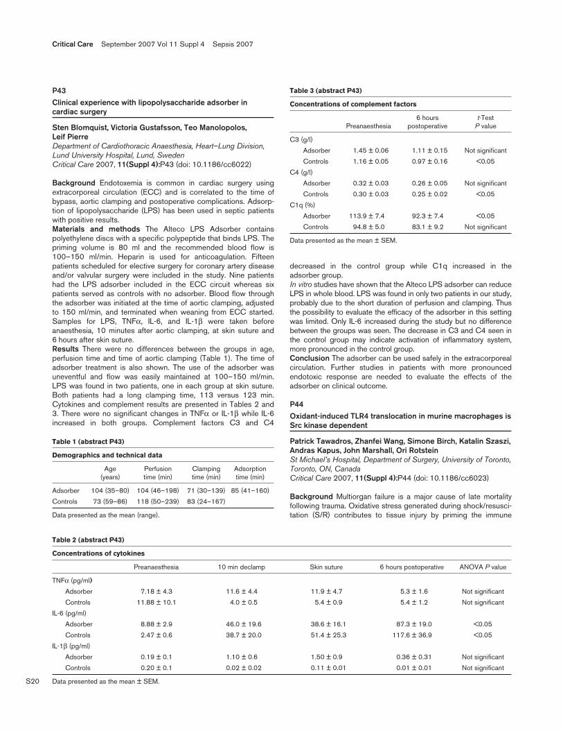
S20
P43Clinical experience with lipopolysaccharide adsorber incardiac surgery
Sten Blomquist, Victoria Gustafsson, Teo Manolopolos, Leif PierreDepartment of Cardiothoracic Anaesthesia, Heart–Lung Division,Lund University Hospital, Lund, SwedenCritical Care 2007, 11(Suppl 4):P43 (doi: 10.1186/cc6022)
Background Endotoxemia is common in cardiac surgery usingextracorporeal circulation (ECC) and is correlated to the time ofbypass, aortic clamping and postoperative complications. Adsorp-tion of lipopolysaccharide (LPS) has been used in septic patientswith positive results.Materials and methods The Alteco LPS Adsorber containspolyethylene discs with a specific polypeptide that binds LPS. Thepriming volume is 80 ml and the recommended blood flow is100–150 ml/min. Heparin is used for anticoagulation. Fifteenpatients scheduled for elective surgery for coronary artery diseaseand/or valvular surgery were included in the study. Nine patientshad the LPS adsorber included in the ECC circuit whereas sixpatients served as controls with no adsorber. Blood flow throughthe adsorber was initiated at the time of aortic clamping, adjustedto 150 ml/min, and terminated when weaning from ECC started.Samples for LPS, TNFα, IL-6, and IL-1β were taken beforeanaesthesia, 10 minutes after aortic clamping, at skin suture and6 hours after skin suture.Results There were no differences between the groups in age,perfusion time and time of aortic clamping (Table 1). The time ofadsorber treatment is also shown. The use of the adsorber wasuneventful and flow was easily maintained at 100–150 ml/min.LPS was found in two patients, one in each group at skin suture.Both patients had a long clamping time, 113 versus 123 min.Cytokines and complement results are presented in Tables 2 and3. There were no significant changes in TNFα or IL-1β while IL-6increased in both groups. Complement factors C3 and C4
decreased in the control group while C1q increased in theadsorber group.In vitro studies have shown that the Alteco LPS adsorber can reduceLPS in whole blood. LPS was found in only two patients in our study,probably due to the short duration of perfusion and clamping. Thusthe possibility to evaluate the efficacy of the adsorber in this settingwas limited. Only IL-6 increased during the study but no differencebetween the groups was seen. The decrease in C3 and C4 seen inthe control group may indicate activation of inflammatory system,more pronounced in the control group.Conclusion The adsorber can be used safely in the extracorporealcirculation. Further studies in patients with more pronouncedendotoxic response are needed to evaluate the effects of theadsorber on clinical outcome.
P44Oxidant-induced TLR4 translocation in murine macrophages isSrc kinase dependent
Patrick Tawadros, Zhanfei Wang, Simone Birch, Katalin Szaszi,Andras Kapus, John Marshall, Ori RotsteinSt Michael’s Hospital, Department of Surgery, University of Toronto,Toronto, ON, CanadaCritical Care 2007, 11(Suppl 4):P44 (doi: 10.1186/cc6023)
Background Multiorgan failure is a major cause of late mortalityfollowing trauma. Oxidative stress generated during shock/resusci-tation (S/R) contributes to tissue injury by priming the immune
Critical Care September 2007 Vol 11 Suppl 4 Sepsis 2007
Table 1 (abstract P43)
Demographics and technical data
Age Perfusion Clamping Adsorption (years) time (min) time (min) time (min)
Adsorber 104 (35–80) 104 (46–198) 71 (30–139) 85 (41–160)
Controls 73 (59–86) 118 (50–239) 83 (24–167)
Data presented as the mean (range).
Table 2 (abstract P43)
Concentrations of cytokines
Preanaesthesia 10 min declamp Skin suture 6 hours postoperative ANOVA P value
TNFα (pg/ml)
Adsorber 7.18 ± 4.3 11.6 ± 4.4 11.9 ± 4.7 5.3 ± 1.6 Not significant
Controls 11.88 ± 10.1 4.0 ± 0.5 5.4 ± 0.9 5.4 ± 1.2 Not significant
IL-6 (pg/ml)
Adsorber 8.88 ± 2.9 46.0 ± 19.6 38.6 ± 16.1 87.3 ± 19.0 <0.05
Controls 2.47 ± 0.6 38.7 ± 20.0 51.4 ± 25.3 117.6 ± 36.9 <0.05
IL-1β (pg/ml)
Adsorber 0.19 ± 0.1 1.10 ± 0.6 1.50 ± 0.9 0.36 ± 0.31 Not significant
Controls 0.20 ± 0.1 0.02 ± 0.02 0.11 ± 0.01 0.01 ± 0.01 Not significant
Data presented as the mean ± SEM.
Table 3 (abstract P43)
Concentrations of complement factors
6 hours t-Test Preanaesthesia postoperative P value
C3 (g/l)
Adsorber 1.45 ± 0.06 1.11 ± 0.15 Not significant
Controls 1.16 ± 0.05 0.97 ± 0.16 <0.05
C4 (g/l)
Adsorber 0.32 ± 0.03 0.26 ± 0.05 Not significant
Controls 0.30 ± 0.03 0.25 ± 0.02 <0.05
C1q (%)
Adsorber 113.9 ± 7.4 92.3 ± 7.4 <0.05
Controls 94.8 ± 5.0 83.1 ± 9.2 Not significant
Data presented as the mean ± SEM.

S21
system for an exaggerated response to subsequent inflammatorystimuli such as lipopolysaccharide (LPS): the so-called ‘two-hithypothesis’. The mechanisms of oxidant-induced cell priming,however, remain poorly elucidated. Our group has previouslyreported a role in this priming process for translocation of the LPSreceptor TLR4 to the plasma membrane [1]. We have alsopreviously shown that oxidant priming reprograms LPS signaling inmacrophages to a Src-dependent pathway leading to PI3kactivation [2]. Taken together, we hypothesized that Src activationmay play a role in oxidant-induced TLR4 translocation.Materials and methods Wild-type (WT) and triple Src(hck/fgr/lyn–/–) knockout (KO) mice were subjected to hemorrhagicshock and resuscitation to generate oxidative stress in vivo.Alveolar macrophages (AMs) were then retrieved by broncho-alveolar lavage and analyzed for TLR4 translocation by immuno-fluorescence staining and flow cytometry. In a separate in vitroexperiment, AMs from WT and KO mice were exposed to 200 μMhydrogen peroxide for 60 minutes and similarly analyzed by flowcytometry for surface expression of TLR4.Results In vivo, S/R induced visible translocation of TLR4 to theplasma membrane of AMs from WT mice as seen by immuno-fluorescence. This effect was completely inhibited in the Src KOanimals, and the observation was corroborated quantitatively byflow cytometry (see Figure 1). In order to confirm the role ofoxidative stress in S/R-induced TLR4 translocation, AMs from WTand KO mice were exposed to hydrogen peroxide in vitro.Hydrogen peroxide caused an increase in surface expression ofTLR4 in WT AMs by a ratio of 1.23 compared with WT control.This effect was once again inhibited in the Src KO AMs, where theratio was 0.95 compared with WT control.Conclusion Oxidative stress induces TLR4 translocation to the cellsurface of macrophages in a Src-dependent manner. Since cellularresponsiveness to LPS is known to correlate with surface levels ofTLR4, this novel finding may direct future therapies in modulatingoxidant-induced cellular priming and subsequent organ failure.References1. Powers KA, Szaszi K, Khadaroo RG, et al.: Oxidative stress gen-
erated by hemorrhagic shock recruits Toll-like receptor 4 tothe plasma membrane in macrophages. J Exp Med 2006,203:1951-1961.
2. Khadaroo RG, Kapus A, Powers KA, et al.: Oxidative stressreprograms lipopolysaccharide signaling via Src kinase-dependent pathway in RAW 264.7 macrophage cell line. J BiolChem 2003, 278:47834-47841.
P45Reduction of gap junction proteins and intercalated discstructural remodeling in the hearts of mice submitted tosepsis
Mara Rubia Nunes Celes, Diego Torrez-Dueñas, Jose Carlos Alves-Filho, Djane Braz Duarte, Fernando Queiroz Cunha, Marcos Antonio RossiDepartments of Pathology and Pharmacology, Faculty of Medicineof Ribeirao Preto, University of São Paulo, Ribeirao Preto, SãoPaulo, BrazilCritical Care 2007, 11(Suppl 4):P45 (doi: 10.1186/cc6024)
Background Cardiac dysfunction due to impaired myocardialcontractility has been recognized as an important factor contribu-ting to high mortality in septic patients. A recent study from ourlaboratory gives support to the opinion that myocardial structuralchange, classifiable as inflammatory cardiomyopathy, could beresponsible for sepsis-induced myocardial dysfunction. Thepresent study describes intercalated disc remodeling under bothprotein expression and structural features in experimental severesepsis induced by cecal ligation and puncture (CLP) in mice.Materials and methods Male C57Bl/6 mice weighing between18 and 22 g were subjected to sham-operated surgery, moderateor severe septic injury, both induced by CLP.Results Severe septic injury was accompanied by large number ofbacteria in the peritoneal cavity and blood, high levels of TNFα andMIP-1α in the septic focus and serum, marked hypotension, andhigh mortality rate. Western blot analysis and immunofluorescenceshowed a marked decrease of key gap and adherens junctionproteins (connexin43 and N-cadherin, respectively) in micesubjected to severe septic injury. These changes may result in theloss of intercalated disc structural integrity characterized in theelectron microscopic study by partial separation or dehiscence ofthe gap junction and adherens junctions.Conclusion Our data provide important insight regarding thealterations in intercalated disc components resulting from severeseptic injury. The intercalated disc remodeling under both proteinexpression and structural features in experimental severe sepsisinduced by CLP may be partly responsible for myocardialdepression in sepsis/septic shock. The abnormal parameters mayemerge as therapeutic targets and their modulation might providebeneficial effects on future cardiovascular outcomes and mortalityin sepsis.Acknowledgements Supported by FAPESP (04/01777-0 and04/14578-5) and FAEPA.
P46The critical role of heme oxygenase in neutrophil migrationimpairment in polymicrobial sepsis
Andressa Freitas, Jose Carlos Alves-Filho, Marina Moreira Suavinha, Fernando Spiller, Daniela Dal-Secco,Fernando Queiroz CunhaDepartment of Pharmacology, Faculty of Medicine of RibeiraoPreto, University of São Paulo, Ribeirao Preto, São Paulo, BrazilCritical Care 2007, 11(Suppl 4):P46 (doi: 10.1186/cc6025)
Background Sepsis is a systemic inflammatory response resultingfrom the inability of the host to restrict local infection. Duringsevere sepsis occurs a marked failure of neutrophil migration intothe infectious focus, which is associated with dissemination ofinfection resulting in high mortality. Recently, we showed that hemeoxygenase (HO) products, carbon monoxide and biliverdin,downregulate neutrophil recruitment by reducing the neutrophil/
Available online http://ccforum.com/supplements/11/S4
Figure 1 (abstract P44)
Shock/resuscitation (S/R)-induced TLR4 translocation is inhibited inSrc knockout (KO) mice. WT, wild type.

S22
endothelium rolling and adhesion in a noninfectious inflammatorymodel. This study aimed to investigate a possible role of the HO-1pathway on the failure of neutrophil recruitment in mice subjectedto severe (S-CLP) polymicrobial sepsis induced by cecal ligationand puncture (CLP).Materials and methods To evaluate the role of HO-1 in S-CLP,the Balb/c mice were pretreated with a specific HO-1 inhibitor,zinc protoporphyirin IX (ZnPPIX 30 mg/kg, subcutaneously) 30minutes before severe sepsis induction. Mice were killed 6 hoursafter CLP and HO-1 expression in mesenteric tissue and in bloodneutrophils were determined. In another set of experiments, micewere killed 6 hours and 12 hours after CLP, and neutrophilmigration to the peritoneal cavity, bacteremia, lung neutrophilsequestration assessed by myeloperoxidase activity and lunghistology, cytokines, liver, kidney and cardiac function and meanarterial pressure were determined. The survival rate of animals wasassessed every 12 hours up to 120 hours after surgery.Results A significant increase in HO-1 expression was observed inmesenteric tissue and in blood neutrophils after S-CLP. TheZnPPIX pretreatment prevents the failure of neutrophil endotheliumrolling, adhesion and migration observed in vehicle-pretreated micesubjected to S-CLP. As consequence, these mice presentedreduced bacteremia, low levels of seric TNFα and lung neutrophilsequestration, reduced liver, kidney and cardiac injury andimproved mean arterial pressure, resulting in an increase of thesurvival rate.Conclusions These data suggest that during an infectious processHO-1 displays a crucial role on the failure of neutrophil migration toinfectious focus and consequently on the susceptibility in severesepsis.Acknowledgement Supported by FAPESP/CAPES/FAEPA.
P47TLR2 signaling downregulates chemokine receptor CXCR2and impairs neutrophil migration in severe polymicrobialsepsis
Jose Carlos Alves-Filho, Andressa Freitas, Fernando Spiller,Fabricio Souto, Heitor Paula-Neto, Joao Santana Silva,Fernando Queiroz CunhaDepartment of Pharmacology and Immunology, School of Medicineof Ribeirao Preto, University of São Paulo, São Paulo, BrazilCritical Care 2007, 11(Suppl 4):P47 (doi: 10.1186/cc6026)
Background There is a marked defect in neutrophil migration intothe infectious focus during severe sepsis, which is associated withthe severity of disease. We recently demonstrated that this pheno-menon is a consequence of the downregulation of chemokinereceptor CXCR2 on the surface of circulating neutrophils. Toll-likereceptors (TLRs) are pattern-recognition receptors that are impor-tant in innate immune responses to bacterial infection. TLRactivation in phagocytes produces proinflammatory cytokines andchemokines that contribute directly to elimination of infectiousagents. However, a sustained inflammatory response can result intissue damage and sepsis. Here, we address the role of TLR2 inthe downregulation of CXCR2 and establishment of neutrophilmigration impairment in severe sepsis.Materials and methods TLR2-deficient (TLR2–/–) and C57BL/6(WT) mice were subjected to severe polymicrobial sepsis by thececal ligation and puncture (CLP) model, and neutrophil migration,bacteremia, CXCR2 expression and cytokines levels were evaluated.Results It was observed that TLR2 is critical for downregulation ofCXCR2 expression on circulating neutrophils during severe sepsis,since this event was prevented in TLR2–/– mice. In accordance,TLR2–/– mice did not present failure of neutrophil migration into the
infectious focus and, consequently, they presented lowerbacteremia and did not display systemic inflammation determinedby reduced levels of circulating cytokines, showing an improvedsurvival rate. Furthermore, in vitro, TLR2 agonist (lipoteichoic acid)was able to downregulate CXCR2 expression and to markedlyinhibit neutrophil chemotaxis induced by CXCR2-ligand. Thedownregulation of CXCR2 was associated with enhancedexpression of G-protein-coupled receptor kinase-2 (GRK-2), whichis known to play an important role in the desensitization andinternalization of this chemokine receptor. Finally, we showed thatin vitro lipoteichoic-acid-stimulated neutrophils adoptively transferredinto naïve WT mice display a significantly reduced competence tomigrate into the perinoneal cavity in response to thioglycolate.Conclusion Altogether, these findings suggest that TLR2 throughGRK-2 signaling downregulates CXCR2 expression on the surfaceof circulating neutrophils, which is a critical determinant ofimpairment of neutrophil migration into the infection focus duringsevere sepsis.Acknowledgements Supported by FAPESP and FAEPA.
P48The activity of the endothelial Tie-2 receptor modulatesventilation-induced lung injury in septic mice
Shirley Mei1,2, Jack Haitsma3, Daniel James3, Colleen Parker2,Arthur Slutsky3, Duncan Stewart1,2
1Institute of Medical Science, University of Toronto, Toronto, ON,Canada; 2Terrence Donnelly Heart Centre, St Michael’s Hospital,Toronto, ON, Canada; 3Interdepartmental Division of Critical CareMedicine, University of Toronto, Toronto, ON, CanadaCritical Care 2007, 11(Suppl 4):P48 (doi: 10.1186/cc6027)
Background Patients requiring mechanical ventilation, particularlyin the presence of sepsis, are at risk for ventilation-induced lunginjury (VILI), which is characterized by increased pulmonaryvascular permeability and inflammation. Inflammation in VILI isinitiated through interactions between endothelial and inflammatorycells. The vasoprotective protein angiopoietin-1, a ligand for theendothelial-selective tyrosine kinase receptor Tie-2, has recentlybeen recognized to play a critical role in regulating endothelialinflammation and vascular permeability. In contrast, angiopoietin-2,which binds with equal affinity, has been described as a Tie-2antagonist, inhibiting receptor activation in response to angio-poietin-1. We therefore examined the role of the angiopoietins/Tie-2pathway in mechanically ventilated, septic mice.Materials and methods Wild-type (WT) or Tie-2+/– mice wereanaesthetized and sepsis was induced by cecum ligation andpuncture (CLP). After 24 hours, animals were ventilated for 6 hoursto induce VILI. Animals were ventilated in a pressure-controlledmode, at a fractional inspired oxygen concentration of 0.5, aninspiration to expiration ratio of 1:2, a peak inspiratory pressure of14 cmH2O and a positive end-expiratory pressure of 2 cmH2O.Animals were sacrificed and bronchoalveolar lavage (BAL) wasperformed for total cell count, differential, and permeabilityparameters. Inflammatory cytokine IL-6 levels were measured inBAL and plasma.Results There was no difference between sham-operated or CLP-operated WT mice in total cell numbers in BAL. Sham operatedTie-2+/– mice had similar cell numbers, while ventilation in CLP-operated Tie-2+/– mice resulted in higher cell numbers (P = 0.06versus WT), with an increase mainly in macrophages. IL-6 plasmalevels were increased in septic Tie-2+/– mice subjected tomechanical ventilation compared with the WT mice, whereas IL-6levels in BAL were not significantly different. Despite the highertotal cell counts, permeability parameters measured in Tie-2+/–
Critical Care September 2007 Vol 11 Suppl 4 Sepsis 2007

S23
mice were lower than in WT mice (albumin, P = 0.08; totalproteins, not significant; IgM, P = 0.05).Conclusions Pulmonary vascular permeability in mice withknockdown of endothelial Tie-2 receptor is preserved in ventilatedseptic mice. However, there was an increase in the number ofinflammatory cells and systemic inflammation (IL-6) in theseanimals. These results further emphasize the importance of Tie-2 inregulating vascular permeability in the lungs.
P49Pharmacological implications of the spleen in sepsis
Bolin Cai, Fei Chen, Jared Huston, Luis UlloaLaboratory of Surgical Immunology, University of Medicine andDentistry of New Jersey, Newark, NJ, USACritical Care 2007, 11(Suppl 4):P49 (doi: 10.1186/cc6028)
Background Our previous studies indicated that nicotineattenuated systemic inflammation, and improved survival in bothlethal endotoxemia and cecal ligation and puncture [1].Materials and methods Here we analyzed the pathophysiologicaland pharmacological implications of the spleen in experimentalsepsis. Acetylcholine and nicotine inhibit the production of TNFand HMGB1 from lipopolysaccharide-treated RAW264.7 macro-phage-like cells in a concentration-dependent manner. Nicotineinhibits the production of these cytokine macrophage-like cellsthrough a mechanism dependent on the α7-nicotine acetylcholinereceptor (nAChR). From a physiological perspective, our studiesindicate that the spleen has a significant expression of theα7nAChR. To study the implications of the splenic α7nAChR in thetherapeutic potential of nicotine in sepsis, we analyzed the effectsof treatment with nicotine in splenectomized mice. Adult male8–12-week-old BALB/c mice (25–30 g) were splenectomized and7 days later subjected to cecal ligation and puncture to inducepolymicrobial peritonitis. Control sham animals underwentlaparotomy without splenectomy. Administration of nicotine(400 mg/kg, i.p.) or vehicle injection (1 × PBS, i.p.) began24 hours after surgery, and thereafter twice a day for 3 days.Animals were euthanized after 44 hours for serum cytokine analysis.Results Treatment with nicotine improved survival in sham micebut it worsened the survival of splenectomized mice. Nicotineinhibited the production of TNF and HMGB1 from lipopoly-saccharide-treated macrophages differentiated from peripheralblood mononuclear cells. However, in vivo, nicotine attenuatedserum TNF levels in sham mice but not in splenectomized micewith polymicrobial peritonitis. There were no differences in serumTNF, IL-12, IFNγ or IL-10 levels between vehicle-treated ornicotine-treated splenectomized animals. Unlike systemic TNF,circulating HMGB1 levels correlated with the increased mortalityinduced by nicotine in splenectomized mice. Treatment withnicotine increased circulating serum HMGB1 levels insplenectomized mice with experimental sepsis.Conclusion These results suggest that the spleen has majorpharmacological implications for the treatment of sepsis.Acknowledgements Supported by grants of the AHA and theUSAMRMC Log#05308004 to LU.Reference1. Wang H, Liao H, Ochani M, et al.: Cholinergic agonists inhibit
HMGB1 release and improve survival in experimental sepsis.Nat Med 2004, 10:1216-1221.
P50Peroxynitrite mediates neutrophil migration failure in severepolymicrobial sepsis
Daniela Dal-Secco, Diego Torres-Dueñas, Andressa Freitas,Mara Rubia Nunes Celes, José Carlos Alves-Filho, Fernando Spiller, Vitor Faeda Dalto, Marcos Antonio Rossi,Sérgio Henrique Ferreira, Fernando Queiroz CunhaDepartments of Pharmacology and Pathology, Faculty of Medicineof Ribeirao Preto, University of São Paulo, Ribeirao Preto, SãoPaulo, BrazilCritical Care 2007, 11(Suppl 4):P50 (doi: 10.1186/cc6029)
Background Sepsis is a systemic inflammatory response resultingfrom the host inability to restrict local infection. The failure ofneutrophil migration to the infectious focus is one of the mecha-nisms involved in this process. Recently, it was demonstrated thatthis event is mediated by nitric oxide. The present study addressedthe possibility that peroxynitrite (ONOO–), a nitric oxide-derivedpowerful oxidizing and nitrating compound, can be also involved inthe neutrophil migration failure.Materials and methods Male C57Bl/6 mice were pretreated,subcutaneously, with saline or uric acid (UA; 100 mg/kg) 1 hourbefore induction of severe sepsis injury (SSI) or were treated withTetrakis (FeTPPS; 5.0 mg/kg) 15 minutes after SSI, induced bycecal ligation and puncture. Intravital microscopy was used toevaluate leukocyte rolling and adhesion in the mesenteric micro-circulation. TNFα and MIP-1α levels were evaluated by ELISA, lungneutrophil influx by myeloperoxidase activity and nitrotyrosine byimmunofluorescence.Results The mice pretreated with UA (an ONOO– scavenger) andsubjected to SSI did not present failure of neutrophil rolling,adhesion and migration to the infectious focus. As a consequence,they presented low bacteremia, diminished TNFα and MIP-1αlevels in circulation and reduced myeloperoxidase activity. More-over, increased nitrotyrosine labeling detected in leukocytespresent in mesenteric tissues and neutrophils obtained from SSIperitoneal exudate were reduced by UA pretreatment. Finally, theUA pretreatment enhanced significantly the survival rate of the SSImice. Similarly, treatment with Tetrakis (FeTPPs), a more specificONOO– scavenger, reestablished neutrophil migration andincreased the survival rate.Conclusion Together, the results indicate that ONOO– partiallymediates the reduction of neutrophil/endothelium cell interactionand consequently the neutrophil migration failure to the infectiousfocus and susceptibility in severe sepsis. Therefore, these resultsidentify ONOO– inhibitors as potential targets for novel sepsistherapies.Acknowledgement Supported by CAPES, FAPESP and FAEPA.
P51Selective V1a receptor agonists in experimental septic shock
Daniel TraberDepartment of Anesthesiology, University of Texas Medical Branch,Galveston, TX, USACritical Care 2007, 11(Suppl 4):P51 (doi: 10.1186/cc6030)
Background Septic shock is caused by hypotension secondary tovasodilation. This hypotension is resistant to fluid resuscitation andrequires the use of vasopressor agents. Recently, it was shownthat it may be due to a deficiency in the vasopressor hormonearginine–vasopressin (AVP), a mixed V1a/V2 receptor agonist,leading to the clinical use of AVP in septic shock. We tested thehypothesis that the selective V1a receptor agonist FE 202158 is at
Available online http://ccforum.com/supplements/11/S4

S24
least as effective as AVP in a sheep model of pneumonia-inducedseptic shock.Materials and methods Sheep were surgically instrumentedahead of the study. After obtaining baseline data, they wereanesthetized and insufflated with 48 breaths of cotton smoke and1,011 colony forming units of Pseudomonas aeruginosa instilledinto their airways via a tracheostomy. The sheep were then placedon a ventilator and awakened for the study. They were resuscitatedwith Ringer’s solution to maintain left atrial and central venouspressures and hematocrit at baseline levels. If, despite fluidmanagement, the mean arterial pressure (MAP) fell by 10 mmHgfrom baseline, a continuous intravenous infusion of AVP or FE202158 was initiated and titrated to keep MAP within this limit,except in a septic control group and in a nonseptic sham group.Results In the septic control group, MAP fell by ~30 mmHg andthe systemic vascular resistance index (SVRI) by ~50% over the24-hour study. In the AVP group, although the MAP and SVRI weremaintained above septic control group levels, they could not bebrought back to sham group levels. In contrast, in the FE 202158group, the MAP and SVRI were maintained at sham group levels.The septic control group accumulated ~7 l fluid over the 24-hourstudy (~20% body weight), while this was reduced by ~50% in theAVP group, and was totally prevented in the FE 202158 group.The fluid accumulation was associated with a constant hematocrit,suggesting that it was extravascular. It was also correlated with afall in plasma total protein and oncotic pressure, suggestingprotein leakage from the vasculature and probably reflectingsepsis-induced vascular leak syndrome. The full prevention of thisfluid and protein leakage with FE 202158 was reversed to AVPgroup levels by infusing the selective V2 receptor agonistdesmopressin together with FE 202158.Conclusion FE 202158 was not only fully effective at preventing asepsis-induced decrease in MAP and SVRI, but was superior to
AVP at reducing vascular leak syndrome because of its V1areceptor selectivity.
P52Gender, infection, critical illness, and death: superior survivalfor female trauma patients admitted to the intensive care unittreated for infection in a dedicated, prospective, multicenterstudy
Brian Swenson, Hugo Bonatti, Lesly Dossett, Addison May,Robert SawyerUniversity of Virginia Health System, Department of Surgery,Charlottesville, VA, USACritical Care 2007, 11(Suppl 4):P52 (doi: 10.1186/cc6031)
Background Multiple studies have suggested gender-baseddifferences exist in infection incidence and outcome in critically illpatients, but these relationships have not been clearly defined. Wepresent the largest prospective multicenter study designed tomeasure the impact of gender on infection incidence and outcomein critically ill surgical and trauma patients to date.Methods Patients admitted for at least 3 days to surgical or traumaintensive care units (ICUs) at two academic tertiary care centersfrom October 2001 to May 2006 were prospectively followed forthe occurrence of infection and clinical outcomes. Logisticregressions were performed to assess independent risk of genderon the acquisition of an ICU infection and inhouse mortality inpatients with ICU-acquired infections. Demographic and clinicalvariables that were significant by univariate analyses were includedin the models.Results The final cohort followed 2,291 patients (trauma n = 1,407,nontrauma n = 884) with 383 (27.2%) female trauma patients and413 (46.7%) female nontrauma patients. The proportion of
Critical Care September 2007 Vol 11 Suppl 4 Sepsis 2007
Table 1 (abstract P52)
Logistic regression analysis assessing predictors of mortality in patients with ICU-acquired infections
Infected trauma Infected nontrauma patients (n = 652) patients (n = 397)
Odds 95% confidence P Odds 95% confidence PVariable ratio interval value ratio interval value
Female gender 0.45 0.22–0.95 0.036 1.15 0.696–1.911 0.58Age, per year 1.02 0.10–1.04 0.087 1.04 1.019–1.062 <0.001White race 1.00 – 1.00 – –Black race 0.62 0.24–1.62 0.33 1.08 0.44–2.59 0.87Other race 1.03 0.29–3.67 0.97 3.61 1.08–12.1 0.037Any comorbidity 0.89 0.40–2.00 0.77 0.76 0.29–1.98 0.57Chronic corticosteroid use 0.48 0.05–5.0 0.54 1.35 0.66–2.74 0.41Hepatic insufficiency 4.25 1.33–13.6 0.015 1.61 0.78–3.33 0.20Cardiac disease 1.18 0.49–2.82 0.72Baseline serum creatinine ≥2.0 (units) 7.46 1.11–50.1 0.039Malignancy 0.17 0.018–1.56 0.12 1.39 0.80–2.44 0.24Body mass index, per kg/m2 0.98 0.96–1.01 0.24Admission from home 1.03 0.41–2.60 0.95McCabe score at ICU admission, per point 1.21 0.60–2.44 0.59 1.35 0.91–2.01 0.14APACHE II score at ICU admission, per point 1.07 1.02–1.12 0.0081 1.08 1.04–1.12 <0.001WHO functional status at ICU admission, per point 1.46 0.89–2.39 0.14Multiple Organ Dysfunction score at ICU admission, per point 1.12 1.04–1.22 0.0040Probability of trauma survival 0.49 0.23–1.02 0.055ICU length of stay, per day 1.02 0.97–1.07 0.42Hospital length of stay, per day 0.95 0.91–0.98 0.032
Bold data are statistically significant.

S25
patients with at least one ICU-acquired infection was 47.6% fortrauma males and 43.1% for trauma females (P = 0.13); and was44.2% for nontrauma male patients and 45.8% for nontrauma femalepatients (P = 0.63). The inhospital mortality of patients with at leastone ICU-acquired infection was 12.5% for trauma males and 7.3%for trauma females (P = 0.06); and was 30.8% for nontrauma malepatients and 28.0% for nontrauma female patients (P = 0.55).Logistic regression analysis did not show gender to be a significantvariable in the acquisition of an ICU infection. The results of a logisticregression analysis of predictors of inhospital mortality in patientswith ICU-acquired infections are presented in Table 1. Only variablessignificant by univariate analysis were entered into logistic models.Empty cells represent variables that were significant for only onegroup (for example – only trauma, but not nontrauma).Conclusion Gender does not appear to play a role in acquisition ofan ICU infection in either trauma or nontrauma patients. In contrast,female gender appears to provide a strong survival advantage intrauma patients with ICU-acquired infections. This phenomenonwas not observed in the nontrauma group. Further investigationinto the hormonal and cytokine differences between the genders incritically ill trauma patients and their response to infection mayprove beneficial.
P53Effects of antifungal agents on the production of cytokinesfrom macrophages stimulated by Aspergillus fumigatus conidia
Jung-Hyun Choi1, Eun-Young Kwon2, Chulmin Park2, Sun Hee Park1, Su-Mi Choi1, Dong-Gun Lee1, Jin-Hong Yoo1,Wan-Shik Shin1
1Department of Internal Medicine, Division of Infectious Diseases,College of Medicine, Catholic University of Korea, Korea; 2CatholicResearch Institutes of Medical Science, Catholic University ofKorea, KoreaCritical Care 2007, 11(Suppl 4):P53 (doi: 10.1186/cc6032)
Background We evaluated the immunomodulatory effect of threeantifungal agents, amphotericin B (AmB), micafungin (MF) anditraconazole (ITZ), in the aspect of cytokine production andexpression of NF-κB in Aspergillus fumigatus conidia-treatedRAW264.7 cells, a murine alveolar macrophage cell line.Materials and methods We evaluated the cytotoxic effect ofantifungal agents using a commercial cell proliferation assay. TNFαand IL-10 production according to stimulation (control, A. fumigatusconidia only, conidia + antifungal drug, conidia + antifungals +GM-CSF) was evaluated and each compared using a commercialELISA method. NF-κB activation was evaluated by western blotanalysis.Results AmB, MF and ITZ showed a dose-dependent cytotoxiceffect on the tested cells. Stimulation of cells by A. fumigatusconidia induced TNFα production. Pretreatment of AmB atconcentrations not affecting cellular survival did not change theproduction of TNFα compared with conidia-treated cells, butpretreatment of MF or ITZ showed a reduced amount of TNFαproduction compared with conidia-treated cells. AmB also showeda synergistic effect on TNFα production when simultaneouslytreated with GM-CSF. IL-10 production was markedly increasedwhen the cells were treated with AmB with conidia. MF and ITZinduced a smaller increase of IL-10 production. AmB also showeda synergistic effect on the production of IL-10 when treated withGM-CSF simultaneously. A. fumigatus conidia enhanced expres-sion of NF-κB. The degree of NF-κB expression was associatedwith the amount of TNFα and IL-10 produced.Conclusion The antifungal agents we used in this experimentshowed decreased TNFα production and increased IL-10
production from the RAW264.7 cells stimulated by A. fumigatusconidia after pretreatment of antifungal agents. But more studiessuch as the association between the immunomodulatory effect andantifungal activity and the difference of the signal pathway ofcellular activation according to drugs should be performed.
P54Time to positivity as a novel predictor of outcome in intensivecare unit patients with sepsis
Susie Jerwood1, Sonia Hudson1, Matthew Hankins2, John Paul1, Owen Boyd1, Jonathan Cohen2
1Brighton Sussex County Hospital, Eastern Road, Brighton,Sussex, UK; 2Brighton and Sussex Medical, Falmer, Sussex, UKCritical Care 2007, 11(Suppl 4):P54 (doi: 10.1186/cc6033)
Background Inadequate antibiotic therapy predicts poor outcomefrom sepsis, but there is no simple test of adequacy. We sugges-ted that a time-to-positivity assay (Tpos) might act as a surrogate forantimicrobial activity and predict outcome from sepsis in theintensive care unit (ICU) [1]. We are conducting a prospectiveclinical trial to test this hypothesis.Methods We studied 35 sequential ICU patients with onset ofsepsis who had not had antibiotics for at least 3 days. Sepsis wasdefined as clinical evidence of infection plus at least three criteriafor systemic inflammatory response syndrome. All patients receivedstandard empiric therapy. Sera taken at 24 hours post antibioticswere inoculated into blood culture bottles containing standardisedbacteria, incubated in an automated microbial detection systemand the time to positivity noted. The primary clinical endpoint wasdays in the ICU.Results Cultures that are negative after 5 days of incubationindicate adequate antimicrobial therapy; cultures that becomepositive in <5 days indicate inadequate antimicrobial therapy.Patients with negative Tpos (n = 11) were associated with a stay inthe ICU of less than 6.5 days (P = 0.052). See Figure 1.
Conclusion These data show that some septic patients onstandard antimicrobial regimens are receiving inadequate therapy.The Tpos is a new, simple assay that might be used effectively tomonitor antibiotic use.Reference1. Kaltsas P, Want S, Cohen J: Development of a time-to-positiv-
ity assay as a tool in the antibiotic management of septicpatients. Clin Microbiol Infect 2005, 11:109-114.
Available online http://ccforum.com/supplements/11/S4
Figure 1 (abstract P54)
Average length of stay for patients with negative and positive time-to-positivity 1 (taken at 24 hours after first dose of antibiotics) with 95%confidence intervals.