Data Mining and Polar Coordinates in the Analysis by Gender ...
Upload
khangminh22Category
view
0download
0
Article
A Neutrophil Timer Coordi
nates Immune Defenseand Vascular ProtectionGraphical Abstract
Highlights
d Neutrophil aging is an intrinsically driven, bona fide circadian
process
d Bmal1 and CXCR2 induce neutrophil aging, whereas CXCR4
antagonizes it
d Diurnal aging critically dictates how and when neutrophils
migrate into tissues
d Aging favors neutrophil clearance, thereby protecting the
cardiovascular system
Adrover et al., 2019, Immunity 50, 390–402February 19, 2019 ª 2019 Elsevier Inc.https://doi.org/10.1016/j.immuni.2019.01.002
Authors
Jose M. Adrover, Carlos del Fresno,
Georgiana Crainiciuc, ...,
Marıa A. Moro, Borja Ibanez,
Andres Hidalgo
In Brief
Neutrophils display circadian oscillations
in numbers and phenotype in the
circulation. Adrover and colleagues now
identify the molecular regulators of
neutrophil aging and show that genetic
disruption of this process has major
consequences in immune cell trafficking,
anti-microbial defense, and vascular
health.
Immunity
Article
A Neutrophil Timer CoordinatesImmune Defense and Vascular ProtectionJose M. Adrover,1 Carlos del Fresno,2 Georgiana Crainiciuc,1 Maria Isabel Cuartero,3,4 Marıa Casanova-Acebes,1,16
Linnea A. Weiss,1,17 Hector Huerga-Encabo,5 Carlos Silvestre-Roig,6,7 Jan Rossaint,8 Itziar Cossıo,1
Ana V. Lechuga-Vieco,2 Jaime Garcıa-Prieto,2 Monica Gomez-Parrizas,2 Juan A. Quintana,1 Ivan Ballesteros,1
Sandra Martin-Salamanca,1 Alejandra Aroca-Crevillen,1 Shu Zhen Chong,9 Maximilien Evrard,9 Karl Balabanian,10
Jorge Lopez,11 Kiril Bidzhekov,6 Francoise Bachelerie,10 Francisco Abad-Santos,12 Cecilia Munoz-Calleja,11
Alexander Zarbock,8 Oliver Soehnlein,6,7 Christian Weber,6,13 Lai Guan Ng,9 Cristina Lopez-Rodriguez,5 David Sancho,2
Marıa A. Moro,3,4 Borja Ibanez,2,14,15 and Andres Hidalgo1,6,18,*1Area of Developmental and Cell Biology, Centro Nacional de Investigaciones Cardiovasculares Carlos III (CNIC), Madrid, Spain2Area of Myocardial Pathophysiology, Centro Nacional de Investigaciones Cardiovasculares Carlos III (CNIC), Madrid, Spain3Unidad de Investigacion Neurovascular, Department of Pharmacology, Faculty of Medicine, Universidad Complutense4Instituto de Investigacion Hospital 12 de Octubre (i+12), Madrid, Spain5Immunology Unit, Department of Experimental and Health Sciences, Pompeu Fabra University, Barcelona6Institute for Cardiovascular Prevention (IPEK), Ludwig-Maximillians-Universit€at M€unchen7German Centre for Cardiovascular Research (DZHK), partner site Munich Heart Alliance, Munich, Germany8Department of Anesthesiology, Intensive Care, and Pain Medicine, University of M€unster, Germany9Singapore Immunology Network (SIgN), Agency for Science, Technology and Research (A*STAR), Biopolis, Singapore10Inserm Unite Mixte de Recherche (UMR) S996, Universite Paris-Sud, Laboratory of Excellence in Research on Medication and Innovative
Therapeutics, Clamart, France11Department of Immunology, Instituto de Investigacion Sanitaria Princesa, Hospital Universitario de La Princesa, Madrid, Spain12Department of Clinical Pharmacology, Instituto Teofilo Hernando, Hospital Universitario de La Princesa, Instituto de Investigacion Sanitaria
Princesa, Madrid, Spain13Department of Biochemistry, Cardiovascular Research Institute Maastricht (CARIM), Maastricht University, Maastricht, the Netherlands14CIBER de Enfermedades Cardiovasculares (CIBERCV), Madrid, Spain15Department of Cardiology, Instituto de Investigacion Sanitaria (IIS)-Fundacion Jimenez Dıaz, Madrid, Spain16Present address: Tisch Cancer Institute, Mount Sinai School of Medicine, New York City, New York, USA17Present address: Centro Nacional de Biotecnologıa, Madrid, Spain18Lead Contact*Correspondence: [email protected]
https://doi.org/10.1016/j.immuni.2019.01.002
SUMMARY
Neutrophils eliminate pathogens efficiently but caninflict severe damage to the host if they over-acti-vate within blood vessels. It is unclear how immu-nity solves the dilemma of mounting an efficientanti-microbial defense while preserving vascularhealth. Here, we identify a neutrophil-intrinsicprogram that enabled both. The gene Bmal1 regu-lated expression of the chemokine CXCL2 toinduce chemokine receptor CXCR2-dependentdiurnal changes in the transcriptional and migratoryproperties of circulating neutrophils. These diurnalalterations, referred to as neutrophil aging, wereantagonized by CXCR4 (C-X-C chemokine receptortype 4) and regulated the outer topology of neutro-phils to favor homeostatic egress from blood ves-sels at night, resulting in boosted anti-microbialactivity in tissues. Mice engineered for constitutiveneutrophil aging became resistant to infection, butthe persistence of intravascular aged neutrophilspredisposed them to thrombo-inflammation anddeath. Thus, diurnal compartmentalization of neu-
390 Immunity 50, 390–402, February 19, 2019 ª 2019 Elsevier Inc.
trophils, driven by an internal timer, coordinatesimmune defense and vascular protection.
INTRODUCTION
The capacity of neutrophils not only to kill pathogens but also to
inflict severe damage to tissues suggests the existence of a pro-
tective mechanism that balances these opposing functions.
Both anti-microbial immunity and vascular inflammation are
known to follow circadian patterns (Man et al., 2016; Muller
et al., 1989; Scheiermann et al., 2013), suggesting that such
mechanisms may be temporally regulated. The nature of this
mechanism, however, remains enigmatic.
While studying neutrophils in the steady state, we previously
identified a natural phenotypic shift of circulating neutrophils
that followed a strict diurnal regime (Casanova-Acebes et al.,
2013). Neutrophils released from the bone marrow display high
CD62L that is progressively reduced during the day, while sur-
face CXCR4 (C-X-C chemokine receptor type 4) increases prior
to their natural egress from blood, a process referred to as
clearance (Casanova-Acebes et al., 2013). This process of
neutrophil aging has been proposed to be regulated by the gut
microbiota and to favor a pro-inflammatory phenotype that
predisposes to vascular inflammation (Zhang et al., 2015).

Contrasting with this model of extrinsically driven neutrophil ag-
ing, studies have shown that intrinsic programs controlled by the
molecular clock also regulate immune cell properties (Druzd
et al., 2017; Nguyen et al., 2013; Silver et al., 2012). Because
the mechanisms regulating aging and its physiological conse-
quences remain uncertain, we explored whether neutrophils
are endowed with an intrinsic program that controls diurnal ag-
ing, tunes their anti-microbial functions, and limits vascular
inflammation.
In transcriptome analyses of circulating neutrophils performed
at different times, we found regulation of clock-related genes
and the CXCR2 signaling pathway. Bmal1 (brain and muscle
aryl hydrocarbon receptor nuclear translocator [ARNT]-like 1;
encoded by Arntl) regulated expression of CXCL2 (chemokine
[C-X-Cmotif] ligand 2), a CXCR2 ligand that controlled neutrophil
aging in a cell-autonomous manner. Deletion of Arntl or Cxcr2
from neutrophils prevented phenotypic aging, whereas deletion
of Cxcr4, a negative regulator of CXCR2 signaling, resulted in
unrestrained aging. Neutrophil aging disrupted cytoskeletal
integrity to specifically prevent rolling and accumulation in in-
flamed areas without affecting homeostatic migration into naive
tissues at night. In turn, this temporal regulation of trafficking
regulated diurnal responses to infections, while at the same
time removing neutrophils from the bloodstream, thereby pro-
tecting vessels from inflammatory injury. This process may un-
derlie the circadian susceptibility of mammals to cardiovascular
disease.
RESULTS
A Neutrophil-Intrinsic Timer Drives Diurnal AgingIn a small cohort of healthy volunteers, we found diurnal changes
in neutrophil markers similar to those associated with neutrophil
aging in mice (Casanova-Acebes et al., 2013), suggesting con-
servation of this phenomenon across species (Figure S1A). In
healthy mice, the number of aged neutrophils in blood follows
diurnal patterns with a peak at around zeitgeber time 5 (ZT5,
i.e., 5 h after lights on), while non-aged or ‘‘fresh’’ neutrophils
predominate at ZT13 (Casanova-Acebes et al., 2013). These
diurnal patterns persisted in constant darkness and could be en-
trained by light shift (Figures S1B and S1C), indicating that
neutrophil aging is a bona fide circadian process. To identify ge-
netic programs that were temporally regulated in neutrophils, we
compared the transcriptomes of circulating neutrophils purified
from wild-type (WT) mice at these two times. We identified
changes in over 1,300 genes related to pathways of inflamma-
tion, migration, and apoptosis (Figures 1A, 1B, and S1D; Table
S1), which suggested modulation of these processes during
the day. Given the diurnal pattern of aging, we inspected genes
of the molecular clock because they are known to regulate im-
mune rhythms (Man et al., 2016; Scheiermann et al., 2013).
Expression of clock-related genes, including Arntl (encoding
Bmal1) and Clock, increased at ZT5, while others, like Per2,
were decreased at this time (Figures 1B, S1D, and S1E). Tran-
scriptional analyses at multiple times of day revealed circadian
oscillations for all these genes in circulating neutrophils (Fig-
ure 1C). They also demonstrated reduced expression of Sell
(encoding CD62L) at ZT5 but no changes for Cxcr4 (Figure 1B).
We also noticed reduced expression ofCxcr2, whose expression
displayed diurnal patterns (Figure 1D), at ZT5 (Figure 1B).
Further, CXCR2 agonists induced phenotypic changes in neutro-
phils that resembled those seen during natural aging, namely re-
ductions in CXCR2 and CD62L on the cell surface (Figures 1E
and S1F).
Guided by the temporal expression patterns of these genes,
we predicted that Bmal1 and CXCR2 might be required for
diurnal neutrophil aging. Although CXCR4 did not present tran-
scriptional oscillations (Figure 1D), its presence on the cell sur-
face changed diurnally (Figure 1E) (Casanova-Acebes et al.,
2013), and this receptor is known to antagonize CXCR2 signaling
(Martin et al., 2003), suggesting that CXCR4 might also
contribute to aging. Analyses of blood fromwild-type animals re-
vealed ligands for both receptors in plasma, with oscillating
amounts of CXCL12, and constitutively low amounts of CXCL2
(Figure 1F).
To formally test the possibility that these genes regulated
neutrophil aging, we generated mice with neutrophil-specific
deficiency in Arntl, Cxcr2, or Cxcr4 (herein referred to as ArntlDN,
Cxcr2DN, and Cxcr4DN) by using the hMRP8cre driver line, which
resulted in robust depletion of the receptors from the surface of
neutrophils (Figures S2A and S2B). Immunoblot analysis of the
Bmal1 protein confirmed efficient depletion in ArntlDN neutro-
phils and revealed natural reductions of this protein at ZT13 in
wild-type neutrophils and unchanged amounts in Cxcr2DN and
Cxcr4DN neutrophils (Figure S2C). We then assessed surface
CD62L, a marker reduced during aging (Casanova-Acebes
et al., 2013; Uhl et al., 2016; Zhang et al., 2015) (Figure 1E). To
ensure that potential alterations were cell-intrinsic, we generated
bonemarrow transplant chimeras of wild-type and of each of the
mutant donors. We found elevated CD62L in circulating neutro-
phils from ArntlDN and Cxcr2DN donors, suggesting disrupted
aging in these mutants (Figure 2A). In contrast, Cxcr4DN neutro-
phils were low for CD62L, which suggested constitutive aging
(Figure 2A). If these alterations were caused by disruption of
diurnal aging, we predicted that CD62L would not change over
time. Indeed, in vivo metabolic pulse and chase of neutrophils
with bromodeoxyuridine (BrdU) demonstrated that the temporal
changes in CD62L seen in wild-type neutrophils were abrogated
in ArntlDN and Cxcr4DN mutants (Figure 2B). To further confirm
that these genes regulated the natural dynamics of aging, we
measured CD62L through full diurnal cycles in all mutant mice.
Although surface CD62L exhibited diurnal oscillations in wild-
type neutrophils, all three mutants presented disrupted patterns,
and ArntlDN mutants showed a complete loss of rhythmicity
(Figure 2C). Accordingly, surface CXCR4 also lost diurnal oscilla-
tions in ArntlDN neutrophils (data not shown).
To dissect the antagonistic role of CXCR4 in aging, we pre-
treated wild-type neutrophils with CXCL12, the main ligand for
CXCR4, before exposing them to a CXCR2 agonist. Given the
constitutive aging of Cxcr4DN neutrophils, we hypothesized
that stimulation through CXCR4 might prevent CXCR2-depen-
dent responses. Indeed, CXCL12 blunted both the reductions
in CD62L and the chemotaxis elicited through CXCR2 (Fig-
ure S2D). In addition, neutrophils expressing Cxcr4WHIM, a
hyper-signaling variant of CXCR4 (Balabanian et al., 2012), dis-
played constitutive elevations in CD62L (Figure S2E). Combined,
these data indicated that Bmal1 and CXCR2 promote diurnal ag-
ing and that CXCR4 prevents it.
Immunity 50, 390–402, February 19, 2019 391

A B
C D
F
E
Clo
ckP
er2
Cxc
r2
Cry
2
Atm
Arn
tl
Vav
2C
xcr5
Tlr4
Cxc
r4
Per
1
Lmnb
1
Il1b
Csf
3r
Sel
l
Il17r
a
Mcl
1Fg
r
Il13r
a1
Cry
1
Sel
plg
Icam
1C
xcl2
ZT5ZT13
Arntl
Clock
Per2
Rel
ativ
e ge
ne e
xpre
ssio
n
2.5
2.0
0
0
0
3noi
t ar gi
M & s
i xat o
mehC
Inflammatory responsesi s
ot pop
A
ZT13 ZT5Fresh Aged
mTOR SignalingTec Kinase SignalingCXCR4 SignalingActin Cytoskeleton SignalingGM-CSF SignalingLeukocyte ExtravasationPAK SignalingAMPK SignalingHGF SignalingS1P SignalingNO and ROS productionIL-6 SignalingfMLP SignalingMAPK SignalingTREM1 SignalingNFκB SignalingPPARa/RXRa SignalingSAPK/JNK SignalingVDR/RXR ActivationIL-3 SignalingMacropinocytosisPRR recognition of pathogensFcγR-mediated phagocytosis
PTEN SignalingLXR/RXR Activation
α-Adrenergic SignalingIL-8 Signaling IL-1 Signaling
CD27 SignalingDeath Receptor Signaling
p14/p19ARF SignalingCa-induced Apoptosis
Ceramide Signalingp53 Signaling
1 223 30z-score
1
P=0.008
P=0.03
P=0.0001
Cxcr2
Cxcr41.0
1.2
0.8
1
2
4
45
2510500
85001050
850
3
0
Rel
ativ
e ge
ne e
xpre
ssio
n
p=0.05
p=0.52
Zeitgeber time
Che
mok
ine
(ng/
ml)
CXCL2
CXCL12
2.0
1.5
1.0
0.5
0.0
2.5Plasma
13 17 21 1 5 9
13 17 21 1 5 9
p=0.036
agedfresh
p=0.446
Nr1d1
Zeitgeber time
1.5
0
13 17 21 1 5 9
P=0.014
13 17 21 1 5 9
aging
Fluo
resc
ence
Inte
snsi
ty
CD62L
CXCR2
CXCR4
p=0.0004
p=0.0001
p=0.04
-2.0 1:1 2.0
Circadian clock Migration Inflammation/Survival
Figure 1. Cell-Intrinsic Rhythms in Circulating Neutrophils, Related to Figure S1
(A) Molecular pathways differentially regulated in circulating wild-type neutrophils at ZT5 versus ZT13. Comparisons are presented as Z score values.
(B) Heatmap of selected genes at ZT5 versus ZT13, including genes of the circadian clock and genes encoding proteins related to migration and inflammation.
The color scale indicates fold changes of expression for each gene.
(C) Diurnal expression of the indicated clock genes in neutrophils isolated from the circulation of wild-type mice at the indicated zeitgeber times. Shaded areas
represent night; n = 2–6 mice per time point.
(D) Diurnal expression of Cxcr2 and Cxcr4 in circulating wild-type neutrophils at the indicated times; n = 2–6 mice per time point. The diurnal curves are repeated
(dashed lines) to better appreciate the pattern.
(E) SurfaceCXCR4, CXCR2, andCD62Lmeasured at different diurnal times by flow cytometry; n = 5mice. Highlighted is the time of aging (ZT1–ZT9), whenCD62L
and CXCR2 go down and CXCR4 goes up. The diurnal curves are repeated (dashed lines) to better appreciate the pattern.
(F) Diurnal changes of CXCL2 and CXCL12 in the plasma of wild-type mice; n = 5–10 mice per time point. The diurnal curves are repeated (dashed lines) to better
appreciate the pattern.
All values are presented asmean ± SEM. p values were determined by amplitude versus zero t test analyses (see Quantification and Statistical Analysis) to test for
circadian behavior (C–F).
Aging-Driven Transcriptional ProgramsHaving identified Bmal1, CXCR2, and CXCR4 as intrinsic regula-
tors of diurnal aging, we used the neutrophil-specific mutant
mice as models to examine how programmed diurnal aging
impacted neutrophil physiology. We first performed transcrip-
tomic analyses of blood neutrophils extracted from ArntlDN,
Cxcr2DN, and Cxcr4DN mice at ZT5 and compared them with
the profiles of wild-type neutrophils at ZT5 and ZT13. Principal
392 Immunity 50, 390–402, February 19, 2019
component analyses of the five groups revealed that neutrophils
that displayed a CD62LHI fresh phenotype (including wild-type at
ZT13, ArntlDN, and Cxcr2DN) clustered together, whereas those
that shared an aged phenotype (wild-type at ZT5 and Cxcr4DN)
separated from the fresh cluster (Figure 2D), and many genes
were differentially regulated among the fresh and aged groups
(Figure S2F). Consistent with a role in diurnal aging, when we
contrasted the transcriptomes of wild-type and mutant mice at

A B
D
G
H
FE
C
0
1
2
3
4
5 CD62L (ZT5)******
***
WT
Cxcr
2∆N
Arnt
l∆N
Cxcr4
∆ N
MFI
(x10
00)
Bmal1
CXCR2
CXCL2
CXCL12
Cxcl2CXCR4
Aging
WT
Arntl∆N
Cxcr2∆N
Cxcr4∆N
CD62L
Sur
face
CD
62L
(MFI
x10
3 )
Zeitgeber time13 17 21 1 5 9 13
6
12
6
14
81012
810
810
12
14
0
642
p=0.90
p=0.02
p=0.08
p<0.001WT
Arntl∆N
Cxcr4∆N
Cxcr2∆N
Aged Fresh
BrdU (day 0)
day 2
day 5 Ana
lysi
s
Rel
ativ
e C
D62
L le
vels
in B
rdU
+ ne
utro
phils
2 5 2 5 2 5day:
**
ns ns
0 .0
0 .5
1 .0
1 .5 WT Cxcr4∆NArntl∆N
3-1 0 1 2-240
1
240
PC 1 (50%)
PC
2 (1
0.1%
) ZT13
ZT5
Cxcr2∆N
Arnt
l∆N
Cxcr4∆N
Transplant chimerasWT + Cxcl2-/-
Analysis6 wk
Wild-type Cxcl2-/-
CD
62L
(MFI
x10
00)
**
0
0.5
1.0
1.5
2.0
2.5 ***
0.0
0.5
1.0
1.5
Infil
tratio
n ef
ficie
ncy
PeritonitisAging****
Nr1d1
0.03
Sign
al (%
of i
nput
)
Arntl∆NWTCxcl2
0
0.02
Per2
0.5
*
Cxcl2 / Per2 / Nr1d1Bmal1
E-box
Undetected-1.5 1:1 1.5
Cxcr2Cxcr4SellCxcl2Cxcl1Cxcl12
WT
Arn
tl∆N
ChIP
Figure 2. Bma1, CXCR2, and CXCR4 Form a Diurnal Timer in Neutrophils, Related to Figure S2
(A) Surface expression of CD62L in wild-type andmutant neutrophils at ZT5. Cytometric data are from transplant chimeras of wild-type with each mutant. Bars at
right show median fluorescence intensity; n = 14–30 mice per group.
(B) In vivoBrdU labeling followed by analysis of CD62L in BrdU+ cells 2 and 5 days after injection. Note that as labeled neutrophils enter the bloodstream, they lose
CD62L over time in WT mice, but not in ArntlDN and Cxcr4DN mutants. Data are normalized to day 2 in each group; n = 3–5 mice per group.
(C) Diurnal surface CD62L in circulating neutrophils in wild-type and mutant mice, as determined by flow cytometry. The times when neutrophils are pheno-
typically fresh or aged are indicated on top; n = 3–10 mice per time point. p values were determined by amplitude versus zero t test analyses.
(D) Principal component analyses of differentially expressed genes in circulating neutrophils from wild-type neutrophils at ZT5 and ZT13, and ArntlDN, Cxcr2DN,
and Cxcr4DN mutants at ZT5.
(E–G) Bmal1-regulated expression of CXCL2 controls aging. (E) Heatmap showing differential expression of aging-related genes in wild-type and ArntlDN
neutrophils. Expression of Cxcl1 and Cxcl12 was undetectable. Data are from triplicate samples of each group obtained at ZT5. (F) Experimental design andChIP
analyses of Bmal1 binding to E-box-containing promoter regions of Cxcl2, Per2, and Nr1d1 in wild-type and ArntlDN neutrophils. (G) Experimental setup and
phenotype of Cxcl2�/� neutrophils in transplantation chimeras. Cxcl2�/� neutrophils display elevated CD62L expression and enhanced migration to zymosan-
treated peritoneum, both of which are consistent with disrupted aging.
(H) Model of neutrophil aging: CXCR2 signaling drives aging, whereas CXCR4 antagonizes these signals and prevents it. Bmal1 regulates Cxcl2 expression to
promote autocrine aging.
Except where indicated, all values are mean ± SEM. **p < 0.01; ***p < 0.001 as determined by one-way ANOVA (A), upaired t test (B and F), or paired t test
(D and G).
Immunity 50, 390–402, February 19, 2019 393

both ZT5 and ZT13, we found that the diurnal changes in gene
expression of wild-type neutrophils were absent or blunted in
ArntlDN and Cxcr4DN neutrophils (Figure S2G). These findings
aligned with the phenotypic data (Figures 2A and 2C) and define
diurnal aging as a global transcriptional program of circulating
neutrophils that occurs naturally during the day and that could
be recapitulated in the mutant mice.
We next focused on genetic programs that consistently
changed when independently interrogating the effect of time
(ZT5 versus ZT13) and genotype (ArntlDN versus Cxcr4DN). We
noticed prominent regulation of the IL-8 (interleukin 8) signaling
pathway (a ligand for human CXCR2; Figure S2H), which was
in line with our previous results and suggested engagement of
CXCR2 during aging. Analyses of our sequencing data revealed
that among aging-related genes, only expression of Cxcl2, a
CXCR2 ligand expressed by neutrophils (Li et al., 2016), was
reduced in ArntlDN relative to wild-type neutrophils (Figure 2E),
suggesting that this chemokine could provide a link between
Bmal1 and CXCR2 during aging. Indeed, chromatin immuno-
precipitation (ChIP) assays with wild-type neutrophils revealed
that Bmal1 bound predicted E-box elements in the promoter re-
gions not only of known target clock genes (Per2 andNr1d1), but
also of the Cxcl2 gene (Figure 2F). Further analysis of bone
marrow chimeras fromwild-type andCxcl2�/� donors confirmed
that this chemokine was required for neutrophil aging in a cell-
autonomous manner (Figure 2G). Consistently, in vivo blockade
of CXCL2, but not of another CXCR2 ligand (CXCL1), blunted the
aging phenotype of wild-type neutrophils without affecting
Cxcr2DN mutants (Figure S2I). These findings explained the
defective aging seen in ArntlDN neutrophils (Figure 2A) and re-
vealed that Bmal1-driven production of CXCL2 controlled
neutrophil aging through autocrine CXCR2 signaling.
To independently assess the cell-intrinsic nature of aging, we
tracked the kinetics of fresh neutrophils transferred into recipient
mice at ZT5 (the time of maximal aging). Although host mice
became enriched in fresh neutrophils over time, the transferred
neutrophils became progressively aged (Figure S2J), further
supporting that neutrophil aging is intrinsically driven.
Combined, these findings supported a model whereby diurnal
neutrophil aging is driven by Bmal1 through regulation of Cxcl2
expression. This chemokine in turn signals through CXCR2 to
induce phenotypic aging, whereas CXCR4 antagonizes these
signals and prevents aging (Figure 2H).
Aging-Regulated Migration of NeutrophilsThe transcriptomic analyses additionally identified pathways
that changed significantly (-log (p value) > 1.3), including cyto-
kine signaling, activation of nuclear receptors, toll-like receptor
signaling, leukocyte extravasation, and actin cytoskeleton
signaling (Figures 1A and S2H). Because many of these path-
ways ultimately regulate the migration of neutrophils into tissues
to exert immune functions, we investigated the in vivo trafficking
patterns associated with neutrophil aging.
We considered two migratory modalities that are relevant in
neutrophil physiology: migration into healthy tissues (or clear-
ance, which follows diurnal cycles) (Casanova-Acebes et al.,
2018; Scheiermann et al., 2012), and migration into inflamed
tissues. We took advantage of our neutrophil-specific mouse
models to exclude cell-extrinsic factors influenced by time,
394 Immunity 50, 390–402, February 19, 2019
such as diurnal changes in adhesion molecules reported on
endothelial cells (Scheiermann et al., 2012). In addition, because
CXCR2 plays prominent roles in multiple homeostatic and in-
flammatory scenarios that may not be related to aging, we
restricted our subsequent analyses to Bmal1 and CXCR4 mu-
tants as models for fresh and aged neutrophils, respectively.
We first generated parabiotic pairs of wild-type and mutant
mice to compare the migration efficiency of fresh (ArntlDN) and
aged (Cxcr4DN) neutrophils relative to wild-type neutrophils in
the same physiological context (Figure 3A). We found that ho-
meostatic clearance of ArntlDN neutrophils into multiple tissues
of wild-type partners was strongly impaired, whereas it was un-
affected for Cxcr4DN neutrophils (Figure 3B), indicating that
neutrophil aging was required for clearance into tissues.
We next examined the migration of the aging mutant neutro-
phils into inflamed tissues using zymosan-induced peritonitis in
the parabiotic pairs. To our surprise, we found the opposite
response: enhanced migration of ArntlDN fresh neutrophils
and reduced infiltration by Cxcr4DN aged neutrophils (Fig-
ure 3C). Using an independent model of constitutive aging
(mice lacking endothelial selectins, Selp; Sele�/� mice; Casa-
nova-Acebes et al., 2013), we confirmed that aged neutrophils
displayed intact clearance at steady state (Figure S3A) but
impaired migration to inflamed tissues (Figure 3B). In contrast,
impaired aging of neutrophils expressing the hyper-signaling
Cxcr4WHIM mutation resulted in enhanced migration to inflamed
tissues (Figure S2E). Whole-mount imaging of inflamed cre-
masteric muscles from transplantation chimeras confirmed
the differential capacity of fresh and aged neutrophils to infil-
trate inflamed tissues relative to wild-type cells (Figures 3D
and 3E), and this became even more prominent when
comparing the migration of constitutively aged and fresh neu-
trophils within the same mouse (Figure 3F). Importantly, these
findings aligned with enhanced inflammatory recruitment of
wild-type neutrophils when they were phenotypically fresh
(ZT13), and this diurnal preference was lost in ArntlDN and
Cxcr4DN mutant mice (Figure S3B). These data reveal that ag-
ing instructs a diurnal switch in the migratory preference of
neutrophils, from inflammatory to homeostatic.
Surface Topology and Rolling Efficiency Are Regulatedduring Diurnal AgingTo search for the mechanisms underlying the distinct migratory
patterns of fresh and aged neutrophils, we examined the
different steps of the recruitment cascade (rolling, adhesion,
and extravasation) in the cremasteric microcirculation with intra-
vital microscopy (Figure 4A). We found elevated rolling, adhe-
sion, and extravasation efficiencies of ArntlDN neutrophils and
significant reductions for Cxcr4DN neutrophils (Figure 4B). The
defects in the recruitment cascade of Cxcr4DN aged neutrophils
were independently reproduced in Selp; Sele�/�-derived aged
neutrophils (Figure S4A; Video S1). In contrast to rolling, the
crawling dynamics of neutrophils on the vessel wall and within
tissues (Figures S4B andS4C), aswell as themigration to various
chemoattractants (Figure S4D), were unaffected by aging.
Furthermore, analyses in auto-perfused flow chambers coated
with P-selectin alone or together with ICAM-1 (intercellular adhe-
sion molecule 1) and CXCL1 and connected to the circulation of
wild-type mice (Figure S4E) revealed elevated rolling efficiencies
Aged (Cxcr4∆N)
Homeostatic clearance
Homeostatic clearance & Inflammation
Parabiotic partner
Blo
od Fat
Inte
stin
e
Live
rLu
ngS
kin
Spl
een
Blo
od Fat
Inte
stin
e
Live
rLu
ngS
kin
Spl
een
Fold
-cha
nge
clea
ranc
e
Iinfil
tratio
n ef
ficie
ncy
WT
Arn
tl∆N
Cxc
r4∆N
Sel
p:S
ele-/-
B
A
Zymosan-induced inflammationC
l tnr
A∆N
Wild
-type
DE
R Cr c
x4∆N ele
S:pl
eS
-/ -0.0
0.5
1.5
1.0
0.0
0.5
1.0
Wild-typeArntl∆N
Cxcr4∆N
Selp:Sele-/-
** * * ** ** ***
******
*
0.0
0.5
1.0
CD31 CD31WTCD31Fresh (Arntl∆N) WT Aged Fresh
D EF
Rel
ativ
e ex
trava
satio
n***
0.0
0.5
1.0
1.5
Arntl∆N
Selp:Sele-/-
Wild-type
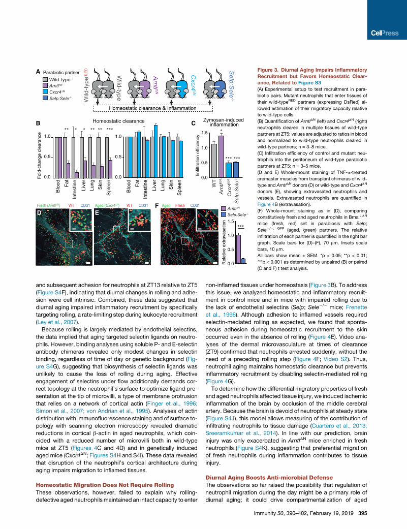
Figure 3. Diurnal Aging Impairs Inflammatory
Recruitment but Favors Homeostatic Clear-
ance, Related to Figure S3
(A) Experimental setup to test recruitment in para-
biotic pairs. Mutant neutrophils that enter tissues of
their wild-typeRED partners (expressing DsRed) al-
lowed estimation of their migratory capacity relative
to wild-type cells.
(B) Quantification of ArntlDN (left) and Cxcr4DN (right)
neutrophils cleared in multiple tissues of wild-type
partners at ZT5; values are adjusted to ratios in blood
and normalized to wild-type neutrophils cleared in
wild-type partners; n = 3–8 mice.
(C) Infiltration efficiency of control and mutant neu-
trophils into the peritoneum of wild-type parabiotic
partners at ZT5; n = 3–5 mice.
(D and E) Whole-mount staining of TNF-a-treated
cremaster muscles from transplant chimeras of wild-
type and ArntlDN donors (D) or wild-type and Cxcr4DN
donors (E), showing extravasated neutrophils and
vessels. Extravasated neutrophils are quantified in
Figure 4B (extravasation).
(F) Whole-mount staining as in (D), comparing
constitutively fresh and aged neutrophils in Bmal1DN
mice (fresh, red) set in parabiosis with Selp;
Sele�/�; GFP (aged, green) partners. The relative
infiltration of each partner is quantified in the right bar
graph. Scale bars for (D)–(F), 70 mm. Insets scale
bars, 10 mm.
All bars show mean ± SEM. *p < 0.05; **p < 0.01;
***p < 0.001 as determined by unpaired (B) or paired
(C and F) t test analysis.
and subsequent adhesion for neutrophils at ZT13 relative to ZT5
(Figure S4F), indicating that diurnal changes in rolling and adhe-
sion were cell intrinsic. Combined, these data suggested that
diurnal aging impaired inflammatory recruitment by specifically
targeting rolling, a rate-limiting step during leukocyte recruitment
(Ley et al., 2007).
Because rolling is largely mediated by endothelial selectins,
the data implied that aging targeted selectin ligands on neutro-
phils. However, binding analyses using soluble P- and E-selectin
antibody chimeras revealed only modest changes in selectin
binding, regardless of time of day or genetic background (Fig-
ure S4G), suggesting that biosynthesis of selectin ligands was
unlikely to cause the loss of rolling during aging. Effective
engagement of selectins under flow additionally demands cor-
rect topology at the neutrophil’s surface to optimize ligand pre-
sentation at the tip of microvilli, a type of membrane protrusion
that relies on a network of cortical actin (Finger et al., 1996;
Simon et al., 2007; von Andrian et al., 1995). Analyses of actin
distribution with immunofluorescence staining and of surface to-
pology with scanning electron microscopy revealed dramatic
reductions in cortical b-actin in aged neutrophils, which coin-
cided with a reduced number of microvilli both in wild-type
mice at ZT5 (Figures 4C and 4D) and in genetically induced
aged mice (Cxcr4DN; Figures S4H and S4I). These data revealed
that disruption of the neutrophil’s cortical architecture during
aging impairs migration to inflamed tissues.
Homeostatic Migration Does Not Require RollingThese observations, however, failed to explain why rolling-
defective aged neutrophils maintained an intact capacity to enter
non-inflamed tissues under homeostasis (Figure 3B). To address
this issue, we analyzed homeostatic and inflammatory recruit-
ment in control mice and in mice with impaired rolling due to
the lack of endothelial selectins (Selp; Sele�/� mice; Frenette
et al., 1996). Although adhesion to inflamed vessels required
selectin-mediated rolling as expected, we found that sponta-
neous adhesion during homeostatic recruitment to the skin
occurred even in the absence of rolling (Figure 4E). Video ana-
lyses of the dermal microvasculature at times of clearance
(ZT9) confirmed that neutrophils arrested suddenly, without the
need of a preceding rolling step (Figure 4F; Video S2). Thus,
neutrophil aging maintains homeostatic clearance but prevents
inflammatory recruitment by disabling selectin-mediated rolling
(Figure 4G).
To determine how the differential migratory properties of fresh
and aged neutrophils affected tissue injury, we induced ischemic
inflammation of the brain by occlusion of the middle cerebral
artery. Because the brain is devoid of neutrophils at steady state
(Figure S4J), this model allows measuring of the contribution of
infiltrating neutrophils to tissue damage (Cuartero et al., 2013;
Sreeramkumar et al., 2014). In line with our prediction, brain
injury was only exacerbated in ArntlDN mice enriched in fresh
neutrophils (Figure S4K), suggesting that preferential migration
of fresh neutrophils during inflammation contributes to tissue
injury.
Diurnal Aging Boosts Anti-microbial DefenseThe observations so far raised the possibility that regulation of
neutrophil migration during the day might be a primary role of
diurnal aging; it could drive compartmentalization of aged
Immunity 50, 390–402, February 19, 2019 395

ZT5ZT13
Arntl∆N
Cxcr4∆N
Rel
ativ
e ro
lling
frac
tion
Rel
ativ
e ad
here
d fra
ctio
n
Rel
ativ
e ex
trav.
frac
tion
0
1
2*
-0
1
2
3
0
1
2
*****
**
**
**Rolling Adhesion ExtravasationA
GF
B
E
WT+
Arntl∆N
WT+
Cxcr4∆N
Transplant chimeras
Intravital imaging
TNFα-inducedinflammation
Wild-type
Wild-type
Selp:Sele-/-
Wild-type Selp:Sele-/-
***
Steady-state (skin, ZT9)
Rolling Adhesion0.0
0.5
1.0
1.5
******
Rolling Adhesion
Inflammation (TNFα)
NDR
ollin
g or
adh
esio
n in
dex
Inflammation
fresh aged
Steady-state
Flow
t = 0s
t = 3s
t = 9s
t = 0s
t = 3s
Time before arrest (s)
Velo
city
( μm
/s)
Arre
stWild-type
Selp:Sele-/-
10 5 00
1000
20000
40
80
DC
***
ZT5
ZT13
ZT5
ZT13
0 010 20 2030 40 40 6050cortical actin / cell
DA
PI β-actin
Villi number / cell
ZT5ZT13
***
Figure 4. Microvilli Collapse and Impaired Rolling Are Hallmarks of Aged Neutrophils, Related to Figure S4
(A) Strategy for competitive recruitment of neutrophils in bone marrow chimeras, at ZT5.
(B) Relative frequencies of rolling, adherent, and extravasated fresh (ArntlDN) and aged (Cxcr4DN) neutrophils, normalized to wild-type controls in chimeric mice;
n = 30–61 venules from 5–6 mice.
(C) b-actin staining in wild-type neutrophils at ZT5 and ZT13, and frequency of neutrophils with cortical distribution of actin; n = 324–330 cells per group.
(D) Scanning electronmicrographs of wild-type neutrophils at ZT5 and ZT13, and number of microvilli on their surface. Scale bar, 5 mm; n = 23–29 cells per group.
(E) Rolling and adhesion of neutrophils on cremasteric venules after treatment with TNF-a (inflammation), or on naive dermal microvessels at ZT9-13 (steady state)
in wild-type or Selp;Sele�/� mice. n = 50–55 venules from 4–5 mice (steady state) and 25–27 venules from 3–5 mice (inflamed cremaster). ND, none detected.
(F) Kinetics of neutrophils (Ly6G+, yellow arrows) prior to firm arrest on dermal microvessels at steady state. Left, representative sequential intravital frames with
neutrophils arresting in the last sequence (reverse arrows). Right, flow or roll dynamics of neutrophils before firm arrest; n = 10 cells shown per group.
(G) Model for the preferential recruitment of fresh and aged neutrophils into inflamed or naive tissues, respectively.
Bars show mean ± SEM. *p < 0.05; **p < 0.01; ***p < 0.001 as determined by paired (B) or unpaired t test analysis (C and D) or non-parametric Mann-
Whitney test (E).
neutrophils into tissues at night in anticipation of pathogens
potentially breaking into tissues, while at the same time reducing
their numbers within vessels to minimize injury when the chance
of immune activation is higher.
To test this possibility, we analyzed the diurnal dynamics of
aged neutrophils in the circulation of wild-type and mutant
mice throughout the day. The analyses revealed striking differ-
ences: in wild-type mice, aged neutrophils peaked at ZT5 and
disappeared at ZT13, whereas in ArntlDN mice, they displayed
non-oscillating low numbers and Cxcr4DN animals presented
396 Immunity 50, 390–402, February 19, 2019
constitutive elevations in aged neutrophils in blood (Figure 5A).
Notably, the absolute number of neutrophils maintained normal
oscillations in the blood of ArntlDN mutants (Figure S5A), indi-
cating that neutrophil numbers and aging are regulated through
different mechanisms. We therefore used these mouse models
to determine how aging-driven trafficking regulated immune
defense and vascular health.
We first infected mice with Candida albicans, using a
protocol that allows systemic spread, targets the kidneys, and
is controlled by neutrophils (Del Fresno et al., 2018; Lionakis

Wei
ght l
oss
(%)
Wei
ght l
oss
(%)
Days post-infection Days post-infection
Days post-infection
0
-5
-10
-15
-20
0
-5
-10
-15
-20
0
-5
-10
-15
-20
0 1 2 3 4 5
P < 0.0001
0 1 2 3 4 5
0 1 2 3 4 5
Arntl∆N ZT5
ZT5
Arntl∆N ZT13
P = 0.46
P < 0.0001
ZT5ZT13
C. albicans infection
0.0
0.5
1.0
1.5
**
n.s.
CFU
/ g
Kid
ney
Fungal load
Wild typen.s.
Arntl∆N
Cxcr4∆N
BA C
D E F
0.0
neut
rphi
ls (x
105)
/ g
0.51.01.5
2468
Kidney***
WT Arntl∆N Cxcr4∆N
0.0
0.71.0
5.0
P = 0.0003
P = 0.1240
P = 0.1094
0.0
0.4
0.8
1.2
1.6
Age
d ne
utro
phils
/ m
l (x1
06 )
01x(
lm /
sli
hport
uen
deg
A6)
Wild type
Arntl∆N
Cxcr4∆N
Zeitgeber time1 5 9 13 17 21
GWT
Cxcr4∆NWT + AMD3100
ns
Wei
ght l
oss
(%)
Days post-infection0 1 2 3 4 5
0
-5
-10
-15
-20
-25 0
1
2
3
CFU
s / g
kid
ney
(x10
5 )
ns
Figure 5. Neutrophil Aging Confers Diurnal Protection against Infection, Related to Figure S5
(A) Diurnal numbers of CD62Llo aged neutrophils in the blood of wild-type, ArntlDN, and Cxcr4DN mice; n = 3–6 mice. See also Figure S5A.
(B and C) Weight loss kinetics of wild-type (B) and ArntlDN mice (C) infected with C. albicans at ZT5 or ZT13; n = 5–21 mice.
(D) Weight-loss curves of wild-type, ArntlDN, and Cxcr4DN mice infected at ZT5; n = 12–14 mice.
(E) Fungal burden at day 5 in the kidneys from the mice in (D), normalized to WT.
(F) Number of neutrophils in the kidneys of non-infected mice; n = 4 mice per group.
(G) Kinetics of weight loss in control or AMD-treatedwild-typemice after systemicC. albicans infection at ZT5. The dashed line showsweight loss inCxcr4DNmice
as in Figure 4D for reference. Bars at right show fungal burden in kidneys at day 5 post-infection; n = 10 mice.
Data are shown as mean ± SEM. **p < 0.01; ***p < 0.001; n.s., not significant, as determined by amplitude versus zero t test (A), two-way ANOVA (B–D and G),
one-way ANOVA with Dunnett’s multigroup correction (E and F), and unpaired t test analysis (G).
et al., 2011). Wild-type mice displayed diurnal patterns of
response to infection, with increased resistance at ZT13 as
defined by reduced weight loss, fungal load in kidneys, and
improved survival (Figures 5B, S5B, and S5C). Importantly, the
initial time of infection was critical for the long-term immune
response because the effect could be seen several days after
infection. Resistance to Candida at ZT13 coincided with more
neutrophils in naive kidneys and fewer in blood (Figures S5D
and S5E), suggesting that their presence in tissues at the time
of infection conferred protection. Remarkably, the diurnal varia-
tion in susceptibility toCandida infection was abolished inArntlDN
mice (Figure 5C), indicating that neutrophil aging was needed to
anticipate the infection. These observations predicted that mice
with constitutively aged neutrophils clearing into tissues might
perform better against infection. Indeed, Cxcr4DN mice had
more neutrophils in naive kidneys and manifested remarkable
resistance to infection and reduced fungal spread (Figures 5D
and 5F). However, because Cxcr4DN mice displayed neutrophilia
(Figure S5A), this observation could be alternatively explained by
elevated numbers rather than by the aging status of neutrophils.
To discriminate between these possibilities, we treated wild-type
mice with a single injection of the CXCR4 antagonist AMD3100, a
treatment that causes acute neutrophilia (Devi et al., 2013) but did
not induce aging (Figures S5F and S5G). Despite neutrophil
counts that were even higher than those in Cxcr4DN mice,
AMD3100-treated mice were as susceptible to Candida infection
as untreated wild-type mice (Figure 5G), indicating that the aging
status, rather than the number of neutrophils, conferred protec-
tion against Candida. In addition, the capacity of fresh (ZT13 or
ArntlDN) and aged (ZT5 and Cxcr4DN) neutrophils to phagocytose
Candida conidia to produce reactive oxygen species (Figures
S5H–S5J) and to secrete cytokines (Figure S5K) were similar to
wild-type cells, which supported the contention that neutrophil
migration, rather than other cellular processes, was the relevant
process regulated by aging. We obtained evidence of similar
diurnal variations in the response to bacterial sepsis, which was
also lost in ArntlDN mice (Figure S5L), further revealing a general
influence of neutrophil aging in responses to infection. Thus,
aging-driven clearance of neutrophils into tissues orchestrates
anti-microbial defense.
Constitutive Neutrophil Aging Predisposes to VascularInflammationTo define whether diurnal neutrophil aging additionally conferred
protection to vessels, we used a model of acute myocardial
infarction (AMI) induced by ischemia reperfusion of the left ante-
rior descending coronary artery, in which inflammation originates
intravascularly without prior neutrophil extravasation (Vinten-
Johansen, 2004). Similar to infections, the extent of cardiac
damage displayed diurnal variations, with larger infarct sizes at
Immunity 50, 390–402, February 19, 2019 397

Figure 6. Aged Neutrophils Aggravate Myocardial Infarction, Related to Figure S6
(A) Representative images of hearts from wild-type mice subjected to ischemia reperfusion at ZT5 or ZT13, or the indicated mutant mice at ZT5. Dotted yellow
lines highlight areas of dead myocardium; n = 4–8 mice per group from 3 experiments.
(B) Infarct sizes in wild-type mice at different diurnal times, after correction for areas at risk (AAR; see related Figure S5); n = 5–8 mice.
(C) Infarct sizes in wild-type, ArntlDN, and Cxcr4DN mice at ZT5 (see related Figure S5); n = 4–8 mice.
(D) Survival curves of wild-type, ArntlDN, and Cxcr4DN mice subjected to myocardial infarction at ZT5; n = 9–11 mice.
(E) Representative images of hearts from untreated or AMD3100-treated wild-type mice and Cxcr4DN mice. Surgeries were performed at ZT5 and dead
myocardium is highlighted as in (A). Bars at right show quantification of infarcted areas in the same groups; n = 4–5 mice from one experiment.
Bars showmean ± SEM. *p < 0.05; ***p < 0.001; n.s., not significant, as determined by one-way ANOVA with Dunnett’s multigroup correction (C and E), unpaired
t test analysis (B), and log-rank test (D).
(F) Molecular regulators and consequences of disrupted neutrophil aging. Defective aging (ArntlDN) impairs the evening boost in anti-microbial defense but
protects from vascular injury; instead constitutive aging (Cxcr4DN) enhances the response to infections but exacerbates thrombo-inflammation.
ZT5 (Figures 6A, 6B, and S6A). Accordingly, infarct sizes after
only 1 h of reperfusion were remarkably larger in Cxcr4DN mice
and smaller in ArntlDN mice (Figure 6C), and this correlated
with early death of all Cxcr4DN mice (Figure 6D). This dramatic
response was not caused by increased numbers of circulating
neutrophils in Cxcr4DN mice because treatment of wild-type
mice with AMD3100 did not aggravate myocardial injury (Fig-
ure 6E). Thus, the presence of aged neutrophils in the circulation
is detrimental for tissues after vascular ischemia and reperfusion,
whereas their diurnal clearance is protective.
We examined potential mechanisms by which aged neutro-
phils might exacerbate vascular injury in Cxcr4DN mice. Using a
model of ischemia reperfusion in the cremaster muscle that
allows high-resolution live imaging of affected vessels, we found
disseminated thrombi in microvessels of Cxcr4DN mice (Fig-
ure S6C; Video S3). Depletion of neutrophils in Cxcr4DN mice
prevented thrombi formation and improved survival after
infarction (Figures S6B and S6C), indicating that both responses
398 Immunity 50, 390–402, February 19, 2019
were mediated by neutrophils. Although neutrophil extracellular
traps (NETs) can promote thrombosis (Fuchs et al., 2010), they
were not responsible for the response of Cxcr4DN mice because
two different NET inhibitors failed to prevent thrombus formation
in reperfused venules (Figure S6D). In addition, endothelial pro-
liferation and apoptosis, as well as vascular permeability, were
not affected at baseline acrossmultiple tissues, including hearts,
of ArntlDN and Cxcr4DN mice (Figures S6E–S6G), indicating that
aging did not directly compromise basal vascular health prior
to the ischemic insult.
Overall, these observations suggest that neutrophil aging is
critically driven by an internal program, as we failed to find con-
tributions from other factors, including reactive oxygen species
(ROS; data not shown) or the intestinal microbiota (Figures
S6H–S6K), both of which had been previously associated
with neutrophil senescence or aging, respectively (Harbort
et al., 2015; Zhang et al., 2015). In turn, aging controls diurnal
compartmentalization of neutrophils into tissues and out of the

circulation, thereby balancing immune protection and vascular
protection (Figure 6F; Video S4).
DISCUSSION
Mammalian immunity is not constant in quantity (e.g., number of
recruited or mobilized leukocytes) or quality throughout the day,
as it adapts to varying diurnal challenges from the environment,
including the chance of exposure to infectious pathogens (Man
et al., 2016). Likewise, damage to the cardiovascular system,
in both humans andmodel organisms, follows circadian patterns
(Muller et al., 1989; Scheiermann et al., 2013). Because neutro-
phils are major mediators of anti-microbial defense and vascular
inflammation, we predicted that the diurnal variations in both
processes could be mechanistically explained by the existence
of a neutrophil-intrinsic program (or ‘‘timer’’) that regulated their
activity through the day. In this study, we identified and charac-
terized this program and revealed that it underlies the circadian
susceptibility of mice to infection and vascular inflammation.
We have found that the diurnal program of neutrophils is coor-
dinated by the circadian-related protein Bmal1 in coordination
with two chemokine receptors: CXCR2, which drives aging,
and CXCR4, which antagonizes it. Multiple functional assays
allowed us to confirm that time-of-day differences in wild-type
cells could be faithfully recapitulated by the respective mutants:
ArntlDN cells resembled night (fresh) neutrophils, whereas
Cxcr4DN mutants behaved similar to daytime (aged) neutrophils.
Before release into the bloodstream,maturing neutrophils are re-
tained within the marrow in an environment with high CXCR4
signaling (Eash et al., 2009, 2010), which raises the intriguing
possibility that this diurnal timer is inhibited until neutrophils
are released into blood. Once in blood, functional analyses of
mice in which we disabled each component of this timer re-
vealed that preferential invasion of inflamed or naive tissues is
compartmentalized in time. Under steady-state conditions, neu-
trophils released from the marrow gradually lost their ability to
enter inflammatory sites and prepared for clearance into tissues.
This migratory switch was intrinsically regulated, but it likely
coordinated with extrinsic programs because disruption of
rhythms in vascular cells can also affect the diurnal entry of
leukocytes in tissues (Scheiermann et al., 2012) and because
CXCL12, which is not produced by neutrophils, negatively regu-
lated diurnal aging through CXCR4.
We found that one potential benefit of diurnal infiltration into
naive tissues was to optimize immune defense, as demonstrated
by the loss of diurnal oscillations in the response against fungal
or bacterial infections when Arntl was deleted from neutrophils.
Removal of Cxcr4, the negative regulator of the neutrophil
timer, instead caused unrestrained aging and enhanced anti-
microbial responses, while at the same time precipitating severe
thrombo-inflammatory reactions following ischemia reperfusion.
In contrast, ArntlDN mutants displayed attenuated damage
during myocardial infarction, altogether indicating that an intact
neutrophil clock was important to balance anti-microbial de-
fense and cardiovascular inflammation.
Among the various transcriptional pathways activated during
aging, we identified those related to leukocyte extravasation
and actin cytoskeleton signaling, an observation that allowed
us to identify disruption of cortical actin polymerization as a
key molecular event linking diurnal aging with alterations in the
migratory properties of neutrophils. Although the mechanisms
underlying these cytoskeletal changes remain to be elucidated,
this observation is consistent with early studies showing disrup-
ted actin polymerization on human CD62Llo neutrophils (Tanji-
Matsuba et al., 1998). Diurnal loss of microvilli was particularly
relevant because these structures allow presentation of glyco-
conjugate ligands to endothelial selectins under flow (von An-
drian et al., 1995), thus explaining the dramatic loss of rolling
and migration of aged neutrophils to inflamed areas. At the
same time, loss of microvilli might conceivably enhance the
exposure of b2 integrins present on the cell body (Erlandsen
et al., 1993) and favor rolling-independent arrest as seen in the
naive dermal microvasculature. The fact that a similar behavior
of constitutive adhesion in non-inflamed vessels is displayed
by patrolling monocytes (Auffray et al., 2007) suggests that this
mechanism could be a common property of myeloid leukocytes
endowed with homeostatic surveillance roles.
Overall, our findings are consistent with a model in which the
oscillatory nature of the aging program enables alert states of
neutrophils that are useful to anticipate infections but must be
shut down when the risk of infection is low to prevent damage
to the vasculature. We note that Bmal1-driven aging of neutro-
phils may not necessarily adjust to behavioral rhythms because
we found that neutrophil aging peaked in the morning in both
humans and mice, which are species with opposed activity pe-
riods. We therefore propose that a major purpose of aging is to
ensure temporal separation of neutrophil-mediated responses
within vessels from those in tissues, thereby optimizing defense
without compromising vascular health.
The diurnal aging pattern of neutrophils aligns with studies
showing temporally gated responses for other leukocyte
subsets, including monocytes, macrophages, or T helper-17
(Th17) cells (Nguyen et al., 2013; Silver et al., 2012; Yu et al.,
2013), which may be useful to temporally concentrate immune
response against specific pathogens in different tissues (Tognini
et al., 2017). Different from these other leukocytes, however, the
existence of a circadian program in neutrophils was not intuitive
because their lifetime in the circulation is generally accepted to
be less than one day (Summers et al., 2010), which implies that
there cannot be true circadian oscillations of gene expression
within a given neutrophil. Further, at present we do not know
whether aging is regulated by the transcriptional properties of
Bmal1 or by the core circadian clock. For these reasons, we
envision this system to function like a cellular timer (rather than
a true circadian clock) that resets with every new wave of
neutrophils released from the bone marrow. In other words, for
short-lived cells such as neutrophils, the clock appears to regu-
late oscillations on a population scale by acting as a timer at the
cellular level.
Given the high prevalence of infections and cardiovascular
disease, a final question is whether the identification of a diurnal
program in neutrophils could offer therapeutic alternatives for
these life-threatening complications. In principle, targeting
CXCR2 or CXCR4 with specific agonists might allow pharmaco-
logical and transient manipulation of the timer. This ‘‘chrono-
programming’’ of neutrophils could allow the generation of
phenotypes that promote defense or protect the vasculature,
as needed. We expect that manipulation of the timer will not
Immunity 50, 390–402, February 19, 2019 399

have detrimental consequences because animals with impaired
or enhanced neutrophil aging do not present gross anomalies or
spontaneous susceptibility to disease at baseline, at least under
specific-pathogen-free conditions (data not shown). Thus, for
humans at risk of cardiovascular events, it might be advisable
to block aging, whereas immunocompromised patients suscep-
tible to infections might benefit from drugs that promote it. We
are currently exploring strategies that exploit the unique tempo-
ral properties of neutrophils.
STAR+METHODS
Detailed methods are provided in the online version of this paper
and include the following:
d KEY RESOURCES TABLE
d CONTACT FOR REAGENT AND RESOURCE SHARING
d EXPERIMENTAL MODEL AND SUBJECT DETAILS
400
B Mice
B Human Studies
d METHOD DETAILS
B Analysis of Human Samples
B Parabiosis
B Cytometry and Cell Sorting
B Whole-Mount Staining of Excised Cremaster Muscles
B RNA Isolation, Reverse Transcription, and rtPCR
B Chromatin Immunoprecipitation (ChIP)
B Intravital Imaging of the Mouse Skin
B Intravital Microscopy of the Cremaster Muscle
B Intravital Imaging of Ischemia and Reperfusion Injury
B Analysis of Neutrophil Clearance in the Steady State
B Quantification of Neutrophil Numbers in Tissues
B Chemokine Quantification in Plasma
B RNA-Sequencing
B Western Blotting
B Generation of Transplant Chimeras
B Circadian Analysis of Aging Markers and RNA
Extraction
B Analysis of Neutrophil Aging in Light-Dark and Dark-
Dark Light Regimes
B Entrainment of Neutrophil Aging by Inverted Light
Regime
B Infection with Candida albicans
B Heat-Killed Candida albicans (HKC) Phagocy-
tosis Assay
B Mouse Model of Acute Myocardial Infarction
B Brain Ischemia
B Scanning Electron Microscopy
B CXCR2 and CXCR4 Cross-Inhibition Assays
B Neutrophil Depletion
B Zymosan-Induced Peritonitis
B Soluble Selectin Binding Assays
B Auto-Perfused Flow Chamber Assay
B Cortical Beta-Actin Quantification
B ROS Quantification
B Chemotaxis Assay
B Multiplex Cytokine Assay
B AMD3100-Induced Neutrophilia
B Neutrophil Transfer Experiments
Immunity 50, 390–402, February 19, 2019
B BrdU Labelling
B In Vivo CXCL1 and CXCL2 Blockade
B Cecal Ligation and Puncture (CLP)-Induced Sepsis
B Evans Blue Vascular Permeability Assay
B Analysis of Endothelial Proliferation and Apoptosis
B Analysis of Neutrophil Aging in Microbiota-Depleted
and Germ-free Mice
d QUANTIFICATION AND STATISTICAL ANALYSIS
B RNA-Sequencing Data Analysis
B Statistical Analysis
B Amplitude versus Zero Test
d DATA AND SOFTWARE AVAILABILITY
SUPPLEMENTAL INFORMATION
Supplemental Information includes six figures, three tables, and four videos
and can be found with this article online at https://doi.org/10.1016/j.immuni.
2019.01.002.
ACKNOWLEDGMENTS
We thank all members of the Hidalgo Lab for discussion and insightful com-
ments; J.M. Ligos, R. Nieto, and M. Viton for help with sorting and cytometric
analyses; I. Ortega and E. Santos for animal husbandry; D. Rico, M.J. Gomez,
C. Torroja, and F. Sanchez-Cabo for insightful comments and help with tran-
scriptomic analyses; V. Labrador, E. Arza, A.M. Santos, and the Microscopy
Unit of the CNIC for help with microscopy; S. Aznar-Benitah, U. Albrecht,
Q.-J. Meng, B. Staels, and H. Duez for the generous gift of mice; J.A. Enriquez
and J. Avila for scientific insights; and J.M. Garcıa and A. Diez de la Cortina for
art. This study was supported by Intramural grants from A*STAR to L.G.N.,
BES-2013-065550 to J.M.A., BES-2010-032828 to M.C.-A, and JCI-2012-
14147 to L.A.W (all from Ministerio de Economıa, Industria y Competitividad;
MEIC). Additional MEIC grants were SAF2014-61993-EXP to C.L.-R.;
SAF2015-68632-R to M.A.M. and SAF-2013-42920R and SAF2016-
79040Rto D.S. D.S. also received 635122-PROCROP H2020 from the Euro-
pean Commission and ERCCoG 725091 from the European Research Council
(ERC). ERC AdG 692511 PROVASC from the ERC and SFB1123-A1 from the
Deutsche Forschungsgemeinschaft were given to C.W.; MHA VD1.2/
81Z1600212 from the German Center for Cardiovascular Research (DZHK)
was given to C.W. and O.S.; SFB1123-A6 was given to O.S.; SFB914-B08
was given to O.S. and C.W.; and INST 211/604-2, ZA 428/12-1, and ZA 428/
13-1 were given to A.Z. This study was also supported by PI12/00494 from
Fondo de Investigaciones Sanitarias (FIS) to C.M.; PI13/01979, Cardiovascular
Network grant RD 12/0042/0054, and CIBERCV to B.I.; SAF2015-65607-R,
SAF2013-49662-EXP, and PCIN-2014-103 from MEIC; and co-funding by
Fondo Europeo de Desarrollo Regional (FEDER) to A.H. The CNIC is supported
by the MEIC and the Pro CNIC Foundation and is a Severo Ochoa Center of
Excellence (MEIC award SEV-2015-0505).
AUTHOR CONTRIBUTIONS
J.M.A., C.d.F., M.I.C., M.C.-A., L.A.W., H.H.-E., C.S.-R., J.R., J.A.Q., G.C.,
J.G.-P., M.G.-P., S.M.-S., M.E., and J.L. performed experiments; C.W., K.B.,
and F.B. contributed essential reagents; A.Z., O.S., C.L.-R., M.A.M., B.I.,
D.S., L.N., J.M.A., and A.H. designed and supervised experiments; F.A. and
C.M. coordinated the study on humans; A.H. designed and supervised the
study. A.H. and J.M.A. wrote the manuscript, which was edited by all authors.
DECLARATION OF INTERESTS
The authors declare no competing interests.
Received: August 24, 2018
Revised: November 23, 2018
Accepted: January 2, 2019
Published: January 29, 2019

REFERENCES
Auffray, C., Fogg, D., Garfa, M., Elain, G., Join-Lambert, O., Kayal, S.,
Sarnacki, S., Cumano, A., Lauvau, G., and Geissmann, F. (2007). Monitoring
of blood vessels and tissues by a population of monocytes with patrolling
behavior. Science 317, 666–670.
Balabanian, K., Brotin, E., Biajoux, V., Bouchet-Delbos, L., Lainey, E.,
Fenneteau, O., Bonnet, D., Fiette, L., Emilie, D., and Bachelerie, F. (2012).
Proper desensitization of CXCR4 is required for lymphocyte development
and peripheral compartmentalization in mice. Blood 119, 5722–5730.
Casanova-Acebes, M., Pitaval, C., Weiss, L.A., Nombela-Arrieta, C., Chevre,
R., A-Gonzalez, N., Kunisaki, Y., Zhang, D., van Rooijen, N., Silberstein, L.E.,
et al. (2013). Rhythmic modulation of the hematopoietic niche through neutro-
phil clearance. Cell 153, 1025–1035.
Casanova-Acebes, M., Nicolas-Avila, J.A., Li, J.L., Garcıa-Silva, S.,
Balachander, A., Rubio-Ponce, A., Weiss, L.A., Adrover, J.M., Burrows, K.,
A-Gonzalez, N., et al. (2018). Neutrophils instruct homeostatic and patholog-
ical states in naive tissues. J. Exp. Med. 215, 2778–2795.
Cuartero, M.I., Ballesteros, I., Moraga, A., Nombela, F., Vivancos, J., Hamilton,
J.A., Corbı, A.L., Lizasoain, I., and Moro, M.A. (2013). N2 neutrophils, novel
players in brain inflammation after stroke: modulation by the PPARg agonist
rosiglitazone. Stroke 44, 3498–3508.
Del Fresno, C., Saz-Leal, P., Enamorado, M., Wculek, S.K., Martınez-Cano, S.,
Blanco-Menendez, N., Schulz, O., Gallizioli, M., Miro-Mur, F., et al. (2018).
DNGR-1 in dendritic cells limits tissue damage by dampening neutrophil
recruitment. Science 362, 351–356.
Devi, S., Wang, Y., Chew,W.K., Lima, R., A-Gonzalez, N., Mattar, C.N., Chong,
S.Z., Schlitzer, A., Bakocevic, N., Chew, S., et al. (2013). Neutrophil mobiliza-
tion via plerixafor-mediated CXCR4 inhibition arises from lung demargination
and blockade of neutrophil homing to the bone marrow. J. Exp. Med. 210,
2321–2336.
Druzd, D., Matveeva, O., Ince, L., Harrison, U., He, W., Schmal, C., Herzel, H.,
Tsang, A.H., Kawakami, N., Leliavski, A., et al. (2017). Lymphocyte Circadian
Clocks Control Lymph Node Trafficking and Adaptive Immune Responses.
Immunity 46, 120–132.
Eash, K.J., Means, J.M., White, D.W., and Link, D.C. (2009). CXCR4 is a key
regulator of neutrophil release from the bone marrow under basal and stress
granulopoiesis conditions. Blood 113, 4711–4719.
Eash, K.J., Greenbaum, A.M., Gopalan, P.K., and Link, D.C. (2010). CXCR2
and CXCR4 antagonistically regulate neutrophil trafficking from murine bone
marrow. J. Clin. Invest. 120, 2423–2431.
Erlandsen, S.L., Hasslen, S.R., and Nelson, R.D. (1993). Detection and spatial
distribution of the beta 2 integrin (Mac-1) and L-selectin (LECAM-1) adherence
receptors on human neutrophils by high-resolution field emission SEM.
J. Histochem. Cytochem. 41, 327–333.
Faust, N., Varas, F., Kelly, L.M., Heck, S., and Graf, T. (2000). Insertion of
enhanced green fluorescent protein into the lysozyme gene creates mice
with green fluorescent granulocytes and macrophages. Blood 96, 719–726.
Finger, E.B., Bruehl, R.E., Bainton, D.F., and Springer, T.A. (1996). A differential
role for cell shape in neutrophil tethering and rolling on endothelial selectins un-
der flow. J. Immunol. 157, 5085–5096.
Frenette, P.S., Mayadas, T.N., Rayburn, H., Hynes, R.O., and Wagner, D.D.
(1996). Susceptibility to infection and altered hematopoiesis in mice deficient
in both P- and E-selectins. Cell 84, 563–574.
Fuchs, T.A., Brill, A., Duerschmied, D., Schatzberg, D., Monestier, M., Myers,
D.D., Jr., Wrobleski, S.K., Wakefield, T.W., Hartwig, J.H., and Wagner, D.D.
(2010). Extracellular DNA traps promote thrombosis. Proc. Natl. Acad. Sci.
USA 107, 15880–15885.
Garcıa-Prieto, J., Villena-Gutierrez, R., Gomez, M., Bernardo, E., Pun-Garcıa,
A., Garcıa-Lunar, I., Crainiciuc, G., Fernandez-Jimenez, R., Sreeramkumar, V.,
Bourio-Martınez, R., et al. (2017). Neutrophil stunning by metoprolol reduces
infarct size. Nat. Commun. 8, 14780.
Harbort, C.J., Soeiro-Pereira, P.V., von Bernuth, H., Kaindl, A.M., Costa-
Carvalho, B.T., Condino-Neto, A., Reichenbach, J., Roesler, J., Zychlinsky,
A., and Amulic, B. (2015). Neutrophil oxidative burst activates ATM to regulate
cytokine production and apoptosis. Blood 126, 2842–2851.
Hasenberg, A., Hasenberg, M., M€ann, L., Neumann, F., Borkenstein, L.,
Stecher, M., Kraus, A., Engel, D.R., Klingberg, A., Seddigh, P., et al. (2015).
Catchup: a mouse model for imaging-based tracking and modulation of
neutrophil granulocytes. Nat. Methods 12, 445–452.
Hidalgo, A., Peired, A.J., Wild, M., Vestweber, D., and Frenette, P.S. (2007).
Complete identification of E-selectin ligands on neutrophils reveals distinct
functions of PSGL-1, ESL-1, and CD44. Immunity 26, 477–489.
Hidalgo, A., Chang, J., Jang, J.E., Peired, A.J., Chiang, E.Y., and Frenette, P.S.
(2009). Heterotypic interactions enabled by polarized neutrophil microdomains
mediate thromboinflammatory injury. Nat. Med. 15, 384–391.
Janich, P., Pascual, G., Merlos-Suarez, A., Batlle, E., Ripperger, J., Albrecht,
U., Cheng, H.Y., Obrietan, K., Di Croce, L., and Benitah, S.A. (2011). The circa-
dian molecular clock creates epidermal stem cell heterogeneity. Nature 480,
209–214.
Ley, K., Laudanna, C., Cybulsky, M.I., and Nourshargh, S. (2007). Getting to
the site of inflammation: the leukocyte adhesion cascade updated. Nat. Rev.
Immunol. 7, 678–689.
Li, B., and Dewey, C.N. (2011). RSEM: accurate transcript quantification from
RNA-Seq data with or without a reference genome. BMC Bioinformatics
12, 323.
Li, J.L., Lim, C.H., Tay, F.W., Goh, C.C., Devi, S., Malleret, B., Lee, B.,
Bakocevic, N., Chong, S.Z., Evrard, M., et al. (2016). Neutrophils Self-
Regulate Immune Complex-Mediated Cutaneous Inflammation through
CXCL2. J. Invest. Dermatol. 136, 416–424.
Lionakis, M.S., Lim, J.K., Lee, C.C., and Murphy, P.M. (2011). Organ-specific
innate immune responses in a mouse model of invasive candidiasis. J. Innate
Immun. 3, 180–199.
Man, K., Loudon, A., and Chawla, A. (2016). Immunity around the clock.
Science 354, 999–1003.
Martin, C., Burdon, P.C., Bridger, G., Gutierrez-Ramos, J.C., Williams, T.J.,
and Rankin, S.M. (2003). Chemokines acting via CXCR2 and CXCR4 control
the release of neutrophils from the bone marrow and their return following
senescence. Immunity 19, 583–593.
Muller, J.E., Tofler, G.H., and Stone, P.H. (1989). Circadian variation and trig-
gers of onset of acute cardiovascular disease. Circulation 79, 733–743.
Nguyen, K.D., Fentress, S.J., Qiu, Y., Yun, K., Cox, J.S., and Chawla, A. (2013).
Circadian gene Bmal1 regulates diurnal oscillations of Ly6C(hi) inflammatory
monocytes. Science 341, 1483–1488.
Nie, Y., Waite, J., Brewer, F., Sunshine, M.J., Littman, D.R., and Zou, Y.R.
(2004). The role of CXCR4 in maintaining peripheral B cell compartments
and humoral immunity. J. Exp. Med. 200, 1145–1156.
Passegue, E., Wagner, E.F., and Weissman, I.L. (2004). JunB deficiency leads
to a myeloproliferative disorder arising from hematopoietic stem cells. Cell
119, 431–443.
Radu, M., and Chernoff, J. (2013). An in vivo assay to test blood vessel perme-
ability. J. Vis. Exp. 73, e50062.
Rittirsch, D., Huber-Lang, M.S., Flierl, M.A., and Ward, P.A. (2009).
Immunodesign of experimental sepsis by cecal ligation and puncture. Nat.
Protoc. 4, 31–36.
Robinson, M.D., McCarthy, D.J., and Smyth, G.K. (2010). edgeR: a
Bioconductor package for differential expression analysis of digital gene
expression data. Bioinformatics 26, 139–140.
Schageman, J., Zeringer, E., Li, M., Barta, T., Lea, K., Gu, J., Magdaleno, S.,
Setterquist, R., and Vlassov, A.V. (2013). The complete exosome workflow so-
lution: from isolation to characterization of RNA cargo. BioMed Res. Int. 2013,
253957.
Scheiermann, C., Kunisaki, Y., Lucas, D., Chow, A., Jang, J.E., Zhang, D.,
Hashimoto, D., Merad, M., and Frenette, P.S. (2012). Adrenergic nerves
govern circadian leukocyte recruitment to tissues. Immunity 37, 290–301.
Scheiermann, C., Kunisaki, Y., and Frenette, P.S. (2013). Circadian control of
the immune system. Nat. Rev. Immunol. 13, 190–198.
Immunity 50, 390–402, February 19, 2019 401

Schindelin, J., Rueden, C.T., Hiner, M.C., and Eliceiri, K.W. (2015). The ImageJ
ecosystem: An open platform for biomedical image analysis. Mol. Reprod.
Dev. 82, 518–529.
Schloss, M.J., Horckmans, M., Nitz, K., Duchene, J., Drechsler, M.,
Bidzhekov, K., Scheiermann, C., Weber, C., Soehnlein, O., and Steffens, S.
(2016). The time-of-day of myocardial infarction onset affects healing through
oscillations in cardiac neutrophil recruitment. EMBO Mol. Med. 8, 937–948.
Silver, A.C., Arjona, A., Walker, W.E., and Fikrig, E. (2012). The circadian clock
controls toll-like receptor 9-mediated innate and adaptive immunity. Immunity
36, 251–261.
Simon, S.I., Nyunt, T., Florine-Casteel, K., Ritchie, K., Ting-Beall, H.P., Evans,
E., and Needham, D. (2007). Dynamics of neutrophil membrane compliance
and microstructure probed with a micropipet-based piconewton force trans-
ducer. Ann. Biomed. Eng. 35, 595–604.
Sreeramkumar, V., Adrover, J.M., Ballesteros, I., Cuartero, M.I., Rossaint, J.,
Bilbao, I., Nacher, M., Pitaval, C., Radovanovic, I., Fukui, Y., et al. (2014).
Neutrophils scan for activated platelets to initiate inflammation. Science 346,
1234–1238.
Sturn, A., Quackenbush, J., and Trajanoski, Z. (2002). Genesis: cluster analysis
of microarray data. Bioinformatics 18, 207–208.
Summers, C., Rankin, S.M., Condliffe, A.M., Singh, N., Peters, A.M., and
Chilvers, E.R. (2010). Neutrophil kinetics in health and disease. Trends
Immunol. 31, 318–324.
Tanji-Matsuba, K., van Eeden, S.F., Saito, Y., Okazawa, M., Klut, M.E.,
Hayashi, S., and Hogg, J.C. (1998). Functional changes in aging polymorpho-
nuclear leukocytes. Circulation 97, 91–98.
402 Immunity 50, 390–402, February 19, 2019
Tognini, P., Thaiss, C.A., Elinav, E., and Sassone-Corsi, P. (2017). Circadian
Coordination of Antimicrobial Responses. Cell Host Microbe 22, 185–192.
Uhl, B., Vadlau, Y., Zuchtriegel, G., Nekolla, K., Sharaf, K., Gaertner, F.,
Massberg, S., Krombach, F., and Reichel, C.A. (2016). Aged neutrophils
contribute to the first line of defense in the acute inflammatory response.
Blood 128, 2327–2337.
Vinten-Johansen, J. (2004). Involvement of neutrophils in the pathogenesis of
lethal myocardial reperfusion injury. Cardiovasc. Res. 61, 481–497.
Vintersten, K.,Monetti, C., Gertsenstein, M., Zhang, P., Laszlo, L., Biechele, S.,
andNagy, A. (2004). Mouse in red: red fluorescent protein expression inmouse
ES cells, embryos, and adult animals. Genesis 40, 241–246.
von Andrian, U.H., Hasslen, S.R., Nelson, R.D., Erlandsen, S.L., and Butcher,
E.C. (1995). A central role for microvillous receptor presentation in leukocyte
adhesion under flow. Cell 82, 989–999.
Yu, X., Rollins, D., Ruhn, K.A., Stubblefield, J.J., Green, C.B., Kashiwada, M.,
Rothman, P.B., Takahashi, J.S., and Hooper, L.V. (2013). TH17 cell differenti-
ation is regulated by the circadian clock. Science 342, 727–730.
Zarbock, A., Lowell, C.A., and Ley, K. (2007). Spleen tyrosine kinase Syk is
necessary for E-selectin-induced alpha(L)beta(2) integrin-mediated rolling on
intercellular adhesion molecule-1. Immunity 26, 773–783.
Zhang, D., Chen, G., Manwani, D., Mortha, A., Xu, C., Faith, J.J., Burk, R.D.,
Kunisaki, Y., Jang, J.E., Scheiermann, C., et al. (2015). Neutrophil ageing is
regulated by the microbiome. Nature 525, 528–532.

STAR+METHODS
KEY RESOURCES TABLE
REAGENT or RESOURCE SOURCE IDENTIFIER
Antibodies
CXCR4-APC (Human) eBioscience Clone 12G5; RRID: AB_1944349
CD11b-FITC (Human) eBioscience Clone ICRF44
CD16-Pacific Blue (Human) BD Clone 3G8
CD62L-PE (Human) BD Clone DREG56
CD11c-APC (Human) BD Clone B-ly6
bio eBioscience Clone 1A8
Ly6G-Dylight 450 BioXcell (conjugated in-house) Clone 1A8; RRID: AB_1107721
Ly6G-Dylight 650 BioXcell (conjugated in-house) Clone 1A8; RRID: AB_1107721
Ly6G-FITC eBioscience Clone 1A8; RRID: AB_2572532
CD45-PerCP-Cy5.5 Biolegend Clone 30-F11; RRID: AB_893344
CD11b-PE Tonbo Biosciences Clone M1/70; RRID: AB_2621746
CD11b-FITC BD Clone M1/70; RRID: AB_394774
CXCR2-PerCP-Cy5.5 Biolegend Clone SA044G4; RRID: AB_2565695
CXCR4-APC eBioscience Clone 2B11; RRID: AB_10670877
CD41-PE eBioscience Clone MWReg30 RRID: AB_2538354
Ly6C-FITC Biolegend Clone HK1.4
CD62L-APC eBioscience Clone MEL-14; RRID: AB_469410
CD62L-FITC eBioscience Clone MEL-14; RRID: AB_465109
Anti-CXCL1 R&D MAB453; RRID: AB_2087696
Anti-CXCL2 R&D MAB452; RRID: AB_2230058
Anti-CXCL12 R&D MAB350; RRID: AB_2088149
Anti-Ly6G (depleting antibody) BioXCell BE0075-1; RRID: AB_1107721
Experimental Models: Organisms/Strains
WT Charles River C57BL/6
Mrp8CRE Passegue et al., 2004 B6.Cg-Tg(S100A8-cre,-EGFP)1Ilw
Cxcr2fl/fl Schloss et al., 2016 C57BL/6-Cxcr2tm1Rmra
Cxcr2DN This paper N/A
Cxcr4fl/fl Nie et al., 2004 B6.129P2-Cxcr4tm2Yzo
Cxcr2DN This paper N/A
Cxcr2WHIM Balabanian et al., 2012 Cxcr4/1013
Arntlfl/fl Janich et al., 2011 B6.129S4(Cg)-Arntltm1Weit
ArntlDN This paper N/A
Selp; Sele�/� Frenette et al., 1996 B6.129S2-Seletm1Hyn Selptm1Hyn
DsRED Vintersten et al., 2004 B6.Cg-Tg(CAG-DsRed*MST)1Nagy
Lyz2GFP Faust et al., 2000 B6.129P-Lyz2tm1(EGFP)1.1Graf/Mmmh
Candida albicans Del Fresno et al., 2018 SC5314
Chemicals, Peptides, and Recombinant Proteins
CXCL12 (recombinant) R&D 460-SD
CXCL1 (recombinant) R&D 453-KC
CXCL2 (recombinant) R&D 452-M2
TNF-a R&D 410-MT-050
AMD3100 Tocris Cat# 3299
(Continued on next page)
Immunity 50, 390–402.e1–e10, February 19, 2019 e1
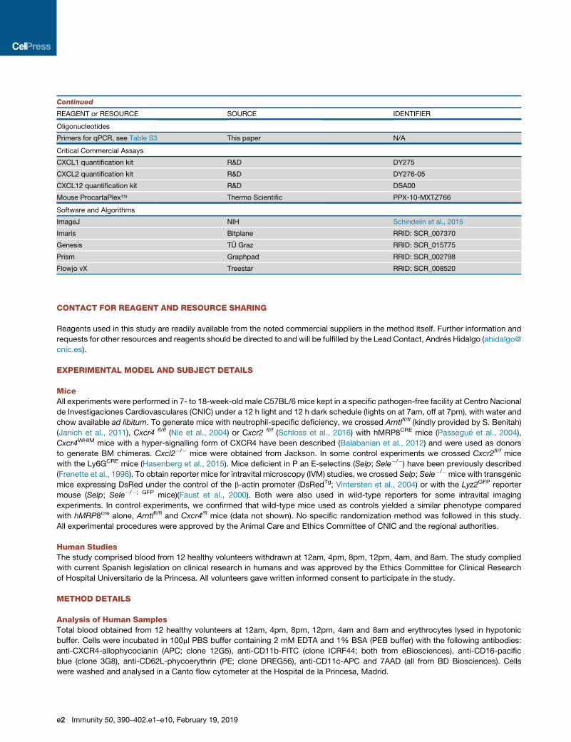
Continued
REAGENT or RESOURCE SOURCE IDENTIFIER
Oligonucleotides
Primers for qPCR, see Table S3 This paper N/A
Critical Commercial Assays
CXCL1 quantification kit R&D DY275
CXCL2 quantification kit R&D DY276-05
CXCL12 quantification kit R&D DSA00
Mouse ProcartaPlex� Thermo Scientific PPX-10-MXTZ766
Software and Algorithms
ImageJ NIH Schindelin et al., 2015
Imaris Bitplane RRID: SCR_007370
Genesis TU Graz RRID: SCR_015775
Prism Graphpad RRID: SCR_002798
Flowjo vX Treestar RRID: SCR_008520
CONTACT FOR REAGENT AND RESOURCE SHARING
Reagents used in this study are readily available from the noted commercial suppliers in the method itself. Further information and
requests for other resources and reagents should be directed to and will be fulfilled by the Lead Contact, Andres Hidalgo (ahidalgo@
cnic.es).
EXPERIMENTAL MODEL AND SUBJECT DETAILS
MiceAll experiments were performed in 7- to 18-week-old male C57BL/6 mice kept in a specific pathogen-free facility at Centro Nacional
de Investigaciones Cardiovasculares (CNIC) under a 12 h light and 12 h dark schedule (lights on at 7am, off at 7pm), with water and
chow available ad libitum. To generate mice with neutrophil-specific deficiency, we crossed Arntlfl/fl (kindly provided by S. Benitah)
(Janich et al., 2011), Cxcr4 fl/fl (Nie et al., 2004) or Cxcr2 fl/f (Schloss et al., 2016) with hMRP8CRE mice (Passegue et al., 2004),
Cxcr4WHIM mice with a hyper-signalling form of CXCR4 have been described (Balabanian et al., 2012) and were used as donors
to generate BM chimeras. Cxcl2�/� mice were obtained from Jackson. In some control experiments we crossed Cxcr2fl/f mice
with the Ly6GCRE mice (Hasenberg et al., 2015). Mice deficient in P an E-selectins (Selp; Sele�/�) have been previously described
(Frenette et al., 1996). To obtain reporter mice for intravital microscopy (IVM) studies, we crossed Selp; Sele�/� mice with transgenic
mice expressing DsRed under the control of the b-actin promoter (DsRedTg; Vintersten et al., 2004) or with the Lyz2GFP reporter
mouse (Selp; Sele�/�; GFP mice)(Faust et al., 2000). Both were also used in wild-type reporters for some intravital imaging
experiments. In control experiments, we confirmed that wild-type mice used as controls yielded a similar phenotype compared
with hMRP8cre alone, Arntlfl/fl and Cxcr4/fl mice (data not shown). No specific randomization method was followed in this study.
All experimental procedures were approved by the Animal Care and Ethics Committee of CNIC and the regional authorities.
Human StudiesThe study comprised blood from 12 healthy volunteers withdrawn at 12am, 4pm, 8pm, 12pm, 4am, and 8am. The study complied
with current Spanish legislation on clinical research in humans and was approved by the Ethics Committee for Clinical Research
of Hospital Universitario de la Princesa. All volunteers gave written informed consent to participate in the study.
METHOD DETAILS
Analysis of Human SamplesTotal blood obtained from 12 healthy volunteers at 12am, 4pm, 8pm, 12pm, 4am and 8am and erythrocytes lysed in hypotonic
buffer. Cells were incubated in 100ml PBS buffer containing 2 mM EDTA and 1% BSA (PEB buffer) with the following antibodies:
anti-CXCR4-allophycocianin (APC; clone 12G5), anti-CD11b-FITC (clone ICRF44; both from eBiosciences), anti-CD16-pacific
blue (clone 3G8), anti-CD62L-phycoerythrin (PE; clone DREG56), anti-CD11c-APC and 7AAD (all from BD Biosciences). Cells
were washed and analysed in a Canto flow cytometer at the Hospital de la Princesa, Madrid.
e2 Immunity 50, 390–402.e1–e10, February 19, 2019

ParabiosisWe followed previously published procedures (Casanova-Acebes et al., 2013). Briefly, anesthetized mice were shaved and matched
incisions were made from the olecranon to the knee joint, then olecranon and knee were attached by a single suture from one mouse
to the other, using 5-0 polypropylene, and the dorsal and ventral skins were stitched by continuous suture. Analyses were done 4 to
6 weeks after surgery.
Cytometry and Cell SortingCytometric analyses were performed using a Sony SP6800 Spectral Analyzer (Sony Biotechnology, Japan) or a LSRII Fortessa. For
human sample cytometry we used a Canto flow cytometer (BD BioSciences). All cell sorting experiments were performed using an
FACS Aria cell sorter (BD Biosciences), except the circadian sorting of blood neutrophils for qPCR analysis, which was performed
using a Sony SH800S Cell Sorter. In all cases we obtained purities > 95%. All analyses, except for human samples, were done at
the Cellomics Unit of the CNIC. All antibodies and streptavidin conjugates used in this study are listed in Table S2.
Whole-Mount Staining of Excised Cremaster MusclesExcised cremaster muscles were fixed in 4% paraformaldehyde at 4�C overnight. Fixed samples were washed 3 times in PBS
containing 0.5% Triton-x 100 (PBST) and blocked for 2 h in PBST 25%FBS at room temperature with shaking. Staining of neutrophils
was performed using a biotinylated anti-Mrp14 antibody (clone 2B10 kindly provided by Dr. N. Hogg, Cancer Research UK, London)
and blood vessels with an anti-CD31 antibody (BDBiosciences) in 10%FBS-PBST overnight at 4�Cwith shaking. Cremastermuscles
were then washed and incubated with secondary antibody (goat anti-rabbit-Alexa 405 or –Alexa 647; Life Technologies) and
Alexa-488 conjugated Streptavidin in 10%FBS-PBST for 4h at room temperature. Samples were then washed and mounted in
Mowiol 4-88 (Mw 31,000; Sigma). Imaging of whole-mount intestines was performed using a Leica SP8 X confocal microscopy
system coupled to a DMI6000 invertedmicroscope, with 10x (HC PL Fluotar 10x/0.3 Dry) or 63x (HC PL Apo CS2 63x/1.4 OIL) magni-
fication objectives. For in-depth quantification, large Z-stack and panoramic-stitched images were taken with a Nikon A1R confocal
system coupled to a Nikon Eclipse-Ti inverted microscope with the following lines: Diode 402nm Argon Laser 457, 476, 488, 514nm
Diode 561nmHeNe Laser 642nm using a Plan Apo 10x/0,45 dry objective and the software NIS Elements AR 4.30.02 (Build 1053 LO,
64 bits, Nikon Instruments, Tokyo, Japan) for acquisition of confocal 3D tile-scan images of the whole cremaster muscle, which were
afterwards analysed using Imaris (Bitplane, Zurich, Switzerland). All imaging was performed at the Microscopy & Dynamic Imaging
Unit of CNIC.
RNA Isolation, Reverse Transcription, and rtPCRTotal RNA was prepared with the RNA Extraction RNeasy Plus Mini-kit (QIAGEN) and RNA was reverse-transcribed with the
High-Capacity cDNA Reverse Transcription kit (Applied Biosystems; Carlsbad, CA) according to the manufacturer’s protocol.
Real-time quantitative PCR (SYBR-green, Applied Biosystems) assays were performed with an Applied Biosystems 7900HT Fast
Real-Time PCR System sequencer detector. Expression was normalized to the expression of the 36b4 housekeeping gene. Primer
sequences are listed in the Table S3.
Chromatin Immunoprecipitation (ChIP)Neutrophils were sorted from the bone marrow as indicated previously and fixed in 0.75% formaldehyde for 10 min at room temper-
ature. Formaldehyde was then quenched with glycine (final concentration 0.26 M) for 5 min. After washing twice with cold PBS, cells
were pelleted and frozen at -80�C. Each sample was lysed in 0.25 ml of lysis buffer (1% SDS, 10 Mm EDTA, 50 mM Tris-HCl pH 8,
1mMPMSF, 5 mg/ml leupeptin-aprotinin, 1 mg/ml pepstatin A, 10mMNaF, 10mMsodiumorthovanadate, and 10mM b-glycerophos-
phate) for 30 min with rotation at room temperature. Lysates were sonicated using the Diagenode Bioruptor sonication system
(Diagenode, Bioruptor UCD-200TM-EX). Each sample was sonicated for two rounds of six cycles (30s ON and 30s OFF) at the
high power setting to obtain DNA fragments between 500 and 1000 bp. After sonication, samples were centrifuged to remove
insoluble debris, supernatants were collected and 5% of each sample was separated to use as a measure of chromatin input for
normalization. The rest of the sample was diluted 1/10 in ChIP dilution buffer (1% TritonX-100, 20 mM Tris-HCl, pH 8, 2 mM
EDTA, 150 mM NaCl, 1 mM PMSF, 5 mg/ml leupeptin, 5 mg/ml aprotinin, 1 mg/ml pepstatin A, 10 mM NaF, 10 mM sodium orthova-
nadate, and 10 mM b-glycerophosphate) for immunoprecipitation. Samples were precleared with protein A Sepharose beads (GE
Healthcare, 17-0780-01) that were previously pre-adsorbed with fish sperm DNA (Roche, 11 467 140 001) and bovine serum albumin
(New England Biolabs, Ref. B9001S) for 1 hour at 4�C. Anti-Bmal1 antibody (ChIP Grade [ab3350]) was added to the lysates after
removing the preclearing beads and incubated overnight at 4�C. Pre-adsorbed protein A Sepharose beads were then added, incu-
bated for 1 hour at 4�C, and then washed three times with ChIP washing buffer (0.1% SDS, 1% TX-100, 20 mM Tris-HCl, pH 8, 2 mM
EDTA, and 150mMNaCl) and once with final washing buffer (0.1%SDS, 1%TX-100, 20mMTris-HCl pH 8, 2mMEDTA, and 500mM
NaCl). To elute DNA, beads were gently shaken with 200 ml elution buffer (1% SDS and 100 mM NaHCO3) for 45 min at room
temperature. To reverse the crosslinking, samples were incubated overnight at 65�C. Then samples were incubated with 3 ml RNase
(Roche, 11119915001) 30 min at 37�C prior to the addition of 8 ml of Proteinase K (Roche, 3115828001) for 1 hour at 50�C and DNA
was purified by ethanol precipitation. Immunoprecipitated chromatins and their respective inputs before immunoprecipitation were
analyzed by RT-qPCR using the primers listed in Table S3. The primers for the Cxcl2, Per2 and Nr1d1 promoter regions were de-
signed in the vicinity of E-box sequences.
Immunity 50, 390–402.e1–e10, February 19, 2019 e3

Intravital Imaging of the Mouse SkinFor intravital microscopy of the dermal microcirculation, the dorsal side of the ear of anesthetized mice was mounted on a custom-
built support, and we acquired images from several venules in 2 minute-long time-lapse videos at 3 s intervals. We used the VIVO
system built by 3i (Intelligent Imaging Innovations, Dever, CO) upon an Axio Examiner Z.1 workstation (Zeiss, Oberkochen, Germany)
and mounted on a 3D motorized stage (Sutter Instrument, Novato, CA). The system was equipped with a CoolLED pE widefield
fluorescence LED light source (CoolLED Ltd. UK) and a quad pass filter cube with Semrock Di01-R405/488/561/635 dichroic and
FF01-446/523/600/677 emitter. A plan-apochromat 40x W NA1.0 objective (Zeiss) was used and images were collected with a
CoolSnap HQ2 camera (Photometrics, Tucson, AZ). The systemwas run on aDell Precision T7500 computer system (Dell Inc., Round
Rock, TX) using the SlideBook software (Intelligent Imaging Innovations). Acquisitions were made at ZT5, ZT9 or ZT13 and neutro-
phils were stained with an AF647-conjugated anti-Ly6G antibody (clone 1A8, BioXcell), while blood vessels were visualized using red
fluorescent Dextran (Molecular Probes). In some groups, anti-P and E-selectin antibodies or Rat IgG control antibody (25 mg/mouse)
were injected 2h before imaging. Quantification was done using the ImageJ (NIH, Bethesda, MD). Cells were considered adhered if
they remained stationary on the venule for over 30 seconds. Vessel diameters were measured using the ImageJ software (Schindelin
et al., 2015) to normalize the number of adherent cells.
Intravital Microscopy of the Cremaster MuscleIntravital microscopy of the cremaster muscle after TNF-a stimulation (R&D Systems, 0.5mg intrascrotal injection) was performed as
previously reported (Hidalgo et al., 2009) using the VIVO system indicated above. For confocal IVM, we used laser stacks for 488, 561
and 640nm beams coupled with a confocal scanner (Yokogawa CSUX-A1; Yokogawa, Japan) and images were acquired with 0.5mm
Z-intervals. The SlideBook softwarewas used for acquisition and analysis. Ten to twenty venules segments permousewere analysed
150 to 210min after TNF-a treatment inmultiple fluorescence channels (Cy3/561 for PE, FITC/488 for FITC and Cy5/640 for APC) and
bright-field images with 1x1 or 232 binning with 3 second interval for 2 min on each field of view. For double staining with PE- and
FITC-conjugated antibodies, acquisition was facilitated in single (FITC) and quadrant (PE) filters in order to avoid bleed-through of
fluorescent signals between channels. For the visualization of leukocytes, fluorescently labelled anti-Ly6G-APC, anti-Ly6C-FITC
and anti-CD62L-APC antibodies were injected intravenously at 1 mg/mouse.
For analysis of rolling and adhered cells to the inflamed endothelium we used the SlideBook software. Counts of rolling or adhered
cells in 2-minute captures (captured at 3 second intervals) were normalized using the width of the vessel to allow comparison among
all vessels. For adhesion or rolling efficiency indices, these data were compared with the frequency of free-flowing WT and exper-
imental cells in the blood for each mouse, which was obtained from cytometric analysis of blood neutrophils for each parabiont
or BM chimeric mouse. Kinetic parameters for crawling neutrophils were calculated using ImageJ, with the help of the Manual
Tracking plugin (Fabrice Cordelieres, Institut Curie, France) and the Chemotaxis and Migration Tool (Gerhard Trapp and Elias
Horn, ibidi GmbH, Germany). Analyses of extravasated neutrophil were performed on large tile-scans of whole-mounted cremaster
muscles with Imaris (Bitplane AG, Switzerland). We performed blind automatic counting of extravasated neutrophils by masking out
the vessels using CD31+ fluorescence (on the BMT WT:ArntlDN, WT: Cxcr4DN chimeras and Selp; Sele�/�: Arntl DN parabionts) or
manually delimiting the vessels using brightfield or laminin fluorescence (on Selp; Sele�/� with WT parabionts). Kinetic parameters
for extravasated neutrophils were obtained in the DsRedTg with Selp; Sele�/�-Lyz2GFP parabionts using automatic tracking of cells
with Imaris.
Intravital Imaging of Ischemia and Reperfusion InjuryMouse cremaster were prepared as indicated without TNF-a stimulation. Upon exteriorization of the muscle we intravenously in-
jected fluorescent antibodies to label cellular populations. Ly6G-APC (clone 1A8; BioXcell) for neutrophils, CD41-PE (eBioscience)
for platelets, Ly6C-FITC (Biolegend) and CD62L-APC (BD Bioscience). Ischemia was achieved by occlusion of the incoming and
outgoing vessels by clamping the tissue connecting the muscle and the animal’s body with a 15mm Micro Serrefine clamp (Fine
Science Tools, Heidelberg, Germany) for 45 minutes. Reperfusion was achieved by removal of the clamp. In some experiments neu-
trophils were depleted prior ischemia by injecting an anti-Ly6G-antibody (see below). Recordings were made with 3 second interval
for 2 minutes for each field of view. Imaging was performed before and during ischemia, and during reperfusion. Some mice were
treated intravenously with 300mg of Cl-amidine (Cayman Chemical Company, Ann Arbor) 1h before imaging, or with 500mg DNAse
I (Roche, Basel, Switzerland) immediately before imaging.
Analysis of Neutrophil Clearance in the Steady State11- to 18-week-old DsRedTg mice were analysed after 1 month in parabiosis with non-fluorescent Cxcr2DN, Cxcr4DN, ArntlDN and
Selp; Sele�/�mice or wild-type controls. The blood of eachWTmice was analysed and used to obtain the ratio of neutrophils derived
from each partner. Mice were sacrificed with CO2 and carefully perfused with 30 ml of PBS to remove all blood. Tissues (white
adipose tissue or WAT, large intestine, liver, lung, skin and spleen) were extracted and kept in cold PBS (except liver, kept at
room temperature in HBSS) and processed immediately. Skin, large intestine (colon), lung and WAT were digested in HBSS with
liberase (1U/ml, Roche) and DNAse I (1 mU/ml, Sigma) for 30 min at 37�C. Bone marrow and spleen were mechanically dissociated
to prepare single-cell suspensions by flushing and straining, respectively. Enrichment of leukocytes in liver was performed by
centrifugation using a 36% Percoll (GE Healthcare, diluted in HBSS) gradient. Colons were pre-incubated with HBSS containing
5mM EDTA for 45 min at 37�C before digestion to remove epithelial cells. Blood counts were analysed in an automated
e4 Immunity 50, 390–402.e1–e10, February 19, 2019

hemocytometer (Abacus Junior, Diatron; Holliston, USA) and blood red blood cells (RBC) lysed in a hypotonic buffer. Single-cell sus-
pensions from tissues were incubated with fluorescently-conjugated antibodies against CD45, CD11b and Ly6G (BioXcell) and
analysed in a Sony SP6800 Spectral Analyzer. DsRed+ Ly6Ghi cells and DsRedNEG Ly6GHI cells discriminated host- (DsRedTg)
from partner-derived neutrophils. To normalize values between the different parabiotic pairs we corrected the ratios of host versus
partner-derived neutrophils in each tissue with the ratios present in blood of each parabiotic pair. Deviations from the original ratio in
blood was used to estimate the efficiency of migration of partner-derived neutrophils in each organ. Finally, infiltration efficiencies
were normalized to the values obtained for the DsRedTg:WT pairs, which was set as the control group.
Quantification of Neutrophil Numbers in TissuesIn some experiments, wemeasured absolute numbers of neutrophils present in tissues (see Neutrophil clearance assays). Truecount
beads (Truecount absolute counting tubes, BD) were prepared at a concentration of 10,000 beads per ml of PBS buffer. 300ml of the
bead suspension were added to single cell suspensions stained for flow cytometry, and then neutrophil number values were calcu-
lated based on the number of beads per tube and corrected by the weight or volume of tissue analysed.
Chemokine Quantification in PlasmaCXCL12, CXCL1 and CXCL2 amounts weremeasured in plasma samples taken every 4h fromWTmice using commercially available
ELISA reagents, following the manufacturer’s protocol (R&D Systems; Minneapolis; MN).
RNA-SequencingBlood neutrophils were FACS sorted using by Ly6G and DAPI labelling, with typical purities > 95%. cDNA amplification from neutro-
phil RNA (1275pg) and generation of index-tagged sequencing libraries were carried out using the Ovation� Single Cell RNA-Seq
System (NuGEN� Technologies, San Carlos CA). Libraries were quantified using a Quant-iTTM dsDNA HS assay with the Q-bit
fluorometer (Life Technologies, Carlsbad, California). Average library size and size distribution were determined using a High Sensi-
tivity DNA assay in an Agilent 2100 Bioanalyzer (Agilent Technologies, Santa Clara CA). Libraries were normalized to 10nM using
Tris-Cl 10mM, pH8.5 with 0.1% Tween 20. Libraries were applied to an Illumina flow cell for cluster generation (True Seq SR Cluster
Kit V2 cBot) and 61 nt long, single-end reads were generated on a Genome Analyzer IIx, using the TruSeq SBS Kit v5 (Illumina) and
following the standard RNA sequencing protocol. Readswere further processed using the CASAVA package (Illumina) to demultiplex
reads according to adapter indexes and to produce fastq files. Read quality was determined with the application FastQC (Schage-
man et al., 2013). The RNA sequencing experiments were performed at the Genomics Unit at CNIC.
Western BlottingBlood neutrophils were sorted based on Ly6G (clone 1A8) expression. Purified cells were lysed in RIPA buffer containing 50 mM
Tris-HCl, pH 8; 150mMNaCl; 1% Triton X-100; 0.5% sodium deoxycholate; 0.1% SDS; 1mMPMSF (Sigma) and a protease inhibitor
cocktail (Sigma). Proteins from 2x10⁵ lysed cells were separated by 10% SDS-PAGE and transferred onto PVDF membrane.
Membranes were incubated overnight with antibodies against Bmal1 (Bethyl labs) and b-Actin (Abcam) at 1:1000 dilutions, and
then thoroughly washed and incubated with HRP-conjugated anti-rabbit or anti-mouse antibody (1:1000; GE Healthcare Life
Sciences). Blots were visualized using the chemiluminescent Luminata Forte Western HRP Substrate (Millipore).
Generation of Transplant ChimerasTo address the aging status of mutants in the same physiological context as wild-type cells, we generated mixed bone marrow chi-
meras, which has the added advantage of allowing simultaneous staining and analysis of willd-type andmutant cells. Donor BM cells
were harvested from DsRedTg or experimental models by flushing the femur with PBS. Recipient wild-type C57BL/6 mice were
lethally irradiated (two 6Gy doses, 3 h apart) before receiving 1 million bone marrow nucleated cells by intravenous injection. For
mixed chimeras, equal numbers of experimental and DsRedTg BM cells were mixed before intravenous injection. Engraftment of
recipient animals was assessed 8–10 weeks after transplantation by analysis of the percentage of mutant and DsRedTg leukocytes
in blood by flow cytometry.
Circadian Analysis of Aging Markers and RNA ExtractionCircadian blood samples were extracted every 4 h during 24 h from wild-type or experimental mice, starting at ZT1 (Zeitgeber time,
1 hour after the onset of light, 7:00 at the CNIC’s animal facility). For circadian surface marker analysis, RBC were lysed in hypotonic
lysis buffer (0.15M KH4Cl, 0.01M KHCO3 and 0.01M EDTA in water) and incubated 15 minutes with 0.25 mg anti-Ly6G (1A8 clone,
BioXcell), -CXCR2 (Biolegend) and -CXCR4 (eBioscience) antibodies, washed and analysed in a Sony SP6800 Spectral Analyser.
Analysis was performed using Flowjo vX (Tree Star Inc, Ashland, OR). For circadian RNA assays, blood taken at above circadian
time points was lysed for RBCs and sorted using a Sony SH800S Cell Sorter (Sony Biotechnology, Japan) based on viable Ly6G+
cells. RNA was extracted as indicated above.
Analysis of Neutrophil Aging in Light-Dark and Dark-Dark Light RegimesTo check the circadian nature of neutrophil aging we compared twice a day (ZT5 and ZT13) the number of CD62LLO aged neutrophils
for 4 consecutive days in WT mice subjected to 12h:12h light: dark cycles, and in mice subjected to constant darkness in a light
Immunity 50, 390–402.e1–e10, February 19, 2019 e5

cabinet (LFC 3-16 with EF700ET programmer from E. Becker & Co) starting 24h after the onset of the light schedule. We analysed
blood neutrophils at the indicated time points by flow cytometry as indicated above.
Entrainment of Neutrophil Aging by Inverted Light RegimeWe maintained wild-type mice in 12h: 12h light-dark or dark-light (inverted light cycle) regimes using light-cabinets (Light/ Dark
Chamber LT400 from Parkbio). After 3 weeks of this inverted regime we analysed peripheral blood neutrophils every 6 hours for
24 hours by flow cytometry, as indicated above.
Infection with Candida albicans
Mice were intravenously infected with 1.5x10⁵ C. albicans conidia (SC5314 strain) and monitored daily for weight loss and general
health following our institutional guidance. Infections were performed either at ZT5 or ZT13. Kidney fungal burden was determined
at day 6 post-infection by plating organ homogenates in serial dilutions on YPD plates (Sigma); colony-forming units (CFUs) were
counted after growth for 48 hr at 30�C. Flow cytometry analysis of renal leukocyte infiltrates was performed on cell suspensions
obtained from kidney homogenates obtained by digestion with 0.025 mg/ml of Liberase TL (Roche) for 10 min at 37�C and filtered
through 40 mm cell strainers (BD PharMingen). Phenotypic analyses of renal leukocyte infiltration were performed by flow cytometry.
Heat-Killed Candida albicans (HKC) Phagocytosis AssayCandida albicans conidia were heat-killed by boiling for 30minutes. To quantify the phagocytic capacity, BM-sorted neutrophils were
stained with 5 mMCFSE and exposed to labelled HKC labelled with 2.5 mMCell Violet-labelled (both fromMolecular probes) at a 30:1
ratio, for 15 min at 37�. After washing, cells were recovered on ice with PBS containing 5 mM EDTA. To remove bound but not
internalized HKC, cells were incubated in Trypsin-EDTA (0.25%; Life Technologies) for 15 minutes at 37� prior to analysis by flow
cytometry. Cells were fixed in 4% paraformaldehyde and neutrophils engulfing HKC were identified as double-positive cells for
Cell Violet and CFSE. Cytospin preparations were also done for microscopic inspection of phagocytosis.
Mouse Model of Acute Myocardial InfarctionMale 8- to 12-week-old mice were subjected to 45 min occlusion of the left anterior descending (LAD) coronary artery followed by 1h
reperfusion (for infarct size). For survival experiments, the LADwas reperfused for up to 24h andmicemonitored hourly for the first 6h,
and at 16, 20 and 24h. The I/R procedure was performed as previously described (Garcıa-Prieto et al., 2017). In some experiments
mice were depleted of neutrophils as indicated below. Briefly, fully anesthetized animals were intubated and temperature controlled
throughout the experiment at 36.5�C to prevent hypothermic cardioprotection. Thoracotomy was then performed and the LAD was
ligated with a nylon 8/0 monofilament suture for 45 min. The electrocardiogram was monitored (MP36R, Biopac Systems Inc.) to
confirm total coronary artery occlusion (ST-segment elevation) throughout the 45 min ischemia. At the end of the ischemia, the chest
was closed and animals were kept with 100% O2 and analgesized with buprenorphine (subcutaneous injection, 0.1 mg/kg). For
quantification of infarct size, mice were re-anesthetized and re-intubated, and the LAD coronary artery was re-occluded by ligating
the suture in the same position as the original infarction. Animals were then sacrificed and 1 mL of 1% Evans Blue dye (Sigma) was
infused IV to delineate the Area at Risk (AAR:myocardium lacking blood flow, i.e., negative for blue dye staining). The left ventricle (LV)
was isolated, cut into transverse slices (5-7 1-mm thick slices per LV), and both sides were imaged. To delineate the infarcted
(necrotic) myocardium, slices were incubated in triphenyltetrazolium chloride (TTC, Sigma) at 37 �C for 15 min. The slices were
then re-photographed, weighed, and regions negative for Evans Blue staining (AAR) and for TTC (infarcted myocardium) were
quantified using ImageJ (NIH, Bethesda, MD). Percentage values for AAR and infarcted myocardium were corrected to mg indepen-
dently for each slice. Absolute AAR and infarct size were determined as the mg:mg ratio of AAR:LV and infarcted myocardium:AAR,
respectively. Outcome assessment was performed blind to condition (mouse type, zeitgeber time or treatment).
Brain IschemiaTo induce brain ischemia (without reperfusion injury) we followed previously described protocols (Sreeramkumar et al., 2014). Mice
were anesthetized with isoflurane 1.5%–2% in a mixture of 80% air and 20% oxygen, and body temperature was maintained with a
heating pad during the surgical procedure and anaesthesia recovery. Mice were subjected to permanent focal cerebral ischemia
(middle cerebral artery occlusion - pMCAO) through the distal occlusion of middle cerebral artery by ligature of the trunk just before
its bifurcation between the frontal and parietal brancheswith a 9-0 suture, in combination with the occlusion of the ipsilateral common
carotid artery. Following surgery, individual animals were returned to their cages with free access to water and food. All the groups
were performed and quantified in a randomized fashion by investigators blinded to groups. Physiological parameters were not
significantly different among the different groups studied. Infarct size was determined by magnetic resonance imaging 48 hours after
MCAO using a BIOSPEC BMT 47/40 (Bruker, Ettlingen, Germany). Infarct volume was calculated using the ImageJ software (NIH,
USA) from the T2-weighted images. With the observer masked to the experimental conditions, the areas of the infarcted tissue
(InfArea), the whole ipsilesional hemisphere (IpsArea) and the whole contralesional hemisphere (ContrArea) were delineated for
each slice. Then, infarct volume, expressed as percentage of the hemisphere that is infarcted (%IH) was calculated using the
formula: %IH = InfVol/ContrVol*100 where InfVol (Infarcted Tissue Volume) = SInfAreai / SwellingIndexi, ContrVol (Contralesional
Hemisphere Volume) = SContrAreai and SwellingIndexi = IpsAreai/ContrAreai. Neutrophil quantification in brains was performed
48h after surgery. The ipsilateral cortex was dissected, placed in ice-cold PBS and dissociated into a single cell suspension by
e6 Immunity 50, 390–402.e1–e10, February 19, 2019

mechanical dissociation. Cell suspensions were filtered on 70-mm nylon mesh strainers and centrifuged at 300g for 10 min at room
temperature. Pellets were resuspended in 8mL of 35%Percoll and overlaid on the top of 5ml HBSS. The gradient was centrifuged at
800g for 40 minutes at 4�C and cell pellets resuspended for staining with anti-CD11b-FITC and anti-Ly-6G-PE antibodies (BD
Bioscence). Stained cells were analysed in a FACSCalibur flow cytometer with CellQuest software (BD Pharmingen, San Jose,
CA) and data were analysed using FlowJo software (Tree Star Inc, Ashland, OR).
Scanning Electron MicroscopyBlood fromWT, Arntl DN andCxcr4DN was harvested, RBC lysed and leukocytes stained with Ly6G (1A8, BioXcell) before sorting in a
FACS Aria sorter (BD Biosciences). Sorted cells were immediately centrifuged and fixed using 4% PFA plus 2.5% glutaraldehyde in
PBS for 2h at 4�C. Cells were then dehydrated by serial 5 min incubations in increasing concentrations of ethanol, (30%>50%>70%
>80%>90%>100%). Samples were dried in an automated critical point dryer (Leica EM CDP 300) and then coated in a rotary-
pumped coating system (Quorum Technologies Q150RS). Imaging was performed at 10kV with a field emission microscope
(JEOL 6335F). Critical point drying, coating and imaging of the samples was performed at ICTS National Centre of Electron Micro-
scopy (UCM, Madrid, Spain).
CXCR2 and CXCR4 Cross-Inhibition AssaysWild-type mice were bled, RBC lysed and leukocytes resuspended in RPMI 1640 (Invitrogen). Some cells were pretreated with
CXCL12 (50 ng/ml, R&D Systems) for 5 minutes at 37�C while others were left untreated. Cells were allowed to migrate towards
CXCL1 (50ng/ml, R&D Systems) or CXCL2 (50ng/ml, R&D Systems) through 6.5mm transwells with 5mm pore polycarbonate
membrane insert (Corning, NY, USA), for 1h at 37�C. Transmigrated cells were collected and stained with anti-Ly6G and anti-
CD62L antibodies for cytometric analysis. Migration to only media was also used as a control. Quantification was performed using
Truecount beads, as indicated above.
Neutrophil DepletionIn some experiments mice were depleted of neutrophils prior to ischemia/reperfusion. Mice were injected 100mg of anti-Ly6G anti-
body (1A8 clone; BioXCell; West Lebanon, NH) intraperitoneally for 2 consecutive days resulting in >93% reduction in blood neutro-
phil counts. Lymphocyte and monocyte counts were not affected by this treatment (Casanova-Acebes et al., 2013).
Zymosan-Induced PeritonitisTransplantation chimeras or the wild-type partner in parabiotic pairs were treated with zymosan (1mg, intraperitoneal injection,
Sigma). After 2 h we took blood samples and obtained the peritoneal lavage for cytometric analyses and cell count. We compared
the ratios of neutrophils from each donor in the peritoneum and blood to estimate the migration efficiencies of mutant cells (ratio in
peritoneum / ratio in blood). We also compared the migration efficiency of neutrophils in wild-type mice at ZT5 and ZT13. In this case
we measured the absolute number of neutrophils in the peritoneal lavage using counting beads (Truecount absolute counting tubes,
BD) and normalized migration relative to the absolute number of neutrophils in blood.
For experiments analyzing the dynamics of neutrophil aging during inflammation, we treated wild-type mice with zymosan (1mg,
i.p.) and after 24h we analyzed blood neutrophils twice per day (ZT5 and ZT13) for 4 consecutive days. We estimated the absolute
number of aged (CD62LLO) neutrophils by flow cytometry as indicated above.
Soluble Selectin Binding AssaysTransplantation chimeraswere bled andRBC lysed, then cells washed in RPMI 1640 containing 5%FBSand stained themusing E- or
P-selectin/human IgM chimeras as reported (Hidalgo et al., 2007). Cells were further incubated for 15 min with a FITC-conjugated
anti-human IgM (Jackson Immunoresearch) and anti-Ly6G-APC (BioXcell) antibodies. Control samples contained 5mM EDTA. Cells
were analysed in a Sony SP6800 Spectral Analyzer.
Auto-Perfused Flow Chamber AssayIn order to investigate the number of rolling and adherent cells, we used a microflow chamber system (Zarbock et al., 2007). 20 x
200 ⎧m rectangular glass capillaries were filled with P-selectin (50 ⎧g/ml) alone or in combination with ICAM-1 (15 mg/ml) and/or
CXCL1 (25 ⎧g/ml)⎧for 2 hr and blocked for 2 hr with 1% casein (Pierce Chemicals, Dallas, TX). One side of the chamber was
connected to a PE 10 tubing and inserted into the carotid artery. The other side of the chamber was connected to a PE 50 tubing
and used to control the wall shear stress, which was calculated as described (Zarbock et al., 2007). Microscopy was conducted
with a Zeiss Axioskop (Carl Zeiss, Inc., Thornwood, NY) with a saline immersion objective (SW 20, N.A. 0.5). Recordings were taken
using an SW40/0.75 objective and a digital camera (SensicamQE). Capturing on P-selectin was analyzed after 2minutes and chemo-
kine-induced arrest was analyzed after 6 minutes of perfusion.
Cortical Beta-Actin QuantificationSorted neutrophils were cytospun onto Superfrost Plus microscope slides (Thermo Scientific, Waltham, USA) with a Shandon
Cytospin 4 (Thermo Scientific) for 5 minutes at 500 RPM in medium acceleration. Then cells were fixed with 4%PFA in PBS for 10mi-
nutes and blocked with 5% goat serum, 5%BSA in saline in a humid chamber for 30 minutes. Finally, cells were stained with a rabbit
Immunity 50, 390–402.e1–e10, February 19, 2019 e7

anti-mouse beta-actin antibody (ab8227, Abcam) andwith a secondary goat anti-rabbit antibody conjugated with AF568. Then slides
were mounted with Mowiol and captured with a Leica SP8 X confocal microscopy system. Analysis of captured images was per-
formed using Imaris (Bitplane).
ROS QuantificationRed blood cell-lysed blood was plated in RPMI in 96-well polystyrene microplates (Corning Falcon, New York, USA) and stimulated
with 50nM of phorbol 12-myristate 13-acetate (PMA) for 1h. Cells were then stained with 5mMDihydroethidium (DHE, Thermo Fisher,
Waltham, USA) for 20 minutes and stained for cytometric analysis.
Chemotaxis AssayWhole blood was harvested and red blood cells were lysed. Cells were plated in 6.5mm polycarbonate transwells with 5mm pores
(Corning, Corning, USA) in RPMI medium. In the bottom well, a single chemokine was added to allow chemotactic migration:
25mg/ml CXCL12 (R&D), 20ng/ml CXCL1 (R&D), 5ng/ml LTB4 (Tocris), 100mM fMLP (Sigma) or 10ng/ml CCL2 (R&D). Transwells
were incubated 2h at 37�C and transmigrated cells were harvested from the bottom well and stained for cytometric analysis. The
number of transmigrated cells was assessed by the presence of a known number of Truecount beads (BD Biosciences).
Multiplex Cytokine AssayMice were i.p. injected with Zymosan as indicated above. After 2 hours peritoneal lavages were collected and neutrophils stained for
FACS sorting. Onemillion sorted neutrophils were incubated for 3h in RPMI containing 0.5%BSA and 100,000 heat-killedC. albicans
conidia. Supernatants were collected and frozen at -80�C until cytokine quantification was performed. CXCL1, IL-10, IL-1b, IL-12,
TNF-a, G-CSF, IL-23, CXCL2, IL-6 and CCL2 were measured in neutrophil supernatants using the Mouse ProcartaPlex� Multiplex
Immunoassay (PPX-10-MXTZ766), following the manufacturer’s protocol (Thermo Scientific, Waltham, USA). For TGF-b1 detection,
samples were activated with HCl and measured using commercial TGF beta-1 Mouse ProcartaPlex� Simplex Kit (EPX01A-20608-
901). Data acquisition was performed on a MagPix instrument (Luminex Inc, Houston, TX) using xPONENT v4.2 software (Luminex)
and analyzed with ProcartaPlex Analyst software (v1.0; Thermo Scientific).
AMD3100-Induced Neutrophilia2.5mg/kg of AMD3100 (Tocris) was injected intraperitoneally into wild-type mice 1h before analysis. Then blood neutrophils were
analyzed at ZT5 in an automated hemocytometer (Abacus Junior) and stained for cytometric analysis as previously described. At
this time (1h after injection of AMD3100) we subjected control or AMD3100-treated mice to AMI and Candida albicans infection,
as previously described.
Neutrophil Transfer ExperimentsTo increase the yield of fresh neutrophils we used AMD3100-treated mice as neutrophil donors (see Figure S5G). To minimize ex vivo
manipulation of neutrophils we transferred 200 ml of freshly extracted blood from donor mice by i.v. injection into host wild-type mice
at ZT5. Aging markers in host and donor cells were then analyzed 5 minutes and 5 hours after inoculation in the peripheral blood of
host mice by flow cytometry as indicated above.
BrdU LabellingFor metabolic labelling with 5-Bromodeoxyuridine, mice were intraperitoneally injected with a single dose of 2.5mg BrdU (BD Bio-
sciences). Blood samples were collected at indicated times and stained for Ly6G, CD62L, CXCR2 and CXCR4, followed by fixation
and intracellular labelling of BrdU using an APC-conjugated anti-BrdU antibody as per manufacturer’s instructions (BD Biosciences).
In Vivo CXCL1 and CXCL2 BlockadeFor in vivo blockade of CXCL1 and CXCL2, mice were injected intraperitoneally twice with 50mg of isotype or monoclonal antibody
against CXCL1 (MAB453, R&D) or CXCL2 (MAB452, R&D) the night before the analysis (-17h) and the same day (-5h). Blood from
treated mice was harvested and analysed by flow cytometry as previously described. In a set of mice, intravital imaging was per-
formed to analyse neutrophil behaviour in the microvasculature of the cremaster muscle by intravital microscopy as previously
described.
Cecal Ligation and Puncture (CLP)-Induced SepsisCLP was performed as previously described (Rittirsch et al., 2009). Briefly, the peritoneal cavity of ketamine/xylazine-anesthetised
mice was exposed with a small incision and the cecum was exteriorized. 80% of the cecum distal to the ileo-cecal valve was ligated
using non-absorbable 7-0 suture. A 23-gauge needle was then used to puncture bothwalls of the distal end of the cecum, and a small
drop of faeceswas extruded through the perforation. The ligated and punctured cecumwas relocated inside the peritoneal cavity and
both peritoneum and skin were closed. Mice were then treated with s.c. injection of Buprenorphine. Control mice (sham) were
included with the same procedure but without ligation or puncture.
e8 Immunity 50, 390–402.e1–e10, February 19, 2019

Evans Blue Vascular Permeability AssayTo address whether the constitutive presence of fresh or aged neutrophils in the mutant mice affected the vascular integrity, we
performed vascular permeability assays as previously described (Radu and Chernoff, 2013). In brief, a 0.5% solution of Evans
blue in sterile PBS was prepared and 200ml of the solution was i.v. injected into WT or mutant mice. 5 minutes after the transfer
mice were sacrificed and tissues extracted and weighted. Then, tissues were submerged in 1ml formamide and incubated at
50�C for 24h. Tissues were removed and the tubes centrifuged for 5minutes at 645 g. Finally, supernatants weremeasured for absor-
bance at 610nm using an xMarkMicroplate Spectrophotometer (BioRad) plate reader. The vascular permeability test was performed
in untreated and LPS-treated (10 mg/kg) mice.
Analysis of Endothelial Proliferation and ApoptosisWe processed tissues from WT or mutant mice as previously described (see section Analysis of neutrophil clearance in the steady-
state) to obtain single cell suspensions. One half of the suspension was used to measure apoptosis and the rest to assess
proliferation. For proliferation wemeasured Ki67 by intra-nuclear staining in endothelial cells. Cells were stained with anti-CD45 con-
jugated with PerCP/Cy5.5 (BioLegend) and anti-CD31-APC (eBioScience) and then fixed and permeabilized using the Fix/Perm and
Perm Buffers (eBiosciences) according to manufacturer’s instructions. Cells were then stained for 20 minutes at 4�C with an anti-
mouse and rat Ki67 antibody labelled with eFluor660 (Thermo Fisher) and analysed by flow cytometry.
For quantification of apoptotic endothelial cells we measured Annexin V binding and DAPI labelling. Cell suspensions were
incubated with 1:200 of anti-CD45 and anti-CD31, washed twice in cold PBS and resuspended in Annexin V Binding Buffer
(10 mM Hepes, pH adjusted to 7.4 with NaOH, 140 mM NaCl, 2.5 mM CaCl2) at a concentration of 1 million cells per ml. 100ml of
this cell suspension (1x10⁵ cells) was stained with PE-conjugated Annexin V (Invitrogen) for 15 minutes at room temperature in
the dark. Finally, 400ml of binding buffer containing DAPI was added to each tube and analyzed by flow cytometry within one hour.
Analysis of Neutrophil Aging in Microbiota-Depleted and Germ-free MiceFor microbiota depletion studies we followed a previously published protocol (Zhang et al., 2015). Briefly, mice were fed with a cock-
tail of antibiotics (ABX) including ampicillin (1 g/l), neomycin (1 g/l), metronidazol (1 g/l) and vancomycin (1 g/l) in drinking water for
4 weeks prior to analysis by flow cytometry. For germ-free mice we compared by flow cytometry SPF-housed with germ free (GF)
mice for markers of aging state (see method of Circadian analysis of aging markers and RNA extraction).
QUANTIFICATION AND STATISTICAL ANALYSIS
Specific quantification protocols are detailed in each method above.
RNA-Sequencing Data AnalysisFor data analysis, sequencing adaptor contaminations were removed from reads using Cutadapt and the resulting reads were map-
ped on the transcriptome (GRCm38 Ensembl gene-build 70) and quantified using RSEM v1.17 (Li and Dewey, 2011). Only genes with
at least one count per million in at least 2 samples were considered for statistical analysis. Data were then normalized and differential
expression was tested using the Bioconductor package EdgeR (Robinson et al., 2010). Raw and Benjamini-Hochberg adjusted
p values were calculated for each of the comparisons of interest. Non-adjusted p values were used to identify overrepresented
pathways using Ingenuity Pathway Analysis (IPA, Quiagen, https://www.ingenuity.com/). These results were quantitatively validated
by qPCR analyses for a collection of relevant genes, and functionally validated using in vivo assays as detailed in the manuscript.
K-means clustering, PCA analysis and heatmap representations were produced using the Genesis software (Sturn et al., 2002).
Statistical AnalysisUnless otherwise indicated, data are represented as mean values ± standard error of the mean (SEM). Paired or unpaired t test
was used when 2 groups were compared, and comparison of more than two datasets was done using one-way analysis of variance
(ANOVA) with Turkey’s post-test. Where applicable, normality was estimated using D’Agostino & Pearson or Shapiro-Wilk normality
test. Log-rank analysis was used for Kaplan-Meier survival curves. Sample exclusion was not performed unless evident signs of
disease were found in a mouse, in which case statistically significant outliers were identified using Grubb’s test (ESD method).
Comparisons of two-time curves were performed using two-way ANOVA. All statistical analyses were performed using Prism v6
(GraphPad Software, California, USA). A p value below 0.05 was considered statistically significant; non-significant differences
(ns) are indicated accordingly.
Amplitude versus Zero TestFor determination of diurnal patterns, we performed COSINOR fitting of circadian curves, using the curve-fitting module of Graphpad
Prism with the equation Y = Baseline + Amplitude x cos (Frecuency X + Phaseshift), where Baseline = average of Ymax and Ymin;
Amplitude = 0.5 x (Ymax – Ymin), Frecuency=0.2618 (2*p/24) and Phaseshift= value of X at Ymax. To determine whether a diurnal
curve displayed an oscillating pattern we used the COSINOR-calculated amplitudes and compared them with a hypothetical zero-
amplitude curve (i.e., with no circadian behaviour) assuming that both curves have identical standard deviations.We finally compared
Immunity 50, 390–402.e1–e10, February 19, 2019 e9

the two curves’ amplitudes using unpaired t test analyses. This analysis gave a better estimation of circadian patterns considering all
time points rather than comparing only two times. Through the text this is termed ‘‘amplitude versus zero test.’’
DATA AND SOFTWARE AVAILABILITY
The accession number for the raw data for the RNA sequencing analyses is GEO: GSE102310. Any other pieces of data are available
on request. The data are presented in the main manuscript and in the supplementary materials. RNA-seq data are deposited in the
Genome Expression Omnibus under accession number GSE86619.
e10 Immunity 50, 390–402.e1–e10, February 19, 2019
Copyright © 2022 FDOKUMEN