Development of temperamental effortful control mediates the
14
Author's personal copy Developmental Cognitive Neuroscience 9 (2014) 30–43 Contents lists available at ScienceDirect Developmental Cognitive Neuroscience j our na l ho me pa g e: h ttp://www.elsevier.com/locate/dcn Development of temperamental effortful control mediates the relationship between maturation of the prefrontal cortex and psychopathology during adolescence: A 4-year longitudinal study Nandita Vijayakumar a , Sarah Whittle b,c , Meg Dennison a , Murat Yücel b,c,1 , Julian Simmons a,b , Nicholas B. Allen a,b,∗ a Melbourne School of Psychological Sciences, The University of Melbourne, Melbourne, Australia b Orygen Youth Health Research Centre, Centre for Youth Mental Health, The University of Melbourne, Melbourne, Australia c Melbourne Neuropsychiatry Centre, Department of Psychiatry, The University of Melbourne and Melbourne Health, Melbourne, Australia a r t i c l e i n f o Article history: Received 12 June 2013 Received in revised form 28 November 2013 Accepted 3 December 2013 Keywords: Self-regulation Effortful control Cortical development Adolescence Longitudinal study a b s t r a c t This study investigated the relationship between the development of effortful control (EC), a temperamental measure of self-regulation, and concurrent development of three regions of the prefrontal cortex (anterior cingulate cortex, ACC; dorsolateral prefrontal cor- tex, dlPFC; ventrolateral prefrontal cortex, vlPFC) between early- and mid-adolescence. It also examined whether development of EC mediated the relationship between cortical maturation and emotional and behavioral symptoms. Ninety-two adolescents underwent baseline assessments when they were approximately 12 years old and follow-up assess- ments approximately 4 years later. At each assessment, participants had MRI scans and completed the Early Adolescent Temperament Questionnaire-Revised, as well as measures of depressive and anxious symptoms, and aggressive and risk taking behavior. Cortical thicknesses of the ACC, dlPFC and vlPFC, estimated using the FreeSurfer software, were found to decrease over time. EC also decreased over time in females. Greater thinning of the left ACC was associated with less reduction in EC. Furthermore, change in effortful control mediated the relationship between greater thinning of the left ACC and improvements in socioemotional functioning, including reductions in psychopathological symptoms. These findings highlight the dynamic association between EC and the maturation of the anterior cingulate cortex, and the importance of this relationship for socioemotional functioning during adolescence. © 2013 The Authors. Published by Elsevier Ltd. All rights reserved. This is an open-access article distributed under the terms of the Cre- ative Commons Attribution-NonCommercial-ShareAlike License, which permits non-commercial use, distribution, and reproduction in any medium, provided the original author and source are credited. ∗ Corresponding author at: Psychological Sciences, University of Mel- bourne, Victoria 3010, Australia. Tel.: +61 3 8344 6325. E-mail address: [email protected] (N.B. Allen). 1 Present address: Monash Clinical and Imaging Neuroscience, School of Psychology and Psychiatry, Monash University. 1. Introduction Self-regulation refers to internal and/or transactional processes that regulate emotion, cognition and behavior (Karoly et al., 2005; Steinberg, 2005). It plays a crucial role in the development of various aspects of functioning, and has been associated with increased social compe- tence (Spinrad et al., 2006), superior coping abilities (Eisenberg et al., 1997), and academic success (Duckworth and Seligman, 2005; Eisenberg et al., 2010). Poorer 1878-9293/$ – see front matter © 2013 The Authors. Published by Elsevier Ltd. All rights reserved. http://dx.doi.org/10.1016/j.dcn.2013.12.002
-
Upload
independent -
Category
Documents
-
view
3 -
download
0
Transcript of Development of temperamental effortful control mediates the
Author's personal copy
Developmental Cognitive Neuroscience 9 (2014) 30– 43
Contents lists available at ScienceDirect
Developmental Cognitive Neuroscience
j our na l ho me pa g e: h t tp : / /www.e lsev ier .com/ locate /dcn
Development of temperamental effortful control mediatesthe relationship between maturation of the prefrontal cortexand psychopathology during adolescence: A 4-yearlongitudinal study�
Nandita Vijayakumara, Sarah Whittleb,c, Meg Dennisona, Murat Yücelb,c,1,Julian Simmonsa,b, Nicholas B. Allena,b,∗
a Melbourne School of Psychological Sciences, The University of Melbourne, Melbourne, Australiab Orygen Youth Health Research Centre, Centre for Youth Mental Health, The University of Melbourne, Melbourne, Australiac Melbourne Neuropsychiatry Centre, Department of Psychiatry, The University of Melbourne and Melbourne Health, Melbourne,Australia
a r t i c l e i n f o
Article history:Received 12 June 2013Received in revised form28 November 2013Accepted 3 December 2013
Keywords:Self-regulationEffortful controlCortical developmentAdolescenceLongitudinal study
a b s t r a c t
This study investigated the relationship between the development of effortful control(EC), a temperamental measure of self-regulation, and concurrent development of threeregions of the prefrontal cortex (anterior cingulate cortex, ACC; dorsolateral prefrontal cor-tex, dlPFC; ventrolateral prefrontal cortex, vlPFC) between early- and mid-adolescence.It also examined whether development of EC mediated the relationship between corticalmaturation and emotional and behavioral symptoms. Ninety-two adolescents underwentbaseline assessments when they were approximately 12 years old and follow-up assess-ments approximately 4 years later. At each assessment, participants had MRI scans andcompleted the Early Adolescent Temperament Questionnaire-Revised, as well as measuresof depressive and anxious symptoms, and aggressive and risk taking behavior. Corticalthicknesses of the ACC, dlPFC and vlPFC, estimated using the FreeSurfer software, werefound to decrease over time. EC also decreased over time in females. Greater thinning of theleft ACC was associated with less reduction in EC. Furthermore, change in effortful controlmediated the relationship between greater thinning of the left ACC and improvements insocioemotional functioning, including reductions in psychopathological symptoms. Thesefindings highlight the dynamic association between EC and the maturation of the anteriorcingulate cortex, and the importance of this relationship for socioemotional functioningduring adolescence.
© 2013 The Authors. Published by Elsevier Ltd. All rights reserved.
� This is an open-access article distributed under the terms of the Cre-ative Commons Attribution-NonCommercial-ShareAlike License, whichpermits non-commercial use, distribution, and reproduction in anymedium, provided the original author and source are credited.
∗ Corresponding author at: Psychological Sciences, University of Mel-bourne, Victoria 3010, Australia. Tel.: +61 3 8344 6325.
E-mail address: [email protected] (N.B. Allen).1 Present address: Monash Clinical and Imaging Neuroscience, School
of Psychology and Psychiatry, Monash University.
1. Introduction
Self-regulation refers to internal and/or transactionalprocesses that regulate emotion, cognition and behavior(Karoly et al., 2005; Steinberg, 2005). It plays a crucialrole in the development of various aspects of functioning,and has been associated with increased social compe-tence (Spinrad et al., 2006), superior coping abilities(Eisenberg et al., 1997), and academic success (Duckworthand Seligman, 2005; Eisenberg et al., 2010). Poorer
1878-9293/$ – see front matter © 2013 The Authors. Published by Elsevier Ltd. All rights reserved.http://dx.doi.org/10.1016/j.dcn.2013.12.002
Author's personal copy
N. Vijayakumar et al. / Developmental Cognitive Neuroscience 9 (2014) 30– 43 31
regulatory capabilities, on the other hand, are related toproblems with attention and hyperactivity, sexual risktaking, and addiction (Crockett et al., 2006; Quinn andFromme, 2010), as well as other externalizing and inter-nalizing problems (Eisenberg et al., 2010; Valiente et al.,2004).
Self-regulation has often been studied by develop-mental researchers within a temperamental framework(Bridgett et al., 2013; Zhou et al., 2012). Effortful con-trol (EC), a construct that consistently arises from factorialanalyses of temperament questionnaires, describes theability to shift and focus attention, and inhibit a dom-inant response and/or activate a subdominant response(Rothbart and Rueda, 2005). The current study examinedEC using a self-report measure of temperament specificallydesigned for adolescents: the Early Adolescent Tem-perament Questionnaire-Revised (EATQ-R; Capaldi andRothbart, 1992; Ellis and Rothbart, 2001). EC is composedof three subscales within the EATQ-R; activity control,inhibitory control, and attentional control. Individuals withhigh levels of EC have been found to exhibit greater regu-lation of their attention, emotions and behavior, which isthought to have positive effects for their social and emo-tional functioning (Eisenberg and Spinrad, 2004).
Rothbart et al. (2003) propose that individual differ-ences in EC are determined by the functioning of theexecutive attention network (Posner, 2012; Rothbart et al.,2007), which is thought to comprise the anterior cingu-late cortex (ACC) and lateral prefrontal cortices (Posner,2012; Rothbart and Rueda, 2005). Specifically, the ACCis thought to monitor competition between conflictingprocesses and signal this information to the lateral PFC(Botvinick et al., 1999; Casey et al., 2001). Within the lateralPFC, the dorsolateral prefrontal cortex (dlPFC) is involved inkeeping goals active within working memory and directingattention, and the ventrolateral prefrontal cortex (vlPFC)is implicated in selection of goal-appropriate responsesbased on information from semantic memory, such as thecauses, significance and potential outcomes of the situation(Kalisch, 2009; Ochsner and Gross, 2008; Ochsner et al.,2012).
1.1. Adolescent development
Adolescence is characterized by significant improve-ments in self-regulatory abilities. Although the develop-ment of EC has not been examined during this period,numerous studies have identified improvements in relatedconstructs. For example, performance on tasks of cognitivecontrol that tap into the same executive attention net-work that is implicated in EC has been found to improveinto early adulthood (Leon-carrion et al., 2004; Lunaet al., 2004). Similarly, emotion regulatory abilities havebeen found to continue to develop through adolescence(Garnefski and Kraaij, 2006; McRae et al., 2012). Further-more, there is also marked cortical maturation duringadolescence, including post-pubertal reductions in graymatter thickness (Brown et al., 2012; Mills et al., 2012;Mutlu et al., 2013; Shaw et al., 2008; Tamnes et al., 2010a).Specifically, higher-order association cortices within theprefrontal region, such as those underlying EC, have been
found to exhibit protracted development into the earlyadult years (Gogtay et al., 2004).
However, little is known about the development ofEC in relation to structural maturation of its presumedneurobiological substrates during adolescence. Althoughinvestigators have postulated that the protracted devel-opment of self-regulation may be attributed to delayedprefrontal cortical maturation (Nelson et al., 2005; Sowell,2004), limited research has specifically investigated thisrelationship. To our knowledge, only one longitudinalstudy has directly examined within-subject change in graymatter structure in relation to change in self-regulatoryabilities. Tamnes et al. (2013) found that improvementsin working memory were related to reductions in corti-cal volume of the prefrontal and posterior parietal regionsbetween 9 and 20 years of age. Cross-sectional studiesare also limited, with inconsistent results to date (Fjellet al., 2012; Tamnes et al., 2010b; Østby et al., 2011).Therefore, further research is needed to examine the rela-tionship between cortical maturation and development ofself-regulatory processes.
Adolescence is also a period of increasing prevalenceof various forms of psychopathology, such as depression,anxiety, substance use, and conduct problems (Paus et al.,2008). There is now a well-established role of regulatoryprocesses in the development of psychopathology. Low lev-els of EC have consistently been related to the presenceof internalizing symptoms, such as depression and anxi-ety, as well as externalizing symptoms, such as aggression,conduct problems, risk taking and hyperactivity (Eisenberget al., 2001; Muris and Meesters, 2009; Oldehinkel et al.,2004; Valiente et al., 2004). Therefore, EC may be onepotential indirect pathway through which neurobiologi-cal changes in the prefrontal regulatory regions impact onbehavioral outcomes.
1.2. The current study
Given the paucity of research examining concurrentdevelopment of neurobiology and behavior, the currentstudy investigated the relationship between developmentof EC and concurrent maturation of three hypothesizedneural substrates (ACC, dlPFC, and vlPFC), in a communitysample of adolescents. The study also investigated whetherchange in EC mediated the relationship between corti-cal maturation and the development of internalizing andexternalizing symptoms. Sex effects in these relationshipswere also explored as there is some evidence of differencesbetween males and females in cortical maturation (Mutluet al., 2013; Raznahan et al., 2010) and in EC (Else-Questet al., 2006).
A longitudinal design was employed to assess within-subject change between early- and mid-adolescence,with the age range of the sample being strictly con-strained at both periods of assessment. To our knowledge,no longitudinal research has been conducted in thisarea using self-reported termperamental assessment ofself-regulation, or using measures of psychopathologysymptoms. Given that cortical maturation is characterizedby reductions in thickness during adolescence, and morerapid thinning has been associated with higher levels of
Author's personal copy
32 N. Vijayakumar et al. / Developmental Cognitive Neuroscience 9 (2014) 30– 43
Table 1Sample characteristics.
Sex Total
Male Female
Sample size 45 47 92Age at baseline (years) 12.67; 0.422; 11.37–13.61 12.65; 0.457; 11.95–14.08 12.67; 0.438; 11.37–14.08Age at follow up (years) 16.42; 0.464; 14.96–17.27 16.44; 0.54; 15.28–17.49 16.43; 0.503; 14.96–17.69Time interval (years)a 3.76; 0.166; 3.48–4.12 3.78; 0.295; 2.69–4.56 3.77; 0.240; 2.69–4.56Estimated full scale IQ 107.06; 11.023; 79–128 104.07; 10.924; 87–123 105.53; 11.015; 79–128SESb 58.98; 20.22; 14.4–96.0 58.53; 22.13; 14.0–100.0 58.76; 21.220; 14.0–100.0
NB: Values represent Mean; standard deviation; range.a Time interval between baseline and follow-up MRI scan.b Data missing for one female participant.
cognitive ability (Tamnes et al., 2013), it was hypothesizedthat greater thinning of the lateral PFC and ACC would beassociated with an increase in EC, which would in turnbe associated with improvements in socioemotional func-tioning and reductions in symptoms of psychopathologybetween early- and mid-adolescence.
2. Methodology
2.1. Participants
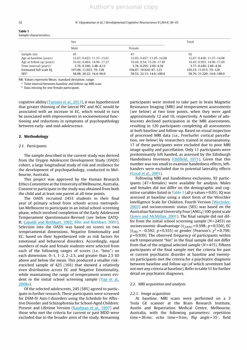
The sample described in the current study was derivedfrom the Orygen Adolescent Development Study (OADS)cohort, a large longitudinal study of risk and resilience forthe development of psychopathology, conducted in Mel-bourne, Australia.
This project was approved by the Human ResearchEthics Committee at the University of Melbourne, Australia.Consent to participate in the study was obtained from boththe child and at least one parent at all time points.
The OADS recruited 2453 students in their finalyear of primary school from schools across metropoli-tan Melbourne to participate in an initial school-screeningphase, which involved completion of the Early AdolescentTemperament Questionnaire-Revised (see below EATQ-R; Capaldi and Rothbart, 1992; Ellis and Rothbart, 2001).Selection into the OADS was based on scores on twotemperamental dimensions, Negative Emotionality andEC, based on their hypothesized role as risk factors foremotional and behavioral disorders. Accordingly, equalnumbers of male and female students were selected fromeach of the following ranges of scores (i.e. “bins”) oneach dimension: 0–1, 1–2, 2–2.5, and greater than 2.5 SDabove and below the mean. This produced a smaller risk-enriched sample of 425 (16%) that showed a relativelyeven distribution across EC and Negative Emotionality,while maintaining the range of temperament scores evi-dent in the initial school screening sample (Yap et al.,2008a).
Of the selected adolescents, 245 (58%) agreed to partic-ipate in further research. These participants were screenedfor DSM-IV Axis-I disorders using the Schedule for Affec-tive Disorder and Schizophrenia for School-Aged Children:Present and Lifetime Version (Kaufman et al., 1997) andthose who met the criteria for current or past MDD wereexcluded due to the broader aims of the study. Remaining
participants were invited to take part in brain MagneticResonance Imaging (MRI) and temperament assessments(see below) at two time points, when they were agedapproximately 12 and 16, respectively. A number of ado-lescents declined participation in the MRI assessments,resulting in 120 participants completing all assessmentsat both baseline and follow-up. Based on visual inspectionof processed MRI data (i.e., FreeSurfer cortical parcella-tion, see below) by researchers trained in neuroanatomy,17 of these participants were excluded due to poor MRIimage quality and parcellation. Only 11 participants werepredominantly left handed, as assessed by the EdinburghHandedness Inventory (Oldfield, 1971). Given that thisnumber was too small to examine handedness effects, left-handers were excluded due to potential laterality effects(Good et al., 2001).
Following MRI and handedness exclusions, 92 partic-ipants (47 = females) were available for analysis. Malesand females did not differ on the demographic and cog-nitive variables listed in Table 1 (all p values > 0.05). IQ wasassessed at baseline using a short form of the WeschlerIntelligence Scale for Children, Fourth Version (Wechsler,2003) and socioeconomic status (SES) was based on theAustralian National University Four (ANU4) 100-point scale(Jones and McMillan, 2001). The final sample did not dif-fer from the initial school screening sample (N = 2453) onsocioeconomic disadvantage (t(2439) = 0.598; p = 0.550), EC(t(96) = −0.592; p = 0.555) or gender (Pearson’s �2 = 0.799;p = 0.939). The observed frequency of participants withineach temperament “bin” in the final sample did not differfrom that of the original selected sample (N = 415). Fifteenparticipants of the final sample met the criteria for pastor current psychiatric disorder at baseline and twenty-six participants met the criteria for a psychiatric diagnosisbetween baseline and follow-up (of which seventeen hadnot met any criteria at baseline). Refer to table S1 for furtherdetail on psychiatric diagnoses.
2.2. MRI acquisition and analysis
2.2.1. Image acquisitionAt baseline, MRI scans were performed on a 3
Tesla GE scanner at the Brain Research Institute,Austin and Repatriation Medical Centre, Melbourne,Australia, with the following parameters: repetitiontime = 36 ms; echo time = 9 ms; flip angle = 35◦, field
Author's personal copy
N. Vijayakumar et al. / Developmental Cognitive Neuroscience 9 (2014) 30– 43 33
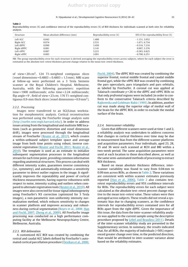
Table 2Reproducibility errors (%) and confidence interval of the reproducibility errors (%) of ROI thickness for individuals scanned at both sites for reliabilityanalysis.
Structure Mean absolute difference (mm) Reproducibility error (%) 95% CI for reproducibility Error (%)
Left ACC 0.043 1.400 −1.151, 3.952Right ACC 0.074 2.531 −1.671, 6.734Left dlPFC 0.090 3.450 −0.316, 7.216Right dlPFC 0.081 3.141 0.907, 5.376Left vlPFC 0.087 3.221 −0.383, 6.825Right vlPFC 0.083 3.091 −0.700, 6.251
NB: The group reproducibility error for each structure is derived averaging the reproducibility errors across subjects, where for each subject the error isestimated as the absolute test–retest thickness percent change relative to the mean test–retest thickness.
of view = 20 cm2, 124 T1-weighted contiguous slices(voxel dimensions = 0.4883 × 0.4883 × 1.5 mm). MRI scansat follow-up were performed on a 3 Tesla Siemensscanner at the Royal Children’s Hospital, Melbourne,Australia, with the following parameters: repetitiontime = 1900 milliseconds; echo time = 2.24 milliseconds;flip angle = 9◦, field of view = 23 cm; 176 T1-weighted con-tiguous 0.9-mm thick slices (voxel dimensions = 0.9 mm3).
2.2.2. ProcessingImages were transferred to an SGI/Linux worksta-
tion for morphometric analysis. Cortical reconstructionwas performed using the FreeSurfer image analysis suite(http://surfer.nmr.mgh.harvard.edu/). In order to addressissues arising from the longitudinal and multisite MRI loca-tions (such as geometric distortion and voxel dimensiondrift), images were processed through the longitudinalstream of FreeSurfer (Reuter et al., 2012), which createsa within-unbiased subject template space and averageimage from both time points using robust, inverse con-sistent registration (Reuter and Fischl, 2011; Reuter et al.,2010). The template is used as an estimate to initializesubsequent segmentation processes in the longitudinalstream for each time point, providing common informationregarding anatomical structures. This process can deal withdifferent intensity scales, guarantees inverse consistency(i.e., symmetry), and automatically estimates a sensitivityparameter to detect outlier regions in the image. It signif-icantly improves the repeatability and power of corticalthickness measurements, having superior robustness withrespect to noise, intensity scaling and outliers when com-pared to alternate registration tools (Reuter et al., 2010). Allimages were also corrected for tissue signal inhomogeneityusing FreeSurfer’s N3 correction (optimized for 3 Teslaimages), a non-parametric non-uniformity intensity nor-malization method, which reduces sensitivity to changesin scanner platform and improves accuracy and robust-ness during cortical segmentation (Boyes et al., 2008; Hanand Fischl, 2007; Zheng et al., 2009). All FreeSurfer imageprocessing was conducted on a high performance com-puting facility at the Melbourne Neuropsychiatry Centre,Melbourne, Australia.
2.2.3. ROI delineationA customized ACC ROI was created by combining the
rostral and caudal ACC labels defined by FreeSurfer’s auto-mated cortical parcellation procedure (Desikan et al., 2006;
Fischl, 2004). The dlPFC ROI was created by combining thesuperior frontal, rostral middle frontal and caudal middlefrontal gyri, while the vlPFC ROI was created by combiningthe pars opercularis, pars triangularis and pars orbitalis,as labeled by FreeSurfer. A coronal cut was applied atTalairach coordinate y = 26 to the dlPFC and vlPFC ROIs sothat only prefrontal regions were included (in order to con-form to the conservative Talairach criteria described byRajkowska and Goldman-Rakic (1995). In addition, anothercut was made along the superior edge of medial wall ofthe brain for the dlPFC ROI, in order to exclude the medialsurface of the brain.
2.2.4. Interscanner reliabilityGiven that different scanners were used at time 1 and 2,
a reliability analysis was undertaken to address concernsthat changes in cortical thickness over time may be dueto measurement bias from the different scanner platformsand acquisition parameters. Four individuals, aged 23, 28,35 and 36 were each scanned at RCH and BRI within atwo-week period. The same acquisition parameters wereused at each location to those described above, as well asthe same semi-automated methods of processing to extractROI thickness.
Based on mean absolute thickness difference, inter-scanner variability was found to vary from 0.04 mm to0.09 mm across ROIs, as shown in Table 2. These variationsare consistent with within scanner estimates previouslyreported (Han et al., 2006). Table 2 also contains test-retest reproducibility errors and 95% confidence intervalsfor ROIs. The reproducibility errors for each subject werecalculated as the absolute test–retest percent change rela-tive to the mean test–retest value. These values were thenaveraged across subjects. These results did not reveal a sys-tematic bias due to changing scanners, as the confidenceintervals for reproducibility errors contained zero for allROIs apart from the right dlPFC. Given the importance ofthis issue, the data from the inter-scanner reliability analy-sis was applied to the current sample using the descriptiveprocedure proposed by Lebel and Beaulieu (2011). Detailsof the inter-scanner reliability study are presented in theSupplementary section. In summary, the results indicatedthat, for all ROIs, the majority of individuals (>50%) experi-enced greater change over time, in the predicted direction,than would be attributed to inter-scanner variance alonebased on the reliability estimates.
Author's personal copy
34 N. Vijayakumar et al. / Developmental Cognitive Neuroscience 9 (2014) 30– 43
2.3. Effortful control
Participants completed the EATQ-R, which consists of65 items used to derive ten subscales that load onto fourhigher order factors (EC, negative affectivity, surgency, andaffiliation), and two additional behavioral scales to exam-ine socioemotional functioning. Of interest to the currentstudy is the EC factor, which is comprised of the activitycontrol, inhibitory control, and attentional control sub-scales. Attentional control is the capacity to focus andshift attention as required, inhibitory control refers to thecapacity to plan and suppress inappropriate behavior, andactivity control is the ability to perform an action whenthere is a strong desire/tendency to avoid it. Higher scoresreflect higher EC.
2.4. Socioemotional functioning
Participants completed a number of measures assessingsocioemotional functioning. At baseline and follow-up,depressive symptoms were measured using the self-reportCenter for Epidemiological Studies-Depression Scale (CES-D; Radloff, 1977). The questionnaire contains 20 items thatrelate to mood, somatic complaints, relations with others,and motor functioning during the past week. The CES-Dhas been found to be a valid and reliable measure for use inadolescent populations (Garrison et al., 1991). Based on therecommended clinical cut-off, 28% of adolescents at base-line and 17% of adolescents at follow-up exhibited clinicallysignificant levels of depressive symptoms. Anxiety symp-toms were also measured at baseline and follow-up usingthe self-report Beck Anxiety Inventory (BAI). The question-naire contains 21 items relating to anxiety symptoms inthe past week. The BAI has been found to be valid andhighly reliable (Beck et al., 1988). Based on the recom-mended clinical cut-off, 18% of adolescents at baseline and11% of adolescents at follow-up experienced clinically sig-nificant levels of anxiety symptoms. Aggressive behaviorwas also measured at both time points using the EATQ-R.As mentioned above, the EATQ-R is a self-report question-naire with strong validity and reliability. The aggressivebehavior scale is one of the two behavioral scales withinthe EATQ-R that assess socioemotional functioning (Ellis& Rothbart, 2001). Risk taking behavior was measured atfollow-up using the Youth Risk Behavior Scale (YRBS). ThisYRBS is a self-report questionnaire assessing engagementin substance use, risky sexual behavior, unhealthy dietarypractices, violence, and other reckless behavior adoles-cents. It has been found to be reliable and valid (Breneret al., 1995).
2.5. Puberty
Pubertal development was assessed at baseline usingthe parent-report Pubertal Development Scale (Petersenet al., 1988). For females, this measure includes 8 itemsassessing the stage of breast development, hair growth,acne presence, hip width and menarcheal status. Formales, this measure includes 11 items assessing geni-talia development, hair growth, acne presence and voicechange. Reliability and validity of the PDS has been well
established (Brooks-Gunn et al., 1987; Petersen et al.,1988). For descriptive purposes, the PDS data was codedinto a 5-point scale in accordance with the Tanner stagesbased on Shirtcliff et al. (2009). This information is pre-sented in table S2.
2.6. Statistical analysis
All analyses were conducted using SPSS version 20and results were considered significant at p < 0.05. Sepa-rate linear mixed models (LMMs) were used to investigatechange in each ROI thickness (6 models), EC (1 model) andeach behavior/psychopathology measure (5 models). AllLMMs included the main effect of time (within-subjectsfactor) and sex (between-subjects factor), the interactionbetween time and sex, as well as baseline puberty and SESas covariates. All models were re-analyzed using baselineage, instead of baseline puberty, as a covariate.
Given that the time interval between baseline andfollow-up varied between individuals (see Table 1), cor-relational analyses were conducted to examine whetherdifferences in time interval between baseline and follow-up assessments (i.e., T2–T1 age) were associated withdevelopment (i.e., T2–T1 cortical thickness/behavioralscores). Statistically significant associations were identifiedbetween time interval and change in right dlPFC thickness(r = −0.216) and EC (r = −.207). When time interval was alsoincluded in the LMMs for right dlPFC development and ECdevelopment, however, the pattern of significant findingsdid not change. Thus, variation in time interval was deemedto be non-meaningful, and time interval was modeled as acategorical (rather than nuisance) variable.
Subsequently, in order to examine associations betweenchange in each measure, change scores for each measurewere calculated as standardized residuals, by regressingfollow-up scores on baseline scores. These residual changescores were used in mediation analyses to examine theindirect effect of changes in cortical thickness on changesin behavior/psychopathology symptoms, through changes
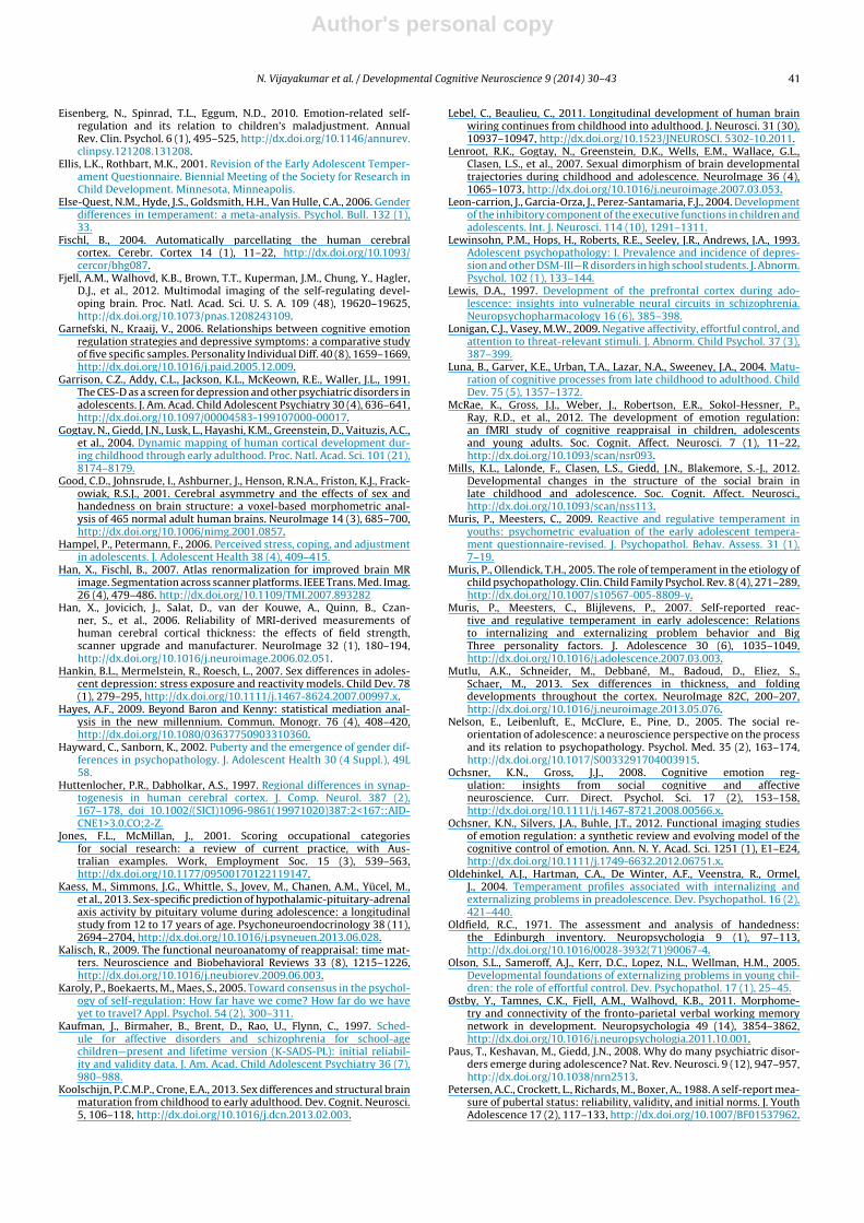
Change in Left ACC
thickne ss
Change i n C-ESD
depressive symptom s
Change i n EC
Change in Left ACC
thickne ss
Change i n C-ESD
depressive symptom s
a b
c
c’
Fig. 1. Mediation analysis of left ACC maturation on development ofdepressive symptoms through change in EC. Path a is the total effect ofleft ACC maturation (i.e. change in cortical thickness) on development ofEC (i.e. change in EC), path b is the total effect of development of EC ondevelopment of depressive symptoms (i.e. change in depressive symp-toms), path c is the total effect of left ACC maturation on developmentof depressive symptoms (i.e., not controlling for EC change), and path c’is the direct effect of left ACC maturation on development of depressivesymptoms (i.e. controlling for EC change).
Author's personal copy
N. Vijayakumar et al. / Developmental Cognitive Neuroscience 9 (2014) 30– 43 35
in EC. Mediation analyses were performed using macrosdeveloped by Preacher and Hayes (2004), implemented inSPSS 20. Mediation was tested by assessing the significanceof the cross product of the coefficients for the IV (brainchange) to mediator (EC change) relation (the a path), andthe mediator to DV (change in behavior/psychopathologysymptoms) relation, controlling for the IV (the b path).The a-b cross product tests the statistical significance ofthe difference between the total effect, or c path, and thedirect effect, or c’ path, which is the impact of the IV onthe DV adjusting for the effect of the mediator (see Fig. 1for an example of the mediation analysis). Sex, SES andpuberty at baseline were used as covariates in the media-tion analyses. Finally, moderated-mediation analyses wereused to assess whether the relationship between brainchange and change in behavior/psychopathological symp-toms, as mediated by change in EC, was moderated bysex. SES and puberty at baseline were used as covariatesin these moderated-mediation analyses. Again, all media-tion and moderated-mediation models were re-analyzedusing baseline age, instead of baseline puberty, as acovariate.
A bootstrapping method was applied to test the signif-icance of the mediation effect (Preacher and Hayes, 2004).This method generates an empirical approximation of thesampling distribution of a statistic by repeated randomresampling from the available data, and uses this distribu-tion to construct bias corrected and accelerated confidenceintervals (Hayes, 2009). If the confidence intervals do notcontain zero, the point estimate is significant at the levelindicated.
2.7. Treatment of missing data
Six, seven and three participants in the final sam-ple had missing EATQ-R (EC and Aggressive behaviorscales), CESD and BAI data at baseline or follow-up, respec-tively. Ten participants had missing puberty data at T1,and one participant had missing YRBS data at T2. Lit-tle’s MCAR test was found to be non-significant for allvariables (p > .05), indicating that the data were missing
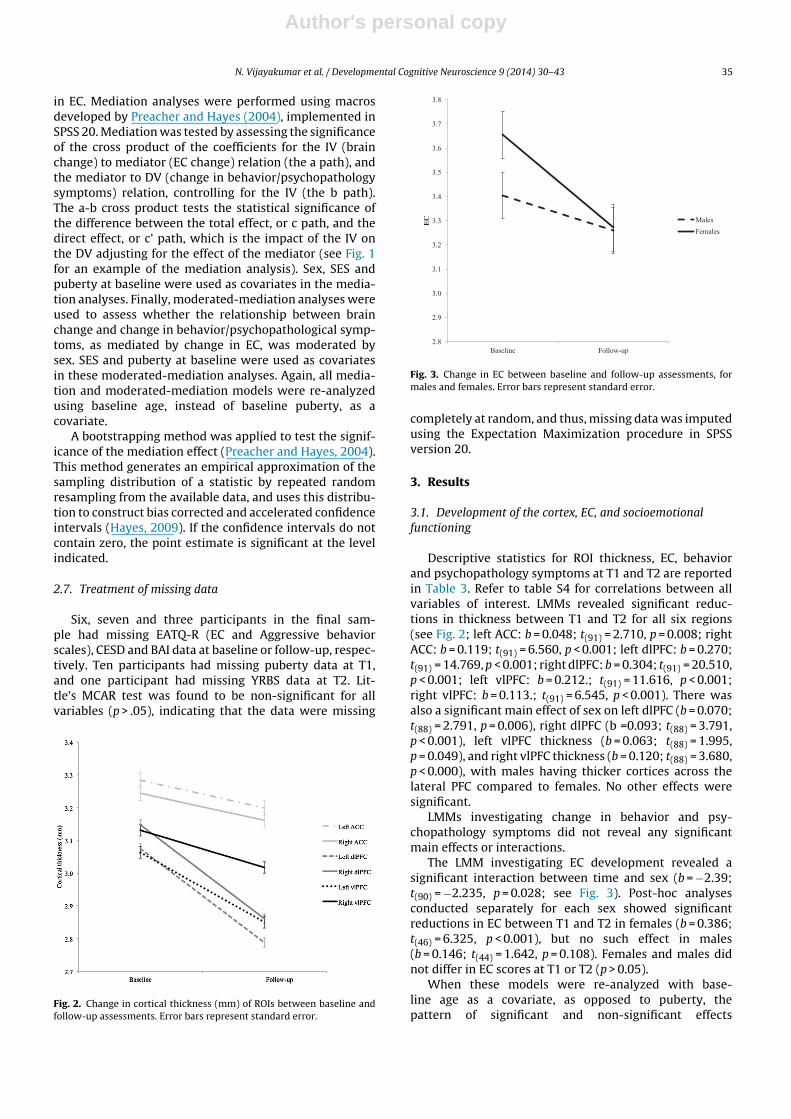
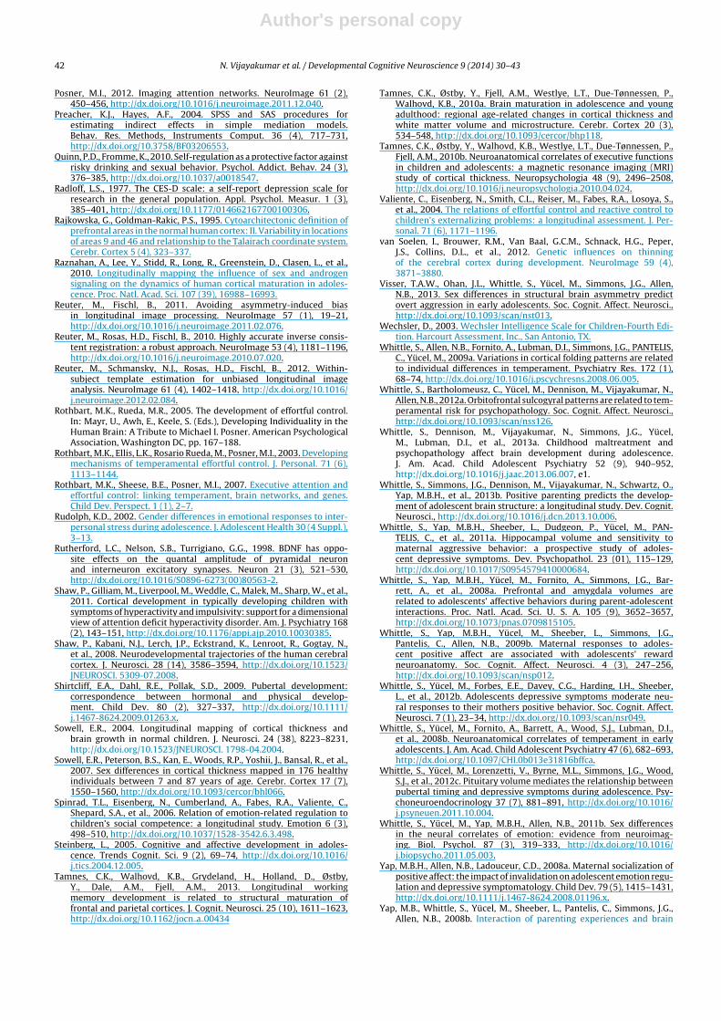
Fig. 2. Change in cortical thickness (mm) of ROIs between baseline andfollow-up assessments. Error bars represent standard error.
2.8
2.9
3.0
3.1
3.2
3.3
3.4
3.5
3.6
3.7
3.8
Baseline Follow-up
EC
Males
Females
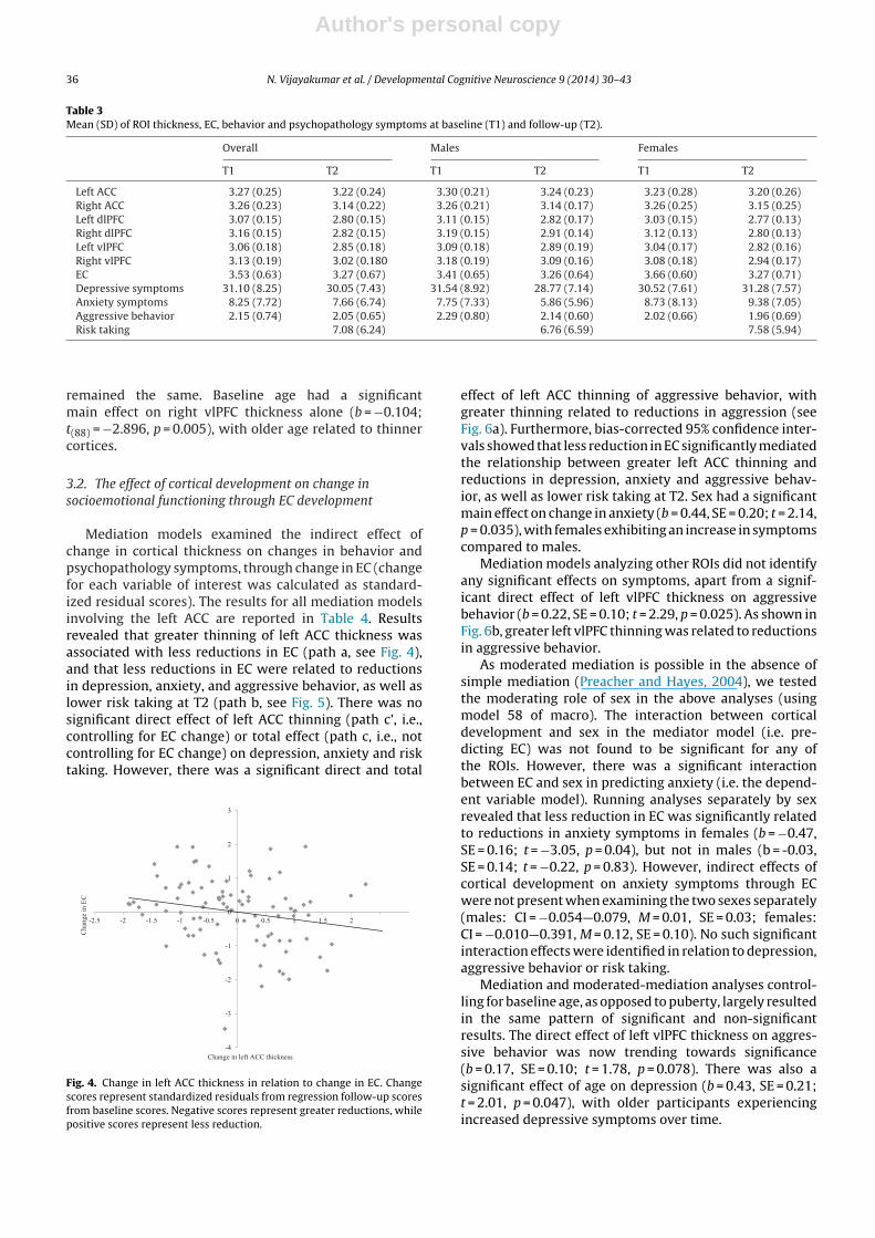
Fig. 3. Change in EC between baseline and follow-up assessments, formales and females. Error bars represent standard error.
completely at random, and thus, missing data was imputedusing the Expectation Maximization procedure in SPSSversion 20.
3. Results
3.1. Development of the cortex, EC, and socioemotionalfunctioning
Descriptive statistics for ROI thickness, EC, behaviorand psychopathology symptoms at T1 and T2 are reportedin Table 3. Refer to table S4 for correlations between allvariables of interest. LMMs revealed significant reduc-tions in thickness between T1 and T2 for all six regions(see Fig. 2; left ACC: b = 0.048; t(91) = 2.710, p = 0.008; rightACC: b = 0.119; t(91) = 6.560, p < 0.001; left dlPFC: b = 0.270;t(91) = 14.769, p < 0.001; right dlPFC: b = 0.304; t(91) = 20.510,p < 0.001; left vlPFC: b = 0.212.; t(91) = 11.616, p < 0.001;right vlPFC: b = 0.113.; t(91) = 6.545, p < 0.001). There wasalso a significant main effect of sex on left dlPFC (b = 0.070;t(88) = 2.791, p = 0.006), right dlPFC (b =0.093; t(88) = 3.791,p < 0.001), left vlPFC thickness (b = 0.063; t(88) = 1.995,p = 0.049), and right vlPFC thickness (b = 0.120; t(88) = 3.680,p < 0.000), with males having thicker cortices across thelateral PFC compared to females. No other effects weresignificant.
LMMs investigating change in behavior and psy-chopathology symptoms did not reveal any significantmain effects or interactions.
The LMM investigating EC development revealed asignificant interaction between time and sex (b = −2.39;t(90) = −2.235, p = 0.028; see Fig. 3). Post-hoc analysesconducted separately for each sex showed significantreductions in EC between T1 and T2 in females (b = 0.386;t(46) = 6.325, p < 0.001), but no such effect in males(b = 0.146; t(44) = 1.642, p = 0.108). Females and males didnot differ in EC scores at T1 or T2 (p > 0.05).
When these models were re-analyzed with base-line age as a covariate, as opposed to puberty, thepattern of significant and non-significant effects
Author's personal copy
36 N. Vijayakumar et al. / Developmental Cognitive Neuroscience 9 (2014) 30– 43
Table 3Mean (SD) of ROI thickness, EC, behavior and psychopathology symptoms at baseline (T1) and follow-up (T2).
Overall Males Females
T1 T2 T1 T2 T1 T2
Left ACC 3.27 (0.25) 3.22 (0.24) 3.30 (0.21) 3.24 (0.23) 3.23 (0.28) 3.20 (0.26)Right ACC 3.26 (0.23) 3.14 (0.22) 3.26 (0.21) 3.14 (0.17) 3.26 (0.25) 3.15 (0.25)Left dlPFC 3.07 (0.15) 2.80 (0.15) 3.11 (0.15) 2.82 (0.17) 3.03 (0.15) 2.77 (0.13)Right dlPFC 3.16 (0.15) 2.82 (0.15) 3.19 (0.15) 2.91 (0.14) 3.12 (0.13) 2.80 (0.13)Left vlPFC 3.06 (0.18) 2.85 (0.18) 3.09 (0.18) 2.89 (0.19) 3.04 (0.17) 2.82 (0.16)Right vlPFC 3.13 (0.19) 3.02 (0.180 3.18 (0.19) 3.09 (0.16) 3.08 (0.18) 2.94 (0.17)EC 3.53 (0.63) 3.27 (0.67) 3.41 (0.65) 3.26 (0.64) 3.66 (0.60) 3.27 (0.71)Depressive symptoms 31.10 (8.25) 30.05 (7.43) 31.54 (8.92) 28.77 (7.14) 30.52 (7.61) 31.28 (7.57)Anxiety symptoms 8.25 (7.72) 7.66 (6.74) 7.75 (7.33) 5.86 (5.96) 8.73 (8.13) 9.38 (7.05)Aggressive behavior 2.15 (0.74) 2.05 (0.65) 2.29 (0.80) 2.14 (0.60) 2.02 (0.66) 1.96 (0.69)Risk taking 7.08 (6.24) 6.76 (6.59) 7.58 (5.94)
remained the same. Baseline age had a significantmain effect on right vlPFC thickness alone (b = −0.104;t(88) = −2.896, p = 0.005), with older age related to thinnercortices.
3.2. The effect of cortical development on change insocioemotional functioning through EC development
Mediation models examined the indirect effect ofchange in cortical thickness on changes in behavior andpsychopathology symptoms, through change in EC (changefor each variable of interest was calculated as standard-ized residual scores). The results for all mediation modelsinvolving the left ACC are reported in Table 4. Resultsrevealed that greater thinning of left ACC thickness wasassociated with less reductions in EC (path a, see Fig. 4),and that less reductions in EC were related to reductionsin depression, anxiety, and aggressive behavior, as well aslower risk taking at T2 (path b, see Fig. 5). There was nosignificant direct effect of left ACC thinning (path c’, i.e.,controlling for EC change) or total effect (path c, i.e., notcontrolling for EC change) on depression, anxiety and risktaking. However, there was a significant direct and total
-4
-3
-2
-1
0
1
2
3
-2.5 -2 -1.5 -1 -0.5 0 0.5 1 1.5 2
Chan
ge
in E
C
Change in left AC C thickn ess
Fig. 4. Change in left ACC thickness in relation to change in EC. Changescores represent standardized residuals from regression follow-up scoresfrom baseline scores. Negative scores represent greater reductions, whilepositive scores represent less reduction.
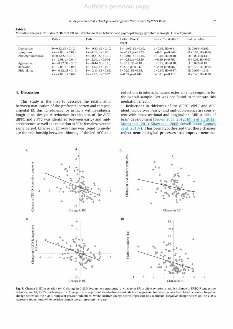
effect of left ACC thinning of aggressive behavior, withgreater thinning related to reductions in aggression (seeFig. 6a). Furthermore, bias-corrected 95% confidence inter-vals showed that less reduction in EC significantly mediatedthe relationship between greater left ACC thinning andreductions in depression, anxiety and aggressive behav-ior, as well as lower risk taking at T2. Sex had a significantmain effect on change in anxiety (b = 0.44, SE = 0.20; t = 2.14,p = 0.035), with females exhibiting an increase in symptomscompared to males.
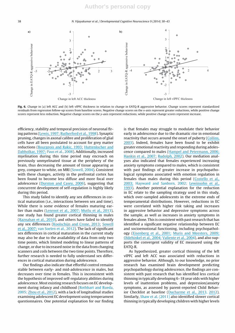
Mediation models analyzing other ROIs did not identifyany significant effects on symptoms, apart from a signif-icant direct effect of left vlPFC thickness on aggressivebehavior (b = 0.22, SE = 0.10; t = 2.29, p = 0.025). As shown inFig. 6b, greater left vlPFC thinning was related to reductionsin aggressive behavior.
As moderated mediation is possible in the absence ofsimple mediation (Preacher and Hayes, 2004), we testedthe moderating role of sex in the above analyses (usingmodel 58 of macro). The interaction between corticaldevelopment and sex in the mediator model (i.e. pre-dicting EC) was not found to be significant for any ofthe ROIs. However, there was a significant interactionbetween EC and sex in predicting anxiety (i.e. the depend-ent variable model). Running analyses separately by sexrevealed that less reduction in EC was significantly relatedto reductions in anxiety symptoms in females (b = −0.47,SE = 0.16; t = −3.05, p = 0.04), but not in males (b = -0.03,SE = 0.14; t = −0.22, p = 0.83). However, indirect effects ofcortical development on anxiety symptoms through ECwere not present when examining the two sexes separately(males: CI = −0.054—0.079, M = 0.01, SE = 0.03; females:CI = −0.010—0.391, M = 0.12, SE = 0.10). No such significantinteraction effects were identified in relation to depression,aggressive behavior or risk taking.
Mediation and moderated-mediation analyses control-ling for baseline age, as opposed to puberty, largely resultedin the same pattern of significant and non-significantresults. The direct effect of left vlPFC thickness on aggres-sive behavior was now trending towards significance(b = 0.17, SE = 0.10; t = 1.78, p = 0.078). There was also asignificant effect of age on depression (b = 0.43, SE = 0.21;t = 2.01, p = 0.047), with older participants experiencingincreased depressive symptoms over time.
Author's personal copy
N. Vijayakumar et al. / Developmental Cognitive Neuroscience 9 (2014) 30– 43 37
Table 4Mediation analyses: the indirect effect of left ACC development on behavior and psychopathology symptoms through EC development.
Path a Path b Path c’: Directeffect
Path c: Total effect Indirect effect
Depressivesymptoms
b = 0.22, SE = 0.10,t = −2.08, p = 0.041
b = −0.43, SE = 0.10,t = −4.33, p < 0.001
b = −0.03, SE = 0.10,t = −0.29, p = 0.773
b = 0.06, SE = 0.11,t = 0.61, p = 0.544
CI: 0.018—0.195,M = 0.09, SE = 0.04
Anxiety symptoms b = 0.22, SE = 0.10,t = −2.08, p = 0.041
b = −0.21, SE = 0.10,t = −2.04, p = 0.044
b = −0.01, SE = 0.10,t = −0.14, p = 0.886
b = 0.03, SE = 0.10,t = 0.30, p = 0.762
CI: 0.003—0.143,M = 0.05, SE = 0.03
Aggressivebehavior
b = −0.22, SE = 0.10,t = −2.08, p = 0.041
b = −0.44, SE = 0.10,t = −4.67, p < 0.001
b = 0.19, SE = 0.10,t = 2.01, p = 0.047
b = 0.28, SE = 0.10,t = 2.79, p = 0.007
CI: 0.022—0.19,M = 0.10, SE = 0.04
Risk taking b = −0.22, SE = 0.10,t = −2.08, p = 0.041
b = −2.13, SE = 0.66,t = −3.23, p = 0.002
b = 0.22, SE = 0.65,t = 0.33, p = 0.742
b = 0.67, SE = 0.67,t = 1.01, p = 0.318
CI: 0.060—1.215,M = 0.46, SE = 0.28
4. Discussion
This study is the first to describe the relationshipbetween maturation of the prefrontal cortex and temper-amental EC during adolescence using a within-subjectslongitudinal design. A reduction in thickness of the ACC,dlPFC and vlPFC was identified between early- and mid-adolescence, as well as a reduction in EC in females over thesame period. Change in EC over time was found to medi-ate the relationship between thinning of the left ACC and
reductions in internalizing and externalizing symptoms forthe overall sample. Sex was not found to moderate thismediation effect.
Reductions in thickness of the dlPFC, vlPFC and ACCidentified between early- and mid-adolescence are consis-tent with cross-sectional and longitudinal MRI studies ofbrain development (Brown et al., 2012; Mills et al., 2012;Mutlu et al., 2013; Shaw et al., 2008; Sowell, 2004; Tamneset al., 2010a). It has been hypothesized that these changesreflect neurobiological processes that improve neuronal
-3
-2
-1
0
1
2
3
4
-4 -3 -2 -1 0 1 2 3
Chan
ge
in E
AT
Q-R
ag
gre
ssiv
e
beh
avio
ur
Change in EC
Ch
ange
in B
AI
anx
iety
sym
pto
ms
Change in EC
0
5
10
15
20
25
30
35
-4 -3 -2 -1 0 1 2 3
YR
BS
ris
k t
akin
g (
T2
)
Change in EC
a) b)
c) d)
-2
-1
0
1
2
3
4
-4 -3 -2 -1 0 1 2 3
Chan
ge
in C
ES
-D d
epre
ssiv
e sy
mp
tom
s
Change in EC
Fig. 5. Change in EC in relation to (a) change in C-ESD depressive symptoms, (b) change in BAI anxiety symptoms and (c) change in EATQ-R aggressivebehavior, and (d) YRBS risk taking at T2. Change scores represent standardized residuals from regression follow-up scores from baseline scores. Negativechange scores on the x-axis represent greater reductions, while positive change scores represent less reduction. Negative change scores on the y-axisrepresent reductions, while positive change scores represent increase.
Author's personal copy
38 N. Vijayakumar et al. / Developmental Cognitive Neuroscience 9 (2014) 30– 43
a) b)
-3
-2
-1
0
1
2
3
4
-3 -2 -1 0 1 2 3
Chan
ge
in E
AT
Q-R
aggre
ssiv
e beh
avio
ur
Change in left ACC thickness
-3
-2
-1
0
1
2
3
4
-4 -2 0 2 4
Chan
ge
in E
AT
Q-R
aggre
ssiv
e beh
avio
ur
Change in le ft vlPFC thickn ess
Fig. 6. Change in (a) left ACC and (b) left vlPFC thickness in relation to change in EATQ-R aggressive behavior. Change scores represent standardizedresiduals from regression follow-up scores from baseline scores. Negative change scores on the x-axis represent greater reductions, while positive changescores represent less reduction. Negative change scores on the y-axis represent reductions, while positive change scores represent increase.
efficiency, stability and temporal precision of neuronal fir-ing patterns (Lewis, 1997; Rutherford et al., 1998). Synapticpruning, changes in axonal calibre and proliferation of glialcells have all been postulated to account for grey matterreductions (Bourgeois and Rakic, 1993; Huttenlocher andDabholkar, 1997; Paus et al., 2008). Additionally, increasedmyelination during this time period may encroach onpreviously unmyelinated tissue at the periphery of thebrain, thus decreasing the amount of tissue appearing asgrey, compare to white, on MRI (Sowell, 2004). Consistentwith these changes, activity in the prefrontal cortex hasbeen found to become less diffuse and more focal overadolescence (Durston and Casey, 2006), suggesting thatconcurrent development of self-regulation is highly likelyduring this period.
This study failed to identify any sex differences in cor-tical maturation (i.e., interactions between sex and time).While there is some evidence of females maturing ear-lier than males (Lenroot et al., 2007; Mutlu et al., 2013),one study has found greater cortical thinning in males(Raznahan et al., 2010), and others have failed to identifyany sex differences (Koolschijn and Crone, 2013; Sowellet al., 2007; van Soelen et al., 2012). The lack of significantsex differences in cortical maturation in the current studymay also be due to the availability of data from only twotime points, which limited modeling to linear patterns ofchange, or due to increased noise in the data from changingscanners and coils between the two time points. Therefore,further research is needed to fully understand sex differ-ences in cortical maturation during adolescence.
Our findings also indicate that effortful control remainsstable between early- and mid-adolescence in males, butdecreases over time in females. This is inconsistent withthe hypothesis of improved self-regulatory abilities duringadolescence. Most existing research focuses on EC develop-ment during infancy and childhood (Rothbart and Rueda,2005; Zhou et al., 2012), with a lack of longitudinal studiesexamining adolescent EC development using temperamentquestionnaires. One potential explanation for our finding
is that females may struggle to modulate their behaviorearly in adolescence due to the dramatic rise in emotionalreactivity that occurs around the onset of puberty (Collins,2003). Indeed, females have been found to be exhibitgreater emotional reactivity and responding during adoles-cence compared to males (Hampel and Petermann, 2006;Hankin et al., 2007; Rudolph, 2002). Our mediation anal-yses also indicated that females experienced increasinganxiety symptoms compared to males, which is consistentwith past findings of greater increase in psychopatho-logical symptoms associated with emotion regulation infemales than males during this period (Costello et al.,2003; Hayward and Sanborn, 2002; Lewinsohn et al.,1993). Another potential explanation for the reductionin EC relate to the sampling strategy used in this study,which over-sampled adolescents in the extreme ends oftemperamental distributions. However, reductions in ECwere correlated with higher risk taking and increasesin aggressive behavior and depressive symptoms acrossthe sample, as well as increases in anxiety symptoms infemales alone. This is consistent with past research that hasidentified a significant negative relationship between ECand socioemotional functioning, including psychopathol-ogy (Eisenberg et al., 2001; Muris and Meesters, 2009;Oldehinkel et al., 2004; Valiente et al., 2004), and also sup-ports the convergent validity of EC measured using theEATQ-R.
As hypothesized, greater cortical thinning of the leftvlPFC and left ACC was associated with reductions inaggressive behavior. Although, to our knowledge, no priorresearch has examined brain development predictingpsychopathology during adolescence, the findings are con-sistent with past research that has identified less corticalthinning in typically developing 6–18 year olds with higherlevels of inattention problems, and depression/anxietysymptoms, as assessed by parent-reported Child Behav-ior Checklist at baseline (Ducharme et al., 2013, 2012).Similarly, Shaw et al. (2011) also identified slower corticalthinning in typically developing children with higher levels
Author's personal copy
N. Vijayakumar et al. / Developmental Cognitive Neuroscience 9 (2014) 30– 43 39
of hyperactivity and impulsivity during baseline assess-ments, as well as in children with ADHD. Interestingly,Whittle et al. (2013a,b), using the same sample of adoles-cents as the current study, found that the development ofpsychopathology between early and mid-adolescence wasassociated with greater thickening of the left superior pari-etal cortex. However, Whittle et al. (2013a,b) examined thedevelopment of case-level psychopathology (combiningexternalizing and internalizing disorders) as a predictor ofbrain change, while the current study examined external-izing behaviors and internalizing symptoms as an outcome.
Furthermore, normative thinning of the cortex wasassociated with maturation of regulatory abilities. Specifi-cally, greater thinning of the left ACC was associated withless reduction in EC for the overall sample. This find-ing is consistent with past research that has identifieda positive relationship between prefrontal and posteriorcortical thinning and cognitive control abilities (Tamneset al., 2013, 2010b). Greater thinning of the left lateral dor-sal frontal and left lateral parietal regions has also beenassociated with improved vocabulary abilities in a longi-tudinal study of children aged 5–11 (Sowell, 2004). Thisarea of research is, however, in its infancy, and is char-acterized by inconsistent findings, which are likely due todifferences in age groups, methodology and sample sizes.Indeed, Tamnes et al. (2010b) also found that less corti-cal thinning was related to superior Stroop performance,Fjell et al. (2012) and Østby et al. (2011) failed to iden-tify any relationships between cortical development ofcognitive control in adolescents. Whether this relates totask-specific effects or the cross-sectional nature of somestudies remains to be discovered, but our findings highlightthe importance of conducting longitudinal research ondevelopment.
Finally, this study revealed that the relationshipbetween cortical thinning and change in socioemotionalfunctioning during adolescence was mediated by concur-rent change in EC. Specifically, less reduction in EC wasfound to mediate the relationship between greater thin-ning of the left ACC and reductions in depression, anxietyand aggressive behavior, as well as lower risk taking, acrossthe sample. EC is a particularly suited to mediate this rela-tionship given that is thought to play a central role in theetiology and maintenance of internalizing and externalis-ing disorder (Muris and Ollendick, 2005). It is postulatedthat higher levels of EC allow individuals to restrain theirimpulses and undesirable urges, thus preventing external-izing problems. Higher levels of EC enable the restraintof impulses and undesirable urges, as well as regulateemotions through appropriate deployment of attentionalresources. As mentioned above, numerous studies havealso found that lower levels of EC are related to the pres-ence of internalizing symptoms, such as depression andanxiety, as well as externalizing symptoms, such as aggres-sion, conduct problems and risk taking (Eisenberg et al.,2001; Muris and Meesters, 2009; Oldehinkel et al., 2004;Valiente et al., 2004). These relationships have been foundto remain significant after accounting for the effects ofemotionality (Muris and Ollendick, 2005; Muris et al.,2007; Olson et al., 2005). There is also evidence thatEC moderates the relationship between emotionality and
psychopathology, with low levels of EC in combinationwith high levels of emotionality being associated with psy-chopathology (Lonigan and Vasey, 2009). Therefore, ourfindings provide valuable insight into one psychologicalfactor that mediates the relationship between neurobio-logical development and mental health outcomes duringadolescence.
4.1. Limitations
These findings must be considered within the context ofseveral limitations. The availability of data from only twotime points limited modeling to linear patterns develop-ment between early- and mid-adolescence, thus we cannotcomment on non-linear trajectories of brain developmentor changes occurring during the late adolescent period.
Another limitation of the study was that scans at base-line and follow-up were acquired from different scanners,with different head coils and sequences. The inter-scannerreliability study, however, suggested that the significantchange in cortical thickness observed in our sample of ado-lescents was unlikely to be due to inter-scanner bias, andthus reflected true developmental thinning.
It is important to keep in mind that a selective samplingbias was introduced in the study, whereby a greater propor-tion of participants in the extreme ends of the distributionof temperamental factors EC and Negative Affectivity wererecruited. As mentioned above, this may affect the overallpattern of change in EC and explain discrepancies betweenour findings and the hypothesized results. However, thesampling strategy should not have influenced the rela-tionship between cortical development and EC, but ratherprovided greater variance to model this relationship.
Alternatively, our findings of EC reductions in femalesmay reflect a lack of relevance of the questionnaire items toolder adolescents. That is, certain items on the EATQ-R maybecome less relevant with increasing age, such as comple-tion of homework or concentrating on studies (functionstapped by the instrument). Given these issues, futureresearch may benefit from incorporating other measuresof self-regulation, such as tasks of cognitive control oremotion regulation that have been found to be correlatedwith EC. Indeed, there is no current consensus on how tobest measure self-regulation and there is a general lackof research investigating associations between differentmeasures of self-regulation (Bridgett et al., 2013; Zhouet al., 2012). Consequently, future studies should employa multi-method approach to cover different aspects of self-regulation.
While the localized ROI approach employed by thecurrent study provides valuable knowledge about theACC, dlPFC, and vlPFC, three regions integral to the exec-utive attention network, future studies should employwhole brain analyses to explore other cortical regionsand networks underlying self-regulatory abilities. Indeed,previous research with this sample has shown that thedevelopment of psychopathology is related to thick-ness development in the parietal cortex, and volumetricdevelopment of the hippocampus and amygdala (Whittleet al., 2013a,b).
Author's personal copy
40 N. Vijayakumar et al. / Developmental Cognitive Neuroscience 9 (2014) 30– 43
4.2. Conclusions
The findings from this study shed light on the relation-ship between cortical maturation and temperamental ECin adolescents. While previous research has focused onthe neural correlates of EC using cross-sectional analyses,our findings revealed a dynamic relationship between thedevelopment of the ACC, EC and socioemotional behaviorand psychopathology symptoms during adolescence. Thishighlights the importance of studying neural correlates ofself-regulation using a longitudinal framework during thisdevelopmental period.
Conflict of interest
Drs Whittle, Dennison, Yücel, Simmons, and Allen, andMs Vijayakumar report no biomedical financial interests orpotential conflicts of interest.
Note
For further information on studies using neuroimag-ing data from the Orygen Adolescent Development Study,see Cheetham et al. (2012), Dennison et al. (2013), Kaesset al. (2013), Visser et al. (2013), Whittle et al. (2009a,b,2012a,b,c, 2011a,b, 2008a,b, 2013a,b), Yap et al. (2008b),Zipursky et al. (2011).
Acknowledgments
This research was supported by grants from the Colo-nial Foundation, the National Health and Medical ResearchCouncil (NHMRC; Australia; Program Grant 350241) andthe Australian Research Council (ARC; Discovery GrantDP0878136). Ms. Vijayakumar is supported by a MelbourneInternational Research Scholarship. Dr. Whittle is sup-ported by a NHMRC Career Development Fellowship (ID:1007716). Ms Dennison is supported by an Australian Post-graduate Award. Prof. Yücel is supported by an NHMRCFellowship (ID: 1021973).
Neuroimaging analysis was facilitated by the Neuropsy-chiatry Imaging Laboratory at the Melbourne Neuropsy-chiatry Centre. The authors would like to thank the BrainResearch Institute and Royal Children’s Hospital for sup-port in acquiring the neuroimaging data, and the familieswho participated in the study.
Appendix A. Supplementary data
Supplementary material related to this article can befound, in the online version, at http://dx.doi.org/10.1016/j.dcn.2013.12.002.
References
Beck, A.T., Epstein, N., Brown, G., Steer, R.A., 1988. An inventory formeasuring clinical anxiety: psychometric properties. J. Consult. Clin.Psychol. 56 (6), 893–897.
Botvinick, M., Nystrom, L.E., Fissell, K., Carter, C.S., Cohen, J.D.,1999. Conflict monitoring versus selection-for-action inanterior cingulate cortex. Nature 402 (6758), 179–181, doi10.1038/46035.
Bourgeois, J.P., Rakic, P., 1993. Changes of synaptic density in the pri-mary visual cortex of the macaque monkey from fetal to adult stage.J. Neurosci. 13 (7), 2801–2820.
Boyes, R.G., Gunter, J.L., Frost, C., Janke, A.L., Yeatman, T., Hill, D.L.G., et al.,2008. Intensity non-uniformity correction using N3 on 3-T scannerswith multichannel phased array coils. NeuroImage 39 (4), 1752–1762,doi 10.1016/j.neuroimage.2007.10.026.
Brener, N.D., Collins, J.L., Kann, L., Warren, C.W., Williams, B.I., 1995.Reliability of the youth risk behavior survey questionnaire. Am. J.Epidemiol. 141 (6), 575–580.
Bridgett, D.J., Oddi, K.B., Laake, L.M., Murdock, K.W., Bachmann, M.N.,2013. Integrating and differentiating aspects of self-regulation: effort-ful control, executive functioning, and links to negative affectivity.Emotion 13 (1), 47–63, DOI 10.1037/a0029536.
Brooks-Gunn, J., Warren, M.P., Rosso, J., Gargiulo, J., 1987. Validity ofself-report measures of girls’ pubertal status. Child Dev. 58 (3),829–841.
Brown, T.T., Kuperman, J.M., Chung, Y., Erhart, M., McCabe, C., Hagler,D.J., et al., 2012. Neuroanatomical assessment of biological matu-rity. Curr. Biol. 22 (18), 1693–1698, doi 10.1016/j.cub.2012.07.002.
Capaldi, D.M., Rothbart, M.K., 1992. Development and validation of anearly adolescent temperament measure. J. Early Adolescence 12 (2),153–173, http://dx.doi.org/10.1177/0272431692012002002.
Casey, B.J., Durston, S., Fossella, J.A., 2001. Evidence for a mechanis-tic model of cognitive control. Clin. Neurosci. Res. 1 (4), 267–282,http://dx.doi.org/10.1016/S1566-2772(01)00013-5.
Cheetham, A., Allen, N.B., Whittle, S., Simmons, J.G., Yücel, M.,Lubman, D.I., 2012. Orbitofrontal volumes in early adoles-cence predict initiation of cannabis use: a 4-year longitudinaland prospective study. Biol. Psychiatry 71 (8), 684–692,http://dx.doi.org/10.1016/j.biopsych.2011.10.029.
Collins, W.A., 2003. More than myth: the developmental significance ofromantic relationships during adolescence. J. Res. Adolescence 13 (1),1–24, http://dx.doi.org/10.1111/1532-7795.1301001.
Costello, E.J., Mustillo, S., Erkanli, A., Keeler, G., Angold, A., 2003.Prevalence and development of psychiatric disorders in child-hood and adolescence. Arch. Gen. Psychiatry 60 (8), 837–844,http://dx.doi.org/10.1001/archpsyc.60.8.837.
Crockett, L.J., Raffaelli, M., Shen, Y.-L., 2006. Linking self-regulation andrisk proneness to risky sexual behavior: pathways through peer pres-sure and early substance use. J. Res. Adolescence 16 (4), 503–525,http://dx.doi.org/10.1111/j. 1532-7795.2006.00505.x.
Dennison, M., Whittle, S., Yücel, M., Vijayakumar, N., Kline, A., SIMMONS, J.,Allen, N.B., 2013. Mapping subcortical brain maturation during adoles-cence: evidence of hemisphere- and sex-specific longitudinal changes.Dev. Sci., http://dx.doi.org/10.1111/desc.12057.
Desikan, R.S., Ségonne, F., Fischl, B., Quinn, B.T., Dickerson, B.C.,Blacker, D., et al., 2006. An automated labeling system forsubdividing the human cerebral cortex on MRI scans intogyral based regions of interest. NeuroImage 31 (3), 968–980,http://dx.doi.org/10.1016/j.neuroimage.2006.01.021.
Ducharme, S., Albaugh, M.D., Hudziak, J.J., Botteron, K.N., Nguyen,T.-V., Truong, C., et al., 2013. Anxious/depressed symptoms arelinked to right ventromedial prefrontal cortical thickness mat-uration in healthy children and young adults. Cerebr. Cortex,http://dx.doi.org/10.1093/cercor/bht151.
Ducharme, S., Hudziak, J.J., Botteron, K.N., Albaugh, M.D., Nguyen,T.-V., Karama, S., Evans, A.C., 2012. Decreased regional cor-tical thickness and thinning rate are associated with inat-tention symptoms in healthy children. JAAC 51 (1), 18–27,http://dx.doi.org/10.1016/j.jaac.2011.09.022, e2.
Duckworth, A.L., Seligman, M.E.P., 2005. Self-discipline outdoes IQ in pre-dicting academic performance of adolescents. Psychol. Sci. 16 (12),939–944, http://dx.doi.org/10.1111/j.1467-9280.2005.01641.x.
Durston, S., Casey, B.J., 2006. What have we learned about cognitive devel-opment from neuroimaging? Neuropsychologia 44 (11), 2149–2157,http://dx.doi.org/10.1016/j.neuropsychologia.2005.10.010
Eisenberg, N., Spinrad, T.L., 2004. Emotion-related regulation: sharpeningthe definition. Child Dev. 75 (2), 334–339.
Eisenberg, N., Cumberland, A., Spinrad, T.L., Fabes, R.A., Shepard, S.A.,Reiser, M., et al., 2001. The relations of regulation and emotionalityto children’s externalizing and internalizing problem behavior. ChildDev. 72 (4), 1112–1134, http://dx.doi.org/10.1111/1467-8624.00337.
Eisenberg, N., Fabes, R.A., Shepard, S.A., Murphy, B.C., Guthrie,I.K., Jones, S., et al., 1997. Contemporaneous and longi-tudinal prediction of children’s social functioning fromregulation and emotionality. Child Dev. 68 (4), 642–664,http://dx.doi.org/10.1111/j.1467-8624.1997.tb04227.x.
Author's personal copy
N. Vijayakumar et al. / Developmental Cognitive Neuroscience 9 (2014) 30– 43 41
Eisenberg, N., Spinrad, T.L., Eggum, N.D., 2010. Emotion-related self-regulation and its relation to children’s maladjustment. AnnualRev. Clin. Psychol. 6 (1), 495–525, http://dx.doi.org/10.1146/annurev.clinpsy.121208.131208.
Ellis, L.K., Rothbart, M.K., 2001. Revision of the Early Adolescent Temper-ament Questionnaire. Biennial Meeting of the Society for Research inChild Development. Minnesota, Minneapolis.
Else-Quest, N.M., Hyde, J.S., Goldsmith, H.H., Van Hulle, C.A., 2006. Genderdifferences in temperament: a meta-analysis. Psychol. Bull. 132 (1),33.
Fischl, B., 2004. Automatically parcellating the human cerebralcortex. Cerebr. Cortex 14 (1), 11–22, http://dx.doi.org/10.1093/cercor/bhg087.
Fjell, A.M., Walhovd, K.B., Brown, T.T., Kuperman, J.M., Chung, Y., Hagler,D.J., et al., 2012. Multimodal imaging of the self-regulating devel-oping brain. Proc. Natl. Acad. Sci. U. S. A. 109 (48), 19620–19625,http://dx.doi.org/10.1073/pnas.1208243109.
Garnefski, N., Kraaij, V., 2006. Relationships between cognitive emotionregulation strategies and depressive symptoms: a comparative studyof five specific samples. Personality Individual Diff. 40 (8), 1659–1669,http://dx.doi.org/10.1016/j.paid.2005.12.009.
Garrison, C.Z., Addy, C.L., Jackson, K.L., McKeown, R.E., Waller, J.L., 1991.The CES-D as a screen for depression and other psychiatric disorders inadolescents. J. Am. Acad. Child Adolescent Psychiatry 30 (4), 636–641,http://dx.doi.org/10.1097/00004583-199107000-00017.
Gogtay, N., Giedd, J.N., Lusk, L., Hayashi, K.M., Greenstein, D., Vaituzis, A.C.,et al., 2004. Dynamic mapping of human cortical development dur-ing childhood through early adulthood. Proc. Natl. Acad. Sci. 101 (21),8174–8179.
Good, C.D., Johnsrude, I., Ashburner, J., Henson, R.N.A., Friston, K.J., Frack-owiak, R.S.J., 2001. Cerebral asymmetry and the effects of sex andhandedness on brain structure: a voxel-based morphometric anal-ysis of 465 normal adult human brains. NeuroImage 14 (3), 685–700,http://dx.doi.org/10.1006/nimg.2001.0857.
Hampel, P., Petermann, F., 2006. Perceived stress, coping, and adjustmentin adolescents. J. Adolescent Health 38 (4), 409–415.
Han, X., Fischl, B., 2007. Atlas renormalization for improved brain MRimage. Segmentation across scanner platforms. IEEE Trans. Med. Imag.26 (4), 479–486. http://dx.doi.org/10.1109/TMI.2007.893282
Han, X., Jovicich, J., Salat, D., van der Kouwe, A., Quinn, B., Czan-ner, S., et al., 2006. Reliability of MRI-derived measurements ofhuman cerebral cortical thickness: the effects of field strength,scanner upgrade and manufacturer. NeuroImage 32 (1), 180–194,http://dx.doi.org/10.1016/j.neuroimage.2006.02.051.
Hankin, B.L., Mermelstein, R., Roesch, L., 2007. Sex differences in adoles-cent depression: stress exposure and reactivity models. Child Dev. 78(1), 279–295, http://dx.doi.org/10.1111/j.1467-8624.2007.00997.x.
Hayes, A.F., 2009. Beyond Baron and Kenny: statistical mediation anal-ysis in the new millennium. Commun. Monogr. 76 (4), 408–420,http://dx.doi.org/10.1080/03637750903310360.
Hayward, C., Sanborn, K., 2002. Puberty and the emergence of gender dif-ferences in psychopathology. J. Adolescent Health 30 (4 Suppl.), 49L58.
Huttenlocher, P.R., Dabholkar, A.S., 1997. Regional differences in synap-togenesis in human cerebral cortex. J. Comp. Neurol. 387 (2),167–178, doi 10.1002/(SICI)1096-9861(19971020)387:2<167::AID-CNE1>3.0.CO;2-Z.
Jones, F.L., McMillan, J., 2001. Scoring occupational categoriesfor social research: a review of current practice, with Aus-tralian examples. Work, Employment Soc. 15 (3), 539–563,http://dx.doi.org/10.1177/09500170122119147.
Kaess, M., Simmons, J.G., Whittle, S., Jovev, M., Chanen, A.M., Yücel, M.,et al., 2013. Sex-specific prediction of hypothalamic-pituitary-adrenalaxis activity by pituitary volume during adolescence: a longitudinalstudy from 12 to 17 years of age. Psychoneuroendocrinology 38 (11),2694–2704, http://dx.doi.org/10.1016/j.psyneuen.2013.06.028.
Kalisch, R., 2009. The functional neuroanatomy of reappraisal: time mat-ters. Neuroscience and Biobehavioral Reviews 33 (8), 1215–1226,http://dx.doi.org/10.1016/j.neubiorev.2009.06.003.
Karoly, P., Boekaerts, M., Maes, S., 2005. Toward consensus in the psychol-ogy of self-regulation: How far have we come? How far do we haveyet to travel? Appl. Psychol. 54 (2), 300–311.
Kaufman, J., Birmaher, B., Brent, D., Rao, U., Flynn, C., 1997. Sched-ule for affective disorders and schizophrenia for school-agechildren—present and lifetime version (K-SADS-PL): initial reliabil-ity and validity data. J. Am. Acad. Child Adolescent Psychiatry 36 (7),980–988.
Koolschijn, P.C.M.P., Crone, E.A., 2013. Sex differences and structural brainmaturation from childhood to early adulthood. Dev. Cognit. Neurosci.5, 106–118, http://dx.doi.org/10.1016/j.dcn.2013.02.003.
Lebel, C., Beaulieu, C., 2011. Longitudinal development of human brainwiring continues from childhood into adulthood. J. Neurosci. 31 (30),10937–10947, http://dx.doi.org/10.1523/JNEUROSCI. 5302-10.2011.
Lenroot, R.K., Gogtay, N., Greenstein, D.K., Wells, E.M., Wallace, G.L.,Clasen, L.S., et al., 2007. Sexual dimorphism of brain developmentaltrajectories during childhood and adolescence. NeuroImage 36 (4),1065–1073, http://dx.doi.org/10.1016/j.neuroimage.2007.03.053.
Leon-carrion, J., Garcia-Orza, J., Perez-Santamaria, F.J., 2004. Developmentof the inhibitory component of the executive functions in children andadolescents. Int. J. Neurosci. 114 (10), 1291–1311.
Lewinsohn, P.M., Hops, H., Roberts, R.E., Seeley, J.R., Andrews, J.A., 1993.Adolescent psychopathology: I. Prevalence and incidence of depres-sion and other DSM-III—R disorders in high school students. J. Abnorm.Psychol. 102 (1), 133–144.
Lewis, D.A., 1997. Development of the prefrontal cortex during ado-lescence: insights into vulnerable neural circuits in schizophrenia.Neuropsychopharmacology 16 (6), 385–398.
Lonigan, C.J., Vasey, M.W., 2009. Negative affectivity, effortful control, andattention to threat-relevant stimuli. J. Abnorm. Child Psychol. 37 (3),387–399.
Luna, B., Garver, K.E., Urban, T.A., Lazar, N.A., Sweeney, J.A., 2004. Matu-ration of cognitive processes from late childhood to adulthood. ChildDev. 75 (5), 1357–1372.
McRae, K., Gross, J.J., Weber, J., Robertson, E.R., Sokol-Hessner, P.,Ray, R.D., et al., 2012. The development of emotion regulation:an fMRI study of cognitive reappraisal in children, adolescentsand young adults. Soc. Cognit. Affect. Neurosci. 7 (1), 11–22,http://dx.doi.org/10.1093/scan/nsr093.
Mills, K.L., Lalonde, F., Clasen, L.S., Giedd, J.N., Blakemore, S.-J., 2012.Developmental changes in the structure of the social brain inlate childhood and adolescence. Soc. Cognit. Affect. Neurosci.,http://dx.doi.org/10.1093/scan/nss113.
Muris, P., Meesters, C., 2009. Reactive and regulative temperament inyouths: psychometric evaluation of the early adolescent tempera-ment questionnaire-revised. J. Psychopathol. Behav. Assess. 31 (1),7–19.
Muris, P., Ollendick, T.H., 2005. The role of temperament in the etiology ofchild psychopathology. Clin. Child Family Psychol. Rev. 8 (4), 271–289,http://dx.doi.org/10.1007/s10567-005-8809-y.
Muris, P., Meesters, C., Blijlevens, P., 2007. Self-reported reac-tive and regulative temperament in early adolescence: Relationsto internalizing and externalizing problem behavior and BigThree personality factors. J. Adolescence 30 (6), 1035–1049,http://dx.doi.org/10.1016/j.adolescence.2007.03.003.
Mutlu, A.K., Schneider, M., Debbané, M., Badoud, D., Eliez, S.,Schaer, M., 2013. Sex differences in thickness, and foldingdevelopments throughout the cortex. NeuroImage 82C, 200–207,http://dx.doi.org/10.1016/j.neuroimage.2013.05.076.
Nelson, E., Leibenluft, E., McClure, E., Pine, D., 2005. The social re-orientation of adolescence: a neuroscience perspective on the processand its relation to psychopathology. Psychol. Med. 35 (2), 163–174,http://dx.doi.org/10.1017/S0033291704003915.
Ochsner, K.N., Gross, J.J., 2008. Cognitive emotion reg-ulation: insights from social cognitive and affectiveneuroscience. Curr. Direct. Psychol. Sci. 17 (2), 153–158,http://dx.doi.org/10.1111/j.1467-8721.2008.00566.x.
Ochsner, K.N., Silvers, J.A., Buhle, J.T., 2012. Functional imaging studiesof emotion regulation: a synthetic review and evolving model of thecognitive control of emotion. Ann. N. Y. Acad. Sci. 1251 (1), E1–E24,http://dx.doi.org/10.1111/j.1749-6632.2012.06751.x.
Oldehinkel, A.J., Hartman, C.A., De Winter, A.F., Veenstra, R., Ormel,J., 2004. Temperament profiles associated with internalizing andexternalizing problems in preadolescence. Dev. Psychopathol. 16 (2),421–440.
Oldfield, R.C., 1971. The assessment and analysis of handedness:the Edinburgh inventory. Neuropsychologia 9 (1), 97–113,http://dx.doi.org/10.1016/0028-3932(71)90067-4.
Olson, S.L., Sameroff, A.J., Kerr, D.C., Lopez, N.L., Wellman, H.M., 2005.Developmental foundations of externalizing problems in young chil-dren: the role of effortful control. Dev. Psychopathol. 17 (1), 25–45.
Østby, Y., Tamnes, C.K., Fjell, A.M., Walhovd, K.B., 2011. Morphome-try and connectivity of the fronto-parietal verbal working memorynetwork in development. Neuropsychologia 49 (14), 3854–3862,http://dx.doi.org/10.1016/j.neuropsychologia.2011.10.001.
Paus, T., Keshavan, M., Giedd, J.N., 2008. Why do many psychiatric disor-ders emerge during adolescence? Nat. Rev. Neurosci. 9 (12), 947–957,http://dx.doi.org/10.1038/nrn2513.
Petersen, A.C., Crockett, L., Richards, M., Boxer, A., 1988. A self-report mea-sure of pubertal status: reliability, validity, and initial norms. J. YouthAdolescence 17 (2), 117–133, http://dx.doi.org/10.1007/BF01537962.
Author's personal copy
42 N. Vijayakumar et al. / Developmental Cognitive Neuroscience 9 (2014) 30– 43
Posner, M.I., 2012. Imaging attention networks. NeuroImage 61 (2),450–456, http://dx.doi.org/10.1016/j.neuroimage.2011.12.040.
Preacher, K.J., Hayes, A.F., 2004. SPSS and SAS procedures forestimating indirect effects in simple mediation models.Behav. Res. Methods, Instruments Comput. 36 (4), 717–731,http://dx.doi.org/10.3758/BF03206553.
Quinn, P.D., Fromme, K., 2010. Self-regulation as a protective factor againstrisky drinking and sexual behavior. Psychol. Addict. Behav. 24 (3),376–385, http://dx.doi.org/10.1037/a0018547.
Radloff, L.S., 1977. The CES-D scale: a self-report depression scale forresearch in the general population. Appl. Psychol. Measur. 1 (3),385–401, http://dx.doi.org/10.1177/014662167700100306.
Rajkowska, G., Goldman-Rakic, P.S., 1995. Cytoarchitectonic definition ofprefrontal areas in the normal human cortex: II. Variability in locationsof areas 9 and 46 and relationship to the Talairach coordinate system.Cerebr. Cortex 5 (4), 323–337.
Raznahan, A., Lee, Y., Stidd, R., Long, R., Greenstein, D., Clasen, L., et al.,2010. Longitudinally mapping the influence of sex and androgensignaling on the dynamics of human cortical maturation in adoles-cence. Proc. Natl. Acad. Sci. 107 (39), 16988–16993.
Reuter, M., Fischl, B., 2011. Avoiding asymmetry-induced biasin longitudinal image processing. NeuroImage 57 (1), 19–21,http://dx.doi.org/10.1016/j.neuroimage.2011.02.076.
Reuter, M., Rosas, H.D., Fischl, B., 2010. Highly accurate inverse consis-tent registration: a robust approach. NeuroImage 53 (4), 1181–1196,http://dx.doi.org/10.1016/j.neuroimage.2010.07.020.
Reuter, M., Schmansky, N.J., Rosas, H.D., Fischl, B., 2012. Within-subject template estimation for unbiased longitudinal imageanalysis. NeuroImage 61 (4), 1402–1418, http://dx.doi.org/10.1016/j.neuroimage.2012.02.084.
Rothbart, M.K., Rueda, M.R., 2005. The development of effortful control.In: Mayr, U., Awh, E., Keele, S. (Eds.), Developing Individuality in theHuman Brain: A Tribute to Michael I. Posner. American PsychologicalAssociation, Washington DC, pp. 167–188.
Rothbart, M.K., Ellis, L.K., Rosario Rueda, M., Posner, M.I., 2003. Developingmechanisms of temperamental effortful control. J. Personal. 71 (6),1113–1144.
Rothbart, M.K., Sheese, B.E., Posner, M.I., 2007. Executive attention andeffortful control: linking temperament, brain networks, and genes.Child Dev. Perspect. 1 (1), 2–7.
Rudolph, K.D., 2002. Gender differences in emotional responses to inter-personal stress during adolescence. J. Adolescent Health 30 (4 Suppl.),3–13.
Rutherford, L.C., Nelson, S.B., Turrigiano, G.G., 1998. BDNF has oppo-site effects on the quantal amplitude of pyramidal neuronand interneuron excitatory synapses. Neuron 21 (3), 521–530,http://dx.doi.org/10.1016/S0896-6273(00)80563-2.
Shaw, P., Gilliam, M., Liverpool, M., Weddle, C., Malek, M., Sharp, W., et al.,2011. Cortical development in typically developing children withsymptoms of hyperactivity and impulsivity: support for a dimensionalview of attention deficit hyperactivity disorder. Am. J. Psychiatry 168(2), 143–151, http://dx.doi.org/10.1176/appi.ajp.2010.10030385.
Shaw, P., Kabani, N.J., Lerch, J.P., Eckstrand, K., Lenroot, R., Gogtay, N.,et al., 2008. Neurodevelopmental trajectories of the human cerebralcortex. J. Neurosci. 28 (14), 3586–3594, http://dx.doi.org/10.1523/JNEUROSCI. 5309-07.2008.
Shirtcliff, E.A., Dahl, R.E., Pollak, S.D., 2009. Pubertal development:correspondence between hormonal and physical develop-ment. Child Dev. 80 (2), 327–337, http://dx.doi.org/10.1111/j.1467-8624.2009.01263.x.
Sowell, E.R., 2004. Longitudinal mapping of cortical thickness andbrain growth in normal children. J. Neurosci. 24 (38), 8223–8231,http://dx.doi.org/10.1523/JNEUROSCI. 1798-04.2004.
Sowell, E.R., Peterson, B.S., Kan, E., Woods, R.P., Yoshii, J., Bansal, R., et al.,2007. Sex differences in cortical thickness mapped in 176 healthyindividuals between 7 and 87 years of age. Cerebr. Cortex 17 (7),1550–1560, http://dx.doi.org/10.1093/cercor/bhl066.
Spinrad, T.L., Eisenberg, N., Cumberland, A., Fabes, R.A., Valiente, C.,Shepard, S.A., et al., 2006. Relation of emotion-related regulation tochildren’s social competence: a longitudinal study. Emotion 6 (3),498–510, http://dx.doi.org/10.1037/1528-3542.6.3.498.
Steinberg, L., 2005. Cognitive and affective development in adoles-cence. Trends Cognit. Sci. 9 (2), 69–74, http://dx.doi.org/10.1016/j.tics.2004.12.005.
Tamnes, C.K., Walhovd, K.B., Grydeland, H., Holland, D., Østby,Y., Dale, A.M., Fjell, A.M., 2013. Longitudinal workingmemory development is related to structural maturation offrontal and parietal cortices. J. Cognit. Neurosci. 25 (10), 1611–1623,http://dx.doi.org/10.1162/jocn a 00434
Tamnes, C.K., Østby, Y., Fjell, A.M., Westlye, L.T., Due-Tønnessen, P.,Walhovd, K.B., 2010a. Brain maturation in adolescence and youngadulthood: regional age-related changes in cortical thickness andwhite matter volume and microstructure. Cerebr. Cortex 20 (3),534–548, http://dx.doi.org/10.1093/cercor/bhp118.
Tamnes, C.K., Østby, Y., Walhovd, K.B., Westlye, L.T., Due-Tønnessen, P.,Fjell, A.M., 2010b. Neuroanatomical correlates of executive functionsin children and adolescents: a magnetic resonance imaging (MRI)study of cortical thickness. Neuropsychologia 48 (9), 2496–2508,http://dx.doi.org/10.1016/j.neuropsychologia.2010.04.024.
Valiente, C., Eisenberg, N., Smith, C.L., Reiser, M., Fabes, R.A., Losoya, S.,et al., 2004. The relations of effortful control and reactive control tochildren’s externalizing problems: a longitudinal assessment. J. Per-sonal. 71 (6), 1171–1196.
van Soelen, I., Brouwer, R.M., Van Baal, G.C.M., Schnack, H.G., Peper,J.S., Collins, D.L., et al., 2012. Genetic influences on thinningof the cerebral cortex during development. NeuroImage 59 (4),3871–3880.
Visser, T.A.W., Ohan, J.L., Whittle, S., Yücel, M., Simmons, J.G., Allen,N.B., 2013. Sex differences in structural brain asymmetry predictovert aggression in early adolescents. Soc. Cognit. Affect. Neurosci.,http://dx.doi.org/10.1093/scan/nst013.
Wechsler, D., 2003. Wechsler Intelligence Scale for Children-Fourth Edi-tion. Harcourt Assessment, Inc., San Antonio, TX.
Whittle, S., Allen, N.B., Fornito, A., Lubman, D.I., Simmons, J.G., PANTELIS,C., Yücel, M., 2009a. Variations in cortical folding patterns are relatedto individual differences in temperament. Psychiatry Res. 172 (1),68–74, http://dx.doi.org/10.1016/j.pscychresns.2008.06.005.
Whittle, S., Bartholomeusz, C., Yücel, M., Dennison, M., Vijayakumar, N.,Allen, N.B., 2012a. Orbitofrontal sulcogyral patterns are related to tem-peramental risk for psychopathology. Soc. Cognit. Affect. Neurosci.,http://dx.doi.org/10.1093/scan/nss126.
Whittle, S., Dennison, M., Vijayakumar, N., Simmons, J.G., Yücel,M., Lubman, D.I., et al., 2013a. Childhood maltreatment andpsychopathology affect brain development during adolescence.J. Am. Acad. Child Adolescent Psychiatry 52 (9), 940–952,http://dx.doi.org/10.1016/j.jaac.2013.06.007, e1.
Whittle, S., Simmons, J.G., Dennison, M., Vijayakumar, N., Schwartz, O.,Yap, M.B.H., et al., 2013b. Positive parenting predicts the develop-ment of adolescent brain structure: a longitudinal study. Dev. Cognit.Neurosci., http://dx.doi.org/10.1016/j.dcn.2013.10.006.
Whittle, S., Yap, M.B.H., Sheeber, L., Dudgeon, P., Yücel, M., PAN-TELIS, C., et al., 2011a. Hippocampal volume and sensitivity tomaternal aggressive behavior: a prospective study of adoles-cent depressive symptoms. Dev. Psychopathol. 23 (01), 115–129,http://dx.doi.org/10.1017/S0954579410000684.
Whittle, S., Yap, M.B.H., Yücel, M., Fornito, A., Simmons, J.G., Bar-rett, A., et al., 2008a. Prefrontal and amygdala volumes arerelated to adolescents’ affective behaviors during parent-adolescentinteractions. Proc. Natl. Acad. Sci. U. S. A. 105 (9), 3652–3657,http://dx.doi.org/10.1073/pnas.0709815105.
Whittle, S., Yap, M.B.H., Yücel, M., Sheeber, L., Simmons, J.G.,Pantelis, C., Allen, N.B., 2009b. Maternal responses to adoles-cent positive affect are associated with adolescents’ rewardneuroanatomy. Soc. Cognit. Affect. Neurosci. 4 (3), 247–256,http://dx.doi.org/10.1093/scan/nsp012.
Whittle, S., Yücel, M., Forbes, E.E., Davey, C.G., Harding, I.H., Sheeber,L., et al., 2012b. Adolescents depressive symptoms moderate neu-ral responses to their mothers positive behavior. Soc. Cognit. Affect.Neurosci. 7 (1), 23–34, http://dx.doi.org/10.1093/scan/nsr049.
Whittle, S., Yücel, M., Fornito, A., Barrett, A., Wood, S.J., Lubman, D.I.,et al., 2008b. Neuroanatomical correlates of temperament in earlyadolescents. J. Am. Acad. Child Adolescent Psychiatry 47 (6), 682–693,http://dx.doi.org/10.1097/CHI.0b013e31816bffca.
Whittle, S., Yücel, M., Lorenzetti, V., Byrne, M.L., Simmons, J.G., Wood,S.J., et al., 2012c. Pituitary volume mediates the relationship betweenpubertal timing and depressive symptoms during adolescence. Psy-choneuroendocrinology 37 (7), 881–891, http://dx.doi.org/10.1016/j.psyneuen.2011.10.004.
Whittle, S., Yücel, M., Yap, M.B.H., Allen, N.B., 2011b. Sex differencesin the neural correlates of emotion: evidence from neuroimag-ing. Biol. Psychol. 87 (3), 319–333, http://dx.doi.org/10.1016/j.biopsycho.2011.05.003.
Yap, M.B.H., Allen, N.B., Ladouceur, C.D., 2008a. Maternal socialization ofpositive affect: the impact of invalidation on adolescent emotion regu-lation and depressive symptomatology. Child Dev. 79 (5), 1415–1431,http://dx.doi.org/10.1111/j.1467-8624.2008.01196.x.
Yap, M.B., Whittle, S., Yücel, M., Sheeber, L., Pantelis, C., Simmons, J.G.,Allen, N.B., 2008b. Interaction of parenting experiences and brain

Author's personal copy
N. Vijayakumar et al. / Developmental Cognitive Neuroscience 9 (2014) 30– 43 43
structure in the prediction of depressive symptoms in adolescents.Arch. Gen. Psychiatry 65 (12), 1377.
Zheng, W., Chee, M.W.L., Zagorodnov, V., 2009. Improvement of brainsegmentation accuracy by optimizing non-uniformity correctionusing N3. NeuroImage 48 (1), 73–83, http://dx.doi.org/10.1016/j.neuroimage.2009.06.039.
Zhou, Q., Chen, S.H., Main, A., 2012. Commonalities and differences in theresearch on children’s effortful control and executive function: a call
for an integrated model of self-regulation. Child Dev. Perspect. 6 (2),112–121, http://dx.doi.org/10.1111/j.1750-8606.2011.00176.x.
Zipursky, A.R., Whittle, S., Yücel, M., Lorenzetti, V., Wood, S.J., Lubman,D.I., et al., 2011. Pituitary volume prospectively predicts internalizingsymptoms in adolescence. J. Child Psychol. Psychiatry 52 (3), 315–323,http://dx.doi.org/10.1111/j.1469-7610.2010.02337.x.