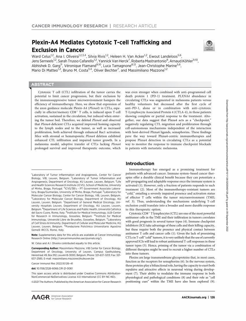
Plexin-A4 Mediates Cytotoxic T-cell Trafficking and Exclusion ...
16
CANCER IMMUNOLOGY RESEARCH | RESEARCH ARTICLE Plexin-A4 Mediates Cytotoxic T-cell Trafficking and Exclusion in Cancer Ward Celus 1,2 , Ana I. Oliveira 1,2,3,4 , Silvia Rivis 1,2 , Heleen H. Van Acker 1,2 , Ewout Landeloos 5,6 , Jens Serneels 1,2 , Sarah Trusso Cafarello 1,2 , Yannick Van Herck 7 , Roberta Mastrantonio 8 , Arnaud K€ ohler 9,10 , Abhishek D. Garg 11 ,V eronique Flamand 9,10 , Luca Tamagnone 8,12 , Jean-Christophe Marine 5,6 , Mario Di Matteo 1,2 , Bruno M. Costa 3,4 , Oliver Bechter 7 , and Massimiliano Mazzone 1,2 ABSTRACT ◥ Cytotoxic T cell (CTL) infiltration of the tumor carries the potential to limit cancer progression, but their exclusion by the immunosuppressive tumor microenvironment hampers the efficiency of immunotherapy. Here, we show that expression of the axon guidance molecule Plexin-A4 (Plxna4) in CTLs, espe- cially in effector/memory CD8 þ T cells, is induced upon T-cell activation, sustained in the circulation, but reduced when enter- ing the tumor bed. Therefore, we deleted Plxna4 and observed that Plxna4-deficient CTLs acquired improved homing capacity to the lymph nodes and to the tumor, as well as increased proliferation, both achieved through enhanced Rac1 activation. Mice with stromal or hematopoietic Plxna4 deletion exhibited enhanced CTL infiltration and impaired tumor growth. In a melanoma model, adoptive transfer of CTLs lacking Plxna4 prolonged survival and improved therapeutic outcome, which was even stronger when combined with anti–programmed cell death protein 1 (PD-1) treatment. PLXNA4 abundance in circulating CTLs was augmented in melanoma patients versus healthy volunteers but decreased after the first cycle of anti–PD-1, alone or in combination with anti–cytotoxic T-Lymphocyte Associated Protein 4 (CTLA-4), in those patients showing complete or partial response to the treatment. Alto- gether, our data suggest that Plxna4 acts as a “checkpoint,” negatively regulating CTL migration and proliferation through cell-autonomous mechanisms independent of the interaction with host-derived Plxna4 ligands, semaphorins. These findings pave the way toward Plxna4-centric immunotherapies and propose Plxna4 detection in circulating CTLs as a potential way to monitor the response to immune checkpoint blockade in patients with metastatic melanoma. Introduction Immunotherapy has emerged as a promising treatment for patients with advanced cancer. Immune system–based cancer ther- apies offer a durable clinical benefit because they can potentiate a self-propagating and adaptable response once the immune system is activated (1). However, only a fraction of patients responds to such treatment (2). Most of the immunotherapy-resistant tumors are “cold,” entailing a severely impaired presence and activation status of effector T cells within the tumor microenvironment (TME; ref. 3). Thus, understanding the mechanisms underlying T-cell exclusion could translate into a broader and more durable response to this therapeutic option. Cytotoxic CD8 þ T lymphocytes (CTL) are one of the most powerful antitumor cells in the TME and their infiltration in tumors correlates with good prognosis in several tumor types (4). Immune checkpoint inhibitors (ICI) take advantage of these cells and their killing capacity, but these require both the presence and physical contact between antitumor T cells and cancer cells (1). Given the lack of preexisting CTLs in T-cell “cold” tumors, it is very unlikely that the use of currently approved ICIs will lead to robust antitumoral T-cell responses in these tumor types (5). Hence, priming of the tumor via a combination of different therapies might be used to recruit a higher number of CTLs into these tumors. Plexins are large transmembrane glycoproteins that, in most cases, function as the receptors for semaphorins (6). In the nervous system, these proteins play a bifunctional role, having the capacity to exert both repulsive and attractive effects in neuronal wiring during develop- ment (7). Their ability to modulate the immune response in both physiological and pathological conditions (8) and their role as “cell positioning cues” within the TME have also been explored (9). 1 Laboratory of Tumor Inflammation and Angiogenesis, Center for Cancer Biology, VIB, Leuven, Belgium. 2 Laboratory of Tumor Inflammation and Angiogenesis, Department of Oncology, KU Leuven, Leuven, Belgium. 3 Life and Health Sciences Research Institute (ICVS), School of Medicine, University of Minho, Braga, Portugal. 4 ICVS/3B’s - PT Government Associate Labora- tory, Braga/Guimar~ aes, University of Minho, Braga, Portugal. 5 Laboratory for Molecular Cancer Biology, Center for Cancer Biology, VIB, Leuven, Belgium. 6 Laboratory for Molecular Cancer Biology, Department of Oncology, KU Leuven, Leuven, Belgium. 7 Department of General Medical Oncology, Uni- versity Hospitals Leuven, Department of Oncology, KU Leuven, Leuven, Belgium. 8 Department of Life Sciences and Public Health, Universit a Cattolica del Sacro Cuore, Rome, Italy. 9 Institute for Medical Immunology, ULB-Center for Research in Immunology, Gosselies, Belgium. 10 Institute for Medical Immunology, Universit e Libre de Bruxelles, Gosselies, Belgium. 11 Laboratory of Cell Stress & Immunity, Department of Cellular and Molecular Medicine, KU Leuven, Leuven, Belgium. 12 Fondazione Policlinico Universitario Agostino Gemelli IRCCS, Rome, Italy. Note: Supplementary data for this article are available at Cancer Immunology Research Online (http://cancerimmunolres.aacrjournals.org/). W. Celus and A.I. Oliveira contributed equally to this article. Corresponding Author: Massimiliano Mazzone, VIB Center for Cancer Biology, Department of Oncology, University of Leuven, Campus Gasthuisberg, Herestraat 49, Box 912, Leuven B-3000, Belgium. Phone: 321-637-3213; Fax: 321- 637-2585; E-mail: [email protected] Cancer Immunol Res 2022;10:126–41 doi: 10.1158/2326-6066.CIR-21-0061 This open access article is distributed under Creative Commons Attribution- NonCommercial-NoDerivatives License 4.0 International (CC BY-NC-ND). Ó2021 The Authors; Published by the American Association for Cancer Research AACRJournals.org | 126 Downloaded from http://aacrjournals.org/cancerimmunolres/article-pdf/10/1/126/3202040/126.pdf by guest on 09 September 2022
-
Upload
khangminh22 -
Category
Documents
-
view
0 -
download
0
Transcript of Plexin-A4 Mediates Cytotoxic T-cell Trafficking and Exclusion ...
CANCER IMMUNOLOGY RESEARCH | RESEARCH ARTICLE
Plexin-A4 Mediates Cytotoxic T-cell Trafficking andExclusion in CancerWard Celus1,2, Ana I. Oliveira1,2,3,4, Silvia Rivis1,2, Heleen H. Van Acker1,2, Ewout Landeloos5,6,Jens Serneels1,2, Sarah Trusso Cafarello1,2, Yannick Van Herck7, RobertaMastrantonio8, Arnaud K€ohler9,10,Abhishek D. Garg11, V�eronique Flamand9,10, Luca Tamagnone8,12, Jean-Christophe Marine5,6,Mario Di Matteo1,2, Bruno M. Costa3,4, Oliver Bechter7, and Massimiliano Mazzone1,2
ABSTRACT◥
Cytotoxic T cell (CTL) infiltration of the tumor carries thepotential to limit cancer progression, but their exclusion bythe immunosuppressive tumor microenvironment hampers theefficiency of immunotherapy. Here, we show that expression ofthe axon guidance molecule Plexin-A4 (Plxna4) in CTLs, espe-cially in effector/memory CD8þ T cells, is induced upon T-cellactivation, sustained in the circulation, but reduced when enter-ing the tumor bed. Therefore, we deleted Plxna4 and observedthat Plxna4-deficient CTLs acquired improved homing capacityto the lymph nodes and to the tumor, as well as increasedproliferation, both achieved through enhanced Rac1 activation.Mice with stromal or hematopoietic Plxna4 deletion exhibitedenhanced CTL infiltration and impaired tumor growth. In amelanoma model, adoptive transfer of CTLs lacking Plxna4prolonged survival and improved therapeutic outcome, which
was even stronger when combined with anti–programmed celldeath protein 1 (PD-1) treatment. PLXNA4 abundance incirculating CTLs was augmented in melanoma patients versushealthy volunteers but decreased after the first cycle ofanti–PD-1, alone or in combination with anti–cytotoxicT-Lymphocyte Associated Protein 4 (CTLA-4), in those patientsshowing complete or partial response to the treatment. Alto-gether, our data suggest that Plxna4 acts as a “checkpoint,”negatively regulating CTL migration and proliferation throughcell-autonomous mechanisms independent of the interactionwith host-derived Plxna4 ligands, semaphorins. These findingspave the way toward Plxna4-centric immunotherapies andpropose Plxna4 detection in circulating CTLs as a potentialway to monitor the response to immune checkpoint blockadein patients with metastatic melanoma.
IntroductionImmunotherapy has emerged as a promising treatment for
patients with advanced cancer. Immune system–based cancer ther-apies offer a durable clinical benefit because they can potentiate aself-propagating and adaptable response once the immune system isactivated (1). However, only a fraction of patients responds to suchtreatment (2). Most of the immunotherapy-resistant tumors are“cold,” entailing a severely impaired presence and activation statusof effector T cells within the tumor microenvironment (TME;ref. 3). Thus, understanding the mechanisms underlying T-cellexclusion could translate into a broader and more durable responseto this therapeutic option.
Cytotoxic CD8þT lymphocytes (CTL) are one of themost powerfulantitumor cells in the TME and their infiltration in tumors correlateswith good prognosis in several tumor types (4). Immune checkpointinhibitors (ICI) take advantage of these cells and their killing capacity,but these require both the presence and physical contact betweenantitumor T cells and cancer cells (1). Given the lack of preexistingCTLs inT-cell “cold” tumors, it is very unlikely that the use of currentlyapproved ICIs will lead to robust antitumoral T-cell responses in thesetumor types (5). Hence, priming of the tumor via a combination ofdifferent therapies might be used to recruit a higher number of CTLsinto these tumors.
Plexins are large transmembrane glycoproteins that, in most cases,function as the receptors for semaphorins (6). In the nervous system,these proteins play a bifunctional role, having the capacity to exert bothrepulsive and attractive effects in neuronal wiring during develop-ment (7). Their ability to modulate the immune response in bothphysiological and pathological conditions (8) and their role as “cellpositioning cues” within the TME have also been explored (9).
1Laboratory of Tumor Inflammation and Angiogenesis, Center for CancerBiology, VIB, Leuven, Belgium. 2Laboratory of Tumor Inflammation andAngiogenesis, Department of Oncology, KU Leuven, Leuven, Belgium. 3Lifeand Health Sciences Research Institute (ICVS), School of Medicine, Universityof Minho, Braga, Portugal. 4ICVS/3B’s - PT Government Associate Labora-tory, Braga/Guimar~aes, University of Minho, Braga, Portugal. 5Laboratory forMolecular Cancer Biology, Center for Cancer Biology, VIB, Leuven, Belgium.6Laboratory for Molecular Cancer Biology, Department of Oncology, KULeuven, Leuven, Belgium. 7Department of General Medical Oncology, Uni-versity Hospitals Leuven, Department of Oncology, KU Leuven, Leuven,Belgium. 8Department of Life Sciences and Public Health, Universit�a Cattolicadel Sacro Cuore, Rome, Italy. 9Institute for Medical Immunology, ULB-Centerfor Research in Immunology, Gosselies, Belgium. 10Institute for MedicalImmunology, Universit�e Libre de Bruxelles, Gosselies, Belgium. 11Laboratoryof Cell Stress & Immunity, Department of Cellular and Molecular Medicine, KULeuven, Leuven, Belgium. 12Fondazione Policlinico Universitario AgostinoGemelli IRCCS, Rome, Italy.
Note: Supplementary data for this article are available at Cancer ImmunologyResearch Online (http://cancerimmunolres.aacrjournals.org/).
W. Celus and A.I. Oliveira contributed equally to this article.
Corresponding Author: Massimiliano Mazzone, VIB Center for Cancer Biology,Department of Oncology, University of Leuven, Campus Gasthuisberg,Herestraat 49, Box 912, Leuven B-3000, Belgium. Phone: 321-637-3213; Fax: 321-637-2585; E-mail: [email protected]
Cancer Immunol Res 2022;10:126–41
doi: 10.1158/2326-6066.CIR-21-0061
This open access article is distributed under Creative Commons Attribution-NonCommercial-NoDerivatives License 4.0 International (CC BY-NC-ND).
�2021 TheAuthors; Published by the American Association for Cancer Research
AACRJournals.org | 126
Dow
nloaded from http://aacrjournals.org/cancerim
munolres/article-pdf/10/1/126/3202040/126.pdf by guest on 09 Septem
ber 2022

Targeting plexin signals (or semaphorins) is therefore a promisingtherapeutic strategy to restore antitumor immunity.
Plexin-A4 (Plxna4) is a member of class A plexins (10), whichinteracts with Sema6A and Sema6B (11). When in association withneuropilin-1 (NRP1), it can also function as a coreceptor forSema3A (12). In the central nervous system, Plxna4 mediates axonrepulsion by the direct binding to Sema6A and Sema6B (13, 14). In theimmune system, Plxna4 has been implicated in macrophage Toll-likereceptor (TLR)-mediated signaling and cytokine production in sep-sis (15), anti-inflammatory polarization in colitis (16), and entry intohypoxic niches in cancer (17). In T cells, Plxna4 negatively regulatesT-cell–mediated immune responses, withPlxna4-deficientmice show-ing exacerbated disease in a mouse model of experimental autoim-mune encephalomyelitis (EAE; ref. 18). On the basis of these findings,we defined the function of PlxnA4 in the control of the immuneresponse in the context of cancer.
Materials and MethodsAnimals
Plxna4 knockout (KO) mice on a C57BL/6 background wereobtained from Dr. Castellani (Institut NeuroMyoG�ene, Universit�e deLyon, Lyon, France). C57BL/6 mice were purchased from CharlesRiver. OT-I mice were purchased fromTaconic. Sema6aKOmice on aC57BL/6 backgroundwere obtained fromProf. Dr. Pasterkamp (Dept.of Translational Neuroscience, University Medical Center Utrecht,The Netherlands). All mice used were between 6 and 12 weeks old,without specific gender selection. In all experiments, littermate con-trols were used. Euthanasia was performed by cervical dislocation.Housing conditions and all experimental animal procedures wereapproved by the Animal Ethics Committee of the KU Leuven.
Bone marrow transplantationSix-week-old C56BL/6 recipient mice were lethally irradiated with a
dose of 9.5 Gy using the Small Animal Radiation Research Platform(SARRP, XSTRAHL). Femur and tibia bones were collected fromdonormice of the appropriate genotype. In a sterile culture hood, bonemarrow (BM) cells were obtained by flushing the bones with a syringefilled with Roswell Park Memorial Institute (RPMI) 1640 medium(Gibco, Thermo Fisher Scientific, 21875034) supplemented with 10%heat-inactivated FBS (Biowest, S1810). The cells were subsequentlyfiltered using a 40-mm pore–sized mesh and centrifuged for 5 minutesat 200 � g. BM cells were counted and 1 � 107 cells were injectedintravenously (i.v.) via tail vein in the irradiated recipient mice. Tumorexperiments were initiated 6 to 8 weeks after BM reconstitution. Redand white blood cell count was determined using a hemocytometer onperipheral blood, collected in heparin with capillary pipettes by retro-orbital bleeding.
Cell linesMurine Lewis lung carcinoma cells (LLC) and B16F10 melanoma
cells were obtained from the ATCC. E0771 medullary breast adeno-carcinoma cells were obtained from CH3Biosystems. All cells werecultured in Dulbecco’s Modified Eagle Medium (DMEM; Gibco,Thermo Fisher Scientific, 41965039) supplemented with 10% heat-inactivated FBS, 2 mmol/L glutamine (Gibco, Thermo Fisher Scien-tific, 25030024), 100 U/mL penicillin, and 100 mg/mL streptomycin(Gibco, Thermo Fisher Scientific, 15140122) at 37�C in a humidifiedatmosphere containing 5% CO2. For the overexpression of ovalbumin(OVA), the following plasmid was used: pCDH_CMV7-OVA-EFI-G418. LLCandB16F10 cancer cells were transducedwith concentrated
lentiviral vectors and further selected with G418 antibiotics (1mg/mL,Invivogen, ant-gn) to generate a homogenous population of OVA-overexpressing cancer cells (LLC-OVA and B16F10-OVA). Over-expression of the OVA protein was confirmed by Western blotanalysis. All cell lines were tested for Mycoplasma and passaged inthe laboratory for no longer than 6 months after receipt.
Tumor modelsAdherent growing murine cells, 1 � 106 LLC and 5 � 105 B16F10,
were injected subcutaneously (s.c.) for LLC and orthotopically forB16F10 at the right side of the mouse in PBS (Gibco, Thermo FisherScientific, 14190094). Alternatively, 5 � 105 E0771 medullary breastadenocarcinoma cells were injected orthotopically in themammary fatpad of the second nipple on the right side in a volume of 50 mL PBS.Tumor volumes were measured three times a week with a caliper andcalculated using the formula: V¼ p� d2 � D/6, where d is the minortumor axis andD is themajor tumor axis. At the end stage, tumorswereweighed and collected for immunofluorescence and/orflowcytometricanalyses. For survival analysis, a tumor volume of 1,800mm3 was usedas the humane endpoint.
Histology and immunostainingTumors and lymph nodes (LN) were collected and fixed in 4%
formaldehyde (37% stock, VWR, ACRO119690250) diluted in PBSovernight at 4�C, dehydrated and embedded in paraffin. Serial sectionswere cut at 7-mm thickness with an HM 355S automatic microtome(Thermo Fisher Scientific). Paraffin slides were first rehydrated tofurther proceed with antigen retrieval in Target Retrieval Solution,Citrate pH 6.1 (DAKO, Agilent, S1699). If necessary, 0.3% hydrogenperoxide (Stock 30%, Millipore, 1072090250) was added to methanol(VWR, 20848.320), to block endogenous peroxidases. The sectionswere blocked with the appropriate serum (DAKO, Agilent), matchingthe species of the secondary antibody, and incubated overnight at roomtemperature with the following antibodies: rat anti-CD8a (ThermoFisher Scientific, 4SM15, 1:100), rat anti-CD4 (Thermo Fisher Scien-tific, 4SM95, 1:100), rat anti-F4/80 (AbD Serotec, MCA497, 1:100),rabbit anti-FITC (AbD Serotec, 4510–7604, 1:200), rat anti-CD31 (BDPharmingen, 550274, 1:50), rat anti-CD34 (BD Pharmingen, 553731,1:100), rabbit anti-NG2 (Millipore, AB5320, 1:200), and rat anti-PNAd(BioLegend, MECA-79, 1:100). Appropriate secondary antibodiesraised against the species of the primary antibody were used: Alexa488 (Molecular Probes, A21208, 1:200), 647 (Molecular Probes,A31573, 1:100) or 568-conjugated secondary antibodies (MolecularProbes, A11077, 1:200), biotin-labeled antibodies (Jackson Immunor-esearch, 711–065–152 and 712–065–153, 1:300), and, when necessary,TSA Plus Cyanine 3 and Cyanine 5 System amplification (PerkinElmer, Life Sciences, NEL744001KT and NEL745001KT, 1:50) wereperformed according to the manufacturer’s instructions. Hoechst-33342 solution (Thermo Fisher Scientific, H3570, 1:1,000) was used tovisualize nuclei. Mounting of slides was done with ProLong Goldmounting medium without DAPI (Invitrogen, P36930). Imaging andmicroscopic analysis was performed with an Olympus BX41 micro-scope and CellSense imaging software.
Tumor hypoxia assessment and tumor perfusionTumor hypoxia was detected 1 hour after intraperitoneal (i.p.)
injection of 60 mg/kg pimonidazole hydrochloride (Hypoxyprobe kit,Chemicon, HP3–100Kit) in LLC tumor–bearing mice. Tumors wereharvested and fixed in 4% formaldehyde overnight. To detect theformation of pimonidazole adducts, 7-mm thick sections were immu-nostained with rabbit anti-hypoxyprobe monoclonal (Hypoxyprobe
Plexin-A4 Mediates CTL Trafficking and Exclusion in Cancer
AACRJournals.org Cancer Immunol Res; 10(1) January 2022 127
Dow
nloaded from http://aacrjournals.org/cancerim
munolres/article-pdf/10/1/126/3202040/126.pdf by guest on 09 Septem
ber 2022

Kit, Chemicon, HP3–100 Kit, 1:100) following the manufacturer’sinstructions. Perfused tumor vessels were counted on tumor sectionsfrom mice injected i.v. with 0.05 mg FITC-conjugated lectin (Lyco-persicon esculentum; Vector Laboratories, B-1175–1).
Flow cytometryMice were sacrificed by cervical dislocation, and tumors, livers,
blood, LNs (inguinal and axillary LNs) or the tumor-draining LN(tdLN; the closest LN draining the tumor bed) were collected. Tumorswere minced ina-minimum essential medium (MEM) (Lonza, BE12–169F), containing 50 mmol/L b-mercaptoethanol (Gibco, ThermoFisher Scientific, 21985023), 5 U/mLDeoxyribonuclease I 0.85mg/mL(Roche, 10104159001), Collagenase V (Sigma-Aldrich, C9263–1G),1.25 mg/mL Collagenase D (Roche, 11 088 882 001), and 1 mg/mLDispase (Gibco, Thermo Fisher Scientific, 17105–041), and incubatedin the same solution for 30 minutes at 37�C. Livers were processedin RPMI 1640 medium, supplemented with 10 U/mL Deoxyribonu-clease I (Roche, 10104159001) and 120 U/ml Collagenase III(Worthington Biochemical, LS004182), using the gentleMACS Dis-sociator (Miltenyi Biotec). The digested tissues were filtered using a70-mm pore–sized mesh and cells were centrifuged for 5 minutes at300 � g. Blood samples were collected in heparin with capillarypipettes by retro-orbital bleeding. Red blood cell lysis was performedby using a homemade red blood cell lysis buffer (150 mmol/L NH4Cl,0.1 mmol/L EDTA, 10 mmol/L KHCO3, pH 7.4). LNs were processedon a 40-mm pore cell strainer in sterile PBS and cells were centrifugedfor 10 minutes at 300� g. Red blood cell lysis was performed by usingHybri-Max (Sigma-Aldrich, R7757). Single cells were resuspended inFACS buffer (PBS containing 2% FBS and 2 mmol/L EDTA) andincubated for 15minutes withMouse BDFc Block purified anti-mouseCD16/CD32 (BD Pharmingen, 553142). Extracellular staining wasperformed for 30 minutes at 4�C. When necessary, permeabilizationwas performed using the eBioscience Foxp3/Transcription FactorFixation/Permeabilization Kit (Thermo Fisher Scientific, 00–5521–00) according to the manufacturer’s instructions and cells wereincubated overnight at 4�C with the intracellular antibodies. Allantibodies used are described in Supplementary Table S1. Cells weresubsequently washed and resuspended in FACS buffer before flowcytometric analysis by a FACS Canto II, Fortessa X-20, or flow sortingby a FACS Aria III, Aria Fusion (BD Biosciences). Data were analyzedby FlowJo (TreeStar, Version 10.7). Fluorescence Minus One (FMO)controls were utilized to ensure proper gating of positive populations.
Mouse T-cell isolation and activationNa€�ve mouse T cells were isolated from the spleen, inguinal, and
axillary LNs. In brief, tissues were processed on a 40-mm pore cellstrainer in sterile PBS and cells were centrifuged for 10 minutes at300 � g. Red blood cell lysis was performed using Hybri-Max. Totalsplenocytes were cultured in T-cell medium [RPMI 1640 mediumsupplemented with 10% heat-inactivated FBS, 100 U/mL penicillinand 100 mg/mL streptomycin, 1% MEM non-essential amino acids(NEAA, Gibco, Thermo Fisher Scientific, 11140035), 25 mmol/Lb-mercaptoethanol, and 1 mmol/L sodium pyruvate (Gibco, ThermoFisher Scientific, 11360070)] at 37�C in a humidified atmospherecontaining 5% CO2.
According to the experimental requirements, T cells were activat-ed for 3 days by adding mouse anti-CD3/CD28–coated Dynabeads(Thermo Fisher Scientific, 11453D) at a 1:1 bead-to-cell ratio. At day 3of activation, the beads were magnetically removed and activatedT cells were further expanded for a maximum of 3 additionaldays in the presence of 10 ng/mL recombinant murine IL2 (mIL2,
PeproTech, 212–12). CD8þ T cells were isolated by using MagniSortMouse CD8þ T Cell Negative Selection Kit (eBioscience, ThermoFisher Scientific, 8804–6822–74) according to the manufacturer’sinstructions. CD4þ T cells were isolated by usingMACSMouse CD4þ
T Cell Isolation Kit (Miltenyi Biotec, 130–104–454) according to themanufacturer’s instructions. According to the experimentalrequirements, activated T cells (at day 3 of stimulation) weretreated for 48 hours with 15 mg/mL anti–programmed cell deathprotein 1 (PD-1; RMP1–14, BioLegend) or the appropriate isotypecontrol. FOXO inhibition in activated T cells at day 3 of stimu-lation was performed by 48 hours of treatment with 80 mmol/Lcarbenoxolone (CBX, Sigma-Aldrich, C4790) or the DMSO vehiclecontrol (dimethyl sulfoxide, Sigma, D2438).
Human T-cell isolation and activationBuffy coat samples fromhealthy donors were obtained from the Red
Cross-Flanders. HumanCD4þ and CD8þT cells were directly isolatedby using the StraightFrom Buffy Coat CD4 and CD8 MicroBead Kit(Miltenyi Biotec, 130–114–980 and 130–114–978, respectively)according to the manufacturer’s instructions. Red blood cell lysis wasperformed using Hybri-Max. T cells were activated in T-cell mediumfor 3 days by adding human anti-CD3/CD28–coated Dynabeads(Thermo Fisher Scientific, 11132D) at a 1:1 bead-to-cell ratio. At day3 of activation, the beads were magnetically removed and activatedT cells were further expanded for a maximum of 7 additional days inthe presence of 10 ng/mL recombinant human IL2 (hIL2, PeproTech,200–02). According to the experimental requirements, activatedT cells (at day 7 of stimulation) were treated for 48 hours with15 mg/mL anti–PD-1 (J116, BioXCell) or the appropriate isotypecontrol.
Cytospin stainingCD8þ T cells and peripheral blood leukocytes were seeded onto
glass slides by cytospin centrifugation and fixed in 4% formaldehydefor 10 minutes, followed by incubation with 0.2% Triton-X (VWR,1.086.031.000) diluted in PBS for 15 minutes. To reduce the immunebackground, sections were blocked with 10% donkey serum (Sigma,D9663) in PBS for 1 hour, followed by blocking with FAB fragmentanti-mouse IgG (Jackson Immunoresearch, 715–007–003, 1:10)for 1 hour. Samples were then probed overnight with mouse anti-Plexin-A4 (Plxna4; R&D Systems, 707201, 1:500) and incubated withDonkey Alexa 568–conjugated secondary antibodies (MolecularProbes, A10037, 1:100) for 45 minutes. Nuclei were counterstainedwith Hoechst-33342 and mounting of the slides was performed withProLong Gold mounting medium without DAPI. All steps wereperformed at room temperature. Microscopy was conducted with anOlympus BX41 microscope and cellSens imaging software.
Quantitative RT-PCRRNA was extracted from T cells using the TRIzol Reagent (Life
Technologies, 15596018) according to the manufacturer’s instruc-tions. Reverse transcription to cDNA was performed with the Super-Script III First Strand cDNA Synthesis Kit (Life Technologies,18080051) according to the manufacturer’s instructions. Premadeassays were purchased from Integrated DNA Technologies. ThecDNA, primer/probe mix, and TaqMan Fast Universal PCR MasterMix were prepared according to the manufacturer’s instructions(Applied Biosystems, 4352042). Samples were loaded into an optical96-well Fast Thermal Cycling Plate (Applied Biosystems) andqRT-PCR was performed using a QuantStudio 12K Flex Real-TimePCR System (Applied Biosystems). Samples were run in technical
Celus et al.
Cancer Immunol Res; 10(1) January 2022 CANCER IMMUNOLOGY RESEARCH128
Dow
nloaded from http://aacrjournals.org/cancerim
munolres/article-pdf/10/1/126/3202040/126.pdf by guest on 09 Septem
ber 2022

duplicates. Data was normalized to housekeeping gene expression(Hprt for mouse and TBP for human genes). The commerciallyavailable probes (Integrated DNA technologies) used are listed inSupplementary Table S2.
Lymphocytic choriomeningitis virus expressing OVA modelWild-type (WT) mice were preconditioned by i.v. injection of 1 �
104 na€�ve OT-I T cells. Twenty-four hours later, the mice werevaccinated i.p. with 105 plaque-forming units (PFU) of a recombinantlymphocytic choriomeningitis virus expressing OVA (LCMV-OVA; akind gift from Prof. Dr. Daniel Pinschewer, University of Basel, Basel,Switzerland), as described in Flatz and colleagues (19). After 7 days ofLCMV-OVA infection, OT-I T cells were FACS sorted from the blood.
T-cell proliferation assayTo monitor cell proliferation, activated T cells were labeled with
3.5 mmol/L Violet Cell Tracer (Thermo Fisher Scientific, C34557) at37�C for 20 minutes. The cells were subsequently washed with FACSbuffer and cultured according to the experimental requirements.Absolute numbers of T cells in culture were counted by flow cytometryusing Precision Count Beads (BioLegend, 424902). According to theexperimental requirements, activated T cells (at day 3 of stimulation)were treated with 100 mmol/L Rac1 inhibitor NSC23766 (Selleckchem,S8031) or the DMSO vehicle control.
Annexin V/propidium iodide apoptosis assayActivated CD8þ T cells were collected, washed, and resuspended
in 100 mL Annexin V Binding Buffer (BioLegend, 422201) contain-ing 4 mL of Annexin V (BioLegend, 640941) and 0.1 mL propidiumiodide solution (1 mg/mL stock, Sigma Aldrich, P4864). After15 minutes of incubation at room temperature, samples were ana-lyzed by flow cytometry.
Transwell migration assayMigration of T cells was assessed by using Transwell permeable
supports with 5-mm polycarbonate membrane (Costar, 3387). Todetermine cell migration in response to soluble factors, the bottomchamber was loaded with 0.1% FBS, 200 ng/mL CCL21 (PeproTech,250–13), 200 ng/mL CCL19 (PeproTech, 250–27B), 150 ng/mLCXCL9 (PeproTech 250–18), or 50 ng/mL CXCL10 (PeproTech,250–16) in T-cell medium. T cells were incubated for 2 (na€�ve) or3 hours (activated) at 37�C and migrated cells in the bottomchamber were collected and counted by flow cytometry usingPrecision Count Beads. According to the experimental require-ments, activated T cells were pretreated for 1 hour with 10 mg/mLanti-CCR7 (R&D Systems, 4B12), 250 mg/mL anti-CXCR3 (BioLegend,CXCR3–173), 100 mmol/L Rac1 inhibitor NSC23766, or the appro-priate isotype, or vehicle control.
LN homing assayNa€�veCD8þT cells were isolated fromWTandPlxna4KOmice and
labeled with either 3.5 mmol/L Violet Cell Tracer or 1 mmol/Lcarboxyfluorescein diacetate succinimidyl ester (CFSE) Cell Tracer(Thermo Fisher Scientific, C34554). For CFSE labeling, cells werestained in PBS for 8minutes at room temperaturewith gentle agitation.To label cells with Violet Cell Tracer, the staining was conducted for 20minutes at 37�C with gentle agitation. Afterwards, a brief wash withcomplete RPMImediumwas performed to quench any remaining dye.Healthy WTmice were injected i.v. with a 1:1 mixture between 1–2�106 labeled WT and Plxna4 KO T cells. After 2 hours, LNs of therecipient mice were harvested and analyzed by IHC and/or flow
cytometry. To exclude probe-specific cell toxicity, the fluorescentcell tracers were switched accordingly, showing identical experimentalresults.
Tumor homing assayOT-I T cells were isolated from transgenicWT andPlxna4KOOT-I
mice, generated by the intercross of Plxna4 heterozygous mice withOT-I–positive mice. These mice have a monoclonal population ofna€�veT-cell receptor (TCR) transgenic CD8þT cells (OT-I T cells) thatrecognize the immunodominant cytosolic chicken OVA “SIINFEKL”peptide. For activation of OT-I T cells, total splenocytes from OT-Imice were isolated and cultured for 3 days in T-cell medium with1 mg/mL SIINFEKL peptide (IBA - LifeSciences, 6–7015–901) and10 ng/mL mIL2. At day 3 of activation, OT-I T cells were furtherexpanded for a maximum of 3 additional days in the presence of10 ng/mL mIL2.
For the tumor homing assay, activated WT and Plxna4 KO OT-I Tcells were labeledwith either 3.5mmol/LViolet Cell Tracer or 1mmol/LCFSE and injected i.v. with a 1:1 mixture between 2–3� 106 WT andPlxna4 KO OT-I T cells into WT recipient mice with establishedB16F10-OVA or LLC-OVA tumors. The tumors of recipient micewere harvested 24 and 48 hours after T-cell transfer and analyzed byflow cytometry.
Liver homing assayWT mice received a plasmid DNA by hydrodynamic injection
(HDI). Each mouse was injected rapidly (<8 seconds) in the tail veinwith 40 mg of pcDNA3 empty vector (EV) or pcDNA3-OVA (OVA)diluted in PBS in an injection volume of 10% of the body weight. Fourdays after HDI, activatedWT and Plxna4KOOT-I T cells were labeledwith either 3.5 mmol/L Violet Cell Tracer or 1 mmol/L CFSE andinjected i.v. with a 1:1 mixture between 2–3� 106WT and KOOT-I Tcells into mice. Twenty-four hours after T-cell transfer, blood andlivers were harvested and analyzed by flow cytometry.
Plasmids and lentiviral vectorsIn the overexpression experiments, the following plasmids
were used: pCDH-CMV-Sema3a-DYK-EF1-Puro (Sema3a OE),pCDH-CMV-Sema6a-DYK-EF1-Puro (Sema6a OE), pCDH-CMV-Sema6b-DYK-EF1-Puro (Sema6b OE), and pCDH-CMV-MCS-EF1-Puro (EV). B16F10-OVA cancer cells were transduced withconcentrated lentiviral vectors and further selected with puromy-cin antibiotics (1 mg/mL, Sigma, P9620) to allow the generationof a homogenous population of overexpressed (and empty vectorcontrol) cancer cells.
GTPase pull-down assayRac1 and Rap1 activation were measured by using a Rac1 or Rap1
Activation Assay Kit (Thermo Fisher Scientific, 16118 and 16120,respectively) according to the manufacturer’s instructions. Briefly,cell lysis was performed by incubating activated T cells (at day 5of stimulation) with the lysis buffer for 5 minutes on ice. Thelysates were centrifuged for 15 minutes at 16,000 � g and subse-quently incubated with the glutathione S-transferase (GST)-fusedwith: (i) p21-binding domain of Pak1 (GST-Pak1-PBD, 20 mg) or(ii) RalGDS-binding domain of Rap1 (GST-RalGDS-RBD, 20 mg),bound to glutathione resin at 4�C for 60 minutes with gentlerocking. After being washed three times with lysis buffer, thesamples were eluted in 2� SDS reducing sample buffer and ana-lyzed for bound Rac1 (GTP-Rac1) or Rap1 (GTP-Rap1) by Westernblot analysis.
Plexin-A4 Mediates CTL Trafficking and Exclusion in Cancer
AACRJournals.org Cancer Immunol Res; 10(1) January 2022 129
Dow
nloaded from http://aacrjournals.org/cancerim
munolres/article-pdf/10/1/126/3202040/126.pdf by guest on 09 Septem
ber 2022

Western blottingProtein extraction of liver samples was performed by using a home-
made RIPA lysis buffer (50 mmol/L Tris HCl pH 8, 150 mmol/LNaCl, 1% Triton X-100, 0.5% sodium deoxycholate, 0.1% SDS)supplemented with Complete Protease Inhibitor Cocktail (Roche,11697498001) and PhosSTOP Phosphatase Inhibitor (Roche,04906837001). Lysates were incubated on ice for 30 minutes beforecentrifuging for 15 minutes at 4�C to remove cellular debris. Proteinconcentration of cell extracts was determined by using Piercebicinchoninic acid (BCA) reagent (Thermo Fisher Scientific,23227) according to the manufacturer’s instructions. Protein sam-ples were denaturated by adding a homemade 6X loading buffer(b-mercaptoethanol 0.6 mol/L; SDS 8%; Tris-HCl 0.25 mol/LpH 6,8; glycerol 40%; bromophenol blue 0.2%), incubated at 95�Cfor 5 minutes. Samples containing equivalent amounts of proteinwere subjected to 12% SDS-PAGE. Proteins were transferred ontoa nitrocellulose membrane using the Trans-Blot Turbo TransferSystem (Bio-Rad) according to manufacturer’s instructions. Themembranes were blocked for nonspecific binding in 5% nonfatdry milk (Cell Signaling Technology, 9999S) in homemadeTris-buffered saline-Tween 0.1% (50 mmol/L Tris HCl pH 7.6,150 mmol/L NaCl, 0.1% Tween; TBS-T) for 1 hour at roomtemperature and incubated with primary antibody overnight at4�C. The following antibodies were used: mouse anti-Rac1 (ThermoFisher Scientific, 16118, 1:1,000), rabbit anti-Rap1 (Thermo FisherScientific, 16120, 1:1,000), mouse anti-Vinculin (Sigma-Aldrich,V9131, 1:200), and mouse anti-OVA (Abcam, ab17293, 1:500).After incubation with the primary antibodies, the membranes werewashed for 15 minutes in TBS-T and incubated with the appropriatesecondary antibody (1:5,000 in 5% nonfat dry milk in TBS-T) for1 hour at room temperature. The following secondary antibodieswere used: goat anti-mouse and goat anti-rabbit IgG-HRP (SantaCruz Biotechnology, sc-2005 and sc-2004, respectively). The signalwas visualized with Enhanced Chemiluminescent Reagents (ECL;Invitrogen, WP20005) or SuperSignal West Femto Chemilumines-cent Substrate (Thermo Fisher Scientific, 34094) with a digitalimager (ImageQuant LAS 4000, GE Health Care Life ScienceTechnologies). The results of the GTPase pull-down assay werenormalized against the corresponding band of the total proteins.
Adoptive T-cell transferAdoptive T-cell transfer (ACT) experiments were performed
with either na€�ve or activated OT-I T cells. WT recipient micecarrying subcutaneous LLC-OVA or orthotopic B16F10-OVAtumors (average tumor size of 30–50 mm3) were injected i.v. witheither PBS, 1–3 � 106 WT or the same number of Plxna4 KO OT-IT cells. Starting from the day of ACT, recipient mice were injecteddaily i.p. with 5 mg/mouse of recombinant human IL2 in a volume of200 mL of PBS for 4 consecutive days. Recipient mice were addi-tionally treated i.p. three times per week with 10 mg/kg anti–PD-1(RMP1–14, BioLegend) or the appropriate isotype control, startingfrom an average tumor size of 200 mm3. The tumors were measuredevery day and were weighted and collected at the end stage for flowcytometric analysis.
Human samplesBlood samples were freshly collected from patients with meta-
static melanoma before and 3 weeks after the first cycle of ICI therapy(anti–PD-1 alone or in combination with anti–CTLA-4). Responseassessment of patients with melanoma with stage III and IV non-resectable disease was performed as per response evaluation criteria in
solid tumors (RECIST v1.1; ref. 20). Patients with complete or partialresponses were categorized as responders, while nonresponders onlyachieved stable or progressive disease as their best overall response.Patients with resectable stage III disease all underwent complete LNdissection, which coincided with the “on-treatment” sampling timepoint. Pathologic response was assessed on the resection specimen.Patients with a complete response were categorized as responders,while patients without pathologic complete response were categorizedas nonresponders. All relevant clinicopathologic information of thehuman subjects is provided in Supplementary Table S3. Inclusion andexclusion criteria for the study can be found in SupplementaryTable S4. The research using human samples was conducted accordingto institutional and European Union ethical standards, and all subjectsensured written informed consent to participate in this study.
In brief, peripheral blood mononuclear cells from patients andhealthy volunteers were immediately isolated by density gradientcentrifugation using Lymphoprep (Stemcell, 07811). CD4þ and CD8þ
T cells were negatively selected usingMojoSort Human CD4 and CD8T Cell Isolation Kit (Miltenyi Biotec, 480010 and 480129, respectively)according to manufacturer’s instructions. For the expression analysisof circulating monocytes, cDNA samples from patients with differenttumor types and age-matched healthy controls (SupplementaryTable S5) were provided by the “Monomark" clinical study (21).
Statistical analysisData entry and all analyses were performed in a blinded fashion. All
statistical analyses were performed using GraphPad Prism Software(Version 9.2). Pairwise comparisons on two experimental conditionswere performed using an unpaired Student t test or a paired t test forcompetition assays. Grouped data were assessed by two-way ANOVAwith Bonferroni multiple comparison correction. Survival curves werecomparedwith the log-rank (Mantel–Cox) test. Statistical details of theexperiments can be found in the figure legends. Detection of math-ematical outliers was performed using the Grubbs test in GraphPad.Sample sizes for all experiments were chosen based on previousexperiences. All graphs show mean values � SEM.
ResultsGenetic knockout of Plxna4 in the stroma inhibits tumorprogression and increases CTL infiltration
To study the role of Plxna4 in the TME,we took advantage ofPlxna4KO mice (Supplementary Fig. S1A; ref. 22). Compared with WTcontrols, Plxna4 KO mice were phenotypically identical and hadsimilar blood counts (Supplementary Table S6). By implanting LLCcancer cells s.c., we observed a significantly slower tumor growth inPlxna4 KO versus WT mice (Fig. 1A and B). Because previousexperiments with human umbilical vein endothelial cells (HUVEC)have shown the involvement ofPlxna4 in basicfibroblast growth factor(bFGF)-induced angiogenic signaling (23), we analyzed tumor bloodvessel parameters in WT and Plxna4 KO mice. Tumor vessel density,perfusion, and pericyte coverage were comparable between WT andPlxna4 KO mice (Supplementary Fig. S1B–S1D), resulting in nodifferences in tumor hypoxic areas (Supplementary Fig. S1E). Plxna4was also reported to be part of the signaling complex involved in thepositioning of tumor-associated macrophages (TAM) inside hypoxicniches (17). However, we could not observe any difference in eitherTAM infiltration (Supplementary Fig. S1F) or localization withinhypoxic regions (Supplementary Fig. S1G and S1H). In addition, geneexpression markers typically used to characterize classically (M1-like)and alternatively activated (M2-like) macrophages were unaltered in
Celus et al.
Cancer Immunol Res; 10(1) January 2022 CANCER IMMUNOLOGY RESEARCH130
Dow
nloaded from http://aacrjournals.org/cancerim
munolres/article-pdf/10/1/126/3202040/126.pdf by guest on 09 Septem
ber 2022

sorted TAMs from WT and Plxna4 KO tumor-bearing mice (Sup-plementary Fig. S1I), suggesting that, at least in these conditions,Plxna4 is not required for macrophage localization or polarizationwithin the TME.
Plxna4 was described as a negative regulator of T-cell–mediatedimmune responses (18, 24), so we therefore investigated if the tumor-suppressing phenotype observed in Plxna4 KO mice was related toT-cell functions. Flow cytometric analysis showed that tumor-bearingPlxna4 KO versus WT mice had increased numbers of CTLs in thetdLNs (Fig. 1C; Supplementary Fig. S1J). Histologically, we found thatmice lacking Plxna4 had increased infiltration of CTLs into the core ofthe tumor when compared withWTmice (Fig. 1D). Paired analysis of
CTLs in the outer area versus the inner area of the same tumorsuggested that, compared with their WT counterparts, Plxna4 KOCTLs had increased capacity of migrating from the outer rim into thecore of the tumor (Fig. 1E and F). Therefore, we hypothesized thatPlxna4 KO CTLs subvert a Plxna4-dependent T-cell exclusion mech-anism seen in WT mice. Consistently, in an orthotopic B16F10melanoma model, we observed a higher infiltration of CTLs, in boththe tdLN (Fig. 1G) and primary tumor (Fig. 1H) of Plxna4 KO versusWT mice. When looking more closely into the different CD8þ T-cellsubsets (na€�ve, central memory, effector/memory, and terminallyexhausted T cells), the proportion of each CD8þ T-cell subset in boththe tdLNs and tumors remained the same in Plxna4 KO versus WT
Figure 1.
Genetic knockout of Plxna4 in the stroma or inthe hematopoietic lineage abates tumor pro-gression and increases CTL infiltration. Subcu-taneous LLC tumor growth (A) and weight (B)inWT and Plxna4 KOmice. C, Flow cytometricanalysis of CTLs in the tdLNs from WT andPlxna4 KO mice bearing subcutaneous LLCtumors. Histologic quantification of CTLs inthe inner tumor bed (D), representativemicro-graphs (scale bar, 50 mm; E), and paired anal-ysis of the inner (core) and outer (border)tumor areas of LLC tumor sections obtainedfrom WT and Plxna4 KO tumor-bearing mice(F). Flow cytometric analysis of CTLs in thetdLNs (G) and orthotopic B16F10 tumors (H)from WT and Plxna4 KO tumor-bearing mice.LLC tumor growth (I) and tumor weight (J) oflethally irradiated WT mice reconstituted withWT (WT!WT) or Plxna4 KO (KO!WT) BMcells. Tumor growth (K) and tumor weight (L)in an orthotopic E0771 breast cancer model.Flow cytometric analysis of CTLs in thetdLNs (M) and primary tumor (N) fromWT!WT and Plxna4 KO!WT chimeras. Forthe in vivo experiments, n ¼ 4–8 mice pergroup. � , P < 0.05; �� , P < 0.01; ��� , P < 0.001;and ���� , P < 0.0001 versus WT (A–D, G, andH), CTLs in the tumor border (F) and versusWT!WT (I–N). All graphs show mean � SEM.
Plexin-A4 Mediates CTL Trafficking and Exclusion in Cancer
AACRJournals.org Cancer Immunol Res; 10(1) January 2022 131
Dow
nloaded from http://aacrjournals.org/cancerim
munolres/article-pdf/10/1/126/3202040/126.pdf by guest on 09 Septem
ber 2022

mice (Supplementary Fig. S1K and S1L). Conversely, in both tumormodels, total CD4þ T cells did not change, either in numbers(Supplementary Fig. S1M–S1P) or in their localization within theTME (Supplementary Fig. S1Q and S1R).
To further restrict the KO of Plxna4 to the immune system, wegenerated BM chimeras by transplanting BM cells fromWT or Plxna4KO mice into lethally irradiated WT recipient mice, WT!WT andPlxna4 KO!WT, respectively. Upon reconstitution, Plxna4KO!WT chimeras displayed normal blood counts, comparable withthose of WT!WT mice (Supplementary Table S7). Upon subcuta-neous engraftment, LLC tumor growth in Plxna4 KO!WT chimeraswas slower than inWT!WT chimeras (Fig. 1I and J), resembling ourresults in Plxna4 KO mice (Fig. 1A and B). In an alternative tumormodel, obtained by the orthotopic injection of E0771 breast cancercells, tumor progression was significantly decreased upon deletion ofPlxna4 in the BM (Fig. 1K and L). Similar to Plxna4 KO mice, highernumbers of CTLs, but not of CD4þ T cells, were found in both thetdLN (Fig. 1M; Supplementary Fig. S1S) and primary tumor(Fig. 1N; Supplementary Fig. S1T) in Plxna4 KO!WT comparedwithWT!WTchimeras. Altogether, these results show that a Plxna4-deficient tumor stroma or immune system results in impaired tumorgrowth and in the selective increase of CTLs inside both the tdLNand the tumor core.
Plxna4 expression is dynamically regulated in CTLsElevated CTL infiltration in the TME correlates with a good prog-
nosis in several tumor types (4). Because we detected higher CTLnumbers in the tumor core of Plxna4KOmice (Fig. 1D,H, andN), weinvestigated the direct role of Plxna4 in these cells. First, we observedthat Plxna4 expression was upregulated in activated CTLs upon 3 daysof anti-CD3/CD28 stimulation in vitro (Fig. 2A). Purified CD4þ
T cells showed similar kinetics of Plxna4 expression, but at about onethird the abundance of CTLs, both in the na€�ve and active state(Fig. 2A). In linewith regulation at the transcript level, protein stainingrevealed that the number of Plxna4-expressing activated CTLs wasincreased compared with na€�ve cells (Fig. 2B). Similar to murine data,the expression of PLXNA4 in humanT cells was also upregulated uponlater stages of T-cell activation, and this to a greater degree in CTLsthan in CD4þ T cells (Fig. 2C). On the basis of these observations, wecharacterized the expression of Plxna4 in CTLs sorted from thecirculation of healthy or tumor-bearing mice. Compared to healthymice, Plxna4 was upregulated in CTLs in both an orthotopic mela-noma model (B16F10) and a subcutaneous LLC lung cancer model(Fig. 2D).Plxna4was also upregulated in circulatingCTLs aftermouseinfection with a LCMV-OVA, which has a specific tropism fordendritic cells (Supplementary Fig. S2A; ref. 19). These data suggestan involvement of Plxna4 in CTLs upon antigen recognition.
To assess the relation between Plxna4 expression, activation statusand tissue of origin, we sorted different CD8þ T cell subsets from theblood and LNs in healthy mice (Supplementary Fig. S2B), or from theblood, tdLN, and tumor tissue in B16F10 tumor–bearing mice (Sup-plementary Fig. S2C). In healthy mice, Plxna4 was expressed in all thecirculating T-cell subsets, but it was decreased in effector/memory Tcells or undetectable in na€�ve and central memory T cells sorted fromthe LNs (Fig. 2E). In B16F10 tumor–bearing mice, Plxna4 abundancein circulating na€�ve and central memory T cells were comparable withthose measured in healthy mice but was strongly augmented incirculating effector/memory T cells, which have encountered theantigen in the tdLNs and consequently express CD44 (Fig. 2F).However, in the tdLNs or in the tumor bed, Plxna4 expression ineffector/memory T cells was reduced and was undetectable in na€�ve,
central memory, and exhausted CTLs, the latter being the mostabundant subset in the tumor bed (Fig. 2F; SupplementaryFig. S2C). Altogether, our data suggest that T-cell activation increasesPlxna4 in effector/memory CTLs, but this expression is downregulatedin all T-cell subsets when entering the inflammatory site. Theseobservations held also true in a liver inflammation model, inducedby HDI of a plasmid directing the expression of OVA (25). In thismodel, circulating antigen-primed CD8þCD44Hi T cells expressedmore Plxna4 than their na€�ve CD44Lo counterparts (Fig. 2G). Similarto what was observed in tumor-infiltrating CTLs (Fig. 2F), Plxna4expression was also halved in CD8þCD44Hi T cells infiltratinginflamed livers (Fig. 2G). Altogether, these data show that Plxna4expression is induced in effector/memory CTLs upon T-cellactivation and sustained in circulation, while reduced at the inflam-matory sites.
Genetic knockout of Plxna4 in CTLs increases their motility andproliferation via enhanced Rac1 activity
As the expression of Plxna4 in circulating CTLs was increased intumor-bearing mice compared with healthy mice, but decreased upontheir infiltration into the tumor bed (Fig. 2D and F), we studied thefunctional relevance of Plxna4 in CTLs. We analyzed the proliferationof WT and Plxna4 KO CTLs in an in vitro time course experiment.From day 3 of activation, Plxna4 KO CTLs showed a higher prolif-eration rate than WT cells (Fig. 3A and B; Supplementary Fig. S3A).This difference in proliferation was increasing with time, reachingalmost two-fold higher rate at day 5 of stimulation (Fig. 3A), consistentwith the observation that Plxna4 expression increases over timefollowing stimulation (Fig. 2A). In contrast, the apoptotic rate of WTand Plxna4KOCTLs at different timepoints following stimulation didnot change (Supplementary Fig. S3B). Downstream effector functionsof in vitro activated CTLswere also not affected, because the analysis ofIFNg and granzyme B (GrzmB) expression did not show any differ-ences between WT and Plxna4 KO CTLs (Supplementary Fig. S3C–S3F). Unlike CTL proliferation, expansion of activated CD4þ T cellsdid not change significantly between WT and Plxna4 KO T cells(Supplementary Fig. S3G).
Given the increased numbers of Plxna4 KO CTLs in the tdLNs(Fig. 1C,G, andM) and in the primary tumor site (Fig. 1D,H, andN),we questioned whether chemotaxis could also be affected. In vitrotranswell migration assays, in the presence of CCL21 and CCL19,chemokines involved in T-cell homing to the LNs (26), or CXCL9 andCXCL10, chemokines involved in T-cell homing to tumors (27),showed that Plxna4 KO CTLs had increased migratory capacity thanWT cells (Fig. 3C and D), whereas CD4þ T-cell migration did notchange (Supplementary Fig. S3H). The surface abundance of theCCL21 and CCL19 receptor CCR7 in na€�ve CTLs, or the CXCL9 andCXCL10 receptor CXCR3 in activated CTLs, were comparable in bothgenotypes (Supplementary Fig. S3I and S3J). However, transwellmigration assays with na€�ve CTLs, treated with anti-CCR7, andactivated CTLs, treated with anti-CXCR3, demonstrated reducedmigration of Plxna4 KO CTLs (Fig. 3E and F). This suggests thatthe increased migration potential of Plxna4 KOCTLs is dependent onchemokine receptor signaling. Because the intracellular portion ofPlxna4 contains a GTPase-activating protein (GAP) domain with aRho GTPase-binding domain insert (28), and Rac1, a member of theRho small GTPases family, is necessary for the correct homing ofT cells to the LNs (29), we hypothesized that Plxna4 could regulatethe activation of small GTPases in CTLs. We performed a GTPasepull-down assay to measure GTP-bound Rac1 in bothWT and Plxna4KO activated CTLs. Compared with WT, Plxna4 KO T cells had
Celus et al.
Cancer Immunol Res; 10(1) January 2022 CANCER IMMUNOLOGY RESEARCH132
Dow
nloaded from http://aacrjournals.org/cancerim
munolres/article-pdf/10/1/126/3202040/126.pdf by guest on 09 Septem
ber 2022
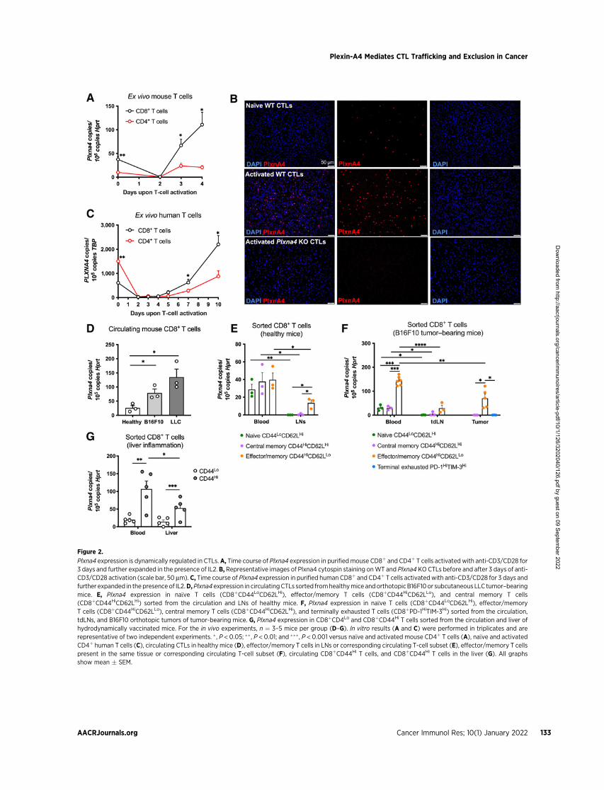
Figure 2.
Plxna4 expression is dynamically regulated in CTLs. A, Time course of Plxna4 expression in purifiedmouse CD8þ and CD4þ T cells activated with anti-CD3/CD28 for3 days and further expanded in the presence of IL2. B, Representative images of Plxna4 cytospin staining onWT and Plxna4 KOCTLs before and after 3 days of anti-CD3/CD28 activation (scale bar, 50 mm). C, Time course of Plxna4 expression in purified human CD8þ and CD4þ T cells activatedwith anti-CD3/CD28 for 3 days andfurther expanded in the presence of IL2.D,Plxna4 expression in circulatingCTLs sorted fromhealthymice andorthotopicB16F10or subcutaneous LLC tumor–bearingmice. E, Plxna4 expression in na€�ve T cells (CD8þCD44LoCD62LHi), effector/memory T cells (CD8þCD44HiCD62LLo), and central memory T cells(CD8þCD44HiCD62LHi) sorted from the circulation and LNs of healthy mice. F, Plxna4 expression in na€�ve T cells (CD8þCD44LoCD62LHi), effector/memoryT cells (CD8þCD44HiCD62LLo), central memory T cells (CD8þCD44HiCD62LHi), and terminally exhausted T cells (CD8þPD-1HiTIM-3Hi) sorted from the circulation,tdLNs, and B16F10 orthotopic tumors of tumor-bearing mice. G, Plxna4 expression in CD8þCD4Lo and CD8þCD44Hi T cells sorted from the circulation and liver ofhydrodynamically vaccinated mice. For the in vivo experiments, n ¼ 3–5 mice per group (D–G). In vitro results (A and C) were performed in triplicates and arerepresentative of two independent experiments. �, P < 0.05; �� , P < 0.01; and ��� , P < 0.001 versus na€�ve and activated mouse CD4þ T cells (A), na€�ve and activatedCD4þ human T cells (C), circulating CTLs in healthymice (D), effector/memory T cells in LNs or corresponding circulating T-cell subset (E), effector/memory T cellspresent in the same tissue or corresponding circulating T-cell subset (F), circulating CD8þCD44Hi T cells, and CD8þCD44Hi T cells in the liver (G). All graphsshow mean � SEM.
Plexin-A4 Mediates CTL Trafficking and Exclusion in Cancer
AACRJournals.org Cancer Immunol Res; 10(1) January 2022 133
Dow
nloaded from http://aacrjournals.org/cancerim
munolres/article-pdf/10/1/126/3202040/126.pdf by guest on 09 Septem
ber 2022

an increased amount of active (GTP-bound) Rac1 (Fig. 3G). Theamount of GTP-bound Rap1, however, was similar between bothconditions (Supplementary Fig. S3K and S3L). In line with thepreviously reported involvement of Rac1 in CTL migration andproliferation (30), pharmacologic Rac1 inhibition on activatedCTLs was sufficient to reduce migration and proliferation ofboth Plxna4 WT and KO cells (Fig. 3H and I). The latter suggeststhat active Rac1 is required for the phenotype observed in Plxna4KO CTLs.
Plxna4 KO CTLs show increased homing and proliferationcapacity in inflammatory sites
To assess the in vivo role of Plxna4 on CTL migration andproliferation, we performed competition assays in which WT andPlxna4 KO CTLs, either na€�ve (LN-homing) or in vitro preactivated(inflamed tissue-homing), were coinjected i.v. into mice. When na€�veWT and Plxna4 KO CTLs were transferred into healthy WT mice,Plxna4 KO CTLs were more efficient in reaching the LNs (Fig. 4Aand B; Supplementary Fig. S4A and S4B). Both WT and Plxna4 KO
Figure 3.
Genetic knockout of Plxna4 in CTLs increases motility and proliferation via enhanced Rac1 activity. In vitro proliferation of WT and Plxna4 KO CTLs uponanti-CD3/CD28 activation showing absolute cell numbers (A) and a representative histogram (B) of Violet Cell Tracer fluorescence intensity, gatedon CD8þ T cells, after 4 days in culture. C, Transmigration assay of na€�ve WT and Plxna4 KO CTLs toward CCL21 and CCL19 chemokines. D, Transmigrationassay of activated WT and Plxna4 KO CTLs toward CXCL9 and CXCL10 chemokines. E, Transmigration assay of na€�ve WT and Plxna4 KO CTLs towardthe CCL21 chemokine, treated with IgG or anti-CCR7. F, Transmigration assay of activated WT and Plxna4 KO CTLs toward the CXCL10 chemokine,treated with IgG or anti-CXCR3. G, GTP-bound Rac1 pull-down assay on activated WT and Plxna4 KO CTLs. Quantification by densitometry, paired analysison each individual experiment (left) and a representative image of the Western blot analysis (right). H, Transmigration assay of activated WT and Plxna4KO CTLs toward the CXCL10 chemokine, treated with either vehicle or the Rac1 inhibitor NSC23766. I, In vitro proliferation of activated WT and Plxna4KO CTLs, treated at day 3 with either vehicle or the Rac1 inhibitor NSC23766. In vitro results were performed in triplicates (A, D–F, H, and I) or duplicates(C) and are representative of at least two independent experiments. Pull-down assay (G) is represented as the pooled data from four independentexperiments (left), and the Western blot analysis is representative of at least three independent experiments (right). � , P < 0.05; �� , P < 0.01; ��� , P < 0.001;and ���� , P < 0.0001 versus WT CTLs (A, C, and D), IgG-treated WT and Plxna4 KO CTLs (E and F), and vehicle-treated WT and Plxna4 KO CTLs (H and I).All graphs show mean � SEM.
Celus et al.
Cancer Immunol Res; 10(1) January 2022 CANCER IMMUNOLOGY RESEARCH134
Dow
nloaded from http://aacrjournals.org/cancerim
munolres/article-pdf/10/1/126/3202040/126.pdf by guest on 09 Septem
ber 2022

CTLs were able to enter the paracortical areas of the LNs withoutentrapment in the high endothelial venules (HEV; Fig. 4B). Toevaluate whether increased detection of Plxna4 KO CTLs was due aninability to exit the LNs, we treated WT mice with fingolimod(FTY720), an immunomodulatory drug that inhibits lymphocyteegress from lymphoid tissues by downregulating sphingosine-1 phos-phate receptor (S1PR; ref. 31). Treatment with FTY720 entrapped
both WT and Plxna4 KO CTLs in the LNs, but Plxna4 KO CTLs werestill present in higher numbers in the LNs compared withWT controls(Fig. 4C). To further investigate if Plxna4 deficiency would affectT-cell egression from the LNs, we generatedWT and Plxna4KOOT-IT cells, expressing a TCR which specifically recognizes the OVApeptide presented in MHC class I. Thereafter, we took advantage ofthe LCMV-OVA model, where infected dendritic cells present the
Figure 4.
Plxna4 KO CTLs show increased homing and proliferation capacity in inflammatory sites. Homing of na€�ve WT and Plxna4 KO CTLs to the LNs of healthy micequantified by histology (A) and representativemicrograph (scale bar, 100 mm;B).C,Homing of na€�veWT and Plxna4KOCTLs to the LNs of healthymice treatedwitheither vehicle or the S1PR inhibitor FTY720. D, Ratio of CD8þCD44Hi to CD8þCD44Lo circulating WT and Plxna4 KO OT-I T cells in mice 32 hours and 48 hours afterLCMV-OVA infection, analyzed by flow cytometry. E,Tumor homing of activatedWT andPlxna4KOOT-I T cells in B16F10-OVA tumor–bearingmice assessed by flowcytometry 24 hours and 48 hours after tail vein injection. F, Flow cytometric analysis of labeled T cells in B16F10-OVA tumors 24 hours after intratumoral injection ofactivated WT and Plxna4 KO OT-I T cells. G, Liver homing of activated WT and Plxna4 KO OT-I T cells in mice hydrodynamically injected with empty vector (EV) orovalbumin vector (OVA).H, Tumor homing of activatedWTandPlxna4KOOT-I T cells in tumor-bearingmice, injectedwith B16F10-OVAcancer cells transducedwithEV or overexpressing (OE) Sema3a, Sema6a, or Sema6b. T-cell infiltration was assessed by flow cytometry 24 hours after tail vein injection. I, Tumor homing ofactivated WT and Plxna4 KO OT-I T cells in WT and Sema6a KO tumor-bearing mice, injected with B16F10-OVA cancer cells. T-cell infiltration was assessed byflow cytometry 24 hours after tail vein injection. For the in vivo experiments, n ¼ 4–5 mice per group. � , P < 0.05; �� , P < 0.01; ��� , P < 0.001; and ���� , P < 0.0001versus WT CTLs (A and C) and WT OT-I T cells (E–I). All graphs show mean � SEM.
Plexin-A4 Mediates CTL Trafficking and Exclusion in Cancer
AACRJournals.org Cancer Immunol Res; 10(1) January 2022 135
Dow
nloaded from http://aacrjournals.org/cancerim
munolres/article-pdf/10/1/126/3202040/126.pdf by guest on 09 Septem
ber 2022
OVA peptide in MHC class I (19). Following the systemic injectionof na€�ve WT or Plxna4 KO OT-I T cells in LCMV-OVA–infectedmice, we calculated the ratio between antigen-primed CD44Hi andna€�ve CD44Lo WT or Plxna4 KO OT-I T cells in circulation overtime. In this way, we found that similar numbers of CD44Hi Plxna4KO OT-I T cells were released into the blood 16 hours earlier thanCD44Hi WT OT-I T cells but their speed of egression (graphicallyrepresented by the slope of the line) was the same (Fig. 4D). Thisobservation suggests that the exit from the LNs of activated Plxna4KO CTLs was not altered but only anticipated due to their fasterarrival into the LN.
Next, we studied the biological functions of Plxna4 in CTLswithin the tumor or other sites of inflammation. In competitionassays, we found increased tumor homing of Plxna4 KO versusWT OT-I T cells 24 hours after their systemic coinjection in micebearing OVA-expressing B16F10 (Fig. 4E) or LLC tumors (Supple-mentary Fig. S4C). To differentiate if the increased number of Plxna4KO CTLs in the tumor was a consequence of increased migratory/infiltrative capacity or higher proliferative ability, as observed in vitro(Fig. 3A and B), we followed OVA-expressing melanoma tumorsfor an additional 24 hours. Forty-eight hours after systemic injection ofOT-I T cells, the ratio between Plxna4 KO/WT OT-I T cells in thetumor bed was higher compared with what was calculated at 24 hours(Fig. 4E; Supplementary Fig. S4D), suggesting that the increasedintratumoral accumulation of Plxna4 KO CTLs was the result ofboth augmented migration and proliferation. The fluorescent probelabelling Plxna4 KO tumor-infiltrating OT-I T cells was more dilutedthan in WT OT-I cells 24 hours after the systemic coinjection ofactivated WT and KO CTLs (Supplementary Fig. S4E), further sug-gesting a higher proliferation rate of Plxna4 KO CTLs. To controlfor potential differential recruitment, we coinjected activated WTand Plxna4 KO OT-I T cells intratumorally in B16F10-OVA tumorsand observed increased proliferation in cells lacking Plxna4 (Fig. 4F).Then, we assessed if the loss of Plxna4 could confer both a migratoryand proliferative advantage in other inflammatory conditions aswell. To this end, we used the HDI model described above and foundthat in vitro activated Plxna4 KO OT-I T cells infiltrated the inflamedlivers more efficiently than WT OT-I T cells (Fig. 4G). This effectwas more evident when hepatocytes were forced to express OVA(Fig. 4G; Supplementary Fig. S4F and S4G), which would boostproliferation as well. Taken together, these results suggest that Plxna4deletion in CTLs increases both their migratory/infiltrative capacityand proliferation rate in response to TCR activation.
Finally, we evaluated whether infiltrating CTLs were attracted orrepulsed by interactions in trans with semaphorins expressed in theTME. To this end, B16F10-OVA cancer cells overexpressing the mostrelevant Plxna4 ligands (Sema3a, Sema6a, or Sema6b) were injected inWTmice (Supplementary Fig. S4H–S4J). Consistent with our previousobservations (Fig. 4E), 24 hours after systemic injection, Plxna4 KOOT-I T cells were more efficient in infiltrating melanoma tumorstransduced with amock empty vector (Fig. 4H). This finding held trueeven when the tumors overexpressed Sema3a, Sema6a, or Sema6b(Fig. 4H). Moreover, the increased homing capacity of Plxna4 KOversus WT OT-I T cells was also seen in Sema6a KO tumor-bearingmice (Fig. 4I). Ruling out the engagement of Sema6a presentation byT cells themselves, activated Sema6a KO CTLs did not mimicPlxna4 deficiency in terms of in vitro migration towards CXCL10,but rather showed the same chemotaxis as observed in WT cells(Supplementary Fig. S4K). Altogether, these findings suggest thatpresentation of semaphorins by the host (or autocrine signals bySema6a) do not affect CTL infiltration and provide evidence for
another nonconventional Plxna4-dependent but semaphorin-independent mechanism controlling CTL motility in cancer.
Adoptive transfer ofPlxna4KOCTLs shows improved antitumorefficacy in a melanoma model
Prompted by our observations that Plxna4 functions as a negativeregulator of CTL proliferation and migration, we evaluated if thedeletion of Plxna4 in CTLs was sufficient to increase antitumorimmunity in the context of ACT. As a proof of concept of immuno-therapeutic validation of Plxna4 deficiency in CTLs, we first injectedna€�ve WT or Plxna4 KO OT-I T cells i.v. into LLC-OVA lung andB16F10-OVA melanoma tumor–bearing WT recipient mice. Micereceiving Plxna4 KO OT-I T cells displayed a higher degree of tumorinhibition (Supplementary Fig. S5A and S5B), pointing to a directeffect of Plxna4 deficiency in CTL activity. Secondly, consistent witha therapeutic approach, activated OT-I T cells were transferred intothe circulation of B16F10-OVA tumor-bearing WT recipient mice.In this setting, Plxna4 KO OT-I T cells exerted increased antitumoreffects than their WT counterparts, as demonstrated by decreasedtumor growth and weight (Fig. 5A–C). As expected, WT CTLs werealso able to control tumor growth, but to a lesser extent than Plxna4KO OT-I T cells (Fig. 5A–C). When we analyzed the number oftumor-infiltrating CTLs 4 days after ACT, melanoma tumors thatreceived Plxna4 KO OT-I T cells contained an increased numberof intratumoral OT-I T cells compared with the ones treated withWT OT-I cells (Fig. 5D). Nevertheless, these cells showed nodifference in cytotoxicity, evaluated by their expression of IFNgand GzmB after T-cell restimulation ex vivo (SupplementaryFig. S5C and S5D). In this model of ACT, the administration ofPlxna4 KO OT-I T cells also extended overall survival when com-pared with the adoptive transfer of WT OT-I T cells (Fig. 5E). Takentogether, these data demonstrate that Plxna4 deletion in CTLs issufficient to increase antitumor immunity in a conventional ACTsetting and that targeting of Plxna4 might thus be a valuable strategyto improve CTL infiltration.
Potential clinical relevance of Plxna4 in CTLs in patients withmelanoma
To translate our findings to human cancer, we analyzed PLXNA4expression in circulating CTLs in a cohort of patients with metastaticmelanoma (Supplementary Table S3). Similar to our observations in amurine model (Fig. 2), CTLs isolated from the peripheral blood ofpatients with melanoma expressed higher quantities of PLXNA4, butnot of other type-A plexins, compared with those measured incirculating CTLs from healthy donors (Fig. 6A; SupplementaryFig. S6A–S6C). PLXNA4 abundance was also higher in circulatingCD4þ T cells isolated from the same patients (SupplementaryFig. S6D). In contrast, expression of PLXNA4 in circulatingmonocytesisolated from both healthy volunteers and patients across differenttumor types were low or undetectable (Supplementary Fig. S6E;Supplementary Table S5).
We then analyzed Plxna4 expression in circulating CTLs ofpatients with melanoma prior to and after the first cycle of ICItherapy (anti–PD-1 alone or in combination with anti–CTLA-4).We observed a significant decrease of Plxna4 in CTLs followingtreatment (Fig. 6B). When stratifying these patients for theirclinical response to ICI therapy, we found that the decrease inPlxna4 expression in circulating CTLs was significant in theresponders but not in the nonresponders (Fig. 6C and D).
On the basis of these clinical data, we speculated that the benefitfrom ICI therapymay depend, at least partly, on the downregulation of
Celus et al.
Cancer Immunol Res; 10(1) January 2022 CANCER IMMUNOLOGY RESEARCH136
Dow
nloaded from http://aacrjournals.org/cancerim
munolres/article-pdf/10/1/126/3202040/126.pdf by guest on 09 Septem
ber 2022

Plxna4. This hypothesis was tested by treating B16F10 tumor–bearingmice with anti–PD-1 for a week, resulting in a reduction of Plxna4expression in circulating CTLs compared with IgG-treated tumor-bearing mice (Fig. 6E). This downregulation of Plxna4 expressionupon anti–PD-1 blockade was also true for in vitro activated humanCTLs, but not for CD4þ T cells (Fig. 6F). The hypothesis that PD-1blockade acts partly, but not only, through the downregulation ofPlxna4 in CTLs was also suggested by our findings that anti–PD-1therapy in combination with ACT of WT OT-I cells reached a similarextent of tumor inhibition as observed with the transfer of Plxna4 KOOT-I T cells alone (Fig. 6G). Perhaps by means of Plxna4-unrelatedmechanisms of anti–PD-1, such as unleashing the PI3K–Akt path-way (32), the combination of Plxna4 KO OT-I ACT and anti–PD-1therapy led to an even stronger reduction in tumor growth than withPlxna4 KO OT-I ACT alone (Fig. 6G). Members of the Forkhead boxO (FOXO) family of transcription factors (TF), which are activateddownstream to PD-1 and upon persistent TCR stimulation (33, 34),are described to regulate Plxna4 expression (35). When treatingin vitro activated CTLs with CBX, an inhibitor of FOXO1, 3, 4, and6 (36), we observed that Plxna4 expression was reduced even furtherthan upon anti–PD-1 treatment (Fig. 6H), providing initial supportthat FOXO TFs may promote Plxna4 expression upon chronic TCRstimulation and PD-1 induction.
DiscussionIn the last decade, immunotherapy has emerged as a promising
therapeutic option for patients with cancer. The use of ICIs, inparticular, has revolutionized the field, enabling durable control of
previously highly refractory and aggressive cancers, such asmelanomaand lung cancer.However, a significant percentage of patients—60% to80%—still do not benefit from this strategy (37). One of the mainfactors hampering ICIs is the exclusion of CTLs from the tumor bed,making these tumors T-cell “cold”. In this study, we show thattargeting Plxna4 might represent a novel strategy to drive T-cellinflammation in T-cell nonenriched tumors. Our data show thatPlxna4 is dynamically regulated in CTLs and that its absence promotesan increased proliferative and homing capacity to the tdLNs and thetumor bed, leading to a more effective antitumor response (Fig. 6I).
In our study, Plxna4 deletion in the stroma reduced tumor growthby specifically enhancing CTL infiltration, while the phenotype ofother stromal cells remained unaffected. Although Plxna4 was shownto play a role in macrophages in the context of cancer or otherpathologic conditions (15–17), we observed that deletion of Plxna4was not sufficient to impact on macrophage localization and pheno-type. Plxna4 was also described as a negative regulator of CD4þ T-cellimmune responses in experimental autoimmune diseases (18, 24). Inour models, Plxna4 is expressed by CD4þ T cells but at lowerabundance than in CTLs. Moreover, neither the infiltration nor thedistribution of Plxna4 KO CD4þ T cells within the tumor was altered,pointing to a function of Plxna4 specifically in CTLs. Given thefunctional redundancy among class A plexins (17, 38), homologousreceptors expressed in TAMs, CD4þ T cells, or endothelial cells, suchas Plexin-A1, could compensate for the absence of Plxna4. In contrast,Plxna4 is indispensable for the fine-tuning of CTL proliferation andmigration.
In terms of expression, na€�ve T cells found in the LNs showundetectable transcription of Plxna4 compared with circulating na€�ve
Figure 5.
Adoptive transfer of Plxna4 KO CTLs leads to improved antitumor immunity. Tumor growth (A), tumor weight (B), and representative images (C) of end-stagetumors following adoptive transfer (ACT) of activated WT and Plxna4 KO OT-I T cells in B16F10-OVA tumor–bearing WT mice (scale bar, 2 cm). D, Flow cytometricanalysis of intratumoral OT-Iþ T cells in B16F10-OVA tumors isolated 4 days after ACT. E, Kaplan–Meier overall survival curves. For the in vivo experiments, n¼ 6–8mice per group. � , P < 0.05; ��� , P < 0.001 versus WT OT-I T cells. All graphs show mean � SEM.
Plexin-A4 Mediates CTL Trafficking and Exclusion in Cancer
AACRJournals.org Cancer Immunol Res; 10(1) January 2022 137
Dow
nloaded from http://aacrjournals.org/cancerim
munolres/article-pdf/10/1/126/3202040/126.pdf by guest on 09 Septem
ber 2022

Figure 6.
PLXNA4 expression is upregulated in circulatingCTLsof patientswithmetastaticmelanoma.A,PLXNA4 expression in isolatedCTLs from the circulationof treatment-na€�ve melanoma patients versus healthy controls. B, Na€�ve versus paired ICI (anti–PD-1 alone or in combination with anti–CTLA-4)–treated melanoma patients3weeks after one cycle of treatment.PLXNA4 expression in isolated CTLs from the circulation of treatment-na€�ve and paired ICI-treated patientswithmelanoma thatclinically responded to ICIs (C) and patients with melanoma that did not respond to ICIs (D). E, Plxna4 expression in circulating CTLs isolated from orthotopic B16F10tumor–bearing mice treated with IgG or anti–PD-1. F, PLXNA4 expression in ex vivo activated human CD8þ and CD4þ WT T cells treated with IgG or anti–PD-1.G,Weight of end-stage B16F10-OVA tumors receiving ACT of activatedWT and Plxna4 KOOT-I T cells in combination with IgG or anti–PD-1 therapy.H, Fold changeofPlxna4 expression in ex vivo activatedmouse CTLs treatedwith vehicle, anti–PD-1, or CBX. I, Schematic overviewof the data presented. For the patient data (A–D),n ¼ 16 healthy controls, n ¼ 31 treatment-na€�ve melanoma patients, and n ¼ 20 ICI-treated patients with melanoma (see Supplementary Table S3). In vitro results(F andH)were performed in triplicates and are represented as the pooled data from two independent experiments. For the in vivo experiment (E andG), n¼ 5–9miceper group. � , P <0.05; �� , P <0.01; ��� , P <0.001; and ���� , P <0.0001 versus circulating CTLs in healthy individuals (A), circulating CTLs in treatment-na€�vemelanomapatients (B and C), circulating CTLs isolated from IgG-treatedmice (E), IgG-treated human CTLs (F), B16F10-OVA tumors receiving ACT of activatedWTOT-I T cellsand IgG-treated B16F10-OVA tumors (G), and vehicle-treated mouse CTLs (H). All graphs show mean � SEM.
Celus et al.
Cancer Immunol Res; 10(1) January 2022 CANCER IMMUNOLOGY RESEARCH138
Dow
nloaded from http://aacrjournals.org/cancerim
munolres/article-pdf/10/1/126/3202040/126.pdf by guest on 09 Septem
ber 2022

T cells, suggesting that the downregulation of Plxna4 is required forCTLs to enter the lymphoid tissue. Indeed, homing of na€�ve Plxna4KO CTLs towards LNs of healthy mice was increased compared withWT CTLs. Upon T-cell activation, Plxna4 transcripts were upregu-lated in CTLs in tumors, as well as in experimental models ofliver inflammation or viral infection, demonstrating that differentmicroenvironmental signals associated with inflammation, infections,or tumor progression can result in similar expression patterns, asdescribed previously (39). In particular, Plxna4 protein expression wasincreased in CTLs activated in vitro when compared with their na€�vecounterpart, suggesting that Plxna4 is upregulated upon T-cell acti-vation. Consistently, Plxna4 transcription was specifically induced inthe circulating effector/memory CTL fraction in tumor-bearing micebut reduced at inflammatory sites (cancer and liver), suggesting thatPlxna4 downregulation is required for their entry in these tissues.Indeed, genetic KO of Plxna4 in activated CTLs increased theirinfiltration to both the tumor and the inflamed liver parenchyma. Inaddition, we hypothesized that higher Plxna4 transcription observedin the pool of tumor-associated effector/memory T cells, comparedwith the undetectable levels in central memory and terminallyexhausted T cells, was mainly reflecting the presence of Plxna4Hi
excluded T cells at the tumor border. In contrast, few Plxna4Lo T cellsenter the tumor core and proliferate when encountering tumor-associated antigens presented in MHC class I by the cancer cells orby tumor-infiltrating DCs (Fig. 6I). In line with this idea, tumor-bearing mice with stromal or hematopoietic Plxna4 deletion hadincreased CTL numbers in the tumor core, which is likely the resultof improved infiltration and proliferation in response to antigenrepriming (Fig. 6I). This is also suggested by the fact that adoptivetransfer of in vitro preactivated Plxna4 KO CTLs exhibit increas-ed homing and proliferation within OVA-overexpressing tumors.Altogether, it is reasonable to speculate that Plxna4 could work as anegative regulator of both na€�ve and effector/memory CTLs, bybreaking their recruitment into the LNs and inflammatory sites. Thismechanism is possibly mediated by the presence of proinflammatory,type I cytokines in these tissues (such as IL10, IFNg , or others), whichcounter this break in a dynamic process and promote the down-regulation of Plxna4 and thus the entry and the proliferation ofna€�ve CTLs in the LNs, and effector/memory CTLs in the inflamedtissue. However, in the context of cancer, this process fails to occurcompletely, possibly because the TME is enriched in anti-inflamma-tory, type II cytokines which impede the downregulation of Plxna4in effector/memory CTLs that are thus excluded at the border ofthe tumor. The concept of Plxna4 as a “break” of inflammatoryprocesses agrees with previously published data showing that theabsence of Plxna4 exacerbates ongoing autoimmune diseases (18, 24)but does not result per se in the spontaneous development of auto-immune disorders or other pathologies, since Plxna4 KOmice appearto be healthy (18, 22).
The intracellular portion of plexins contains GTPase-bindingdomains (28, 40), which can interact and negatively regulate Rac1,a Rho family GTPase involved in the modulation of thecytoskeleton (30, 41–43). Our data suggest that enhanced CTL migra-tion in the absence of Plxna4 might be due to increased activation ofRac1, since we observed that Plxna4 deficiency was associated withaugmented GTP-bound Rac1, in line with what has been shown beforein Plxna4-silenced endothelial cells (23). The modulation of Rac1 byPlxna4 is here functionally supported by the observation that treat-ment with the Rac1 inhibitor NSC23766 completely prevented theadvantageous migration of Plxna4 KO CTLs. Besides migration, CTLproliferation is also instrumental to mount a proper immune
response (44). In this regard, Plxna4 KO CTLs displayed increasedproliferative capacity, arguing that Plxna4 negatively regulates CTLproliferation as well, akin to what has been suggested for Plxna4 KOCD4þ T cells in the context of autoimmune diseases (18). Thisincreased proliferation can be attributed to enhanced Rac1 activityas well (30), as supported by our in vitro data comparing Plxna4WT and KO CTLs treated with Rac1 inhibitors. However, futurework is warranted to assess if direct or indirect mechanisms areenrolled by Plxna4 to regulate Rac1 activation.
Given the repulsive activities propagated by Plxna4 when interact-ing with semaphorins (13, 14, 45), it may be attractive to hypothesizethat the exclusion of CTLs from the tumor bed is a consequence ofrepulsive cues. However, our data illustrate that the overexpression incancer cells of the most relevant Plxna4 ligands (Sema3a, Sema6a, andSema6b), or the expression of the high-affinity ligand Sema6a in theTME and by T cells themselves, does not modulate CTL infiltration inthe tumor bed. Because plexins are evolutionarily more conservedthan semaphorins (46), we are tempted to believe that semaphorin-independent mechanisms are likely to be present in eukaryotic cells.Furthermore, we foresee that the inhibitory effect of Plxna4 on CTLexpansion and chemotaxis is cell autonomous, and thereforeindependent of the interaction in trans of Plxna4 with semaphorinsexpressed in the TME. Of note, interactions in cis between plexinsand transmembrane semaphorins, or other cell surface receptors,have been reported to modulate intracellular signaling and biologicalfunctions (45, 47–49). Thus, currently unknown interactions ofPlxna4 with molecules coexpressed in T cells (other than Sema6a)could play a role in the observed phenotype, possibly by regulatingRac1 activation.
Our work suggests potential therapeutic and clinical implications.In an adoptive transfer setting, Plxna4 KO CTLs outperformed WTcells in shrinking established B16F10 melanoma tumors, providing aproof of concept that Plxna4 targeting in CTLs facilitates antitumorimmunity and could be integrated in existing cancer immunotherapyregimens. Because enforcing the trafficking of CTLs to malignanttissues can improve the efficacy of any immunotherapeutic regi-mens (50), and combination treatments are often needed to unleasha proper antitumor immune response (51), we believe that targetingPlxna4 also holds the potential to act synergistically with conventionalimmunotherapies, including ICIs. In our preclinical tumor models,ACT with Plxna4 KO CTLs in combination with anti–PD-1 led to amore potent antitumor immune response compared with monother-apy, demonstrating a potential clinical value of Plxna4-targeted ther-apies. However, future studies should determine to which extent andwith which implications on homeostatic immune control, the com-bined targeting of Plxna4 and other inhibitory receptors on CTLs canfurther increase the antitumor immune response.
Supporting our preclinical data, Plxna4 expression was increased incirculating CTLs from patients with metastatic melanoma comparedwith healthy controls, further strengthening a possible translation ofour mechanistic and therapeutic conclusions to human disease. More-over, Plxna4 expression in circulating CTLs was decreased after thefirst cycle of ICI therapy in melanoma patients that responded to thetherapy. ICI therapy frequently leads to an increase in proliferation andmigration of preexisting CTLs (52, 53), features related to the observedphenotype of Plxna4 KO CTLs. This indicates that ICIs might partlywork in circulation via the downregulation of Plxna4 in CTLs sepa-rately from previously characterizedmechanisms (54). The expressionof PD-1 (Pdcd1) is induced after TCR stimulation via members of thenuclear factor of activated T-cell (NFAT) family of transcriptionfactors (55). Upon persistent antigen stimulation, PD-1 signaling
Plexin-A4 Mediates CTL Trafficking and Exclusion in Cancer
AACRJournals.org Cancer Immunol Res; 10(1) January 2022 139
Dow
nloaded from http://aacrjournals.org/cancerim
munolres/article-pdf/10/1/126/3202040/126.pdf by guest on 09 Septem
ber 2022

inhibits multiple TCR downstream pathways and FOXO TFs areactivated, further promoting Pdcd1 transcription (33). Consistent withPlxna4 being upregulated as a late activation marker, FOXOs areessential for activated T cells within days following primary TCRactivation (33, 34). FOXOs are known to positively regulate severaleffector/memory functions such as T-cell homing and effector T-celldifferentiation (56, 57), features that were all enhanced in Plxna4 KOCTLs. Becausewe observed that FOXOTFs can regulate the expressionof Plxna4 (35), we speculate that later stages of T-cell activation arecharacterized by the upregulation of negative regulators of T-cellfunctions (immune checkpoint molecules), which include PD-1 itselfbut also the Plxna4 pathway. However, the link between FOXOtranscription factors and expression of Plxna4 remain incompletelydefined and will need further investigation in future studies.
In terms of clinical management, biomarkers of tumor response toICIs are needed to help guide treatment strategy. Our results suggestthat expression of PLXNA4 in circulating CTLs could be used as anearly, noninvasive biomarker for monitoring patients’ responses toimmune checkpoint blockade. In line with our observations, changesin T-cell subsets of patients with metastatic melanoma were reportedto occur within a short period after initiating ICI therapy (58).Although our CTL sampling was done in a prospective manner andlarger cohorts of patients will be needed to further confirm our clinicalresults, the current data well-position PLXNA4 expression in circu-lating CTLs as a potential biomarker of disease or response toimmunotherapy.
Authors’ DisclosuresW.Celus reports grants fromResearch Foundation Flanders during the conduct of
the study. A.I. Oliveira reports grants from Research Foundation Flanders andpersonal fees from Fundac~ao para a Ciencia e a Tecnologia during the conduct ofthe study. A.D. Garg reports personal fees from Boehringer Ingelheim and MiltenyiBiotec outside the submitted work. M. Mazzone reports grants from ResearchFoundation Flanders, Fundac~ao para a Ciencia e a Tecnologia, FWO-Strategic BasicResearch (SB) doctoral fellowship, Kom op tegen Kanker (Stand up to Cancer), theFlemish cancer society, FWO-Fundamental Research (FR) doctoral fellowship,fundamental postdoctoral mandate of the Belgian Foundation against Cancer (Sticht-ing tegen Kanker), the FNRS, theWalloon Region, FWO, KU Leuven, Kom op TegenKanker, the Grand Challenges Program of VIB (which received support from theFlemish Government under the Management Agreement 2017-2021), the NorthernPortugal Regional Operational Programme, European Regional Development Fund(ERDF), and ERC Consolidator Grants during the conduct of the study. W. Celus,A.I. Oliveira, and M. Mazzone report a patent for PCT application no. PCT/EP2020/064884 with title “CD8þ T cells lacking plexins and their application in cancertreatment” issued, a patent for PCT application no. PCT/EP2020/064905 with title“Cancer treatment by targeting plexins in the immune compartment” issued, and apatent for PCT application no. PCT/EP2021/076314 with title “Biomarker forantitumor therapy” issued. No disclosures were reported by the other authors.
Authors’ ContributionsW. Celus: Conceptualization, formal analysis, funding acquisition, validation,
investigation, visualization, methodology, writing–original draft, writing–reviewand editing. A.I. Oliveira: Conceptualization, formal analysis, funding acquisi-tion, validation, investigation, visualization, methodology, writing–original draft,writing–review and editing. S. Rivis: Validation, investigation. H.H. Van Acker:Validation, investigation. E. Landeloos: Resources. J. Serneels: Investigation;J. Serneels provided technical support. S.T. Cafarello: Investigation. Y. Van Herck:Resources. R. Mastrantonio: Investigation. A. K€ohler: Investigation. A.D. Garg:Conceptualization. V. Flamand: Conceptualization. L. Tamagnone: Conceptualiza-tion. J.C. Marine: Conceptualization, resources. M. Di Matteo: Conceptualization,resources, methodology. B.M. Costa: Conceptualization. O. Bechter: Conceptu-alization, resources. M. Mazzone: Conceptualization, resources, data curation,supervision, funding acquisition, writing–original draft, project administration,writing–review and editing.
AcknowledgmentsThis work was supported by the Research Foundation Flanders (FWO;
G0A8219N). A.I. Oliveira was supported by Fundac~ao para a Ciencia e Tecnologia(SFRH/BD/52287/2013) and by FWO under the project G0A8219N. W. Celus wassupported by an FWO-Strategic Basic Research (SB) doctoral fellowship (1S26917N).S. Rivis was supported by an FWO-Fundamental Research (FR) doctoral fellowship(1197720N). H.H. Van Acker was supported by a fundamental postdoctoral mandateof the Belgian Foundation against Cancer (Stichting tegen Kanker). E. Landeloos wasfunded by Kom op tegen Kanker (Stand up to Cancer), the Flemish cancer society.A. K€ohler and V. Flamand were funded by the FNRS (CDR 33685316 grant) and theWalloon Region (FEDER Wallonia-Biomed portfolio). A.D. Garg was supported byFWO (G0B4620N; EOS grant: 30837538), KU Leuven (C14/19/098; POR/16/040),and Kom op Tegen Kanker (KOTK/2018/11509/1). E. Landeloos, O. Bechter, andJ.C. Marine are funded within the Grand Challenges Program of VIB, which receivedsupport from the Flemish Government under the Management Agreement 2017–2021 (VR 2016 2312 Doc.1521/4). B.M. Costa was supported by the NorthernPortugal Regional Operational Programme (NORTE 2020) under the projectNORTE-01–0145-FEDER-000013 and the European Regional Development Fund(FEDER) under the Portugal 2020 Partnership Agreement. M. Mazzone was grantedby an ERC Consolidator Grant (773208) and by FWO (G0A8219N). The authorsthank S. Willox, A. Tzoumpa, E. Achten, C. Serrano, J. Grieten, R. Tavares, andM. Cogels for their technical assistance. Plxna4 KO mice were kindly offered byDr. Castellani (Institut NeuroMyoG�ene, Universit�e de Lyon, Lyon, France). Sema6aKO mice were kindly offered by Prof. Dr. Pasterkamp (Department of TranslationalNeuroscience, UniversityMedical CenterUtrecht, Utrecht, the Netherlands). LCMV-OVA virus was a kind gift from Prof. Dr. Pinschewer (University of Basel, Basel,Switzerland). Flow cytometry data were generated with the support from the VIBFACS Expertise Center.
The costs of publication of this article were defrayed in part by the payment of pagecharges. This article must therefore be hereby marked advertisement in accordancewith 18 U.S.C. Section 1734 solely to indicate this fact.
Received January 25, 2021; revised September 7, 2021; acceptedNovember 16, 2021;published first November 23, 2021.
References1. Chen DS, Mellman I. Oncology meets immunology: the cancer-immunity cycle.
Immunity 2013;39:1–10.2. Kruger S, IlmerM,Kobold S, Cadilha BL, Endres S, Ormanns S, et al. Advances in
cancer immunotherapy 2019 - latest trends. J Exp Clin Cancer Res 2019;38:268.3. Galluzzi L, Chan TA, Kroemer G, Wolchok JD, Lopez-Soto A. The hallmarks of
successful anticancer immunotherapy. Sci Transl Med 2018;10:eaat7807.4. Fridman WH, Pages F, Sautes-Fridman C, Galon J. The immune contexture in
human tumors: impact on clinical outcome. Nat Rev Cancer 2012;12:298–306.5. Teng MW, Ngiow SF, Ribas A, Smyth MJ. Classifying cancers based on T-cell
infiltration and PD-L1. Cancer Res 2015;75:2139–45.6. Perala N, Sariola H, Immonen T. More than nervous: the emerging roles of
plexins. Differentiation 2012;83:77–91.7. He Z, Wang KC, Koprivica V, Ming G, Song HJ. Knowing how to navigate:
mechanisms of semaphorin signaling in the nervous system. Sci STKE 2002;2002:re1.
8. Kumanogoh A, Kikutani H. Immunologic functions of the neuropilins andplexins as receptors for semaphorins. Nat Rev Immunol 2013;13:802–14.
9. Capparuccia L, Tamagnone L. Semaphorin signaling in cancer cells and incells of the tumor microenvironment–two sides of a coin. J Cell Sci 2009;122:1723–36.
10. Suto F, Murakami Y, Nakamura F, Goshima Y, Fujisawa H. Identification andcharacterization of a novel mouse plexin, plexin-A4. Mech Dev 2003;120:385–96.
11. Battistini C, Tamagnone L. Transmembrane semaphorins, forward and reversesignaling: have a look both ways. Cell Mol Life Sci 2016;73:1609–22.
12. Fujisawa H. Discovery of semaphorin receptors, neuropilin and plexin, and theirfunctions in neural development. J Neurobiol 2004;59:24–33.
13. Suto F, Ito K, Uemura M, Shimizu M, Shinkawa Y, Sanbo M, et al. Plexin-A4mediates axon-repulsive activities of both secreted and transmembrane sema-phorins and plays roles in nerve fiber guidance. J Neurosci 2005;25:3628–37.
Celus et al.
Cancer Immunol Res; 10(1) January 2022 CANCER IMMUNOLOGY RESEARCH140
Dow
nloaded from http://aacrjournals.org/cancerim
munolres/article-pdf/10/1/126/3202040/126.pdf by guest on 09 Septem
ber 2022

14. TawarayamaH, Yoshida Y, Suto F,Mitchell KJ, FujisawaH. Roles of semaphorin6B and plexin-A2 in lamina-restricted projection of hippocampal mossy fibers.J Neurosci 2010;30:7049–60.
15. Wen H, Lei Y, Eun SY, Ting JP. Plexin-A4–semaphorin 3A signaling is requiredfor Toll-like receptor- and sepsis-induced cytokine storm. J Exp Med 2010;207:2943–57.
16. Kang S, Nakanishi Y, Kioi Y, Okuzaki D, Kimura T, Takamatsu H, et al.Semaphorin 6D reverse signaling controls macrophage lipid metabolism andanti-inflammatory polarization. Nat Immunol 2018;19:561–70.
17. Casazza A, Laoui D, Wenes M, Rizzolio S, Bassani N, Mambretti M, et al.Impeding macrophage entry into hypoxic tumor areas by Sema3a/Nrp1 signal-ing blockade inhibits angiogenesis and restores antitumor immunity.Cancer Cell 2013;24:695–709.
18. Yamamoto M, Suzuki K, Okuno T, Ogata T, Takegahara N, Takamatsu H, et al.Plexin-A4 negatively regulates T lymphocyte responses. Int Immunol 2008;20:413–20.
19. Flatz L, Hegazy AN, Bergthaler A, Verschoor A, Claus C, Fernandez M, et al.Development of replication-defective lymphocytic choriomeningitis virusvectors for the induction of potent CD8þ T-cell immunity. Nat Med 2010;16:339–45.
20. Eisenhauer EA, Therasse P, Bogaerts J, Schwartz LH, Sargent D, Ford R, et al.New response evaluation criteria in solid tumors: revised RECIST guideline(version 1.1). Eur J Cancer 2009;45:228–47.
21. Hamm A, Prenen H, Van DelmW, Di Matteo M, Wenes M, Delamarre E, et al.Tumor-educated circulating monocytes are powerful candidate biomarkers fordiagnosis and disease follow-up of colorectal cancer. Gut 2016;65:990–1000.
22. Yaron A, Huang PH, Cheng HJ, Tessier-LavigneM. Differential requirement forPlexin-A3 and -A4 in mediating responses of sensory and sympathetic neuronsto distinct class 3 Semaphorins. Neuron 2005;45:513–23.
23. Kigel B, Rabinowicz N, Varshavsky A, Kessler O,NeufeldG. Plexin-A4 promotestumor progression and tumor angiogenesis by enhancement of VEGF and bFGFsignaling. Blood 2011;118:4285–96.
24. Podojil JR, Chiang MY, Ifergan I, Copeland R, Liu LN, Maloveste S, et al. B7-H4 modulates regulatory CD4þ T-cell induction and function via ligation ofa semaphorin 3A/plexin-A4/neuropilin-1 complex. J Immunol 2018;201:897–907.
25. Suda T, Liu D. Hydrodynamic gene delivery: its principles and applications.Mol Ther 2007;15:2063–9.
26. Girard JP, Moussion C, Forster R. HEVs, lymphatics, and homeostatic immunecell trafficking in lymph nodes. Nat Rev Immunol 2012;12:762–73.
27. Sackstein R, Schatton T, Barthel SR. T-lymphocyte homing: an underappre-ciated yet critical hurdle for successful cancer immunotherapy. Lab Invest2017;97:669–97.
28. Kong Y, Janssen BJ, Malinauskas T, Vangoor VR, Coles CH, Kaufmann R, et al.Structural basis for plexin activation and regulation. Neuron 2016;91:548–60.
29. Faroudi M, Hons M, Zachacz A, Dumont C, Lyck R, Stein JV, et al. Critical rolesfor Rac GTPases in T-cell migration to andwithin lymph nodes. Blood 2010;116:5536–47.
30. Gomez M, Tybulewicz V, Cantrell DA. Control of pre–T-cell proliferation anddifferentiation by the GTPase Rac-I. Nat Immunol 2000;1:348–52.
31. Morris MA, Gibb DR, Picard F, Brinkmann V, Straume M, Ley K. TransientT-cell accumulation in lymph nodes and sustained lymphopenia in mice treatedwith FTY720. Eur J Immunol 2005;35:3570–80.
32. Boussiotis VA. Molecular and biochemical aspects of the PD-1 checkpointpathway. N Engl J Med 2016;375:1767–78.
33. Staron MM, Gray SM, Marshall HD, Parish IA, Chen JH, Perry CJ, et al. Thetranscription factor FoxO1 sustains expression of the inhibitory receptor PD-1and survival of antiviral CD8þ T cells during chronic infection. Immunity 2014;41:802–14.
34. Delpoux A, Lai CY, Hedrick SM, Doedens AL. FOXO1 opposition of CD8þ T-cell effector programming confers early memory properties and phenotypicdiversity. Proc Natl Acad Sci U S A 2017;114:E8865–E74.
35. Paap RH, Oosterbroek S, Wagemans C, von Oerthel L, Schellevis RD, Vasten-houw-van der Linden AJA, et al. FoxO6 affects Plxna4-mediated neuronalmigration during mouse cortical development. Proc Natl Acad Sci U S A2016;113:E7087–E96.
36. Salcher S, Spoden G, Hagenbuchner J, Fuhrer S, Kaserer T, Tollinger M, et al. Adrug library screen identifies Carbenoxolone as novel FOXO inhibitor thatovercomes FOXO3-mediated chemoprotection in high-stage neuroblastoma.Oncogene 2020;39:1080–97.
37. Ribas A, Wolchok JD. Cancer immunotherapy using checkpoint blockade.Science 2018;359:1350–5.
38. Sharma A, Verhaagen J, Harvey AR. Receptor complexes for each of the class 3semaphorins. Front Cell Neurosci 2012;6:28.
39. Greten FR, Grivennikov SI. Inflammation and cancer: triggers, mechanisms, andconsequences. Immunity 2019;51:27–41.
40. Pascoe HG, Wang Y, Zhang X. Structural mechanisms of plexin signaling.Prog Biophys Mol Biol 2015;118:161–8.
41. Bustelo XR, Sauzeau V, Berenjeno IM. GTP-binding proteins of the Rho/Racfamily: regulation, effectors, and functions in vivo. Bioessays 2007;29:356–70.
42. Roney KE, O’Connor BP, Wen H, Holl EK, Guthrie EH, Davis BK, et al. Plexin-B2 negatively regulates macrophage motility, Rac, and Cdc42 activation.PLoS One 2011;6:e24795.
43. Vikis HG, Li W, Guan KL. The plexin-B1/Rac interaction inhibits PAK acti-vation and enhances Sema4D ligand binding. Genes Dev 2002;16:836–45.
44. ZhangN, BevanMJ. CD8þT cells: foot soldiers of the immune system. Immunity2011;35:161–8.
45. Suto F, Tsuboi M, Kamiya H, Mizuno H, Kiyama Y, Komai S, et al. Interactionsbetween plexin-A2, plexin-A4, and semaphorin 6A control lamina-restrictedprojection of hippocampal mossy fibers. Neuron 2007;53:535–47.
46. Junqueira Alves C, YotokoK, ZouH, Friedel RH.Origin and evolution of plexins,semaphorins, and Met receptor tyrosine kinases. Sci Rep 2019;9:1970.
47. Haklai-Topper L, Mlechkovich G, Savariego D, Gokhman I, Yaron A. Cisinteraction between semaphorin 6A and plexin-A4 modulates the repulsiveresponse to Sema6a. EMBO J 2010;29:2635–45.
48. Rozbesky D, Verhagen MG, Karia D, Nagy GN, Alvarez L, Robinson RA,et al. Structural basis of semaphorin–plexin cis interaction. EMBO J 2020;39:e102926.
49. Cagnoni G, Tamagnone L. Semaphorin receptors meet receptor tyrosine kinaseson the way of tumor progression. Oncogene 2014;33:4795–802.
50. Melero I, Rouzaut A, Motz GT, Coukos G. T-cell and NK-cell infiltration intosolid tumors: a key limiting factor for efficacious cancer immunotherapy.Cancer Discov 2014;4:522–6.
51. Mahoney KM, Rennert PD, Freeman GJ. Combination cancer immuno-therapy and new immunomodulatory targets. Nat Rev Drug Discov 2015;14:561–84.
52. Tumeh PC, Harview CL, Yearley JH, Shintaku IP, Taylor EJ, Robert L, et al. PD-1blockade induces responses by inhibiting adaptive immune resistance. Nature2014;515:568–71.
53. Brunner-Weinzierl MC, Rudd CE. CTLA-4 and PD-1 control of T-cell motilityand migration: implications for tumor immunotherapy. Front Immunol 2018;9:2737.
54. Wei SC, Duffy CR, Allison JP. Fundamental mechanisms of immune checkpointblockade therapy. Cancer Discov 2018;8:1069–86.
55. Oestreich KJ, Yoon H, Ahmed R, Boss JM. NFATc1 regulates PD-1 expressionupon T-cell activation. J Immunol 2008;181:4832–9.
56. Luo CT, Li MO. Foxo transcription factors in T-cell biology and tumorimmunity. Semin Cancer Biol 2018;50:13–20.
57. Hedrick SM, Hess Michelini R, Doedens AL, Goldrath AW, Stone EL. FOXOtranscription factors throughout T-cell biology. Nat Rev Immunol 2012;12:649–61.
58. Valpione S, Galvani E, Tweedy J, Mundra PA, Banyard A, Middlehurst P, et al.Immune-awakening revealed by peripheral T-cell dynamics after one cycle ofimmunotherapy. Nat Cancer 2020;1:210–21.
AACRJournals.org Cancer Immunol Res; 10(1) January 2022 141
Plexin-A4 Mediates CTL Trafficking and Exclusion in Cancer
Dow
nloaded from http://aacrjournals.org/cancerim
munolres/article-pdf/10/1/126/3202040/126.pdf by guest on 09 Septem
ber 2022