A specific secretion system mediates PPE41 transport in pathogenic mycobacteria
13
A specific secretion system mediates PPE41 transport in pathogenic mycobacteria Abdallah M. Abdallah, 1 Theo Verboom, 1 Fredericke Hannes, 1 Mohamad Safi, 1 Michael Strong, 2 David Eisenberg, 2 René J. P. Musters, 3 Christina M. J. E. Vandenbroucke-Grauls, 1 Ben J. Appelmelk, 1 Joen Luirink 4 and Wilbert Bitter 1 * Departments of 1 Medical Microbiology and Infection Control and 3 Physiology, VU Medical Centre, Amsterdam, the Netherlands. 2 UCLA-DOE Institute for Genomics and Proteomics, University of California, Los Angeles, CA, USA. 4 Department of Molecular Microbiology, Vrije Universiteit, Amsterdam, the Netherlands. Summary Mycobacterial genomes contain two unique gene families, the so-called PE and PPE gene families, which are highly expanded in the pathogenic members of this genus. Here we report that one of the PPE proteins, i.e. PPE41, is secreted by pathogenic mycobacteria, both in culture and in infected macrophages. As PPE41 lacks a signal sequence a dedicated secretion system must be involved. A single gene was identified in Mycobacterium marinum that showed strongly reduced PPE41 secretion. This gene was located in a gene cluster whose predicted proteins encode components of an ESAT-6-like secre- tion system. This cluster, designated ESX-5, is con- served in various pathogenic mycobacteria, but not in the saprophytic species Mycobacterium smegmatis. Therefore, different regions of this cluster were intro- duced in M. smegmatis. Only introduction of the com- plete ESX-5 locus resulted in efficient secretion of heterologously expressed PPE41. This PPE secretion system is also involved in the virulence of pathogenic mycobacteria, as the ESX-5 mutant of M. marinum was affected in spreading to uninfected macrophages. Introduction Mycobacterium tuberculosis is the causative agent of tuberculosis, a chronic infectious disease that is respon- sible for the death of over two million people each year. One of the major surprises of the M. tuberculosis genome sequence was that almost 10% of its coding capacity (167 genes) is devoted to two new gene families, the PE and PPE genes, named for the Proline and Glutamic acid (PE) and Pro–Pro–Glu (PPE) motifs near the N terminus of their gene-products (Cole et al., 1998). In addition to these PE and PPE motifs, the family members share homologous N-terminal domains of approximately 110 amino acids for PE proteins and 180 amino acids for PPE proteins. Many PE and PPE proteins are composed only of these homologous domains, whereas other members have an additional C-terminal segment of variable length. These additional segments are often composed of mul- tiple copies of polymorphic repetitive sequences, which led to the hypothesis that these proteins have a structural role (Cole et al., 1998; Brennan and Delogu, 2002). PE and PPE genes have not been identified yet in any non- mycobacterial species, not even in closely related bacte- ria such as Nocardia farcinica (Ishikawa et al., 2004). Surprisingly, although PPE genes are widely present in pathogenic mycobacteria, such as Mycobacterium avium and Mycobacterium marinum, the non-pathogenic species Mycobacterium smegmatis shows a conspicuous lack of these genes. The M. smegmatis genome, which is more than 50% larger than that of M. tuberculosis, contains only two putative PPE genes (http://cmr.tigr.org/tigr-scripts/ CMR/CmrHomePage.cgi). Apparently, there is a strong selection for PPE proteins in pathogenic mycobacteria. The function of these proteins is still an enigma, but the available data suggest that PPE proteins are located at the cell surface (Sampson et al., 2001; Pym et al., 2002) and that they are involved in virulence (Li et al., 2005). As PPE proteins lack a distinguishable signal sequence we rea- soned that, if these proteins are indeed surface exposed, a dedicated secretion system must be present. Therefore, we analysed the expression and localization of PPE41, a small and soluble PPE protein. This hydrophilic protein, which is encoded by the M. tuberculosis Rv2430c gene, has been shown to induce a strong B-cell response in humans (Choudhary et al., 2003). Rv2430c forms together with the PE25-encoding gene Rv2431c an operon (Tundup et al., 2006) and recently the structure of the heterodimeric complex formed by PPE41 and PE25 has been determined (Strong et al., 2006). In this study we show that PPE41 is secreted by the pathogenic species M. marinum and Accepted 28 August, 2006. *For correspondence. E-mail w.bitter@ vumc.nl; Tel. (+31) 20 4448319; Fax (+31) 20 4448318. Molecular Microbiology (2006) 62(3), 667–679 doi:10.1111/j.1365-2958.2006.05409.x First published online 20 September 2006 © 2006 The Authors Journal compilation © 2006 Blackwell Publishing Ltd
-
Upload
independent -
Category
Documents
-
view
3 -
download
0
Transcript of A specific secretion system mediates PPE41 transport in pathogenic mycobacteria
A specific secretion system mediates PPE41 transportin pathogenic mycobacteria
Abdallah M. Abdallah,1 Theo Verboom,1
Fredericke Hannes,1 Mohamad Safi,1
Michael Strong,2 David Eisenberg,2
René J. P. Musters,3
Christina M. J. E. Vandenbroucke-Grauls,1
Ben J. Appelmelk,1 Joen Luirink4 and Wilbert Bitter1*Departments of 1Medical Microbiology and InfectionControl and 3Physiology, VU Medical Centre,Amsterdam, the Netherlands.2UCLA-DOE Institute for Genomics and Proteomics,University of California, Los Angeles, CA, USA.4Department of Molecular Microbiology, VrijeUniversiteit, Amsterdam, the Netherlands.
Summary
Mycobacterial genomes contain two unique genefamilies, the so-called PE and PPE gene families,which are highly expanded in the pathogenicmembers of this genus. Here we report that one of thePPE proteins, i.e. PPE41, is secreted by pathogenicmycobacteria, both in culture and in infectedmacrophages. As PPE41 lacks a signal sequence adedicated secretion system must be involved. Asingle gene was identified in Mycobacterium marinumthat showed strongly reduced PPE41 secretion. Thisgene was located in a gene cluster whose predictedproteins encode components of an ESAT-6-like secre-tion system. This cluster, designated ESX-5, is con-served in various pathogenic mycobacteria, but not inthe saprophytic species Mycobacterium smegmatis.Therefore, different regions of this cluster were intro-duced in M. smegmatis. Only introduction of the com-plete ESX-5 locus resulted in efficient secretion ofheterologously expressed PPE41. This PPE secretionsystem is also involved in the virulence of pathogenicmycobacteria, as the ESX-5 mutant of M. marinumwas affected in spreading to uninfectedmacrophages.
Introduction
Mycobacterium tuberculosis is the causative agent oftuberculosis, a chronic infectious disease that is respon-
sible for the death of over two million people each year.One of the major surprises of the M. tuberculosis genomesequence was that almost 10% of its coding capacity (167genes) is devoted to two new gene families, the PE andPPE genes, named for the Proline and Glutamic acid (PE)and Pro–Pro–Glu (PPE) motifs near the N terminus oftheir gene-products (Cole et al., 1998). In addition tothese PE and PPE motifs, the family members sharehomologous N-terminal domains of approximately 110amino acids for PE proteins and 180 amino acids for PPEproteins. Many PE and PPE proteins are composed onlyof these homologous domains, whereas other membershave an additional C-terminal segment of variable length.These additional segments are often composed of mul-tiple copies of polymorphic repetitive sequences, whichled to the hypothesis that these proteins have a structuralrole (Cole et al., 1998; Brennan and Delogu, 2002). PEand PPE genes have not been identified yet in any non-mycobacterial species, not even in closely related bacte-ria such as Nocardia farcinica (Ishikawa et al., 2004).
Surprisingly, although PPE genes are widely present inpathogenic mycobacteria, such as Mycobacterium aviumand Mycobacterium marinum, the non-pathogenic speciesMycobacterium smegmatis shows a conspicuous lack ofthese genes. The M. smegmatis genome, which is morethan 50% larger than that of M. tuberculosis, contains onlytwo putative PPE genes (http://cmr.tigr.org/tigr-scripts/CMR/CmrHomePage.cgi). Apparently, there is a strongselection for PPE proteins in pathogenic mycobacteria.The function of these proteins is still an enigma, but theavailable data suggest that PPE proteins are located at thecell surface (Sampson et al., 2001; Pym et al., 2002) andthat they are involved in virulence (Li et al., 2005). As PPEproteins lack a distinguishable signal sequence we rea-soned that, if these proteins are indeed surface exposed, adedicated secretion system must be present. Therefore,we analysed the expression and localization of PPE41, asmall and soluble PPE protein. This hydrophilic protein,which is encoded by the M. tuberculosis Rv2430c gene,has been shown to induce a strong B-cell response inhumans (Choudhary et al., 2003). Rv2430c forms togetherwith the PE25-encoding gene Rv2431c an operon (Tundupet al., 2006) and recently the structure of the heterodimericcomplex formed by PPE41 and PE25 has been determined(Strong et al., 2006). In this study we show that PPE41 issecreted by the pathogenic species M. marinum and
Accepted 28 August, 2006. *For correspondence. E-mail [email protected]; Tel. (+31) 20 4448319; Fax (+31) 20 4448318.
Molecular Microbiology (2006) 62(3), 667–679 doi:10.1111/j.1365-2958.2006.05409.xFirst published online 20 September 2006
© 2006 The AuthorsJournal compilation © 2006 Blackwell Publishing Ltd

Mycobacterium bovis, but not by M. smegmatis. Further-more, the secretion system involved in this process hasbeen identified and belongs to the class of ESAT-6-likesecretion systems.
Results
PPE41 is secreted by M. marinum and M. bovis
PPE41 is a small hydrophilic protein, which is encoded bythe M. tuberculosis Rv2430c gene. First, the expressionand localization of PPE41 was examined in M. bovisBCG, which contains an operon encoding a PPE proteinwith 100% identity to PPE41 of M. tuberculosis. Analysisof the culture supernatant showed that M. bovis BCGsecretes a protein that is recognized by the antiserumdirected against PPE41 (Fig. 1A). Introduction of thePE25/PPE41 operon placed under the control of theHsp60 promoter into M. bovis BCG resulted in the over-production of a protein with an identical apparent molecu-lar weight, indicating that this protein is PPE41 (Fig. 1A).Introduction of this operon also resulted in the presence of
bands at higher and lower molecular weight, which prob-ably represent partially degraded, modified or complexedPPE41 (Fig. 1A). Because not all of PPE41 is secreted inthe culture supernatant by M. bovis BCG (in Fig. 1B 63%is secreted), we also examined whether the cell-associated PPE41 molecules are intracellular or locatedon the surface using a protease sensitivity assay (protein-ase K). The cytoplasmic control protein GroEL was notaffected by proteinase K treatment (Fig. 1B), although thisprotein was degraded completely if lysed bacteria wereused (Fig. S1). However, PPE41 present in the cell frac-tion could be efficiently removed by proteinase K(Fig. 1B), which indicates that the cell-associated form ofPPE41 is located on the surface.
To determine whether PPE41 is also secreted by othermycobacterial species, the PE25/PPE41 operon placedunder control of the Hsp60 promoter was introduced inM. marinum and M. smegmatis. These two species do notcontain an endogenous copy of this operon, althoughM. marinum does contain a large number of other PPEgenes. Upon introduction of the PE25/PPE41 operon inM. marinum this bacterium efficiently secreted PPE41;
Fig. 1. Secretion of PPE41 by M. bovis BCG(A and B) and M. smegmatis (C) andM. marinum (C and D). Equivalent OD units ofthe cell pellet (P) and the supernatant (S)fractions were analysed for the presence ofPPE41 or GroEL by immunoblot. Whenindicated, bacterial cell pellet fractions weretreated with proteinase K (pK).A. PPE41 secretion of M. bovis BCG andM. bovis BCG complemented with thePE25/PPE41 operon under control of thehsp60 promoter. Molecular size markers areshown in kDa on the left.B. PPE41 secretion in M. bovis BCG usingGroEL as intracellular control and analysingthe protease sensitivity of cell-associatedPPE41.C. Introduction of the PPE41/PE25 operon inM. smegmatis (Ms) does not result in PPE41secretion, whereas introduction in M. marinum(Mm) results in efficient secretion.D. In M. marinum the expression and/orstability of PPE41 is dependent on PE25.
A
B
C
D
668 A. M. Abdallah et al.
© 2006 The AuthorsJournal compilation © 2006 Blackwell Publishing Ltd, Molecular Microbiology, 62, 667–679
73% of the protein could be detected in the culture super-natant (Fig. 1C). Also in M. marinum leakage of thecytoplasmic protein GroEL in the culture supernatantwas minimal (Fig. 1C). The non-pathogenic speciesM. smegmatis efficiently expressed the PPE41 protein(Fig. 1C); however, only minimal amounts of PPE41(� 5%) were observed in the culture supernatant. In thiscase, the cell-associated PPE41 material was resistant toproteinase K (Fig. 1C). Apparently, M. smegmatis doesnot secrete PPE41 efficiently.
The PPE41-encoding gene Rv2430c forms an operontogether with the PE25-encoding gene Rv2431c. Thesetwo proteins form a stable heterodimeric complex (Stronget al., 2006), and therefore the secretion of PPE41 mightbe dependent on the presence of PE25. Introduction inM. marinum of the Rv2430c gene, under control of theHsp60 promoter, did not result in the presence of signifi-cant amounts of PPE41 (Fig. 1D), which indicates thatPE25 is needed for secretion and/or stability of PPE41.
PPE41 is secreted by M. marinum inside macrophages
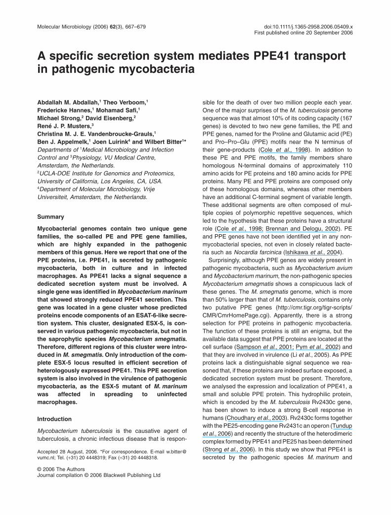
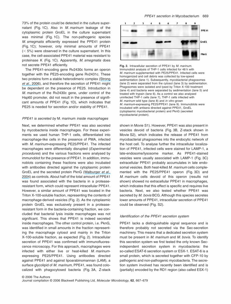
Next, we determined whether PPE41 was also secretedby mycobacteria inside macrophages. For these experi-ments we used human THP-1 cells, differentiated intomacrophage-like cells in the presence of PMA, infectedwith M. marinum-expressing PE25/PPE41. The infectedmacrophages were differentially disrupted (Experimentalprocedures) and the various fractions were analysed byimmunoblot for the presence of PPE41. In addition, immu-noblots containing these fractions were also incubatedwith antibodies directed against the cytoplasmic proteinGroEL and the secreted protein PknG (Walburger et al.,2004) as controls. About half of the total amount of PPE41was found associated with the bacteria in a protease-resistant form, which could represent intracellular PPE41.However, a similar amount of PPE41 was located in theTriton X-100-soluble fraction, representing the contents ofmacrophage-derived vesicles (Fig. 2). As the cytoplasmicprotein GroEL was exclusively present in a protease-resistant form in the bacteria-containing fraction, we con-cluded that bacterial lysis inside macrophages was notsignificant. This shows that PPE41 is indeed secretedinside macrophages. The other control protein, i.e. PknG,was identified in small amounts in the fraction represent-ing the macrophage cytosol and mainly in the TritonX-100-soluble fraction, as expected (Fig. 2). Intracellularsecretion of PPE41 was confirmed with immunofluores-cence microscopy. For this approach, macrophages wereinfected with either live or heat-killed M. marinum-expressing PE25/PPE41. Using antibodies directedagainst PPE41 and against lipoarabinomannan (LAM), asurface glycolipid of M. marinum, PPE41, was found colo-calized with phagocytosed bacteria (Fig. 3A, Z-stack
shown in Movie S1). However, PPE41 was also present invesicles devoid of bacteria (Fig. 3B, Z-stack shown inMovie S2), which indicates the release of PPE41 frommycobacterial phagosomes into the endocytic network ofthe host cell. To analyse further the intracellular localiza-tion of PPE41, infected cells were stained for LAMP-1, alate-endosome/lysosome marker. As PPE41-stainedvesicles were usually associated with LAMP-1 (Fig. 3C)extracellular PPE41 probably accumulates in late endo-somal vesicles. Both heat-killed M. marinum cells comple-mented with the PE25/PPE41 operon (Fig. 3D) andM. marinum cells devoid of this operon (results notshown) showed no extracellular PPE41 in macrophages,which indicates that this effect is specific and requires livebacteria. Next, we also tested whether PPE41 wassecreted by M. bovis BCG. Although this species secreteslower amounts of PPE41, intracellular secretion of PPE41could be observed (Fig. S2).
Identification of the PPE41 secretion system
PPE41 lacks a distinguishable signal sequence and istherefore probably not secreted via the Sec-secretionmachinery. This means that a dedicated secretion systemmust be present in M. marinum and M. bovis. To identifythis secretion system we first tested the only known Sec-independent secretion system in mycobacteria: theso-called ESAT-6 secretion system or ESX-1. ESAT-6 is asmall protein, which is secreted together with CFP-10 bypathogenic and non-pathogenic mycobacteria. The secre-tion system involved has recently been identified and is(partially) encoded by the RD1 region (also called ESX-1)
Fig. 2. Intracellular secretion of PPE41 by M. marinum.Immunoblot analysis of THP-1 cells infected for 48 h withM. marinum supplemented with PE25/PPE41. Infected cells werehomogenized and cell debris was collected by low-speedsedimentation (lane 1). Subsequently, mycobacterial phagosomes(lane 2) were separated from the cytosol (lane 3) by sedimentation.Phagosomes were isolated and lysed by Triton X-100 treatment(lane 4) and bacteria were separated by sedimentation (lane 5) andtreated with trypsin (lane 6). As a control we also analyseduninfected THP-1 cells (lane 7), THP-1 cells infected withM. marinum wild type (lane 8) and in vitro grownM. marinum-expressing PE25/PPE41 (lane 9). Immunoblots wereincubated with antisera directed against PPE41, GroEL(cytoplasmic mycobacterial protein) and PknG (secretedmycobacterial protein).
PPE41 secretion in Mycobacterium 669
© 2006 The AuthorsJournal compilation © 2006 Blackwell Publishing Ltd, Molecular Microbiology, 62, 667–679
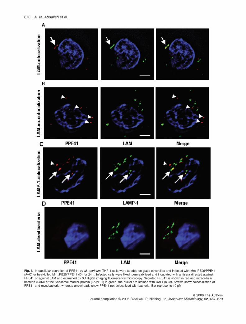
Fig. 3. Intracellular secretion of PPE41 by M. marinum. THP-1 cells were seeded on glass coverslips and infected with Mm::PE25/PPE41(A–C) or heat-killed Mm::PE25/PPE41 (D) for 24 h. Infected cells were fixed, permeabilized and incubated with antisera directed againstPPE41 or against LAM and examined by 3D digital imaging fluorescence microscopy. Secreted PPE41 is shown in red and intracellularbacteria (LAM) or the lysosomal marker protein (LAMP-1) in green, the nuclei are stained with DAPI (blue). Arrows show colocalization ofPPE41 and mycobacteria, whereas arrowheads show PPE41 not colocalized with bacteria. Bar represents 10 mM.
670 A. M. Abdallah et al.
© 2006 The AuthorsJournal compilation © 2006 Blackwell Publishing Ltd, Molecular Microbiology, 62, 667–679
(Hsu et al., 2003; Stanley et al., 2003; Gao et al., 2004;Guinn et al., 2004; Converse and Cox, 2005), which isdeleted in the vaccine strain M. bovis BCG (Mahairaset al., 1996). Although we already know that M. bovisBCG is able to secrete PPE41, we still wanted to comparethe level of PPE41 secretion in wild-type and mutantstrains. Therefore, two RD1 mutants of M. marinum wereanalysed for PPE41 secretion, i.e. M8, which is mutated inone of the components of ESX-1, and DCE, which con-tains a deletion for the cfp10/esat-6 operon (Gao et al.,2004). The M8 secreted normal amounts of PPE41 in theculture supernatant (Fig. 4A), which shows that ESX-1 isindeed not required for PPE41 secretion. However, sur-prisingly the mutant devoid of the ESX-1 substrates (DCE)showed significantly increased secretion of PPE41(Fig. 4A), indicating a link between the two secretionsystems. The intracellular levels of PPE41 were compa-rable for these strains (Fig. 4A).
A genetic screen was used to identify the PPE41secretion system in M. marinum. M. marinum strainM-expressing PE25/PPE41 was subjected to transposonmutagenesis, using the mycobacterial specific phagephiMycoMarT7 containing the mariner-like transposonHimar1 (Experimental procedures). Subsequently, themutants were grown on nitrocellulose filters andscreened for PPE41 secretion using a double-filtercolony blot approach (Experimental procedures,Fig. S3). Screening of 10 000 transposon mutantsresulted in the isolation of two mutants with stronglyreduced amounts of extracellular PPE41 (Fig. 4A). Thetransposon insertion sites of both mutants with reducedsecretion, i.e. mutant Mx2 and Mx12, were determinedby ligation-mediated PCR and sequence analysis. Com-parison of these sequences with the complete genomeof M. marinum M (http://www.sanger.ac.uk/Projects/M_marinum/) showed that both mutants contained the
mariner transposon at different positions in a singlecoding sequence (CDS). The gene product of this CDSshows high homology (93% identity) to Rv1798 of M. tu-berculosis (Fig. S4) and was therefore designatedMh1798. Complementation of Mx2 and Mx12 with thecorresponding gene on a mycobacterial shuttle plasmidresulted in restoration of PPE41 secretion (Fig. 4B).Mh1798 and Rv1798 belong to the AAA+ protein familyof ATPases. Members of this family are chaperones thatassist in the assembly and/or operation of protein com-plexes. Therefore, Mh1798 probably does not form thesecretion channel, but is part of a multiprotein secretionmachinery. Alternatively, as Mh1798 belongs to a familyof chaperones, the transposon insertion could affect thefolding and/or stability of the PPE41 protein. To discrimi-nate between these two possibilities we used the non-secreting species M. smegmatis.
Reconstitution of the PPE41 secretion system inM. smegmatis
The genomic organization of Rv1798 and Mh1798 ishighly conserved between M. tuberculosis andM. marinum (Fig. 5A). Furthermore, both regions containCDSs homologous to the M. tuberculosis ESX-1 cluster(Table S1). Four gene clusters homologous to the ESAT-6secretion system ESX-1 have been identified inM. tuberculosis, including the Rv1798-containing regionthat is called ESX-5 (Gey Van Pittius et al., 2001). Thiscould mean that ESX-5 encodes an ESAT-6-like secretionmachinery involved in PPE41 transport. Interestingly,ESX-5 is conserved in M. marinum, but is not present inthe non-secreting M. smegmatis (Gey Van Pittius et al.,2001). To determine whether cluster ESX-5 is directlyinvolved in PPE41 secretion, different regions (Fig. 5A) ofthe ESX-5 cluster of M. marinum were isolated by long-
Fig. 4. Isolation and characterization of theESX-5 secretion mutants.A. Equivalent OD units of cell pellets (P) andculture supernatant (S) of M. marinum wildtype (Mm wt), the ESX-1 mutants M8 andDCE and the isolated secretion mutants Mx2and 12 were separated on SDS-PAGE,immunoblotted and incubated with polyclonalantiserum against PPE41 or GroEL.B. Complementation of PPE41 secretionmutants. Equivalent amounts of culturesupernatant of M. marinum wild type, with orwithout PE25/PPE41, the secretion mutantsMx2 and 12, supplemented with PE25/PPE41and with the empty vector pSMT3 or withpSMT3 containing the Mh1798 gene, wereseparated on SDS-PAGE and analysed forthe presence of PPE41 by immunoblot.
A
B
PPE41 secretion in Mycobacterium 671
© 2006 The AuthorsJournal compilation © 2006 Blackwell Publishing Ltd, Molecular Microbiology, 62, 667–679
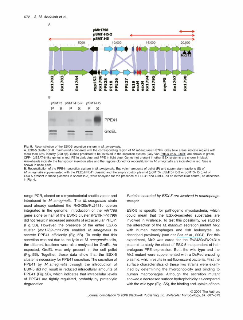
range PCR, cloned on a mycobacterial shuttle vector andintroduced in M. smegmatis. The M. smegmatis strainused already contained the Rv2430c/Rv2431c operonintegrated in the genome. Introduction of the mh1798gene alone or half of the ESX-5 cluster (PE19-mh1798)did not result in increased amounts of extracellular PPE41(Fig. 5B). However, the presence of the entire ESX-5cluster (mh1782–mh1798) enabled M. smegmatis tosecrete PPE41 efficiently (Fig. 5B). To verify that thissecretion was not due to the lysis of M. smegmatis cells,the different fractions were also analysed for GroEL. Asexpected, GroEL was only present in the cell pellet(Fig. 5B). Together, these data show that the ESX-5cluster is necessary for PPE41 secretion. The secretion ofPPE41 by M. smegmatis through the introduction ofESX-5 did not result in reduced intracellular amounts ofPPE41 (Fig. 5B), which indicates that intracellular levelsof PPE41 are tightly regulated, probably by proteolyticdegradation.
Proteins secreted by ESX-5 are involved in macrophageescape
ESX-5 is specific for pathogenic mycobacteria, whichcould mean that the ESX-5-secreted substrates areinvolved in virulence. To test this possibility, we studiedthe interaction of the M. marinum secretion mutant Mx2with human macrophages and fish leukocytes, asdescribed previously (van der Sar et al., 2004). For thisexperiment, Mx2 was cured for the Rv2430c/Rv2431cplasmid to study the effect of ESX-5 independent of het-erologous PPE expression. Both the wild type and theMx2 mutant were supplemented with a DsRed encodingplasmid, which results in red fluorescent bacteria. First thesurface characteristics of these two strains were exam-ined by determining the hydrophobicity and binding tohuman macrophages. Although the secretion mutantshowed a decreased surface hydrophobicity as comparedwith the wild type (Fig. S5), the binding and uptake of both
A
B
Fig. 5. Reconstitution of the ESX-5 secretion system in M. smegmatis.A. ESX-5 cluster of M. marinum M compared with the corresponding region of M. tuberculosis H37Rv. Grey blue areas indicate regions withmore than 65% identity (200 bp). Genes predicted to be involved in the secretion system (Gey Van Pittius et al., 2001) are shown in green,CFP-10/ESAT-6-like genes in red, PE in dark blue and PPE in light blue. Genes not present in other ESX systems are shown in black.Arrowheads indicate the transposon insertion sites and the regions cloned for reconstitution in M. smegmatis are indicated in red. Size isshown in base pairs.B. Reconstitution of the PPE41 secretion system in M. smegmatis. Equivalent amounts of pellet (P) and supernatant fractions (S) ofM. smegmatis supplemented with the PE25/PPE41 plasmid and the empty control plasmid (pSMT3), pSMT3-H5-2 or pSMT3-H5 (part ofESX-5 present in these plasmids is shown in A) were analysed for the presence of PPE41 and GroEL, as an intracellular control, as describedin Fig. 4.
672 A. M. Abdallah et al.
© 2006 The AuthorsJournal compilation © 2006 Blackwell Publishing Ltd, Molecular Microbiology, 62, 667–679

cells by macrophages were similar (results not shown).Subsequent infection experiments showed that ESX-5mutants survived and multiplied in both types ofleukocytes. However, a clear difference was observedwhen the percentage of infected cells was determined(Fig. 6A). With M. marinum wild type, the percentage ofinfected cells increased throughout the assay, reaching avalue of 53% by day 3. In contrast, the Mx2 mutantinfected only 17% of the host cells by day 3. This couldindicate that the secretion mutant was impaired in spread-ing to new cells. To examine this phenotype in more detail,also the number of bacteria per infected cell was deter-mined (Fig. 6B). Throughout the assay, most ofM. marinum wild-type infected cells contain less than 10bacteria, although the number of M. marinum wild-typeinfected cells increased (Fig. 6B). For the secretionmutant Mx2 the total number of infected cells remainedrelatively constant, whereas the percentage of cells con-taining more than 10 bacteria progressively increased(Fig. 6B). This result indicates that Mx2 mutant is capableof intracellular growth, but is impaired in macrophageescape. Together our data show that ESX-5 plays animportant role in the infection cycle of M. marinum, pos-sibly by facilitating host cell lysis.
Discussion
Most pathogenic bacteria are dependent on specializedsecretion systems for the export of crucial virulencefactors. Over the last decade a number of these secre-tion systems have been identified, ranging from the typeI to the type V secretion pathway. Until recently, patho-
genic mycobacteria were thought to be an exception tothe rule. M. tuberculosis contains, apart from the omni-present Sec-dependent transport system, no homologueof any known specialized secretion system and onlyrelatively small amounts of extracellular proteins couldbe identified as compared with other bacterial species.However, 3 years ago this idea changed when it wasshown that M. tuberculosis possesses a Sec-independent secretion system to transport two smallvirulence proteins, ESAT-6 and CFP-10 (Hsu et al.,2003; Stanley et al., 2003; Gao et al., 2004; Guinn et al.,2004; Converse and Cox, 2005). This secretion pathwaywas called the ESAT-6 secretion pathway or ESX-1 andrepresented a new class of secretion systems. Genomeanalysis showed that ESAT-6-like secretion systems arein fact also present in other Gram-positive bacteria (GeyVan Pittius et al., 2001; Pallen, 2002) and a homologueof this secretion pathway was found to be involved in thesecretion of two small proteins of Staphylococcusaureus and necessary for pathogenesis of this bacterium(Burts et al., 2005). M. tuberculosis itself contains thegenetic information for five of these ESAT-6-like secre-tion machineries (Gey Van Pittius et al., 2001), whichindicates that Sec-independent protein secretion is animportant feature of this pathogen. These ESAT-6-likegene clusters are conserved in the genomes of othermycobacteria, although not all species contain the fullextent of these gene clusters (Gey Van Pittius et al.,2001). In this study, we show that the M. marinum ortho-logue of cluster ESX-5 encodes a protein secretionsystem, which is specific for pathogenic mycobacteriaand is involved in the secretion of PPE41.
Fig. 6. Effect of ESX-5 on intracellular replication and spreading of M. marinum. THP-1 macrophage-like cells were seeded on glasscoverslips and infected with the wild type (closed bars) and Mx2 mutant (open bars) expressing DsRed. At each time point the percentage ofinfected cells (A) and the percentage of the infected cells with more than 10 bacteria (B) were determined. The results are the average ofthree separate experiments and the standard deviation is shown. Enumeration of bacilli within THP-1 cells was visualized by microscopy, witha minimal of 50 infected cells per time point.
PPE41 secretion in Mycobacterium 673
© 2006 The AuthorsJournal compilation © 2006 Blackwell Publishing Ltd, Molecular Microbiology, 62, 667–679
Introduction of the entire ESX-5 gene cluster inM. smegmatis resulted in the secretion of heterologouslyexpressed PPE41. Smaller genome fragments did notresult in the reconstitution of this ESAT-6-like secretionsystem, which shows that the predictions of Gey VanPittius et al. (2001), based on genome comparisons, werevery accurate. This ESX-5 cluster encodes several pro-teins that are likely to be directly associated with a secre-tion apparatus, such as a membrane protein with aputative ATP binding site (Mh1783/84) and various otherputative membrane proteins (Mh1782, Mh1794, Mh1795and Mh1797), but also proteins that are perhaps notinvolved, such as several PEs and PPEs, a putative cyto-chrome P450 and a mycobacteriophage protein (TableS1). Future experiments will have to show which of thesegenes are actually involved in PPE41 secretion. But, ifmost genes of the ESX-5 gene cluster are needed forsecretion, why are only transposon insertion mutants in asingle gene (Mh1798) isolated in the genetic screen inM. marinum? The results from two separate high-densitytransposon mutagenesis studies in M. tuberculosisprovide a possible explanation for this apparentdiscrepancy. In both these studies transposon insertionsin the Mh1798 homologue Rv1798 were isolated, butmutations in the other genes of ESX-5 encoding structuralcomponents of the secretion system, i.e. the homologuesof Rv1782–Rv1784 and Rv1794–Rv1797 (CDSs shownin green in Fig. 5A), were not detected (Lamichhane et al.,2003; Sassetti et al., 2003). This indicates that thesetransposon insertions are, in contrast to the ESX-1 secre-tion system, lethal. The Mh1798 mutants isolated in ourscreen showed only a strongly reduced secretion ofPPE41, and perhaps mutations in the other ESX-5 genescould block secretion completely. Interestingly, we iso-lated one mutant that showed no PPE41 secretion at allon colony blot. Unfortunately, this mutant grew very slowlyand could not be resuscitated after two passages and wastherefore not further analysed. Future experiments will bedirected to prove the essential nature of ESX-5.
The saprophytic species M. smegmatis does notcontain an ESX-5 cluster and because this bacteriumproduces a cell wall similar to that of M. tuberculosis andM. marinum, it is not likely that ESX-5-secreted substratesplay an important role in cell wall biosynthesis. Therefore,the lethality of ESX-5 mutations is probably due to theaccumulation of normally secreted proteins. As ESX-5secretes PPE41, the logical candidates for these toxicproteins are PPE proteins. M. smegmatis contains onlytwo genes encoding putative PPEs (http://cmr.tigr.org/tigr-scripts/CMR/CmrHomePage.cgi) and these are located inthe ESX-1 and ESX-3 region respectively (Gey Van Pittiuset al., 2001). Apart from ESX-4, all ESX regions have oneor more PPE genes. Comparison of the conservedN-terminal PPE motif of all PPE proteins of
M. tuberculosis shows that the majority of PPEs clustertogether with PPE41 and the PPEs of ESX-5. This clus-tering indicates that the expansion of PPEs is correlatedwith ESX-5 (N.C. Gey Van Pittius, in preparation). There-fore, we hypothesize that more PPEs are secreted via theESX-5 system. The PPEs encoded by ESX-1, -2 or -3 aremore distantly related and could have a different function.Thus far, the PPE protein of the ESX-1 region (PPE68)has been most extensively studied. This protein is notinvolved in the secretion of ESAT-6 and CFP-10 (Deman-gel et al., 2004). Overlay experiments showed that PPE68specifically interacts with both of these secreted proteins(Okkels and Andersen, 2004), which indicates that PPE68is secreted, together with ESAT-6/CFP-10, via the ESX-1secretion system. However, proteomic analysis of ESX-1secretion mutants failed to show secretion of PPE68,although the secretion of the ESX-1-encoded PE35 couldbe determined (Fortune et al., 2005). Another reportshowed that PPE68 is in fact located in the cell wall(Demangel et al., 2004). Therefore, the exact role and fateof PPEs in the various ESX systems need to be examinedin more detail.
An interesting observation was that the DCE mutantstrain in fact secreted significantly higher amounts ofPPE41, whereas a deletion of a gene encoding a struc-tural component of the ESX-1 system did not affect thelevel of PPE41 secretion. These data show that there issome level of cross-talk between the two secretionsystems ESX-1 and ESX-5. Deletion of the ESAT-6- andCFP-10-encoding genes could perhaps result in upregu-lation of the ESX-5 secretion system. Alternatively, theabsence of the ESX-1-secreted substrates ESAT-6 andCFP-10 could result in cross-secretion of ESX-5 sub-strates via ESX-1.
The ESX-5 secretion system seems to be specific forpathogenic mycobacteria and our data show that ESX-5plays an important role in the macrophage infection cycle.Interestingly, the observed effects are highly similar tothose described for ESX-1 mutations in M. tubercsulosis/M. bovis (Hsu et al., 2003; Stanley et al., 2003; Gao et al.,2004; Guinn et al., 2004). This probably means thatESAT-6 secretion itself is not sufficient to perturb themacrophage cell membrane and complete the macroph-age infection cycle. A recent study of Li et al. (2005)identified a PPE gene of M. avium that was associatedwith virulence and the ability to grow in macrophages.Interestingly, this PPE gene is highly homologous to theRv1787 gene of M. tuberculosis, which is located withinthe ESX-5 cluster and encodes PPE25. Therefore, thisprotein could very well be one of the virulence-associatedESX-5-secreted substrates needed for the macrophageinfection cycle. Future experiments will be directed tounravel the role of extracellular PPE proteins within thehost cell.
674 A. M. Abdallah et al.
© 2006 The AuthorsJournal compilation © 2006 Blackwell Publishing Ltd, Molecular Microbiology, 62, 667–679
Experimental procedures
Bacterial strains and growth conditions
Three different mycobacterial species were used in thisstudy: M. marinum strain M (Ramakrishnan and Falkow,1994), M. smegmatis strain mc2155 (Snapper et al., 1990)and M. bovis BCG Copenhagen. Mycobacteria were grown inshaking cultures in (i) Middlebrook 7H9 liquid medium,supplemented either with Middlebrook ADC (BD, Bio-sciences) and 0.2% Tween, or (ii) modified Sauton’s medium,enriched with 0.5% sodium pyruvate and 0.5% glucose. Forsecretion in M. bovis BCG and M. marinum 7H9 medium wasused, supplemented with 0.2% (w/v) dextrose, 0.2% Tweenand 0.1% or 0.01% of the advised amount of ADC supple-ment respectively. The presence of BSA in the medium (partof the ADC supplement) is essential for secretion in these twospecies. For secretion in M. smegmatis we used the pre-culture method described by Converse and Cox (2005). As asolid medium Middlebrook 7H10 plates supplemented withOADC (BD, Biosciences) were used. Both M. smegmatis andBCG were grown at 37°C, whereas M. marinum was grown at30°C. If M. marinum cells were cultured for electroporationexperiments, 2.5 mg ml-1 glycine was added to the mediaonce the culture reached an optical density at 600 nm (OD600)of 0.5, to increase the electroporation efficiency (D. Lakey,pers. comm.). For cloning experiments, Escherichia colistrain DH5a was used. Hygromycin, chloramphenicol andkanamycin were used at final concentrations of 50 mg ml-1,30 mg ml-1 and 25 mg ml-1, respectively, both for E. coli andfor mycobacteria, and gentamicin was used at a final concen-tration of 5 mg ml-1 for M. smegmatis and 10 mg ml-1 forE. coli.
Expression of Rv2430c/Rv2431c in M. smegmatis andM. marinum
H37Rv genomic DNA was used as a template to amplify theRv2431c/Rv2430c genes with Expand polymerase (Boe-hringer) using the primers Rv2430cR (GACACGAAATCCGCAGGTAT) and Rv2431cF (CTCATCTGTCACGAGCCGTA).The resulting PCR product was cloned in pUC18 digestedwith SmaI, which resulted in pUC18-30c/31cL and pUC18-30c/31cR. Subsequently, the operon was cloned in pSMT3-eGFP (Hayward et al., 1999) and placed under the control ofthe Hsp60 promoter, resulting in pSMT30/31c. The PPE41-encoding gene Rv2430c was also inserted without theRv2431c gene in pSMT3eGFP by digesting the pUC18-30c/31cL plasmid with HpaI, which overlaps the stop codon ofRv2431c and HindIII. As a second construction vector pBH10was used, which is compatible with pAL5000 derivatives suchas pSMT3. pBH10 is a derivate of pBP10 (Bachrach et al.,2000) that contains the Hsp60 promoter of pSMT3-eGFPby cloning a XbaI and BamHI fragment of pSMT3 in SpeIand BamHI-digested pBP10. Subsequently, the putativeRv2431c/30c operon was cloned in pBH10 using BamHI/PstI,resulting in plasmid pBH30c/31c. A chloramphenicol-resistantversion of this plasmid, designated pBH30/31cat, was pro-duced using pEMCat. pEMcat contains the chloramphenicolacetyl transferase-encoding gene (cat) of pACYC184, clonedas a Sau3A fragment in BamHI-digested pEM37 (Pashley
and Parish, 2003). This places the cat gene under control ofthe mycobacterial Ag85 promoter. pBH30/31cat was createdby cloning the cat-containing Ecl136II–EcoRV fragment ofpEMCat in pBH30/31c restricted with EcoRV. For reconstitu-tion of the secretion system in M. smegmatis an integrationconstruct expressing Rv2430c/Rv2431c was used. This con-struct was created by isolating the integration region ofplasmid pUC-Gm-Int plasmid (Lee et al., 1991) as a HindIIIfragment and clone it into pBH30c/31c, thereby deletingthe OriM. The resulting plasmid is designated pBH30c/31c-Int. Gentamicin-resistant colonies of M. smegmatiswere analysed for correct genome insertion by PCR, usingprimers specific for the Rv2431c/Rv2430c operon andprimers surrounding the integration site, i.e. wbINTgen(CTACCAAGCTGCGCTACACC) and wbINTpls (TCGTTTGTCAGCATCGAAAG).
Polyclonal antiserum directed against Rv2430c
Rv2431c and Rv2430c were expressed together in E. coliwith an optimized translation initiation site and a His-tag atthe C-terminal end of Rv2430c (Strong et al., 2006). Rv2430cwas purified from disrupted E. coli cells and the Rv2431cprotein co-purified. This purified material was used to raisepolyclonal antibodies. Two rabbits were immunized subcuta-neously with 0.4 ml of Rv2430c/Rv2431c preparation (con-taining 100 mg of protein) 4:5 diluted in the mineral oil-basedadjuvant Stimune (Cedi Diagnostics BV, Lelystad NL) andsubcutaneously booster-immunized after 4 weeks with 0.2 mlof plain antigen. The resulting antisera were analysed foractivity against the purified proteins, which showed only aresponse against PPE41.
SDS-PAGE and immunoblot
Mycobacteria were grown to mid-logarithmic phase. Subse-quently, secreted proteins were precipitated from cell-freesupernatant with 5% TCA (w/v). The cell pellets were resus-pended in PBS and disrupted with a mini-BeadBeater(BioSpec Products) using 0.1 mm zirconia/silica beads. Celllysates and supernatants were separated by SDS/PAGE on12% polyacrylamide gels. For the treatment of intact cellswith protease bacteria were grown to mid-log phase andisolated by centrifugation. Cells were resuspended in waterand left untreated or incubated with 0.1 mg ml-1 proteinase K(Qiagen). After a 30 min incubation at room temperature,sample buffer was added to all samples, samples were boiledand separated on 12% polyacrylamide gels. Proteins werevisualized by immunoblotting using antibodies directedagainst PPE41 (see above), antibodies against PknG (kindlyprovided by Y. Avenue-Gay, Vancouver, Canada), and anti-bodies against GroEL and GroES. The GroEL monoclonalantibody used is CS44, obtained from J. Belisle (ColoradoState University, Fort Collins, CO, and the National Institutesof Health, Bethesda, MD, contract NO1 AI-75320). The pres-ence of the second antibody, GARpo or GAMpo, was visual-ized using 4-chloronaphtol/3,3-diaminobenzidine staining orusing ECL detection (PIERCE). For protein quantificationnitrocellulose membranes were probed with a-PPE41 polyclonal antibody and detected by Lumi-Light Western Blotting
PPE41 secretion in Mycobacterium 675
© 2006 The AuthorsJournal compilation © 2006 Blackwell Publishing Ltd, Molecular Microbiology, 62, 667–679

Substrate (Roche Applied Science). Quantification was per-formed on a Fluor-S MultiImager (Bio-Rad) using Bio-Radmultianalyst software, version 1.0.2.
Transposon mutagenesis
Transposon mutagenesis was performed using the mycobac-terial specific phage phiMycoMarT7 containing the mariner-like transposon Himar1 (Sassetti et al., 2003). Transductantsof M. marinum strain M supplemented with the Rv2430c con-taining vector pSMT3-30/31c were plated on a nitrocellulosefilter, which was placed on 7H10 agar plates supplementedwith kanamycin (to select for the presence of the transposon)and hygromycin (pSMT3-30/31c). After visible colonies hadappeared, the filter was removed and placed on a new filter,on top of another 7H10 plate. This sandwich was incubatedfor 5–6 h at 30°C and subsequently the top filter with thecolonies was preserved at 4°C, whereas the bottom filter withthe secreted proteins was incubated with antibodies directedagainst PPE41. Colonies of interest, i.e. those that showedreduced mounts of PPE41, were first checked on a similarcolony blot assay and subsequently checked in culture forPPE41 secretion. To establish the chromosomal location ofthe transposon insertion, ligation-mediated PCR was usedessentially as described by Prod’hom et al. (1998), with thefollowing modifications. Chromosomal DNA (50 ng), isolatedfrom plate-grown mutants with the DNA tissue kit (Qiagen),was digested with BamHI and BglII in stead of SalI. Thesetwo restriction enzymes together have approximately thesame digestion frequency in mycobacterial DNA as SalI, butin addition are functional in a buffer also suited for ligation. Inthe same incubation reaction (overnight at room temperature)as the digestion, an adaptor, consisting of the SalgD primertogether with the Bampt primer (GATCGCTCGTGCC), isligated to the digested DNA. This adaptor does not restorethe BamHI/BglII restriction sites. Subsequently, this ligationmixture is used as a template in a PCR reaction with thepSalg primer (GCTTATTCCTCAAGGCACGA), which is iden-tical to the single-stranded part of the adaptor, and the pMycoprimer (CCGGGGACTTATCAGCCAAC), which binds to bothsides of the Himar1 transposon. PCR fragments weresequenced using an ABIprism300 (Applied biosystems) andanalysed using the sequence information of the M. marinumSequencing Group at the Sanger Institute (http://www.sanger.ac.uk/Projects/M_marinum/). In both mutants theMycoMar transposon inserted into the dinucleotide TA, ascan be expected for this mariner-based transposon.
Infection of leukocytes
For intracellular experiments the human acute monocytic leu-kaemia cell line THP-1 and the carp leukocyte cell line CLCwere used as described previously (van der Sar et al., 2004).THP-1 cells were cultured in RPMI-1640 with glutamax-1media (Gibco, BRL) supplemented with 10% fetal calf serum(FCS), and differentiated into macrophage-like cells in thepresence of PMA (phorbol 12-myristate 13-acetate, 1 ng ml-1,Sigma). For phagosome isolation, cells were seeded in75 cm diameter flasks whereas 24-well plates (Nunc) wereused for survival assays and microscopy. For the infection,
the mycobacteria were grown to logarithmic phase(OD600 = 0.5–0.8) in 7H9 media, washed and diluted in RPMI-1640 with 10% FCS and used at a multiplicity of infection(moi) of 10 bacteria per cell. After 1 h of uptake at 33°C,infected cells were washed three times with PBS to removeextracellular bacteria and incubated in fresh medium plusamikacin (200 mg ml-1; Sigma Chemicals, St Louis, MO) for2 h. The cells were then washed once with PBS and incu-bated in fresh medium plus amikacin (30 mg ml-1) for indi-cated time periods. Mycobacterial survival was analysed bylysing the infected macrophages in 0.1% Triton X-100 andplating serial dilutions on 7H10 agar (Difco) supplementedwith 10% OADC. For microscopic examination, cells weredifferentiated with coverslips in wells and infection was per-formed with a pre-treatment of amikacin as described, butincubated in the absence of amikacin.
Mycobacterial phagosomes isolation
Isolation of mycobacterial phagosomes was performed asdescribed previously (Schuller et al., 2001). Briefly, infectedTHP-1 cells (7–10 ¥ 107 with moi of 25 for 48 h) were homog-enized in DGE buffer (10 mM triethanolamine, 10 mM aceticacid, 1 mM EDTA, 0.25 M sucrose, pH 7.4) by passagingthrough a 23-gauge needle. After removal of the cell nuclei bylow-speed centrifugation (250 g, 10 min), the resulting post-nuclear supernatant (PNS) was transferred to a fresh Eppen-dorf tube and sedimented at 35 000 g for 30 min. Theresulting supernatant corresponds to the macrophage cytosolwhereas the pellet corresponds to the mycobacterialphagosomes. The bacteria-free phagosomes were recov-ered, by treating them with 1% Triton X-100 for 15 min atroom temperature and sediment the intact mycobacteria at35 000 g for 30 min. The resulting supernatant correspondsto the phagosomes and the pellet to the mycobacteria.
Digital imaging fluorescence microscopy
THP-1 cells (7.5 ¥ 105 per well) were seeded on glass cov-erslips in 24-well plates in the presence of PMA and infectedwith different strains of mycobacteria as described in theinfection procedures for 24 h. Cells were fixed with 4%paraformaldehyde in PBS for 15 min at room temperature,permeabilized with 0.1% Triton X-100 in PBS for 15 min, andblocked in 2% BSA (Sigma) in PBS for 30 min. This blockingsolution was also used for dilution of antibodies. The slideswere incubated with antibodies directed against PPE41,monoclonal antibody F30-5 (Kolk et al., 1984) directedagainst mycobacterial lipoarabinomannan (LAM), obtainedfrom Arend Kolk (KIT, Amsterdam), or against LAMP-1(H4A3, obtained from the Hybridoma Bank of the Universityof Iowa, Iowa City). After overnight incubation with primaryantibodies at 4°C, coverslips were washed with PBST andincubated with Alexa fluor 488 or 546-conjugated secondaryantibodies (Molecular Probes). Coverslips were mounted inVectashield (Vector Laboratories) and analysed using aZEISS Axiovert 200 MarianasTM digital imaging microscopyworkstation as described previously (van der Sar et al.,2003). The microscope, camera and data viewing/processingwere conducted/controlled by SlidebookTM software [Slide-
676 A. M. Abdallah et al.
© 2006 The AuthorsJournal compilation © 2006 Blackwell Publishing Ltd, Molecular Microbiology, 62, 667–679
book version 4.1.0.4 (Intelligent Imaging Innovations, Denver,CO)]. The data acquisition protocol included confocal opticalplanes to obtain three-dimensional (3D) definition.
Cloning and expression of the M. marinum ESX-5cluster
Mycobacterium marinum genomic DNA was used as a tem-plate to amplify the ESX-5 region in two fragments with Exten-sor Hi-Fidelity PCR Master Mix (ABgene) using the primersAbMmfront (CAAAGTGTCCTGAGAGACGGGTA) andAbMmMiddle-H5-R (ACCTAGCTTCCCACTCAGCAAAC) forthe first part and AbMmMiddle-H5-F (AAGGGAGCACCGAAAATGTTAAA) and AbMmEnd (CGAGTTGATCTCAATCCATCCAC) for the second part. The resulting PCRproducts were cloned in pCRII-TOPO (invitrogen). To transferthese constructs into M. smegmatis mc2155, the pAL5000replicon and the hygromycin resistance gene was cloned frompSMT3-eGFP using the restriction enzymes DraI/EcoRV forpSMT3-eGFP and DraI for the TOPO-PCR constructs, result-ing in pSMT-H5-1 and pSMT-H5-2 respectively. Subsequently,a complete ESX-5 region was constructed by digesting bothplasmids with the restriction enzymes DraI/MluI andre-ligation. The resulting plasmid was designated pSMT-H5.Also the mh1798 gene alone was amplified by PCR, usingchromosomal DNA of M. marinum M as a template and theprimers 1798R (TGTTAACTGATGCCAATTCCGATTTC) and1798F (CGTTAACTCGATGAGGTCTGGCTCTC). This frag-ment was cloned in pCRII-Topo (Invitrogen) and checked bysequence analysis. Subsequently, mh1798 was cloned down-stream of the mycobacterial hsp60 promoter and the geneencoding DsRed, by ligating the HindIII/NotI fragmentof pTOPO-Mh1798 with the NotI/BamHI fragment ofpSMT3DsRed and the BamHI/HindIII vector fragment ofpSMT3eGFP. This tripartite ligation was checked by restrictiondigestion and designated pMh1798.
Hydrophobicity
Cell surface hydrophobicity was assessed in three separateexperiments as described previously (Rosenberg et al.,1980). Briefly, M. marinum wild-type and Mx2 strains weregrown on 7H9 broth for 1 week. Cells were harvested bycentrifugation at 4°C and 2000 g and washed twice with PBS.Bacterial suspensions were prepared in PBS to an OD600 of1.0. Duplicate (3 ml) samples of each suspension wereplaced in test tubes. Xylene was added to each suspension atconcentrations of 0%, 0.5%, 1% and 2% (v/v). The tubecontents were mixed for 45 s with a vortex mixer at themaximum setting. The aqueous and organic phases wereallowed to separate for 30 min, after which the aqueousphase was carefully removed with a Pasteur pipette andtransferred to cuvettes. Contaminating xylene was allowed toevaporate for 60 min. After this, each cuvette was vortexedfor 1 s to resuspend the remaining bacteria. The level ofabsorption of the cells to the xylene droplets was calculatedas the loss in OD600 of the aqueous phase compared with theinitial cell suspension, expressed as a percentage. Similarresults were obtained if hexadecane (0%, 5%, 10% and 15%)was used instead of xylene.
Acknowledgements
The authors thank Jonathan Kadouch, Widad Rifi andCorrine Ten Hagen-Jongman for technical assistance, Astridvan der Sar and Marian Llamas for helpful discussions,Yossef Av-Gay, Arend Kolk, Eric Rubin, Janisha Patel, NeilStoker, Papinavinasasundaram and Eric Brown for strains,plasmids, phages and antibodies.
References
Bachrach, G., Colston, M.J., Bercovier, H., Bar-Nir, D.,Anderson, C., and Papavinasasundaram, K.G. (2000) Anew single-copy mycobacterial plasmid, pMF1, from Myco-bacterium fortuitum which is compatible with the pAL5000replicon. Microbiology 146: 297–303.
Brennan, M.J., and Delogu, G. (2002) The PE multigenefamily: a ‘molecular mantra’ for mycobacteria. TrendsMicrobiol 10: 246–249.
Burts, M.L., Williams, W.A., DeBord, K., and Missiakas, D.M.(2005) EsxA and EsxB are secreted by an ESAT-6-likesystem that is required for the pathogenesis of Staphylo-coccus aureus infections. Proc Natl Acad Sci USA 102:1169–1174.
Choudhary, R.K., Mukhopadhyay, S., Chakhaiyar, P.,Sharma, N., Murthy, K.J., Katoch, V.M., and Hasnain, S.E.(2003) PPE antigen Rv2430c of Mycobacterium tuberculo-sis induces a strong B-cell response. Infect Immun 71:6338–6343.
Cole, S.T., Brosch, R., Parkhill, J., Garnier, T., Churcher, C.,Harris, D., et al. (1998) Deciphering the biology of Myco-bacterium tuberculosis from the complete genomesequence. Nature 393: 537–544.
Converse, S.E., and Cox, J.S. (2005) A protein secretionpathway critical for Mycobacterium tuberculosis virulenceis conserved and functional in Mycobacterium smegmatis.J Bacteriol 187: 1238–1245.
Demangel, C., Brodin, P., Cockle, P.J., Brosch, R., Majlessi,L., Leclerc, C., and Cole, S.T. (2004) Cell envelope proteinPPE68 contributes to Mycobacterium tuberculosis RD1immunogenicity independently of a 10-kilodalton culturefiltrate protein and ESAT-6. Infect Immun 72: 2170–2176.
Fortune, S.M., Jaeger, A., Sarracino, D.A., Chase, M.R.,Sassetti, C.M., Sherman, D.R., et al. (2005) Mutuallydependent secretion of proteins required for mycobacterialvirulence. Proc Natl Acad Sci USA 102: 10676–10681.
Gao, L.Y., Guo, S., McLaughlin, B., Morisaki, H., Engel, J.N.,and Brown, E.J. (2004) A mycobacterial virulence genecluster extending RD1 is required for cytolysis, bacterialspreading and ESAT-6 secretion. Mol Microbiol 53: 1677–1693.
Gey Van Pittius, N.C., Gamieldien, J., Hide, W., Brown,G.D., Siezen, R.J., and Beyers, A.D. (2001) The ESAT-6gene cluster of Mycobacterium tuberculosis and otherhigh G+C Gram-positive bacteria. Genome Biol 2:RESEARCH0044.
Guinn, K.M., Hickey, M.J., Mathur, S.K., Zakel, K.L., Grotzke,J.E., Lewinsohn, D.M., et al. (2004) Individual RD1-regiongenes are required for export of ESAT-6/CFP-10 and forvirulence of Mycobacterium tuberculosis. Mol Microbiol 51:359–370.
PPE41 secretion in Mycobacterium 677
© 2006 The AuthorsJournal compilation © 2006 Blackwell Publishing Ltd, Molecular Microbiology, 62, 667–679

Hayward, C.M., O’Gaora, P., Young, D.B., Griffin, G.E.,Thole, J., Hirst, T.R., et al. (1999) Construction and murineimmunogenicity of recombinant Bacille Calmette Guerinvaccines expressing the B subunit of Escherichia coli heatlabile enterotoxin. Vaccine 17: 1272–1281.
Hsu, T., Hingley-Wilson, S.M., Chen, B., Chen, M., Dai, A.Z.,Morin, P.M., et al. (2003) The primary mechanism ofattenuation of bacillus Calmette-Guerin is a loss ofsecreted lytic function required for invasion of lung intersti-tial tissue. Proc Natl Acad Sci USA 100: 12420–12425.
Ishikawa, J., Yamashita, A., Mikami, Y., Hoshino, Y., Kurita,H., Hotta, K., et al. (2004) The complete genomicsequence of Nocardia farcinica IFM 10152. Proc Natl AcadSci USA 101: 14925–14930.
Kolk, A.H., Ho, M.L., Klatser, P.R., Eggelte, T.A., Kuijper, S.,de Jonge, S., and van Leeuwen, J. (1984) Production andcharacterization of monoclonal antibodies to Mycobacte-rium tuberculosis, M. bovis (BCG) and M. leprae. Clin ExpImmunol 58: 511–521.
Lamichhane, G., Zignol, M., Blades, N.J., Geiman, D.E.,Dougherty, A., Grosset, J., et al. (2003) A postgenomicmethod for predicting essential genes at subsaturationlevels of mutagenesis: application to Mycobacteriumtuberculosis. Proc Natl Acad Sci USA 100: 7213–7218.
Lee, M.H., Pascopella, L., Jacobs, W.R., and Hatfull, G.F.(1991) Site-specific integration of Mycobacteriophage-L5 –integration-proficient vectors for Mycobacterium smegma-tis, Mycobacterium tuberculosis, and Bacille Calmette-Guerin. Proc Natl Acad Sci USA 88: 3111–3115.
Li, Y., Miltner, E., Wu, M., Petrofsky, M., and Bermudez, L.E.(2005) A Mycobacterium avium PPE gene is associatedwith the ability of the bacterium to grow in macrophagesand virulence in mice. Cell Microbiol 7: 539–548.
Mahairas, G.G., Sabo, P.J., Hickey, M.J., Singh, D.C., andStover, C.K. (1996) Molecular analysis of genetic differ-ences between Mycobacterium bovis BCG and virulentM. bovis. J Bacteriol 178: 1274–1282.
Okkels, L.M., and Andersen, P. (2004) Protein–protein inter-actions of proteins from the ESAT-6 family of Mycobacte-rium tuberculosis. J Bacteriol 186: 2487–2491.
Pallen, M.J. (2002) The ESAT-6/WXG100 superfamily – anda new Gram-positive secretion system? Trends Microbiol10: 209–212.
Pashley, C.A., and Parish, T. (2003) Efficient switching ofmycobacteriophage L5-based integrating plasmids inMycobacterium tuberculosis. FEMS Microbiol Lett 229:211–215.
Prod’hom, G., Lagier, B., Pelicic, V., Hance, A.J., Gicquel, B.,and Guilhot, C. (1998) A reliable amplification technique forthe characterization of genomic DNA sequences flankinginsertion sequences. FEMS Microbiol Lett 158: 75–81.
Pym, A.S., Brodin, P., Brosch, R., Huerre, M., and Cole, S.T.(2002) Loss of RD1 contributed to the attenuation of thelive tuberculosis vaccines Mycobacterium bovis BCG andMycobacterium microti. Mol Microbiol 4: 709–717.
Ramakrishnan, L., and Falkow, S. (1994) Mycobacteriummarinum persists in cultured mammalian cells in atemperature-restricted fashion. Infect Immun 62: 3222–3229.
Rosenberg, M., Gutnick, D., and Rosenberg, E. (1980)Adherence of bacteria to hydrocarbons – a simple method
for measuring cell-surface hydrophobicity. FEMS MicrobiolLett 9: 29–33.
Sampson, S.L., Lukey, P., Warren, R.M., van Helden, P.D.,Richardson, M., and Everett, M.J. (2001) Expression, char-acterization and subcellular localization of the Mycobacte-rium tuberculosis PPE gene Rv1917c. Tuberculosis(Edinb) 81: 305–317.
van der Sar, A.M., Musters, R.J., van Eeden, F.J., Appelmelk,B.J., Vandenbroucke-Grauls, C.M., and Bitter, W. (2003)Zebrafish embryos as a model host for the real time analy-sis of Salmonella typhimurium infections. Cell Microbiol 5:601–611.
van der Sar, A.M., Abdallah, A.M., Sparrius, M., Reinders, E.,Vandenbroucke-Grauls, C.M., and Bitter, W. (2004) Myco-bacterium marinum strains can be divided into two distincttypes based on genetic diversity and virulence. InfectImmun 72: 6306–6312.
Sassetti, C.M., Boyd, D.H., and Rubin, E.J. (2003) Genesrequired for mycobacterial growth defined by high densitymutagenesis. Mol Microbiol 48: 77–84.
Schuller, S., Neefjes, J., Ottenhoff, T., Thole, J., and Young,D. (2001) Coronin is involved in uptake of Mycobacteriumbovis BCG in human macrophages but not in phagosomemaintenance. Cell Microbiol 3: 785–793.
Snapper, S.B., Melton, R.E., Mustafa, S., Kieser, T., andJacobs, W.R., Jr (1990) Isolation and characterization ofefficient plasmid transformation mutants of Mycobacteriumsmegmatis. Mol Microbiol 4: 1911–1919.
Stanley, S.A., Raghavan, S., Hwang, W.W., and Cox, J.S.(2003) Acute infection and macrophage subversion byMycobacterium tuberculosis require a specialized secre-tion system. Proc Natl Acad Sci USA 100: 13001–13006.
Strong, M., Sawaya, M.R., Wang, S., Phillips, M., Cascio, D.,and Eisenberg, D. (2006) Toward the structural genomicsof complexes: crystal structure of a PE/PPE proteincomplex from Mycobacterium tuberculosis. Proc Natl AcadSci USA 103: 8060–8065.
Tundup, S., Akhter, Y., Thiagarajan, D., and Hasnain, S.E.(2006) Clusters of PE and PPE genes of Mycobacteriumtuberculosis are organized in operons: evidence that PERv2431c is co-transcribed with PPE Rv2430c and theirgene products interact with each other. FEBS Lett 580:1285–1293.
Walburger, A., Koul, A., Ferrari, G., Nguyen, L., Prescianotto-Baschong, C., Huygen, K., et al. (2004) Protein kinase Gfrom pathogenic mycobacteria promotes survival withinmacrophages. Science 304: 1800–1804.
Supplementary material
The following supplementary material is available for thisarticle online:Fig. S1. Immunoblot showing that GroEL proteins ofM. bovis BCG (BCG), M. smegmatis (Ms) and M. marinum(Mm) are sensitive to proteinase K (pK) if lysed cells aresubjected to the protease.Fig. S2. Intracellular secretion of PPE41 by M. marinummutant Mx2 supplemented with PE25/PPE41 and Mycobac-terium bovis BCG. THP-1 cells were seeded on glass cover-slips and infected with M. marinum Mx2 (A) or M. bovis BCG
678 A. M. Abdallah et al.
© 2006 The AuthorsJournal compilation © 2006 Blackwell Publishing Ltd, Molecular Microbiology, 62, 667–679

(B) for 24 h. Infected cells were fixed, permeabilized andincubated with antisera directed against PPE41 and LAM,and examined by 3D digital imaging fluorescencemicroscopy. Secreted PPE41 is shown in red and intracellularbacteria (LAM) in green; the nuclei are stained with DAPI(blue). Arrows indicate colocalization, whereas arrowheadsindicate no colocalization. Bar represents 10 mM.Fig. S3. Quality of the two-filter colony blot screening proce-dure used in our experiments to isolate secretion mutants ofM. marinum. The colony-containing filter (shown on the left) isplaced for 5 h on a second filter after 9–10 days of incubation(time needed to grow the colonies). The second filter (shownon the right) is treated with antiserum directed against PPE41and stained for the presence of peroxidase. Non-secretingcolony is indicated.Fig. S4. Alignment of Mh1798 and Rv1798. Amino acidscorresponding with the Tn insertion sites of Mx2 and 12 areindicated with arrows, identical residues are shown in greyand amino acid numbers are indicated.Fig. S5. Cell surface hydrophobicity of M. marinum wild type(�) and the Mx2 mutant (�).
Movie S1. Z-stack of macrophage infected with M. marinumsupplemented with PE25/PPE41. Colocalization of PPE41and M. marinum (identical cell shown in Fig. 2B). Infectedcells were fixed, permeabilized and incubated with antiseradirected against PPE41 (red) and against LAM (green) andexamined by 3D digital imaging fluorescence microscopy.Nuclei are stained with DAPI (blue).Movie S2. Z-stack of macrophage infected with M. marinumsupplemented with PE25/PPE41. No colocalization of PPE41and M. marinum (identical cell shown in Fig. 2C). Infectedcells were fixed, permeabilized and incubated with antiseradirected against PPE41 (red) and against LAM (green) andexamined by 3D digital imaging fluorescence microscopy.Nuclei are stained with DAPI (blue).Table S1. CDSs of the ESX-5 region of M. marinum M, theirhomologues in the EXS-5 and EXS-1 region ofM. tuberculosis H37Rv and the proposed function of theirgene products, localization or protein family they belong to.
This material is available as part of the online article fromhttp://www.blackwell-synergy.com
PPE41 secretion in Mycobacterium 679
© 2006 The AuthorsJournal compilation © 2006 Blackwell Publishing Ltd, Molecular Microbiology, 62, 667–679