The descending corticocollicular pathway mediates learning-induced auditory plasticity
22
The descending corticocollicular pathway mediates learning- induced auditory plasticity Victoria M Bajo 1 , Fernando R Nodal 1 , David R Moore 1,2 , and Andrew J King 1 1 Department of Physiology, Anatomy and Genetics, University of Oxford, Oxford, UK Abstract Descending projections from sensory areas of the cerebral cortex are among the largest pathways in the brain, suggesting that they are important for subcortical processing. Although corticofugal inputs have been shown to modulate neuronal responses in the thalamus and midbrain, the behavioral importance of these changes remains unknown. In the auditory system, one of the major descending pathways is from cortical layer V pyramidal cells to the inferior colliculus in the midbrain. We examined the role of these neurons in experience-dependent recalibration of sound localization in adult ferrets by selectively killing the neurons using chromophore-targeted laser photolysis. When provided with appropriate training, animals normally relearn to localize sound accurately after altering the spatial cues available by reversibly occluding one ear. However, this ability was lost after eliminating corticocollicular neurons, whereas normal sound-localization accuracy was unaffected. The integrity of this descending pathway is therefore critical for learning-induced localization plasticity. Keywords Auditory cortex; corticofugal projection; inferior colliculus; chromophore-targeted neuronal degeneration; apoptosis; adult plasticity; sound localization; binaural cues; ferret Introduction Plasticity of cortical processing is important for enabling humans and other species to interact effectively with their constantly changing environments and is believed to provide the basis by which learning can improve perceptual abilities 1, 2 . However, there is growing evidence, particularly for the auditory system, that learning is also associated with plasticity at lower levels of the pathway 3, 4, 5 . Electrical stimulation and inactivation studies have shown that cortical feedback can alter the representation of sensory information at almost every stage of subcortical processing 5, 6, 7, 8, 9, 10, 11 . Although these findings suggest that corticofugal modulation could contribute to learning-induced plasticity and subsequent changes in behavior, there is currently no direct evidence for this and the role of descending cortical projections in sensory processing remains unclear. The most widely studied descending sensory pathway is the projection from the auditory cortex to the inferior colliculus in the midbrain 12 . The sensitivity of inferior colliculus Corresponding author, Correspondence to: Andrew J King. 2 Present address: MRC Institute of Hearing Research, Nottingham, UK. Contributions This study was conceived by V.M.B., A.J.K. and D.R.M. and designed by V.M.B. and A.J.K. The behavioral experiments were performed by V.M.B., F.R.N. and A.J.K. The anatomical studies were carried out by V.M.B., who jointly analyzed all of the data with F.R.N. A.J.K., V.M.B. and F.R.N. wrote the paper with assistance from D.R.M. Europe PMC Funders Group Author Manuscript Nat Neurosci. Author manuscript; available in PMC 2013 April 24. Published in final edited form as: Nat Neurosci. 2010 February ; 13(2): 253–260. doi:10.1038/nn.2466. Europe PMC Funders Author Manuscripts Europe PMC Funders Author Manuscripts
Transcript of The descending corticocollicular pathway mediates learning-induced auditory plasticity
The descending corticocollicular pathway mediates learning-induced auditory plasticity
Victoria M Bajo1, Fernando R Nodal1, David R Moore1,2, and Andrew J King1
1Department of Physiology, Anatomy and Genetics, University of Oxford, Oxford, UK
AbstractDescending projections from sensory areas of the cerebral cortex are among the largest pathwaysin the brain, suggesting that they are important for subcortical processing. Although corticofugalinputs have been shown to modulate neuronal responses in the thalamus and midbrain, thebehavioral importance of these changes remains unknown. In the auditory system, one of themajor descending pathways is from cortical layer V pyramidal cells to the inferior colliculus in themidbrain. We examined the role of these neurons in experience-dependent recalibration of soundlocalization in adult ferrets by selectively killing the neurons using chromophore-targeted laserphotolysis. When provided with appropriate training, animals normally relearn to localize soundaccurately after altering the spatial cues available by reversibly occluding one ear. However, thisability was lost after eliminating corticocollicular neurons, whereas normal sound-localizationaccuracy was unaffected. The integrity of this descending pathway is therefore critical forlearning-induced localization plasticity.
KeywordsAuditory cortex; corticofugal projection; inferior colliculus; chromophore-targeted neuronaldegeneration; apoptosis; adult plasticity; sound localization; binaural cues; ferret
IntroductionPlasticity of cortical processing is important for enabling humans and other species tointeract effectively with their constantly changing environments and is believed to providethe basis by which learning can improve perceptual abilities1, 2. However, there is growingevidence, particularly for the auditory system, that learning is also associated with plasticityat lower levels of the pathway3, 4, 5. Electrical stimulation and inactivation studies haveshown that cortical feedback can alter the representation of sensory information at almostevery stage of subcortical processing5, 6, 7, 8, 9, 10, 11. Although these findings suggest thatcorticofugal modulation could contribute to learning-induced plasticity and subsequentchanges in behavior, there is currently no direct evidence for this and the role of descendingcortical projections in sensory processing remains unclear.
The most widely studied descending sensory pathway is the projection from the auditorycortex to the inferior colliculus in the midbrain12. The sensitivity of inferior colliculus
Corresponding author, Correspondence to: Andrew J King.2Present address: MRC Institute of Hearing Research, Nottingham, UK.ContributionsThis study was conceived by V.M.B., A.J.K. and D.R.M. and designed by V.M.B. and A.J.K. The behavioral experiments wereperformed by V.M.B., F.R.N. and A.J.K. The anatomical studies were carried out by V.M.B., who jointly analyzed all of the data withF.R.N. A.J.K., V.M.B. and F.R.N. wrote the paper with assistance from D.R.M.
Europe PMC Funders GroupAuthor ManuscriptNat Neurosci. Author manuscript; available in PMC 2013 April 24.
Published in final edited form as:Nat Neurosci. 2010 February ; 13(2): 253–260. doi:10.1038/nn.2466.
Europe PM
C Funders A
uthor Manuscripts
Europe PM
C Funders A
uthor Manuscripts
neurons to sound frequency13, 14, intensity15, duration16 and location17, 18 has been shownto change after manipulating activity in the auditory cortex in various ways. However, theelectrical stimulation and inactivation methods that have been used in these studies are alsolikely to affect other cells in the cortex in addition to those projecting to the inferiorcolliculus. We used a chromophore-targeted neuronal degeneration technique19, 20 toinvestigate the behavioral consequences of selectively eliminating layer V neurons in theprimary auditory cortical areas that project to the inferior colliculus (Fig. 1).
We examined the role of this pathway in auditory localization and its recalibration byexperience. Differences in the level and time of arrival of the sound at the two ears, alongwith spectral cues provided by each external ear, provide the basis for determining thespatial location of a sound source21. The processing of these localization cues takes place inthe brainstem and information from all three is combined in the inferior colliculus22.However, an intact auditory cortex is necessary for normal sound localization23, 24, 25,implying that further processing takes place at higher levels. Moreover, the cortex appears tobe engaged during both training-induced improvements in auditory spatial discrimination26
and adaptation to changes in the balance of inputs between the two ears (F.R.N., O.Kacelnik, V.M.B., J.K. Bizley, D.R.M., A.J.K., unpublished observation).
We found that adaptation to altered sound-localization cues was prevented in thecontralateral hemifield after eliminating the auditory corticocollicular projection on one sideof the brain. Thus, one function of the auditory cortex in spatial hearing is to provide signalsthat are transmitted via descending cortical pathways to bring about experience-drivenchanges in localization.
ResultsCorticollicular neuron loss does not impair localization
We determined the ability of ferrets with lesions of the left corticocollicular pathway tolocalize sound by measuring the accuracy with which they approached each of 12loudspeakers located at 30° intervals in the horizontal plane. Auditory localization behaviorwas measured using broadband noise bursts presented at a range of sound levels anddurations before and after each stage of the procedure to ablate the left corticocollicularpathway (Fig. 1). The performance of all ferrets at each of these stages overlapped with thenormal range of values27 (Fig. 2). Briefly, all of the ferrets showed a reduction inlocalization accuracy for lateral and posterior targets as the stimulus was shortened induration (Fig. 2a-c). However, localization precision remained consistent, with >80% of allincorrect responses being made to an adjacent loudspeaker location. Consequently, theoverall incidence of both left-right errors (0.18 ± 0.23%) and front-back errors (1.35 ±1.75%) was very low.
We used univariate logistic regression to cumulatively model the percentage of correctresponses as a function of different factors (stimulus duration, target location, stimulus level,individual animal and surgical procedure). We measured the percentage of responsesexplained by the cumulative model after each factor was introduced, and used the Wald χ2
statistic to determine the significance of the contribution of each factor. The Wald χ2 is
given as
and can be used to estimate the probability of obtaining the model parameters ( ) by chance,under the null hypothesis that the mean of is θ0. We found that stimulus duration was themost important predictor of response accuracy, accounting for 79.8% of responses (χ2 =
Bajo et al. Page 2
Nat Neurosci. Author manuscript; available in PMC 2013 April 24.
Europe PM
C Funders A
uthor Manuscripts
Europe PM
C Funders A
uthor Manuscripts
1757, P < 0.001, degrees of freedom = 5). Successive inclusion of the other factorscontributed much less to the percentage of responses explained by the cumulative model:target location improved the fits by only 1.3% (so that the cumulative model explained81.1% of responses, χ2 = 863, P < 0.001, degrees of freedom = 11), animal by 0.4%(cumulative prediction 81.5%, χ2 = 67, P < 0.001, degrees of freedom = 2) and stimuluslevel by just 0.2% (81.7%, χ2 = 35, P < 0.001, degrees of freedom = 4). In contrast, thefactor surgical procedure was not significant (χ2 = 5, P = 0.091, degrees of freedom = 2),indicating that injection of microbeads into the midbrain and subsequent exposure of theprimary auditory cortex to near-infrared light did not alter the localization ability of theseferrets.
The time taken by the ferrets to respond in the approach-to-target task (Fig. 2d) did notdiffer either between ferrets (F2,7247 = 2.858, P = 0.121) or with surgical procedure (F2,9447= 0.691, P = 0.525), indicating that their mobility was unimpaired. As in normal ferrets27,correct responses were made more quickly than incorrect ones (χ2
2 = 903.229, P < 0.001)and this relationship was preserved at each stage of the procedure that we used to killcorticocollicular neurons (Fig. 2d).
Because our neuronal degeneration technique involved making multiple tracer injectionsinto the inferior colliculus, we also measured the accuracy and latency of the ferrets’ sound-evoked orienting movements, as these are likely to rely on midbrain circuitry28, 29. Finalbearing increased with target eccentricity in the frontal hemifield (F11,22037 = 85.441, P <0.0001) (Fig. 2e) but did not vary between animals (F2,12782 = 0.622, P = 0.553), followingthe corticocollicular lesions (F2,4024 = 0.779, P = 0.518) or with stimulus duration (F5,10355= 0.706, P = 0.631). Similarly, we found no differences in head movement latency (Fig. 2f)between animals (F2,9015 = 1.420, P = 0.291), after surgical manipulations (F2,4027 = 0.047,P = 0.955) or with stimulus duration (F5,10221 = 2.530, P = 0.098). In summary, these dataindicate that ablation of the left auditory corticocollicular pathway did not affect soundlocalization, as measured by either the initial orienting response or the subsequent selectionof sound-source location.
Plasticity is impaired after corticocollicular neuron lossLocalization in the horizontal plane relies principally on binaural cues21. Consequently, ifthe relationship between these cues and sound direction is altered by occluding one ear,localization accuracy will be impaired. We have shown previously, however, that adultferrets can adapt to a substantial degree to the altered cues if auditory localization training isprovided30. We examined whether this was also the case in ferrets with corticocollicularpathway lesions. This was initially done by inserting an earplug in the right ear, contralateralto the corticocollicular lesion (Fig. 1). Their performance was compared with that of twoother groups of ferrets, one that followed the same sequence of behavioral testing, but with adifferent procedure of tracer injections and/or laser illumination (see Online Methods), and asecond that received an earplug in one ear only without any manipulation of thecorticocollicular pathway. Because of the similarity between the data (data not shown), all ofthese ferrets were included in a single control group.
In both control animals and those with corticocollicular lesions, plugging the right earresulted in an increase in the number and magnitude of localization errors throughout thehorizontal plane (shown for 1,000-ms noise bursts in Figs. 3 and 4). Consistent with ourprevious study30, most ferrets had slightly larger deficits in the hemifield ipsilateral to theearplug, and we found no difference between the control and lesion groups in the scoresachieved on the first day of monaural occlusion (t test, P = 0.096). With daily training, theperformance of the control group steadily improved and approached pre-plug levels by thetenth day of monaural occlusion (Figs. 3a,c,e and 4a,b). We quantified this improvement by
Bajo et al. Page 3
Nat Neurosci. Author manuscript; available in PMC 2013 April 24.
Europe PM
C Funders A
uthor Manuscripts
Europe PM
C Funders A
uthor Manuscripts
measuring the mutual information between target and response locations. The mutualinformation values fell from 3.17 bits (close to the theoretical maximum of 3.58 bits) beforeearplug insertion to 1.25 bits on the day the ear was plugged, but rose to 2.21 bits by thetenth day. The percentage correct scores increased significantly between these sessions (ttest, P < 0.01), and this improvement was seen for both the left (P < 0.01) and right (P <0.01) sides of space (Fig. 4a,b).
The ferrets with corticocollicular lesions showed a more modest increase in mutualinformation values during the period of monaural occlusion (Fig. 3d,f) and the differencesbetween the percentage correct scores on the first and tenth days of monaural occlusion werenot significant, as a result of a lack of improvement in the right hemifield (t tests, P > 0.05;Fig. 4a,b). The regression line fitted to the scores averaged across animal and speakerlocation over the period of earplug wearing were steeper in the controls (y = 4.43x + 28.58,R2 = 0.96) than in the lesion group (y = 1.76x + 22.7, R2 = 0.74, F3,32 = 7.76, P < 0.001).This was a result of a lack of plasticity in the right hemifield for the ferrets with leftcorticocollicular lesions, where the regression line slope was lower than that on the left sideor on either side for the control group (post hoc t tests, P < 0.05). We obtained the samefindings when the ferrets were tested with shorter noise bursts of either 200 ms or 40 ms induration. Again, no learning occurred in the contralateral hemifield (Supplementary Fig. 1),although normal ferrets do exhibit adaptive plasticity at these shorter sound durations30.
Training-induced plasticity of sound-localization accuracy after occluding one ear in normalferrets is also seen in the head-orienting responses30. In both the control and lesion groups,acoustic-orienting behavior was disrupted when the right ear was occluded. This wasparticularly the case for stimuli presented on the side of the earplug, in response to whichferrets with corticocollicular lesions often turned toward the other side (Fig. 4c andSupplementary Fig. 2). Head-orienting accuracy largely recovered in the control ferrets, asindicated by the change in mutual information between final head bearing and targetlocation. Indeed, by the end of the period of monaural occlusion, these ferrets wereconsistently turning toward the appropriate side and a clear correlation was evident betweenthe location of targets presented within ±120° of the anterior midline and final head bearing.In contrast, less adaptation occurred in the ferrets with corticocollicular pathway lesions,whose head movements toward targets contralateral to the lesion remained extremelyvariable and inaccurate. These results therefore suggest that the integrity of the auditorycorticocollicular pathways is necessary for learning-induced plasticity of both measures ofsound localization.
Removal of the earplug restored both head-orienting and approach-to-target responseaccuracy to near pre-plug levels (Figs. 3 and 4 and Supplementary Fig. 2) in all of theferrets. This indicates that the ferrets with corticocollicular lesions, which performed poorlywhile one ear was occluded, could still localize sounds accurately but only if a balancedbinaural input was restored. A small after-effect was apparent in the approach data, asshown by a tendency to respond toward the side of the previously plugged ear for soundspresented in the frontal region of space (Fig. 3g,h). After an interval of at least 1 week, weplugged the right ear again (Fig. 1). In the control group, the initial deficits were smallerthan when the earplug was first occluded (data not shown), consistent with the idea that theneural changes associated with a previous period of learning partially persist30. These ferretsthen showed a significant improvement in performance with training (t tests for comparisonof day 1 and day 10 scores, P < 0.05). Again, no adaptation took place in the right hemifieldwhen the right ears of the ferrets with corticocollicular lesions were plugged for the secondtime (P > 0.05), although a significant improvement in scores was observed in the lefthemifield (P < 0.05)
Bajo et al. Page 4
Nat Neurosci. Author manuscript; available in PMC 2013 April 24.
Europe PM
C Funders A
uthor Manuscripts
Europe PM
C Funders A
uthor Manuscripts
To determine whether this asymmetry in adaptive plasticity reflected the ear that wasoccluded or the side of space from which the stimuli were presented, we removed theearplug from the right ear at the end of the training period and plugged the left ear instead(Fig. 1). Comparison with data from naive animals in which the left ear was occluded30
revealed that there was no difference in the initial deficits produced by plugging the left orright ears (ANOVA, F2,11 = 0.838, P = 0.45). In contrast, ‘reverse occlusion’ of the ears hada particularly disruptive effect on the performance of all of the ferrets in the left hemifield(Fig. 5), most likely because they had learned to make greater use of spectral cues providedby the left ear when the plug was in the other ear. Both of the groups showed an overallimprovement in approach-to-target response accuracy and, as before, this occurred at aslower rate in the ferrets with corticocollicular lesions than in the controls (F3,32 = 4.18, P =0.013; Fig. 5a). Similarly, head-orienting accuracy partially improved with training in thecontrols but remained severely impaired in the lesion group (Fig. 5c). A small andcomparable increase in the percentage of correct scores occurred on the side of the earplug(comparison of slopes, F3,32 = 2.49, P = 0.310), whereas, once again, the rate of recovery inthe right hemifield was lower in the ferrets with left corticocollicular pathway lesions (F3,32= 4.23, P = 0.026) (Fig. 5b).
Cortical cell loss following laser photolysisFollowing behavioral testing, we assessed the effectiveness of the lesions histologically. Thedensity of NeuN-positive neurons in the primary auditory areas in the middle ectosylviangyrus (MEG) of the ferrets with corticocollicular lesions differed both between the left andright hemispheres (multivariate ANOVA (MANOVA), F1,4 = 10.596, P = 0.009) and acrosscortical layers (F4,10 = 7.009, P = 0.006) (Supplementary Table 1). Significant left-rightdifferences were restricted to layer V (paired t tests, P = 0.023; Fig. 6).
To confirm that this was the result of a loss of layer V projection neurons rather than ofnonspecific damage, we used a monoclonal antibody to nonphosphorylated neurofilament H(SMI32) to label pyramidal cells (Fig. 7). Layers III and V were more sparsely stained in theleft MEG, which had been illuminated with near-infrared light, than in the right controlMEG (Fig. 7a,b), whereas no left-right differences were found in other auditory areas (Fig.7c,d). This was because of a reduction in the number of SMI32-positive neurons in layer Vin the left MEG (Fig.7e and Supplementary Table 2), resulting in a left-right ratio of 0.58 ±0.11, compared with a ratio of 1.07 ± 0.17 in control ferrets. Having confirmed that thesamples were normally distributed (Kolmogorov-Smirnov two-tailed tests, P > 0.6 in eachcase), we found no difference in the number of SMI32-positive neurons in layer III betweenthe left and right sides of the MEG (MANOVA, F1,8 = 3.252, P = 0.088) or along itsanteroposterior extent (F8,18 = 1.726, P = 0.160). However, the number of pyramidal cells inlayer V was significantly lower on the left side than on the right (F1,8 = 60.628, P < 0.001),without any anteroposterior variation (F8,18 = 2.083, P = 0.093). These results thereforeindicate that chromophore-targeted laser photolysis produced a selective loss of ~40% ofpyramidal cells in layer V of the primary auditory cortical areas in the MEG.
Selective loss of corticocollicular neuronsIn each case, injections of fluorescent microbeads were similar in volume and location andincluded all subdivisions of the left inferior colliculus, extending medially into theperiaqueductal gray (Supplementary Fig. 3). Labeled cells were found bilaterally in theinferior colliculus, in other subcortical nuclei and in cortical layer V of all three subdivisionsof the ectosylvian gyrus (Fig. 8 and Supplementary Table 3). In normal ferrets31 and incontrol ferrets in which inferior colliculus injections were not followed by laser illumination(case F0702; Fig. 8b,c), the highest density of retrograde labeling was in the ipsilateral leftMEG. However, following corticocollicular lesions, the density of labeling was reduced in
Bajo et al. Page 5
Nat Neurosci. Author manuscript; available in PMC 2013 April 24.
Europe PM
C Funders A
uthor Manuscripts
Europe PM
C Funders A
uthor Manuscripts
the ipsilateral MEG, but not in other auditory cortical areas (Fig. 8c,d), including thecontralateral MEG, indicating that the lesion was restricted to the region illuminated by thelaser.
Because no differences in labeling density were found in the right hemisphere betweencontrols and ferrets with left corticocollicular lesions, we used the proportion of fluorescentcells in the right auditory cortex as an index of cell loss (Fig. 8d). In control ferrets, 16.37 ±6.73% of labeled cells were in the right hemisphere and this proportion was consistentacross different auditory cortical areas (ANOVA, F1,8 = 35.598, P = 1.973). This indicatesthat >80% of the corticocollicular input is normally uncrossed. In contrast, followingchromophore-targeted laser photolysis, the proportion rose significantly to >60% in theMEG (F1,5 = 11.740, P = 0.006), suggesting that at least two-thirds of the cells potentiallylabeled in the left primary auditory cortex could have been lost (Fig. 8d).
DiscussionUsing a technique for inducing targeted neuronal apoptosis, we were able to selectivelyeliminate most of the descending projection from the primary auditory cortical areas on oneside of the brain to the ipsilateral inferior colliculus in the midbrain. This manipulation hadno effect on the ferrets’ sound-localization abilities but severely disrupted their capacity tolearn to localize using markedly altered spatial cue values, even under closed-loopconditions, where the stimulus was long enough for the ferrets to have potentially benefitedfrom dynamic spatial cues or made corrective adjustments in their responses.
Learning was disrupted when we examined the effects of corticocollicular lesions on boththe initial orienting response of the ferrets and their perceived sound direction, as assessedby measuring which loudspeaker was approached in order to receive a fluid reward.Although it is well established that the integrity of several cortical fields, including theprimary auditory cortex, is required for normal sound localization when animals have toselect or discriminate between different targets23, 24, 25, the accuracy of orienting responsesis unaffected by lesions of the primary auditory cortex alone28 (F.R.N., O. Kacelnik,V.M.B., J.K. Bizley, D.R.M., A.J.K., unpublished observation). Nevertheless, our resultsindicate that descending projections from the primary auditory areas are an essentialcomponent of the pathway responsible for the adaptive plasticity of head-orienting responsesthat is required when different spatial cues provide conflicting information. The circuitryinvolved has yet to be determined, but the auditory cortex projects to different regions of theinferior colliculus, including its external cortex31, 32. This part of the inferior colliculus is amajor source of auditory input to the superior colliculus33, which has a well-established rolein sensory-guided orienting behavior29.
Dynamic processing of spatial information is necessary if humans and other species are tomaintain accurate sound localization in different acoustic environments. Over a longertimescale, adaptation to altered spatial cues is possible, both during development, when thecorrespondence between cue values and directions in space changes naturally as a result ofhead and ear growth34, 35, and in adulthood30, 36, 37, 38. The training-dependent recovery ofaccurate sound localization by adult ferrets when inputs to one ear were reduced with aunilateral earplug seems to involve a reweighting of different cues; the ferrets appear tolearn to ignore the altered cues and to rely more on other information, particularly thespectral cues provided by the contralateral external ear, that are less affected by theearplug30. Our results indicate that the corticocollicular pathway is critical for this process.
Compared with the control ferrets, plasticity was particularly impaired on the side of spacecontralateral to the ablated corticocollicular pathway. This implies that the predominantly
Bajo et al. Page 6
Nat Neurosci. Author manuscript; available in PMC 2013 April 24.
Europe PM
C Funders A
uthor Manuscripts
Europe PM
C Funders A
uthor Manuscripts
ipsilateral corticocollicular pathway mediates plasticity in the opposite hemifield,presumably because each auditory cortex mainly represents the contralateral side ofspace39, 40. Similarly, unilateral cooling of cat cortical association areas disrupts themultisensory enhancement of orientation behavior for targets in the contralateral hemifieldwithout affecting responses to modality-specific targets41.
In humans, vertical localization and front-back discrimination rely primarily on the spectralcues provided by the near ear42, 43. Moreover, humans can adapt to novel spectral cuesindependently for each ear38. This supports the possibility that plasticity in the neuralprocessing of the spectral cues provided by the contralateral open ear contributes to theadaptation seen when the other ear is plugged. But in the horizontal plane, adaptation mustalso involve either a shift or a reduction in sensitivity to the unnatural binaural localizationcues produced by the earplug. The minimal aftereffect observed following earplug removalindicates that sensitivity to interaural level differences (ILDs) is suppressed, whereasinteraural time differences do seem to be used following adaptation30. As long as thecorticocollicular pathway is intact, the auditory system continues to reweight the cuesaccording to their relative salience, thereby enabling accurate sound localization to berecovered when an earplug is inserted into the same ear for a second time or even into theopposite ear.
The laser photolysis technique19 can be used to target specific neuronal populations veryeffectively without damaging nearby nontargeted neurons, glia or axons of passage20. Ouranatomical data suggest that by focusing the beam of near-infrared light at the level of layerV, neurons in other cortical layers were preserved, as were those labeled in layer V outsideof the beam spot. Because lesions of the primary auditory cortex impair the localization ofbrief sounds23, 24, 25, the lack of any such deficits also suggests that there was nononspecific damage.
We estimated that at least two-thirds of the ipsilateral auditory corticocollicular neuronswere killed. This is consistent with the value reported when the same technique was used totarget layer VI visual corticogeniculate cells, which resulted in changes in the responseproperties of both cortical and thalamic neurons44. It was not possible to reach layer Vprojection neurons lying deep in the sulci surrounding the MEG. Nevertheless, eliminationof most of this pathway led to a clear behavioral change, as the ferrets exhibited greatlyreduced auditory-localization plasticity. It seems likely that the surviving neurons in thispathway, potentially including the minority that project to the contralateral inferiorcolliculus, together with nonprimary corticocollicular neurons, were responsible for thelimited adaptive changes that were observed.
Electrical stimulation and inactivation studies have shown that the auditory cortex canmodulate various response properties of inferior colliculus neurons, including theirselectivity to sound frequency13, 14 and location17, 18. Although sensitivity to ILDs isinherited from the brainstem45 or created in inferior colliculus itself46, it is stronglyinfluenced by descending inputs18. The presence of corticofugal terminals in the part of theinferior colliculus31 that receives inputs from brainstem areas that process ILDs and spectralcues47 suggests that sensitivity to those cues is also likely to be under cortical control. Infact, individual inferior colliculus neurons can carry information about all three cues indifferent aspects of their spike discharge patterns22, raising the possibility that selectivecorticofugal modulation of particular coding strategies could alter the contribution of eachcue to the output of the neurons and therefore to the location percept.
On the basis of the results of electrical stimulation studies, it was previously proposed12 thatcortical neurons can facilitate or inhibit the responses of inferior colliculus neurons
Bajo et al. Page 7
Nat Neurosci. Author manuscript; available in PMC 2013 April 24.
Europe PM
C Funders A
uthor Manuscripts
Europe PM
C Funders A
uthor Manuscripts

according to how well matched their response properties are. According to this model,activity in the corticocollicular pathway should reinforce spatial response properties sharedbetween primary auditory cortex and inferior colliculus neurons, shift the spatial selectivityof unmatched inferior colliculus neurons toward that of the cortical neurons, and inhibit theresponses of inferior colliculus neurons to sound locations that differ from those to whichthe cortical neurons are tuned. If corticofugal modulation does contribute to training-inducedlocalization plasticity, activity in the cortex and midbrain would have to be influenced by theearplug in different ways for the primary auditory cortex to trigger a reorganization ofsubcortical processing. This could be achieved by top-down mechanisms48 andreinforcement-based neuromodulator release49, 50, which enable learning-induced plasticityto take place in the auditory cortex. Our results indicate that descending corticofugalpathways are also part of the circuitry responsible for learning, suggesting that thephysiological changes that bring about adaptive changes in behavior may actually occursubcortically.
MethodsAnimals
The experiments were approved by the Committee on Animal Care and Ethical Review ofthe University of Oxford and licensed by the UK Home Office. We used 24 ferrets (Mustelaputorius furo) in this study.
Five ferrets received fluorescent microbead injections (retrobeads, Lumafluor) in the inferiorcolliculus to anatomically determine the optimal number and location of injections in theinferior colliculus and the survival time for maximizing the retrograde labeling ofcorticocollicular neurons. Three ferrets were used to examine the behavioral consequencesof chromophore-targeted laser photolysis of the descending pathway from the auditorycortex to the inferior colliculus. The photolysis protocol included two steps, injection ofchlorine e6-conjugated fluorescent microbeads in the left inferior colliculus followed byillumination of the left auditory cortex with near-infrared light. Before and after each ofthese steps, the ferrets were tested for their ability to localize sound in the horizontal plane(Fig. 1). Their capacity to compensate for the altered spatial cues produced by occludingeach ear was then examined; this was always tested first for the ear contralateral to thecorticocollicular lesion. Another 16 ferrets provided control data for the behavioralexperiments. Three of these followed the same sequence of behavioral testing as the ferretswith corticollicular lesions (that is, both ears were plugged sequentially). In one case(F0538), no surgical manipulations were carried out; in a second case (F0702), fluorescentmicrobeads were injected in the inferior colliculus, but no laser photolysis of the cortex wascarried out, and a third ferret (F0543) received fluorescent microbead injections in the leftsuperior colliculus followed by laser illumination of the left MEG. The remaining 13 controlferrets either provided normal sound-localization data (n = 10), or were used to examine theeffects of plugging one ear only (n = 3).
Behavioral testingThe ferrets were maintained on a water-regulation procedure and trained by positivereinforcement to carry out a sound-localization task27, 30. They were tested twice daily inblocks of 14 d in a circular arena (radius, 75 cm) housed in a double-walled sound-attenuated room with 12 loudspeakers (Audax TW025MO) and reward spouts located at 30°intervals around the perimeter. Each ferret was trained to initiate a trial by mounting acentral raised platform and licking a spout, at which point its head was at the center of thearena. This triggered the presentation of a single burst of broadband noise with a low-passcut-off frequency of 30 kHz from one of the speakers. The ferret was rewarded with water if
Bajo et al. Page 8
Nat Neurosci. Author manuscript; available in PMC 2013 April 24.
Europe PM
C Funders A
uthor Manuscripts
Europe PM
C Funders A
uthor Manuscripts
it approached the speaker from which the stimulus had been played and licked the spoutbeneath. After an incorrect response, the stimulus was presented from the same location upto two more times (correction trials). If the ferret still made an error, a continuous burst ofnoise was presented (easy trials) until the ferret made a correct response. Correction trialsand easy trials were not included in the data analysis.
Stimuli were generated by TDT System II hardware (Tucker-Davis Technologies) and theoutput of each speaker was digitally flattened and matched for sound level. The test sessionswere controlled by a custom-built program that registered the position of the ferret at thedifferent spout locations, presented the stimuli and delivered the rewards accordingly. Thesound durations were 2,000, 1,000, 500, 200,100 and 40 ms, presented at five differentlevels that ranged from 84 to 56 dB sound pressure level in 7-dB steps. In each session, thesound duration was kept constant while the sound levels were randomly varied.
The initial head movement was measured by tracking the position of a reflective stripattached to the ferret’s head with a vertically mounted infrared-sensitive camera and a videocontrast–detection device (HVS Image). Our software calculated the angles relative to theinitial head position at a 50-Hz frame rate during the first second after the onset of sound.The latency of the head movement was taken as the time when the head first moved in thesame angular direction for at least three successive frames. Trials in which the initial headangle deviated by ≥7° or in which the head movement latency was ≥500 ms were excludedfrom the analysis. The final head bearing was automatically assigned by the software on thebasis of the mean of the last three data points after the peak angular acceleration wasreached.
Each 14-d testing block started with the longest sound duration, which was sequentiallyreduced after at least 300 trials were completed per sound duration. Two blocks of testingwere provided before and after injecting microbeads in the inferior colliculus and after laserillumination of the auditory cortex. The effects of occluding one ear were examined at threedifferent sound durations, 1,000, 200 and 40 ms.
Chromophore-targeted laser photolysis of the corticocollicular pathwayThe photolysis technique was developed previously19, 20. Chlorine e6 (Porphirine Products)was activated with N-cyclohexyl-N′-(2-morpholinoethyl) carboiimide methyl-p-toluenesulfonate (Sigma-Aldrich) and attached to the latex surface of fluorescentmicrobeads (red retrobeads IX) using gentle agitation on a rocker table (70 rpm) at 0 °C. Thereaction was stopped after 65 min with 0.1 M glycine buffer (pH = 8) and a pellet wasproduced by a series of high-speed centrifugations (169,537 g).
Conjugated microbeads were injected in the inferior colliculus (or superior colliculus) ofanesthetized ferrets. After sedation with Domitor (medetomidine hydrochloride, 0.1 mg perkg intramuscular, Pfizer), anesthesia was induced with Saffan (0.3% alphadolone acetate(wt/vol), 0.9% alphaxolone (wt/vol), 2 ml per kg body weight, intramuscular, Schering-Plough Animal Health) and maintained with an intravenous infusion of Domitor (0.22 mgper kg per h) and Ketaset (5 mg per kg per h, ketamine hydrochloride, Fort Dodge AnimalHealth) in saline. Atrocare (0.06 mg per kg per h of atropine sulfate, Animalcare) andDexadreson (0.5 mg per kg per h of dexamethasone, Intervet UK) were administered tominimize pulmonary secretions and prevent cerebral edema, respectively. Perioperativeanalgesia was provided with Vetergesic (buprenorphine hydrochloride, 0.03 mg per kg,subcutaneous, Alstoe Animal Health) and Metacam (meloxicam, 0.2 mg per kg,subcutaneous, Boehringer Ingelheim). The electrocardiogram was monitored and bodytemperature maintained at 38 °C.
Bajo et al. Page 9
Nat Neurosci. Author manuscript; available in PMC 2013 April 24.
Europe PM
C Funders A
uthor Manuscripts
Europe PM
C Funders A
uthor Manuscripts
The ferret was mounted in a sterotaxic frame and its eyes were protected with Viscotears(Novartis Pharmaceuticals). After exposing the skull, a craniotomy revealed the mostposterior corner of the occipital cortex, which was aspirated to expose the inferior colliculus.Pressure injections of conjugated microbeads were made with a microinjector (Nanojet II,Drummond Scientific) in the left inferior colliculus using a glass micropipette with a 15–30-μm tip diameter. Three to four individual injections of 18.4 nl were made in each of thepenetrations (Supplementary Fig. 3). In one of the control cases (F0543), the same numberof injections were centered in the superior colliculus.
After 4–6 weeks, during which the ferrets recommenced behavioral testing, the three ferretsin the corticocollicular lesion group and two of the three control ferrets (cases F0702 withbeads in the inferior colliculus and F0543 with beads in the superior colliculus) wereanesthetized as before and the left MEG was exposed. Nothing further was done withF0702, which therefore represented a sham-operated control. In the three ferrets in thecortico-collicular lesion group and in the control ferret with superior colliculus injections(F0543), the MEG was illuminated with a 670-nm wavelength near-infrared light from a300-mW laser diode (Flatbeam-Laser 670, Schäfter + Kirchhoff). The laser light wasadjusted with beam-shaping optics to create a 1.35-mm spot focused at the level of layer V,~1 mm from the pial surface. The center of the spot was always placed in the center of theMEG, where primary auditory cortex is located. The laser intensity was increased to 247mW and maintained for 10 min.
EarpluggingEarplug insertion and removal were carried out under sedation (Domitor, 0.1 mg per kg,intramuscular). The ear was checked before earplug insertion and following its removal byotoscopic examination and tympanometry (Kamplex KLT25 Audiometer, P.C. Werth). Theear was occluded by inserting a customized foam plug (Earfit, Aearo) into the auditorymeatus and by filling the concha of the external ear with Otoform-K2 silicone impressionmaterial (Dreve Otoplastik). Auditory brainstem response audiometry showed that theseplugs produced ~40 dB of attenuation across a broad range of frequencies, and acousticalmeasurements showed that this attenuation gradually rolls off for frequencies of <3.5 kHz.
Histological processingAfter the behavioral experiments or after a survival period of at least 4 weeks followingmicrobead injections in the inferior colliculus in the ferrets used only for anatomy, theferrets were killed by overdose (Euthatal, 2 ml of 200 mg ml−1 of pentobarbital sodium,Merial Animal Health) and perfused intracardially with 0.9% saline (wt/vol) followed by 4%paraformaldehyde (wt/vol) in 0.1 M phosphate buffer, pH 7.4. Ten series of 35-μm-thickfrozen brain sections were cut.
Sections from the even-numbered serials were washed in phosphate buffer, mounted ingelatin-coated slides, dried and coverslipped using Krystalon as mounting media(HARLECO Krystalon). These sections were used to analyze the number and distribution ofretrograde-labeled cells in different auditory brain regions under fluorescence microscopy.Sections from odd series were stained to visualize Nissl substance (using 0.2% cresyl violet(wt/vol)), cytochrome oxidase activity (using 0.025% cytochrome C (wt/vol, Sigma-Aldrich) and 0.05% DAB (wt/vol) in 0.1 M phosphate buffer containing 4% sucrose (wt/vol)at 37 °C), SMI32 neurofilament (monoclonal mouse antibody to SMI32, 1:2,000, Covance),neuronal nuclei (NeuN, monoclonal mouse antibody to NeuN, 1:500, Chemicon Europe) orDoublecortin immunoreactivity (guinea pig polyclonal antibody to Doublecortin, 1:500,Chemicon). Appropriate secondary biotinylated antibodies were used (biotinylatedantibodies to mouse or guinea pig IgG, Vector Labs) and the reaction product was visualized
Bajo et al. Page 10
Nat Neurosci. Author manuscript; available in PMC 2013 April 24.
Europe PM
C Funders A
uthor Manuscripts
Europe PM
C Funders A
uthor Manuscripts
by incubating the sections in the avidin biotin complex (Vectastain Elite ABC Kit, VectorLabs) and using 3,3′-diaminobenzidine (0.4 mM of DAB, Sigma-Aldrich) as chromogen inthe presence of 9.14 mM H2O2.
Data analysisBehavioral data were analyzed using MATLAB (MathWorks) and Microsoft Office Excel2007 software (Microsoft). Statistical comparisons were carried out using SPSS software(version 16 for Windows, SPSS).
The approach-to-target data were considered to be independent events and to follow abinomial distribution (correct or incorrect response). Therefore, we fitted a univariatelogistic regression to our data to model the influence of different factors (stimulusparameters, surgical manipulations) on the percentage of correct responses. Both forwardand backward methods were used to test the validity of the analysis.
The mutual information between the approach-to-target response location or final headbearing and target location was calculated as
where r is the response location or final head bearing, s is the target location, MI(r;s) is themutual information between r and s, p(r,s) is the joint probability of r and s (obtained fromthe conditional probability values and equivalent to p(r|s)p(s)), p(s) equals 1/12, as therewere 12 equiprobable speakers, and p(r) is obtained from the overall distribution of theresponse (either approach-to-target or head bearing) locations or, equivalently, fromsumming p(r,s) across all s. ANOVAs and t tests were also used, as indicated.
Histological analysis and photography were carried out using a Leica DMR microscope(Leica Microsystems), fitted with filters for fluorescence (530-nm light emission) and adigital camera (Microfire, Olympus America). Histological reconstructions were performedusing Neurolucida 7 software (MBF Bioscience, MicroBrightField). Unbiased stereologicalestimates of neuronal numbers and cortical layer volumes were made using the opticalfractionator probe with StereoInvestigator software (version 7, MBF Bioscience). Theparameters were set to obtain a coefficient of error, which represents the precision of thepopulation size estimate, of <0.05). Kolmogorov-Smirnov two-tailed tests were performedto test whether the samples were normally distributed, and differences between hemispheresand cortical layers were explored by repeated-measures MANOVA and Scheffé post hoctests, or by paired, two-tailed Student’s t tests.
Supplementary MaterialRefer to Web version on PubMed Central for supplementary material.
AcknowledgmentsWe are grateful to J.D. Macklis and R. Fricker-Gates for helping us to set up the chromophore-targeted laserphotolysis technique and to B. Willmore for statistical advice. K. Allen and A. Fieger assisted with the early stagesof the project, and J. Bizley, R. Campbell, D. Kumpik and S. Spires contributed to the behavioral testing andprovided valuable discussion. This work was supported by the Wellcome Trust through a Principal ResearchFellowship to A.J.K. (WT076508AIA) and a project grant to A.J.K. and D.R.M. (WT069600/Z/02/Z).
Bajo et al. Page 11
Nat Neurosci. Author manuscript; available in PMC 2013 April 24.
Europe PM
C Funders A
uthor Manuscripts
Europe PM
C Funders A
uthor Manuscripts
REFERENCES1. Gilbert CD, Li W, Piech V. Perceptual learning and adult cortical plasticity. J. Physiol. 2009;
587:2743–2751. [PubMed: 19525560]
2. Dahmen JC, King AJ. Learning to hear: plasticity of auditory cortical processing. Curr. Opin.Neurobiol. 2007; 17:456–464. [PubMed: 17714932]
3. Edeline JM, Weinberger NM. Thalamic short-term plasticity in the auditory system: associativereturning of receptive fields in the ventral medial geniculate body. Behav. Neurosci. 1991; 105:618–639. [PubMed: 1815615]
4. Tzounopoulos T, Kraus N. Learning to encode timing: mechanisms of plasticity in the auditorybrainstem. Neuron. 2009; 62:463–469. [PubMed: 19477149]
5. Suga N, Ma X. Multiparametric corticofugal modulation and plasticity in the auditory system. Nat.Rev. Neurosci. 2003; 4:783–794. [PubMed: 14523378]
6. Sillito AM, Jones HE, Gerstein GL, West DC. Feature-linked synchronization of thalamic relay cellfiring induced by feedback from the visual cortex. Nature. 1994; 369:479–482. [PubMed: 8202137]
7. Krupa DJ, Ghazanfar AA, Nicolelis MA. Immediate thalamic sensory plasticity depends oncorticothalamic feedback. Proc. Natl. Acad. Sci. USA. 1999; 96:8200–8205. [PubMed: 10393972]
8. Xiao Z, Suga N. Modulation of cochlear hair cells by the auditory cortex in the mustached bat. Nat.Neurosci. 2002; 5:57–63. [PubMed: 11753417]
9. Perrot X, Ryvlin P, Isnard J, Guénot M, Catenoix H, Fischer C, Mauguière F, Collet L. Evidence forcorticofugal modulation of peripheral auditory activity in humans. Cereb. Cortex. 2006; 16:941–948. [PubMed: 16151174]
10. Alvarado JC, Stanford TR, Vaughan JW, Stein BE. Cortex mediates multisensory but notunisensory integration in superior colliculus. J. Neurosci. 2007; 27:12775–12786. [PubMed:18032649]
11. Luo F, Wang Q, Kashani A, Yan J. Corticofugal modulation of initial sound processing in thebrain. J. Neurosci. 2008; 28:11615–11621. [PubMed: 18987197]
12. Suga N. Role of corticofugal feedback in hearing. J. Comp. Physiol. A Neuroethol. Sens. Neural.Behav. Physiol. 2008; 194:169–183. [PubMed: 18228080]
13. Ma X, Suga N. Plasticity of bat’s central auditory system evoked by focal electric stimulation ofauditory and/or somatosensory cortices. J. Neurophysiol. 2001; 85:1078–1087. [PubMed:11247978]
14. Yan J, Zhang Y, Ehret G. Corticofugal shaping of frequency tuning curves in the central nucleus ofthe inferior colliculus of mice. J. Neurophysiol. 2005; 93:71–83. [PubMed: 15331615]
15. Yan J, Ehret G. Corticofugal modulation of midbrain sound processing in the house mouse. Eur. J.Neurosci. 2002; 16:119–128. [PubMed: 12153536]
16. Ma X, Suga N. Corticofugal modulation of duration-tuned neurons in the midbrain auditorynucleus in bats. Proc. Natl. Acad. Sci. USA. 2001; 98:14060–14065. [PubMed: 11707597]
17. Zhou X, Jen PH. Corticofugal modulation of directional sensitivity in the midbrain of the bigbrown bat, Eptesicus fuscus. Hear. Res. 2005; 203:201–215. [PubMed: 15855045]
18. Nakamoto KT, Jones SJ, Palmer AR. Descending projections from auditory cortex modulatesensitivity in the midbrain to cues for spatial position. J. Neurophysiol. 2008; 99:2347–2356.[PubMed: 18385487]
19. Macklis JD. Transplanted neocortical neurons migrate selectively into regions of neuronaldegeneration produced by chromophore-targeted laser photolysis. J. Neurosci. 1993; 13:3848–3863. [PubMed: 8366349]
20. Magavi SS, Leavitt BR, Macklis JD. Induction of neurogenesis in the neocortex of adult mice.Nature. 2000; 405:951–955. [PubMed: 10879536]
21. King AJ, Doubell TP, Schnupp JWH. The shape of ears to come. Trends Cog. Neurosci. 2001;5:261–270.
22. Chase SM, Young ED. Cues for sound localization are encoded in multiple aspects of spike trainsin the inferior colliculus. J. Neurophysiol. 2008; 99:1672–1682. [PubMed: 18234986]
Bajo et al. Page 12
Nat Neurosci. Author manuscript; available in PMC 2013 April 24.
Europe PM
C Funders A
uthor Manuscripts
Europe PM
C Funders A
uthor Manuscripts
23. Jenkins WM, Merzenich MM. Role of cat primary auditory cortex for sound-localization behavior.J. Neurophysiol. 1984; 52:819–847. [PubMed: 6512590]
24. Kavanagh GL, Kelly JB. Contributions of auditory cortex to sound localization in the ferret(Mustela putorius). J. Neurophysiol. 1987; 57:1746–1766. [PubMed: 3598629]
25. Heffner HE, Heffner RS. Effect of bilateral auditory cortex lesions on sound localization inJapanese macaques. J. Neurophysiol. 1990; 64:915–931. [PubMed: 2230934]
26. Spierer L, Tardif E, Sperdin H, Murray MM, Clarke S. Learning-induced plasticity in auditoryspatial representations revealed by electrical neuroimaging. J. Neurosci. 2007; 27:5474–5483.[PubMed: 17507569]
27. Nodal FR, Bajo VM, Parsons CH, Schnupp JWH, King AJ. Sound localization behavior in ferrets:comparison of acoustic orientation and approach-to-target responses. Neuroscience. 2008; 2008;154:397–408. [PubMed: 18281159]
28. Thompson GC, Masterton RB. Brain stem auditory pathways involved in reflexive head orientationto sound. J. Neurophysiol. 1978; 41:1183–1202. [PubMed: 702191]
29. Lomber SG, Payne BR, Cornwell P. Role of the superior colliculus in analyses of space: superficialand intermediate layer contributions to visual orienting, auditory orienting, and visuospatialdiscriminations during unilateral and bilateral deactivations. J. Comp. Neurol. 2001; 2001;441:44–57. [PubMed: 11745634]
30. Kacelnik O, Nodal FR, Parsons CH, King AJ. Training-induced plasticity of auditory localizationin adult mammals. PLoS Biol. 2006; 4:e71. [PubMed: 16509769]
31. Bajo VM, Nodal FR, Bizley JK, Moore DR, King AJ. The ferret auditory cortex: descendingprojections to the inferior colliculus. Cereb. Cortex. 2007; 17:475–491. [PubMed: 16581982]
32. Winer JA, Larue DT, Diehl JJ, Hefti BJ. Auditory cortical projections to the cat inferior colliculus.J. Comp. Neurol. 1998; 400:147–174. [PubMed: 9766397]
33. King AJ, Jiang ZD, Moore DR. Auditory brainstem projections to the ferret superior colliculus:anatomical contribution to the neural coding of sound azimuth. J. Comp. Neurol. 1998; 390:342–365. [PubMed: 9455897]
34. Knudsen EI, Esterly SD, Knudsen PF. Monaural occlusion alters sound localization during asensitive period in the barn owl. J. Neurosci. 1984; 4:1001–1011. [PubMed: 6716127]
35. King AJ, Parsons CH, Moore DR. Plasticity in the neural coding of auditory space in themammalian brain. Proc. Natl. Acad. Sci. USA. 2000; 97:11821–11828. [PubMed: 11050215]
36. Bauer RW, Matuzsa JL, Blackmer F, Glucksberg S. Noise localization after unilateral attenuation.J. Acoust. Soc. Am. 1966; 40:441–444.
37. Florentine M. Relation between lateralization and loudness in asymmetrical hearing losses. J. Am.Audiol. Soc. 1976; 1:243–251. [PubMed: 931759]
38. Van Wanrooij MM, Van Opstal AJ. Sound localization under perturbed binaural hearing. J.Neurophysiol. 2007; 97:715–726. [PubMed: 17065242]
39. Mrsic-Flogel TD, King AJ, Schnupp JWH. Encoding of virtual acoustic space stimuli by neuronsin ferret primary auditory cortex. J. Neurophysiol. 2005; 93:3489–3503. [PubMed: 15659534]
40. Woods TM, Lopez SE, Long JH, Rahman JE, Recanzone GH. Effects of stimulus azimuth andintensity on the single-neuron activity in the auditory cortex of the alert macaque monkey. J.Neurophysiol. 2006; 96:3323–3337. [PubMed: 16943318]
41. Jiang W, Jiang H, Stein BE. Two corticotectal areas facilitate multisensory orientation behavior. J.Cogn. Neurosci. 2002; 14:1240–1255. [PubMed: 12495529]
42. Hofman PM, van Opstal AJ. Binaural weighting of pinna cues in human sound localization. Exp.Brain Res. 2003; 148:458–470. [PubMed: 12582829]
43. Jin C, Corderoy A, Carlile S, van Schaik A. Contrasting monaural and interaural spectral cues forhuman sound localization. J. Acoust. Soc. Am. 2004; 115:3124–3141. [PubMed: 15237837]
44. Eyding D, Macklis JD, Neubacher U, Funke K, Wörgötter F. Selective elimination ofcorticogeniculate feedback abolishes the electroencephalogram dependence of primary visualcortical receptive fields and reduces their spatial specificity. J. Neurosci. 2003; 23:7021–7033.[PubMed: 12904463]
Bajo et al. Page 13
Nat Neurosci. Author manuscript; available in PMC 2013 April 24.
Europe PM
C Funders A
uthor Manuscripts
Europe PM
C Funders A
uthor Manuscripts
45. Davis KA, Ramachandran R, May BJ. Single-unit responses in the inferior colliculus ofdecerebrate cats. II. Sensitivity to interaural level differences. J. Neurophysiol. 1999; 82:164–175.[PubMed: 10400945]
46. Pollak GD, Burger RM, Park TJ, Klug A, Bauer EE. Roles of inhibition for transforming binauralproperties in the brainstem auditory system. Hear. Res. 2002; 168:60–78. [PubMed: 12117510]
47. Oliver, DL. Neuronal organization of the inferior colliculus. In: Winer, JA.; Schreiner, CE.,editors. The Inferior Colliculus. Springer; New York: 2005. p. 69-114.
48. Polley DB, Steinberg EE, Merzenich MM. Perceptual learning directs auditory cortical mapreorganization through top-down influences. J. Neurosci. 2006; 26:4970–4982. [PubMed:16672673]
49. Kilgard MP, Merzenich MM. Cortical map reorganization enabled by nucleus basalis activity.Science. 1998; 279:1714–1718. [PubMed: 9497289]
50. Weinberger NM. Specific long-term memory traces in primary auditory cortex. Nat. Rev.Neurosci. 2004; 5:279–290. [PubMed: 15034553]
Bajo et al. Page 14
Nat Neurosci. Author manuscript; available in PMC 2013 April 24.
Europe PM
C Funders A
uthor Manuscripts
Europe PM
C Funders A
uthor Manuscripts
Figure 1. Experimental designThe vertical gray bar represents the chronological order with behavioral measurements onthe right and surgical procedures on the left. After obtaining baseline data from two blocksof testing on a 12-speaker sound-localization task, we gave the ferrets multiple injections offluorescent microspheres conjugated with chlorine e6 monoethylene diamine disodium(chlorine e6) in the left inferior colliculus. Two more blocks of behavioral testing werefollowed by ablation of retrograde-labeled layer V corticocollicular neurons by illuminationof the ipsilateral auditory cortex with near-infrared light. After re-testing the ability of theanimals to localize sound, we examined their capacity to relearn to localize sound afteraltering the spatial cues available. This was done by providing sound localization trainingover a 2-week period while the ferrets wore a unilateral earplug, first in the right ear,contralateral to the corticocollicular pathway lesion, then again in the right ear, and finally inthe left ear. A, anterior; AAF, anterior auditory field; A1, primary auditory cortex; IC,inferior colliculus; M, medial. Calibration bar represents 5 mm.
Bajo et al. Page 15
Nat Neurosci. Author manuscript; available in PMC 2013 April 24.
Europe PM
C Funders A
uthor Manuscripts
Europe PM
C Funders A
uthor Manuscripts
Figure 2. Effect of unilateral auditory corticocollicular lesions on sound-localization accuracy(a,b,c) Percentage correct scores at each of the 12 loudspeakers positioned at equal intervalsin the horizontal plane. 0° is directly in front of the ferret and negative speaker angles denotestimulus locations on the ferret’s left. Accuracy is shown at three different stimulus sounddurations, 1,000 ms (a), 200 ms (b) and 40 ms (c). (d) Cumulative response time in theauditory-localization approach-to-target task, subdivided by whether the ferrets approachedthe correct reward spout (solid lines) or not (dotted lines). (e,f) The initial sound-evokedhead-orienting responses were recorded in the same trials, from which we derived the meanfinal head bearing for each stimulus location (e) and the latency of those movements (f). Thegray bands correspond to 1 s.d. on either side of the mean values achieved by control ferretsand the different colors represent the mean values for the ferrets with corticocollicularlesions before and after each stage of the chromophore-targeted laser photolysis procedure.AC, auditory cortex.
Bajo et al. Page 16
Nat Neurosci. Author manuscript; available in PMC 2013 April 24.
Europe PM
C Funders A
uthor Manuscripts
Europe PM
C Funders A
uthor Manuscripts
Figure 3. Effect of monaural occlusion on auditory-localization accuracy by the control ferrets(left column) and the ferrets with left auditory corticocollicular lesions (right column)Each plot shows the distribution of the conditional probabilities of the response. (a,b) Datafrom the last session before inserting an earplug. (c,d) Data from the first day with anearplug in the right ear. (e,f) Data from the last (tenth) day with an earplug in the right ear.(g,h) Data from the first session after the plug was removed. Stimulus (target) location isplotted along the abscissa and response location along the ordinate of each panel, withnegative numbers representing the left hemifield. Gray scale represents the conditionalprobability of the response location selected by the ferrets for each target location. Themutual information (MI) in bits between response and stimulus locations is given in eachpanel.
Bajo et al. Page 17
Nat Neurosci. Author manuscript; available in PMC 2013 April 24.
Europe PM
C Funders A
uthor Manuscripts
Europe PM
C Funders A
uthor Manuscripts
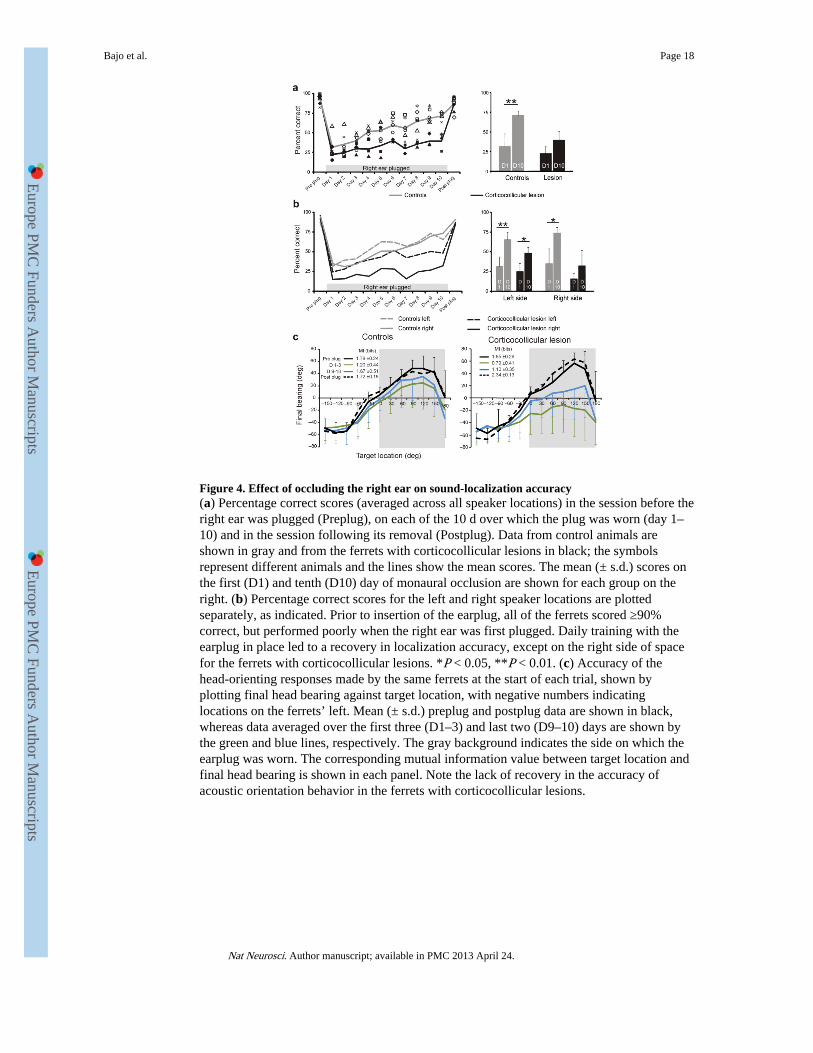
Figure 4. Effect of occluding the right ear on sound-localization accuracy(a) Percentage correct scores (averaged across all speaker locations) in the session before theright ear was plugged (Preplug), on each of the 10 d over which the plug was worn (day 1–10) and in the session following its removal (Postplug). Data from control animals areshown in gray and from the ferrets with corticocollicular lesions in black; the symbolsrepresent different animals and the lines show the mean scores. The mean (± s.d.) scores onthe first (D1) and tenth (D10) day of monaural occlusion are shown for each group on theright. (b) Percentage correct scores for the left and right speaker locations are plottedseparately, as indicated. Prior to insertion of the earplug, all of the ferrets scored ≥90%correct, but performed poorly when the right ear was first plugged. Daily training with theearplug in place led to a recovery in localization accuracy, except on the right side of spacefor the ferrets with corticocollicular lesions. *P < 0.05, **P < 0.01. (c) Accuracy of thehead-orienting responses made by the same ferrets at the start of each trial, shown byplotting final head bearing against target location, with negative numbers indicatinglocations on the ferrets’ left. Mean (± s.d.) preplug and postplug data are shown in black,whereas data averaged over the first three (D1–3) and last two (D9–10) days are shown bythe green and blue lines, respectively. The gray background indicates the side on which theearplug was worn. The corresponding mutual information value between target location andfinal head bearing is shown in each panel. Note the lack of recovery in the accuracy ofacoustic orientation behavior in the ferrets with corticocollicular lesions.
Bajo et al. Page 18
Nat Neurosci. Author manuscript; available in PMC 2013 April 24.
Europe PM
C Funders A
uthor Manuscripts
Europe PM
C Funders A
uthor Manuscripts
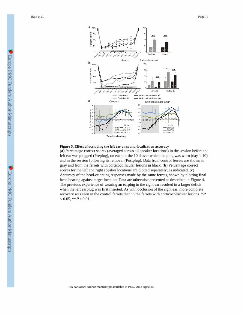
Figure 5. Effect of occluding the left ear on sound-localization accuracy(a) Percentage correct scores (averaged across all speaker locations) in the session before theleft ear was plugged (Preplug), on each of the 10 d over which the plug was worn (day 1-10)and in the session following its removal (Postplug). Data from control ferrets are shown ingray and from the ferrets with corticocollicular lesions in black. (b) Percentage correctscores for the left and right speaker locations are plotted separately, as indicated. (c)Accuracy of the head-orienting responses made by the same ferrets, shown by plotting finalhead bearing against target location. Data are otherwise presented as described in Figure 4.The previous experience of wearing an earplug in the right ear resulted in a larger deficitwhen the left earplug was first inserted. As with occlusion of the right ear, more completerecovery was seen in the control ferrets than in the ferrets with corticocollicular lesions. *P< 0.05, **P < 0.01.
Bajo et al. Page 19
Nat Neurosci. Author manuscript; available in PMC 2013 April 24.
Europe PM
C Funders A
uthor Manuscripts
Europe PM
C Funders A
uthor Manuscripts
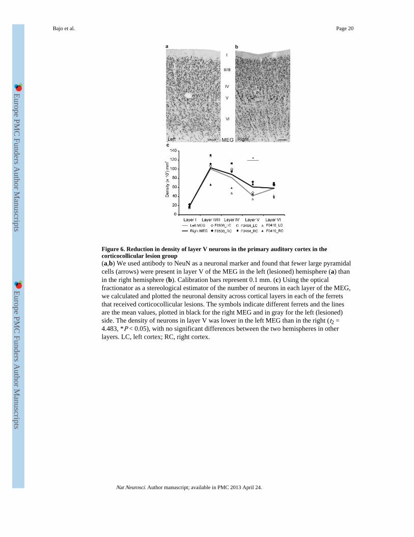
Figure 6. Reduction in density of layer V neurons in the primary auditory cortex in thecorticocollicular lesion group(a,b) We used antibody to NeuN as a neuronal marker and found that fewer large pyramidalcells (arrows) were present in layer V of the MEG in the left (lesioned) hemisphere (a) thanin the right hemisphere (b). Calibration bars represent 0.1 mm. (c) Using the opticalfractionator as a stereological estimator of the number of neurons in each layer of the MEG,we calculated and plotted the neuronal density across cortical layers in each of the ferretsthat received corticocollicular lesions. The symbols indicate different ferrets and the linesare the mean values, plotted in black for the right MEG and in gray for the left (lesioned)side. The density of neurons in layer V was lower in the left MEG than in the right (t2 =4.483, *P < 0.05), with no significant differences between the two hemispheres in otherlayers. LC, left cortex; RC, right cortex.
Bajo et al. Page 20
Nat Neurosci. Author manuscript; available in PMC 2013 April 24.
Europe PM
C Funders A
uthor Manuscripts
Europe PM
C Funders A
uthor Manuscripts
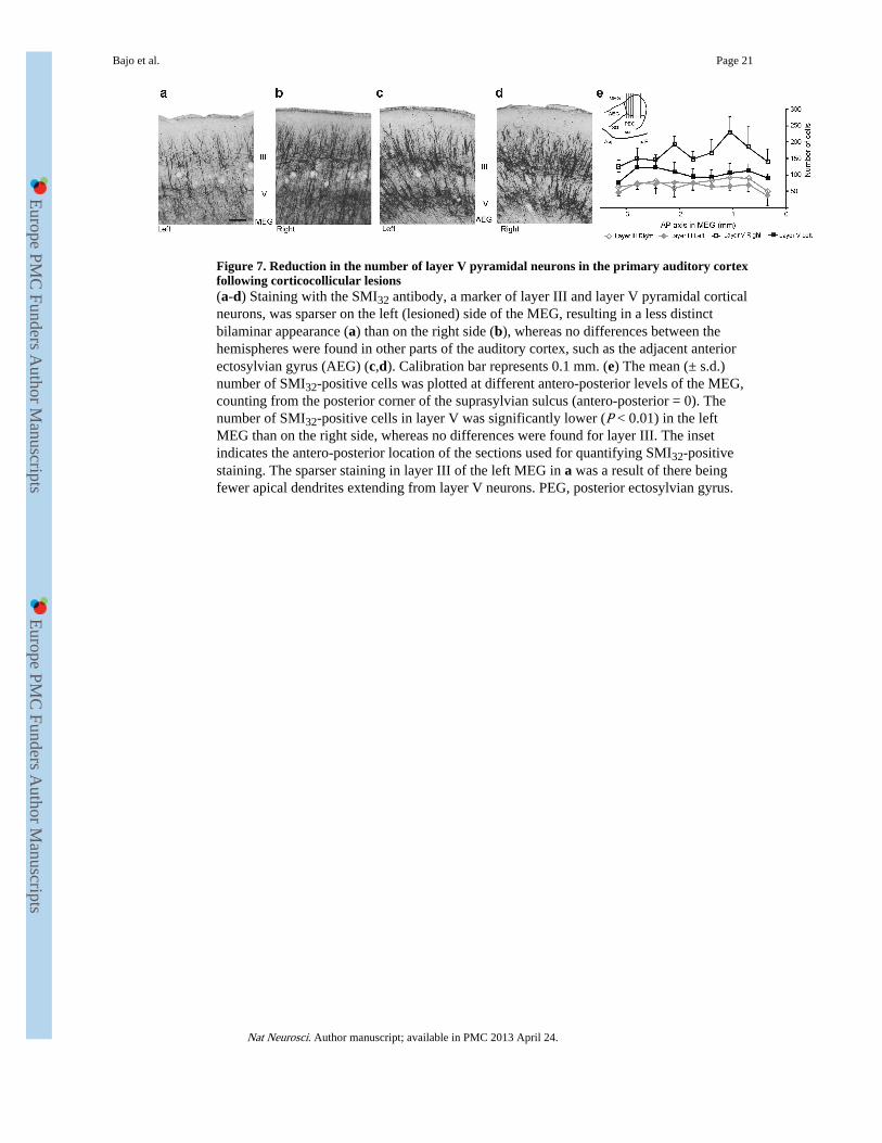
Figure 7. Reduction in the number of layer V pyramidal neurons in the primary auditory cortexfollowing corticocollicular lesions(a-d) Staining with the SMI32 antibody, a marker of layer III and layer V pyramidal corticalneurons, was sparser on the left (lesioned) side of the MEG, resulting in a less distinctbilaminar appearance (a) than on the right side (b), whereas no differences between thehemispheres were found in other parts of the auditory cortex, such as the adjacent anteriorectosylvian gyrus (AEG) (c,d). Calibration bar represents 0.1 mm. (e) The mean (± s.d.)number of SMI32-positive cells was plotted at different antero-posterior levels of the MEG,counting from the posterior corner of the suprasylvian sulcus (antero-posterior = 0). Thenumber of SMI32-positive cells in layer V was significantly lower (P < 0.01) in the leftMEG than on the right side, whereas no differences were found for layer III. The insetindicates the antero-posterior location of the sections used for quantifying SMI32-positivestaining. The sparser staining in layer III of the left MEG in a was a result of there beingfewer apical dendrites extending from layer V neurons. PEG, posterior ectosylvian gyrus.
Bajo et al. Page 21
Nat Neurosci. Author manuscript; available in PMC 2013 April 24.
Europe PM
C Funders A
uthor Manuscripts
Europe PM
C Funders A
uthor Manuscripts
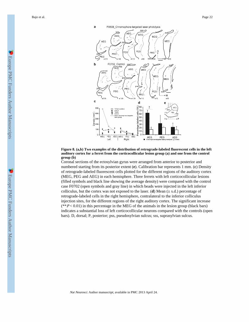
Figure 8. (a,b) Two examples of the distribution of retrograde-labeled fluorescent cells in the leftauditory cortex for a ferret from the corticocollicular lesion group (a) and one from the controlgroup (b)Coronal sections of the ectosylvian gyrus were arranged from anterior to posterior andnumbered starting from its posterior extent (e). Calibration bar represents 1 mm. (c) Densityof retrograde-labeled fluorescent cells plotted for the different regions of the auditory cortex(MEG, PEG and AEG) in each hemisphere. Three ferrets with left corticocollicular lesions(filled symbols and black line showing the average density) were compared with the controlcase F0702 (open symbols and gray line) in which beads were injected in the left inferiorcolliculus, but the cortex was not exposed to the laser. (d) Mean (± s.d.) percentage ofretrograde-labeled cells in the right hemisphere, contralateral to the inferior colliculusinjection sites, for the different regions of the right auditory cortex. The significant increase(**P < 0.01) in this percentage in the MEG of the animals in the lesion group (black bars)indicates a substantial loss of left corticocollicular neurons compared with the controls (openbars). D, dorsal; P, posterior; pss, pseudosylvian sulcus; sss, suprasylvian sulcus.
Bajo et al. Page 22
Nat Neurosci. Author manuscript; available in PMC 2013 April 24.
Europe PM
C Funders A
uthor Manuscripts
Europe PM
C Funders A
uthor Manuscripts