Stem cell transplantation for auditory nerve replacement
15
Stem Cell Transplantation for Auditory Nerve Replacement Richard A. Altschuler 1,2 , K. Sue O’Shea 2 , and Josef M. Miller 1 1Kresge Hearing Research Institute, Department of Otolaryngology, University of Michigan, 1301 East Ann, Ann Arbor, MI 48109-0506 2Department of Cell and Developmental Biology, University of Michigan, 1301 East Ann, Ann Arbor, MI 48109-0506 Abstract The successful function of cochlear prostheses depends on activation of auditory nerve. The survival of auditory nerve neurons, however, can vary widely in candidates for cochlear implants and influence implant efficacy. Stem cells offer the potential for improving the function of cochlear prostheses and increasing the candidate pool by replacing lost auditory nerve. The first phase of studies for stem cell replacement of auditory nerve has examined the in vitro survival and differentiation as well as in vivo differentiation and survival of exogenous embryonic and tissue stem cells placed into scala tympani and/or modiolus. These studies are reviewed and new results on in vivo placement of B-5 mouse embryonic stem cells into scala tympani of the guinea pig cochleae with differentiation into a glutamatergic neuronal phenotype are presented. Research on the integration and connections of stem cell derived neurons in the cochlea is described. Finally, an alternative approach is considered, based on the use of endogenous progenitors rather than exogenous stem cells, with a review of promising findings that have identified stem cell-like progenitors in cochlear and vestibular tissues to provide the potential for auditory nerve replacement. Keywords spiral ganglion neurons; cochlea; auditory; deafness; embryonic stem cells 1. Introduction 1.1 – Rationale and Challenges Cochlear implants now provide an increasingly successful therapy to restore hearing to a large number of the severely deaf. Cochlear prostheses function by directly activating the auditory nerve in the cochlea. When there is loss of auditory nerve, as is often associated with deafness, this can influence the efficacy of cochlear prostheses (e.g. Blamey et al., 1996; Kileny et al., 1991; Incesulu and Nadol, 1998; Rubinstein et al., 1999). The minimal amount of remaining auditory nerve necessary for effective use of cochlear prostheses has not yet been established and the correlation between total amount of auditory nerve survival and performance remains in contention. Even a low number of surviving auditory neurons may still allow good speech perception (Blamey, 1997). Psychophysical measures in monkeys demonstrated increased *Corresponding Author: Richard A Altschuler, KHRI, University of Michigan, 1301 East Ann, Ann Arbor, MI 48109-0506, Tel: 734 763-0060, Fax: 734 764-0014, Email: [email protected]. Publisher's Disclaimer: This is a PDF file of an unedited manuscript that has been accepted for publication. As a service to our customers we are providing this early version of the manuscript. The manuscript will undergo copyediting, typesetting, and review of the resulting proof before it is published in its final citable form. Please note that during the production process errors may be discovered which could affect the content, and all legal disclaimers that apply to the journal pertain. NIH Public Access Author Manuscript Hear Res. Author manuscript; available in PMC 2009 August 1. Published in final edited form as: Hear Res. 2008 August ; 242(1-2): 110–116. doi:10.1016/j.heares.2008.06.004. NIH-PA Author Manuscript NIH-PA Author Manuscript NIH-PA Author Manuscript
-
Upload
independent -
Category
Documents
-
view
0 -
download
0
Transcript of Stem cell transplantation for auditory nerve replacement

Stem Cell Transplantation for Auditory Nerve Replacement
Richard A. Altschuler1,2, K. Sue O’Shea2, and Josef M. Miller11Kresge Hearing Research Institute, Department of Otolaryngology, University of Michigan, 1301 East Ann,Ann Arbor, MI 48109-0506
2Department of Cell and Developmental Biology, University of Michigan, 1301 East Ann, Ann Arbor, MI48109-0506
AbstractThe successful function of cochlear prostheses depends on activation of auditory nerve. The survivalof auditory nerve neurons, however, can vary widely in candidates for cochlear implants andinfluence implant efficacy. Stem cells offer the potential for improving the function of cochlearprostheses and increasing the candidate pool by replacing lost auditory nerve. The first phase ofstudies for stem cell replacement of auditory nerve has examined the in vitro survival anddifferentiation as well as in vivo differentiation and survival of exogenous embryonic and tissue stemcells placed into scala tympani and/or modiolus. These studies are reviewed and new results on invivo placement of B-5 mouse embryonic stem cells into scala tympani of the guinea pig cochleaewith differentiation into a glutamatergic neuronal phenotype are presented. Research on theintegration and connections of stem cell derived neurons in the cochlea is described. Finally, analternative approach is considered, based on the use of endogenous progenitors rather than exogenousstem cells, with a review of promising findings that have identified stem cell-like progenitors incochlear and vestibular tissues to provide the potential for auditory nerve replacement.
Keywordsspiral ganglion neurons; cochlea; auditory; deafness; embryonic stem cells
1. Introduction1.1 – Rationale and Challenges
Cochlear implants now provide an increasingly successful therapy to restore hearing to a largenumber of the severely deaf. Cochlear prostheses function by directly activating the auditorynerve in the cochlea. When there is loss of auditory nerve, as is often associated with deafness,this can influence the efficacy of cochlear prostheses (e.g. Blamey et al., 1996; Kileny et al.,1991; Incesulu and Nadol, 1998; Rubinstein et al., 1999). The minimal amount of remainingauditory nerve necessary for effective use of cochlear prostheses has not yet been establishedand the correlation between total amount of auditory nerve survival and performance remainsin contention. Even a low number of surviving auditory neurons may still allow good speechperception (Blamey, 1997). Psychophysical measures in monkeys demonstrated increased
*Corresponding Author: Richard A Altschuler, KHRI, University of Michigan, 1301 East Ann, Ann Arbor, MI 48109-0506, Tel: 734763-0060, Fax: 734 764-0014, Email: [email protected]'s Disclaimer: This is a PDF file of an unedited manuscript that has been accepted for publication. As a service to our customerswe are providing this early version of the manuscript. The manuscript will undergo copyediting, typesetting, and review of the resultingproof before it is published in its final citable form. Please note that during the production process errors may be discovered which couldaffect the content, and all legal disclaimers that apply to the journal pertain.
NIH Public AccessAuthor ManuscriptHear Res. Author manuscript; available in PMC 2009 August 1.
Published in final edited form as:Hear Res. 2008 August ; 242(1-2): 110–116. doi:10.1016/j.heares.2008.06.004.
NIH
-PA Author Manuscript
NIH
-PA Author Manuscript
NIH
-PA Author Manuscript
thresholds and smaller dynamic range from cochlear electrical stimulation, associated withincreased sensorineural damage (Pfingst and Sutton, 1983; Pfingst et al, 1981). Shepherd andJavel (1997) compared physiological responses to cochlear electrical stimulation with the statusof the auditory nerve in the cat and reported elevation of thresholds in animals with extensiveauditory nerve pathology but only moderate reductions in response amplitudes. They alsofound decreases in entrainment at higher stimulation rates and increases in bursting. Sly et al(2007) recently reported a decrease in spike latency and a reduction in threshold as auditorynerve survival decreased following ototoxic deafening in guinea pig. Certainly in cases ofauditory neuropathology, where there is very large loss of auditory nerve, cochlear prosthesescould be expected to be less effective. Replacement of lost auditory nerve could thereforeimprove the function of cochlear prostheses as well as increase the candidate pool. Stem celltechnology provides a potential mechanism to accomplish replacement of lost auditory nerveneurons. There are three basic challenges which must be met for successful use of stem cellsto replace auditory nerve for use with cochlear prostheses: 1) Differentiation into an appropriatephenotype, 2) long-term survival after reaching appropriate phenotype, and 3) functionalcentral connection.
1.2. Differentiation into an appropriate phenotypeIn order to accomplish auditory nerve replacement, the stem cells must be induced todifferentiate into a neuronal phenotype. However, even if stem cells successfully reach aneuronal phenotype, if they are inhibitory, they would not be useful for activating the centralauditory pathways. Therefore, it is important that an auditory nerve-like excitatoryglutamatergic phenotype be obtained. If differentiated stem cells can also exhibit a molecularsignature reflecting appropriate ion channels and other characteristics of auditory nerve, likelythey could be even more capable of substituting for its function.
Several studies have examined the survival and differentiation of embryonic and tissue stemcells following their placement into the scala tympani and/or modiolus of the cochlea. Iguchiet al. (2003) placed neural stem cells from fetal mouse neuroepithelium into the mouse cochleaand found 10% survival at 25 days following placement, with the majority showing a glialphenotype based on glial fibrillary acidic protein (GFAP) immunostaining and a few showinga pre-neural phenotype based on nestin immunostaining. Tamura et al. (2004) placed mouseneural stem cells into the modiolus of mouse cochleae following deafening by treatment withcisplatin. They also found the majority had differentiation into a glial phenotype, based onGFAP immunostaining and only a small percentage reached a neuronal type phenotype.Coleman et al. (2006) placed mouse embryonic stem cells (mESCs) into guinea pig cochleaand found poor survival, with 4–6 mESCs per profile through scala tympani and 1 per profilethrough Rosenthal’s canal, with a significant decline in the number in scala tympani between2 and 4 weeks following placement. A portion of the surviving mESCs (not specified in thetext) showed a neuronal phenotype based on neurofilament immunolabeling. Hu et al.(2005) placed neural stem cells from the lateral wall of mouse lateral ventricle into normal ordeafened guinea pig cochlea and found only a few reached a neuronal phenotype, based onTUJ1 (neuronal class III β-tubulin) immunostaining, with many showing a glial fated basedon GFAP immunostaining. More recently, Parker et al. (2007) used neural stem cells derivedfrom mouse fetal cerebellum, placed into noise exposed mouse or guinea pig cochleae. Theyfound moderate levels of survival with differentiation into satellite cell-, Spiral GanglionNeurons (SGN)-, Schwann cell-, hair cell- and supporting cell-like phenotypes. Interestingly,they found that this differentiation was influenced by the microenvironment, with cerebellumderived cells often expressing genes characteristics of local endogenous cells. Matsuoka et al.(2006) tested the placement of mouse bone marrow derived stem cells into the gerbil cochleaand found these stem cells would survive 7 days of placement into scala tympani or modiolus.
Altschuler et al. Page 2
Hear Res. Author manuscript; available in PMC 2009 August 1.
NIH
-PA Author Manuscript
NIH
-PA Author Manuscript
NIH
-PA Author Manuscript
None of these studies examined the transmitter phenotype of the cells reaching a neuronalphenotype.
Two approaches have been applied to increase the number of neuronal stem cells followingtransplantation into the cochlea. One approach is to apply cell signaling factors in vitro todifferentiate the stem cells toward the desired neural phenotype prior to their placement in thecochlea. The second approach is to place undifferentiated stem cells in the cochlea and thenapply growth factors in vivo, using the cochlear fluids for delivery. Advantages of the firstapproach include that differentiation can be monitored and that cell sorting can be applied tofurther refine the population before placement. Advantages of the second approach includethat undifferentiated stem cells can induce less of an immune response to their placement andthat their differentiation can be influenced and modified by “natural” endogenous factors, assuggested by Parker et al. (2007), to form the desired lineage.
Rivolta et al. (2006) used the “in vitro” approach and were successful in inducing mouse ESCsto form a variety of inner ear-like phenotypes, with a focus on generating a hair cell-likephenotype. Coleman et al. (2007) found that the in vitro differentiation of stem cells intoneuron-like cells was facilitated by co-culture with auditory neurons or hair cell explants,isolated from post-natal day five rats. This in vitro approach has also been applied to bonemarrow derived stem cells. Kondo et al. (2005) demonstrated sonic hedgehog and retinoic acidwhen in combination induced differentiation of marrow stromal cells to a neuronal phenotypewith a greater than 200-fold increase in GATA3, Irx2, Sox10, calretinin and GluR4 mRNAs(all expressed during normal development in auditory nerve neurons) and Wnt 1 induced a400-fold increase in the expression level of Brn3a, Ngn2 (neuroD) and Ngn1, which play a rolein the differentiation of auditory neurons during normal development. Falk et al. (2002) foundthat transduction of stem cells with Neurogenin 2 (Ngn2) induced over 90% towards a neuronalphenotype. Hu et al. (2005) tested this approach in the cochlea, transfecting Ngn2 into neuralstem cells prior to placement and found the number reaching a neuronal phenotype (based onimmunostaining for TUJ1) increased from 15% to over 50%.
Hu et al. (2004) co-transplantated mouse ESCs and a neuronal co-graft of dorsal root gangliontissue into guinea pig cochlea, to influence stem cell differentiation in vivo. They foundimproved survival of TUJ1 immunolabeled stem cells at 4 weeks following placement withthe co-graft. The in vivo approach can also take advantage of the opportunity that the cochlearfluids offer for intrascalar delivery. Undifferentiated or partially differentiated stem cells canbe placed into the cochlea and then exogenous neurotrophic factors or induction of geneticsignaling applied in vivo. Our studies, described in a later section, showed intrascalar deliveryof the neurotrophic factor (NTF) glial cell line derived neurotrophic factor (GDNF) increasedthe percentage of undifferentiated mouse embryonic stem cells reaching a neuronal phenotypein vivo, based on TUJ1 immunolabeling.
1.3. Challenge Two - SurvivalIn addition to generating the appropriate phenotype, a second challenge is survival afterreaching this phenotype. Long-term survival is necessary, of course, for auditory nervereplacement to have therapeutic value. Hu et al. (2004) found moderate survival of mouse stemcell derived neurons several weeks following placement into guinea pig modiolus, but alsofound that the survival time of stem cells transduced into neural phenotype by expression ofNgn2 was decreased compared to the non-transduced stem cells (Hu et al.., 2005). These neuralstem cells demonstrated good survival at 2 weeks; but this was greatly diminished at 4 weekspost-placement (Hu et al., 2005). This diminished survival could have been due to theneurophysiologically “unnatural” sustained rather than transient expression of Ngn2 and/orthat the site of integration of the transgene interfered with differentiation. These studies alsodid not provide intracochlear infusion of NTF(s) or other survival factors that Ulfendahl et al.
Altschuler et al. Page 3
Hear Res. Author manuscript; available in PMC 2009 August 1.
NIH
-PA Author Manuscript
NIH
-PA Author Manuscript
NIH
-PA Author Manuscript
(2007) suggested could be needed for continued survival. Just as SGN require trophic supportfor survival and begin to degenerate within weeks following their deafferentation from innerhair cell loss (see Shepherd article this volume, which addresses this in detail), when stem cellsassume a neuronal phenotype they may also require appropriate trophic support. This issupported by results from our lab, which show that chronic intrascalar infusion of glial cellline derived neurotrophic factor (GNDF) (see later section) and/or brain derived neurotrophicfactor (BDNF) (Hernandez et al., 2007) provided for excellent survival of neuronal stem cellsin the cochlea at five to six weeks following their placement. Clearly longer survival times andthe efficacy of various NTFs and electrical stimulation alone and combined should be assessedin future studies.
1.4. Challenge Three – Integration and Functional Central ConnectionThe stem cells that differentiate into a neuronal phenotype in cochlear compartments areusually seen to extend processes (e.g. Figure 1c,d). We find the processes from ESC derivedneurons in scala tympani are most often directed towards the organ of Corti (or the scarreplacing it) (Figure 1b) or towards remaining SGN neurons in the modiolus. Matsumoto etal.., (2005) found that mouse embryonic stem cells co-cultured (in vitro) with mouse auditoryepithelium send processes towards hair cells. Corrales et al., (2005) placed mouse neural stemcells into the cochlear nerve trunk of the gerbil 1 week after oubain injection which eliminatedall SGN without loss of hair cells. Twelve days following placement these stem cells expressedneuronal phenotype markers and sent processes into the space formerly occupied by theauditory nerve. Neurites projected peripherally towards the organ of Corti and contacted haircells.
To achieve functional replacement of lost auditory nerve, the stem cell derived neurons mustalso send a central axonal projection that crosses the peripheral nervous system – centralnervous system barrier and connects with central auditory neurons in the Cochlear Nucleus(CN). Hu et al. (2004) have demonstrated migration of mouse embryonic dorsal root ganglioncells from the site of placement in the rat scala tympani into the modiolus with projections bothperipherally toward the hair cells, as well as centrally, and formation of axonal connectionsbetween the projections of the implants and SGN central projections. Hu et al. (2005) alsoobserved mouse embryonic stem cells placed into rat scala tympani migrating into the auditorynerve and entering into the CN. While this suggests there might not be a PNS/CNS barrierconstraint, the less differentiated embryonic stem cells may be less inhibited by this barrierthan processes with a more mature neuronal phenotype. Recent studies suggest that centralconnections can be enhanced by blocking the natural inhibition at the PNS/CNS barrier fromproteins such as Nogo and myelin inhibitors (He and Koprivica, 2004 for review). Koprivicaet al. (2005) have developed small molecules to block the downstream pathway in thisinhibition, for example by blocking EGFR kinase. This resulted in greatly improvedregeneration of optic nerve fibers. Gharabaghi and Tatagiba (2005) applied IN-1, a Nogo-Ainhibitor, and growth factors to the auditory nerve through a cannula at the cerebellopontineangle and found improved functional regeneration of the auditory nerve after axotomy. It maybe necessary to apply such factors to enhance central connections to the cochlear nucleus fromnew stem cell derived neurons in the cochlea.
We have done studies addressing the critical first challenge of increasing the number of stemcells reaching an appropriate phenotype for auditory nerve replacement. Coleman et al..,(2006) implanted B-5 mESCs into guinea pig cochlea and found moderate survival with mostdifferentiated into a glial phenotype. We used the same B-5 cell line of undifferentiated mESCsand added intrascalar application of the neurotrophic factor (NTF) glial cell line derivedneurotrophic factor (GDNF) following the mESC placement into guinea pig cochlea. Rask-Anderson et al.., (2005) had used GDNF to successfully induce neurospheres gathered from
Altschuler et al. Page 4
Hear Res. Author manuscript; available in PMC 2009 August 1.
NIH
-PA Author Manuscript
NIH
-PA Author Manuscript
NIH
-PA Author Manuscript

human or guinea pig spiral ganglion into a neuronal phenotype. We hypothesized that GDNFcould also induce mESCs into a SGN-like phenotype. GDNF could also increase the numberof mESCs with a neuronal phenotype found at 4 weeks following placement by acting as asurvival factor, as it does for de-afferented spiral ganglion neurons (Altschuler et al., 1999;Kanzaki et al., 2002; Miller et al., 1997; Miller et al., 1998; Shepherd et al., this issue).
2. MethodsAll experimental procedures were approved by the University of Michigan Committee on theUse and Care of Animals. Eight normal hearing adult NIH strain guinea pigs (Elm Hill)weighing 300 to 400 grams, were systemically deafened by administration of 450-mg/kgkanamycin s.c., followed by anesthesia with ketamine/xylazine and 60 mg/kg ethacrynic acid,administered i.v. (jugular vein) 2 hours later (West et al., 1973). Three days followingdeafening, mouse ESCs from the B-5 line (Hadjantonakis et al., 1998) that express enhancedgreen fluorescent protein (eGFP) were were aspirated into a 1cc syringe and carefullytransferred to a glass microliter syringe. A 30g needle was attached to the syringe and insertedinto a micro-cannula (Prieskorn & Miller, 2000). The fine cannula tip was then inserted througha cochleostomy in the basal turn of the cochlear spiral with a silastic ball serving as a stop toplug the hole during the injection to prevent leakage. A 5µl bolus containing approximately500,000 B-5 mouse ESCs was slowly injected into the modiolus and scala tympani and thecannula kept in place for an additional 10 minutes. The cannula was then removed and replacedby a second cannula into scala tympani primed with GDNF (10 µg/ml) attached to aminiosmotic pump (Alza Corporation, model 2002, 14 day reservoir, flow rate 0.5 µl/hr). TheGDNF was chronically infused over two weeks. At 14 days following placement the guineapigs were heavily anesthetized with 0.5 ml i.p. sodium pentobarbital (Fatal Plus, Vortech,Dearborn, MI) and terminated with a vascular fixation of phosphate buffer followed by 4%paraformaldehyde fixative in phosphate buffer. Immunocytochemistry was carried out on 6µm paramodiolar cryostat sections through the cochleae. Mouse ESCs in differentcompartments of the guinea pig cochlear were identified by their eGFP expression and also byin situ hybridization for genomic mouse DNA. Differentiation of the mouse ESCs into aneuronal or glial phenotype was assessed using TUJ1 as a marker for neuronal phenotype andGFAP as a marker for an astrocyte phentotype. Mouse monoclonal antibody to TUJ1 (Covance,Berkeley, CA) was applied at a 1:300 dilution in phosphate buffered saline (PBS) with 0.2%triton X-100 and polyclonal rabbit antibody to GFAP (MP Biomedicals, Aurora, OH) was usedat a 1:500 dilution) in PBS with triton X100. The TUJ1 immunolabeled stem cells were furtherassessed for neurotransmitter phenotype with co-immunolabeling for vesicular glutamatetransporters 1 or 2 (VGLUT1,2) or the GABA transporter (VGAT) using rabbit polyclonalantibodies (Synaptic Systems, SYSY, Germany) all used at a 1:500 dilution in PBS with 0.2%triton X-100. Secondary anti-mouse or anti-rabbit antibodies (Molecular Probes, Carlsbad, CA)were linked to Alexa 350 (TUJ1) or Alexa 594 (anti-rabbit) for co-localization of TUJ1,VGLUTs and VGAT. Other sections were co-labeled with in fluorescent in situ hybridization(FISH) for genomic mouse DNA and immunostaining for TUJ1 using mouse COT-1 DNA(Invitogen Corporation, Carlsbad, CA) as the DNA probe for mouse genomic DNA. The probewas labeled with digoxigenin-16-dUTP using a nick translation kit (both from Roche AppliedScience, Indianapolis, IN). Sections were incubated with the COT-1 DNA probe, diluted 1:20in hybridization buffer (Enzo Life Sciences, Farmingdale, NY) for 5 minutes at 92°C. Sectionswere then rinsed in PBS, followed by in situ hybridization wash reagent (Enzo Life Sciences)for 15 minutes at 37°C and then additional rinses in PBS at room temperature. Slides wereincubated with rhodamine conjugated antibody to digoxigenin (1:200, Roche Applied Science,Indianapolis, IN) for 1 hour at room temperature in the dark. The sections were co-immunolabeled with antibody to TUJ1 (as above) and the TUJ1 antibody visualized with ananti-mouse antibody conjugated to Alexa 594. Confocal fluorescent images were acquired onOlympus FV-500 or Zeiss LSM510 confocal microscope.
Altschuler et al. Page 5
Hear Res. Author manuscript; available in PMC 2009 August 1.
NIH
-PA Author Manuscript
NIH
-PA Author Manuscript
NIH
-PA Author Manuscript
3. ResultsMany cells in sections could be identified as mouse ESCs, based on eGFP labeling (Figure2a,c,d) or expression of mouse genomic DNA (Figure 2e). A typical profile through scalatympani of the basal turn of the cochlear spiral, contained 100–200 eGFP labeled cells, withfewer in more apical turns and scala vestibuli. Smaller numbers of eGFP labeled cells (5–12per section) were seen in the modiolus, most often among SGN (Figure 2d) or their peripheralprocesses (Figure 2c) in Rosenthal’s canal. Most of the eGFP labeled cells were small (10microns) and round and without processes; they did not have a neuronal-like appearance.
There were many cells in scala tympani that were immunolabeled for TUJ1 (30–40 in a typicalsection through basal scala tympani (Figure 1). These had a more neuronal-like appearance,often with a fusiform shape and processes. The majority of the TUJ1 immunolabeled cells inscala tympani did not co-label with eGFP (Figure 1a–c) and it required co-labeling with FISHfor mouse genomic DNA (Figure 2e) to show they had differentiated from the mouse ESCS,although any other origin would have been unlikely. Fewer cells were identified with anastrocyte phenotype based on GFAP immunolabeling, with 8 – 20 per profile through basalscala tympani (Figure 2b). These also did not co-label for eGFP. This suggests that the B-5mouse embryonic stem cells also down regulate eGFP following in vivo differentiation.
Co-labeling of TUJ1 with glutamate or GABA vesicular transporters was used to assess theneurotransmitter phenotype of the embryonic stem cell derived neurons. The majority of TUJ1cells in scala tympani (50–75%) were co-labeled for VGLUT1 or VGLUT2, with VGLUT2co-immunolabeling most common (Figure 1d). The endogenous SGN were brightlyimmunolabeled with VGLUT1 and had only light VGLUT2 immunolabeling. Fewer TUJ1cells (7–12%) in basal scala tympani were found co-immunolabeled for GAT (Figure 1e), amarker for GABA. GABA is inhibitory and would not be a suitable transmitter for providingreplacement of auditory nerve.
4. DiscussionOur results show that chronic intrascalar infusion of GDNF following mESC placement intoscala tympani/modiolus results in a much higher percentage of mESCs reaching a neuronalrather than a glial phenotype and increased survival at 4 weeks compared to an earlier study(Coleman et al., 2006) which did not supply NTFs. It also found that most of the mESCsdifferentiating into a neuronal phenotype were glutamatergic and therefore suitable for auditorynerve replacement. The question still remains as to how such an in vivo approach compares inefficacy to the vitro approach of inducing stem cell differentiation prior to cochlear placementand this awaits future study. It will also be important to determine which approach providesbest long-term survival as well as integration and central connections, the other two challenges.
An alternative approach to implanting differentiated or undifferentiated exogenous stem cellsinto the cochlea is to mobilize and activate endogenous stem cells already present in the cochleato differentiate into a neuronal phenotype and form a central connection. In the last few yearsa number of observations support the potential of this approach. Li et al. (2003) found spheresof inner ear stem cells could be isolated and generated from adult utricular epilethelium invitro. Rask-Anderson et al. (2005) identified progenitor stem cells in cultures from human andguinea pig spiral ganglion. These neurospheres could be induced to differentiate into a neuronalor glial phenotype with application of neurotrophic factors such as GDNF. Yerukhimovich etal. (2007) found spheres isolated from mouse neonate cochleae that could differentiate intoastrocytes and oligodendrocytes in vitro. Oshima et al. (2007) were able to generate sphere-forming stem cells from early postnatal organ of Corti, vestibular sensory epithelium, SGNregion, and stria vascularis of the mouse cochlea. Those from the SGN region displayed features
Altschuler et al. Page 6
Hear Res. Author manuscript; available in PMC 2009 August 1.
NIH
-PA Author Manuscript
NIH
-PA Author Manuscript
NIH
-PA Author Manuscript

of neural stem cells or glial cells. This ability to form spheres decreased with postnatal age andwas greatly reduced by the third postnatal week. Lou et al. (2007) found that otospheres couldbe derived from the cochleae of young rats when cultured in vitro with EGF and FGF2 andthat these could differentiate into neuron, glial, hair cell and supporting cell phenotypes. Thesestudies show that endogenous stem cells are indeed present in the cochlea, although thenumbers decrease with age. The challenge remains to prospectively identify these cells so thattheir location, identity and growth factor responsiveness can be determined. With those datait should be possible to identify mechanisms to induce and control their differentiation into anauditory nerve-like phenotype and to form a central auditory connection.
While the potential for stem cell replacement of auditory nerve is therefore receiving increasingsupport, critical questions remain regarding the challenges of inducing appropriate phenotype,long term survival, functional central connections and ability to provide improvement of thefunction of cochlear prostheses.
The auditory nerve phenotype is well suited for its normal function of providing an excitatoryconnection between inner hair cells and the cochlear nucleus and this phenotype should be thetarget for stem cell differentiation for connection to regenerated hair cells. While an excitatoryphenotype is still necessary for cochlear electrical stimulation, however, the auditory nerveproperties may not be optimal. Perhaps stem cells can be differentiated into a phenotype withdifferent ion channels and different firing properties that would be even better than the auditorynerve for being excited and coded by electrical stimulation.
Challenges also remain for long term survival. Loss of auditory nerve following inner hair cellloss is most likely due to loss of survival factors such as neurotrophic factors (see Shepherdarticle, this issue). Stem cell derived neurons in the cochlea may be subject to the samedependence on survival factors once they reach the mature auditory nerve-like phenotype. Inthat case it would be likely that chronic cochlear electrical stimulation would enhance theirsurvival as has been shown for auditory nerve following deafness (Hartshorn et al., 1991;Leake et al., 1991, 1992, 1995, this issue; Lousteau, 1987; Mitchell et al., 1997; Miller et al.,2003; Miller & Altschuler, 1995). It may also be the case that neurotrophic factors would alsoenhance survival, as for auditory nerve following deafness (Altschuler et al., 1999; Ernfors etal., 1996; Malgrange et al., 1999; Marzella and Clark, 1999; McGuinness and Shepherd,2005; Miller et al., 1997; Miller et al., 1998; Schindler et al., 1995; Shepherd et al., thisissue; Staecker et al., 1996; Van de Water et al., 1996) with the combination of neurotrophicfactors and chronic electrical stimulation potentially the most effective (Kanzaki et al., 2002;Shepherd et al., 2005, this issue).
There are also challenges for establishing functional central connections. Remaining auditorynerve may provide a substrate and cues for guiding new central processes, but the PNS-CNSbarrier may play a critical role limiting the extent and functional integrity of the integration ofstem cells with CNS. With partial survival of the auditory nerve, an infrastructure may existto help overcome this barrier; however in the absence of most or all of the auditory neurons,this PNS-CNS interface may be a key limiting barrier. Studies with NoGo suggest there maybe molecules that will allow us to overcome this barrier. Clearly, additional studies will benecessary to investigate this and if necessary develop clinically applicable interventions toencourage central projections.
The presence of precursor cells in the cochlea that have many characteristics of stem cells alsooffers promise. These could be used to develop new human cell lines for implantation in humansubjects. The endogenous precursors might also be induced to differentiate into an auditoryneuron phenotype in vivo and form central connections to replace lost auditory nerve allowingthe damaged cochlea to help “repair itself”.
Altschuler et al. Page 7
Hear Res. Author manuscript; available in PMC 2009 August 1.
NIH
-PA Author Manuscript
NIH
-PA Author Manuscript
NIH
-PA Author Manuscript
While challenges remain, these studies to date show the great potential for using bothexogenous as well as endogenous stem cells to replace lost auditory nerve. There is an obviousclinical application to improve the efficacy of cochlear prostheses which are dependent onremaining auditory nerve for their function. Still another approach for clinical applicationwould be to place stem cells reaching a neuronal glutamatergic phenotype directly onto siteson the cochlear prostheses and direct their processes towards new synapses with remainingSGN along the cochlear spiral. This addition of this “extra” neuron could lower thresholds bybringing the neuronal target for excitation closer to the stimulation site. Further, placing thesestem cells close to different stimulation sites could also increase channel separation withdifferent groups of stem cells preferentially excited by different stimulation sites. Lowerthreshold and increased channel separation could be expected to improve cochlear implantfunction. The remaining challenges and the multiple approaches will be the focus of manyinteresting future studies in our and other laboratories of the field.
AcknowledgementsWe would like to thank Mat Velkey, Diane Prieskorn and Noel Wys for their contributions to generating the new datadescribed in this manuscript. These studies were supported by NIH grants DC03820 (RAA, JMM), GM069985 (KSO),NS04187 (KSO), P30 DC05188 and by GM/UAW funds.
REFERENCESAltschuler RA, Cho Y, Ylikoski J, Pirvola U, Magal E, Miller JM. Rescue and regrowth of sensory nerves
following deafferentation by neurotrophic factors. Ann. N. Y. Acad. Sci 1999;884:305–311. [PubMed:10842602]
Blamey P, Arndt P, Bergeron F, Bredberg G, Brimacombe J, Facer G. Factors affecting auditoryperformance of postlinguistically deaf adults using cochlear implants. Audiol. Neurootol 1996;1:293–306. [PubMed: 9390810]
Blamey P. Are spiral ganglion cell numbers important for speech perception with a cochlear implant?Am. J. Otol 1997;18(6 Suppl):S11–S12. [PubMed: 9391577]
Coleman B, Hardman J, Coco A, Epp S, de Silva M, Crook J, Shepherd R. Fate of embryonic stem cellstransplanted into the deafened mammalian cochlea. Cell Transplant 2006;15:369–380. [PubMed:16970279]
Coleman B, Fallon JB, Pettingill LN, de Silva MG, Shepherd RK. Auditory hair cell explant co-culturespromote the differentiation of stem cells into bipolar neurons. Exp. Cell Res 2007;313:232–243.[PubMed: 17112512]
Corrales CE, Pan L, Li H, Liberman MC, Heller S, Edge AS. Engraftment and differentiation ofembryonic stem cell-derived neural progenitor cells in the cochlear nerve trunk: growth of processesinto the organ of Corti. J. Neurobiol 2006;66:1489–1500. [PubMed: 17013931]
Ernfors P, Duan ML, El Shamy WM, Canlon B. Protection of auditory neurons from aminoglycosidetoxicity by NT3. Nat. Med 1996;2:463–467. [PubMed: 8597959]
Falk A, Holmstrom N, Carlen M, Cassidy R, Lundberg C, Frisen J. Gene delivery to adult neural stemcells. Exper. Cell Res 2002;279:34–39. [PubMed: 12213211]
Gharabaghi A, Tatagiba M. Functional regeneration of the axotomized auditory nerve with combinedneurotrophic and anti-inhibitory strategies. Acta Neurochir 2005;93:89–91.
Hadjantonakis A-K, Gertsenstein M, Ikawa M, Okabe M, Nagy A. Generating green fluorescent miceby germline transmission of green fluorescent ES cells. Mech. Dev 1998;76:79–90. [PubMed:9867352]
Hartshorn DO, Miller JM, Altschuler RA. Protective effect of electrical stimulation in the deafened guineapig cochlea. Otolaryngol. Head Neck Surg 1991;04(3):311–319. [PubMed: 1902931]
He Z, Koprivica V. The NOGO signaling pathway for regeneration block. Ann. Rev. Neurosci2004;27:341–368. [PubMed: 15217336]
Hernandez Reyes J, Wys NL, Velkey M, Prieskorn DM, Wesolowski K, O’Shea KS, Miller JM,Altschuler RA. Neuronal differentiation of mouse embryonic stem cells following transient
Altschuler et al. Page 8
Hear Res. Author manuscript; available in PMC 2009 August 1.
NIH
-PA Author Manuscript
NIH
-PA Author Manuscript
NIH
-PA Author Manuscript
neurogenin 1 expression and treatment with BDNF and GDNF: in vitro and in vivo with placementinto guinea pig cochlea. Soc. Neurosci. Abst. 2007
Hu Z, Ulfendahl M, Olivius NP. Central migration of neuronal tissue and embryonic stem cells followingtransplantation along the auditory nerve root. Brain Res 2004;1026:68–73. [PubMed: 15476698]
Hu Z, Wei D, Johansson CB, Holmstrom N, Duan M, Frisen J, Ulfendahl M. Survival and neuraldifferentiation of adult neural stem cells transplanted into the mature inner ear. Exp. Cell Res2005;302:40–47. [PubMed: 15541724]
Iguchi F, Nakagawa T, Tateya I, Kim TS, Endo T, Taniguchi Z, Naito Y, Ito J. Trophic support of mouseinner ear by neural stem cell transplantation. Neuroreport 2003;14(1):77–80. [PubMed: 12544835]
Incesulu A, Nadol JB Jr. Correlation of acoustic threshold measures and spiral ganglion cell survival insevere to profound sensorineural hearing loss: implications for cochlear implantation. Ann. Otol.Rhinol. Laryngol 1998;107:906–913. [PubMed: 9823838]
Kanzaki S, Stover T, Kawamoto K, Prieskorn DM, Altschuler RA, Miller JM, Raphael Y. Glial cell line-derived neurotrophic factor and chronic electrical stimulation prevent VIII cranial nerve degenerationfollowing denervation. J. Comp. Neurol 2002;454:350–360. [PubMed: 12442325]
Kileny PR, Zimmerman-Phillips S, Kemink JL, Schmaltz SP. Effects of preoperative electricalstimulability and historical factors on performance with multichannel cochlear implant. Ann. Otol.Rhinol. Laryngol 1991;100:563–568. [PubMed: 2064268]
Kondo T, Johnson SA, Yoder MC, Romand R, Hashino E. Sonic hedgehog and retinoic acidsynergistically promote sensory fate specification from bone marrow-derived pluripotent stem cells.Proc. Natl. Acad. Sci. U. S. A 2005;102:4789–4799. [PubMed: 15778294]
Koprivica V, Cho KS, Park JB, Yiu G, Atwal J, Gore B, Kim JA, Lin E, Tessier-Lavigne M, Chen DF,He Z. EGFR activation mediates inhibition of axon regeneration by myelin and chondroitin sulfateproteoglycans. Science 2005;310:106–110. [PubMed: 16210539]
Li H, Liu H, Heller S. Pluripotent stem cells from the adult mouse inner ear. Nat. Med 2003;9(10):1293–1299. [PubMed: 12949502]
Leake PA, Hradek GT, Rebscher SJ, Snyder RL. Chronic intracochlear electrical stimulation inducesselective survival of spiral ganglion neurons in neonatally deafened cats. Hear. Res 1991;54(2):251–257. [PubMed: 1938628]
Leake PA, Snyder RL, Hradek GT, Rebscher SJ. Chronic intracochlear stimulation in neonatally deafenedcats: Effects of intensity and stimulating electrode position. Hear. Res 1992;64:99–117. [PubMed:1490906]
Leake PA, Snyder RL, Hradek GT, Rebscher SJ. Consequences of chronic extracochlear electricalstimulation in neonatally deafened cats. Hear. Res 1995;82(1):65–80. [PubMed: 7744715]
Leake PA, Hradek GT, Sttakhovskaya O. Factors influencing neurotrophic effects of electricalstimulation in the deafened, developing auditory system. Hear. Res. this issue.
Lou X, Zhang Y, Yuan C. Multipotent stem cells from the young rat inner ear. Neurosci. Lett2007;416:28–33. [PubMed: 17350759]
Lousteau RJ. Increased spiral ganglion cell survival in electrically stimulated, deafened guinea pigcochleae. Laryngoscope 1987;97:836–842. [PubMed: 3600136]
Malgrange B, Rigo JM, Van de Water TR, Staecker H, Moonen G, Lefebvre PP. Growth factor therapyto the damaged inner ear: clinical prospects. Inter. J. Ped. Otorhinolaryngol 1999;49:S19–S25.
Marzella PL, Clark GM. Growth factors, auditory neurones and cochlear implants: a review. ActaOtolaryngol 1999;119:407–412. [PubMed: 10445053]
Matsuoka AJ, Kondo T, Miyamoto RT, Hashino E. In vivo and in vitro characterization of bone marrow-derived stem cells in the cochlea. Laryngoscope 2006;116:1363–1367. [PubMed: 16885736]
Matsumoto M, Nakagawa T, Higashi T, Kim TS, Kojima K, Kita T, Sakamoto T, Ito J. Innervation ofstem cell-derived neurons into auditory epithelia of mice. Neuroreport 2005;16(8):787–790.[PubMed: 15891570]
McGuinness SL, Shepherd RK. Exogenous BDNF rescues rat spiral ganglion neurons in vivo. Otol.Neurotol 2005;26:1064–1072. [PubMed: 16151360]
Miller AL, Yamasoba T, Altschuler RA. Hair cell and spiral ganglion neuron preservation andregeneration - influence of growth factors. Curr. Opin. Otolaryngol. Head Neck Surg 1998;6:301–307.
Altschuler et al. Page 9
Hear Res. Author manuscript; available in PMC 2009 August 1.
NIH
-PA Author Manuscript
NIH
-PA Author Manuscript
NIH
-PA Author Manuscript
Miller JM, Altschuler RA. Effectiveness of different electrical stimulation conditions in preservation ofspiral ganglion cells following deafness. Ann. Otol. Rhinol. Laryngol 1995;166:57–60.
Miller JM, Chi DH, O'Keeffe LJ, Kruszka P, Raphael Y, Altschuler RA. Neurotrophins can enhancespiral ganglion cell survival after inner hair cell loss. Inter. J. Develop. Neurosci 1997;15:631–643.
Miller AL, Prieskorn DM, Altschuler RA, Miller JM. Mechanism of electrical stimulation-inducedneuroprotection: effects of verapamil on protection of primary auditory afferents. Brain Res 2003;966(2):218–230. [PubMed: 12618345]
Mitchell A, Miller JM, Finger PA, Heller JW, Raphael Y, Altschuler RA. Effects of chronic high-rateelectrical stimulation on the cochlea and eighth nerve in the deafened guinea pig. Hear. Res 1997;105(1–2):30–43. [PubMed: 9083802]
Oshima K, Grimm CM, Corrales CE, Senn P, Martinez Monedero R, Geleoc GS, Edge A, Holt JR, HellerS. Differential distribution of stem cells in the auditory and vestibular organs of the inner ear. J.Assoc. Res. Otolaryngol 2007;8:18–31. [PubMed: 17171473]
Parker MA, Corliss DA, Gray B, Anderson JK, Bobbin RP, Snyder EY, Cotanche DA. Neural stem cellsinjected into the sound-damaged cochlea migrate throughout the cochlea and express markers of haircells, supporting cells, and spiral ganglion cells. Hear. Res 2007;232:29–43. [PubMed: 17659854]
Pfingst BE, Sutton D, Miller JM, Bohne BA. Relation of psychophysical data to histopathology inmonkeys with cochlear implants. Acta Otolaryngol 1981;92:1–13. [PubMed: 6895572]
Pfingst BE, Sutton D. Relation of cochlear implant function to histopathology in monkeys. Ann. N. Y.Acad. Sci 1983;405:224–239. [PubMed: 6575647]
Prieskorn DM, Miller JM. Technical report: chronic and acute intracochlear infusion in rodents. Hear.Res 2000;140:212–215. [PubMed: 10675648]
Rask-Andersen H, Bostrom M, Gerdin B, Kinnefors A, Nyberg G, Engstrand T, Miller JM, LindholmD. Regeneration of human auditory nerve. In vitro/in video demonstration of neural progenitor cellsin adult human and guinea pig spiral ganglion. Hear. Res 2005;203:180–191. [PubMed: 15855043]
Rivolta MN, Li H, Heller S. Generation of inner ear cell types from embryonic stem cells. Methods Mol.Biol 2006;330:71–79. [PubMed: 16846017]
Rubinstein JT, Parkinson WS, Tyler RS, Gantz BJ. Residual speech recognition and cochlear implantperformance: effects of implantation criteria. American J. Otol 1999;20(4):445–452.
Schindler RA, Gladstone HB, Scott N, Hradek GT, Williams H, Shah SB. Enhanced preservation of theauditory nerve following cochlear perfusion with nerve growth factors. Amer. J. Otol 1995;16:304–309. [PubMed: 8588623]
Shepherd RK, Coco A, Epp SB, Crook JM. Chronic depolarizes enhances the trophic effects of BDNFin rescuing auditory neurons following a sensorineural hearing loss. J. Comp. Neurol 2005;486:145–158. [PubMed: 15844207]
Shepherd RK, Javel E. Electrical stimulation of the auditory nerve. I. Correlation of physiologicalresponses with cochlear status. Hear. Res 1997;108:112–144. [PubMed: 9213127]
Shepherd RK, Coco A, Epp SN. Neurotrophins and electrical stimulation for protection and repair ofspiral ganglion neurons following sensorineural hearing loss. Hear. Res. this issue.
Sly DJ, Heffer LF, White MW, Shepherd RK, Birch MG, Minter RL, Nelson NE, Wise AK, O'Leary SJ.Deafness alters auditory nerve fibre responses to cochlear implant stimulation. Eur J Neurosci2007;26(2):510–522. [PubMed: 17650121]
Staecker H, Kopke R, Malgrange B, Lefebvre P, Van de Water TR. NT-3 and/or BDNF therapy preventsloss of auditory neurons following loss of hair cells. Neuroreport 1996;7(4):889–894. [PubMed:8724667]
Tamura T, Nakagawa T, Iguchi F, Tateya I, Endo T, Kim TS, Dong Y, Kita T, Kojima K, Naito Y, OmoriK, Ito J. Transplantation of neural stem cells into the modiolus of mouse cochleae injured by cisplatin.Acta Otolaryngol. Suppl 2004;551:65–68. [PubMed: 15078082]
Ulfendahl M, Hu Z, Olivius P, Duan M, Wei D. A cell therapy approach to substitute neural elements inthe inner ear. Physiol. Behav 2007;92:75–79. [PubMed: 17585968]
Van de Water, TR.; Staecker, H.; Ernfors, P.; Moonen, G.; Lefebvre, PP. Neurotrophic factors aspharmacological agents for the treatment of injured auditory neurons; Ciba Foundation Symposium;1996. p. 149-162.
Altschuler et al. Page 10
Hear Res. Author manuscript; available in PMC 2009 August 1.
NIH
-PA Author Manuscript
NIH
-PA Author Manuscript
NIH
-PA Author Manuscript
Yerukhimovich MV, Bai L, Chen DH, Miller RH, Alagramam KN. Identification and characterizationof mouse cochlear stem cells. Dev. Neurosci 2007;29:251–260. [PubMed: 17047322]
West BA, Brummett RE, Himes DL. Interaction of kanamycin and ethacrynic acid. Arch. Otolaryngol1973;98:32–37. [PubMed: 4713139]
Altschuler et al. Page 11
Hear Res. Author manuscript; available in PMC 2009 August 1.
NIH
-PA Author Manuscript
NIH
-PA Author Manuscript
NIH
-PA Author Manuscript
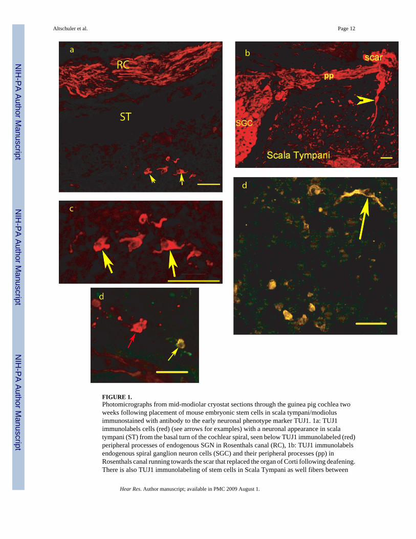
FIGURE 1.Photomicrographs from mid-modiolar cryostat sections through the guinea pig cochlea twoweeks following placement of mouse embryonic stem cells in scala tympani/modiolusimmunostained with antibody to the early neuronal phenotype marker TUJ1. 1a: TUJ1immunolabels cells (red) (see arrows for examples) with a neuronal appearance in scalatympani (ST) from the basal turn of the cochlear spiral, seen below TUJ1 immunolabeled (red)peripheral processes of endogenous SGN in Rosenthals canal (RC), 1b: TUJ1 immunolabelsendogenous spiral ganglion neuron cells (SGC) and their peripheral processes (pp) inRosenthals canal running towards the scar that replaced the organ of Corti following deafening.There is also TUJ1 immunolabeling of stem cells in Scala Tympani as well fibers between
Altschuler et al. Page 12
Hear Res. Author manuscript; available in PMC 2009 August 1.
NIH
-PA Author Manuscript
NIH
-PA Author Manuscript
NIH
-PA Author Manuscript

scala tympani and the scar region (arrowhead); 1c: Higher magnification view of the TUJ1immunolabeled cells in 1a (see arrows for examples).. 2d: Co-immunostaining for TUJ1 andVGLUT2, co-localization is yellow (see arrow for examples) and found in all labeled cells inthis field, 3d: Co-immunostaining for TUJ1 (Red) and GAT (green), co-localization is yellowand found in one cell (yellow arrow) in this field, while another cell in the center of the field(red arrow) is TUJ1 but not GAT immunolabeled. Bars = 50 microns.
Altschuler et al. Page 13
Hear Res. Author manuscript; available in PMC 2009 August 1.
NIH
-PA Author Manuscript
NIH
-PA Author Manuscript
NIH
-PA Author Manuscript
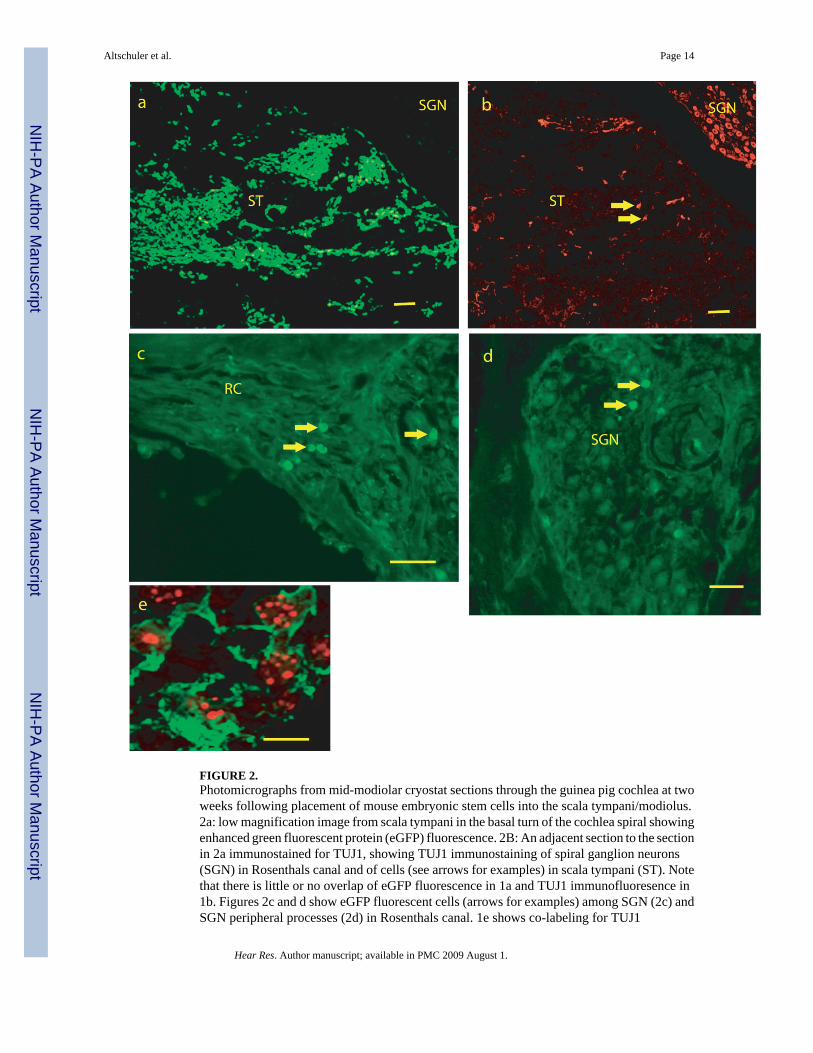
FIGURE 2.Photomicrographs from mid-modiolar cryostat sections through the guinea pig cochlea at twoweeks following placement of mouse embryonic stem cells into the scala tympani/modiolus.2a: low magnification image from scala tympani in the basal turn of the cochlea spiral showingenhanced green fluorescent protein (eGFP) fluorescence. 2B: An adjacent section to the sectionin 2a immunostained for TUJ1, showing TUJ1 immunostaining of spiral ganglion neurons(SGN) in Rosenthals canal and of cells (see arrows for examples) in scala tympani (ST). Notethat there is little or no overlap of eGFP fluorescence in 1a and TUJ1 immunofluoresence in1b. Figures 2c and d show eGFP fluorescent cells (arrows for examples) among SGN (2c) andSGN peripheral processes (2d) in Rosenthals canal. 1e shows co-labeling for TUJ1
Altschuler et al. Page 14
Hear Res. Author manuscript; available in PMC 2009 August 1.
NIH
-PA Author Manuscript
NIH
-PA Author Manuscript
NIH
-PA Author Manuscript
immunostaining (green and cytoplasmic) and in situ hybridization labeling of mouse genomicDNA (red, punctuate and nuclear) in cells in scala tympani of the basal turn of the cochlearspiral. SGN = region of Spiral Ganglion Neurons, RC = region of SGN peripheral processesin Rosenthals Canal, ST = Scala Tympani. Bars for 1 a–c = 100 microns, 1d = 50 microns, 1e = 10 microns.
Altschuler et al. Page 15
Hear Res. Author manuscript; available in PMC 2009 August 1.
NIH
-PA Author Manuscript
NIH
-PA Author Manuscript
NIH
-PA Author Manuscript