Regulation of Pol I-Transcribed 45S rDNA and Pol III-Transcribed 5S rDNA in Arabidopsis
Upload
independentCategory
view
0download
0
Cdc48/p97 mediates UV-dependent turnover of RNA Pol II
Rati Verma*, Robert Oania*, Ruihua Fang, Geoffrey T. Smith, and Raymond J. Deshaies*
*Howard Hughes Medical Institute, Division of Biology, California Institute of Technology,Pasadena, CA 91125
SUMMARYCdc48/p97 is an essential ATPase whose role in targeting substrates to the ubiquitin-proteasomesystem (UPS) remains unclear. Existing models posit that Cdc48 acts upstream of UPS receptors.To address this hypothesis, we examined the association of ubiquitin (Ub) conjugates with 26Sproteasomes. Unexpectedly, proteasomes isolated from cdc48 mutants contain high levels of Ubconjugates and mass spectrometry identified numerous non-proteasomal proteins, including Rpb1,the largest subunit of RNA Pol II. UV-induced turnover of Rpb1 depends upon Cdc48–Ufd1–Npl4, Ubx4 and the uncharacterized adaptor, Ubx5. Ubiquitinated Rpb1, proteasomes, and Cdc48accumulate on chromatin in UV-treated wildtype cells and the former two accumulate to higherlevels in mutant cells, suggesting that degradation of Rpb1 is facilitated by Cdc48 at sites ofstalled transcription. These data reveal an intimate coupling of function between proteasomes andCdc48 that we suggest is necessary to sustain processive degradation of unstable subunits of somemacromolecular protein complexes.
INTRODUCTIONBudding yeast Cdc48 is a member of the AAA (ATPases associated with various cellularactivities) protein family. Cdc48 has been implicated in a plethora of functions that includecell cycle regulation, membrane fusion, the stress response, and ER-associated degradation(ERAD) (Hirsch et al., 2009; Vembar and Brodsky, 2008). Its highly conserved mammaliancounterpart, p97, has been additionally implicated in reformation of the nucleus (Ramadanet al., 2007), organelle biogenesis (Halawani and Latterich, 2006), myofibril organization(Janiesch et al., 2007) and the degradation of proteins such as Hif1-α (Alexandru et al.,2008) and HMG-CoA reductase (DeBose-Boyd, 2008).
Two underlying properties of Cdc48/p97 contribute to its myriad functions: its ATPaseactivity and the ability to bind Ub (Ye, 2006). Together, these activities are thought tounderpin a ‘segregase’ function that separates ubiquitinated polypeptides from tightly-boundpartner proteins, enabling selective degradation of the former (Braun et al., 2002;Johnson etal., 1990). The Ub-binding activity of Cdc48 is synergistically enhanced by the binding ofadaptors belonging to the UFD (Ub-fusion degradation) pathway (Johnson et al., 1995) and/or to the UBX (Ub regulatory X) family, members of which possess Ub-binding domains(Schuberth and Buchberger, 2008). In its best-understood function – ERAD – Cdc48, inconjunction with its cofactors Ufd1 and Npl4, extracts misfolded proteins from the ER
© 2010 Elsevier Inc. All rights reserved.Correspondence to Raymond J. Deshaies ([email protected]) or Rati Verma ([email protected]).Publisher's Disclaimer: This is a PDF file of an unedited manuscript that has been accepted for publication. As a service to ourcustomers we are providing this early version of the manuscript. The manuscript will undergo copyediting, typesetting, and review ofthe resulting proof before it is published in its final citable form. Please note that during the production process errors may bediscovered which could affect the content, and all legal disclaimers that apply to the journal pertain.
Published as: Mol Cell. 2011 January 7; 41(1): 82–92.
HH
MI Author M
anuscriptH
HM
I Author Manuscript
HH
MI Author M
anuscript
membrane by virtue of its ATPase activity. The ubiquitinated substrates subsequentlyengage the UBA (Ub-associated) domain-containing receptors Rad23 and Dsk2, which inturn bind to the proteasome via their UbL (Ub-like) domains, thereby delivering substratesfor degradation (Raasi and Wolf, 2007).
The breadth of Cdc48’s involvement in the UPS remains poorly understood. Whereas Cdc48function clearly plays a prominent role in turnover of ER proteins (Jarosch et al., 2002;Rabinovich et al., 2002; Ravid et al., 2006; Ye et al., 2001), the connection between Cdc48and the UPS was first discovered based on the requirement of Cdc48 and UFD proteins forturnover of non-ER, soluble reporter substrates (Ghislain et al., 1996). Fractionation studiesand live imaging of Cdc48-GFP indicate that only a portion of yeast Cdc48 is peripherallybound to the ER/nuclear envelope, with most being partitioned between the cytosol andnucleus (Madeo et al., 1998)(Huh et al., 2003). Cdc48 has recently been shown to berequired for the degradation of the cytosolic protein fructose-1,6-bisphosphatase [FBPase](Barbin et al., 2010). Given the relative dearth of soluble (and physiological) UPSsubstrates, we wished to identify substrates whose degradation depends on Cdc48 anddetermine at what stage in their degradation Cdc48 is required.
Although there is general agreement that Cdc48 functions between Ub ligases and theproteasome, the only data that address the specific targeting step on which Cdc48 acts topromote turnover of soluble proteins are those underlying the ‘escort’ model (Richly et al.,2005). This model posits that Cdc48 takes over from Ub ligases by coordinating theelongation of a size-restricted yet degradation-competent chain upon substrate, whereuponthe substrate is handed off to an Ub chain receptor such as Rad23 for delivery to theproteasome. Sculpting of the Ub chain is achieved by a combination of trimming bydeubiquitinating enzymes (DUBs) such as Otu1 and chain extension by the ‘E4’ enzymeUfd2 (Rumpf and Jentsch, 2006). A central prediction of the escort model is thatproteasome-bound Ub conjugates should become depleted in Cdc48 mutants, much as isseen in rad23Δdsk2Δ double mutants (Elsasser et al., 2004), but this has not been evaluated.
Here, we characterize 26S proteasomes isolated from wildtype and temperature-sensitiveconditional cdc48-3 mutant cells. Contrary to expectation, Ub conjugates and many non-proteasomal proteins accumulated to high levels on proteasomes from cdc48-3 mutants. In-depth analysis of one particular Cdc48-dependent turnover substrate, Rpb1, reveals thatCdc48 and its adaptor Ubx5 function downstream of Cul3 Ub ligase to facilitate degradationof chromatin-bound Rpb1.
RESULTSUb conjugates and numerous proteins accumulate on proteasomes isolated from cdc48-3mutants
To determine if Ub conjugates are targeted to proteasomes in the absence of Cdc48 function,we isolated 26S proteasomes from wildtype and cdc48-3 cells. Cdc48 was not required forproteasome assembly (Figure S1A). We next evaluated Ub by immunoblotting andsurprisingly observed that proteasomes purified from cdc48-3 cells contained an increasedlevel of Ub conjugates compared to wildtype (Figure 1A). Indeed, the level of conjugatesassociated with proteasomes isolated from mutant cells was comparable to that observedwith proteasomes purified from wildtype cells treated with the proteasome inhibitor MG132.Additionally, the conjugates were of unusually high molecular weight (HMW; 110 kDa andgreater).
Verma et al. Page 2
Mol Cell. Author manuscript; available in PMC 2011 July 7.
HH
MI Author M
anuscriptH
HM
I Author Manuscript
HH
MI Author M
anuscript

One explanation for this result is that Cdc48 was required for proteasomal peptidase activity,but this was ruled out by an in-gel peptidase assay on 26S proteasomes fractionated bynative PAGE (Figure 1B).
The temperature-sensitive cdc48-3 allele bears two point mutations, P257L and R387K(Jeffrey Laney, personal communication). The molecular effects of these mutations are notknown, but the mutant protein is stable at the restrictive temperature (not shown). To assessmore directly if the ATPase activity of Cdc48 contributed to degradation of proteasome-bound Ub conjugates, mutant cells were transformed with plasmids that expressed eitherwild type or ATPase-dead (pcdc48Q2) Cdc48 (Ye et al., 2003). Ub conjugate accumulationon 26S proteasomes isolated from cdc48-3 was suppressed by co-expression of wild typeCdc48 but was further enhanced in cells expressing the ATPase-deficient Cdc48 (FigureS1B).
To compare the protein composition of 26S proteasome complexes affinity-purified fromwild type and cdc48-3 cells, we subjected the preparations to multidimensional massspectrometry (MudPIT) (Graumann et al., 2004; Mayor et al., 2005) using an LTQ (Figures1C and S1D) or LCQ (Figure S1C) mass spectrometer and measured spectrum counts,which correlate with protein abundance (Weiss et al., 2010). The ratio of total proteasomalsubunit spectral counts from mutant to wildtype was close to 1, indicating consistentrecovery of equivalent amounts of proteasome from wildtype and cdc48-3 cells. Moreover,the spectrum counts observed for individual subunits (Figure S1D) were similar for bothpreparations. We next determined the spectral counts for proteasome-interacting proteins(PIPs) in each preparation and calculated the ratio as above. Notably, and in striking contrastto proteasome subunits, spectrum counts for most PIPs were higher in cdc48-3 proteasomesand a large number of PIPs were found only in the mutant preparation (Figures 1C andS1C).
We reasoned that proteins found at elevated levels in proteasomes isolated from cdc48-3cells might be substrates that could not be degraded due to incomplete unfolding orextraction from binding partners. To identify candidate substrates, we mined data from fourdatasets: (i) all proteins in yeast with a half-life of less than 55 min (Belle et al., 2006); (ii)proteins reported to be ubiquitinated in a large-scale proteomic study (Peng et al., 2003);(iii) proteins that accumulate as Ub conjugates upon UPS inhibition (Mayor et al., 2007) and(iv) proteins reported in the Saccharomyces Genome Database (SGD,www.yeastgenome.org) to have physical or genetic links with Ub ligases. Several PIPs werefound to overlap between two datasets, an example being the septin-localized checkpointkinase Hsl1 that is degraded via APC/C in G1 (Burton and Solomon, 2000). However, onecandidate – the largest subunit of RNA polymerase II, Rpb1 – was identified in all fourdatasets. Although we chose to focus the remaining study on Rpb1, we did confirm that Hsl1degradation was dependent on Cdc48 (Figure S2).
Rpb1 is ubiquitinated in the absence of any inducing signal (Daulny et al., 2008; Peng et al.,2003) and also upon stalling of transcription by drugs such as 6-azauracil (6-AU) thatdeplete intracellular nucleotide pools (Somesh et al., 2007). These data suggest that theremay be basal turnover of Rpb1 fueled by stalling of transcription throughout the genome(Sigurdsson et al., 2010). However, upon induction of DNA damage by ultraviolet radiation(UV) or by the UV-mimetic 4-nitroquinoline-1-oxide (4-NQO), there is a large induction ofRpb1 turnover (Beaudenon et al., 1999; Chen et al., 2007; Ribar et al., 2006, 2007; Someshet al., 2005). To evaluate whether Rpb1 accumulates on proteasomes in UV-treated cdc48-3cells, we irradiated wild type (+/−MG132) and cdc48-3 cells with UV, isolated proteasomes,and immunoblotted for Rpb1. As shown in Figure 1D, MG132 induced accumulation of
Verma et al. Page 3
Mol Cell. Author manuscript; available in PMC 2011 July 7.
HH
MI Author M
anuscriptH
HM
I Author Manuscript
HH
MI Author M
anuscript
Rpb1 on proteasomes from UV-irradiated wildtype cells. Strikingly, there was also a strongaccumulation of Rpb1 on proteasomes isolated from UV-irradiated cdc48-3 cells.
UV-induced degradation of Rpb1 is dependent on Cdc48To address whether Cdc48 is involved in the degradation of Rpb1, we treated wildtype andcdc48-3 cells with UV and allowed them to recover in the presence of cycloheximide toblock protein synthesis. Aliquots were collected at various time intervals and Rpb1 levelsmonitored by immunoblotting. Rpb1 was degraded upon UV irradiation of wildtype but notcdc48-3 mutant cells (Figure 2A). Quantification of the immunoblot and normalization withtubulin yielded data similar to Figure 2C (not shown). A similar stabilization was observedwhen Rpb1 turnover was induced by 4-NQO, and Rpb1 accumulated on 26S proteasomesisolated from 4-NQO-treated cdc48-3 cells to levels comparable to wildtype cells treatedsimultaneously with 4NQO and MG132 (Figures S3C, D). Stabilization of Rpb1 in UV-treated cdc48-3 was not due to arrest of this mutant at the mitosis checkpoint (Cheng andChen, 2010), because we also observed stabilization in checkpoint-deficient cdc48-3mad2Δmutants (Figure S3A) and in cdc48-3 at a temperature (30°C) that is permissive for mitosis(Figure S3B). To determine if the ATPase activity of Cdc48 was required for Rpb1degradation, cdc48-3 mutants transformed with the same plasmids used for the experimentin Figure S1B were analyzed as described above. Whereas wildtype CDC48 rescued thedegradation defect of the cdc48-3 allele (only allele tested), the ATPase mutant did not(Figure 2B).
Prior work has shown that Rpb1 ubiquitination and degradation is compromised in UV-irradiated def1Δ cells. Surprisingly, Def1’s function in Rpb1 degradation can be fullybypassed if RAD26 is deleted, suggesting that Rad26, which is a DNA-dependent ATPasehomologous to mammalian Cockayne Syndrome B (CSB), prevents access to theubiquitination machinery (Woudstra et al., 2002).We wished to determine if Cdc48 mightfunction indirectly to overcome a RAD26-dependent barrier to Rpb1 degradation. WhereasRpb1 degradation was accelerated in rad26Δ cells, the cdc48-3 rad26Δ double mutant wasas compromised for degradation as cdc48-3 alone (Figure 2C). Given that Cdc48 remainedessential for Rpb1 turnover in rad26Δ cells, we reasoned that its role might be direct. Toassess this, we performed a coimmunoprecipitation experiment (Figure 2D). Rpb1 wasspecifically co-precipitated with Cdc48, but their interaction was UV-independent. Thisobservation suggests an additional, yet-to-be-discovered role for Cdc48 in Rpb1 transactionsindependent of UV damage (see below).
Dependency of Rpb1 degradation on Cdc48 adaptor proteinsCdc48 is a mechanochemical transducer that is coupled to its ubiquitinated substrates byadaptor proteins. As shown in Figure 3A, UV-dependent turnover of Rpb1 was alsodependent on the Ufd1–Npl4 adaptor complex. Cdc48/p97 also interacts with a second set ofputative substrate receptors, the UBX domain proteins. Shp1/Ubx1 and its mammalianhomolog p47 are believed to be substrate recruitment factors for Cdc48/p97 that aremutually exclusive with Ufd1–Npl4 (Table S2), and Ubx1 is required for the degradation ofUFD pathway reporter substrates in budding yeast (Schuberth et al., 2004). We evaluatedUV-induced Rpb1 turnover in mutants individually lacking each one of the UBX proteinsand found that Rpb1 was stabilized in ubx1Δ, ubx5Δ, and ubx4Δ, whereas degradation wasunimpeded in ubx2Δ, ubx3Δ, ubx6Δ, and ubx7Δ mutants (Figures 3B and C). As will beshown below, the defect in ubx1Δ was due to an indirect effect on UV signaling and thus wefocused our effort on Ubx4 and Ubx5. To determine if these proteins function in the same orparallel pathways, we quantified UV-induced Rpb1 turnover in single and double mutants.Rpb1 was more stable in ubx4Δ ubx5Δ than in either single mutant, suggesting that theseproteins act in parallel to promote Rpb1 degradation (Figure 3D).
Verma et al. Page 4
Mol Cell. Author manuscript; available in PMC 2011 July 7.
HH
MI Author M
anuscriptH
HM
I Author Manuscript
HH
MI Author M
anuscript
Ongoing transcription is a prerequisite for UV-induced RNA Pol II degradation (Anindya etal., 2007). To evaluate the possibility that the UBX proteins were involved in globaltranscription, we determined the sensitivity of the adaptor null mutants to the nucleotide-depleting drug 6-AU, which affects both elongation rate and processivity of RNA Pol II(Mason and Struhl, 2005). Whereas a null mutant lacking Dst1/TFIIS, a generaltranscription elongation factor, was 6-AU-sensitive, the ubxΔ mutants were 6-AU-insensitive (Figure S4A). Additionally, because Rpb1 degradation was significantlyimpaired in cdc48-3 mutants even at the semi-restrictive temperature 30°C (Figure S3B), weevaluated 6-AU-, and UV-sensitivity of cdc48-3 at 30°C. The sensitivities of the mutantwere akin to wildtype (Figures S4A, B). Thus, transcription elongation and UV signalingwere intact in cdc48-3 cells under conditions where UV-induced degradation of Rpb1 wasseverely compromised.
Ubiquitinated Rpb1 accumulates in cdc48-3, ubx4Δ, and ubx5Δ cellsWe have shown above that Cdc48 and three of its UBX domain adaptors were required forRpb1 turnover. The dependency on Cdc48–Ubx could reflect a requirement for segregase(Braun et al., 2002) activity such that the Rpb1 degron(s) remains masked throughinteraction with its holoenzyme partners in cdc48-3 or ubxΔ cells, blocking access to Ubligases, or the dependency could reflect a requirement for segregase/unfolding activity post-ubiquitination. To assess the modification status of Rpb1 in mutants, we immunoprecipitatedRpb1 from control and UV-irradiated wildtype cells and then blotted the sample with anRpb1 antibody. A discrete, UV-stimulated modification of a small fraction of Rpb1 wasreproducibly observed (Figure S5A). This modification was unaffected by cdc48-3, ubx4Δ,and ubx5Δ mutations but was compromised in ubx1Δ mutants (see Inputs/asterisk, Figure 4).Prior work established that this modification is due to sumoylation (Chen et al., 2009). Weconfirmed that Rpb1 was sumoylated in cdc48-3 but not ubx1Δ cells following UVirradiation (Figure S5B), and that sumoylation did not affect Rpb1 degradation (FigureS5C). These data establish that the UV damage response remains intact in cdc48-3, ubx4Δ,and ubx5Δ but not ubx1Δ mutants.
Typically, when degradation of a protein is blocked downstream of ubiquitination (e.g. withMG132), the fraction that accumulates as Ub conjugates is very small (Liu et al., 2007). Toenhance our ability to detect the HMW polyubiquitinated pool of Rpb1 (UbRpb1), weexploited the ability of the UBA domain of Dsk2 to bind polyUb conjugates (Mayor et al.,2005). Lysates from wildtype and mutant cells were bound to GstDsk2. As expected,GstDsk2 retrieved Ub conjugates but Gst did not (middle panels, Figures 4A, B, C).Immunoblotting for Rpb1 revealed the accumulation of HMW conjugates in cdc48-3 cells.No accumulation of Rpb1 Ub conjugates was observed in either the Ub-conjugating enzymemutant cdc34-2 or in the Ub ligase-deficient mutants rsp5-1 and cul3Δ. Cul3 generates theK48-linked Ub conjugates on Rpb1 that signal its degradation. It has been suggested thatCul3 functions either alone (Ribar et al., 2007) or downstream of Rsp5 (Harreman et al.,2009).
Amongst the ubxΔ mutants, UbRpb1 was recovered from UV-treated ubx4Δ and ubx5Δ cellsbut not ubx1Δ cells. Moreover, accumulation of constitutively ubiquitinated Rpb1 was alsodetected in cdc48-3, ubx4Δ, and ubx5Δ (Figures 4A, B and C), suggesting that Cdc48 and itsadaptors act upon Rpb1 stalled at naturally occurring pause sites such as at the 5’ end ofORFs (Sigurdsson et al., 2010; Wade and Struhl, 2008) or at non-canonical DNA structures(Hanawalt and Spivak, 2008). Interestingly, accumulation of UbRpb1 was enhanced inubx4Δubx5Δ double mutants compared to either single mutant (Figure 4C), providingfurther evidence that Ubx4 and Ubx5 function in parallel pathways. To determine if ubx4Δor ubx5Δ elicits a more global defect in proteolysis, proteasomes were isolated fromwildtype and mutant cells and immunoblotted for Ub conjugates. Strikingly, loss of either
Verma et al. Page 5
Mol Cell. Author manuscript; available in PMC 2011 July 7.
HH
MI Author M
anuscriptH
HM
I Author Manuscript
HH
MI Author M
anuscript
adaptor resulted in accumulation of HMW conjugates (Figure 4D), as was observed uponthermal inactivation of Cdc48 function (Figure 1A).
Ubx5 binds Rpb1 and Cul3 and functions downstream of Cul3To investigate in greater detail how UBX protein function contributes to Rpb1 degradation,we focused our attention on Ubx5 because ubx5Δ exhibits a stronger phenotype than ubx4Δin a) Rpb1 stabilization, b) UbRpb1 conjugate accumulation and c) Ub conjugateaccumulation at the proteasome. Since Cdc48 bound Rpb1 (Figure 2D) we tested whetherUbx5 behaves likewise. Ubx5 bound Cdc48 constitutively, as expected, but its associationwith Rpb1 was strongly enhanced by UV irradiation (Figure 5A). This is an interestingcounterpoint to Cdc48, which bound Rpb1 independently of DNA damage (Figure 2D).These observations suggest that Cdc48 engages in multiple transactions with Rpb1, withdifferent transactions mediated by different adaptors. Additionally, as observed for cdc48-3mutants, Rpb1 accumulation was enhanced on 26S proteasomes isolated under denaturingconditions from formaldehyde-crosslinked ubx5Δ cells (Figure 5B).
As noted above, both Rsp5 and Cul3 have been implicated in Rpb1 degradation. To testwhether Ubx5 functioned downstream of one or both of these Ub ligases, we evaluatedUbRpb1 levels in ubx5Δ mutants lacking one or the other enzyme. Interestingly, cul3Δ butnot rsp5-1 severely attenuated recovery of UbRpb1 from ubx5Δ cells (Figure 5C). Giventhat Cul3 and Ubx5 could be coimmunoprecipitated (Figure 5D), we suggest that theseproteins normally act sequentially, such that Rpb1 that has been ubiquitinated by Cul3 is‘handed off’ to Ubx5. These data extend observations made in human cells, where UBXD7is linked to the CUL2–VHL complex and its substrate HIF1α (Alexandru et al., 2008).Bioinformatic analysis suggests that Ubx5 is the ortholog of UBXD7 (Schuberth andBuchberger, 2008).
Accumulation of ubiquitinated Rpb1, Cdc48, and proteasome on chromatinThe results presented thus far suggest that Ubx5–Cdc48 acts on Rpb1, downstream of Cul3,to mediate degradation of ubiquitinated Rpb1 formed upon UV irradiation. The question thatarises next is whether this sequence of events occurs on chromatin. If so, we would expect todetect the core components of the system – including Cul3, Cdc48, and the proteasome – onchromatin in UV-treated cells. To address this question, we employed a standard chromatinfractionation method (Liang and Stillman, 1997). Interestingly, the bulk of Cul3 fractionatedwith chromatin (Figure 6A). Cul3 was constitutively associated with chromatin, which is notsurprising given that this enzyme is likely to have multiple substrates. To stringently probethe association of the abundant Cdc48 and proteasome complexes with chromatin, wesubjected a crude chromatin pellet to limited micrococcal nuclease (MNase) digestionfollowed by centrifugation to yield a highly purified chromatin pellet consisting ofpolynucleosomes (Frc6), and the supernatant (Frc7). Frc6, which provides a stringentassessment of chromatin association (Liang and Stillman, 1997), was enriched for Cdc48 aswell as both 19S and 20S proteasome components following UV irradiation, whereasHistone H3 was constitutively present and served as a loading control (Figure 6B). Giventhat both Cdc48 and the proteasome are likely to have many substrates, our data suggest thatUV damage triggers a generalized response that may result in the removal or degradation ofmany chromatin proteins.
Based on the observation that Cul3, Cdc48, and the proteasome can all be found onchromatin in UV-stressed cells, we hypothesized that ubiquitination and degradation ofRpb1 occurs on chromatin. To test this idea, we examined chromatin-bound Rpb1. Crudechromatin (see Figures S6A, S6B for fractionation and loading controls) isolated from cellsthat expressed Myc-tagged Ub was solubilized with Benzonase and ubiquitinated chromatin
Verma et al. Page 6
Mol Cell. Author manuscript; available in PMC 2011 July 7.
HH
MI Author M
anuscriptH
HM
I Author Manuscript
HH
MI Author M
anuscript

proteins released into the supernatant were isolated by virtue of the Myc tag.Immunoblotting for Rpb1 revealed the presence of Ub ladders on chromatin-associatedRpb1 (Figure 6C). Importantly, the abundance of chromatin-bound Rpb1 Ub conjugates wasgreatly increased upon UV irradiation of wild type cells, and increased to even higher levelsin UV irradiated cdc48-3 cells. Thus, it appears that Rpb1 is ubiquitinated on chromatin,consistent with the idea that the degradation cascade initiates with ubiquitination of Rpb1arrested at a thymidine dimer.
Since the recruitment of both Cdc48 and the 26S proteasome to chromatin was enhancedfollowing UV, we wished to determine if both complexes could be coimmunoprecipitated.Earlier work had relied on in vivo crosslinking to detect binding of Cdc48 to the proteasome(Guerrero et al., 2008). Immunoprecipitation of 26S proteasome under native conditions inthe presence of ATP and 0.15M salt resulted in barely-detectable recovery of Cdc48 (Figure6D), although Ubx5 could be readily detected after mild overproduction. Treating cells withthe proteasome inhibitor MG132 enhanced interaction of both Cdc48 and Ubx5 with theproteasome, suggesting that binding was mediated in part by ubiquitinated substrates. This isconsistent with the data in Figure 1A and supports the idea that Cdc48 can act uponsubstrates bound to the proteasome to enable their degradation.
DISCUSSIONImplicit in the models for Cdc48/p97 function based on its well-documented role inretrotranslocation of ubiquitinated proteins from the ER into the cytosol (Hirsch et al., 2009;Raasi and Wolf, 2007) and in the degradation of Ub fusion proteins (Richly et al., 2005) isthe notion that it functions upstream of the proteasome shuttle receptors Rad23 and Dsk2.These models imply that Cdc48 is required for delivery of a subset of UPS substrates (i.e.Cdc48/p97-dependent substrates) to the proteasome. Our data in the current studydemonstrating accumulation of Ub conjugates and PIPs on proteasomes isolated fromcdc48-3 mutants challenge whether these models serve as a general paradigm for thetemporal staging of Cdc48 function. Here, we validated Rpb1 as a bona-fide substrate ofCdc48 and show that in cells in which Rpb1 degradation was induced by UV radiation,Rpb1 was delivered to the proteasome in the absence of Cdc48 function.
The pathway for degradation of Rpb1 in UV-treated cellsOur model for Rbp1 degradation (Figure 7) is that Rpb1 stalled at lesion sites is most oftendislocated from the damage to enable repair followed by resumption of transcription.However, Rpb1 that is persistently blocked or refractory to dislocation is eventuallymodified by the Ub ligases Rsp5 and Cul3, of which the latter is believed to decorate Rpb1with degradation-competent Ub chains. These chains enable recruitment of both Cdc48 and26S proteasomes to the stalled complex, resulting in degradation of Rpb1 and recycling ofthe other holoenzyme subunits, which are not degraded (Malik et al., 2008). We envisionthat Cdc48 mediates extraction of UbRpb1 from chromatin-bound Pol II holoenzyme andthat this extraction and subsequent Rpb1 degradation are tightly coupled, possibly evencontemporaneous. Cdc48 carries out its function in conjunction with Ufd1–Npl4, Ubx4, andUbx5. We do not know how the activities of Ufd1–Npl4 relate to those of the Ubx proteins.On the other hand, Ubx4 and Ubx5 appear to act in parallel pathways to promote Rpb1degradation.
It has been proposed that Rsp5 attaches an initiator Ub to Rpb1, which primespolymerization of an Ub chain by Cul3 (Harreman et al., 2009). Alternatively, it has beensuggested that Cul3 carries out the entire reaction and the role of Rsp5 is indirect (Ribar etal., 2007). Ubiquitinated Rpb1 that accumulated in ubx5Δ cells was greatly diminished bydeletion of CUL3 but barely affected by rsp5-1. Either Rsp5 is not required to generate the
Verma et al. Page 7
Mol Cell. Author manuscript; available in PMC 2011 July 7.
HH
MI Author M
anuscriptH
HM
I Author Manuscript
HH
MI Author M
anuscript
UbRpb1 that accumulates in ubx5Δ or the rsp5-1 mutation was leaky under our conditions.Regardless of whether or not Cul3 extends Ub chains initiated by Rsp5, the epistasis dataand the protein-protein interactions point to Ubx5 acting directly upon ubiquitinated Rpb1formed by Cul3.
Components involved in the Rpb1 turnover pathway have been linked with chromatinregulation in other contexts. Cul3 functions as part of an Ub ligase that ubiquitinates theUV-damage sensor Rad4 (XPC)(Gillette et al., 2006; Ramsey et al., 2004). Cdc48 promotesdissociation of a repressor from its target promoter (Wilcox and Laney, 2009) and p97extracts protein kinase Aurora B from chromatin during nuclear envelope reassembly(Ramadan et al., 2007). Similarly, 19S and 20S subunits of the proteasome have been shownto bind promoters, ORFs, and termination regions, both constitutively and in response tosignals such as HO endonuclease-induced double strand breaks and UV (Auld et al., 2006;Collins and Tansey, 2006; Gillette et al., 2004; Krogan et al., 2004). These data suggest thatextraction of proteins from chromatin by Cdc48/p97 – either coupled to proteasomaldegradation or not – is likely to be a recurrent theme in chromatin regulation.
Proteasomal ATPases are insufficient for segregation and unfolding of unstable subunitsof some macromolecular complexes: Implications for dependency on Cdc48
An unexpected insight from our work is that in cells deprived of Ubx5-Cdc48 function,substrates such as Rpb1 can gain access to the proteasome. This raises the question of whysubstrates accumulate on the proteasome in these cells? We suggest that extraction ofUbRpb1 from chromatin-bound holoenzyme and targeting of UbRpb1 to the proteasome arenormally coupled processes that do not occur obligately in a fixed order. Thus, dependingupon the relative rates of disassembly versus targeting, in some instances the action ofCdc48 may precede substrate association with the proteasome (but this may be undesirableas explained below), whereas in other cases the substrate may associate with proteasomebefore Cdc48 can act. We suggest that the latter occurs far more commonly becausedisassembly/unfolding is likely to be much slower than targeting. For some substrates, likeRpb1 and the checkpoint kinase Hsl1, the disassembling and/or unfolding activitycontributed by the resident proteasomal ATPases (Rpt1-6,(Finley, 2009)) may beinsufficient to extract them from the macromolecular complexes in which they reside andthus they must wait for Cdc48 to complete its job before their degradation can commence.Coupling Cdc48 function to substrate degradation at the proteasome surface would reducethe probability that substrate that has been disassembled/unfolded by Cdc48 has anopportunity to reassemble, refold, or aggregate.
What is the molecular basis for determining whether or not a particular substrate requiresCdc48 for degradation? Prior work has established that stable domains within proteins areunfolded sequentially starting from the attachment point of the degradation signal (Lee et al.,2001; Prakash et al., 2004; Schrader et al., 2009). Rpb1 bound to multiple interactingproteins in the Pol II holoenzyme complex may lack an unstructured initiation site and thusmay additionally require Cdc48 for preprocessing, as recently reported for stably foldedUPS reporter substrates (Beskow et al., 2009). In stark contrast, the unfolding power of the26S proteasome is sufficient to degrade to completion ubiquitinated Sic1 (UbSic1), evenwhen it is tightly bound to S phase cyclin-dependent kinase (Verma et al., 2001). Apotentially distinguishing feature of Sic1 is that it contains an intrinsically disordered N-terminal domain that mediates interaction with Ub ligase SCF (Mittag et al., 2008) andcontains the primary sites of ubiquitination (Petroski and Deshaies, 2003). We speculate thatbecause unraveling of UbSic1 initiates from the disordered region proximal to the Ub chainattachment site, the proteasome can complete the task without assistance. By contrast, wepredict that Cdc48/p97 and its adaptors are required to sustain disassembly, unfolding, and
Verma et al. Page 8
Mol Cell. Author manuscript; available in PMC 2011 July 7.
HH
MI Author M
anuscriptH
HM
I Author Manuscript
HH
MI Author M
anuscript

processive degradation of soluble 26S proteasome substrates whose structural complexityoverwhelms the proteasomal ATPases.
EXPERIMENTAL PROCEDURESTurnover Analysis of UPS substrates
Aliquots of cultures treated as described in the respective figure legends (final A600 between1–2) were collected and drop-frozen in liquid nitrogen. Frozen cell pellets were thawed andwashed with ice-cold Buffer A (50 mM Tris, pH 7.5, 10 mM sodium azide, 10 mM EDTA,10 mM EGTA, 1X protease inhibitor tablet (Roche), 10 mM NEM, 50 mM NaF, 60 mM β-glycerophosphate, 10 mM sodium pyrophoshate). The cell pellets were then immersed inboiling water for three minutes after which they were suspended in 1X SDS buffer (37.5 µl /O.D. unit). An equal volume of glass beads (Sigma, 425–600 microns, acid washed) wasadded, and cells were lysed by vortexing in Fast Prep-24 (MP) for 45 seconds at a setting of6.5, and boiled again for 4 mins. Boiled lysates were centrifuged at 16,000 X g for 1 minute.Aliquots were resolved by SDS-PAGE, transferred to nitrocellulose, and stained withPonceau S to determine equivalent loading of protein extracts. The nitrocellulose filters wereimmunoblotted with desired antibody and developed by ECL, or quantified by LI-COROdyssey using IR dye-linked secondary antibodies (Invitrogen). Anti-PSTAIRE (SantaCruz), and anti-tubulin (Sigma) served as loading controls. Following quantification, all datawere plotted using Prism software on a logarithmic scale on the y-axis. Anti-Rpb1 (clones8WG, 4H8), anti-Myc, and anti-HA antibodies were from Covance.
UV and 4-NQO treatment of yeast cellsOvernight cultures were diluted to an optical density (O.D.) of around 0.2. When cellsreached an O.D. around 1, they were centrifuged and resuspended in 80% of the originalculture volume in 2% Dextrose (or the sugar being used for the experiment). All thefollowing steps were carried out in the dark, under red safe light. Cultures were exposed to400 J/m2 of UV irradiation in either petri dishes, or glass trays, depending on volume.Irradiation was performed by using calibrated germicidal lamps (254 nm UV Lamp, UVP,Model # XX-405) or a pre-warmed and calibrated Stratalinker unit (Stratagene, Model2400). The UV meter used for calibration was purchased from UVP, Inc, CA (Model #J225).
Cells were harvested immediately after UV, or when recovery was monitored, the 2%dextrose suspension was diluted into 20 % volume of 5X prewarmed media containing 100ug /ml cycloheximde, and outgrowth continued in the dark. For 4-NQO treatment,exponential cultures were treated with the desired concentration from a 10 mg /ml stockstored in the dark.
Isolation of Chromatin FractionEssentially, the method of Liang and Stillman (Liang and Stillman, 1997) was followed withsome modifications. In the original protocol, chromatin was isolated from yeast spheroplastsand this method was followed in Figure 6A. Because Rpb1 was considerably degradedduring the process of spheroplasting (particularly in UV-treated cultures), the method wasmodified to isolate chromatin fractions from yeast cells ground with a mortar pestle chilledin liquid nitrogen as described earlier (Verma and Deshaies, 2005) for Figures 6B and 6C.The minimum culture volume that could be ground with easy recovery of cell powder was350 ml. Ground powder was weighed, and 2X (wt /volume) Extraction Buffer (EB)containing 50 mM Hepes, pH 7.5, 100 mM KCl, 0.25 % Triton, 2.5 mM MgCl2, 25 mMNEM, 50 mM β-glycerophosphate, 5 mM sodium pyrophosphate, 1X protease inhibitortablet +EDTA, 0.5 mM AEBSF was added. Unlysed cells were pelleted at 5000 r.p.m (all
Verma et al. Page 9
Mol Cell. Author manuscript; available in PMC 2011 July 7.
HH
MI Author M
anuscriptH
HM
I Author Manuscript
HH
MI Author M
anuscript
centrifugations in refrigerated Eppendorf model 5417R) for 1 min. Ubiquitin-aldehyde (1µM) was added to the supernatant and an aliquot (25 % vol/vol) was saved as WCE (Frc1).Another 25 % of lysate was underlayered with 50 % volume of 30 % sucrose andcentrifuged at 16,4000 rpm for 15 minutes. The pellet was saved as low speed pellet (Frc3)and the supernatant was designated “low-speed supernatant” (Frc2). The remaining 50 % oflysate was underlayered with sucrose and centrifuged to generate a second set of Fractions 2and 3. Crude Fraction 3 was washed with EB at 12,000 rpm for 8 mins and the pellet wasresuspended in EB. Resuspended chromatin pellet (375 µl) was pre-warmed to 37°C for 3min after supplementing with CaCl2 (2 mM final) and digested with 2 µl 1:10 MNase (50 %glycerol stock solution; from Sigma, 204.3 Units / mg protein) for another 3 min. Digestionwas stopped by the addition of EGTA to 5 mM. A low-speed micrococcal nuclease-treatedpellet (Frc4) was generated by centrifuging at 10,000 rpm for 2 min at 4°C. The supernatantwas centrifuged at 50,000 r.p.m. (Sorvall RC M120EX, Rotor # RP100AT4) for one hour togenerate the high-speed pellet (Frc6), and supernatant (Frc7). Limited nuclease digestionretains chromatin proteins in the high-speed pellet fraction.
Solubilization of Chromatin—Washed crude chromatin pellet (FrC) was re-suspendedin EB (750 µl) and NaCl was added to yield a final salt concentration of 0.6M. Ubiquitinaldehyde (1 µM) and benzonase (1 µl of 250 U/µl, Novagen) were added and the samplewas incubated for 30 min on ice, sonicated for 15 sec (Amplitude at 20%), and thencentrifuged at 10,000 rpm for 2 min. The supernatant was used for immunoprecipitations.
Supplementary MaterialRefer to Web version on PubMed Central for supplementary material.
AcknowledgmentsWe thank D. G. Drubin, G. Hartzog, M. Hochstrasser, J. Huibregtse, E. Johnson, D. Kellogg, T. Miyakawa, Y.Saeki, W. Seufert, P. Silver, T. Sommer, A. Toh-E, A. Varshavsky, F. Winston and Y.Ye for yeast strains,expression plasmids, and antibodies. We thank the members of the Deshaies lab for helpful discussions. R.J.D. is anInvestigator of the Howard Hughes Medical Institute, which supported this work.
REFERENCESAlberts SM, Sonntag C, Schafer A, Wolf DH. Ubx4 modulates cdc48 activity and influences
degradation of misfolded proteins of the endoplasmic reticulum. J Biol Chem. 2009; 284:16082–16089. [PubMed: 19359248]
Alexandru G, Graumann J, Smith GT, Kolawa NJ, Fang R, Deshaies RJ. UBXD7 binds multipleubiquitin ligases and implicates p97 in HIF1alpha turnover. Cell. 2008; 134:804–816. [PubMed:18775313]
Anindya R, Aygun O, Svejstrup JQ. Damage-induced ubiquitylation of human RNA polymerase II bythe ubiquitin ligase Nedd4, but not Cockayne syndrome proteins or BRCA1. Mol Cell. 2007;28:386–397. [PubMed: 17996703]
Auld KL, Brown CR, Casolari JM, Komili S, Silver PA. Genomic association of the proteasomedemonstrates overlapping gene regulatory activity with transcription factor substrates. Mol Cell.2006; 21:861–871. [PubMed: 16543154]
Barbin L, Eisele F, Santt O, Wolf DH. The Cdc48-Ufd1-Npl4 complex is central in ubiquitin-proteasome triggered catabolite degradation of fructose-1,6-bisphosphatase. Biochem Biophys ResCommun. 2010; 394:335–341. [PubMed: 20206597]
Beaudenon SL, Huacani MR, Wang G, McDonnell DP, Huibregtse JM. Rsp5 ubiquitin-protein ligasemediates DNA damage-induced degradation of the large subunit of RNA polymerase II inSaccharomyces cerevisiae. Mol Cell Biol. 1999; 19:6972–6979. [PubMed: 10490634]
Verma et al. Page 10
Mol Cell. Author manuscript; available in PMC 2011 July 7.
HH
MI Author M
anuscriptH
HM
I Author Manuscript
HH
MI Author M
anuscript
Belle A, Tanay A, Bitincka L, Shamir R, O'Shea EK. Quantification of protein half-lives in thebudding yeast proteome. Proc Natl Acad Sci U S A. 2006; 103:13004–13009. [PubMed: 16916930]
Beskow A, Grimberg KB, Bott LC, Salomons FA, Dantuma NP, Young P. A conserved unfoldaseactivity for the p97 AAA-ATPase in proteasomal degradation. J Mol Biol. 2009; 394:732–746.[PubMed: 19782090]
Braun S, Matuschewski K, Rape M, Thoms S, Jentsch S. Role of the ubiquitin-selectiveCDC48(UFD1/NPL4)chaperone (segregase) in ERAD of OLE1 and other substrates. EMBO J.2002; 21:615–621. [PubMed: 11847109]
Burton JL, Solomon MJ. Hsl1p, a Swe1p inhibitor, is degraded via the anaphase-promoting complex.Mol Cell Biol. 2000; 20:4614–4625. [PubMed: 10848588]
Chen X, Ding B, LeJeune D, Ruggiero C, Li S. Rpb1 sumoylation in response to UV radiation ortranscriptional impairment in yeast. PLoS One. 2009; 4:e5267. [PubMed: 19384408]
Chen X, Ruggiero C, Li S. Yeast Rpb9 plays an important role in ubiquitylation and degradation ofRpb1 in response to UV-induced DNA damage. Mol Cell Biol. 2007; 27:4617–4625. [PubMed:17452455]
Cheng YL, Chen RH. The AAA-ATPase Cdc48 and cofactor Shp1 promote chromosome bi-orientation by balancing Aurora B activity. J Cell Sci. 2010; 123:2025–2034. [PubMed:20483956]
Collins GA, Tansey WP. The proteasome: a utility tool for transcription? Curr Opin Genet Dev. 2006;16:197–202. [PubMed: 16503126]
Daulny A, Geng F, Muratani M, Geisinger JM, Salghetti SE, Tansey WP. Modulation of RNApolymerase II subunit composition by ubiquitylation. Proc Natl Acad Sci U S A. 2008;105:19649–19654. [PubMed: 19064926]
Daulny A, Tansey WP. Damage control: DNA repair, transcription, and the ubiquitin-proteasomesystem. DNA Repair (Amst). 2009; 8:444–448. [PubMed: 19272841]
DeBose-Boyd RA. Feedback regulation of cholesterol synthesis: sterol-accelerated ubiquitination anddegradation of HMG CoA reductase. Cell Res. 2008; 18:609–621. [PubMed: 18504457]
Decottignies A, Evain A, Ghislain M. Binding of Cdc48p to a ubiquitin-related UBX domain fromnovel yeast proteins involved in intracellular proteolysis and sporulation. Yeast. 2004; 21:127–139. [PubMed: 14755638]
Elsasser S, Chandler-Militello D, Muller B, Hanna J, Finley D. Rad23 and Rpn10 serve as alternativeubiquitin receptors for the proteasome. J Biol Chem. 2004; 279:26817–26822. [PubMed:15117949]
Finley D. Recognition and processing of ubiquitin-protein conjugates by the proteasome. Annu RevBiochem. 2009; 78:477–513. [PubMed: 19489727]
Ghislain M, Dohmen RJ, Levy F, Varshavsky A. Cdc48p interacts with Ufd3p, a WD repeat proteinrequired for ubiquitin-mediated proteolysis in Saccharomyces cerevisiae. EMBO J. 1996;15:4884–4899. [PubMed: 8890162]
Gillette TG, Gonzalez F, Delahodde A, Johnston SA, Kodadek T. Physical and functional associationof RNA polymerase II and the proteasome. Proc Natl Acad Sci U S A. 2004; 101:5904–5909.[PubMed: 15069196]
Gillette TG, Yu S, Zhou Z, Waters R, Johnston SA, Reed SH. Distinct functions of the ubiquitin-proteasome pathway influence nucleotide excision repair. EMBO J. 2006; 25:2529–2538.[PubMed: 16675952]
Graumann J, Dunipace LA, Seol JH, McDonald WH, Yates JR 3rd, Wold BJ, Deshaies RJ.Applicability of tandem affinity purification MudPIT to pathway proteomics in yeast. Mol CellProteomics. 2004; 3:226–237. [PubMed: 14660704]
Guerrero C, Milenkovic T, Przulj N, Kaiser P, Huang L. Characterization of the proteasomeinteraction network using a QTAX-based tag-team strategy and protein interaction networkanalysis. Proc Natl Acad Sci U S A. 2008; 105:13333–13338. [PubMed: 18757749]
Halawani D, Latterich M. p97: The cell's molecular purgatory? Mol Cell. 2006; 22:713–717.[PubMed: 16793541]
Hanawalt PC, Spivak G. Transcription-coupled DNA repair: two decades of progress and surprises.Nat Rev Mol Cell Biol. 2008; 9:958–970. [PubMed: 19023283]
Verma et al. Page 11
Mol Cell. Author manuscript; available in PMC 2011 July 7.
HH
MI Author M
anuscriptH
HM
I Author Manuscript
HH
MI Author M
anuscript
Harreman M, Taschner M, Sigurdsson S, Anindya R, Reid J, Somesh B, Kong SE, Banks CA,Conaway RC, Conaway JW, et al. Distinct ubiquitin ligases act sequentially for RNA polymeraseII polyubiquitylation. Proc Natl Acad Sci U S A. 2009
Hirsch C, Gauss R, Horn SC, Neuber O, Sommer T. The ubiquitylation machinery of the endoplasmicreticulum. Nature. 2009; 458:453–460. [PubMed: 19325625]
Huh WK, Falvo JV, Gerke LC, Carroll AS, Howson RW, Weissman JS, O'Shea EK. Global analysisof protein localization in budding yeast. Nature. 2003; 425:686–691. [PubMed: 14562095]
Janiesch PC, Kim J, Mouysset J, Barikbin R, Lochmuller H, Cassata G, Krause S, Hoppe T. Theubiquitin-selective chaperone CDC-48/p97 links myosin assembly to human myopathy. Nat CellBiol. 2007; 9:379–390. [PubMed: 17369820]
Jarosch E, Taxis C, Volkwein C, Bordallo J, Finley D, Wolf DH, Sommer T. Protein dislocation fromthe ER requires polyubiquitination and the AAA-ATPase Cdc48. Nat Cell Biol. 2002; 4:134–139.[PubMed: 11813000]
Johnson ES, Gonda DK, Varshavsky A. cis-trans recognition and subunit-specific degradation ofshort-lived proteins. Nature. 1990; 346:287–291. [PubMed: 2165217]
Johnson ES, Ma PC, Ota IM, Varshavsky A. A proteolytic pathway that recognizes ubiquitin as adegradation signal. J Biol Chem. 1995; 270:17442–17456. [PubMed: 7615550]
Krogan NJ, Lam MH, Fillingham J, Keogh MC, Gebbia M, Li J, Datta N, Cagney G, Buratowski S,Emili A, et al. Proteasome involvement in the repair of DNA double-strand breaks. Mol Cell.2004; 16:1027–1034. [PubMed: 15610744]
Lee C, Schwartz MP, Prakash S, Iwakura M, Matouschek A. ATP-dependent proteases degrade theirsubstrates by processively unraveling them from the degradation signal. Mol Cell. 2001; 7:627–637. [PubMed: 11463387]
Liang C, Stillman B. Persistent initiation of DNA replication and chromatin-bound MCM proteinsduring the cell cycle in cdc6 mutants. Genes Dev. 1997; 11:3375–3386. [PubMed: 9407030]
Liu C, Apodaca J, Davis LE, Rao H. Proteasome inhibition in wild-type yeast Saccharomycescerevisiae cells. Biotechniques. 2007; 42:158, 160, 162. [PubMed: 17373478]
Madeo F, Schlauer J, Zischka H, Mecke D, Frohlich KU. Tyrosine phosphorylation regulates cellcycle-dependent nuclear localization of Cdc48p. Mol Biol Cell. 1998; 9:131–141. [PubMed:9436996]
Malik S, Bagla S, Chaurasia P, Duan Z, Bhaumik SR. Elongating RNA polymerase II is disassembledthrough specific degradation of its largest but not other subunits in response to DNA damage invivo. J Biol Chem. 2008; 283:6897–6905. [PubMed: 18195014]
Mason PB, Struhl K. Distinction and relationship between elongation rate and processivity of RNApolymerase II in vivo. Mol Cell. 2005; 17:831–840. [PubMed: 15780939]
Mayor T, Graumann J, Bryan J, MacCoss MJ, Deshaies RJ. Quantitative profiling of ubiquitylatedproteins reveals proteasome substrates and the substrate repertoire influenced by the Rpn10receptor pathway. Mol Cell Proteomics. 2007; 6:1885–1895. [PubMed: 17644757]
Mayor T, Lipford JR, Graumann J, Smith GT, Deshaies RJ. Analysis of polyubiquitin conjugatesreveals that the Rpn10 substrate receptor contributes to the turnover of multiple proteasometargets. Mol Cell Proteomics. 2005; 4:741–751. [PubMed: 15699485]
Mittag T, Orlicky S, Choy WY, Tang X, Lin H, Sicheri F, Kay LE, Tyers M, Forman-Kay JD.Dynamic equilibrium engagement of a polyvalent ligand with a single-site receptor. Proc NatlAcad Sci U S A. 2008; 105:17772–17777. [PubMed: 19008353]
Neuber O, Jarosch E, Volkwein C, Walter J, Sommer T. Ubx2 links the Cdc48 complex to ER-associated protein degradation. Nat Cell Biol. 2005; 7:993–998. [PubMed: 16179953]
Peng J, Schwartz D, Elias JE, Thoreen CC, Cheng D, Marsischky G, Roelofs J, Finley D, Gygi SP. Aproteomics approach to understanding protein ubiquitination. Nat Biotechnol. 2003; 21:921–926.[PubMed: 12872131]
Petroski MD, Deshaies RJ. Context of multiubiquitin chain attachment influences the rate of Sic1degradation. Mol Cell. 2003; 11:1435–1444. [PubMed: 12820958]
Prakash S, Tian L, Ratliff KS, Lehotzky RE, Matouschek A. An unstructured initiation site is requiredfor efficient proteasome-mediated degradation. Nat Struct Mol Biol. 2004; 11:830–837. [PubMed:15311270]
Verma et al. Page 12
Mol Cell. Author manuscript; available in PMC 2011 July 7.
HH
MI Author M
anuscriptH
HM
I Author Manuscript
HH
MI Author M
anuscript
Raasi S, Wolf DH. Ubiquitin receptors and ERAD: a network of pathways to the proteasome. SeminCell Dev Biol. 2007; 18:780–791. [PubMed: 17942349]
Rabinovich E, Kerem A, Frohlich KU, Diamant N, Bar-Nun S. AAA-ATPase p97/Cdc48p, a cytosolicchaperone required for endoplasmic reticulum-associated protein degradation. Mol Cell Biol.2002; 22:626–634. [PubMed: 11756557]
Ramadan K, Bruderer R, Spiga FM, Popp O, Baur T, Gotta M, Meyer HH. Cdc48/p97 promotesreformation of the nucleus by extracting the kinase Aurora B from chromatin. Nature. 2007;450:1258–1262. [PubMed: 18097415]
Ramsey KL, Smith JJ, Dasgupta A, Maqani N, Grant P, Auble DT. The NEF4 complex regulates Rad4levels and utilizes Snf2/Swi2-related ATPase activity for nucleotide excision repair. Mol Cell Biol.2004; 24:6362–6378. [PubMed: 15226437]
Ravid T, Kreft SG, Hochstrasser M. Membrane and soluble substrates of the Doa10 ubiquitin ligaseare degraded by distinct pathways. EMBO J. 2006; 25:533–543. [PubMed: 16437165]
Reid J, Svejstrup JQ. DNA damage-induced Def1-RNA polymerase II interaction and Def1requirement for polymerase ubiquitylation in vitro. J Biol Chem. 2004; 279:29875–29878.[PubMed: 15166235]
Ribar B, Prakash L, Prakash S. Requirement of ELC1 for RNA polymerase II polyubiquitylation anddegradation in response to DNA damage in Saccharomyces cerevisiae. Mol Cell Biol. 2006;26:3999–4005. [PubMed: 16705154]
Ribar B, Prakash L, Prakash S. ELA1 and CUL3 are required along with ELC1 for RNA polymerase IIpolyubiquitylation and degradation in DNA-damaged yeast cells. Mol Cell Biol. 2007; 27:3211–3216. [PubMed: 17296727]
Richly H, Rape M, Braun S, Rumpf S, Hoege C, Jentsch S. A series of ubiquitin binding factorsconnects CDC48/p97 to substrate multiubiquitylation and proteasomal targeting. Cell. 2005;120:73–84. [PubMed: 15652483]
Rumpf S, Jentsch S. Functional division of substrate processing cofactors of the ubiquitin-selectiveCdc48 chaperone. Mol Cell. 2006; 21:261–269. [PubMed: 16427015]
Schrader EK, Harstad KG, Matouschek A. Targeting proteins for degradation. Nat Chem Biol. 2009;5:815–822. [PubMed: 19841631]
Schuberth C, Buchberger A. Membrane-bound Ubx2 recruits Cdc48 to ubiquitin ligases and theirsubstrates to ensure efficient ER-associated protein degradation. Nat Cell Biol. 2005; 7:999–1006.[PubMed: 16179952]
Schuberth C, Buchberger A. UBX domain proteins: major regulators of the AAA ATPase Cdc48/p97.Cell Mol Life Sci. 2008; 65:2360–2371. [PubMed: 18438607]
Schuberth C, Richly H, Rumpf S, Buchberger A. Shp1 and Ubx2 are adaptors of Cdc48 involved inubiquitin-dependent protein degradation. EMBO Rep. 2004; 5:818–824. [PubMed: 15258615]
Sigurdsson S, Dirac-Svejstrup AB, Svejstrup JQ. Evidence that transcript cleavage is essential forRNA polymerase II transcription and cell viability. Mol Cell. 2010; 38:202–210. [PubMed:20417599]
Somesh BP, Reid J, Liu WF, Sogaard TM, Erdjument-Bromage H, Tempst P, Svejstrup JQ. Multiplemechanisms confining RNA polymerase II ubiquitylation to polymerases undergoingtranscriptional arrest. Cell. 2005; 121:913–923. [PubMed: 15960978]
Somesh BP, Sigurdsson S, Saeki H, Erdjument-Bromage H, Tempst P, Svejstrup JQ. Communicationbetween distant sites in RNA polymerase II through ubiquitylation factors and the polymeraseCTD. Cell. 2007; 129:57–68. [PubMed: 17418786]
Vembar SS, Brodsky JL. One step at a time: endoplasmic reticulum-associated degradation. Nat RevMol Cell Biol. 2008; 9:944–957. [PubMed: 19002207]
Verma R, Deshaies RJ. Assaying degradation and deubiquitination of a ubiquitinated substrate bypurified 26S proteasomes. Methods Enzymol. 2005; 398:391–399. [PubMed: 16275345]
Verma R, McDonald H, Yates JR 3rd, Deshaies RJ. Selective degradation of ubiquitinated Sic1 bypurified 26S proteasome yields active S phase cyclin-Cdk. Mol Cell. 2001; 8:439–448. [PubMed:11545745]
Wade JT, Struhl K. The transition from transcriptional initiation to elongation. Curr Opin Genet Dev.2008; 18:130–136. [PubMed: 18282700]
Verma et al. Page 13
Mol Cell. Author manuscript; available in PMC 2011 July 7.
HH
MI Author M
anuscriptH
HM
I Author Manuscript
HH
MI Author M
anuscript
Weiss M, Schrimpf S, Hengartner MO, Lercher MJ, von Mering C. Shotgun proteomics data frommultiple organisms reveals remarkable quantitative conservation of the eukaryotic core proteome.Proteomics. 2010; 10:1297–1306. [PubMed: 20077411]
Wilcox AJ, Laney JD. A ubiquitin-selective AAA-ATPase mediates transcriptional switching byremodelling a repressor-promoter DNA complex. Nat Cell Biol. 2009; 11:1481–1486. [PubMed:19915556]
Woudstra EC, Gilbert C, Fellows J, Jansen L, Brouwer J, Erdjument-Bromage H, Tempst P, SvejstrupJQ. A Rad26-Def1 complex coordinates repair and RNA pol II proteolysis in response to DNAdamage. Nature. 2002; 415:929–933. [PubMed: 11859374]
Ye Y. Diverse functions with a common regulator: ubiquitin takes command of an AAA ATPase. JStruct Biol. 2006; 156:29–40. [PubMed: 16529947]
Ye Y, Meyer HH, Rapoport TA. The AAA ATPase Cdc48/p97 and its partners transport proteins fromthe ER into the cytosol. Nature. 2001; 414:652–656. [PubMed: 11740563]
Ye Y, Meyer HH, Rapoport TA. Function of the p97-Ufd1-Npl4 complex in retrotranslocation fromthe ER to the cytosol: dual recognition of nonubiquitinated polypeptide segments andpolyubiquitin chains. J Cell Biol. 2003; 162:71–84. [PubMed: 12847084]
Verma et al. Page 14
Mol Cell. Author manuscript; available in PMC 2011 July 7.
HH
MI Author M
anuscriptH
HM
I Author Manuscript
HH
MI Author M
anuscript

Figure 1. Ubiquitinated proteins are targeted to 26S proteasomes in cdc48-3 mutant cells(A) Ub conjugates accumulated on proteasomes in cdc48-3. Wildtype (WT; RJD3437) andmutant (RJD3454) cells expressing myc-tagged Pre1 (or not, RJD4090) were shifted to 37°Cfor 90 min and treated with 40 µM MG132 for 30 min. Proteasomes were isolated by anti-myc immunoprecipitation (IP) and immunoblotted (IB) for Ub and Pre1-myc (upper panels).Input extracts were blotted for Ub and Rpn3 (lower panels).(B) Accumulation of conjugates on proteasome was not due to a defect in chymotrypticactivity in cdc48-3 mutants. Isolated 26S proteasomes fractionated by native PAGE wereeither stained with Coomassie Blue (CB) or processed for in-gel peptidase activity using thefluorescent reporter substrate LLVY-AMC. R1C and R2C refer to 20S complexes capped by1 or 2 19S regulatory particles, respectively.(C) Numerous proteins accumulated on proteasomes in cdc48-3 mutants. Multidimensionalmass spectrometry (MudPIT) was performed on an LTQ mass spectrometer using 26Sproteasomes isolated from WT and mutant (RJD2902) cells grown at 37°C for 2 hours. Theactual ratio of total spectral counts of proteasomal subunits from mutant to WT was 0.8 and
Verma et al. Page 15
Mol Cell. Author manuscript; available in PMC 2011 July 7.
HH
MI Author M
anuscriptH
HM
I Author Manuscript
HH
MI Author M
anuscript
was normalized to one. The same normalization factor was applied for spectral countsobtained for the proteasome-interacting proteins (PIPs). ∞ refers to proteins found incdc48-3 but not WT proteasomes.(D) Rpb1 accumulated on proteasome in cdc48-3 mutant treated with UV. WT and mutantcells were shifted to 37°C for 90 minutes and UV irradiated. Cells were recovered at 37°Cfor 30 minutes in the absence or presence of MG132. Input cell extracts and proteasomesimmunoprecipitated (IP) with anti-myc were blotted (IB) for Rpb1 using 4H8 antibody.See also Figure S1.
Verma et al. Page 16
Mol Cell. Author manuscript; available in PMC 2011 July 7.
HH
MI Author M
anuscriptH
HM
I Author Manuscript
HH
MI Author M
anuscript
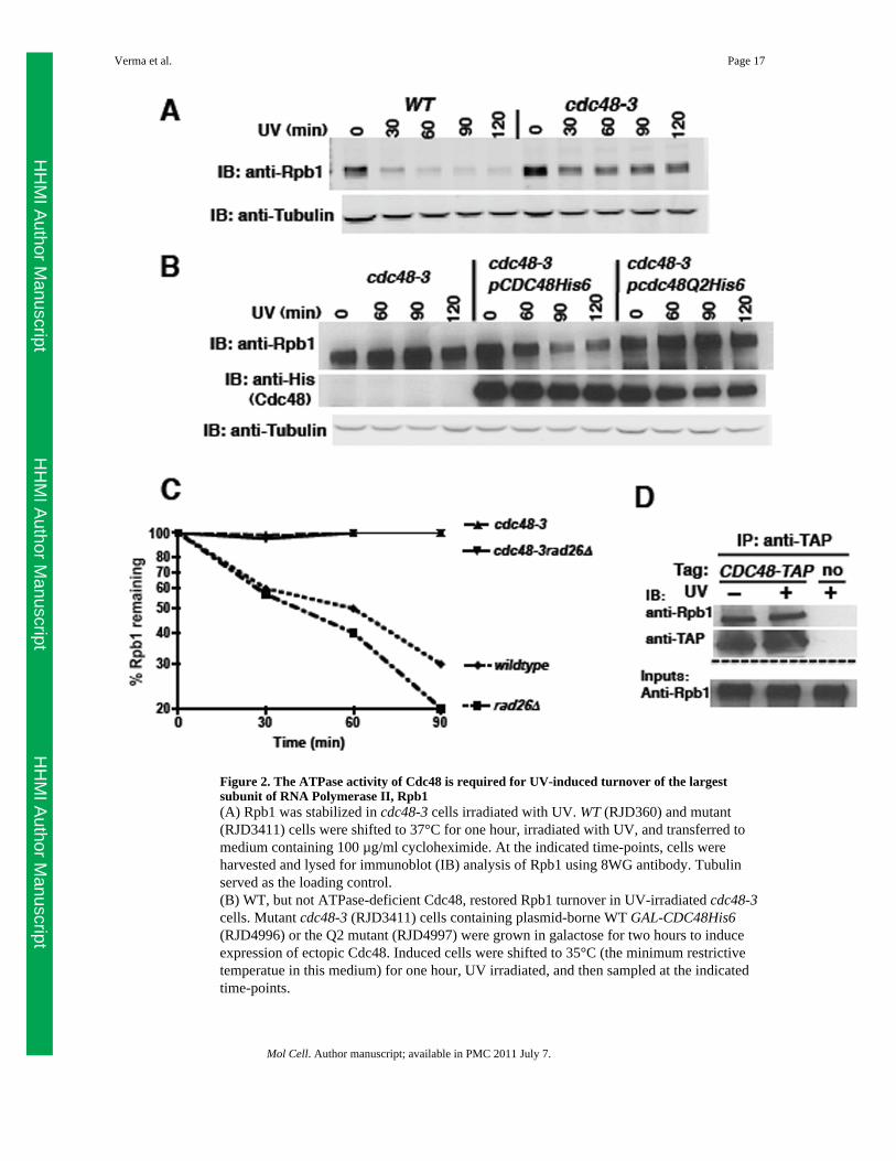
Figure 2. The ATPase activity of Cdc48 is required for UV-induced turnover of the largestsubunit of RNA Polymerase II, Rpb1(A) Rpb1 was stabilized in cdc48-3 cells irradiated with UV. WT (RJD360) and mutant(RJD3411) cells were shifted to 37°C for one hour, irradiated with UV, and transferred tomedium containing 100 µg/ml cycloheximide. At the indicated time-points, cells wereharvested and lysed for immunoblot (IB) analysis of Rpb1 using 8WG antibody. Tubulinserved as the loading control.(B) WT, but not ATPase-deficient Cdc48, restored Rpb1 turnover in UV-irradiated cdc48-3cells. Mutant cdc48-3 (RJD3411) cells containing plasmid-borne WT GAL-CDC48His6(RJD4996) or the Q2 mutant (RJD4997) were grown in galactose for two hours to induceexpression of ectopic Cdc48. Induced cells were shifted to 35°C (the minimum restrictivetemperatue in this medium) for one hour, UV irradiated, and then sampled at the indicatedtime-points.
Verma et al. Page 17
Mol Cell. Author manuscript; available in PMC 2011 July 7.
HH
MI Author M
anuscriptH
HM
I Author Manuscript
HH
MI Author M
anuscript
(C) Stabilization of Rpb1 in cdc48-3 was not suppressed by rad26Δ. Single (RJD3411 and4523) and double (RJD4570) mutants were shifted to 37°C for one hour, UV irradiated, andprocessed as above. Quantification was performed on LI-COR Odyssey with normalizationto tubulin. Following quantification, all data were plotted using Prism software on alogarithmic scale on the y-axis.(D) Cdc48 associated with Rpb1. Cells were UV treated or not and lysates prepared forimmunoprecipitation (IP). Aliquots of inputs and immunoprecipitates (IP) were analyzed fortheir content of Cdc48 and Rpb1 by blotting (IB) with TAP and 4H8 antibodies,respectively.See also Figure S3.
Verma et al. Page 18
Mol Cell. Author manuscript; available in PMC 2011 July 7.
HH
MI Author M
anuscriptH
HM
I Author Manuscript
HH
MI Author M
anuscript
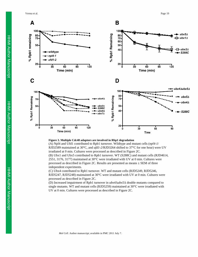
Figure 3. Multiple Cdc48 adaptors are involved in Rbp1 degradation(A) Npl4 and Ufd1 contributed to Rpb1 turnover. Wildtype and mutant cells (npl4-1/RJD2589 maintained at 30°C, and ufd1-2/RJD3264 shifted to 37°C for one hour) were UVirradiated at 0 min. Cultures were processed as described in Figure 2C.(B) Ubx1 and Ubx5 contributed to Rpb1 turnover. WT (S288C) and mutant cells (RJD4614,2551, 3176, 3177) maintained at 30°C were irradiated with UV at 0 min. Cultures wereprocessed as described in Figure 2C. Results are presented as means ± SEM of threeindependent experiments.(C) Ubx4 contributed to Rpb1 turnover. WT and mutant cells (RJD5249, RJD5246,RJD5247, RJD5248) maintained at 30°C were irradiated with UV at 0 min. Cultures wereprocessed as described in Figure 2C.(D) Increased impairment of Rpb1 turnover in ubx4Δubx5Δ double mutants compared tosingle mutants. WT and mutant cells (RJD5259) maintained at 30°C were irradiated withUV at 0 min. Cultures were processed as described in Figure 2C.
Verma et al. Page 19
Mol Cell. Author manuscript; available in PMC 2011 July 7.
HH
MI Author M
anuscriptH
HM
I Author Manuscript
HH
MI Author M
anuscript
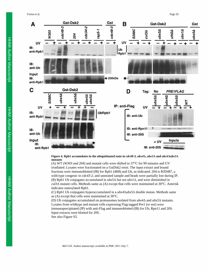
Figure 4. Rpb1 accumulates in the ubiquitinated state in cdc48-3, ubx4Δ, ubx5Δ and ubx4Δubx5Δmutants(A) WT (W303 and 204) and mutant cells were shifted to 37°C for 90 minutes and UVirradiated. Lysates were fractionated on a GstDsk2 resin. The input extract and boundfractions were immunoblotted (IB) for Rpb1 (4H8) and Ub, as indicated. 204 is RJD487, awild-type congenic to cdc43-2, and untreated sample and beads were partially lost during IP.(B) Rpb1 Ub conjugates accumulated in ubx5Δ but not ubx1Δ, and were diminished incul3Δ mutant cells. Methods same as (A) except that cells were maintained at 30°C. Asteriskindicates sumoylated Rpb1.(C) Rpb1 Ub conjugates hyperaccumulated in a ubx4Δubx5Δ double mutan. Methods sameas (A) except that cells were maintained at 30°C.(D) Ub conjugates accumulated on proteasomes isolated from ubx4Δ and ubx5Δ mutants.Lysates from wildtype and mutant cells expressing Flag-tagged Pre1 (or not) wereimmunoprecipitated (IP) with anti-Flag and immunoblotted (IB) for Ub, Rpn11 and 20S.Input extracts were blotted for 20S.See also Figure S5.
Verma et al. Page 20
Mol Cell. Author manuscript; available in PMC 2011 July 7.
HH
MI Author M
anuscriptH
HM
I Author Manuscript
HH
MI Author M
anuscript
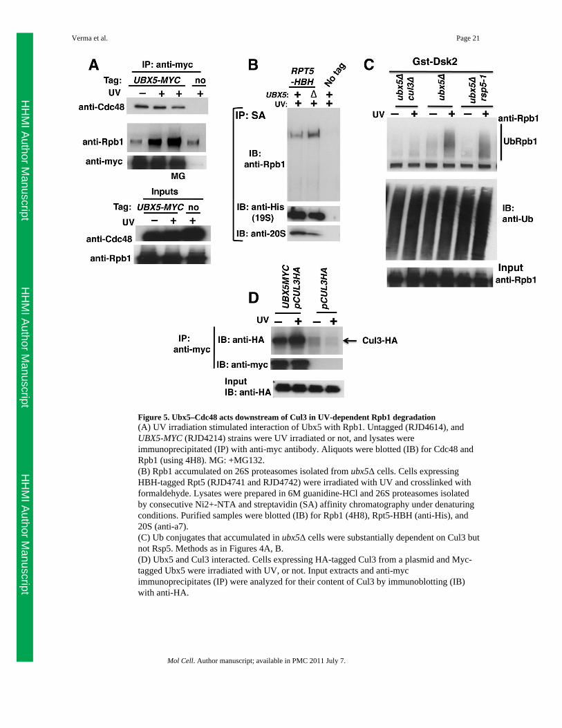
Figure 5. Ubx5–Cdc48 acts downstream of Cul3 in UV-dependent Rpb1 degradation(A) UV irradiation stimulated interaction of Ubx5 with Rpb1. Untagged (RJD4614), andUBX5-MYC (RJD4214) strains were UV irradiated or not, and lysates wereimmunoprecipitated (IP) with anti-myc antibody. Aliquots were blotted (IB) for Cdc48 andRpb1 (using 4H8). MG: +MG132.(B) Rpb1 accumulated on 26S proteasomes isolated from ubx5Δ cells. Cells expressingHBH-tagged Rpt5 (RJD4741 and RJD4742) were irradiated with UV and crosslinked withformaldehyde. Lysates were prepared in 6M guanidine-HCl and 26S proteasomes isolatedby consecutive Ni2+-NTA and streptavidin (SA) affinity chromatography under denaturingconditions. Purified samples were blotted (IB) for Rpb1 (4H8), Rpt5-HBH (anti-His), and20S (anti-a7).(C) Ub conjugates that accumulated in ubx5Δ cells were substantially dependent on Cul3 butnot Rsp5. Methods as in Figures 4A, B.(D) Ubx5 and Cul3 interacted. Cells expressing HA-tagged Cul3 from a plasmid and Myc-tagged Ubx5 were irradiated with UV, or not. Input extracts and anti-mycimmunoprecipitates (IP) were analyzed for their content of Cul3 by immunoblotting (IB)with anti-HA.
Verma et al. Page 21
Mol Cell. Author manuscript; available in PMC 2011 July 7.
HH
MI Author M
anuscriptH
HM
I Author Manuscript
HH
MI Author M
anuscript

Figure 6. Ubiquitinated Rpb1 accumulates on chromatin in UV-treated cdc48-3 mutant(A) CUL3-TAP (RJD4680) cells were treated with UV or not, and harvested after theaddition of 0.1% azide. Spheroplasts were prepared using 25 units of Zymolyase 100T for 2× 10⁹ cells. The three fractions: Whole cell extract (WCE), supernatant (Sup), and chromatinpellet (ChrPell) derived from spheroplasts were immunoblotted (IB) with the indicatedantibodies.(B) Cdc48 and 26S proteasome recruitment to chromatin was enhanced following UV.Crude chromatin pellets (Frc3) obtained by centrifuging WCE (Frc1) through a sucrosecushion were fractionated further following limited digestion with micrococcal nuclease(MNase) to generate high-speed pellet (Frc6) and supernatant (Frc7) fractions. Frc6 isenriched in released polynucleosomes. All fractions were immunoblotted (IB) for histoneH3, 19S (Rpn3), 20S proteasome subunits, and Cdc48. Fraction numbers correspond to lanenumbers.(C) Ubiquitinated Rpb1 accumulated on chromatin in response to UV and inactivation ofCdc48. WT and mutant cells expressing Myc-tagged Ub were shifted to 37°C, and Cu2+was added to induce expression of Ub. After two hours, cultures were irradiated with UV (ornot) and harvested. Crude chromatin pellets (FrC) were isolated as in (B) above and treated
Verma et al. Page 22
Mol Cell. Author manuscript; available in PMC 2011 July 7.
HH
MI Author M
anuscriptH
HM
I Author Manuscript
HH
MI Author M
anuscript
with Benzonase. Solubilized material was immunoprecipitated (IP) with anti-myc, andaliquots were blotted (IB) for Rpb1 (4H8), Spt5, and Rpn3.(D) Cells of the indicated genotype were galactose-induced for two hours, then treated (ornot) with MG132 for 30 min, lysed, and subjected to immunoprecipitation with anti-myc.The resulting samples were immunoblotted with antibodies to detect the indicated antigens.pGST-UBX5 is a plasmid that expresses GST-tagged Ubx5 under the GAL promoter.See also Figure S6
Verma et al. Page 23
Mol Cell. Author manuscript; available in PMC 2011 July 7.
HH
MI Author M
anuscriptH
HM
I Author Manuscript
HH
MI Author M
anuscript
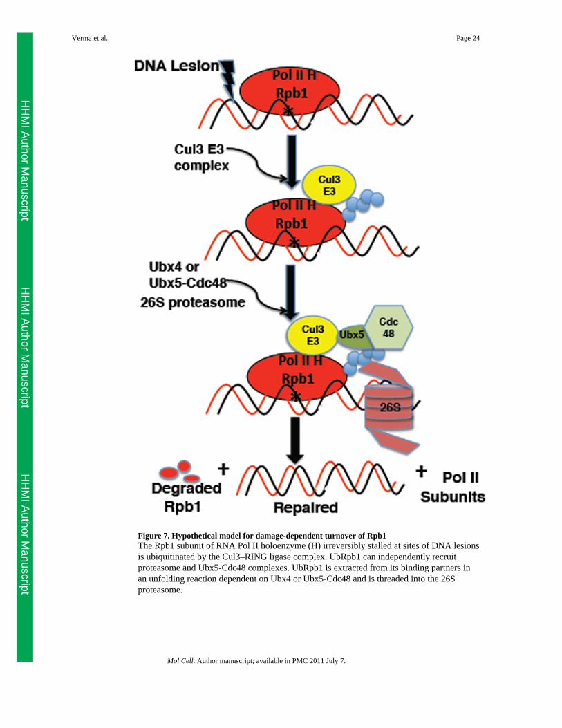
Figure 7. Hypothetical model for damage-dependent turnover of Rpb1The Rpb1 subunit of RNA Pol II holoenzyme (H) irreversibly stalled at sites of DNA lesionsis ubiquitinated by the Cul3–RING ligase complex. UbRpb1 can independently recruitproteasome and Ubx5-Cdc48 complexes. UbRpb1 is extracted from its binding partners inan unfolding reaction dependent on Ubx4 or Ubx5-Cdc48 and is threaded into the 26Sproteasome.
Verma et al. Page 24
Mol Cell. Author manuscript; available in PMC 2011 July 7.
HH
MI Author M
anuscriptH
HM
I Author Manuscript
HH
MI Author M
anuscript
Copyright © 2022 FDOKUMEN