Social comparison mediates chimpanzees' responses to loss, not frustration

Cell Stem Cell
Article
Wnt Signaling Mediates Self-Organizationand Axis Formation in Embryoid BodiesDerk ten Berge,1,2,3,* Wouter Koole,1,2,3 Christophe Fuerer,1,2 Matt Fish,1,2 Elif Eroglu,1,2 and Roel Nusse1,2,*1Howard Hughes Medical Institute2Department of Developmental Biology
Stanford University School of Medicine, Stanford, CA 94305, USA3These authors contributed equally to this work
*Correspondence: [email protected] (D.t.B.), [email protected] (R.N.)
DOI 10.1016/j.stem.2008.09.013
SUMMARY
Embryonic stem cells (ESCs) form descendants of allthree germ layers when differentiated as aggregates,termed embryoid bodies. In vivo, differentiation ofcells depends on signals and morphogen gradientsthat provide instructive and positional cues, but dosuch gradients exist in embryoid bodies? We reporthere the establishment of anteroposterior polarityand the formation of a primitive streak-like region inthe embryoid body, dependent on local activationof the Wnt pathway. In this region, cells undergo anepithelial-to-mesenchymal transition and differenti-ate into mesendodermal progenitors. ExogenousWnt3a protein posteriorizes the embryoid body, re-sulting in predominantly mesendodermal differentia-tion. Conversely, inhibiting Wnt signaling promotesanterior character and results in neurectodermaldifferentiation. The activation of Wnt signaling andprimitive streak formation requires external signalsbut is self-reinforcing after initiation. Our findingsshow that the Wnt pathway mediates the local exe-cution of a gastrulation-like process in the embryoidbody, which displays an unexpected degree ofself-organization.
INTRODUCTION
A major goal of ESC research is to direct the differentiation of
stem cells into specific developmental lineages. Descendants
from all three germ layers can be produced by allowing ESCs
to differentiate as an aggregate, called an embryoid body
(Burkert et al., 1991; Doetschman et al., 1985). Under these con-
ditions, ESCs can differentiate into a wide variety of cell types,
including hematopoietic, vascular, pancreatic, hepatic, and neu-
ral lineages (Murry and Keller, 2008), and even into germ cells
(Geijsen et al., 2004). It is thought that tissue differentiation in em-
bryoid bodies occurs in a disorganized fashion (Murry and Keller,
2008). In vivo, however, morphogen gradients impart positional
information and influence cell-lineage decisions, and such
cues can also promote the formation of specific cell types in em-
bryoid bodies. For example, adding retinoic acid to embryoid
508 Cell Stem Cell 3, 508–518, November 6, 2008 ª2008 Elsevier In
bodies caudalizes cells, a treatment that together with the
ventralizing activity of Sonic hedgehog leads to motor neuron
differentiation (Wichterle et al., 2002). Upon transplantation,
these cells segregate and innervate tissues according to their
positional specification (Wichterle et al., 2002). This demon-
strates the importance of providing differentiating cells with not
only instructive but also positional signals. In this study, we
sought to determine to what extent cells in the supposedly unor-
ganized embryoid body are exposed to morphogen gradients
and positional signals.
ESCs are derived from the inner cell mass of blastocyst stage
embryos. Following implantation, the inner cell mass forms
a solid mass of pluripotent unpolarized embryonic ectoderm,
separated by a basement membrane from an outer layer of
primitive endoderm. The central cells of the embryonic ectoderm
undergo programmed cell death and disappear in a process
called cavitation, whereas the surrounding embryonic ectoderm
differentiates into a pseudostratified columnar epithelium. The
embryonic ectoderm, or epiblast, is further characterized by
expression of the pluripotency marker Oct4 (Rosner et al.,
1990). Upon further development, the primary germ layers are
established and patterned during gastrulation, when the primi-
tive streak forms on the posterior side of the embryo. Epiblast
cells ingressing through the primitive streak are the source of de-
finitive endoderm and mesoderm, whereas the anterior epiblast
differentiates into ectodermal derivatives (reviewed in Tam and
Loebel, 2007). The posterior expression of several Wnt ligands
and Wnt signaling components is essential for the establishment
of the primitive streak and anteroposterior polarity, the epithelial-
to-mesenchymal transition (EMT) of epiblast cells in the primitive
streak, and the formation of mesoderm (Galceran et al., 1999;
Greco et al., 1996; Haegel et al., 1995; Huelsken et al., 2000;
Kelly et al., 2004; Kemler et al., 2004; Liu et al., 1999; Takada
et al., 1994; Yoshikawa et al., 1997). In vitro, manipulation of
the Wnt signaling pathway during embryoid body formation
greatly influences the direction of cell differentiation (Aubert
et al., 2002; Gadue et al., 2006; Lindsley et al., 2006).
Wnt signaling can be visualized through the activity of several
reporter constructs, including 7xTCF-eGFP (Brugmann et al.,
2007), and the targeted mouse mutant Axin2lacZ/+, which ex-
presses a lacZ reporter from the locus of the Wnt target gene
Axin2 (Aulehla et al., 2003; Jho et al., 2002; Lustig et al., 2002).
Using these reporters, we find that endogenous Wnt signals
polarize the embryoid body and mediate the local execution of
a gastrulation-like process.
c.
Cell Stem Cell
Self-Organization in Embryoid Bodies
RESULTS
Wnt signaling is required for establishment of the primitive streak,
and therefore we asked whether activation of the Wnt pathway
could serve to visualize the location of primitive streak formation.
Using Axin2lacZ/+ reporter mice, we found no evidence of Wnt
signaling in E5.5 and E6.0 embryos, but reporter activity became
visible around E6.5 in the posterior region of the embryo (Figure 1).
At this stage, the primitive streak begins to form, and reporter ac-
tivity is visible in the newly formed mesodermal cells that migrate
anteriorly and proximally (Jho et al., 2002; Morkel et al., 2003). At
late streak stage (E7.5), reporter activity was visible in the entire
posterior half of the embryo and in the extraembryonic allantois
and chorion (Figure1).The firstactivationof the Axin2lacZ/+ reporter
in the postimplantation embryo therefore coincides with the estab-
lishment of the primitive streak and its derived cell populations.
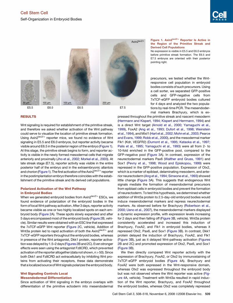
Polarized Activation of the Wnt Pathwayin Embryoid BodiesWhen we generated embryoid bodies from Axin2lacZ/+ ESCs, we
found evidence of polarization of the embryoid bodies in the
form of local Wnt pathway activation. After 3 days, reporter activity
became visible as one or two highly localized spots on each em-
bryoid body (Figure 2A). These spots slowly expanded and after
5 days encompassed most of the embryoid body (Figure 2B, vehi-
cle). Similar results were obtained with an ESC line that expressed
the 7xTCF-eGFP Wnt reporter (Figure 2C, vehicle). Addition of
Wnt3a protein led to rapid activation of both the Axin2lacZ/+ and
7xTCF-eGFP reporters throughout the embryoid bodies, whereas
in presence of the Wnt antagonist, Dkk1 protein, reporter activa-
tion wasdelayed by 1.5–2 days (Figures 2B and 2C). Even stronger
effects were seen using the antagonist Fz8CRD, which prevented
activation of the reporter altogether (data not shown, n > 50). Since
both Dkk1 and Fz8CRD act extracellularly by inhibiting Wnt pro-
teins from activating their receptors, these data demonstrate
thata localizedsourceofWntsignals polarizes the embryoidbody.
Wnt Signaling Controls LocalMesendodermal DifferentiationSince activation of Wnt signaling in the embryo overlaps with
differentiation of the primitive ectoderm into mesendodermal
Figure 1. Axin2lacZ/+ Reporter Is Active in
the Region of the Primitive Streak and
Derived Cell Populations
No expression is visible in E5.5 and E6.0 embryos
before primitive streak formation. The E6.5 and
E7.5 embryos are oriented with their posterior
pointing right.
precursors, we tested whether the Wnt-
responsive cell population in embryoid
bodies consists of such precursors. Using
a cell sorter, we separated GFP-positive
cells and GFP-negative cells from
7xTCF-eGFP embryoid bodies cultured
for 4 days and analyzed the two popula-
tions by real-time PCR. The mesendoder-
mal markers Brachyury, which is ex-
pressed throughout the primitive streak and nascent mesoderm
(Herrmann and Kispert, 1994; Kispert and Herrmann, 1994) and
is a direct Wnt target (Arnold et al., 2000; Yamaguchi et al.,
1999), FoxA2 (Ang et al., 1993; Dufort et al., 1998; Weinstein
et al., 1994), and Mixl1 (Hart et al., 2002; Mohn et al., 2003; Pearce
and Evans, 1999; Robb et al., 2000), and the mesodermal marker
Flk1 (Kdr, VEGFR2) (Dumont et al., 1995; Kataoka et al., 1997;
Palis et al., 1995; Yamaguchi et al., 1993) were all from 2- to
10-fold enriched in the GFP-positive pool, compared to the
GFP-negative pool (Figure 3A). In contrast, expression of the
neurectodermal markers Pax6 (Walther and Gruss, 1991) and
Sox1 (Pevny et al., 1998; Wood and Episkopou, 1999) were
repressed in the GFP-positive population. Expression of Otx2,
which is a marker of epiblast, delaminating mesoderm, and ante-
rior neurectoderm (Ang et al., 1994; Simeone et al., 1993) showed
little change (Figure 3A). This suggests that endogenous Wnt
signals mediate the formation of mesendodermal precursors
from epiblast cells in embryoid bodies and prevent the formation
of neurectoderm. To test this hypothesis, we determined whether
addition of Wnt3a protein to 2.5-day-old embryoid bodies would
induce mesendodermal markers and repress neurectodermal
markers. As observed before for Brachyury (Robertson et al.,
2000; Ueno et al., 2007), the mesendodermal markers displayed
a dynamic expression profile, with expression levels increasing
for 2 days and then falling off (Figure 3B, vehicle). Wnt3a protein
consistently accelerated and increased this induction of
Brachyury, FoxA2, and Flk1 in embryoid bodies, whereas it
repressed Otx2, Pax6, and Sox1 (Figure 3B). In contrast, Dkk1
protein delayed the induction of Brachyury, FoxA2, and Flk1
(Figure 3B), just as it delayed Wnt-pathway activation (Figures
2B and 2C) and promoted expression of Otx2, Pax6, and Sox1
(Figure 3B).
We then directly compared Wnt reporter activity with the
expression of Brachyury, FoxA2, or Otx2 by immunostaining of
7xTCF-eGFP embryoid bodies (Figure 4A). Brachyury and
FoxA2 were both expressed in the Wnt-responsive domain,
whereas Otx2 was expressed throughout the embryoid body
but was not observed where the Wnt reporter was active (Fig-
ure 4A, vehicle). Treatment with Wnt3a resulted in rapid induc-
tion of the Wnt reporter, Brachyury, and FoxA2 throughout
the embryoid bodies, whereas Otx2 was completely repressed
Cell Stem Cell 3, 508–518, November 6, 2008 ª2008 Elsevier Inc. 509
Cell Stem Cell
Self-Organization in Embryoid Bodies
(Figure 4A, Wnt3a). Conversely, in the presence of Dkk1, the Wnt
reporter, Brachyury, and FoxA2 were not induced, and Otx2
remained expressed throughout the embryoid body (Figure 4A,
Dkk1). We confirmed the local coregulation of Brachyury and
Wnt-signaling using an ESC line in which a GFP reporter is
expressed from the Brachyury locus (Fehling et al., 2003). We
derived two clones from this cell line transduced with a 7xTCF-
mCherry Wnt reporter, in which Wnt signaling is visualized by
expression of the red fluorescent protein mCherry. Embryoid
bodies derived from either clone displayed local coexpression
of GFP and mCherry (Figure 4B). Treatment with Wnt3a resulted
in ubiquitous expression of both reporters throughout the embry-
oid bodies, whereas treatment with Dkk1 prevented expression
of the reporters (Figure 4C). Combined, these data show that
Wnt signaling regulates the local formation of mesendoderm at
the expense of neurectoderm in embryoid bodies.
Wnt Signaling Controls LocalEpithelial-to-Mesenchymal TransitionOne of the hallmarks of gastrulation is the EMT that epiblast cells
undergo while they ingress through the primitive streak. During
EMT, their adhesive interactions change from cell-cell adhesion,
mediated by E-cadherin, to cell-substratum interactions, medi-
ated by integrins and fibronectin (Burdsal et al., 1993). The tran-
scriptional repressor Snail triggers this process (Carver et al.,
2001), in part by repressing E-cadherin (Cano et al., 2000).
Thus, gastrulating cells express Snail (Nieto et al., 1992; Smith
Figure 2. Localized Wnt Signaling in Embry-
oid Bodies
(A) Axin2lacZ/+ embryoid bodies spontaneously
activated the reporter in a localized fashion after
3 days in culture (n > 200).
(B) Wnt signaling expanded overtime throughout
the entire Axin2lacZ/+ embryoid body. Addition of
recombinant Wnt3a (200 ng/ml) led to accelerated
activation of the reporter, whereas Dkk1 (80 ng/ml)
delayed reporter activation by 2 days (n > 200).
(C) 7xTCF-eGFP embryoid bodies displayed ex-
pression of the reporter similar as in Axin2lacZ/+
embryoid bodies but with a delay of approximately
1 day. The delay is probably due to the larger size
of the embryoid bodies and time required for GFP
maturation (n > 200). Scale bars, 500 mm (A and B),
100 mm (C).
et al., 1992) and fibronectin (Franke
et al., 1983; Jackson et al., 1981), and
cease to express E-cadherin (Damjanov
et al., 1986). To test whether the Wnt-re-
sponsive cell population in 7xTCF-eGFP
embryoid bodies is undergoing EMT, we
separated GFP-positive from GFP-nega-
tive cells using a cell sorter and analyzed
the two populations by real-time PCR.
This analysis revealed that expression of
Snail and fibronectin was upregulated in
the GFP-positive population, relative to
the GFP-negative population, whereas
E-cadherin was repressed (Figure 5A).
To test whether Wnt signaling could regulate this EMT signature,
we treated embryoid bodies with Wnt3a and found that this
indeed resulted in the accelerated upregulation of Snail and
fibronectin and in the repression of E-cadherin (Figure 5B). Con-
versely, Dkk1 treatment repressed the upregulation of Snail and
fibronectin and maintained E-cadherin expression (Figure 5B).
We then compared Wnt reporter activity with the expression of
Brachyury or E-cadherin by immunostaining cryosections from
7xTCF-eGFP embryoid bodies. Whereas the Wnt reporter and
Brachyury expression always overlapped to a great extent,
E-cadherin was expressed throughout the embryoid body, ex-
cept where strong Wnt reporter activity was visible (Figure 5C).
E-cadherin and GFP expression was visible in the same cells at
the edges of the GFP-positive domain, where presumably the
EMT was still underway (Figure 5C, arrows).
In vivo, the epiblast that undergoes the EMT is a polarized
columnar epithelium that is attached to a basement membrane
and expresses Oct4. What is the nature of the epithelium on
which Wnt signals act in embryoid bodies? Using immunostain-
ing for laminin, we detected no basement membrane at the stage
that the Wnt reporter was active (Figure 6A) or in earlier stages
(2.5 and 3.5 days, data not shown), whereas we were able to de-
tect basement membranes surrounding more advanced (7 days)
embryoid bodies (Figure 6B). Similar results were obtained with
an antibody against collagen IV, another marker for basement
membranes (data not shown). Also, staining of the actin cytoskel-
eton with phalloidin did not reveal epithelial polarity (Figure 6C).
510 Cell Stem Cell 3, 508–518, November 6, 2008 ª2008 Elsevier Inc.

Cell Stem Cell
Self-Organization in Embryoid Bodies
The epithelia present in 7 day embryoid bodies were not repre-
sentative of epiblast, as they did not express the pluripotency
marker Oct4 (data not shown), whereas Oct4 expression was de-
tected throughout 2.5 and 3.5 day embryoid bodies (Figure 6D).
Upon activation of Wnt signaling, Oct4 became downregulated
in the Wnt-responsive cells (Figure 6E), consistent with their
differentiation to mesendoderm (Rosner et al., 1990). These
data suggest that differentiation of the ESCs to a polarized epi-
thelium is not required for their differentiation into mesendoder-
mal precursors. This is consistent with earlier studies showing
that polarization and differentiation of ESCs in embryoid bodies
can occur independently from each other (Li et al., 2001; Murray
and Edgar, 2001).
Combined, our data show that Wnt signaling is required and
sufficient for the generation of a domain with primitive streak
characteristics in the embryoid body. In this domain, cells
undergo EMT and acquire mesendodermal character. When
Wnt signaling is suppressed, this primitive streak-like structure
does not form and differentiation toward the neurectodermal lin-
eage prevails. Thus, at this stage the embryoid body displays an
organization and polarity reminiscent of that seen in gastrulating
embryos, where neurectodermal character defines the anterior,
and the posterior is characterized by the conversion of epiblast
into mesendoderm. By manipulating Wnt signaling, we are able
to shift the embryoid body between these two fates.
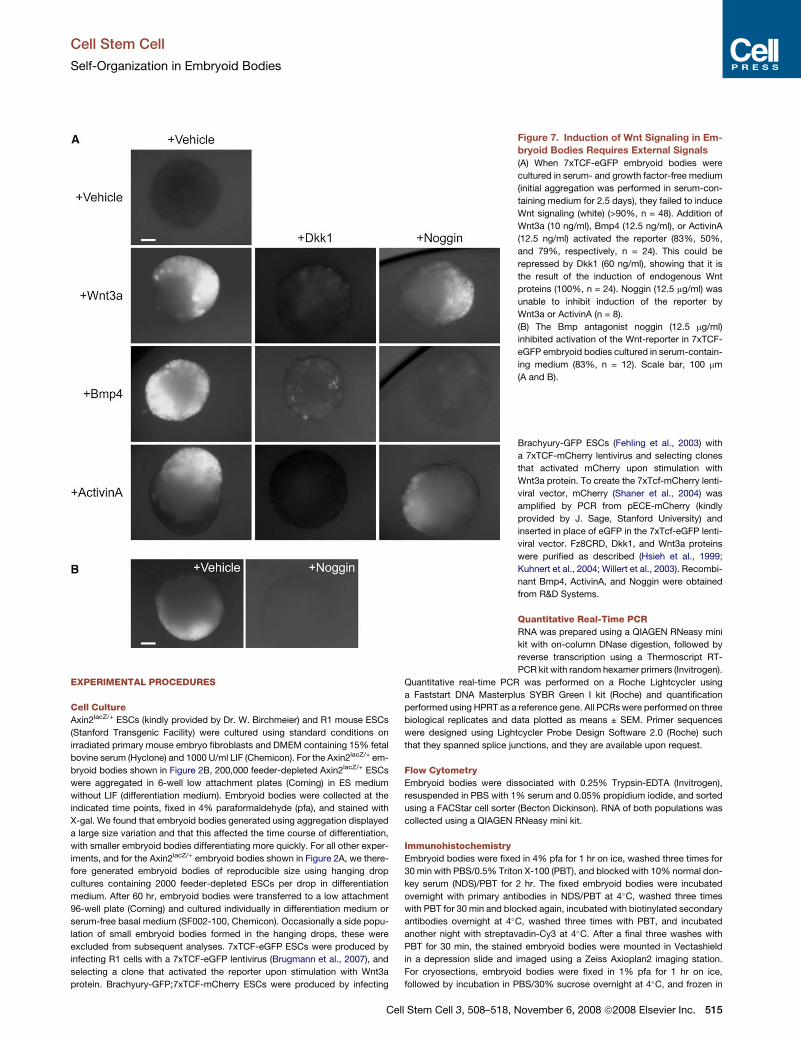
Polarized Wnt Signaling Is Activated by External SignalsIn the mouse embryo, establishment of the primitive streak in-
volves reciprocal and mutually reinforcing interactions between
epiblast and extraembryonic ectoderm that are mediated by
Figure 3. Wnt Signaling Regulates the Local
Expression of Markers for the Primary Germ
Layers in Embryoid Bodies
(A) 7xTCF-eGFP embryoid bodies were cultured
for 4 days and separated into GFP-positive and
GFP-negative cell pools using a cell sorter, and
the pools were analyzed by real-time PCR. Ex-
pression of Brachyury, FoxA2, Flk1, and Mixl1
was increased in the GFP-positive cells compared
to the GFP-negative cells, whereas Otx2, Pax6,
and Sox1 were repressed (mean ± SEM, n = 3).
(B) Real-time PCR analysis of RNA collected from
embryoid bodies cultured in presence of the
indicated factors. Factors were added 2.5 days
after the start of aggregation. Addition of Wnt3a
(200 ng/ml) resulted in early peaking levels of
Brachyury, Foxa2, and Flk1, whereas the neurec-
todermal markers Otx2 and Pax6 became rapidly
downregulated, and induction of Sox1 was pre-
vented. Addition of Dkk1 (80 ng/ml) resulted in
delayed induction of Brachyury, Foxa2, and Flk1
and promoted expression of Otx2, Pax6, and
Sox1 (mean ± SEM, n = 3).
nodal, Bmp4, and Wnt3 (Ben-Haim
et al., 2006; Brennan et al., 2001). We ad-
dressed the role and interdependency of
these factors in our in vitro model of prim-
itive streak formation. The extraembry-
onic ectoderm is an important source of
signals in vivo, but since ESCs differentiate poorly into extraem-
bryonic ectoderm (Beddington and Robertson, 1989), we asked
instead whether activation of the Wnt pathway required external
signals from the culture medium. When embryoid bodies were
cultured in serum-free defined medium devoid of all signaling
proteins, we could not detect Wnt reporter expression over the
course of 8 days of culture (Figure 7A). However, supplementing
the medium with low concentrations of Wnt3a, Bmp4, or ActivinA
(which activates the nodal pathway [Schier and Shen, 2000]) all
resulted in local 7xTCF-eGFP activation (Figure 7A). In all cases,
Dkk1 blocked reporter activation, showing that Bmp4 and
ActivinA induced the endogenous production of Wnt signals
(Figure 7A). Since the low Wnt3a stimulus was insufficient to
induce widespread reporter activity, it too likely induced local
endogenous production of Wnt signals, similar to Bmp4 and
ActivinA. An external signal is therefore required to start Wnt
signaling and primitive streak formation in embryoid bodies.
Our observation that Wnt3a, Bmp4, and ActivinA can each indi-
vidually perform this function confirms the mutually reinforcing
nature of the signaling interactions that start gastrulation. Inter-
estingly, while the Bmp antagonist Noggin blocked local reporter
activation by Bmp4, it was unable to block the activation by
ActivinA or Wnt3a (Figure 7A). Thus, local activation of the re-
porter by ActivinA and Wnt3a is independent of Bmp signaling,
and although Bmp signaling is sufficient to start this process, it
is not required.
Finally, we addressed how initiation of Wnt signaling in the
embryoid body is regulated by serum-containing medium.
When we cultured embryoid bodies in presence of serum and
the Bmp antagonist Noggin, virtually no activation of the Wnt
Cell Stem Cell 3, 508–518, November 6, 2008 ª2008 Elsevier Inc. 511

Cell Stem Cell
Self-Organization in Embryoid Bodies
reporter occurred (Figure 7B). Since Noggin inhibits local activa-
tion of the Wnt reporter by Bmp4, but not by Wnt3a or ActivinA,
the role of serum in embryoid body differentiation is likely to
provide a Bmp activity that activates Wnt signaling and starts
primitive streak formation.
DISCUSSION
Almost 50 years ago, it was realized that aggregates of pluripo-
tent embryocarcinoma cells recapitulated aspects of early
embryonic development (Pierce and Dixon, 1959; Stevens,
1959). This was quickly recognized as a model system for the
understanding of normal embryonic cell differentiation. These
aggregates—termed embryoid bodies because of their superfi-
cial resemblance to mouse blastocysts—generally show few
Figure 4. Wnt Signaling Regulates the Local
Formation of Mesendoderm in Embryoid
Bodies
(A) Whole-mount immunostaining of 7xTCF-eGFP
embryoid bodies showed colocalization (yellow) of
primitive streak markers Brachyury and Foxa2
(red) with the Wnt reporter (green). Expression of
Otx2 (red) was mutually exclusive with the Wnt
reporter. Treatment with Wnt3a (200 ng/ml) accel-
erated the upregulation of the Wnt reporter,
Brachyury, and FoxA2, whereas it repressed
Otx2. Dkk1 treatment (80 ng/ml) had the opposite
effect, repressing Wnt signaling, Brachyury, and
FoxA2 expression and promoting Otx2 expression
throughout the embryoid body. Factors were
added 2.5 days after the start of aggregation.
(B) In embryoid bodies derived from a Brachyury-
GFP;7xTCF-mCherry cell line, both reporters ex-
pressed in close proximity or overlapping domains
(GFP, green; mCherry, red; 80%, n > 120). The
side population of small embryoid bodies that
formed during aggregation was excluded from
subsequent analyses.
(C) Wnt3a treatment induced expression of both
reporters throughout Brachyury-GFP;7xTCF-
mCherry embryoid bodies (50%, n > 60), whereas
Dkk1 repressed expression of both reporters
(>90%, n > 60). Scale bars, 100 mm (A and C),
200 mm (B).
morphological signs of gastrulation and
display unorganized tissue differentiation
(Martin et al., 1977; Wiley et al., 1978).
However, this issue has not been read-
dressed since the advent of ESCs with
their superior ability to generate embry-
onic tissues (Evans and Kaufman, 1981;
Martin, 1981). Our data show that embry-
oid bodies derived from ESCs display
a large degree of self-organization: they
establish anteroposterior polarity and
develop a domain with characteristics of
the primitive streak, where cells undergo
EMT and form mesendoderm precursors
in a process that is dependent on local
activation of the Wnt pathway. Embryoid
body development therefore resembles normal embryonic de-
velopment much closer than previously thought and provides
an easily accessible model for the formation of anteroposterior
polarity and the establishment of the primitive streak. Moreover,
the presence of self-organization and polarity suggests that em-
bryoid bodies can establish morphogen gradients controlling cell
differentiation. This would not only explain the wide repertoire of
developmental activities present in the embryoid body but also
would provide a new tool for investigating the establishment
and mode of action of such gradients.
In vivo, primitive streak formation and the establishment of
anteroposterior polarity depend on interactions between the
epiblast and two extraembryonic tissues, the visceral endoderm
and the extraembryonic ectoderm (reviewed in Tam and Loebel,
2007). Primitive-streak formation requires specification of the
512 Cell Stem Cell 3, 508–518, November 6, 2008 ª2008 Elsevier Inc.

Cell Stem Cell
Self-Organization in Embryoid Bodies
Figure 5. Wnt Signaling Controls Epithelial-to-Mesenchymal Transition in Embryoid Bodies
(A) 7xTCF-eGFP embryoid bodies were cultured for 4 days and separated into GFP-positive and GFP-negative cell pools using a cell sorter, and the pools were
analyzed by real-time PCR. Expression of Snail and fibronectin was enriched in the GFP-positive cells compared to the GFP-negative cells, whereas E-cadherin
was repressed (mean ± SEM, n = 3).
(B) Real-time PCR analysis of RNA collected from embryoid bodies cultured in presence of the indicated factors. Addition of Wnt3a (200 ng/ml) resulted in
accelerated induction of Snail and fibronectin, whereas E-cadherin was rapidly downregulated. Addition of Dkk1 (80 ng/ml) resulted in delayed induction of Snail
and fibronectin and maintained expression of E-cadherin (mean ± SEM, n = 3).
(C) Cryosections through 7xTCF-eGFP (green) embryoid bodies were immunostained for Brachyury or E-cadherin (red). Brachyury and GFP expression overlap
to a large extent (80%, n > 25), whereas E-cadherin and GFP were mutually exclusive except at the edges of the GFP domain (arrows) (>90%, n > 25). Scale bar,
100 mm.
posterior epiblast by Wnt3 (Huelsken et al., 2000; Liu et al.,
1999). It is thought that expression of Wnt3 is activated by nodal
through BMP4 signaling in the extraembryonic ectoderm (Ben-
Haim et al., 2006; Brennan et al., 2001). In turn, Wnt3 activates
a feedback loop that maintains nodal expression in the epiblast
(Ben-Haim et al., 2006). Since all three factors can start the feed-
C
back loop, this explains why they were all able to initiate self-
organization in the embryoid body. However, our results indicate
that Bmp4 is not required for this process and that nodal/
ActivinA can activate Wnt signaling in the absence of a functional
Bmp pathway. This conclusion is in agreement with a recent
study that found that Bmp4 has a posteriorizing effect on
ell Stem Cell 3, 508–518, November 6, 2008 ª2008 Elsevier Inc. 513

Cell Stem Cell
Self-Organization in Embryoid Bodies
mesoderm but is not required for generation of mesoderm in
embryoid bodies (Nostro et al., 2008). In vivo, loss of Bmp4
severely affects gastrulation and establishment of the primitive
streak (Winnier et al., 1995). Combined, these results suggest
that Bmp4 is necessary for induction of Wnt3 and nodal signaling
in the embryo but is not longer required once these pathways are
active. This also implies that Bmp4, not nodal, is the signal that
starts the feedback loop in vivo.
During embryogenesis, the visceral endoderm promotes
anterior patterning by producing nodal and Wnt antagonists,
including Cerl, Lefty1, and Dkk1, in the anterior region of the
embryo (Glinka et al., 1998; Kimura-Yoshida et al., 2005;
Perea-Gomez et al., 2002; Yamamoto et al., 2004). Establish-
ment of anteroposterior polarity is therefore the result of a
balance between posteriorizing signals and their antagonists.
Using Wnt3a and Dkk1, we could manipulate this balance in
embryoid bodies to favor either posterior or anterior character.
By providing exogenous Wnt proteins, we posteriorized the
embryoid body and promoted mesendodermal fate. Conversely,
Wnt antagonists promoted anterior character and neurectoder-
mal differentiation. The status of the Wnt signaling pathway is
therefore a critical parameter in protocols for directed ESC
differentiation.
Figure 6. Tissue Organization in 7xTCF-
eGFP Embryoid Bodies
Cryosections through 7xTCF-eGFP (green in [A],
[C], and [E]) embryoid bodies stained for laminin
(A and B), phalloidin (C), or Oct4 (D and E) (red).
Nuclei were stained with dapi (blue). (A) No basal
lamina could be detected in 4-day-old embryoid
bodies (n > 60). (B) Basal lamina were readily
detectable around the periphery of 7-day-old
embryoid bodies. (C) Phalloidin staining (red)
revealed no epithelial polarity in cells prior to acti-
vating 7xTCF-eGFP. (D) Oct4 was expressed
throughout 3.5 day (shown) and earlier (data not
shown) embryoid bodies before expression of
the Wnt reporter became apparent. (E) Oct4
expression was downregulated in cells activating
7xTCF-eGFP (green). No GFP was imaged in (B)
and (D).
How is the initial polarity of the embry-
oid body established? A deterministic
mechanism seems unlikely, since the em-
bryoid body consists of cells that cannot
be distinguished by virtue of lineage or
inductive environment. In a stochastic
mechanism, differences in cells arise
from developmental noise, which can
have multiple sources. These differences
are amplified and stabilized and can lead
to asymmetric cell fate decisions in a
uniform cellular assembly (Losick and
Desplan, 2008). In the great majority of
the embryoid bodies we studied, a single
domain of Wnt signaling slowly expanded
throughout the embryoid body. This
suggests a cell-nonautonomous deci-
sion-making system, in which cells nearby the original inducing
cell are recruited into the polarizing center, whereas an inhibiting
signal, acting over a longer range, prevents formation of multiple
polarizing centers. Indeed, in larger embryoid bodies, produced
by aggregating more cells, we found more instances of embryoid
bodies with two domains of Wnt signaling (data not shown and
Figure 2B, 3 days vehicle). This suggests that the range of the
putative inhibitory signal was insufficient in these larger embry-
oid bodies. Nodal and the Wnt signal itself are candidates for
the recruiting signal, since a single, low-intensity pulse of these
signals suffices to induce a polarizing center. A better under-
standing of this phenomenon could have implications for the
in vivo establishment of anteroposterior polarity.
A persistent problem in understanding the regulation of cell
differentiation in ESC cultures is the presence of fetal calf serum,
which consists of undefined mixtures of growth factors and in-
hibitors. We show that an important role of serum in embryoid
body differentiation is to provide a Bmp activity that activates
Wnt signaling and starts primitive streak formation. For this func-
tion, we could replace serum-containing medium with purified
growth factors in chemically defined medium. The ability to
differentiate ESCs in defined conditions should facilitate the
derivation of pure cell populations from ESCs.
514 Cell Stem Cell 3, 508–518, November 6, 2008 ª2008 Elsevier Inc.
Cell Stem Cell
Self-Organization in Embryoid Bodies
EXPERIMENTAL PROCEDURES
Cell Culture
Axin2lacZ/+ ESCs (kindly provided by Dr. W. Birchmeier) and R1 mouse ESCs
(Stanford Transgenic Facility) were cultured using standard conditions on
irradiated primary mouse embryo fibroblasts and DMEM containing 15% fetal
bovine serum (Hyclone) and 1000 U/ml LIF (Chemicon). For the Axin2lacZ/+ em-
bryoid bodies shown in Figure 2B, 200,000 feeder-depleted Axin2lacZ/+ ESCs
were aggregated in 6-well low attachment plates (Corning) in ES medium
without LIF (differentiation medium). Embryoid bodies were collected at the
indicated time points, fixed in 4% paraformaldehyde (pfa), and stained with
X-gal. We found that embryoid bodies generated using aggregation displayed
a large size variation and that this affected the time course of differentiation,
with smaller embryoid bodies differentiating more quickly. For all other exper-
iments, and for the Axin2lacZ/+ embryoid bodies shown in Figure 2A, we there-
fore generated embryoid bodies of reproducible size using hanging drop
cultures containing 2000 feeder-depleted ESCs per drop in differentiation
medium. After 60 hr, embryoid bodies were transferred to a low attachment
96-well plate (Corning) and cultured individually in differentiation medium or
serum-free basal medium (SF002-100, Chemicon). Occasionally a side popu-
lation of small embryoid bodies formed in the hanging drops, these were
excluded from subsequent analyses. 7xTCF-eGFP ESCs were produced by
infecting R1 cells with a 7xTCF-eGFP lentivirus (Brugmann et al., 2007), and
selecting a clone that activated the reporter upon stimulation with Wnt3a
protein. Brachyury-GFP;7xTCF-mCherry ESCs were produced by infecting
Figure 7. Induction of Wnt Signaling in Em-
bryoid Bodies Requires External Signals
(A) When 7xTCF-eGFP embryoid bodies were
cultured in serum- and growth factor-free medium
(initial aggregation was performed in serum-con-
taining medium for 2.5 days), they failed to induce
Wnt signaling (white) (>90%, n = 48). Addition of
Wnt3a (10 ng/ml), Bmp4 (12.5 ng/ml), or ActivinA
(12.5 ng/ml) activated the reporter (83%, 50%,
and 79%, respectively, n = 24). This could be
repressed by Dkk1 (60 ng/ml), showing that it is
the result of the induction of endogenous Wnt
proteins (100%, n = 24). Noggin (12.5 mg/ml) was
unable to inhibit induction of the reporter by
Wnt3a or ActivinA (n = 8).
(B) The Bmp antagonist noggin (12.5 mg/ml)
inhibited activation of the Wnt-reporter in 7xTCF-
eGFP embryoid bodies cultured in serum-contain-
ing medium (83%, n = 12). Scale bar, 100 mm
(A and B).
Brachyury-GFP ESCs (Fehling et al., 2003) with
a 7xTCF-mCherry lentivirus and selecting clones
that activated mCherry upon stimulation with
Wnt3a protein. To create the 7xTcf-mCherry lenti-
viral vector, mCherry (Shaner et al., 2004) was
amplified by PCR from pECE-mCherry (kindly
provided by J. Sage, Stanford University) and
inserted in place of eGFP in the 7xTcf-eGFP lenti-
viral vector. Fz8CRD, Dkk1, and Wnt3a proteins
were purified as described (Hsieh et al., 1999;
Kuhnert et al., 2004; Willert et al., 2003). Recombi-
nant Bmp4, ActivinA, and Noggin were obtained
from R&D Systems.
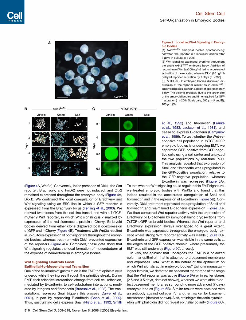
Quantitative Real-Time PCR
RNA was prepared using a QIAGEN RNeasy mini
kit with on-column DNase digestion, followed by
reverse transcription using a Thermoscript RT-
PCR kit with random hexamer primers (Invitrogen).
Quantitative real-time PCR was performed on a Roche Lightcycler using
a Faststart DNA Masterplus SYBR Green I kit (Roche) and quantification
performed using HPRT as a reference gene. All PCRs were performed on three
biological replicates and data plotted as means ± SEM. Primer sequences
were designed using Lightcycler Probe Design Software 2.0 (Roche) such
that they spanned splice junctions, and they are available upon request.
Flow Cytometry
Embryoid bodies were dissociated with 0.25% Trypsin-EDTA (Invitrogen),
resuspended in PBS with 1% serum and 0.05% propidium iodide, and sorted
using a FACStar cell sorter (Becton Dickinson). RNA of both populations was
collected using a QIAGEN RNeasy mini kit.
Immunohistochemistry
Embryoid bodies were fixed in 4% pfa for 1 hr on ice, washed three times for
30 min with PBS/0.5% Triton X-100 (PBT), and blocked with 10% normal don-
key serum (NDS)/PBT for 2 hr. The fixed embryoid bodies were incubated
overnight with primary antibodies in NDS/PBT at 4�C, washed three times
with PBT for 30 min and blocked again, incubated with biotinylated secondary
antibodies overnight at 4�C, washed three times with PBT, and incubated
another night with streptavadin-Cy3 at 4�C. After a final three washes with
PBT for 30 min, the stained embryoid bodies were mounted in Vectashield
in a depression slide and imaged using a Zeiss Axioplan2 imaging station.
For cryosections, embryoid bodies were fixed in 1% pfa for 1 hr on ice,
followed by incubation in PBS/30% sucrose overnight at 4�C, and frozen in
Cell Stem Cell 3, 508–518, November 6, 2008 ª2008 Elsevier Inc. 515
Cell Stem Cell
Self-Organization in Embryoid Bodies
OCT (Tissue-Tek). Cryosections (10 mm) were fixed in acetone at �20�C for
5 min, blocked with avidin, biotin, and finally 5% NDS/PBT, each for 15 min,
and incubated with primary antibodies in 5% NDS/PBT overnight at 4�C.
The slides were then washed three times with PBT for 5 min, incubated with
secondary antibodies (Alexa 488-conjugated anti-rabbit for the GFP antibody
and biotin-conjugated anti-goat/rat for anti-Brachyury/E-cadherin, Jackson
Immunoresearch, 1:2000) in 5% NDS/PBT for 1 hr at room temperature,
washed three times for 5 min with PBT, incubated with streptavidin-Cy3 (Jack-
son immunoresearch 1:1,000) for 30 min at room temperature, washed three
times for 5 min with PBT, and coverslipped with Vectashield. For GFP and
laminin, phalloidin, or Oct4 coimaging, cryosections were first imaged for
GFP and dapi, followed by removal of the coverslips and immunostaining for
Oct4 or laminin, or Alexa 568-phalloidin staining. The slides were then reim-
aged, and the two sets of images overlaid to produce a single merged image.
Antibodies and concentrations: GFP, Otx2 (ab290-50, 1:2000; ab21990
0.5 mg/ml; Abcam), Brachyury, Foxa2 (sc-17743, 1 mg/ml, sc-6554 2 mg/ml;
Santa Cruz Biotechnology), E-cadherin (205604 1:20,000; Calbiochem), Lam-
inin (AB2034 1:80, Millipore), Collagen IV (T40263R 1:500, Biodesign [Saco,
ME]), Alexa 568-phalloidin (2 U/ml, Invitrogen).
ACKNOWLEDGMENTS
These studies were supported by the Howard Hughes Medical Institute, a grant
from the California Institute of Regenerative Medicine (RC1-00133-1), a grant
from the National Institutes of Health (DK67834-01), and the Swiss National
Science Foundation (C. Fuerer). Axin2lacZ/+ ESCs and Brachyury-GFP ESCs
were kindly provided by Drs. W. Birchmeier and G. Keller, respectively.
Received: August 4, 2007
Revised: August 25, 2008
Accepted: September 18, 2008
Published: November 5, 2008
REFERENCES
Ang, S.L., Wierda, A., Wong, D., Stevens, K.A., Cascio, S., Rossant, J., and
Zaret, K.S. (1993). The formation and maintenance of the definitive endoderm
lineage in the mouse: involvement of HNF3/forkhead proteins. Development
119, 1301–1315.
Ang, S.L., Conlon, R.A., Jin, O., and Rossant, J. (1994). Positive and negative
signals from mesoderm regulate the expression of mouse Otx2 in ectoderm
explants. Development 120, 2979–2989.
Arnold, S.J., Stappert, J., Bauer, A., Kispert, A., Herrmann, B.G., and Kemler,
R. (2000). Brachyury is a target gene of the Wnt/beta-catenin signaling path-
way. Mech. Dev. 91, 249–258.
Aubert, J., Dunstan, H., Chambers, I., and Smith, A. (2002). Functional gene
screening in embryonic stem cells implicates Wnt antagonism in neural
differentiation. Nat. Biotechnol. 20, 1240–1245.
Aulehla, A., Wehrle, C., Brand-Saberi, B., Kemler, R., Gossler, A., Kanzler, B.,
and Herrmann, B.G. (2003). Wnt3a plays a major role in the segmentation clock
controlling somitogenesis. Dev. Cell 4, 395–406.
Beddington, R.S., and Robertson, E.J. (1989). An assessment of the develop-
mental potential of embryonic stem cells in the midgestation mouse embryo.
Development 105, 733–737.
Ben-Haim, N., Lu, C., Guzman-Ayala, M., Pescatore, L., Mesnard, D., Bischof-
berger, M., Naef, F., Robertson, E.J., and Constam, D.B. (2006). The nodal
precursor acting via activin receptors induces mesoderm by maintaining
a source of its convertases and BMP4. Dev. Cell 11, 313–323.
Brennan, J., Lu, C.C., Norris, D.P., Rodriguez, T.A., Beddington, R.S., and
Robertson, E.J. (2001). Nodal signalling in the epiblast patterns the early
mouse embryo. Nature 411, 965–969.
Brugmann, S.A., Goodnough, L.H., Gregorieff, A., Leucht, P., ten Berge, D.,
Fuerer, C., Clevers, H., Nusse, R., and Helms, J.A. (2007). Wnt signaling
mediates regional specification in the vertebrate face. Development 134,
3283–3295.
516 Cell Stem Cell 3, 508–518, November 6, 2008 ª2008 Elsevier In
Burdsal, C.A., Damsky, C.H., and Pedersen, R.A. (1993). The role of
E-cadherin and integrins in mesoderm differentiation and migration at the
mammalian primitive streak. Development 118, 829–844.
Burkert, U., von Ruden, T., and Wagner, E.F. (1991). Early fetal hematopoietic
development from in vitro differentiated embryonic stem cells. New Biol. 3,
698–708.
Cano, A., Perez-Moreno, M.A., Rodrigo, I., Locascio, A., Blanco, M.J., del
Barrio, M.G., Portillo, F., and Nieto, M.A. (2000). The transcription factor snail
controls epithelial-mesenchymal transitions by repressing E-cadherin expres-
sion. Nat. Cell Biol. 2, 76–83.
Carver, E.A., Jiang, R., Lan, Y., Oram, K.F., and Gridley, T. (2001). The mouse
snail gene encodes a key regulator of the epithelial-mesenchymal transition.
Mol. Cell. Biol. 21, 8184–8188.
Damjanov, I., Damjanov, A., and Damsky, C.H. (1986). Developmentally
regulated expression of the cell-cell adhesion glycoprotein cell-CAM 120/80
in peri-implantation mouse embryos and extraembryonic membranes. Dev.
Biol. 116, 194–202.
Doetschman, T.C., Eistetter, H., Katz, M., Schmidt, W., and Kemler, R. (1985).
The in vitro development of blastocyst-derived embryonic stem cell lines:
formation of visceral yolk sac, blood islands and myocardium. J. Embryol.
Exp. Morphol. 87, 27–45.
Dufort, D., Schwartz, L., Harpal, K., and Rossant, J. (1998). The transcription
factor HNF3beta is required in visceral endoderm for normal primitive streak
morphogenesis. Development 125, 3015–3025.
Dumont, D.J., Fong, G.H., Puri, M.C., Gradwohl, G., Alitalo, K., and Breitman,
M.L. (1995). Vascularization of the mouse embryo: a study of flk-1, tek, tie, and
vascular endothelial growth factor expression during development. Dev. Dyn.
203, 80–92.
Evans, M.J., and Kaufman, M.H. (1981). Establishment in culture of pluripoten-
tial cells from mouse embryos. Nature 292, 154–156.
Fehling, H.J., Lacaud, G., Kubo, A., Kennedy, M., Robertson, S., Keller, G., and
Kouskoff, V. (2003). Tracking mesoderm induction and its specification to the
hemangioblast during embryonic stem cell differentiation. Development 130,
4217–4227.
Franke, W.W., Grund, C., Jackson, B.W., and Illmensee, K. (1983). Formation
of cytoskeletal elements during mouse embryogenesis. IV. Ultrastructure of
primary mesenchymal cells and their cell-cell interactions. Differentiation 25,
121–141.
Gadue, P., Huber, T.L., Paddison, P.J., and Keller, G.M. (2006). Wnt and
TGF-beta signaling are required for the induction of an in vitro model of prim-
itive streak formation using embryonic stem cells. Proc. Natl. Acad. Sci. USA
103, 16806–16811.
Galceran, J., Farinas, I., Depew, M.J., Clevers, H., and Grosschedl, R. (1999).
Wnt3a�/�-like phenotype and limb deficiency in Lef1(�/�)Tcf1(�/�) mice.
Genes Dev. 13, 709–717.
Geijsen, N., Horoschak, M., Kim, K., Gribnau, J., Eggan, K., and Daley, G.Q.
(2004). Derivation of embryonic germ cells and male gametes from embryonic
stem cells. Nature 427, 148–154.
Glinka, A., Wu, W., Delius, H., Monaghan, A.P., Blumenstock, C., and Niehrs,
C. (1998). Dickkopf-1 is a member of a new family of secreted proteins and
functions in head induction. Nature 391, 357–362.
Greco, T.L., Takada, S., Newhouse, M.M., McMahon, J.A., McMahon, A.P.,
and Camper, S.A. (1996). Analysis of the vestigial tail mutation demonstrates
that Wnt-3a gene dosage regulates mouse axial development. Genes Dev.
10, 313–324.
Haegel, H., Larue, L., Ohsugi, M., Fedorov, L., Herrenknecht, K., and Kemler,
R. (1995). Lack of beta-catenin affects mouse development at gastrulation.
Development 121, 3529–3537.
Hart, A.H., Hartley, L., Sourris, K., Stadler, E.S., Li, R., Stanley, E.G., Tam,
P.P.L., Elefanty, A.G., and Robb, L. (2002). Mixl1 is required for axial mesendo-
derm morphogenesis and patterning in the murine embryo. Development 129,
3597–3608.
Herrmann, B.G., and Kispert, A. (1994). The T genes in embryogenesis. Trends
Genet. 10, 280–286.
c.
Cell Stem Cell
Self-Organization in Embryoid Bodies
Hsieh, J.-C., Rattner, A., Smallwood, P.M., and Nathans, J. (1999). Biochem-
ical characterization of Wnt-Frizzled interactions using a soluble, biologically
active vertebrate Wnt protein. Proc. Natl. Acad. Sci. USA 96, 3546–3551.
Huelsken, J., Vogel, R., Brinkmann, V., Erdmann, B., Birchmeier, C., and
Birchmeier, W. (2000). Requirement for {beta}-catenin in anterior-posterior
axis formation in mice. J. Cell Biol. 148, 567–578.
Jackson, B.W., Grund, C., Winter, S., Franke, W.W., and Illmensee, K. (1981).
Formation of cytoskeletal elements during mouse embryogenesis. II. Epithelial
differentiation and intermediate-sized filaments in early postimplantation
embryos. Differentiation 20, 203–216.
Jho, E.H., Zhang, T., Domon, C., Joo, C.K., Freund, J.N., and Costantini, F.
(2002). Wnt/beta-catenin/Tcf signaling induces the transcription of Axin2,
a negative regulator of the signaling pathway. Mol. Cell. Biol. 22, 1172–1183.
Kataoka, H., Takakura, N., Nishikawa, S., Tsuchida, K., Kodama, H., Kunisada,
T., Risau, W., Kita, T., and Nishikawa, S.I. (1997). Expressions of PDGF recep-
tor alpha, c-Kit and Flk1 genes clustering in mouse chromosome 5 define dis-
tinct subsets of nascent mesodermal cells. Dev. Growth Differ. 39, 729–740.
Kelly, O.G., Pinson, K.I., and Skarnes, W.C. (2004). The Wnt co-receptors Lrp5
and Lrp6 are essential for gastrulation in mice. Development 131, 2803–2815.
Kemler, R., Hierholzer, A., Kanzler, B., Kuppig, S., Hansen, K., Taketo, M.M.,
de Vries, W.N., Knowles, B.B., and Solter, D. (2004). Stabilization of beta-
catenin in the mouse zygote leads to premature epithelial-mesenchymal
transition in the epiblast. Development 131, 5817–5824.
Kimura-Yoshida, C., Nakano, H., Okamura, D., Nakao, K., Yonemura, S., Belo,
J.A., Aizawa, S., Matsui, Y., and Matsuo, I. (2005). Canonical Wnt signaling and
its antagonist regulate anterior-posterior axis polarization by guiding cell
migration in mouse visceral endoderm. Dev. Cell 9, 639–650.
Kispert, A., and Herrmann, B.G. (1994). Immunohistochemical analysis of the
Brachyury protein in wild-type and mutant mouse embryos. Dev. Biol. 161,
179–193.
Kuhnert, F., Davis, C.R., Wang, H.-T., Chu, P., Lee, M., Yuan, J., Nusse, R., and
Kuo, C.J. (2004). Essential requirement for Wnt signaling in proliferation of
adult small intestine and colon revealed by adenoviral expression of
Dickkopf-1. Proc. Natl. Acad. Sci. USA 101, 266–271.
Li, X., Chen, Y., Scheele, S., Arman, E., Haffner-Krausz, R., Ekblom, P., and
Lonai, P. (2001). Fibroblast growth factor signaling and basement membrane
assembly are connected during epithelial morphogenesis of the embryoid
body. J. Cell Biol. 153, 811–822.
Lindsley, R.C., Gill, J.G., Kyba, M., Murphy, T.L., and Murphy, K.M. (2006).
Canonical Wnt signaling is required for development of embryonic stem
cell-derived mesoderm. Development 133, 3787–3796.
Liu, P., Wakamiya, M., Shea, M.J., Albrecht, U., Behringer, R.R., and Bradley,
A. (1999). Requirement for Wnt3 in vertebrate axis formation. Nat. Genet. 22,
361–365.
Losick, R., and Desplan, C. (2008). Stochasticity and cell fate. Science 320,
65–68.
Lustig, B., Jerchow, B., Sachs, M., Weiler, S., Pietsch, T., Karsten, U., van de
Wetering, M., Clevers, H., Schlag, P.M., Birchmeier, W., et al. (2002). Negative
feedback loop of Wnt signaling through upregulation of conductin/axin2 in
colorectal and liver tumors. Mol. Cell. Biol. 22, 1184–1193.
Martin, G.R. (1981). Isolation of a pluripotent cell line from early mouse
embryos cultured in medium conditioned by teratocarcinoma stem cells.
Proc. Natl. Acad. Sci. USA 78, 7634–7638.
Martin, G.R., Wiley, L.M., and Damjanov, I. (1977). The development of cystic
embryoid bodies in vitro from clonal teratocarcinoma stem cells. Dev. Biol. 61,
230–244.
Mohn, D., Chen, S.W., Dias, D.C., Weinstein, D.C., Dyer, M.A., Sahr, K.,
Ducker, C.E., Zahradka, E., Keller, G., Zaret, K.S., et al. (2003). Mouse Mix
gene is activated early during differentiation of ES and F9 stem cells and
induces endoderm in frog embryos. Dev. Dyn. 226, 446–459.
Morkel, M., Huelsken, J., Wakamiya, M., Ding, J., van de Wetering, M.,
Clevers, H., Taketo, M.M., Behringer, R.R., Shen, M.M., and Birchmeier, W.
(2003). Beta-catenin regulates Cripto- and Wnt3-dependent gene expression
Ce
programs in mouse axis and mesoderm formation. Development 130,
6283–6294.
Murray, P., and Edgar, D. (2001). The regulation of embryonic stem cell differ-
entiation by leukaemia inhibitory factor (LIF). Differentiation 68, 227–234.
Murry, C.E., and Keller, G. (2008). Differentiation of embryonic stem cells to
clinically relevant populations: lessons from embryonic development. Cell
132, 661–680.
Nieto, M.A., Bennett, M.F., Sargent, M.G., and Wilkinson, D.G. (1992). Cloning
and developmental expression of Sna, a murine homologue of the Drosophila
snail gene. Development 116, 227–237.
Nostro, M.C., Cheng, X., Keller, G.M., and Gadue, P. (2008). Wnt, activin, and
BMP signaling regulate distinct stages in the developmental pathway from
embryonic stem cells to blood. Cell Stem Cell 2, 60–71.
Palis, J., McGrath, K.E., and Kingsley, P.D. (1995). Initiation of hematopoiesis
and vasculogenesis in murine yolk sac explants. Blood 86, 156–163.
Pearce, J.J.H., and Evans, M.J. (1999). Mml, a mouse Mix-like gene expressed
in the primitive streak. Mech. Dev. 87, 189–192.
Perea-Gomez, A., Vella, F.D., Shawlot, W., Oulad-Abdelghani, M., Chazaud,
C., Meno, C., Pfister, V., Chen, L., Robertson, E., Hamada, H., et al. (2002).
Nodal antagonists in the anterior visceral endoderm prevent the formation of
multiple primitive streaks. Dev. Cell 3, 745–756.
Pevny, L.H., Sockanathan, S., Placzek, M., and Lovell-Badge, R. (1998). A role
for SOX1 in neural determination. Development 125, 1967–1978.
Pierce, G.B., and Dixon, F.J., Jr. (1959). Testicular teratomas. I. Demonstration
of teratogenesis by metamorphosis of multipotential cells. Cancer 12,
573–583.
Robb, L., Hartley, L., Begley, C.G., Brodnicki, T.C., Copeland, N.G., Gilbert,
D.J., Jenkins, N.A., and Elefanty, A.G. (2000). Cloning, expression analysis,
and chromosomal localization of murine and human homologues of a Xenopus
mix gene. Dev. Dyn. 219, 497–504.
Robertson, S.M., Kennedy, M., Shannon, J.M., and Keller, G. (2000). A transi-
tional stage in the commitment of mesoderm to hematopoiesis requiring the
transcription factor SCL/tal-1. Development 127, 2447–2459.
Rosner, M.H., Vigano, M.A., Ozato, K., Timmons, P.M., Poirier, F., Rigby, P.W.,
and Staudt, L.M. (1990). A POU-domain transcription factor in early stem cells
and germ cells of the mammalian embryo. Nature 345, 686–692.
Schier, A.F., and Shen, M.M. (2000). Nodal signalling in vertebrate develop-
ment. Nature 403, 385–389.
Shaner, N.C., Campbell, R.E., Steinbach, P.A., Giepmans, B.N., Palmer, A.E.,
and Tsien, R.Y. (2004). Improved monomeric red, orange and yellow fluores-
cent proteins derived from Discosoma sp. red fluorescent protein. Nat.
Biotechnol. 22, 1567–1572.
Simeone, A., Acampora, D., Mallamaci, A., Stornaiuolo, A., D’Apice, M.R.,
Nigro, V., and Boncinelli, E. (1993). A vertebrate gene related to orthodenticle
contains a homeodomain of the bicoid class and demarcates anterior neuro-
ectoderm in the gastrulating mouse embryo. EMBO J. 12, 2735–2747.
Smith, D.E., Franco del Amo, F., and Gridley, T. (1992). Isolation of Sna,
a mouse gene homologous to the Drosophila genes snail and escargot: its
expression pattern suggests multiple roles during postimplantation develop-
ment. Development 116, 1033–1039.
Stevens, L.C. (1959). Embryology of testicular teratomas in strain 129 mice.
J. Natl. Cancer Inst. 23, 1249–1295.
Takada, S., Stark, K.L., Shea, M.J., Vassileva, G., McMahon, J.A., and McMa-
hon, A.P. (1994). Wnt-3a regulates somite and tailbud formation in the mouse
embryo. Genes Dev. 8, 174–189.
Tam, P.P., and Loebel, D.A. (2007). Gene function in mouse embryogenesis:
get set for gastrulation. Nat. Rev. Genet. 8, 368–381.
Ueno, S., Weidinger, G., Osugi, T., Kohn, A.D., Golob, J.L., Pabon, L., Rein-
ecke, H., Moon, R.T., and Murry, C.E. (2007). Biphasic role for Wnt/beta-
catenin signaling in cardiac specification in zebrafish and embryonic stem
cells. Proc. Natl. Acad. Sci. USA 104, 9685–9690.
Walther, C., and Gruss, P. (1991). Pax-6, a murine paired box gene, is
expressed in the developing CNS. Development 113, 1435–1449.
ll Stem Cell 3, 508–518, November 6, 2008 ª2008 Elsevier Inc. 517

Cell Stem Cell
Self-Organization in Embryoid Bodies
Weinstein, D.C., Ruiz i Altaba, A., Chen, W.S., Hoodless, P., Prezioso, V.R.,
Jessell, T.M., and Darnell, J.E. (1994). The winged-helix transcription factor
HNF-3[beta] is required for notochord development in the mouse embryo.
Cell 78, 575–588.
Wichterle, H., Lieberam, I., Porter, J.A., and Jessell, T.M. (2002). Directed
differentiation of embryonic stem cells into motor neurons. Cell 110, 385–397.
Wiley, L.M., Spindle, A.I., and Pedersen, R.A. (1978). Morphology of isolated
mouse inner cell masses developing in vitro. Dev. Biol. 63, 1–10.
Willert, K., Brown, J.D., Danenberg, E., Duncan, A.W., Weissman, I.L., Reya,
T., Yates, J.R., 3rd, and Nusse, R. (2003). Wnt proteins are lipid-modified
and can act as stem cell growth factors. Nature 423, 448–452.
Winnier, G., Blessing, M., Labosky, P.A., and Hogan, B.L. (1995). Bone
morphogenetic protein-4 is required for mesoderm formation and patterning
in the mouse. Genes Dev. 9, 2105–2116.
518 Cell Stem Cell 3, 508–518, November 6, 2008 ª2008 Elsevier Inc
Wood, H.B., and Episkopou, V. (1999). Comparative expression of the mouse
Sox1, Sox2 and Sox3 genes from pre-gastrulation to early somite stages.
Mech. Dev. 86, 197–201.
Yamaguchi, T.P., Dumont, D.J., Conlon, R.A., Breitman, M.L., and Rossant, J.
(1993). flk-1, an flt-related receptor tyrosine kinase is an early marker for endo-
thelial cell precursors. Development 118, 489–498.
Yamaguchi, T.P., Takada, S., Yoshikawa, Y., Wu, N., and McMahon, A.P.
(1999). T (Brachyury) is a direct target of Wnt3a during paraxial mesoderm
specification. Genes Dev. 13, 3185–3190.
Yamamoto, M., Saijoh, Y., Perea-Gomez, A., Shawlot, W., Behringer, R.R.,
Ang, S.L., Hamada, H., and Meno, C. (2004). Nodal antagonists regulate for-
mation of the anteroposterior axis of the mouse embryo. Nature 428, 387–392.
Yoshikawa, Y., Fujimori, T., McMahon, A.P., and Takada, S. (1997). Evidence
that absence of Wnt-3a signaling promotes neuralization instead of paraxial
mesoderm development in the mouse. Dev. Biol. 183, 234–242.
.
Copyright © 2022 FDOKUMEN